CN110732094A - tumor radiotherapy image guide equipment - Google Patents
tumor radiotherapy image guide equipment Download PDFInfo
- Publication number
- CN110732094A CN110732094A CN201911121650.0A CN201911121650A CN110732094A CN 110732094 A CN110732094 A CN 110732094A CN 201911121650 A CN201911121650 A CN 201911121650A CN 110732094 A CN110732094 A CN 110732094A
- Authority
- CN
- China
- Prior art keywords
- ray
- treatment head
- ray imaging
- treatment
- epid
- Prior art date
- Legal status (The legal status is an assumption and is not a legal conclusion. Google has not performed a legal analysis and makes no representation as to the accuracy of the status listed.)
- Pending
Links
Images


Classifications
-
- A—HUMAN NECESSITIES
- A61—MEDICAL OR VETERINARY SCIENCE; HYGIENE
- A61N—ELECTROTHERAPY; MAGNETOTHERAPY; RADIATION THERAPY; ULTRASOUND THERAPY
- A61N5/00—Radiation therapy
- A61N5/10—X-ray therapy; Gamma-ray therapy; Particle-irradiation therapy
- A61N5/1048—Monitoring, verifying, controlling systems and methods
-
- A—HUMAN NECESSITIES
- A61—MEDICAL OR VETERINARY SCIENCE; HYGIENE
- A61N—ELECTROTHERAPY; MAGNETOTHERAPY; RADIATION THERAPY; ULTRASOUND THERAPY
- A61N5/00—Radiation therapy
- A61N5/10—X-ray therapy; Gamma-ray therapy; Particle-irradiation therapy
- A61N5/103—Treatment planning systems
-
- A—HUMAN NECESSITIES
- A61—MEDICAL OR VETERINARY SCIENCE; HYGIENE
- A61N—ELECTROTHERAPY; MAGNETOTHERAPY; RADIATION THERAPY; ULTRASOUND THERAPY
- A61N5/00—Radiation therapy
- A61N5/10—X-ray therapy; Gamma-ray therapy; Particle-irradiation therapy
- A61N5/1048—Monitoring, verifying, controlling systems and methods
- A61N5/1049—Monitoring, verifying, controlling systems and methods for verifying the position of the patient with respect to the radiation beam
- A61N2005/1061—Monitoring, verifying, controlling systems and methods for verifying the position of the patient with respect to the radiation beam using an x-ray imaging system having a separate imaging source
-
- A—HUMAN NECESSITIES
- A61—MEDICAL OR VETERINARY SCIENCE; HYGIENE
- A61N—ELECTROTHERAPY; MAGNETOTHERAPY; RADIATION THERAPY; ULTRASOUND THERAPY
- A61N5/00—Radiation therapy
- A61N5/10—X-ray therapy; Gamma-ray therapy; Particle-irradiation therapy
- A61N5/103—Treatment planning systems
- A61N5/1031—Treatment planning systems using a specific method of dose optimization
Landscapes
- Health & Medical Sciences (AREA)
- Engineering & Computer Science (AREA)
- Biomedical Technology (AREA)
- Pathology (AREA)
- Nuclear Medicine, Radiotherapy & Molecular Imaging (AREA)
- Radiology & Medical Imaging (AREA)
- Life Sciences & Earth Sciences (AREA)
- Animal Behavior & Ethology (AREA)
- General Health & Medical Sciences (AREA)
- Public Health (AREA)
- Veterinary Medicine (AREA)
- Radiation-Therapy Devices (AREA)
Abstract
本发明公开一种肿瘤放射治疗影像引导设备,包括环形机架以及安装于环形机架外周侧面的治疗头和多个X射线成像系统,环形机架内周侧面安装有EPID图像采集系统平板接收器和多个X射线平板接收器,EPID图像采集系统平板接收器与治疗头相对设置于环形机架的两侧,并用于接收治疗头发出的射线,多个X射线平板接收器分别与对应的X射线成像系统相对设置于环形机架的两侧,并用于接收X射线成像系统发出的射线。集成了EPID图像采集成像和多个X射线成像,快速的完成2D/2D及3DCBCT图像引导,实现实时影像引导和剂量引导,兼顾了图像质量和采集时间,提高临床治疗效率,实现和后装治疗机的融合治疗,同时进行内照射和外照射,或各自单独进行。
The invention discloses an image guidance device for tumor radiation therapy, comprising a ring frame, a treatment head and a plurality of X-ray imaging systems installed on the outer peripheral side of the ring frame, and an EPID image acquisition system flat receiver installed on the inner peripheral side of the ring frame and a plurality of X-ray flat panel receivers, the flat panel receivers of the EPID image acquisition system are arranged on both sides of the ring frame opposite to the treatment head, and are used to receive rays from the treatment head, and the plurality of X-ray flat panel receivers are respectively associated with the corresponding X-ray The radiographic imaging systems are relatively arranged on both sides of the annular gantry, and are used for receiving rays from the X-ray imaging system. It integrates EPID image acquisition imaging and multiple X-ray imaging, quickly completes 2D/2D and 3DCBCT image guidance, realizes real-time image guidance and dose guidance, takes into account image quality and acquisition time, improves clinical treatment efficiency, and realizes and after-loading treatment. The fusion treatment of the machine, internal irradiation and external irradiation at the same time, or each separately.
Description
技术领域technical field
本发明涉及肿瘤治疗设备领域,特别是涉及一种肿瘤放射治疗影像引导设备。The invention relates to the field of tumor treatment equipment, in particular to an image guidance device for tumor radiotherapy.
背景技术Background technique
肿瘤放射治疗设备通过高能射线治疗肿瘤,具有高效精确的优点,肿瘤放射治疗设备的主要部件为射线加速器治疗头,通过加速器将电子加速输出高能射线,照射在肿瘤位置进行治疗。The tumor radiotherapy equipment uses high-energy radiation to treat tumors, which has the advantages of high efficiency and precision. The main component of the tumor radiation therapy equipment is the radiation accelerator treatment head, which accelerates electrons to output high-energy radiation through the accelerator, and irradiates the tumor location for treatment.
同时还需要图像引导放射治疗,是在患者进行治疗前、治疗中利用各种影像设备,对肿瘤及正常器官进行监控,并根据肿瘤及器官位置的变化调整治疗位置、治疗条件,使照射野紧紧“追随”靶区。临床上对于治疗中的肿瘤实时位置非常关注,采用EPID图像采集系统,其射线源与治疗射束同源,但是图像空间分辨率较低,同时只能在2D方向上进行图像监控;单X线成像系统较EPID图像采集系统的图像质量有焦点外提高,可以完成2D/2D和3DCBCT图像引导,但其2D/2D方式,对于空间定位需要至少两个位置的图像采集,位置的选择和拍摄都需要花费多一些时间,3DCBCT需要选择更多的机架角度,因此传统模式无法完成融合治疗。At the same time, image-guided radiation therapy is also required. Various imaging equipment is used to monitor the tumor and normal organs before and during the treatment, and the treatment position and treatment conditions are adjusted according to the changes in the position of the tumor and organs, so that the irradiation field is tight. Tightly "follow" the target area. Clinically, we are very concerned about the real-time position of the tumor during treatment. The EPID image acquisition system is used, and its radiation source is the same as that of the treatment beam, but the spatial resolution of the image is low, and image monitoring can only be performed in the 2D direction; single X-ray Compared with the EPID image acquisition system, the image quality of the imaging system is improved out of focus, and it can complete 2D/2D and 3DCBCT image guidance, but its 2D/2D mode requires image acquisition of at least two positions for spatial positioning, and the selection and shooting of the position are both It takes more time, and 3DCBCT needs to select more gantry angles, so the traditional mode cannot complete fusion treatment.
因此,如何提供一种能够同时进行两者成像的肿瘤放射治疗影像引导设备是本领域技术人员目前需要解决的技术问题。Therefore, how to provide an image-guided device for tumor radiation therapy that can simultaneously perform both imaging is a technical problem that needs to be solved by those skilled in the art.
发明内容SUMMARY OF THE INVENTION
本发明的目的是提供一种肿瘤放射治疗影像引导设备,集成了EPID图像采集成像和多个X射线成像,兼顾了图像质量和采集时间,提高临床治疗效率,实现和后装治疗机的融合治疗。The purpose of the present invention is to provide an image guidance device for tumor radiation therapy, which integrates EPID image acquisition imaging and multiple X-ray imaging, takes into account image quality and acquisition time, improves clinical treatment efficiency, and realizes fusion therapy with post-installed treatment machines. .
为解决上述技术问题,本发明提供一种肿瘤放射治疗影像引导设备,包括环形机架以及安装于所述环形机架外周侧面的治疗头和多个X射线成像系统,所述环形机架内周侧面安装有EPID图像采集系统平板接收器和多个X射线平板接收器,所述EPID图像采集系统平板接收器与所述治疗头相对设置于所述环形机架的两侧,并用于接收所述治疗头发出的射线,多个所述X射线平板接收器分别与对应的所述X射线成像系统相对设置于所述环形机架的两侧,并用于接收所述X射线成像系统发出的射线。In order to solve the above technical problems, the present invention provides an image guidance device for tumor radiation therapy, comprising a ring frame, a treatment head and a plurality of X-ray imaging systems installed on the outer peripheral side of the ring frame, and the inner circumference of the ring frame is An EPID image acquisition system flat panel receiver and a plurality of X-ray flat panel receivers are installed on the side, the EPID image acquisition system flat panel receiver and the treatment head are arranged on both sides of the annular frame opposite to the treatment head, and are used for receiving the For the radiation emitted by the treatment head, a plurality of the X-ray flat panel receivers are respectively disposed on both sides of the annular frame opposite to the corresponding X-ray imaging system, and are used for receiving the radiation emitted by the X-ray imaging system.
优选地,所述EPID图像采集系统平板接收器与所述治疗头的连线穿过所述环形机架的环形截面的圆心,所述X射线平板接收器与对应的所述X射线成像系统的连线穿过所述环形机架的环形截面的圆心。Preferably, the line connecting the flat plate receiver of the EPID image acquisition system and the treatment head passes through the center of the annular section of the annular frame, and the X-ray flat plate receiver and the corresponding X-ray imaging system are connected to each other. The connecting line passes through the center of the annular section of the annular frame.
优选地,包括两个所述X射线成像系统,两个所述X射线成像系统对称设置于所述治疗头两侧,还包括对应设置的两个所述X射线平板接收器。Preferably, two X-ray imaging systems are included, the two X-ray imaging systems are symmetrically arranged on both sides of the treatment head, and two X-ray flat panel receivers are arranged correspondingly.
优选地,所述X射线成像系统包括X射线球管和角度调节装置,所述角度调节装置用于调节与所述治疗头的夹角。Preferably, the X-ray imaging system includes an X-ray tube and an angle adjustment device, and the angle adjustment device is used to adjust the included angle with the treatment head.
优选地,所述X射线球管前端安装有滤线栅。Preferably, a wire grid is installed at the front end of the X-ray tube.
优选地,所述EPID图像采集系统平板接收器具体为MV级EPID图像采集系统平板接收器,所述X射线成像系统具体为kV级X射线成像系统。Preferably, the flat-panel receiver of the EPID image acquisition system is specifically an MV-level EPID image acquisition system flat-panel receiver, and the X-ray imaging system is specifically a kV-level X-ray imaging system.
优选地,治疗头包括竖直设置的加速管、安装于所述加速管下端的初级准直锥和位于所述初级准直锥下方的多叶准直器,所述初级准直锥下方安装有移动档位托架和档位切换驱动器,所述移动档位托架上设置有多个射线档位,所述档位切换驱动器能够驱动所述移动档位托架移动,使不同的所述射线档位对准所述初级准直锥,以切换治疗模式。Preferably, the treatment head comprises a vertically arranged acceleration tube, a primary collimating cone installed at the lower end of the acceleration tube, and a multi-leaf collimator located below the primary collimating cone, and a multi-leaf collimator is installed below the primary collimating cone. A moving gear bracket and a gear switching driver, the moving gear bracket is provided with a plurality of ray gears, and the gear switching driver can drive the moving gear bracket to move, so that the different rays The gears are aligned with the primary collimation cone to switch treatment modes.
优选地,所述移动档位托架上沿水平方向并列设置有非均整档位和均整档位,所述非均整档位为竖直通孔,用于输出非均整的高剂量率模式射线,所述均整档位为竖直通孔内设置有均整器,用于输出均整的普通模式射线,所述档位切换驱动器能够驱动所述移动档位托架水平移动。Preferably, a non-uniform gear position and a uniform gear position are arranged in parallel along the horizontal direction on the moving gear bracket, and the non-uniform gear position is a vertical through hole for outputting non-uniform high dose rate mode rays, The leveling gear is provided with a leveling device in the vertical through hole for outputting leveled normal mode rays, and the gear switching driver can drive the moving gear bracket to move horizontally.
优选地,所述多叶准直器包括上层叶片和下层叶片,所述下层叶片的数量多于所述上层叶片的数量,每个所述上层叶片及每个所述下层叶片的侧面的前端边缘均设置有直角倒角。Preferably, the multi-leaf collimator includes upper-layer blades and lower-layer blades, the number of the lower-layer blades is more than the number of the upper-layer blades, and the front edges of the sides of each of the upper-layer blades and each of the lower-layer blades are All are provided with right-angle chamfers.
优选地,还包括驱动所述环形机架绕其自身转轴旋转的驱动装置,所述治疗头、所述X射线成像系统、所述EPID图像采集系统平板接收器和所述X射线平板接收器均与所述环形机架同步转动。Preferably, it also includes a driving device for driving the annular frame to rotate around its own axis of rotation, the treatment head, the X-ray imaging system, the EPID image acquisition system flat panel receiver and the X-ray flat panel receiver are all Rotate synchronously with the ring frame.
本发明提供一种肿瘤放射治疗影像引导设备,包括环形机架以及安装于环形机架外周侧面的治疗头和多个X射线成像系统,环形机架内周侧面安装有EPID图像采集系统平板接收器和多个X射线平板接收器,EPID图像采集系统平板接收器与治疗头相对设置于环形机架的两侧,并用于接收治疗头发出的射线,多个X射线平板接收器分别与对应的X射线成像系统相对设置于环形机架的两侧,并用于接收X射线成像系统发出的射线。集成了EPID图像采集成像和多个X射线成像,快速的完成2D/2D及3DCBCT图像引导,实现实时影像引导和剂量引导,兼顾了图像质量和采集时间,提高临床治疗效率,实现和后装治疗机的融合治疗,同时进行内照射和外照射,或各自单独进行。The invention provides an image guidance device for tumor radiotherapy, comprising a ring frame, a treatment head and a plurality of X-ray imaging systems installed on the outer peripheral side of the ring frame, and an EPID image acquisition system flat receiver installed on the inner peripheral side of the ring frame and a plurality of X-ray flat panel receivers, the flat panel receivers of the EPID image acquisition system are arranged on both sides of the ring frame opposite to the treatment head, and are used to receive rays from the treatment head, and the plurality of X-ray flat panel receivers are respectively associated with the corresponding X-ray flat panel receivers. The radiographic imaging systems are arranged on opposite sides of the annular gantry, and are used for receiving rays from the X-ray imaging system. It integrates EPID image acquisition imaging and multiple X-ray imaging, quickly completes 2D/2D and 3DCBCT image guidance, realizes real-time image guidance and dose guidance, takes into account image quality and acquisition time, improves clinical treatment efficiency, and realizes and after-loading treatment. The fusion treatment of the machine, internal irradiation and external irradiation at the same time, or each separately.
附图说明Description of drawings
图1为本发明所提供的肿瘤放射治疗影像引导设备的一种具体实施方式的结构示意图;FIG. 1 is a schematic structural diagram of a specific embodiment of a tumor radiation therapy image guidance device provided by the present invention;
图2为本发明所提供的肿瘤放射治疗影像引导设备的一种具体实施方式中治疗头的结构示意图;2 is a schematic structural diagram of a treatment head in a specific embodiment of the tumor radiation therapy image guidance device provided by the present invention;
图3为本发明所提供的肿瘤放射治疗影像引导设备的一种具体实施方式的非均整档位示意图;3 is a schematic diagram of a non-uniform gear position of a specific embodiment of the tumor radiation therapy image guidance device provided by the present invention;
图4为本发明所提供的肿瘤放射治疗影像引导设备的一种具体实施方式的均整档位示意图。FIG. 4 is a schematic diagram of leveling gears of a specific embodiment of the image guidance device for tumor radiation therapy provided by the present invention.
具体实施方式Detailed ways
本发明的核心是提供一种肿瘤放射治疗影像引导设备,集成了EPID图像采集成像和多个X射线成像,兼顾了图像质量和采集时间,提高临床治疗效率,实现和后装治疗机的融合治疗。The core of the present invention is to provide an image guidance device for tumor radiation therapy, which integrates EPID image acquisition imaging and multiple X-ray imaging, takes into account image quality and acquisition time, improves clinical treatment efficiency, and realizes fusion therapy with post-installed treatment machines. .
为了使本技术领域的人员更好地理解本发明方案,下面结合附图和具体实施方式对本发明作进一步的详细说明。In order to make those skilled in the art better understand the solution of the present invention, the present invention will be further described in detail below with reference to the accompanying drawings and specific embodiments.
请参考图1至图2,图1为本发明所提供的肿瘤放射治疗影像引导设备的一种具体实施方式的结构示意图;图2为本发明所提供的肿瘤放射治疗影像引导设备的一种具体实施方式中治疗头的结构示意图;图3为本发明所提供的肿瘤放射治疗影像引导设备的一种具体实施方式的非均整档位示意图;图4为本发明所提供的肿瘤放射治疗影像引导设备的一种具体实施方式的均整档位示意图。Please refer to FIG. 1 to FIG. 2. FIG. 1 is a schematic structural diagram of a specific embodiment of the tumor radiation therapy image guidance device provided by the present invention; FIG. 2 is a specific implementation of the tumor radiation therapy image guidance device provided by the present invention. Schematic diagram of the structure of the treatment head in the embodiment; FIG. 3 is a schematic diagram of a non-uniform gear position of a specific embodiment of the tumor radiation therapy image guidance device provided by the present invention; FIG. 4 is the tumor radiation therapy image guidance device provided by the present invention. A schematic diagram of the leveling gear of a specific embodiment of .
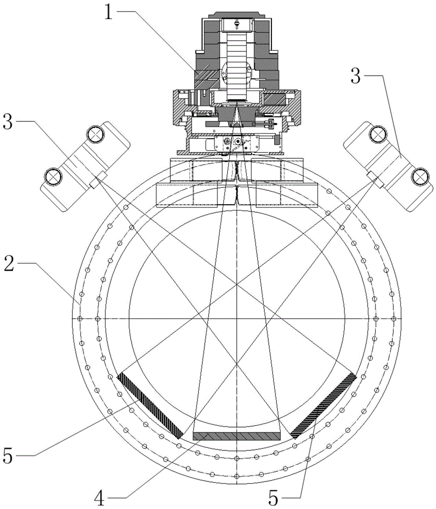
本发明具体实施方式提供一种肿瘤放射治疗影像引导设备,包括环形机架2、治疗头1、EPID图像采集系统平板接收器4、多个X射线成像系统3和多个X射线平板接收器5,其中治疗头1和多个X射线成像系统3安装于环形机架2外周侧面EPID图像采集系统平板接收器4和多个X射线平板接收器5安装于环形机架2内周侧面,EPID为电子射野影像系统。治疗位置位于环形机架2的环形截面的圆心位置,EPID图像采集系统平板接收器4与治疗头1相对设置于环形机架2的两侧,并用于接收治疗头1发出的射线,即EPID图像采集系统平板接收器4与治疗头1位于治疗位置的两侧,多个X射线平板接收器5分别与对应的X射线成像系统3相对设置于环形机架2的两侧,并用于接收X射线成像系统3发出的射线,即对应的X射线平板接收器5和X射线成像系统3位于治疗位置的两侧。The specific embodiment of the present invention provides an image guidance device for tumor radiation therapy, including a
具体地,EPID图像采集系统平板接收器4为MV级EPID图像采集系统平板接收器4,X射线成像系统3为kV级X射线成像系统3。Specifically, the flat panel receiver 4 of the EPID image acquisition system is a flat panel receiver 4 of an MV-level EPID image acquisition system, and the
治疗前,成角度排布的kV级X射线成像系统3,在等中心处投照到患者治疗位置,同时采集多个不同方向的图像,通过一定的算法得到治疗部位的三维图像,也可以采用旋转的方式进行采集,得到锥形束CT图像,与之前存储在计算机中的图像比对,从而得出肿瘤位置是否存在偏差;治疗过程中,多kV级X射线成像系统3实时采集肿瘤图像并进行比对,可以得到实时的肿瘤位置。治疗过程中,MV级EPID对照射剂量进行测量,既可以用作治疗后的剂量数据记录分析使用,也可以实现剂量引导的放射治疗。Before the treatment, the kV-level
集成了EPID图像采集成像和多个X射线成像,快速的完成2D/2D及3DCBCT图像引导,实现实时影像引导和剂量引导,兼顾了图像质量和采集时间,提高临床治疗效率,实现和后装治疗机的融合治疗,同时进行内照射和外照射,或各自单独进行。It integrates EPID image acquisition imaging and multiple X-ray imaging, quickly completes 2D/2D and 3DCBCT image guidance, realizes real-time image guidance and dose guidance, takes into account image quality and acquisition time, improves clinical treatment efficiency, and realizes and after-loading treatment. The fusion treatment of the machine, internal irradiation and external irradiation at the same time, or each separately.
为了保证成像位置的精确,EPID图像采集系统平板接收器4与治疗头1的连线穿过环形机架2的环形截面的圆心,X射线平板接收器5与对应的X射线成像系统3的连线穿过环形机架2的环形截面的圆心。或根据情况调整位置,均在本发明的保护范围之内。In order to ensure the accuracy of the imaging position, the connection line between the flat panel receiver 4 of the EPID image acquisition system and the
具体地,可以设置两个X射线成像系统3,两个X射线成像系统3对称设置于治疗头1两侧,并对应设置两个X射线平板接收器5。X射线成像系统3包括X射线球管和角度调节装置,X射线球管用于发射X射线,角度调节装置用于调节与治疗头1的夹角。MV级EPID正交于治疗头1的射束,双kV级X射线成像系统3以治疗头1射束的等中心为轴,以一定的角度对称主射束排布,X射线成像系统3靠近治疗头1或X射线成像系统3远离治疗头1。还可在X射线球管前端安装滤线栅,提高射线质量。Specifically, two
在本发明具体实施方式提供的肿瘤放射治疗影像引导设备中,治疗头1包括铅防护1-4、加速管1-1、初级准直锥1-2、二级准直锥1-5、多叶准直器1-3、移动档位托架1-6和档位切换驱动器1-6-3,加速管1-1竖直设置,用于将电子加速并且打靶输出高能射线,初级准直锥1-2安装于加速管1-1下端,将高能射束限定在一定的范围内,多叶准直器1-3位于初级准直锥1-2下方,对射束塑形,使得射束形状在患者平面与肿瘤形状基本一致,对肿瘤进行照射。移动档位托架1-6安装于初级准直锥1-2下方,并连接档位切换驱动器1-6-3,移动档位托架1-6上设置有多个射线档位,档位切换驱动器1-6-3驱动移动档位托架1-6移动,使不同的射线档位对准初级准直锥1-2,以切换治疗模式。In the image guidance device for tumor radiation therapy provided by the specific embodiment of the present invention, the
通过档位切换驱动器1-6-3驱动移动档位托架1-6移动,进而实现射线档位切换,使加速管1-1发出的射线经过不同的档位向下照射,即可形成不同类型的射线,对应不同的治疗模式,能够在有限空间内实现射野范围的限定与多种治疗模式的切换,使设备集成多种治疗模式,且档位切换方便快捷,提高设备适用性,可满足快捷、安全、精准的临床需求。The moving gear bracket 1-6 is driven to move by the gear switching driver 1-6-3, thereby realizing the switching of the ray gears, so that the rays emitted by the acceleration tube 1-1 are irradiated downward through different gears, and different gears can be formed. Types of rays, corresponding to different treatment modes, can realize the limitation of the field range and the switching of multiple treatment modes in a limited space, so that the device integrates multiple treatment modes, and the gear switching is convenient and fast, improving the applicability of the device, and can Meet fast, safe and accurate clinical needs.
移动档位托架1-6上设置有非均整档位1-6-1和均整档位1-6-2,两个档位沿水平方向并列设置,非均整档位1-6-1为竖直通孔,射束直接通过后,输出非均整的高剂量率模式射线,用于非均整治疗模式,均整档位1-6-2为竖直通孔内设置有均整器,射束会通过均整器处理,输出均整过的普通模式射线,用于均正治疗模式,提高大尺寸射野照射效果。可以增加更多数量的不同档位,提供不同类型的射束,进一步提高设备适用性。The moving gear brackets 1-6 are provided with a non-uniform gear position 1-6-1 and a uniform gear position 1-6-2, and the two gear positions are arranged side by side in the horizontal direction. The non-uniform gear position 1-6-1 is Vertical through-hole, after the beam passes directly, it outputs non-uniform high dose rate mode rays, which are used in non-uniform treatment mode. After being processed by the equalizer, the normal mode ray that has been equalized is output, which is used in the equalizing treatment mode to improve the irradiation effect of large-scale radiation fields. A greater number of different gears can be added to provide different types of beams to further improve the applicability of the equipment.
由于两个射线档位水平设置,因此档位切换驱动器1-6-3驱动移动档位托架1-6水平移动即可实现档位切换。移动档位托架1-6为水平托架,两个档位并列设置于水平托架的中部,档位切换驱动器1-6-3驱动可以为电机,安装于水平托架的一端,也可采用其他类型的驱动器等。Since the two ray gears are set horizontally, the gear switching driver 1-6-3 drives the moving gear bracket 1-6 to move horizontally to realize gear switching. The moving gear brackets 1-6 are horizontal brackets, and the two gears are arranged side by side in the middle of the horizontal bracket. Adopt other types of drives, etc.
多叶准直器1-3包括上层叶片1-3-1和下层叶片1-3-2,下层叶片1-3-2的数量多于上层叶片1-3-1的数量,每个上层叶片1-3-1及每个下层叶片1-3-2的侧面的前端边缘均设置有直角倒角。每个叶片侧面的前端边缘设置有直角倒角。每个叶片都可以在运动范围内的任意位置停留,多个叶片停留位置组合在一起,可以形成不同的孔,数量可以是一个也可以是多个,射束穿过这些孔,可以在患者平面形成与孔形状相同按固定比例变化的剂量分布。The multi-leaf collimator 1-3 includes upper-layer blades 1-3-1 and lower-layer blades 1-3-2, the number of lower-layer blades 1-3-2 is more than that of upper-layer blades 1-3-1, and each upper-layer blade 1-3-1 and the front edge of the side surface of each lower blade 1-3-2 are provided with right-angle chamfers. The front edge of each blade side is provided with a right angle chamfer. Each vane can stop at any position within the range of motion, and multiple vane stop positions can be combined to form different holes, either one or more in number, through which the beam passes and can be positioned at the patient plane A dose distribution that varies in fixed proportions with the same hole shape is formed.
治疗过程中,各个驱动器驱动叶片运动,形成与肿瘤形状匹配的照射孔,射线通过照射孔后投射在肿瘤上,通过设置多层结构,并在叶片侧面前端设置直角倒角,相较于现有的直角端面,更加有效贴近肿瘤边缘,从而实现更好的射野适形效果,从而提高射野适形分辨率、降低射线辐射泄漏、提高射野转换效率。During the treatment, each driver drives the movement of the blade to form an irradiation hole that matches the shape of the tumor. The rays pass through the irradiation hole and then project on the tumor. By setting a multi-layer structure, and setting a right-angle chamfer at the front end of the side of the blade, compared with the existing method The right-angle end face is more effectively close to the edge of the tumor, so as to achieve a better field conformation effect, thereby improving the field conformal resolution, reducing radiation leakage, and improving the field conversion efficiency.
在上述各具体实施方式提供的肿瘤放射治疗影像引导设备的基础上,还包括驱动环形机架2绕其自身转轴旋转的驱动装置,治疗头1、X射线成像系统3、EPID图像采集系统平板接收器4和X射线平板接收器5均与环形机架2同步转动,实现多角度照射、成像、治疗。On the basis of the image guidance device for tumor radiation therapy provided by the above specific embodiments, it also includes a driving device for driving the
以上对本发明所提供的肿瘤放射治疗影像引导设备进行了详细介绍。本文中应用了具体个例对本发明的原理及实施方式进行了阐述,以上实施例的说明只是用于帮助理解本发明的方法及其核心思想。应当指出,对于本技术领域的普通技术人员来说,在不脱离本发明原理的前提下,还可以对本发明进行若干改进和修饰,这些改进和修饰也落入本发明权利要求的保护范围内。The image guidance device for tumor radiation therapy provided by the present invention has been described in detail above. The principles and implementations of the present invention are described herein by using specific examples, and the descriptions of the above embodiments are only used to help understand the method and the core idea of the present invention. It should be pointed out that for those skilled in the art, without departing from the principle of the present invention, several improvements and modifications can also be made to the present invention, and these improvements and modifications also fall within the protection scope of the claims of the present invention.
Claims (10)
- The tumor radiotherapy image guiding device is characterized by comprising an annular rack (2), a treatment head (1) and a plurality of X-ray imaging systems (3), wherein the treatment head (1) is arranged on the outer peripheral side surface of the annular rack (2), an EPID image acquisition system flat receiver (4) and a plurality of X-ray flat receivers (5) are arranged on the inner peripheral side surface of the annular rack (2), the EPID image acquisition system flat receivers (4) and the treatment head (1) are arranged on two sides of the annular rack (2) relatively and used for receiving rays emitted by the treatment head (1), and the X-ray flat receivers (5) are arranged on two sides of the annular rack (2) relatively and used for receiving rays emitted by the X-ray imaging systems (3) respectively and correspondingly.
- 2. Tumor radiotherapy image guidance apparatus according to claim 1, characterized in that the connection line of the EPID image acquisition system flat receiver (4) and the treatment head (1) passes through the center of the circular cross section of the circular gantry (2), and the connection line of the X-ray flat receiver (5) and the corresponding X-ray imaging system (3) passes through the center of the circular cross section of the circular gantry (2).
- 3. Tumor radiotherapy image guidance device according to claim 2, characterized by comprising two X-ray imaging systems (3), wherein the two X-ray imaging systems (3) are symmetrically arranged on both sides of the treatment head (1), and further comprising two X-ray flat receivers (5) correspondingly arranged.
- 4. Tumor radiotherapy image guidance device according to claim 3, characterized in that the X-ray imaging system (3) comprises an X-ray bulb and an angle adjustment device for adjusting the angle with the treatment head (1).
- 5. A tumor radiation therapy image guidance apparatus according to claim 4, characterized in that a grid is mounted at the front end of the X-ray tube.
- 6. Tumor radiotherapy image guidance device according to claim 1, characterized in that the EPID image acquisition system flat receiver (4) is in particular a MV-class EPID image acquisition system flat receiver (4) and the X-ray imaging system (3) is in particular a kV-class X-ray imaging system (3).
- 7. Tumor radiotherapy image guidance device according to claim 1, characterized in that the treatment head (1) comprises a vertically disposed accelerating tube (1-1), a primary collimation cone (1-2) mounted at the lower end of the accelerating tube (1-1), and a multi-leaf collimator (1-3) located below the primary collimation cone (1-2), a moving gear bracket (1-6) and a gear switching driver (1-6-3) are mounted below the primary collimation cone (1-2), a plurality of radiation gears are disposed on the moving gear bracket (1-6), the gear switching driver (1-6-3) can drive the moving gear bracket (1-6) to move to align different radiation gears with the primary collimation cone (1-2), to switch the treatment mode.
- 8. The tumor radiotherapy image guiding apparatus according to claim 7, wherein the moving gear bracket (1-6) is provided with a non-uniform gear (1-6-1) and a uniform gear (1-6-2) in parallel along a horizontal direction, the non-uniform gear (1-6-1) is a vertical through hole for outputting non-uniform high dose rate mode rays, the uniform gear (1-6-2) is a vertical through hole provided with a homogenizer for outputting uniform normal mode rays, and the gear switching driver (1-6-3) can drive the moving gear bracket (1-6) to move horizontally.
- 9. Tumor radiotherapy image guidance device according to claim 8, characterized in that the multi-leaf collimator (1-3) comprises an upper leaf (1-3-1) and a lower leaf (1-3-2), the number of the lower leaf (1-3-2) is greater than the number of the upper leaf (1-3-1), and the front end edge of the side face of each upper leaf (1-3-1) and each lower leaf (1-3-2) is provided with a right angle chamfer.
- 10. Tumor radiotherapy image guidance apparatus according to any of claims 1-9, further comprising a driving device for driving the ring gantry (2) to rotate around its own rotation axis, wherein the treatment head (1), the X-ray imaging system (3), the EPID image acquisition system flat receiver (4) and the X-ray flat receiver (5) all rotate synchronously with the ring gantry (2).
Priority Applications (1)
| Application Number | Priority Date | Filing Date | Title |
|---|---|---|---|
| CN201911121650.0A CN110732094A (en) | 2019-11-15 | 2019-11-15 | tumor radiotherapy image guide equipment |
Applications Claiming Priority (1)
| Application Number | Priority Date | Filing Date | Title |
|---|---|---|---|
| CN201911121650.0A CN110732094A (en) | 2019-11-15 | 2019-11-15 | tumor radiotherapy image guide equipment |
Publications (1)
| Publication Number | Publication Date |
|---|---|
| CN110732094A true CN110732094A (en) | 2020-01-31 |
Family
ID=69273088
Family Applications (1)
| Application Number | Title | Priority Date | Filing Date |
|---|---|---|---|
| CN201911121650.0A Pending CN110732094A (en) | 2019-11-15 | 2019-11-15 | tumor radiotherapy image guide equipment |
Country Status (1)
| Country | Link |
|---|---|
| CN (1) | CN110732094A (en) |
Cited By (4)
| Publication number | Priority date | Publication date | Assignee | Title |
|---|---|---|---|---|
| CN114569898A (en) * | 2022-02-18 | 2022-06-03 | 新里程医疗技术(深圳)有限责任公司 | Image-guided real-time positioning system for particle accelerator |
| WO2022170600A1 (en) * | 2021-02-10 | 2022-08-18 | 西安大医集团股份有限公司 | Accelerating tube shielding cylinder, accelerator treatment head, and radiotherapy apparatus |
| CN116650851A (en) * | 2023-06-21 | 2023-08-29 | 北京华科先锋医疗器械有限公司 | A radiotherapy system for atrial fibrillation based on a robotic arm |
| CN117398116A (en) * | 2023-10-24 | 2024-01-16 | 山东新华医疗器械股份有限公司 | Kilovoltage image acquisition device and image acquisition method at any angle |
Citations (17)
| Publication number | Priority date | Publication date | Assignee | Title |
|---|---|---|---|---|
| CN201223647Y (en) * | 2008-07-01 | 2009-04-22 | 山东新华医疗器械股份有限公司 | Two-photon accelerator treatment head |
| CN102068764A (en) * | 2010-10-29 | 2011-05-25 | 夏廷毅 | Treatment and verification system for guiding gamma knife by images |
| US20120008734A1 (en) * | 2010-06-08 | 2012-01-12 | Accuray, Inc. | Target Tracking for Image-Guided Radiation Treatment |
| CN202136702U (en) * | 2011-06-23 | 2012-02-08 | 山东新华医疗器械股份有限公司 | Homologous dual-energy IGRT (image guided radiation therapy) medical electronic linear accelerator |
| CN103071241A (en) * | 2011-10-25 | 2013-05-01 | 苏州雷泰医疗科技有限公司 | Stereotactic radiotherapeutic device |
| CN203634660U (en) * | 2013-07-15 | 2014-06-11 | 上海联影医疗科技有限公司 | Radiotherapy equipment |
| CN104605882A (en) * | 2015-01-23 | 2015-05-13 | 上海联影医疗科技有限公司 | Image obtaining method and device in radiotherapy system and radiotherapy system |
| CN104795121A (en) * | 2014-01-22 | 2015-07-22 | 上海联影医疗科技有限公司 | Split type primary collimator |
| CN107358607A (en) * | 2017-08-13 | 2017-11-17 | 强深智能医疗科技(昆山)有限公司 | Tumour radiotherapy visual monitoring and visual servo intelligent control method |
| CN107929955A (en) * | 2017-11-24 | 2018-04-20 | 沈阳东软医疗系统有限公司 | Radiotherapy apparatus, multi-leaf optical grating and its blade construction |
| CN108744310A (en) * | 2018-06-14 | 2018-11-06 | 中科超精(安徽)科技有限公司 | Multi-mode guides adaptive radiotherapy system |
| CN109224320A (en) * | 2018-10-23 | 2019-01-18 | 四川大学华西医院 | Accelerator non-coplanar radiotherapy device based on composite dual-rotating rack |
| CN208678191U (en) * | 2018-04-09 | 2019-04-02 | 西安大医数码科技有限公司 | Radiotherapy head and radiotherapy unit |
| US20190126070A1 (en) * | 2016-04-11 | 2019-05-02 | The Regents Of The University Of California | Real-Time, Parallel X-Ray Tomosynthesis |
| CN209137785U (en) * | 2018-06-08 | 2019-07-23 | 西安大医数码科技有限公司 | Radiotherapy head and radiotherapy unit |
| CN211097111U (en) * | 2019-11-15 | 2020-07-28 | 山东新华医疗器械股份有限公司 | An image-guided device for tumor radiation therapy |
| CN113195051A (en) * | 2018-07-28 | 2021-07-30 | 瓦里安医疗系统公司 | Radiation therapy system with multiple X-ray imagers for near real time positioning |
-
2019
- 2019-11-15 CN CN201911121650.0A patent/CN110732094A/en active Pending
Patent Citations (17)
| Publication number | Priority date | Publication date | Assignee | Title |
|---|---|---|---|---|
| CN201223647Y (en) * | 2008-07-01 | 2009-04-22 | 山东新华医疗器械股份有限公司 | Two-photon accelerator treatment head |
| US20120008734A1 (en) * | 2010-06-08 | 2012-01-12 | Accuray, Inc. | Target Tracking for Image-Guided Radiation Treatment |
| CN102068764A (en) * | 2010-10-29 | 2011-05-25 | 夏廷毅 | Treatment and verification system for guiding gamma knife by images |
| CN202136702U (en) * | 2011-06-23 | 2012-02-08 | 山东新华医疗器械股份有限公司 | Homologous dual-energy IGRT (image guided radiation therapy) medical electronic linear accelerator |
| CN103071241A (en) * | 2011-10-25 | 2013-05-01 | 苏州雷泰医疗科技有限公司 | Stereotactic radiotherapeutic device |
| CN203634660U (en) * | 2013-07-15 | 2014-06-11 | 上海联影医疗科技有限公司 | Radiotherapy equipment |
| CN104795121A (en) * | 2014-01-22 | 2015-07-22 | 上海联影医疗科技有限公司 | Split type primary collimator |
| CN104605882A (en) * | 2015-01-23 | 2015-05-13 | 上海联影医疗科技有限公司 | Image obtaining method and device in radiotherapy system and radiotherapy system |
| US20190126070A1 (en) * | 2016-04-11 | 2019-05-02 | The Regents Of The University Of California | Real-Time, Parallel X-Ray Tomosynthesis |
| CN107358607A (en) * | 2017-08-13 | 2017-11-17 | 强深智能医疗科技(昆山)有限公司 | Tumour radiotherapy visual monitoring and visual servo intelligent control method |
| CN107929955A (en) * | 2017-11-24 | 2018-04-20 | 沈阳东软医疗系统有限公司 | Radiotherapy apparatus, multi-leaf optical grating and its blade construction |
| CN208678191U (en) * | 2018-04-09 | 2019-04-02 | 西安大医数码科技有限公司 | Radiotherapy head and radiotherapy unit |
| CN209137785U (en) * | 2018-06-08 | 2019-07-23 | 西安大医数码科技有限公司 | Radiotherapy head and radiotherapy unit |
| CN108744310A (en) * | 2018-06-14 | 2018-11-06 | 中科超精(安徽)科技有限公司 | Multi-mode guides adaptive radiotherapy system |
| CN113195051A (en) * | 2018-07-28 | 2021-07-30 | 瓦里安医疗系统公司 | Radiation therapy system with multiple X-ray imagers for near real time positioning |
| CN109224320A (en) * | 2018-10-23 | 2019-01-18 | 四川大学华西医院 | Accelerator non-coplanar radiotherapy device based on composite dual-rotating rack |
| CN211097111U (en) * | 2019-11-15 | 2020-07-28 | 山东新华医疗器械股份有限公司 | An image-guided device for tumor radiation therapy |
Cited By (4)
| Publication number | Priority date | Publication date | Assignee | Title |
|---|---|---|---|---|
| WO2022170600A1 (en) * | 2021-02-10 | 2022-08-18 | 西安大医集团股份有限公司 | Accelerating tube shielding cylinder, accelerator treatment head, and radiotherapy apparatus |
| CN114569898A (en) * | 2022-02-18 | 2022-06-03 | 新里程医疗技术(深圳)有限责任公司 | Image-guided real-time positioning system for particle accelerator |
| CN116650851A (en) * | 2023-06-21 | 2023-08-29 | 北京华科先锋医疗器械有限公司 | A radiotherapy system for atrial fibrillation based on a robotic arm |
| CN117398116A (en) * | 2023-10-24 | 2024-01-16 | 山东新华医疗器械股份有限公司 | Kilovoltage image acquisition device and image acquisition method at any angle |
Similar Documents
| Publication | Publication Date | Title |
|---|---|---|
| US11759654B2 (en) | Radiotherapy device and control driving method thereof | |
| JP6177245B2 (en) | Hadron radiation equipment and verification method | |
| CN110732094A (en) | tumor radiotherapy image guide equipment | |
| CN103977506B (en) | One kind of proton tomography method and device | |
| CN107569780B (en) | Cancer treatment room fiducial marker system and method of use | |
| CN105764421A (en) | Radiation medical device | |
| CN1889995A (en) | Multiple room radiation treatment system | |
| CN105636331B (en) | Electron linear accelerator | |
| CN108379748B (en) | Radiotherapy head and radiotherapy unit | |
| US11452886B2 (en) | Radiotherapy equipment | |
| US11033756B2 (en) | Portal imaging during radiotherapy | |
| US9999787B1 (en) | Beam limiting device for intensity modulated proton therapy | |
| CN106139421B (en) | A kind of Proton therapy system of double fixed chamber two-beams irradiation of dislocation arrangement | |
| US7587024B2 (en) | Particle beam irradiation system | |
| CN211097111U (en) | An image-guided device for tumor radiation therapy | |
| US20200206537A1 (en) | Radiation treatment device | |
| WO2023184420A1 (en) | Radiotherapy head, apparatus, method, control device and non-volatile storage medium | |
| JP2004065808A (en) | Radiotherapeutic system | |
| WO2019008793A1 (en) | Particle beam irradiation apparatus | |
| JP2003024459A (en) | Radiotherapy equipment | |
| CN209075883U (en) | Focus head, collimator and gamma knife | |
| JP2013013613A (en) | Charged particle beam irradiation device | |
| CN115518306A (en) | Positively Charged Particle Cancer Therapy Beam State Determination System and Method of Use | |
| CN208756809U (en) | Radiotherapy unit | |
| US20140169519A1 (en) | Cone-beam CT Scanning |
Legal Events
| Date | Code | Title | Description |
|---|---|---|---|
| PB01 | Publication | ||
| PB01 | Publication | ||
| SE01 | Entry into force of request for substantive examination | ||
| SE01 | Entry into force of request for substantive examination | ||
| RJ01 | Rejection of invention patent application after publication |
Application publication date: 20200131 |
|
| RJ01 | Rejection of invention patent application after publication |