CN110684733A - Midbrain Dopamine (DA) neurons for implantation - Google Patents
Midbrain Dopamine (DA) neurons for implantation Download PDFInfo
- Publication number
- CN110684733A CN110684733A CN201910438266.7A CN201910438266A CN110684733A CN 110684733 A CN110684733 A CN 110684733A CN 201910438266 A CN201910438266 A CN 201910438266A CN 110684733 A CN110684733 A CN 110684733A
- Authority
- CN
- China
- Prior art keywords
- cells
- neurons
- cell
- midbrain
- inhibitor
- Prior art date
- Legal status (The legal status is an assumption and is not a legal conclusion. Google has not performed a legal analysis and makes no representation as to the accuracy of the status listed.)
- Pending
Links
Images


Classifications
-
- C—CHEMISTRY; METALLURGY
- C12—BIOCHEMISTRY; BEER; SPIRITS; WINE; VINEGAR; MICROBIOLOGY; ENZYMOLOGY; MUTATION OR GENETIC ENGINEERING
- C12N—MICROORGANISMS OR ENZYMES; COMPOSITIONS THEREOF; PROPAGATING, PRESERVING, OR MAINTAINING MICROORGANISMS; MUTATION OR GENETIC ENGINEERING; CULTURE MEDIA
- C12N5/00—Undifferentiated human, animal or plant cells, e.g. cell lines; Tissues; Cultivation or maintenance thereof; Culture media therefor
- C12N5/06—Animal cells or tissues; Human cells or tissues
- C12N5/0602—Vertebrate cells
- C12N5/0618—Cells of the nervous system
- C12N5/0619—Neurons
-
- A—HUMAN NECESSITIES
- A61—MEDICAL OR VETERINARY SCIENCE; HYGIENE
- A61K—PREPARATIONS FOR MEDICAL, DENTAL OR TOILETRY PURPOSES
- A61K35/00—Medicinal preparations containing materials or reaction products thereof with undetermined constitution
- A61K35/12—Materials from mammals; Compositions comprising non-specified tissues or cells; Compositions comprising non-embryonic stem cells; Genetically modified cells
- A61K35/30—Nerves; Brain; Eyes; Corneal cells; Cerebrospinal fluid; Neuronal stem cells; Neuronal precursor cells; Glial cells; Oligodendrocytes; Schwann cells; Astroglia; Astrocytes; Choroid plexus; Spinal cord tissue
-
- A—HUMAN NECESSITIES
- A61—MEDICAL OR VETERINARY SCIENCE; HYGIENE
- A61P—SPECIFIC THERAPEUTIC ACTIVITY OF CHEMICAL COMPOUNDS OR MEDICINAL PREPARATIONS
- A61P25/00—Drugs for disorders of the nervous system
-
- A—HUMAN NECESSITIES
- A61—MEDICAL OR VETERINARY SCIENCE; HYGIENE
- A61P—SPECIFIC THERAPEUTIC ACTIVITY OF CHEMICAL COMPOUNDS OR MEDICINAL PREPARATIONS
- A61P25/00—Drugs for disorders of the nervous system
- A61P25/14—Drugs for disorders of the nervous system for treating abnormal movements, e.g. chorea, dyskinesia
- A61P25/16—Anti-Parkinson drugs
-
- A—HUMAN NECESSITIES
- A61—MEDICAL OR VETERINARY SCIENCE; HYGIENE
- A61P—SPECIFIC THERAPEUTIC ACTIVITY OF CHEMICAL COMPOUNDS OR MEDICINAL PREPARATIONS
- A61P25/00—Drugs for disorders of the nervous system
- A61P25/28—Drugs for disorders of the nervous system for treating neurodegenerative disorders of the central nervous system, e.g. nootropic agents, cognition enhancers, drugs for treating Alzheimer's disease or other forms of dementia
-
- A—HUMAN NECESSITIES
- A61—MEDICAL OR VETERINARY SCIENCE; HYGIENE
- A61P—SPECIFIC THERAPEUTIC ACTIVITY OF CHEMICAL COMPOUNDS OR MEDICINAL PREPARATIONS
- A61P43/00—Drugs for specific purposes, not provided for in groups A61P1/00-A61P41/00
-
- C—CHEMISTRY; METALLURGY
- C12—BIOCHEMISTRY; BEER; SPIRITS; WINE; VINEGAR; MICROBIOLOGY; ENZYMOLOGY; MUTATION OR GENETIC ENGINEERING
- C12N—MICROORGANISMS OR ENZYMES; COMPOSITIONS THEREOF; PROPAGATING, PRESERVING, OR MAINTAINING MICROORGANISMS; MUTATION OR GENETIC ENGINEERING; CULTURE MEDIA
- C12N5/00—Undifferentiated human, animal or plant cells, e.g. cell lines; Tissues; Cultivation or maintenance thereof; Culture media therefor
- C12N5/06—Animal cells or tissues; Human cells or tissues
- C12N5/0602—Vertebrate cells
- C12N5/0618—Cells of the nervous system
- C12N5/0623—Stem cells
-
- C—CHEMISTRY; METALLURGY
- C12—BIOCHEMISTRY; BEER; SPIRITS; WINE; VINEGAR; MICROBIOLOGY; ENZYMOLOGY; MUTATION OR GENETIC ENGINEERING
- C12N—MICROORGANISMS OR ENZYMES; COMPOSITIONS THEREOF; PROPAGATING, PRESERVING, OR MAINTAINING MICROORGANISMS; MUTATION OR GENETIC ENGINEERING; CULTURE MEDIA
- C12N2501/00—Active agents used in cell culture processes, e.g. differentation
- C12N2501/01—Modulators of cAMP or cGMP, e.g. non-hydrolysable analogs, phosphodiesterase inhibitors, cholera toxin
-
- C—CHEMISTRY; METALLURGY
- C12—BIOCHEMISTRY; BEER; SPIRITS; WINE; VINEGAR; MICROBIOLOGY; ENZYMOLOGY; MUTATION OR GENETIC ENGINEERING
- C12N—MICROORGANISMS OR ENZYMES; COMPOSITIONS THEREOF; PROPAGATING, PRESERVING, OR MAINTAINING MICROORGANISMS; MUTATION OR GENETIC ENGINEERING; CULTURE MEDIA
- C12N2501/00—Active agents used in cell culture processes, e.g. differentation
- C12N2501/10—Growth factors
- C12N2501/119—Other fibroblast growth factors, e.g. FGF-4, FGF-8, FGF-10
-
- C—CHEMISTRY; METALLURGY
- C12—BIOCHEMISTRY; BEER; SPIRITS; WINE; VINEGAR; MICROBIOLOGY; ENZYMOLOGY; MUTATION OR GENETIC ENGINEERING
- C12N—MICROORGANISMS OR ENZYMES; COMPOSITIONS THEREOF; PROPAGATING, PRESERVING, OR MAINTAINING MICROORGANISMS; MUTATION OR GENETIC ENGINEERING; CULTURE MEDIA
- C12N2501/00—Active agents used in cell culture processes, e.g. differentation
- C12N2501/10—Growth factors
- C12N2501/13—Nerve growth factor [NGF]; Brain-derived neurotrophic factor [BDNF]; Cilliary neurotrophic factor [CNTF]; Glial-derived neurotrophic factor [GDNF]; Neurotrophins [NT]; Neuregulins
-
- C—CHEMISTRY; METALLURGY
- C12—BIOCHEMISTRY; BEER; SPIRITS; WINE; VINEGAR; MICROBIOLOGY; ENZYMOLOGY; MUTATION OR GENETIC ENGINEERING
- C12N—MICROORGANISMS OR ENZYMES; COMPOSITIONS THEREOF; PROPAGATING, PRESERVING, OR MAINTAINING MICROORGANISMS; MUTATION OR GENETIC ENGINEERING; CULTURE MEDIA
- C12N2501/00—Active agents used in cell culture processes, e.g. differentation
- C12N2501/10—Growth factors
- C12N2501/15—Transforming growth factor beta (TGF-β)
-
- C—CHEMISTRY; METALLURGY
- C12—BIOCHEMISTRY; BEER; SPIRITS; WINE; VINEGAR; MICROBIOLOGY; ENZYMOLOGY; MUTATION OR GENETIC ENGINEERING
- C12N—MICROORGANISMS OR ENZYMES; COMPOSITIONS THEREOF; PROPAGATING, PRESERVING, OR MAINTAINING MICROORGANISMS; MUTATION OR GENETIC ENGINEERING; CULTURE MEDIA
- C12N2501/00—Active agents used in cell culture processes, e.g. differentation
- C12N2501/10—Growth factors
- C12N2501/155—Bone morphogenic proteins [BMP]; Osteogenins; Osteogenic factor; Bone inducing factor
-
- C—CHEMISTRY; METALLURGY
- C12—BIOCHEMISTRY; BEER; SPIRITS; WINE; VINEGAR; MICROBIOLOGY; ENZYMOLOGY; MUTATION OR GENETIC ENGINEERING
- C12N—MICROORGANISMS OR ENZYMES; COMPOSITIONS THEREOF; PROPAGATING, PRESERVING, OR MAINTAINING MICROORGANISMS; MUTATION OR GENETIC ENGINEERING; CULTURE MEDIA
- C12N2501/00—Active agents used in cell culture processes, e.g. differentation
- C12N2501/10—Growth factors
- C12N2501/16—Activin; Inhibin; Mullerian inhibiting substance
-
- C—CHEMISTRY; METALLURGY
- C12—BIOCHEMISTRY; BEER; SPIRITS; WINE; VINEGAR; MICROBIOLOGY; ENZYMOLOGY; MUTATION OR GENETIC ENGINEERING
- C12N—MICROORGANISMS OR ENZYMES; COMPOSITIONS THEREOF; PROPAGATING, PRESERVING, OR MAINTAINING MICROORGANISMS; MUTATION OR GENETIC ENGINEERING; CULTURE MEDIA
- C12N2501/00—Active agents used in cell culture processes, e.g. differentation
- C12N2501/40—Regulators of development
- C12N2501/41—Hedgehog proteins; Cyclopamine (inhibitor)
-
- C—CHEMISTRY; METALLURGY
- C12—BIOCHEMISTRY; BEER; SPIRITS; WINE; VINEGAR; MICROBIOLOGY; ENZYMOLOGY; MUTATION OR GENETIC ENGINEERING
- C12N—MICROORGANISMS OR ENZYMES; COMPOSITIONS THEREOF; PROPAGATING, PRESERVING, OR MAINTAINING MICROORGANISMS; MUTATION OR GENETIC ENGINEERING; CULTURE MEDIA
- C12N2501/00—Active agents used in cell culture processes, e.g. differentation
- C12N2501/40—Regulators of development
- C12N2501/415—Wnt; Frizzeled
-
- C—CHEMISTRY; METALLURGY
- C12—BIOCHEMISTRY; BEER; SPIRITS; WINE; VINEGAR; MICROBIOLOGY; ENZYMOLOGY; MUTATION OR GENETIC ENGINEERING
- C12N—MICROORGANISMS OR ENZYMES; COMPOSITIONS THEREOF; PROPAGATING, PRESERVING, OR MAINTAINING MICROORGANISMS; MUTATION OR GENETIC ENGINEERING; CULTURE MEDIA
- C12N2501/00—Active agents used in cell culture processes, e.g. differentation
- C12N2501/70—Enzymes
- C12N2501/72—Transferases [EC 2.]
- C12N2501/727—Kinases (EC 2.7.)
-
- C—CHEMISTRY; METALLURGY
- C12—BIOCHEMISTRY; BEER; SPIRITS; WINE; VINEGAR; MICROBIOLOGY; ENZYMOLOGY; MUTATION OR GENETIC ENGINEERING
- C12N—MICROORGANISMS OR ENZYMES; COMPOSITIONS THEREOF; PROPAGATING, PRESERVING, OR MAINTAINING MICROORGANISMS; MUTATION OR GENETIC ENGINEERING; CULTURE MEDIA
- C12N2501/00—Active agents used in cell culture processes, e.g. differentation
- C12N2501/999—Small molecules not provided for elsewhere
-
- C—CHEMISTRY; METALLURGY
- C12—BIOCHEMISTRY; BEER; SPIRITS; WINE; VINEGAR; MICROBIOLOGY; ENZYMOLOGY; MUTATION OR GENETIC ENGINEERING
- C12N—MICROORGANISMS OR ENZYMES; COMPOSITIONS THEREOF; PROPAGATING, PRESERVING, OR MAINTAINING MICROORGANISMS; MUTATION OR GENETIC ENGINEERING; CULTURE MEDIA
- C12N2506/00—Differentiation of animal cells from one lineage to another; Differentiation of pluripotent cells
- C12N2506/02—Differentiation of animal cells from one lineage to another; Differentiation of pluripotent cells from embryonic cells
-
- C—CHEMISTRY; METALLURGY
- C12—BIOCHEMISTRY; BEER; SPIRITS; WINE; VINEGAR; MICROBIOLOGY; ENZYMOLOGY; MUTATION OR GENETIC ENGINEERING
- C12N—MICROORGANISMS OR ENZYMES; COMPOSITIONS THEREOF; PROPAGATING, PRESERVING, OR MAINTAINING MICROORGANISMS; MUTATION OR GENETIC ENGINEERING; CULTURE MEDIA
- C12N2506/00—Differentiation of animal cells from one lineage to another; Differentiation of pluripotent cells
- C12N2506/45—Differentiation of animal cells from one lineage to another; Differentiation of pluripotent cells from artificially induced pluripotent stem cells
Landscapes
- Health & Medical Sciences (AREA)
- Engineering & Computer Science (AREA)
- Biomedical Technology (AREA)
- Life Sciences & Earth Sciences (AREA)
- Neurology (AREA)
- Bioinformatics & Cheminformatics (AREA)
- Chemical & Material Sciences (AREA)
- Neurosurgery (AREA)
- Organic Chemistry (AREA)
- General Health & Medical Sciences (AREA)
- Zoology (AREA)
- Biotechnology (AREA)
- Medicinal Chemistry (AREA)
- Veterinary Medicine (AREA)
- Public Health (AREA)
- Animal Behavior & Ethology (AREA)
- Pharmacology & Pharmacy (AREA)
- Cell Biology (AREA)
- Genetics & Genomics (AREA)
- Wood Science & Technology (AREA)
- Nuclear Medicine, Radiotherapy & Molecular Imaging (AREA)
- General Chemical & Material Sciences (AREA)
- Chemical Kinetics & Catalysis (AREA)
- Developmental Biology & Embryology (AREA)
- Biochemistry (AREA)
- General Engineering & Computer Science (AREA)
- Microbiology (AREA)
- Psychology (AREA)
- Immunology (AREA)
- Ophthalmology & Optometry (AREA)
- Virology (AREA)
- Epidemiology (AREA)
- Psychiatry (AREA)
- Hospice & Palliative Care (AREA)
- Medicines Containing Material From Animals Or Micro-Organisms (AREA)
- Micro-Organisms Or Cultivation Processes Thereof (AREA)
- Macromolecular Compounds Obtained By Forming Nitrogen-Containing Linkages In General (AREA)
- Medicines That Contain Protein Lipid Enzymes And Other Medicines (AREA)
- Nitrogen Condensed Heterocyclic Rings (AREA)
- Materials For Medical Uses (AREA)
Abstract
本发明涉及干细胞生物学领域,具体地,涉及多能或多潜能干细胞的谱系特异性分化,上述多能或多潜能干细胞可以包括,但不限于,除了非胚胎人诱导的多能干细胞(hiPSC)以外还有的人胚胎干细胞(hESC)、成体干细胞、来自患有疾病的患者的干细胞或者任何能够进行谱系特异性分化的其它干细胞。具体描述了以下方法:引导hESC和/或hiPSC谱系特异性分化成底板中脑祖细胞,然后使用新的培养条件进一步分化成大量的中脑结局FOXA2+LMX1A+TH+多巴胺(DA)神经元。使用本发明的方法产生的中脑结局FOXA2+LMX1A+TH+多巴胺(DA)神经元进一步预期用于多种用途,其包括,但不限于,在体外药物发现检测中的用途、神经学研究、以及作为患者多巴胺神经元的疾病逆转或损伤或缺乏的治疗剂。
The present invention relates to the field of stem cell biology, in particular, to the lineage-specific differentiation of pluripotent or pluripotent stem cells, which may include, but are not limited to, in addition to non-embryonic human induced pluripotent stem cells (hiPSC) In addition to human embryonic stem cells (hESC), adult stem cells, stem cells from a patient with a disease or any other stem cell capable of lineage-specific differentiation. Specifically, the following method is described: lineage-specific differentiation of hESCs and/or hiPSCs into floor plate midbrain progenitors, which are then further differentiated into large numbers of midbrain fate FOXA2+LMX1A+TH+dopamine (DA) neurons using novel culture conditions. The midbrain outcome FOXA2+LMX1A+TH+dopamine (DA) neurons generated using the methods of the present invention are further contemplated for a variety of uses including, but not limited to, use in in vitro drug discovery assays, neurological research, and As a therapeutic agent for disease reversal or damage or deficiency of dopamine neurons in patients.
Description
本申请是申请日为2012年12月2日、申请号为201280066145.0、发明名称为“用于植入的中脑多巴胺(DA)神经元”的专利申请的分案申请。This application is a divisional application of a patent application with an application date of December 2, 2012, an application number of 201280066145.0, and an invention title of "midbrain dopamine (DA) neurons for implantation".
政府许可权利Government Licensing Rights
本发明部分地在来自美国国立卫生研究所(United States National Instituteof Health,NIH)和国立神经病学和中风研究所(National Institute of Neurologicaldisorders and Stroke,NINDS)的NIH/NINDS 资助金NS052671和NIH/NINDS资助金P50NS047085政府资助下作出。因此,美国政府对于本发明具有某些权利。This invention was made in part under NIH/NINDS grants NS052671 and NIH/NINDS grants from the United States National Institute of Health (NIH) and the National Institute of Neurological disorders and Stroke (NINDS) Gold P50NS047085 made with government funding. Accordingly, the United States Government has certain rights in this invention.
发明领域Field of Invention
本发明涉及干细胞生物学领域,具体地,涉及多能或多潜能干细胞的谱系特异性分化,上述多能或多潜能干细胞可以包括,但不限于,除了非胚胎人诱导的多能干细胞(hiPSC)以外还有的人胚胎干细胞 (hESC)、成体干细胞、来自患有疾病的患者的干细胞或者任何能够进行谱系特异性分化的其它干细胞。具体描述了以下方法:引导hESC 和/或hiPSC谱系特异性分化成底板中脑祖细胞,然后使用新的培养条件进一步分化成大量的中脑结局FOXA2+LMX1A+TH+多巴胺(DA) 神经元。使用本发明的方法产生的中脑结局FOXA2+LMX1A+TH+多巴胺(DA)神经元进一步预期用于多种用途,其包括,但不限于,在体外药物发现检测中的用途、神经学研究、以及作为患者多巴胺神经元的疾病逆转或损伤或缺乏的治疗剂。而且,提供了从人多能干细胞分化中脑结局FOXA2+LMX1A+TH+多巴胺(DA)神经元用于疾病建模特别是帕金森氏病的组合物和方法。此外,真正的DA神经元富含标记物,例如CD142和A9型神经元细胞。The present invention relates to the field of stem cell biology, in particular, to the lineage-specific differentiation of pluripotent or pluripotent stem cells, which may include, but are not limited to, in addition to non-embryonic human induced pluripotent stem cells (hiPSC) In addition to human embryonic stem cells (hESC), adult stem cells, stem cells from a patient with a disease or any other stem cell capable of lineage-specific differentiation. Specifically described are methods for directing lineage-specific differentiation of hESC and/or hiPSCs into floor plate midbrain progenitors, which are then further differentiated into large numbers of midbrain fate FOXA2+LMX1A+TH+dopamine (DA) neurons using novel culture conditions. The midbrain outcome FOXA2+LMX1A+TH+dopamine (DA) neurons generated using the methods of the present invention are further contemplated for a variety of uses including, but not limited to, use in in vitro drug discovery assays, neurological research, and As a therapeutic agent for disease reversal or damage or deficiency of dopamine neurons in patients. Furthermore, compositions and methods are provided for differentiating midbrain outcome FOXA2+LMX1A+TH+dopamine (DA) neurons from human pluripotent stem cells for disease modeling, particularly Parkinson's disease. Furthermore, bona fide DA neurons are enriched for markers such as CD142 and A9-type neuronal cells.
背景技术Background technique
保留分化成许多种特化细胞类型的能力的细胞群体可用于形成大量的谱系特异性分化细胞群体。这些保留进一步分化成特化细胞的能力的细胞群体含有多能细胞。多能细胞可以来源自胚胎和/或非胚胎成体干细胞。Populations of cells that retain the ability to differentiate into many specialized cell types can be used to form large populations of lineage-specific differentiated cells. These cell populations that retain the ability to further differentiate into specialized cells contain pluripotent cells. Pluripotent cells can be derived from embryonic and/or non-embryonic adult stem cells.
这些谱系特异性分化的细胞群体预期应用于具有导致特定细胞群体功能丧失的疾病的患者的细胞置换疗法中。除其直接治疗价值以外,谱系特异性分化细胞还是许多种用途的有价值的研究工具,其包括筛选检测,以鉴定、确认和测试功能特异性,或用于测试治疗分子的输送,以治疗细胞谱系特异性疾病。These lineage-specifically differentiated cell populations are expected to be used in cell replacement therapy for patients with diseases that result in the loss of function of a particular cell population. In addition to their direct therapeutic value, lineage-specific differentiated cells are valuable research tools for a variety of purposes, including screening assays to identify, confirm and test functional specificity, or for testing delivery of therapeutic molecules to treat cells Lineage-specific diseases.
之前使用胚胎干细胞和成体干细胞作为神经退行性疾病的治疗剂和模型系统。有关胚胎干细胞和成体干细胞定向分化的研究和技术发展已经在中枢神经系统(CNS)疾病领域中进行,例如,用于亨廷顿氏舞蹈病、阿尔茨海默氏病、帕金森氏病和多发性硬化。但是,这些研究的结果表明,这些细胞用于体内使患者恢复神经功能的能力几乎没有,而且常常导致患者中不期望的肿瘤生长。Embryonic and adult stem cells have previously been used as therapeutics and model systems for neurodegenerative diseases. Research and technical development on the directed differentiation of embryonic and adult stem cells has been carried out in the field of central nervous system (CNS) diseases, for example, for Huntington's disease, Alzheimer's disease, Parkinson's disease and multiple sclerosis . However, the results of these studies suggest that these cells have little in vivo ability to restore neurological function in patients, and often lead to undesired tumor growth in patients.
因此,对于获得能够既用于研究且用作治疗导致具有特定功能的细胞丧失的疾病的治疗剂的细胞群体的组合物和方法存在有需求。Accordingly, there is a need for compositions and methods for obtaining cell populations that can be used both for research and as therapeutic agents for the treatment of diseases that result in loss of cells with specific functions.
发明内容SUMMARY OF THE INVENTION
本发明涉及干细胞生物学领域,具体地,涉及多能或多潜能干细胞的谱系特异性分化,上述多能或多潜能干细胞可以包括,但不限于,除了非胚胎人诱导的多能干细胞(hiPSC)以外还有的人胚胎干细胞 (hESC)、成体干细胞、来自患有疾病的患者的干细胞或者任何能够进行谱系特异性分化的其它干细胞。The present invention relates to the field of stem cell biology, in particular, to the lineage-specific differentiation of pluripotent or pluripotent stem cells, which may include, but are not limited to, in addition to non-embryonic human induced pluripotent stem cells (hiPSC) In addition to human embryonic stem cells (hESC), adult stem cells, stem cells from a patient with a disease or any other stem cell capable of lineage-specific differentiation.
具体描述了以下方法:引导hESC和/或hiPSC谱系特异性分化成底板中脑祖细胞,然后使用新的培养条件进一步分化成中脑结局 FOXA2+LMX1A+TH+多巴胺(DA)神经元的大群体。使用本发明的方法产生的中脑结局FOXA2+LMX1A+TH+多巴胺(DA)神经元进一步预期用于多种用途,其包括,但不限于,在体外药物发现检测中的用途、神经学研究、以及作为患者多巴胺神经元的疾病逆转或损伤或缺乏的治疗剂。而且,提供了从人多能干细胞分化中脑结局 FOXA2+LMX1A+TH+多巴胺(DA)神经元用于疾病建模特别是帕金森氏病的组合物和方法。此外,真正的DA神经元富含标记物,例如 CD 142和A9型神经元细胞。Specifically, the following method is described: lineage-specific differentiation of hESCs and/or hiPSCs into floor plate midbrain progenitors, which are then further differentiated into large populations of midbrain fate FOXA2+LMX1A+TH+dopamine (DA) neurons using novel culture conditions. The midbrain outcome FOXA2+LMX1A+TH+dopamine (DA) neurons generated using the methods of the present invention are further contemplated for a variety of uses including, but not limited to, use in in vitro drug discovery assays, neurological research, and As a therapeutic agent for disease reversal or damage or deficiency of dopamine neurons in patients. Furthermore, compositions and methods are provided for differentiating midbrain outcome FOXA2+LMX1A+TH+dopamine (DA) neurons from human pluripotent stem cells for disease modeling, particularly Parkinson's disease. Furthermore, bona fide DA neurons are enriched for markers such as CD 142 and A9 type neuronal cells.
一种试剂盒,其包括第一信号传导抑制剂、第二信号传导抑制剂和第三信号传导抑制剂,其中所述第一抑制剂能够降低转化生长因子β (TGFβ)/Activin-Nodal(活化素-结节蛋白)信号传导,所述第二抑制剂能够降低SMAD(Small Mothers AgainstDecapentaplegic)信号传导,并且所述第三抑制剂能够降低用于活化wingless(Wnt)信号传导的糖原合成酶激酶3β(GSK3β)。在一实施方式中,所述第一抑制剂是 LDN-193189。在其他实施方式中,所述第一抑制剂选自LDN-193189、其衍生物及其混合物。在一实施方式中,所述第二抑制剂是SB431542。在其他实施方式中,所述第二抑制剂选自SB431542、其衍生物及其混合物。在一实施方式中,所述第三抑制剂是CHIR99021。在其他实施方式中,所述第三抑制剂选自CHIR99021、其衍生物及其混合物。在一实施方式中,所述试剂盒进一步包括音猬因子(Sonic hedgehog, SHH)信号传导的激活剂和成纤维细胞生长因子(FGF)8受体家族信号传导的激活剂。在一实施方式中,所述试剂盒进一步包括脑源性神经营养因子(BDNF)、抗坏血酸(AA)、胶质细胞系源性神经营养因子、二丁酰cAMP和β3型转化生长因子。在一实施方式中,所述试剂盒进一步包括选自抗酪氨酸羟化酶(TH)、抗叉头框蛋白A2 (anti-forkhead box protein A2,FOXA2)和抗LIM同源盒转录因子1α (LMX1A)的抗体。在一实施方式中,所述试剂盒进一步包括选自干细胞、胚胎干细胞、诱导的多能干细胞和工程细胞的细胞。在一实施方式中,所述试剂盒进一步包括用于分化祖细胞和中脑结局 FOXA2/LMX1A+多巴胺(DA)神经元的说明书。在一实施方式中,所述试剂盒进一步包括从患有帕金森氏病(PD)的患者获取细胞的说明书。A kit comprising a first signal transduction inhibitor, a second signal transduction inhibitor and a third signal transduction inhibitor, wherein the first inhibitor is capable of reducing transforming growth factor beta (TGF beta)/Activin-Nodal (activating (Small Mothers Against Decapentaplegic) signaling, the second inhibitor reduces SMAD (Small Mothers Against Decapentaplegic) signaling, and the third inhibitor reduces glycogen synthase kinase for activation of wingless (Wnt) signaling 3β (GSK3β). In one embodiment, the first inhibitor is LDN-193189. In other embodiments, the first inhibitor is selected from LDN-193189, derivatives thereof, and mixtures thereof. In one embodiment, the second inhibitor is SB431542. In other embodiments, the second inhibitor is selected from the group consisting of SB431542, derivatives thereof, and mixtures thereof. In one embodiment, the third inhibitor is CHIR99021. In other embodiments, the third inhibitor is selected from CHIR99021, derivatives thereof, and mixtures thereof. In one embodiment, the kit further comprises an activator of Sonic hedgehog (SHH) signaling and an activator of fibroblast growth factor (FGF) 8 receptor family signaling. In one embodiment, the kit further comprises brain-derived neurotrophic factor (BDNF), ascorbic acid (AA), glial cell line-derived neurotrophic factor, dibutyryl cAMP, and
一种组合物,其包括与第一信号传导抑制剂和第二信号传导抑制剂接触的细胞群体,其中大于40%的所述细胞群体对于叉头框蛋白A2 (FOXA2)是阳性的,其中所述细胞群体预先与第一信号传导抑制剂、第三信号传导抑制剂和音猬因子(SHH)信号传导激活剂接触,其中所述第一抑制剂能够降低转化生长因子β(TGFβ)/Activin-Nodal信号传导,所述第二抑制剂能够降低用于活化wingless(Wnt)信号传导的糖原合成酶激酶3β(GSK3β)信号传导,并且所述第三抑制剂能够降低SMAD(Small Mothers Against Decapentaplegic)信号传导。在一实施方式中,所述第一抑制剂是选自LDN-193189、其衍生物及其混合物的小分子。在一实施方式中,所述第二抑制剂选自CHIR99021及其衍生物。在一实施方式中,所述第三抑制剂选自SB431542、其衍生物及其混合物。在一实施方式中,所述音猬因子(SHH)信号传导激活剂选自音猬因子(SHH)C25II和平滑(SMO)受体小分子激动剂,其中所述激动剂是purmorphamine。在一些实施方式中,所述细胞群体进一步预先与成纤维细胞生长因子8(FGF8)接触。在一实施方式中,包括所述细胞群体的所述大多数细胞是叉头框蛋白A2(FOXA2)+LIM+ 同源盒转录因子1+,α(LMX1A),+NGN2+和DDC+底板中脑祖细胞。在一实施方式中,所述细胞群体选自啮齿类动物细胞、灵长类动物细胞和人细胞。在一实施方式中,所述细胞源自帕金森氏病(PD)患者细胞。在一实施方式中,所述细胞群体对于叉头框蛋白A2(FOXA2) 是至少50%阳性的。在一实施方式中,所述细胞群体对于叉头框蛋白 A2是至少60%阳性的。在一实施方式中,所述细胞群体对于叉头框蛋白A2是至少70%阳性的。在一实施方式中,所述细胞群体对于叉头框蛋白A2是至少80%阳性的。在一实施方式中,所述细胞群体对于叉头框蛋白A2是至少90%阳性的。在一实施方式中,所述细胞群体对于叉头框蛋白A2是至少95%至多100%阳性的。A composition comprising a cell population in contact with a first signaling inhibitor and a second signaling inhibitor, wherein greater than 40% of the cell population is positive for forkhead box protein A2 (FOXA2), wherein all The cell population is previously contacted with a first signaling inhibitor, a third signaling inhibitor, and a sonic hedgehog (SHH) signaling activator, wherein the first inhibitor is capable of reducing transforming growth factor beta (TGFβ)/Activin-Nodal signaling, the second inhibitor is capable of reducing glycogen synthase kinase 3β (GSK3β) signaling for activation of wingless (Wnt) signaling, and the third inhibitor is capable of reducing SMAD (Small Mothers Against Decapentaplegic) signaling Conduction. In one embodiment, the first inhibitor is a small molecule selected from LDN-193189, derivatives thereof, and mixtures thereof. In one embodiment, the second inhibitor is selected from CHIR99021 and derivatives thereof. In one embodiment, the third inhibitor is selected from SB431542, derivatives thereof, and mixtures thereof. In one embodiment, the activator of sonic hedgehog (SHH) signaling is selected from the group consisting of sonic hedgehog (SHH) C25II and a smooth (SMO) receptor small molecule agonist, wherein the agonist is purmorphamine. In some embodiments, the cell population is further pre-contacted with fibroblast growth factor 8 (FGF8). In one embodiment, said majority of cells comprising said cell population are forkhead box protein A2 (FOXA2) + LIM +
一种组合物,其包括体外细胞群体,其中包括所述细胞群体的大多数细胞是酪氨酸羟化酶(TH)+叉头框蛋白A2(FOXA2)+LIM同源盒转录因子1+,α(LMX1A)+底板中脑多巴胺(DA)神经元。在一实施方式中,所述大于40%的所述底板中脑多巴胺(DA)神经元是酪氨酸羟化酶阳性的(TH+)。在一实施方式中,所述细胞群体是至少50%酪氨酸羟化酶阳性的。在一实施方式中,所述细胞群体是至少60%酪氨酸羟化酶阳性的。在一实施方式中,所述细胞群体是至少70%酪氨酸羟化酶阳性的。在一实施方式中,所述细胞群体是至少80%酪氨酸羟化酶阳性的。在一实施方式中,所述细胞群体是至少90%酪氨酸羟化酶阳性的。在一实施方式中,所述细胞群体是至少95%至多100%酪氨酸羟化酶阳性的。在一些实施方式中,所述细胞群体包括大多数中脑结局FOXA2/LMX1A+多巴胺(DA)神经元。在一实施方式中,所述底板中脑多巴胺(DA)神经元对于选自以下的标记物是阳性的:核受体NURR1(NR4A2)、神经元特异性III类β-微管蛋白(Tuj1)、TTF3、配对样同源域3(PITX3)、achaete-scute复合物(ASCL)、早期B细胞因子1(EBF-1)、早期B细胞因子3(EBF-3)和转甲状腺素蛋白(TTR)。在一实施方式中,所述中脑结局FOXA2/LMX1A+多巴胺(DA)神经元群体对于选自DA、3,4-二羟基-苯乙酸(DOPAC)和高香草酸(HVA) 的分子是阳性的。在一实施方式中,所述标记物选自蛋白质和核酸。在一些实施方式中,所述中脑结局FOXA2/LMX1A+多巴胺(DA)神经元群体能够在选自帕金森病(PD)患者的患者中体内植入。在一实施方式中,所述中脑结局FOXA2/LMX1A+多巴胺(DA)神经元能够体内植入,并提供多巴胺(DA)神经元功能。A composition comprising an in vitro cell population, wherein the majority of cells comprising the cell population are tyrosine hydroxylase (TH) + forkhead box protein A2 (FOXA2) + LIM
在一些实施方式中,本发明提供一种组合物,其包括与 LDN-193189和CHIR99021接触的细胞群体,其中大于40%的所述细胞群体对于叉头框蛋白A2(FOXA2)是阳性的,并且其中所述细胞群体预先与LDN-193189、SB431542、音猬因子(SHH)信号传导激活剂和CHIR99021接触。在一实施方式中,所述音猬因子(SHH)信号传导激活剂选自音猬因子(SHH)C25II和purmorphamine。在一实施方式中,所述大于10%的所述细胞群体对于叉头框蛋白A2(FOXA2)和 LIM同源盒转录因子1,α(LMX1A)是双阳性的。在一实施方式中,所述大多数的所述细胞群体是底板中脑祖细胞群体。在一实施方式中,所述细胞群体预先与成纤维细胞生长因子8(FGF8)接触。在一实施方式中,所述细胞群体选自啮齿类动物细胞、灵长类动物细胞和人细胞。在一实施方式中,所述人细胞是来自具有帕金森氏病(PD)神经系统症状的患者的细胞。在一实施方式中,所述细胞群体源自诱导的多能干细胞(iPSC)。在一实施方式中,大于10%的所述细胞群体选自对于叉头框蛋白A2(FOXA2)/LIM同源盒转录因子1,α(LMX1A) 是双阳性的,以及对于叉头框蛋白A2(FOXA2)/orthodenticle同源盒2(OTX2)是双阳性的。In some embodiments, the invention provides a composition comprising a population of cells contacted with LDN-193189 and CHIR99021, wherein greater than 40% of the population of cells is positive for Forkhead Box A2 (FOXA2), and wherein the cell population was previously contacted with LDN-193189, SB431542, activator of sonic hedgehog (SHH) signaling, and CHIR99021. In one embodiment, the activator of sonic hedgehog (SHH) signaling is selected from sonic hedgehog (SHH) C25II and purmorphamine. In one embodiment, said greater than 10% of said cell population is double positive for forkhead box protein A2 (FOXA2) and LIM
在一实施方式中,本发明提供一种诱导细胞定向分化成底板中脑祖细胞群体的方法,其包括,a)提供:i)细胞群体,其中所述细胞群体选自非胚胎干细胞、胚胎干细胞、诱导的非胚胎多能细胞和工程多能细胞;和ii)第一信号传导抑制剂、第二信号传导抑制剂、音猬因子 (SHH)信号传导激活剂和第三信号传导抑制剂,其中所述第一抑制剂能够降低转化生长因子β(TGFβ)/Activin-Nodal信号传导,所述第二抑制剂能够降低SMAD(SmallMothers Against Decapentaplegic)信号传导,并且所述第三抑制剂能够降低用于活化wingless(Wnt)信号传导的糖原合成酶激酶3β(GSK3β)信号传导;b)使所述细胞群体与所述第一抑制剂和所述第二抑制剂接触,c)在使所述细胞群体与所述第一抑制剂和所述第二抑制剂接触之后,在使底板中脑祖细胞群体分化的条件下,进一步使所述细胞与所述音猬因子(SHH)信号传导激活剂接触;和d)在使所述细胞群体与所述音猬因子(SHH)信号传导激活剂接触之后,进一步使所述细胞与所述第三抑制剂接触,以使所述细胞群体分化成底板中脑祖细胞群体。在一实施方式中,与所述第一抑制剂和所述第二抑制剂的所述接触在能够产生分化的底板中脑祖细胞群体的条件下进行。在一实施方式中,与所述第一抑制剂和所述第二抑制剂的所述接触在将细胞体外铺布的1小时内发生。在一实施方式中,与所述第一抑制剂和所述第二抑制剂的所述接触在将细胞体外铺布的48小时内发生。在一实施方式中,与所述第一抑制剂和所述第二抑制剂的所述接触在将细胞体外铺布的62小时内发生。在一实施方式中,所述细胞与所述音猬因子(SHH)信号传导激活剂的所述接触在能够产生分化的底板中脑祖细胞群体的条件下进行。在一实施方式中,在使所述细胞群体与所述第一抑制剂和所述第二抑制剂接触之后,所述细胞与所述音猬因子(SHH)信号传导激活剂的所述接触为至少24小时且至多36小时。在一实施方式中,所述细胞与所述音猬因子(SHH)信号传导激活剂的所述接触为至多144小时。在一实施方式中,所述细胞与所述第三抑制剂的所述接触在能够产生分化的底板中脑祖细胞群体的条件下进行。在一实施方式中,在使所述细胞群体与所述音猬因子(SHH)信号传导激活剂接触之后,所述细胞与所述第三抑制剂的所述接触为至少24小时,至多36小时。在一实施方式中,所述细胞与所述第三抑制剂的所述接触为至多192小时。在一实施方式中,在使所述细胞与所述第一抑制剂和所述第二抑制剂接触之后至少第11天,所述细胞群体分化成所述底板中脑祖细胞。在一实施方式中,所述第一抑制剂是SB431542。在一实施方式中,所述第二抑制剂是LDN-193189。在一实施方式中,所述第三抑制剂是 CHIR99021。在一实施方式中,所述音猬因子(SHH)信号传导激活剂选自音猬因子(SHH)C25II和purmorphamine。在一实施方式中,所述方法进一步提供成纤维细胞生长因子8(FGF8),并在能够产生分化的底板中脑祖细胞群体的条件下使细胞群体与所述FGF8接触。在一实施方式中,在使所述细胞群体与所述第一抑制剂和所述第二抑制剂接触之后,所述细胞与所述FGF8的所述接触为至少24小时,至多36 小时。在一实施方式中,所述细胞与所述FGF8的所述接触为至多144 小时。在一实施方式中,所述底板中脑祖细胞群体包括大于40%的叉头框蛋白A2(FOXA2)+细胞。在一实施方式中,所述底板中脑祖细胞群体包括大于40%的叉头框蛋白A2(FOXA2)+LIM同源盒转录因子1,α(LMX1A)+细胞。在一实施方式中,所述方法进一步包括步骤 e)使所述底板中脑祖细胞群体与神经元成熟培养基接触,所述培养基包括N2培养基、脑源性神经营养因子(BDNF)、抗坏血酸(AA)、胶质细胞系源性神经营养因子、二丁酰cAMP(dbcAMP)和β3型转化生长因子,用于使底板中脑祖细胞分化成底板中脑多巴胺(DA)神经元。在一实施方式中,所述方法进一步包括步骤e)使所述底板中脑祖细胞群体与具有B27补充物的神经元成熟培养基接触,用于使底板中脑祖细胞分化成底板中脑多巴胺(DA)神经元。在一实施方式中,使与具有B27补充物的神经基础(neurobasal)培养基接触的所述细胞与脑源性神经营养因子(BDNF)、抗坏血酸(AA)、胶质细胞系源性神经营养因子、二丁酰cAMP(dbcAMP)和β3型转化生长因子接触,用于使底板中脑祖细胞分化成底板中脑多巴胺(DA)神经元。在一实施方式中,所述底板中脑多巴胺(DA)神经元为叉头框蛋白A2 (FOXA2)+LIM同源盒转录因子1,α(LMX1A)+、核受体相关1蛋白(NURR1)+以及酪氨酸羟化酶(TH)+。在一实施方式中,大于40%的所述底板中脑多巴胺(DA)神经元为酪氨酸羟化酶(TH)+。在一实施方式中,在使所述细胞群体与所述第一抑制剂和所述第二抑制剂接触之后至少第25天,分化出所述底板中脑多巴胺(DA)神经元群体。在一实施方式中,所述底板中脑多巴胺(DA)神经元对于鉴定一种分子的标记物是阳性的。在一实施方式中,所述标记物选自酪氨酸羟化酶(TH)、叉头框蛋白A2(FOXA2)、LIM同源盒转录因子1、多巴胺、3,4-二羟基-苯乙酸(DOPAC)和高香草酸(HVA),α,核受体 NURR1(NR4A2)、神经元特异性III类β-微管蛋白(Tuj1)、TTF3、配对样同源域3(PITX3)、achaete-scute复合物(ASCL)、早期B细胞因子1(EBF-1)、早期B细胞因子3(EBF-3)、转甲状腺素蛋白(TTR)、突触蛋白、多巴胺转运蛋白(DAT)和G蛋白偶联的内向整流钾通道 (Kir3.2/GIRK2)。在一实施方式中,所述分子选自蛋白质和核酸。在一实施方式中,所述分子使用选自抗体、PCR引物、核酸序列的标记物和酶检测来鉴定。在一实施方式中,所述底板中脑多巴胺(DA)神经元能够体内植入到具有帕金森氏病(PD)的患者中,用于提供多巴胺(DA)神经元功能。在一实施方式中,所述方法进一步包括,提供对产生多巴胺的神经元有需求的患者,其中所述患者表现出至少一种神经系统症状,以及将底板中脑多巴胺(DA)神经元移植到所述患者中用于提供多巴胺(DA)神经元功能的步骤。在一实施方式中,所述神经系统症状选自震颤、运动徐缓(活动极为缓慢)、屈曲位、姿势不稳和僵直。在一实施方式中,所述患者表现出至少一种所述神经系统症状的减少。在一实施方式中,所述细胞选自啮齿类动物细胞、灵长类动物细胞和人细胞。在一实施方式中,所述人细胞是来自具有帕金森氏病(PD)神经系统症状的患者的细胞。In one embodiment, the present invention provides a method of inducing directed differentiation of cells into a population of floor plate midbrain progenitor cells, comprising, a) providing: i) a population of cells, wherein the population of cells is selected from the group consisting of non-embryonic stem cells, embryonic stem cells , induced non-embryonic pluripotent cells and engineered pluripotent cells; and ii) a first signaling inhibitor, a second signaling inhibitor, a sonic hedgehog (SHH) signaling activator, and a third signaling inhibitor, wherein The first inhibitor can reduce Transforming Growth Factor β (TGFβ)/Activin-Nodal signaling, the second inhibitor can reduce SMAD (Small Mothers Against Decapentaplegic) signaling, and the third inhibitor can reduce SMAD (Small Mothers Against Decapentaplegic) signaling. activating
在一实施方式中,本发明提供一种用于治疗性处理的体内植入方法,其包括,a)提供:i)底板中脑多巴胺(DA)神经元群体,其中大于40%的所述群体表达酪氨酸羟化酶(TH);和ii)受试者,其中所述受试者表现出至少一种神经系统症状;和b)在允许体内植入的条件下将所述底板中脑多巴胺(DA)神经元移植到所述受试者中,用于提供多巴胺(DA)神经元功能。在一实施方式中,所述神经系统症状选自震颤、运动徐缓(活动极为缓慢)、屈曲位、姿势不稳和僵直。在一实施方式中,所述受试者表现出至少一种所述神经系统症状的减少。在一实施方式中,所述底板中脑多巴胺(DA)神经元群体源自根据本发明的方法处理的底板中脑祖细胞群体。在一实施方式中,所述底板中脑多巴胺(DA)神经元群体源自根据进一步包括使底板中脑祖细胞群体与神经元成熟培养基接触的步骤的方法处理的底板中脑祖细胞群体,所述培养基包括N2培养基、脑源性神经营养因子(BDNF)、抗坏血酸(AA)、胶质细胞系源性神经营养因子、二丁酰cAMP和β3型转化生长因子,用于使底板中脑祖细胞分化成底板中脑多巴胺(DA) 神经元。在一实施方式中,所述底板中脑祖细胞群体源自根据本发明的方法处理的细胞群体。在一实施方式中,所述底板中脑祖细胞群体源自根据诱导细胞定向分化成底板中脑祖细胞群体的方法处理的细胞群体,所述方法包括:提供:i)细胞群体,其中所述细胞群体选自非胚胎干细胞、胚胎干细胞、诱导的非胚胎多能细胞和工程多能细胞;和ii)第一信号传导抑制剂、第二信号传导抑制剂、音猬因子(SHH) 信号传导激活剂和第三信号传导抑制剂,其中所述第一抑制剂能够降低转化生长因子β(TGFβ)/Activin-Nodal信号传导,所述第二抑制剂能够降低SMAD(Small Mothers Against Decapentaplegic)信号传导,并且所述第三抑制剂能够降低用于活化wingless(Wnt)信号传导的糖原合成酶激酶3β(GSK3β)信号传导;b)使所述细胞群体与所述第一抑制剂和所述第二抑制剂接触,c)在使所述细胞群体与所述第一抑制剂和所述第二抑制剂接触之后,在使底板中脑祖细胞群体分化的条件下,进一步使所述细胞与所述音猬因子(SHH)信号传导激活剂接触;和d)在使所述细胞群体与所述音猬因子(SHH)信号传导激活剂接触之后,进一步使所述细胞与所述第三抑制剂接触,以使所述细胞群体分化成底板中脑祖细胞群体。在一实施方式中,所述底板中脑多巴胺 (DA)神经元群体源自选自动物、灵长类动物和人的细胞群体。在一实施方式中,所述人细胞是来自具有帕金森氏病(PD)症状的患者的细胞。In one embodiment, the present invention provides an in vivo implantation method for therapeutic treatment comprising, a) providing: i) a population of floor plate midbrain dopamine (DA) neurons, wherein greater than 40% of said population expressing tyrosine hydroxylase (TH); and ii) a subject, wherein the subject exhibits at least one neurological symptom; and b) the floor plate midbrain under conditions permitting in vivo implantation Dopamine (DA) neurons are transplanted into the subject to provide dopamine (DA) neuron function. In one embodiment, the neurological symptom is selected from the group consisting of tremor, bradykinesia (extreme slowness of movement), flexion, postural instability and rigidity. In one embodiment, the subject exhibits a reduction in at least one of the neurological symptoms. In one embodiment, the population of floor plate midbrain dopamine (DA) neurons is derived from a population of floor plate midbrain progenitor cells treated according to the methods of the present invention. In one embodiment, the population of floor plate midbrain dopamine (DA) neurons is derived from a population of floor plate midbrain progenitor cells treated according to a method further comprising the step of contacting the population of floor plate midbrain progenitor cells with a neuronal maturation medium, The medium includes N2 medium, brain-derived neurotrophic factor (BDNF), ascorbic acid (AA), glial cell line-derived neurotrophic factor, dibutyryl cAMP, and transforming growth factor β3, which are used to make the bottom plate medium. Brain progenitor cells differentiate into floor plate midbrain dopamine (DA) neurons. In one embodiment, the population of floor plate midbrain progenitor cells is derived from a population of cells treated according to the methods of the present invention. In one embodiment, the population of floor plate midbrain progenitor cells is derived from a population of cells processed according to a method of inducing directed differentiation of cells into a population of floor plate midbrain progenitor cells, the method comprising: providing: i) a population of cells, wherein the the cell population is selected from the group consisting of non-embryonic stem cells, embryonic stem cells, induced non-embryonic pluripotent cells, and engineered pluripotent cells; and ii) a first signaling inhibitor, a second signaling inhibitor, sonic hedgehog (SHH) signaling activation and a third signaling inhibitor, wherein the first inhibitor is capable of reducing transforming growth factor beta (TGFβ)/Activin-Nodal signaling, and the second inhibitor is capable of reducing SMAD (Small Mothers Against Decapentaplegic) signaling, and the third inhibitor is capable of reducing glycogen synthase kinase 3β (GSK3β) signaling for activating wingless (Wnt) signaling; b) aligning the cell population with the first inhibitor and the second Inhibitor contacting, c) after contacting the cell population with the first inhibitor and the second inhibitor, further subjecting the cells to the contacting an activator of sonic hedgehog (SHH) signaling; and d) after contacting the population of cells with the activator of sonic hedgehog (SHH) signaling, further contacting the cells with the third inhibitor , to differentiate the cell population into a floor plate midbrain progenitor cell population. In one embodiment, the population of floor plate midbrain dopamine (DA) neurons is derived from a population of cells selected from the group consisting of animals, primates, and humans. In one embodiment, the human cells are cells from a patient with symptoms of Parkinson's disease (PD).
在一实施方式中,本发明提供一种组合物,其包括与LDN-193189 和CHIR99021接触的细胞群体,其中大于40%的所述细胞群体对于叉头框蛋白A2(FOXA2)是阳性的,并且其中所述细胞群体预先与LDN-193189、SB431542、音猬因子(SHH)信号传导激活剂和CHIR99021接触,其中所述音猬因子(SHH)信号传导激活剂选自音猬因子(SHH)C25II和purmorphamine。在一实施方式中,所述细胞群体对于叉头框蛋白A2(FOXA2)是至少50%阳性的。在一实施方式中,所述细胞群体对于叉头框蛋白A2是至少60%阳性的。在一实施方式中,所述细胞群体对于叉头框蛋白A2是至少70%阳性的。在一实施方式中,所述细胞群体对于叉头框蛋白A2是至少80%阳性的。在一实施方式中,所述细胞群体对于叉头框蛋白A2是至少90%阳性的。在一实施方式中,所述细胞群体对于叉头框蛋白A2是至少95%至多100%阳性的。在一实施方式中,大于10%的所述细胞群体选自对于叉头框蛋白A2(FOXA2)/LIM同源盒转录因子1,α(LMX1A)是双阳性的,以及对于叉头框蛋白A2(FOXA2)/orthodenticle同源盒2(OTX2)是双阳性的。在一实施方式中,所述细胞群体是至少20%、30%、40%、 50%、60%、70%、80%、90%、至少95%至多100%阳性的。在一实施方式中,至少20%的所述细胞群体对于选自Nurr1+、CD142、DCSM1、 CD63和CD99的标记物是阳性的。在一些实施方式中,所述细胞群体是至少30%、40%、50%、60%、70%、80%、90%、至少95%至多100%阳性的。在一些实施方式中,所述细胞群体是至少50%、60%、70%、 80%、90%、至少95%至多100%阳性的。在一实施方式中,所述细胞群体选自啮齿类动物细胞、灵长类动物细胞、人细胞和来自具有帕金森氏病(PD)神经系统症状的患者的人细胞。In one embodiment, the invention provides a composition comprising a population of cells contacted with LDN-193189 and CHIR99021, wherein greater than 40% of the population of cells is positive for Forkhead Box A2 (FOXA2), and wherein the cell population is previously contacted with LDN-193189, SB431542, a sonic hedgehog (SHH) signaling activator, and CHIR99021, wherein the sonic hedgehog (SHH) signaling activator is selected from the group consisting of sonic hedgehog (SHH) C25II and purmorphamine. In one embodiment, the cell population is at least 50% positive for Forkhead Box A2 (FOXA2). In one embodiment, the cell population is at least 60% positive for Forkhead Boxin A2. In one embodiment, the cell population is at least 70% positive for forkhead box protein A2. In one embodiment, the cell population is at least 80% positive for forkhead box protein A2. In one embodiment, the cell population is at least 90% positive for forkhead boxin A2. In one embodiment, the population of cells is at least 95% up to 100% positive for Forkhead Boxin A2. In one embodiment, greater than 10% of said cell population is selected from being double positive for Forkhead Box A2 (FOXA2)/LIM
在一实施方式中,本发明提供一种诱导细胞定向分化成底板中脑祖细胞群体的方法,其包括,a)提供:i)细胞群体,其中所述细胞群体选自非胚胎干细胞、胚胎干细胞、诱导的非胚胎多能细胞和工程多能细胞;和ii)第一信号传导抑制剂、第二信号传导抑制剂、音猬因子 (SHH)信号传导激活剂和第三信号传导抑制剂,其中所述第一抑制剂是SB431542,所述第二抑制剂是LDN-193189,所述音猬因子(SHH) 信号传导激活剂选自音猬因子(SHH)C25II和purmorphamine,并且所述第三抑制剂是CHIR99021;b)使所述细胞群体与所述第一抑制剂和所述第二抑制剂接触,其中与所述第一抑制剂和所述第二抑制剂的所述接触在能够产生分化的底板中脑祖细胞群体的条件下进行,使得与所述第一抑制剂和所述第二抑制剂的所述接触在将细胞体外铺布的 48小时内发生,c)在使所述细胞群体与所述第一抑制剂和所述第二抑制剂接触之后,在使底板中脑祖细胞群体分化的条件下,进一步使所述细胞与所述音猬因子(SHH)信号传导激活剂接触;和d)在使所述细胞群体与所述音猬因子(SHH)信号传导激活剂接触之后,进一步使所述细胞与所述第三抑制剂接触,以使所述细胞群体分化成底板中脑祖细胞群体。在一实施方式中,所述细胞与所述音猬因子(SHH) 信号传导激活剂的接触在能够产生分化的底板中脑祖细胞群体的条件下进行,使得在使所述细胞群体与所述第一抑制剂和所述第二抑制剂接触之后,所述细胞与所述音猬因子(SHH)信号传导激活剂的所述接触为至少24小时且至多36小时。在一实施方式中,所述细胞与所述第三抑制剂的接触在能够产生分化的底板中脑祖细胞群体的条件下进行,使得在使所述细胞群体与所述音猬因子(SHH)信号传导激活剂接触之后,所述细胞与所述第三抑制剂的接触为至少24小时且至多 36小时。在一实施方式中,底板中脑祖细胞群体包括大于40%的叉头框蛋白A2(FOXA2)+LIM同源盒转录因子1+,α(LMX1A)+细胞。在一实施方式中,该方法进一步包括步骤e)使所述底板中脑祖细胞群体与神经元成熟培养基接触,所述培养基包括N2培养基、脑源性神经营养因子(BDNF)、抗坏血酸(AA)、胶质细胞系源性神经营养因子、二丁酰cAMP和β3型转化生长因子,用于使底板中脑祖细胞分化成底板中脑多巴胺(DA)神经元。在一实施方式中,所述底板中脑多巴胺 (DA)神经元对于选自叉头框蛋白A2(FOXA2)/LIM同源盒转录因子1α(LMX1A)/酪氨酸羟化酶(TH);叉头框蛋白A2/LIM同源盒转录因子1α/酪氨酸羟化酶/CD142;叉头框蛋白A2/LIM同源盒转录因子 1α/酪氨酸羟化酶/核受体相关1蛋白(NURR1);叉头框蛋白A2/LIM 同源盒转录因子1α/酪氨酸羟化酶/CD142/核受体相关1蛋白和酪氨酸羟化酶/α-突触核蛋白的标记物组是阳性的。在一实施方式中,所述方法进一步包括步骤f)将用于CD 142表达的所述底板中脑多巴胺(DA) 神经元分选成对于CD 142至少80%阳性的细胞群体。在一实施方式中,所述底板中脑多巴胺(DA)神经元对于鉴定一种分子的标记物是阳性的,所述标记物选自:酪氨酸羟化酶(TH)、叉头框蛋白A2(FOXA2)、LIM同源盒转录因子1、多巴胺、3,4-二羟基-苯乙酸(DOPAC)和高香草酸(HVA),α,核受体NURR1(NR4A2)、神经元特异性III类β- 微管蛋白(Tuj1)、TTF3、配对样同源域3(PITX3)、achaete-scute复合物(ASCL)、早期B细胞因子1(EBF-1)、早期B细胞因子3(EBF-3)、转甲状腺素蛋白(TTR)、突触蛋白、多巴胺转运蛋白(DAT)和G蛋白偶联的内向整流钾通道(Kir3.2/GIRK2)、CD142、DCSM1、CD63 和CD99。在一实施方式中,该方法进一步包括,提供对产生多巴胺的神经元有需求的患者,以及在e)之后的步骤,通过移植用于提供多巴胺(DA)神经元功能的所述底板中脑多巴胺(DA)神经元治疗所述患者。在一实施方式中,所述患者包括至少一种选自震颤、运动徐缓 (活动极为缓慢)、屈曲位、姿势不稳和僵直的神经系统症状。在一实施方式中,观察到所述患者具有至少一种选自震颤、运动徐缓(活动极为缓慢)、屈曲位、姿势不稳和僵直的神经系统症状。在一实施方式中,所述患者表现出至少一种所述神经系统症状的减少。In one embodiment, the present invention provides a method of inducing directed differentiation of cells into a population of floor plate midbrain progenitor cells, comprising, a) providing: i) a population of cells, wherein the population of cells is selected from the group consisting of non-embryonic stem cells, embryonic stem cells , induced non-embryonic pluripotent cells and engineered pluripotent cells; and ii) a first signaling inhibitor, a second signaling inhibitor, a sonic hedgehog (SHH) signaling activator, and a third signaling inhibitor, wherein The first inhibitor is SB431542, the second inhibitor is LDN-193189, the activator of sonic hedgehog (SHH) signaling is selected from sonic hedgehog (SHH) C25II and purmorphamine, and the third inhibitor The agent is CHIR99021; b) contacting said cell population with said first inhibitor and said second inhibitor, wherein said contact with said first inhibitor and said second inhibitor is capable of producing differentiation performed under conditions of a population of midbrain progenitor cells in the floor of After the population is contacted with the first inhibitor and the second inhibitor, the cells are further contacted with the activator of sonic hedgehog (SHH) signaling under conditions that allow the population of floor plate midbrain progenitor cells to differentiate and d) after contacting the cell population with the sonic hedgehog (SHH) signaling activator, further contacting the cells with the third inhibitor to differentiate the cell population into a bottom plate Brain progenitor cell populations. In one embodiment, the contacting of the cell with the activator of sonic hedgehog (SHH) signaling is performed under conditions capable of producing a population of differentiated floor plate midbrain progenitor cells such that the The contact of the cell with the activator of sonic hedgehog (SHH) signaling is at least 24 hours and at most 36 hours after contacting the first inhibitor and the second inhibitor. In one embodiment, the contacting of the cells with the third inhibitor is performed under conditions capable of producing a population of differentiated floor plate midbrain progenitor cells such that the cell population is combined with the sonic hedgehog (SHH) The cell is contacted with the third inhibitor for at least 24 hours and at most 36 hours after the signaling activator is contacted. In one embodiment, the population of floor plate midbrain progenitor cells comprises greater than 40% forkhead box protein A2 (FOXA2) + LIM
在一实施方式中,本发明提供一种用于治疗性处理的体内植入方法,其包括,a)提供:i)底板中脑多巴胺(DA)神经元群体,其中大于40%的所述群体表达酪氨酸羟化酶(TH);和ii)受试者,其中所述受试者表现出至少一种神经系统症状,其中所述神经系统症状选自震颤、运动徐缓(活动极为缓慢)、屈曲位、姿势不稳和僵直;和b) 在允许体内植入的条件下将所述底板中脑多巴胺(DA)神经元移植到所述受试者中,用于提供多巴胺(DA)神经元功能。在一实施方式中,所述受试者表现出至少一种所述神经系统症状的减少。在一实施方式中,所述底板中脑多巴胺(DA)神经元群体源自进一步包括以下步骤的细胞群体:将用于CD142表达的所述底板中脑多巴胺(DA)神经元分选成对于CD142为至少80%阳性的细胞群体。在一实施方式中,所述底板中脑多巴胺(DA)神经元群体源自在以下步骤之后的底板中脑祖细胞群体:将用于CD142表达的所述底板中脑多巴胺(DA)神经元分选成对于CD142为至少80%阳性的细胞群体。在一实施方式中,所述底板中脑多巴胺(DA)神经元群体源自选自动物、灵长类动物、人和具有帕金森氏病(PD)症状的患者的细胞群体。在一实施方式中,所述底板中脑多巴胺(DA)神经元群体源自分离自动物、灵长类动物、人和具有帕金森氏病(PD)症状的患者的细胞群体。In one embodiment, the present invention provides an in vivo implantation method for therapeutic treatment comprising, a) providing: i) a population of floor plate midbrain dopamine (DA) neurons, wherein greater than 40% of said population expressing tyrosine hydroxylase (TH); and ii) a subject, wherein the subject exhibits at least one neurological symptom, wherein the neurological symptom is selected from tremor, bradykinesia (extremely slow activity) , flexion, postural instability, and rigidity; and b) transplanting said floor plate midbrain dopamine (DA) neurons into said subject under conditions permitting in vivo implantation for supplying dopamine (DA) neurons Meta function. In one embodiment, the subject exhibits a reduction in at least one of the neurological symptoms. In one embodiment, the population of floor plate midbrain dopamine (DA) neurons is derived from a population of cells further comprising the step of sorting the floor plate midbrain dopamine (DA) neurons for CD142 expression A population of cells that are at least 80% positive. In one embodiment, the population of floor plate midbrain dopamine (DA) neurons is derived from a population of floor plate midbrain progenitor cells following the step of dividing the floor plate midbrain dopamine (DA) neurons for CD142 expression into Cell populations were selected to be at least 80% positive for CD142. In one embodiment, the population of floor plate midbrain dopamine (DA) neurons is derived from a population of cells selected from animals, primates, humans and patients with symptoms of Parkinson's disease (PD). In one embodiment, the population of floor plate midbrain dopamine (DA) neurons is derived from a population of cells isolated from animals, primates, humans and patients with symptoms of Parkinson's disease (PD).
定义definition
如本文所用,术语“疾病建模”是指使用实验有机体或体外细胞培养物来模拟由于疾病的原因在人体中观察到的特定病征或症状的过程。在一实施方式中,可以将源自具有导致神经系统疾病例如帕金森氏病(PD)的基因突变的动物模型的多能干细胞培养并分化成神经细胞,用于鉴定与PD有关的神经元的新的特征。在一实施方式中,可以将源自具有导致神经系统疾病例如帕金森氏病(PD)的基因突变的人的人多能干细胞培养并分化成携带有在人类观察到的类似缺陷的神经细胞。As used herein, the term "disease modeling" refers to the process of using experimental organisms or in vitro cell cultures to simulate specific signs or symptoms observed in humans as a result of a disease. In one embodiment, pluripotent stem cells derived from animal models with genetic mutations that cause neurological disorders such as Parkinson's disease (PD) can be cultured and differentiated into neural cells for the identification of PD-related neuronal new features. In one embodiment, human pluripotent stem cells derived from humans with genetic mutations that cause neurological disorders such as Parkinson's disease (PD) can be cultured and differentiated into neural cells bearing similar defects observed in humans.
如本文所用,术语“帕金森病(parkinsonism)”是指一组均与基底神经节(其是大脑中控制运动的部分)中多巴胺不足有关的疾病。症状包括震颤、运动徐缓(活动极为缓慢)、屈曲位、姿势不稳和僵直。帕金森病的诊断要求存在有这些症状中的至少两种,其中之一必须为震颤或运动徐缓。帕金森病最常见的形式是特发性或典型帕金森氏病 (PD),但是对显著少数的诊断来说,大约为总数的15%,可以存在有帕金森氏叠加综合症(Parkinson's plus syndromes,PPS)之一。这些综合症也称作非典型帕金森病,其包括皮质基底核退化症(corticobasal degeneration)、路易体痴呆症(Lewy body dementia)、多系统萎缩症(multiple systematrophy)和进行性核上性麻痹。通常,帕金森氏病牵涉到主要在称作黑质(substantia nigra)的大脑区域中在脑中的重要神经细胞的功能障碍和死亡。许多这些重要神经细胞产生多巴胺,当这些神经元相继死亡时,脑中由分化产生的多巴胺的量下降,使人不能正常控制运动。肠也具有在帕金森氏病患者中退化的多巴胺细胞,这可以是作为疾病一部分的胃肠道症状中的重要致病因素。个体经历的症状群因人而异。帕金森氏病的主要运动病征包括以下:手、臂、腿、下颌和面部的震颤、运动徐缓或运动缓慢、肢体和躯干的僵直或僵硬以及姿势不稳或者平衡和协调受损。As used herein, the term "parkinsonism" refers to a group of diseases all associated with insufficient dopamine in the basal ganglia, the part of the brain that controls movement. Symptoms include tremor, bradykinesia (extremely slow movement), flexion, postural instability, and rigidity. A diagnosis of Parkinson's disease requires the presence of at least two of these symptoms, one of which must be tremor or bradykinesia. The most common form of Parkinson's disease is idiopathic or classic Parkinson's disease (PD), but for a significant minority of diagnoses, approximately 15% of the total, Parkinson's plus syndromes can be present , PPS) one. These syndromes, also known as atypical Parkinson's disease, include corticobasal degeneration, Lewy body dementia, multiple systematrophy, and progressive supranuclear palsy. Generally, Parkinson's disease involves the dysfunction and death of important nerve cells in the brain, primarily in a brain region called the substantia nigra. Many of these important nerve cells produce dopamine, and when these neurons die one after another, the amount of dopamine produced by differentiation in the brain drops, making it impossible to control movement normally. The intestine also has dopamine cells that degenerate in Parkinson's disease patients, which can be an important causative factor in gastrointestinal symptoms that are part of the disease. The group of symptoms that an individual experiences varies from person to person. The main motor symptoms of Parkinson's disease include the following: tremors of the hands, arms, legs, jaw and face, bradykinesia or slowness of movement, stiffness or rigidity of the limbs and trunk, and postural instability or impaired balance and coordination.
如本文所用,术语“受试者”是指要成为特定治疗包括任何类型对照的受者的哺乳动物(人和动物(即非人的动物))。通常,术语“受试者”和“患者”在本文中指代人受试者时可互换使用。As used herein, the term "subject" refers to a mammal (human and animal (ie, non-human animal)) who is to be the recipient of a particular treatment, including any type of control. Generally, the terms "subject" and "patient" are used interchangeably herein to refer to a human subject.
如本文所用,术语“非人动物”是指所有非人的动物,其包括,但不限于,脊椎动物,例如啮齿类动物、非人灵长类动物、绵羊、牛、反刍动物、兔类动物、猪、山羊、马、犬、猫、鸟类等。As used herein, the term "non-human animal" refers to all non-human animals including, but not limited to, vertebrates such as rodents, non-human primates, sheep, cattle, ruminants, lagomorphs , pigs, goats, horses, dogs, cats, birds, etc.
如本文所用,术语“多巴胺”是指由传递信息至含有控制运动和协调的神经元的大脑部分的多巴胺神经元所制造的化合物。As used herein, the term "dopamine" refers to a compound made by dopamine neurons that transmit information to parts of the brain that contain neurons that control movement and coordination.
如本文所用,术语“LSB”是指两种化合物LDN-193189和SB431542 的组合,其能够降低或阻断由细胞中转化生长因子β(TGFβ) /Activin-Nodal信号传导和SMAD(SmallMothers Against Decapentaplegic)信号传导组成的信号传导。As used herein, the term "LSB" refers to the combination of two compounds, L DN-193189 and SB 431542, which are capable of reducing or blocking the signaling by Transforming Growth Factor β (TGFβ)/Activin-Nodal and SMAD (Small Mothers Against Decapentaplegic) signaling consists of signaling.
如本文所用,术语“SB431542”是指能够降低或阻断转化生长因子β(TGFβ)/Activin-Nodal信号传导的分子,其编号为CAS 301836-41-9,分子式为C22H18N4O3,名称为4-[4-(1,3-苯并二氧戊环-5- 基)-5-(2-吡啶基)-1H-咪唑-2-基]-苯酰胺,例如,参见以下结构式:As used herein, the term "SB431542" refers to a molecule capable of reducing or blocking Transforming Growth Factor β (TGFβ)/Activin-Nodal signaling, which has the number CAS 301836-41-9 and has the formula C 22 H 18 N 4 O 3 , named 4-[4-(1,3-benzodioxolan-5-yl)-5-(2-pyridyl)-1H-imidazol-2-yl]-benzamide, for example, see The following structural formula:

如本文所用,术语“LDN-193189”是指小分子DM-3189,其IUPAC 名称为4-(6-(4-(哌嗪-1-基)苯基)吡唑并[1,5-a]嘧啶-3-基)喹啉,化学式为 C25H22N6。LDN-193189能够作为SMAD信号传导抑制剂发挥功能。 LDN-193189还是高度有效的ALK2、ALK3和ALK6、蛋白质酪氨酸激酶(PTK)的小分子抑制剂,抑制I型TGFβ受体的ALK1和ALK3 家族成员的信号传导,导致多种生物信号传递的抑制,其包括骨形态发生蛋白(BMP)BMP2、BMP4、BMP6、BMP7和活化素细胞因子信号以及随后Smad1、Smad5和Smad8的SMAD磷酸化(Yu等人.(2008) Nat Med14:1363-1369;Cuny等人.(2008)Bioorg.Med.Chem.Lett.18: 4388-4392,在此将其并入作为参考)。
如本文所用,术语“糖原合成酶激酶3β抑制剂”或“GSK3β抑制剂”是指抑制糖原合成酶激酶3β酶的化合物,例如,参见Doble等人, J Cell Sci.2003;116:1175-1186,在此将其并入作为参考。出于本发明的目的,GSK3β抑制剂能够激活WNT信号传导通路,参见,例如,Cadigan 等人,J Cell Sci.2006;119:395-402;Kikuchi等人,Cell Signaling. 2007;19:659-671,在此将其并入作为参考。As used herein, the term "
如本文所用,术语“CHIR99021”或“CHIR”或“氨基嘧啶”或“3-[3-(2-羧乙基)-4-甲基吡咯-2-亚甲基]-2-二氢吲哚酮”是指IUPAC名称6-(2-(4-(2,4-二氯苯基)-5-(4-甲基-1H-咪唑-2-基)嘧啶-2-基氨基)乙基氨基)烟腈。CHIR99021是激活WNT信号传导通路的糖原合成酶激酶 3β(GSK3β)的小分子化学抑制剂的一个例子,其为高度选择性的,针对一组相关和不相关的激酶表现出近千倍的选择性,针对人GSK3β的IC50=6.7nM,针对啮齿动物同源物的IC50值为纳摩尔级。As used herein, the term "CHIR99021" or "CHIR" or "aminopyrimidine" or "3-[3-(2-carboxyethyl)-4-methylpyrrole-2-methylene]-2-indoline "Dolinone" refers to the IUPAC name 6-(2-(4-(2,4-dichlorophenyl)-5-(4-methyl-1H-imidazol-2-yl)pyrimidin-2-ylamino)ethyl amino) nicotinonitrile. CHIR99021 is an example of a small-molecule chemical inhibitor of glycogen synthase kinase 3β (GSK3β) that activates the WNT signaling pathway and is highly selective, exhibiting nearly a thousand-fold selection against a panel of related and unrelated kinases sex, IC50 = 6.7 nM against human GSK3β and nanomolar IC50 values against the rodent homologue.
如本文所用,术语“purmorphamine”是指一种嘌呤衍生物,例如 CAS编号:483367-10-8,例如以下结构的一个例子,其激活Hedgehog 通路,包括通过靶向Smoothened。As used herein, the term "purmorphamine" refers to a purine derivative, eg, CAS No: 483367-10-8, such as an example of the following structure, which activates the Hedgehog pathway, including by targeting Smoothened.
如本文所用,术语“信号传导”在关联“信号转导蛋白”时是指被与膜受体蛋白结合的配体或一些其他刺激因子激活或者受到另外的影响的蛋白。信号转导蛋白的例子包括SMAD、WNT复合蛋白,在另一实施方式中WNT复合蛋白包括β-连环蛋白,音猬因子(SHH)、NOTCH、转化生长因子β(TGFβ)、Activin、Nodal、糖原合成酶激酶 3β(GSK3β)蛋白和类似物。对于许多细胞表面受体或内部受体蛋白,配体-受体相互作用不与细胞响应直接关联。在产生最终的配体对细胞行为的生理作用之前,配体激活的受体必须首先与细胞内部的其他蛋白相互作用。常常地,若干相互作用的细胞蛋白链的行为在受体激活或抑制之后发生变化。受体激活引起的整套细胞变化被称作信号转导机制或信号传导通路。As used herein, the term "signaling" in relation to "signal transduction protein" refers to a protein that is activated or otherwise affected by a ligand bound to a membrane receptor protein or some other stimulatory factor. Examples of signal transduction proteins include SMAD, WNT complex proteins, in another embodiment WNT complex proteins include beta-catenin, sonic hedgehog (SHH), NOTCH, transforming growth factor beta (TGF beta), Activin, Nodal,
如本文所用,术语“LSB/S/F8/CHIR”或“LSB/SHH/FGF8/CHIR”是指除本发明的S(音猬因子激活剂)、F8(FGF8)和CHIR之外,还使细胞与LDN-193189和SB431542(即LSB)接触。相反地,术语“LSB/S/F8”或“SHH/FGF8”或“SHH/FGF”是指除S(音猬因子激活剂)、F8(FGF8)之外,还使细胞与LDN-193189和SB431542(即 LSB)接触,但没有之前所公开方法中的CHIR。在类似的缩写中,“LDN/SB”是指使细胞与LDN,LDN-193189和SB,SB431542接触。As used herein, the term "LSB/S/F8/CHIR" or "LSB/SHH/FGF8/CHIR" means that in addition to S (Sonic hedgehog activator), F8 (FGF8) and CHIR of the present invention, Cells were contacted with L DN-193189 and SB 431542 (ie LSB). Conversely, the term "LSB/S/F8" or "SHH/FGF8" or "SHH/FGF" means that in addition to S (Sonic hedgehog activator), F8 (FGF8), the Contact with SB 431542 (ie LSB), but without the CHIR of the previously disclosed method. In a similar abbreviation, "LDN/SB" refers to contacting cells with LDN, L DN-193189 and SB, SB 431542.
如本文所用,术语“抑制”或“阻断”是指,当用化合物(即抑制剂)进行处理时,细胞的特定信号传导通路的活性水平与未经该化合物处理或者用对照处理的细胞的所述信号传导通路的活性相比而言降低。As used herein, the term "inhibit" or "block" means that when treated with a compound (ie, inhibitor), the level of activity of a particular signaling pathway in cells is comparable to that of cells not treated with the compound or treated with a control The activity of the signaling pathway was reduced in comparison.
如本文所用,术语“激活”是指,当用化合物(即激活剂)进行处理时,细胞的特定信号传导通路的活性水平与未经该化合物处理或者用对照处理的细胞的所述信号传导通路的活性相比而言增加。如果这样的抑制或激活导致干细胞的定向分化,特定信号传导通路的任何水平的抑制或激活均视作是本发明的实施方式。As used herein, the term "activation" refers to the level of activity of a particular signaling pathway in cells when treated with a compound (ie, an activator) that is comparable to that signaling pathway in cells not treated with the compound or treated with a control activity was increased in comparison. If such inhibition or activation results in directed differentiation of stem cells, any level of inhibition or activation of a particular signaling pathway is considered an embodiment of the present invention.
如本文所用,术语“Sma Mothers Against Decapentaplegic”或“Small MothersAgainst Decapentaplegic”或“SMAD”是指信号传导分子。As used herein, the term "Sma Mothers Against Decapentaplegic" or "Small Mothers Against Decapentaplegic" or "SMAD" refers to a signaling molecule.
如本文所用,术语“WNT”或“wingless”在关联配体时是指一组能够与WNT受体例如Frizzled和LRPDerailed/RYK受体家族中的受体相互作用的分泌蛋白(即人中的Int1(integration 1))。As used herein, the term "WNT" or "wingless" in relation to a ligand refers to a group of secreted proteins capable of interacting with WNT receptors such as those in the Frizzled and LRPDerailed/RYK receptor families (ie, Int1 in humans). (integration 1)).
如本文所用,术语“WNT”或“wingless”在关联信号传导通路时是指由Wnt家族配体和Wnt家族受体例如Frizzled和LRPDerailed/RYK 受体组成的信号传导通路,其由β-连环蛋白介导或不由其介导。出于本文所述的目的,优选的WNT信号传导通路包括β-连环蛋白介导,即 WNT/β-连环蛋白。As used herein, the term "WNT" or "wingless" in relation to a signaling pathway refers to a signaling pathway consisting of Wnt family ligands and Wnt family receptors such as Frizzled and LRPDerailed/RYK receptors, which are composed of β-catenin mediated or not mediated by it. For the purposes described herein, preferred WNT signaling pathways include beta-catenin mediated, i.e., WNT/beta-catenin.
如本文所用,“典型通路”或“经典激活”在关联WNT时是指多种Wnt下游信号通路中的一种,例如,在典型通路中,与其受体结合的Wnt配体的主要作用是通过对β-连环蛋白降解复合物的抑制对细胞质β-连环蛋白进行稳定化。其他Wnt通路是非典型的。As used herein, "canonical pathway" or "canonical activation" in relation to WNT refers to one of a variety of Wnt downstream signaling pathways, eg, in the canonical pathway, the primary role of Wnt ligands bound to their receptors is through Inhibition of the β-catenin degradation complex stabilizes cytoplasmic β-catenin. Other Wnt pathways are atypical.
作为一个例子,小分子CHIR影响典型的Wnt信号传导下游通路。As an example, small molecule CHIR affects canonical Wnt signaling downstream pathways.
如本文所用,术语“音猬因子(SHH或Shh)”是指哺乳动物信号传导通路家族中至少三种蛋白中的一种蛋白,其一称作刺猬因子 (hedgehog),另一个是沙漠刺猬因子(deserthedgehog,DHH),而第三种是印度刺猬因子(Indian hedgehog,IHH)。Shh通过与跨膜分子Patched(PTC)和Smoothened(SMO)相互作用而与至少两种跨膜蛋白相互作用。Shh通常与PCT结合,其然后使得SMO作为信号转导物被激活。在不存在SHH的情况下,PTC通常抑制SMO,这进而又激活转录阻遏物,使得某些基因的转录不会发生。当存在Shh并且与PTC 结合时,PTC不会干扰SMO发挥作用。SMO不受抑制时,某些蛋白能够进入细胞核,并起到转录因子的作用,使得某些基因得以激活(参见,Gilbert,2000 Developmental Biology(Sunderland,Massachusetts: Sinauer Associates,Inc.,Publishers))。As used herein, the term "Sonic Hedgehog (SHH or Shh)" refers to one of at least three proteins in the mammalian family of signaling pathways, one of which is called hedgehog and the other is desert hedgehog (deserthedgehog, DHH), while the third is Indian hedgehog (IHH). Shh interacts with at least two transmembrane proteins by interacting with the transmembrane molecules Patched (PTC) and Smoothened (SMO). Shh normally binds to PCT, which then causes SMO to be activated as a signal transducer. In the absence of SHH, PTC normally inhibits SMO, which in turn activates transcriptional repressors so that transcription of certain genes does not occur. When Shh is present and bound to PTC, PTC does not interfere with SMO functioning. When SMO is not inhibited, certain proteins are able to enter the nucleus and function as transcription factors, enabling certain genes to be activated (see, Gilbert, 2000 Developmental Biology (Sunderland, Massachusetts: Sinauer Associates, Inc., Publishers)).
如本文所用,术语“激活剂”或“激活”是指用于激活导致本发明的细胞定向分化的分子的小分子、肽、蛋白质和化合物。示例性的激活剂包括但不限于:CHIR、音猬因子(SHH)C25II、小分子Smoothened 激动剂purmorphamine、成纤维细胞生长因子(FGF)等。As used herein, the term "activator" or "activation" refers to small molecules, peptides, proteins and compounds used to activate molecules that lead to the directed differentiation of cells of the invention. Exemplary activators include, but are not limited to, CHIR, sonic hedgehog (SHH) C25II, the small molecule Smoothened agonist purmorphamine, fibroblast growth factor (FGF), and the like.
如本文所用,术语“音猬因子(SHH)信号传导激活剂”是指任何激活SHH信号传导通路的分子或化合物,其包括与PCT或 Smoothened激动剂等结合的分子或化合物。这些化合物的例子为蛋白质音猬因子(SHH)C25II和小分子Smoothened激动剂purmorphamine。As used herein, the term "Sonic Hedgehog (SHH) signaling activator" refers to any molecule or compound that activates the SHH signaling pathway, including molecules or compounds that bind to PCT or Smoothened agonists and the like. Examples of these compounds are the protein sonic hedgehog (SHH) C25II and the small molecule Smoothened agonist purmorphamine.
如本文所用,术语“音猬因子(SHH)C25II”是指全长鼠科音猬因子蛋白的能够与SHH受体结合用于激活SHH的重组N-端片段,一个例子为R和D系统目录编号:464-SH-025/CF。As used herein, the term "Sonic Hedgehog (SHH) C25II" refers to a recombinant N-terminal fragment of the full-length murine Sonic Hedgehog protein capable of binding to the SHH receptor for activation of SHH, an example being the R and D Systems Table of Contents Number: 464-SH-025/CF.
如本文所用,术语“信号”是指控制细胞结构和功能变化的内部和外部因素。它们在性质上是化学的或物理的。As used herein, the term "signal" refers to internal and external factors that control changes in cell structure and function. They are chemical or physical in nature.
如本文所用,术语“配体”是指与受体(R)结合的分子和蛋白质,其例子包括但不限于转化生长因子-β、活化素、结节蛋白、骨形态发生蛋白(BMP)等。As used herein, the term "ligand" refers to molecules and proteins that bind to receptors (R), examples of which include, but are not limited to, transforming growth factor-beta, activin, nodulin, bone morphogenetic protein (BMP), and the like .
如本文所用,术语“抑制剂”或“信号传导抑制剂”在关联于抑制信号传导分子或信号传导分子的通路时,例如SMAD信号传导抑制剂、糖原合成酶激酶3β(GSK3β)抑制剂,是指干扰(即,降低或抑制或消除或阻断)分子或通路的信号传导功能的化合物或分子(例如,小分子、肽、肽模拟物(peptidomimetic)、天然化合物、蛋白质、siRNA、反义核酸、适体或抗体)。换言之,抑制剂是任何改变指定蛋白质(信号传导分子,任何与指定信号传导分子有关的分子,指定缔合分子,例如糖原合成激酶3β(GSK3β))(例如,包括但不限于,本文所述的信号传导分子)任意活性的化合物或分子,例如,经由直接接触SMAD 信号传导、接触SMADmRNA、导致SMAD构象变化、降低SMAD 蛋白水平或者干扰SMAD与信号传导配对物(例如,包括本文所述者) 的相互作用以及影响SMAD目标基因(例如,本文所述者)的表达。抑制剂还包括通过拦截上游信号传导分子而间接调节SMAD生物活性的分子。因此,在一实施方式中,本发明的抑制剂诱导(改变)或变更从缺省(default)到非缺省的细胞类型的分化,例如,本发明的一个包括LDN/SB、CHIR和SHH激活剂(其可以抑制糖原合成酶激酶3β) 的方法使祖细胞分化成非缺省的神经祖细胞。在优选的实施方式中,本发明的抑制剂“变更”或“降低”或“阻断”缺省信号传导,以便引导细胞向非缺省细胞类型分化,例如本文所述用于分化本发明的底板中脑祖细胞和中脑结局FOXA2/LMX1A+多巴胺(DA)神经元。因此,本发明的抑制剂是以有助于起始细胞群体(第0天)分化成底板中脑祖细胞的方式改变信号分子活性的天然化合物或小分子。当祖细胞与抑制剂接触时,这些小分子可以有助于进一步分化成本发明的中脑结局FOXA2/LMX1A+多巴胺(DA)神经元。除了与指定信号传导分子上游的分子结合并对其产生影响,其进而导致指定分子的抑制所诱导的抑制以外,抑制剂还就竞争抑制(以排斥或降低与另一已知结合化合物的结合的方式与活性位点结合)和变构抑制(以干扰化合物与蛋白质活性位点的结合的方式改变蛋白质构象,以此方式与蛋白质结合)方面加以描述。在一些情形中,抑制剂称作“直接抑制剂”,这是指通过与信号传导目标实际接触来抑制信号传导目标或信号传导目标通路;例如,γ分泌酶的直接抑制剂是与γ分泌酶蛋白结合的DAPT 分子。As used herein, the term "inhibitor" or "signaling inhibitor" in relation to inhibition of a signaling molecule or pathway of a signaling molecule, eg, SMAD signaling inhibitor,
如本文所用,术语“衍生物”是指具有类似核心结构的化合物。As used herein, the term "derivative" refers to a compound having a similar core structure.
如本文所用,术语“底板中脑祖细胞”在关联位于中脑中的体内细胞(包括在中脑神经元的胚胎发育期间)时,是指可以分化成产生多巴胺的细胞的细胞。在一些实施方式中,“底板中脑祖细胞”是指用于在体外人工产生培养细胞的培养中的细胞,所培养的细胞在与体内细胞表达的标记物比较时表达重叠或相同的标记物集合,即,底板标记物FOXA2和顶板标记物LMX1A、OTX2、NGN2和DDC共表达,例如,本文所述的定向分化起始之后大约第11天在本发明的培养细胞中。优选地,底板中脑祖细胞是“FOXA2+LMX1A+”或“FOXA2/LMX1A+”。在一些实施方式中,分化的祖细胞群体中数目低的细胞为FOXA2/LMX1A/TH+。As used herein, the term "floor plate midbrain progenitor cells" in relation to in vivo cells located in the midbrain, including during embryonic development of midbrain neurons, refers to cells that can differentiate into dopamine-producing cells. In some embodiments, "floor plate midbrain progenitor cells" refer to cells in culture used to artificially generate cultured cells in vitro that express overlapping or identical markers when compared to markers expressed by cells in vivo Pooling, ie, the bottom panel marker FOXA2 and the top panel markers LMX1A, OTX2, NGN2, and DDC are co-expressed, eg, in cultured cells of the invention at approximately
如本文所用,术语“源自底板的DA神经元”或“真正的中脑DA 神经元”或“中脑结局FOXA2+LMX1A+多巴胺(DA)神经元”或“底板中脑多巴胺(DA)神经元”或“可植入的中脑DA神经元”或“mDA 神经元”或“FOXA2+LMX1A+TH+”或“FOXA2/LMX1A/TH”或“FOXA2+LMX1A+NURR1+TH+”或“FOXA2/LMX1A/NURR1/TH”是指通过本文所述的方法得到的可植入的中脑DA神经元群体,典型地是在定向分化起始之后大约或到第25天。在优选的实施方式中,“真正的中脑DA神经元”是FOXA2+/LMX1A+/NURR1+/TH+。这些神经元在小鼠和灵长类动物中的移植实验之后标记为“可植入”,显示出这些神经元逆转帕金森样神经系统病况的能力,来自神经过度增长和畸胎瘤形成的干扰更少。本发明的中脑结局FOXA2/LMX1A+多巴胺 (DA)神经元在保持植入能力的同时在体外保持数月。As used herein, the term "floor plate derived DA neurons" or "true midbrain DA neurons" or "midbrain fate FOXA2+LMX1A+ dopamine (DA) neurons" or "floor plate midbrain dopamine (DA) neurons" "or "implantable midbrain DA neurons" or "mDA neurons" or "FOXA2+LMX1A+TH+" or "FOXA2/LMX1A/TH" or "FOXA2+LMX1A+NURR1+TH+" or "FOXA2/LMX1A" /NURR1/TH" refers to a population of implantable midbrain DA neurons obtained by the methods described herein, typically at about or by
如本文所用,用于获得底板中脑祖细胞和中脑结局 FOXA2/LMX1A+多巴胺(DA)神经元的细胞获自许多种来源,其包括胚胎和非胚胎来源,例如hESC和非胚胎hiPSC、成体干细胞、疾病干细胞(即分离自帕金森氏病患者的分离的多能细胞和工程来源干细胞)、癌症干细胞、人或哺乳动物多能细胞等。As used herein, cells used to obtain floor plate midbrain progenitors and midbrain fate FOXA2/LMX1A+ dopamine (DA) neurons are obtained from a variety of sources including embryonic and non-embryonic sources such as hESCs and non-embryonic hiPSCs, adult stem cells , disease stem cells (ie isolated pluripotent cells and stem cells of engineered origin isolated from Parkinson's disease patients), cancer stem cells, human or mammalian pluripotent cells, and the like.
如本文所用,术语“干细胞”是指具有在培养物中无限期分裂并产生特化细胞的能力的细胞。干细胞可以获自动物和患者,包括人;例如,人干细胞是指人的干细胞。干细胞可以获自许多种来源,其包括胚胎和非胚胎,例如,脐带细胞,来自儿童的细胞和来自成人的细胞。出于本发明的目的,成人干细胞通常是指并非最初获自胎儿的细胞,换言之,来自婴儿的细胞、脱落的脐带、脱落的胎盘细胞、来自儿童的细胞、来自成人的细胞等。As used herein, the term "stem cell" refers to a cell that has the ability to divide indefinitely in culture and generate specialized cells. Stem cells can be obtained from animals and patients, including humans; for example, human stem cells refer to human stem cells. Stem cells can be obtained from a variety of sources, including embryonic and non-embryonic, eg, umbilical cord cells, cells from children, and cells from adults. For the purposes of the present invention, adult stem cells generally refer to cells not originally obtained from a fetus, in other words, cells from infants, exfoliated umbilical cords, exfoliated placental cells, cells from children, cells from adults, and the like.
如本文所用,术语“脐带血干细胞”是指自出生时的脐带收集的干细胞,其具有至少产生身体中(造血的)全部血细胞的能力。As used herein, the term "umbilical cord blood stem cells" refers to stem cells collected from the umbilical cord at birth, which have the ability to generate at least all blood cells (hematopoietic) in the body.
如本文所用,术语“体(成人)干细胞”是指在许多器官和分化组织中发现的相对罕见的未分化细胞,其自我更新(在实验室中)和分化的能力有限。这些细胞在其分化能力方面不同,但其通常限于来源器官的细胞类型。As used herein, the term "somatic (adult) stem cells" refers to the relatively rare undifferentiated cells found in many organs and differentiated tissues with limited capacity for self-renewal (in the laboratory) and differentiation. These cells differ in their ability to differentiate, but are generally limited to the cell type of the organ of origin.
如本文所用,术语“体细胞”是指身体中除了配子(卵子或精子) 以外的任何细胞;有时称作“成人”细胞。As used herein, the term "somatic cell" refers to any cell in the body other than a gamete (egg or sperm); sometimes referred to as an "adult" cell.
如本文所用,术语“神经谱系细胞”是指在发育过程中或者在成人中有助于神经系统(中枢和外周)或神经嵴细胞命运(fate)的细胞。神经系统包括脑、脊髓和外周神经系统。神经嵴细胞命运包括颅、躯干、迷走神经、骶骨和心脏,产生中外胚层、头软骨、颅骨、胸腺、牙齿、黑色素细胞、虹膜色素细胞、脑神经节、背根神经节、交感/副交感神经节、内分泌细胞、肠道神经系统和心脏的部分。As used herein, the term "neural lineage cells" refers to cells that contribute to the nervous system (central and peripheral) or neural crest cell fate during development or in adults. The nervous system includes the brain, spinal cord, and peripheral nervous system. Neural crest cell fates include cranial, trunk, vagus, sacrum, and heart, giving rise to mesectoderm, cranial cartilage, skull, thymus, teeth, melanocytes, iris pigment cells, brain ganglia, dorsal root ganglia, sympathetic/parasympathetic ganglia, Endocrine cells, enteric nervous system and parts of the heart.
如本文所用,术语“成人干细胞”是指成体干细胞,例如,“造血干细胞”是指婴儿、儿童和成人中的干细胞,其产生所有红血球和白血球和血小板。As used herein, the term "adult stem cells" refers to adult stem cells, eg, "hematopoietic stem cells" refers to stem cells in infants, children, and adults, which produce all red and white blood cells and platelets.
如本文所用,术语“胚胎干细胞”是指原始的(未分化)细胞,其源自若干种来源之一,包括但不限于胚胎植入前阶段的胚胎、人工产生即通过体外授精产生的胚胎等,其能够在不分化的情况下在培养物中分裂很长时间,已知具有发育成三个原胚层——外胚层、中胚层和内胚层的细胞和或组织的能力。As used herein, the term "embryonic stem cells" refers to primitive (undifferentiated) cells derived from one of several sources including, but not limited to, preimplantation stage embryos, artificially generated embryos, i.e. by in vitro fertilization, etc. , which are capable of dividing in culture for long periods of time without differentiation, are known to have the ability to develop into cells and or tissues of the three progerm layers - ectoderm, mesoderm and endoderm.
如本文所用,术语“内胚层”是指源自囊胚内细胞团的细胞层;其具有形成肺、其他呼吸结构和消化器官的能力,或者通常“在体内”形成“肠”,在体外形成许多种细胞类型。As used herein, the term "endoderm" refers to the layer of cells derived from the blastocyst inner cell mass; it has the capacity to form the lungs, other respiratory structures and digestive organs, or generally "in vivo" to form the "gut", which is formed in vitro Many cell types.
如本文所用,术语“胚胎干细胞系”是指已经在使得在不分化的情况下增殖的体外条件下培养至多数天、数月至数年的胚胎干细胞群体,例如,人WA-09细胞系中的细胞。As used herein, the term "embryonic stem cell line" refers to a population of embryonic stem cells that have been cultured for days, months to years under in vitro conditions that allow for proliferation without differentiation, eg, in the human WA-09 cell line cells.
如本文所用,术语“人胚胎干细胞”或“hESC”是指源自早期人胚胎的直至且包括胚囊阶段的多能干细胞类型,且能够在不分化的情况下在培养物中分裂很长时间,已知发育成具有三个原胚层——外胚层、中胚层和内胚层的细胞和组织。As used herein, the term "human embryonic stem cell" or "hESC" refers to a type of pluripotent stem cell derived from early human embryos up to and including the blastocyst stage and capable of dividing in culture for extended periods of time without differentiation , is known to develop into cells and tissues with three progerm layers - ectoderm, mesoderm and endoderm.
如本文所用,术语“诱导的多能干细胞”或“iPSC”是指与胚胎干细胞类似的多能干细胞类型,借此对成体(成人)细胞进行重编,以通过强制表达对于保持胚胎干细胞(ESC)的“干性(stemness)”来说重要的因子来进入类似胚胎干细胞的状态。小鼠iPSC在2006年报道(Takahashi和Yamanaka),人iPSC在2007年末报道(Takahashi 等人,以及Yu等人)。小鼠iPSC证明了多能干细胞的重要特征,包括表达干细胞标记物、形成含有来自所有三个胚层的细胞的肿瘤以及在发育极早期注入小鼠胚胎中时能够促成许多不同组织的能力。人iPSC也表达干细胞标记物,并且能够产生所有三个胚层特征性的细胞。与胚胎干细胞不同,iPSC通过将某些胚胎基因(例如,OCT4、SOX2和 KLF4转基因)(参见,例如,Takahashi和Yamanaka,Cell 126,663-676 (2006),在此将其引入作为参考)引入到成体细胞中而人工形成,例如来自诱导的细胞C14、C72等的细胞系。iPSC的另一例子是成人皮肤细胞或成纤维细胞,其使用克隆到质粒中的基因(OCT4、SOX2、 NANOG、LIN28和KLF4)转化,参见,例如,Yu等人,Science DOI: 10.1126/science.1172482,在此将其并入作为参考。As used herein, the term "induced pluripotent stem cells" or "iPSCs" refers to a type of pluripotent stem cells similar to embryonic stem cells whereby adult (adult) cells are reprogrammed to maintain embryonic stem cells (ESCs) through forced expression ) is an important factor in terms of "stemness" to enter into a state similar to embryonic stem cells. Mouse iPSCs were reported in 2006 (Takahashi and Yamanaka) and human iPSCs in late 2007 (Takahashi et al., and Yu et al.). Mouse iPSCs demonstrated important characteristics of pluripotent stem cells, including the ability to express stem cell markers, form tumors containing cells from all three germ layers, and be able to contribute to many different tissues when injected into mouse embryos at very early stages of development. Human iPSCs also express stem cell markers and are able to generate cells characteristic of all three germ layers. Unlike embryonic stem cells, iPSCs are introduced into the adult body by introducing certain embryonic genes (eg, OCT4, SOX2, and KLF4 transgenes) (see, eg, Takahashi and Yamanaka, Cell 126, 663-676 (2006), which is hereby incorporated by reference) artificially formed in cells, such as cell lines derived from induced cells C14, C72, etc. Another example of iPSCs are adult skin cells or fibroblasts transformed with genes (OCT4, SOX2, NANOG, LIN28 and KLF4) cloned into plasmids, see, e.g., Yu et al., Science DOI: 10.1126/science. 1172482, which is hereby incorporated by reference.
如本文所用,术语“全能的”是指能够形成所有的身体细胞类型以及所有的形成胚胎外组织例如胎盘的细胞类型的能力。As used herein, the term "totipotent" refers to the ability to form all body cell types as well as all cell types that form extraembryonic tissues such as the placenta.
如本文所用,术语“多能力的”(multipotent)是指能够发育成一种以上身体细胞类型的能力。As used herein, the term "multipotent" refers to the ability to develop into more than one body cell type.
如本文所用,术语“多能的”(pluripotent)是指细胞具有形成至少两种但常常为许多种不同身体细胞类型的能力。多能细胞在注入到免疫抑制的小鼠中之后常常形成畸胎瘤。As used herein, the term "pluripotent" refers to the ability of a cell to form at least two, but often many, different types of body cells. Pluripotent cells often form teratomas after infusion into immunosuppressed mice.
如本文所用,术语“多能干细胞”是指该细胞取决于环境因素,即形态发生素、生长因子、信号传导分子激活剂或抑制剂等发育成至少两种不同细胞类型的能力。在一些实施方式中,多能干细胞是指细胞发育成有机体的三个发育胚层包括内胚层、中胚层和外胚层中的任意一种的能力。As used herein, the term "pluripotent stem cell" refers to the ability of the cell to develop into at least two different cell types depending on environmental factors, ie, morphogens, growth factors, activators or inhibitors of signaling molecules, and the like. In some embodiments, a pluripotent stem cell refers to the ability of a cell to develop into any of the three developmental germ layers of an organism, including endoderm, mesoderm, and ectoderm.
如本文所用,术语“特化细胞”是指在多细胞有机体中发挥特定功能的细胞类型。例如,特化细胞的集合,例如神经元,一起工作形成系统,例如神经系统。As used herein, the term "specialized cell" refers to a cell type that performs a specific function in a multicellular organism. For example, collections of specialized cells, such as neurons, work together to form systems, such as the nervous system.
如本文所用,术语“神经外胚层”是指在发育早期或者在多能干细胞分化期间发现的细胞或细胞命运,其可以形成神经谱系细胞。As used herein, the term "neurectoderm" refers to a cell or cell fate found early in development or during differentiation of pluripotent stem cells that can form cells of neural lineage.
如本文所用,术语“细胞增殖的标记物”是指与快速循环细胞有关的分子的表达,其通常在成熟的慢循环或不循环细胞中是不存在的,即,旺盛分裂vs循环时间延长的细胞或不循环的细胞。这类标记物的例子包括细胞增殖的Ki67标记物(Gerdes等人,Int JCancer 31:13-20 (1983),并入本文以作参考)和有丝分裂G2/M-期的含磷-组蛋白H3标记物(Hendzel等人,Chromosoma 106:348-360(1997),并入本文以作参考)。As used herein, the term "marker of cell proliferation" refers to the expression of molecules associated with rapidly cycling cells that are not normally present in mature slow cycling or non-cycling cells, ie vigorous division vs prolonged cycling time cells or cells that do not circulate. Examples of such markers include the Ki67 marker for cell proliferation (Gerdes et al., Int JCancer 31:13-20 (1983), incorporated herein by reference) and phospho-histone H3 for mitotic G2/M-phase markers (Hendzel et al., Chromosoma 106:348-360 (1997), incorporated herein by reference).
如本文所用,术语“增殖”是指细胞数目的增加。As used herein, the term "proliferation" refers to an increase in the number of cells.
如本文所用,术语“分化”是指其中未特化的胚胎细胞获得特化细胞例如特定类型的神经元、脑细胞、心脏、肝或肌肉细胞的特征的过程。分化由细胞基因与细胞外的物理和化学条件之间的相互作用控制,通常通过涉及嵌在细胞表面中的蛋白质的信号传导通路而控制。As used herein, the term "differentiation" refers to the process by which unspecialized embryonic cells acquire characteristics of specialized cells such as specific types of neurons, brain cells, heart, liver or muscle cells. Differentiation is controlled by interactions between cellular genes and extracellular physical and chemical conditions, usually through signaling pathways involving proteins embedded in the cell surface.
如本文所用,术语“分化”就分化细胞系统中的细胞而言使用时,是指细胞从一种细胞类型(例如,多潜能、全能或多能可分化细胞) 分化成另一细胞类型例如目标分化的细胞的过程。As used herein, the term "differentiation" when used in reference to cells in a differentiated cell system refers to the differentiation of a cell from one cell type (eg, a pluripotent, totipotent or pluripotent differentiated cell) into another cell type such as a target The process of differentiated cells.
如本文所用,术语“细胞分化”是指较少特化的细胞(即干细胞) 发育或成熟为具有更为明确的形式和功能的路径(例如,iPSC发展成神经嵴祖先,至神经元谱系细胞,至底板中脑祖细胞,至本发明的中脑结局FOXA2/LMX1A+多巴胺(DA)神经元)。As used herein, the term "cell differentiation" refers to the development or maturation of less specialized cells (ie, stem cells) into pathways with more defined form and function (eg, iPSCs develop into neural crest progenitors, to neuronal lineage cells) , to floor plate midbrain progenitors, to midbrain fate FOXA2/LMX1A+ dopamine (DA) neurons of the present invention).
如本文所用,术语“未分化的”是指细胞尚未发育成特化细胞类型的细胞。As used herein, the term "undifferentiated" refers to cells in which cells have not yet developed into specialized cell types.
如本文所用,术语“缺省”(default)或“被动”(passive)在关联细胞分化路径时是指如下路径:其中较少特化的细胞在不用某些化合物处理即在不与至少一种形态发生素接触的正常细胞培养条件下,在培养中成为某个分化的细胞类型。换言之,当细胞不与能够改变分化细胞类型的分子(即形态发生素)接触时,例如,培养物用单独的LSB 处理,但不用SHH激活剂或Wnt激活剂处理以产生本发明的叉头框蛋白A2(FOXA2)+细胞,此时产生缺省细胞,取代带之的是导致标记物HES5、PAX6、LHX2和EMX2的表达。相反,“非缺省”在关联细胞时是指导致与缺省细胞不同的细胞类型的分化细胞类型,即,非缺省细胞是由非缺省条件产生的分化细胞类型,例如本发明的细胞,包括本发明的叉头框蛋白A2(FOXA2)+神经元细胞、底板中脑祖细胞和中脑结局FOXA2/LMX1A+多巴胺(DA)神经元等。缺省细胞还可以是在没有随后的形态发生化合物的情况下使细胞已与形态发生素接触以形成非缺省细胞之后的缺省细胞,例如,随后成为缺省细胞的非缺省底板中脑祖细胞因为缺乏与形态发生素例如CHIR接触而不是中脑结局FOXA2/LMX1A+多巴胺(DA)神经元。As used herein, the term "default" or "passive" in relation to a cellular differentiation pathway refers to a pathway in which less specialized cells are Under normal cell culture conditions exposed to morphogens, it becomes a differentiated cell type in culture. In other words, when cells are not contacted with molecules capable of altering differentiated cell types (ie, morphogens), for example, cultures are treated with LSB alone, but not with SHH activators or Wnt activators to generate the forkhead frames of the invention Protein A2 (FOXA2)+ cells, which now generate default cells, instead lead to expression of the markers HES5, PAX6, LHX2 and EMX2. In contrast, "non-default" in relation to cells refers to a differentiated cell type that results in a different cell type than a default cell, ie, a non-default cell is a differentiated cell type that results from non-default conditions, such as a cell of the invention , including forkhead box protein A2 (FOXA2) + neuron cells, floor plate midbrain progenitor cells and midbrain fate FOXA2/LMX1A + dopamine (DA) neurons of the present invention. A default cell can also be a default cell after the cell has been contacted with a morphogen to form a non-default cell in the absence of a subsequent morphogenic compound, e.g., a non-default floor plate midbrain that subsequently becomes a default cell Progenitors do not end up in midbrain FOXA2/LMX1A+ dopamine (DA) neurons due to lack of contacts with morphogens such as CHIR.
如本文所用,术语“形态发生素”是指影响细胞的分化,即至少部分地决定细胞命运的化合物。形态发生素还可以影响细胞分化成为非缺省细胞的类型。As used herein, the term "morphogen" refers to a compound that affects the differentiation of cells, ie determines, at least in part, cell fate. Morphogens can also influence cell differentiation into non-default cell types.
如本文所用,术语“定向分化”是指对干细胞培养条件的操纵,以诱导分化成为特定(例如,所需的)细胞类型,例如本发明的底板中脑祖细胞和中脑结局FOXA2/LMX1A+多巴胺(DA)神经元。在一实施方式中,术语“定向分化”在关联细胞时是指使用小分子、生长因子蛋白质和其他生长条件来促进细胞从多能状态转变为更加成熟或特化的细胞结局(例如,中枢神经系统细胞、神经细胞、底板中脑祖细胞和本发明的中脑结局FOXA2/LMX1A+多巴胺(DA)神经元等)。在一优选的实施方式中,定向分化的开始是在第0天使细胞与LDN/SB 接触。经历本文所述定向分化的细胞导致形成本发明的底板中脑祖细胞和中脑结局FOXA2/LMX1A+(DA)神经元的非缺省细胞类型。As used herein, the term "directed differentiation" refers to the manipulation of stem cell culture conditions to induce differentiation into a specific (eg, desired) cell type, such as the floor plate midbrain progenitor cells of the present invention and the midbrain fate FOXA2/LMX1A+dopamine (DA) neurons. In one embodiment, the term "directed differentiation" in relation to cells refers to the use of small molecules, growth factor proteins, and other growth conditions to promote the transition of cells from a pluripotent state to a more mature or specialized cellular fate (eg, central nervous system). systemic cells, neural cells, floor plate midbrain progenitor cells and midbrain fate FOXA2/LMX1A+ dopamine (DA) neurons of the present invention, etc.). In a preferred embodiment, the onset of directed differentiation is on
如本文所用,术语“诱导分化”在关联细胞时是指将缺省细胞类型(基因型和/或表型)改变成非缺省细胞类型(基因型和/或表型)。因此,“诱导干细胞分化”是指诱导细胞分裂成具有与干细胞不同的特性的子代细胞,例如基因型(即,基因表达变化,如通过基因分析例如微阵列所确定)和/或表型(即,蛋白表达变化,例如叉头框蛋白A2 (FOXA2)或一组蛋白,例如叉头框蛋白A2(FOXA2)和LIM同源盒转录因子1,α(LMXIA)阳性(+)而对于PAX6阴性(-))。As used herein, the term "inducing differentiation" in relation to a cell refers to changing a default cell type (genotype and/or phenotype) to a non-default cell type (genotype and/or phenotype). Thus, "inducing stem cell differentiation" refers to inducing cell division into progeny cells with properties that differ from stem cells, such as genotype (ie, changes in gene expression, as determined by genetic analysis, such as microarrays) and/or phenotype ( That is, changes in protein expression, such as forkhead box protein A2 (FOXA2) or a group of proteins such as forkhead box protein A2 (FOXA2) and LIM
如本文所用,术语“命运”在关联细胞例如“细胞命运确定”时,通常是指具有遗传确定谱系的细胞,其子代细胞能够取决于体内或体外的培养环境而成为许多种细胞类型或少数特定的细胞类型。换言之,细胞预定的命运通过其环境来确定,以预定为特定的分化路径,使得细胞成为一种细胞类型而不是其他细胞类型,例如,干细胞的子代细胞的“神经命运”是成为神经细胞,而不是肌肉细胞或皮肤细胞。As used herein, the term "fate" in reference to cells such as "cell fate determination" generally refers to cells of genetically defined lineages whose progeny cells are capable of becoming many cell types or a few depending on the culture environment in vivo or in vitro specific cell types. In other words, the predetermined fate of a cell is determined by its environment, prescribing a specific differentiation path, so that the cell becomes one cell type and not the other, e.g. the "neural fate" of the progeny cells of a stem cell is to become a nerve cell, rather than muscle cells or skin cells.
如本文所用,术语“神经突突起生长”(neurite outgrowth)或“神经突起生长”是指细胞质自细胞延伸、膜封闭的突出的观察结果。As used herein, the term "neurite outgrowth" or "neurite outgrowth" refers to the observation of cytoplasmic, membrane-enclosed protrusions extending from the cell.
相反地,“神经过度生长”是指不需要的、不受约束的神经生长,即,不受控制的神经元生长,植入位置处移植细胞不受控制的生长。如本文所用,术语“畸胎瘤”是指来自从移植的细胞生长的任意组织类型的非癌肿瘤。Conversely, "nerve overgrowth" refers to unwanted, unrestrained nerve growth, ie, uncontrolled neuronal growth, uncontrolled growth of transplanted cells at the site of implantation. As used herein, the term "teratoma" refers to a noncancerous tumor from any tissue type grown from transplanted cells.
如本文所用,术语“畸胎瘤形成”是指许多种组织类型由于移植细胞的生长而不需要地生长为非癌肿瘤。As used herein, the term "teratoma formation" refers to the unwanted growth of many tissue types into noncancerous tumors due to the growth of transplanted cells.
如本文所用,术语“多巴胺神经元”或“多巴胺能神经元”通常是指能够表达多巴胺的细胞。“中脑多巴胺神经元”或“mDA”是指前脑结构中的推定多巴胺表达细胞和前脑结构中的多巴胺表达细胞。As used herein, the term "dopaminergic neuron" or "dopaminergic neuron" generally refers to cells capable of expressing dopamine. "Midbrain dopamine neuron" or "mDA" refers to putative dopamine expressing cells in forebrain structures and dopamine expressing cells in forebrain structures.
如本文所用,术语“神经干细胞”是指在成人神经组织中发现的干细胞,其可以产生神经元和神经胶质(支持)细胞。神经胶质细胞的例子包括星形胶质细胞和少突胶质细胞。As used herein, the term "neural stem cells" refers to stem cells found in adult neural tissue that can give rise to neurons and glial (supporting) cells. Examples of glial cells include astrocytes and oligodendrocytes.
如本文所用,术语“底板”或“FP”或“fp”是指神经管在体内沿着整个腹中线延伸的区域,也描述为神经管的不成对腹纵向区,或者称作神经管信号传导中心。换言之,神经管分为不同的区域,其中与中线最接近的腹细胞构成底板。作为进一步鉴定细胞的例子,鸡中脑FP可以基于基因表达、诱导模式和功能划分为中间(MFP)和侧面 (LFP)区域。底板细胞在体内在发育胚胎的若干区域中发现,例如,中脑中、后脑中等的底板细胞。在体内,中脑区中的底板细胞预期产生与分化自其他区域中的底板细胞的细胞不同的细胞。中脑区中一个主要的底板标记物为FOXA2。As used herein, the term "floor" or "FP" or "fp" refers to the region of the neural tube that extends along the entire ventral midline in the body, also described as the unpaired ventral longitudinal region of the neural tube, or referred to as neural tube signaling center. In other words, the neural tube is divided into distinct regions in which the ventral cells closest to the midline constitute the floor. As an example of further identification of cells, chicken midbrain FPs can be divided into middle (MFP) and lateral (LFP) regions based on gene expression, induction pattern and function. Floor plate cells are found in vivo in several regions of the developing embryo, eg, floor plate cells in the midbrain, hindbrain, etc. In vivo, floor plate cells in midbrain regions are expected to generate different cells than cells differentiated from floor plate cells in other regions. A major floor-plate marker in the midbrain is FOXA2.
如本文所用,术语“顶板”是指与中线最接近的背细胞。一种顶板标记物为LMX1A。在胚胎发育期间,底板和顶板细胞位于CNS的不同位置处(腹部vs背部),其诱导具有完全相反的模式需求。As used herein, the term "apical plate" refers to the dorsal cells closest to the midline. One roof marker is LMX1A. During embryonic development, the floor and top plate cells are located at different locations in the CNS (abdominal vs dorsal) and are induced with diametrically opposite pattern requirements.
如本文所用,术语“中脑”是指发育中的脊椎动物脑在前脑(前部)和后脑(后部)之间的区域。中脑区域产生许多脑区域,包括但不限于网状结构,其为被盖的一部分,是脑干的影响运动功能的区域, cms cerebri,其由连接大脑半球与小脑的神经纤维组成,以及称作黑质的大的有色核。发育的中脑的独特特征是底板标记物FOXA2和顶板标记物LMX1A的共表达。As used herein, the term "midbrain" refers to the area of the developing vertebrate brain between the forebrain (front) and hindbrain (back). The midbrain region gives rise to a number of brain regions, including but not limited to the reticular formation, which is part of the tegmentum, an area of the brainstem that affects motor function, the cms cerebri, which consists of nerve fibers that connect the cerebral hemispheres to the cerebellum, and the called Large pigmented nucleus for substantia nigra. A unique feature of the developing midbrain is the co-expression of the floor panel marker FOXA2 and the top panel marker LMX1A.
如本文所用,术语“神经元”是指神经细胞,其是神经系统主要的功能单元。神经元由细胞体及其突起轴突以及一个或多个树突组成。神经元通过在突触处释放神经递质向其他神经元或细胞传递信息。As used herein, the term "neuron" refers to nerve cells, which are the main functional units of the nervous system. A neuron consists of a cell body and its protruding axon and one or more dendrites. Neurons transmit information to other neurons or cells by releasing neurotransmitters at synapses.
如本文所用,术语“细胞培养”是指人工培养基中的体外细胞生长,用于研究或医学治疗。As used herein, the term "cell culture" refers to the in vitro growth of cells in artificial media for research or medical treatment.
如本文所用,术语“培养基”是指在培养器皿例如陪替氏平皿、多孔板等中覆盖细胞的液体,其含有营养物,以滋养和支持细胞。培养物也可以包括加入的生长因子,以诱导细胞中所需的变化。As used herein, the term "medium" refers to the liquid that coats cells in a culture vessel, such as a petri dish, multi-well plate, etc., that contains nutrients to nourish and support the cells. The culture may also include added growth factors to induce desired changes in the cells.
如本文所用,术语“神经元成熟培养基”或“BAGCT”培养基是指包括N2培养基的培养基,其进一步包括脑源性神经营养因子 (BDNF)、抗坏血酸(AA)、胶质细胞系源性神经营养因子、二丁酰 cAMP和β3型转化生长因子,用于分化中脑结局FOXA2/LMX1A+多巴胺(DA)神经元。As used herein, the term "neuronal maturation medium" or "BAGCT" medium refers to a medium comprising N2 medium, which further comprises brain-derived neurotrophic factor (BDNF), ascorbic acid (AA), glial cell line Derived neurotrophic factor, dibutyryl cAMP, and β3-type transforming growth factor for differentiation of midbrain outcome FOXA2/LMX1A+ dopamine (DA) neurons.
如本文所用,术语“饲养层”是指共培养中使用的维持多能干细胞的细胞。对于人胚胎干细胞培养,典型的饲养层包括小鼠胚胎成纤维细胞(MEF)或人胚胎成纤维细胞,其已经过处理,以防止它们在培养中分裂。As used herein, the term "feeder layer" refers to cells used in co-culture to maintain pluripotent stem cells. For human embryonic stem cell cultures, typical feeder layers include mouse embryonic fibroblasts (MEFs) or human embryonic fibroblasts, which have been treated to prevent them from dividing in culture.
如本文所用,术语“传代”在关联细胞培养时是指如下过程:其中在一轮细胞生长和增殖之后将细胞分离,洗涤,并接种到新的培养器皿中。培养的细胞系经过的传代数是其年龄和预期稳定性的指征。As used herein, the term "passage" in relation to cell culture refers to a process in which cells are isolated, washed, and seeded into new culture vessels after a round of cell growth and proliferation. The number of passages a cultured cell line has undergone is an indication of its age and expected stability.
如本文所用,术语“表达”在涉及基因或蛋白质时是指产生mRNA 或蛋白质,其可以使用检测例如微阵列检测、抗体染色检测等观察。As used herein, the term "expression" when referring to a gene or protein refers to the production of mRNA or protein, which can be observed using assays such as microarray assays, antibody staining assays, and the like.
如本文所用,术语“配对盒基因6”或“PAX6”是指非缺省神经祖细胞的标记物。As used herein, the term "paired
如本文所用,术语“TUJ1”或“神经元特异性III类β-微管蛋白”在关联本发明的细胞分化时是指早期神经人细胞分化的标记物,例如神经祖细胞,其发现表达于PNS和CNS的神经元中。As used herein, the term "TUJ1" or "neuron-specific class III beta-tubulin" in relation to cell differentiation of the present invention refers to markers of early neural human cell differentiation, such as neural progenitor cells, which are found to be expressed in in neurons of the PNS and CNS.
如本文所用,术语“同型二聚体”在关联SMAD分子时是指至少两个例如通过二硫键连接在一起的SMAD分子。As used herein, the term "homodimer" in relation to SMAD molecules refers to at least two SMAD molecules linked together, eg, by disulfide bonds.
如本文所用,术语使细胞与本发明的化合物“接触”是指将化合物置于将使其接触细胞的位置,以便产生(获得)“接触”的细胞。接触可以使用任何适当的方法进行。例如,在一实施方式中,接触是通过将化合物加入至细胞试管中。接触也可以通过将化合物加入到细胞培养物中实现。As used herein, the term "contacting" a cell with a compound of the invention refers to placing the compound in a location where it will contact the cell in order to produce (obtain) the "contacted" cell. Contacting can be carried out using any suitable method. For example, in one embodiment, the contacting is by adding the compound to a test tube of cells. Contacting can also be accomplished by adding the compound to the cell culture.
如本文所用,术语“贴壁细胞”是指在体外生长的细胞,其中细胞附着于培养器皿的底部或侧边,贴壁细胞可以经由细胞外基质分子等与器皿接触,并需要使用酶将该细胞与培养皿/容器分离,即胰蛋白酶、分散酶等。“贴壁细胞”与悬浮培养中的细胞相反,悬浮培养中的细胞不附着,并且不需要使用酶将细胞从培养器皿中移除。As used herein, the term "adherent cells" refers to cells grown in vitro, wherein the cells are attached to the bottom or sides of a culture vessel, adherent cells can contact the vessel via extracellular matrix molecules, etc., and require the use of enzymes to Cells are separated from the dish/vessel, i.e. trypsin, dispase, etc. "Adherent cells" are in contrast to cells in suspension culture, which do not attach and do not require the use of enzymes to remove the cells from the culture vessel.
如本文所用,术语“标记物”或“细胞标记物”是指鉴定特定细胞或细胞类型的基因或蛋白质。细胞标记物可以不限于一种标记物,标记物可以指标记物“图谱”,使得指定的一组标记物可以从其他细胞或细胞类型中鉴定出细胞或细胞类型。例如,本发明的中脑结局FOXA2/LMX1A+多巴胺(DA)神经元表达一种或多种标记物,其将底板中脑祖细胞区分于前体较少分化的细胞,即,叉头框蛋白A2 (FOXA2)阳性和LIM同源盒转录因子1,α(LMX1A)阳性vs.HES5+ 和PAX6+细胞,例如,如图1e和1f中的示例性基因表达图谱所示。As used herein, the term "marker" or "cellular marker" refers to a gene or protein that identifies a particular cell or cell type. A cell marker may not be limited to one marker, a marker may refer to a marker "atlas" such that a given set of markers can identify cells or cell types from other cells or cell types. For example, midbrain fate FOXA2/LMX1A+ dopamine (DA) neurons of the present invention express one or more markers that differentiate floor plate midbrain progenitors from less differentiated precursor cells, i.e., forkhead box protein A2 (FOXA2) positive and LIM
如本文所用,术语“阳性”在涉及细胞时(包括“阳性细胞”)是指表达标记物的细胞,例如,当使用抗体染色(检测)系统时,对于该标记物“染色”的抗体,或者以在对照或比较细胞之上的可检测的定量和/或定性的量,通过报道分子即连接在双链核酸序列上的荧光分子而测量的与标记物核酸序列杂交的核酸序列。例如,对于标记物例如叉头框蛋白A2(FOXA2)等阳性的细胞是指,当在测定例如分别在基因阵列或抗体测定中检测时表达FOXA2mRNA和/或蛋白质的细胞。这样的阳性细胞可以称作FOXA2+。当细胞对于一种以上的标记物为阳性时,例如,当使用FOXA2/LMX1A+表示法时,细胞或细胞群体的大多数对于FOXA2和LMX1A二者是阳性的。As used herein, the term "positive" when referring to cells (including "positive cells") refers to cells that express a marker, eg, an antibody that "stains" for that marker when an antibody staining (detection) system is used, or A nucleic acid sequence that hybridizes to a marker nucleic acid sequence as measured by a reporter molecule, a fluorescent molecule attached to a double-stranded nucleic acid sequence, in a detectable quantitative and/or qualitative amount above control or comparison cells. For example, cells positive for markers such as Forkhead Box A2 (FOXA2) refer to cells that express FOXA2 mRNA and/or protein when detected in an assay such as a gene array or antibody assay, respectively. Such positive cells can be referred to as FOXA2+. When cells are positive for more than one marker, eg, when the FOXA2/LMX1A+ notation is used, the majority of cells or cell populations are positive for both FOXA2 and LMX1A.
如本文所用,术语“阴性”在涉及细胞或细胞群体时(包括“阴性细胞”)是指细胞或细胞群体不存在可检测的标记物信号或者对照群体水平的信号。例如,在与叉头框蛋白A2(FOXA2)抗体检测法或包括FOXA2 mRNA检测的基因阵列等接触以后未被染色的细胞是FOXA2-的,或者对于FOXA2为阴性。As used herein, the term "negative" when referring to a cell or population of cells (including "negative cells") refers to the absence of a detectable marker signal or a signal at the level of a control population for the cell or population of cells. For example, cells that are not stained after contact with forkhead box protein A2 (FOXA2) antibody assays or gene arrays including FOXA2 mRNA detection, etc. are FOXA2- or negative for FOXA2.
如本文所用,术语“报道子基因”或“报道子构建体”是指包括编码以下蛋白的核酸的基因构建体:该蛋白易于检出或易于检测,如着色蛋白、荧光蛋白例如GFP,或酶,如β-半乳糖苷酶(lacZ基因)。As used herein, the term "reporter gene" or "reporter construct" refers to a genetic construct comprising a nucleic acid encoding a protein that is readily detectable or readily detectable, such as a pigmented protein, a fluorescent protein such as GFP, or an enzyme , such as β-galactosidase (lacZ gene).
如本文所用,术语“GFP”是指在细胞中表达时能够产生荧光蛋白的任何绿色荧光蛋白DNA序列,其通常用作目标基因表达的指示标记物。GFP的例子包括分离自腔肠动物例如太平洋水母Aequoria Victoria 的GFP序列及其合成序列衍生物,例如“eGFP”。As used herein, the term "GFP" refers to any green fluorescent protein DNA sequence capable of producing a fluorescent protein when expressed in a cell, which is often used as an indicator marker for the expression of a target gene. Examples of GFPs include GFP sequences isolated from coelenterates such as the Pacific jellyfish Aequoria Victoria and synthetic sequence derivatives thereof, such as "eGFP".
术语“样品”以其最宽泛的意义使用。在一种意义上,其可以指细胞或组织。在另一意义上,其是要包括获自任何来源的标本或培养物,包括流体、固体和组织。环境样品包括环境材料,例如表面物质、土壤、水和工业样品。这些例子不应理解成限制适用于本发明的样品类型。The term "sample" is used in its broadest sense. In one sense, it can refer to cells or tissues. In another sense, it is intended to include specimens or cultures obtained from any source, including fluids, solids and tissues. Environmental samples include environmental materials such as surface materials, soil, water, and industrial samples. These examples should not be construed as limiting the types of samples suitable for use in the present invention.
如本文所用,术语“提纯的”、“提纯”、“纯化”“分离的”、“使分离”、“分离”以及语法上的等同物是指样品中至少一种污染物的量减少。例如,将所需的细胞类型提纯至少10%,优选至少30%,更优选至少50%,再更优选至少75%,最优选至少90%,对应于不需要的细胞类型的量的降低,例如,本发明的定向分化导致所需的本发明的分化底板中脑祖细胞或中脑结局FOXA2/LMX1A+多巴胺(DA)神经元的纯度增加。换言之,“纯化”及其等同物是指机械地例如通过流式细胞仪细胞分选或通过定向分化从样品中除去某些细胞(例如,不需要的细胞)。例如,对于本发明的叉头框蛋白A2(FOXA2)+LIM同源盒转录因子1,α(LMX1A)+祖细胞的提纯群体的分化,通过流式细胞仪将混合的细胞群体分选成双阳性叉头框蛋白A2(FOXA2)+LIM同源盒转录因子1,α(LMX1A)+细胞,从而通过除去污染的PAX6神经元细胞对祖细胞进行提纯;通过使用特定的细胞培养方法,包括本发明的组合物和方法,中脑结局FOXA2/LMX1A+多巴胺(DA)神经元也从非多巴胺(DA)(缺省细胞)中提纯或“选择”。非中脑结局 FOXA2/LMX1A+多巴胺(DA)神经元细胞的除去或选择导致样品中所需中脑结局FOXA2/LMX1A+多巴胺(DA)神经元的百分含量增加。因此,细胞类型的提纯导致“富集”,即,样品中所需细胞即中脑结局 FOXA2/LMX1A+多巴胺(DA)神经元的量增加。As used herein, the terms "purified," "purified," "purified," "isolated," "separated," "isolated," and grammatical equivalents refer to a reduction in the amount of at least one contaminant in a sample. For example, the desired cell type is purified by at least 10%, preferably at least 30%, more preferably at least 50%, still more preferably at least 75%, most preferably at least 90%, corresponding to a reduction in the amount of undesired cell type, e.g. , directed differentiation of the present invention results in increased purity of the desired differentiated floor plate midbrain progenitor cells or midbrain fate FOXA2/LMX1A+ dopamine (DA) neurons of the present invention. In other words, "purifying" and its equivalents refer to mechanically removing certain cells (eg, unwanted cells) from a sample, such as by flow cytometric cell sorting or by directed differentiation. For example, for the differentiation of purified populations of Forkhead Box Protein A2 (FOXA2) + LIM
如本文所用,术语“天然出现的”在应用于物体(例如细胞、组织等)和/或化合物(例如蛋白质、氨基酸序列、核酸序列、密码子等) 时,是指物品和/或化合物是自然界发现的。例如,天然出现的细胞是指细胞存在于有机体中,其可以从天然来源分离出,例如胚胎细胞,其中细胞没有在实验室中人为有意修饰。As used herein, the term "naturally occurring" as applied to objects (eg, cells, tissues, etc.) and/or compounds (eg, proteins, amino acid sequences, nucleic acid sequences, codons, etc.) found. For example, naturally occurring cells refer to cells present in an organism that can be isolated from natural sources, such as embryonic cells, where the cells have not been intentionally modified in the laboratory.
如本文所用,术语“体外”是指人工环境,以及人工环境内发生的过程或反应。体外环境的例子有,但不限于,试管和细胞培养物。As used herein, the term "in vitro" refers to an artificial environment, as well as processes or reactions that take place within an artificial environment. Examples of in vitro settings are, but are not limited to, test tubes and cell cultures.
如本文所用,术语“体内”是指天然环境(例如,动物或细胞),以及天然环境内发生的过程或反应,例如胚胎发育、细胞分化、形成神经管等。As used herein, the term "in vivo" refers to the natural environment (eg, animals or cells), as well as processes or reactions that occur within the natural environment, such as embryonic development, cell differentiation, formation of neural tubes, and the like.
术语“源自”或“建立自”或“分化自”在关联本文公开的任意细胞时是指,使用任何操纵方式,例如,但不限于,单细胞分离、体内培养、处理和/或诱变,从细胞系、组织(例如,分离的胚胎)或流体中的母体细胞获得(例如,分离,提纯等)的细胞。细胞可以来源自其他细胞,使用,例如化学处理、辐射、诱导新的蛋白表达,例如,通过用病毒感染、用DNA序列转染、用形态发生素接触(处理)等,以及对培养的母体细胞中所含有的任意细胞类型的选择(例如,通过连续培养)。衍生的细胞可以通过对生长因子、细胞因子的响应,对细胞因子处理的选择进展,粘着性,缺乏粘着性,分选过程等从混合群体中选择。The terms "derived from" or "established from" or "differentiated from" in relation to any cell disclosed herein refer to the use of any manipulation means such as, but not limited to, single cell isolation, in vivo culture, treatment and/or mutagenesis , cells obtained (eg, isolated, purified, etc.) from a cell line, tissue (eg, an isolated embryo), or a parental cell in a fluid. Cells can be derived from other cells using, for example, chemical treatment, irradiation, induction of new protein expression, for example, by infection with viruses, transfection with DNA sequences, contact (treatment) with morphogens, etc., as well as on cultured parent cells. Selection of any cell type contained in (eg, by continuous culture). Derived cells can be selected from mixed populations by response to growth factors, cytokines, selective progression to cytokine treatment, adhesion, lack of adhesion, sorting processes, etc.
如本文所用,术语“细胞”是指单个细胞以及细胞群体(即多于一个)。群体可以是包括一种细胞类型的单纯群体,例如神经元细胞群体或未分化的胚胎细胞群体。另外可选地,群体可以包括多于一种的细胞类型,例如混合细胞群体。无意于限制群体中细胞的数目;例如,在一实施方式中,混合细胞群体可以包括至少一种分化细胞。在本发明中,对细胞群体可以包括的细胞类型的数目没有限定。As used herein, the term "cell" refers to single cells as well as populations of cells (ie, more than one). A population can be a simple population comprising one cell type, such as a population of neuronal cells or a population of undifferentiated embryonic cells. Alternatively, the population may include more than one cell type, eg, a mixed population of cells. It is not intended to limit the number of cells in a population; for example, in one embodiment, a mixed population of cells may include at least one differentiated cell. In the present invention, there is no limit to the number of cell types that a cell population can include.
如本文所用,术语“高度富集的群体”是指细胞群体,例如培养皿中的所表达标记物的百分含量或者量高于对照群体的细胞群体,例如,与用单独的LSB处理相比较,与LSB接触的细胞培养物在第1天用purmorphamine处理并在第3天用CHIR处理,导致高度富集的底板中脑祖细胞群体。在其他例子中,富集群体是将表达一种或多种标记物的细胞从不表达所需标记物的细胞中分选或分离而产生的群体,例如,CD 142富集的群体、A9富集的群体等。As used herein, the term "highly enriched population" refers to a population of cells, eg, a population of cells that has a higher percentage or amount of expressed marker in a dish than a control population, eg, compared to treatment with LSB alone , cell cultures exposed to LSB were treated with purmorphamine on
术语“细胞生物学”或“细胞的生物学”是指对活细胞的研究,例如,细胞的解剖学和功能,例如,细胞的生理特性、结构、细胞器以及其与环境的相互作用、其生命周期、分裂和死亡。The term "cell biology" or "biology of cells" refers to the study of living cells, e.g., the anatomy and function of cells, e.g., their physiological properties, structure, organelles and their interaction with the environment, their life Cycle, division and death.
术语“关注的核苷酸序列”是指任何如下的核苷酸序列(例如, RNA或DNA),其操纵可以出于任意原因(例如,治疗疾病、赋予改进的品质、在宿主细胞中表达所关注的蛋白质、表达核酶等)被本领域普通技术人员视作是需要的。这些核苷酸序列包括但不限于,结构基因(报道子基因、选择标记物基因、致癌基因、药物抗性基因、生长因子等)的编码基因,以及不编码mRNA或蛋白质产物的非编码调节序列(例如,启动子序列、聚腺苷酸化序列、终止序列、增强子序列等)。The term "nucleotide sequence of interest" refers to any nucleotide sequence (eg, RNA or DNA) that can be manipulated for any reason (eg, to treat a disease, confer an improved quality, express the desired Proteins of interest, expressed ribozymes, etc.) are deemed desirable by those of ordinary skill in the art. These nucleotide sequences include, but are not limited to, genes encoding structural genes (reporter genes, selectable marker genes, oncogenes, drug resistance genes, growth factors, etc.), as well as non-coding regulatory sequences that do not encode mRNA or protein products (eg, promoter sequences, polyadenylation sequences, termination sequences, enhancer sequences, etc.).
如本文所用,术语“关注的蛋白”是指由关注的核酸编码的蛋白。As used herein, the term "protein of interest" refers to a protein encoded by a nucleic acid of interest.
术语“基因”是指包括产生多肽或前驱体(例如,胰岛素原)所必需的编码序列的核酸(例如,DNA或RNA)序列。多肽可以由全长编码序列编码,或者由编码序列的任意部分编码,只要保留全长或所保留片段的所需活性或功能特性(例如,酶活性、配体结合、信号转导等)即可。该术语还涵盖结构基因的编码区,并包括在任一端上位置与5’端和3’端上的编码区相邻且距离为大约1kb或更大的序列,使得基因对应于全长mRNA的长度。位于编码区5’且存在于mRNA上的序列称作5’非翻译序列。位于编码区3’或下游且存在于mRNA上的序列称作3’非翻译序列。术语“基因”包括基因的cDNA和基因组形式。基因组形式或基因克隆含有被称作“内含子”或“间插区”或“间插序列”的非编码序列打断的编码区。内含子是转录成核RNA(hnRNA) 的基因片段;内含子可以含有调节元件,例如增强子。内含子从核或初级转录物中除去或“剪接出”;因此,内含子在信使RNA(mRNA) 转录物中不存在。mRNA在翻译过程中发挥作用,以指定新生多肽中的氨基酸序列或顺序。The term "gene" refers to a nucleic acid (eg, DNA or RNA) sequence that includes coding sequences necessary for the production of a polypeptide or precursor (eg, proinsulin). The polypeptide can be encoded by the full-length coding sequence, or by any portion of the coding sequence, so long as the desired activity or functional properties (eg, enzymatic activity, ligand binding, signal transduction, etc.) of the full-length or retained fragment are retained . The term also encompasses the coding regions of structural genes and includes sequences located at either end adjacent to the coding regions on the 5' and 3' ends and at a distance of about 1 kb or more, such that the gene corresponds to the length of a full-length mRNA . Sequences located 5' to the coding region and present on the mRNA are referred to as 5' untranslated sequences. Sequences located 3' or downstream of the coding region and present on the mRNA are referred to as 3' untranslated sequences. The term "gene" includes both cDNA and genomic forms of a gene. Genomic forms or gene clones contain coding regions interrupted by non-coding sequences called "introns" or "intervening regions" or "intervening sequences." Introns are gene segments that are transcribed into nuclear RNA (hnRNA); introns may contain regulatory elements, such as enhancers. Introns are removed or "spliced out" from nuclear or primary transcripts; thus, introns are absent from messenger RNA (mRNA) transcripts. mRNA functions during translation to specify the amino acid sequence or sequence in a nascent polypeptide.
如本文所用,术语“基因表达”是指通过基因的“转录”(即,经由RNA聚合酶的酶作用)将基因中编码的遗传信息转化成RNA(例如mRNA、rRNA、tRNA或snRNA)以及对于编码蛋白的基因还通过 mRNA的“翻译”转化成蛋白的过程。基因表达可以在该过程中的许多阶段加以调节。“上调”或“激活”是指增加基因表达产物(即RNA 或蛋白质)的产生的调节,而“下调”或“阻抑”是指降低产生的调节。参与上调或下调的分子(例如,转录因子)常常分别称作“激活剂”和“阻抑剂”。As used herein, the term "gene expression" refers to the conversion of genetic information encoded in a gene into RNA (eg, mRNA, rRNA, tRNA, or snRNA) by the "transcription" of the gene (ie, via the enzymatic action of RNA polymerase) and for Genes that encode proteins are also converted into proteins by "translation" of mRNA. Gene expression can be regulated at many stages in the process. "Up-regulation" or "activation" refers to regulation that increases the production of a gene expression product (ie, RNA or protein), while "down-regulation" or "repression" refers to regulation that decreases production. Molecules (eg, transcription factors) involved in up-regulation or down-regulation are often referred to as "activators" and "repressors," respectively.
如本文所用,术语“核酸分子编码”、“DNA序列编码”、“DNA编码”、“RNA序列编码”和“RNA编码”是指脱氧核糖核苷酸或核糖核苷酸沿着脱氧核糖核酸或核糖核酸的链的顺序或序列。这些脱氧核糖核苷酸或核糖核苷酸的顺序决定了氨基酸沿着多肽(蛋白质)链的顺序。由此,DNA或RNA编码了氨基酸序列。As used herein, the terms "nucleic acid molecule encoding", "DNA sequence encoding", "DNA encoding", "RNA sequence encoding" and "RNA encoding" refer to deoxyribonucleotides or ribonucleotides along deoxyribonucleic acid or The order or sequence of strands of ribonucleic acid. The sequence of these deoxyribonucleotides or ribonucleotides determines the sequence of amino acids along the polypeptide (protein) chain. Thus, DNA or RNA encodes an amino acid sequence.
术语“分离的”在涉及核酸使用时,如在“分离的寡核苷酸”或“分离的多核苷酸”中,是指核酸序列从起天然来源中通常缔合的至少一种组分或污染物中鉴定并分离出。分离的核酸存在的形式或场景使得其不同于天然发现的形式。相反,非分离的核酸作为核酸例如DNA 和RNA以其天然存在的状态存在。例如,指定的DNA序列(例如,基因)在宿主细胞染色体中接近于相邻的基因发现;RNA序列,例如编码特定蛋白质的特定mRNA序列,在细胞中发现是与许多种编码多种蛋白质的其他mRNA的混合物。然而,例如,分离的编码指定蛋白的核酸包括,在常规表达指定蛋白的细胞中这样的核酸,其中核酸在染色体中的位置不同于在天然细胞中,或者与天然发现的相比在侧翼有不同的核酸序列。分离的核酸、寡核苷酸或多核苷酸可以单链或双链的形式存在。如果分离的核酸、寡核苷酸或多核苷酸要用于表达蛋白,寡核苷酸或多核苷酸将最少含有正义链或编码链(即,寡核苷酸或多核苷酸可以是单链的),但可以含有正义链和反义链二者(即,寡核苷酸或多核苷酸可以是双链的)。The term "isolated" when used in reference to nucleic acids, as in "isolated oligonucleotides" or "isolated polynucleotides", refers to at least one component or Contaminants are identified and isolated. The isolated nucleic acid is present in a form or context such that it differs from the form in which it is found in nature. In contrast, non-isolated nucleic acids exist in their naturally occurring state as nucleic acids such as DNA and RNA. For example, a given DNA sequence (eg, a gene) is found in the host cell chromosome near adjacent genes; an RNA sequence, eg, a specific mRNA sequence encoding a specific protein, is found in the cell in association with many other genes encoding a variety of proteins mRNA mixture. However, for example, an isolated nucleic acid encoding a specified protein includes a nucleic acid in a cell that routinely expresses the specified protein, wherein the nucleic acid is located in a chromosome different from that in natural cells, or is flanked differently than that found in nature nucleic acid sequence. An isolated nucleic acid, oligonucleotide or polynucleotide can exist in single-stranded or double-stranded form. If an isolated nucleic acid, oligonucleotide or polynucleotide is to be used to express a protein, the oligonucleotide or polynucleotide will contain at least the sense or coding strand (ie, the oligonucleotide or polynucleotide may be single-stranded) ), but may contain both sense and antisense strands (ie, the oligonucleotide or polynucleotide may be double-stranded).
如本文所使用,术语“试剂盒”是指任何用于递送材料的递送系统。在细胞分化的上下文中,试剂盒可以指用于使干细胞接触的材料的组合,这样的递送系统包括如下的系统,其使得人们可以对反应试剂和/或支持材料(例如,缓冲剂、进行细胞分化的书面说明书等)进行存储、运输或将其在适当的容器(例如,管等)中从一处递送至另一处(例如,化合物、蛋白质、检测试剂(例如与酪氨酸羟化酶(TH)、叉头框蛋白A2(FOXA2)、LIM同源盒转录因子1,α(LMX1A)等结合的抗体等)等等。例如,试剂盒包括含有以下材料的一个或多个附件(例如,盒子,或袋子、试管、Eppendorf离心管、毛细管、多孔板等):用于抑制信号传导通路的相关反应试剂,例如,用于降低转化生长因子β(TGFβ)/Activin-Nodal信号传导的抑制剂,例如SB431542 (或SB431542替代物)等,用于降低SMAD信号传导的抑制剂 LDN-193189(或LDN-193189替代物)等,用于降低糖原合成酶激酶 3β(GSK3β)的抑制剂,例如,用于激活wingless(Wnt或Wnts)信号传导,也称作WNT信号传导激活剂(WNT激动剂),例如CHIR99021(或CHIR99021替代物)等和类似物,音猬因子(SHH)信号传导激活剂(例如smoothened(SMO)受体小分子激动剂),例如,音猬因子(SHH)C25II分子、purmorphamine和类似物,具有成纤维细胞生长因子8(FGF8)活性的分子,例如成纤维细胞生长因子8(FGF8)等,以及神经成熟分子,例如,脑源性神经营养因子(BDNF)、抗坏血酸 (AA)、胶质细胞系源性神经营养因子、二丁酰cAMP和β3型转化生长因子,包括能够代替这些组分和/或支持材料的分子。试剂盒中的试剂在一实施方式中可以是在溶液中,或者是冷冻的,或者可以是冻干的。试剂盒中的试剂在一实施方式中可以是在单独的容器中,或者作为特定的组合提供,例如以下组合:LSB(LDN-193189与SB431542),音猬因子(SHH)C25II分子与purmorphamine,音猬因子(SHH)C25II 分子与purmorphamine与CHIR99021或者purmorphamine与 CHIR99021、神经成熟分子和类似物。As used herein, the term "kit" refers to any delivery system for delivering materials. In the context of cell differentiation, a kit can refer to a combination of materials for contacting stem cells, such delivery systems include systems that allow one to react reagents and/or support materials (eg, buffers, written instructions for differentiation, etc.) for storage, transportation, or delivery in appropriate containers (eg, tubes, etc.) from one place to another (eg, compounds, proteins, detection reagents (eg, with tyrosine hydroxylase) (TH), forkhead box protein A2 (FOXA2), LIM
附图说明Description of drawings
图1显示了示例性的人ES细胞衍生的中脑底板前体取决于 CHIR990221加入的诱导和神经原性转化,a)在分化第11天时对于 FOXA2(红色)、NESTIN(绿色,上面的小图)、LMX1A(绿色,中间的小图)和OTX2(绿色,下面的小图)表达的免疫细胞化学分析。 b、c)对(a)中所示数据的量化。数据来自三组分别进行三次的独立实验(平均值±SEM)。单个标记物的显著性水平通过相比较于仅用LSB 处理来表示:ANOVA;Dunnett检验:***p<0.001;**p<0.01;p< 0.05)。d)用于三种处理条件的培养条件的示意性图示。e、f)所选择的差异表达的基因在第11天的列表,其将LSB/S/F8/CHIR条件与LSB (e)或LSB/S/F8(f)进行比较。g、h)中脑DA前体身份(g)、前脑和腹侧非DA前体身份(h)特征性的所选择标记物的时序基因表达分析。标尺对应于50μm。Figure 1 shows the induction and neurogenic transformation of exemplary human ES cell-derived midbrain floor plate precursors dependent on CHIR990221 addition, a) at
图2显示了LSB/S/F8/CHIR处理中的中脑DA神经元命运相比于 LSB/S/F8(腹侧/下丘脑)和LSB(背部前脑)命运的示例性免疫细胞化学和分子学分析,a)第25天时对于FOXA2(蓝色)结合Tuj1(红色)/LMX1A(绿色)(上面的小图)和NURR1(红色)/TH(绿色) (下面的小图)表达的免疫细胞化学分析,b)对LSB/S/F8/CHIR处理过的培养物的定量共表达分析。数据来自三组分别进行三次的独立实验(平均值±SEM)。c、d)全局基因表达分析在第25天进行(对于所有三种条件均为三份样品)。最为差异表达的基因的选择列表,在 LSB/S/F8/CHIR条件下第13天与第25天进行比较(c),以及将 LSB/S/F8/CHIR处理与LSB(d,左边的小图)和LSB/S/F8(d,右边的小图)进行比较。e)对于关键中脑DA神经元标记物的标准化分化基因表达分析。单个标记物的显著性水平通过相比较于仅用LSB处理来表示:ANOVA;Dunnett检验:***p<0.001;**p<0.01;p<0.05)。标尺对应于50μm。Figure 2 shows exemplary immunocytochemical and Molecular analysis, a) Immunity on
图3-1和3-2显示了底板衍生的相比于菊形团(rosette)衍生的中脑DA神经元的示例性体外成熟、表征和功能评价。图3-1:显示了示例性的:a)TH(红色),结合有LMX1A(绿色,左边的小图)、FOXA2 (蓝色,左边的小图)和NURR1(绿色,右边的小图)的分化第50 天的免疫细胞化学分析。b)菊形团衍生的与底板衍生的 (LSB/S/F8/CHIR)培养物中全部细胞当中TH+、FOXA2+、LMX1+ 和NURR1+细胞的定量。c)底板和菊形团衍生的DA神经元培养物中第50天时5-羟色胺+(5-HT)和GABA+神经元亚型的定量。d,e) 用于测量DA和代谢物的HPLC分析。d)底板衍生的培养物样品中对 DA的电化学检测的代表性色谱图。e)底板与菊形团衍生的培养物之间DA、DOPAC和HVA水平的比较。f)底板衍生的培养物(第80天) 中对TH(红色)和突触蛋白(绿色)的免疫细胞化学分析。g-i)DA 神经元分化第80天的底板培养物的电生理学分析。斑块神经元(g) 和对应的记录(h)的相衬图像(Phase contrast image)。i)显示DA神经元身份的膜电位震荡特征的功率分析(2至大约5Hz)。单个标记物 (小图b、c、e)的显著性水平表示为FP-与菊形团-衍生的培养物的比较:学生t检验:***p<0.001;**p<0.01;p<0.05。标尺在(a)中对应于50μm,在(f,上面的小图)中对应于20μm,在(f,下面的小图)中对应于5μm,在(g)中对应于20μm。j:mDA神经元在体外(d65)的成熟。TH阳性神经元仍表达FoxA2并沿着mDA神经元典型的长纤维延伸,以及k:通过HPLC进行的DA释放测量:d65老龄TH+神经元在体外是有功能的。图3-2:显示了示例性的对通过本文所述的基于底板的中脑DA神经元方案产生的细胞的总结,a)相比于过去的策略(例如,Perrier,A.L.等人.Derivation ofmidbrain dopamine neurons from human embryonic stem cells.Proc Natl Acad SciU S A 101, 12543-8(2004)),本文所述的新的方案基于产生LMX1A/FOXA2阳性中脑底板(左边的小图),然后是神经元转化(中间的小图)和DA神经元成熟(右边的小图)。成熟底板产生的DA神经元细胞保留了 FOXA2/LMX1A的表达。Figures 3-1 and 3-2 show exemplary in vitro maturation, characterization and functional evaluation of floor plate-derived versus rosette-derived midbrain DA neurons. Figure 3-1: Shows exemplary: a) TH (red) combined with LMX1A (green, left panel), FOXA2 (blue, left panel) and NURR1 (green, right panel) Immunocytochemical analysis of
图4显示了示例性的小鼠、大鼠和猴PD模型宿主脑中底板衍生的人DA神经元的体内存活和功能。a-d)底板衍生的DA神经元移植到 6-OHDA损伤的成年小鼠(NOD-SCIDIL2Rgc空白株(null strain))中, a)移植后4.5月的TH表达和移植物形态学,b)人特异性标记物 (hNCAM,蓝色)、TH(绿色)和FOXA2(红色)的表达,c)底板衍生的移植物中FOXA2+、TH+和双标记细胞的定量(平均值±SEM,移植后4.5月,n=4),d)底板衍生的(蓝色)与菊形团衍生的(绿色) 移植物中安非他明(Amphetamine)诱导的旋转分析。标尺在(a)中对应于500μm,在(b)中对应于100μm,在(4c)中对应于40μm。 e-p)将底板衍生的DA神经元移植到6-OHDA损伤的成年大鼠中。TH (绿色)和人特异性标记物(红色)hNA共表达的免疫组织化学分析。 e)和hNCAM(4f)。g)总(hNA+)细胞、TH+细胞和共表达FOXA2 的TH+细胞的数目的立体学定量。平均移植物体积为2.6+/-0.6mm3。 h-j)显示TH(绿色)与中脑特异性转录因子FOXA2、PITX3和NURR1 (红色)的共表达的高功率图像,k-m)用底板衍生的DA神经元移植物治疗的动物与假治疗(sham-treated)的动物的行为学分析,k)安非他明诱导的旋转不对称性。l)台阶试验(stepping test):测量感染侧和非感染侧的前肢运动不能(akinesia),m)圆柱体试验(Cylinder test):测量饲养时身体同侧和对侧爪的偏好。移植的动物在至少三个试验中表现出显著改善(4.5-5月时p<0.01;各自n=4-6),n-p)TH(绿色) 和与DAT(n)、GIRK2(o)和钙结合蛋白(p)共表达(红色)的免疫组织化学分析。显著性水平(小图d、k、l、m)为:**p<0.01;p< 0.05。标尺在(e)中对应于200μm,在f中对应于50μm,在(h-j) 中对应于20μm,在(n-p)中对应于40μm。q-t)将底板衍生的DA 神经元移植到1-甲基-4-苯基-1,2,3,6-四氢吡啶(MPTP)损伤的成年猕猴中,q)由人特异性细胞质标记物SC-121(绿色)的表达标记的移植后1月的代表性移植物位置的概观,r)具有围绕的TH+纤维(箭头) 晕环的移植物中的TH表达,s)移植物核心中SC-121(红色)与TH (绿色)的共表达分析,t)TH+神经元(绿色)中FOXA2(红色)的共表达分析。标尺对于(q)对应于2mm,对于(r)对应于500μm,对于(s)对应于200μm,对于(t)对应于50μm。u)移植物衍生的到宿主纹状体中的纤维突起生长(huNCAM+),v)移植物衍生的细胞不分化成神经胶质细胞。但是,除TH+细胞群体以外,移植物还含有一些5-羟色胺能纤维。Figure 4 shows the in vivo survival and function of midsole-derived human DA neurons in exemplary mouse, rat and monkey PD model hosts. ad) Bottom plate-derived DA neurons transplanted into 6-OHDA-injured adult mice (NOD-SCIDIL2Rgc null strain), a) TH expression and graft morphology at 4.5 months post-transplantation, b) human-specific Expression of sex markers (hNCAM, blue), TH (green), and FOXA2 (red), c) Quantification of FOXA2+, TH+, and double-labeled cells in bottom plate-derived grafts (mean ± SEM, 4.5 months post-transplant, n=4), d) Analysis of amphetamine-induced rotation in bottom plate-derived (blue) and rosette-derived (green) grafts. The scale corresponds to 500 μm in (a), 100 μm in (b) and 40 μm in (4c). ep) Bottom plate-derived DA neurons were transplanted into 6-OHDA-injured adult rats. Immunohistochemical analysis of co-expression of TH (green) and human-specific marker (red) hNA. e) and hNCAM (4f). g) Stereological quantification of the number of total (hNA+) cells, TH+ cells and TH+ cells co-expressing FOXA2. The mean graft volume was 2.6+/- 0.6mm3 . hj) High-power images showing co-expression of TH (green) with midbrain-specific transcription factors FOXA2, PITX3, and NURR1 (red), km) animals treated with bottom plate-derived DA neuron grafts versus sham (sham- Behavioral analysis of animals treated), k) Amphetamine-induced rotational asymmetry. l) Stepping test: measures akinesia on the infected and non-infected sides, m) Cylinder test: measures the ipsilateral and contralateral paw preferences of the body during rearing. Transplanted animals showed significant improvement in at least three trials (p<0.01 at 4.5-5 months; n=4-6 each), np) TH (green) and DAT (n), GIRK2 (o) and calcium Immunohistochemical analysis of binding protein (p) co-expression (red). Significance levels (panels d, k, l, m) are: ** p<0.01;p<0.05. The scale corresponds to 200 μm in (e), 50 μm in f, 20 μm in (hj) and 40 μm in (np). qt) Bottom plate-derived DA neurons were transplanted into 1-methyl-4-phenyl-1,2,3,6-tetrahydropyridine (MPTP)-injured adult macaques, q) by human-specific cytoplasmic markers Overview of
图5显示,示例性的CHIR99021暴露的定时决定了 FOXA2/LMX1A中脑底板前体的诱导。a)在指出的不同时间点开始的单独的LSB/S/F8处理或者其与CHIR组合处理之后分化第11天的 FOXA2(红色)/LMX1A(绿色)的免疫细胞化学分析,b)在(a) 中所述的CHIR暴露的分化开始之后分化第11天FOXA2+、LMX1A+ 和双标记细胞的百分比量化。各个标记物的显著性水平相比较于无 CHIR的条件显示:ANOVA;Dunnett检验:***p<0.001;**p<0.01; p<0.05。标尺对应于50μm。Figure 5 shows that the timing of exemplary CHIR99021 exposure determines the induction of FOXA2/LMX1A midbrain floor plate precursors. a) Immunocytochemical analysis of FOXA2 (red)/LMX1A (green) on
图6显示,示例性的FGF8暴露在FOXA2/LMX1A中脑底板前体的诱导中不发挥主要作用。分化第11天通过免疫细胞化学得到的代表性的FOXA2(红色)/LMX1A(绿色)表达图像。将细胞在存在或不存在SHH(purmorphamine+SHH C25II)和FGF8的条件下暴露于 LSB/CHIR。标尺对应于50μm。Figure 6 shows that exemplary FGF8 exposure does not play a major role in the induction of FOXA2/LMX1A midbrain floor plate precursors. Representative images of FOXA2 (red)/LMX1A (green) expression by immunocytochemistry on
图7显示,示例性的暴露于高剂量SHH和/或smoothened小分子激动剂(purmorphamine)对于在CHIR99021的存在下有效的中脑底板诱导来说是必需的。代表性的分化第11天的FOXA2(红色)/LMX1A (绿色)免疫细胞化学图像。细胞在各种浓度SHH(SHH-C25II)和 smoothened激动剂purmorphamine的存在下用LSB/F8/CHIR处理。标尺对应于50μm。Figure 7 shows that exemplary exposure to high doses of SHH and/or a smoothened small molecule agonist (purmorphamine) is required for efficient midbrain floor plate induction in the presence of CHIR99021. Representative immunocytochemical images of FOXA2 (red)/LMX1A (green) at
图8显示了在分化第11和25天用LSB/S/F8/CHIR处理的与用LSB 和LSB/S/F8处理的培养物中差异表达的基因的示例性分析。a)在第0、 1、3、5、7、11和13天自三种条件得到的全局基因表达数据的分级聚类(对于每种条件和每天样品评价一式三份),b)使用DAVID根据 GO分类进行的基因富集分析;http://david.abcc.ncifcrf.gov。在第11天的比较显示,在LSB/S/F8/CHIR条件下对于SHH和WNT信号传导二者的富集符合CHIR99021介导的典型WNT信号传导的激活。另外可选的细胞命运例如“前脑发育”和“间脑”以及“同源盒”是在第11 和25天高度富集的其他GO项。c)对于在中脑DA神经元条件下 (LSB/S/F8/CHIR)富集的候选标记物使用Allen基因人表达数据库 (http://human.brain-map.org)进行的在线验证。TTF3、EBF1、EBF3 和TTR基于可得的人脑区域特异性微阵列数据表达。TTR——FOXA2 的经典转录目标在成年黑质中仅有弱的表达,表明其主要作用可能是在发育过程中,或者表明SHH处理可以在体外分化过程中人工导致高的TTR表达水平。Figure 8 shows an exemplary analysis of differentially expressed genes in cultures treated with LSB/S/F8/CHIR versus LSB and LSB/S/F8 on
图9显示了在代表性的独立hESC和hiPSC系中对于底板诱导和中脑DA神经元发育的示例性分化方案。显示了来自hESC系H1和 hiPSC系2C6(来自散发性PD患者)和SeV6(基于仙台(Sendai),无整合)的数据。使用图1d和图10中所述的基于底板的方案,然后在分化第11天对FOXA2(红色)表达进行分析,在分化第25天对TH (绿色)/FOXA2(红色)进行分析。Figure 9 shows an exemplary differentiation protocol for floor plate induction and midbrain DA neuron development in representative independent hESC and hiPSC lines. Data from hESC line H1 and hiPSC line 2C6 (from sporadic PD patients) and SeV6 (Sendai based, no integration) are shown. FOXA2 (red) expression was then analyzed on
图10显示了示例性的用于底板衍生的和菊形团衍生的DA神经元培养物的分化条件的图示总结。两种方案均使用双-SMAD抑制来加速神经命运获得。在底板方案中LDN用于BMP抑制,而传统noggin诱导则用于菊形团培养物。缩写为:LDN:LDN-193189,SB:SB431542,SHH(purmorphamine+SHH C25II),FGF8:FGF8,BAGCT:BDNF+ 抗坏血酸+GDNF+dbcAMP+TGFβ3。按照菊形团衍生的DA神经元培养物图案化的初始推荐,菊形团方案中SHH/FGF8在不存在purmorphamine的条件下使用单独的SHH C25I。注意:菊形团阶段的 Purmorphamine处理在适合于底板细胞图案化的浓度下表现出毒性。 BASF:BDNF+抗坏血酸+SHH/FGF8。Figure 10 shows a graphical summary of exemplary differentiation conditions for bottom plate-derived and rosette-derived DA neuron cultures. Both protocols use dual-SMAD inhibition to accelerate neural fate acquisition. LDN was used for BMP inhibition in the bottom plate protocol, while traditional noggin induction was used for rosette cultures. Abbreviations are: LDN: LDN-193189, SB: SB431542, SHH (purmorphamine+SHH C25II), FGF8: FGF8, BAGCT: BDNF+ascorbic acid+GDNF+dbcAMP+TGFβ3. Following the initial recommendation for patterning of rosette-derived DA neuron cultures, SHH/FGF8 in the rosette protocol was used in the absence of purmorphamine using SHH C25I alone. NOTE: Purmorphamine treatment at the rosette stage exhibits toxicity at concentrations suitable for patterning of bottom plate cells. BASF: BDNF+ascorbic acid+SHH/FGF8.
图11显示了示例性的底板衍生的DA神经元培养物的体外成熟, a)用于DAT表达(红色;第80天)的底板衍生TH神经元(绿色) 的免疫细胞化学分析。b)分化60天观察到大量Tuj1+(红色)纤维束,其在>2mm的距离上延伸。c)在分化第80天,TH+神经元(绿色) 中GIRK2(红色)的共表达。标尺在(a)中对应于20μm,在(b) 中对应于100μm,在(c)中对应于20μm。Figure 11 shows in vitro maturation of exemplary floor plate-derived DA neuron cultures, a) Immunocytochemical analysis of floor plate-derived TH neurons (green) for DAT expression (red; day 80). b) Numerous Tuj1+ (red) fiber bundles were observed at 60 days of differentiation, extending over a distance of >2 mm. c) Co-expression of GIRK2 (red) in TH+ neurons (green) at
图12显示了示例性的成年完整(未损伤)小鼠纹状体(NOD-SCID IL2Rgc空白株)中的短期(6周)体内存活研究的免疫组织化学分析。底板衍生移植物的分析(第25天细胞;150x 103细胞/动物),a)代表性的显示被TH+宿主纤维围绕的TH+细胞的移植物核心的图像。移植后6周移植物中存在有平均6,200个TH+细胞(n=3),b)表达FOXA2 的细胞(红色)仅在移植物中发现,但在围绕的表明移植物来源和细胞中脑身份的宿主纹状体中并未发现。几乎所有的TH+神经元(绿色) 均共表达FOXA2(红色)。但是,相当大部分的FOXA2+细胞并未共表达TH,这表明这些细胞尚未获得成熟的DA表型,或者代表着另一种对于DA神经元标记物为阴性的FOXA2+神经元群体。标尺对应于 50μm。Figure 12 shows immunohistochemical analysis of an exemplary short-term (6-week) in vivo survival study in adult intact (uninjured) mouse striatum (NOD-SCID IL2Rgc null strain). Analysis of bottom plate derived grafts (
图13显示了示例性的长期(4.5月)移植的6-OHDA损伤的小鼠 (NOD-SCID IL2Rgc空白株)的组织学分析,其对底板与菊形团衍生的移植物的表现进行了比较。底板衍生的移植物的分析:a)最大的底板衍生移植物之一的例子。移植物保留着外接良好的hNCAM+(红色) 细胞结构。b)在移植物外围处观察到坚固的hNCAM+纤维突起生长, c)移植物中的5-羟色胺能(5-HT+)纤维(绿色)对于hNCAM(红色)在很大程度上是阴性的,表明是宿主来源,d)移植物中的GABA 能(GABAergic)神经元和纤维(绿色)。菊形团衍生移植物的分析:e) 压缩宿主脑组织的神经过度生长。大多数细胞对于NCAM(红色)和 DCX(绿色)二者都是阳性的,表明了神经元命运,f)NCAM+/DCX+ 纤维延伸至未移植的脑对侧,g)在移植物核心内观察到多个DCX+聚簇,h)在移植物外周处观察到极少的TH+细胞(绿色)和纤维,几乎所有的菊形团衍生TH+细胞在体内对于FOXA2(蓝色)是阴性的。在第4.5月对底板衍生的与菊形团衍生的移植物的免疫组织化学分析。代表性的图像用于表示增殖标记物Ki67(红色)和神经元前体标记物 PAX6(绿色)的表达(i),FOXA2/DCX(j)、FOXG1/hNCAM(j,嵌入图)和星形胶质细胞标记物GFAP(绿色)(k)的表达。标尺在(a) 中对应于500μm,在(b-d)中对应于50μm,在(e)中对应于500μm,在(g-h)中对应于50μm,在(i)中对应于50μm,在(j)中对应于 40μm,在(k)中对应于20μm。Figure 13 shows histological analysis of an exemplary long-term (4.5 months) transplanted 6-OHDA-injured mouse (NOD-SCID IL2Rgc null strain) comparing the performance of baseplate and rosette-derived grafts . Analysis of floor plate derived grafts: a) Example of one of the largest floor plate derived grafts. Grafts retain well-exposed hNCAM+ (red) cellularity. b) Robust hNCAM+ fibrous process growth was observed at the graft periphery, c) Serotonergic (5-HT+) fibers (green) in the graft were largely negative for hNCAM (red), indicating that is of host origin, d) GABAergic neurons and fibers (green) in grafts. Analysis of rosette-derived grafts: e) Neural overgrowth that compresses host brain tissue. Most cells were positive for both NCAM (red) and DCX (green), indicating neuronal fate, f) NCAM+/DCX+ fibers extended to the contralateral side of the ungrafted brain, g) observed within the graft core Multiple DCX+ clusters, h) Few TH+ cells (green) and fibers were observed at the graft periphery, almost all rosette-derived TH+ cells were negative for FOXA2 (blue) in vivo. Immunohistochemical analysis of bottom plate-derived and rosette-derived grafts at 4.5 months. Representative images are used to represent the expression of the proliferation marker Ki67 (red) and the neuronal precursor marker PAX6 (green) (i), FOXA2/DCX (j), FOXG1/hNCAM (j, inset) and asterisks Expression of the glial cell marker GFAP (green) (k). Scale bars correspond to 500 μm in (a), 50 μm in (b–d), 500 μm in (e), 50 μm in (g–h), 50 μm in (i), and 50 μm in (j) corresponds to 40 μm in (k) and 20 μm in (k).
图14显示了示例性的长期(5月)移植的6-OHDA损伤的SD大鼠的组织学分析,a)绝大多数hNA+(红色)细胞表达Tuj1(绿色),表明了神经元命运身份(identity),b)移植物内的GFAP+纤维(绿色) 并未共表达hNA(红色),表明宿主来源,c)观察到少数人5-羟色胺能(5-HT+)细胞体(绿色),而大多数5-羟色胺能纤维是宿主衍生的,与小鼠宿主中的数据类似(图13C)。d)移植物内TH+神经元(绿色) 的额外例子,其特征在于hNCAM(红色)的表达,以确认宿主脑中细胞的人身份。标尺在小图中对应于25μm。Figure 14 shows histological analysis of exemplary long-term (5 months) transplanted 6-OHDA-injured SD rats, a) the vast majority of hNA+ (red) cells express Tuj1 (green), indicating neuronal fate identity ( identity), b) GFAP+ fibers (green) within the graft did not co-express hNA (red), indicating host origin, c) few human serotonergic (5-HT+) cell bodies (green) were observed, whereas large Most serotonergic fibers were host-derived, similar to the data in mouse hosts (Figure 13C). d) Additional example of TH+ neurons (green) within the graft, characterized by the expression of hNCAM (red) to confirm the human identity of the cells in the host brain. The ruler corresponds to 25 μm in the inset.
图15显示了示例性的灵长类动物脑中的底板衍生移植物的组织学分析。将源自H9hESC和源自H9-GFP hESC(第25天培养物;1.25x 10⁶细胞/束,3束/半球,共计6束)的底板衍生DA神经元移植到MPTP 损伤的成年猕猴的纹状体中(=2)。a)左上和右边的小图:代表性的移植位点(移植后1月)的高分辨图像重构。移植物细胞结构通过人特异性标记物SC-121(与非人哺乳动物组织没有交叉反应性)的免疫组织化学表示。左下小图:从移植物核心延伸的SC-121+纤维。右下小图:显示神经元前体形态的SC-121+细胞的较高分辨率的图像,b)对移植物核心中GFP表达的分析进一步证实了细胞的人身份,补充了 SC-121数据。注意:对于两种动物,每一种一个半球移植未标记的H9 衍生细胞,而另一半球接受衍生自H9-GFP细胞的细胞(在Ef1a启动子控制下的GFP表达),c)使用DAB检测的GFP+移植物较高分辨率的图像,显示了移植物外周处表现出成神经细胞形态的大量GFP+细胞,d)移植物核心中对于GFP和SC-121为阴性的区域的免疫组织化学分析。这些区域含有大量的基于Ibal表达(红色)的宿主小胶质细胞以及极少数ED1+巨噬细胞(蓝色),这表明尽管有环孢霉素免疫抑制,仍有持续的炎症。标尺在(a,顶部小图)中对应于500μm,在(a,左下小图)中对应于50μm,在(a,右下小图)中对应于20μm,在 (b)中对应于2mm,在(c,左边的小图)中对应于500μm,在(c,右上小图)中对应于50μm,在(c,右下小图)中对应于100μm,在(d)中对应于100μm。Figure 15 shows histological analysis of floor-plate derived grafts in exemplary primate brains. Platelet-derived DA neurons derived from H9 hESCs and H9-GFP hESCs (
图16显示了示例性的使用基于之前MS5饲养细胞的方法由hESC 得到TH+细胞,上述方法经由(通过)菊形团细胞中间体将细胞分化成DA神经元样细胞。顶部)在P0使用MS5饲养细胞使hESC与用于将Oct4+细胞开始神经诱导为菊形团细胞的分子接触(Perrier等人,2004)。在P1阶段,通过使细胞与用于在P2阶段将细胞分化成具有特定表达模式的细胞的额外的分子接触,使菊形团细胞扩张,上述特定表达模式包括进一步分化成TH+/En1+DA神经元的Pax2+/En1+DA祖细胞。这些细胞用于植入到经由环孢霉素A处理而被免疫抑制的6OHDA损伤大鼠中。这些移植研究表明了DA样神经元非常差的体内生存力,其包括TH+表型丧失,以及表明了有关以下方面的顾虑:移植动物中不希望的、可能致死的细胞的进一步生长,即畸胎瘤,以及细胞生长成对于患者来说会造成额外的医学问题的不适当的神经类型。A:在来自菊形团衍生DA神经元前体的移植物中在移植(<50TH+ 细胞/动物)后4.5月有非常少量的存活TH+神经元。但是,与TH+细胞相反,GFP标记的细胞(GFP由遍在启动子驱动)确实在移植之后存活相当良好。这表明,大多数在移植之后存活的细胞是非DA神经元身份的神经细胞(16B)。图16C:极少数移植物衍生的细胞(hNA+ (绿色)共表达TH(红色),表明大多数移植的人细胞采取非DA神经元表型。小图16D-E表明D-E,尽管在体内的存活很差,但在一些行为试验例如安非他明诱导的旋转(D)和圆柱体试验和自发旋转(E) 中有一些(虽然非常适度但高度可变的)改善。Figure 16 shows an exemplary generation of TH+ cells from hESCs using a previous MS5 feeder cell-based method that differentiated cells into DA neuron-like cells via (pass) rosette cell intermediates. Top) MS5 feeder cells were used at PO to contact hESCs with molecules used to initiate neural induction of Oct4+ cells into rosette cells (Perrier et al., 2004). At the P1 stage, rosette cells are expanded by contacting the cells with additional molecules used to differentiate the cells into cells with a specific expression pattern at the P2 stage, including further differentiation into TH+/En1+DA neurons Meta Pax2+/En1+ DA progenitor cells. These cells were used to engraft into 6OHDA-injured rats that were immunosuppressed by cyclosporine A treatment. These transplantation studies demonstrated very poor in vivo viability of DA-like neurons, including loss of the TH+ phenotype, and raised concerns about the further growth of unwanted, potentially lethal cells, ie, teratogenesis, in transplanted animals tumors, and cells grow into inappropriate nerve types that can cause additional medical problems for the patient. A: Very few viable TH+ neurons in grafts from rosette-derived DA neuron precursors at 4.5 months post-transplantation (<50 TH+ cells/animal). However, in contrast to TH+ cells, GFP-tagged cells (GFP driven by a ubiquitous promoter) did survive fairly well after transplantation. This indicates that the majority of cells that survived transplantation were neural cells in non-DA neuron identity (16B). Figure 16C: Very few graft-derived cells (hNA+ (green) co-express TH (red), indicating that the majority of transplanted human cells adopt a non-DA neuronal phenotype. Panels 16D-E demonstrate D-E, despite survival in vivo Poor, but some (albeit very modest but highly variable) improvement in behavioral tests such as amphetamine-induced rotation (D) and cylinder test and spontaneous rotation (E).
图17显示了示例性的得到少量底板细胞的方案。改良的双重 SMAD抑制方案产生底板(fb)细胞。高浓度的SHH对于FoxA2+fp 细胞的诱导是必需的,并且加入尾化图案化线索(caudalizing patterning cues)例如FGF8、Wnt1或RA并未导致FOXA2表达的降低,而是区域身份的变化。A)左边的小图:用NSB(Noggin/SB431542)处理之后分化第11天的细胞;左中的小图:NSB+SHH(音猬因子C25II)处理;中间的小图:NSB+SHH+RA;右中的小图:NSB+SHH+Wnt1; NSB+SHH+FGF8;注意:仅用NSB处理不会诱导FoxA2表达。FoxA2+ 底板细胞仅在高剂量SHH的存在下被诱导。加入RA、Wntl和FGF8 不会抑制FoxA2诱导。B)FOXA2和SIX6基因表达的qRT-PCR分析,其表明在用RA或FGF处理以及相伴的标记最前部的底板样细胞的 SIX6表达下调之后,FOXA2的表达保持(或甚至增加)。C)在存在 FGF8和Wnt1的条件下中脑前体和中脑底板标记物基因表达的诱导。Figure 17 shows an exemplary protocol for obtaining a small number of floor plate cells. A modified dual SMAD inhibition protocol generates floor plate (fb) cells. High concentrations of SHH are required for induction of FoxA2+fp cells, and the addition of caudalizing patterning cues such as FGF8, Wnt1 or RA did not result in a decrease in FOXA2 expression, but a change in regional identity. A) Left panel: cells on
图18显示了示例性的得到底板细胞的方案,其与图16和17中使用的方法制成的细胞中的低水平或不存在的水平相比较,表现出高的中脑特征水平。A:在与高水平SHH、FGF8和CHIR接触的细胞群体中的中脑底板诱导导致,与如图16和17所示的两种其他方法中所述的分子(分别为N/SB和SHH/FGF8)接触的细胞相反,在分化第11 天FoxA2与中脑标记物LMX1A的共表达,以及Otx2的表达。B:在第11天的全局基因表达分析,其比较了三组与来自包括本发明第三种方法的三种方法中每一种的分子接触的细胞(LDN/SB,SHH/FGF8, LSB/SHH/FGF8/CHIR)。图B具体显示了三种处理条件之间共同的基因与每种单独的条件独特的基因。图B是用于图1所示微阵列结果的初步分析。C:与SHH/FGF处理的组相比较,在LSB/SHH/FGF8/CHIR- 处理的组中中脑标记物FoxA2、LMX1A的mRNA水平是高度富集的。Figure 18 shows an exemplary protocol for obtaining floor plate cells that exhibit high levels of midbrain signatures compared to low or absent levels in cells made by the methods used in Figures 16 and 17. A: Midbrain floor plate induction in cell populations exposed to high levels of SHH, FGF8, and CHIR results in molecules similar to those described in the two other methods shown in Figures 16 and 17 (N/SB and SHH/ FGF8)-contacted cells, in contrast, co-expression of FoxA2 with the midbrain marker LMX1A, and expression of Otx2 at
图19显示了示例性的源自底板中脑区的多巴胺神经元的体外表征。A:在分化第25天FoxA2+神经元与mDA神经元标记物TH、Nurr1 和LMX1A的共标记。B:通过QRT-PCR表征的mDA神经元标记物以及LDN/SB+SHH/FGF8+SHH/FGF8+CHIR处理的组中的其他中脑细胞类型的mRNA表达水平。Figure 19 shows an exemplary in vitro characterization of dopamine neurons derived from the floor plate midbrain region. A: Co-labeling of FoxA2+ neurons with mDA neuronal markers TH, Nurr1 and LMX1A at
图20显示了示例性的可比较的PINK1突变PD-iPS细胞与野生型 hES(或iPS)细胞朝向中脑DA神经元命运的分化潜能。PINK1 Q456X 突变PD-iPSC系使用本发明的新的基于底板的中脑DA神经元方法分化,产生可与自H9系获得者相比较的中脑分化图谱。A-C)分化第11 天(中脑前体阶段)PINK1突变PD-iPSC系对于FOXA2(红色)、LMX1A (绿色)和DAPI(蓝色)的免疫细胞化学分析(A),分化25天(早期有丝分裂后DA神经元阶段)对于FOXA2(红色)和TH(绿色)(B) 和对于NURR1(红色)和TH(绿色)(C)的免疫细胞化学分析。D-F):在分化第11天对于FOXA2(红色)、LMX1A(绿色)和DAPI(蓝色) (D),在分化第25天对于FOXA2(红色)和TH(绿色)(E)和对于RURR1(红色)和TH(绿色)(F),使用H9衍生的细胞进行的相同组的免疫细胞化学分析。Figure 20 shows exemplary comparable differentiation potential of PINK1 mutant PD-iPS cells and wild-type hES (or iPS) cells towards midbrain DA neuron fate. The PINK1 Q456X mutant PD-iPSC line was differentiated using the novel floor-plate-based midbrain DA neuron approach of the present invention, resulting in a midbrain differentiation map comparable to that obtained from the H9 line. A-C) Immunocytochemical analysis of FOXA2 (red), LMX1A (green) and DAPI (blue) of PINK1 mutant PD-iPSC lines at
图21显示,在体外长期分化和成熟之后,示例性的PINK1突变 PD-iPSC显示出蛋白聚集的PD样表型。在分化第55天,PINK1突变 PD-iPSC表现出在TH+DA神经元的胞质中α-突触核蛋白(PD患者中路易体形成的主要组分)表达的证据。细胞还表现出高的泛素(典型的路易体标记物)表达。相反,源自对照iPS系的DA神经元表现出正常突触(与胞质的相对)α-突触核蛋白表达,以及非常低水平的泛素。 A,B)在分化第55天PINK1突变PD-iPSC系对于α-突触核蛋白 (LB509,红色)、TH(绿色)和合并图像(A)以及α-突触核蛋白(红色)和泛素(绿色)(B)的免疫细胞化学分析。C,D)在分化第55 天对照-iPSC系对于α-突触核蛋白(红色)和TH(绿色)(C)以及α- 突触核蛋白(红色)和泛素(绿色)(D)的免疫细胞化学分析。Figure 21 shows that exemplary PINK1 mutant PD-iPSCs display a PD-like phenotype of protein aggregation after long-term differentiation and maturation in vitro. On day 55 of differentiation, PINK1 mutant PD-iPSCs showed evidence of α-synuclein (a major component of Lewy body formation in PD patients) expression in the cytoplasm of TH+DA neurons. The cells also showed high expression of ubiquitin, a typical Lewy body marker. In contrast, DA neurons derived from control iPS lines exhibited normal synaptic (as opposed to cytoplasmic) alpha-synuclein expression, and very low levels of ubiquitin. A, B) PINK1 mutant PD-iPSC line at day 55 of differentiation for α-synuclein (LB509, red), TH (green) and merged images (A) as well as α-synuclein (red) and pan Immunocytochemical analysis of protein (green) (B). C, D) Control-iPSC lines on day 55 of differentiation for α-synuclein (red) and TH (green) (C) and α-synuclein (red) and ubiquitin (green) (D) immunocytochemical analysis.
图22显示了示例性的α-突触核蛋白聚集形式的表达。在PD患者的脑中,二聚的不溶形式的α-突触核蛋白导致在路易体中的聚集。二聚形式的α-突触核蛋白在α-突触核蛋白上表现出丝氨酸129的磷酸化。与表现出非常低水平表达的对照-iPSC衍生的细胞相反,PINK1突变 PD-iPSC衍生的细胞对于Ser129磷酸化的α-突触核蛋白表现出强的表达。A、B)在分化第55天在PINK1突变PD-iPSC衍生的细胞中(A) 以及在匹配的对照-iPSC衍生的细胞中(B)对于Serl29磷酸化α-突触核蛋白(绿色)和DAPI(蓝色)的免疫细胞化学分析。Figure 22 shows the expression of exemplary aggregated forms of alpha-synuclein. In the brains of PD patients, a dimeric insoluble form of alpha-synuclein leads to aggregation in Lewy bodies. The dimeric form of alpha-synuclein exhibits phosphorylation at serine 129 on alpha-synuclein. In contrast to control-iPSC-derived cells, which exhibited very low levels of expression, PINK1 mutant PD-iPSC-derived cells exhibited strong expression of Ser129-phosphorylated α-synuclein. A, B) Phosphorylation of α-synuclein (green) and Serl29 in PINK1 mutant PD-iPSC-derived cells (A) and in matched control-iPSC-derived cells (B) at day 55 of differentiation Immunocytochemical analysis of DAPI (blue).
图23表示,取决于分化方案,观察到α-突触核蛋白表达模式的差异。发明人表明,“真正的”中脑DA神经元具有PD特异性易损性,以及对应的特异性体外表型。使用经典的基于MS5基质饲养的分化方案(Perrier等人,PNAS 2004,在此将其并入作为参考)得到的DA神经元可以产生大量的TH+神经元。但是,基于本发明的数据,通过经典MS5基质饲养方案分化得到的TH+细胞不是真正的中脑DA神经元。在经由MS5方案分化的培养物中,有许多α-突触核蛋白阳性细胞。但是,这些细胞并未共表达TH。而且,当使用MS5分化策略时,在 PD-iPSC和对照-iPSC之间在表达模式方面并没有差异。这些数据表明,α-突触核蛋白在其他的非DA细胞类型中也得以表达,并且这些非DA α-突触核蛋白在疾病与对照-iPSC衍生的细胞中是未改变的,特别是当使用标准MS5分化策略时。最后,本发明的新的基于底板的分化策略产生大量的共表达α-突触核蛋白的TH+细胞。这些TH+细胞以胞质表达模式表达α-突触核蛋白。A,B)在基于MS5的分化第60天对于PINK1 突变PD-iPSC系的α-突触核蛋白(LB509,红色)、TH(绿色)(A) 以及对照-iPSC(B)的免疫细胞化学分析。C)在基于底板的分化第 55天PINK1突变PD-iPSC系对于α-突触核蛋白(红色)、TH(绿色) 的免疫细胞化学分析。Figure 23 shows that, depending on the differentiation protocol, differences in the expression patterns of alpha-synuclein were observed. The inventors show that "true" midbrain DA neurons have PD-specific vulnerability, and a corresponding specific in vitro phenotype. DA neurons derived using a classical MS5 matrix-feeding-based differentiation protocol (Perrier et al., PNAS 2004, which is hereby incorporated by reference) can give rise to large numbers of TH+ neurons. However, based on the data of the present invention, TH+ cells differentiated by the classical MS5 stromal feeding protocol are not true midbrain DA neurons. In cultures differentiated via the MS5 protocol, there were many alpha-synuclein positive cells. However, these cells did not co-express TH. Furthermore, there were no differences in expression patterns between PD-iPSCs and control-iPSCs when using the MS5 differentiation strategy. These data suggest that α-synuclein is also expressed in other non-DA cell types and that these non-DA α-synuclein proteins are unchanged in disease and control-iPSC-derived cells, especially when When using standard MS5 differentiation strategies. Finally, the novel floor-plate-based differentiation strategy of the present invention yields large numbers of TH+ cells co-expressing α-synuclein. These TH+ cells express alpha-synuclein in a cytoplasmic expression pattern. A,B) Immunocytochemistry of α-synuclein (LB509, red), TH (green) (A) and control-iPSCs (B) of the PINK1 mutant PD-iPSC line at
图24表明,示例性的源自PINK1突变PD-iPSC的DA神经元对于毒性刺激是更易损的。比起对照-iPSC衍生的细胞,经由本发明的基于底板的方案得到的PD-iPSC衍生的TH+DA神经元对于毒素攻击是更易损的(缬氨霉素:线粒体离子载体,5um,48小时)。相反,经由经典的基于MS5的方案得到的TH+神经元在PD-与对照衍生的细胞之间并未表现出差异的易损性。A-F)在分化第60天的代表性TH免疫细胞化学:对于经由基于底板的方案得到的PD-和对照-iPSC衍生的细胞二者均为正常条件(无毒素处理)(A,示出PD-iPSC衍生细胞),在毒素处理之后PD-iPSC中的TH+DA神经元几乎完全退化(B),来自对照-iPSC的TH+DA神经元部分退化(C),对于经由基于MS5的方案得到的PD-和对照-iPSC衍生的培养物为正常条件(D,示出PD-iPSC衍生细胞),在PD-iPSC中毒素攻击之后的TH+神经元(E),以及经由MS5方案得到的对照-iPSC衍生培养物(F)。G-H)在分化第60天通过基于底板的方案对于Tuj1(红色)和TH(绿色)的免疫细胞化学低功率图像:正常(G)与毒素攻击(H)条件的PD-iPSC,以及正常(I)与毒素攻击(J)条件的对照iPSC。K-N)在分化第60 天通过基于MS5的方案对于Tuj1(红色)和TH(绿色)的免疫细胞化学低功率图像:正常(K)与毒素攻击(L)条件的PD-iPSC,以及正常(M)与毒素攻击(N)条件的对照iPSC。Figure 24 shows that exemplary DA neurons derived from PINK1 mutant PD-iPSCs are more vulnerable to toxic stimuli. PD-iPSC-derived TH+DA neurons via the bottom plate-based protocol of the invention were more vulnerable to toxin challenge than control-iPSC-derived cells (Valinomycin: mitochondrial ionophore, 5um, 48h ). In contrast, TH+ neurons obtained via the classical MS5-based protocol did not show differential vulnerability between PD- and control-derived cells. A-F) Representative TH immunocytochemistry at
图25显示了示例性的对于毒素攻击的细胞生存力-剂量响应测定的定量。在缬氨霉素处理48小时后用阿尔玛蓝(alamar-blue)进行细胞生存力测定表明,当对PD-iPSC和对照iPSC进行比较时(本发明的基于底板的分化第60天),对于毒素攻击(5和10μM)在特定的剂量范围内表现出差异的细胞存活。注意:该测定测试总的细胞死亡,而在DA神经元中特异性地观察到最引人注目的效果(参见图14)。因此,基于阿尔玛蓝的定量将有可能低估DA神经元谱系上观察到的差异效果的程度。Figure 25 shows the quantification of an exemplary cell viability-dose response assay for toxin challenge. Cell viability assays with alamar-blue after 48 hours of valinomycin treatment showed that when PD-iPSCs were compared to control iPSCs (
图26显示,示例性的移植的衍生自多能干细胞的人DA神经元具有小鼠黑质致密部(SNpc)中看到的典型的电生理学特性。A)上图-移植物区域中起搏神经元的重构。下图-自9个月前注入hES-衍生神经元的大鼠取得的脑片的显微照片;给出移植物的轮廓;底部嵌入显示放大率更大的图像。对切片进行酪氨酸羟化酶处理,看到白色区域。B)上图-移植物中推定DA神经元的贴附细胞膜片钳记录(cell -attached patch recording);下图:对相同细胞的全细胞记录。记录在谷氨酸和GABA受体拮抗剂(50μM AP5、10μM CNQX和10μMGABAzine)的存在下进行,以消除突触输入。这些记录结果表明,PS- 衍生的神经元是具有正常体细胞内电压轨迹的自治起搏器。移植物中记录的另一神经元具有类似的特性。C)出于比较,显示了成年小鼠 SNpc中多巴胺能神经元的细胞贴附和全细胞记录。缩写(CTx=皮层, STr=纹状体,SNpc=黑质致密部,DA=多巴胺能)。Figure 26 shows that exemplary transplanted human DA neurons derived from pluripotent stem cells have typical electrophysiological properties seen in mouse substantia nigra pars compacta (SNpc). A) Top panel - remodeling of pacemaker neurons in the graft region. Bottom - Micrographs of brain slices taken from rats injected with hES-derived
图27显示了示例性的A9候选表面标记物和hPSC中的CD-筛选。 a)FACS提纯的小鼠ESC衍生的mDA神经元的转录组数据的维恩图。在PITX3和Nurr1之间共有的107个基因当中,大多数是已知的中脑 DA神经元标记物,以及新的已被确认在腹侧中脑内表达的标记物:b)这些标记物之一是推定的表面标记物DCSM1,基于mRNA原位表达数据,其似乎在A9区域内富集(Allen Brain Atlas,Lein,E.S.等人, Genome-wide atlas of gene expression inthe adult mouse brain,Nature 445:168-176(2007))。c-f)CD筛选:c)代表性的96孔板(3个用于筛选完全CD图的96孔板中的一个)。暗孔标记在第25天在hESC DA 神经元中高度表达的CD标记物。e)由hESC衍生的DA神经元的CD 筛选结果的小结,f)一示例性标记物CD142(在Nurr1+阶段对于DA 神经元特异性富集的表面标记物)被发现于由FACS介导的CD142+vsCD142-细胞的分离之后,以及分选后第7天的分析。Figure 27 shows exemplary A9 candidate surface markers and CD-screening in hPSCs. a) Venn diagram of transcriptomic data of FACS-purified mouse ESC-derived mDA neurons. Of the 107 genes shared between PITX3 and Nurr1, most are known midbrain DA neuron markers, as well as new ones that have been confirmed to be expressed in the ventral midbrain: b) Either of these markers One is the putative surface marker DCSM1, which appears to be enriched in the A9 region based on mRNA in situ expression data (Allen Brain Atlas, Lein, E.S. et al., Genome-wide atlas of gene expression in the adult mouse brain, Nature 445: 168-176 (2007)). c-f) CD screening: c) A representative 96-well plate (one of the 3 96-well plates used to screen the complete CD profile). Dark wells mark CD markers that are highly expressed in hESC DA neurons at
图28显示了示例性的在Nurr1+中脑DA神经元阶段富集而使GABA能神经元和Serotonin能神经元减少的CD142,a)流式细胞术显示了在分化第25天的代表性CD142表达,b)在所测试的hESC(例如,WA09;图27f)和hiPSC系(C29和M3X代表分化第25天的两个人源iPSC系)中的FOXA2/TH中脑DA神经元当中对于Nurr1+阶段富集的CD142,c,d)在第25天分选和体外培养3周后CD142使 GABA能和5-羟色胺能的污染物减少,e,f)移植之后3月CD142使体内GABA能神经元和5-羟色胺能神经元减少。在第25天对细胞进行 CD142分选。注意:CD142细胞还富集TH和AADC。Figure 28 shows exemplary CD142 enrichment at the Nurr1+ midbrain DA neuron stage while depleting GABAergic and Serotoninergic neurons, a) Flow cytometry showing representative CD142 expression at
图29显示了示例性的PSA的预期实验用途。将在体外评价表达 PST的PSTnm暴露的hESC衍生DA神经元对DA表型和纤维突起生长的影响。在6OHDA大鼠模型中的体内研究将对以下进行测试:较低数量的具有强制PSA表达的DA神经元是否可以与标准移植物的行为恢复匹配,以及hESC-衍生DA神经元中的强制PSA表达是否能够诱导复杂运动机能检验中的恢复。Figure 29 shows the intended experimental use of an exemplary PSA. The effect of PSTnm-exposed hESC-derived DA neurons expressing PST on DA phenotype and fibrous outgrowth will be evaluated in vitro. In vivo studies in the 6OHDA rat model will test whether lower numbers of DA neurons with forced PSA expression can match the behavioral recovery of standard grafts, and whether forced PSA expression in hESC-derived DA neurons Whether it can induce recovery in complex motor function tests.
图30显示了PST的示例性用途。过表达(小鼠PST)导致在小鼠 ES细胞的分化中PSA-NCAM水平的增加。(A)在对照细胞(Nurrl) 和过表达PST的细胞(Nurrl/PST)中通过qPCR对PST mRNA进行定量。数据表示为Nurr1/PST vs Nurr1细胞中PST水平的倍数富集。(B) 在分化第14天在DA神经元培养物中的PST免疫染色显示,Nurr1/PST 细胞中PSA的水平增加(标尺:100μm)。(C)分化细胞中NCAM的蛋白质印迹。与对照(条带1)相比较,Nurr1/PST细胞(条带2)表现出多唾液酸化(polysialylated)形式的NCAM(涂片,括号)水平的增加。endoN处理之后PSA从NCAM中被除去(条带3)。(D)以任意单位表示的PSA涂片强度的定量。(E)分化第14天的PSA FACS 分析。分化结束之前24小时用20单位endoN对细胞进行处理,消除 PST的影响。(F)将Nurr1和Nurr1/PST分化细胞对于GFP免疫荧光和DA标记物进行比较的代表性显微照片。GFP分选并再铺板的细胞保持DA表型(分选后)。标尺:100μm。Figure 30 shows an exemplary use of PST. Overexpression (mouse PST) resulted in an increase in PSA-NCAM levels in the differentiation of mouse ES cells. (A) PST mRNA was quantified by qPCR in control cells (Nurrl) and PST overexpressing cells (Nurrl/PST). Data are expressed as fold enrichment of PST levels in Nurr1/PST vs Nurr1 cells. (B) PST immunostaining in DA neuronal cultures at day 14 of differentiation showed increased levels of PSA in Nurr1/PST cells (bar: 100 μm). (C) Western blot of NCAM in differentiated cells. Nurr1/PST cells (lane 2) exhibited increased levels of polysialylated forms of NCAM (smear, brackets) compared to controls (lane 1). PSA was removed from NCAM after endoN treatment (lane 3). (D) Quantification of PSA smear strength in arbitrary units. (E) PSA FACS analysis on day 14 of differentiation. Cells were treated with 20 units of
图31显示了ES衍生DA神经元的示例性FACS分析。GFP+和 SSEA-1-细胞的基于流式细胞术的分离。作为双阴性和GFP阴性的对照,使用J1小鼠ES细胞。大约5-10%的细胞被阳性分选。Figure 31 shows an exemplary FACS analysis of ES-derived DA neurons. Flow cytometry-based isolation of GFP+ and SSEA-1- cells. As a double negative and GFP negative control, J1 mouse ES cells were used. Approximately 5-10% of cells were positively sorted.
图32显示,示例性的Nurr1/PST移植物在6OHDA小鼠模型中在诱导行为恢复方面更加有效。使Nurr1::GFP细胞分化,并在第14天分选出GFP+/SSEA-1-群体。在用20单位的酶进行分选之前,将用endoN 处理的细胞培养12小时。将55,000细胞移至1ml具有BDNF和AA 的N2培养基中。(A)在移植之前对动物针对安非他明诱导的旋转(20 min期间,转动/min)评价3周,然后在移植之后评价7周。与Nurr1 对照相比较,Nurr1/PST细胞显著改善了结果(2-way-ANOVA:p<0.01, Bonferroni事后检验:*p<0.05,**p<0.01,***p<0.001;6动物/组)。通过endoN除去PSA消除了PST的影响(p=0.26)。(B)相比较于在对照中,在终点时在PST移植物中有更多的GFP+细胞(p<0.05,t- 检验)。(C)核心处PSA/GFP免疫荧光的比率。**p<0.01。Student's t 检验(n=5/移植物类型)。(D)GFP、TH和PSA免疫荧光。标尺:200 μm。(E)移植的细胞表达DA标记物。示出了共焦显微照片单独的z- 平面。标尺:20μm。数值为平均值+/-SEM。Figure 32 shows that exemplary Nurr1/PST grafts are more effective in inducing behavioral recovery in the 6OHDA mouse model. Nurr1::GFP cells were differentiated and the GFP+/SSEA-1- population was sorted on day 14. Cells treated with endoN were incubated for 12 hours before sorting with 20 units of enzyme. 55,000 cells were transferred to 1 ml of N2 medium with BDNF and AA. (A) Animals were evaluated for amphetamine-induced rotation (rotation/min during 20 min) for 3 weeks before transplantation and then for 7 weeks after transplantation. Nurr1/PST cells significantly improved outcomes compared to Nurr1 controls (2-way-ANOVA: p<0.01, Bonferroni post hoc test: * p<0.05, ** p<0.01, *** p<0.001; 6 animals/ Group). Removal of PSA by endoN eliminated the effect of PST (p=0.26). (B) There were more GFP+ cells in PST grafts at endpoint than in controls (p<0.05, t-test). (C) Ratio of PSA/GFP immunofluorescence at the core. ** p<0.01. Student's t-test (n=5/graft type). (D) GFP, TH and PSA immunofluorescence. Scale bar: 200 μm. (E) Transplanted cells express DA markers. Confocal micrographs are shown for the z-plane alone. Scale bar: 20 μm. Values are mean +/- SEM.
图33显示了示例性的使ES衍生DA神经元的宿主纹状体神经支配提高的PSA增加(augmentation)。PSA-NCAM过表达使突起的突起生长增加。(A)对照中GFP/TH+、GFP/Girk2+和GFP/突触蛋白+突起的代表性的显微照片。Nurr1/PST样品中的染色是类似的。标尺:20μm。 (B)显示从Nurr1和Nurr1/PST移植物伸出的GFP+突起的代表性的 z-堆叠投影(stackprojection)。有更多的GFP+突起从Nurr1/PST移植物伸出(标尺:50μm)。内嵌图显示相同切片中的GFP+/TH+突起。箭头:生长方向。(C-E)在距离移植物位点不同距离处归一化至最接近注入位点的强度的GFP+(C、E)和TH+(D)突起的强度定量(区域分成5个相距~100μm的区。归一化是相对于区I;双向ANOVA,对于GFP和TH数据,p<0.01,n=5/细胞类型;数值为平均值+/-SEM)。 Nurr1/PST移植物向宿主纹状体中生长出更多的神经突,其被endoN处理部分抑制(E)。(F)与突起的突出生长程度相关的动物恢复。图中显示了IV区中GFP+神经突的强度与未处理的和endoN-处理的Nurr1 和Nurr1/PST移植动物中的动物恢复之间的相关性(线性回归分析)。每个数值代表1个动物(r 2=0.65,p<0.001,n=17)。Figure 33 shows exemplary PSA augmentation that increases host striatal innervation of ES-derived DA neurons. PSA-NCAM overexpression increases neurite outgrowth of neurites. (A) Representative photomicrographs of GFP/TH+, GFP/Girk2+ and GFP/Synapsin+ protrusions in control. Staining in Nurr1/PST samples was similar. Scale bar: 20 μm. (B) Shows representative z-stack projections of GFP+ protrusions protruding from Nurr1 and Nurr1/PST grafts. There were more GFP+ protrusions protruding from Nurr1/PST grafts (bar: 50 μm). Inset plots show GFP+/TH+ protrusions in the same section. Arrows: growth direction. (C-E) Intensity quantification of GFP+ (C, E) and TH+ (D) protrusions normalized to intensities closest to the implant site at different distances from the graft site (area divided into 5 zones ∼100 μm apart. Normalization is relative to Region I; two-way ANOVA, p<0.01 for GFP and TH data, n=5/cell type; values are mean +/- SEM). Nurr1/PST grafts grew more neurites into the host striatum, which was partially inhibited by endoN treatment (E). (F) Animal recovery in relation to the degree of prominence of protrusions. The graph shows the correlation (linear regression analysis) between the intensity of GFP+ neurites in zone IV and animal recovery in untreated and endoN-treated Nurr1 and Nurr1/PST transplanted animals. Each value represents 1 animal (r2=0.65, p<0.001, n=17).
图34显示了PST在脊髓损伤相关方法中以及用于运动神经元上的表达的示例性用途。在对照脊髓中,移植的GFP Schwann细胞(SC) 通过宿主瘢痕组织局限于损伤位点(A),而PST-修饰的SC很容易迁移相当长的距离(B)。该PSA工程化导致以下的改善,运动,BBB分项分数;所示(C)中上方的线vs下方的对照线和后肢灵活性(网格行走(gridwalk)测试:(D)中下方的线vs上方的对照线)。E,H): HB9::GFP小鼠ESC分化成运动神经元,其中PST引入使体外纤维萌枝(sprouting)和细胞迁移(箭头)增加(E,F)。将这些PST-细胞植入坐骨神经导致目标神经支配更佳,如EDL肌肉中神经肌肉接头(箭头)的数目所示(G,H)。Figure 34 shows an exemplary use of PST in spinal cord injury related methods and for expression on motor neurons. In control spinal cord, transplanted GFP Schwann cells (SC) were confined to the site of injury through host scar tissue (A), whereas PST-modified SCs readily migrated considerable distances (B). This PSA engineering resulted in improvements in the following, motor, BBB sub-scores; upper line in (C) vs lower control line and hindlimb mobility (gridwalk test) shown in (C): lower line in (D) vs control line above). E, H): Differentiation of HB9::GFP mouse ESCs into motor neurons in which PST introduction increased fiber sprouting and cell migration (arrows) in vitro (E, F). Implantation of these PST-cells into the sciatic nerve resulted in better innervation of the target nerve, as indicated by the number of neuromuscular junctions (arrows) in the EDL muscle (G,H).
图35显示了PSTnm酶的示例性用途。(A)比起通过强制PST表达产生的PSA(绿色线-中间的线),PSTnm产生的PSA更加有效地抑制Schwann细胞在悬浮液中贴附至Schwann细胞单层(红色线-最下的线)。(B)ESC衍生的HB9运动神经元中的PSA免疫印迹表明,用 PSTnm处理的对照样品的PSA水平不可检出。与PSTnm+CMP-唾液酸底物一起孵育产生大的PSA条带,其经endoN处理除去。(C,D) 与使用PST基因得到的效果类似,在分化过程中PSTnm和底物所引起的这些细胞的多唾液酸化促进神经突的突起生长和细胞迁移(箭头)。 (E)第30天hESC衍生的DA神经元的PSA免疫染色。(F)在用PSTnm 和底物处理之后该染色显著增加。(G)单独的PSTnm体内注射没有效果,而其与底物一起施用(H)在小鼠纹状体中产生大量的PSA表达。Figure 35 shows an exemplary use of PSTnm enzymes. (A) PSA produced by PSTnm more effectively inhibits Schwann cell attachment to Schwann cell monolayers in suspension (red line - bottom line) than PSA produced by forced PST expression (green line - middle line) ). (B) PSA immunoblotting in ESC-derived HB9 motor neurons showed that PSA levels were not detectable in control samples treated with PSTnm. Incubation with PSTnm+CMP-sialic acid substrate resulted in a large PSA band, which was removed by endoN treatment. (C,D) Similar to the effect obtained using the PST gene, polysialylation of these cells by PSTnm and substrates during differentiation promotes neurite outgrowth and cell migration (arrows). (E) PSA immunostaining of hESC-derived DA neurons at
具体实施方式Detailed ways
本发明涉及干细胞生物学领域,具体地,涉及多能或多潜能干细胞的谱系特异性分化,上述多能或多潜能干细胞可以包括,但不限于,除了非胚胎人诱导的多能干细胞(hiPSC)以外还有的人胚胎干细胞 (hESC)、成体干细胞、来自患有疾病的患者的干细胞或者任何能够进行谱系特异性分化的其它干细胞。具体描述了以下方法:引导hESC 和/或hiPSC谱系特异性分化成底板中脑祖细胞,然后使用新的培养条件进一步分化成大量的中脑结局FOXA2+LMX1A+TH+多巴胺(DA) 神经元。使用本发明的方法产生的中脑结局FOXA2+LMX1A+TH+多巴胺(DA)神经元进一步预期用于多种用途,其包括,但不限于,在体外药物发现检测中的用途、神经学研究、以及作为患者多巴胺神经元的疾病逆转或损伤或缺乏的治疗剂。而且,提供了从人多能干细胞分化中脑结局FOXA2+LMX1A+TH+多巴胺(DA)神经元用于疾病建模特别是帕金森氏病的组合物和方法。The present invention relates to the field of stem cell biology, in particular, to the lineage-specific differentiation of pluripotent or pluripotent stem cells, which may include, but are not limited to, in addition to non-embryonic human induced pluripotent stem cells (hiPSC) In addition to human embryonic stem cells (hESC), adult stem cells, stem cells from a patient with a disease or any other stem cell capable of lineage-specific differentiation. Specifically described are methods for directing lineage-specific differentiation of hESC and/or hiPSCs into floor plate midbrain progenitors, which are then further differentiated into large numbers of midbrain fate FOXA2+LMX1A+TH+dopamine (DA) neurons using novel culture conditions. The midbrain outcome FOXA2+LMX1A+TH+dopamine (DA) neurons generated using the methods of the present invention are further contemplated for a variety of uses including, but not limited to, use in in vitro drug discovery assays, neurological research, and As a therapeutic agent for disease reversal or damage or deficiency of dopamine neurons in patients. Furthermore, compositions and methods are provided for differentiating midbrain outcome FOXA2+LMX1A+TH+dopamine (DA) neurons from human pluripotent stem cells for disease modeling, particularly Parkinson's disease.
本发明涉及到帕金森氏病(PD)的特征,其包括患者脑中的中脑多巴胺(mDA)神经元的选择性退化。由于PD症状主要是由于腹侧中脑的黑质中DA神经元的选择性丧失,PD被视作是最适合于采用细胞置换治疗策略来治疗的疾病之一。因此,进行了许多尝试将细胞移植到患者脑中,以取代中脑多巴胺(mDA)神经元功能的丧失。但是,这些实验并不成功,目前在患者身上使用的对症治疗的成功情况有宽泛的可变性。因此,PD患者需要有新的治疗以减慢神经元功能的丧失。The present invention relates to a feature of Parkinson's disease (PD), which involves the selective degeneration of midbrain dopamine (mDA) neurons in the patient's brain. Since PD symptoms are primarily due to the selective loss of DA neurons in the substantia nigra of the ventral midbrain, PD is considered to be one of the diseases most amenable to treatment with cell replacement therapy strategies. Consequently, many attempts have been made to transplant cells into patient brains to replace the loss of midbrain dopamine (mDA) neuron function. However, these experiments were unsuccessful, and there is wide variability in the success of symptomatic treatments currently used in patients. Therefore, PD patients need new treatments to slow down the loss of neuronal function.
人多能干细胞(hPSC)是再生医学中用于应用的细胞的来源。hPSC 定向分化成特化细胞例如脊髓运动神经元(Li等人,Nat.Biotechnol.23, 215-221(2005),在此将其并入作为参考),或由通过本发明的方法以外的方法的分化导致的中脑多巴胺(DA)样神经元。如本文所述,发明人发现之前的多巴胺(DA)神经元(即,在Perrier等人,Proc Natl Acad SciUSA 101,12543-8(2004)中,在此将其并入作为参考)不是真正的本发明的中脑多巴胺(DA)神经元(参见图3、10、13和16),其在本文中称作多巴胺(DA)样神经元。因此,发明人将通过本文所述方法产生的底板衍生的产生多巴胺的神经元,即本发明的产生多巴胺的神经元标记为“真正的”,因为与通过公开方法产生的产生多巴胺的神经元不同,当将本发明的“真正的”产生多巴胺的神经元移植到啮齿动物和灵长类动物中时,它们以更少的来自神经生长过度和畸胎瘤形成的干扰逆转帕金森样神经系统病状。另外,与之前的产生多巴胺的神经元的制造方法不同,本发明的“真正的”产生多巴胺的神经元由起始群体以更高的百分含量产生,并在培养中保持移植能力数月。Human pluripotent stem cells (hPSCs) are a source of cells for applications in regenerative medicine. hPSCs are directed to differentiate into specialized cells such as spinal cord motor neurons (Li et al., Nat. Biotechnol. 23, 215-221 (2005), which is hereby incorporated by reference), or by methods other than those of the present invention differentiation of midbrain dopamine (DA)-like neurons. As described herein, the inventors discovered that previous dopamine (DA) neurons (ie, in Perrier et al., Proc
因此,通过使用本文所述的分化方法,发现了产生真正的中脑DA 神经元的方法。但是,hPSC用于细胞疗法的有效使用远远落后于细胞培养的进展。尽管小鼠PSC-衍生的DA神经元在帕金森氏病(PD)模型中已经表现出效力(Tabar等人,Nature Med.14,379-381(2008); Wernig等人,Proc.Natl.Acad.Sci.U.S.A 105,5856-5861(2008),在此将其均并入作为参考),源自人PSC的DA神经元通常呈现出很差的体内表现(Lindvall和Kokaia,J.Clin.Invest 120,29-40(2010),在此并入作为参考)。除了并未补偿神经元功能的内源性丧失以外,当使用hPSC 衍生神经元用于移植时还有严重的安全性顾虑,涉及其形成畸胎瘤或神经过度生长的可能性(Roy等人,Nature Med.12,1259-1268(2006); Elkabetz等人,Genes Dev.22,152-165(2008),在其将其并入作为参考)。Thus, by using the differentiation methods described herein, methods were discovered to generate true midbrain DA neurons. However, the effective use of hPSCs for cell therapy lags far behind advances in cell culture. Although mouse PSC-derived DA neurons have shown efficacy in Parkinson's disease (PD) models (Tabar et al, Nature Med. 14, 379-381 (2008); Wernig et al, Proc. Natl. Acad. Sci U.S.A 105, 5856-5861 (2008), all of which are hereby incorporated by reference), DA neurons derived from human PSCs generally exhibit poor in vivo performance (Lindvall and Kokaia, J.Clin.Invest 120, 29-40 (2010), incorporated herein by reference). In addition to not compensating for the endogenous loss of neuronal function, there are serious safety concerns when using hPSC-derived neurons for transplantation, concerning their potential to form teratomas or neural overgrowth (Roy et al., Nature Med. 12, 1259-1268 (2006); Elkabetz et al, Genes Dev. 22, 152-165 (2008), which are hereby incorporated by reference).
用于移植的细胞的另一可能的来源是源自人ESC的DA神经元。之前的使用这些细胞作为起始细胞群体来产生移植到啮齿动物PD模型中的似乎是源自人胚胎干细胞(hESC)的DA样-中脑神经元的分化细胞的尝试导致在移植之后移植物的体内存活很差。审视这一失败最可能是由于体外的不完全中脑DA神经元分化,导致看起来是中脑DA 神经元、但不能植入以取代丧失的神经元功能的细胞。事实上,发明人在此表明,之前在其实验室中产生并在公开出版物中描述的DA-样神经元与本发明的底板中脑DA神经元细胞既不是相同的细胞类型,也不具有类似的功能或植入能力,例如,参见图16和17。因此,发明人还发现,为了在实验室使细胞经历定向分化以产生含有大量适当发挥功能的神经元的细胞群体,细胞需要经过特异性发育阶段,以成为适用于基于细胞的置换疗法的置换细胞群体。发明人还发现,至少对于获得本发明的可植入DA神经元来说,某些发育阶段必需存在,例如,FOX2A/LIM1A+第11天中间体。如果这样的发育阶段不存在,发明人发现,产生的DA-样神经元不会具有与来自FOX2A/LIM1A+第11 天中间体的本发明的中脑DA神经元相同的功能能力。Another possible source of cells for transplantation are DA neurons derived from human ESCs. Previous attempts to use these cells as starting cell populations to generate differentiated cells that appear to be derived from DA-like-midbrain neurons from human embryonic stem cells (hESCs) for transplantation into rodent PD models have resulted in the loss of grafts after transplantation. Survival in the body is poor. Examining this failure is most likely due to incomplete midbrain DA neuron differentiation in vitro, resulting in cells that appear to be midbrain DA neurons but cannot be engrafted to replace lost neuronal function. In fact, the inventors show here that the DA-like neurons previously generated in their laboratory and described in public publications are neither the same cell type nor have the floor-plate midbrain DA neuron cells of the present invention For similar functions or implant capabilities, see Figures 16 and 17, for example. Accordingly, the inventors have also discovered that in order for cells to undergo directed differentiation in the laboratory to generate cell populations containing large numbers of properly functioning neurons, cells need to go through specific developmental stages to become replacement cells suitable for cell-based replacement therapy group. The inventors have also discovered that certain developmental stages must be present, eg, the FOX2A/LIM1A+
与之前的观察相反,本文描述了新的培养技术,其涉及基于底板细胞诱导的用于得到在体内有效植入的人DA神经元的策略。因此,过去在获得细胞群体包括本发明所主要致力于的DA神经元(即,能够有效植入的FOXA2+和LMX1A+DA神经元)方面的失败,被视作是DA样神经元植入失败的原因,即,因为DA样细胞的不完全特化。之前的假设是移植失败是因为细胞的特异性易损性,即DA样培养的神经元不能幸免于移植的应激。如本文所述,中脑FOXA2+/LMX1A+ 底板前体源自暴露于音猬因子(SHH)和经典WNT信号传导的小分子激活剂之后11天的hPSC。将这些第11天的对于FOXA2+和LMX1A+ 双阳性的细胞与额外的小分子接触,以诱导在第25天进一步分化成对于TH+FOXA2+和LMX1A+为阳性的可植入中脑DA神经元。这些成熟的底板中脑DA神经元可以在体外保持数月。广泛的体外分子图谱 (molecularprofiling)、生物化学和电生理学数据界定了发育进程,并确认了hPSC-衍生的中脑DA神经元的身份。体内存活和功能在三种宿主物种中在PD动物模型中加以论证。在6-OHDA-损伤的小鼠和大鼠中长期植入证明了中脑DA神经元的稳健存活、对安非他明诱导的旋转行为的完全恢复以及在前肢使用和运动不能试验中的改善。最后,可扩展性通过移植到帕金森病猴子中得以证明。在所试验的三种动物模型中优异的DA神经元存活、功能发挥和没有神经过度生长表明,基于本发明的组合物和方法开发在PD中基于细胞的疗法是有希望的。In contrast to previous observations, new culture techniques are described herein involving floor-plate cell induction-based strategies for obtaining efficiently engrafted human DA neurons in vivo. Thus, past failures in obtaining cell populations including DA neurons to which the present invention is primarily addressed (ie, FOXA2+ and LMX1A+ DA neurons capable of efficient engraftment) are considered DA-like neuron engraftment failures The reason, ie, because of the incomplete specialization of DA-like cells. The previous hypothesis was that transplantation failure was due to the specific vulnerability of the cells, that DA-like cultured neurons were not immune to the stress of transplantation. As described herein, midbrain FOXA2+/LMX1A+ floor plate precursors were derived from
因此,本发明人预期其发现的主要用途是能够产生无限供应的功能完全的适用于临床前和临床治疗应用的底板衍生的中脑DA神经元。具体地,发明人发现了来自分离自啮齿动物和人的至少多能细胞群体 (人胚胎干细胞(hESC)和人类诱导多能干细胞(hiPSC))的mDA 神经元的有效分化的新方案。这些研究包括了源自患有遗传形式帕金森氏病的人患者的PINK1突变iPS细胞系。Seibler等人,The Journal of Neuroscience,2011,31(16):5970-5976,在此将其并入作为参考。人类干细胞群体(hESC或hiPSC)分化成中脑表型,其在与神经成熟分子接触以后产生了更加真正的可植入DA神经元。该方案用于在定向分化成中脑DA(mDA)神经元表型的第11天表现出hESC子代的高收率,其包括关键转录因子例如TH、FoxA2和LMX1A的表达,上述转录因子在进一步分化时产生额外的关键蛋白质,例如TH。将这些hESC衍生的mDA神经元移植到免疫低下的啮齿动物和灵长类动物宿主中,与之前的体外衍生DA神经元不同,其表现出良好的移植细胞的体内存活,以及行为不足的功能性恢复。Accordingly, the inventors anticipate that the primary utility of their discovery is the ability to generate an unlimited supply of fully functional floor plate-derived midbrain DA neurons suitable for preclinical and clinical therapeutic applications. Specifically, the inventors discovered a new protocol for the efficient differentiation of mDA neurons from at least pluripotent cell populations (human embryonic stem cells (hESCs) and human induced pluripotent stem cells (hiPSCs)) isolated from rodents and humans. These studies included PINK1 mutant iPS cell lines derived from human patients with inherited forms of Parkinson's disease. Seibler et al., The Journal of Neuroscience, 2011, 31(16):5970-5976, which is hereby incorporated by reference. Human stem cell populations (hESCs or hiPSCs) differentiate into a midbrain phenotype that, upon contact with neural maturation molecules, yields more genuine implantable DA neurons. This protocol was used to demonstrate high yields of hESC progeny at
使用本发明的方法制造DA神经元细胞相比于其他方法的优势部分地由以下信息证明。成体干细胞和神经干细胞在其他的目标在于产生有效植入体内的真正的中脑DA神经元的方法中的应用尚未成功(综述参见Kriks&Studer,Protocols for generating EScell-derived dopamine neurons in Development and engineering of dopamineneurons(Pasterkamp 等人编著)(Landes Biosciences,2008,在此将其并入作为参考)。然后使用多能干细胞例如ES细胞作为产生可植入细胞的来源。1990年代使用小鼠ES细胞的早期研究表明了由多能细胞在体外得到特定谱系包括神经元的可行性(Okabe等人,Mech.Dev.59:89-102(1996);Bain 等人,Dev.Biol.168v342-357(1995),在此将其均并入作为参考)。事实上,从早期的外植体研究(Ye等人,Cell 93:755-766(1998),在此将其并入作为参考),基于发育见解使用定向分化策略(Lee等人,Nat. Biotechnol.18v675-679(2000),在此将其并入作为参考)生成中脑DA 神经元。其他定向分化策略用于产生其他的神经元类型,例如成体运动神经元(Wichterle等人,Cell 110,385-397(2002),在此将其并入作为参考)。但是,这些努力没有产生含有高百分含量的能够在体内恢复神经元功能的中脑DA神经元或细胞的细胞群体。事实上,产生的群体含有除中脑DA神经元以外的细胞类型的混合物。即使发明人也开发了其他制造中脑DA神经元的方法,部分参见下文,但是,这些细胞群体也是多种细胞类型的混合物,不能恢复神经元功能。具体地,将人类ES细胞分化成称作神经菊形团的早期神经上皮细胞,然后将菊形团期细胞诱导为表达中脑DA神经元前体和分化标记物的细胞。这些细胞进而表现出电生理学上的功能性神经元特征、体外DA释放和形成TH-免疫胶体金阳性突触接触(Perrier等人,From the Cover: Derivation ofmidbrain dopamine neurons from human embryonic stem cells.Proc Natl Acad SciU S A 101,12543-8(2004),在此将其并入作为参考)。但是,尽管在体外数据中有希望,将细胞移植到6OHDA损伤的鼠宿主中产生非常少量的存活多巴胺能神经元。考虑到以下的有力证据,这是令人惊讶的消极结果:小鼠ES衍生的DA神经元的体内功能性(Barberi等人,Nat.Biotechnol.21:1200-1207(2003);Tabar等人, Nature Med.14:379-381(2008);Bjorklund等人,Proc.Natl.Acad.Sci.U. S A.99:2344-2349(2002);Kim等人,Nature418:50-56(2002),在此将其均并入作为参考),人类ES衍生的DA神经元在体外稳健的功能性特征(Perrier等人,From the Cover:Derivation of midbrain dopamine neuronsfrom human embryonic stem cells.Proc Natl Acad Sci U S A 101: 12543-8(2004),在此将其并入作为参考),以及人类胎儿DA神经元可以作为纹状体异种移植物而存活的明显证据(Brundin等人,Exp.Brain Res.70:192-208(1988);等人,Neuronalreplacement by intracerebral neural implants in animal models ofneurodegenerative disease.Raven.Press.,New.York.455-492(1988),在此将其均并入作为参考)。在最初的移植人类ES细胞衍生的DA神经元的尝试失败之后将近8年,在该领域进展甚微。使用以下所列观察到一些有限的改善:灵长类动物多能干细胞来源(Sanchez-Pernaute等人,Long-term survival of dopamine neurons derived fromparthenogenetic primate embryonic stem cells(Cynol)in rat and primatestriatum.Stem Cells 23:914-922 (2005),在此将其并入作为参考),用FGF20或Wnt5A预处理的细胞,或者在自永生化中脑星形胶质细胞分泌的因子的存在下分化的人类ES 细胞(Roy等人,Nature Med.12:1259-1268(2006),在此将其并入作为参考)。但是,之前的策略无一成功地产生用于在体内恢复神经元功能的植入方法的本发明的DA神经元富集群体。The advantages of producing DA neuronal cells using the methods of the present invention over other methods are demonstrated in part by the following information. The use of adult and neural stem cells in other approaches aimed at generating true midbrain DA neurons for efficient engraftment in vivo has not been successful (for review see Kriks & Studer, Protocols for generating EScell-derived dopamine neurons in Development and engineering of dopamineneurons ( Pastrkamp et al., eds. (Landes Biosciences, 2008, which is hereby incorporated by reference). Pluripotent stem cells such as ES cells were then used as a source for generating implantable cells. Early studies using mouse ES cells in the 1990s showed that In vitro viability of specific lineages including neurons from pluripotent cells (Okabe et al., Mech. Dev. 59:89-102 (1996); Bain et al., Dev. Biol. 168v342-357 (1995), herein are all incorporated by reference). In fact, from earlier explant studies (Ye et al., Cell 93:755-766 (1998), which is hereby incorporated by reference), directed differentiation was used based on developmental insights The strategy (Lee et al., Nat. Biotechnol. 18v675-679 (2000), which is hereby incorporated by reference) generates midbrain DA neurons. Other directed differentiation strategies are used to generate other neuron types, such as adult motor nerves (Wichterle et al., Cell 110, 385-397 (2002), which is hereby incorporated by reference). However, these efforts have not resulted in midbrain DA neurons containing high percentages capable of restoring neuronal function in vivo or A cell population of cells. In fact, the resulting population contains a mixture of cell types other than midbrain DA neurons. Even the inventors have developed other methods of making midbrain DA neurons, see section below, however, these cell populations is also a mixture of multiple cell types and cannot restore neuronal function. Specifically, human ES cells were differentiated into early neuroepithelial cells called neural rosettes, and rosette-stage cells were then induced to express midbrain DA neurons Precursor and differentiation marker cells. These cells in turn exhibit functional neuronal characteristics electrophysiologically, DA release in vitro and the formation of TH-immunocolloidal gold-positive synaptic contacts (Perrier et al., From the Cover: Derivation of midbrain dopamine neurons from human embryonic stem cells. Proc Natl
I.用于诱导神经元前体(谱系)细胞的细胞培养方法:使人多能干细胞与SB431542I. Cell Culture Method for Inducing Neuronal Precursor (Lineage) Cells: Making Human Pluripotent Stem Cells with SB431542 和LDN-193189接触产生分化的神经谱系细胞。Contact with LDN-193189 produces differentiated neural lineage cells.
以下实施例描述了提供本发明开发过程中使用的神经谱系细胞的示例性方法。The following examples describe exemplary methods of providing neural lineage cells used in the development of the present invention.
双重SMAD抑制早先被用作从hPSC诱导一种类型的神经谱系细胞的快速、高效的方法(Chambers等人,Nat Biotechnol 27,(2009),在此将其并入作为参考)。这些通过分子(包括Noggin)诱导的神经谱系细胞具有允许发育成中枢神经系统细胞即神经细胞命运的缺省路径。跟进的研究报道了使用小分子dorsomorphin(DM)代替Noggin,其至少部分地产生类似、但不相同的分化细胞,其主要差异在于培养一致性(Kim等人,Robust enhancementof neural differentiation from human ES and iPS cells regardless of theirinnate difference in differentiation propensity.Stem Cell Rev 6,270-281,(2010);Zhou等人,High-Efficiency Induction of Neural Conversion in hESCs andhiPSCs with a Single Chemical Inhibitor of TGF-beta SuperfamilyReceptors.Stem Cells,504, (2010),在此将其并入作为参考)。Dual SMAD inhibition was previously used as a rapid, efficient method to induce one type of neural lineage cells from hPSCs (Chambers et al., Nat Biotechnol 27, (2009), which is hereby incorporated by reference). These neural lineage cells induced by molecules, including Noggin, have a default pathway that allows development into central nervous system cells, ie, neural cell fates. A follow-up study reported the use of the small molecule dorsomorphin (DM) in place of Noggin, which at least partially yielded similar, but not identical, differentiated cells, with the main difference being culture consistency (Kim et al., Robust enhancement of neural differentiation from human ES and iPS cells regardless of their innate difference in differentiation propensity.
发明人观察到,使用Noggin产生的细胞尽管表现出与LDN处理的细胞相同的发育阶段,表达绝大多数相同的标记物,并能够有类似的产生各种神经谱系的发育潜能,但与使用LDN诱导的神经细胞相比较,也表现出差异,例如,在前后轴上更加靠前(即,更为前脑,更多细胞表达FOXG1等)。因此,尽管使用LDN代替Noggin在其他信号传导通路之中抑制BMP,但Noggin和LDN可以具有不同于抑制 BMP的其他类型的活性。The inventors observed that cells generated using Noggin, while exhibiting the same developmental stage as LDN-treated cells, expressing most of the same markers, and being able to have similar developmental potential to generate various neural lineages, were significantly more potent than LDN-treated cells. The induced neurons also showed differences, eg, more anterior on the anterior-posterior axis (ie, more forebrain, more cells expressing FOXG1, etc.). Thus, while LDN is used instead of Noggin to inhibit BMP among other signaling pathways, Noggin and LDN may have other types of activity than inhibition of BMP.
部分地由于使用Noggin的费用高昂,发明人考虑使用BMP抑制剂可能能够在使神经细胞结局的细胞分化方面替代Noggin。因此,使用小分子BMP抑制剂LDN-193189(Yu等人,Nat Med 14,1363-1369, (2008),在此将其并入作为参考),并发现在本发明的研发过程中其代替Noggin与SB431542结合,用于由hPSC——具有神经细胞结局的细胞即CNS细胞,产生原始的神经外胚层(图2A)。该组合处理称作LSB,为两种抑制剂LDN-193189和SB431542的组合。In part due to the high cost of using Noggin, the inventors considered that the use of BMP inhibitors might be able to replace Noggin in the cellular differentiation of neural cell fate. Therefore, the small molecule BMP inhibitor LDN-193189 (Yu et al., Nat Med 14, 1363-1369, (2008), which is hereby incorporated by reference) was used and found to replace Noggin with SB 431542 binding was used to generate primitive neuroectoderm from hPSCs, cells with neural cell fate, ie CNS cells (Fig. 2A). This combined treatment, called LSB, is a combination of two inhibitors, LDN-193189 and SB 431542.
通常,通过用SMAD信号传导的双重抑制处理高汇合度(high confluency)的单层hES或hiPS来引发细胞分化。优选的实施方式采用50%-100%的百分比汇合度,最优选的实施方式为70%-80%汇合度。对本领域技术人员来说显而易见的是,实现本发明优选汇合度所需的起始铺板密度将会取决于细胞类型、大小、铺板效率、存活、贴附和其他参数,其可以由技术人员根据经验而无需过度实验确定。SMAD 的双重抑制可以用许多种化合物实现,其包括Noggin、SB431542、 LDN-193189、Dorsomorphin或阻断TGFβ、BMP和Activin/Noggin信号传导的其他分子。优选的实施方式使用以下浓度的包括SB431542和 LDN-193189(总称LSB)的组合物:0.1μM-250μM、或更优选1-25μM 或最优选10μM的SB431542,以及10-5000nM或最优选100-500nM 的LDN-193189。Typically, cell differentiation is initiated by treating high confluency monolayers of hES or hiPS with dual inhibition of SMAD signaling. The preferred embodiment employs a percent confluency of 50%-100%, and the most preferred embodiment is 70%-80% confluency. It will be apparent to those skilled in the art that the initial plating density required to achieve the preferred confluency of the present invention will depend on cell type, size, plating efficiency, survival, attachment and other parameters, which can be determined empirically by the skilled artisan. No undue experimental determination is required. Dual inhibition of SMAD can be achieved with a wide variety of compounds including Noggin, SB431542, LDN-193189, Dorsomorphin or other molecules that block TGF[beta], BMP and Activin/Noggin signaling. A preferred embodiment uses a composition comprising SB431542 and LDN-193189 (collectively LSB) at the following concentrations: 0.1 μM-250 μM, or more preferably 1-25 μM or most preferably 10 μM of SB431542, and 10-5000 nM or most preferably 100-500 nM LDN-193189.
II.由hESC通过菊形团细胞中间体得到DA神经元,以及使用这些DA-样神经元进行II. Deriving DA neurons from hESCs via rosette cell intermediates, and using these DA-like neurons 移植研究的结果。Results of transplantation studies.
发明人之前使用若干种其他的产生含有DA-样神经元的细胞群体的定向分化方法。将这些DA-样神经元用于移植研究中,导致对将这些细胞进一步用于治疗应用方面的顾虑。例如,Perrier等人,2004和 Fasano等人,2010中描述的包括MS5神经诱导的方法导致形成菊形团细胞,将其用于产生第11天前体,例如参见图2、16和17,其进一步用于得到DA-样神经元。这些神经元由产生的第11天细胞群体中低百分含量的前体细胞产生。使用这些神经元的移植研究表现出很差的移植后生存力和DA-样神经元表型的丧失,此外还观察到不适当的神经类型的移植后发育以及生长控制的丧失,这导致发展出畸胎瘤。参见图16和17。The inventors have previously used several other directed differentiation methods to generate cell populations containing DA-like neurons. The use of these DA-like neurons in transplantation studies has led to concerns about the further use of these cells in therapeutic applications. For example, the methods described in Perrier et al., 2004 and Fasano et al., 2010, including neural induction of MS5, resulted in the formation of rosette cells, which were used to generate
具体地,在P0使hESC与用于使Oct4+细胞开始神经诱导为菊形团细胞的分子接触,使用MS5饲养细胞(Perrier等人,2004)。在P1 阶段,通过使细胞与额外的分子接触使菊形团细胞扩张,上述额外的分子用于在P2阶段使细胞分化成具有特定表达模式的细胞包括Pax2+/En1+DA祖细胞,并进一步分化成TH+/En1+DA神经元。这些细胞用于植入到用环孢霉素A免疫抑制的6OHDA损伤的大鼠中。这些移植研究表现出很差的体内生存力,TH+表型的丧失,关于以下方面的顾虑:进一步生长成不希望的、可能致死的细胞即畸胎瘤,以及细胞生长成不适当的神经类型,其对于患者来说可以造成额外的医学问题。Specifically, hESCs were contacted at PO with molecules used to initiate neural induction of Oct4+ cells into rosette cells, using MS5 feeder cells (Perrier et al., 2004). At the P1 stage, rosette cells are expanded by contacting the cells with additional molecules that are used at the P2 stage to differentiate the cells into cells with specific expression patterns, including Pax2+/En1+ DA progenitors, and further differentiation into TH+/En1+DA neurons. These cells were used for engraftment into 6OHDA-injured rats immunosuppressed with cyclosporine A. These transplantation studies demonstrated poor in vivo viability, loss of the TH+ phenotype, concerns regarding further growth into undesired, potentially lethal cells i.e. teratomas, and cell growth into inappropriate neural types, It can cause additional medical problems for the patient.
在来自菊形团衍生DA神经元前体的移植物中,在移植后第4.5月有数量非常少的存活TH+神经元(<50TH+细胞/动物),图16A。但是,与TH+细胞相反,GFP标记的细胞(GFP由遍在的启动子驱动)在移植之后确实存活相当良好。这表明移植之后大多数存活的细胞是来自非-DA神经元身份的神经细胞(16B)。极少有移植物衍生的细胞(hNA+ (绿色)再次共表达TH(红色),说明大多数植入的人细胞采取非-DA 神经元表型,图16C。小图16D-E表明,D-E尽管在体内的存活很差,在一些行为试验例如安非他明诱导的旋转(D)和圆柱体试验和自发旋转(E)中,有一些(低的、但高度可变的)改善。如之前所述进行无饲养层神经诱导(Chambers等人,2009,在此将其并入作为参考),但进行进一步改良,以产生底板细胞(Fasano等人,2010,在此将其并入作为参考)。In grafts from rosette-derived DA neuron precursors, there were very few viable TH+ neurons (<50 TH+ cells/animal) at 4.5 months post-transplantation, Figure 16A. However, in contrast to TH+ cells, GFP-tagged cells (GFP driven by a ubiquitous promoter) did survive fairly well after transplantation. This indicates that the majority of surviving cells after transplantation are neural cells from non-DA neuronal identity (16B). Few of the graft-derived cells (hNA+ (green) again co-expressed TH (red), indicating that the majority of engrafted human cells adopted a non-DA neuronal phenotype, Figure 16C. Panels 16D-E show that D-E despite the Survival in vivo was poor, with some (low, but highly variable) improvement in behavioral tests such as amphetamine-induced rotation (D) and cylinder test and spontaneous rotation (E). As before Feeder-free neural induction was performed as described (Chambers et al., 2009, which is incorporated herein by reference), but further modified to generate floor plate cells (Fasano et al., 2010, which is incorporated herein by reference) .
在改良的将多能细胞分化成底板细胞的双重SMAD抑制方法中,发明人之前发现,到第11天需要高浓度的SHH进行FP诱导。例如,在一些实施方式中,加入200ng/ml的音猬因子C25II。在一些实验中,加入DKK-1(R&D;100ng/ml)、FGF8(R&D;50ng/ml)、Wnt-1(Peprotech;50ng/ml)和视黄酸(R&D;1mM),参见图17。但是,使用之前的方法在第11天产生的细胞群体无一含有高百分比含量的使用本发明方法得到的FOXA2+/LMX1A+中脑底板祖细胞。In a modified dual SMAD inhibition approach to differentiate pluripotent cells into floor plate cells, the inventors previously found that high concentrations of SHH are required for FP induction by
III.用于定向分化的化合物:使用本发明的神经元谱系细胞筛选小分子,得到在III. Compounds for Directed Differentiation: Screening of small molecules using neuronal lineage cells of the invention yields
培养第11天使细胞分化成FOX2A+和LIMX1A+神经元细胞的化合物。Compounds that differentiate cells into FOX2A+ and LIMX1A+ neuronal cells on
以下实施例描述了使用来自部分I的示例性细胞筛选小分子候选化合物,并确定其应用是否在与双重SMAD抑制剂的初始接触之后第 11天导致含有高百分含量中脑底板神经元的细胞群体的定向分化。该筛选的结果最初表明,SHH激活分子与FGF8和Wnt的激活一起导致在分化第11天有效地自hESC得到FOXA2+/LMX1A+阳性中脑底板细胞。发明人在此显示了示例性实验的结果,上述实验确定哪些分子是必需的,以及接触的最佳时间,以在第11天得到所需的 FOXA2+/LMX1A+阳性细胞群体。The following examples describe the use of exemplary cells from Part I to screen small molecule candidate compounds and determine whether their application results in cells containing a high percentage of midbrain floor plate neurons on
最近的小鼠遗传研究表明,转录因子FOXA2在中脑DA神经元发育和存活中发挥重要作用。中脑发育独特的特征是底板标记物FOXA2 和顶板标记物LMX1A的共表达。正常情况下,底板和顶板细胞位于 CNS(腹侧和背侧)的不同位置处,对于其诱导需要完全相反的模式。最近描述了使用改良的双重SMAD抑制方案自hESC得到区域特异性底板前体。经典Wnt信号传导对于顶板功能和中脑DA神经元发育二者都很重要。Recent genetic studies in mice suggest that the transcription factor FOXA2 plays an important role in midbrain DA neuron development and survival. A unique feature of midbrain development is the co-expression of the floor panel marker FOXA2 and the ceiling panel marker LMX1A. Normally, bottom and top plate cells are located at different locations in the CNS (ventral and dorsal), requiring diametrically opposite patterns for their induction. The use of a modified dual SMAD inhibition protocol to obtain region-specific floor plate precursors from hESCs was recently described. Canonical Wnt signaling is important for both apical plate function and midbrain DA neuron development.
A.CHIR99021(CHIR)在培养第11天诱导高产量的中脑DA神经元前体结局。如本文所述,暴露于已知强力激活WNT信号传导的有效GSK3β抑制剂CHIR99021(CHIR)诱导FOXA2+底板前体中的 LMX1A(图1a)。CHIR在诱导LMX1A表达方面比重组Wnt3A或Wnt1 更加有效。LMX1A诱导的效率取决于CHIR暴露的时机,在第3-11 天效果最大(图5)。CHIR诱导FOXA2/LMX1A的共表达,而其他因子例如FGF8仅具有边际效应(图6)。FOXA2/LMX1A共表达的诱导需要使用单独的或与重组SHH结合的小分子激动剂purmorphamine强烈激活SHH信号传导(图7)。A. CHIR99021 (CHIR) induces high yields of midbrain DA neuron precursor fate on
在不存在CHIR99021的条件下用SHH激动剂(purmorphamine+ SHH)和FGF8(S/F8)处理,到第11天表现出显著较低的FOXA2表达,并完全缺乏LMX1A表达(图1a,b)。双重SMAD抑制(暴露于 LDN-13189+SB431542=“LSB”)并未产生表达FOXA2的细胞,但是确实产生了LMX1A+细胞的小群体(subset)(图1a,b)。先前的标记物OTX2在LSB和LSB/S/F8/CHIR处理过的培养物中得以强烈诱导,但是在LSB/S/F8条件下没有(图1a,c)。使用全局时序基因表达图谱对3种培养条件(图1d)进行系统比较。到分化第11天,差异表达基因的分级聚类分离出三种处理条件(图8a)。在LSB/S/F8/CHIR与LSB 处理组中,FOXA1、FOXA2和若干其他的SHH下游目标(包括PTCH1) 在最差异调节的转录物之中(图1e)。LMX1A、NGN2和DDC的表达表明,到第11天已经建立了中脑DA神经元前体结局(图1e,f)。相反地,LSB培养物富集了背侧的前脑前体标记物,例如HES5、PAX6、 LHX2和EMX2。LSB/S/F8/CHIR与LSB/S/F8处理的直接比较(图1f) 确认了在LSB/S/F8/CHIR组中对于中脑DA前体标记物的选择性富集,并且基于RAX1、SIX3和SIX6的差异表达表明了LSB/S/F8处理的培养物中的下丘脑前体身份(另外参见以下图2d中的POMC、OTP表达)。差异表达转录物表1、表2的完整列表以及基因本体论分析图8b(DAVID;http://david.abcc.ncifcrf.gov)证实了CHIR处理时经典WNT 信号传导的富集。原始数据在GEO worldwideweb.ncbi.nlm.nih.gov/geo/ accession#:[TBD]尚未可获得。中脑DA前体标记物(图1g)与前面和腹侧非-DA神经元结局标记物(图1h)的基因表达的比较时序分析将三种诱导条件划分为:i)LSB:背侧前脑;ii)LSB/S/F8:腹侧/下丘脑;和iii)LSB/S/F8/CHIR:中脑DA前体身份。Treatment with SHH agonists (purmorphamine + SHH) and FGF8 (S/F8) in the absence of CHIR99021 exhibited significantly lower FOXA2 expression and complete lack of LMX1A expression by day 11 (Fig. 1a,b). Dual SMAD inhibition (exposure to LDN-13189+SB431542="LSB") did not generate FOXA2 expressing cells, but did generate a subset of LMX1A+ cells (Fig. la,b). The previous marker, OTX2, was strongly induced in LSB and LSB/S/F8/CHIR-treated cultures, but not in LSB/S/F8 conditions (Fig. 1a,c). The three culture conditions (Fig. 1d) were systematically compared using global temporal gene expression profiles. By
将本发明研发过程中得到的方案产生的细胞与之前的方法得到的细胞进行比较。参见图2和图18,对本发明的SHH/FGF8+CHIR处理的细胞进行示例性的视觉比较。图19A显示了在LSB/S/F8/CHIR处理 (上面的小图)和用TH、Nurr1和LMX1A与FOXA2共标记(下面的小图)之后的FOXA2/Tuj1双标记细胞的例子。这些标记物的组合对于早期中脑DA神经元前体是诊断性的。图19B显示了关键的多巴胺神经元前体标记物的基因表达数据(与图2E比较)。The cells produced by the protocol obtained during the development of the present invention were compared with the cells obtained by the previous method. See Figure 2 and Figure 18 for an exemplary visual comparison of SHH/FGF8+CHIR treated cells of the invention. Figure 19A shows an example of FOXA2/Tuj1 dual-labeled cells following LSB/S/F8/CHIR treatment (upper panel) and co-labeling with FOXA2 with TH, Nurr1 and LMX1A (lower panel). The combination of these markers is diagnostic for early midbrain DA neuron precursors. Figure 19B shows gene expression data for key dopamine neuronal precursor markers (compare to Figure 2E).
本文通常使用的材料和方法描述如下:对人ESC(H9、H1)和iPSC 系(2C6和SeV6)进行基于底板诱导(Fasano等人,Cell Stem Cell 6:336-347(2010),在此将其并入作为参考)方案的改良的双重SMAD 抑制(Chambers等人,Nat.Biotechnol.27:275-280(2009),在此将其并入作为参考)。出于中脑底板和新的DA神经元群体的产量,对于暴露于SHH C25II、Purmorphamine、FGF8和CHIR99021进行优化(参见图1d)。在底板诱导之后,在DA神经元存活和成熟因子(Perrier,等人.Proc Natl Acad Sci USA 101:12543-8(2004),在此将其并入作为参考)例如AA、BDNF、GDNF、TGFβ3和dbcAMP(具体参见完整的方法)的存在下,在基于Neurobasal/B27的分化培养基中进行进一步的成熟(在培养物中11-25天或长于25天,最高在培养物中至少100 天)。经由免疫细胞化学、qRT-PCR、全局基因表达图谱、用于检测多巴胺的HPLC分析和体外电生理学记录,对产生的DA神经元群体进行广泛的表型表征。体内研究在半帕金森氏病啮齿动物(在动物脑的一侧注有6OHDA毒素的小鼠或大鼠)中进行。研究在通过立体定位注入之前所述的毒素而接受6-羟基多巴胺损伤的成年NOD-SCID IL2Rgc小鼠(Jackson labs)和成年Sprague Dawley大鼠Taconic Farms 以及用MPTP单侧颈动脉注射处理的两只成年猕猴中进行。Materials and methods generally used herein are described below: Bottom plate-based induction of human ESC (H9, H1 ) and iPSC lines (2C6 and SeV6) (Fasano et al., Cell Stem Cell 6:336-347 (2010), incorporated herein by reference Modified dual SMAD inhibition of the protocol (Chambers et al., Nat. Biotechnol. 27:275-280 (2009), which is incorporated herein by reference). Exposure to SHH C25II, Purmorphamine, FGF8 and CHIR99021 was optimized for yield of midbrain floor plates and new DA neuron populations (see Figure Id). Following floor plate induction, in DA neuronal survival and maturation factors (Perrier, et al. Proc Natl Acad Sci USA 101:12543-8 (2004), which is hereby incorporated by reference) such as AA, BDNF, GDNF, TGFβ3 Further maturation in Neurobasal/B27-based differentiation medium in the presence of dbcAMP and dbcAMP (see full methods for details) (11-25 days in culture or longer than 25 days, up to at least 100 days in culture) . The resulting population of DA neurons was extensively phenotypically characterized via immunocytochemistry, qRT-PCR, global gene expression profiling, HPLC analysis for dopamine detection, and in vitro electrophysiological recordings. In vivo studies were performed in semi-Parkinsonian rodents (mice or rats injected with 6OHDA toxin on one side of the animal's brain). Adult NOD-SCID IL2Rgc mice (Jackson labs) and adult Sprague Dawley rats Taconic Farms receiving 6-hydroxydopamine damage by stereotaxic injection of the toxin described previously and two treated with MPTP unilateral carotid artery injection were studied. in adult macaques.
将DA神经元立体定位地注射到动物的纹状体中(小鼠中为150x 103细胞,大鼠中为250x 103细胞),猴子中共计7.5x 10⁶细胞(分布在6束中;脑的每一侧3束)。植入之后每隔一个月进行行为试验,包括安非他明介导的旋转分析,以及对于病灶运动不能(focalakinesia) (“踏步试验”)和肢体使用(圆柱体试验)的试验。植入之后,将大鼠和小鼠在18-20周时处死,灵长类动物在1个月时处死。经由细胞数目和移植物体积的体视学分析以及经由免疫细胞化学的综合表型表征,对移植物进行表征。DA neurons were stereotaxically injected into the striatum of animals (150 x 10 3 cells in mice, 250 x 10 3 cells in rats) for a total of 7.5 x 10 6 cells in monkeys (distributed in 6 bundles; 3 bundles on each side of the brain). Behavioral tests, including amphetamine-mediated rotational analysis, and tests for focal akinesia ("stepping test") and limb use (cylinder test), were performed every other month after implantation. Following implantation, rats and mice were sacrificed at 18-20 weeks and primates at 1 month. Grafts were characterized via stereological analysis of cell number and graft volume and comprehensive phenotypic characterization via immunocytochemistry.
未分化人类ES细胞的培养。将hESC系H9(WA-09,XX,自10/2009 时传代29-55次)、H1(WA-01,XY,自6/2010时传代30-40次)和 iPS细胞系2C6(Kim等人,Cell Stem Cell 8:695-706(2011),在此将其并入作为参考)(XY,传代20-30次)和SeV6(XY,传代20-30次;使用非整合4因子仙台病毒载体系统自MRC-5胚胎成纤维细胞得到 (Ban等人,Proc.Natl.Acad.Sci.U.S.A(2011) 108(34):14234-14239:10.1073/pnas.1103509108,在此将其并入作为参考))以基于人ES细胞所估计的范围在0.5x 103/cm2至100x103/cm2的铺开浓度保持在小鼠胚胎成纤维细胞上,其倾向于在含有最优20%敲除血清替代物(KSR,Invitrogen,Carlsbad,California)的人ES细胞培养基中细胞聚集(MEF,Global Stem,Rockville,MD)(如之前所述 (Kim等人,Cell Stem Cell 8:695-706(2011),在此将其并入作为参考))。敲除血清替代物的使用可以在范围0%至40%。Culture of undifferentiated human ES cells . hESC lines H9 (WA-09, XX, passage 29-55 since 10/2009), H1 (WA-01, XY, passage 30-40 since 6/2010) and iPS cell line 2C6 (Kim et al. Human, Cell Stem Cell 8:695-706 (2011), which is hereby incorporated by reference) (XY, passage 20-30) and SeV6 (XY, passage 20-30; using non-integrating 4-factor Sendai virus The vector system was derived from MRC-5 embryonic fibroblasts (Ban et al., Proc. Natl. Acad. Sci. USA (2011) 108(34):14234-14239:10.1073/pnas.1103509108, which is incorporated herein as Reference)) were maintained on mouse embryonic fibroblasts at a spreading concentration ranging from 0.5 x 10 3 /cm 2 to 100 x 10 3 /cm 2 estimated based on human ES cells, which tended to contain optimal 20% knockout Cell aggregation in human ES cell culture medium (MEF, Global Stem, Rockville, MD) except serum replacement (KSR, Invitrogen, Carlsbad, California) (as previously described (Kim et al., Cell Stem Cell 8:695-706). (2011), which is hereby incorporated by reference)). The use of knockout serum replacements can range from 0% to 40%.
神经诱导.对于基于底板的中脑多巴胺神经元诱导,基于定时暴露于LDN-193189(100nM(浓度范围0.5-50μM,Stemgent,Cambridge, Massachusetts)、SB431542(10μM(浓度范围0.5-50μM,Tocris,Ellisville, MI)、SHH C25II(l00 ng/ml(浓度范围10-2000ng/ml,R&D,Minneapolis, MN)、Purmorphamine(2μM(浓度范围10-500ng/ml,Stemgent)、FGF8(100ng/ml(浓度范围10-500ng/ml,R&D)和CHIR99021(CHIR;3 μM(浓度范围0.1-10μM,Stemgent),使用改良版本的双重SMAD抑制(Chambers等人,Nat.Biotechnol.27:275-280(2009),在此将其并入作为参考)和底板诱导(Fasano等人,Cell Stem Cell 6:336-347(2010),在此将其并入作为参考)方案。 Neural induction . For floor-plate-based induction of midbrain dopamine neurons, based on timed exposure to , MI), SHH C25II (100 ng/ml (concentration range 10-2000ng/ml, R&D, Minneapolis, MN), Purmorphamine (2μM (concentration range 10-500ng/ml, Stemgent), FGF8 (100ng/ml (concentration range 10-500ng/ml, Stemgent) 10-500ng/ml, R&D) and CHIR99021 (CHIR; 3 μM (concentration range 0.1-10 μM, Stemgent) using a modified version of dual SMAD inhibition (Chambers et al., Nat. Biotechnol. 27:275-280 (2009), It is incorporated herein by reference) and floor plate induction (Fasano et al., Cell Stem Cell 6:336-347 (2010), which is incorporated herein by reference) protocols.
对于底板诱导方案,“SHH”处理是指使细胞暴露,即接触于SHH C25II 100ng/ml+Purmorphamine(2μM)的组合。将细胞铺(35-40x 103细胞/cm2)在含有DMEM、15%敲除血清替代物、2mM L-谷氨酰胺和10-μM(浓度范围1-25μM)β-巯基乙醇的敲除血清替代物培养基(KSR)中的基质胶(matrigel)或geltrex(购得即用)(BD,Franklin Lakes, New Jersey)上,并培养11天。在分化第5天开始,通过在第5-6天以75%(KSR):25%(N2)的比例混合,在第7-8天以50%(KSR):50% (N2)的比例混合,并在第9-10天以25%(KSR):75%(N2)的比例混合,将KSR培养基逐渐变换为N2培养基,如之前所述(Chambers等人,Nat.Biotechnol.27:275-280(2009),在此将其并入作为参考)。在第11天,将培养基改变成补充有CHIR(直至第13天)并补充有BDNF(脑源性神经营养因子,20ng/ml范围5至100;R&D)、抗坏血酸(AA;0.2mM (浓度范围0.01-1mM),Sigma,St Louis,MI)、GDNF(胶质细胞系源性神经营养因子,20ng/ml(浓度范围1-200ng/ml);R&D)、TGFβ3 (β3型转化生长因子,l ng/ml(浓度范围0.1-25ng/ml);R&D)、二丁酰cAMP(0.5mM(浓度范围0.05-2mM);Sigma)和DAPT(10nm(浓度范围0.5-50nM);Tocris)的含有Neurobasal培养基/B27培养基 (1:50稀释)/L-Glut(有效范围0.2-2mM))的培养基(NB/B27; Invitrogen),培养9天。在第20天,将细胞使用
对于基于菊形团的DA神经元诱导,部分地遵照之前所述的方案 (Perrier等人,Proc Natl Acad Sci U S A 101:12543-8(2004),在此将其并入作为参考),至少一处例外在于其中使用双重SMAD抑制来加速最初的神经诱导步骤。简言之,通过与从分化第2-8天补充有SB431542 和Noggin(250ng/ml(浓度范围10-1000ng/ml);R&D)并从分化第 6-11天补充有SHH+FGF8的KSR中的辐射MS5细胞共培养,将hESC 朝向神经结局诱导。在KSR中11天之后,将神经菊形团人工分离出,并在如之前所述补充有SHH、FGF8、BDNF和AA的N2培养基(Perrier 等人,Proc Natl Acad Sci U S A 101:12543-8(2004),在此将其并入作为参考)中培养(P1阶段)。P1阶段中5-7天之后,再次机械方式收获菊形团,并在不含Ca2/Mg2的汉克氏平衡盐溶液(HBSS)中孵育1小时之后加以磨碎,再铺在覆有多鸟氨酸(PO)/层粘连蛋白/纤连蛋白的板上。在P2阶段用SHH/FGF8图形化(patterning)持续7天,然后如上所述在BDNF、AA、GDNF、TGFb3和dbcAMP的存在下进行最终的分化,直至对给定的实验达到所需的成熟阶段(对于移植研究通常为5-7天,或者,对于体外功能研究通常为32天)。For rosette-based induction of DA neurons, following in part the protocol previously described (Perrier et al., Proc Natl Acad Sci USA 101:12543-8 (2004), which is hereby incorporated by reference), at least one The exception is where dual SMAD inhibition is used to accelerate the initial neural induction step. Briefly, by comparison with KSR supplemented with SB431542 and Noggin (250ng/ml (concentration range 10-1000ng/ml); R&D) from differentiation days 2-8 and SHH+FGF8 supplemented from differentiation days 6-11 Co-culture of irradiated MS5 cells induces hESC towards neural fate. After 11 days in KSR, neural rosettes were manually isolated and cultured in N2 medium supplemented with SHH, FGF8, BDNF and AA as previously described (Perrier et al., Proc Natl Acad Sci USA 101:12543-8 (2004), which is hereby incorporated by reference (P1 stage). After 5-7 days in the P1 stage, the rosettes were harvested again mechanically, ground after 1 hour incubation in Hank's Balanced Salt Solution (HBSS) without Ca 2 /Mg 2 , and then spread on a Plates of polyornithine (PO)/laminin/fibronectin. Patterning with SHH/FGF8 at the P2 stage for 7 days followed by final differentiation in the presence of BDNF, AA, GDNF, TGFb3 and dbcAMP as described above, until the desired stage of maturation was reached for a given experiment (typically 5-7 days for transplantation studies, or 32 days for in vitro functional studies).
基因表达分析.使用Rneasy试剂盒(Qiagen,Valencia,CA)从对照LSB、LSB/SHH/FGF8和LSB/SHH/FGF8/CHIR的各个条件下在第0、 1、3、5、7、9、11、13和25天在分化过程中提取总RNA。对于微阵列分析,将总RNA通过MSKCC基因组核心设施处理,并根据制造商说明书在Illumina人ref-12微珠阵列上杂交。使用来自Bioconductor(worldwideweb.bioconductor.org)的LIMMA软件包对每天和每个条件之间进行比较。发现基因的调节P-值<0.05且倍数变化大于2被视作是显著的。一些描述性微阵列数据分析和展示使用市售的软件包 (Partek Genomics Suite(6.10.0915版本))进行。对于qRT-PCR分析,对每个条件下第25天的总RNA进行逆转录(Quantitech,Qiagen),使用市售的Taqman基因表达阵列(Applied Biosystems,Carlsbad,CA)检测扩增的材料,数据标准化至HPRT。每个数据点代表3个独立的生物样品的9组技术上的重复。微阵列研究的原始数据在GEOworldwideweb.ncbi.nlm.nih.gov/geo尚未可获得。 Gene expression analysis . Using the Rneasy kit (Qiagen, Valencia, CA) from each condition of control LSB, LSB/SHH/FGF8 and LSB/SHH/FGF8/CHIR at 0, 1, 3, 5, 7, 9, Total RNA was extracted at 11, 13 and 25 days during differentiation. For microarray analysis, total RNA was processed through the MSKCC Genomic Core Facility and hybridized on Illumina human ref-12 microbead arrays according to the manufacturer's instructions. Comparisons between each day and each condition were made using the LIMMA software package from Bioconductor (worldwideweb.bioconductor.org). Genes were found to have regulatory P-values < 0.05 and fold changes greater than 2 were considered significant. Some descriptive microarray data analysis and presentation were performed using a commercially available software package (Partek Genomics Suite (version 6.10.0915)). For qRT-PCR analysis, total RNA at
动物手术.啮齿动物和猴的手术过程遵照NIH指南进行,并取得当地机构动物关爱和使用委员会(Institutional Animal Care and Use Committee,IACUC)、生物安全性委员会(Institutional Biosafety Committee,IBC)以及胚胎干细胞研究委员会(EmbryonicStem Cell Research Committee,ESCRO)的许可。 Animal surgery . Rodent and monkey surgical procedures were performed in accordance with NIH guidelines and were approved by the local Institutional Animal Care and Use Committee (IACUC), Institutional Biosafety Committee (IBC), and embryonic stem cell research Permission from the Embryonic Stem Cell Research Committee (ESCRO).
小鼠.将NOD-SCID IL2Rgc空白小鼠(重20-35g;Jackson Laboratory,BarHarbor,ME)用氯胺酮(90mg/kg;Akorn,Decatur,IL) 和甲苯噻嗪(4mg/kg;Fort Dodge,IA)麻醉。将6-羟基多巴胺(10μg (浓度范围1-20μg)6-OHDA(Sigma-Aldrich))在以下坐标(单位毫米)处立体定位地注入纹状体:AP,0.5(从前卤;颅骨缝合用作立体定位手术的参照);ML,-2.0;DV,-3.0(从硬脑膜,覆盖脑的膜用作参照)。选择成功损伤的小鼠(平均>6转/分钟)进行移植。将体积1.5 μl的共计150x 103细胞在以下坐标处(单位mm)注入纹状体:AP,0.5;ML,-1.8;DV,3.2。移植后18周将小鼠处死。 Mice . NOD-SCID IL2Rgc null mice (weight 20-35 g; Jackson Laboratory, BarHarbor, ME) were treated with ketamine (90 mg/kg; Akorn, Decatur, IL) and xylazine (4 mg/kg; Fort Dodge, IA) )anaesthetization. 6-Hydroxydopamine (10 μg (concentration range 1-20 μg) 6-OHDA (Sigma-Aldrich)) was stereotaxically injected into the striatum at the following coordinates (in mm): AP, 0.5 (from anterior halo; cranial suture was used as Reference for stereotaxic surgery); ML, -2.0; DV, -3.0 (from the dura mater, the membrane covering the brain was used as a reference). Successfully injured mice (average >6 rpm) were selected for transplantation. A total of 150 x 103 cells in a volume of 1.5 μl were injected into the striatum at the following coordinates (in mm): AP, 0.5; ML, -1.8; DV, 3.2. Mice were sacrificed 18 weeks after transplantation.
大鼠.在手术过程中将成年雌性Sprague-Dawley(Taconic,Hudson, NY)大鼠(180-230g)用氯胺酮(90mg/kg)和甲苯噻嗪(4mg/kg) 麻醉。通过在两处位点立体定位注入6-OHDA(0.2%抗坏血酸和0.9%盐水中3.6mg/ml,Sigma),建立nigro-striatal路径的单边前脑中间束损伤(Studer等人,Nature Neurosci.1:290-295(1998),在此将其并入作为参考)。注入之后6-8周,如果安非他明诱导的转动超过6转/分钟,选择大鼠用于移植。将250x103细胞移植到每个动物的纹状体中(坐标:AP+1.0mm,ML-2.5mm和V-4.7mm;齿条设定在-2.5)。对照大鼠接受PBS作为替代。手术过程之前有过记载(Studer等人,NatureNeurosci.1:290-295(1998),在此将其并入作为参考)。细胞植入之前24 小时开始每日腹膜内注射环孢霉素15mg/kg(Bedford Labs,Bedford, OH),并持续至细胞植入后20周处死。灵长类动物.经由颈动脉MPTP 给药,然后每周I.V.MPTP给药,产生双侧帕金森氏病症状,使2只成年(17-18岁;10-12kg;雌性)猕猴患半帕金森氏病(Kordower等人, Science 290:767-773(2000),在此将其并入作为参考)。基于行为分析,两个动物表现出符合中重度损伤的帕金森氏病症状,上述行为分析包括:姿势弯曲、腿拖行和僵直症状(运动不灵活)、疏忽(对单侧刺激的运动意识)和运动徐缓(缓慢运动开始)。在猴中这些参数可以使用改良的帕金森氏病临床评定量表(CRS)来评价。在移植手术这天,将动物用氯胺酮(3.0mg/kg,IM)和dexdomitor(0.02-0.04mg/kg,IM) 镇静,插管以保持稳定的气道,并用异氟烷麻醉。然后将其置于立体定位框中用于手术。两只猕猴均经历单次手术,基于立体定位坐标三次颅内注入人类底板衍生DA培养物(Paxinos等人,The Rhesus Monkey Brain in StereotaxicCoordinates(Academic Press,2000),在此将其全文并入作为参考)。在三个位点处(1-后尾状核,2-前连合壳和上覆的白质)进行双侧细胞注入(10μl/注入;125,000细胞/μl),总体积30μl/ 半球。使用附接至立体定位显微操纵器(micromanipulator)的输液泵以1μl/分钟的速率通过具有28G针头的50μl Hamilton注射器递送细胞。注射完成之后,将针头保留在原处额外的2-5分钟,以使注入物扩散离开针尖,然后缓缓缩回注射器。手术之后,动物立刻接受止痛剂 (buprenex,0.01mg/kg IM,手术后BID 72小时;美洛昔康,0.1mg/kg SQ,手术后SID 72小时)以及抗生素(头孢唑林,25mg/kg IM,BID),直至手术后72小时。手术之前48小时开始动物每日一次口服接受环孢霉素A(Neoral,Sandimmune)(30mg/kg逐渐减少至15mg/kg),直至在移植之后一个月处死。 Rats . Adult female Sprague-Dawley (Taconic, Hudson, NY) rats (180-230 g) were anesthetized with ketamine (90 mg/kg) and xylazine (4 mg/kg) during surgery. Unilateral forebrain-medial tract injury of the nigro-striatal pathway was established by stereotaxic infusion of 6-OHDA (3.6 mg/ml in 0.2% ascorbic acid and 0.9% saline, Sigma) at two sites (Studer et al., Nature Neurosci. 1:290-295 (1998), which is hereby incorporated by reference). Six to eight weeks after infusion, if amphetamine-induced rotation exceeds 6 rpm, rats are selected for transplantation. 250x103 cells were transplanted into the striatum of each animal (coordinates: AP+1.0mm, ML-2.5mm and V-4.7mm; rack set at -2.5). Control rats received PBS instead. The surgical procedure has been previously described (Studer et al., Nature Neurosci. 1:290-295 (1998), which is hereby incorporated by reference). Daily intraperitoneal injection of
行为试验.在移植之前和移植之后4、8、12、18周进行安非他明诱导的旋转(小鼠和大鼠)和踏步试验(大鼠)。在i.p.注射d-安非他明(10mg/kg,Sigma)之后10分钟记录小鼠的旋转行为,记录30分钟。大鼠中的旋转行为在i.p.注射d-安非他明(5mg/kg)之后40分钟记录,并通过TSE VideoMot2系统(Germany)进行自动化评价。数据表示为每分钟旋转的平均次数。踏步试验自以下文献改良:Blume等人,Exp.Neurol.219:208-211(2009)和Crawley等人,What's Wrong With My Mouse:Behavioral Phenotyping of Transgenic andKnockout Mice (Wiley-Liss,2000),在此将其均并入作为参考。简言之,将每只大鼠置于平的表面上;通过轻轻举起尾巴将后腿抬高,使得仅有前爪接触台面。实验者以稳定的步速将大鼠拉回1米。对侧和同侧前爪的调节踏步次数加以计数。数据表示为对侧/(对侧+同侧)的调节踏步的百分比。圆柱体试验通过将每只动物置于玻璃圆柱体中并对同侧与对侧爪接触 (在20次接触中)圆柱体壁的次数进行计数进行,如之前所述(Tabar 等人,NatureMed.14:379-381(2008),在此将其并入作为参考)。下文描述对于啮齿动物和灵长类动物的组织处理。 Behavioral tests . Amphetamine-induced rotation (mice and rats) and stepping tests (rats) were performed before transplantation and at 4, 8, 12, 18 weeks after transplantation. The rotational behavior of the mice was recorded 10 minutes after ip injection of d-amphetamine (10 mg/kg, Sigma) for 30 minutes. Rotational behavior in rats was recorded 40 minutes after ip injection of d-amphetamine (5 mg/kg) and was assessed automatically by the TSE VideoMot2 system (Germany). Data are expressed as the average number of rotations per minute. The stepping test was modified from Blume et al., Exp. Neurol. 219:208-211 (2009) and Crawley et al., What's Wrong With My Mouse: Behavioral Phenotyping of Transgenic and Knockout Mice (Wiley-Liss, 2000), here All are incorporated by reference. Briefly, each rat was placed on a flat surface; the hind legs were raised by gently raising the tail so that only the front paws were touching the table. The experimenter pulled the rat back 1 meter at a steady pace. Conditioning steps on the contralateral and ipsilateral forepaws were counted. Data are expressed as a percentage of conditioned steps contralateral/(contralateral + ipsilateral). The cylinder test was performed by placing each animal in a glass cylinder and counting the number of times the ipsilateral and contralateral paws made contact (in 20 contacts) with the cylinder wall, as described previously (Tabar et al., NatureMed. 14:379-381 (2008), which is hereby incorporated by reference). Tissue processing for rodents and primates is described below.
小鼠和大鼠:动物(小鼠和大鼠)腹膜内接受超剂量的戊巴比妥 (50mg/kg),以诱导深度麻醉,并灌注4%多聚甲醛(PFA)。提取脑,将其在4%PFA中后固定,然后在30%蔗糖溶液中浸泡2-5天。包埋在O.C.T.化合物(Sakura-Finetek,Torrance,California)中之后在恒冷箱上进行切片。 Mice and Rats : Animals (mice and rats) received an intraperitoneal overdose of pentobarbital (50 mg/kg) to induce deep anesthesia and were infused with 4% paraformaldehyde (PFA). Brains were extracted, postfixed in 4% PFA, and then soaked in 30% sucrose solution for 2-5 days. Sectioning was performed on a cryostat after embedding in OCT compound (Sakura-Finetek, Torrance, California).
灵长类动物:将动物在以氯胺酮(10mg/kg,肌肉内(IM))和戊巴比妥(25mg kg,静脉内(IV))深度麻醉下经由用肝素化0.9%盐水、然后是冷的新鲜4%PFA固定剂(pH7.4)心脏灌注而处死。初始固定之后,立刻将脑从颅骨中取出,在4%PFA中后固定,自由漂浮24-36小时。然后将其漂洗,并在慢速摇动器上在4℃下再悬浮在10%蔗糖中,使其“下沉”。然后在20%蔗糖中重复该过程,然后在30%蔗糖中重复。将全脑冠状地在冷冻滑动式切片机上切成40μm的连续切片,并在 -20℃下在低温贮藏培养基中自由漂浮储存。 Primates : Animals were deeply anesthetized with ketamine (10 mg/kg, intramuscular (IM)) and pentobarbital (25 mg kg, intravenous (IV)) via heparinized 0.9% saline followed by cold The hearts were sacrificed by perfusion of fresh 4% PFA fixative (pH 7.4). Immediately after initial fixation, brains were removed from the skull, postfixed in 4% PFA, and free-floated for 24-36 hours. It was then rinsed and resuspended in 10% sucrose at 4°C on a slow shaker to "sink". The process was then repeated in 20% sucrose, then 30% sucrose. Whole brains were coronally cut into 40 μm serial sections on a cryo-slide microtome and stored free-floating in cryopreservation medium at -20°C.
免疫组织化学:将细胞在4%PFA中固定,并用具有0.3%Triton 的1%牛血清白蛋白(BSA)封闭。将脑组织切片在冷PBS中洗涤,并加以类似处理。将一抗在1-5%BSA或正常山羊血清中稀释,并根据制造商的推荐孵育。抗体和来源的综合列表如表6所提供。使用适当的Alexa488、Alexa555和Alexa647-结合的二抗(Molecular Probes, Carlsbad,California),进行4',6-二脒基-2-苯基吲哚(DAPI)核复染色 (Thermo Fisher,Rockford,Illinois)。对于一些分析,使用生物素化的二抗,然后经由DAB(3,3'-二氨基联苯胺)色原进行可视化。 Immunohistochemistry : Cells were fixed in 4% PFA and blocked with 1% bovine serum albumin (BSA) with 0.3% Triton. Brain tissue sections were washed in cold PBS and treated similarly. Primary antibodies were diluted in 1-5% BSA or normal goat serum and incubated according to the manufacturer's recommendations. A comprehensive list of antibodies and sources is provided in Table 6. 4',6-diamidino-2-phenylindole (DAPI) nuclear counterstaining (Thermo Fisher, Rockford, CA) was performed using the appropriate Alexa488, Alexa555 and Alexa647-conjugated secondary antibodies (Molecular Probes, Carlsbad, California). Illinois). For some analyses, biotinylated secondary antibodies were used, followed by visualization via DAB (3,3'-diaminobenzidine) chromogen.
HPLC分析.如之前所述(Roy等人,Nature Med.12:1259-1268 (2006);Studer等人,Brain Res.Bull.41:143-150(1996),在此将其均并入作为参考),进行电化学检测的反相HPLC,用于测量多巴胺、高香草酸(HVA)和DOPAC(3,4-二羟基-苯基乙酸)的水平。在分化第65 天收集高氯酸中的培养物样品。对于一些试验,使用相同的检测系统直接测量培养基中的DA,但随后的多巴胺及其代谢物的铝提取使用市售的试剂盒,如之前所述(Studer等人,Brain Res.Bull.41:143-150 (1996),在此将其并入作为参考)。 HPLC analysis . As previously described (Roy et al, Nature Med. 12:1259-1268 (2006); Studer et al, Brain Res. Bull. 41:143-150 (1996), all incorporated herein as Reference), reverse-phase HPLC with electrochemical detection was performed to measure levels of dopamine, homovanillic acid (HVA), and DOPAC (3,4-dihydroxy-phenylacetic acid). Culture samples in perchloric acid were collected on day 65 of differentiation. For some experiments, the same assay system was used to measure DA directly in the medium, but the subsequent aluminum extraction of dopamine and its metabolites used a commercially available kit, as described previously (Studer et al., Brain Res. Bull. 41 : 143-150 (1996), which is hereby incorporated by reference).
电生理学记录:将培养物转移至装有40X水下物镜(Eclipse E600FN;Nikon)的直立显微镜上的记录室中;对培养物灌注以mM 为单位含有125NaCl、2.5KC1、25NaHCO3、1.25NaH2PO4、2CaCl、 1MgCl2和25葡萄糖(34℃;以95%O2-5%CO2;pH 7.4;298mOsm/L) 的盐水。盐水流速为2-3ml/min,经直列加热器(in-line heater)运行(SH-27B,具有TC-324B控制器;Warner Instruments)。神经元通过具有冷却CCD数码照相机的视频显微镜可视化(CoolSNAP ES2, Photometries,Roper Scientific,Tucson,Arizona)。选择用于电生理学记录的细胞具有带细小的分枝神经突的神经元样形状。用MultiClamp 700B 放大器(Molecular Devices)进行电流钳结构中成体全细胞膜片箝记录。信号在1-4kHz下过滤,在5-20kHz下用Digidata 1440A(Molecular Devices)数字化。记录膜片电极由在Flaming-Brown牵引机(P-97,Sutter Instruments)上拉出的细丝硼硅酸盐玻璃(SutterInstruments)制造,在浴中的电阻为4-6MΩ。将电极装有含有mM单位的135K-MeSO4、 5KC1、5HEPES、0.25EGTA、10phosphocroeatine-di(tris)、2ATP-Mg 和0.5GTP-Na(pH 7.3,重量摩尔渗透压浓度(osmolality)调节为 290-300mOsm/L)的内部溶液。调节放大器桥接电路以补偿电极电阻并加以监测。补偿电极电容。当记录过程中串联电阻增加>20%时,弃去数据,因为电阻增加表明记录过程中有部分技术故障。 Electrophysiological recordings : Cultures were transferred to a recording chamber on an upright microscope equipped with a 40X underwater objective (Eclipse E600FN; Nikon); cultures were perfused with 125NaCl, 2.5KCl, 25NaHCO3 , 1.25NaH2 in mM PO4 , 2CaCl , 1MgCl2 , and 25 glucose (34°C; with 95% O2-5% CO2 ; pH 7.4; 298 mOsm/L) in saline. The brine flow rate was 2-3 ml/min and was run through an in-line heater (SH-27B with TC-324B controller; Warner Instruments). Neurons were visualized by video microscopy with a cooled CCD digital camera ( CoolSNAP ES2, Photometries, Roper Scientific, Tucson, Arizona). Cells selected for electrophysiological recordings have a neuron-like shape with finely branched neurites. Adult whole-cell patch-clamp recordings in a current-clamp configuration were performed with a MultiClamp 700B amplifier (Molecular Devices). Signals were filtered at 1-4 kHz and digitized with a Digidata 1440A (Molecular Devices) at 5-20 kHz. The recording patch electrodes were fabricated from filament borosilicate glass (Sutter Instruments) drawn on a Flaming-Brown puller (P-97, Sutter Instruments) with a resistance in the bath of 4-6 MΩ. Electrodes were loaded with 135K-MeSO4, 5KC1 , 5HEPES, 0.25EGTA, 10phosphocroeatine-di(tris), 2ATP-Mg, and 0.5GTP-Na (pH 7.3, osmolality adjusted to 290) containing mM units -300mOsm/L) internal solution. The amplifier bridge circuit was adjusted to compensate for electrode resistance and monitored. Compensate for electrode capacitance. Data were discarded when the series resistance increased by >20% during recording, as the increase in resistance indicated a partial technical failure during recording.
细胞计数和体视学分析.确定在从至少3组各自独立的实验得到的样品中在底板(第11天)图1、中脑多巴胺神经元前体(第25天) 图2和成熟DA神经元阶段(第50天或更晚)图3和11的标记物阳性细胞的百分含量。以均匀随机的方法选择用于定量的图片,每个图片首先对DAPI-阳性细胞核的数目进行打分,然后对表达所关注标记物的细胞数目进行计数。数据表示成平均值±SEM。每10个切片进行对移植物中人类细胞(以抗-hNA鉴别)和TH+神经元的定量,其中移植物是可鉴定的。使用Stereo Investigator软件(MBF bioscience,Vermont),使用光学分馏器探针和Cavalieri估计器确定细胞计数和移植物体积,如之前所述(Tabar等人,Nat.Biotechnol.23:601-606(2005),在此将其并入作为参考)。数据表示为估计的总细胞数和总移植物体积 +/-平均标准误差(SEM)。 Cytometric and stereological analysis . Determined in samples from at least 3 separate experiments in the baseplate (day 11) Figure 1, midbrain dopamine neuron precursors (day 25) Figure 2 and mature DA neurons Percentage of marker positive cells for metaphase (
以下配方说明了用于开发本发明实施方式的示例性细胞培养基。The following formulations illustrate exemplary cell culture media used to develop embodiments of the present invention.
用于维持的hESC培养基(1升):800mL DMEM/F12,200mL敲除血清替代物,5mL200mM L-谷氨酰胺,5mL Pen/Strep,10mL 10 mM MEM最低限非必需氨基酸15酸溶液,55μM13-巯基乙醇,和 bFGF(最终浓度为4ng/mL)。 hESC medium for maintenance (1 L): 800 mL DMEM/F12, 200 mL Knockout Serum Replacement, 5 mL 200 mM L-Glutamine, 5 mL Pen/Strep, 10
用于hESC分化的KSR培养基(1升):820mL敲除DMEM,150 mL敲除血清替代物,10mL200mM L-谷氨酰胺,10mL of Pen/Strep, 10mL 10mM MEM,和55μM 13-巯基乙醇。 KSR medium (1 liter) for hESC differentiation : 820 mL knockout DMEM, 150 mL knockout serum replacement, 10 mL 200 mM L-glutamine, 10 mL of Pen/Strep, 10
用于hESC分化的N2培养基(1升):985ml具有DMEM/F12粉末的蒸馏H2O,1.55g葡萄糖(Sigma,cat.no.G7021),2.00g碳酸氢钠(Sigma,cat.no.S5761),腐胺(1.61g于100mL蒸馏水中的100μL 等份物;Sigma,cat.no.P5780),孕酮(0.032g于100mL 100%乙醇中的20μL等份物;Sigma,cat.no.P8783),亚硒酸钠(0.5mM溶液于去离子水中的60μL等份物;BioshopCanada,cat.no.SEL888),和100mg 转铁蛋白(Celliance/Millipore,cat.no.4452-01),和10mL 5mM NaOH 中25mg胰岛素(Sigma,cat.no.I6634)。 N2 medium (1 liter) for hESC differentiation : 985 ml distilled H2O with DMEM/F12 powder, 1.55 g glucose (Sigma, cat. no. G7021), 2.00 g sodium bicarbonate (Sigma, cat. no. S5761), putrescine (100 μL aliquot of 1.61 g in 100 mL distilled water; Sigma, cat. no. P5780), progesterone (20 μL aliquot of 0.032 g in 100
Dulbecco改良的Eagles培养基(DMEM),具有10%FBS,用于制备PMEF((初级小鼠成 纤维细胞(PMEF)饲养细胞)(1升):885mL DMEM,100mL FBS,10mL Pen/Strep,和5mL L-谷氨酰胺。 Dulbecco's Modified Eagles Medium (DMEM) with 10% FBS for PMEF ((Primary Mouse Fibroblast (PMEF) Feeder Cells) (1 L): 885 mL DMEM, 100 mL FBS, 10 mL Pen/Strep, and 5 mL L-glutamine.
α最低限必需培养基(MEM),具有10%FBS,用于制备MS-5饲养细胞培养基(1升):890mLαMEM,100mL FBS,10mL Pen/Strep 凝胶化溶液(500ml):将0.5g明胶溶解于500ml温的(50-60℃)Milli- Q水中。冷却至室温。 Alpha Minimum Essential Medium (MEM) with 10% FBS for preparation of MS-5 feeder medium (1 liter): 890 mL αMEM, 100 mL FBS, 10 mL Pen/Strep gelling solution (500 ml): 0.5 g The gelatin was dissolved in 500 ml of warm (50-60°C) Milli-Q water. Cool to room temperature.
通常,以下是监测移植用成熟DA神经元的产生的示例性方法的简要总结(另外参见表7所示的条件)。第13天是中脑底板阶段,特征在于FOXA2/LMX1A的共表达。除FOXA2和LMX1A的表达以外,发现了OCT4表达的丧失和前脑标记物PAX6和FOXG1诱导的丧失。第25天是中脑DA神经元前体阶段,特征在于FOXA2/LMX1A的继续表达以及神经元(TUJ1)和DA标记物(NURRl,TH)的表达。监测到K167+细胞的增殖和PAX6和FOXG1前脑神经前体的数目,其中这些标记物是不需要的。出于无偏性、迅速的免疫荧光数据的定量,使用Operetta(PerkinElmer)高含量显微镜用于测量。另外对于每种标记物使用qRT-PCR测定,以确认免疫荧光数据。在一些实施方式中,通过这些初步体外测试的细胞系(培养物)用于植入,参见表7。在一些实施方式中,将成熟DA神经元在第25天(范围在第20天到第25 天)不加血清地在培养基+7%DMSO(范围在3%-12%)中冻存,直至解冻用于植入。在一些实施方式中,细胞样品储存在液氮中。在一些实施方式中,细胞样品储存在低温冷冻箱中。在其他实施方式中,除 DMSO以外,还预期使用冷冻保护剂,例如肌醇、聚乙烯醇、血清替代物、胱天蛋白酶(caspase)抑制化合物。解冻之后,在用于植入之前,对细胞进行生存力、标记物表达等的测试。在一些实施方式中,对解冻的细胞在体外和体内试验中进行长期功能保持的测试,以监测冷冻和储存条件。In general, the following is a brief summary of exemplary methods for monitoring the production of mature DA neurons for transplantation (see also the conditions shown in Table 7). Day 13 was the midbrain floor plate stage, characterized by co-expression of FOXA2/LMX1A. In addition to the expression of FOXA2 and LMX1A, loss of OCT4 expression and induction of the forebrain markers PAX6 and FOXG1 were found.
B.鉴定产生功能性DA神经元的额外因子的研究。预期对组织培养物成分进行额外的“退出”和“插入”实验,用于产生本发明的细胞。例如,显示出对于FGF8,尽管其使用产生本发明的细胞,但其并不是产生这些细胞必需的。这些实验将扩展至额外的试剂,例如表8所列者,作为与四种产生本发明DA神经元细胞的“核心”分子一起的细胞培养物添加物,上述核心分子即i)Alk4/5/7(“TGFβ”-)抑制剂(SB431542),ii)Alk2/3(“BMP”-抑制剂(LDN-193189),iii) Smoothened(“SHH”)-激动剂(Purmorphamine),和iv)GSK3-抑制剂(CHIR99021)。B. Studies to identify additional factors that generate functional DA neurons. Additional "dropout" and "insertion" experiments on tissue culture components are contemplated for use in generating cells of the present invention. For example, it has been shown that for FGF8, although its use produces cells of the invention, it is not required to produce these cells. These experiments will be extended to additional reagents, such as those listed in Table 8, as cell culture additions along with the four "core" molecules that generate DA neuronal cells of the invention, i.e. i) Alk4/5/ 7 ("TGFβ"-) inhibitor (SB431542), ii) Alk2/3 ("BMP"-inhibitor (LDN-193189), iii) Smoothened ("SHH")-agonist (Purmorphamine), and iv) GSK3 - Inhibitor (CHIR99021).
如本文所述,使用SB431542和LDN193189表现出多能干细胞的有效神经转化,而向这些细胞中加入Purmorphamine和CHIR99021表现出中脑底板诱导。加入或可以加入其他的化合物和重组蛋白质,以提供长期营养支持和/或加速分化。在进一步的测定中使用这些化合物中的一些,以确定其在本文所述细胞分化中的作用。As described herein, efficient neural transformation of pluripotent stem cells was demonstrated using SB431542 and LDN193189, whereas addition of Purmorphamine and CHIR99021 to these cells demonstrated midbrain floor plate induction. Additional compounds and recombinant proteins are or may be added to provide long-term nutritional support and/or to accelerate differentiation. Some of these compounds were used in further assays to determine their role in the differentiation of the cells described herein.
对于这些实验,将其他化合物的表现对于DA神经元分化的示例性限度(表7)vs 4种核心因子(表8)的使用进行比较。For these experiments, the performance of other compounds was compared for exemplary limits of DA neuron differentiation (Table 7) versus the use of 4 core factors (Table 8).
预期这些类型的实验确定产生本发明的真正的DA神经元细胞所需的最少因子数目。These types of experiments are expected to determine the minimum number of factors required to generate bona fide DA neuronal cells of the present invention.
C.建立对于可能的增殖污染物(多能hESC、神经菊形团)的剂量反应曲线的实施方式。在本发明的研发过程中,在体内存活的至少5 个月,在移植物内观察不到畸胎瘤或过度生长。为了监测对于更长期研究的安全性以反映人体内移植物的预期寿命(longitivity),预计细胞数阈值,用于确定有问题的细胞类型,例如可以发育成畸胎瘤或能够显著增殖的原始神经外胚层前体的未分化hESC的临床相关污染限度。因此,考虑一些实施方式用于进一步富集细胞中的多巴胺能神经元从而用于植入,即,在植入前减少污染细胞类型,示例性的限度参见表7。C. Embodiments for establishing dose-response curves for possible proliferative contaminants (pluripotent hESCs, neural rosettes). During the development of the present invention, no teratoma or overgrowth was observed within the graft for at least 5 months of in vivo survival. In order to monitor safety for longer-term studies to reflect the longitivity of the graft in humans, a cell number threshold is expected to identify problematic cell types, such as teratomas that can develop or primitive nerves that can proliferate significantly Clinically relevant contamination limits for undifferentiated hESCs of ectodermal precursors. Accordingly, some embodiments are contemplated for further enriching dopaminergic neurons in cells for engraftment, ie, reducing contaminating cell types prior to engraftment, see Table 7 for exemplary limits.
以下描述了用于验证增强策略的额外的示例性标准化检测集合。对于hESC,使用预先确定的未分化(Oct4+/Nanog+)细胞与hES衍生 DA神经元的混合物来监测表现占位效应(mass effect)和/或动物实验中动物死亡的临床症状。将会进行1个hES细胞/10,000hESC-衍生DA 细胞、1/5000、1/1000和1/100的剂量响应。细胞将会在纹状体内注射,动物将会接受免疫抑制。将会对大鼠进行密切监测,并将会在表现出神经系统症状时或者在最多6个月处死。将会对脑进行本文所述的移植物体积和组成的分析。对细胞比例进行调节,直至建立起明确的发生畸胎瘤的体内阈值。对于测定原始神经外胚层前体的污染水平,将遵照类似的策略。早期神经前体的存在具有增殖和广泛分化为中枢神经系统以及外周神经系统(PNS)结局的显著潜能。移植物分析由以下组成:对于菊形团细胞、其CNS子代(表达Nestin/Sox2的神经前体,或表达FoxG1的前脑前体)的IHC(PLZF表达)以及移植物体积和增殖指数(总存活细胞的%Ki67+)。Additional exemplary normalized detection sets for validating augmentation strategies are described below. For hESCs, a pre-defined mixture of undifferentiated (Oct4+/Nanog+) cells and hES-derived DA neurons was used to monitor clinical signs showing mass effect and/or animal death in animal experiments. Dose responses of 1 hES cell/10,000 hESC-derived DA cells, 1/5000, 1/1000 and 1/100 will be performed. The cells will be injected within the striatum and the animals will be immunosuppressed. Rats will be closely monitored and will be sacrificed when they exhibit neurological symptoms or up to 6 months old. Brains will be analyzed for graft volume and composition as described herein. Cell ratios were adjusted until a well-defined in vivo threshold for teratoma development was established. A similar strategy will be followed for determining contamination levels of primitive neuroectodermal precursors. The presence of early neural precursors has a significant potential for proliferation and extensive differentiation into the central nervous system as well as peripheral nervous system (PNS) outcomes. Graft analysis consisted of IHC (PLZF expression) for rosette cells, their CNS progeny (Nestin/Sox2-expressing neural precursors, or FoxG1-expressing forebrain precursors) and graft volume and proliferation index ( % Ki67+ of total viable cells).
IV.帕金森氏病.IV. Parkinson's disease.
帕金森氏病(PD)是第二大最常见的神经退行性疾病,预计在全球影响到4.1-4.6百万的患者,预测数字到2030年不止翻番。它是在阿尔茨海默氏症之后的第二大最常见的神经退行性疾病,在美国大约感染到1百万患者,每年诊断出60,000例新的患者。该疾病具有重大的社会经济学影响,导致严重的发病率和死亡率,PD的组合直接和间接损失,包括健康护理成本和收入丧失,预计仅在美国就约为250亿美元。目前年龄相关的进行性失能疾病帕金森氏病(PD)无以治愈。PD 在病理学上的特征在于黑质中中脑DA神经元的选择性丧失。因此, PD基本的特征在于进行性的、严重的、不可逆的中脑多巴胺(DA) 神经元丧失,其最终导致失能的运动功能障碍。尽管对于PD已经发展出药理学的、基于锻炼的、基因和手术疗法,但这些方法中尚无一能够恢复适当的DA神经元功能。患者运动症状的长期控制常常保持次优,并且尽管认识到进行性非多巴胺反应的运动症状和非运动症状的重要性,但长期多巴胺反应症状控制的基本问题仍是临界治疗需求的领域。在PD中认识到广泛的病理学,影响中枢和外周神经系统二者, PD的主要特征(为运动徐缓、僵直和部分震颤)从根本上与DA神经元丧失有关,并且是多巴胺反应性的。因此,尤其由于作为该疾病大多数运动症状的原因的中脑DA神经元选择性丧失,预期使用神经元细胞置换治疗PD。健康人脑含有大约一百万DA神经元。因此,在一实施方式中,与大多数其他CNS疾病相比较,预期DA神经元置换要求相对少量的存活细胞。Parkinson's disease (PD) is the second most common neurodegenerative disease and is expected to affect 4.1-4.6 million patients globally, with numbers projected to more than double by 2030. It is the second most common neurodegenerative disease after Alzheimer's, infecting approximately 1 million patients in the United States and 60,000 new patients diagnosed each year. The disease has a significant socioeconomic impact, causing severe morbidity and mortality, and the combined direct and indirect costs of PD, including health care costs and lost income, are estimated to be approximately $25 billion in the United States alone. There is currently no cure for the age-related progressive disabling disease Parkinson's disease (PD). PD is pathologically characterized by selective loss of midbrain DA neurons in the substantia nigra. Thus, PD is fundamentally characterized by progressive, severe, and irreversible loss of midbrain dopamine (DA) neurons, which ultimately leads to disabling motor dysfunction. Although pharmacological, exercise-based, genetic and surgical therapies have been developed for PD, none of these approaches have been able to restore proper DA neuronal function. Long-term control of patients' motor symptoms often remains suboptimal, and despite the recognition of the importance of progressive non-dopamine-responsive motor and non-motor symptoms, the underlying issue of long-term dopamine-responsive symptom control remains an area of borderline therapeutic need. A wide range of pathologies are recognized in PD, affecting both the central and peripheral nervous systems, and the main features of PD (bradykinesia, rigidity, and partial tremors) are fundamentally related to DA neuron loss and are dopamine responsive. Therefore, the use of neuronal cell replacement to treat PD is expected, among other things, due to the selective loss of midbrain DA neurons that are responsible for most motor symptoms of the disease. A healthy human brain contains approximately one million DA neurons. Thus, in one embodiment, DA neuron replacement is expected to require a relatively small number of viable cells compared to most other CNS diseases.
发展基于细胞的PD疗法的一个挑战在于鉴定用于神经元置换的合适的细胞来源。该研究持续进行了30年以上,提出了许多DA神经元置换的潜在来源(Kriks,Protocols forgenerating ES cell-derived dopamine neurons in Development and engineering ofdopamine neurons (eds.Pasterkamp,R.J.,Smidt,&Burbach)(Landes Biosciences,2008; Fitzpatrick等人,Antioxid.Redox.Signal.11:2189-2208(2009))。在过去,这些来源中的一些已经进展到早期临床试验,包括来自肾上腺髓质的含有儿茶酚胺的细胞(Madrazo,等人,N.Engl.J.Med.316,831-834 (1987)),颈动脉体移植物(Arjona等人,Neurosurgery 53:321-328 (2003)),或包封的视网膜色素上皮细胞(Spheramine trialBakay等人, Front Biosci.9,592-602(2004))。但是,这些试验大多数不能表现出临床效力,并导致差的长期存活,以及低的来自植入细胞的释放。另一方法是在全球300名以上患者中进行的胎儿中脑DA神经元的移植 (Brundin等人,Prog.Brain Res.184,265-294(2010);Lindvall,&Kokaia, J.Clin.Invest 120:29-40(2010))。在这些患者中使用人胎儿组织的疗法表现出DA神经元存活的证据,在一些患者中在移植之后长至10或20 年表现出体内的DA释放。但是,在许多患者中,胎儿组织移植不能替代DA神经元功能。而且,胎儿组织移植面临多种挑战,包括给体组织的数量和质量低、围绕组织获取的伦理和操作问题以及移植细胞的异种性质界定很差,它们都是导致临床结果可变的一些因素。胎儿移植的例子描述于以下文献中:Mendez等人,Nature Med.(2008)); Kordower等人,N.Engl.J.Med.332:1118-1124(1995);Piccini等人, Nature Neuroscience 2:1137-1140(1999)。但是,临床结果与早期开放标签研究(open-label studies)的一些阳性数据杂糅(Lindvall等人,Science 247:574-577(1990);Widner等人,N.Engl.J.Med.327:1556-1563(1992);Brundin等人,Brain 123:1380-1390(2000);Freed等人,N.Engl. J.Med.327:1549-1555(1992);Freeman等人,Bilateral fetal nigral transplantation into thepostcommissural putamen in Parkinson's disease. Ann Neurol 38:379-388(1995))。但是,最适度的结果发现于美国两个更大的NIH资助的安慰剂控制的临床试验中(Freed等人,N.Engl.J. Med.344,710-719(2001);Olanow等人,Ann.Neurol.54:403-414 (2003))。关于人胎儿移植试验中观察到的效力有限有许多假说,其包括胎儿移植可能没有提供对于最佳治疗益处来说足够数量的正确发育阶段的细胞。而且,胎儿组织的细胞类型界定相当差,并且对于各组织样品的阶段和质量是可变的(Bjorklund等人,Lancet Neurol.2, 437-445(2003))。另一起作用的因素可能是对移植物的低水平炎症宿主反应(Bjorklund等人,Lancet Neurol.2,437-445(2003))。One challenge in developing cell-based PD therapies lies in identifying suitable cell sources for neuronal replacement. The study, which lasted more than 30 years, suggested many potential sources of DA neuron replacement (Kriks, Protocols forgenerating ES cell-derived dopamine neurons in Development and engineering of dopamine neurons (eds. Pasterkamp, R.J., Smidt, & Burbach) (Landes Biosciences) , 2008; Fitzpatrick et al., Antioxid. Redox. Signal. 11:2189-2208 (2009). Some of these sources have progressed to early clinical trials in the past, including catecholamine-containing cells from the adrenal medulla (Madrazo , et al, N. Engl. J. Med. 316, 831-834 (1987)), carotid body grafts (Arjona et al, Neurosurgery 53: 321-328 (2003)), or encapsulated retinal pigment epithelial cells ( Spheramine trial Bakay et al, Front Biosci. 9, 592-602 (2004). However, most of these trials failed to demonstrate clinical efficacy and resulted in poor long-term survival and low release from engrafted cells. Another approach is Transplantation of fetal midbrain DA neurons in more than 300 patients worldwide (Brundin et al., Prog. Brain Res. 184, 265-294 (2010); Lindvall, & Kokaia, J. Clin. Invest 120:29-40 (2010) ). Therapy using human fetal tissue in these patients showed evidence of DA neuron survival, and in some patients DA release in vivo up to 10 or 20 years after transplantation. However, in many patients, fetal tissue Transplantation does not replace DA neuron function. Furthermore, fetal tissue transplantation faces multiple challenges, including the low quantity and quality of donor tissue, ethical and operational issues surrounding tissue acquisition, and the poorly defined xenogeneic nature of transplanted cells, all of which lead to Some of the factors that make clinical outcomes variable. Examples of fetal transplantation are described in: Mendez et al, Nature Med. (2008)); Kordower et al, N. Engl. J. Med. 332: 1118-1124 (1995) ; Piccini et al., Nature Neuroscience 2:1137-1140 (1999). However, clinical results are mixed with some positive data from earlier open-label studies (Lindvall et al., Science 247:574-577 (1990); Widner et al., N.Engl.J.Med.327:1556 -1563 (1992); Brundin et al, Brain 123:1380-1390 (2000); Freed et al, N. Engl. J. Med. 327:1549-1555 (1992); Freeman et al, Bilateral fetal nigral transplantation into the postcommissural putamen in Parkinson's disease. Ann Neurol 38:379-388 (1995)). However, the most modest results were found in two larger NIH-funded placebo-controlled clinical trials in the United States (Freed et al, N. Engl. J. Med. 344, 710-719 (2001); Olanow et al, Ann. Neurol. 54:403-414 (2003)). There are a number of hypotheses regarding the limited efficacy observed in human fetal transplantation trials, including that fetal transplantation may not provide sufficient numbers of cells at the correct developmental stage for optimal therapeutic benefit. Furthermore, fetal tissue is rather poorly defined by cell type and is variable in stage and quality for each tissue sample (Bjorklund et al., Lancet Neurol. 2, 437-445 (2003)). Another contributing factor may be the low-level inflammatory host response to the graft (Bjorklund et al., Lancet Neurol. 2, 437-445 (2003)).
相反地,考虑干细胞衍生的细胞来源或用于提供移植用细胞的其他类型的一致细胞类型来克服与胎儿组织移植相关的许多挑战,并提供移植用最佳阶段的DA神经元的无限量来源。在使用各种潜在干细胞来源的将近20年的努力之后,发明人成功地自多能干细胞获得能够在鼠科动物和灵长类动物中逆转神经系统缺陷的真正的人中脑DA神经元。这一新的分化策略是高度有效的,并导致稳健的细胞体内植入,诱导疾病PD模型中的功能恢复,并且没有副作用,例如不适当的细胞增殖,其由临床数据支持。预计FDA许可基于人类ES细胞的策略用于治疗PD,因为FDA许可了在脊髓损伤(Strauss,Nat.Biotechnol. 28:989-990(2010))和黄斑变性(Schwartz等人.Lancet 379:713-720 (2012))中测试其他的人ES细胞衍生物。Conversely, stem cell-derived cell sources or other types of consistent cell types for providing cells for transplantation are considered to overcome many of the challenges associated with fetal tissue transplantation and provide an unlimited source of DA neurons at the optimal stage for transplantation. After nearly 20 years of efforts using various potential stem cell sources, the inventors succeeded in obtaining bona fide human midbrain DA neurons from pluripotent stem cells capable of reversing neurological deficits in murine and primates. This new differentiation strategy is highly efficient and results in robust engraftment of cells in vivo, inducing functional recovery in PD models of disease, and without side effects, such as inappropriate cell proliferation, supported by clinical data. FDA clearance of human ES cell-based strategies for the treatment of PD is expected as FDA clearances in spinal cord injury (Strauss, Nat. Biotechnol. 28:989-990 (2010)) and macular degeneration (Schwartz et al. Lancet 379:713- 720 (2012)) other human ES cell derivatives were tested.
预期有额外的包括“提高”策略的实施方式,以控制细胞纯度,促进轴突纤维突起生长,并包括植入策略上的新的安全性/调节特征。例如,在一些实施方式中,所阐明的细胞提纯方法始于简单的针对所关注细胞类型(例如CD142)的表面标记物筛选,其朝向对于特定神经元类型有意义的富集策略。在一些实施方式中,该方法预计用于提供用于人的细胞。而且,在其他实施方式中,PSA-NCAM的工程改造的表达预计用于提高用于体内神经修复的轴突突起生长。这些应用包括促进长距离轴突生长,用于治疗神经运动员疾病、亨廷顿氏舞蹈病或其他主要影响投射神经元的疾病。其他的实施方式预计用于GMP认证的多能细胞来源等。由于本文所述的植入方法要求少量的DA神经元,并且是基于相对简单的、成本有效的开发用于DA神经元诱导的小分子方法,因此预计DA神经元置换疗法在每位患者的基础上成本将会是合理的。Additional embodiments are contemplated that include "boosting" strategies to control cell purity, promote neurite outgrowth, and include new safety/regulatory features on implantation strategies. For example, in some embodiments, the elucidated cell purification method begins with a simple surface marker screen for a cell type of interest (eg, CD142) toward a meaningful enrichment strategy for a particular neuron type. In some embodiments, the method is intended to provide cells for use in humans. Also, in other embodiments, engineered expression of PSA-NCAM is expected to enhance axonal outgrowth for nerve repair in vivo. These applications include promoting long-distance axonal growth for the treatment of neuroathletic disease, Huntington's disease, or other diseases that primarily affect projection neurons. Other embodiments are contemplated for GMP certified sources of pluripotent cells, and the like. Since the implantation method described herein requires a small number of DA neurons and is based on the relatively simple, cost-effective development of small-molecule methods for DA neuron induction, DA neuron replacement therapy is expected to be used on a per-patient basis The cost will be reasonable.
PD中移植方法的一个潜在的生物学限制在于以下事实:PD中的神经元退化发生影响许多中脑多巴胺神经元以外的细胞类型,特别是在疾病的晚期。事实上,在长期研究中,非DA反应的症状在PD后期占优势,导致吞咽困难、摔倒、痴呆和其他显著发病率。但是,一些非运动症状预计受益于多巴胺能功能的恢复。而且,预计在疾病早期使用hESC衍生的DA神经元将会预防一些继发性PD症状,包括纹状体多巴胺接受群体的退化。但是,即使是在不影响疾病的非DA反应症状的情况下,纹状体的长期功能性多巴胺能恢复将是治疗该目前不可治愈疾病的主要成就。在帕金森氏病的情况下,有若干可提供的另外可选的治疗,其包括基于药物的策略和手术方法,例如深度脑刺激。在一些实施方式中,预计恢复效果将可比得上或超越替代治疗达到的水平。在其他实施方式中,预计使用该成熟DA神经元细胞植入疗法对于特定亚型的患者来说特别有益。在其他实施方式中,该DA神经元细胞植入疗法的使用预计在现有药物和手术型方法以外也使用。使用本发明的成熟DA神经元细胞植入疗法的一个主要益处在于独特的移植后神经恢复性质,即,神经元功能的长期恢复,其预期用于渐进弃去药物疗法的患者。预计细胞移植影响相比于药物或其他疗法对应者不同范围的DA相关症状。因此,在一实施方式中,预计成熟DA 神经元移植与DBS一起使用。在另一实施方式中,预计成熟DA神经元与疗法一起使用。A potential biological limitation of the transplantation approach in PD lies in the fact that neuronal degeneration in PD affects many cell types other than midbrain dopamine neurons, especially in the later stages of the disease. In fact, in long-term studies, non-DA-responsive symptoms predominate late in PD, leading to dysphagia, falls, dementia, and other significant morbidity. However, some non-motor symptoms are expected to benefit from restoration of dopaminergic function. Furthermore, the use of hESC-derived DA neurons early in the disease is expected to prevent some secondary PD symptoms, including the degeneration of striatal dopamine-receiving populations. However, even without affecting the non-DA-responsive symptoms of the disease, long-term functional dopaminergic restoration of the striatum would be a major achievement in the treatment of this currently incurable disease. In the case of Parkinson's disease, there are several additional alternative treatments available, including drug-based strategies and surgical approaches, such as deep brain stimulation. In some embodiments, the recovery effect is expected to be comparable to or beyond that achieved with replacement therapy. In other embodiments, the use of such mature DA neuron cell implantation therapy is expected to be of particular benefit to patients of a particular subtype. In other embodiments, the use of the DA neuronal cell implant therapy is contemplated in addition to existing pharmaceutical and surgical-type methods. A major benefit of using the mature DA neuronal cell implantation therapy of the present invention lies in the unique post-transplantation neurorestorative properties, ie, long-term restoration of neuronal function, which is expected for patients who are progressively withdrawing from drug therapy. Cell transplantation is expected to affect a different range of DA-related symptoms than drug or other therapy counterparts. Thus, in one embodiment, mature DA neuron transplantation is contemplated for use with DBS. In another embodiment, mature DA neurons are contemplated for use with therapy.
A)帕金森氏病和目前的疗法。在导致家族形式PD的罕见基因改变的确定上取得了重大进展。但是,对于大多数PD病例,任何潜在遗传易感性的作用仍不明了。传统的PD治疗策略受到以下事实的限制:在临床症状开始时,黑质中所有DA神经元的30-70%已经不可逆地退化。一个治疗选择是使用DA前体L-多巴进行DA神经元缺陷的药理学置换。但是,尽管一些PD患者对L-多巴疗法的初始反应引入注目,但长期临床结果扔很差,并且L-多巴疗法有严重的副作用,包括运动波动和运动障碍,经常在疾病晚期发生。L-多巴的脉动递送在这些较晚期的运动并发症的发展中起到重要作用,因此,“更平滑的”多巴胺生理释放,即,例如来自本发明的植入的细胞,将是高度合意的。除药理学策略以外,还有若干手术治疗选择。这些包括经由苍白球切开术(pallidotomy)或通过靶向丘脑底核或苍白球内侧(globuspallidus pars interna)的深度脑刺激进行基底神经节内细胞的切除或功能失活。尽管这些手术选择对于一些患者来说是另外可选的,它们提供了疾病症状的减轻,但不会恢复正常的DA功能。而且,已经报道了手术和非手术的副作用,其包括硬件功能障碍、感染、中风、出血等。其他治疗选择包括使用直接的薄壁组织内输注或基因疗法病毒表达来递送生长因子,例如GDNF或Neurrurin。尽管最初的PD开放标签研究表现出 GDNF有前途的结果,随后在更大集合的患者中进行的对照试验不能证实任何益处,并且由于在一部分的患者中产生抗-GDNF抗体而引发潜在安全性的顾虑。基于AAV的Neurturin(与GDNF相关的分子) 的递送也不能在大的安慰剂对照的多中心试验中表现出任何显著的临床益处。最近报道了具有AAV的谷氨酸脱羧酶(GAD)注射到丘脑底核中的II期试验的早期数据(Lancet Neurology,April 2011),但是,在该研究中临床益处最多是中等的。尽管在基于神经营养因子或替代的神经保护策略方面的努力可以为患者带来暂时的缓解,但它们无一可以恢复/替代由于疾病已经丧失的DA神经元,这是细胞置换疗法的主要目标。A) Parkinson's disease and current therapies. Significant progress has been made in the identification of rare genetic alterations that cause familial forms of PD. However, for most PD cases, the role of any underlying genetic susceptibility remains unclear. Traditional PD treatment strategies are limited by the fact that by the onset of clinical symptoms, 30–70% of all DA neurons in the substantia nigra have irreversibly degenerated. One therapeutic option is the pharmacological replacement of DA neuronal defects using the DA precursor L-dopa. However, despite the impressive initial response to L-dopa therapy in some PD patients, long-term clinical outcomes are poor, and L-dopa therapy has serious side effects, including motor fluctuations and dyskinesia, that often occur late in the disease. The pulsatile delivery of L-dopa plays an important role in the development of these later motor complications, therefore, a "smoother" physiological release of dopamine, ie, eg, from the implanted cells of the present invention, would be highly desirable of. In addition to pharmacological strategies, there are several surgical treatment options. These include resection or functional inactivation of cells within the basal ganglia via pallidotomy or by deep brain stimulation targeting the subthalamic nucleus or the globuspallidus pars interna. Although these surgical options are otherwise optional for some patients, they provide relief of disease symptoms but do not restore normal DA function. Moreover, surgical and non-surgical side effects have been reported, including hardware dysfunction, infection, stroke, bleeding, and the like. Other treatment options include the use of direct intraparenchymal infusion or gene therapy viral expression to deliver growth factors such as GDNF or Neurrurin. Although the initial PD open-label study showed promising results for GDNF, subsequent controlled trials in larger sets of patients failed to demonstrate any benefit and raised concerns about potential safety due to the production of anti-GDNF antibodies in a subset of patients concern. AAV-based delivery of Neurturin, a GDNF-related molecule, also failed to demonstrate any significant clinical benefit in a large placebo-controlled multicenter trial. Early data from a phase II trial of glutamate decarboxylase (GAD) injection with AAV into the subthalamic nucleus were recently reported (Lancet Neurology, April 2011), however, clinical benefit was at best moderate in this study. Although efforts on neurotrophic factor-based or alternative neuroprotective strategies can bring temporary relief to patients, none of them can restore/replace DA neurons that have been lost due to disease, which is the main goal of cell replacement therapy.
B)PD细胞疗法。在70-80%的纹状体多巴胺和大约50%的黑质多巴胺神经元丧失之后PD中的临床症状变得明显。但是,中脑多巴胺神经元在受孕(conception)后8.5周发育,在剩余的生命中几乎没有多巴胺神经元置换的证据。因此,在疾病发作时多巴胺神经元有数十年寿命,没有置换这些细胞的自然基质,因此可能需要细胞移植以置换 PD患者脑中的那些细胞。在1980年代基于使用肾上腺髓质衍生的嗜铬细胞进行了PD中的DA神经元置换。这些产生激素的细胞显示,当异位置于CNS中时将神经递质表型从肾上腺素转换成DA。尽管全球有数百名PD患者使用该方法进行了植入,但随着时间变得明显的是,植入的细胞存活很差,最多为瞬时效应。因此,该方法很快在临床应用上被抛弃。相反地,使用胎儿中脑组织移植是基于更广泛的在啮齿动物模型中的临床前研究,其跨一组DA相关行为试验表现出稳健的长期植入和功能改善。受这些临床前数据的鼓励,在1980年代后期和 1990年代早期在多个中心进行了胎儿移植。这些研究表现出清楚的功能性长期植入且在移植区域中DA释放增加的证据,如通过Fluorodopa PET和随后在一些由于不相关的原因死亡的患者中的组织学研究所测量。但是,使用胎儿移植为基于细胞的治疗方法带来两个潜在问题。首先,胎儿移植物未预料的问题是在大约15%的患者中诱发了移植物诱导的运动障碍(GID)。尽管GID的机制仍有争议,最近的证据表明, 5-羟色胺能神经元能够不适当地储存和释放DA。另一提出来解释GID 的可能机理是DA神经元的不均匀分布,即造成DA释放的热点。相反地,在本发明的研发过程中,发现了检测和降低5-羟色胺能神经元的方法,其可以降低GID的发生率。而且,注入成熟DA神经元会提供成熟神经元的均匀分布,包括在宿主纹状体内延伸的多巴胺能纤维末端。胎儿移植治疗的另一问题在于处于合适发育阶段的胎儿中脑组织的可获得性有限。临床上尝试的一替代策略是通过使用胎猪衍生的 DA神经元解决供应有限的问题。但是,在这些异种移植物中DA神经元存活很差,整个方法被放弃。最近使用视网膜色素上皮细胞的尝试也未能表现出任何益处。相反地,使用人ES细胞作为移植细胞来源预计提供无限的用于移植的产生多巴胺神经元的细胞的来源。B) PD cell therapy. Clinical symptoms in PD become apparent after a loss of 70-80% of striatal dopamine and approximately 50% of substantia nigra dopamine neurons. However, midbrain dopamine neurons develop at 8.5 weeks after conception, with little evidence of dopamine neuron replacement for the remainder of life. Thus, dopamine neurons have decades of lifespan at the onset of the disease and have no natural substrate to replace these cells, so cell transplantation may be required to replace those cells in the brains of PD patients. DA neuron replacement in PD was performed in the 1980s based on the use of adrenal medulla-derived chromaffin cells. These hormone-producing cells were shown to switch the neurotransmitter phenotype from epinephrine to DA when ectopically located in the CNS. Although hundreds of PD patients worldwide have been engrafted using this method, it became apparent over time that the engrafted cells survived poorly, with transient effects at best. Therefore, this method was quickly abandoned for clinical application. Conversely, the use of fetal midbrain tissue transplantation is based on broader preclinical studies in rodent models that have demonstrated robust long-term engraftment and functional improvement across a panel of DA-related behavioral assays. Encouraged by these preclinical data, fetal transplantation was performed at multiple centers in the late 1980s and early 1990s. These studies showed clear evidence of functional long-term engraftment with increased DA release in the engrafted area, as measured by Fluorodopa PET and subsequent histological studies in some patients who died of unrelated causes. However, the use of fetal transplantation presents two potential problems for cell-based therapies. First, an unanticipated problem with fetal grafts is the induction of graft-induced dyskinesia (GID) in approximately 15% of patients. Although the mechanism of GID remains controversial, recent evidence suggests that serotonergic neurons are capable of inappropriately storing and releasing DA. Another possible mechanism proposed to explain GID is the uneven distribution of DA neurons, ie, the hotspots responsible for DA release. Conversely, during the development of the present invention, methods were discovered to detect and reduce serotonergic neurons, which can reduce the incidence of GID. Furthermore, infusion of mature DA neurons provided a uniform distribution of mature neurons, including dopaminergic fiber terminals extending within the host striatum. Another problem with fetal transplantation therapy is the limited availability of fetal midbrain tissue at the appropriate developmental stage. An alternative strategy tried clinically is to address the limited supply by using fetal pig-derived DA neurons. However, DA neurons survived poorly in these xenografts and the entire approach was abandoned. Recent attempts to use retinal pigment epithelial cells have also failed to show any benefit. Conversely, the use of human ES cells as a source of transplanted cells is expected to provide an unlimited source of dopamine neuron-producing cells for transplantation.
V.发现了将FOXA2+和LMX1A+阳性神经元前体细胞定向分化成本发明的中脑DA(mDA)神经元的化合物和培养方法。V. Discovery of compounds and culture methods for the directed differentiation of FOXA2+ and LMX1A+ positive neuronal precursor cells into midbrain DA (mDA) neurons of the present invention.
本发明人在本发明的研发过程中发现,CHIR99021暴露的时机决定着FOXA2/LMX1A中脑底板前体的诱导。因此,在不同时间点:第 0天至第11天、第1天至第11天、第3天至第11天、第5天至第11 天、第7天至第11天开始的单独的或者与CHIR组合的LSB/S/F8(即,将细胞用LSB、S即SHH和FGF8(F8)处理)处理之后,在分化第 11天,发明人测试了FOXA2/LMX1A的免疫细胞化学分析,与没有 CHIR处理的重复细胞培养进行比较。然后在CHIR暴露的分化开始之后在第11天确定FOXA2+、LMX1A+和双标记细胞百分含量的定量,如免疫细胞化学分析中所述。During the research and development of the present invention, the inventors found that the timing of CHIR99021 exposure determines the induction of FOXA2/LMX1A midbrain floor plate precursors. Therefore, at different time points:
A.CHIR99021(C)是到第11天从与音猬因子激活剂和 Purmorphamine接触的LSB培养的细胞中诱导FOXA2+/LMX1A+细胞的因子。以下实施例描述使用示例性的方法测试各个化合物诱导 mDA神经元的定向神经元分化的效力。A. CHIR99021 (C) is a factor that induces FOXA2+/LMX1A+ cells by
该实施例描述了用于提供本发明的FOXA2+LMX1A+DA神经元的定向分化的小分子和接触时机的发现。以下是本文所述的一些实验发现的简要小结:在不存在CHIR99021的条件下将双重SMAD抑制的细胞用SHH激动剂(purmorphamine+SHH)和FGF8(S/F8)处理,到第11天表现出显著较低的FOX2的表达,并且完全缺乏LMX1A的表达(图1a、b)。前部的标记物OTX2在LSB和LSB/S/F8/CHIR处理的培养物中被强烈地诱导,但在LSB/S/F8条件下不被诱导(图1a、c)。This example describes the discovery of small molecules and timing of contacts used to provide directed differentiation of FOXA2+LMX1A+DA neurons of the present invention. The following is a brief summary of some of the experimental findings described here: Dual SMAD-inhibited cells treated with SHH agonists (purmorphamine + SHH) and FGF8 (S/F8) in the absence of CHIR99021 exhibited by
发明人选择含有多能细胞的细胞群体用于起始群体,并在第0天铺板。在分化之前细胞生长至接近汇合(汇合60-100%)。在第0天将这些细胞与双重SMAD抑制剂接触(即暴露于LDN-193189+ SB431542=“LSB”)。发明人跟随具有含有新鲜LSB的常规饲养的细胞群体直至第11天,发现一些剩余的细胞是LMX1A+的,但不表达 FOXA2(图1a、b)。发明人平板培养两份起始细胞群体,然后在与含有任意以下SHH激动剂(purmorphamine+SHH)和FGF8(S/F8)的混合物之后测试细胞类型(即基因/蛋白质表达谱),将细胞用不同的暴露方案接触,即,在第0天或第1天或第2天等接触细胞,接触特定的时间,即24小时、48小时等。测试的三种主要的示例性培养条件为 1)细胞在第0天与LDN/SB(LSB)接触,然后与新鲜LSB接触直至第5天,在第5天使细胞与没有SB的新鲜LDN接触直至第11天,2) 细胞在第0天与LDN/SB(LSB)接触,然后与新鲜LSB接触直至第5 天,在第5天使细胞与没有SB的新鲜LDN接触直至第11天,同时在这段时间使细胞额外地与新鲜purmorphamine、SHH和FGF8接触直到第7天,和3)细胞在第0天与LDN/SB(LSB)接触,然后与新鲜LSB 接触直至第5天,在第5天使细胞与没有SB的新鲜LDN接触直至第 11天,同时在这段时间使细胞额外地与新鲜purmorphamine、SHH和FGF8接触直到第7天,同时在培养第3天开始额外地与新鲜CHIR接触直至第11天,这些主要条件有若干变化以确定细胞类型的最佳产量。The inventors selected a cell population containing pluripotent cells for the starting population and plated on
B.源自底板中脑区的FOXA2+/LMX1A+细胞与其他技术产生的 DA前体细胞比较的体外表征。使用全局时序基因表达图谱对3种培养条件(图1d)进行系统比较。到分化第11天,差异表达基因的分级聚类分离出三种处理条件(图8a)。在LSB/S/F8/CHIR vs LSB处理组中, FOXA1、FOXA2和若干其他的SHH下游目标(包括PTCH1)在最差异调节的转录物之中(图1e)。LMX1A、NGN2和DDC的表达表明,到第11天已经建立了中脑DA神经元前体结局(图1e,f)。相反地, LSB培养物富集了背侧的前脑前体标记物,例如HES5、PAX6、LHX2 和EMX2。LSB/S/F8/CHIR与LSB/S/F8处理的直接比较(图1f)确认了在LSB/S/F8/CHIR组中对于中脑DA前体标记物的选择性富集,并且基于RAX1、SIX3和SIX6的差异表达表明了LSB/S/F8处理的培养物中的下丘脑前体身份(另外参见以下图2d中的POMC、OTP表达)。示出了差异表达转录物的示例性列表,即,表1、2和第11天的基因本体论分析,图8b(DAVID;http://david.abcc.ncifcrf.gov)证实了CHIR 处理时经典WNT信号传导的富集。原始数据在GEOworldwideweb.ncbi.nlm.nih.gov/geo/accession#:[TBD]尚未可获得。中脑 DA前体标记物(图1g)与前侧和腹侧非-DA神经元结局标记物(图 1h)的基因表达的比较时序分析将三种诱导条件划分为:i)LSB:背侧前脑;ii)LSB/S/F8:腹侧/下丘脑;和iii)LSB/S/F8/CHIR:中脑 DA前体身份。B. In vitro characterization of FOXA2+/LMX1A+ cells derived from floor plate midbrain regions compared to DA precursor cells generated by other techniques. The three culture conditions (Fig. 1d) were systematically compared using global temporal gene expression profiles. By
VI.FOXA2+/LMX1A+第11天细胞到第25天进一步分化成中脑 DA神经元并保持直到第65天。VI.FOXA2+
为了进一步进行分化,将前体FOXA2+/LMX1A+细胞保持在促进神经元成熟的培养基中(BAGCT,参见实施例I)。为进行比较,使用两种其他技术来产生DA神经元前体细胞。在从之前的方法和本发明的方法得到的分化细胞的群体之间进行以下类型的比较:A)在分化第 50天对于TH结合LMX1A、FOXA2和NURR1进行免疫细胞化学分析,B)对总的细胞当中TH+、FOXA2+、LMX1+和NURR1+细胞的定量,比较菊形团衍生与底板衍生(LSB/S/F8/CHIR)的培养物。C) 在底板和菊形团衍生的DA神经元培养物中在第50天时5-羟色胺+ (5-HT)和GABA+神经元亚型(非DA神经元污染物)百分含量的定量。和D)测量多巴胺和代谢物的HPLC分析:比较底板与菊形团衍生的培养物之间DA、DOPAC和HVA的水平。For further differentiation, precursor FOXA2+/LMX1A+ cells were maintained in a medium promoting neuronal maturation (BAGCT, see Example I). For comparison, two other techniques were used to generate DA neuron precursor cells. The following types of comparisons were made between the populations of differentiated cells obtained from the previous method and the method of the present invention: A) immunocytochemical analysis for TH binding LMX1A, FOXA2 and NURR1 at
到第25天,三种前体细胞群体产生了Tuj1+神经元(图2a)和表达DA合成中的限速酶——TH的细胞。但是,LSB/S/F8/CHIR处理产生了共表达LMX1A和FOXA2的TH+细胞,并且表现出核受体NURR1 (NR4A2)的强烈诱导(图2a、b)。在第13天与第25天培养物中基因表达的比较证实了其他有丝分裂后DA神经元标记物的强烈诱导(图 2c)。在第25天表征DA神经元身份与LSB和LSB/S/F8处理的培养物相比较,证实了已知的中脑DA神经元转录物和所鉴定的多种新候选标记物的富集(图2d,表3-5,图8b)。例如,LSB/S/F8/CHIR(中脑 DA组)中最高度富集的转录物是TTF3——之前与中脑DA神经元发育不相关、但在人黑质中高度表达的基因(图8c;Allen Brain Atlas: http://human.brain-map.org)。By
对于EBF-1、EBF-3(图8c)以及已知的肝中的FOXA2的转录目标TTR,获得了类似的数据。本发明研发过程中获得的数据表明了若干PITX基因在中脑DA前体细胞中的富集。PITX3是经典的中脑DA 神经元标记物,其也在分化第25天强烈表达(图2e)。最后,中脑底板和DA神经元诱导均可以很容易地在独立的hESC和hiPSC系中再现 (图9)。在此表明的数据说明,与其他测试的方案不同,LSB/S/F8/CHIR 方案产生表达与中脑DA神经元结局匹配的标记物图谱的细胞。Similar data were obtained for EBF-1, EBF-3 (Fig. 8c) and the known transcriptional target TTR of FOXA2 in liver. The data obtained during the development of the present invention demonstrate the enrichment of several PITX genes in midbrain DA precursor cells. PITX3, a canonical midbrain DA neuron marker, was also strongly expressed at
将底板衍生DA神经元的体外和体内特性与经由神经菊形团中间体得到的DA-样神经元进行比较(图10和16)。神经菊形团的图案化 (patterning)代表了目前最广泛使用的由hPSC得到DA神经元的策略。基于底板的和基于菊形团的方案均有效地产生能够长期体外存活的 TH+神经元(分化第50天;图3-1a)。但是,在底板衍生的培养物中, TH+细胞的百分含量显著更高(图3-1b)。尽管两种方案中的TH+细胞均表现出NURR1的共表达,但底板衍生的DA神经元共表达FOXA2 和LMX1A(图3-1a、b和3-2)。The in vitro and in vivo properties of floor plate-derived DA neurons were compared to DA-like neurons obtained via neural rosette intermediates (Figures 10 and 16). Patterning of neural rosettes represents the most widely used strategy for obtaining DA neurons from hPSCs. Both floor-plate-based and rosette-based protocols efficiently generated TH+ neurons capable of long-term in vitro survival (
几乎观察不到GABA和5-羟色胺(5-HT)-阳性的神经元(图3-1c)。 DA及其代谢物DOPAC和HVA在任一方案产生的培养物中均存在,但底板培养物中的DA水平大约高8倍(图3-1d、e)。中脑DA神经元表现出广泛的纤维突起生长和成熟神经元标记物的强烈表达,其包括突触蛋白、多巴胺转运蛋白(DAT)、和G-蛋白偶联的内向整流钾通道(Kir3.2,也称作GIRK2,在黑质致密部(SNpc)DA神经元中表达) (图3-1f、图11)。SNpc DA神经元在体内表现出使其与脑中大多数其他神经元不同的电生理学表型。具体地,它们以缓慢速度(1-3Hz)自发地放电。而且,该缓慢放电(slow spiking)伴随有缓慢的阈下震荡电位。在体外2-3周后,从出生后早期小鼠培养的SNpc DA神经元表现出这些相同的生理学特性。从hESC分化的DA神经元始终如一地 (4/4试验)表现出这一有特色的电生理学表型(图3-1g-i)。Few GABA and serotonin (5-HT)-positive neurons were observed (Fig. 3-1c). DA and its metabolites DOPAC and HVA were present in cultures produced by either protocol, but DA levels were approximately 8-fold higher in bottom plate cultures (Fig. 3-1d,e). Midbrain DA neurons exhibit extensive fibrous outgrowth and strong expression of mature neuronal markers, including synaptophysin, dopamine transporter (DAT), and G-protein-coupled inwardly rectifying potassium channels (Kir3.2 , also known as GIRK2, is expressed in substantia nigra pars compacta (SNpc) DA neurons) (Fig. 3-1f, Fig. 11). SNpc DA neurons exhibit an electrophysiological phenotype in vivo that makes them distinct from most other neurons in the brain. Specifically, they discharge spontaneously at a slow rate (1-3 Hz). Furthermore, this slow spiking is accompanied by a slow subthreshold oscillatory potential. SNpc DA neurons cultured from early postnatal mice exhibited these same physiological properties after 2-3 weeks in vitro. DA neurons differentiated from hESCs consistently (4/4 trials) exhibited this characteristic electrophysiological phenotype (Fig. 3-1g-i).
在体外维持mDA神经元的第65天显示,TH阳性神经元仍在表达 FoxA2,并延伸出mDA神经元典型的长纤维。图3-1j。HPLC的DA 释放测量显示,65天龄的TH+神经元在体外是功能性的。图3-1k。Maintenance of mDA neurons in vitro on day 65 showed that TH-positive neurons still expressed FoxA2 and extended long fibers typical of mDA neurons. Figure 3-1j. DA release measurements by HPLC showed that 65-day-old TH+ neurons were functional in vitro. Figure 3-1k.
简言之,在此描述了中脑底板前体的神经源性转化和最优化底板中脑DA神经元分化方案的发展。在早期分化阶段过程中在基于小分子的激活SHH和典型WNT信号传导之后由人ES细胞得到底板衍生的DA神经元(图3-2)。这些hES细胞从FOXA2/LMX1A双阳性中脑底板阶段发展到共表达FOXA2/LMX1A的Tuj1+不成熟神经元、再到成熟DA神经元(图3-2a、b),其具有稳健的DA释放以及黑质致密部(SNpc;A9-型)中脑DA神经元特征性的电生理学特性,包括自主起搏活性(图3-2c)。令人惊讶地,这是高度有效的过程,培养皿中半数以上的细胞采取成熟中脑标记物图谱(参见图3-2b)。Briefly, here is described the neurogenic transformation of midbrain floor plate precursors and the development of a protocol for optimal floor plate midbrain DA neuron differentiation. Floorplate-derived DA neurons were obtained from human ES cells during early differentiation stages following small molecule-based activation of SHH and canonical WNT signaling (FIG. 3-2). These hES cells progressed from the FOXA2/LMX1A double-positive midbrain floor plate stage to Tuj1+ immature neurons co-expressing FOXA2/LMX1A, to mature DA neurons (Fig. 3-2a,b) with robust DA release and black Characteristic electrophysiological properties of midbrain DA neurons in the pars compacta (SNpc; A9-type), including autonomous pacing activity (Fig. 3-2c). Surprisingly, this was a highly efficient process, with more than half of the cells in the dish taking the mature midbrain marker profile (see Figure 3-2b).
VII.底板衍生中脑多巴胺神经元在体内作为植入神经元的表征。VII. Characterization of floor plate-derived midbrain dopamine neurons as implanted neurons in vivo.
以下实施例描述了使用本发明的示例性方法用于治疗性细胞置换。该领域的一个主要挑战在于在没有神经过度生长或不适当地分化成非中脑神经元或发展畸胎瘤的风险的情况下产生在体内功能性植入的hPSC衍生的中脑DA神经元的能力。基于胎儿组织移植研究,发明人预计由NURR1的表达标记的细胞周期退出时间可以是移植的适当阶段(大约分化第25天,图2)。最初使用第25天细胞在未损伤的成年小鼠中的研究表明,在移植后第6周hPSC-衍生的FOXA2+/TH+神经元稳健存活(图12)。在帕金森氏病宿主中在不导致神经过度生长的情况下对FOXA2+/TH+细胞的长期存活进行测试。为这一目的,在 NOD-SCID IL2Rgc空白小鼠中制造6-羟基-多巴胺(6-OHDA)损伤 (Tabar等人,Nature Med.14:379-381(2008),在此将其并入作为参考),其是有效支持异种移植存活的品系,对于暴露于少有的致瘤细胞具有特别的敏感度(Quintana等人,Efficient tumour formation by singlehuman melanoma cells.Nature 456:593-598(2008),在此将其并入作为参考)。将底板和菊形团衍生的DA神经元培养物不经在先提纯而移植 (150x 103细胞/动物),以体现具有高增殖潜力的潜在污染细胞。移植之后4.5个月,底板衍生DA神经元移植物表现出明确的移植物核心,其由共表达FOXA2和人特异性标记物hNCAM的TH+细胞组成(图 4a-c)。功能分析表明,对安非他明诱导的旋转行为有完全的挽救。相反地,菊形团衍生的神经元移植物几乎不表现出TH+神经元,在旋转行为方面不产生显著降低(图4d),并表现出大量的神经过度生长(移植物体积>20mm3;图13)。在此报道的用于移植的菊形团衍生神经元细胞的广泛过度生长与发明人小组(Kim等人,miR-371-3Expression Predicts Neural DifferentiationPropensity in Human Pluripotent Stem Cells.Cell Stem Cell 8:695-706(2011),在此将其并入作为参考)以及其他人(Hargus等人,Proceedings of the National Academyof Sciences of the United States of America 107:15921-15926(2010),在此将其并入作为参考)之前的菊形团衍生DA移植物的工作是相当的。过度生长可能是由于存活期较长(4.5月vs 6周),在移植之前缺少FACS提纯以及 NOD-SCID IL2Rgc空白宿主的选择。增殖的Ki67+细胞在底板衍生移植物中的数目是最小的(<全部细胞的1%),而菊形团衍生移植物保持增殖神经前体的囊(pocket)。神经过度生长被认为是由移植物内的原始前部神经外胚层细胞造成的(Elkabetz等人,Genes Dev.22:152-165 (2008);Aubry等人,Proc.NatlAcad.Sci.U.S A 105:16707-16712(2008),在此将其并入作为参考)。该假设由前脑标记物FOXG1在菊形团衍生移植物中的表达而在底板衍生移植物中的不表达所支持。在底板和菊形团衍生的移植物中均存在有低百分含量的星形胶质细胞,尽管大多数GFAP+细胞对于指示宿主来源的人标记物是阴性的(图13)。The following examples describe the use of exemplary methods of the present invention for therapeutic cell replacement. A major challenge in the field lies in the generation of functionally engrafted hPSC-derived midbrain DA neurons in vivo without the risk of neural overgrowth or inappropriate differentiation into non-midbrain neurons or the development of teratomas. ability. Based on fetal tissue transplantation studies, the inventors expected that the cell cycle exit time marked by the expression of NURR1 may be an appropriate stage for transplantation (approximately
本文所述的NOD-SCID IL2Rgc空白小鼠中的结果表现出 FOXA2+/TH+神经元的稳健的长期存活,安非他明诱导的旋转行为的完全逆转,并且没有神经过度生长的任何迹象。但是,这些结果中的一些可能归因于NOD-SCID IL2Rgc空白小鼠的特定使用。为检验这一假设,将底板衍生DA神经元培养物(250x 103细胞)移植到使用环孢霉素A在药理学上免疫抑制的6-OHDA损伤的成年大鼠中。移植物移植之后5个月,存活很稳健(图4e-h),平均为大于15,000个共表达 FOXA2(图4g)以及人核抗原(hNA)(图4e)的TH+细胞;TH+/hNCAM+ 纤维从移植物核心放射到周围的宿主纹状体中(图4f)。除FOXA2以外,TH+细胞还表达中脑DA神经元标记物PITX3和NURR1(图4h-j)。与未显示改善的假移植动物相反,行为分析表现出安非他明诱导的旋转不对称的完全挽救(图4k)。移植的动物还在测量前肢运动不能的踏步试验(图4l)中以及在不依赖于DA系统药理学刺激的检验的圆柱体试验(图4m)中表现出改善。预计人DA神经元的恢复开始较晚(移植之后大约3-4个月),其取决于体内成熟的速率,例如DAT表达的水平(图4n)。表达Kir3.2通道(GIRK2)或钙结合蛋白的TH+细胞的存在表明SNpc(A9)和腹侧被盖区(A10)DA神经元二者均存在于移植物中(图4o、p)。The results described herein in NOD-SCID IL2Rgc null mice demonstrate robust long-term survival of FOXA2+/TH+ neurons, complete reversal of amphetamine-induced rotational behavior, and no sign of neural overgrowth. However, some of these results may be attributed to the specific use of NOD-SCID IL2Rgc null mice. To test this hypothesis, baseplate-derived DA neuron cultures (250 x 103 cells) were transplanted into 6-OHDA-injured adult rats that were pharmacologically immunosuppressed with cyclosporine A. Five months after graft transplantation, survival was robust (Fig. 4e-h), with an average of >15,000 TH+ cells co-expressing FOXA2 (Fig. 4g) as well as human nuclear antigen (hNA) (Fig. 4e); TH+/hNCAM+ fibers were derived from The graft core radiated into the surrounding host striatum (Fig. 4f). In addition to FOXA2, TH+ cells expressed the midbrain DA neuron markers PITX3 and NURR1 (Fig. 4h-j). In contrast to sham-transplanted animals that did not show improvement, behavioral analysis demonstrated complete rescue of amphetamine-induced rotational asymmetry (Fig. 4k). Transplanted animals also showed improvement in the step test (Fig. 4l), which measures forelimb akinesia, and in the cylinder test (Fig. 4m), a test that does not rely on pharmacological stimulation of the DA system. The recovery of human DA neurons is expected to start late (approximately 3-4 months after transplantation), depending on the rate of in vivo maturation, eg the level of DAT expression (Fig. 4n). The presence of TH+ cells expressing Kir3.2 channels (GIRK2) or calbindin indicated that both SNpc (A9) and ventral tegmental area (A10) DA neurons were present in the grafts (Fig. 4o,p).
如在小鼠中那样(图13),5-羟色胺能细胞和GABA能细胞在大鼠细胞中是少见的(<全部细胞的1%),主要为宿主衍生的GFAP+神经胶质细胞(全部细胞的7%;(图14))也如此。尽管在移植物中几乎检测不到5-羟色胺+神经元,观察到有可能为宿主衍生的5-羟色胺能纤维的hNCAM-阴性细胞(图14)。As in mice (Figure 13), serotonergic and GABAergic cells are rare (<1% of all cells) in rat cells, mainly host-derived GFAP+ glial cells (all
将底板衍生DA神经元植入到小鼠、大鼠和猴子中在啮齿动物和灵长类动物物种中表现出出人意料的神经元功能的恢复。移植到 6OHDA损伤和免疫受损的宿主小鼠的小鼠纹状体中之后,短期(6周) 存活试验出人意料地延长到多达5个月的长期存活。事实上,对基于菊形团的传统DA神经元产生方法(Perrier等人,Proc Natl Acad Sci U S A 101,12543-8(2004))与本文所述的新的基于底板的DA神经元分化方案进行直接比较,表明底板衍生的DA神经元能够进行长期的DA 神经元植入,而基于菊形团的神经元不能(图4a-c)。Implantation of floor-plate derived DA neurons into mice, rats and monkeys demonstrated unexpected restoration of neuronal function in rodent and primate species. After transplantation into the mouse striatum of 6OHDA-lesioned and immunocompromised host mice, the short-term (6-week) survival assay was unexpectedly extended to long-term survival of up to 5 months. In fact, the traditional rosette-based method of DA neuron generation (Perrier et al., Proc Natl Acad
具体地,在移植有底板(fp)衍生的(蓝色线)神经元和菊形团衍生的(红色线)DA神经元(图4d)的6-OH损伤的小鼠中对安非他明导致的旋转行为恢复的稳健诱导表明,fp衍生的神经元恢复速率更高。移植有底板衍生DA神经元的小鼠在安非他明评分中表现出几乎完全恢复。移植有菊形团衍生DA神经元的动物表现出较少的行为改善,一些随着时间倒退至初始的高旋转数。Specifically, in 6-OH-injured mice transplanted with floor plate (fp)-derived (blue line) neurons and rosette-derived (red line) DA neurons (Fig. The resulting robust induction of rotational behavior recovery suggests that FP-derived neurons recover at a higher rate. Mice transplanted with floor plate-derived DA neurons showed almost complete recovery in amphetamine scores. Animals transplanted with rosette-derived DA neurons showed less behavioral improvement, with some regressing to an initial high number of rotations over time.
在6OHDA损伤的大鼠模型中也发现了稳健的移植物功能。与PD 小鼠不同,大鼠使得人们可以进行更加复杂的行为试验,并在药理学免疫抑制之后的异种移植设定中解决DA神经元的存活(更接近地模拟人移植方案的疗法)。优异的移植物存活、DA纤维突起生长的证据以及中脑特异性转录因子表达的保持证实了表达真正的中脑DA神经元标记物的底板衍生神经元的长期存活(图4e-j)。一连串的功能试验表明,在药物诱导的(安非他明诱导的旋转)和自发的行为试验(圆柱体和踏步试验)中均有显著的改善。Robust graft function was also found in a rat model of 6OHDA injury. Unlike PD mice, rats enable more complex behavioral assays and address DA neuron survival in a xenograft setting following pharmacological immunosuppression (a therapy that more closely mimics the human transplant protocol). Excellent graft survival, evidence of DA fibrous outgrowth, and maintenance of midbrain-specific transcription factor expression confirmed the long-term survival of floor-plate-derived neurons expressing bona fide midbrain DA neuron markers (Fig. 4e-j). A battery of functional tests showed significant improvements in both drug-induced (amphetamine-induced rotation) and spontaneous behavioral tests (cylinder and stepping tests).
在此所述的结果表明了优异的移植物存活和两种独立的鼠科动物模型中的行为结果。但是,小鼠或大鼠脑中需要的DA神经元的数目只占植入灵长类动物和人中所需的更大数目细胞的一小部分。为了检验该效果的可扩展性(scalability),在两只MPTP损伤的成年猕猴中进行试验移植研究。The results described here demonstrate excellent graft survival and behavioral outcomes in two independent murine models. However, the number of DA neurons required in the mouse or rat brain is a fraction of the larger number of cells required for implantation in primates and humans. To examine the scalability of this effect, a pilot transplantation study was performed in two MPTP-injured adult rhesus monkeys.
此外,最初并不知晓本文所述的方法和细胞是否也可以恢复灵长类动物中的神经功能,该信息可以用于支持实现这些方法和细胞在人中的应用。因此,在至少2只猴子中进行最初的研究组,以检验在灵长类动物脑中的短期(4-6周)体内存活和中脑DA神经元表型的保持。在此所述的这些研究表现出TH/FOXA2阳性中脑DA神经元的强壮存活和宿主纹状体神经再支配(reinnervation)的证据(图4q-t)。移植与未来人移植研究中所需的估计数量范围的类似的较大数量的细胞,结果中脑DA神经元稳健存活。除这些短期数据以外,进行了一组猕猴的长期研究,评价了细胞的3个月存活,以及每日CellCept、Prograf 和强的松的最优化免疫抑制方案(用作联合三重疗法)。出人意料地,在哺乳动物脑中观察到人类ES衍生的中脑DA神经元稳健的3个月存活,以及与最初的移植研究中观察到的强烈的宿主小神经胶质细胞反应相比较,对移植物的炎症宿主反应大大降低(参见图15)。Furthermore, it was not initially known whether the methods and cells described herein could also restore neural function in primates, and this information could be used to support the realization of the use of these methods and cells in humans. Therefore, an initial set of studies was performed in at least 2 monkeys to examine short-term (4-6 weeks) in vivo survival in primate brain and maintenance of midbrain DA neuron phenotype. The studies described here showed robust survival of TH/FOXA2 positive midbrain DA neurons and evidence of host striatal reinnervation (Figure 4q-t). Transplantation of larger numbers of cells similar to the estimated number range required in future human transplantation studies resulted in robust survival of midbrain DA neurons. In addition to these short-term data, a long-term study in a cohort of rhesus monkeys was performed evaluating 3-month survival of cells and an optimized immunosuppressive regimen of daily CellCept, Prograf, and Prednisone (as combined triple therapy). Unexpectedly, robust 3-month survival of human ES-derived midbrain DA neurons was observed in the mammalian brain, as well as robust host microglial responses to transplantation compared to the robust host microglial responses observed in the original transplantation study. The inflammatory host response to the drug was greatly reduced (see Figure 15).
具体地,涉及猴子研究的方法包括,使用基于底板的方案在分化第25天获得多批次的50x 106可移植DA神经元前体。经典剂量为注射到颈动脉中3mg MPTP-HCL(范围0.5-5mg)。在这之后是系统注射 MPTP 0.2mg/kg IV MPTP。在脑的每一侧在三个位置处(后尾核和前合缝处硬膜(precommissural putamen)注入细胞(共计6束,1.25x 106细胞/束),并用环孢霉素A对动物进行免疫抑制。脑的一侧注射来自表达GFP的H9亚克隆的DA前体,而另一侧植入源自未标记H9细胞的细胞。显示具有持续的FOX2A表达和TH产生的猕猴中神经元植入的结果显示于图4q-t中。移植1个月之后,基于GFP(图15)和人特异性细胞质标记物(SC-121)(图4q)的表达观察到中脑DA神经元的稳健存活。每个移植核心均被TH+纤维向宿主内延长多达3mm的晕环包围(图4r)。移植核心由共表达SC-121(图4s)和FOXA2(图4t) 的TH+神经元组成。移植物内的SC-121和GFP阴性区域含有Ibal+宿主小神经胶质细胞(图15),表明免疫抑制不完全。Specifically, methods involving monkey studies included obtaining multiple batches of 50 x 106 transplantable DA neuron precursors at day 25 of differentiation using a bottom plate-based protocol. A typical dose is 3 mg MPTP-HCL (range 0.5-5 mg) injected into the carotid artery. This was followed by a systemic injection of MPTP 0.2 mg/kg IV MPTP. Cells ( 6 bundles total, 1.25 x 10 cells/bundle) were injected at three locations (posterior caudal nucleus and precommissural putamen) on each side of the brain and animals were treated with cyclosporine A Immunosuppression was performed. One side of the brain was injected with DA precursors from GFP-expressing H9 subclones, while the other side was implanted with cells derived from unlabeled H9 cells. Rhesus monkey middle neurons were shown with sustained FOX2A expression and TH production The results of engraftment are shown in Figures 4q-t. One month after transplantation, midbrain DA neurons were observed based on the expression of GFP (Figure 15) and a human-specific cytoplasmic marker (SC-121) (Figure 4q). Robust survival. Each graft core is surrounded by a halo that extends TH+ fibers into the host by up to 3 mm (Fig. 4r). The graft core consists of TH+ neurons that co-express SC-121 (Fig. 4s) and FOXA2 (Fig. 4t). SC-121 and GFP negative regions within the graft contained Ibal+ host microglia (Figure 15), indicating incomplete immunosuppression.
概括而言,在灵长类动物即MPTP(3mg MPTP-HCL(1-甲基-4- 苯基-1,2,3,6-四氢吡啶);浓度范围0.5-5mg MPTP-HCl)损伤的含有严重的内源中脑DA神经元>95%丧失的成年猕猴中植入新的DA神经元细胞群体。MPTP暴露导致可观察到的改变和类似于人类帕金森氏病的症状。In summary, in primates i.e. MPTP (3 mg MPTP-HCL (1-methyl-4-phenyl-1,2,3,6-tetrahydropyridine); concentration range 0.5-5 mg MPTP-HCl) injury A new DA neuron cell population was implanted in adult rhesus monkeys containing severe >95% loss of endogenous midbrain DA neurons. MPTP exposure resulted in observable changes and symptoms resembling Parkinson's disease in humans.
概括而言,发现了忠实重演中脑DA神经元发育的新的基于底板的hPSC分化方案。得到具有中脑DA神经元主要特征的细胞将会使得人们能够进行很宽范围的生物医学应用,例如基础发育研究、高通量药物发现和基于PD-iPSC的疾病建模。重要的是,该研究最终建立了获得用于神经移植的FOXA2+/TH+神经元的可扩展来源的方式——在朝着考虑基于细胞的PD疗法的大路上迈开了重要的一步。In summary, a novel floor-plate-based hPSC differentiation protocol was discovered that faithfully recapitulates midbrain DA neuron development. Obtaining cells with key features of midbrain DA neurons will enable a wide range of biomedical applications, such as basic developmental research, high-throughput drug discovery, and PD-iPSC-based disease modeling. Importantly, the study finally establishes a way to obtain a scalable source of FOXA2+/TH+ neurons for neural transplantation—an important step toward consideration of cell-based PD therapies.
而且,从hESC得到真正的中脑DA神经元在移植时表现出优异的体内性能(参见图4)和DA神经元产率(在分化第25-30天在群体中大约有40%成熟神经元,其超过了人胎儿腹侧中脑组织解剖之后得到的百分含量(通常约10%))(Sauer等人,Restor.Neurol.Neurosci.2: 123-135(1991))。Furthermore, bona fide midbrain DA neurons derived from hESCs exhibited excellent in vivo performance upon transplantation (see Figure 4) and DA neuron yield (approximately 40% mature neurons in the population at day 25-30 of differentiation) , which exceeds the percentage obtained after dissection of human fetal ventral midbrain tissue (usually about 10%) (Sauer et al., Restor. Neurol. Neurosci. 2: 123-135 (1991)).
以下是对用于评价移植后两项主要参数(存活时间和行为程度评定)的材料和方法的简要描述。对于短期研究,例如目标在于确认细胞存活或表型组成,考虑行为评定,将在移植后大约4-8周对动物进行存活试验。长期研究将会包括移植后的行为评定和移植后至少5个月的动物存活。当分析改进策略例如PSA-NCAM改良时,预计行为评定包括更多的复杂参数,例如用于熟练使用前肢的楼梯试验。The following is a brief description of the materials and methods used to evaluate the two main parameters post-transplantation (survival time and behavioral degree assessment). For short-term studies, eg aimed at confirming cell survival or phenotypic composition, taking into account behavioral assessments, animals will be subjected to survival testing approximately 4-8 weeks post-transplantation. Long-term studies will include post-transplant behavioral assessment and animal survival for at least 5 months post-transplant. When analyzing improvement strategies such as PSA-NCAM improvement, behavioral assessments are expected to include more complex parameters, such as the stair test for skilled use of the forelimb.
评价体内性能的示例性方案具有至少四个主要组成部分,包括以下:i-损伤诱导,ii-核心行为分析,iii移植,和iv-组织分析。尽管这些方法用于大鼠,在一些实施方式中这些方法也可以用于其他物种。An exemplary protocol for evaluating in vivo performance has at least four major components, including the following: i-injury induction, ii-core behavioral analysis, iii-transplantation, and iv-tissue analysis. Although these methods are used in rats, in some embodiments these methods can also be used in other species.
i-损伤诱导。在内侧前脑束(median forebrain bundle,MFB)中单侧注入6-羟基多巴胺(6-OHDA)是在大鼠中诱导帕金森样症状的一种标准方法。6-OHDA是在MFB中经由黑质纹状体络路径而后退转运回到黑质的神经毒素,从而其经由线粒体呼吸酶的损伤而导致神经元细胞死亡。靶向的神经元包括黑质致密部(SNC)内的A9多巴胺能神经元以及腹侧被盖区中的A10神经元。该损伤模型得以广泛研究,并被认可为用于研究晚期PD神经化学和行为结果的优异的临床前模型,因为其导致尾状壳核复合物(CPU)内的广泛的多巴胺单侧耗尽。行为结果也得以良好描述,包括自发的和药物诱导的旋转以及肢体使用受损(参见下文)。帕金森氏病的双侧模型可以更好地模拟人的疾病,但它们在大鼠中导致渴感缺乏和吞咽不能。 i-Injury induction . Unilateral infusion of 6-hydroxydopamine (6-OHDA) into the medial forebrain bundle (MFB) is a standard method for inducing Parkinson-like symptoms in rats. 6-OHDA is a neurotoxin that is transported retrograde back into the substantia nigra via the nigrostriatal network pathway in the MFB, where it leads to neuronal cell death via impairment of mitochondrial respiratory enzymes. Targeted neurons included A9 dopaminergic neurons within the substantia nigra pars compacta (SNC) and A10 neurons in the ventral tegmental area. This injury model has been extensively studied and is recognized as an excellent preclinical model for studying neurochemical and behavioral outcomes in advanced PD because it results in extensive unilateral depletion of dopamine within the caudate putamen complex (CPU). Behavioral outcomes were also well described, including spontaneous and drug-induced rotation and impairment of limb use (see below). Bilateral models of Parkinson's disease better mimic the human disease, but they lead to thirst and inability to swallow in rats.
该过程在麻醉的动物(氯胺酮/甲苯噻嗪)中经由沿着内侧前脑束在2个位点处立体定向注射进行。完全损伤诱导的效率高度取决于操作者的经验,在本发明的研发过程中范围在60-80%。使动物恢复,然后在手术之后2周开始进行一连串的行为试验。The procedure was performed in anesthetized animals (ketamine/xylazine) via stereotaxic injections at 2 sites along the medial forebrain tract. The efficiency of complete injury induction is highly dependent on operator experience, ranging from 60-80% during the development of the present invention. Animals were allowed to recover and then a series of behavioral tests started 2 weeks after surgery.
ii-核心行为分析。行为分析在手术之后2周开始,并在移植之后持续,直至动物被处死。其目的在于1)确立损伤是稳定和完整的,并且动物没有表现出自发的症状逆转——这是在部分损伤的动物中表现出的现象,2)证实多巴胺神经元移植对确定的行为参数的影响。 ii - Core Behavioural Analysis . Behavioral analysis began 2 weeks after surgery and continued after transplantation until animals were sacrificed. The aim was to 1) establish that the injury was stable and complete and that the animals did not exhibit spontaneous symptom reversal - a phenomenon seen in partially injured animals, and 2) demonstrate the effect of dopamine neuron transplantation on defined behavioral parameters. influences.
a-旋转行为。观察大鼠的自发旋转以及D-安非他明诱导的(同侧) 旋转(10mg/kg)。要求>6转/分钟的阈值作为显著损伤的指示。也可以对阿朴吗啡(Apomorphine)诱导的旋转进行分析,但是与CPU损伤相比较,预期阳性结果要求在尾状壳核中>80-90%多巴胺神经分配耗尽,并且在MFB损伤中不太一致。以2周的间隔得到三组数据,并加以平均。转动打分<6的大鼠不包括在该研究中。 a - Rotation behavior . The rats were observed for spontaneous rotation as well as D-amphetamine-induced (ipsilateral) rotation (10 mg/kg). A threshold of >6 rpm was required as an indication of significant damage. Apomorphine-induced rotation can also be analyzed, but a positive result is expected to require >80-90% depletion of dopamine distribution in the caudate putamen and less in MFB injury compared to CPU injury Consistent. Three sets of data were obtained at 2-week intervals and averaged. Rats with a turning score <6 were not included in the study.
b-踏步试验。这是用于前肢运动不能的试验。实验人员以一只手捉住大鼠,将后肢固定(稍抬起躯干),另一只手将不进行监测的前肢固定。以该方式另一前爪必需承受体重。使大鼠以正手位置和反手位置缓慢向侧面活动。对两个方向和两只爪子调节脚步的次数进行计数。b-stepping test. This is a test for forelimb akinesia. The experimenter grasped the rat with one hand, immobilized the hind limb (lifted the trunk slightly), and immobilized the forelimb that was not monitored with the other hand. In this way the other front paw must bear the weight. The rat was moved slowly sideways in forehand and backhand positions. Count the number of times the pace is adjusted in both directions and with both paws.
c-圆柱体试验。这是用于前肢使用不对称的试验。将大鼠置于透明圆柱体中。在5分钟的时间内对大鼠的后腿站立(rearing)行为进行评分。在后腿站立和着地(landing)过程中对行为进行分析。确定在这些活动过程中同时、不对称使用爪的百分比。这可以经由计算机化的视频监测系统进行。已知这些试验中的阴性结果与多巴胺耗尽相关,在本文所述的恢复性移植之后结果表现出改善。 c - Cylinder test . This is a test for forelimb use asymmetry. Rats are placed in transparent cylinders. Rats were scored for rearing behavior over a 5 minute period. Behavior was analyzed during hind leg stance and landing. Determine the percentage of simultaneous, asymmetrical use of the paw during these activities. This can be done via a computerized video surveillance system. Negative results in these assays are known to correlate with dopamine depletion, and results show improvement after restorative transplantation as described herein.
iii-移植。在诱导帕金森氏病的损伤之后3周,如果行为试验证实有适当的损伤,则动物接受向纹状体内的多巴胺神经元立体定位注射。注射坐标是广泛确立的。移植之后,在可变的持续时间内(要求平均5 个月,以达到稳定的行为恢复),两月一次进行相同组的行为试验。 iii-Transplantation . Three weeks after the Parkinson's disease-inducing injury, animals received stereotaxic injections into dopamine neurons in the striatum if behavioral testing confirmed appropriate injury. Injection coordinates are widely established. Following transplantation, the same set of behavioral trials were conducted bimonthly for variable durations (requiring an average of 5 months to achieve stable behavioral recovery).
iv-组织分析。将动物安乐死并灌注。将脑切片,进行免疫组织化学和体视学分析处理。抗体包括用于多巴胺神经元身份和功能的TH、 FoxA2、Pitx3、Nurr1、Lmx1a、Girk2、DAT;鉴定5-羟色胺能神经元的5-HT;用于人身份的人NCAM或人核抗原;用于神经前体的Nestin、 Sox2;用于增殖的Ki-67;用于多能性标记物的Oct4、Nanog;α-胎蛋白;肌球蛋白;用于多谱系标记物以排除畸胎瘤形成的细胞角蛋白。定量参数包括移植物体积(Cavalieri估计器)、总细胞计数和多巴胺能细胞计数(使用TH/FoxA2双标记神经元)和增殖系数(%Ki67+)。列出的抗体是市售的,在本发明的研发过程中使用。 iv - Tissue Analysis . Animals were euthanized and perfused. Brains were sectioned and processed for immunohistochemical and stereological analysis. Antibodies include TH, FoxA2, Pitx3, Nurr1, Lmx1a, Girk2, DAT for dopamine neuron identity and function; 5-HT for identification of serotonergic neurons; human NCAM or human nuclear antigen for human identity; Nestin, Sox2 for neural precursors; Ki-67 for proliferation; Oct4, Nanog for pluripotency markers; alpha-fetoprotein; myosin; for multilineage markers to rule out teratoma formation cytokeratin. Quantitative parameters included graft volume (Cavalieri estimator), total and dopaminergic cell counts (using TH/FoxA2 double-labeled neurons) and proliferation coefficient (%Ki67+). The listed antibodies are commercially available and used in the development of the present invention.
在一些实施方式中,预期使用Sprague-Dawley(SD)大鼠,以更好地模拟人的情况。在移植前一天开始SD大鼠将每日接受环孢霉素腹膜内注射(15mg/kg),直至处死。长期注射水相形式的环孢霉素 (Neoral,用于人的口服溶液),发病率可以忽略。In some embodiments, the use of Sprague-Dawley (SD) rats is contemplated to better mimic the human situation. SD rats will receive daily intraperitoneal injections of cyclosporine (15 mg/kg) starting the day before transplantation until sacrifice. Chronic injection of cyclosporine in aqueous form (Neoral, oral solution in humans) has negligible incidence.
在一些实施方式中,提供了扩大mDA神经元培养物的示例性方法,参见表9。在具体实施方式中,预计这样的方法用于制造临床使用的GMP级培养物。In some embodiments, exemplary methods of expanding mDA neuron cultures are provided, see Table 9. In particular embodiments, such methods are contemplated for the manufacture of GMP grade cultures for clinical use.
评价参数。体内试验预计用于移植有效性的短期和长期方法。本发明研发过程中得到的结果表现出在两种情况下相同的组织特性,预期在长期存活者中由于移植前体的分化使TH+细胞的比例增加。 Evaluation parameters . In vivo testing is anticipated for short- and long-term approaches to transplant effectiveness. The results obtained during the development of the present invention show the same tissue properties in both cases, with an expected increase in the proportion of TH+ cells in long-term survivors due to differentiation of the transplanted precursors.
平均上,当移植15,000TH+/FoxA2+神经元/250,000细胞时,动物在移植之后存活5个月。显著较少的双标记细胞存活,并计入短期存活。因此,如表9所示,预期的体内产量是保守估计。用于评价移植物组成部分结果的通过/失败状态的示例性参数如表10所示。On average, when transplanted with 15,000 TH+/FoxA2+ neurons/250,000 cells, animals survived 5 months after transplantation. Significantly fewer double-labeled cells survived and were counted as short-term survival. Therefore, as shown in Table 9, the expected in vivo yields are conservative estimates. Exemplary parameters for evaluating pass/fail status of graft component outcomes are shown in Table 10.
在长期移植中,行为评价是hES系产物的特性的必要组成部分。对于安非他明旋转,定义了功能丧失和恢复的指南有很好的描述,从而稳健的移植物应当使行为标准化,有时由于DA不平衡导致对侧的运动。表11所列的界限是本发明研发过程中确定的示例性指南。In long-term transplantation, behavioral evaluation is an essential part of the characterization of hES line products. For amphetamine rotation, guidelines that define loss of function and recovery are well described, so a robust graft should standardize behavior, sometimes with contralateral movement due to DA imbalance. The boundaries listed in Table 11 are exemplary guidelines established during the development of the present invention.
在一些实施方式中,用于本发明的分化方法的细胞来源包括但不限于WA09、ACT(M09)、Bio-Time和Roslin细胞系。在一些实施方式中,用于本发明的分化方法的细胞来源包括但不限于GMP级细胞系。在一些实施方式中,用于本发明的分化方法的细胞来源包括但不限于用于短期移植物存活分析的成熟DA神经元的产生。在一些实施方式中,用于本发明的分化方法的细胞来源包括但不限于用于包括行为评价的植入实验中使用的成熟DA神经元的产生。对照由研究级 WA09和假盐水组(sham saline group)组成。统计分析将使用具有 Dunnett post-hoc检验的ANOVA。In some embodiments, cell sources used in the differentiation methods of the present invention include, but are not limited to, WA09, ACT (M09), Bio-Time, and Roslin cell lines. In some embodiments, cell sources used in the differentiation methods of the present invention include, but are not limited to, GMP grade cell lines. In some embodiments, cell sources for use in the differentiation methods of the present invention include, but are not limited to, the generation of mature DA neurons for short-term graft survival assays. In some embodiments, sources of cells for use in the differentiation methods of the present invention include, but are not limited to, the generation of mature DA neurons for use in engraftment experiments including behavioral assessment. Controls consisted of research grade WA09 and a sham saline group. Statistical analysis will use ANOVA with Dunnett post-hoc test.
A.用于评价纹状体神经再支配程度的复杂行为试验。标准行为试验(如本文所述)表现出与移植物内存活多巴胺神经元数目的直接相关性。在用本发明的成熟DA神经元细胞处理的大鼠中,行为试验表现出减少安非他明诱导的旋转所需的估计大约30%DA神经元恢复的最大恢复。因此,一旦达到宿主中DA神经元存活的阈值(在大鼠模型中估计为800-1200DA神经元),会很难区分反映增强策略的行为差异。描述了微移植方法,其与大的移植物相反,导致多个小移植物遍及纹状体安置。在控制移植的DA细胞的总数时,当对两组进行比较时在行为结果方面观察到差异:与具有单个大的移植物和相同数目DA 神经元和相同量多巴胺释放的动物相比较,具有多个小移植物的大鼠在药物诱导的旋转和踏步试验中表现出更早的、更为广泛的行为恢复。出人意料地,在前肢熟练使用方面存在明显的差异,其中具有广泛分布的小移植物的动物表现出改善,而具有标准移植物的大鼠没有。熟练前肢应用被视作是要求对于DA释放的空间和时序控制、从而要求移植的神经元和宿主纹状体之间有适当的连接性的任务。因此,在一些实施方式中,本发明的成熟DA神经元在移植方法中的应用导致前肢使用的恢复。因此,在一些实施方式中,将本发明的成熟DA神经元给予纹状体的一处。在其他实施方式中,将本发明的成熟DA神经元给予纹状体内的至少2处或更多处。A. Complex behavioral test used to assess the extent of striatal reinnervation. Standard behavioral assays (as described herein) showed a direct correlation with the number of surviving dopamine neurons within the graft. In rats treated with mature DA neuronal cells of the present invention, behavioral assays showed a maximal recovery of an estimated approximately 30% of DA neuron recovery required to reduce amphetamine-induced rotation. Thus, once the threshold for DA neuron survival in the host (estimated as 800-1200 DA neurons in the rat model) is reached, it can be difficult to distinguish between behavioral differences reflecting reinforcement strategies. A micrograft approach is described, which, as opposed to large grafts, results in the placement of multiple small grafts throughout the striatum. Controlling for the total number of DA cells transplanted, differences were observed in behavioral outcomes when the two groups were compared: compared to animals with a single large graft with the same number of DA neurons and the same amount of dopamine release Rats with a small graft showed earlier and more extensive behavioral recovery in drug-induced spinning and stepping tests. Unexpectedly, there was a clear difference in forelimb proficiency, with animals with widely distributed small grafts showing improvement but not rats with standard grafts. Proficient forelimb application is seen as a task that requires spatial and temporal control over DA release and thus requires proper connectivity between the transplanted neurons and the host striatum. Thus, in some embodiments, the use of mature DA neurons of the invention in transplantation methods results in restoration of forelimb use. Thus, in some embodiments, mature DA neurons of the invention are administered to one location of the striatum. In other embodiments, mature DA neurons of the invention are administered to at least 2 or more sites within the striatum.
B.熟练前肢使用(或楼梯)试验。前肢试验分析前肢的伸出和抓取,在损伤之前和之后以及移植之后进行。试验之前将动物禁食48小时,在损伤之前每日试验持续5天,然后在损伤之后以3周的间隔试验两次。移植之后,在第3个月和第5个月,试验再重复2次。将动物置于装有双楼梯的树脂玻璃室中。食物丸粒置于5个台阶上,对于移植之前的试验,双侧放置,移植之后,单侧(在受影响的肢体侧,旋转的对侧)放置。随着设定的时间范围(例如10分钟)对动物进行试验,计算吃掉的丸粒(成功够到)和拿取的丸粒的数目和比例,并与单个动物损伤之前的表现进行比较。该试验有若干变化,包括重复的次数和试验的时机。B. Skilled forelimb use (or stairs) test. The forelimb test analyzes forelimb extension and grasping, performed before and after injury and after transplantation. Animals were fasted for 48 hours prior to testing, tested daily for 5 days prior to injury, and then tested twice at 3-week intervals after injury. After transplantation, the experiment was repeated 2 more times at 3 and 5 months. Animals were placed in a Plexiglas chamber equipped with a double staircase. Food pellets were placed on 5 steps, bilaterally for trials before transplantation and unilateral (on the affected limb side, contralateral to rotation) after transplantation. Animals are tested over a set time frame (eg, 10 minutes), and the number and ratio of pellets eaten (successfully reached) and pellets taken are counted and compared to individual animal performance prior to injury. There were several variations to the test, including the number of repetitions and the timing of the test.
VIII.使用本文所述方法得到的分化细胞表现出与体内DA细胞类似的电生理学反应。VIII. Differentiated cells obtained using the methods described herein exhibit similar electrophysiological responses to DA cells in vivo.
以下实施例描述了使用本发明的示例性方法用于确定通过本文所述方法分化产生的中脑DA神经元的功能性能力。黑质致密部(SNpc) DA神经元在体内表现出使其与脑中大多数其他神经元不同的电生理学表型。具体地,它们以缓慢的(1-3Hz)速率自发放电。而且,该缓慢的放电伴随有缓慢的阈下震荡电位。在体外2-3周后,从出生后早期小鼠培养的SNpc DA神经元表现出这些相同的生理学特性。为了确定本发明的mDA神经元是否表现出可比较的电生理学表型,对本发明的 mDA神经元进行其电反应特征的试验。通过单细胞记录对培养80-100 天的本发明的中脑DA神经元进行试验。通过表现出特定的自发放电行为和震荡的膜电位变化,自hESC分化的这些mDA神经元始终如一地(4/4试验)表现出这一明显的生理学表型(图3g-i)。这一行为已知是自主起搏活性和中脑DA神经元、特别是与帕金森氏病相关的中脑多巴胺神经元亚型(黑质型中脑DA神经元)的特异性特性。The following examples describe the use of exemplary methods of the present invention for determining the functional capacity of midbrain DA neurons differentiated by the methods described herein. Substantia nigra pars compacta (SNpc) DA neurons exhibit an electrophysiological phenotype in vivo that distinguishes them from most other neurons in the brain. Specifically, they spontaneously discharge at a slow (1-3 Hz) rate. Furthermore, this slow discharge is accompanied by a slow subthreshold oscillatory potential. SNpc DA neurons cultured from early postnatal mice exhibited these same physiological properties after 2-3 weeks in vitro. To determine whether mDA neurons of the invention exhibit comparable electrophysiological phenotypes, mDA neurons of the invention were tested for their electrical response characteristics. Midbrain DA neurons of the invention cultured for 80-100 days were tested by single cell recording. By exhibiting specific spontaneous firing behavior and oscillatory membrane potential changes, these mDA neurons differentiated from hESCs consistently (4/4 trials) exhibited this distinct physiological phenotype (Fig. 3g-i). This behavior is known to be a specific property of autonomic pacing activity and midbrain DA neurons, particularly a subtype of midbrain dopamine neurons associated with Parkinson's disease (substantia nigra midbrain DA neurons).
电生理学测量预计用于急性切片标本,即,植入区域的活组织检查。在一实施方式中,基于测试对PD影响最大的A9型多巴胺神经元特异性的自主起搏活性,在体内鉴定A9与A10型移植物衍生的DA 神经元。换言之,A10型神经元不具有起搏活性。Electrophysiological measurements are expected to be used for acute biopsy specimens, ie, biopsies of the implanted area. In one embodiment, A9 and A10 graft-derived DA neurons are identified in vivo based on testing for autonomic pacing activity specific to A9 type dopamine neurons that have the greatest effect on PD. In other words, A10-type neurons do not have pacing activity.
建立在体内记录急性切片标本中的人多能干细胞衍生DA神经元的条件,参见图26。具体地,测量自多能干细胞得到的移植的人DA 神经元,发现其具有在小鼠黑质致密部(SNpc)中看到的典型电生理学特征,图26A,其中顶部的图表明移植区域中起搏神经元的重建。底部显示了取自9个月之前注入hES衍生的神经元的大鼠的脑切片的示例性显微照片;给出了移植物的轮廓;底部嵌入显示放大率更大的图像。对切片进行酪氨酸羟化酶处理,其作为白色出现,图26B。而且,顶部的图显示示例性的对移植物中推定DA神经元的贴附细胞膜片钳记录;底部显示了示例性的对同一细胞的全细胞记录。记录在谷氨酸和GABA受体拮抗剂(50μM AP5、10μM CNQX和10μM GABAzine)的存在下进行,以消除突触输入。这些记录结果表明,PS- 衍生的神经元是具有正常体细胞内电压轨迹的自主起搏器。移植物样品中记录的另一神经元具有类似的特性,图26C。出于比较,显示了成年小鼠SNpc中多巴胺能神经元的细胞贴附和全细胞记录。缩写 (CTx=皮层,STr=纹状体,SNpc=黑质致密部,DA=多巴胺能)。该数据显示了移植后数月对移植的大鼠纹状体中的体内功能研究。因此,在一些实施方式中,关于移植组织的体内功能研究表明了黑质致密部(SNpc)的恢复。Conditions were established for in vivo recording of human pluripotent stem cell-derived DA neurons in acute slice specimens, see FIG. 26 . Specifically, transplanted human DA neurons derived from pluripotent stem cells were measured and found to have typical electrophysiological characteristics seen in the substantia nigra pars compacta (SNpc) in mice, Figure 26A, where the top panel indicates that the Reconstruction of pacemaker neurons. The bottom shows an exemplary photomicrograph of a brain section taken from a rat injected with hES-derived
IX.PINK1突变遗传PD-iPS细胞(PINK1突变)定向分化成DA 神经元显示出成熟DA神经元中的帕金森样异常。IX. PINK1 mutant inherited PD-iPS cells (PINK1 mutant) directed differentiation into DA neurons displayed Parkinson-like abnormalities in mature DA neurons.
该实施例描述了以下发现:当使用以不导致胚胎破坏的方式得到的PD患者细胞系即PINK1突变PD-iPSC细胞作为获得本发明的FOXA2/LIM1XA/TH+DA神经元的细胞群体时,大的中脑DA神经元群体发展出PD患者神经元的特性。This example describes the finding that when a PD patient cell line obtained in a manner that does not result in embryo destruction, namely PINK1 mutant PD-iPSC cells, is used as the cell population for obtaining the FOXA2/LIM1XA/TH+DA neurons of the present invention, a large The midbrain DA neuron population develops properties of PD patient neurons.
在一实施方式中,出于使用在体外试验中处理的细胞的潜在优势,发明人预计从患者分离起始细胞群体用于在体外产生真正的DA神经元的方法,其中患者具有帕金森氏病(PD)症状,用于1)观察与来自不具有神经系统症状的人的真正的DA神经元相比较的分化或功能异常,然后2)使用观察到的异常用于发展治疗性处理以逆转该异常,和3)用治疗性处理来治疗患者,用于降低,即逆转帕金森氏病的症状。In one embodiment, due to the potential advantages of using cells processed in in vitro assays, the inventors envision a method for isolating a starting cell population from a patient for generating bona fide DA neurons in vitro, wherein the patient has Parkinson's disease (PD) symptoms to 1) observe differentiation or functional abnormalities compared to bona fide DA neurons from people without neurological symptoms, and then 2) use the observed abnormalities to develop therapeutic treatments to reverse this abnormal, and 3) treating the patient with a therapeutic treatment for reducing, ie, reversing, the symptoms of Parkinson's disease.
在一实施方式中,出于降低免疫排斥即移植排斥的潜在优势,发明人预期从获得真正的DA神经元的相同患者分离起始细胞群体用于移植处理,其中患者具有帕金森氏病(PD)症状。在其他实施方式中,通过使用分离自主要组织相容性抗原(MHC)匹配的人(即双胞胎) 或具有可接受的MHC组织匹配的人(例如患者的亲戚或表达重叠的 MHC分子的不相关的人)的起始细胞来源用于移植,从而预期降低移植排斥。In one embodiment, due to the potential advantage of reducing immune rejection, ie transplant rejection, the inventors anticipate isolating the starting cell population for transplantation processing from the same patient from which bona fide DA neurons were obtained, wherein the patient has Parkinson's disease (PD). )symptom. In other embodiments, by using isolation from major histocompatibility antigen (MHC) matched persons (ie twins) or persons with an acceptable MHC tissue match (eg relatives of the patient or unrelated persons expressing overlapping MHC molecules) human) starting cell source for transplantation, which is expected to reduce transplant rejection.
A.定向分化表明遗传PD-iPS细胞PINK1细胞具有发育成中脑样 DA神经元的能力。参见图20-25。使用本文所述的新的基于底板的中脑DA神经元方案(方法)使PINK1 Q456X突变PD-iPSC系分化,其中该方案产生所表达的分化图谱与获自新的基于底板的中脑DA神经元方案分化的H9系可比较的中脑DA神经元(图20)。该实施例描述了以下发现:当使用以不导致胚胎破坏的方式得到的PD患者细胞系即 PINK1突变PD-iPSC细胞作为获得本发明的FOXA2/LIM1XA/TH+DA 神经元的细胞群体时,大的中脑DA神经元群体发展出PD患者神经元的特性。A. Directed differentiation demonstrates that genetic PD-iPS cells PINK1 cells have the ability to develop into midbrain-like DA neurons. See Figures 20-25. The PINK1 Q456X mutant PD-iPSC line was differentiated using the novel floor-plate-based midbrain DA neuron protocol described herein (Methods), which resulted in an expressed differentiation map that was comparable to that obtained from the new floor-plate-based midbrain DA neurons. Meta-scheme differentiated H9 line comparable midbrain DA neurons (Figure 20). This example describes the finding that when a PD patient cell line obtained in a manner that does not result in embryo destruction, namely PINK1 mutant PD-iPSC cells, is used as the cell population for obtaining the FOXA2/LIM1XA/TH+DA neurons of the present invention, a large The midbrain DA neuron population develops properties of PD patient neurons.
使用本发明的新的基于底板的中脑DA神经元方案(方法)使 PINK1 Q456X突变PD-iPSC系分化,产生与获自iPSC H9系的分化图谱可比较的中脑分化图谱。A-C)分化第11天(中脑前体阶段)PINK1 突变PD-iPSC系对于FOXA2(红色)、LMX1A(绿色)和DAPI(蓝色)的免疫细胞化学分析(A),分化25天(早期有丝分裂后DA神经元阶段)对于FOXA2(红色)和TH(绿色)(B)和对于NURRl(红色)和TH(绿色)(C)的免疫细胞化学分析。D-F):在分化第11天对于FOXA2(红色)、LMX1A(绿色)和DAPI(蓝色)(D),在分化第25天对于FOXA2(红色)和TH(绿色)(E)和对于RURR1(红色)和TH(绿色)(F),使用H9衍生的细胞进行的相同组的免疫细胞化学分析。The PINK1 Q456X mutant PD-iPSC line was differentiated using the novel floor-plate-based midbrain DA neuron protocol (Methods) of the present invention, resulting in a midbrain differentiation map comparable to that obtained from the iPSC H9 line. A-C) Immunocytochemical analysis of FOXA2 (red), LMX1A (green) and DAPI (blue) of PINK1 mutant PD-iPSC lines at
B.遗传PD-iPSC表达蛋白质聚集的PD样表型。图21-24。发明人发现,在分化第55天,使用新的基于底板的中脑DA神经元诱导方案,PINK1突变PD-iPSC表现出在TH+DA神经元的胞质中α-突触核蛋白(PD患者中路易体形成的主要组分)表达的证据,(图21a-b)。 A,B)在分化第55天PINK1突变PD-iPSC系对于α-突触核蛋白 (LB509,红色)、TH(绿色)和合并图像(A)以及α-突触核蛋白(红色)和泛素(绿色)(B)的免疫细胞化学分析。这些α-突触核蛋白阳性细胞还表现出高的泛素(典型的路易体标记物)表达。相反,源自对照iPS系的DA神经元表现出正常突触(与胞质的相对)α-突触核蛋白表达,以及非常低水平的泛素(图21c-d)。C,D)在分化第55天对照-iPSC系对于α-突触核蛋白(红色)和TH(绿色)(C)以及α-突触核蛋白(红色)和泛素(绿色)(D)的免疫细胞化学分析。B. Inherited PD-iPSCs express a PD-like phenotype of protein aggregation. Figures 21-24. The inventors found that at day 55 of differentiation, using a novel floor-plate-based induction protocol for midbrain DA neurons, PINK1-mutant PD-iPSCs exhibited α-synuclein in the cytoplasm of TH+DA neurons (PD patients). Evidence for the expression of a major component of Lewy body formation in Lewy bodies (Fig. 21a-b). A,B) PINK1 mutant PD-iPSC line at day 55 of differentiation for α-synuclein (LB509, red), TH (green) and merged images (A) as well as α-synuclein (red) and pan Immunocytochemical analysis of protein (green) (B). These alpha-synuclein positive cells also showed high expression of ubiquitin, a typical marker for Lewy bodies. In contrast, DA neurons derived from control iPS lines exhibited normal synaptic (as opposed to cytoplasmic) alpha-synuclein expression, and very low levels of ubiquitin (Fig. 21c-d). C,D) Control-iPSC lines at day 55 of differentiation for α-synuclein (red) and TH (green) (C) and α-synuclein (red) and ubiquitin (green) (D) immunocytochemical analysis.
C.α-突触核蛋白聚集形式的表达。在PD患者的脑中,二聚的不溶形式的α-突触核蛋白导致在路易体中的聚集。二聚形式的α-突触核蛋白在α-突触核蛋白上表现出丝氨酸129的磷酸化。在相同的分化天数时,与表现出非常低水平表达的对照-iPSC衍生的细胞相反,PINK1 突变PD-iPSC衍生的细胞对于Ser129磷酸化的α-突触核蛋白表现出强的表达(图22)。C. Expression of aggregated forms of alpha-synuclein. In the brains of PD patients, a dimeric insoluble form of alpha-synuclein leads to aggregation in Lewy bodies. The dimeric form of alpha-synuclein exhibits phosphorylation at serine 129 on alpha-synuclein. At the same days of differentiation, PINK1 mutant PD-iPSC-derived cells exhibited strong expression of Ser129 phosphorylated α-synuclein, in contrast to control-iPSC-derived cells, which exhibited very low levels of expression (Figure 22 ). ).
与表现出非常低水平表达的对照-iPSC衍生的细胞相反,PINK1突变PD-iPSC衍生的细胞对于Ser129磷酸化的α-突触核蛋白表现出强的表达。A,B)在分化第55天在PINK1突变PD-iPSC衍生的细胞中(A) 以及在匹配的对照-iPSC衍生的细胞中(B)对于Ser129磷酸化α-突触核蛋白(绿色)和DAPI(蓝色)的免疫细胞化学分析。In contrast to control-iPSC-derived cells, which exhibited very low levels of expression, PINK1 mutant PD-iPSC-derived cells exhibited strong expression of Ser129-phosphorylated α-synuclein. A,B) Phosphorylated α-synuclein at Ser129 (green) and in matched control-iPSC-derived cells (B) at day 55 of differentiation in PINK1 mutant PD-iPSC-derived cells (A) and Immunocytochemical analysis of DAPI (blue).
D.取决于分化方案,观察到α-突触核蛋白表达模式的差异。发明人预计,底板衍生的“真正的”中脑DA神经元具有PD特异性易损性,以及对应的特异性体外表型。使用经典的基于MS5基质饲养的分化方案(Perrier等人,PNAS 2004,在此将其并入作为参考)得到的DA神经元产生大量的TH+神经元。但是,基于本发明研发过程中得到的数据,发明人表明,基于MS5的TH+细胞不是真正的底板衍生的中脑 DA神经元。在经由MS5方案分化的培养物中,有许多α-突触核蛋白阳性细胞。但是,这些细胞并未共表达TH。而且,当使用MS5分化策略时,在PD-iPSC和对照iPSC之间在表达模式方面并没有差异(图 23a-b)。这些数据表明,α-突触核蛋白在其他的非DA细胞类型中也得以表达,并且这些非DA α-突触核蛋白在疾病与对照-iPSC衍生的细胞中是未改变的,特别是当使用标准MS5分化策略时。这些是公开出版物中报道的DA样菊形团衍生的神经元(例如,Perrier PNAS 2004)。使用这些基于MS5的TH+(=DA样)细胞用于在图3、10、13和16 中进行比较。这些数据表明,α-突触核蛋白在其他的非DA细胞类型中也得以表达,并且这些非DA α-突触核蛋白在疾病与对照-iPSC衍生的细胞中是未改变的,特别是当使用标准MS5分化策略时。最后,本文所述的新的基于底板的分化方案产生大量的共表达α-突触核蛋白的TH+细胞。这些TH+细胞以胞质表达模式表达α-突触核蛋白。图24A, B)在基于MS5的分化第60天对于PINK1突变PD-iPSC系的α-突触核蛋白(LB509,红色)、TH(绿色)(A)以及对照-iPSC(B)的免疫细胞化学分析。C)在基于底板的分化第55天PINK1突变PD-iPSC 系对于α-突触核蛋白(红色)、TH(绿色)的免疫细胞化学分析。D. Differences in α-synuclein expression patterns were observed depending on the differentiation protocol. The inventors anticipate that floor-plate derived "true" midbrain DA neurons have PD-specific vulnerability, and a corresponding specific in vitro phenotype. DA neurons derived using a classical MS5 substrate-feeding-based differentiation protocol (Perrier et al., PNAS 2004, which is hereby incorporated by reference) yielded large numbers of TH+ neurons. However, based on the data obtained during the development of the present invention, the inventors have shown that MS5-based TH+ cells are not true floor-plate derived midbrain DA neurons. In cultures differentiated via the MS5 protocol, there were many alpha-synuclein positive cells. However, these cells did not co-express TH. Furthermore, there were no differences in expression patterns between PD-iPSCs and control iPSCs when using the MS5 differentiation strategy (Fig. 23a-b). These data suggest that α-synuclein is also expressed in other non-DA cell types and that these non-DA α-synuclein proteins are unchanged in disease and control-iPSC-derived cells, especially when When using standard MS5 differentiation strategies. These are DA-like rosette-derived neurons reported in open publications (eg, Perrier PNAS 2004). These MS5-based TH+ (=DA-like) cells were used for comparison in Figures 3, 10, 13 and 16. These data suggest that α-synuclein is also expressed in other non-DA cell types and that these non-DA α-synuclein proteins are unchanged in disease and control-iPSC-derived cells, especially when When using standard MS5 differentiation strategies. Finally, the novel floor-plate-based differentiation protocol described herein yields large numbers of TH+ cells co-expressing α-synuclein. These TH+ cells express alpha-synuclein in a cytoplasmic expression pattern. Figure 24A, B) Immune cells for alpha-synuclein (LB509, red), TH (green) (A) and control-iPSCs (B) of the PINK1 mutant PD-iPSC line at
E.源自遗传PD-iPS细胞的DA神经元对于毒性刺激是更易损的。图24-25。比起对照-iPSC衍生的细胞,经由基于底板的方案得到的 PD-iPSC衍生的TH+DA神经元对于毒素攻击是更易损的(缬氨霉素:线粒体离子载体,5μm(浓度范围1-10μm),48小时)。相反,经由经典的基于MS5的方案得到的TH+神经元在PD-与对照衍生的细胞之间并未表现出差异的易损性(图24)。缬氨霉素处理48小时后用阿尔玛蓝进行整体细胞生存力测定,也表明当对PD-iPSC和对照iPSC进行比较时,对于毒素攻击(5和10μM)在特定的剂量范围内表现出差异的细胞存活(图25)。对于经由基于MS5的方案得到的PD-和对照-iPSC 衍生的培养物为正常条件(D,示出PD-iPSC衍生细胞),在PD-iPSC 中毒素攻击之后的TH+神经元(E),以及经由MS5方案得到的对照 -iPSC衍生培养物(F)。在分化第60天通过基于底板的方案对于Tuj1 (红色)和TH(绿色)的免疫细胞化学低功率图像:正常(G)与毒素攻击(H)条件的PD-iPSC,以及正常(I)与毒素攻击(J)条件的对照iPSC。K-N)在分化第60天通过基于MS5的方案对于Tuj1(红色)和TH(绿色)的免疫细胞化学低功率图像:正常(K)与毒素攻击(L)条件的PD-iPSC,以及正常(M)与毒素攻击(N)条件的对照iPSC。E. DA neurons derived from genetic PD-iPS cells are more vulnerable to toxic stimuli. Figures 24-25. PD-iPSC-derived TH+DA neurons obtained via the bottom plate-based protocol were more vulnerable to toxin challenge than control-iPSC-derived cells (Valinomycin: mitochondrial ionophore, 5 μm (concentration range 1-10 μm) ),48 hours). In contrast, TH+ neurons obtained via the classical MS5-based protocol did not show differential vulnerability between PD- and control-derived cells (Figure 24). Overall cell viability assays with alamar blue 48 hours after valinomycin treatment also showed differences in specific dose ranges for toxin challenge (5 and 10 μM) when PD-iPSCs and control iPSCs were compared cell survival (Figure 25). Normal conditions for PD- and control-iPSC-derived cultures via MS5-based protocol (D, PD-iPSC-derived cells are shown), TH+ neurons after toxin challenge in PD-iPSCs (E), and Control-iPSC derived cultures obtained via MS5 protocol (F). Immunocytochemical low power images of Tuj1 (red) and TH (green) by bottom plate-based protocol at
F.示例性的对于毒素攻击的细胞生存力-剂量反应测定的定量。在缬氨霉素处理48小时后用阿尔玛蓝进行细胞生存力测定表明,当对 PD-iPSC和对照iPSC进行比较时(基于底板的分化第60天),对于毒素攻击(5和10μM)在特定的剂量范围内表现出差异的细胞存活。注意:该测定测试总的细胞死亡,而在DA神经元中特异性地观察到最引人注目的效果(参见图14)。因此,基于阿尔玛蓝的定量将有可能低估DA神经元谱系上观察到的差异效果的程度。F. Quantification of an exemplary cell viability-dose response assay for toxin challenge. Cell viability assays with alamar blue after 48 hours of valinomycin treatment showed that when PD-iPSCs and control iPSCs were compared (
X.考虑使用本发明的组合物和方法进行大规模培养,用于提供示例性的mDA神经元X. Consider large scale cultures using the compositions and methods of the present invention for providing exemplary mDA neurons
在此的描述显示了通过本文所述的组合物和方法经由分化而大规模地制造mDA神经元细胞的示例性方法和应用。可扩展的产生(预期成功地由含有相对少量细胞的培养物产生mDA神经元细胞的方法)显示,生成了能够在第25天成为可移植的mDA神经元的细胞群体。具体是对于PINK iPSC细胞。参见表9。The descriptions herein show exemplary methods and applications for the large-scale production of mDA neuronal cells via differentiation by the compositions and methods described herein. Scalable generation, a method expected to successfully generate mDA neuronal cells from cultures containing relatively small numbers of cells, showed that a population of cells capable of becoming transplantable mDA neurons by
XI.富集中脑DA神经元的方法XI. Methods of Enriching Midbrain DA Neurons
在本发明研究之前以及在该过程中,开发并测试了若干目标在于通过克服问题例如通过耗尽污染性细胞群体(包括但不限于污染性多能干细胞)来富集中脑DA神经元前体的细胞群体的方法。最初的方法使用初级胚胎小鼠神经元、胚胎大鼠神经元和小鼠ESC衍生的群体进行。除了目标在于增加用于DA神经元富集的神经群体以外,小鼠 ESC研究包括开发防止在之前的方法中成问题的畸胎瘤形成的方法。这些策略包括对多能细胞上表达的细胞表面标记物(例如SSEA1)的阴性选择,以及对表达神经标记物(NCAM)的细胞的阳性选择。使用小鼠细胞,提出了若干遗传报告子策略用于鉴定DA神经元移植范例中神经细胞的富集(例如,通过SOX1、Corin/Lmx1a、Ngn2、TH、 Pitx或DAT的表达来鉴定细胞)。功能测试在Ngn2-报告子小鼠 (Thomposon等人,Exp Neurol.198(1):183-98(2006))和还对于Corin分类的Lmx1A-报告子小鼠(
在本发明的研发过程中,由hESC产生真正的中脑DA神经元表现出优异的体内性能(参见图4),并且使用本文所述的方案导致DA神经元的产率在移植时为大约40%。这一百分比超过了在解剖人胎儿腹侧中脑组织得到DA神经元的百分比(通常为约10%)。因此,使用本文所述的基于hESC的DA神经元来源预计再进一步地改善使用细胞提纯策略时的纯度。此外,开发类似于小鼠研究中所用者的与hESC一起使用的遗传报告子细胞系,包括人细胞系,如Nurr1::GFP系。但是,使用遗传报告子对于在人体中的实践应用可能会有问题,因为GFP在人中是免疫原性的,因此不适合于人的应用。而且,除可能高昂的费用和存储恢复之后细胞产量降低以外,还考虑到每次移植要求的分选成批的大约109细胞可能需要的时间长度,FACS分选对于建立临床级 DA神经元主细胞库(即,开发用于移植的人DA神经元的冻存物)可能是有问题的。During the development of the present invention, the generation of bona fide midbrain DA neurons from hESCs exhibited excellent in vivo performance (see Figure 4), and the use of the protocol described herein resulted in a yield of DA neurons of approximately 40% at the time of transplantation %. This percentage exceeds the percentage of DA neurons (usually about 10%) obtained in dissected human fetal ventral midbrain tissue. Therefore, the use of the hESC-based source of DA neurons described herein is expected to further improve purity when using cell purification strategies. In addition, genetic reporter cell lines similar to those used in mouse studies were developed for use with hESCs, including human cell lines such as the Nurr1::GFP line. However, the use of genetic reporters can be problematic for practical applications in humans, where GFP is immunogenic and therefore not suitable for human applications. Also, given the length of time it may take to sort a batch of approximately 10 cells per transplant required, in addition to the potentially high cost and reduced cell yield after storage recovery, FACS sorting is essential for establishing clinical-grade DA neuron primary Cell banks (ie, cryopreservations of human DA neurons developed for transplantation) can be problematic.
与遗传报告子系统和基于FACS的细胞分离不同,本发明预期的是,使用细胞分离技术的替代策略基于表面标记物表达来分离DA神经元,其预期用于PD患者。例如,磁珠分选(例如
XII.鉴定用于提供成熟DA神经元的方法中的细胞表面标记物。XII. Identification of cell surface markers in methods for providing mature DA neurons.
在本发明研发过程中收集的细胞表面标记物表达数据表明,鉴定出若干在中脑DA神经元上表达的新的细胞表面标记物。具体地,发现了进一步鉴定细胞,例如以下细胞的标记物:将成熟为DA神经元的特定细胞、本发明的成熟DA神经元和A9细胞。使用两种主要策略来鉴定这样的表面标记物:第一,在遗传报告子细胞系中的无偏性基因表达筛选(图27a)显示出若干候选标记物,包括CD142和称作 DCSM1的标记物,其在中脑DA神经元中得以选择性表达,并且似乎特异性地标记A9型DA神经元(图27b)。第二个策略是在hESC衍生的DA神经元中使用CD细胞表面标记物筛选,其在96孔格式中对242 种市售抗体进行测试(图27c、d)。示例性筛选(图27e)的结果导致鉴定出至少5种经验证的在中脑DA神经元中富集的标记物,其除了 CD63、CD99和DCSM1以外还包括选择性标记Nurr1+DA神经元阶段的标记物CD142(图27f)。Cell surface marker expression data collected during the development of the present invention indicated that several novel cell surface markers expressed on midbrain DA neurons were identified. Specifically, markers were found to further identify cells, such as specific cells that will mature into DA neurons, mature DA neurons of the present invention, and A9 cells. Two main strategies were used to identify such surface markers: first, an unbiased gene expression screen in a genetic reporter cell line (Figure 27a) revealed several candidate markers, including CD142 and a marker called DCSM1 , which is selectively expressed in midbrain DA neurons and appears to specifically label A9 type DA neurons (Figure 27b). The second strategy was to use CD cell surface marker screening in hESC-derived DA neurons, which tested 242 commercially available antibodies in a 96-well format (Fig. 27c,d). The results of an exemplary screen (Figure 27e) led to the identification of at least 5 markers validated to be enriched in midbrain DA neurons, which in addition to CD63, CD99 and DCSM1 also included the selective marker Nurr1+DA neuron stage the marker CD142 (Fig. 27f).
具体地,如图27所示,在分化第25天对于WA09衍生的DA神经元的CD表面标记物筛选对多达242种单独的抗体进行了测试。将这些结果与范围很宽的其他WA09衍生的神经细胞类型(例如,hESC 衍生的HB9::GFP+运动神经元、hESC衍生的皮质神经元、hESC衍生的Nkx2.1::GFP+腹侧前脑前体和若干其他的hESC衍生的神经元类型) 的重复筛选进行比较。然后使用生成的表面标记物表达图谱数据库来选择在任一指定亚型例如中脑DA神经元中选择性富集的候选CD标记物(图27)。发现与hESC DA神经元分化有关的标记物之一是CD142。细胞的CD142选择对于Nurr1+阶段的hESC衍生DA神经元特异性地富集,而对于其他的神经元亚型是耗尽的。在一些实施方式中,CD142 在Nurr1+之前表达。在一些实施方式中,CD142分选的中脑DA神经元细胞群体具有Nurr1+和Nurr1-细胞。在一些实施方式中,CD142分选的中脑DA神经元细胞群体具有Nurr1-细胞。在一些实施方式中,培养的经CD142+分选的Nurr1-中脑DA神经元细胞群体在分选之后至多2天开始表达Nurr1(即,成为Nurr1+)。Specifically, as shown in Figure 27, up to 242 individual antibodies were tested for CD surface marker screening of WA09-derived DA neurons at
除CD142以外,CD63和CD99也是在hESC衍生DA神经元中富集的标记物。因此,在一些实施方式中,通过从包括但不限于CD142、 CD63、CD99、DCSM1、Nurr1+等的标记物分选或选择,对DA神经元培养物进行DA神经元的富集。在分化第25天CD142通常标记大约 30%的总细胞群体(图28a)。CD142对于Nurr1+DA神经元阶段的选择性在多个独立的hESC和hiPSC系中得以确认(图28b)。重要的是,除了对于DA神经元是富集的以外,CD142还选择性地使其他神经元亚型减少,例如GABA能神经元和5-羟色胺能神经元(图28c-f)。进行了体内研究,证明了CD142能够产生没有可检出的污染性GABA能神经元和5-羟色胺能神经元的高纯度DA神经元移植物。5-羟色胺能神经元是已经在人胎儿组织移植中涉及作为移植物诱导运动障碍的潜在来源的细胞类型。尽管本文所述的移植方法使用未提纯的细胞已经几乎不产生5-羟色胺能神经元,但使用CD142应当会进一步降低该风险。In addition to CD142, CD63 and CD99 were also markers enriched in hESC-derived DA neurons. Thus, in some embodiments, DA neuron cultures are enriched for DA neurons by sorting or selecting from markers including, but not limited to, CD142, CD63, CD99, DCSM1, Nurr1+, and the like. CD142 typically labeled approximately 30% of the total cell population at
A.用于鉴定A9型成熟mDA神经元的标记物。已经发现,A9衍生的与A10衍生的DA神经元具有不同的体外和体内功能特性,以及对其在中纹状体与中脑缘功能中的作用有特异性的神经支配模式。在本发明的研发过程中,发明人发现,由本发明方法产生的真正的mDA神经元(图4)产生了A9特性比A10特性更多的神经元。具体地,TH+ 的真正的mDA神经元至少部分地表达用于定义A9型DA神经元的标记物,Girk2。此外,许多成熟DA神经元表现出自主起搏活性,其是 A9型DA神经元中存在但A10型中不存在的功能特性。但是,体外产生的一些TH+细胞不是A9身份的。因此,发明人考虑了富集方法,例如本文所述的方法,用于提供提纯的人类A9型的真正的mDA神经元 (相对于A10)神经元群体。如本文所述,发明人发现了至少两种A9 型神经元独有的标记物和至少两种至少为A10型神经元独有的标记物。因此,在一些实施方式中,相对于A10(钙结合蛋白,Otx2)标记物,A9型神经元由(Girk2,Aldh1)鉴定。A. Markers used to identify A9-type mature mDA neurons. A9-derived and A10-derived DA neurons have been found to have distinct in vitro and in vivo functional properties, as well as innervation patterns specific to their roles in mesostriatal and mesolimbic function. During the development of the present invention, the inventors discovered that bona fide mDA neurons (FIG. 4) generated by the method of the present invention produced neurons with more A9 properties than A10 properties. Specifically, TH+ bona fide mDA neurons express, at least in part, a marker used to define A9-type DA neurons, Girk2. In addition, many mature DA neurons exhibit autonomous pacing activity, a functional property present in A9 type DA neurons but not present in A10 type. However, some TH+ cells generated in vitro were not A9-identified. Accordingly, the inventors considered enrichment methods, such as those described herein, for providing a purified population of bona fide mDA neurons of the human A9 type (as opposed to A10). As described herein, the inventors have discovered at least two markers that are unique to A9 type neurons and at least two markers that are at least unique to A10 type neurons. Thus, in some embodiments, A9 type neurons are identified by (Girk2, Aldh1) relative to A10 (calbindin, Otx2) markers.
B.确定提高A9亚型中脑DA神经元产量的标记物组。考虑了至少两种策略用于确定A9特异性表面标记物:由基因表达筛选得到候选标记物(例如本文所述的),和由表面标记物筛选得到候选CD-抗体(如本文所述的)。在另一方法中,在不同分化阶段在提纯的小鼠ESC衍生 mDA神经元群体中进行全局转录组分析(使用BAC转基因技术;参见图27a、b)。用以下的示例性方法在衍生自WA09 RCB的DA神经元的表面标记物图谱中发现表面标记物。分离分化第25天的RCB WA09衍生的DA神经元,并重铺在96孔板上,然后暴露于242种CD 抗体,使用Operetta高含量扫描仪进行数据分析。对于至少5种与这些筛选中鉴定的CD标记物结合的额外的抗体进行DA富集量的测试 (例如,CD142、CD63和CD99)。使用DA QC检测,包括FOXA2/TH 和TH/Nurr1的表达,评价候选的CD-阳性与CD-阴性细胞(参见表7)。在一些实施方式中,考虑全局基因表达图谱用于对未分选的CD142+ 细胞进行比较。在一些实施方式中,如本文所述在短期和长期的体内研究中使用经分选/分离表达所需标记物的细胞。在这些研究中鉴定出的DA神经元特异性标记物当中,有称作DCSM1(DA细胞表面标记物1)的表面标记物基因。基于原位表达数据,腹侧中脑中存在表达,但更出人意料地发现,表达至少是部分A9选择性的,在发育中的和成年小鼠脑中(图27)以及在成人脑中均是如此。在hESC衍生的DA 神经元中观察到许多表达DCSM1表达的细胞。对于A9与A10身份的体外检测包括标记物阳性细胞的长期分化(分化第50天和第75天) 以及对以下的分析:i)成熟神经元中A9(Girk2,Aldh1)与A10(钙结合蛋白,Otx2)标记物的表达,ii)对Netrin-1和Sema3的差异轴突导向反应,和iii)通过电生理学测试评价A9富集的神经元。如本文对于hESC衍生的A9神经元所述,A9 DA神经元表现出特定的功能特性。对表达DCSM1和其他标记物的细胞进行体内研究,以确认i)A9标记物表达(Girk2,Aldh1)与A10(钙结合蛋白,Otx2),ii)移植物DA 纤维突起生长,和iii)移植细胞的切片标本中的电生理学A9特性(参见图26)。B. Identification of a marker panel that increases the production of midbrain DA neurons of the A9 subtype. At least two strategies were contemplated for determining A9-specific surface markers: candidate markers from gene expression screening (eg, as described herein), and surface marker screening for candidate CD-antibodies (as described herein) . In another approach, global transcriptome analysis (using BAC transgenic technology; see Figure 27a,b) was performed in purified mouse ESC-derived mDA neuron populations at different stages of differentiation. Surface markers were found in surface marker maps of DA neurons derived from WA09 RCB using the following exemplary method. RCB WA09-derived DA neurons at
XIII.使用多唾液酸(PSA)和多唾液酸转移酶(PST)酶。XIII. Use of polysialic acid (PSA) and polysialyltransferase (PST) enzymes.
移植物融合和DA纤维突起生长的程度是PD患者中移植方法包括胎儿移植研究中的挑战。移植组织和细胞遇到的一个问题是当处理患者时纤维从这些移植物的突起生长有限。这一问题在人中尤其至关重要,因为患者的恢复要求广泛的纹状体神经再支配。在之前的方法中,组织移植之后实现充分的神经再支配需要跨纹状体多次注射细胞沉积物。每次注射可能导致纹状体受损和炎症连同其他的手术风险。这些风险包括细胞注射过程中的血管损伤,其有可能诱发患者中风或癫痫。 PSA是天然的细胞表面唾液酸均聚物(即,α2,8-连接的唾液酸),其已经被鉴定为其他细胞表面分子例如(多唾液酸化)神经细胞黏附分子(NCAM)、NCAM(CD56)等的翻译后修饰(通过多唾液酸转移酶 (PST)酶的作用)。PSA似乎在调节一些需要细胞-细胞相互作用的变化的细胞行为(包括细胞迁移和轴突突起生长)的可塑性(plasticity) 中发挥作用。尽管在胚胎中高度表达,PSA在成人组织中是下调的,除保持结构和生理可塑性的CNS局部区域(例如海马、视交叉上核、 SVZ)以外。因此,在一些实施方式中,考虑多唾液酸(PSA)用于促进植入细胞的纤维突起生长。用于其他细胞类型中的PSA的例子描述于WO 2006/042105中,在此将其全文并入作为参考。在一些实施方式中,发明人考虑真正的DA神经元与PSA结合使用,如本文所述。The extent of graft fusion and DA fibrous outgrowth is a challenge in studies of transplantation methods including fetal transplantation in PD patients. One problem encountered with transplanted tissues and cells is the limited growth of fibers from the protrusions of these grafts when handling patients. This issue is especially critical in humans, where extensive striatal reinnervation is required for patient recovery. In previous approaches, achieving adequate reinnervation following tissue transplantation required multiple injections of cell deposits across the striatum. Each injection may result in striatal damage and inflammation along with other surgical risks. These risks include vascular damage during cell injection, which could potentially induce stroke or epilepsy in patients. PSA is a natural cell surface sialic acid homopolymer (ie, α2,8-linked sialic acid) that has been identified as other cell surface molecules such as (polysialylated) neural cell adhesion molecule (NCAM), NCAM (CD56 ) and other post-translational modifications (through the action of polysialyltransferase (PST) enzymes). PSA appears to play a role in regulating the plasticity of several cellular behaviors that require changes in cell-cell interactions, including cell migration and axonal outgrowth. Although highly expressed in embryos, PSA is downregulated in adult tissues, except in localized regions of the CNS (eg hippocampus, suprachiasmatic nucleus, SVZ) that maintain structural and physiological plasticity. Thus, in some embodiments, polysialic acid (PSA) is contemplated for use in promoting fibrous outgrowth of implanted cells. Examples of PSAs for use in other cell types are described in WO 2006/042105, which is hereby incorporated by reference in its entirety. In some embodiments, the inventors contemplate the use of bona fide DA neurons in combination with PSA, as described herein.
A.用于PD患者的DA神经元中增加的PSA。基于之前使用小动物模型的结果提供方法和细胞的主要挑战仍然包括:移植细胞的存活有限以及宿主组织的纤维神经支配差。这些局限的影响预计在更大的人纹状体中更加严重,因此增加存活和神经支配是ES衍生的DA神经元有效临床应用所必需的。考虑增加动物模型中的纤维突起生长和移植物融合(包括使用至少一次、至多数次注射),从而呈现与多次注射或DA神经元体内分布差有关的风险的降低。A. Increased PSA in DA neurons for PD patients. The main challenges in providing methods and cells based on previous results using small animal models remain: limited survival of transplanted cells and poor fibrotic innervation of host tissue. The effects of these limitations are expected to be more severe in the larger human striatum, and thus increased survival and innervation are required for effective clinical application of ES-derived DA neurons. Consideration is given to increasing fibrous outgrowth and graft fusion in animal models (including the use of at least one, up to multiple injections), thereby presenting a reduction in risk associated with multiple injections or poor distribution of DA neurons in vivo.
通过多唾液酸(PSA)调节细胞相互作用是在脊椎动物发育过程中促进细胞分布、轴突突起生长和目标神经支配的因素之一,参见,例如Rutishauser,Polysialic acid inthe plasticity of the developing and adult vertebrate nervous system.Nat RevNeurosci 9,26-35(2008)。PSA是与神经细胞黏附分子(NCAM)连接的碳水化合物聚合物,其减弱细胞-细胞相互作用,从而促进组织可塑性。在胶质瘢痕中,PSA在成年大脑中的表达提高促进了神经元前体从脑室下区迁移到皮质,并改善了轴突生长(El等人,Use ofpolysialic acid in repair of the central nervous system.Proc Natl Acad Sci US A 103,16989-16994(2006))。如本文所述,在提纯的小鼠ES衍生的DA神经元上PSA表达的工程化增加导致移植细胞计数提高,DA神经元纤维广泛突起生长到宿主纹状体中,并且出人意料地,帕金森氏病小鼠的行为恢复提高。而且,为增加细胞表面PSA水平对ESC衍生的DA神经元进行的基因工程同时增加了体内存活和纤维向宿主纹状体中的突起生长。图29中显示了使用哺乳动物PST基因即小鼠或人的一示例性实施方式。图29所示另一示例性实施方式显示了细菌PST即PSTnm的使用。具体地,如本文所述,考虑增加用于基于细胞疗法的成熟DA神经元的细胞上的PSA用于PD 治疗中。Regulation of cellular interactions by polysialic acid (PSA) is one of the factors that promotes cell distribution, axonal outgrowth and target innervation during vertebrate development, see e.g. Rutishauser, Polysialic acid in the plasticity of the developing and adult vertebrate nervous system.
B.使用小鼠PST增加PSA表达。体内结果表明,接受小鼠PST- 细胞修饰移植物的6-OHDA小鼠中神经突延伸增加和安非他明诱导的旋转有显著逆转,而未经PST修饰的相同数目的细胞不能实现该相同效果(图33)。而且,观察到DA纤维神经支配的改善与PD小鼠模型中行为结果的改善有关,例如,与使用较大(100,000或更多细胞)的比较移植物相比,当产生小的(注射大约50,000细胞)PSA阳性细胞移植物时,它们提供所覆盖面积约70%的移植物融合和纤维突起生长。 PSA提高的和对照处理的ES衍生的DA神经元移植物的并行比较在PSA组中表现出行为恢复(当移植物是基于各55,000细胞的移植时),但在对照细胞中观察不到。在小鼠脑中移植100,000ES衍生的DA神经元在PSA处理的与对照处理的ES衍生的DA神经元中均显示出行为恢复,这表明衍生自100,000细胞的移植物在没有PSA提高的情况下足以对小鼠大脑神经再支配。因此,在另一实施方式中,考虑PSA 表达在真正的DA神经元的表面上的工程化表达用于PD患者的治疗方法。当在本发明的神经元细胞上引入PSA时,其与在脑细胞中天然产生的PSA聚合物相同,由此,其与使用其他工程化治疗性细胞类型的细胞表面分子不同,预期工程改造用于PSA表达的细胞在用于人植入方法的细胞上时几乎没有体内抗原性。而且,植入的细胞(内源性神经前体(Battista等人,J Neurosci.30(11):3995-4003(2010))、Schwann 细胞(Ghosh等人,Glia.60(6):979-92(2012))和本发明的ES衍生DA神经元)上的高PSA水平在用于多种成年啮齿动物模型时并未造成可检出的副作用。PSA工程表达至有效的水平涉及到单一多唾液酸转移酶(PST)的作用,上述酶的唯一产物是这一独特的含糖聚合物。出人意料地,蛋白的表达量和酶产物的性质非常恒定且与胚胎组织中天然发现的PSA密切相似。B. Increased PSA expression using mouse PST. The in vivo results demonstrated a marked reversal of increased neurite extension and amphetamine-induced rotation in 6-OHDA mice receiving mouse PST-cell-modified grafts, which were not achieved by the same number of cells without PST modification. effect (Figure 33). Furthermore, improvements in DA fiber innervation were observed to correlate with improved behavioral outcomes in PD mouse models, e.g., when producing small (injected approximately 50,000 cells) compared to using larger (100,000 or more cells) comparative grafts cells) PSA-positive cell grafts, they provided about 70% of the covered area for graft fusion and fibrous outgrowth. A side-by-side comparison of PSA-enhanced and control-treated ES-derived DA neuron grafts showed behavioral recovery in the PSA group (when the grafts were based on transplantation of 55,000 cells each), but not in control cells. Transplantation of 100,000 ES-derived DA neurons in mouse brains showed behavioral recovery in both PSA-treated and control-treated ES-derived DA neurons, suggesting that grafts derived from 100,000 cells were in the absence of PSA elevation sufficient to reinnervate the mouse brain. Therefore, in another embodiment, engineered expression of PSA expression on the surface of bona fide DA neurons is contemplated for a method of treatment of PD patients. When PSA is introduced on the neuronal cells of the present invention, it is the same polymer of PSA that occurs naturally in brain cells, and thus, unlike cell surface molecules that use other engineered therapeutic cell types, is expected to be engineered with Cells expressing PSA have little in vivo antigenicity on cells used in human implantation methods. Furthermore, implanted cells (endogenous neural precursors (Battista et al., J Neurosci. 30(11):3995-4003 (2010)), Schwann cells (Ghosh et al., Glia. 60(6):979- 92 (2012)) and high PSA levels on ES-derived DA neurons of the present invention) did not cause detectable side effects when used in various adult rodent models. Engineering PSA to efficient levels involves the action of a single polysialyltransferase (PST) whose only product is this unique sugar-containing polymer. Unexpectedly, the expression level of the protein and the nature of the enzyme product were very constant and closely resembled PSA naturally found in embryonic tissue.
如本文所述,对用于植入方法的神经元细胞考虑工程化PSA表达。具体地,通过克服使用其他类型的细胞表面标记物诱导表达时遇到的问题,考虑PSA用于制备治疗用途的细胞。而且,PSA表达的应用在多种脊椎动物物种(例如使用在人细胞中表达的小鼠PST基因)的方案中是可复制的,因为其受体在多物种间在结构上也是一致的,并在大多数细胞表面上发现。将PST基因工程改造到hESC中,以增加DA 神经元上的PSA。使用慢病毒载体(pLenty,Invitrogen)将编码人多唾液酸转移酶(hPST)的基因引入到hESC系(WA01)中,如本文所述。扩大20种选择的克隆,并进行PST表达分析。使表达PST的hESC 克隆分化,以保证PST在DA神经元中不被沉默。PSA-NCAM在不同分化阶段(第0、11、25和50)天的定量使用FACS分析和免疫荧光法(Operetta)进行。对阳性克隆进行表7所概括的DA神经元QC参数的组件。将至少3个在分化过程中保持高度、均匀的PSA-NCAM水平并在QC参数(表7)中表现良好的克隆推进至对过量表达PST的 hESC衍生DA神经元的神经突突起评价。使用本文所述的标准方案将选择的对照和过量表达PST的hESC克隆分化成DA神经元,然后在第25和50天进行细胞固定和分析。使用Operetta高含量显微镜(High Content Microscope)对这样的培养物中的TH-阳性纤维的数量和长度进行定量。Harmony软件3.0中的神经突分析模块对具有和不具有PST 的神经突数量和长度进行定量,使用双向ANOVA对数据进行统计学分析。将来自上述体外研究的过量表达PST的和对照的hESC克隆再次分化成DA神经元,并移植到PD大鼠模型中。确定短期移植物(4-6 周)的存活、PSA-NCAM表达和神经突突起生长。对于每个通过短期体内参数的克隆进行长期移植研究。对于这些研究,动物接受一半或四分之一标准(200x103)剂量的细胞。这些研究是要解决增加PSA 是否导致移植后的长期存活增加(5个月),以及较少的DA神经元数目是否能够匹敌或胜过以标准细胞剂量移植的非PST的移植物的功能能力(图27)。As described herein, engineered PSA expression is contemplated for neuronal cells used in the implantation method. Specifically, PSA is contemplated for the preparation of cells for therapeutic use by overcoming the problems encountered when using other types of cell surface markers to induce expression. Furthermore, the use of PSA expression is reproducible in protocols across multiple vertebrate species (eg, using the mouse PST gene expressed in human cells) because its receptors are also structurally identical across species, and Found on the surface of most cells. PST was genetically engineered into hESCs to increase PSA on DA neurons. The gene encoding human polysialyltransferase (hPST) was introduced into the hESC line (WA01) using a lentiviral vector (pLenty, Invitrogen), as described herein. 20 selected clones were expanded and analyzed for PST expression. PST-expressing hESC clones were differentiated to ensure that PST was not silenced in DA neurons. Quantification of PSA-NCAM at different differentiation stages (
此外,监测对纹状体神经再支配程度敏感的复杂行为试验,以进一步区分PST-与对照DA神经元移植物的功能性潜质。将动物在完成行为试验之后处死,使用人特异性抗体NCAM和SC121以及抗TH的抗体对纤维突起生长进行定量(还参见图29)。测量hNCAM+、SC121+和TH+移植物的强度和散布,以及共表达DA神经元标记物(TH、 FOXA2)和PSA的人细胞的百分含量。在不同距离对从移植物放射出的神经突NCAM/TH+晕环的密度进行定量。采用Bonferroni事后检验使用双向ANOVA对组之间的数据进行比较。此外,检查切片的性质变化(例如,分枝、厚度、移植物分布和形状)。此外,将对一些移植物进行处理用于在A9表型、宿主纹状体突触形成以及内源性传入神经的神经支配方面进行切片电生理学评价(参见图26)。In addition, complex behavioral assays sensitive to the degree of striatal reinnervation were monitored to further differentiate the functional potential of PST- versus control DA neuron grafts. Animals were sacrificed after completion of behavioral assays and fibrous outgrowth was quantified using human specific antibodies NCAM and SC121 and antibodies against TH (see also Figure 29). The strength and spread of hNCAM+, SC121+ and TH+ grafts were measured, as well as the percentage of human cells co-expressing DA neuronal markers (TH, FOXA2) and PSA. The density of neurite NCAM/TH+ halo radiating from the graft was quantified at different distances. Data between groups were compared using a two-way ANOVA using the Bonferroni post hoc test. In addition, sections were examined for changes in properties (eg, branching, thickness, graft distribution and shape). In addition, some grafts will be processed for slice electrophysiological evaluation with respect to A9 phenotype, host striatal synapse formation, and innervation of endogenous afferent nerves (see Figure 26).
以下实施例显示了多唾液酸表达的增强,其改善了帕金森氏病小鼠中ES衍生的多巴胺神经元移植物的功能。The following examples show that enhancement of polysialic acid expression improves the function of ES-derived dopamine neuron grafts in Parkinson's disease mice.
将在Nurr1启动子的控制下表达GFP的ES细胞(Nurr1::GFP ES 细胞)用遍在表达多唾液酸转移酶(PST)的慢病毒载体稳定转导。与对照相比,转导的细胞表现出PST mRNA的显著增加(图30A)。观察到PST的表达对于NCAM上的PSA合成来说是足够的。因此,在DA 神经元分化第14天在PST修饰的细胞中PSA-NCAM表达大大增加(图 30B-E)。ES衍生的DA神经元上的内源性和诱导的细胞表面PSA均可以通过特异性地切割PSA独特的α-2,8-连接的唾液酸聚合物的噬菌体内神经氨酸苷酶(endoneuraminidase,endoN)除去(图30E)。出人意料地,未观察到PST转导影响GFP-提纯的DA神经元中神经元标记物或中脑标记物的表达(图30F)。ES cells expressing GFP under the control of the Nurr1 promoter (Nurrl::GFP ES cells) were stably transduced with lentiviral vectors ubiquitously expressing polysialyltransferase (PST). Transduced cells exhibited a significant increase in PST mRNA compared to controls (FIG. 30A). Expression of PST was observed to be sufficient for PSA synthesis on NCAM. Thus, PSA-NCAM expression was greatly increased in PST-modified cells at day 14 of DA neuron differentiation (FIG. 30B-E). Both endogenous and induced cell-surface PSA on ES-derived DA neurons can be induced by endophage neuraminidase (endoneuraminidase, endoN) was removed (FIG. 30E). Unexpectedly, PST transduction was not observed to affect the expression of neuronal markers or midbrain markers in GFP-purified DA neurons (FIG. 30F).
其他在6OHDA-损伤的半帕金森氏病小鼠中的研究表明,产生强烈的功能恢复需要移植大约100,000ES衍生的DA神经元前体,如通过安非他明增强的旋转试验所测量。在本研究中,旨在移植非最优数量的细胞,以评价增强PSA表达所引起的增加。为了移植污染性多能细胞耗尽的高度富集的DA神经元群体,在分化第14天针对Nurr1-驱动的GFP表达和没有SSEA-1表达对培养物进行FACS提纯(图31)。在没有PST过量表达的情况下,将最低限有效移植物的大小减少一半 (55,000Nurr1+DA细胞)不能产生可检测到的行为恢复。相反,在 PSA表达增强时,相同数量的Nurr1/PST DA神经元导致PD行为损伤的显著矫正(p<0.01;双向ANOVA),在手术之后大约5周完全恢复 (图32A)。在移植之前通过与endoN一起孵育除去PSA表明了PSA 提高的特异性,即endoN处理使得用Nurr1/PST得到的功能恢复发生部分逆转(图32A)。Other studies in 6OHDA-lesioned hemiparkinsonian mice have shown that engraftment of approximately 100,000 ES-derived DA neuronal precursors is required to produce robust functional recovery, as measured by the amphetamine-enhanced rotation assay. In this study, a suboptimal number of cells were transplanted to evaluate the increase caused by enhanced PSA expression. To engraft a highly enriched population of DA neurons depleted of contaminating pluripotent cells, cultures were FACS purified on day 14 of differentiation for Nurr1-driven GFP expression and no SSEA-1 expression (Figure 31). In the absence of PST overexpression, halving the size of the minimally effective graft (55,000 Nurr1+DA cells) failed to produce a detectable recovery of behavior. In contrast, upon enhanced PSA expression, the same number of Nurr1/PST DA neurons resulted in a significant correction of PD behavioral impairment (p<0.01; two-way ANOVA), with complete recovery approximately 5 weeks after surgery (Figure 32A). Removal of PSA by incubation with endoN prior to transplantation demonstrated the increased specificity of PSA, ie endoN treatment partially reversed the functional recovery obtained with Nurr1/PST (Figure 32A).
为了考察移植细胞的特性,移植之后两个月对动物进行免疫组织化学检验。在存活Nurr1+神经元的数量方面存在差异,移植有PST-转导细胞系的动物具有平均两倍于移植对照细胞的动物的GFP+细胞(在 PST与对照样本中分别为9,300+/-1,400与4,230+/-1010GFP+细胞/ 移植物;图32B,p<0.05,Student's t检验)。而且,Nurr1/PST移植物还表现出更高水平的体内PSA表达(图32C、D)。但是,移植核心中表达中脑DA标记物TH和FoxA2的细胞的比例对于Nurr1和 Nurr1/PST细胞来说是可比较的(分别为,TH:62.0%+/-8.0vs.51.3%+/- 7.0,p=0.33;FoxA2:63.2%+/-8.6vs.55.4%+/-2.0,p=0.3;图32E)。To investigate the characteristics of the transplanted cells, animals were subjected to immunohistochemistry two months after transplantation. There were differences in the number of surviving Nurr1+ neurons, with animals transplanted with PST-transduced cell lines having an average of twice as many GFP+ cells as animals transplanted with control cells (9,300+/-1,400 vs 4,230 in PST vs. control samples, respectively +/- 1010 GFP+ cells/graft; Figure 32B, p<0.05, Student's t-test). Furthermore, Nurr1/PST grafts also exhibited higher levels of PSA expression in vivo (Fig. 32C,D). However, the proportions of cells expressing the midbrain DA markers TH and FoxA2 in the transplanted cores were comparable for Nurr1 and Nurr1/PST cells (TH: 62.0%+/-8.0 vs. 51.3%+/-, respectively 7.0, p=0.33; FoxA2: 63.2% +/- 8.6 vs. 55.4% +/- 2.0, p=0.3; Figure 32E).
自Nurr1和Nurr1/PST细胞出现的神经元突起表现出可比较水平的 TH、Girk2(G-蛋白偶联的内向整流钾通道)和突触蛋白(图33A)。与移植Schwann细胞的其他研究(Ghosh等人,Glia 60,979-992(2012)) 不同,PSA表达的增强对DA细胞从移植位点的迁移几乎没有影响。但是,在神经突突起生长方面有明显的变化。如图33B所示,与Nurr1+ 对照相比较,自Nurr1/PST细胞出现有更多的DA神经元突起。当在远离移植物的连续5个100μm区对GFP和TH免疫荧光强度进行定量时, Nurr1/PST移植物表现出高得多的相对突起密度(图33C、D;对于GFP 和TH,p<0.01,双向ANOVA)。在对这一效果进行定量时,将突起的相对密度标准化为基于在最紧邻移植核心的区中观察到的密度。需要进行这种标准化,从而补偿Nurr1/PST移植物中较大数量的存活细胞,并确认PSA对神经突突起生长的特异性影响。当通过移植之前的 endoN处理来除去细胞表面PSA时,特异性也得以证明。因此,用endoN 预处理使远端的神经突起生长降低,回到对照水平(图33E)。Neuronal processes emerging from Nurr1 and Nurr1/PST cells exhibited comparable levels of TH, Girk2 (G-protein coupled inward rectifier potassium channel) and synapsin (Figure 33A). Unlike other studies of transplanting Schwann cells (Ghosh et al.,
这些发现表明,PSA对移植物功能的至少一些影响的原因在于纹状体的纤维神经支配增强。因此,在移植物功能与GFP-阳性纤维突起生长例如生长到IV区的相对程度之间存在强烈的关联(图33F;p< 0.001,r2=0.65,n=17)。These findings suggest that at least some of the effects of PSA on graft function are due to enhanced fiber innervation of the striatum. Thus, there was a strong correlation between graft function and the relative extent of GFP-positive fibrous process growth, eg, to zone IV (FIG. 33F; p<0.001, r2=0.65, n=17).
出人意料地,纤维突起生长/行为关系对于实验组(对照组、PSA 增强组和endoN-处理组)是一致的,表明移植物-宿主神经分配是小鼠帕金森氏病模型中的行为恢复参数。若干因素以机械方式有助于增加纤维突起生长,例如包封移植物核心的反应性神经胶质区的贯穿性 (penetration)提高,萌枝能力增加,向周围宿主组织的突起生长提高 (例如,生长锥转位更容易),以及预防过早与靠近移植物核心的宿主组织连接。示例性的机制与PSA在正常发育过程中以及在成年神经系统中促进突起生长的作用相一致。Unexpectedly, the fibrous outgrowth/behavioral relationship was consistent for the experimental groups (control, PSA-enhanced, and endoN-treated), suggesting that graft-host neural assignment is a behavioral recovery parameter in a mouse Parkinson's disease model. Several factors mechanically contribute to increased fibrous outgrowth, such as increased penetration of the reactive glial region encapsulating the graft core, increased ability to sprout, and increased outgrowth into surrounding host tissue (eg, easier transposition of growth cones), as well as preventing premature attachment to host tissue close to the graft core. Exemplary mechanisms are consistent with the role of PSA in promoting neurite outgrowth during normal development as well as in the adult nervous system.
本文所述的实验表明,与来自其他类型细胞的移植物相比较,在 DA神经元移植中使用工程化PSA提供了优越的结果。数据明显表明, PSA提高导致移植的DA神经元使宿主纹状体受神经支配的能力显著增加,并减弱了PD功能缺陷。因此,考虑临床实践包括本发明的DA 神经元用于在移植之前提供细胞。在一些实施方式中,将会对细胞进行遗传操纵,以表达PSA。在一些实施方式中,可以通过在转移之前体外暴露于提纯的酶和底物,将PST直接递送给细胞。在一些实施方式中,考虑PSA策略用于PD移植人类实践,以使对多次注射的需求最小化,从而降低这些多次注射造成的手术风险。The experiments described herein demonstrate that the use of engineered PSA in DA neuron transplantation provides superior results compared to grafts from other types of cells. The data clearly show that elevated PSA results in a significant increase in the ability of transplanted DA neurons to innervate the host striatum and attenuates PD functional deficits. Therefore, clinical practice is contemplated to include the DA neurons of the present invention for providing cells prior to transplantation. In some embodiments, cells will be genetically manipulated to express PSA. In some embodiments, PST can be delivered directly to cells by in vitro exposure to purified enzymes and substrates prior to transfer. In some embodiments, a PSA strategy is contemplated for use in PD transplant human practice to minimize the need for multiple injections, thereby reducing the surgical risk posed by these multiple injections.
在其他实施方式中,该技术被考虑用于其他的细胞类型和物种,例如,在生成桥接(例如,细胞-细胞通讯)用于脊髓损伤部位处轴突的再生长时增加移植的Schwann细胞的迁移。In other embodiments, this technique is contemplated for use with other cell types and species, eg, to increase the number of transplanted Schwann cells in the generation of bridges (eg, cell-cell communication) for the regrowth of axons at the site of spinal cord injury migrate.
以下是该实施例中使用的示例性材料和方法。The following are exemplary materials and methods used in this example.
动物:将六周龄129S3/SvImJ小鼠(Jackson Laboratory)在受控的温度下保持,食物和水随意可得。实验过程根据NHI和机构动物使用指南进行,并取得当地机构动物关爱和使用委员会(IACUC)和生物安全性委员会(IBC)的许可。Animals: Six-week-old 129S3/SvImJ mice (Jackson Laboratory) were maintained at controlled temperature with food and water ad libitum. Experimental procedures were performed in accordance with the NHI and Institutional Animal Use Guidelines and were approved by the local Institutional Animal Care and Use Committee (IACUC) and Biosafety Committee (IBC).
6OHDA注射和安非他明诱导的试验:将动物用戊巴比妥钠(10 mg/kg)麻醉,并在右纹状体中注射2μl 6OHDA(盐水中4μg/μl,0.5%抗坏血酸)。注射在以下坐标处用Hamilton注射器进行:后侧0.5mm,相对于前卤的侧面1.8mm,脑表面的腹侧2.5mm。在手术之前,动物接受单次去郁敏(desipramine)i.p.注射(25mg/Kg,Sigma)。手术之后2周,在安非他明诱导的旋转试验中对动物进行评分。将动物置于直径30cm的清晰塑料圆柱体上半小时,之后接受单次安非他明i.p. 注射(10mg/Kg,Sigma)。20分钟之后,在再20分钟的过程中对同侧 /对侧旋转的次数进行评分。对动物每周评价一次,评价7周,然后深度麻醉,用PBS和4%多聚甲醛的0.1M磷酸盐缓冲液(PB,pH 7.4) 进行心脏灌注。取出脑,在4℃下在4%多聚甲醛中固定过夜,然后用振动切片机(Pelco-101,Ted Pella)切成40μm厚的纵向切片。6OHDA injection and amphetamine-induced experiments: Animals were anesthetized with sodium pentobarbital (10 mg/kg) and injected with 2 μl of 6OHDA (4 μg/μl in saline, 0.5% ascorbic acid) in the right striatum. Injections were performed with a Hamilton syringe at the following coordinates: 0.5 mm posterior, 1.8 mm lateral to the anterior halo, and 2.5 mm ventral to the brain surface. Before surgery, animals received a single i.p. injection of desipramine (25 mg/Kg, Sigma). Two weeks after surgery, animals were scored in the amphetamine-induced rotation test. Animals were placed on
细胞分化和移植:将Nurr1::GFP BAC转基因BAC小鼠ES报告子细胞系(即,GFP表达由Nurr1启动子驱动)5用含有处于CMV启动子控制下的小鼠PST基因的慢病毒(pLenti,Invitrogen)转导。ES细胞在补充有1,400单位/ml LIF(ESGRO;Invitrogen)、2mM L-谷氨酰胺、1mMβ-巯基乙醇、100U/ml青霉素和100μg/ml链霉素(Invitrogen) 的DMEM(Invitrogen)、10%FBS(HyClone)中在丝裂霉素C处理的MEF(StemCell Technologies)上繁殖。根据Barberi等人,Nat Biotechnol 21,1200-1207(2003)并加以改良,诱导DA分化。简言之,使细胞在覆有明胶的皿中(10,000细胞/10cm皿)在MS5饲养细胞上分化,并在血清替代培养基(SRM)上培养4天。在第4天时,加入音猬因子 (SHH,200ng/ml)和FGF8(100ng/ml)。在分化第7天时,将培养基换成补充有SHH、FGF8和bFGF(10ng/ml)的N2。在第11天时,通过收回SHH、FGF8和bFGF并加入抗坏血酸(AA,200μM)和BDNF (20ng/ml)诱导终末分化。Cell differentiation and transplantation: The Nurr1::GFP BAC transgenic BAC mouse ES reporter cell line (ie, GFP expression is driven by the Nurr1 promoter)5 was treated with a lentivirus (pLenti) containing the mouse PST gene under the control of the CMV promoter. , Invitrogen) transduction. ES cells were cultured in DMEM (Invitrogen), 10% supplemented with 1,400 units/ml LIF (ESGRO; Invitrogen), 2 mM L-glutamine, 1 mM β-mercaptoethanol, 100 U/ml penicillin and 100 μg/ml streptomycin (Invitrogen), 10% Propagation on mitomycin C-treated MEFs (StemCell Technologies) in FBS (HyClone). DA differentiation was induced according to Barberi et al., Nat Biotechnol 21, 1200-1207 (2003) with modifications. Briefly, cells were differentiated on MS5 feeder cells in gelatin-coated dishes (10,000 cells/10 cm dish) and cultured on serum replacement medium (SRM) for 4 days. On
在第14-15天收获细胞,用accutase处理45分钟,用N2洗涤一次,与AlexaFluor-647结合的抗-SSEA-1抗体(BD Pharmingen)一起孵育25分钟。将细胞用N2洗涤一次,再悬浮在具有0.1%BSA的 HEPES缓冲液中。加入DAPI,对生存力进行评价。用MoFlo细胞分选器进行FACS,对于GFP荧光(Nurr1)分选目标群体。对 AlexaFluor-647(SSEA-1)阳性群体进行阴性分选。对于GFP阴性对照,在相同的分化阶段使用
对Nurr1::GFP分选的细胞进行生存力分析,并再悬浮在具有BDN 和AA的N2中,至最终浓度为55,000细胞/μl。用50μm焊有尖头的细玻璃毛细管在如下坐标处将1μl注射到损伤的小鼠纹状体中:后侧 0.3mm,前卤侧面1.5mm,脑表面腹侧2.2mm。将等份细胞悬浮液再铺在覆有基质胶的6mm皿中,用于进一步的表征。Nurr1::GFP sorted cells were subjected to viability analysis and resuspended in N2 with BDN and AA to a final concentration of 55,000 cells/μl. 1 μl was injected into the injured mouse striatum using a 50 μm fine glass capillary welded with a tip at the following coordinates: 0.3 mm posterior, 1.5 mm lateral to the anterior halo, and 2.2 mm ventral to the brain surface. Aliquots of the cell suspension were replated in Matrigel-coated 6 mm dishes for further characterization.
对于免疫荧光分析,将细胞在4℃下用多聚甲醛固定10分钟,用 PBS洗涤两次,用5%BSA(PBS中0.1%Triton X-100)封闭,并在室温下与一抗一起孵育2小时:兔抗-GFP(1:1000,Invitrogen)、小鼠IgM 抗-PSA(1:2000,5A5)、小鼠抗-NeuN(1:800,Chemicon)、小鼠抗-TH (1:1000,Sigma)、山羊抗-FoxA2(1:800,Santa Cruz)、山羊抗-Engrailed (1:800,Santa Cruz)。然后将细胞与Cy-结合的二抗(1:1000,Jackson) 一起孵育。For immunofluorescence analysis, cells were fixed with paraformaldehyde for 10 min at 4°C, washed twice with PBS, blocked with 5% BSA (0.1% Triton X-100 in PBS), and incubated with primary antibodies at
EndoN处理:为了从NCAM中除去PSA,在收获前一夜,将细胞用20单位endoN(一种特异性除去PSA 7-9的噬菌体酶)处理。然后如前所述收获细胞并注射,但是再悬浮在具有BDNF和AA和5单位 endoN的N2中。我们之前评价过,单独向损伤的小鼠中注射相同量的endoN不会改善动物行为。EndoN treatment: To remove PSA from NCAM, cells were treated with 20 units of endoN, a phage enzyme that specifically removes PSA 7-9, overnight before harvest. Cells were then harvested and injected as previously described, but resuspended in N2 with BDNF and AA and 5 units of endoN. We previously evaluated that injecting the same amount of endoN alone into injured mice did not improve animal behavior.
体外PST mRNA和PSA-NCAM分析:对于蛋白质印迹分析,将细胞用WB缓冲液(PBS,具有1%NP40、150mM NaCl、1mM EDTA 和lx提取之前即刻加入的蛋白酶/磷酸酶抑制剂,pH7.4)处理,并超声5秒两次,离心,并再悬浮在Laemli缓冲液(LB)中。保存没有 LB的等份物用于蛋白测定。将等量蛋白装载到6%十二烷基硫酸钠-聚丙烯酰胺凝胶电泳凝胶(BioRad)中。将蛋白通过电泳转移到聚乙二烯膜(Millipore)上。将膜在含有5%脱脂奶粉的0.1%Triton X-100 TBS (TBS-T)中封闭1-6小时,并在具有5%奶的TBS-T中与抗-NCAM 抗体(1:10,000,Santa Cruz)一起孵育过夜。然后将印迹与过氧化物酶缀合的二抗(1:10,000,Jackson)一起孵育,用ECL检测法进行检测(Amersham Pharmacia Biotech)。使用ImageJ软件对蛋白质水平进行定量。In vitro PST mRNA and PSA-NCAM analysis: For Western blot analysis, cells were extracted with WB buffer (PBS, with 1% NP40, 150 mM NaCl, 1 mM EDTA, and lx protease/phosphatase inhibitors added immediately before extraction, pH 7.4 ) and sonicated twice for 5 seconds, centrifuged, and resuspended in Laemli buffer (LB). Save aliquots without LB for protein assays. Equal amounts of protein were loaded into 6% sodium dodecyl sulfate-polyacrylamide gel electrophoresis gels (BioRad). Proteins were electrophoretically transferred to polyethylene membranes (Millipore). Membranes were blocked in 0.1% Triton X-100 TBS (TBS-T) with 5% non-fat dry milk for 1-6 hours and incubated with anti-NCAM antibody (1:10,000, Santa Claus) in TBS-T with 5% milk. Cruz) overnight. Blots were then incubated with peroxidase-conjugated secondary antibodies (1:10,000, Jackson) and detected with ECL detection (Amersham Pharmacia Biotech). Protein levels were quantified using ImageJ software.
对于qRT-PCR分析,用Trizol(Sigma)提取总RNA,逆转录 (Qiagen),用10μl 2xSYBR反应混合物和0.2μM正向和反向引物扩增至最终体积为20μl。对于PSA-NCAM FACS分析,用accutase处理 45分钟收获细胞,洗涤一次,并在冰上与小鼠IgM抗-PSA(1:250, 5A5)一起孵育25分钟,用N2培养基洗涤一次,并与Cy3-缀合的抗小鼠-IgM(1:250,Jackson)在冰上再一起孵育25分钟。将细胞用N2 洗涤一次,并用含有7AAD的0.1%BSA再悬浮,并在FACSCalibur 细胞分选器上进行分析。作为对照,不加入一抗。For qRT-PCR analysis, total RNA was extracted with Trizol (Sigma), reverse transcribed (Qiagen) and amplified to a final volume of 20 μl with 10 μl 2xSYBR reaction mix and 0.2 μM forward and reverse primers. For PSA-NCAM FACS analysis, cells were harvested with accutase for 45 min, washed once, and incubated with mouse IgM anti-PSA (1:250, 5A5) for 25 min on ice, washed once with N2 medium, and mixed with Cy3-conjugated anti-mouse-IgM (1:250, Jackson) was incubated together for an additional 25 minutes on ice. Cells were washed once with N2 and resuspended with 0.1% BSA containing 7AAD and analyzed on a FACSCalibur cell sorter. As a control, no primary antibody was added.
免疫组织学和体视学方法:将自由悬浮的冠状切片用PBS中0.1% Triton X-100、5%驴血清在室温下封闭30分钟,并与不同的抗体一起在4℃下孵育48小时:兔抗-GFP(1:300)、鸡抗-GFP(1:200,Chemicon)、小鼠抗-TH(1:200)、小鼠IgM抗-PSA(1:1000)、小鼠抗-NeuN(1:400)、山羊抗-FoxA2(1:300)、兔抗-Girk2(1:300,Alomone Labs)、小鼠抗- 突触蛋白(1:200,BD Transduction Laboratories)。然后将切片洗涤,并与二抗一起孵育:Cy2、Cy3和Cy5-缀合的驴抗体(1:400,Jackson)。对于PSA,使用Cy5-缀合的驴抗-IgM(1:500,Jackson)。孵育在室温下进行2小时。将切片在PBS中洗涤两次,并安装在Mowiol(Calbiochem)中。对三合一(one-in-three)的脑冠状切片进行各种免疫标记分析。通过具有三种激光(Argon 488、HeNe 543和HeNe 633) 并具有c-Apochromat 40x物镜(水下式)的Zeiss LSM 510激光扫描共焦显微镜收集数字图像。在40x物镜下对包括全脑的三合一切片中 GFP+和TH+细胞进行计数,估计细胞/移植物的总数。在穿过整个z- 轴的单一光学平面上对双标记的细胞进行分析。Immunohistological and Stereological Methods: Free-suspended coronal sections were blocked with 0.1% Triton X-100, 5% donkey serum in PBS for 30 min at room temperature and incubated with different antibodies for 48 h at 4°C: Rabbit anti-GFP (1:300), chicken anti-GFP (1:200, Chemicon), mouse anti-TH (1:200), mouse IgM anti-PSA (1:1000), mouse anti-NeuN (1:400), goat anti-FoxA2 (1:300), rabbit anti-Girk2 (1:300, Alomone Labs), mouse anti-Synapsin (1:200, BD Transduction Laboratories). Sections were then washed and incubated with secondary antibodies: Cy2, Cy3 and Cy5-conjugated donkey antibodies (1:400, Jackson). For PSA, Cy5-conjugated donkey anti-IgM (1:500, Jackson) was used. Incubation was performed at room temperature for 2 hours. Sections were washed twice in PBS and mounted in Mowiol (Calbiochem). Various immunolabeling analyses were performed on one-in-three brain coronal sections. Digital images were collected by a Zeiss LSM 510 laser scanning confocal microscope with three lasers (Argon 488, HeNe 543 and HeNe 633) with a c-Apochromat 40x objective (underwater). The total number of cells/graft was estimated by enumerating GFP+ and TH+ cells in triplicate sections including whole brains under a 40x objective. Double-labeled cells were analyzed in a single optical plane across the entire z-axis.
对于GFP/TH+和GFP/FoxA2+标记细胞百分含量的分析,将100 GFP+细胞对每种标记物进行分析。对于突起突出生长的分析,在40x 物镜下用1μm针孔通过整个z-轴(20-40μm)以0.8μm的间隔进行共焦z-扫描。将切片从注射位点侧向扫描,直至观察不到突起。连续匹配围绕整个扫描区域的3-D突出物。对于GFP和TH强度分析,将整个扫描区域分成远离移植物的5个连续的100μm的区域,并使用 ImageJ软件测量强度。将数据标准化至最靠近移植物的区域(I区)中的强度,以对于移植物大小的任何可能的差异作对照。For analysis of the percentage of GFP/TH+ and GFP/FoxA2+ labeled cells, 100 GFP+ cells were analyzed for each marker. For analysis of protrusion growth, confocal z-scans were performed at 0.8 μm intervals through the entire z-axis (20-40 μm) with a 1 μm pinhole under a 40× objective. Slices were scanned laterally from the injection site until no protrusions were observed. The 3-D protrusions surrounding the entire scan area are continuously matched. For GFP and TH intensity analysis, the entire scanned area was divided into 5 contiguous 100 μm regions away from the graft and the intensity was measured using ImageJ software. Data were normalized to intensities in the region closest to the graft (Zone I) to control for any possible differences in graft size.
统计分析:数据表示成平均值±平均值标准误差(SEM)。使用 Student's t检验或方差双向分析(ANOVA),然后是Bonferroni事后检验,进行比较。使用Pearson相关进行线性回归分析,并进行定量。Statistical analysis: Data are presented as mean ± standard error of the mean (SEM). Comparisons were made using Student's t-test or two-way analysis of variance (ANOVA) followed by Bonferroni's post hoc test. Linear regression analysis was performed using Pearson correlation and quantification was performed.
C.人PST基因(多唾液酸转移酶)的过量表达。C. Overexpression of the human PST gene (polysialyltransferase).
损伤的动物组接受3剂量的野生型细胞或表达PST的细胞或用 PST酶预处理的细胞中的一种。遵循表11所讨论的范例对其进行行为试验处理(安非他明旋转、圆柱体试验和踏步试验)。所选的剂量(例如,200k、100k、50k细胞)在小鼠中成功使用,使得产生的神经元表现出如本文对于小鼠PST基因所述的结果。Groups of injured animals received 3 doses of either wild-type cells or cells expressing PST or cells pretreated with PST enzyme. Behavioural test treatments (amphetamine spin, cylinder test, and step test) were performed following the paradigm discussed in Table 11. Selected doses (eg, 200k, 100k, 50k cells) were used successfully in mice such that the resulting neurons exhibited results as described herein for the mouse PST gene.
D.提纯的PST酶对细胞直接施用。换言之,PSA诱导的非转基因方法。用PST酶对细胞进行预处理,导致多唾液酸化以及表面PSA 的增加表达。尽管哺乳动物PST是在高尔基体中运转的低丰度膜蛋白,当使用市售的无毒底物(即CMP-唾液酸)时,提纯的脑膜炎奈瑟氏菌 (Neisseria meningitides)α2,8-多唾液酸转移酶(PSTnm)在细胞外环境中运转,并产生在化学上与哺乳动物PSA相同的聚合物。作为一个例子,该酶的活性片段对于将PSA在体外加入到治疗性蛋白质中以增加体内药代动力学是有效的。在存在CMP唾液酸的情况下,PSA在许多种细胞类型包括小鼠和人ESC的表面上在体外由PSTnm直接合成(图35A-E)。在体内直接注射PSTnm和底物触发成人脑区域中细胞表面上PSA积累的增加,上述区域包括大脑皮层、纹状体和脊髓(图 35GH)。PSTnm产生的PSA被endoN(其去除诱导的PSA,图35B) 降解,说明PSTnm产生的PSA具有与内源性PSA可比较的功能特性 (图35A、C、D)。经由PSTnm的PSA表达在不到1小时内发生,这优于缓慢的PST转基因诱导。表达在体内持续数周,之后PSA标记物降低,从而克服了PST转基因长期诱导的副作用。因此,预期细胞在体外暴露于PSTnm+底物这一应用是触发hESC衍生的DA神经元和植入方法中所使用的其他细胞类型中PSA水平增加的简单的替代策略。部分地,这一替代实践方法,即用于人植入方法,具有非侵入性质的优势,即,避免使用转基因方法,在这些方法中使用GMP级药剂用于满足临床使用方案,并且生物化学产生的PSA表达具有瞬时性质,其与DA纤维离开移植物核心并进入宿主脑中预计的时间框架相匹配,对于避免使用植入用工程改造细胞的一些危险副作用来说是必需的。D. Direct administration of purified PST enzyme to cells. In other words, a non-transgenic approach to PSA induction. Pretreatment of cells with PST enzyme resulted in polysialylation and increased expression of surface PSA. Although mammalian PST is a low-abundance membrane protein operating in the Golgi apparatus, purified Neisseria meningitides α2,8 - Polysialyltransferase (PSTnm) operates in the extracellular environment and produces polymers that are chemically identical to mammalian PSA. As an example, the active fragment of this enzyme is effective for adding PSA to therapeutic proteins in vitro to increase pharmacokinetics in vivo. In the presence of CMP sialic acid, PSA is directly synthesized in vitro from PSTnm on the surface of many cell types including mouse and human ESCs (FIG. 35A-E). Direct in vivo injection of PSTnm and substrate triggers an increase in PSA accumulation on the cell surface in adult brain regions including the cerebral cortex, striatum, and spinal cord (Figure 35GH). PSTnm-produced PSA was degraded by endoN (which removes induced PSA, Figure 35B), indicating that PSTnm-produced PSA has functional properties comparable to endogenous PSA (Figure 35A,C,D). PSA expression via PSTnm occurred in less than 1 hour, which is superior to slow PST transgene induction. Expression persists for several weeks in vivo, after which PSA markers decrease, overcoming the long-term side effects of PST transgene induction. Therefore, the use of exposing cells to PSTnm+ substrates in vitro is expected to be a simple alternative strategy to trigger increased PSA levels in hESC-derived DA neurons and other cell types used in engraftment methods. In part, this alternative practice method, i.e. for human implantation, has the advantage of being non-invasive in nature, i.e., avoiding the use of transgenic methods, where GMP-grade agents are used to meet clinical use protocols, and biochemically produced The transient nature of the PSA expression of DA, which matches the expected time frame for DA fibers to leave the graft core and enter the host brain, is necessary to avoid some of the dangerous side effects of using engineered cells for implantation.
以下实施例说明了使用提纯的细菌多唾液酸转移酶PSTnm提高移植效力的hESC衍生的DA神经元上的PSA酶工程改造。The following example illustrates PSA enzyme engineering on hESC-derived DA neurons using the purified bacterial polysialyltransferase PSTnm to improve engraftment efficacy.
尽管有效,PST基因转染必须进行hESC的遗传修饰,对多唾液酸化所持续时间的控制有限。该实施例描述了以下发现:外部PSTnm诱导PSA,而不是基因递送(参见图35)。在图35A中,PST处理的Schwann 细胞(SC)(绿色线-中间线)的贴附时间增加,而PSTnm产生的PSA抑制贴附。具体地,(A)比起强制PST表达产生的PSA(绿色线-中间线),PSTnm产生的PSA更加有效地抑制Schwann细胞在悬浮液中贴附至Schwann细胞单层(红色线-最下的线)。(B)ESC衍生的HB9运动神经元的PSA免疫印迹表明,单独用PSTnm处理的对照样品的PSA 水平不可检出。与PSTnm+CMP-唾液酸底物一起孵育产生大的PSA 条带,其用endoN处理去除。(C,D)与使用PST基因得到的效果类似,在分化过程中PSTnm和底物所引起的这些细胞的多唾液酸化促进神经突的突起生长和细胞迁移(箭头)。(E)第30天hESC衍生的DA神经元的PSA免疫染色。(F)在用PSTnm和底物处理之后该染色显著增加。(G)单独的PSTnm体内注射没有效果,而其与底物一起施用(H) 在小鼠纹状体中产生大量的PSA表达。Although effective, PST gene transfection necessitates genetic modification of hESCs, with limited control over the duration of polysialylation. This example describes the finding that external PSTnm induces PSA, but not gene delivery (see Figure 35). In Figure 35A, the attachment time of PST-treated Schwann cells (SC) (green line-middle line) was increased, while PSA produced by PSTnm inhibited attachment. Specifically, (A) PSA produced by PSTnm more effectively inhibited Schwann cell attachment to Schwann cell monolayers in suspension (red line - bottommost line) than PSA produced by forced PST expression (green line - middle line). Wire). (B) PSA immunoblotting of ESC-derived HB9 motor neurons showed that PSA levels were undetectable in control samples treated with PSTnm alone. Incubation with PSTnm+CMP-sialic acid substrate resulted in a large PSA band, which was removed with endoN treatment. (C,D) Similar to the effect obtained using the PST gene, polysialylation of these cells by PSTnm and substrates during differentiation promotes neurite outgrowth and cell migration (arrows). (E) PSA immunostaining of hESC-derived DA neurons at
因此,在外部用PSTnm处理的成熟DA神经元预期用于产生用于植入的细胞。哺乳动物PST和PSTnm产生化学上相同的PSA链。hESC 衍生的DA神经元上PSA的增加(图35F)应当持续数周,足以使DA 纤维离开移植物核心。由于PSTnm在移植之前除去,污染移植细胞的该酶的免疫原性不应成为因素。Therefore, mature DA neurons treated externally with PSTnm are expected to be used to generate cells for engraftment. Mammalian PST and PSTnm produce chemically identical PSA chains. The increase in PSA on hESC-derived DA neurons (FIG. 35F) should persist for several weeks, sufficient to allow DA fibers to leave the graft core. Since PSTnm is removed prior to transplantation, the immunogenicity of this enzyme contaminating the transplanted cells should not be a factor.
PSTnm由溶解性和活性特性增强的工程片段产生(Willis等人,Characterization of the alpha-2,8-polysialyltransferase from Neisseriameningitidis with synthetic acceptors,and the development of a self-primingpolysialyltransferase fusion enzyme.Glycobiology 18, 177-186(2008))。在暴露于PSTnm、暴露于底物或暴露于二者之前,诱导hESC的培养物分化成DA神经元。在不同的暴露时间点(10分钟至6小时)通过定量免疫荧光(Operetta)和蛋白质印迹对培养物进行考察,以确定多唾液酸化的速度和水平。因此,使用本文所述的条件,第25天分化的hESC衍生DA神经元将要与最优浓度的PSTnm和底物一起孵育。PSA+mDA神经元将在短期和长期试验中移植,如本文所述,见图29。PSTnm is generated from engineered fragments with enhanced solubility and activity properties (Willis et al., Characterization of the alpha-2,8-polysialyltransferase from Neisseriameningitidis with synthetic acceptors, and the development of a self-priming polysialyltransferase fusion enzyme.
E.用于脊髓损伤的应用。若干致力于人类患者中的最终应用的研究和本文所述的策略表明,这些方法具有广泛的可能性,包括PST-基因递送,其通过将表达PST的慢病毒载体直接注射到CNS中,以促进轴突再生和内源性祖细胞迁移。一种这样的用于人类方法的策略是靶向脊髓损伤。在一种类型的脊髓再生方法中,使用Schwann细胞移植作为疗法的一部分,用以重建细胞桥用于体内轴突再生。但是,患者运动功能(包括精细肌肉控制)的恢复抗拒任何治疗性干预。如本文所示,提高用于植入的Schwann细胞上的PSA表达导致Schwann细胞迁移和轴突生长的增加,其进一步导致增加运动功能的戏剧性效果(图 30A-D)。因此,在一些实施方式中,考虑体外增加Schwann细胞上的 PSA表达用于具有脊髓损伤的人群的植入方法。在人方法中通过增加 PST基因表达用于PSA工程化表达的另一策略涉及到HB9 ESC衍生的运动神经元,其中经由基于慢病毒载体的基因表达在这些神经元中引入PST基因,导致培养物中以及移植到小鼠之后轴突突起的大幅增加,用于修复机械诱导的坐骨神经损伤。后者导致肌肉组织的改善和特异性靶向(图30E-H)。因此,在另一实施方式中,在ESC衍生的运动神经元表面上PSA表达的工程化表达用于恢复坐骨神经的功能。E. Applications for Spinal Cord Injury. Several studies dedicated to end-use applications in human patients and the strategies described herein suggest that these approaches have broad possibilities, including PST-gene delivery, which facilitates the direct injection of PST-expressing lentiviral vectors into the CNS to facilitate Axon regeneration and migration of endogenous progenitor cells. One such strategy for human approaches is to target spinal cord injury. In one type of spinal cord regeneration approach, Schwann cell transplantation is used as part of therapy to rebuild cellular bridges for axonal regeneration in vivo. However, recovery of the patient's motor function, including fine muscle control, resists any therapeutic intervention. As shown herein, increasing PSA expression on Schwann cells used for implantation resulted in an increase in Schwann cell migration and axonal outgrowth, which further resulted in dramatic effects of increasing motor function (Figure 30A-D). Accordingly, in some embodiments, an implantation method for increasing PSA expression on Schwann cells in vitro is contemplated for use in a population with spinal cord injury. Another strategy for engineered expression of PSA by increasing PST gene expression in a human approach involves HB9 ESC-derived motor neurons, wherein the PST gene is introduced in these neurons via lentiviral vector-based gene expression, resulting in culture Substantial increase in axonal protrusions in and after transplantation into mice for repair of mechanically induced sciatic nerve injury. The latter resulted in improved and specific targeting of muscle tissue (Figure 30E-H). Thus, in another embodiment, engineered expression of PSA expression on the surface of ESC-derived motor neurons is used to restore sciatic nerve function.
IVX.增加DA神经元移植的安全性IVX. Increase the safety of DA neuron transplantation
以下描述了进一步降低应用本发明的细胞制造方法的患者健康风险的预期实施方式。Contemplated embodiments for further reducing health risks to patients applying the cell manufacturing methods of the present invention are described below.
尽管本文所述的方法制造的细胞在用于植入时表现出降低患者风险的特性,但考虑了额外的实施方式,以进一步降低接受植入细胞的患者的健康风险可能性。基于hESC的细胞疗法方法的若干顾虑之一在于可能引入抗拒分化的污染性的未分化细胞,其在植入后的条件下发育成对患者造成伤害的细胞。在多能细胞的情况下,一种有害的结果是形成危及患者生命的畸胎瘤。据报道使用hESC衍生的细胞在基于自发细胞分化的短期神经分化方案之后形成畸胎瘤。但是,本发明人使用人类ES衍生的神经细胞类型,与其小鼠ESC衍生的对应物不同,在如本发明所述的适当神经分化策略(即,单层培养,双重SMAD抑制方案和在不促进分化的细胞因子中生长)之后极少导致形成畸胎瘤。事实上,在过去十多年来,在使用多种神经分化策略,分析数百种具有人类细胞移植物的动物之后,没有观察到畸胎瘤的形成。而且,在本发明的使用人细胞用于移植的PD移植方法中,观察不到畸胎瘤。使用人细胞与小鼠细胞用于人类移植方法之间的差别预期与在人和小鼠 ESC中获得的不同多能性阶段有关,从而认为人细胞匹配称作Epi-SC 的多能阶段的特性,其与可能处于不同发育阶段的小鼠ESC不同。Although cells produced by the methods described herein exhibit patient risk-reducing properties when used for implantation, additional embodiments are contemplated to further reduce the likelihood of health risk in patients receiving implanted cells. One of several concerns with hESC-based cell therapy approaches is the possible introduction of differentiation-resistant contaminating undifferentiated cells that develop into cells that are harmful to the patient under post-implantation conditions. In the case of pluripotent cells, a deleterious outcome is the formation of life-threatening teratomas in the patient. Teratoma formation has been reported using hESC-derived cells following a short-term neural differentiation protocol based on spontaneous cell differentiation. However, the inventors used human ES-derived neural cell types, unlike their mouse ESC-derived counterparts, under appropriate neural differentiation strategies as described in the present invention (i.e., monolayer cultures, dual SMAD inhibition protocols and in the absence of promoting growth in differentiated cytokines) rarely results in the formation of teratomas. In fact, after analyzing hundreds of animals with human cell grafts using multiple neural differentiation strategies over the past decade, no teratoma formation has been observed. Furthermore, in the PD transplantation method using human cells for transplantation of the present invention, no teratoma was observed. The difference between the use of human cells and mouse cells for human transplantation methods is expected to be related to the different pluripotency stages obtained in human and mouse ESCs, so that human cells are believed to match the properties of the pluripotent stage called Epi-SC. , which are distinct from mouse ESCs that may be at different developmental stages.
但是,使用来自之前的移植研究的细胞的至少一个问题在于神经过度增长的持续风险,这在Perrier等人,PNAS 2004和类似方案中相当严重,出人意料地,当使用本发明的成熟DA神经元和移植方法时不存在上述风险。而且,之前的研究中在植入试验中发现的另一问题是形成在体内继续增殖的hESC衍生的神经上皮结构(即神经菊形团型)。在多种神经移植范例中,包括在啮齿动物帕金森氏病和亨廷顿舞蹈病模型中的hESC衍生DA神经元移植研究中,均观察到这一移植的神经上皮细胞的体内扩张。这些“神经菊形团型”增殖细胞代表着具有较高的内在生长潜能的非转化初级细胞,其在移植的动物中产生由异位的大多数为皮质型的组织组成的大移植物。如本文所述,使用若干策略(例如,对于SSEA-4(多能标记物)或Forse-1(神经上皮标记物) 的选择)在移植时消除污染性的多能或神经上皮细胞。当使用基于菊形团的分化策略时,这些策略取得部分成功,在对于Forse1分选或对于SSEA-4阴性分选的移植物的子集中仍观察到神经过度生长。出人意料地,随着这种基于底板的而不是基于菊形团的DA神经元分化方法的开发,神经上皮过度生长的问题得以克服。在功能性hESC底板衍生的DA神经元移植物内几乎观察不到移植物衍生的增殖细胞。However, at least one problem with using cells from previous transplantation studies is the ongoing risk of neural overgrowth, which is quite serious in Perrier et al., PNAS 2004 and similar protocols, and surprisingly, when using the mature DA neurons of the present invention and The above-mentioned risks are not present with the transplantation method. Furthermore, another problem identified in implantation experiments in previous studies is the formation of hESC-derived neuroepithelial structures (ie, neural rosettes) that continue to proliferate in vivo. In vivo expansion of this transplanted neuroepithelial cell was observed in multiple neural transplantation paradigms, including hESC-derived DA neuron transplantation studies in rodent models of Parkinson's disease and Huntington's disease. These "neural rosette-type" proliferating cells represent non-transformed primary cells with high intrinsic growth potential, which in transplanted animals give rise to large grafts consisting of ectopic, mostly cortical-type tissue. As described herein, contaminating pluripotent or neuroepithelial cells were eliminated at the time of transplantation using several strategies (eg, selection for SSEA-4 (a marker of pluripotency) or Forse-1 (a marker of neuroepithelial cells). When rosette-based differentiation strategies were used, these strategies were partially successful, with neural overgrowth still observed in a subset of grafts sorted for Forse1 or negative for SSEA-4. Surprisingly, with the development of this floor-plate-based, rather than rosette-based, method of DA neuron differentiation, the problem of neuroepithelial overgrowth was overcome. Few graft-derived proliferating cells were observed within functional hESC floor plate-derived DA neuron grafts.
基于其他植入方法的负面结果,使用DA神经元移植到人体内的另一安全性考虑在于该疗法的副作用,例如移植物诱导的运动障碍 (GID)的发作,其在移植试验中在大约15%的接受胎儿组织的患者中观察到。但是,如本文所讨论,相比于其他提供植入方法所用细胞的方法,使用本发明的方法主要优势有:几乎完全不存在移植物衍生的 5-羟色胺能细胞,以及使用更加始终如一的细胞来源,进一步耗尽不想要的细胞类型的可能性(参见图28的相关文字),以及控制体内DA 纤维分布的可能性(参见图29;即,防止分泌L-多巴和其他化合物的神经聚簇“热点”)。因此,使用本发明的方法预期使患者的风险得以最小化。Based on the negative results of other implantation methods, another safety concern for using DA neurons to transplant into humans is the side effects of this therapy, such as the onset of graft-induced dyskinesia (GID), which was reported at approximately 15 in transplant trials. % observed in patients who received fetal tissue. However, as discussed herein, the main advantages of using the method of the present invention over other methods of providing cells for implantation methods are the almost complete absence of graft-derived serotonergic cells and the use of more consistent cells source, the potential for further depletion of unwanted cell types (see related text in Figure 28), and the potential to control the distribution of DA fibers in vivo (see Figure 29; ie, prevention of neuronal aggregation that secretes L-dopa and other compounds) cluster "hot spots"). Therefore, the risk to the patient is expected to be minimized using the methods of the present invention.
VX.使用人ESC用于临床实践。VX. Use of human ESCs for clinical practice.
在优选的实施方式中,考虑人ESC用于人体内植入方法所用细胞的制造和使用方法中,换言之,用于治疗PD的细胞疗法。具体地,在本发明的方法中,相比于使用人iPSC,人ESC具有许多优势,例如,作为一例子,用于提供可移植的中脑DA神经元用作PD细胞疗法。具体地,使用诱导的多能干细胞(iPSC)作为得到DA神经元的细胞来源具有若干优势,例如,为每位患者提供遗传匹配的细胞来源。但是,若干最近的研究在iPSC的安全性和全遗传相容性方面产生不确定性。已经显示,重编程的细胞具有潜在危险的遗传和后生异常(epigenetic abnormalities),其对于临床应用是不合需要的。而且,小鼠iPSC的工作显示,iPSC衍生的细胞不是完全免疫相容的,这是支持其在人体移植中应用的主要争论。而且,还没有FDA许可的使用iPSC用于植入的方法。最后,设想为每位个体患者生成符合GMP的完全QC可控的细胞库是不现实的且成本过高。相比之下,相较于hESC,hiPSC的遗传稳定型得以广泛研究。尽管观察到hESC在培养中随时间获得突变,但这种突变的时机和比率似乎与hiPSC不同,其中通常认为hESC在遗传上比iPSC更加稳定,例如,参见,Hussein等人,Nature 471,58-62 (2011);Mayshar等人,Cell Stem Cell 7,521-531(2010);Lister等人, Nature 471,68-73(2011);Laurent等人,Cell Stem Cell 8,106-118(2011)。此外,对于若干hESC系设计了符合FDA对于细胞和基因疗法产品要求的严格安全性测试的标准操作方法。FDA已经批准了两个小组在美国将基于hESC的细胞疗法推进至临床应用。例如,GeronCorporation 进入hESC衍生的少突细胞前体细胞(GRNOPC1)的I期试验,Advanced CellTechnology(ACT),Inc.目前有两项I/II期试验,其使用hESC-衍生的视网膜色素上皮细胞治疗Stargardt's黄斑营养不良(试验 NCT01345006)和晚期干燥年龄相关型黄斑变性(试验NCT01344993)。In a preferred embodiment, human ESCs are contemplated for use in methods of manufacture and use of cells for implantation methods in humans, in other words, cell therapy for the treatment of PD. Specifically, human ESCs have many advantages over the use of human iPSCs in the methods of the present invention, eg, for providing transplantable midbrain DA neurons for PD cell therapy, as an example. In particular, using induced pluripotent stem cells (iPSCs) as a source of cells to obtain DA neurons has several advantages, eg, providing each patient with a source of genetically matched cells. However, several recent studies have created uncertainties regarding the safety and full genetic compatibility of iPSCs. Reprogrammed cells have been shown to have potentially dangerous genetic and epigenetic abnormalities that are undesirable for clinical applications. Moreover, work with mouse iPSCs has shown that iPSC-derived cells are not fully immunocompatible, a major debate supporting their use in human transplantation. Moreover, there is no FDA-approved method for using iPSCs for implantation. Finally, it is impractical and cost prohibitive to envisage generating a fully QC-controlled cell bank in compliance with GMP for each individual patient. In contrast, the genetically stable form of hiPSCs has been extensively studied compared to hESCs. Although hESCs have been observed to acquire mutations over time in culture, the timing and rate of such mutations appear to be different from hiPSCs, where hESCs are generally considered to be more genetically stable than iPSCs, see, e.g., Hussein et al., Nature 471,58- 62 (2011); Mayshar et al,
最后,两项FDA批准的基于hESC的临床试验均靶向神经系统疾病这一事实表明,使用基于hESC的方法提供用于治疗神经系统疾病和损伤的植入材料是具有优势的。神经系统被认为是免疫豁免的位点,因为当与置于外围的相同移植物相比较时,外来组织(同种异体移植物)引发弱的免疫反应。事实上,将胎儿细胞移植到人脑中25年之后,发现一些同种异体神经元在人脑中存活多达16年,带有短暂的免疫抑制。因此,似乎细胞来源和移植物受体之间相同的抗原匹配不是必需的。因此,除其他神经系统疾病、病症和损伤以外,预期hESC对于用于治疗PD的DA神经元也是普遍的同种异体来源。预期接受本发明细胞的患者临床上诊断为PD。真正的DA神经元移植预期用于早期干预和中度至重度PD,包括其中以可得到的药物例如左旋多巴、辅助药物等对症状控制不足的患者。在一些实施方式中,预期接受神经元移植的患者具有早期PD的细微征兆(例如,通过使用用于检测多巴胺能缺陷的神经成像,FDG-PET)以及运动障碍征兆等。通过统一运动障碍评定量表(Unified Dyskinesia Ratingscale,UdysRS)(Goetz等人,Mov Disord.23,2398-2403(2008))测量的运动障碍预期用于在植入之前和之后进行监测。患者还可以进行“无多巴胺能缺陷证据的扫描” (SWEDDS),其中一些可以具有肌张力障碍或特发性震颤。可以进行脑MRI,以鉴定具有其他(非多巴)帕金森氏病促成因素的患者。在一些实施方式中,患者可以对左旋多巴有阳性反应。确定移植前和移植后的参数和受试者监测终点,例如运动评价、非运动评价、生活质量还有神经成像和其他生物标记物的使用。运动功能:UPDRS和新生效的MDS-UPDRS(Goetz等人,MovDisord.23,2129-2170(2008))广泛用于测量PD运动症状。但是,对于更加患者定向的结果测量,考虑其他试验,例如10m行走或6分钟行走试验、计时起立-行走试验、功能性步态评价、功能性前伸(functional reach)等。患者可以在“关”的状态以及“开”的状态下测试,对于“关”的时间将包括评定量表 (例如,基于生效的药效渐退评级(wearing off scales)(Antonini等人, Mov Disord.26,2169-2175(2011))和运动障碍评定量表(例如,UdysRS(Goetz等人,Mov Disord.23,2398-2403(2008))临床计量特性,遵照运动疾病协会(Movement Disorder Society)的推荐)。考虑了在“开”和“关”状态下的录像标准化患者检查。非运动功能:在植入之前和之后,除解决认知、抑郁、焦虑、冷漠、睡眠、疲劳、精神病和其他非运动症状以外,衡量的主要目标在于认知的精神病学结果和家族性自主神经异常。生活质量:考虑使用PD专用问卷(PD-QUALIF)和/ 或良好验证的生活质量量表,例如SF-36,对患者的结果进行监测。Finally, the fact that both FDA-approved hESC-based clinical trials target neurological diseases suggests the advantages of using hESC-based approaches to provide implant materials for the treatment of neurological diseases and injuries. The nervous system is considered to be the site of immunoprivilege because the foreign tissue (allogeneic graft) elicits a weak immune response when compared to the same graft placed in the periphery. In fact, 25 years after transplantation of fetal cells into the human brain, some allogeneic neurons were found to survive in the human brain for up to 16 years with transient immunosuppression. Therefore, it appears that the same antigenic match between the cell source and the graft recipient is not necessary. Therefore, hESCs are expected to be a common allogeneic source of DA neurons for the treatment of PD, among other neurological diseases, disorders and injuries. Patients who receive cells of the invention are expected to be clinically diagnosed with PD. True DA neuron transplantation is expected for early intervention and moderate-to-severe PD, including patients in whom symptoms are inadequately controlled with available drugs such as levodopa, adjunctive drugs, and the like. In some embodiments, patients receiving neuron transplants are expected to have subtle signs of early PD (eg, by using neuroimaging for detection of dopaminergic deficits, FDG-PET) as well as signs of dyskinesia, among others. Dyskinesia measured by the Unified Dyskinesia Ratingscale (UdysRS) (Goetz et al., Mov Disord. 23, 2398-2403 (2008)) is intended for monitoring before and after implantation. Patients can also have a "scan without evidence of dopaminergic deficiency" (SWEDDS), some of whom can have dystonia or essential tremor. Brain MRI can be performed to identify patients with other (non-dopa) Parkinson's disease contributing factors. In some embodiments, the patient may be positive for levodopa. Determine pre- and post-transplant parameters and subject monitoring endpoints such as motor assessments, non-motor assessments, quality of life and use of neuroimaging and other biomarkers. Motor function: UPDRS and the newly effective MDS-UPDRS (Goetz et al, MovDisord. 23, 2129-2170 (2008)) are widely used to measure PD motor symptoms. However, for more patient-oriented outcome measures, other tests are considered, such as the 10 m walk or 6 minute walk test, timed stand-and-walk test, functional gait assessment, functional reach, and the like. Patients can be tested in the "off" state as well as in the "on" state, and for the time "off" a rating scale will be included (eg, wearing off scales based on taking effect (Antonini et al., Mov. Disord. 26, 2169-2175 (2011)) and Movement Disorder Rating Scale (eg, UdysRS (Goetz et al, Mov Disord. 23, 2398-2403 (2008)) clinical measurement properties, following the Movement Disorder Society ) of ). Standardized patient examinations with video recordings in the “on” and “off” states were considered. Non-motor function: before and after implantation, in addition to addressing cognition, depression, anxiety, apathy, sleep, fatigue, psychosis In addition to other non-motor symptoms, the primary goals measured are cognitive psychiatric outcomes and familial autonomic abnormalities. Quality of life: Consider using PD-specific questionnaires (PD-QUALIF) and/or well-validated quality of life scales such as SF-36, to monitor patient outcomes.
神经成像和其他生物标记物:功能成像在手术PD试验中广泛应用。尽管基于多巴胺的成像(例如FDOPA-PET)被考虑用于检查移植物的维持情况,但也考虑采用使用其他配体的神经成像技术,包括基于成像的标记物(例如靶向炎症的)以及非成像系统标记物,用作实验室数据收集。成像将在术前规划中加以应用,例如,在为每位患者订制手术计划时,术前规划基底神经节内DA消除的程度和位置,以及PET数据的结合,移植物安置的位置和细胞沉积物的数量。在一些实施方式中,硬膜(Freed等人,N.Engl.J.Med.344,710-719(2001); Lindvall等人,Prog.Brain Res.82,729-734(1990))和合缝后硬膜 (Olanow等人,Ann.Neurol.54,403-414(2003))是细胞植入(给药) 位点。在一些实施方式中,考虑经由不同手术路径进行多次安置。考虑使用具有Clearpoint系统的MRI,其提供轨迹路径的实时成像和可视化以及靶向精确度,用于监测植入的细胞。该系统用于PD患者中深度脑刺激电极的安放。细胞数量和移植物组成。胎儿试验以数量基本上未知的DA神经元进行,因为其使用多种细胞类型的胎儿移植材料。基于在此提供的数据,考虑估计为100,000-200,000存活的TH+神经元用于DA神经元功能的恢复。Neuroimaging and other biomarkers: Functional imaging is widely used in surgical PD trials. While dopamine-based imaging (eg, FDOPA-PET) is considered for examining graft maintenance, neuroimaging techniques using other ligands are also being considered, including imaging-based markers (eg, targeting inflammation) and non- Imaging system markers for laboratory data collection. Imaging will be used in preoperative planning, for example, in tailoring the surgical plan for each patient, preoperative planning for the extent and location of DA elimination in the basal ganglia, as well as the combination of PET data, location and cells for graft placement the amount of sediment. In some embodiments, the dura (Freed et al, N. Engl. J. Med. 344, 710-719 (2001); Lindvall et al, Prog. Brain Res. 82, 729-734 (1990)) and the postcommissural dura ( Olanow et al., Ann. Neurol. 54, 403-414 (2003)) is the site of cell implantation (dosing). In some embodiments, multiple placements via different surgical paths are contemplated. Consider using MRI with the Clearpoint system, which provides real-time imaging and visualization of trajectory paths and targeting accuracy for monitoring implanted cells. This system is used for the placement of deep brain stimulation electrodes in PD patients. Cell numbers and graft composition. Fetal experiments were performed with essentially unknown numbers of DA neurons, as they used fetal graft material of multiple cell types. Based on the data presented here, an estimated 100,000-200,000 surviving TH+ neurons were considered for restoration of DA neuron function.
免疫抑制。在一些实施方式中,在至少6个月,最长患者终生,考虑移植患者的免疫抑制。在一些实施方式中,出于植入组织的目的,患者将不会受到免疫抑制。Immunosuppressive. In some embodiments, immunosuppression in transplant patients is considered for at least 6 months, up to the life of the patient. In some embodiments, the patient will not be immunosuppressed for the purpose of implanting the tissue.
表1:在SHH/FGF8/Chir处理的基于底板的群体中相对于对照 LSB处理的群体在分化第11天显著上调和下调的基因的基因表达阵列数据Table 1: Gene expression array data for genes significantly up- and down-regulated at

表2:在SHH/FGF8/Chir处理的基于底板的群体中相对于仅 SHH/FGF8处理的群体在分化第11天显著上调和下调的基因的基因表达阵列数据Table 2: Gene expression array data for genes significantly up- and down-regulated at

表3:在SHH/FGF8/Chir处理的基于底板的群体中在分化第25 天显著上调和下调的基因相对于分化第13天的基因表达阵列数据Table 3: Gene expression array data for genes significantly up- and down-regulated at


表4:在SHH/FGF8/Chir处理的基于底板的群体中相对于对照 LSB处理的群体在分化第25天显著上调和下调的基因的基因表达阵列数据Table 4: Gene expression array data for genes significantly up- and down-regulated at
表5:在SHH/FGF8/Chir处理的基于底板的群体中相对于仅 SHH/FGF8处理的群体在分化第25天显著上调和下调的基因的基因表达阵列数据Table 5: Gene expression array data for genes significantly up- and down-regulated at
表6:显示了用作标记物的抗体的示例性列表,包括所用抗体的浓度(即稀释)和抗体的示例性来源。在一些实施方式中,结合的抗体用Alexa488、Alexa555和Alexa647-结合的二抗(Molecular Probes, Carlsbad,California)中的任意者鉴定。在一些实施方式中,使用生物素化的二抗鉴定结合的一抗,然后通过DAB(3,3'-二氨基联苯胺) 色原进行可视化。Table 6: Shows an exemplary list of antibodies used as markers, including concentrations of antibodies used (ie, dilutions) and exemplary sources of antibodies. In some embodiments, bound antibodies are identified with any of Alexa488, Alexa555, and Alexa647-conjugated secondary antibodies (Molecular Probes, Carlsbad, California). In some embodiments, the bound primary antibody is identified using a biotinylated secondary antibody and then visualized by DAB (3,3'-diaminobenzidine) chromogen.

表7:所考虑的根据细胞类型限制的向DA神经元的示例性分化Table 7: Exemplary differentiation into DA neurons by cell type restriction considered
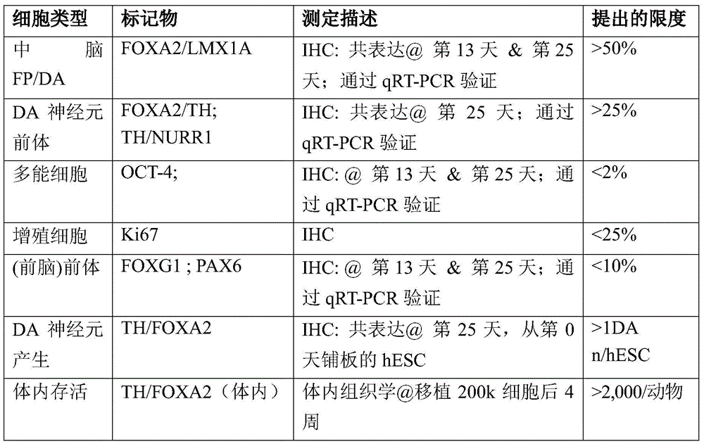
表8.确定某些因子对于产生DA神经元的作用的示例性实验Table 8. Exemplary experiments to determine the effect of certain factors on generating DA neurons
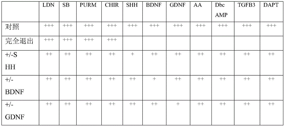
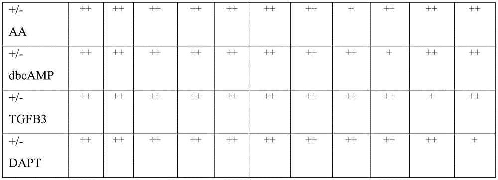
表9.扩大mDA神经元培养特别是用于制造临床应用的GMP级培养物的示例性方法。Table 9. Exemplary methods for scaling up mDA neuronal cultures, particularly for the manufacture of GMP grade cultures for clinical applications.
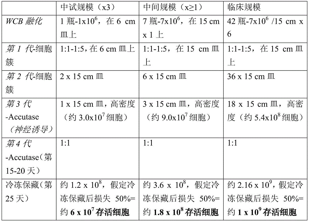
表10.hES系产物的示例性体内评价–移植物组成Table 10. Exemplary in vivo evaluation of hES line products - graft composition
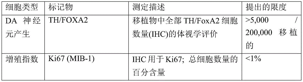

表11.hES系产物的示例性体内评价–行为分析Table 11. Exemplary in vivo evaluation of hES line products - behavioral analysis
(*)在一些实施方式中,表现出>6转/分钟的大鼠稳定接受移植物(*) In some embodiments, rats exhibiting >6 rev/min stably received the graft
实验部分Experimental part
以下实施例用于说明本发明的某些实施方式和方面,而并非是要限制其范围。在以下公开的实验部分中,应用以下缩写:N(当量); M(摩尔的);mM(毫摩尔的);μM(微摩尔的);mol(摩尔);mmol (毫摩尔);μmol(微摩尔);nmol(纳摩尔);pmol(皮摩尔);g(克); mg(毫克);μg(微克);ng(纳克);pg(皮克);L(升);ml(毫升);μl(微升);cm(厘米);mm(毫米);μm(微米);nm(纳米);U(单位);min(分钟);s和sec(秒);deg(度);pen(青霉素);strep(链霉素)和℃(摄氏度)。The following examples are intended to illustrate certain embodiments and aspects of the invention, but are not intended to limit its scope. In the experimental sections disclosed below, the following abbreviations are used: N (equivalent); M (molar); mM (millimolar); μM (micromolar); μmol); nmol (nanomol); pmol (picomolar); g (gram); mg (mg); μg (microgram); ng (nanogram); pg (picogram); L (liter); ml ( milliliter); μl (microliter); cm (centimeter); mm (millimeter); μm (micrometer); nm (nanometer); U (unit); min (minute); s and sec (second); deg (degree) ; pen (penicillin); strep (streptomycin) and °C (degree Celsius).
实施例I.Example I.
材料和方法Materials and methods
方法概述:对人ESC(H9、H1)和iPSC系(2C6和SeV6)进行基于底板诱导(Fasano等人,Cell Stem Cell 6:336-347(2010),在此将其并入作为参考)方案的改良的双重SMAD抑制(Chambers等人,Nat. Biotechnol.27:275-280(2009),在此将其并入作为参考)。出于中脑底板和新的DA神经元群体的产量,对于暴露于SHH C25II、Purmorphamine、FGF8和CHIR99021进行优化(参见图1d)。在底板诱导之后,在DA 神经元存活和成熟因子(Perrier,等人.Proc Natl Acad Sci USA 101:12543-8(2004),在此将其并入作为参考)例如AA、BDNF、GDNF、 TGFβ3和dbcAMP(具体参见完整的方法)的存在下,在基于 Neurobasal/B27的分化培养基中进行进一步的成熟(在培养物中11-25 天或长于25天,最高在培养物中至少100天)。经由免疫细胞化学、 qRT-PCR、全局基因表达图谱、用于检测多巴胺的HPLC分析和体外电生理学记录,对产生的DA神经元群体进行广泛的表型表征。体内研究在半帕金森氏病啮齿动物(在动物脑的一侧注有6OHDA毒素的小鼠或大鼠)中进行。研究在通过立体定位注入之前所述的毒素而接受6-羟基多巴胺损伤的成年NOD-SCID IL2Rgc小鼠(Jacksonlabs)和成年Sprague Dawley大鼠Taconic Farms以及用MPTP单侧颈动脉注射处理的两只成年猕猴中进行。将DA神经元立体定位地注射到动物的纹状体中(小鼠中为150x 103细胞,大鼠中为250x 103细胞),猴子中共计7.5x 10⁶细胞(分布在6束中;脑的每一侧3束)。植入之后每隔一个月进行行为试验,包括安非他明介导的旋转分析,以及对于病灶运动不能(focal akinesia)(“踏步试验”)和肢体使用(圆柱体试验) 的试验。植入之后,将大鼠和小鼠在18-20周时处死,灵长类动物在1 个月时处死。经由细胞数目和移植物体积的体视学分析以及经由免疫细胞化学的综合表型表征,对移植物进行表征。未分化人类ES细胞的培养。将hESC系H9(WA-09,XX,自10/2009时传代29-55次)、 H1(WA-01,XY,自6/2010时传代30-40次)和iPS细胞系2C6(Kim 等人,Cell Stem Cell 8:695-706(2011),在此将其并入作为参考)(XY,传代20-30次)和SeV6(XY,传代20-30次;使用非整合4因子仙台病毒载体系统自MRC-5胚胎成纤维细胞得到(Ban等人,Proc.Natl. Acad.Sci.U.S.A(2011)108(34):14234-14239:10.1073/ pnas.1103509108,在此将其并入作为参考))以基于人ES细胞所估计的范围在0.5x 10³/cm2至100x103/cm2的铺开浓度保持在小鼠胚胎成纤维细胞上,其倾向于在含有最优20%敲除血清替代物(KSR,Invitrogen, Carlsbad,California)的人ES细胞培养基中细胞聚集(MEF,Global Stem, Rockville,MD)(如之前所述(Kim等人,Cell Stem Cell8:695-706 (2011),在此将其并入作为参考))。敲除血清替代物的使用可以在范围 0%至40%。 Method overview : Bottom plate-based induction (Fasano et al., Cell Stem Cell 6:336-347 (2010), which is incorporated herein by reference) protocol for human ESC (H9, H1) and iPSC lines (2C6 and SeV6) Modified dual SMAD inhibition of (Chambers et al., Nat. Biotechnol. 27:275-280 (2009), which is hereby incorporated by reference). Exposure to SHH C25II, Purmorphamine, FGF8 and CHIR99021 was optimized for yield of midbrain floor plates and new DA neuron populations (see Figure Id). Following floor plate induction, in DA neuronal survival and maturation factors (Perrier, et al. Proc Natl Acad Sci USA 101:12543-8 (2004), which is hereby incorporated by reference) such as AA, BDNF, GDNF, TGFβ3 Further maturation in Neurobasal/B27-based differentiation medium (11-25 days in culture or longer than 25 days, up to at least 100 days in culture) in the presence of dbcAMP and dbcAMP (see full methods for details) . The resulting population of DA neurons was extensively phenotypically characterized via immunocytochemistry, qRT-PCR, global gene expression profiling, HPLC analysis for dopamine detection, and in vitro electrophysiological recordings. In vivo studies were performed in semi-Parkinsonian rodents (mice or rats injected with 6OHDA toxin on one side of the animal's brain). To study adult NOD-SCID IL2Rgc mice (Jacksonlabs) and adult Sprague Dawley rats Taconic Farms receiving 6-hydroxydopamine injury via stereotaxic injection of the toxin described previously and two adults treated with unilateral carotid injection of MPTP in macaques. DA neurons were stereotaxically injected into the striatum of animals (150 x 10 3 cells in mice, 250 x 10 3 cells in rats) for a total of 7.5 x 10 6 cells in monkeys (distributed in 6 bundles; 3 bundles on each side of the brain). Behavioral tests, including amphetamine-mediated rotational analysis, and tests for focal akinesia ("stepping test") and limb use (cylinder test), were performed every other month after implantation. Following implantation, rats and mice were sacrificed at 18-20 weeks and primates at 1 month. Grafts were characterized via stereological analysis of cell number and graft volume and comprehensive phenotypic characterization via immunocytochemistry. Culture of undifferentiated human ES cells . hESC lines H9 (WA-09, XX, passage 29-55 since 10/2009), H1 (WA-01, XY, passage 30-40 since 6/2010) and iPS cell line 2C6 (Kim et al. Human, Cell Stem Cell 8:695-706 (2011), which is hereby incorporated by reference) (XY, passage 20-30) and SeV6 (XY, passage 20-30; using non-integrating 4-factor Sendai virus The vector system was derived from MRC-5 embryonic fibroblasts (Ban et al., Proc. Natl. Acad. Sci. USA (2011) 108(34):14234-14239:10.1073/pnas.1103509108, which is hereby incorporated as Reference)) were maintained on mouse embryonic fibroblasts at a spreading concentration ranging from 0.5 x 10 3 /cm 2 to 100 x 10 3 /cm 2 estimated based on human ES cells, which tended to contain optimal 20% knockout Cell aggregation (MEF, Global Stem, Rockville, MD) in human ES cell culture medium except serum replacement (KSR, Invitrogen, Carlsbad, California) (as previously described (Kim et al., Cell Stem Cell 8:695-706 ( 2011), which is hereby incorporated by reference)). The use of knockout serum replacements can range from 0% to 40%.
神经诱导.对于基于底板的中脑多巴胺神经元诱导,基于定时暴露于 LDN-193189(100nM(浓度范围0.5-50μM,Stemgent,Cambridge, Massachusetts)、SB431542(10μM(浓度范围0.5-50μM,Tocris,Ellisville, MI)、SHH C25II(l00 ng/ml(浓度范围10-2000ng/ml,R&D,Minneapolis, MN)、Purmorphamine(2μM(浓度范围10-500ng/ml,Stemgent)、FGF8(100ng/ml(浓度范围10-500ng/ml,R&D)和CHIR99021(CHIR;3 μM(浓度范围0.1-10μM,Stemgent),使用改良版本的双重SMAD抑制(Chambers等人,Nat.Biotechnol.27:275-280(2009),在此将其并入作为参考)和底板诱导(Fasano等人,Cell Stem Cell 6:336-347(2010),在此将其并入作为参考)方案。注意:对于底板诱导方案,“SHH”处理是指使细胞暴露,即接触于SHH C25II 100ng/ml+Purmorphamine (2μM)的组合。将细胞铺(35-40x 103细胞/cm2)在含有DMEM、 15%敲除血清替代物、2mM L-谷氨酰胺和10-μM(浓度范围1-25μM)β-巯基乙醇的敲除血清替代物培养基(KSR)中的基质胶(matrigel) 或geltrex(购得即用)(BD,Franklin Lakes,New Jersey)上,并培养11 天。在分化第5天开始,通过在第5-6天以75%(KSR):25%(N2)的比例混合,在第7-8天以50%(KSR):50%(N2)的比例混合,并在第9-10 天以25%(KSR):75%(N2)的比例混合,将KSR培养基逐渐变换为N2 培养基,如之前所述(Chambers等人,Nat.Biotechnol.27:275-280 (2009),在此将其并入作为参考)。在第11天,将培养基改变成补充有 CHIR(直至第13天)并补充有BDNF(脑源性神经营养因子,20ng/ml 范围5至100;R&D)、抗坏血酸(AA;0.2mM(浓度范围0.01-1mM), Sigma,St Louis,MI)、GDNF(胶质细胞系源性神经营养因子,20ng/ml (浓度范围1-200ng/ml);R&D)、TGFβ3(β3型转化生长因子,l ng/ml (浓度范围0.1-25ng/ml);R&D)、二丁酰cAMP(0.5mM(浓度范围0.05-2mM);Sigma)和DAPT(10nm(浓度范围0.5-50nM);Tocris) 的含有Neurobasal培养基/B27培养基(1:50稀释)/L-Glut(有效范围 0.2-2mM))的培养基(NB/B27;Invitrogen),培养9天。在第20天,将细胞使用
对于基于菊形团的DA神经元诱导,部分地遵照之前所述的方案 (Perrier等人,Proc Natl Acad Sci U S A 101:12543-8(2004),在此将其并入作为参考),至少一处例外在于其中使用双重SMAD抑制来加速最初的神经诱导步骤。简言之,通过与从分化第2-8天补充有SB431542 和Noggin(250ng/ml(浓度范围10-1000ng/ml);R&D)并从分化第 6-11天补充有SHH+FGF8的KSR中的辐射MS5细胞共培养,将hESC 朝向神经结局诱导。在KSR中11天之后,将神经菊形团人工分离出,并在如之前所述补充有SHH、FGF8、BDNF和AA的N2培养基(Perrier 等人,Proc Natl Acad Sci U S A 101:12543-8(2004),在此将其并入作为参考)中培养(P1阶段)。P1阶段中5-7天之后,再次机械方式收获菊形团,并在不含Ca2/Mg2的汉克氏平衡盐溶液(HBSS)中孵育1小时之后加以磨碎,再铺在覆有多鸟氨酸(PO)/层粘连蛋白/纤连蛋白的板上。在P2阶段用SHH/FGF8图形化(patterning)持续7天,然后如上所述在BDNF、AA、GDNF、TGFb3和dbcAMP的存在下进行最终的分化,直至对给定的实验达到所需的成熟阶段(对于移植研究通常为5-7天,或者,对于体外功能研究通常为32天)。For rosette-based induction of DA neurons, following in part the protocol previously described (Perrier et al., Proc Natl Acad Sci USA 101:12543-8 (2004), which is hereby incorporated by reference), at least one The exception is where dual SMAD inhibition is used to accelerate the initial neural induction step. Briefly, by comparison with KSR supplemented with SB431542 and Noggin (250ng/ml (concentration range 10-1000ng/ml); R&D) from differentiation days 2-8 and SHH+FGF8 supplemented from differentiation days 6-11 Co-culture of irradiated MS5 cells induces hESC towards neural fate. After 11 days in KSR, neural rosettes were manually isolated and cultured in N2 medium supplemented with SHH, FGF8, BDNF and AA as previously described (Perrier et al., Proc Natl Acad Sci USA 101:12543-8 (2004), which is hereby incorporated by reference (P1 stage). After 5-7 days in the P1 stage, the rosettes were harvested again mechanically, ground after 1 hour incubation in Hank's Balanced Salt Solution (HBSS) without Ca 2 /Mg 2 , and then spread on a Plates of polyornithine (PO)/laminin/fibronectin. Patterning with SHH/FGF8 at the P2 stage for 7 days followed by final differentiation in the presence of BDNF, AA, GDNF, TGFb3 and dbcAMP as described above, until the desired stage of maturation was reached for a given experiment (typically 5-7 days for transplantation studies, or 32 days for in vitro functional studies).
基因表达分析.使用Rneasy试剂盒(Qiagen,Valencia,CA)从对照LSB、LSB/SHH/FGF8和LSB/SHH/FGF8/CHIR的各个条件下在第0、 1、3、5、7、9、11、13和25天在分化过程中提取总RNA。对于微阵列分析,将总RNA通过MSKCC基因组核心设施处理,并根据制造商说明书在Illumina人ref-12微珠阵列上杂交。使用来自Bioconductor(worldwideweb.bioconductor.org)的LIMMA软件包对每天和每个条件之间进行比较。发现基因的调节P-值<0.05且倍数变化大于2被视作是显著的。一些描述性微阵列数据分析和展示使用市售的软件包 (Partek Genomics Suite(6.10.0915版本))进行。对于qRT-PCR分析,对每个条件下第25天的总RNA进行逆转录(Quantitech,Qiagen),使用市售的Taqman基因表达阵列(Applied Biosystems,Carlsbad,CA)检测扩增的材料,数据标准化至HPRT。每个数据点代表3个独立的生物样品的9组技术上的重复。微阵列研究的原始数据在GEOworldwideweb.ncbi.nlm.nih.gov/geo尚未可获得。动物手术.啮齿动物和猴的手术过程遵照NIH指南进行,并取得当地机构动物关爱和使用委员会(Institutional Animal Careand Use Committee,IACUC)、生物安全性委员会(Institutional Biosafety Committee,IBC)和胚胎干细胞研究委员会(Embryonic Stem Cell Research Committee,ESCRO)的许可。 Gene expression analysis . Using the Rneasy kit (Qiagen, Valencia, CA) from each condition of control LSB, LSB/SHH/FGF8 and LSB/SHH/FGF8/CHIR at 0, 1, 3, 5, 7, 9, Total RNA was extracted at 11, 13 and 25 days during differentiation. For microarray analysis, total RNA was processed through the MSKCC Genomic Core Facility and hybridized on Illumina human ref-12 microbead arrays according to the manufacturer's instructions. Comparisons between each day and each condition were made using the LIMMA software package from Bioconductor (worldwideweb.bioconductor.org). Genes were found to have regulatory P-values < 0.05 and fold changes greater than 2 were considered significant. Some descriptive microarray data analysis and presentation were performed using a commercially available software package (Partek Genomics Suite (version 6.10.0915)). For qRT-PCR analysis, total RNA at
小鼠.将NOD-SCID IL2Rgc空白小鼠(重20-35g;Jackson Laboratory,BarHarbor,ME)用氯胺酮(90mg/kg;Akorn,Decatur,IL) 和甲苯噻嗪(4mg/kg;Fort Dodge,IA)麻醉。将6-羟基多巴胺(10μg (浓度范围0.1-20μg)6-OHDA(Sigma-Aldrich))在以下坐标(单位毫米)处立体定位地注入纹状体:AP,0.5(从前卤;颅骨缝合用作立体定位手术的参照);ML,-2.0;DV,-3.0(从硬脑膜,覆盖脑的膜用作参照)。选择成功损伤的小鼠(平均>6转/分钟)进行移植。将体积 1.5μl的共计150x 103细胞在以下坐标处(单位mm)注入纹状体:AP, 0.5;ML,-1.8;DV,3.2。移植后18周将小鼠处死。 Mice . NOD-SCID IL2Rgc null mice (weight 20-35 g; Jackson Laboratory, BarHarbor, ME) were treated with ketamine (90 mg/kg; Akorn, Decatur, IL) and xylazine (4 mg/kg; Fort Dodge, IA) )anaesthetization. 6-Hydroxydopamine (10 μg (concentration range 0.1-20 μg) 6-OHDA (Sigma-Aldrich)) was stereotaxically injected into the striatum at the following coordinates (in mm): AP, 0.5 (from anterior halogen; cranial suture was used as Reference for stereotaxic surgery); ML, -2.0; DV, -3.0 (from the dura mater, the membrane covering the brain was used as a reference). Successfully injured mice (average >6 rpm) were selected for transplantation. A total of 150 x 103 cells in a volume of 1.5 μl were injected into the striatum at the following coordinates (in mm): AP, 0.5; ML, -1.8; DV, 3.2. Mice were sacrificed 18 weeks after transplantation.
大鼠.在手术过程中将成年雌性Sprague-Dawley(Taconic,Hudson, NY)大鼠(180-230g)用氯胺酮(90mg/kg)和甲苯噻嗪(4mg/kg) 麻醉。通过在两处位点立体定位注入6-OHDA(0.2%抗坏血酸和0.9%盐水中3.6mg/ml,Sigma),建立nigro-striatal路径的单边前脑中间束损伤(Studer等人,Nature Neurosci.1:290-295(1998),在此将其并入作为参考)。注入之后6-8周,如果安非他明诱导的转动超过6转/分钟,选择大鼠用于移植。将250x103细胞移植到每个动物的纹状体中(坐标:AP+1.0mm,ML-2.5mm和V-4.7mm;齿条设定在-2.5)。对照大鼠接受PBS作为替代。手术过程之前有过记载(Studer等人,NatureNeurosci.1:290-295(1998),在此将其并入作为参考)。细胞植入之前24 小时开始每日腹膜内注射环孢霉素15mg/kg(Bedford Labs,Bedford, OH),并持续至细胞植入后20周处死。 Rats . Adult female Sprague-Dawley (Taconic, Hudson, NY) rats (180-230 g) were anesthetized with ketamine (90 mg/kg) and xylazine (4 mg/kg) during surgery. Unilateral forebrain-medial tract injury of the nigro-striatal pathway was established by stereotaxic infusion of 6-OHDA (3.6 mg/ml in 0.2% ascorbic acid and 0.9% saline, Sigma) at two sites (Studer et al., Nature Neurosci. 1:290-295 (1998), which is hereby incorporated by reference). Six to eight weeks after infusion, if amphetamine-induced rotation exceeds 6 rpm, rats are selected for transplantation. 250x103 cells were transplanted into the striatum of each animal (coordinates: AP+1.0mm, ML-2.5mm and V-4.7mm; rack set at -2.5). Control rats received PBS instead. The surgical procedure has been previously described (Studer et al., Nature Neurosci. 1:290-295 (1998), which is hereby incorporated by reference). Daily intraperitoneal injection of
灵长类动物.经由颈动脉MPTP给药,然后每周I.V.MPTP给药,产生双侧帕金森氏病症状,使2只成年(17-18岁;10-12kg;雌性) 猕猴患半帕金森氏病(Kordower等人,Science290:767-773(2000),在此将其并入作为参考)。基于行为分析,两个动物表现出符合中重度损伤的帕金森氏病症状,上述行为分析包括:姿势弯曲、腿拖行和僵直症状(运动不灵活)、疏忽(对单侧刺激的运动意识)和运动徐缓(缓慢运动开始)。在猴中这些参数可以使用改良的帕金森氏病临床评定量表(CRS)来评价。在移植手术这天,将动物用氯胺酮(3.0mg/kg,IM) 和dexdomitor(0.02-0.04mg/kg,IM)镇静,插管以保持稳定的气道,并用异氟烷麻醉。然后将其置于立体定位框中用于手术。两只猕猴均经历单次手术,基于立体定位坐标三次颅内注入人类底板衍生DA培养物(Paxinos等人,The Rhesus Monkey Brain inStereotaxic Coordinates (Academic Press,2000),在此将其全文并入作为参考)。在三个位点处 (1-后尾状核,2-前连合壳和上覆的白质)进行双侧细胞注入(10μl/ 注入;125,000细胞/μl),总体积30μl/半球。使用附接至立体定位显微操纵器(micromanipulator)的输液泵以1μl/分钟的速率通过具有28G 针头的50μl Hamilton注射器递送细胞。注射完成之后,将针头保留在原处额外的2-5分钟,以使注入物扩散离开针尖,然后缓缓缩回注射器。手术之后,动物立刻接受止痛剂(buprenex,0.01mg/kg IM,手术后 BID 72小时;美洛昔康,0.1mg/kg SQ,手术后SID 72小时)以及抗生素(头孢唑林,25mg/kg IM,BID),直至手术后72小时。手术之前48小时开始动物每日一次口服接受环孢霉素A(Neoral, Sandimmune)(30mg/kg逐渐减少至15mg/kg),直至在移植之后一个月处死。 Primates . Administration of MPTP via carotid followed by weekly IVMPTP to produce bilateral Parkinson's disease symptoms in 2 adult (17-18 years; 10-12 kg; female) rhesus macaques with half Parkinson's disease disease (Kordower et al., Science 290:767-773 (2000), which is hereby incorporated by reference). Two animals exhibited Parkinson's disease symptoms consistent with moderate to severe impairment based on behavioral analysis including: postural flexion, leg dragging and rigidity symptoms (inflexibility of movement), inattentiveness (motor awareness to unilateral stimuli) and bradykinesia (slow movement onset). These parameters can be assessed in monkeys using a modified Parkinson's Disease Clinical Rating Scale (CRS). On the day of transplantation, animals were sedated with ketamine (3.0 mg/kg, IM) and dexdomitor (0.02-0.04 mg/kg, IM), intubated to maintain a stable airway, and anesthetized with isoflurane. It is then placed in a stereotaxic frame for surgery. Both macaques underwent a single surgery with three intracranial injections of human floor plate-derived DA cultures based on stereotaxic coordinates (Paxinos et al., The Rhesus Monkey Brain in Stereotaxic Coordinates (Academic Press, 2000), which is hereby incorporated by reference in its entirety. ). Bilateral cell infusions (10 μl/injection; 125,000 cells/μl) were performed at three sites (1 - posterior caudate nucleus, 2 - anterior commissural putamen and overlying white matter) in a total volume of 30 μl/hemisphere. Cells were delivered at a rate of 1 μl/min through a 50 μl Hamilton syringe with a 28G needle using an infusion pump attached to a stereotaxic micromanipulator. After the injection is complete, leave the needle in place for an additional 2-5 minutes to allow the infusion to diffuse away from the needle tip, then slowly retract the syringe. Immediately after surgery, animals received analgesics (buprenex, 0.01 mg/kg IM, 72 hours post-surgery BID; meloxicam, 0.1 mg/kg SQ, 72 hours post-surgery SID) and antibiotics (ceftazolin, 25 mg/kg) IM, BID) until 72 hours after surgery. Animals received cyclosporine A (Neoral, Sandimmune) orally once daily starting 48 hours prior to surgery (30 mg/kg tapering to 15 mg/kg) until sacrificed one month after transplantation.
行为试验.在移植之前和移植之后4、8、12、18周进行安非他明诱导的旋转(小鼠和大鼠)和踏步试验(大鼠)。在i.p.注射d-安非他明(10mg/kg,Sigma)之后10分钟记录小鼠的旋转行为,记录30分钟。大鼠中的旋转行为在i.p.注射d-安非他明(5mg/kg)之后40分钟记录,并通过TSE VideoMot2系统(Germany)进行自动化评价。数据表示为每分钟旋转的平均次数。踏步试验自以下文献改良:Blume等人,Exp.Neurol.219:208-211(2009)和Crawley等人,What's Wrong With My Mouse:Behavioral Phenotyping of Transgenic andKnockout Mice (Wiley-Liss,2000),在此将其均并入作为参考。简言之,将每只大鼠置于平的表面上;通过轻轻举起尾巴将后腿抬高,使得仅有前爪接触台面。实验者以稳定的步速将大鼠拉回1米。对侧和同侧前爪的调节踏步次数加以计数。数据表示为对侧/(对侧+同侧)的调节踏步的百分比。圆柱体试验通过将每只动物置于玻璃圆柱体中并对同侧与对侧爪接触 (在20次接触中)圆柱体壁的次数进行计数进行,如之前所述(Tabar 等人,NatureMed.14:379-381(2008),在此将其并入作为参考)。组织处理。小鼠和大鼠:动物(小鼠和大鼠)腹膜内接受超剂量的戊巴比妥 (50mg/kg),以诱导深度麻醉,并灌注4%多聚甲醛(PFA)。提取脑,将其在4%PFA中后固定,然后在30%蔗糖溶液中浸泡2-5天。包埋在 O.C.T.化合物(Sakura-Finetek,Torrance,California)中之后在恒冷箱上进行切片。 Behavioral tests . Amphetamine-induced rotation (mice and rats) and stepping tests (rats) were performed before transplantation and at 4, 8, 12, 18 weeks after transplantation. The rotational behavior of the mice was recorded 10 minutes after ip injection of d-amphetamine (10 mg/kg, Sigma) for 30 minutes. Rotational behavior in rats was recorded 40 minutes after ip injection of d-amphetamine (5 mg/kg) and was assessed automatically by the TSE VideoMot2 system (Germany). Data are expressed as the average number of rotations per minute. The stepping test was modified from Blume et al., Exp. Neurol. 219:208-211 (2009) and Crawley et al., What's Wrong With My Mouse: Behavioral Phenotyping of Transgenic and Knockout Mice (Wiley-Liss, 2000), here All are incorporated by reference. Briefly, each rat was placed on a flat surface; the hind legs were raised by gently raising the tail so that only the front paws were touching the table. The experimenter pulled the rat back 1 meter at a steady pace. Conditioning steps on the contralateral and ipsilateral forepaws were counted. Data are expressed as a percentage of conditioned steps contralateral/(contralateral + ipsilateral). The cylinder test was performed by placing each animal in a glass cylinder and counting the number of times the ipsilateral and contralateral paws made contact (in 20 contacts) with the cylinder wall, as described previously (Tabar et al., NatureMed. 14:379-381 (2008), which is hereby incorporated by reference). Organizational processing. Mice and Rats : Animals (mice and rats) received an intraperitoneal overdose of pentobarbital (50 mg/kg) to induce deep anesthesia and were infused with 4% paraformaldehyde (PFA). Brains were extracted, postfixed in 4% PFA, and then soaked in 30% sucrose solution for 2-5 days. Sectioning was performed on a cryostat after embedding in OCT compound (Sakura-Finetek, Torrance, California).
灵长类动物:将动物在以氯胺酮(10mg/kg,肌肉内(IM))和戊巴比妥(25mg kg,静脉内(IV))深度麻醉下经由用肝素化0.9%盐水、然后是冷的新鲜4%PFA固定剂(pH7.4)心脏灌注而处死。初始固定之后,立刻将脑从颅骨中取出,在4%PFA中后固定,自由漂浮24-36小时。然后将其漂洗,并在慢速摇动器上在4℃下再悬浮在10%蔗糖中,使其“下沉”。然后在20%蔗糖中重复该过程,然后在30%蔗糖中重复。将全脑冠状地在冷冻滑动式切片机上切成40μm的连续切片,并在 -20℃下在低温贮藏培养基中自由漂浮储存。 Primates : Animals were deeply anesthetized with ketamine (10 mg/kg, intramuscular (IM)) and pentobarbital (25 mg kg, intravenous (IV)) via heparinized 0.9% saline followed by cold The hearts were sacrificed by perfusion of fresh 4% PFA fixative (pH 7.4). Immediately after initial fixation, brains were removed from the skull, postfixed in 4% PFA, and free-floated for 24-36 hours. It was then rinsed and resuspended in 10% sucrose at 4°C on a slow shaker to "sink". The process was then repeated in 20% sucrose, then 30% sucrose. Whole brains were coronally cut into 40 μm serial sections on a cryo-slide microtome and stored free-floating in cryopreservation medium at -20°C.
免疫组织化学:将细胞在4%PFA中固定,并用具有0.3%Triton 的1%牛血清白蛋白(BSA)封闭。将脑组织切片在冷PBS中洗涤,并加以类似处理。将一抗在1-5%BSA或正常山羊血清中稀释,并根据制造商的推荐孵育。抗体和来源的综合列表如表6所提供。使用适当的 Alexa488、Alexa555和Alexa647-结合的二抗(Molecular Probes, Carlsbad,California),进行4',6-二脒基-2-苯基吲哚(DAPI)核复染色 (Thermo Fisher,Rockford,Illinois)。对于一些分析,使用生物素化的二抗,然后经由DAB(3,3'-二氨基联苯胺)色原进行可视化。HPLC分析.如之前所述(Roy等人,Nature Med.12:1259-1268(2006);Studer等人,Brain Res.Bull.41:143-150(1996),在此将其均并入作为参考),进行电化学检测的反相HPLC,用于测量多巴胺、高香草酸(HVA)和DOPAC(3,4-二羟基-苯基乙酸)的水平。在分化第65天收集高氯酸中的培养物样品。对于一些试验,使用相同的检测系统直接测量培养基中的DA,但随后的多巴胺及其代谢物的铝提取使用市售的试剂盒,如之前所述(Studer等人,Brain Res.Bull.41:143-150(1996),在此将其并入作为参考)。电生理学记录:将培养物转移至装有40X水下物镜 (Eclipse E600FN;Nikon)的直立显微镜上的记录室中;对培养物灌注以mM为单位含有125NaCl、2.5KC1、25NaHCO3、1.25NaH2PO4、 2CaCl、1MgCl2和25葡萄糖(34℃;以95%O2-5%CO2;pH 7.4;298 mOsm/L)的盐水。盐水流速为2-3ml/min,经直列加热器(in-line heater) 运行(SH-27B,具有TC-324B控制器;Warner Instruments)。神经元通过具有冷却CCD数码照相机的视频显微镜可视化(CoolSNAP ES2, Photometries,RoperScientific,Tucson,Arizona)。选择用于电生理学记录的细胞具有带细小的分枝神经突的神经元样形状。用MultiClamp 700B 放大器(Molecular Devices)进行电流钳结构中成体全细胞膜片箝记录。信号在1-4kHz下过滤,在5-20kHz下用Digidata 1440A(MolecularDevices)数字化。记录膜片电极由在Flaming-Brown牵引机(P-97,Sutter Instruments)上拉出的细丝硼硅酸盐玻璃(Sutter Instruments)制造,在浴中的电阻为4-6MΩ。将电极装有含有mM单位的135K-MeSO4、 5KC1、5HEPES、0.25EGTA、10phosphocroeatine-di(tris)、2ATP-Mg 和0.5GTP-Na(pH 7.3,重量摩尔渗透压浓度(osmolality)调节为 290-300mOsm/L)的内部溶液。调节放大器桥接电路以补偿电极电阻并加以监测。补偿电极电容。当记录过程中串联电阻增加>20%时,弃去数据,因为电阻增加表明记录过程中有部分技术故障。 Immunohistochemistry : Cells were fixed in 4% PFA and blocked with 1% bovine serum albumin (BSA) with 0.3% Triton. Brain tissue sections were washed in cold PBS and treated similarly. Primary antibodies were diluted in 1-5% BSA or normal goat serum and incubated according to the manufacturer's recommendations. A comprehensive list of antibodies and sources is provided in Table 6. 4',6-diamidino-2-phenylindole (DAPI) nuclear counterstaining (Thermo Fisher, Rockford, CA) was performed using the appropriate Alexa488, Alexa555 and Alexa647-conjugated secondary antibodies (Molecular Probes, Carlsbad, California). Illinois). For some analyses, biotinylated secondary antibodies were used, followed by visualization via DAB (3,3'-diaminobenzidine) chromogen. HPLC analysis . As previously described (Roy et al, Nature Med. 12:1259-1268 (2006); Studer et al, Brain Res. Bull. 41:143-150 (1996), all incorporated herein as Reference), reverse-phase HPLC with electrochemical detection was performed to measure levels of dopamine, homovanillic acid (HVA), and DOPAC (3,4-dihydroxy-phenylacetic acid). Culture samples in perchloric acid were collected on day 65 of differentiation. For some experiments, the same detection system was used to measure DA directly in the medium, but the subsequent aluminum extraction of dopamine and its metabolites used a commercially available kit, as previously described (Studer et al., Brain Res. Bull. 41 : 143-150 (1996), which is hereby incorporated by reference). Electrophysiological recordings : Cultures were transferred to a recording chamber on an upright microscope equipped with a 40X underwater objective (Eclipse E600FN; Nikon); cultures were perfused with 125NaCl, 2.5KCl, 25NaHCO3 , 1.25NaH2 in mM PO4 , 2CaCl , 1MgCl2 and 25 glucose (34°C; with 95% O2-5% CO2 ; pH 7.4; 298 mOsm/L) in saline. The brine flow rate was 2-3 ml/min and was run through an in-line heater (SH-27B with TC-324B controller; Warner Instruments). Neurons were visualized by video microscopy with a cooled CCD digital camera ( CoolSNAP ES2, Photometries, Roper Scientific, Tucson, Arizona). Cells selected for electrophysiological recordings have a neuron-like shape with finely branched neurites. Adult whole-cell patch-clamp recordings in a current-clamp configuration were performed with a MultiClamp 700B amplifier (Molecular Devices). Signals were filtered at 1-4 kHz and digitized with a Digidata 1440A (Molecular Devices) at 5-20 kHz. The recording patch electrodes were fabricated from filament borosilicate glass (Sutter Instruments) drawn on a Flaming-Brown puller (P-97, Sutter Instruments) with a resistance in the bath of 4-6 MΩ. Electrodes were loaded with 135K-MeSO4, 5KC1 , 5HEPES, 0.25EGTA, 10phosphocroeatine-di(tris), 2ATP-Mg, and 0.5GTP-Na (pH 7.3, osmolality adjusted to 290) containing mM units -300mOsm/L) internal solution. The amplifier bridge circuit was adjusted to compensate for electrode resistance and monitored. Compensate for electrode capacitance. Data were discarded when the series resistance increased by >20% during recording, as the increase in resistance indicated a partial technical failure during recording.
细胞计数和体视学分析.确定在从3组各自独立的实验得到的样品中在底板(第11天)图1、中脑多巴胺神经元前体(第25天)图2 和成熟DA神经元阶段(第50天或更晚)图3和11的标记物阳性细胞的百分含量。以均匀随机的方法选择用于定量的图片,每个图片首先对DAPI-阳性细胞核的数目进行打分,然后对表达所关注标记物的细胞数目进行计数。数据表示成平均值±SEM。每10个切片进行对移植物中人类细胞(以抗-hNA鉴别)和TH+神经元的定量,其中移植物是可鉴定的。使用Stereo Investigator软件(MBF bioscience,Vermont),使用光学分馏器探针和Cavalieri估计器确定细胞计数和移植物体积,如之前所述(Tabar等人,Nat.Biotechnol.23:601-606(2005),在此将其并入作为参考)。数据表示为估计的总细胞数和总移植物体积+/-平均标准误差(SEM)。 Cytometry and stereological analysis . Determined in samples from 3 separate experiments in baseplate (day 11) Figure 1, midbrain dopamine neuron precursors (day 25) Figure 2 and mature DA neurons Stage (
以下配方说明了用于开发本发明实施方式的示例性细胞培养基。The following formulations illustrate exemplary cell culture media used to develop embodiments of the present invention.
用于维持的hESC培养基(1升):800mL DMEM/F12,200mL敲除血清替代物,5mL200mM L-谷氨酰胺,5mL Pen/Strep,10mL 10 mM MEM最低限非必需氨基酸15酸溶液,55μM13-巯基乙醇,和 bFGF(最终浓度为4ng/mL)。 hESC medium for maintenance (1 L): 800 mL DMEM/F12, 200 mL Knockout Serum Replacement, 5 mL 200 mM L-Glutamine, 5 mL Pen/Strep, 10
用于hESC分化的KSR培养基(1升):820mL敲除DMEM,150 mL敲除血清替代物,10mL200mM L-谷氨酰胺,10mL of Pen/Strep, 10mL 10mM MEM,和55μM 13-巯基乙醇。 KSR medium (1 liter) for hESC differentiation : 820 mL knockout DMEM, 150 mL knockout serum replacement, 10 mL 200 mM L-glutamine, 10 mL of Pen/Strep, 10
用于hESC分化的N2培养基(1升):985ml具有DMEM/F12粉末的蒸馏H2O,1.55g葡萄糖(Sigma,cat.no.G7021),2.00g碳酸氢钠(Sigma,cat.no.S5761),腐胺(1.61g于100mL蒸馏水中的100μL 等份物;Sigma,cat.no.P5780),孕酮(0.032g于100mL 100%乙醇中的20μL等份物;Sigma,cat.no.P8783),亚硒酸钠(0.5mM溶液于去离子水中的60μL等份物;BioshopCanada,cat.no.SEL888),和100mg 转铁蛋白(Celliance/Millipore,cat.no.4452-01),和10mL 5mM NaOH 中25mg胰岛素(Sigma,cat.no.I6634)。 N2 medium (1 liter) for hESC differentiation : 985 ml distilled H2O with DMEM/F12 powder, 1.55 g glucose (Sigma, cat. no. G7021), 2.00 g sodium bicarbonate (Sigma, cat. no. S5761), putrescine (100 μL aliquot of 1.61 g in 100 mL distilled water; Sigma, cat. no. P5780), progesterone (20 μL aliquot of 0.032 g in 100
Dulbecco改良的Eagles培养基(DMEM),具有10%FBS,用于制备PMEF((初级小鼠成 纤维细胞(PMEF)饲养细胞)(1升):885mL DMEM,100mL FBS,10mL Pen/Strep,和5mL L-谷氨酰胺。 Dulbecco's Modified Eagles Medium (DMEM) with 10% FBS for PMEF ((Primary Mouse Fibroblast (PMEF) Feeder Cells) (1 L): 885 mL DMEM, 100 mL FBS, 10 mL Pen/Strep, and 5 mL L-glutamine.
α最低限必需培养基(MEM),具有10%FBS,用于制备MS-5饲养细胞培养基(1升):890mLαMEM,100mL FBS,10mL Pen/Strep 凝胶化溶液(500ml):将0.5g明胶溶解于500ml温的(50-60℃)Milli- Q水中。冷却至室温。 Alpha Minimum Essential Medium (MEM) with 10% FBS for preparation of MS-5 feeder medium (1 liter): 890 mL αMEM, 100 mL FBS, 10 mL Pen/Strep gelling solution (500 ml): 0.5 g The gelatin was dissolved in 500 ml of warm (50-60°C) Milli-Q water. Cool to room temperature.
实施例II.Example II.
该实施例描述了用于提供本发明的FOXA2+LMX1A+DA神经元的定向分化的小分子和接触时机的发现。This example describes the discovery of small molecules and timing of contacts used to provide directed differentiation of FOXA2+LMX1A+DA neurons of the present invention.
以下是本文所述的一些实验发现的简要小结:在不存在CHIR99021的条件下将双重SMAD抑制的细胞用SHH激动剂 (purmorphamine+SHH)和FGF8(S/F8)处理,到第11天表现出显著较低的FOX2的表达,并且完全缺乏LMX1A的表达(图1a、b)。前部的标记物OTX2在LSB和LSB/S/F8/CHIR处理的培养物中被强烈地诱导,但在LSB/S/F8条件下不被诱导(图1a、c)。The following is a brief summary of some of the experimental findings described here: Dual SMAD-inhibited cells treated with SHH agonists (purmorphamine + SHH) and FGF8 (S/F8) in the absence of CHIR99021 exhibited by
发明人之前使用若干种其他的产生含有DA-样神经元的细胞群体的定向分化方法。将这些DA-样神经元用于移植研究中,导致对将这些细胞进一步用于治疗应用方面的顾虑。例如,Perrier等人,2004和 Fasano等人,2010中描述的包括MS5神经诱导的方法导致形成菊形团细胞,将其用于产生第11天前体,例如参见图2、16和17,其进一步用于得到DA-样神经元。这些神经元由产生的第11天细胞群体中低百分含量的前体细胞产生。使用这些神经元的移植研究表现出很差的移植后生存力和DA-样神经元表型的丧失,此外还观察到不适当的神经类型的移植后发育以及生长控制的丧失,这导致发展出畸胎瘤。参见图16和17。The inventors have previously used several other directed differentiation methods to generate cell populations containing DA-like neurons. The use of these DA-like neurons in transplantation studies has led to concerns about the further use of these cells in therapeutic applications. For example, the methods described in Perrier et al., 2004 and Fasano et al., 2010, including neural induction of MS5, resulted in the formation of rosette cells, which were used to generate
具体地,在P0使hESC与用于使Oct4+细胞开始神经诱导为菊形团细胞的分子接触,使用MS5饲养细胞(Perrier等人,2004)。在P1 阶段,通过使细胞与额外的分子接触使菊形团细胞扩张,上述额外的分子用于在P2阶段使细胞分化成具有特定表达模式的细胞包括Pax2+/En1+DA祖细胞,并进一步分化成TH+/En1+DA神经元。这些细胞用于植入到用环孢霉素A免疫抑制的6OHDA损伤的大鼠中。这些移植研究表现出很差的体内生存力,TH+表型的丧失,关于以下方面的顾虑:进一步生长成不希望的、可能致死的细胞即畸胎瘤,以及细胞生长成不适当的神经类型,其对于患者来说可以造成额外的医学问题。Specifically, hESCs were contacted at PO with molecules used to initiate neural induction of Oct4+ cells into rosette cells, using MS5 feeder cells (Perrier et al., 2004). At the P1 stage, rosette cells are expanded by contacting the cells with additional molecules that are used at the P2 stage to differentiate the cells into cells with specific expression patterns, including Pax2+/En1+ DA progenitors, and further differentiation into TH+/En1+DA neurons. These cells were used for engraftment into 6OHDA-injured rats immunosuppressed with cyclosporine A. These transplantation studies demonstrated poor in vivo viability, loss of the TH+ phenotype, concerns regarding further growth into undesired, potentially lethal cells i.e. teratomas, and cell growth into inappropriate neural types, It can cause additional medical problems for the patient.
在来自菊形团衍生DA神经元前体的移植物中,在移植后第4.5月有数量非常少的存活TH+神经元(<50TH+细胞/动物),图16A。但是,与TH+细胞相反,GFP标记的细胞(GFP由遍在的启动子驱动)在移植之后确实存活相当良好。这表明移植之后大多数存活的细胞是来自非-DA神经元身份的神经细胞(16B)。极少有移植物衍生的细胞(hNA+ (绿色)再次共表达TH(红色),说明大多数植入的人细胞采取非-DA 神经元表型,图16C。小图16D-E表明,D-E尽管在体内的存活很差,在一些行为试验例如安非他明诱导的旋转(D)和圆柱体试验和自发旋转(E)中,有一些(低的、但高度可变的)改善。如之前所述进行无饲养层神经诱导(Chambers等人,2009),但进行进一步改良,以产生底板细胞(Fasano等人,2010)。在改良的将多能细胞分化成底板细胞的双重SMAD抑制方法中,发明人之前发现,到第11天需要高浓度的 SHH进行FP诱导。例如,在一些实施方式中,加入200ng/ml的音猬因子C25II。在一些实验中,加入DKK-1(R&D;100ng/ml)、FGF8 (R&D;50ng/ml)、Wnt-1(Peprotech;50ng/ml)和视黄酸(R&D; 1mM),参见图17。但是,使用之前的方法在第11天产生的细胞群体无一含有高百分比含量的使用本发明方法得到的FOXA2+/LMX1A+中脑底板祖细胞。In grafts from rosette-derived DA neuron precursors, there were very few viable TH+ neurons (<50 TH+ cells/animal) at 4.5 months post-transplantation, Figure 16A. However, in contrast to TH+ cells, GFP-tagged cells (GFP driven by a ubiquitous promoter) did survive fairly well after transplantation. This indicates that the majority of surviving cells after transplantation are neural cells from non-DA neuronal identity (16B). Few of the graft-derived cells (hNA+ (green) again co-expressed TH (red), indicating that the majority of engrafted human cells adopted a non-DA neuronal phenotype, Figure 16C. Panels 16D-E show that D-E despite the Survival in vivo was poor, with some (low, but highly variable) improvement in behavioral tests such as amphetamine-induced rotation (D) and cylinder test and spontaneous rotation (E). As before Feeder-free neural induction was performed as described (Chambers et al., 2009), but further modified to generate floor plate cells (Fasano et al., 2010). In a modified dual SMAD inhibition approach to differentiate pluripotent cells into floor plate cells , the inventors previously found that high concentrations of SHH are required for FP induction by
如本文所示,发明人选择含有多能细胞的细胞群体用于起始群体,并在第0天铺板。在分化之前细胞生长至接近汇合(汇合60-100%)。在第0天将这些细胞与双重SMAD抑制剂接触(即暴露于LDN-193189 +SB431542=“LSB”)。发明人跟随具有含有新鲜LSB的常规饲养的细胞群体直至第11天,发现一些剩余的细胞是LMX1A+的,但不表达 FOXA2(图1a、b)。发明人平板培养两份起始细胞群体,然后在与含有任意以下SHH激动剂(purmorphamine+SHH)和FGF8(S/F8)的混合物之后测试细胞类型(即基因/蛋白质表达模式谱),将细胞用不同的暴露方案接触,即,在第0天或第1天或第2天等接触细胞,接触特定量的时间,即24小时、48小时等。测试的三种主要的示例性培养条件为1)细胞在第0天与LDN/SB(LSB)接触,然后与新鲜LSB接触直至直到第5天,在第5天使将细胞与没有SB的新鲜LDN接触直至第11天,2)细胞在第0天与LDN/SB(LSB)接触,然后与新鲜 LSB接触直至第5天,在第5天使细胞与没有SB的新鲜LDN接触直至直到第11天,同时在这段时间使细胞额外地与新鲜purmorphamine、SHH和FGF8接触直到第7天,和3)细胞在第0天与LDN/SB(LSB) 接触,然后与新鲜LSB接触直至第5天,在第5天使细胞与没有SB 的新鲜LDN接触直至直到第11天,同时在这段时间使细胞额外地与新鲜purmorphamine、SHH和FGF8接触直到第7天,同时在培养第3 天开始额外地与新鲜CHIR接触直到直至第11天,这些主要条件有若干变化,以确定细胞类型的最佳产量。使用全局时序基因表达图谱对3 种培养条件(图1d)进行系统比较。参见示例性图8和表1-6。到分化第11天,差异表达基因的分级聚类分离出三种处理条件(图8a)。在 LSB/S/F8/CHIR vs LSB处理组中,FOXA1、FOXA2和若干其他的SHH 下游目标(包括PTCH1)在最差异调节的转录物之中(图1e)。LMX1A、 NGN2和DDC的表达表明,到第11天已经建立了中脑DA神经元前体结局(图1e,f)。相反地,LSB培养物到第11天时富集了背侧的前脑前体标记物,例如HES5、PAX6、LHX2和EMX2。LSB/S/F8/CHIR与 LSB/S/F8处理的直接比较(图1f)确认了在LSB/S/F8/CHIR组中对于中脑DA前体标记物的选择性富集,并且基于RAX1、SIX3和SIX6的差异表达表明了LSB/S/F8处理的培养物中的下丘脑前体身份(另外参见图2d中的POMC、OTP表达)。As shown herein, the inventors selected a cell population containing pluripotent cells for the starting population and plated on
第11天的差异表达转录物的示例性列表在表1、2中示出,第25 天的在表3-5中示出,基因本体论分析图8b(DAVID; http://david.abcc.ncifcrf.gov)证实了CHIR处理时经典WNT信号传导的富集。原始数据在GEO worldwideweb.ncbi.nlm.nih.gov/geo/accession#:[TBD]尚未可获得。中脑DA前体标记物(图1g)与前侧和腹侧非-DA神经元结局标记物(图1h)的基因表达的比较时序分析将三种诱导条件划分为:i)LSB:背侧前脑;ii)LSB/S/F8:腹侧/下丘脑;和iii)LSB/S/F8/CHIR:中脑DA前体身份。Exemplary lists of differentially expressed transcripts for
实施例III.Example III.
DA神经元的分化。为了进一步进行分化,将前体细胞保持在促进神经元成熟的培养基中(BAGCT——参见材料和方法部分)。在从之前的方法和本发明的方法得到的分化细胞的群体之间进行以下类型的比较:A)在分化第50天对于TH结合LMX1A、FOXA2和NURR1 进行免疫细胞化学分析,B)对总的细胞当中TH+、FOXA2+、LMX1+ 和NURR1+细胞的定量,比较菊形团衍生与底板衍生(LSB/S/F8/CHIR) 的培养物。C)在底板和菊形团衍生的DA神经元培养物中在第50天时5-羟色胺+(5-HT)和GABA+神经元亚型(非DA神经元污染物) 百分含量的定量。和D)测量多巴胺和代谢物的HPLC分析:比较底板与菊形团衍生的培养物之间DA、DOPAC和HVA的水平。到第25 天,三种前体细胞群体产生了Tuj1+神经元(图2a)和表达DA合成中的限速酶——TH的细胞。但是,LSB/S/F8/CHIR处理产生了共表达 LMX1A和FOXA2的TH+细胞,并且表现出核受体NURR1(NR4A2) 的强烈诱导(图2a、b)。在第13天与第25天培养物中基因表达的比较证实了其他有丝分裂后DA神经元标记物的强烈诱导(图2c)。在第 25天表征DA神经元身份与LSB和LSB/S/F8处理的培养物相比较,证实了已知的中脑DA神经元转录物和所鉴定的多种新候选标记物的富集(图2d,表3-5,图8b)。例如,LSB/S/F8/CHIR(中脑DA组)中最高度富集的转录物是TTF3——之前与中脑DA神经元发育不相关、但在人黑质中高度表达的基因(图8c;Allen Brain Atlas: http://human.brain-map.org)。Differentiation of DA neurons. For further differentiation, precursor cells were maintained in a medium that promotes neuronal maturation (BAGCT - see Materials and methods section). The following types of comparisons were made between the populations of differentiated cells obtained from the previous method and the method of the present invention: A) immunocytochemical analysis for TH binding LMX1A, FOXA2 and NURR1 at
对于EBF-1、EBF-3(图8c)以及已知的肝中的FOXA2的转录目标TTR,获得了类似的数据。本发明研发过程中获得的数据表明了若干PITX基因在中脑DA前体细胞中的富集。PITX3是经典的中脑DA 神经元标记物,其也在分化第25天强烈表达(图2e)。最后,中脑底板和DA神经元诱导均可以很容易地在独立的hESC和hiPSC系中再现 (图9)。在此表明的数据说明,与其他测试的方案不同,LSB/S/F8/CHIR 方案产生表达与中脑DA神经元结局匹配的标记物图谱的细胞。Similar data were obtained for EBF-1, EBF-3 (Fig. 8c) and the known transcriptional target TTR of FOXA2 in liver. The data obtained during the development of the present invention demonstrate the enrichment of several PITX genes in midbrain DA precursor cells. PITX3, a canonical midbrain DA neuron marker, was also strongly expressed at
将底板衍生DA神经元的体外和体内特性与经由神经菊形团中间体得到的DA-样神经元进行比较(图10和16)。神经菊形团的图案化 (patterning)代表了目前最广泛使用的由hPSC得到DA神经元的策略。基于底板的和基于菊形团的方案均有效地产生能够长期体外存活的 TH+神经元(分化第50天;图3a)。但是,在底板衍生的培养物中, TH+细胞的百分含量显著更高(图3b)。尽管两种方案中的TH+细胞均表现出NURR1的共表达,但底板衍生的DA神经元共表达FOXA2和 LMX1A(图3a、b)。几乎观察不到GABA和5-羟色胺(5-HT)-阳性的神经元(图3c)。DA及其代谢物DOPAC和HVA在任一方案产生的培养物中均存在,但底板培养物中的DA水平大约高8倍(图3d、e)。中脑DA神经元表现出广泛的纤维突起和成熟神经元标记物的强烈表达,其包括突触蛋白、多巴胺转运蛋白(DAT)、和G-蛋白偶联的内向整流钾通道(Kir3.2,也称作GIRK2,在黑质致密部(SNpc)DA神经元中表达)(图3f、图11)。SNpc DA神经元在体内表现出使其与脑中大多数其他神经元不同的电生理学表型。具体地,它们以缓慢速度(1-3 Hz)自发地放电。而且,该缓慢放电(slow spiking)伴随有缓慢的阈下震荡电位。在体外2-3周后,从出生后早期小鼠培养的SNpc DA神经元表现出这些相同的生理学特性。从hESC分化的DA神经元始终如一地(4/4)表现出这一有特色的电生理学表型(图3g-i)。The in vitro and in vivo properties of floor plate-derived DA neurons were compared to DA-like neurons obtained via neural rosette intermediates (Figures 10 and 16). Patterning of neural rosettes represents the most widely used strategy for obtaining DA neurons from hPSCs. Both floor-plate-based and rosette-based protocols efficiently generated TH+ neurons capable of long-term in vitro survival (
在体外维持mDA神经元的第65天显示,TH阳性神经元仍在表达 FoxA2,并延伸出mDA神经元典型的长纤维。图3A。HPLC的DA释放测量显示,65天龄的TH+神经元在体外是功能性的。图3B。Maintenance of mDA neurons in vitro on day 65 showed that TH-positive neurons still expressed FoxA2 and extended long fibers typical of mDA neurons. Figure 3A. DA release measurements by HPLC showed that 65-day-old TH+ neurons were functional in vitro. Figure 3B.
实施例IV.Example IV.
在含有损伤神经元的啮齿动物即小鼠和大鼠中植入新的DA神经元细胞群体。New DA neuron cell populations are implanted in rodents containing damaged neurons, ie, mice and rats.
该领域的挑战之一在于在没有神经过度生长或不适当地分化成非中脑神经元或发展畸胎瘤的风险的情况下产生在体内功能性植入的 hPSC衍生的中脑DA神经元的能力。基于胎儿组织移植研究,发明人预计由NURR1的表达标记的细胞周期退出时间可以是移植的适当阶段(大约分化第25天-图2)。最初使用第25天细胞在未损伤的成年小鼠中的研究表明,在移植后第6周hPSC-衍生的FOXA2+/TH+神经元稳健存活(图12)。在帕金森氏病宿主中在不导致神经过度生长的情况下对FOXA2+/TH+细胞的长期存活进行测试。为这一目的,在NOD-SCID IL2Rgc空白小鼠中制造6-羟基-多巴胺(6-OHDA)损伤 (Tabar等人,NatureMed.14:379-381(2008),在此将其并入作为参考),其是有效支持异种移植存活的品系,对于暴露于少有的致瘤细胞具有特别的敏感度(Quintana等人,Efficient tumourformation by single human melanoma cells.Nature 456:593-598(2008),在此将其并入作为参考)。将底板和菊形团衍生的DA神经元培养物不经在先提纯而移植 (150x 103/动物),以体现具有高增殖潜力的潜在污染细胞。移植之后 4.5个月,底板衍生DA神经元移植物表现出明确的移植物核心,其由共表达FOXA2和人特异性标记物hNCAM的TH+细胞组成(图4a-c)。功能分析表明,对安非他明诱导的旋转行为有完全的挽救。相反地,菊形团衍生的神经元移植物几乎不表现出TH+神经元,在旋转行为方面不产生显著降低(图4d),并表现出大量的神经过度生长(移植物体积>20mm3;图13)。在此报道的用于移植的菊形团衍生神经元细胞的广泛过度生长与发明人小组(Kim等人,miR-371-3Expression Predicts NeuralDifferentiation Propensity in Human Pluripotent Stem Cells.Cell Stem Cell 8:695-706(2011),在此将其并入作为参考)以及其他人 (Hargus等人,Proceedings of theNational Academy of Sciences of the United States of America 107:15921-15926(2010),在此将其并入作为参考)之前的菊形团衍生DA移植物的工作是相当的。过度生长可能是由于存活期较长(4.5月vs 6周),在移植之前缺少FACS提纯以及 NOD-SCID IL2Rgc空白宿主的选择。增殖的Ki67+细胞在底板衍生移植物中的数目是最小的(<全部细胞的1%),而菊形团衍生移植物保持增殖神经前体的囊(pocket)。神经过度生长被认为是由移植物内的原始前部神经外胚层细胞造成的(Elkabetz等人,Genes Dev.22:152-165 (2008);Aubry等人,Proc.Natl Acad.Sci.U.S A 105:16707-16712(2008),在此将其并入作为参考)。该假设由前脑标记物FOXG1在菊形团衍生移植物中的表达而在底板衍生移植物中的不表达所支持。在底板和菊形团衍生的移植物中均存在有低百分含量的星形胶质细胞,尽管大多数GFAP+细胞对于指示宿主来源的人标记物是阴性的(图13)。One of the challenges in this field is to generate functionally engrafted hPSC-derived midbrain DA neurons in vivo without the risk of neural overgrowth or inappropriate differentiation into non-midbrain neurons or development of teratomas. ability. Based on fetal tissue transplantation studies, the inventors anticipate that the time of cell cycle exit marked by expression of NURR1 may be an appropriate stage for transplantation (approximately
本文所述的NOD-SCID IL2Rgc空白小鼠中的结果表现出 FOXA2+/TH+神经元的稳健的长期存活,安非他明诱导的旋转行为的完全逆转,并且没有神经过度生长的任何迹象。但是,这些结果中的一些可能归因于NOD-SCID IL2Rgc空白小鼠的特定使用。为检验这一假设,将底板衍生DA神经元培养物(250x 103细胞)移植到使用环孢霉素A在药理学上免疫抑制的6-OHDA损伤的成年大鼠中。移植物移植之后5个月,存活很稳健(图4e-h),平均为大于15,000个共表达 FOXA2(图4g)以及人核抗原(hNA)(图4e)的TH+细胞;TH+/hNCAM+ 纤维从移植物核心放射到周围的宿主纹状体中(图4f)。除FOXA2以外,TH+细胞还表达中脑DA神经元标记物PITX3和NURR1(图4h-j)。与未显示改善的假移植动物相反,行为分析表现出安非他明诱导的旋转不对称的完全挽救(图4k)。移植的动物还在测量前肢运动不能的踏步试验(图4l)中以及在不依赖于DA系统药理学刺激的检验的圆柱体试验(图4m)中表现出改善。预计人DA神经元的恢复开始较晚(移植之后大约3-4个月),其取决于体内成熟的速率,例如DAT表达的水平(图4n)。表达Kir3.2通道(GIRK2)或钙结合蛋白的TH+细胞的存在表明SNpc(A9)和腹侧被盖区(A10)DA神经元二者均存在于移植物中(图4o、p)。The results in NOD-SCID IL2Rgc null mice described herein demonstrate robust long-term survival of FOXA2+/TH+ neurons, complete reversal of amphetamine-induced rotational behavior, and no sign of neural overgrowth. However, some of these results may be attributable to the specific use of NOD-SCID IL2Rgc null mice. To test this hypothesis, baseplate-derived DA neuron cultures (250 x 103 cells) were transplanted into 6-OHDA-injured adult rats that were pharmacologically immunosuppressed with cyclosporine A. Five months after graft transplantation, survival was robust (Fig. 4e-h), with an average of >15,000 TH+ cells co-expressing FOXA2 (Fig. 4g) as well as human nuclear antigen (hNA) (Fig. 4e); TH+/hNCAM+ fibers were derived from The graft core radiated into the surrounding host striatum (Fig. 4f). In addition to FOXA2, TH+ cells expressed the midbrain DA neuron markers PITX3 and NURR1 (Fig. 4h-j). In contrast to sham-transplanted animals that did not show improvement, behavioral analysis demonstrated complete rescue of amphetamine-induced rotational asymmetry (Fig. 4k). Transplanted animals also showed improvement in the stepping test (Fig. 4l), which measures forelimb akinesia, and in the cylinder test (Fig. 4m), a test that does not rely on pharmacological stimulation of the DA system. The recovery of human DA neurons is expected to start late (approximately 3-4 months after transplantation), depending on the rate of in vivo maturation, eg the level of DAT expression (Fig. 4n). The presence of TH+ cells expressing Kir3.2 channels (GIRK2) or calbindin indicated that both SNpc (A9) and ventral tegmental area (A10) DA neurons were present in the grafts (Fig. 4o,p).
如在小鼠中那样(图13),5-羟色胺能细胞和GABA能细胞在大鼠细胞中是少见的(<全部细胞的1%),主要为宿主衍生的GFAP+神经胶质细胞(全部细胞的7%;(图14))也如此。尽管在移植物中几乎检测不到5-羟色胺+神经元,观察到有可能为宿主衍生的5-羟色胺能纤维的hNCAM-阴性细胞(图14)。As in mice (Figure 13), serotonergic and GABAergic cells are rare (<1% of all cells) in rat cells, mainly host-derived GFAP+ glial cells (all
实施例V.Example V.
在含有损伤神经元的灵长类动物中植入新的DA神经元细胞群体。Engraftment of new DA neuron cell populations in primates containing damaged neurons.
在此所述的结果表明了优异的移植物存活和两种独立的鼠科动物模型中的行为结果。但是,小鼠或大鼠脑中需要的DA神经元的数目只占植入灵长类动物和人中所需的更大数目细胞的一小部分。为了检验该效果的可扩展性,在两只MPTP损伤的成年猕猴中进行试验移植研究。The results described here demonstrate excellent graft survival and behavioral outcomes in two independent murine models. However, the number of DA neurons required in the mouse or rat brain is a fraction of the larger number of cells required for implantation in primates and humans. To examine the scalability of this effect, a pilot transplantation study was performed in two MPTP-injured adult rhesus monkeys.
使用基于底板的方案在分化第25天获得多批次的50x 106可移植 DA神经元前体。用于诱导帕金森样病症的经典剂量为,以0.2mg/kg IV 的MPTP将3mg MPTP-HCL(范围0.5-5mg)注射到颈动脉中。在这之后是系统注射MPTP 0.2mg/kg IV MPTP。在脑的每一侧在三个位置处(后尾核和前合缝处硬膜(precommissural putamen)注入细胞(共计6束,1.25x 106细胞/束),并用环孢霉素A对动物进行免疫抑制。脑的一侧注射来自表达GFP的H9亚克隆的DA前体,而另一侧植入源自未标记H9细胞的细胞。显示具有持续的FOX2A表达和TH产生的猕猴中神经元植入的结果显示于图4q-t中。移植1个月之后,基于 GFP(图15)和人特异性细胞质标记物(SC-121)(图4q)的表达观察到中脑DA神经元的稳健存活。每个移植核心均被TH+纤维向宿主内延长多达3mm的晕环包围(图4r)。移植核心由共表达SC-121(图 4s)和FOXA2(图4t)的TH+神经元组成。移植物内的SC-121和GFP 阴性区域含有Ibal+宿主小神经胶质细胞(图15),表明免疫抑制不完全。概括而言,在灵长类动物即MPTP(3mg MPTP-HCL(1-甲基-4- 苯基-1,2,3,6-四氢吡啶);浓度范围0.5-5mg MPTP-HCl)损伤的含有严重的内源中脑DA神经元>95%丧失的成年猕猴中植入新的DA神经元细胞群体。MPTP暴露导致可观察到的改变和类似于人类帕金森氏病的症状。Multiple batches of 50 x 106 transplantable DA neuron precursors were obtained on
实施例VI.Example VI.
可比较的PINK1突变PD-iPSC细胞与野生型hES(或iPSC)细胞朝向中脑DA神经元结局的分化潜能。Comparable differentiation potential of PINK1 mutant PD-iPSC cells and wild-type hES (or iPSC) cells towards midbrain DA neuronal fate.
该实施例描述了以下发现:当使用以不导致胚胎破坏的方式得到的PD患者细胞系即PINK1突变PD-iPSC细胞作为获得本发明的 FOXA2/LIM1XA/TH+DA神经元的细胞群体时,大的中脑DA神经元群体发展出PD患者神经元的特性。This example describes the finding that when a PD patient cell line obtained in a manner that does not result in embryo destruction, namely PINK1 mutant PD-iPSC cells, is used as the cell population for obtaining the FOXA2/LIM1XA/TH+DA neurons of the present invention, a large The midbrain DA neuron population develops properties of PD patient neurons.
使用本发明的新的基于底板的中脑DA神经元方案(方法)使PINK1 Q456X突变PD-iPSC系分化,产生与获自iPSC H9系的分化图谱可比较的中脑分化图谱(图20)。A-C)分化第11天(中脑前体阶段)PINK1突变PD-iPSC系对于FOXA2(红色)、LMX1A(绿色)和 DAPI(蓝色)的免疫细胞化学分析(A),分化25天(早期有丝分裂后DA神经元阶段)对于FOXA2(红色)和TH(绿色)(B)和对于 NURRl(红色)和TH(绿色)(C)的免疫细胞化学分析。D-F):在分化第11天对于FOXA2(红色)、LMX1A(绿色)和DAPI(蓝色) (D),在分化第25天对于FOXA2(红色)和TH(绿色)(E)和对于RURR1(红色)和TH(绿色)(F),使用H9衍生的细胞进行的相同组的免疫细胞化学分析。The PINK1 Q456X mutant PD-iPSC line was differentiated using the novel floor-plate-based midbrain DA neuron protocol (Methods) of the present invention, resulting in a midbrain differentiation map comparable to that obtained from the iPSC H9 line (Figure 20). A-C) Immunocytochemical analysis of FOXA2 (red), LMX1A (green) and DAPI (blue) of PINK1 mutant PD-iPSC line at
在体外长期分化和成熟之后,PINK1突变PD-iPSC显示出蛋白聚集的PD样表型。发明人发现,在分化第55天,使用新的基于底板的中脑DA神经元诱导方案,PINK1突变PD-iPSC表现出在TH+DA神经元的胞质中α-突触核蛋白(PD患者中路易体形成的主要组分)表达的证据,(图21a-b)。A,B)在分化第55天PINK1突变PD-iPSC系对于α-突触核蛋白(LB509,红色)、TH(绿色)和合并图像(A)以及α-突触核蛋白(红色)和泛素(绿色)(B)的免疫细胞化学分析。这些α-突触核蛋白阳性细胞还表现出高的泛素(典型的路易体标记物) 表达。相反,源自对照iPS系的DA神经元表现出正常突触(与胞质的相对)α-突触核蛋白表达,以及非常低水平的泛素(图21c-d)。C,D) 在分化第55天对照-iPSC系对于α-突触核蛋白(红色)和TH(绿色)(C)以及α-突触核蛋白(红色)和泛素(绿色)(D)的免疫细胞化学分析。After long-term differentiation and maturation in vitro, PINK1 mutant PD-iPSCs displayed a PD-like phenotype of protein aggregation. The inventors found that at day 55 of differentiation, using a novel floor-plate-based induction protocol for midbrain DA neurons, PINK1-mutant PD-iPSCs exhibited α-synuclein in the cytoplasm of TH+DA neurons (PD patients). Evidence for the expression of a major component of Lewy body formation in Lewy bodies (Fig. 21a-b). A,B) PINK1 mutant PD-iPSC line at day 55 of differentiation for α-synuclein (LB509, red), TH (green) and merged images (A) as well as α-synuclein (red) and pan Immunocytochemical analysis of protein (green) (B). These alpha-synuclein positive cells also showed high expression of ubiquitin, a typical marker for Lewy bodies. In contrast, DA neurons derived from control iPS lines exhibited normal synaptic (as opposed to cytoplasmic) alpha-synuclein expression, and very low levels of ubiquitin (Fig. 21c-d). C,D) Control-iPSC lines at day 55 of differentiation for α-synuclein (red) and TH (green) (C) and α-synuclein (red) and ubiquitin (green) (D) immunocytochemical analysis.
α-突触核蛋白聚集形式的表达。在PD患者的脑中,二聚的不溶形式的α-突触核蛋白导致在路易体中的聚集。二聚形式的α-突触核蛋白在α-突触核蛋白上表现出丝氨酸129的磷酸化。Expression of aggregated forms of alpha-synuclein. In the brains of PD patients, a dimeric insoluble form of alpha-synuclein leads to aggregation in Lewy bodies. The dimeric form of alpha-synuclein exhibits phosphorylation at serine 129 on alpha-synuclein.
在相同的分化天数时,与表现出非常低水平表达的对照-iPSC衍生的细胞相反,PINK1突变PD-iPSC衍生的细胞对于Ser129磷酸化的α- 突触核蛋白表现出强的表达(图22)。与表现出非常低水平表达的对照 -iPSC衍生的细胞相反,PINK1突变PD-iPSC衍生的细胞对于Ser129 磷酸化的α-突触核蛋白表现出强的表达。A,B)在分化第55天在PINK1 突变PD-iPSC衍生的细胞中(A)以及在匹配的对照-iPSC衍生的细胞中(B)对于Ser129磷酸化α-突触核蛋白(绿色)和DAPI(蓝色)的免疫细胞化学分析。At the same number of days of differentiation, PINK1 mutant PD-iPSC-derived cells exhibited strong expression of Ser129 phosphorylated α-synuclein, in contrast to control-iPSC-derived cells, which exhibited very low levels of expression (Figure 22). ). In contrast to control-iPSC-derived cells, which exhibited very low levels of expression, PINK1 mutant PD-iPSC-derived cells exhibited strong expression of Ser129-phosphorylated α-synuclein. A,B) Phosphorylated α-synuclein at Ser129 (green) and in matched control-iPSC-derived cells (B) at day 55 of differentiation in PINK1 mutant PD-iPSC-derived cells (A) and Immunocytochemical analysis of DAPI (blue).
取决于分化方案,观察到α-突触核蛋白表达模式的差异。发明人预计,底板衍生的“真正的”中脑DA神经元具有PD特异性易损性,以及对应的特异性体外表型。使用经典的基于MS5基质饲养的分化方案(Perrier等人,PNAS 2004,在此将其并入作为参考)得到的DA神经元产生大量的TH+神经元。但是,基于本发明研发过程中得到的数据,发明人表明,基于MS5的TH+细胞不是真正的底板衍生的中脑 DA神经元。在经由MS5方案分化的培养物中,有许多α-突触核蛋白阳性细胞。但是,这些细胞并未共表达TH。而且,当使用MS5分化策略时,在PD-iPSC和对照iPSC之间在表达模式方面并没有差异(图 23a-b)。这些数据表明,α-突触核蛋白在其他的非DA细胞类型中也得以表达,并且这些非DAα-突触核蛋白在疾病与对照-iPSC衍生的细胞中是未改变的,特别是当使用标准MS5分化策略时。这些是公开出版物中报道的DA样菊形团衍生的神经元(例如,Perrier PNAS 2004)。使用这些基于MS5的TH+(=DA样)细胞用于在图3、10、13和16 中进行比较。这些数据表明,α-突触核蛋白在其他的非DA细胞类型中也得以表达,并且这些非DAα-突触核蛋白在疾病与对照-iPSC衍生的细胞中是未改变的,特别是当使用标准MS5分化策略时。最后,本文所述的新的基于底板的分化方案产生大量的共表达α-突触核蛋白的 TH+细胞。这些TH+细胞以胞质表达模式表达α-突触核蛋白。图24A, B)在基于MS5的分化第60天对于PINK1突变PD-iPSC系的α-突触核蛋白(LB509,红色)、TH(绿色)(A)以及对照-iPSC(B)的免疫细胞化学分析。C)在基于底板的分化第55天PINK1突变PD-iPSC 系对于α-突触核蛋白(红色)、TH(绿色)的免疫细胞化学分析。Depending on the differentiation protocol, differences in α-synuclein expression patterns were observed. The inventors anticipate that floor-plate derived "true" midbrain DA neurons have PD-specific vulnerability, and a corresponding specific in vitro phenotype. DA neurons derived using a classical MS5 substrate-feeding-based differentiation protocol (Perrier et al., PNAS 2004, which is hereby incorporated by reference) yielded large numbers of TH+ neurons. However, based on the data obtained during the development of the present invention, the inventors have shown that MS5-based TH+ cells are not true floor-plate derived midbrain DA neurons. In cultures differentiated via the MS5 protocol, there were many alpha-synuclein positive cells. However, these cells did not co-express TH. Furthermore, there were no differences in expression patterns between PD-iPSCs and control iPSCs when using the MS5 differentiation strategy (Fig. 23a-b). These data suggest that α-synuclein is also expressed in other non-DA cell types and that these non-DA α-synuclein proteins are unchanged in disease and control-iPSC-derived cells, especially when using standard MS5 differentiation strategy. These are DA-like rosette-derived neurons reported in open publications (eg, Perrier PNAS 2004). These MS5-based TH+ (=DA-like) cells were used for comparison in Figures 3, 10, 13 and 16. These data suggest that α-synuclein is also expressed in other non-DA cell types and that these non-DA α-synuclein proteins are unchanged in disease and control-iPSC-derived cells, especially when using standard MS5 differentiation strategy. Finally, the novel floor-plate-based differentiation protocol described here yields large numbers of TH+ cells co-expressing α-synuclein. These TH+ cells express alpha-synuclein in a cytoplasmic expression pattern. Figure 24A, B) Immune cells for alpha-synuclein (LB509, red), TH (green) (A) and control-iPSCs (B) of the PINK1 mutant PD-iPSC line at
源自PINK1突变PD-iPSC的示例性DA神经元对于毒性刺激是更易损的。比起对照-iPSC衍生的细胞,经由基于底板的方案得到的 PD-iPSC衍生的TH+DA神经元对于毒素攻击是更易损的(缬氨霉素:线粒体离子载体,5μm(浓度范围1-10μm),48小时)。相反,经由经典的基于MS5的方案得到的TH+神经元在PD-与对照衍生的细胞之间并未表现出差异的易损性(图24)。A-F)在分化第60天的代表性 TH免疫细胞化学:对于经由基于底板的方案得到的PD-和对照-iPSC 衍生的细胞二者均为正常条件(无毒素处理)(A,示出PD-iPSC衍生细胞),在毒素处理之后PD-iPSC中的TH+DA神经元几乎完全退化 (B),来自对照-iPSC的TH+DA神经元部分退化(C)。缬氨霉素处理48小时后用阿尔玛蓝进行整体细胞生存力测定,也表明当对PD-iPSC和对照iPSC进行比较时,对于毒素攻击(5和10μM)在特定的剂量范围内表现出差异的细胞存活(图25)。Exemplary DA neurons derived from PINK1 mutant PD-iPSCs are more vulnerable to toxic stimulation. PD-iPSC-derived TH+DA neurons obtained via the bottom plate-based protocol were more vulnerable to toxin challenge than control-iPSC-derived cells (Valinomycin: mitochondrial ionophore, 5 μm (concentration range 1-10 μm) ),48 hours). In contrast, TH+ neurons obtained via the classical MS5-based protocol did not show differential vulnerability between PD- and control-derived cells (Figure 24). A-F) Representative TH immunocytochemistry at
对于经由基于MS5的方案得到的PD-和对照-iPSC衍生的培养物为正常条件(D,示出PD-iPSC衍生细胞),在PD-iPSC中毒素攻击之后的TH+神经元(E),以及经由MS5方案得到的对照-iPSC衍生培养物(F)。在分化第60天通过基于底板的方案对于Tuj1(红色)和TH (绿色)的免疫细胞化学低功率图像:正常(G)与毒素攻击(H)条件的PD-iPSC,以及正常(I)与毒素攻击(J)条件的对照iPSC。K-N) 在分化第60天通过基于MS5的方案对于Tuj1(红色)和TH(绿色) 的免疫细胞化学低功率图像:正常(K)与毒素攻击(L)条件的PD-iPSC,以及正常(M)与毒素攻击(N)条件的对照iPSC。Normal conditions for PD- and control-iPSC-derived cultures via MS5-based protocol (D, PD-iPSC-derived cells are shown), TH+ neurons after toxin challenge in PD-iPSCs (E), and Control-iPSC derived cultures obtained via MS5 protocol (F). Immunocytochemical low power images of Tuj1 (red) and TH (green) by bottom plate-based protocol at
示例性的对于毒素攻击的细胞生存力-剂量反应测定的定量。在缬氨霉素处理48小时后用阿尔玛蓝进行细胞生存力测定表明,当对 PD-iPSC和对照iPSC进行比较时(基于底板的分化第60天),对于毒素攻击(5和10μM)在特定的剂量范围内表现出差异的细胞存活。注意:该测定测试总的细胞死亡,而在DA神经元中特异性地观察到最引人注目的效果(参见图14)。因此,基于阿尔玛蓝的定量将有可能低估DA神经元谱系上观察到的差异效果的程度。Exemplary Quantification of Cell Viability-Dose Response Assay for Toxin Challenge. Cell viability assays with alamar blue after 48 hours of valinomycin treatment showed that when PD-iPSCs and control iPSCs were compared (
在此并入以下参考文献作为参考:Li等人,Nat.Biotechnol.23,215- 221(2005);Perrier等人,Proc Natl Acad Sci US 101,12543-8(2004); Perrier等人,Proc NatlAcad Sci USA 101,12543-8(2004);Tabar等人, Nature Med.14,379-381(2008);Perrier等人,Proc Natl Acad Sci USA 101, 12543-8(2004);Wernig等人,Proc.Natl.Acad.Sci.U.S.A 105, 5856-5861(2008);Lindvall等人,J.Clin.Invest 120,29-40(2010);Roy 等人,Nature Med.12,1259-1268(2006);Elkabetz等人,Genes Dev.22,152-165(2008);Kittappa等人,PLoS.Biol.5,e325(2007);Ferri等人, Development 134,2761-2769(2007);Roelink等人,Cell 76,761-775 (1994);Liem等人,Cell 82,969-979(1995);Fasano等人,Cell Stem Cell 6,336-347(2010);Chambers等人,Nat.Biotechnol.27,275-280(2009); Muroyama等人,Genes Dev.16,548-553(2002);Joksimovic等人,Nat Neurosci 12,125-131(2009);Lyashenko等人,Nat.Cell Biol.13,753-761 (2011);VanDunk等人,J.Neurosci.31,6457-6467(2011);Huang等人,Nat.Protoc.4,44-57(2009);Costa等人,Mol.Cell Biol.9,1415-1425 (1989);Elkabetz等人,Genes Dev.22,152-165(2008);Soldner等人,Cell 136,964-977(2009);Guzman等人,J.Neurosci.29,11011-11019(2009); Nedergaard等人,J.Physiol 466,727-747(1993);Ferrari等人,Eur.J. Neurosci.24,1885-1896(2006);Olanow等人,TrendsNeurosci.19, 102-109(1996);
实施例VII.Embodiment VII.
建立体内记录急性切片标本中的人多能干细胞衍生的DA神经元的示例性条件;参见图26所示的示例性结果。Exemplary conditions were established for in vivo recording of human pluripotent stem cell-derived DA neurons in acute slice specimens; see Figure 26 for exemplary results.
电生理学测量预计用于急性切片标本,即,植入区域的活组织检查。在一实施方式中,基于测试对PD影响最大的A9型多巴胺神经元特异性的自主起搏活性,在体内鉴定A9与A10型移植物衍生的DA 神经元。换言之,A10型神经元不具有起搏活性。Electrophysiological measurements are expected to be used for acute biopsy specimens, ie, biopsies of the implanted area. In one embodiment, A9 and A10 graft-derived DA neurons are identified in vivo based on testing for autonomic pacing activity specific to A9 type dopamine neurons that have the greatest effect on PD. In other words, A10-type neurons do not have pacing activity.
建立体内记录急性切片标本中的人多能干细胞衍生的DA神经元的条件,参见图26。具体地,测量自多能干细胞得到的移植的人DA 神经元,发现其具有在小鼠黑质致密部(SNpc)中看到的典型电生理学特征,图26A,其中顶部的图表明移植区域中起搏神经元的重建。底部显示了取自9个月之前注入hES衍生的神经元的大鼠的脑切片的示例性显微照片;给出了移植物的轮廓;底部嵌入显示放大率更大的图像。对切片进行酪氨酸羟化酶处理,其作为白色出现,图26B。而且,顶部的图显示示例性的对移植物中推定DA神经元的贴附细胞膜片钳记录;底部显示了示例性的对同一细胞的全细胞记录。记录在谷氨酸和GABA受体拮抗剂(50μM AP5、10μM CNQX和10μM GABAzine)的存在下进行,以消除突触输入。这些记录结果表明,PS- 衍生的神经元是具有正常体细胞内电压轨迹的自主起搏器。移植物样品中记录的另一神经元具有类似的特性,图26C。出于比较,显示了成年小鼠SNpc中多巴胺能神经元的细胞贴附和全细胞记录。缩写 (CTx=皮层,STr=纹状体,SNpc=黑质致密部,DA=多巴胺能)。该数据显示了移植后数月对移植的大鼠纹状体中的体内功能研究。因此,在一些实施方式中,关于移植组织的体内功能研究表明了黑质致密部(SNpc)的恢复。Conditions were established for in vivo recording of human pluripotent stem cell-derived DA neurons in acute slice specimens, see FIG. 26 . Specifically, transplanted human DA neurons derived from pluripotent stem cells were measured and found to have typical electrophysiological characteristics seen in the substantia nigra pars compacta (SNpc) in mice, Figure 26A, where the top panel indicates that the Reconstruction of pacemaker neurons. The bottom shows an exemplary photomicrograph of a brain section taken from a rat injected with hES-derived
实施例VIII.Example VIII.
示例性的鉴定用于本发明方法中的细胞表面标记物的方法。具体地,用这些方法确定CD142。Exemplary methods of identifying cell surface markers for use in the methods of the invention. Specifically, CD142 was determined using these methods.
使用两种主要策略来鉴定候选的表面标记物:在遗传报告子细胞系中的无偏性基因表达筛选(图27a),其找到若干候选标记物,包括称作DCSM1的标记物,该标记物在中脑DA神经元中得以选择性表达,并且似乎特异性地标记A9型DA神经元(图27b)。第二个策略是在hESC衍生的DA神经元中使用CD细胞表面标记物筛选,其在96孔格式中对242种市售抗体进行测试(图27c、d)。这样的筛选(图27e) 的结果导致鉴定出至少5种经验证的在中脑DA神经元中富集的标记物,包括CD142,一种选择性标记Nurr1+DA神经元阶段的标记物(图 27f)。使用本文所述的DA神经元细胞方法,在分化第25天CD142通常标记大约30%的总细胞群体(图28a)。CD142对于Nurr1+DA神经元阶段的选择性在多个独立的hESC和hiPSC系中得以确认(图28b)。除了对于DA神经元是富集的以外,CD142阳性细胞的富集导致对不期望的神经元亚型例如GABA能和5-羟色胺能神经元的选择性耗尽。 (图28c-f)。体内研究证实了CD142阳性细胞群体的产生克服GABA 能神经元和5-羟色胺能神经元污染问题的高纯度DA神经元移植物的能力。尽管移植方法使用未提纯的细胞已经几乎不产生5-羟色胺能神经元,但使用基于CD142应用对前体细胞的选择被考虑用于进一步降低引入5-羟色胺能神经元的风险,上述5-羟色胺能神经元是在失败的人胎儿组织移植中涉及作为不期望的胎儿组织移植物所诱导的运动障碍的潜在来源的细胞类型。Two main strategies were used to identify candidate surface markers: an unbiased gene expression screen in a genetic reporter cell line (Figure 27a), which found several candidate markers, including a marker called DCSM1, which It was selectively expressed in midbrain DA neurons and appeared to specifically label A9 type DA neurons (Figure 27b). The second strategy was to use CD cell surface marker screening in hESC-derived DA neurons, which was tested against 242 commercially available antibodies in a 96-well format (Fig. 27c,d). The results of such a screen (Fig. 27e) led to the identification of at least 5 markers validated to be enriched in midbrain DA neurons, including CD142, a marker that selectively marks the Nurr1+DA neuron stage (Fig. 27f). Using the DA neuron cell method described herein, CD142 typically labeled approximately 30% of the total cell population at
实施例IX.Example IX.
该实施例描述了用人类PST基因转化细胞用以增强PSA细胞表面表达的方法。该实施例还显示了使用PSA细胞表面表达增强的细胞的示例性方法。This example describes a method for transforming cells with the human PST gene to enhance cell surface expression of PSA. This example also shows an exemplary method of using cells with enhanced cell surface expression of PSA.
具体地,该实施例显示了将PST基因工程改造到hESC中,以增加DA神经元上的PSA表达。使用慢病毒载体(pLenty,Invitrogen) 将编码人多唾液酸转移酶(hPST)的基因引入到hESC系(WA01)中。扩大20种选择的克隆,并进行PST表达分析。使表达PST的hESC克隆分化,以保证PST在DA神经元中不被沉默。PSA-NCAM在不同分化阶段(第0、11、25和50)天的定量使用FACS分析和免疫荧光法 (Operetta)进行。对阳性克隆进行表7所概括的DA神经元QC参数的组件。将至少3个在分化过程中保持高度、均匀的PSA-NCAM水平并在QC参数(表7)中表现良好的克隆推进至对过量表达PST的hESC 衍生DA神经元的神经突突起评价。使用本文所述的标准方案将选择的对照和过量表达PST的hESC克隆分化成DA神经元,然后在第25 和50天进行细胞固定和分析。使用Operetta高含量显微镜(High Content Microscope)对这样的培养物中的TH-阳性纤维的数量和长度进行定量。Harmony软件3.0中的神经突分析模块对具有和不具有PST的神经突数量和长度进行定量,使用双向ANOVA对数据进行统计学分析。将来自上述体外研究的过量表达PST的和对照的hESC克隆再次分化成DA神经元,并移植到PD大鼠模型中。确定短期移植物(4-6周) 的存活、PSA-NCAM表达和神经突突起。对于每个通过短期体内参数的克隆进行长期移植研究。对于这些研究,动物接受一半或四分之一标准(200x 103)剂量的细胞。这些研究是要解决增加PSA是否导致移植后的长期存活增加(5个月),以及较少的DA神经元数目是否能够匹敌或胜过以标准细胞剂量移植的非PST的移植物的功能能力(不是图27)。Specifically, this example shows genetic engineering of PST into hESCs to increase PSA expression on DA neurons. The gene encoding human polysialyltransferase (hPST) was introduced into the hESC line (WA01) using a lentiviral vector (pLenty, Invitrogen). 20 selected clones were expanded and analyzed for PST expression. PST-expressing hESC clones were differentiated to ensure that PST was not silenced in DA neurons. Quantification of PSA-NCAM at different differentiation stages (
此外,监测对纹状体神经再支配程度敏感的复杂行为试验,以进一步区分PST-与对照DA神经元移植物的功能性潜质。将动物在完成行为试验之后处死,使用人特异性抗体NCAM和SC121以及抗TH的抗体对纤维突起进行定量(不是图29)。测量hNCAM+、SC121+和TH+移植物的强度和散布,以及共表达DA神经元标记物(TH、FOXA2) 和PSA的人细胞的百分含量。在不同距离对从移植物放射出的神经突 NCAM/TH+晕环的密度进行定量。采用Bonferroni事后检验使用双向 ANOVA对组之间的数据进行比较。此外,检查切片的性质变化(例如,分枝、厚度、移植物分布和形状)。此外,将对一些移植物进行处理用于在A9表型、宿主纹状体突触形成以及内源性传入神经的神经支配方面进行切片电生理学评价。In addition, complex behavioral assays sensitive to the degree of striatal reinnervation were monitored to further differentiate the functional potential of PST- versus control DA neuron grafts. Animals were sacrificed after completion of behavioral assays and fibrous protrusions were quantified using human specific antibodies NCAM and SC121 and anti-TH antibodies (not Figure 29). The strength and spread of hNCAM+, SC121+ and TH+ grafts were measured, as well as the percentage of human cells co-expressing DA neuronal markers (TH, FOXA2) and PSA. The density of neurite NCAM/TH+ halo radiating from the graft was quantified at different distances. Data between groups were compared using a two-way ANOVA using the Bonferroni post hoc test. In addition, sections were examined for changes in properties (eg, branching, thickness, graft distribution and shape). In addition, some grafts will be processed for slice electrophysiological evaluation with respect to A9 phenotype, host striatal synapse formation, and innervation of endogenous afferent nerves.
实施例X.Example X.
以下实施例显示了多唾液酸表达的增强,其改善了帕金森氏病小鼠中ES衍生的多巴胺神经元移植物的功能。The following examples show that enhancement of polysialic acid expression improves the function of ES-derived dopamine neuron grafts in Parkinson's disease mice.
将在Nurr1启动子的控制下表达GFP的ES细胞(Nurr1::GFP ES 细胞)用遍在表达多唾液酸转移酶(PST)的慢病毒载体稳定转导。与对照相比,转导的细胞表现出PST mRNA的显著增加(图30A)。观察到PST的表达对于NCAM上的PSA合成来说是足够的。因此,在DA 神经元分化第14天在PST修饰的细胞中PSA-NCAM表达大大增加(图 30B-E)。ES衍生的DA神经元上的内源性和诱导的细胞表面PSA均可以通过特异性地切割PSA独特的α-2,8-连接的唾液酸聚合物的噬菌体内神经氨酸苷酶(endoneuraminidase,endoN)除去(图30E)。出人意料地,未观察到PST转导影响GFP-提纯的DA神经元中神经元标记物或中脑标记物的表达(图30F)。ES cells expressing GFP under the control of the Nurr1 promoter (Nurrl::GFP ES cells) were stably transduced with lentiviral vectors ubiquitously expressing polysialyltransferase (PST). Transduced cells exhibited a significant increase in PST mRNA compared to controls (FIG. 30A). Expression of PST was observed to be sufficient for PSA synthesis on NCAM. Thus, PSA-NCAM expression was greatly increased in PST-modified cells at day 14 of DA neuron differentiation (FIG. 30B-E). Both endogenous and induced cell-surface PSA on ES-derived DA neurons can be induced by endophage neuraminidase (endoneuraminidase, endoN) was removed (FIG. 30E). Unexpectedly, PST transduction was not observed to affect the expression of neuronal markers or midbrain markers in GFP-purified DA neurons (FIG. 30F).
其他在6OHDA-损伤的半帕金森氏病小鼠中的研究表明,产生强烈的功能恢复需要移植大约100,000ES衍生的DA神经元前体,如通过安非他明增强的旋转试验所测量。在本研究中,旨在移植非最优数量的细胞,以评价增强PSA表达所引起的增加。为了移植污染性多能细胞耗尽的高度富集的DA神经元群体,在分化第14天针对Nurr1-驱动的GFP表达和没有SSEA-1表达对培养物进行FACS提纯(图31)。在没有PST过量表达的情况下,将最低限有效移植物的大小减少一半 (55,000Nurr1+DA细胞)不能产生可检测到的行为恢复。相反,在 PSA表达增强时,相同数量的Nurr1/PST DA神经元导致PD行为损伤的显著矫正(p<0.01;双向ANOVA),在手术之后大约5周完全恢复 (图32A)。在移植之前通过与endoN一起孵育除去PSA表明了PSA 提高的特异性,即endoN处理使得用Nurr1/PST得到的功能恢复发生部分逆转(图32A)。Other studies in 6OHDA-lesioned hemiparkinsonian mice have shown that engraftment of approximately 100,000 ES-derived DA neuronal precursors is required to produce robust functional recovery, as measured by the amphetamine-enhanced rotation assay. In this study, a suboptimal number of cells were transplanted to evaluate the increase caused by enhanced PSA expression. To engraft a highly enriched population of DA neurons depleted of contaminating pluripotent cells, cultures were FACS purified on day 14 of differentiation for Nurr1-driven GFP expression and no SSEA-1 expression (Figure 31). In the absence of PST overexpression, halving the size of the minimally effective graft (55,000 Nurr1+DA cells) failed to produce a detectable recovery of behavior. In contrast, upon enhanced PSA expression, the same number of Nurr1/PST DA neurons resulted in a significant correction of PD behavioral impairment (p<0.01; two-way ANOVA), with complete recovery approximately 5 weeks after surgery (Figure 32A). Removal of PSA by incubation with endoN prior to transplantation demonstrated the increased specificity of PSA, ie endoN treatment partially reversed the functional recovery obtained with Nurr1/PST (Figure 32A).
为了考察移植细胞的特性,移植之后两个月对动物进行免疫组织化学检验。在存活Nurr1+神经元的数量方面存在差异,移植有PST-转导细胞系的动物具有平均两倍于移植对照细胞的动物的GFP+细胞(在PST与对照样本中分别为9,300+/-1,400与4,230+/-1010GFP+细胞/ 移植物;图32B,p<0.05,Student's t检验)。而且,Nurr1/PST移植物还表现出更高水平的体内PSA表达(图32C、D)。但是,移植核心中表达中脑DA标记物TH和FoxA2的细胞的比例对于Nurr1和 Nurr1/PST细胞来说是可比较的(分别为,TH:62.0%+/-8.0vs.51.3%+/- 7.0,p=0.33;FoxA2:63.2%+/-8.6vs.55.4%+/-2.0,p=0.3;图32E)。To investigate the characteristics of the transplanted cells, animals were subjected to immunohistochemistry two months after transplantation. There were differences in the number of surviving Nurr1+ neurons, with animals transplanted with PST-transduced cell lines having an average of twice as many GFP+ cells as animals transplanted with control cells (9,300+/-1,400 vs 4,230 in PST vs. control samples, respectively +/- 1010 GFP+ cells/graft; Figure 32B, p<0.05, Student's t-test). Furthermore, Nurr1/PST grafts also exhibited higher levels of PSA expression in vivo (Fig. 32C,D). However, the proportions of cells expressing the midbrain DA markers TH and FoxA2 in the transplanted cores were comparable for Nurr1 and Nurr1/PST cells (TH: 62.0%+/-8.0 vs. 51.3%+/-, respectively 7.0, p=0.33; FoxA2: 63.2% +/- 8.6 vs. 55.4% +/- 2.0, p=0.3; Figure 32E).
自Nurr1和Nurr1/PST细胞出现的神经元突起表现出可比较水平的TH、Girk2(G-蛋白偶联的内向整流钾通道)和突触蛋白(图33A)。与移植Schwann细胞的其他研究(Ghosh,M等人,Extensive cell migration,axon regeneration,and improved cells afterspinal cord injury. Glia 60,979-992(2012))不同,PSA表达的增强对DA细胞从移植位点的迁移几乎没有影响。但是,在神经突突起生长方面有明显的变化。如图33B所示,与Nurr1+对照相比较,自Nurr1/PST细胞出现有更多的DA神经元突起。当在远离移植物的连续5个100μm区对GFP和 TH免疫荧光强度进行定量时,Nurr1/PST移植物表现出高得多的相对突起密度(图33C、D;对于GFP和TH,p<0.01,双向ANOVA)。在对这一效果进行定量时,将突起的相对密度标准化为基于在最紧邻移植核心的区中观察到的密度。需要进行这种标准化,从而补偿 Nurr1/PST移植物中较大数量的存活细胞,并确认PSA对神经突突起生长的特异性影响。当通过移植之前的endoN处理来除去细胞表面 PSA时,特异性也得以证明。因此,用endoN预处理使远端的神经突起生长降低,回到对照水平(图33E)。Neuronal processes emerging from Nurr1 and Nurr1/PST cells exhibited comparable levels of TH, Girk2 (G-protein coupled inward rectifier potassium channel) and synapsin (FIG. 33A). In contrast to other studies of transplanted Schwann cells (Ghosh, M et al., Extensive cell migration, axon regeneration, and improved cells after spinal cord injury.
这些发现表明,PSA对移植物功能的至少一些影响的原因在于纹状体的纤维神经支配增强。因此,在移植物功能与GFP-阳性纤维突起生长例如生长到IV区的相对程度之间存在强烈的关联(图33F;p< 0.001,r2=0.65,n=17)。出人意料地,纤维突起生长/行为关系对于实验组(对照组、PSA增强组和endoN-处理组)是一致的,表明移植物-宿主神经分配是小鼠帕金森氏病模型中的行为恢复参数。若干因素以机械方式有助于增加纤维突起生长,例如包封移植物核心的反应性神经胶质区的贯穿性(penetration)提高,萌枝能力增加,向周围宿主组织的突起生长提高(例如,生长锥转位更容易),以及预防过早与靠近移植物核心的宿主组织连接。示例性的机制与PSA在正常发育过程中以及在成年神经系统中促进突起生长的作用相一致。These findings suggest that at least some of the effects of PSA on graft function are due to enhanced fiber innervation of the striatum. Thus, there was a strong correlation between graft function and the relative extent of GFP-positive fibrous process growth, eg, to zone IV (FIG. 33F; p<0.001, r2=0.65, n=17). Unexpectedly, the fibrous outgrowth/behavioral relationship was consistent for the experimental groups (control, PSA-enhanced, and endoN-treated), suggesting that graft-host nerve assignment is a behavioral recovery parameter in a mouse model of Parkinson's disease. Several factors mechanically contribute to increased fibrous outgrowth, such as increased penetration of the reactive glial region encapsulating the graft core, increased ability to sprout, and increased outgrowth into surrounding host tissue (eg, easier transposition of growth cones), as well as preventing premature attachment to host tissue close to the graft core. Exemplary mechanisms are consistent with the role of PSA in promoting neurite outgrowth during normal development as well as in the adult nervous system.
本文所述的实验表明,与来自其他类型细胞的移植物相比较,在 DA神经元移植中使用工程化PSA提供了优越的结果。数据明显表明, PSA提高导致移植的DA神经元使宿主纹状体受神经支配的能力显著增加,并减弱了PD功能缺陷。因此,考虑临床实践包括本发明的DA 神经元用于在移植之前提供细胞。在一些实施方式中,将会对细胞进行遗传操纵,以表达PSA。在一些实施方式中,可以通过在转移之前体外暴露于提纯的酶和底物,将PST直接递送给细胞。在一些实施方式中,考虑PSA策略用于PD移植人类实践,以使对多次注射的需求最小化,从而降低这些多次注射造成的手术风险。The experiments described herein demonstrate that the use of engineered PSA in DA neuron transplantation provides superior results compared to grafts from other types of cells. The data clearly show that elevated PSA results in a significant increase in the ability of transplanted DA neurons to innervate the host striatum and attenuates PD functional deficits. Therefore, clinical practice is contemplated to include the DA neurons of the present invention for providing cells prior to transplantation. In some embodiments, cells will be genetically manipulated to express PSA. In some embodiments, PST can be delivered directly to cells by in vitro exposure to purified enzymes and substrates prior to transfer. In some embodiments, a PSA strategy is contemplated for use in PD transplant human practice to minimize the need for multiple injections, thereby reducing the surgical risk posed by these multiple injections.
在其他实施方式中,该技术被考虑用于其他的细胞类型和物种,例如,在生成桥接(例如,细胞-细胞通讯)用于脊髓损伤部位处轴突的再生长时增加移植的Schwann细胞的迁移。In other embodiments, this technique is contemplated for use with other cell types and species, eg, to increase the number of transplanted Schwann cells in the generation of bridges (eg, cell-cell communication) for the regrowth of axons at the site of spinal cord injury migrate.
以下是该实施例中使用的示例性材料和方法。The following are exemplary materials and methods used in this example.
动物:将六周龄129S3/SvImJ小鼠(Jackson Laboratory)在受控的温度下保持,食物和水随意可得。实验过程根据NHI和机构动物使用指南进行,并取得当地机构动物关爱和使用委员会(IACUC)和生物安全性委员会(IBC)的许可。Animals: Six-week-old 129S3/SvImJ mice (Jackson Laboratory) were maintained at controlled temperature with food and water ad libitum. Experimental procedures were performed in accordance with the NHI and Institutional Animal Use Guidelines and were approved by the local Institutional Animal Care and Use Committee (IACUC) and Biosafety Committee (IBC).
6OHDA注射和安非他明诱导的试验:将动物用戊巴比妥钠(10 mg/kg)麻醉,并在右纹状体中注射2μl 6OHDA(盐水中4μg/μl,0.5%抗坏血酸)。注射在以下坐标处用Hamilton注射器进行:后侧0.5mm,相对于前卤的侧面1.8mm,脑表面的腹侧2.5mm。在手术之前,动物接受单次去郁敏(desipramine)i.p.注射(25mg/Kg,Sigma)。手术之后2周,在安非他明诱导的旋转试验中对动物进行评分。将动物置于直径30cm的清晰塑料圆柱体上半小时,之后接受单次安非他明i.p. 注射(10mg/Kg,Sigma)。20分钟之后,在再20分钟的过程中对同侧 /对侧旋转的次数进行评分。对动物每周评价一次,评价7周,然后深度麻醉,用PBS和4%多聚甲醛的0.1M磷酸盐缓冲液(PB,pH 7.4) 进行心脏灌注。取出脑,在4℃下在4%多聚甲醛中固定过夜,然后用振动切片机(Pelco-101,Ted Pella)切成40μm厚的纵向切片。6OHDA injection and amphetamine-induced experiments: Animals were anesthetized with sodium pentobarbital (10 mg/kg) and injected with 2 μl of 6OHDA (4 μg/μl in saline, 0.5% ascorbic acid) in the right striatum. Injections were performed with a Hamilton syringe at the following coordinates: 0.5 mm posterior, 1.8 mm lateral to the anterior halo, and 2.5 mm ventral to the brain surface. Before surgery, animals received a single i.p. injection of desipramine (25 mg/Kg, Sigma). Two weeks after surgery, animals were scored in the amphetamine-induced rotation test. Animals were placed on
细胞分化和移植:将Nurr1::GFP BAC转基因BAC小鼠ES报告子细胞系(即,GFP表达由Nurr1启动子驱动)5用含有处于CMV启动子控制下的小鼠PST基因的慢病毒(pLenti,Invitrogen)转导。ES细胞在补充有1,400单位/ml LIF(ESGRO;Invitrogen)、2mM L-谷氨酰胺、1mMβ-巯基乙醇、100U/ml青霉素和100μg/ml链霉素(Invitrogen) 的DMEM(Invitrogen)、10%FBS(HyClone)中在丝裂霉素C处理的 MEF(StemCell Technologies)上繁殖。根据Barberi等人,Nat Biotechnol 21,1200-1207(2003)并加以改良,诱导DA分化。简言之,使细胞在覆有明胶的皿中(10,000细胞/10cm皿)在MS5饲养细胞上分化,并在血清替代培养基(SRM)上培养4天。在第4天时,加入音猬因子 (SHH,200ng/ml)和FGF8(100ng/ml)。在分化第7天时,将培养基换成补充有SHH、FGF8和bFGF(10ng/ml)的N2。在第11天时,通过收回SHH、FGF8和bFGF并加入抗坏血酸(AA,200μM)和BDNF (20ng/ml)诱导终末分化。Cell differentiation and transplantation: The Nurr1::GFP BAC transgenic BAC mouse ES reporter cell line (ie, GFP expression is driven by the Nurr1 promoter)5 was treated with a lentivirus (pLenti) containing the mouse PST gene under the control of the CMV promoter. , Invitrogen) transduction. ES cells were cultured in DMEM (Invitrogen), 10% supplemented with 1,400 units/ml LIF (ESGRO; Invitrogen), 2 mM L-glutamine, 1 mM β-mercaptoethanol, 100 U/ml penicillin and 100 μg/ml streptomycin (Invitrogen), 10% Propagation on mitomycin C-treated MEFs (StemCell Technologies) in FBS (HyClone). DA differentiation was induced according to Barberi et al., Nat Biotechnol 21, 1200-1207 (2003) with modifications. Briefly, cells were differentiated on MS5 feeder cells in gelatin-coated dishes (10,000 cells/10 cm dish) and cultured on serum replacement medium (SRM) for 4 days. On
在第14-15天收获细胞,用accutase处理45分钟,用N2洗涤一次,与AlexaFluor-647结合的抗-SSEA-1抗体(BD Pharmingen)一起孵育25分钟。将细胞用N2洗涤一次,再悬浮在具有0.1%BSA的 HEPES缓冲液中。加入DAPI,对生存力进行评价。用MoFlo细胞分选器进行FACS,对于GFP荧光(Nurr1)分选目标群体。对 AlexaFluor-647(SSEA-1)阳性群体进行阴性分选。对于GFP阴性对照,在相同的分化阶段使用
对Nurr1::GFP分选的细胞进行生存力分析,并再悬浮在具有BDN 和AA的N2中,至最终浓度为55,000细胞/μl。用50μm焊有尖头的细玻璃毛细管在如下坐标处将1μl注射到损伤的小鼠纹状体中:后侧 0.3mm,前卤侧面1.5mm,脑表面腹侧2.2mm。将等份细胞悬浮液再铺在覆有基质胶的6mm皿中,用于进一步的表征。Nurr1::GFP sorted cells were subjected to viability analysis and resuspended in N2 with BDN and AA to a final concentration of 55,000 cells/μl. 1 μl was injected into the injured mouse striatum using a 50 μm fine glass capillary welded with a tip at the following coordinates: 0.3 mm posterior, 1.5 mm lateral to the anterior halo, and 2.2 mm ventral to the brain surface. Aliquots of the cell suspension were replated in Matrigel-coated 6 mm dishes for further characterization.
对于免疫荧光分析,将细胞在4℃下用多聚甲醛固定10分钟,用 PBS洗涤两次,用5%BSA(PBS中0.1%Triton X-100)封闭,并在室温下与一抗一起孵育2小时:兔抗-GFP(1:1000,Invitrogen)、小鼠IgM 抗-PSA(1:2000,5A5)、小鼠抗-NeuN(1:800,Chemicon)、小鼠抗-TH (1:1000,Sigma)、山羊抗-FoxA2(1:800,Santa Cruz)、山羊抗-Engrailed (1:800,Santa Cruz)。然后将细胞与Cy-结合的二抗(1:1000,Jackson) 一起孵育。For immunofluorescence analysis, cells were fixed with paraformaldehyde for 10 min at 4°C, washed twice with PBS, blocked with 5% BSA (0.1% Triton X-100 in PBS), and incubated with primary antibodies at
EndoN处理:为了从NCAM中除去PSA,在收获前一夜,将细胞用20单位endoN(一种特异性除去PSA7-9的噬菌体酶)处理。然后如前所述收获细胞并注射,但是再悬浮在具有BDNF和AA和5单位 endoN的N2中。我们之前评价过,单独向损伤的小鼠中注射相同量的 endoN不会改善动物行为。EndoN treatment: To remove PSA from NCAM, cells were treated with 20 units of endoN, a phage enzyme that specifically removes PSA7-9, overnight before harvest. Cells were then harvested and injected as previously described, but resuspended in N2 with BDNF and AA and 5 units of endoN. We previously evaluated that injecting the same amount of endoN alone into injured mice did not improve animal behavior.
体外PST mRNA和PSA-NCAM分析:对于蛋白质印迹分析,将细胞用WB缓冲液(PBS,具有1%NP40、150mM NaCl、1mM EDTA 和lx提取之前即刻加入的蛋白酶/磷酸酶抑制剂,pH7.4)处理,并超声5秒两次,离心,并再悬浮在Laemli缓冲液(LB)中。保存没有 LB的等份物用于蛋白测定。将等量蛋白装载到6%十二烷基硫酸钠-聚丙烯酰胺凝胶电泳凝胶(BioRad)中。将蛋白通过电泳转移到聚乙二烯膜(Millipore)上。将膜在含有5%脱脂奶粉的0.1%Triton X-100TBS (TBS-T)中封闭1-6小时,并在具有5%奶的TBS-T中与抗-NCAM 抗体(1:10,000,Santa Cruz)一起孵育过夜。然后将印迹与过氧化物酶缀合的二抗(1:10,000,Jackson)一起孵育,用ECL检测法进行检测(Amersham Pharmacia Biotech)。使用ImageJ软件对蛋白质水平进行定量。In vitro PST mRNA and PSA-NCAM analysis: For Western blot analysis, cells were extracted with WB buffer (PBS, with 1% NP40, 150 mM NaCl, 1 mM EDTA, and lx protease/phosphatase inhibitors added immediately before extraction, pH 7.4 ) and sonicated twice for 5 seconds, centrifuged, and resuspended in Laemli buffer (LB). Save aliquots without LB for protein assays. Equal amounts of protein were loaded into 6% sodium dodecyl sulfate-polyacrylamide gel electrophoresis gels (BioRad). Proteins were electrophoretically transferred to polyethylene membranes (Millipore). Membranes were blocked in 0.1% Triton X-100TBS (TBS-T) with 5% non-fat dry milk for 1-6 hours and incubated with anti-NCAM antibody (1:10,000, Santa Cruz) in TBS-T with 5% milk. ) overnight. Blots were then incubated with peroxidase-conjugated secondary antibodies (1:10,000, Jackson) and detected with ECL detection (Amersham Pharmacia Biotech). Protein levels were quantified using ImageJ software.
对于qRT-PCR分析,用Trizol(Sigma)提取总RNA,逆转录 (Qiagen),用10μl 2xSYBR反应混合物和0.2μM正向和反向引物扩增至最终体积为20μl。对于PSA-NCAM FACS分析,用accutase处理 45分钟收获细胞,洗涤一次,并在冰上与小鼠IgM抗-PSA(1:250, 5A5)一起孵育25分钟,用N2培养基洗涤一次,并与Cy3-缀合的抗小鼠-IgM(1:250,Jackson)在冰上再一起孵育25分钟。将细胞用N2 洗涤一次,并用含有7AAD的0.1%BSA再悬浮,并在FACSCalibur 细胞分选器上进行分析。作为对照,不加入一抗。For qRT-PCR analysis, total RNA was extracted with Trizol (Sigma), reverse transcribed (Qiagen) and amplified to a final volume of 20 μl with 10 μl 2xSYBR reaction mix and 0.2 μM forward and reverse primers. For PSA-NCAM FACS analysis, cells were harvested with accutase for 45 min, washed once, and incubated with mouse IgM anti-PSA (1:250, 5A5) for 25 min on ice, washed once with N2 medium, and mixed with Cy3-conjugated anti-mouse-IgM (1:250, Jackson) was incubated together for an additional 25 minutes on ice. Cells were washed once with N2 and resuspended with 0.1% BSA containing 7AAD and analyzed on a FACSCalibur cell sorter. As a control, no primary antibody was added.
免疫组织学和体视学方法:将自由悬浮的冠状切片用PBS中0.1% Triton X-100、5%驴血清在室温下封闭30分钟,并与不同的抗体一起在4℃下孵育48小时:兔抗-GFP(1:300)、鸡抗-GFP(1:200,Chemicon)、小鼠抗-TH(1:200)、小鼠IgM抗-PSA(1:1000)、小鼠抗-NeuN(1:400)、山羊抗-FoxA2(1:300)、兔抗-Girk2(1:300,Alomone Labs)、小鼠抗- 突触蛋白(1:200,BD Transduction Laboratories)。然后将切片洗涤,并与二抗一起孵育:Cy2、Cy3和Cy5-缀合的驴抗体(1:400,Jackson)。对于PSA,使用Cy5-缀合的驴抗-IgM(1:500,Jackson)。孵育在室温下进行2小时。将切片在PBS中洗涤两次,并安装在Mowiol(Calbiochem)中。对三合一(one-in-three)的脑冠状切片进行各种免疫标记分析。通过具有三种激光(Argon 488、HeNe 543和HeNe 633) 并具有c-Apochromat 40x物镜(水下式)的Zeiss LSM 510激光扫描共焦显微镜收集数字图像。在40x物镜下对包括全脑的三合一切片中 GFP+和TH+细胞进行计数,估计细胞/移植物的总数。在穿过整个z- 轴的单一光学平面上对双标记的细胞进行分析。Immunohistological and Stereological Methods: Free-suspended coronal sections were blocked with 0.1% Triton X-100, 5% donkey serum in PBS for 30 min at room temperature and incubated with different antibodies for 48 h at 4°C: Rabbit anti-GFP (1:300), chicken anti-GFP (1:200, Chemicon), mouse anti-TH (1:200), mouse IgM anti-PSA (1:1000), mouse anti-NeuN (1:400), goat anti-FoxA2 (1:300), rabbit anti-Girk2 (1:300, Alomone Labs), mouse anti-Synapsin (1:200, BD Transduction Laboratories). Sections were then washed and incubated with secondary antibodies: Cy2, Cy3 and Cy5-conjugated donkey antibodies (1:400, Jackson). For PSA, Cy5-conjugated donkey anti-IgM (1:500, Jackson) was used. Incubation was performed at room temperature for 2 hours. Sections were washed twice in PBS and mounted in Mowiol (Calbiochem). Various immunolabeling analyses were performed on one-in-three brain coronal sections. Digital images were collected by a Zeiss LSM 510 laser scanning confocal microscope with three lasers (Argon 488, HeNe 543 and HeNe 633) with a c-Apochromat 40x objective (underwater). The total number of cells/graft was estimated by enumerating GFP+ and TH+ cells in triplicate sections including whole brains under a 40x objective. Double-labeled cells were analyzed in a single optical plane across the entire z-axis.
对于GFP/TH+和GFP/FoxA2+标记细胞百分含量的分析,将100 GFP+细胞对每种标记物进行分析。对于突起突出生长的分析,在40x 物镜下用1μm针孔通过整个z-轴(20-40μm)以0.8μm的间隔进行共焦z-扫描。将切片从注射位点侧向扫描,直至观察不到突起。连续匹配围绕整个扫描区域的3-D突出物。对于GFP和TH强度分析,将整个扫描区域分成远离移植物的5个连续的100μm的区域,并使用 ImageJ软件测量强度。将数据标准化至最靠近移植物的区域(I区)中的强度,以对于移植物大小的任何可能的差异作对照。For analysis of the percentage of GFP/TH+ and GFP/FoxA2+ labeled cells, 100 GFP+ cells were analyzed for each marker. For analysis of protrusion growth, confocal z-scans were performed at 0.8 μm intervals through the entire z-axis (20-40 μm) with a 1 μm pinhole under a 40× objective. Slices were scanned laterally from the injection site until no protrusions were observed. The 3-D protrusions surrounding the entire scan area are continuously matched. For GFP and TH intensity analysis, the entire scanned area was divided into 5 contiguous 100 μm regions away from the graft and the intensity was measured using ImageJ software. Data were normalized to intensities in the region closest to the graft (Zone I) to control for any possible differences in graft size.
统计分析:数据表示成平均值±平均值标准误差(SEM)。使用 Student's t检验或方差双向分析(ANOVA),然后是Bonferroni事后检验,进行比较。使用Pearson相关进行线性回归分析,并进行定量。Statistical analysis: Data are presented as mean ± standard error of the mean (SEM). Comparisons were made using Student's t-test or two-way analysis of variance (ANOVA) followed by Bonferroni's post hoc test. Linear regression analysis was performed using Pearson correlation and quantification was performed.
实施例XI.Example XI.
以下实施例说明了使用提纯的细菌多唾液酸转移酶PSTnm提高移植效力的hESC衍生的DA神经元上的PSA酶工程改造。The following example illustrates PSA enzyme engineering on hESC-derived DA neurons using the purified bacterial polysialyltransferase PSTnm to improve engraftment efficacy.
尽管有效,PST基因转染必须进行hESC的遗传修饰,对多唾液酸化所持续时间的控制有限。该实施例描述了以下发现:外部PSTnm诱导PSA,而不是基因递送(参见图35)。在图35A中,PST处理的Schwann 细胞(SC)(绿色线-中间线)的贴附时间增加,而PSTnm产生的PSA抑制贴附。具体地,(A)比起强制PST表达产生的PSA(绿色线-中间线),PSTnm产生的PSA更加有效地抑制Schwann细胞在悬浮液中贴附至Schwann细胞单层(红色线-最下的线)。(B)ESC衍生的HB9运动神经元的PSA免疫印迹表明,单独用PSTnm处理的对照样品的PSA 水平不可检出。与PSTnm+CMP-唾液酸底物一起孵育产生大的PSA 条带,其用endoN处理去除。(C,D)与使用PST基因得到的效果类似,在分化过程中PSTnm和底物所引起的这些细胞的多唾液酸化促进神经突的突起生长和细胞迁移(箭头)。(E)第30天hESC衍生的DA神经元的PSA免疫染色。(F)在用PSTnm和底物处理之后该染色显著增加。(G)单独的PSTnm体内注射没有效果,而其与底物一起施用(H) 在小鼠纹状体中产生大量的PSA表达。Although effective, PST gene transfection necessitates genetic modification of hESCs, with limited control over the duration of polysialylation. This example describes the finding that external PSTnm induces PSA, but not gene delivery (see Figure 35). In Figure 35A, the attachment time of PST-treated Schwann cells (SC) (green line-middle line) was increased, while PSA produced by PSTnm inhibited attachment. Specifically, (A) PSA produced by PSTnm more effectively inhibited Schwann cell attachment to Schwann cell monolayers in suspension (red line - bottommost line) than PSA produced by forced PST expression (green line - middle line). Wire). (B) PSA immunoblotting of ESC-derived HB9 motor neurons showed that PSA levels were undetectable in control samples treated with PSTnm alone. Incubation with PSTnm+CMP-sialic acid substrate resulted in a large PSA band, which was removed with endoN treatment. (C,D) Similar to the effect obtained using the PST gene, polysialylation of these cells by PSTnm and substrates during differentiation promotes neurite outgrowth and cell migration (arrows). (E) PSA immunostaining of hESC-derived DA neurons at
因此,在外部用PSTnm处理的成熟DA神经元预期用于产生用于植入的细胞。哺乳动物PST和PSTnm产生化学上相同的PSA链。hESC 衍生的DA神经元上PSA的增加(图35F)应当持续数周,足以使DA 纤维离开移植物核心。由于PSTnm在移植之前除去,污染移植细胞的该酶的免疫原性不应成为因素。Therefore, mature DA neurons treated externally with PSTnm are expected to be used to generate cells for engraftment. Mammalian PST and PSTnm produce chemically identical PSA chains. The increase in PSA on hESC-derived DA neurons (FIG. 35F) should persist for several weeks, sufficient to allow DA fibers to leave the graft core. Since PSTnm is removed prior to transplantation, the immunogenicity of this enzyme contaminating the transplanted cells should not be a factor.
PSTnm由溶解性和活性特性增强的工程片段产生(Willis等人,Characterization of the alpha-2,8-polysialyltransferase from Neisseriameningitidis with synthetic acceptors,and the development of a self-primingpolysialyltransferase fusion enzyme.Glycobiology 18, 177-186(2008))。在暴露于PSTnm、暴露于底物或暴露于二者之前,诱导hESC的培养物分化成DA神经元。在不同的暴露时间点(10分钟至6小时)通过定量免疫荧光(Operetta)和蛋白质印迹对培养物进行考察,以确定多唾液酸化的速度和水平。因此,使用本文所述的条件,第25天分化的hESC衍生DA神经元将要与最优浓度的PSTnm和底物一起孵育。PSA+mDA神经元将在短期和长期试验中移植,如本文所述,见图29。PSTnm is generated from engineered fragments with enhanced solubility and activity properties (Willis et al., Characterization of the alpha-2,8-polysialyltransferase from Neisseriameningitidis with synthetic acceptors, and the development of a self-priming polysialyltransferase fusion enzyme.
在此将上述说明书中提到的所有公开出版物和专利均并入作为参考。对本发明所述方法和系统的各种修改和变化对本领域技术人员来说将是显而易见的而不偏离本发明的范围和精神。尽管结合具体的优选实施方式对本发明进行了说明,但应当理解到,要求保护的发明不应当过分限于这些具体的实施方式。事实上,对于细胞生物学、神经生物学、癌细胞生物学、分子生物学、生物化学、化学、有机合成或相关领域的技术人员来说显而易见的为实施本发明对所描述模式的各种修改均意在落入所附权利要求的范围以内。All publications and patents mentioned in the above specification are incorporated herein by reference. Various modifications and variations to the methods and systems of the present invention will be apparent to those skilled in the art without departing from the scope and spirit of the invention. While the invention has been described in conjunction with specific preferred embodiments, it should be understood that the claimed invention should not be unduly limited to these specific embodiments. Indeed, various modifications of the described modes for carrying out the invention will be apparent to those skilled in cell biology, neurobiology, cancer cell biology, molecular biology, biochemistry, chemistry, organic synthesis or related fields All are intended to fall within the scope of the appended claims.
Claims (10)
Applications Claiming Priority (3)
| Application Number | Priority Date | Filing Date | Title |
|---|---|---|---|
| US201161555828P | 2011-11-04 | 2011-11-04 | |
| US61/555,828 | 2011-11-04 | ||
| CN201280066145.0A CN104379731A (en) | 2011-11-04 | 2012-11-02 | Midbrain dopamine (DA) neurons for implantation |
Related Parent Applications (1)
| Application Number | Title | Priority Date | Filing Date |
|---|---|---|---|
| CN201280066145.0A Division CN104379731A (en) | 2011-11-04 | 2012-11-02 | Midbrain dopamine (DA) neurons for implantation |
Publications (1)
| Publication Number | Publication Date |
|---|---|
| CN110684733A true CN110684733A (en) | 2020-01-14 |
Family
ID=48192829
Family Applications (5)
| Application Number | Title | Priority Date | Filing Date |
|---|---|---|---|
| CN201910438266.7A Pending CN110684733A (en) | 2011-11-04 | 2012-11-02 | Midbrain Dopamine (DA) neurons for implantation |
| CN201910438392.2A Pending CN110684734A (en) | 2011-11-04 | 2012-11-02 | Midbrain dopamine (DA) neurons for implantation |
| CN201910438393.7A Pending CN113528442A (en) | 2011-11-04 | 2012-11-02 | Midbrain Dopamine (DA) neurons for implantation |
| CN201910438394.1A Pending CN113481159A (en) | 2011-11-04 | 2012-11-02 | Midbrain Dopamine (DA) neurons for implantation |
| CN201280066145.0A Pending CN104379731A (en) | 2011-11-04 | 2012-11-02 | Midbrain dopamine (DA) neurons for implantation |
Family Applications After (4)
| Application Number | Title | Priority Date | Filing Date |
|---|---|---|---|
| CN201910438392.2A Pending CN110684734A (en) | 2011-11-04 | 2012-11-02 | Midbrain dopamine (DA) neurons for implantation |
| CN201910438393.7A Pending CN113528442A (en) | 2011-11-04 | 2012-11-02 | Midbrain Dopamine (DA) neurons for implantation |
| CN201910438394.1A Pending CN113481159A (en) | 2011-11-04 | 2012-11-02 | Midbrain Dopamine (DA) neurons for implantation |
| CN201280066145.0A Pending CN104379731A (en) | 2011-11-04 | 2012-11-02 | Midbrain dopamine (DA) neurons for implantation |
Country Status (15)
| Country | Link |
|---|---|
| US (4) | US10280398B2 (en) |
| EP (2) | EP2773748B1 (en) |
| JP (7) | JP6461602B2 (en) |
| KR (5) | KR102581669B1 (en) |
| CN (5) | CN110684733A (en) |
| AU (4) | AU2012332269B2 (en) |
| BR (2) | BR122020000182B8 (en) |
| CA (2) | CA3177246A1 (en) |
| DK (1) | DK2773748T3 (en) |
| EA (1) | EA201490796A1 (en) |
| ES (1) | ES2779453T3 (en) |
| IL (5) | IL295556B2 (en) |
| PL (1) | PL2773748T3 (en) |
| PT (1) | PT2773748T (en) |
| WO (1) | WO2013067362A1 (en) |
Cited By (2)
| Publication number | Priority date | Publication date | Assignee | Title |
|---|---|---|---|---|
| CN113481160A (en) * | 2021-08-20 | 2021-10-08 | 北京太东生物科技有限公司 | Retinal pigment epithelial cell induction culture medium and application thereof |
| CN113481159A (en) * | 2011-11-04 | 2021-10-08 | 纪念斯隆-凯特琳癌症中心 | Midbrain Dopamine (DA) neurons for implantation |
Families Citing this family (66)
| Publication number | Priority date | Publication date | Assignee | Title |
|---|---|---|---|---|
| WO2010096496A2 (en) | 2009-02-17 | 2010-08-26 | Memorial Sloan-Kettering Cancer Center | Methods of neural conversion of human embryonic stem cells |
| EP2694644B1 (en) * | 2011-03-30 | 2018-01-24 | Cellular Dynamics International, Inc. | Priming of pluripotent stem cells for neural differentiation |
| JP6756610B2 (en) | 2013-04-26 | 2020-09-16 | メモリアル スローン ケタリング キャンサー センター | Cortical interneurons and other neuronal cells generated by directing the differentiation of pluripotent cells and pluripotent cells |
| US10017734B2 (en) | 2013-08-06 | 2018-07-10 | Takeda Pharmaceutical Company Limited | Method for producing dopaminergic neurons |
| WO2015069736A1 (en) * | 2013-11-08 | 2015-05-14 | The Mclean Hospital Corporation | METHODS FOR EFFICIENT GENERATION OF GABAergic INTERNEURONS FROM PLURIPOTENT STEM CELLS |
| EP3119881B1 (en) * | 2014-03-21 | 2023-03-01 | FUJIFILM Cellular Dynamics, Inc. | Production of midbrain dopaminergic neurons and methods for the use thereof |
| WO2015143622A1 (en) * | 2014-03-25 | 2015-10-01 | 云南中科灵长类生物医学重点实验室 | Medium for establishing neuroepithelial stem cells and method and use thereof |
| US10444227B2 (en) | 2014-08-19 | 2019-10-15 | FUJIFILM Cellular Dynamics, Inc. | Neural networks formed from cells derived from pluripotent stem cells |
| CN109251894B (en) * | 2014-11-25 | 2022-10-04 | 宾州研究基金会 | Chemical reprogramming of human glial cells into neural cells for brain and spinal cord repair |
| EP3061809B1 (en) | 2015-02-27 | 2019-01-30 | Miltenyi Biotec GmbH | Method for generation of a cell composition of mesencephalic dopaminergic progenitor cells |
| US10755078B2 (en) | 2015-04-10 | 2020-08-25 | President And Fellows Of Harvard College | Methods and devices for live cell imaging analysis |
| US10081791B2 (en) | 2015-05-07 | 2018-09-25 | William J. Freed | Modeling connections between dopaminergic neurons and the cerebral cortex |
| SI3303564T1 (en) * | 2015-06-01 | 2022-07-29 | Memorial Sloan Kettering Cancer Center | Procedures for in vitro differentiation of midbrain dopamine neurons (MDAs) |
| EP3359648A4 (en) | 2015-10-07 | 2019-03-20 | Memorial Sloan Kettering Cancer Center | IN VITRO METHODS FOR IDENTIFYING MODULATORS OF NEUROMUSCULAR JUNCTION ACTIVITY |
| CN105567642B (en) * | 2016-02-01 | 2019-07-12 | 中国科学院生物物理研究所 | A kind of preparation method of xeroderma pitmentosum human pluripotent stem cells |
| EP3411048A4 (en) | 2016-02-05 | 2019-07-31 | Memorial Sloan-Kettering Cancer Center | METHODS OF DIFFERENTIATING PRECURSORS OF ECTODERMIC LINES DERIVED FROM STEM CELLS |
| JP2019509744A (en) * | 2016-03-30 | 2019-04-11 | アステリアス バイオセラピューティクス インコーポレイテッド | Oligodendrocyte progenitor cell composition |
| US20190117782A1 (en) * | 2016-04-05 | 2019-04-25 | The Regents Of The University Of California | Cellularized hydrogels and methods of using the same |
| CN105861532A (en) * | 2016-04-08 | 2016-08-17 | 北京大学深圳医院 | Method for regulating DAT (dopamine) gene expression on basis of miRNA-491 (microribonucleic acid-419) |
| KR101870240B1 (en) * | 2016-04-12 | 2018-06-22 | 서울대학교산학협력단 | Differentiation method of adipose-derived mesenchymal stem cells into neural stem cells, neural cells, and GABAergic neural cells |
| WO2017183736A1 (en) * | 2016-04-22 | 2017-10-26 | 国立大学法人京都大学 | Method for producing dopamine-producing neural precursor cells |
| JP7120993B2 (en) | 2016-08-16 | 2022-08-17 | フジフィルム セルラー ダイナミクス,インコーポレイテッド | Methods for Differentiating Pluripotent Cells |
| FR3058892B1 (en) * | 2016-11-23 | 2021-04-09 | Univ Bordeaux | NEURAL TISSUE UNIT AND USE OF SUCH UNIT FOR IMPLEMENTATION IN THE NERVOUS SYSTEM OF A MAMMAL |
| EP3600360A4 (en) * | 2017-03-21 | 2021-04-21 | Memorial Sloan-Kettering Cancer Center | ASTROCYTES FROM STEM CELLS, MANUFACTURING METHODS AND METHODS OF USE |
| US10889800B2 (en) * | 2017-05-25 | 2021-01-12 | New York Stem Cell Foundation, Inc. | Method and composition for generating basal forebrain cholinergic neurons (BFCNS) |
| EP3658666A4 (en) | 2017-07-28 | 2021-04-28 | Memorial Sloan-Kettering Cancer Center | ESTABLISHMENT OF A TOPOGRAPHIC ORGANIZATION IN A THREE-DIMENSIONAL TISSUE CULTURE |
| CN107828728A (en) * | 2017-10-26 | 2018-03-23 | 中山大学 | A kind of method for preparing Mesencephalic Dopaminergic Neurons |
| KR102201417B1 (en) * | 2018-05-02 | 2021-01-11 | (주) 에스바이오메딕스 | A method for separation of dopaminergic neural cells and a pharmaceutical composition comprising dopaminergic neural cells for the treatment of Parkinson's disease |
| KR102111030B1 (en) * | 2018-05-10 | 2020-05-14 | 한국과학기술원 | Composition for biomarker detecting neuronal differentiation and promoting neural cell differentiation containing JAB1 |
| EP3894556A1 (en) * | 2018-12-11 | 2021-10-20 | University of Rochester | Methods of treating schizophrenia and other neuropsychiatric disorders |
| CN112011510B (en) * | 2019-05-30 | 2023-01-03 | 中国科学院上海有机化学研究所 | Method for preparing dopamine neuron by human pluripotent stem cell |
| KR102183208B1 (en) * | 2019-07-25 | 2020-11-25 | 한국과학기술연구원 | Methods for reprogramming astrocytes into neurons in spinal cord injury(SCI) animal model using Ngn2 |
| WO2021016607A1 (en) | 2019-07-25 | 2021-01-28 | The Scripps Research Institute | Methods of identifying dopaminergic neurons and progenitor cells |
| KR20220051388A (en) * | 2019-08-29 | 2022-04-26 | 메모리얼 슬로안 케터링 캔서 센터 | Methods of Generation and Isolation of Midbrain Dopaminergic Neurons |
| WO2021099532A1 (en) * | 2019-11-22 | 2021-05-27 | Novo Nordisk A/S | Spin-aggregated neural microspheres and the application thereof |
| KR20220158046A (en) | 2020-03-25 | 2022-11-29 | 사나 바이오테크놀로지, 인크. | Immunocompromised Neuronal Cells for Treatment of Nervous System Disorders and Conditions |
| KR20220164012A (en) * | 2020-04-02 | 2022-12-12 | 메모리얼 슬로안 케터링 캔서 센터 | Method for generating midbrain dopamine neurons, midbrain neurons and uses thereof |
| WO2021216623A1 (en) | 2020-04-21 | 2021-10-28 | Aspen Neuroscience, Inc. | Gene editing of lrrk2 in stem cells and method of use of cells differentiated therefrom |
| WO2021216622A1 (en) | 2020-04-21 | 2021-10-28 | Aspen Neuroscience, Inc. | Gene editing of gba1 in stem cells and method of use of cells differentiated therefrom |
| GB202006792D0 (en) * | 2020-05-07 | 2020-06-24 | Ericson Lars Johan | Method and uses thereof |
| CN114292814A (en) * | 2020-09-22 | 2022-04-08 | 上海跃赛生物科技有限公司 | Method for differentiating pluripotent stem cells into mesencephalic melanotic dopaminergic nerve cells |
| AU2022256048A1 (en) | 2021-04-07 | 2023-10-05 | FUJIFILM Cellular Dynamics, Inc. | Dopaminergic precursor cells and methods of use |
| CN117500916A (en) | 2021-04-16 | 2024-02-02 | 博恩赛尔公司 | Methods for preparing, expanding and purifying midbrain dopaminergic progenitor cells |
| WO2022222974A1 (en) * | 2021-04-21 | 2022-10-27 | 上海跃赛生物科技有限公司 | Method for quality control and enrichment of human dopaminergic neural precursor cells |
| CN113388580B (en) * | 2021-06-16 | 2022-02-18 | 中国医学科学院医学实验动物研究所 | Method for inducing adipose-derived stem cells to differentiate into functional dopaminergic neurons and application |
| EP4355864A4 (en) * | 2021-06-18 | 2025-04-23 | Memorial Sloan Kettering Cancer Center | METHODS FOR GENERATING SACRAL NEURAL CREST LINEAGES AND THEIR USES |
| EP4370164A4 (en) * | 2021-07-14 | 2025-04-16 | Innopeutics Corporation | COMPOSITION FOR INHIBITING ALPHA-SYNUCLEIN AGGREGATION AND METHOD FOR INHIBITING ALPHA-SYNUCLEIN AGGREGATION |
| US20250082686A1 (en) | 2021-07-21 | 2025-03-13 | Aspen Neuroscience, Inc. | Aav-based modulation of gba1 and related compositions and uses thereof |
| WO2023004366A1 (en) | 2021-07-21 | 2023-01-26 | Aspen Neuroscience, Inc. | Transposon-based modulation of gba1 and related compositions and uses thereof |
| WO2023004371A1 (en) * | 2021-07-21 | 2023-01-26 | Aspen Neuroscience, Inc. | Methods of differentiating neural cells and predicting engraftment thereof and related compositions |
| AU2022344320A1 (en) | 2021-09-10 | 2024-03-07 | FUJIFILM Cellular Dynamics, Inc. | Compositions of induced pluripotent stem cell-derived cells and methods of use thereof |
| CN117089520A (en) * | 2021-10-28 | 2023-11-21 | 中国科学院动物研究所 | A kind of midbrain dopamine cell group, its manufacturing method and use |
| CN118339279A (en) | 2021-10-29 | 2024-07-12 | 富士胶片细胞动力公司 | Dopaminergic neurons comprising mutations and methods of use thereof |
| WO2023104792A1 (en) | 2021-12-07 | 2023-06-15 | Vib Vzw | Enhancers of neuronal maturation |
| WO2023177919A1 (en) * | 2022-03-18 | 2023-09-21 | Memorial Sloan-Kettering Cancer Center | Methods and compositions for improving in vivo survival of midbrain dopamine neurons |
| CA3250467A1 (en) | 2022-04-15 | 2023-10-19 | Aspen Neuroscience, Inc. | Methods of classifying the differentiation state of cells and related compositions of differentiated cells |
| CN115521903B (en) * | 2022-09-30 | 2024-05-17 | 中国科学院生态环境研究中心 | Method for inducing human pluripotent stem cells to differentiate into dopaminergic neurons in vitro |
| WO2024151776A2 (en) * | 2023-01-11 | 2024-07-18 | Ryne Biotechnology Inc. | Compositions and methods for treating a neurologic disease using dopaminergic neurons with enhanced anti-inflammatory activity |
| CN120641566A (en) | 2023-02-06 | 2025-09-12 | 蓝岩治疗有限公司 | Degron fusion protein and its production method and use |
| CN119269789A (en) * | 2023-07-06 | 2025-01-07 | 深圳华大生命科学研究院 | Markers of dopaminergic neuron precursor cells and their applications |
| CN118895250A (en) * | 2023-10-18 | 2024-11-05 | 昆明理工大学 | A pluripotent stem cell capable of stably secreting dopamine and its use |
| US12374424B2 (en) | 2023-11-13 | 2025-07-29 | Aspen Neuroscience, Inc. | Methods of predicting engraftment capability of differentiated neuronal progenitor cells |
| WO2025166063A1 (en) | 2024-01-31 | 2025-08-07 | Bluerock Therapeutics Lp | Igfbp2 in cell culture |
| WO2026006245A1 (en) | 2024-06-25 | 2026-01-02 | Bluerock Therapeutics Lp | Three-dimensional culturing of pluripotent stem cells for improved differentiability |
| WO2026008612A1 (en) | 2024-07-02 | 2026-01-08 | Fondazione Human Technopole | Method for hindbrain neuron generation |
| CN120501872B (en) * | 2025-07-15 | 2025-09-23 | 十堰市太和医院(湖北医药学院附属医院) | Application of Nrsn1 in preparation of medicine for differentiating neural stem cells into neuron cells |
Citations (3)
| Publication number | Priority date | Publication date | Assignee | Title |
|---|---|---|---|---|
| US20030211605A1 (en) * | 2001-05-01 | 2003-11-13 | Lee Sang-Hun | Derivation of midbrain dopaminergic neurons from embryonic stem cells |
| CN1630713A (en) * | 2001-06-21 | 2005-06-22 | 杰龙公司 | Dopaminergic neurons and proliferative precursor cells for the treatment of Parkinson's disease |
| WO2010096496A2 (en) * | 2009-02-17 | 2010-08-26 | Memorial Sloan-Kettering Cancer Center | Methods of neural conversion of human embryonic stem cells |
Family Cites Families (74)
| Publication number | Priority date | Publication date | Assignee | Title |
|---|---|---|---|---|
| CA1341469C (en) | 1988-08-04 | 2004-12-28 | Robert Lindsay Williams | In vitro propagation of embryonic stem cells |
| US5340740A (en) | 1992-05-15 | 1994-08-23 | North Carolina State University | Method of producing an avian embryonic stem cell culture and the avian embryonic stem cell culture produced by the process |
| US5589376A (en) | 1992-07-27 | 1996-12-31 | California Institute Of Technology | Mammalian neural crest stem cells |
| US5453357A (en) | 1992-10-08 | 1995-09-26 | Vanderbilt University | Pluripotential embryonic stem cells and methods of making same |
| US5523226A (en) | 1993-05-14 | 1996-06-04 | Biotechnology Research And Development Corp. | Transgenic swine compositions and methods |
| US6787356B1 (en) | 1998-07-24 | 2004-09-07 | The United States Of America As Represented By The Department Of Health And Human Services | Cell expansion system for use in neural transplantation |
| US6667176B1 (en) | 2000-01-11 | 2003-12-23 | Geron Corporation | cDNA libraries reflecting gene expression during growth and differentiation of human pluripotent stem cells |
| US7410798B2 (en) | 2001-01-10 | 2008-08-12 | Geron Corporation | Culture system for rapid expansion of human embryonic stem cells |
| US6610540B1 (en) | 1998-11-18 | 2003-08-26 | California Institute Of Technology | Low oxygen culturing of central nervous system progenitor cells |
| US20050042749A1 (en) | 2001-05-16 | 2005-02-24 | Carpenter Melissa K. | Dopaminergic neurons and proliferation-competent precursor cells for treating Parkinson's disease |
| US7005252B1 (en) | 2000-03-09 | 2006-02-28 | Wisconsin Alumni Research Foundation | Serum free cultivation of primate embryonic stem cells |
| US7011828B2 (en) | 2000-03-14 | 2006-03-14 | Es Cell International Pte. Ltd. | Implanting neural progenitor cells derived for human embryonic stem cells |
| EP1263932A4 (en) | 2000-03-14 | 2004-06-23 | Es Cell Int Pte Ltd | Embryonic stem cells and neural progenitor cells derived therefrom |
| CA2409698C (en) | 2000-05-17 | 2010-10-26 | Geron Corporation | Neural progenitor cell populations |
| EP2298867A3 (en) | 2000-06-20 | 2013-01-16 | ES Cell International Pte Ltd. | Method of controlling differentiation of embryonic stem (ES) cells by culturing ES cells in the presence of bmp-2 pathway antagonists |
| DE10037759A1 (en) | 2000-08-03 | 2002-07-04 | Gruenenthal Gmbh | screening process |
| AU2001293586A1 (en) | 2000-09-29 | 2002-04-08 | Vincent Tropepe | Primitive neural stem cells and method for differentiation of stem cells to neural cells |
| JP3660601B2 (en) | 2001-03-30 | 2005-06-15 | 独立行政法人科学技術振興機構 | Method for producing neural stem cells, motor neurons and GABAergic neurons from embryonic stem cells |
| WO2002086073A2 (en) | 2001-04-20 | 2002-10-31 | Memorial Sloan-Kettering Cancer Center | Generation of differentiated tissue from nuclear transfer embryonic stem cells and methods of use |
| US6887706B2 (en) | 2001-10-03 | 2005-05-03 | Wisconsin Alumni Research Foundation | Method of in vitro differentiation of transplantable neural precursor cells from primate embryonic stem cells |
| US20040224302A1 (en) | 2002-07-16 | 2004-11-11 | Thomas Jessel | Systems and methods for screening for modulators of neural differentiation |
| US7368115B2 (en) | 2002-07-31 | 2008-05-06 | Stem Cell Therapeutics Inc. | Method of enhancing neural stem cell proliferation, differentiation, and survival using pituitary adenylate cyclase activating polypeptide (PACAP) |
| US7445931B2 (en) | 2002-09-25 | 2008-11-04 | Bresagen, Inc. | Compositions and methods for enrichment of neural stem cells using ceramide analogs |
| AU2003296316A1 (en) | 2002-12-09 | 2004-06-30 | The Mclean Hospital Corporation | Dopaminergic neurons differentiated from embryonic cells for treating neurodegenerative diseases |
| US7252995B2 (en) | 2002-12-13 | 2007-08-07 | Yu-Show Fu | Method of generating neurons from stem cells and medium for culturing stem cells |
| ZA200401646B (en) * | 2003-03-12 | 2004-06-07 | Reliance Life Sciences Pvt Ltd | Derivation of terminally differentiated dopaminergic neurons from human embryonic stem cells. |
| CN1774265A (en) | 2003-04-18 | 2006-05-17 | 协和发酵工业株式会社 | Nerve regenerative medicine |
| US7332336B2 (en) | 2003-08-19 | 2008-02-19 | Effector Cell Institute, Inc. | Methods for inducing differentiation of pluripotent cells |
| US20080095747A1 (en) | 2004-10-05 | 2008-04-24 | Memorial Sloan Kettering Cancer Center | Promotion of Cell Migration and Axon Regeneration in the Cns |
| US8986676B2 (en) * | 2005-11-10 | 2015-03-24 | Genervon Biopharmaceuticals Llc | MNTF differentiation and growth of stem cells |
| AU2007232393A1 (en) | 2006-03-30 | 2007-10-11 | The University Court Of The University Of Edinburgh | Culture medium containing kinase inhibitors. and uses thereof |
| WO2008018190A1 (en) | 2006-08-09 | 2008-02-14 | Biomaster, Inc. | Fat-derived neural crest cells |
| JP2008099662A (en) | 2006-09-22 | 2008-05-01 | Institute Of Physical & Chemical Research | Stem cell culture method |
| GB0622394D0 (en) | 2006-11-09 | 2006-12-20 | Univ Cambridge Tech | Differentiation of pluripotent cells |
| AU2007322033B2 (en) | 2006-11-20 | 2013-07-11 | President And Fellows Of Harvard College | Methods, compositions, and kits for treating pain and pruritis |
| US7892830B2 (en) | 2007-01-17 | 2011-02-22 | Wisconsin Alumni Research Foundation | Clonal culture of human pluripotent stem cells |
| KR20100121499A (en) | 2008-02-06 | 2010-11-17 | 고리츠다이가쿠호진 요코하마시리츠다이가쿠 | Cell culture method and screening method |
| CN101550406B (en) | 2008-04-03 | 2016-02-10 | 北京大学 | Prepare the method for pluripotent stem cell, test kit and purposes |
| US20110135696A1 (en) | 2008-06-17 | 2011-06-09 | The Mclean Hospital Corporation | Multipotent neural cells |
| EP2334804B1 (en) * | 2008-10-07 | 2013-12-04 | True North Therapeutics, Inc. | Co-culture compositions and methods |
| EP2356218B1 (en) | 2008-12-05 | 2017-05-03 | INSERM (Institut National de la Santé et de la Recherche Médicale) | Method and medium for neural differentiation of pluripotent cells |
| US9487751B2 (en) | 2009-06-02 | 2016-11-08 | Cornell University | Method to isolate, identify, and use embryonic stem cells directed to forebrain interneuron fate |
| WO2011019092A1 (en) | 2009-08-12 | 2011-02-17 | Kyoto University | Method for inducing differentiation of pluripotent stem cells into neural precursor cells |
| US10913929B2 (en) * | 2009-10-13 | 2021-02-09 | Stemcell Technologies Inc. | Method of differentiating stem cells |
| AU2010315166B2 (en) | 2009-11-04 | 2015-01-22 | FUJIFILM Cellular Dynamics, Inc. | Episomal reprogramming with chemicals |
| KR101168053B1 (en) | 2009-11-06 | 2012-07-24 | 연세대학교 산학협력단 | Efficient and Universal Method for Neural Differentiation of Pluripotent Stem Cells |
| US9097727B2 (en) | 2010-03-03 | 2015-08-04 | Kyoto University | Method for diagnosing a protein misfolding disease using nerve cells derived from iPS cells |
| CN102191221B (en) | 2010-03-17 | 2014-11-19 | 中国人民解放军第二军医大学东方肝胆外科医院 | A neural stem cell capable of unlimited self-renewal, its preparation method and use |
| WO2011130675A2 (en) | 2010-04-16 | 2011-10-20 | The Mclean Hospital Corporation | Dopaminergic neurons differentiated from pluripotent stem cells and uses of thereof |
| PL2577318T3 (en) | 2010-05-25 | 2019-12-31 | Memorial Sloan-Kettering Cancer Center | Method for the differentiation of human embryonic stem cells into nociceptors and their applications |
| EP2580320B1 (en) | 2010-06-14 | 2018-08-01 | The Scripps Research Institute | Reprogramming of cells to a new fate |
| JP5936134B2 (en) | 2010-06-15 | 2016-06-15 | 国立大学法人京都大学 | Selection method of human induced pluripotent stem cells |
| CN101935635A (en) * | 2010-07-30 | 2011-01-05 | 中国人民解放军第二军医大学 | Method for in vitro expansion of human bone marrow pluripotent stem cells and directed differentiation to dopaminergic neurons |
| WO2012034101A2 (en) | 2010-09-09 | 2012-03-15 | The Regents Of The University Of California | Expandable cell source of neuronal stem cell populations and methods for obtaining and using them |
| CA2836483C (en) | 2011-05-20 | 2020-08-18 | The Mclean Hospital Corporation | Neuronal progenitor cells and uses |
| KR101331034B1 (en) | 2011-06-09 | 2013-11-19 | 연세대학교 산학협력단 | Methods for Preparation of Midbrain Dopaminergic Neurons from Undifferentiated Stem Cells |
| WO2013016662A1 (en) | 2011-07-28 | 2013-01-31 | Memorial Sloan-Kettering Cancer Center | Diagnosis and treatment of parkinson's disease |
| PL2773748T3 (en) | 2011-11-04 | 2020-06-15 | Memorial Sloan Kettering Cancer Center | Midbrain dopamine (da) neurons for engraftment |
| US10472607B2 (en) | 2011-12-14 | 2019-11-12 | National Chung Hsing University | Culture medium and method for inducing differentiation of pluripotent stem cells into neuroepithelial cells |
| EP2614829A1 (en) | 2012-01-11 | 2013-07-17 | Max-Planck-Gesellschaft zur Förderung der Wissenschaften e.V. | Mammalian neural plate border stem cells capable of forming neural tube and neural crest cell lineages including central and peripheral neurons |
| CN102604894B (en) | 2012-02-29 | 2014-07-30 | 中国科学院广州生物医药与健康研究院 | Culture medium for preparing neural stem cells and application thereof |
| HK1201873A1 (en) | 2012-04-24 | 2015-09-11 | International Stem Cell Corporation | Derivation of neural stem cells and dopaminergic neurons from human pluripotent stem cells |
| US9796959B2 (en) | 2012-12-28 | 2017-10-24 | Kyoto University | Method for inducing astrocytes |
| WO2014127289A1 (en) | 2013-02-15 | 2014-08-21 | International Stem Cell Corporation | Use of neural cells derived from human pluripotent stem cells for the treatment of neurodegenerative diseases |
| US20140248696A1 (en) | 2013-03-01 | 2014-09-04 | Wisconsin Alumni Research Foundation | Methods of maintaining, expanding, and diffrentiating neuronal subtype specific progenitors |
| AU2014253960B2 (en) | 2013-04-16 | 2020-06-18 | Memorial Sloan-Kettering Cancer Center | Age-modified cells and methods for making age-modified cells |
| JP6756610B2 (en) | 2013-04-26 | 2020-09-16 | メモリアル スローン ケタリング キャンサー センター | Cortical interneurons and other neuronal cells generated by directing the differentiation of pluripotent cells and pluripotent cells |
| CA2923592A1 (en) | 2013-09-05 | 2015-03-12 | Kyoto University | Method for inducing dopamine-producing neural precursor cells from pluripotent stem cells |
| JP6651440B2 (en) | 2013-10-09 | 2020-02-19 | ザ リージェンツ オブ ザ ユニバーシティ オブ カリフォルニア | Production method and application of mammalian retinal stem cells |
| KR102483685B1 (en) | 2013-11-21 | 2023-01-03 | 메모리얼 슬로안 케터링 캔서 센터 | Specification of functional cranial placode derivatives from human pluripotent stem cells |
| EP3119881B1 (en) | 2014-03-21 | 2023-03-01 | FUJIFILM Cellular Dynamics, Inc. | Production of midbrain dopaminergic neurons and methods for the use thereof |
| WO2015143622A1 (en) | 2014-03-25 | 2015-10-01 | 云南中科灵长类生物医学重点实验室 | Medium for establishing neuroepithelial stem cells and method and use thereof |
| JP6746499B2 (en) | 2014-10-24 | 2020-08-26 | 大日本住友製薬株式会社 | Method of manufacturing nerve tissue |
| EP3280426B1 (en) | 2015-04-09 | 2025-06-25 | Biolamina AB | Methods and compositions for producing stem cell derived dopaminergic cells for use in treatment of neurodegenerative diseases |
-
2012
- 2012-11-02 PL PL12846715T patent/PL2773748T3/en unknown
- 2012-11-02 PT PT128467156T patent/PT2773748T/en unknown
- 2012-11-02 CN CN201910438266.7A patent/CN110684733A/en active Pending
- 2012-11-02 EP EP12846715.6A patent/EP2773748B1/en active Active
- 2012-11-02 BR BR122020000182A patent/BR122020000182B8/en active IP Right Grant
- 2012-11-02 KR KR1020227025750A patent/KR102581669B1/en active Active
- 2012-11-02 CN CN201910438392.2A patent/CN110684734A/en active Pending
- 2012-11-02 CN CN201910438393.7A patent/CN113528442A/en active Pending
- 2012-11-02 KR KR1020147014950A patent/KR102115273B1/en active Active
- 2012-11-02 IL IL295556A patent/IL295556B2/en unknown
- 2012-11-02 EA EA201490796A patent/EA201490796A1/en unknown
- 2012-11-02 CN CN201910438394.1A patent/CN113481159A/en active Pending
- 2012-11-02 IL IL318760A patent/IL318760A/en unknown
- 2012-11-02 WO PCT/US2012/063339 patent/WO2013067362A1/en not_active Ceased
- 2012-11-02 DK DK12846715.6T patent/DK2773748T3/en active
- 2012-11-02 CN CN201280066145.0A patent/CN104379731A/en active Pending
- 2012-11-02 KR KR1020237031951A patent/KR102705334B1/en active Active
- 2012-11-02 US US14/356,042 patent/US10280398B2/en active Active
- 2012-11-02 ES ES12846715T patent/ES2779453T3/en active Active
- 2012-11-02 CA CA3177246A patent/CA3177246A1/en active Pending
- 2012-11-02 CA CA2854578A patent/CA2854578C/en active Active
- 2012-11-02 AU AU2012332269A patent/AU2012332269B2/en active Active
- 2012-11-02 BR BR112014010776-9A patent/BR112014010776B1/en active IP Right Grant
- 2012-11-02 KR KR1020247029866A patent/KR20240136474A/en active Pending
- 2012-11-02 JP JP2014541125A patent/JP6461602B2/en active Active
- 2012-11-02 KR KR1020207014325A patent/KR102426327B1/en active Active
- 2012-11-02 IL IL286793A patent/IL286793B/en unknown
- 2012-11-02 EP EP19215962.2A patent/EP3693455A1/en active Pending
-
2014
- 2014-05-04 IL IL232450A patent/IL232450B/en active IP Right Grant
-
2017
- 2017-06-20 JP JP2017120341A patent/JP6588500B2/en active Active
-
2018
- 2018-08-24 JP JP2018156902A patent/JP6756786B2/en active Active
- 2018-08-30 AU AU2018222997A patent/AU2018222997B2/en active Active
-
2019
- 2019-03-14 US US16/353,546 patent/US10711243B2/en active Active
-
2020
- 2020-04-12 IL IL273910A patent/IL273910B/en unknown
- 2020-06-03 US US16/892,000 patent/US11970712B2/en active Active
- 2020-06-19 JP JP2020106370A patent/JP7225163B2/en active Active
- 2020-09-04 JP JP2020149072A patent/JP2021000104A/en active Pending
-
2021
- 2021-08-19 AU AU2021218116A patent/AU2021218116B2/en active Active
-
2022
- 2022-11-14 JP JP2022181703A patent/JP7617066B2/en active Active
-
2024
- 2024-02-29 US US18/591,734 patent/US20240263134A1/en active Pending
- 2024-10-10 AU AU2024227230A patent/AU2024227230A1/en active Pending
-
2025
- 2025-01-06 JP JP2025001792A patent/JP2025063118A/en active Pending
Patent Citations (3)
| Publication number | Priority date | Publication date | Assignee | Title |
|---|---|---|---|---|
| US20030211605A1 (en) * | 2001-05-01 | 2003-11-13 | Lee Sang-Hun | Derivation of midbrain dopaminergic neurons from embryonic stem cells |
| CN1630713A (en) * | 2001-06-21 | 2005-06-22 | 杰龙公司 | Dopaminergic neurons and proliferative precursor cells for the treatment of Parkinson's disease |
| WO2010096496A2 (en) * | 2009-02-17 | 2010-08-26 | Memorial Sloan-Kettering Cancer Center | Methods of neural conversion of human embryonic stem cells |
Non-Patent Citations (2)
| Title |
|---|
| CHRISTOPHER A. FASANO等: "Efficient derivation of functional floor plate tissue from human embryonic stem cells", 《CELL STEM CELL》 * |
| 张可华等: "神经干细胞向多巴胺能神经元分化机制的研究进展", 《中国康复理论与实践》 * |
Cited By (3)
| Publication number | Priority date | Publication date | Assignee | Title |
|---|---|---|---|---|
| CN113481159A (en) * | 2011-11-04 | 2021-10-08 | 纪念斯隆-凯特琳癌症中心 | Midbrain Dopamine (DA) neurons for implantation |
| CN113481160A (en) * | 2021-08-20 | 2021-10-08 | 北京太东生物科技有限公司 | Retinal pigment epithelial cell induction culture medium and application thereof |
| CN113481160B (en) * | 2021-08-20 | 2022-04-29 | 北京太东生物科技有限公司 | Retinal pigment epithelial cell induction culture medium and application thereof |
Also Published As
Similar Documents
| Publication | Publication Date | Title |
|---|---|---|
| JP7617066B2 (en) | Midbrain dopamine (DA) neurons for transplantation | |
| HK40061521A (en) | Midbrain dopamine (da) neurons for engraftment | |
| HK40062512A (en) | Midbrain dopamine (da) neurons for engraftment | |
| HK40035880A (en) | Midbrain dopamine (da) neurons for engraftment | |
| HK1201879B (en) | Midbrain dopamine (da) neurons for engraftment | |
| EA041083B1 (en) | METHOD FOR INDUCING DIFFERENTIATION OF PLURIPOTENT STEM CELLS IN VITRO |
Legal Events
| Date | Code | Title | Description |
|---|---|---|---|
| PB01 | Publication | ||
| PB01 | Publication | ||
| SE01 | Entry into force of request for substantive examination | ||
| SE01 | Entry into force of request for substantive examination |