CN110607351B - Chemiluminescence biosensor for detecting uracil glycosylase, and preparation method and application thereof - Google Patents
Chemiluminescence biosensor for detecting uracil glycosylase, and preparation method and application thereof Download PDFInfo
- Publication number
- CN110607351B CN110607351B CN201910891123.1A CN201910891123A CN110607351B CN 110607351 B CN110607351 B CN 110607351B CN 201910891123 A CN201910891123 A CN 201910891123A CN 110607351 B CN110607351 B CN 110607351B
- Authority
- CN
- China
- Prior art keywords
- chain
- spherical
- chemiluminescence
- uracil glycosylase
- preparation
- Prior art date
- Legal status (The legal status is an assumption and is not a legal conclusion. Google has not performed a legal analysis and makes no representation as to the accuracy of the status listed.)
- Active
Links
- ISAKRJDGNUQOIC-UHFFFAOYSA-N Uracil Chemical compound O=C1C=CNC(=O)N1 ISAKRJDGNUQOIC-UHFFFAOYSA-N 0.000 title claims abstract description 90
- 229940035893 uracil Drugs 0.000 title claims abstract description 45
- 238000002360 preparation method Methods 0.000 title claims abstract description 25
- 108091061980 Spherical nucleic acid Proteins 0.000 claims abstract description 39
- 238000006243 chemical reaction Methods 0.000 claims abstract description 38
- 238000001514 detection method Methods 0.000 claims abstract description 31
- 239000010931 gold Substances 0.000 claims abstract description 30
- 229910052737 gold Inorganic materials 0.000 claims abstract description 30
- 101710163270 Nuclease Proteins 0.000 claims abstract description 23
- HWYHZTIRURJOHG-UHFFFAOYSA-N luminol Chemical compound O=C1NNC(=O)C2=C1C(N)=CC=C2 HWYHZTIRURJOHG-UHFFFAOYSA-N 0.000 claims abstract description 19
- PCHJSUWPFVWCPO-UHFFFAOYSA-N gold Chemical compound [Au] PCHJSUWPFVWCPO-UHFFFAOYSA-N 0.000 claims abstract description 13
- 238000000034 method Methods 0.000 claims abstract description 11
- 108020004414 DNA Proteins 0.000 claims description 43
- MHAJPDPJQMAIIY-UHFFFAOYSA-N Hydrogen peroxide Chemical compound OO MHAJPDPJQMAIIY-UHFFFAOYSA-N 0.000 claims description 24
- 108010036364 Deoxyribonuclease IV (Phage T4-Induced) Proteins 0.000 claims description 21
- 239000000523 sample Substances 0.000 claims description 16
- 108091081406 G-quadruplex Proteins 0.000 claims description 14
- 239000000243 solution Substances 0.000 claims description 13
- 229910021642 ultra pure water Inorganic materials 0.000 claims description 13
- 239000012498 ultrapure water Substances 0.000 claims description 13
- 239000002244 precipitate Substances 0.000 claims description 12
- 150000003278 haem Chemical class 0.000 claims description 11
- 238000005259 measurement Methods 0.000 claims description 7
- 238000001228 spectrum Methods 0.000 claims description 7
- 230000008569 process Effects 0.000 claims description 6
- WYURNTSHIVDZCO-UHFFFAOYSA-N Tetrahydrofuran Chemical compound C1CCOC1 WYURNTSHIVDZCO-UHFFFAOYSA-N 0.000 claims description 4
- 239000007853 buffer solution Substances 0.000 claims description 4
- KOJCFMYSTWNMQW-RUAJDYCTSA-N UDP-2,3-bis[(3R)-3-hydroxytetradecanoyl]-alpha-D-glucosamine Chemical compound O1[C@H](CO)[C@@H](O)[C@H](OC(=O)C[C@H](O)CCCCCCCCCCC)[C@@H](NC(=O)C[C@H](O)CCCCCCCCCCC)[C@H]1OP(O)(=O)OP(O)(=O)OC[C@@H]1[C@@H](O)[C@@H](O)[C@H](N2C(NC(=O)C=C2)=O)O1 KOJCFMYSTWNMQW-RUAJDYCTSA-N 0.000 claims description 3
- 208000035657 Abasia Diseases 0.000 claims description 2
- 239000002994 raw material Substances 0.000 claims description 2
- YLQBMQCUIZJEEH-UHFFFAOYSA-N tetrahydrofuran Natural products C=1C=COC=1 YLQBMQCUIZJEEH-UHFFFAOYSA-N 0.000 claims description 2
- 239000003153 chemical reaction reagent Substances 0.000 claims 1
- 238000006073 displacement reaction Methods 0.000 abstract description 8
- 230000035945 sensitivity Effects 0.000 abstract description 7
- 238000005516 engineering process Methods 0.000 abstract description 6
- 238000011896 sensitive detection Methods 0.000 abstract description 3
- 238000006555 catalytic reaction Methods 0.000 abstract 1
- 238000011895 specific detection Methods 0.000 abstract 1
- FAPWRFPIFSIZLT-UHFFFAOYSA-M Sodium chloride Chemical compound [Na+].[Cl-] FAPWRFPIFSIZLT-UHFFFAOYSA-M 0.000 description 24
- 239000000203 mixture Substances 0.000 description 16
- 239000000872 buffer Substances 0.000 description 14
- 239000011780 sodium chloride Substances 0.000 description 12
- 230000000295 complement effect Effects 0.000 description 10
- 238000009396 hybridization Methods 0.000 description 7
- 239000012456 homogeneous solution Substances 0.000 description 6
- 102000001554 Hemoglobins Human genes 0.000 description 5
- 108010054147 Hemoglobins Proteins 0.000 description 5
- 230000015572 biosynthetic process Effects 0.000 description 5
- 239000011550 stock solution Substances 0.000 description 5
- 239000006228 supernatant Substances 0.000 description 5
- 108010053770 Deoxyribonucleases Proteins 0.000 description 4
- 102000016911 Deoxyribonucleases Human genes 0.000 description 4
- 230000001404 mediated effect Effects 0.000 description 4
- 239000002105 nanoparticle Substances 0.000 description 4
- 230000037361 pathway Effects 0.000 description 4
- RWQNBRDOKXIBIV-UHFFFAOYSA-N thymine Chemical compound CC1=CNC(=O)NC1=O RWQNBRDOKXIBIV-UHFFFAOYSA-N 0.000 description 4
- 102000004190 Enzymes Human genes 0.000 description 3
- 108090000790 Enzymes Proteins 0.000 description 3
- 239000002253 acid Substances 0.000 description 3
- 230000033590 base-excision repair Effects 0.000 description 3
- 230000006870 function Effects 0.000 description 3
- 238000005457 optimization Methods 0.000 description 3
- 239000001509 sodium citrate Substances 0.000 description 3
- NLJMYIDDQXHKNR-UHFFFAOYSA-K sodium citrate Chemical compound O.O.[Na+].[Na+].[Na+].[O-]C(=O)CC(O)(CC([O-])=O)C([O-])=O NLJMYIDDQXHKNR-UHFFFAOYSA-K 0.000 description 3
- 238000012360 testing method Methods 0.000 description 3
- UFHFLCQGNIYNRP-UHFFFAOYSA-N Hydrogen Chemical compound [H][H] UFHFLCQGNIYNRP-UHFFFAOYSA-N 0.000 description 2
- 206010028980 Neoplasm Diseases 0.000 description 2
- 230000009471 action Effects 0.000 description 2
- 230000008033 biological extinction Effects 0.000 description 2
- 238000009835 boiling Methods 0.000 description 2
- 238000005119 centrifugation Methods 0.000 description 2
- 230000022261 cerebral cortex tangential migration using cell-cell interactions Effects 0.000 description 2
- 238000010516 chain-walking reaction Methods 0.000 description 2
- OPTASPLRGRRNAP-UHFFFAOYSA-N cytosine Chemical compound NC=1C=CNC(=O)N=1 OPTASPLRGRRNAP-UHFFFAOYSA-N 0.000 description 2
- 201000010099 disease Diseases 0.000 description 2
- 208000037265 diseases, disorders, signs and symptoms Diseases 0.000 description 2
- 229910052739 hydrogen Inorganic materials 0.000 description 2
- 239000001257 hydrogen Substances 0.000 description 2
- 238000002372 labelling Methods 0.000 description 2
- 239000002086 nanomaterial Substances 0.000 description 2
- 150000002978 peroxides Chemical class 0.000 description 2
- 230000036632 reaction speed Effects 0.000 description 2
- 230000008439 repair process Effects 0.000 description 2
- 238000011160 research Methods 0.000 description 2
- 125000006850 spacer group Chemical group 0.000 description 2
- 125000003396 thiol group Chemical group [H]S* 0.000 description 2
- 229940113082 thymine Drugs 0.000 description 2
- 108020001738 DNA Glycosylase Proteins 0.000 description 1
- 230000005778 DNA damage Effects 0.000 description 1
- 231100000277 DNA damage Toxicity 0.000 description 1
- 102000028381 DNA glycosylase Human genes 0.000 description 1
- 102000016928 DNA-directed DNA polymerase Human genes 0.000 description 1
- 108010014303 DNA-directed DNA polymerase Proteins 0.000 description 1
- 238000002965 ELISA Methods 0.000 description 1
- 101710180995 Endonuclease 1 Proteins 0.000 description 1
- 208000029462 Immunodeficiency disease Diseases 0.000 description 1
- 102000003960 Ligases Human genes 0.000 description 1
- 108090000364 Ligases Proteins 0.000 description 1
- 108091036060 Linker DNA Proteins 0.000 description 1
- 108091028043 Nucleic acid sequence Proteins 0.000 description 1
- CZPWVGJYEJSRLH-UHFFFAOYSA-N Pyrimidine Chemical compound C1=CN=CN=C1 CZPWVGJYEJSRLH-UHFFFAOYSA-N 0.000 description 1
- 230000002159 abnormal effect Effects 0.000 description 1
- 238000010521 absorption reaction Methods 0.000 description 1
- 230000009286 beneficial effect Effects 0.000 description 1
- 230000008901 benefit Effects 0.000 description 1
- 230000031018 biological processes and functions Effects 0.000 description 1
- 230000000903 blocking effect Effects 0.000 description 1
- 201000011510 cancer Diseases 0.000 description 1
- 230000008859 change Effects 0.000 description 1
- 238000003776 cleavage reaction Methods 0.000 description 1
- 238000004737 colorimetric analysis Methods 0.000 description 1
- 238000010276 construction Methods 0.000 description 1
- 229940104302 cytosine Drugs 0.000 description 1
- 230000009615 deamination Effects 0.000 description 1
- 238000006481 deamination reaction Methods 0.000 description 1
- 230000007547 defect Effects 0.000 description 1
- 230000007812 deficiency Effects 0.000 description 1
- 238000010586 diagram Methods 0.000 description 1
- 238000013399 early diagnosis Methods 0.000 description 1
- 238000002474 experimental method Methods 0.000 description 1
- 238000007306 functionalization reaction Methods 0.000 description 1
- 238000001502 gel electrophoresis Methods 0.000 description 1
- 230000036541 health Effects 0.000 description 1
- 238000010438 heat treatment Methods 0.000 description 1
- 230000009916 joint effect Effects 0.000 description 1
- 230000007246 mechanism Effects 0.000 description 1
- 230000005012 migration Effects 0.000 description 1
- 238000013508 migration Methods 0.000 description 1
- 230000004048 modification Effects 0.000 description 1
- 238000012986 modification Methods 0.000 description 1
- 238000012544 monitoring process Methods 0.000 description 1
- 230000004770 neurodegeneration Effects 0.000 description 1
- 208000015122 neurodegenerative disease Diseases 0.000 description 1
- 150000007523 nucleic acids Chemical group 0.000 description 1
- 108090000623 proteins and genes Proteins 0.000 description 1
- 238000000163 radioactive labelling Methods 0.000 description 1
- 239000001054 red pigment Substances 0.000 description 1
- 230000007017 scission Effects 0.000 description 1
- 230000001568 sexual effect Effects 0.000 description 1
- 238000003756 stirring Methods 0.000 description 1
- 239000000126 substance Substances 0.000 description 1
- 238000006467 substitution reaction Methods 0.000 description 1
Images


Classifications
-
- C—CHEMISTRY; METALLURGY
- C12—BIOCHEMISTRY; BEER; SPIRITS; WINE; VINEGAR; MICROBIOLOGY; ENZYMOLOGY; MUTATION OR GENETIC ENGINEERING
- C12Q—MEASURING OR TESTING PROCESSES INVOLVING ENZYMES, NUCLEIC ACIDS OR MICROORGANISMS; COMPOSITIONS OR TEST PAPERS THEREFOR; PROCESSES OF PREPARING SUCH COMPOSITIONS; CONDITION-RESPONSIVE CONTROL IN MICROBIOLOGICAL OR ENZYMOLOGICAL PROCESSES
- C12Q1/00—Measuring or testing processes involving enzymes, nucleic acids or microorganisms; Compositions therefor; Processes of preparing such compositions
- C12Q1/48—Measuring or testing processes involving enzymes, nucleic acids or microorganisms; Compositions therefor; Processes of preparing such compositions involving transferase
-
- G—PHYSICS
- G01—MEASURING; TESTING
- G01N—INVESTIGATING OR ANALYSING MATERIALS BY DETERMINING THEIR CHEMICAL OR PHYSICAL PROPERTIES
- G01N21/00—Investigating or analysing materials by the use of optical means, i.e. using sub-millimetre waves, infrared, visible or ultraviolet light
- G01N21/75—Systems in which material is subjected to a chemical reaction, the progress or the result of the reaction being investigated
- G01N21/76—Chemiluminescence; Bioluminescence
Landscapes
- Chemical & Material Sciences (AREA)
- Life Sciences & Earth Sciences (AREA)
- Organic Chemistry (AREA)
- Engineering & Computer Science (AREA)
- Physics & Mathematics (AREA)
- Health & Medical Sciences (AREA)
- Biochemistry (AREA)
- Wood Science & Technology (AREA)
- Zoology (AREA)
- Proteomics, Peptides & Aminoacids (AREA)
- Analytical Chemistry (AREA)
- Immunology (AREA)
- General Health & Medical Sciences (AREA)
- General Physics & Mathematics (AREA)
- Chemical Kinetics & Catalysis (AREA)
- Biotechnology (AREA)
- Microbiology (AREA)
- Molecular Biology (AREA)
- Plasma & Fusion (AREA)
- Biophysics (AREA)
- Pathology (AREA)
- Bioinformatics & Cheminformatics (AREA)
- General Engineering & Computer Science (AREA)
- Genetics & Genomics (AREA)
- Measuring Or Testing Involving Enzymes Or Micro-Organisms (AREA)
Abstract
本发明涉及生物传感器技术领域,特别涉及基于三通路结构驱动链置换反应及DNA walker技术驱动球形核酸酶的化学发光技术检测尿嘧啶糖基化酶。为了解决以上现有技术中检测尿嘧啶糖基化酶的方法存在操作复杂、灵敏度比较低、成本高的问题,一种基于三通路结构和DNA walker两种纳米技术的生物传感器利用球形核酸酶催化鲁米诺发生化学发光反应进行检测。制备方法:纳米金的制备;球形核酸的制备;均相中形成球形核酸酶用于催化鲁米诺的化学发光反应。利用了尿嘧啶糖基化酶对U碱基的特异性识别和切除,实现目标的特异性检测;同时采用DNA walker纳米技术实现目标的快速、高灵敏性检测。
The invention relates to the technical field of biosensors, in particular to the detection of uracil glycosylase by chemiluminescence technology based on three-path structure-driven strand displacement reaction and DNA walker technology-driven spherical nuclease. In order to solve the problems of complicated operation, low sensitivity and high cost in the method for detecting uracil glycosylase in the above prior art, a biosensor based on three-channel structure and DNA walker nanotechnology utilizes spherical nuclease catalysis Luminol undergoes a chemiluminescent reaction for detection. Preparation method: preparation of nano gold; preparation of spherical nucleic acid; spherical nuclease formed in a homogeneous phase for catalyzing the chemiluminescence reaction of luminol. The specific recognition and excision of U base by uracil glycosylase is used to realize the specific detection of the target; at the same time, the DNA walker nanotechnology is used to realize the rapid and highly sensitive detection of the target.
Description
技术领域technical field
本发明涉及生物传感器技术领域,特别涉及基于三通路结构和DNA walker纳米技术检测尿嘧啶糖基化酶的化学发光生物传感器及其制备方法,还涉及球形核酸酶技术。The invention relates to the technical field of biosensors, in particular to a chemiluminescent biosensor for detecting uracil glycosylase based on a three-way structure and DNA walker nanotechnology and a preparation method thereof, and also to spherical nuclease technology.
背景技术Background technique
DNA的完整性对于维持生物体的功能是至关重要的,当DNA出现自发性损伤时,会启动碱基切除修复机制(BER)进行DNA损伤的修复。BER修复途径主要是由DNA糖基化酶启动的,其中,尿嘧啶糖基化酶(UDG)是通过特异性识别并且去除DNA中胞嘧啶脱氨形成的尿嘧啶U,形成AP位点,在无嘌呤/嘧啶核酸内切酶1(APE1)或者内切酶IV(Endo IV)的作用下切割DNA链,产生链的断裂,通过后续DNA聚合酶和连接酶的共同作用完成DNA链的修复。尿嘧啶糖基化酶在维持基因的完整性方面起到了非常重要的作用,此外,该酶的异常表达与各种疾病的发生相关,包括免疫缺陷病、神经退行性疾病及癌症等。因此,尿嘧啶糖基化酶的早期检测对于一些生物过程的研究及疾病的早期诊断起到了重要的作用。The integrity of DNA is crucial to maintaining the function of organisms. When DNA is damaged spontaneously, the base excision repair mechanism (BER) will be activated to repair DNA damage. The BER repair pathway is mainly initiated by DNA glycosylase, in which uracil glycosylase (UDG) specifically recognizes and removes uracil U formed by deamination of cytosine in DNA to form an AP site. Under the action of apurine/pyrimidine endonuclease 1 (APE1) or endonuclease IV (Endo IV), the DNA chain is cut to generate a chain break, and the DNA chain is repaired through the joint action of subsequent DNA polymerase and ligase. Uracil glycosylase plays a very important role in maintaining the integrity of genes. In addition, the abnormal expression of this enzyme is related to the occurrence of various diseases, including immunodeficiency diseases, neurodegenerative diseases and cancer. Therefore, the early detection of uracil glycosylase plays an important role in the research of some biological processes and the early diagnosis of diseases.
目前报道的尿嘧啶糖基化酶检测技术有酶联免疫法、比色法、凝胶电泳耦合放射性标记的方法等,这些方法中有些存在抗体的功能化和抗体酶标记等过程,导致检测过程非常复杂,此外,还存在成本高、检测灵敏度差以及重现差等问题。因此,目前需要构建一种高灵敏性、操作简单、高效、可靠的平台用于尿嘧啶糖基化酶的检测。Currently reported detection techniques for uracil glycosylase include enzyme-linked immunoassay, colorimetric method, gel electrophoresis coupled with radioactive labeling, etc. Some of these methods have processes such as antibody functionalization and antibody enzyme labeling, which lead to the detection process Very complicated, in addition, there are problems such as high cost, poor detection sensitivity and poor reproducibility. Therefore, it is currently necessary to construct a highly sensitive, simple, efficient and reliable platform for the detection of uracil glycosylase.
发明内容Contents of the invention
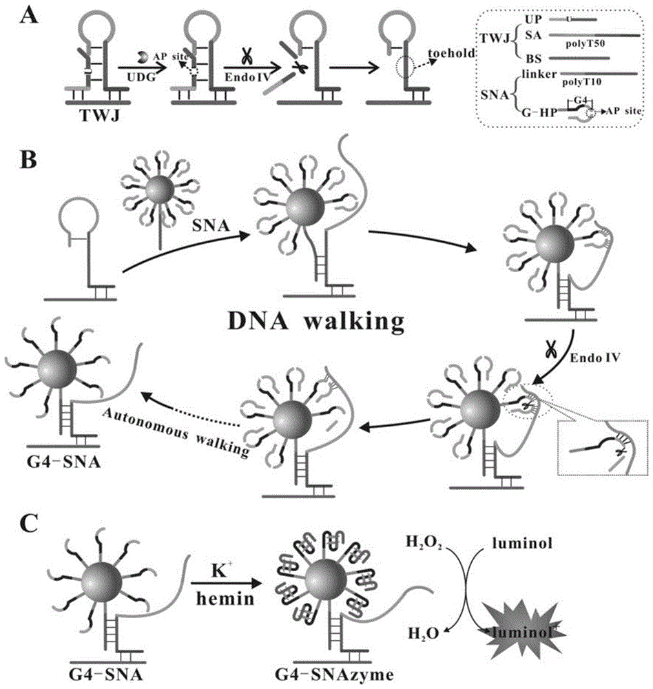
针对目前缺乏一种高效、灵敏便捷的检测尿嘧啶糖基化酶的方法的问题,本发明提供一种基于三通路结构驱动链置换反应及DNA walker技术驱动球形核酸酶的化学发光技术检测尿嘧啶糖基化酶,主要是包括构建三通路结构和球形核酸两种纳米结构,利用尿嘧啶糖基化酶对U碱基的特异性识别和切除,在内切酶IV的辅助下,实现三通路结构的变化和DNA walking反应的驱动,从而形成球形核酸酶用于催化鲁米诺发生反应产生化学发光信号,显著提高了检测的灵敏度和检测速度,更有利于尿嘧啶糖基化酶在实际样本中的检测。Aiming at the current lack of an efficient, sensitive and convenient method for detecting uracil glycosylase, the present invention provides a chemiluminescence technology for detecting uracil based on three-way structure-driven strand displacement reaction and DNA walker technology to drive spherical nuclease Glycosylase mainly includes the construction of two nanostructures of three-way structure and spherical nucleic acid, using uracil glycosylase to specifically recognize and excise U bases, and with the assistance of endonuclease IV, realize three-way Structural changes and DNA walking reactions drive the formation of spherical nucleases to catalyze the luminol reaction to generate chemiluminescent signals, which significantly improves the sensitivity and speed of detection, and is more conducive to the use of uracil glycosylase in actual samples detection in .
本发明是通过以下步骤得到的:The present invention is obtained through the following steps:
本发明中一共用到了5条DNA链,其序列分别是:In the present invention, a total of 5 DNA strands are used, and their sequences are respectively:
UP:AGT CAG TAT GCA CUC GTG TTA AGC GTG SA:TCT TTC GGC CGC GTT CAC GAGTGC ATC CGC GCT TGG G(T)50 G CTC GCC CAA GCG CGG BS:TTTCAC GCT TCG CGG CCG AAA GAT TT linker:SH-(T)10CA AGC GCG GAT GCA C G-HP:SH-(T)10GG GTA GGG CGG G TT GGG XGA GC T TTT CCC TAC。UP: AGT CAG T AT GCA CUC GTG TTA AGC GTG SA: TCT TTC GGC CGC G TT CAC GAGTGC ATC CGC GCT TGG G(T) 50 G CTC GCC CAA GCG CGG BS: TTTCAC GCT T CG CGG CCG AAA GA T TT linker : SH-(T) 10 CA AGC GCG GAT GCA C G-HP: SH-(T) 10 GG GTA GGG CGG G TT GGG XGA GC T TTT CCC TAC.
在UP链中,有一个尿嘧啶碱基U,UP链中的斜体碱基和SA链中的斜体碱基互补,从而封闭SA链的toehold部分和链迁移部分,UP链中的加粗碱基用于和BS链中的加粗碱基互补配对。在发夹SA链中,下划线的碱基和BS链中的下划线碱基互补配对,SA链中斜体加粗部分部分的碱基和linker加粗碱基是互补配对的,SA链中有连续50个胸腺嘧啶碱基作为间隔用于灵活控制摇摆臂,SA链中的斜体加下划线的碱基和G-HP中的斜体加下划线的碱基互补配对,该斜体加下划线的碱基有一部分被封闭在SA链的茎端。在G-HP链中,X代表AP位点,粗体碱基是能够形成G-四联体DNA酶的序列,该粗体碱基中画下划线的碱基被封闭在发夹G-HP中的茎端,降低背景信号。In the UP chain, there is a uracil base U, the italic base in the UP chain is complementary to the italic base in the SA chain, thereby blocking the toehold part and the chain migration part of the SA chain, and the bold base in the UP chain Used for complementary pairing with the bold bases in the BS strand. In the hairpin SA chain, the underlined bases are complementary to the underlined bases in the BS chain, and the bases in the bold italic part of the SA chain are complementary to the bold bases of the linker, and there are 50 consecutive bases in the SA chain. Thymine bases are used as spacers for flexible control of the swing arm. The italic underlined base in the SA chain is complementary to the italic underlined base in G-HP, and a part of the italic underlined base is blocked. at the stem end of the SA chain. In the G-HP chain, X represents the AP site, the bold base is the sequence capable of forming G-quadruplex DNase, and the underlined base in the bold base is blocked in the hairpin G-HP stem ends, reducing background signal.
体系中包含两种纳米结构:一种是三通路结构(TWJ),另一种是用于进行DNAwalker功能的球形核酸结构(SNA)。球形核酸(SNA)是以纳米金为核心,表面通过巯基修饰有两种链,一种链为包含AP位点并且含有被封闭的可形成G-四联体DNA酶的DNA发夹(G-HP),另一种链为能和三通路结构中的Swing arm(SA)链进行碱基互补的linker链。三通路结构(TWJ)是由发挥DNA walker功能的Swing arm(SA)链、一条目标物尿嘧啶糖基化酶特异性识别的UP探针链及一条基底链BS链三部分组成。UP链中包含一个尿嘧啶碱基U并且包含一段和BS链互补的序列,SA链的3’端包含一段和G-HP发夹中的环形loop端部分碱基互补的序列,一段含有50个胸腺嘧啶(polyT50)的spacer端,一段能和linker链互补的序列以及一段和BS链互补的序列这几部分组成。该SA链被设计为发夹构型,SA链的3’端和G-HP发夹中的环形loop端互补碱基有10对,其中有5对被封闭在发夹的茎端,中间有一个AP位点,这10对碱基杂交温度为23.1℃,因此在37℃反应体系中不能稳定存在,因此SA链不能进行DNAwalker反应。The system contains two nanostructures: one is a three-way structure (TWJ), and the other is a spherical nucleic acid structure (SNA) for DNAwalker function. Spherical nucleic acid (SNA) is based on nano-gold as the core, and the surface is modified by sulfhydryl groups. There are two kinds of chains. One chain is a DNA hairpin that contains AP sites and is closed to form G-quadruplex DNase (G- HP), and the other chain is a linker chain that can perform base complementarity with the Swing arm (SA) chain in the three-way structure. The three-way structure (TWJ) is composed of three parts: a Swing arm (SA) chain that functions as a DNA walker, a UP probe chain specifically recognized by the target uracil glycosylase, and a base chain BS chain. The UP chain contains a uracil base U and contains a sequence complementary to the BS chain, and the 3' end of the SA chain contains a sequence complementary to the loop end of the G-HP hairpin, and a section contains 50 bases The spacer end of thymine (polyT50), a sequence that can be complementary to the linker chain and a sequence that is complementary to the BS chain are composed of these parts. The SA chain is designed as a hairpin configuration, and there are 10 pairs of complementary bases between the 3' end of the SA chain and the loop end of the G-HP hairpin, 5 of which are closed at the stem end of the hairpin, and there are An AP site, the hybridization temperature of these 10 pairs of bases is 23.1°C, so it cannot exist stably in the reaction system at 37°C, so the SA chain cannot undergo DNAwalker reaction.
在存在目标物尿嘧啶糖基化酶时,目标物将UP探针中的U碱基去除,形成AP位 点,此时在内切酶IV(Endo IV)的辅助下,在AP位点处进行切割使得UP探针断裂,从 三通路体系中释放出来,暴露出被UP探针封闭的位于SA链中的toehold端,此时,球形核 酸中linkerDNA和SA链进行杂交,通过toehold介导的链置换反应使得SA链被打开,释 放出用于和G-HP杂交的部分,由于linker序列和SA序列的杂交,使得SA和G-HP的10 对碱基邻近,显著增加了局域浓度,由不稳定的分子间杂交变成稳定的分子内杂交,此时的 杂交温度为55.1℃。在这种情况下,内切酶IV作用于AP位点,将G-HP在AP位点处切 断,使得G-HP发夹结构被破坏,由于此时茎端杂交不再稳定,导致纳米金表面的G-HP仅 剩巯基结合的链,此时,SA链被释放,可以和下一个G-HP杂交进行后续的反应,直到将 纳米金表面的G-HP均切断,使得原先被封闭的可形成G-四联体的部分全部被释放出来,形 成G4-SNA结构,在体系中加入K+和血红素,可形成G-四联体球形核酸酶(G4- SNAzyme),在过氧化氢存在的情况下,G4-SNAzyme可催化鲁米诺的化学发光。由于整个 体系基于尿嘧啶糖基化酶的存在启动了toehold介导的链置换反应及后续的DNA walker反 应,进而形成G-四联体球形核酸酶结构,这种方式相比直接在球形核酸表面事先形成G-四 联体DNA酶而言,有效降低了背景信号,提高反应的信噪比。此外,由于采用DNA walker 纳米技术和球形核酸酶技术,从而使得化学发光信号显著放大。因此,我们设计了一种高 效、高灵敏性、高信噪比的化学发光生物传感器,为尿嘧啶糖基化酶的检测及后续的临床研 究提供了一个有效的技术平台。In the presence of the target uracil glycosylase, the target removes the U base in the UP probe to form an AP site. At this time, with the assistance of endonuclease IV (Endo IV), at the AP site The cleavage breaks the UP probe and releases it from the three-way system, exposing the toehold end in the SA chain that is blocked by the UP probe. At this time, the linkerDNA in the spherical nucleic acid hybridizes with the SA chain, through the toehold-mediated The chain displacement reaction causes the SA chain to be opened, releasing the part for hybridization with G-HP. Due to the hybridization of the linker sequence and the SA sequence, the 10 pairs of bases of SA and G-HP are adjacent, which significantly increases the local concentration. From unstable intermolecular hybridization to stable intramolecular hybridization, the hybridization temperature at this time is 55.1°C. In this case, endonuclease IV acts on the AP site and cuts the G-HP at the AP site, causing the G-HP hairpin structure to be destroyed. At this time, the hybridization of the stem end is no longer stable, resulting in gold nanoparticles The G-HP on the surface only has the thiol-bound chain left. At this time, the SA chain is released and can be hybridized with the next G-HP for subsequent reactions until the G-HP on the surface of gold nanoparticles is cut off, so that the previously blocked All the parts that can form the G-quadruplex are released to form the G4-SNA structure, and K + and heme are added to the system to form the G-quadruplex spherical nuclease (G4-SNAzyme). When present, G4-SNAzyme catalyzes the chemiluminescence of luminol. Since the whole system starts the toehold-mediated strand displacement reaction and the subsequent DNA walker reaction based on the existence of uracil glycosylase, and then forms the G-quadruplex spherical nuclease structure, this method is compared directly on the spherical nucleic acid surface. As far as the G-quadruplex DNase is formed in advance, the background signal is effectively reduced and the signal-to-noise ratio of the reaction is improved. In addition, due to the use of DNA walker nanotechnology and spherical nuclease technology, the chemiluminescent signal is significantly amplified. Therefore, we designed a chemiluminescent biosensor with high efficiency, high sensitivity, and high signal-to-noise ratio, which provides an effective technical platform for the detection of uracil glycosylase and subsequent clinical research.
本发明中尿嘧啶糖基化酶的检测是在均相溶液中实现的,通过三通路介导的链置换反应及DNA walker驱动的球形核酸酶的形成用于催化鲁米诺产生化学发光信号,从而实现尿嘧啶糖基化酶的高灵敏检测,并获得较低的检测下限。The detection of uracil glycosylase in the present invention is realized in a homogeneous solution, and the formation of the spherical nuclease driven by the three-pathway-mediated strand displacement reaction and DNA walker is used to catalyze luminol to generate a chemiluminescence signal, In this way, the highly sensitive detection of uracil glycosylase is achieved, and a lower detection limit is obtained.
一种检测尿嘧啶糖基化酶的化学发光生物传感器,包括以下原料:SA链、UP探针、BS链、1×Cutsmart缓冲液、内切酶 IV、标记好G-HP 及linker链的球形核酸SNA、尿嘧啶糖基化酶UDG、血红素、鲁米诺、过氧化氢;A chemiluminescent biosensor for detecting uracil glycosylase, including the following raw materials: SA chain, UP probe, BS chain, 1×Cutsmart buffer, endonuclease IV, spherical shape of labeled G-HP and linker chain Nucleic acid SNA, uracil glycosylase UDG, heme, luminol, hydrogen peroxide;
所述的UP碱基系列如SEQ No.1所示;所述的UP序列中5’端第十四个与第十五个碱基中间为尿嘧啶碱基U;The UP base series is shown in SEQ No.1; the middle of the 14th and 15th bases at the 5' end of the UP sequence is a uracil base U;
所述的SA碱基系列如SEQ No.2所示; 所述的BS碱基系列如SEQ No.3所示; 所述的linker碱基系列如SEQ No.4所示; 所述的G-HP碱基系列如SEQ No.5所示;所述的G-HP序列中5’端第十八个碱基为四氢呋喃脱碱基位点。The SA base series is shown in SEQ No.2; the BS base series is shown in SEQ No.3; the linker base series is shown in SEQ No.4; the G- The HP base series is shown in SEQ No.5; the eighteenth base at the 5' end of the G-HP sequence is a tetrahydrofuran abasic site.
所述的标记好G-HP 及linker链的球形核酸为VG-HP:Vlinker=20:1与纳米金混合物中加入PB缓冲液、 PBS缓冲液,最终溶液中NaCl的浓度为0.3 M。The spherical nucleic acid labeled with G-HP and linker chain is VG-HP:Vlinker =20:1 and nano-gold mixture with PB buffer and PBS buffer, and the concentration of NaCl in the final solution is 0.3 M.
上述的化学发光生物传感器的制备方法,包括以下步骤:The preparation method of the above-mentioned chemiluminescent biosensor comprises the following steps:
(1)纳米金的制备;(1) Preparation of gold nanoparticles;
(2)标记好G-HP 及linker链球形核酸SNA的制备;(2) Preparation of marked G-HP and linker streptospheric nucleic acid SNA;
(3)化学发光反应;(3) Chemiluminescence reaction;
所述的步骤(1)中的纳米金采用柠檬酸钠还原氯金酸工艺制备,纳米金尺寸为20nm,摩尔消光系数为0.878×10⁹ M-1·cm-1。The nano-gold in the step (1) is prepared by reducing chloroauric acid with sodium citrate, the size of the nano-gold is 20nm, and the molar extinction coefficient is 0.878×10⁹ M -1 ·cm -1 .
所述的步骤(2)的标记好G-HP 及linker链球形核酸SNA的制备方法,包括以下步骤:The preparation method of the marked G-HP and linker streptospheric nucleic acid SNA of described step (2), comprises the following steps:
S1调整纳米金溶液浓度为5nM;S1 adjusts the concentration of nano-gold solution to 5nM;
S2将G-HP 及linker链按照VG-HP:Vlinker=20:1加入到步骤S1的纳米金溶液中,加入PB缓冲液,分批加入 PBS,调整溶液中NaCl的浓度为0.3 M;S2 Adding G-HP and linker chains to the nano-gold solution in step S1 according to V G-HP:Vlinker =20:1, adding PB buffer solution, adding PBS in batches, adjusting the concentration of NaCl in the solution to 0.3 M;
S3离心去掉未被标记上的DNA链;S3 centrifugation to remove unlabeled DNA strands;
S4将离心后的沉淀物重新溶解超纯水里,置于4℃备用。S4 Redissolve the centrifuged precipitate in ultrapure water and store at 4°C for later use.
所述的步骤(3)的过程为:The process of the step (3) is:
J1将SA链、UP探针、 BS链加入到1×Cutsmart缓冲液中形成三通路体系;J1 Add SA strand, UP probe, and BS strand to 1×Cutsmart buffer to form a three-way system;
J2将内切酶 IV、标记好G-HP 及linker链的球形核酸、尿嘧啶糖基化酶UDG加入到J1三通路体系中;In J2, endonuclease IV, spherical nucleic acid labeled with G-HP and linker chain, and uracil glycosylase UDG are added to the J1 three-way system;
J3向J2中加入血红素、鲁米诺,形成G-四联体球形核酸酶;J3 adds heme and luminol to J2 to form G-quadruplex spherical nuclease;
J4向J3中加入过氧化氢立即用于化学发光信号的检测,化学发光信号采集时间间隔是1.5s,化学发光光谱测量范围是350nm到550nm。J4 adds hydrogen peroxide to J3 for immediate detection of chemiluminescence signal, the time interval of chemiluminescence signal acquisition is 1.5s, and the measurement range of chemiluminescence spectrum is 350nm to 550nm.
该发明的检测方式是通过化学发光信号的产生来进行尿嘧啶糖基化酶的检测,尿嘧啶糖基化酶会对U碱基的特异性识别和切除,在内切酶IV的辅助下,使得三通路结构发生变化,通过三通路结构的变化暴露出toehold端引发链置换反应,加入球形核酸后,在内切酶IV的作用下进行DNA walker反应,最终纳米金表面只包含可形成G-四联体的DNA序列(G4-SNA),再加入K+和血红素的条件下,可最终形成球形核酸酶(G4-SNAzyme)。该球形核酸酶可在双氧水存在的条件下催化鲁米诺发生化学发光反应。The detection method of the invention is to detect uracil glycosylase through the generation of chemiluminescence signal. Uracil glycosylase will specifically recognize and excise U bases. With the assistance of endonuclease IV, The structure of the three pathways is changed, and the toehold end is exposed through the change of the three pathways structure to trigger the strand displacement reaction. After adding the spherical nucleic acid, the DNA walker reaction is carried out under the action of endonuclease IV. Finally, the surface of the gold nanometer contains only the G- The DNA sequence of the quadruplex (G4-SNA) can finally form a spherical nuclease (G4-SNAzyme) under the condition of adding K + and heme. The spherical nuclease can catalyze the chemiluminescent reaction of luminol in the presence of hydrogen peroxide.
该传感器具有高效、高灵敏性、高特异性的优点,并且仅需借助内切酶IV这一种酶,可以弥补尿嘧啶糖基化酶现有检测方法的缺陷与不足,实现对其快速、准确的定量检测。The sensor has the advantages of high efficiency, high sensitivity, and high specificity, and only needs the enzyme of endonuclease IV, which can make up for the defects and deficiencies of the existing detection methods of uracil glycosylase, and realize rapid and accurate detection of uracil glycosylase. Accurate quantitative detection.
本发明的有益效果:Beneficial effects of the present invention:
1、本发明利用尿嘧啶糖基化酶对U碱基的特异性识别和切除,在内切酶IV的辅助下可将含有U碱基的DNA链切断,从而引发后续的反应,具有高特异性的特点;1. The present invention uses uracil glycosylase to specifically recognize and excise U bases, and with the assistance of endonuclease IV, it can cut off the DNA chain containing U bases, thereby triggering subsequent reactions, with high specificity sexual characteristics;
2、本发明借助三通路结构,可实现DNA链的有序组装,借助toehold介导的链置换反应可巧妙地进行DNA链的迁移,加快DNA链迁移的速率;2. With the help of the three-way structure, the present invention can realize the orderly assembly of DNA chains, and can skillfully carry out the migration of DNA chains by means of the strand displacement reaction mediated by toehold, so as to accelerate the rate of DNA chain migration;
3、本发明借助DNA walker纳米技术,可显著提高反应的效率并且能显著扩大信号,提高监测的灵敏度;3. With the help of DNA walker nanotechnology, the present invention can significantly improve the efficiency of the reaction and can significantly expand the signal and improve the sensitivity of monitoring;
4、本发明利用球形核酸酶,可实现将G-四联体DNA酶在纳米金表面的高度富集作用,从而实现信号的富集,可扩大信号,提高灵敏度;4. The present invention utilizes spherical nuclease to realize high enrichment of G-quadruplex DNase on the surface of gold nanometers, thereby realizing enrichment of signal, expanding signal and improving sensitivity;
5、本发明利用三通路驱动的链置换反应及DNA walker驱动的球形核酸酶的形成相比直接在纳米金表面标记G-四联体DNA链直接形成球形核酸酶而言可显著降低反应的背景信号,提高反应的信噪比;5. Compared with the direct formation of spherical nuclease by labeling the G-quadruplex DNA chain on the surface of gold nanometers, the present invention can significantly reduce the background of the reaction by using the strand displacement reaction driven by three pathways and the formation of spherical nuclease driven by DNA walker Signal, improve the signal-to-noise ratio of the reaction;
6、该传感器的反应条件温和,反应速度迅速。6. The reaction condition of the sensor is mild and the reaction speed is fast.
7、本发明的检测原理的主要过程均是在均相中实现的,提高了反应速度,降低了操作的复杂程度,实现了目标物的快速、简单、灵敏的检测;7. The main process of the detection principle of the present invention is realized in the homogeneous phase, which improves the reaction speed, reduces the complexity of the operation, and realizes the fast, simple and sensitive detection of the target object;
8、制备方法简单,性能稳定,适用于医疗卫生领域对于尿嘧啶糖基化酶的检测以及为后续肿瘤的治疗打下基础以及生物传感器产业化的实际应用;8. The preparation method is simple and the performance is stable, which is suitable for the detection of uracil glycosylase in the medical and health field, laying the foundation for subsequent tumor treatment and the practical application of biosensor industrialization;
9、制作该生物传感器的工艺成本低,适用于产业化中价廉的要求。9. The process cost of making the biosensor is low, which is suitable for the requirement of low price in industrialization.
附图说明Description of drawings
图1为该实验的原理图;Fig. 1 is the schematic diagram of this experiment;
图2为实施例1内切酶IV浓度优化检测结果图;Fig. 2 is
图3为实施例2血红素优化检测结果图;Fig. 3 is
图4为实施例3鲁米诺浓度优化检测结果图;Fig. 4 is
图5为实施例4过氧化氢浓度优化检测结果图;Fig. 5 is the optimized detection result figure of
图6为实施例5传感器检测的标准曲线;Fig. 6 is the standard curve that embodiment 5 sensor detects;
图7为实施例5传感器检测的浓度线性关系曲线。Fig. 7 is the concentration linear relationship curve detected by the sensor of Example 5.
具体实施方式Detailed ways
下面结合具体实施例对本发明进行进一步说明。The present invention will be further described below in conjunction with specific examples.
所述的生物传感器的制备方法,包括以下步骤:The preparation method of described biosensor comprises the following steps:
(1)纳米金的制备;(1) Preparation of gold nanoparticles;
(2)球形核酸的制备;(2) Preparation of spherical nucleic acid;
(3)均相中形成球形核酸酶用于催化鲁米诺的化学发光反应;(3) The formation of spherical nuclease in homogeneous phase is used to catalyze the chemiluminescent reaction of luminol;
所述的制备方法中,纳米金的制备:In the described preparation method, the preparation of nano gold:
纳米金的制备是根据柠檬酸钠还原氯金酸的方法来实现的。将500μL的氯金酸(0.04g/mL)加入到200mL超纯水里进行搅拌加热至煮沸后加入3mL的柠檬酸钠(1%)快速加入到煮沸的溶液里。随后,可以观察到溶液颜色由浅黄色变成黑色最终变成酒红色。在变成酒红色后继续加热15min来确保反应的完全进行。之后,纳米金溶液冷却到室温后放到4℃备用。20nm的纳米金紫外吸收峰约在520nm,摩尔消光系数为 0.878×10⁹ M-1•cm-1。The preparation of nano-gold is realized by reducing chloroauric acid with sodium citrate. Add 500μL of chloroauric acid (0.04g/mL) into 200mL of ultrapure water, stir and heat until boiling, then add 3mL of sodium citrate (1%) and quickly add to the boiling solution. Subsequently, it can be observed that the color of the solution changes from light yellow to black and finally to wine red. Continue heating for 15 min after turning wine red to ensure complete reaction. Afterwards, the nano-gold solution was cooled to room temperature and placed at 4°C for use. The ultraviolet absorption peak of 20nm nano gold is about 520nm, and the molar extinction coefficient is 0.878×10⁹ M -1 •cm -1 .
实施例1Example 1
球形核酸的制备:Preparation of spherical nucleic acids:
首先,纳米金原液在13000r/min,4℃条件下离心20min,之后去掉上清将底部沉淀分散在超纯水中使其浓度为5nM。然后,将150μL 10μM的DNA链(VG-HP:Vlinker=20:1)加入到纳米金混合物中,加完后放置24小时(4℃)。之后,在该混合物中加入50μL PB缓冲液(10mMPB,pH7.4)和27μL PBS(10mM PB,2M NaCl,pH7.4)。48小时(4℃)后,继续加入62μL的PBS,此时溶液中NaCl的浓度为0.3M。24小时后,将该混合物通过13000 r/min离心15min洗脱3次去掉未被标记上的DNA链。最后,将离心后的沉淀物重新溶解在100μL的超纯水里置于4℃备用。First, the nano-gold stock solution was centrifuged at 13000r/min and 4°C for 20min, then the supernatant was removed and the bottom precipitate was dispersed in ultrapure water to make the concentration 5nM. Then, 150 μL of 10 μM DNA chain (V G-HP:Vlinker = 20:1) was added to the nano-gold mixture, and left for 24 hours (4°C) after the addition. Afterwards, 50 μL of PB buffer (10 mM PB, pH 7.4) and 27 μL of PBS (10 mM PB, 2M NaCl, pH 7.4) were added to the mixture. After 48 hours (4°C), continue to add 62 μL of PBS, at this time the concentration of NaCl in the solution is 0.3M. After 24 hours, the mixture was eluted three times by centrifugation at 13000 r/min for 15 minutes to remove unlabeled DNA strands. Finally, the centrifuged precipitate was redissolved in 100 μL of ultrapure water and kept at 4°C for use.
至此已制备成球形核酸,均相溶液中反应过程的主要步骤如下:The spherical nucleic acid has been prepared so far, and the main steps of the reaction process in the homogeneous solution are as follows:
在45μL的反应体系中,事先将3μL Swing arm(SA)链(1μM)、3μL UP探针(1μM)及3μL BS链(1μM)加入到1×Cutsmart缓冲液中在37℃下反应半小时使其形成三通路结构,之后将1μL内切酶 IV(Endo IV,0.25U/μL、0.5U/μL、0.75U/μL、1U/μL、1.25U/μL)和标记好G-HP及linker链的球形核酸(SNA,7μL,1nM)及1μL尿嘧啶糖基化酶UDG加入到三通路体系中反应30min。完成DNA walker反应后,向体系中加入2μL血红素(1μM)、2μL鲁米诺(1mM),在37℃下反应30min使其形成G-四联体球形核酸酶,之后加入2μL过氧化氢(10mM)立即用于化学发光信号的检测。化学发光信号采集时间间隔是1.5s,化学发光光谱测量范围是350nm到550nm。In a 45 μL reaction system, 3 μL Swing arm (SA) chain (1 μM), 3 μL UP probe (1 μM) and 3 μL BS chain (1 μM) were added to 1×Cutsmart buffer and reacted at 37°C for half an hour. It forms a three-way structure, and then 1 μL endonuclease IV (Endo IV, 0.25U/μL, 0.5U/μL, 0.75U/μL, 1U/μL, 1.25U/μL) and labeled G-HP and linker chain Spherical nucleic acid (SNA, 7 μL, 1 nM) and 1 μL uracil glycosylase UDG were added to the three-way system and reacted for 30 minutes. After completing the DNA walker reaction, add 2 μL heme (1 μM) and 2 μL luminol (1 mM) to the system, react at 37°C for 30 minutes to form G-quadruplex spherical nuclease, and then add 2 μL hydrogen peroxide ( 10mM) was used immediately for the detection of chemiluminescent signal. The time interval of chemiluminescence signal acquisition is 1.5s, and the measurement range of chemiluminescence spectrum is 350nm to 550nm.
经检测,如图2,检测到的化学信号强度随着内切酶IV浓度的增加,S/N值逐渐增加,在内切酶IV浓度为1U/μL时S/N值最大,之后保持不变,因此选择1 U/μL的内切酶IV用于后续的反应。After testing, as shown in Figure 2, the detected chemical signal intensity increases with the increase of endonuclease IV concentration, and the S/N value gradually increases. The S/N value is the largest when the endonuclease IV concentration is 1U/μL, and then remains constant Therefore, 1 U/μL endonuclease IV was selected for subsequent reactions.
实施例2Example 2
球形核酸的制备:Preparation of spherical nucleic acids:
首先,纳米金原液在13000r/min,4℃条件下离心20min,之后去掉上清将底部沉淀分散在超纯水中使其浓度为5nM。然后,将150μL 10μM的DNA链(VG-HP:Vlinker=20:1)加入到纳米金混合物中,加完后放置24小时(4℃)。之后,在该混合物中加入50μL PB缓冲液(10 mMPB,pH7.4)和27μL PBS(10mM PB,2M NaCl,pH 7.4)。48小时(4℃)后,继续加入62μL的PBS,此时溶液中NaCl的浓度为0.3M。24小时后,将该混合物通过13000r/min离心15 min洗脱3次去掉未被标记上的DNA链。最后,将离心后的沉淀物重新溶解在100μL的超纯水里置于4℃备用。First, the nano-gold stock solution was centrifuged at 13000r/min and 4°C for 20min, then the supernatant was removed and the bottom precipitate was dispersed in ultrapure water to make the concentration 5nM. Then, 150 μL of 10 μM DNA chain (V G-HP:Vlinker = 20:1) was added to the nano-gold mixture, and left for 24 hours (4°C) after the addition. Afterwards, 50 μL of PB buffer (10 mMPB, pH 7.4) and 27 μL of PBS (10 mM PB, 2M NaCl, pH 7.4) were added to this mixture. After 48 hours (4°C), continue to add 62 μL of PBS, at this time the concentration of NaCl in the solution is 0.3M. After 24 hours, the mixture was centrifuged at 13,000 r/min for 15 min and eluted three times to remove unlabeled DNA strands. Finally, the centrifuged precipitate was redissolved in 100 μL of ultrapure water and kept at 4°C for use.
至此已制备成球形核酸,均相溶液中反应过程的主要步骤如下:The spherical nucleic acid has been prepared so far, and the main steps of the reaction process in the homogeneous solution are as follows:
在45μL的反应体系中,事先将3μL Swing arm(SA)链(1μM)、3μL UP探针(1μM)及3μL BS链(1μM)加入到1×Cutsmart缓冲液中在37℃下反应半小时使其形成三通路结构,之后将1μL内切酶IV(Endo IV,1U/μL)和标记好G-HP及linker链的球形核酸(SNA,7μL,1nM)及1μL尿嘧啶糖基化酶UDG加入到三通路体系中反应30 min。完成DNA walker反应后,向体系中加入2μL血红素(0.2μM、0.4μM、0.6μM、0.8μM、1.0μM、1.2μM)、2μL鲁米诺(1mM),在37℃下反应30min使其形成G-四联体球形核酸酶,之后加入过氧化氢(2μL,10mM)立即用于化学发光信号的检测。化学发光信号采集时间间隔是1.5s,化学发光光谱测量范围是350nm到550nm。In a 45 μL reaction system, 3 μL Swing arm (SA) chain (1 μM), 3 μL UP probe (1 μM) and 3 μL BS chain (1 μM) were added to 1×Cutsmart buffer and reacted at 37°C for half an hour. It forms a three-way structure, and then add 1 μL endonuclease IV (Endo IV, 1U/μL), spherical nucleic acid (SNA, 7 μL, 1 nM) labeled with G-HP and linker chain, and 1 μL uracil glycosylase UDG React in the three-way system for 30 min. After completing the DNA walker reaction, add 2 μL heme (0.2 μM, 0.4 μM, 0.6 μM, 0.8 μM, 1.0 μM, 1.2 μM) and 2 μL luminol (1 mM) to the system, react at 37 ° C for 30 min to form G-quadruplex globular nuclease was immediately followed by hydrogen peroxide (2 μL, 10 mM) for detection of the chemiluminescent signal. The time interval of chemiluminescence signal acquisition is 1.5s, and the measurement range of chemiluminescence spectrum is 350nm to 550nm.
经检测,如图3,检测到的化学发光信号强度随着血红素浓度的增加,阳性样品的化学发光信号显著增加,与此同时,血红素产生的背景信号也随之增加。 当血红素浓度为1μM时,产生的信噪比S/N值最大,因此选择1μM作为血After detection, as shown in Figure 3, the detected chemiluminescence signal intensity increases significantly with the increase of the hemoglobin concentration, and the chemiluminescence signal of the positive sample increases significantly, and at the same time, the background signal generated by the hemoglobin also increases. When the hemoglobin concentration is 1 μM, the signal-to-noise ratio S/N value generated is the largest, so 1 μM is selected as the hemoglobin concentration.
红素的最佳浓度。Optimal concentration of red pigment.
实施案例3
球形核酸的制备:Preparation of spherical nucleic acids:
首先,纳米金原液在13000r/min,4℃条件下离心20min,之后去掉上清将底部沉淀分散在超纯水中使其浓度为5nM。然后,将150μL 10μM的DNA链(VG-HP:Vlinker=20:1)加入到纳米金混合物中,加完后放置24小时(4℃)。之后,在该混合物中加入50μL PB缓冲液(10mMPB,pH7.4)和27μL PBS(10mM PB,2M NaCl,pH7.4)。48小时(4℃)后,继续加入62μL的PBS,此时溶液中NaCl的浓度为0.3 M。24小时后,将该混合物通过13000r/min离心15min洗脱3次去掉未被标记上的DNA链。最后,将离心后的沉淀物重新溶解在100μL的超纯水里置于4℃备用。First, the nano-gold stock solution was centrifuged at 13000r/min and 4°C for 20min, then the supernatant was removed and the bottom precipitate was dispersed in ultrapure water to make the concentration 5nM. Then, 150 μL of 10 μM DNA chain (V G-HP:Vlinker = 20:1) was added to the nano-gold mixture, and left for 24 hours (4°C) after the addition. Afterwards, 50 μL of PB buffer (10 mM PB, pH 7.4) and 27 μL of PBS (10 mM PB, 2M NaCl, pH 7.4) were added to the mixture. After 48 hours (4°C), continue to add 62 μL of PBS, at this time the concentration of NaCl in the solution is 0.3 M. After 24 hours, the mixture was centrifuged at 13,000 r/min for 15 min and eluted three times to remove unlabeled DNA strands. Finally, the centrifuged precipitate was redissolved in 100 μL of ultrapure water and kept at 4°C for use.
至此已制备成球形核酸,均相溶液中反应过程的主要步骤如下:The spherical nucleic acid has been prepared so far, and the main steps of the reaction process in the homogeneous solution are as follows:
在45μL的反应体系中,事先将3μL Swing arm(SA)链(1μM)、3μL UP探针(1μM)及3μL BS链(1μM)加入到1×Cutsmart缓冲液中在37℃下反应半小时使其形成三通路结构,之后将1μL内切酶 IV(Endo IV,1U/μL)和标记好G-HP 及linker链的球形核酸(SNA,7μL,1 nM)及1μL尿嘧啶糖基化酶UDG加入到三通路体系中反应30 min。完成DNA walker反应后,向体系中加入2μL血红素(1.0μM)、2μL鲁米诺(0.2mM、0.4mM、0.6mM、0.8mM、1mM、2mM、3mM),在37℃下反应30 min使其形成G-四联体球形核酸酶,之后加入过氧化氢(2μL,10 mM)立即用于化学发光信号的检测。化学发光信号采集时间间隔是1.5s,化学发光光谱测量范围是350nm到550nm。In a 45 μL reaction system, 3 μL Swing arm (SA) chain (1 μM), 3 μL UP probe (1 μM) and 3 μL BS chain (1 μM) were added to 1×Cutsmart buffer and reacted at 37°C for half an hour. It forms a three-way structure, and then add 1 μL endonuclease IV (Endo IV, 1U/μL) and labeled G-HP and linker chain spherical nucleic acid (SNA, 7 μL, 1 nM) and 1 μL uracil glycosylase UDG Add to the three-way system and react for 30 min. After completing the DNA walker reaction, add 2 μL heme (1.0 μM) and 2 μL luminol (0.2 mM, 0.4 mM, 0.6 mM, 0.8 mM, 1 mM, 2 mM, 3 mM) to the system, and react at 37 °C for 30 min. It forms G-quartet globular nuclease, followed by the addition of hydrogen peroxide (2 μL, 10 mM) immediately for detection of the chemiluminescent signal. The time interval of chemiluminescence signal acquisition is 1.5s, and the measurement range of chemiluminescence spectrum is 350nm to 550nm.
经检测,如图4,检测到的化学发光信号强度随着鲁米诺浓度的增加而逐渐增加,在浓度为1 mM时获得最大的信噪比,选择1mM的鲁米诺用于后续的反应。After detection, as shown in Figure 4, the detected chemiluminescent signal intensity gradually increases with the increase of luminol concentration, and the maximum signal-to-noise ratio is obtained when the concentration is 1 mM, and 1 mM luminol is selected for subsequent reactions .
实施例4Example 4
球形核酸的制备:Preparation of spherical nucleic acids:
首先,纳米金原液在13000r/min,4℃条件下离心20 min,之后去掉上清将底部沉淀分散在超纯水中使其浓度为5nM。然后,将150μL 10μM的DNA链(VG-HP:Vlinker=20:1)加入到纳米金混合物中,加完后放置24小时(4℃)。之后,在该混合物中加入50μL PB缓冲液(10mMPB,pH7.4)和27μL PBS(10mM PB,2M NaCl,pH7.4)。48小时(4℃)后,继续加入62μL的PBS,此时溶液中NaCl的浓度为0.3M。24小时后,将该混合物通过13000r/min离心15min洗脱3次去掉未被标记上的DNA链。最后,将离心后的沉淀物重新溶解在100μL的超纯水里置于4℃备用。First, the nano-gold stock solution was centrifuged at 13000r/min and 4°C for 20 min, then the supernatant was removed and the bottom precipitate was dispersed in ultrapure water to make the concentration 5nM. Then, 150 μL of 10 μM DNA chain (V G-HP:Vlinker = 20:1) was added to the nano-gold mixture, and left for 24 hours (4°C) after the addition. Afterwards, 50 μL of PB buffer (10 mM PB, pH 7.4) and 27 μL of PBS (10 mM PB, 2M NaCl, pH 7.4) were added to the mixture. After 48 hours (4°C), continue to add 62 μL of PBS, at this time the concentration of NaCl in the solution is 0.3M. After 24 hours, the mixture was centrifuged at 13,000 r/min for 15 min and eluted three times to remove unlabeled DNA strands. Finally, the centrifuged precipitate was redissolved in 100 μL of ultrapure water and kept at 4°C for use.
至此已制备成球形核酸,均相溶液中反应过程的主要步骤如下:The spherical nucleic acid has been prepared so far, and the main steps of the reaction process in the homogeneous solution are as follows:
在45μL的反应体系中,事先将3μL Swing arm(SA)链(1μM)、3μL UP探针(1μM)及3μL BS链(1μM)加入到1×Cutsmart缓冲液中在37℃下反应半小时使其形成三通路结构,之后将1μL内切酶IV(Endo IV,1U/μL)和标记好G-HP 及linker链的球形核酸(SNA,7μL,1nM)及1μL尿嘧啶糖基化酶UDG加入到三通路体系中反应30min。完成DNA walker反应后,向体系中加入血红素(2μL,1.0μM)、鲁米诺(2μL,1mM),在37℃下反应30min使其形成G-四联体球形核酸酶,之后加入过氧化氢(2μL,2mM、4mM、6mM、8mM、10mM、15mM、20mM)立即用于化学发光信号的检测。化学发光信号采集时间间隔是1.5s,化学发光光谱测量范围是350nm到550nm。In a 45 μL reaction system, 3 μL Swing arm (SA) chain (1 μM), 3 μL UP probe (1 μM) and 3 μL BS chain (1 μM) were added to 1×Cutsmart buffer and reacted at 37°C for half an hour. It forms a three-way structure, and then add 1 μL endonuclease IV (Endo IV, 1U/μL) and labeled G-HP and linker chain spherical nucleic acid (SNA, 7 μL, 1nM) and 1 μL uracil glycosylase UDG React in the three-way system for 30 minutes. After completing the DNA walker reaction, add heme (2 μL, 1.0 μM) and luminol (2 μL, 1 mM) to the system, react at 37 ° C for 30 min to form G-quadruplex spherical nuclease, and then add peroxide Hydrogen (2 μL, 2 mM, 4 mM, 6 mM, 8 mM, 10 mM, 15 mM, 20 mM) was used immediately for the detection of the chemiluminescence signal. The time interval of chemiluminescence signal acquisition is 1.5s, and the measurement range of chemiluminescence spectrum is 350nm to 550nm.
经检测,如图5,随着过氧化氢的浓度从2mM到20范围内逐渐升高,化学发光强度值也随之上升。可以看出,在过氧化氢浓度为10mM时该化学发光信噪比最大。因此,选择10mM的过氧化氢用于后续的反应。After testing, as shown in Figure 5, as the concentration of hydrogen peroxide gradually increased from 2mM to 20, the chemiluminescent intensity value also increased. It can be seen that the signal-to-noise ratio of the chemiluminescence is maximum when the concentration of hydrogen peroxide is 10 mM. Therefore, 10 mM hydrogen peroxide was selected for subsequent reactions.
实施例5Example 5
球形核酸的制备:Preparation of spherical nucleic acids:
首先,纳米金原液在13000r/min,4℃条件下离心20min,之后去掉上清将底部沉淀分散在超纯水中使其浓度为5nM。然后,将150μL 10μM 的DNA链(VG-HP:Vlinker=20:1)加入到纳米金混合物中,加完后放置24小时(4℃)。之后,在该混合物中加入50μL PB缓冲液(10mMPB,pH7.4)和27μL PBS(10mM PB,2M NaCl,pH7.4)。48小时(4℃)后,继续加入62μL的PBS,此时溶液中NaCl的浓度为0.3M。24小时后,将该混合物通过13000r/min离心15min洗脱3次去掉未被标记上的DNA链。最后,将离心后的沉淀物重新溶解在100μL的超纯水里置于4℃备用。First, the nano-gold stock solution was centrifuged at 13000r/min and 4°C for 20min, then the supernatant was removed and the bottom precipitate was dispersed in ultrapure water to make the concentration 5nM. Then, 150 μL of 10 μM DNA chain (V G-HP:Vlinker =20:1) was added to the nano-gold mixture, and left for 24 hours (4°C) after the addition. Afterwards, 50 μL of PB buffer (10 mM PB, pH 7.4) and 27 μL of PBS (10 mM PB, 2M NaCl, pH 7.4) were added to the mixture. After 48 hours (4°C), continue to add 62 μL of PBS, at this time the concentration of NaCl in the solution is 0.3M. After 24 hours, the mixture was centrifuged at 13,000 r/min for 15 min and eluted three times to remove unlabeled DNA strands. Finally, the centrifuged precipitate was redissolved in 100 μL of ultrapure water and kept at 4°C for use.
至此已制备成球形核酸,均相溶液中反应过程的主要步骤如下:The spherical nucleic acid has been prepared so far, and the main steps of the reaction process in the homogeneous solution are as follows:
在45μL的反应体系中,事先将3μL Swing arm(SA)链(1μM)、3μL UP探针(1μM)及3μL BS链(1μM)加入到1×Cutsmart缓冲液中在37℃下反应半小时使其形成三通路结构,之后将1μL内切酶IV(Endo IV,1U/μL)和标记好G-HP 及linker链的球形核酸(SNA,7μL,1nM)及1μL尿嘧啶糖基化酶UDG(1×10⁻³U/mL、5×10-3U/mL、1×10-2U/mL、5×10-2U/mL、1×10-1U/mL、5×10-1U/mL、1×100U/mL、5×100U/mL、1×101U/mL)加入到三通路体系中反应30min。完成DNA walker反应后,向体系中加入血红素(2μL,1.0μM)、鲁米诺(2μL,1mM),在37℃下反应30min使其形成G-四联体球形核酸酶,之后加入过氧化氢(2μL,10mM)立即用于化学发光信号的检测。化学发光信号采集时间间隔是1.5s,化学发光光谱测量范围是350nm到550nm。In a 45 μL reaction system, 3 μL Swing arm (SA) chain (1 μM), 3 μL UP probe (1 μM) and 3 μL BS chain (1 μM) were added to 1×Cutsmart buffer and reacted at 37°C for half an hour. It forms a three-way structure, and then add 1 μL endonuclease IV (Endo IV, 1U/μL) and labeled G-HP and linker chain spherical nucleic acid (SNA, 7 μL, 1nM) and 1 μL uracil glycosylase UDG ( 1×10 -3 U/mL, 5×10 -3 U/mL, 1×10 -2 U/mL, 5×10 -2 U/mL, 1×10 -1 U/mL, 5×10 -1 U/mL, 1×10 0 U/mL, 5×10 0 U/mL, 1×10 1 U/mL) were added to the three-way system and reacted for 30 minutes. After completing the DNA walker reaction, add heme (2 μL, 1.0 μM) and luminol (2 μL, 1 mM) to the system, react at 37 ° C for 30 min to form G-quadruplex spherical nuclease, and then add peroxide Hydrogen (2 μL, 10 mM) was used immediately for detection of chemiluminescent signal. The time interval of chemiluminescence signal acquisition is 1.5s, and the measurement range of chemiluminescence spectrum is 350nm to 550nm.
经检测,如图6,随着尿嘧啶糖基化酶的浓度从1×10⁻³U/mL到1×101U/mL范围内逐渐升高,化学发光强度值也随之上升。此外,如图7,尿嘧啶糖基化酶浓度的对数值与荧光强度成线性关系。After testing, as shown in Figure 6, as the concentration of uracil glycosylase gradually increased from 1×10 -3 U/mL to 1×10 1 U/mL, the chemiluminescence intensity also increased. In addition, as shown in Figure 7, the logarithmic value of the concentration of uracil glycosylase has a linear relationship with the fluorescence intensity.
上述实施例为本发明较佳的实施方式,但本发明的实施方式并不受实施例的限制,其它任何未背离本发明的精神实质与原理下所做的改变、修饰、组合、替代、简化均应为等效替换方式,都包含在本发明的保护范围之内。The above-mentioned embodiment is a preferred embodiment of the present invention, but the embodiment of the present invention is not limited by the embodiment, and any other changes, modifications, combinations, substitutions, and simplifications that do not deviate from the spirit and principles of the present invention All should be equivalent replacements, and all are included in the protection scope of the present invention.
序列表 sequence listing
<110> 济南大学<110> Jinan University
<120> 一种检测尿嘧啶糖基化酶的化学发光生物传感器及其制备方法与应用<120> A Chemiluminescent Biosensor for Detecting Uracil Glycosylase and Its Preparation Method and Application
<160> 5<160> 5
<170> SIPOSequenceListing 1.0<170> SIPOSequenceListing 1.0
<210> 1<210> 1
<211> 26<211> 26
<212> DNA<212>DNA
<213> 人工序列(artiartificial sequence)<213> artificial sequence (artiartificial sequence)
<400> 1<400> 1
agtcagtatg caccgtgtta agcgtg 26agtcagtatg caccgtgtta agcgtg 26
<210> 2<210> 2
<211> 103<211> 103
<212> DNA<212>DNA
<213> 人工序列(artiartificial sequence)<213> artificial sequence (artiartificial sequence)
<400> 2<400> 2
tctttcggcc gcgttcacga gtgcatccgc gcttgggttt tttttttttt tttttttttt 60tctttcggcc gcgttcacga gtgcatccgc gcttgggttttttttttttttttttttttt 60
tttttttttt tttttttttt tttttttgct cgcccaagcg cgg 103tttttttttttttttttttttttttttgct cgcccaagcg cgg 103
<210> 3<210> 3
<211> 26<211> 26
<212> DNA<212>DNA
<213> 人工序列(artiartificial sequence)<213> artificial sequence (artiartificial sequence)
<400> 3<400> 3
tttcacgctt cgcggccgaa agattt 26tttcacgctt cgcggccgaa agatt 26
<210> 4<210> 4
<211> 25<211> 25
<212> DNA<212>DNA
<213> 人工序列(artiartificial sequence)<213> artificial sequence (artiartificial sequence)
<400> 4<400> 4
tttttttttt caagcgcgga tgcac 25tttttttttt caagcgcgga tgcac 25
<210> 5<210> 5
<211> 41<211> 41
<212> DNA<212>DNA
<213> 人工序列(artiartificial sequence)<213> artificial sequence (artiartificial sequence)
<400> 5<400> 5
tttttttttt gggtagggcg ggttggggag cttttcccta c 41tttttttttt gggtagggcg ggttggggag cttttcccta c 41
Claims (2)
Priority Applications (1)
| Application Number | Priority Date | Filing Date | Title |
|---|---|---|---|
| CN201910891123.1A CN110607351B (en) | 2019-09-20 | 2019-09-20 | Chemiluminescence biosensor for detecting uracil glycosylase, and preparation method and application thereof |
Applications Claiming Priority (1)
| Application Number | Priority Date | Filing Date | Title |
|---|---|---|---|
| CN201910891123.1A CN110607351B (en) | 2019-09-20 | 2019-09-20 | Chemiluminescence biosensor for detecting uracil glycosylase, and preparation method and application thereof |
Publications (2)
| Publication Number | Publication Date |
|---|---|
| CN110607351A CN110607351A (en) | 2019-12-24 |
| CN110607351B true CN110607351B (en) | 2022-11-01 |
Family
ID=68891629
Family Applications (1)
| Application Number | Title | Priority Date | Filing Date |
|---|---|---|---|
| CN201910891123.1A Active CN110607351B (en) | 2019-09-20 | 2019-09-20 | Chemiluminescence biosensor for detecting uracil glycosylase, and preparation method and application thereof |
Country Status (1)
| Country | Link |
|---|---|
| CN (1) | CN110607351B (en) |
Families Citing this family (3)
| Publication number | Priority date | Publication date | Assignee | Title |
|---|---|---|---|---|
| CN112159853A (en) * | 2020-09-03 | 2021-01-01 | 华中农业大学 | A detection method of Huanglongwei based on aPCR and DNA walker |
| CN113584131B (en) * | 2021-07-20 | 2023-07-28 | 济南大学 | A colorimetric biosensor based on Au@Ag for detection of UDG |
| CN114214461B (en) * | 2021-12-26 | 2024-03-26 | 南京大学 | Isothermal HIV nucleic acid detection kit and detection method |
Citations (5)
| Publication number | Priority date | Publication date | Assignee | Title |
|---|---|---|---|---|
| CN104540965A (en) * | 2012-06-18 | 2015-04-22 | 斯比戴克斯私人有限公司 | Target detection and signal amplification |
| CN104884634A (en) * | 2012-09-11 | 2015-09-02 | 尤尼森斯诊断公司 | Detection of non-nucleic acid analytes using strand displacement exchange reactions |
| CN105087755A (en) * | 2015-09-08 | 2015-11-25 | 山东大学 | Application of DNA three-way regulation activation based hybridization chain reaction to high sensitivity detection of DNA methyltransferase |
| CN105132522A (en) * | 2015-09-08 | 2015-12-09 | 山东大学 | High-sensitivity uracil DNA glycosylase (UDG) detection using DNA three-direction section activated hybridization chain reaction |
| CN106995840A (en) * | 2017-03-20 | 2017-08-01 | 山东师范大学 | A kind of method of the dual signal amplification strategy detection thymidine DNA glycosylase activity mediated based on cyclophorase reparation |
Family Cites Families (1)
| Publication number | Priority date | Publication date | Assignee | Title |
|---|---|---|---|---|
| US8962241B2 (en) * | 2010-07-20 | 2015-02-24 | California Institute Of Technology | Triggered molecular geometry based bioimaging probes |
-
2019
- 2019-09-20 CN CN201910891123.1A patent/CN110607351B/en active Active
Patent Citations (5)
| Publication number | Priority date | Publication date | Assignee | Title |
|---|---|---|---|---|
| CN104540965A (en) * | 2012-06-18 | 2015-04-22 | 斯比戴克斯私人有限公司 | Target detection and signal amplification |
| CN104884634A (en) * | 2012-09-11 | 2015-09-02 | 尤尼森斯诊断公司 | Detection of non-nucleic acid analytes using strand displacement exchange reactions |
| CN105087755A (en) * | 2015-09-08 | 2015-11-25 | 山东大学 | Application of DNA three-way regulation activation based hybridization chain reaction to high sensitivity detection of DNA methyltransferase |
| CN105132522A (en) * | 2015-09-08 | 2015-12-09 | 山东大学 | High-sensitivity uracil DNA glycosylase (UDG) detection using DNA three-direction section activated hybridization chain reaction |
| CN106995840A (en) * | 2017-03-20 | 2017-08-01 | 山东师范大学 | A kind of method of the dual signal amplification strategy detection thymidine DNA glycosylase activity mediated based on cyclophorase reparation |
Non-Patent Citations (3)
| Title |
|---|
| A DNA walker powered by endogenous enzymes for imaging uracil-DNA glycosylase activity in living cells;Xiaowen Xu等;《Chem. Commun.》;20190426;第55卷;6026 * |
| Label-free thioflavin T/G-quadruplex-based real-time strand displacement amplification for biosensing applications;Yi-Chen Du等;《Biosensors andBioelectronics》;20160725;第86卷;811–817 * |
| 基于贵金属纳米材料的新型光学传感器构建及其应用于生物标志物检测研究;张雪;《中国优秀博硕士学位论文全文数据库(硕士) 基础科学辑》;20200115;A006-668 * |
Also Published As
| Publication number | Publication date |
|---|---|
| CN110607351A (en) | 2019-12-24 |
Similar Documents
| Publication | Publication Date | Title |
|---|---|---|
| Chen et al. | Photoactivatable CRISPR/Cas12a strategy for one-pot DETECTR molecular diagnosis | |
| CN110607351B (en) | Chemiluminescence biosensor for detecting uracil glycosylase, and preparation method and application thereof | |
| CN105755101B (en) | One kind detecting the active method of DNA glycosylases based on single quantum dot level | |
| CN101519696B (en) | Nucleic acid sensor based on quantum dots and preparation method and detection method thereof | |
| CN102220431A (en) | Probe for detecting nucleic acid in living cells and application method thereof | |
| CN113640268B (en) | Tobramycin detection system and detection method based on CRISPR-Cas12a | |
| CN109540860B (en) | Fluorescent biosensor for detecting kanamycin and preparation method and application thereof | |
| Li et al. | Ratiometric electrochemical biosensor based on silver nanoparticles coupled with walker amplification for sensitive detection of microRNA | |
| CN111676269A (en) | A kind of nucleic acid nanostructure probe and its preparation method and application | |
| CN108192948A (en) | A kind of method using alpha hemolysin nano-pore detection DNA glycosylase activity | |
| Liang et al. | Dithiothreitol-regulated coverage of oligonucleotide-modified gold nanoparticles to achieve optimized biosensor performance | |
| CN105525010A (en) | Stem-loop structured combined probe and application thereof | |
| CN110672694A (en) | Electrochemical method for detecting uracil-DNA glycosylase activity based on DNA NANOTREE | |
| CN118240923A (en) | An AND logic gate biosensor for detecting miRNA-21 and GSH based on rolling circle amplification | |
| Hu et al. | An initial check-reexamination strategy for analysis of H. Pylori DNA and single-nucleotide variants | |
| CN112359143A (en) | Isothermal index amplification method based on Y-type probe set and application thereof | |
| CN110057797A (en) | Method for detecting microRNA-155 based on mesh structure constructed by quantum dots | |
| CN106520972A (en) | Method for detecting nucleic acid concentration by using self-assembly system based on nucleic acid-platinum nano-material | |
| CN115896261A (en) | Spherical nucleic acid based on double-block DNA probe and preparation method and application thereof | |
| CN115851882B (en) | Method for detecting miRNA by using CRISPR/Cas12 a-driven controlled release homogeneous system | |
| CN118109564A (en) | APE1 enzyme high-sensitivity detection method based on biped DNA walker double probes | |
| CN107164366B (en) | Method for obtaining PCR product with double single-chain ends and detection method thereof | |
| CN113249443B (en) | Amplification detection method of prefabricated amplification unit based on DNA self-assembly | |
| JP3454847B2 (en) | Hybridization method | |
| CN111965148B (en) | Silicon nanoparticle fluorescence sensing-based microRNA detection method and application thereof |
Legal Events
| Date | Code | Title | Description |
|---|---|---|---|
| PB01 | Publication | ||
| PB01 | Publication | ||
| SE01 | Entry into force of request for substantive examination | ||
| SE01 | Entry into force of request for substantive examination | ||
| GR01 | Patent grant | ||
| GR01 | Patent grant |