CN110267673B - Optogenetic visual restoration using chrituson - Google Patents
Optogenetic visual restoration using chrituson Download PDFInfo
- Publication number
- CN110267673B CN110267673B CN201780041321.8A CN201780041321A CN110267673B CN 110267673 B CN110267673 B CN 110267673B CN 201780041321 A CN201780041321 A CN 201780041321A CN 110267673 B CN110267673 B CN 110267673B
- Authority
- CN
- China
- Prior art keywords
- light
- protein
- ion channel
- cells
- vector
- Prior art date
- Legal status (The legal status is an assumption and is not a legal conclusion. Google has not performed a legal analysis and makes no representation as to the accuracy of the status listed.)
- Active
Links
Images


Classifications
-
- A—HUMAN NECESSITIES
- A61—MEDICAL OR VETERINARY SCIENCE; HYGIENE
- A61K—PREPARATIONS FOR MEDICAL, DENTAL OR TOILETRY PURPOSES
- A61K38/00—Medicinal preparations containing peptides
- A61K38/16—Peptides having more than 20 amino acids; Gastrins; Somatostatins; Melanotropins; Derivatives thereof
- A61K38/17—Peptides having more than 20 amino acids; Gastrins; Somatostatins; Melanotropins; Derivatives thereof from animals; from humans
- A61K38/1703—Peptides having more than 20 amino acids; Gastrins; Somatostatins; Melanotropins; Derivatives thereof from animals; from humans from vertebrates
- A61K38/1709—Peptides having more than 20 amino acids; Gastrins; Somatostatins; Melanotropins; Derivatives thereof from animals; from humans from vertebrates from mammals
-
- A—HUMAN NECESSITIES
- A61—MEDICAL OR VETERINARY SCIENCE; HYGIENE
- A61K—PREPARATIONS FOR MEDICAL, DENTAL OR TOILETRY PURPOSES
- A61K48/00—Medicinal preparations containing genetic material which is inserted into cells of the living body to treat genetic diseases; Gene therapy
- A61K48/005—Medicinal preparations containing genetic material which is inserted into cells of the living body to treat genetic diseases; Gene therapy characterised by an aspect of the 'active' part of the composition delivered, i.e. the nucleic acid delivered
-
- A—HUMAN NECESSITIES
- A61—MEDICAL OR VETERINARY SCIENCE; HYGIENE
- A61K—PREPARATIONS FOR MEDICAL, DENTAL OR TOILETRY PURPOSES
- A61K35/00—Medicinal preparations containing materials or reaction products thereof with undetermined constitution
- A61K35/66—Microorganisms or materials therefrom
- A61K35/76—Viruses; Subviral particles; Bacteriophages
- A61K35/761—Adenovirus
-
- A—HUMAN NECESSITIES
- A61—MEDICAL OR VETERINARY SCIENCE; HYGIENE
- A61K—PREPARATIONS FOR MEDICAL, DENTAL OR TOILETRY PURPOSES
- A61K36/00—Medicinal preparations of undetermined constitution containing material from algae, lichens, fungi or plants, or derivatives thereof, e.g. traditional herbal medicines
- A61K36/06—Fungi, e.g. yeasts
-
- A—HUMAN NECESSITIES
- A61—MEDICAL OR VETERINARY SCIENCE; HYGIENE
- A61K—PREPARATIONS FOR MEDICAL, DENTAL OR TOILETRY PURPOSES
- A61K38/00—Medicinal preparations containing peptides
- A61K38/16—Peptides having more than 20 amino acids; Gastrins; Somatostatins; Melanotropins; Derivatives thereof
-
- A—HUMAN NECESSITIES
- A61—MEDICAL OR VETERINARY SCIENCE; HYGIENE
- A61K—PREPARATIONS FOR MEDICAL, DENTAL OR TOILETRY PURPOSES
- A61K38/00—Medicinal preparations containing peptides
- A61K38/16—Peptides having more than 20 amino acids; Gastrins; Somatostatins; Melanotropins; Derivatives thereof
- A61K38/168—Peptides having more than 20 amino acids; Gastrins; Somatostatins; Melanotropins; Derivatives thereof from plants
-
- A—HUMAN NECESSITIES
- A61—MEDICAL OR VETERINARY SCIENCE; HYGIENE
- A61K—PREPARATIONS FOR MEDICAL, DENTAL OR TOILETRY PURPOSES
- A61K38/00—Medicinal preparations containing peptides
- A61K38/16—Peptides having more than 20 amino acids; Gastrins; Somatostatins; Melanotropins; Derivatives thereof
- A61K38/17—Peptides having more than 20 amino acids; Gastrins; Somatostatins; Melanotropins; Derivatives thereof from animals; from humans
- A61K38/1767—Peptides having more than 20 amino acids; Gastrins; Somatostatins; Melanotropins; Derivatives thereof from animals; from humans from invertebrates
-
- A—HUMAN NECESSITIES
- A61—MEDICAL OR VETERINARY SCIENCE; HYGIENE
- A61K—PREPARATIONS FOR MEDICAL, DENTAL OR TOILETRY PURPOSES
- A61K41/00—Medicinal preparations obtained by treating materials with wave energy or particle radiation ; Therapies using these preparations
-
- A—HUMAN NECESSITIES
- A61—MEDICAL OR VETERINARY SCIENCE; HYGIENE
- A61K—PREPARATIONS FOR MEDICAL, DENTAL OR TOILETRY PURPOSES
- A61K9/00—Medicinal preparations characterised by special physical form
- A61K9/0012—Galenical forms characterised by the site of application
- A61K9/0019—Injectable compositions; Intramuscular, intravenous, arterial, subcutaneous administration; Compositions to be administered through the skin in an invasive manner
-
- A—HUMAN NECESSITIES
- A61—MEDICAL OR VETERINARY SCIENCE; HYGIENE
- A61K—PREPARATIONS FOR MEDICAL, DENTAL OR TOILETRY PURPOSES
- A61K9/00—Medicinal preparations characterised by special physical form
- A61K9/0012—Galenical forms characterised by the site of application
- A61K9/0048—Eye, e.g. artificial tears
-
- A—HUMAN NECESSITIES
- A61—MEDICAL OR VETERINARY SCIENCE; HYGIENE
- A61P—SPECIFIC THERAPEUTIC ACTIVITY OF CHEMICAL COMPOUNDS OR MEDICINAL PREPARATIONS
- A61P25/00—Drugs for disorders of the nervous system
-
- A—HUMAN NECESSITIES
- A61—MEDICAL OR VETERINARY SCIENCE; HYGIENE
- A61P—SPECIFIC THERAPEUTIC ACTIVITY OF CHEMICAL COMPOUNDS OR MEDICINAL PREPARATIONS
- A61P25/00—Drugs for disorders of the nervous system
- A61P25/02—Drugs for disorders of the nervous system for peripheral neuropathies
-
- A—HUMAN NECESSITIES
- A61—MEDICAL OR VETERINARY SCIENCE; HYGIENE
- A61P—SPECIFIC THERAPEUTIC ACTIVITY OF CHEMICAL COMPOUNDS OR MEDICINAL PREPARATIONS
- A61P27/00—Drugs for disorders of the senses
- A61P27/02—Ophthalmic agents
-
- A—HUMAN NECESSITIES
- A61—MEDICAL OR VETERINARY SCIENCE; HYGIENE
- A61P—SPECIFIC THERAPEUTIC ACTIVITY OF CHEMICAL COMPOUNDS OR MEDICINAL PREPARATIONS
- A61P43/00—Drugs for specific purposes, not provided for in groups A61P1/00-A61P41/00
-
- A—HUMAN NECESSITIES
- A61—MEDICAL OR VETERINARY SCIENCE; HYGIENE
- A61P—SPECIFIC THERAPEUTIC ACTIVITY OF CHEMICAL COMPOUNDS OR MEDICINAL PREPARATIONS
- A61P9/00—Drugs for disorders of the cardiovascular system
- A61P9/10—Drugs for disorders of the cardiovascular system for treating ischaemic or atherosclerotic diseases, e.g. antianginal drugs, coronary vasodilators, drugs for myocardial infarction, retinopathy, cerebrovascula insufficiency, renal arteriosclerosis
-
- C—CHEMISTRY; METALLURGY
- C07—ORGANIC CHEMISTRY
- C07K—PEPTIDES
- C07K14/00—Peptides having more than 20 amino acids; Gastrins; Somatostatins; Melanotropins; Derivatives thereof
- C07K14/405—Peptides having more than 20 amino acids; Gastrins; Somatostatins; Melanotropins; Derivatives thereof from algae
-
- C—CHEMISTRY; METALLURGY
- C12—BIOCHEMISTRY; BEER; SPIRITS; WINE; VINEGAR; MICROBIOLOGY; ENZYMOLOGY; MUTATION OR GENETIC ENGINEERING
- C12N—MICROORGANISMS OR ENZYMES; COMPOSITIONS THEREOF; PROPAGATING, PRESERVING, OR MAINTAINING MICROORGANISMS; MUTATION OR GENETIC ENGINEERING; CULTURE MEDIA
- C12N15/00—Mutation or genetic engineering; DNA or RNA concerning genetic engineering, vectors, e.g. plasmids, or their isolation, preparation or purification; Use of hosts therefor
- C12N15/09—Recombinant DNA-technology
- C12N15/11—DNA or RNA fragments; Modified forms thereof; Non-coding nucleic acids having a biological activity
- C12N15/62—DNA sequences coding for fusion proteins
- C12N15/625—DNA sequences coding for fusion proteins containing a sequence coding for a signal sequence
-
- C—CHEMISTRY; METALLURGY
- C12—BIOCHEMISTRY; BEER; SPIRITS; WINE; VINEGAR; MICROBIOLOGY; ENZYMOLOGY; MUTATION OR GENETIC ENGINEERING
- C12N—MICROORGANISMS OR ENZYMES; COMPOSITIONS THEREOF; PROPAGATING, PRESERVING, OR MAINTAINING MICROORGANISMS; MUTATION OR GENETIC ENGINEERING; CULTURE MEDIA
- C12N15/00—Mutation or genetic engineering; DNA or RNA concerning genetic engineering, vectors, e.g. plasmids, or their isolation, preparation or purification; Use of hosts therefor
- C12N15/09—Recombinant DNA-technology
- C12N15/63—Introduction of foreign genetic material using vectors; Vectors; Use of hosts therefor; Regulation of expression
- C12N15/79—Vectors or expression systems specially adapted for eukaryotic hosts
- C12N15/85—Vectors or expression systems specially adapted for eukaryotic hosts for animal cells
- C12N15/86—Viral vectors
- C12N15/864—Parvoviral vectors, e.g. parvovirus, densovirus
- C12N15/8645—Adeno-associated virus
-
- A—HUMAN NECESSITIES
- A61—MEDICAL OR VETERINARY SCIENCE; HYGIENE
- A61K—PREPARATIONS FOR MEDICAL, DENTAL OR TOILETRY PURPOSES
- A61K48/00—Medicinal preparations containing genetic material which is inserted into cells of the living body to treat genetic diseases; Gene therapy
-
- C—CHEMISTRY; METALLURGY
- C07—ORGANIC CHEMISTRY
- C07K—PEPTIDES
- C07K2319/00—Fusion polypeptide
- C07K2319/60—Fusion polypeptide containing spectroscopic/fluorescent detection, e.g. green fluorescent protein [GFP]
-
- C—CHEMISTRY; METALLURGY
- C12—BIOCHEMISTRY; BEER; SPIRITS; WINE; VINEGAR; MICROBIOLOGY; ENZYMOLOGY; MUTATION OR GENETIC ENGINEERING
- C12N—MICROORGANISMS OR ENZYMES; COMPOSITIONS THEREOF; PROPAGATING, PRESERVING, OR MAINTAINING MICROORGANISMS; MUTATION OR GENETIC ENGINEERING; CULTURE MEDIA
- C12N2750/00—MICROORGANISMS OR ENZYMES; COMPOSITIONS THEREOF; PROPAGATING, PRESERVING, OR MAINTAINING MICROORGANISMS; MUTATION OR GENETIC ENGINEERING; CULTURE MEDIA ssDNA viruses
- C12N2750/00011—Details
- C12N2750/14011—Parvoviridae
- C12N2750/14111—Dependovirus, e.g. adenoassociated viruses
- C12N2750/14141—Use of virus, viral particle or viral elements as a vector
- C12N2750/14143—Use of virus, viral particle or viral elements as a vector viral genome or elements thereof as genetic vector
Landscapes
- Health & Medical Sciences (AREA)
- Life Sciences & Earth Sciences (AREA)
- Chemical & Material Sciences (AREA)
- Engineering & Computer Science (AREA)
- Genetics & Genomics (AREA)
- General Health & Medical Sciences (AREA)
- Medicinal Chemistry (AREA)
- Bioinformatics & Cheminformatics (AREA)
- Organic Chemistry (AREA)
- Animal Behavior & Ethology (AREA)
- Pharmacology & Pharmacy (AREA)
- Public Health (AREA)
- Veterinary Medicine (AREA)
- Biotechnology (AREA)
- Biomedical Technology (AREA)
- Molecular Biology (AREA)
- Epidemiology (AREA)
- Zoology (AREA)
- General Engineering & Computer Science (AREA)
- Wood Science & Technology (AREA)
- Gastroenterology & Hepatology (AREA)
- Proteomics, Peptides & Aminoacids (AREA)
- Biophysics (AREA)
- Biochemistry (AREA)
- Microbiology (AREA)
- Chemical Kinetics & Catalysis (AREA)
- Nuclear Medicine, Radiotherapy & Molecular Imaging (AREA)
- General Chemical & Material Sciences (AREA)
- Immunology (AREA)
- Plant Pathology (AREA)
- Physics & Mathematics (AREA)
- Ophthalmology & Optometry (AREA)
- Virology (AREA)
- Mycology (AREA)
- Botany (AREA)
- Natural Medicines & Medicinal Plants (AREA)
- Neurology (AREA)
- Neurosurgery (AREA)
- Marine Sciences & Fisheries (AREA)
- Dermatology (AREA)
Abstract
Methods for reactivating mammalian retinal ganglion cells, including but not limited to, comprising administering an effective amount of channel rhodopsin in the form of a protein or nucleic acid (e.g., chrismsonr) or an effective amount of such channel rhodopsin fused to a fluorescent protein in the form of a protein or nucleic acid (e.g., chrismsonr) or a combination thereof. The method may include inducing an RGC response at a light stimulus level below a radiation safety limit. The method may comprise administration by an adeno-associated viral vector. The method may comprise using a CAG promoter. The methods may result in long-term expression of an effective amount of channel rhodopsin (e.g., chrismsonr protein).
Description
Cross reference to related applications
The present application claims priority for U.S. provisional application No. 62/329,692 filed on date 2016, 4, 29, the contents of which are incorporated by reference in their entirety.
Sequence listing
The present application contains a sequence listing that is electronically filed in ASCII format and incorporated by reference in its entirety. The copy of the ASCII format file was created on 28 th month 4 of 2017, named "12295_0006-00304.Txt" and was 31 bytes in size.
Technical Field
The present invention provides compositions and methods for altering transmembrane conductance, cellular activity and cellular function, including but not limited to, and to the use of exogenous photoactivated ion channels in cells and subjects of interest. In particular, one aspect according to particular embodiments of the invention relates to a method for reactivating mammalian retinal neuronal cells (RGCs), the method comprising administering to a mammal an effective amount of a Chrismson polypeptide. In some embodiments, the method may include inducing an RGC response at a light stimulus level below a radiation safety margin. In some embodiments, the chrisson polypeptide is fused to a fluorescent protein. In some embodiments, the fluorescent protein is tdTomato (tdT) or Green Fluorescent Protein (GFP).
Background
The retina consists of photoreceptors, which are highly specialized neurons responsible for the photosensitivity of the retina by light transduction, which refers to the conversion of light into electrical and chemical signals that propagate a series of events within the visual system, ultimately producing an image of the world. In the retina of vertebrates, light transduction is initiated by activation of the photoreceptor protein rhodopsin.
Loss or degeneration of photoreceptors, such as Retinitis Pigmentosa (RP) or Macular Degeneration (MD), can severely reduce, if not completely inhibit, the light transduction of visual information within the retina. The loss of photoreceptor cells and/or the loss of photoreceptor cell function are the primary causes of decreased visual acuity, decreased photosensitivity and blindness.
Several therapies specific to retinal degenerative diseases are now under development, including gene therapy, stem cell therapy, optogenetics and retinal prostheses (Scholl et al, 2016, science Translational Medicine, 8 (368), 368, 6 th revision).
For example, it has been proposed to restore photosensitivity to the target subject's retina by controlling the activity of a prescribed number of neurons without affecting other neurons in the brain through genetic and neuropsychological techniques known as optogenetics. In contrast to conventional gene therapies that attempt to replace or repair defective genes or bypass gene defects by correcting protein deletions or dysfunctions, optogenetically treating can be used to confer the ability to respond to light in cells in the retina that are generally insensitive to light, thereby restoring useful vision to the patient. Unlike retinal chip grafts, which provide extracellular stimulation to bipolar or ganglion cells, optogenetic-based therapies stimulate cells from within the cell.
Optogenetics (Deisserith, nat Methods 8 (1): 26-9, 2011) refers to a combination of genetics and optics to control well-defined events within specific cells in an organism's tissue. Optogenetics involves the introduction of light-sensitive channels into cells that allow neural activity to be manipulated with millisecond precision, while maintaining cell type resolution (cell-type resolution) through specific targeting mechanisms. It involves the discovery of a gene that confers light responsiveness and the insertion of the gene into a cell. It also includes related techniques for delivering light into complex, e.g., mammalian, organisms, targeting photosensitivity to cells of interest, and assessing specific output results or effects of the optical control.
For example, WO2007024391, WO2008022772 or WO2009127705 describe the use of protease genes from plants and microorganisms (such as archaebacteria, bacteria and fungi) encoding light-sensitive ion channels and ion pumps (such as channel rhodopsin [ ChR2]; chlororhodopsin [ NpHR ]) engineered to be expressed in mammalian neurons and capable of implanting specific neurological groups at the gene level by viral vectors. When exposed to light of the appropriate wavelength, action potentials can trigger in neurons expressing opsin proteins, rendering these cells photosensitive.
In recent years, some of the channel rhodopsins from the four channel rhodopsins genes of chlamydomonas reinhardtii (Chlamydomonas reinhardtii) or of spirulina (Volvox carteri) have been engineered for neuroscience applications. However, these natural channel rhodopsins have only blue-green (430-550 nm) spectral peaks, and the peak wavelength sensitivity of the engineered red-shifted channel rhodopsins (e.g., C1V1 and ReaChR) is in the green spectrum (-545 nm) (mattis et al, nature Methods, 2011, 12 months 18; 9 (2): 159-72; lin et al, nature Neuroscience, 2013, 10 months; 16 (10): 1499-508).
In 2014, klapoetke et al (Nat Methods, 11 (3), 338-346) have therefore sought to overcome these drawbacks by exploring the genetic diversity of natural channel rhodopsin, aiming at the discovery of novel opsins with unique properties not found in the aforementioned channel rhodopsin. Thus, WO2013071231 discloses new channel rhodopsin Chronos and chrisms on which have different activation spectra from each other and from the prior art (e.g. ChR 2/VChRl) and allow depolarization of different cell populations in the same tissue using a plurality of different wavelengths of light, by: by expressing channels with different activation spectra in different cells, the tissue is then irradiated with light of different colors. More specifically, chrisson has a red shift of 45nm compared to any previous channel rhodopsin; this may be important where red light is preferred, as it is scattered more weakly by the tissue and absorbed less by the blood than the blue to green wavelengths required for other channel rhodopsin variants.
Opsins are typically fused to fluorescent proteins to facilitate visualization in cells expressing the opsins, thereby monitoring their intracellular localization. There are further cases where certain types of fluorescent proteins used may modulate opsin cell localization under specific conditions. For example, arrenberg et al (2009, PNAS, 106 (42), 17968-73) observed that fusion proteins containing the same opsin but different fluorescent tags (i.e., red fluorescent protein mCherry or yellow fluorescent protein YFP) sometimes distributed in different cellular compartments.
However, this finding was not demonstrated on tdTomato fluorescent tags, as no significant differences in expression levels or membrane localization were found in transgenic animals expressing the two-channel rhodopsin fused to tdTomato (Madisten et al 2012, nat Neurosci., 15 (5): 793-802). Furthermore, to date, there has been no report of any improvement in opsin activity associated with changes in the localization or expression level of fusion proteins.
Disclosure of Invention
In one embodiment, the present invention shows that chrisson protein, particularly a specific mutant thereof known as ChrimsonR (ChrR), when fused to tdTomato (tdT) fluorescent protein or Green Fluorescent Protein (GFP) responds to light stimulation more effectively than chrisson protein alone. In some embodiments of the methods, for a given number of cells, the fluorescent protein increases the expression level of the fused chrisson protein, particularly on the plasma membrane, compared to the expression level of the chrisson protein alone/unfused. In other embodiments of the methods, the fluorescent protein increases the cell transport capacity of the fused chrisson to the plasma membrane compared to the cell transport of the chrisson protein alone/unfused. In some embodiments of the methods, the expression level and/or cell trafficking capacity of the fused chrisson protein is increased by enhanced chrisson protein solubility, trafficking capacity, and/or protein conformation.
In one aspect, the invention includes polynucleotide sequences encoding chrisson proteins and fluorescent proteins.
In another aspect, the invention includes a polynucleotide sequence encoding a chrisson protein fused to a fluorescent protein.
In another aspect, the invention includes a composition comprising a carrier. The vector includes a polynucleotide sequence encoding a polypeptide including at least one chrisson protein and a fluorescent protein.
In another aspect, the invention includes a composition comprising a polynucleotide sequence encoding a polypeptide comprising a chrisson protein fused to a fluorescent protein.
In another aspect, the invention includes a method for treating or preventing a subject-targeted neuronal mediated disorder, wherein the method comprises administering to a cell (i.e., neuron) a composition comprising a carrier. The vector includes a polynucleotide sequence encoding a polypeptide including at least one chrisson protein and a fluorescent protein. Preferably, the vector of the composition to be administered comprises a polynucleotide sequence encoding a polypeptide comprising a chrismson protein fused to a fluorescent protein.
In another aspect, the invention includes a method of restoring light sensitivity to cells within the retina. The method comprises administering to the cell a composition comprising a carrier. The vector includes a polynucleotide sequence encoding a polypeptide including at least one chrisson protein and a fluorescent protein. Preferably, the vector of the composition to be administered comprises a polynucleotide sequence encoding a polypeptide comprising a chrismson protein fused to a fluorescent protein.
In another aspect, the invention includes a method of restoring vision to a subject. The method includes confirming that the subject lost vision due to insufficient light perception or sensitivity; administering to the eye a composition comprising a vector comprising a polynucleotide sequence encoding a polypeptide comprising at least one chrismson protein and a fluorescent protein; activating the polypeptide with light; the light sensitivity of the subject is determined, wherein an increased light sensitivity is indicative of vision recovery.
In another aspect, the invention includes a method of restoring vision to a subject, wherein the method includes confirming that the subject has lost vision due to insufficient light perception or sensitivity; administering to the eye a composition comprising a vector comprising a polynucleotide sequence encoding a polypeptide comprising at least one chrisson protein fused to a fluorescent protein; activating the polypeptide with light; the light sensitivity of the subject is determined, wherein an increased light sensitivity is indicative of vision recovery.
In other aspects, the invention includes methods of treating or preventing retinal degeneration in a subject of interest. The method comprises confirming that the subject is retinal degeneration due to loss of photoreceptor function; administering to the eye a composition comprising a vector comprising a polynucleotide sequence encoding a polypeptide comprising at least one chrismson protein and a fluorescent protein; and determining the photosensitivity of the subject, wherein increased photosensitivity is indicative of efficacy in retinal degeneration.
In another aspect, the invention includes a method of treating or preventing retinal degeneration in a subject of interest. The method comprises confirming that the subject is retinal degeneration due to loss of photoreceptor function; administering a composition comprising a vector comprising a polynucleotide sequence encoding a polypeptide comprising at least one chrismson protein fused to a fluorescent protein; and determining the photosensitivity of the subject, wherein increased photosensitivity is indicative of efficacy in retinal degeneration.
In certain aspects, the invention encompasses a method of restoring the function of human eye photoreceptors. The method comprises administering an effective amount of a composition comprising a vector comprising a polynucleotide sequence encoding a polypeptide comprising at least one chrismson protein and a fluorescent protein.
In another aspect, the invention includes a method of restoring the function of photoreceptors in the human eye. The method comprises administering an effective amount of a composition comprising a vector comprising a polynucleotide sequence encoding a polypeptide comprising at least one chrisson protein fused to a fluorescent protein.
In other aspects, the invention includes a method of depolarizing an electroactive cell. The method comprises administering to the cell a composition comprising a vector comprising a polynucleotide sequence encoding a polypeptide comprising at least one chrismson protein and a fluorescent protein.
In another aspect, the invention includes a method of depolarizing an electroactive cell. The method comprises administering to the cell a composition comprising a vector comprising a polynucleotide sequence encoding a polypeptide comprising at least one chrisson protein fused to a fluorescent protein.
In some embodiments of the methods according to the invention, the vector is an adeno-associated virus (AAV) vector. In some embodiments of the methods according to the invention, the vector is an aav2.7m8 vector or an AAV2 vector. In some embodiments, the method further comprises using a CAG promoter.
In some embodiments, the carrier is administered by injection, preferably by vitreous injection.
In some embodiments of the methods according to the invention, an effective amount of chrisson protein is expressed chronically. In some embodiments of the methods according to the invention, chrisson protein is continuously expressed at least 11 months after injection. In some embodiments of the methods according to the invention, chrisson protein is continuously expressed at least 2 months after injection.
In some embodiments of the methods according to the invention, the subject of interest is a mammal. In some embodiments, the subject of interest is a human. In some embodiments, the mammal is a mouse. In some embodiments of the method according to the invention, the mouse is rd1. In some embodiments of the methods according to the invention, the mammal is a rat. In some embodiments of the methods according to the invention, the rat is P23H. In some embodiments of the methods according to the invention, the mammal is a human or a non-human primate. In some embodiments of the methods according to the invention, the non-human primate is a cynomolgus macaque. The following disclosure also provides additional embodiments:
Embodiment 7 provides a method of restoring photoreceptor function to the human eye, wherein the method comprises confirming that a subject of interest has lost vision due to insufficient light perception or sensitivity, and administering to the subject of interest a composition comprising a vector that expresses an effective amount of Chrimson protein fused to a fluorescent protein.
Embodiment 9 provides the method of any one of embodiments 1-8, wherein a light stimulus level below a radiation safety limit induces an RGC response.
Embodiment 13 provides the method of embodiment 10, wherein the fluorescent protein increases the expression level of the fused chrisson protein compared to the expression level of chrisson protein alone for a given number of cells.
Embodiment 17 provides the method of embodiment 16, wherein the AAV vector is an AAV2.7m8 vector.
Embodiment 21 provides the method of embodiment 20, wherein the chrisson protein fused to a fluorescent protein is continuously expressed at least 2 months or at least 11 months after administration.
Drawings
Fig. 1: methods in rdl mice.
Fig. 2A to 2D: denatured rdl mice retina respond to light with wavelengths matching the chrismonr spectral sensitivity for a duration of less than 10ms. FIG. 2A-fundus of rdl mice expressing ChrR-tdT 2 months after injection. FIG. 2B-TdT fluorescence of rdl mouse retina mounted on MEA chip. Fig. 2C-spectral sensitivity of mouse retina expressing ChrR (n=l retina, 188 electrodes). FIG. 2D-jet density le for a wavelength of 590nm 17 photons.cm -2 s -1 Increased discharge frequency (firing rate) in response to stimuli of increasing duration. All recordings were made in the presence of a mixture of L-AP4, CNQX and CCP.
Fig. 3A to 3C: chrismonR was more effective in fusion with tdT in rdl mice. FIG. 3A-retinal comparison of infection with either ChrR or ChrR-tdT, chrR-tdT responds more effectively to light stimulation. FIG. 3B-raw data, grid plot and average PSTH (top to bottom, respectively) of response RGCs of retinas expressing ChrR-tdT. Fig. 3C-intensity plots of retinas expressing ChrR (n=4 retinas, 27 cells) or ChrR-tdT (n=6 retinas, 548 cells), showing the level of activation at different stimulation intensities.
Fig. 4A to 4G: expression of chrismsonr in ganglion cells. Expression of ChrR-tdT in rdl mouse Retinal Ganglion Cells (RGCs). The projections of FIGS. 4A, 4B and 4C-confocal stack (RGC) show membrane-localized expression in two examples of RGCs. FIG. 4A-images of tdTomato generated without immune magnification. FIG. 4B-labeling image of our custom-made ChrR antibodies. Fig. 4C-overlap of two images (fig. 4A and 4B), magenta and cyan are tdmamato and ChrR antibodies, respectively. The image was taken with a 40x objective. The expression of ChrR-tdT is enriched in RGC membranes. FIGS. 4D and 4E-show projections of three optical sheets of two RGC cell bodies (see the small diagram in FIG. 4C) taken with a 60x objective lens. FIGS. 4F and 4G-4D and 4E are 3D curved views of the respective fluorescence intensities of cell bodies. The peak representing the highest fluorescence intensity is concentrated at or near the cell membrane.
Fig. 5A to 5D: chrisson R long term expression. Multi-electrode arrays of rdl mice 10 months after injection were recorded. FIG. 5A-retinal image of the expression of ChrR-tdT, showing that expression persisted 10 months after injection. Figure 5B-grid plot of activity measured on one electrode, top-red light stimulation, middle-same cell response repeated 10 flashes, bottom-average PTSH (block size: SOms). Fig. 5C-increased discharge frequency in response to flash light of increasing intensity (n=4 retinas, 308 electrodes). FIG. 5D-jet density le for 590nm 17 photons.cm -2 s -1 The discharge frequency is increased by the flash response with increasing duration. All recordings were made in the presence of a mixture of L-AP4, CNQX and CCP.
Fig. 6A to 6B: chrison R re-activates the P23H retina. A multi-electrode array of another denatured rodent model, P23H rats, was recorded. FIG. 6A-P23H retinal fluorescence image on a multi-electrode array 1 month after injection. FIG. 6B-jet density le for a wavelength of 590nm 17 photons.cm -2 s -1 Increased discharge frequency in response to stimulus of increasing intensity (n=2 retinas, 91 electrodes). All recordings were made in the presence of a mixture of L-AP4, CNQX and CCP.
Fig. 7: methods in non-human primates. The expression of ChrR in non-human primates (cynomolgus macaques) was examined using four different strategies. 2 different constructs: chrimsonR (ChrR) or the fusion protein ChrissonR-td-Tomato (Chrr R-tdT), all under the action of the CAG promoter. 2 different viral capsids: wild-type AAV2 and mutant AAV2-7m8 (Dalkara et al, 2013, science Translational Medicine, 5 (189): 189ra 76). Single virus doses (5X 10) were taken two months prior to MEA (512 array, MCS) or patch clamp (see Master Chaffiol et al, abstract 599-B0072) recordings 11 vg/eye). All recordings were made in the presence of synaptic blocking agents (LAP 4. Mu.M and CPP 10. Mu.M).
Fig. 8A to 8C: chrisson R was expressed in perifovea (perifovea) after in vivo injection of the construct. In vivo injection of the construct results in expression in RGCs of the surrounding concave ring. Fig. 8A-infrared image of retinal explants, asterisks indicate the concavity of the surrounding pits. Black dots are electrodes of the MEA array. FIG. 8B-fluorescence image of the same retinal sheet, infected with AAV2.7m8-ChrR-tdT construct. Expression is limited to the surrounding concave ring. Spectral sensitivity of retinal explants shown in fig. 8C-8A and 8B. The response is the average of every 10 replicates for all response electrodes. The shape of the spectrum and the presence of synaptic blocking agents indicate that the ChrR in RGCs is the source of recorded activity.
Fig. 9A to 9G: the test construct that resulted in the most efficient transduction was determined. Transduction was assessed by the number of response electrodes and the sensitivity of the light-induced response. Fig. 9A-an example of the response of one electrode to 4 flashes of different intensities. FIG. 9B-4 summary of the set of experiments for constructs. Active electrode: an electrode that detects an action potential. Response electrode: an electrode for increasing the discharge frequency by optical stimulation. FIG. 9C-, FIG. 9D and FIG. 9E-each response retina responded to a population of different constructs. Each color line represents a single electrode response, which is an average of every 10 replicates. Each row of the graph represents the response of one retina, each column of responses of different retinas being directed to the same optical stimulus (intensity unit at the top is photons/cm 2 /sec). FIG. 9F-average of light intensity for each response retina for different light intensitiesIncreased discharge frequency. The natural discharge frequency has been subtracted. Fig. 9G-9F are enlarged detail views to better illustrate the response threshold. All stimuli were performed at 600 nm.
Fig. 10A to 10D: retinas infected with AAV2.7m8-ChR-tdT responded to increasing duration of periconcave RGC stimulation. In retinas infected with AAV2.7m8-chrR-tdT, periconcave RGCs responded to increasing stimulation of time length. Figure 10A-response to light stimulation with increasing time length, each line representing the average of 10 replicates of the individual electrode spike density function for each stimulation. Fig. 10B-average discharge frequency for all measured lengths. Fig. 10C-4 different activity thresholds fraction of active sites at different stimulation durations. Figure 10D-time to first peak, is the average of each 10 stimulus replicates over all measured lengths. The red dots represent the median values, with the edges of the box being 25% and 75% of the data, the remainder being omitted except for outliers drawn separately. The median importance decrease between 1 and 5 ms stimulation suggests that most of the recording sites begin to respond within these durations. All stimuli were 600+/-20nm at an intensity of 2xl0 17 photons.cm -2 .s -1 。
Fig. 11: effect of tdmamato on chrison mRNA levels. Amplification curve of chrismonr in RT-qPCR reaction. The Y-axis represents the incremental Rn value corresponding to the experimental response minus the Rn value of the baseline signal. This parameter reliably calculates the magnitude of a particular signal generated by a given set of PCR primers. Traces of magenta and purple represent chrismsonr; the yellow and orange traces represent ChrismsonR-tdTomato; the dark blue and light blue traces are non-transfectable controls. Experiments were repeated 3 times, each run on 2 plates, for a total of 6 replicates. Three experiments were performed on each panel for each sample.
Fig. 12A to 12B: HEK293 cells and pssAAV-CAG-ChrismsonR-tdTomato, pssAAV-CAG-ChrismsonR and ChrismsonR protein levels at the time of transfection of the pssAAV-CAG-ChrismsonR-GFP plasmid.
Fig. 13: tdmamato affects the number of cells expressing chrismsonr. The percentage of ChrissonR positive cells represents the ratio of cells transfected with plasmids 479 (ChrissonR-tdTomato) and 480 (ChrissonR) to non-transfected controls. The percentage of fluorescent cells is determined by eliminating background fluorescence using a threshold. It should be noted that the number of cells does not represent the fluorescence intensity of each cell. Based on this cell counting method, there was no statistically significant difference from the percentage of cells expressing chrismsonr after transfection of both constructs. In this experiment, error bars represent SEM (standard error of mean), the experiment was repeated 3 times, and the technique was repeated 3 times under each condition.
Fig. 14A to 14B: effect of tdmamato on chrison subcellular localization in HEK293T cells. Images of transfected HEK293T cells; obtained by maximum projection of confocal Z-stack (Z-stack). Nuclei are shown in blue (DAPI) and chrison R is shown in white. FIG. 14A shows the positioning of Chrisson R-tdTomato; fig. 14B shows the distribution of chrismsonr alone. The scale bar is 20 μm.
Fig. 15A to 15B: effect of tdmamto on chrison subcellular localization in HEK293T cells after AAV infection. Images of transfected HEK293T cells; obtained by maximum projection of confocal Z-stack (Z-stack). Nuclei are shown in blue (DAPI) and chrison R is shown in white. FIG. 15A shows the localization of Chrisson R-dtTomato; fig. 15B shows the distribution of chrismsonr alone. The scale bar is 20 μm. Detailed Description
In the present invention, the use of the singular includes the plural, "a" or "an" meaning "at least one" unless specifically stated otherwise, and the use of "or" meaning "and/or". In addition, the use of the terms "include" and "comprise (in general past)" and "include (in terms of noun)" and other forms are not limiting. Furthermore, unless specifically stated otherwise, terms such as "element" or "component" include both elements and components comprising one unit and elements or components comprising more than one unit.
The term "about" as used herein, when used with a percentage or other quantity, refers to the percentage or other quantity plus or minus 10%. For example, "about 80%" includes 80% plus or minus 8%.
All documents or portions of documents cited in this application, including but not limited to patents, patent applications, articles, books, and treatises, are expressly incorporated herein by reference for any purpose. If one or more of the incorporated references and similar materials contradict the definition of a term in this application, the definition of this application controls.
The terms "protein", "polypeptide" and "peptide" as used herein are interchangeable unless otherwise indicated.
As used herein, the term "fusion protein" or "protein fused to another. It refers to a single protein molecule comprising two or more proteins or fragments thereof, covalently linked by peptide bonds in their respective peptide chains, without additional chemical linkers. One protein may be fused to another protein at the N-terminus or C-terminus. The fusion protein may further comprise a linker moiety resulting from the genetic construct.
As used herein, unless otherwise indicated, the terms "treat," "treatment," and "therapy" refer to an action that occurs when a subject of interest has a disease (e.g., a neuronal mediated disorder or vision disorder), thereby reducing the severity of one or more symptoms or the effects of the disease. As used herein, unless otherwise indicated, terms such as "prevent", "preventing" and "preventing" refer to actions that occur before a subject begins to suffer from a disease (e.g., a neuronal-mediated disorder or vision disorder), delay the onset of the disease, and/or inhibit or reduce the severity of the disease. It will be appreciated that the treatment may be prophylactic or may be performed after diagnosis of a disease or condition. The treatment of the present invention may reduce or eliminate symptoms or features of a disorder, disease or condition, as well as eliminate the disorder, disease or condition itself. It will be appreciated that the methods of treatment of the present invention may slow or eliminate the progression of a disease, or disorder condition, and in some cases may result in the reversal of the disease, disorder, or condition. In some embodiments of the invention, one or more of the light-activated ion channel polypeptides of the invention can be expressed in a population of cells and used in a method of treating a disorder or condition.
As used herein, unless otherwise indicated, a "therapeutically effective amount" of a compound is an amount sufficient to provide any therapeutic benefit in the treatment or management of a neuronal-mediated disorder or vision disorder, or to delay or reduce one or more symptoms associated with a disorder (e.g., a neuronal-mediated disorder or vision disorder). A therapeutically effective amount of a compound refers to an amount of the compound that provides any therapeutic benefit in the treatment or management of a disorder (e.g., a neuronal mediated disorder or a visual disorder), alone or in combination with one or more other therapies and therapeutic agents. The term "therapeutically effective amount" may include an amount that reduces a neuronal mediated disorder or vision disorder, improves or reduces a vision disorder, improves overall treatment, or improves the efficacy of another therapeutic agent.
As used herein, "patient" or "subject of interest" includes mammals, such as humans and non-human mammals, non-limiting examples of non-human mammals include rodents, mice, rats, non-human primates, companion animals (e.g., dogs and cats), and livestock (e.g., sheep, cattle, horses, etc.), that are suffering from or susceptible to the diseases described herein.
Retinal neurons, preferably bipolar cells and/or ganglion cells, are provided with photosensitive membrane channels by transfection of retinal neurons with nucleic acids (e.g., vectors) encoding chrisson polypeptides according to the invention. Thus, the transmission of visual stimuli to the animal visual cortex, which is the area of the brain responsible for processing visual signals that constitute a form of vision, can be measured with light stimuli as contemplated by the present invention. This visual aspect may be different from the normal form of human vision, and may also be referred to as the perception of light, also referred to as "light detection" or "light perception". Thus, the term "visual" as used herein is defined as the ability of an organism to effectively detect light as a stimulus. "visual" is intended to encompass the following aspects: light detection or sensing, i.e., the ability to discern whether light is present; (II) light projection, i.e., the ability to discern where the light stimulus comes from; (III) resolution, i.e., the ability to detect different brightness levels (i.e., contrast) in a grid or letter object; (IV) identification, namely the ability to identify the shape of the visual target by reference to different contrasts within the target. Thus, "visual" includes the ability to simply detect the presence of light, preferably red light, more preferably between about 365nm and about 700nm, between about 530nm and about 640nm, and in some embodiments, peak activation may occur upon contact with light having a wavelength of about 590 nm.
As used herein, "functional derivative" includes "mutant", "variant" and "fragment", whether these terms are used in combination or interchangeably. Although conservative substitutions of, for example, 2, 3, 4, or 5 residues are also within the spirit of the invention, preferred variants are single amino acid conservative substitution variants. In some embodiments, the functional derivative is at least 70% homologous, preferably at least 75% homologous, more preferably at least 80% homologous, more preferably at least 85% homologous, more preferably at least 90% homologous, more preferably at least 95% homologous, more preferably at least 99% homologous, more preferably 100% homologous to the full-length amino acid sequence of the original polypeptide. The percentage of homology is determined based on the length of the relevant amino acid sequence. Thus, if a polypeptide according to the invention is comprised within a larger polypeptide, the percentage of homology is determined only for the portion of the polypeptide corresponding to the polypeptide according to the invention, and not for the entire larger polypeptide. "percent homology" in relation to polypeptide sequences refers to the percentage of identical amino acids between at least two polypeptide sequences that are aligned using the base partial alignment search tool (BLAST) engine. See Tatusova et al (1999) (from the past). The BLAST engine is provided to the public by the National Center for Biotechnology Information (NCBI) of bescens, maryland. According to a particular embodiment, the functional derivative is a polypeptide comprising an amino acid sequence having at least 70% homology to the full length sequence of the original polypeptide, wherein it differs from the parent polypeptide only by substitution at one or more positions. The substitutions are preferably "conservative substitutions" or "semi-conservative". In addition, or alternatively, the functional derivative has at least 70% identity, preferably at least 75% identity, more preferably at least 80% identity, more preferably at least 85% identity, more preferably at least 90% identity, more preferably at least 95% identity, more preferably at least 99% identity, more preferably 100% identity to the full-length amino acid sequence of the original polypeptide. Methods for determining sequence identity or homology are known in the art.
As used herein, the term "conservative substitution" generally refers to an amino acid substitution that preserves the structural and functional properties of a protein or polypeptide. Such functionally equivalent (conservative substitutions) peptide amino acid sequences include, but are not limited to, the addition or substitution of amino acid residues in the amino acid sequence encoded by the nucleotide sequence that results in a silent change (silent change), thereby producing a functionally equivalent gene product. Conservative amino acid substitutions may be made based on the similarity in polarity, charge, solubility, hydrophobicity, hydrophilicity, and/or the amphipathic nature of the residues involved. For example, nonpolar (hydrophobic) amino acids include alanine, leucine, isoleucine, valine, proline, phenylalanine, tryptophan, and methionine; polar neutral amino acids include glycine, serine, threonine, cysteine, tyrosine, asparagine, and glutamine; positively charged (basic) amino acids include arginine, lysine and histidine; and negatively charged (acidic) amino acids include aspartic acid and glutamic acid.
The present invention relates in certain aspects to the expression of a light activated ion channel polypeptide in a cell, which may be activated by contact with one or more light pulses, resulting in a strong depolarization of the cell. The light activated channel polypeptides, also referred to as light activated ion channels, according to the present invention can be expressed in specific cells, tissues and/or organisms and used to control the response of cells in vivo, ex vivo and in vitro to light pulses of appropriate wavelength.
In the present invention, the term "ion channel" refers to a pore-forming transmembrane polypeptide that opens when activated, allowing ionic conductance through the pore through the membrane. According to the invention, the light activated ion channel polypeptides include chrismson proteins or functional derivatives thereof, as well as fluorescent proteins.
According to the invention, the photoactivated ion channel polypeptides comprise a chrismson protein or a functional derivative thereof fused to a fluorescent protein.
According to a specific embodiment, the Chrisson protein is selected from the group consisting of the protein Chrismson 88 (also referred to as Chrismson 88-SEQ ID No: 1) or a functional derivative thereof, and the Chrismson 88 protein substituted at K176R (also referred to herein as Chrismson 88 or Chrismson R-SEQ ID No: 2) or a functional derivative thereof, having a substitution of K176R.
According to the invention, the light activated ion channel polypeptides comprise (i) a ChR88 protein (SEQ ID No: 1) or a functional derivative thereof, and (ii) a fluorescent protein. According to a preferred embodiment, the light-activated ion channel polypeptide according to the invention comprises (i) a ChrismsonR protein (SEQ ID No: 2) or a functional derivative thereof and (ii) a fluorescent protein.
According to a particular embodiment, the light-activated ion channel polypeptides according to the invention consist of the Chr88 protein (SEQ ID No: 1) or a functional derivative thereof and a fluorescent protein, both proteins being expressed as separate proteins.
According to another embodiment, the light-activated ion channel polypeptide according to the invention consists of the ChrismsonR protein (SEQ ID No: 2) or a functional derivative thereof and a fluorescent protein, both proteins being expressed as separate proteins.
According to a preferred embodiment, the light-activated ion channel polypeptides according to the invention consist of the Chr88 protein (SEQ ID No: 1) or a functional derivative fused to a fluorescent protein.
According to a more preferred embodiment, the light-activated ion channel polypeptide according to the invention consists of the ChrismsonR protein (SEQ ID No: 2) or a functional derivative thereof fused to a fluorescent protein.
The light-activated ion channel polypeptides according to the invention are strongly activated by contact with red light, preferably with light having a wavelength between about 365nm and about 700nm, with light having a wavelength between about 530nm and about 640nm, and in some embodiments, peak activation occurs when contacted with light having a wavelength of about 590 nm.
Contacting an excitable cell comprising a light activated ion channel polypeptide according to the invention with light having a wavelength in the activation range will strongly depolarize the cell. Exemplary wavelengths of light useful for depolarizing cells expressing a light activated ion channel polypeptide according to the invention include at least about 365nm, 385nm, 405nm, 425nm, 445nm, 465nm, 485nm, 505nm, 525nm, 545nm, 565nm, 585nm;590nm, 605nm, 625nm, 645nm, 665nm, 685nm; and wavelengths of 700nm, including all wavelengths between those described above. In some embodiments, the photoactive ion channel polypeptides according to the present invention have a peak wavelength sensitivity at 590nm and may cause spikes as early as 660 nm.
The light-activated ion channel polypeptides according to the invention can be used to depolarize excitable cells in which one or more light-activated ion channels according to the invention are expressed. In some embodiments, a light activated ion channel polypeptide according to the invention may be expressed in a cell subpopulation in a cell population that further includes one or more additional cell subpopulations that express light activated ion channels that are activated by light wavelengths that do not activate a light activated ion channel polypeptide according to the invention.
Peptide amino acid sequences useful in various embodiments include the photoactivated ion channel polypeptides (SEQ ID No:1 or 2, or 5) of the present invention as well as functionally equivalent polypeptides.
Such functionally equivalent peptide amino acid sequences (conservative substitutions) include, but are not limited to, the addition or substitution of amino acid residues in the amino acid sequences according to the invention, but this results in a silent change, thereby producing a functionally equivalent polypeptide. Amino acid substitutions may be made based on the similarity of polarity, charge, solubility, hydrophobicity, hydrophilicity, and/or the amphipathic nature of the residues involved. For example, nonpolar (hydrophobic) amino acids include alanine, leucine, isoleucine, valine, proline, phenylalanine, tryptophan, and methionine; polar neutral amino acids include glycine, serine, threonine, cysteine, tyrosine, asparagine, and glutamine; positively charged (basic) amino acids include arginine, lysine and histidine; and negatively charged (acidic) amino acids include aspartic acid and glutamic acid. Conservative amino acid substitutions may also be made based on the hydropathic index of amino acids. Each amino acid is assigned a hydropathic index based on its hydrophobicity and charge characteristics. They are isoleucine (+4.5); valine (+4.2); leucine (+3.8); phenylalanine (+2.8); cysteine/cystine (+2.5); methionine (+1.9); alanine (+1.8); glycine (-0.4); threonine (-0.7); serine (-0.8); tryptophan (-0.9); tyrosine (-1.3); proline (-1.6); histidine (-3.2); glutamic acid (-3.5); glutamine (-3.5); aspartic acid (-3.5); asparagine (-3.5); lysine (-3.9); and arginine (-4.5). The use of hydrophilic amino acid indices to confer interactive biological functions on proteins is known in the art (Kyte and Doolittle, J.mol. Biol., 157:105-132, 1982). It is known that in some cases, certain amino acids may be replaced with other amino acids having similar hydropathic indices or scores and still retain similar biological activity. In making modifications based on similar hydrophilicity indices, amino acid substitutions with a hydrophilicity index in the range of + -2 are included in certain embodiments, amino acid substitutions with a hydrophilicity index in the range of + -1 are included in other embodiments, and amino acid substitutions with a hydrophilicity index in the range of + -0.5 are included in other embodiments.
Conservative amino acid substitutions may also be made on the basis of hydrophilicity, particularly where the resulting biologically functional protein or peptide is intended for immunological embodiments. In certain embodiments, the maximum local average hydrophilicity of a protein (determined by the hydrophilicity of its adjacent amino acids) is related to its immunogenicity and antigenicity, i.e., to the biological properties of the protein. These amino acid residues are assigned the following hydrophilic values arginine (+3.0); lysine (+3.0); aspartic acid (+3.0+ -1); glutamic acid (+3.0+ -1); serine (+0.3); asparagine (+0.2); glutamine (+0.2); glycine (0); threonine (-0.4); proline (-0.5+ -1); alanine (-0.5); histidine (-0.5); cysteine (-1.0); methionine (-1.3); valine (-1.5); leucine (-1.8); isoleucine (-1.8); tyrosine (-2.3); phenylalanine (-2.5) and tryptophan (-3.4). In making modifications based on similar hydrophilicity values, amino acid substitutions having a hydrophilicity value within the range of + -2 are included in certain embodiments, amino acid substitutions having a hydrophilicity value within the range of + -1 are included in other embodiments, and amino acid substitutions having a hydrophilicity value within the range of + -0.5 are included in other embodiments.
According to a preferred embodiment, the light-activated ion channel polypeptide according to the invention is a fusion protein of a chrisson polypeptide (e.g. ChR88 protein or a functional derivative thereof, or chrison protein or a functional derivative thereof) and a fluorescent protein. The present invention describes the use of fusion proteins in which a polypeptide or peptide or truncated or mutated version of a peptide is fused to an unrelated protein, polypeptide or peptide and can be designed based on the nucleic acid and/or amino acid sequence encoding the desired peptide. In certain embodiments, the fusion protein may be readily purified by utilizing antibodies that selectively bind to the expressed fusion protein.
In general, the retinal or retinal derivative required for the function of the light activated ion channel polypeptides according to the invention is produced by the cells to be transfected with the channel polypeptides. However, according to the present invention, there is further disclosed a channel rhodopsin and a retinaldehyde or retinaldehyde derivative comprising a light activated ion channel polypeptide according to the present invention, e.g. 3, 4-dehydroretinaldehyde, 13-ethylretinaldehyde, 9-dm-retinaldehyde, 3-hydroxyretinaldehyde, 4-hydroxyretinaldehyde, naphtyl-retinaldehyde; 3,7,1 l-trimethyl-dodecane-2, 4,6,8, 10-pentaenal; 3, 7-dimethyl-decane-2, 4,6, 8-tetraenal; 3, 7-dimethyl-octane-2, 4, 6-trienal; and 6-7-or 8-9-or 10-11-rotation-blocked retinoids (WO 03084994).
Although the desired peptide amino acid sequences described herein may be chemically synthesized (see, e.g., proteins: structures and Molecular Principles "(Cright on, eds., W.H.Freeman, N.Y., 1984), large polypeptide sequences may preferably be produced by recombinant DNA techniques using techniques well known in the art for expressing nucleic acids containing a nucleic acid sequence encoding a desired peptide, such methods may be used to construct expression vectors containing a nucleotide sequence encoding a peptide and appropriate transcriptional and translational control signals, such methods include, e.g., in vitro recombinant DNA techniques, synthetic techniques and in vivo genetic recombination (see, e.g., molar Cloning, A Laboratory Manual", see above, and Current Protocols in Molecular Biology, see above), or alternatively, RNA and/or DNA encoding a nucleotide sequence encoding a desired peptide may be chemically synthesized using, e.g., a synthesizer (see, e.g., oligonucleotide Synthesis: APractical Approach (Gait, editorial, IRL Press, UK, 1984)).
Peptide amino acid sequences useful in various embodiments include the photoactivated ion channel polypeptides (SEQ ID No:1 or 2, 5 or 6) of the present invention and functionally equivalent peptides and functional derivatives thereof, as well as functional fragments thereof. Indeed, in some embodiments, any desired peptide amino acid sequence encoded by a particular nucleotide sequence may be used as well as polynucleotide sequences encoding all or any portion of the desired peptide amino acid sequence. The degenerate nature of gene coding is well known and, accordingly, each nucleotide sequence encoding an amino acid of a photoactivation channel polypeptide is a well known nucleic acid "triplet" codon or, in many cases, represents a general representation of a codon that can encode an amino acid. Thus, as contemplated by the present invention, the channel rhodopsin peptide amino acid sequences described herein, if combined with gene coding (see, e.g., molecular Cell Biology, 109 th page table 4-1 (Darnell et al, edit, w.h.freeman, new york, 1986)), are general representations of all various permutations and combinations of nucleic acid sequences capable of encoding such amino acid sequences.
Some embodiments are isolated nucleic acid molecules comprising a nucleotide sequence encoding a light activated ion channel polypeptide according to the invention. In some embodiments, the nucleotide sequence encodes a polypeptide comprising (i) a ChR88 protein (SEQ ID No: 1) or a functional derivative thereof, and (ii) a fluorescent protein. In other embodiments, the nucleotide sequence encodes a polypeptide comprising (i) a ChrismsonR protein (SEQ ID No: 2) or a functional derivative thereof, and (ii) a fluorescent protein.
According to a particular embodiment, the nucleotide sequence encodes a polypeptide consisting of the ChR88 protein (SEQ ID No: 1) or a functional derivative thereof fused to a fluorescent protein. According to a preferred embodiment, the nucleotide sequence encodes a polypeptide comprising the ChrismsonR protein (SEQ ID No: 2) or a functional derivative thereof fused to a fluorescent protein.
According to certain specific embodiments, the fluorescent protein according to the invention is selected from the group consisting of tdTomato (tdT) fluorescent protein and Green Fluorescent Protein (GFP). tdTomato is a bright red fluorescent protein (tdTomato has an excitation peak of 554nm and an emission wavelength peak of 581 nm) (Shaner NC et al, nat Biotechnol,22, 1567-1572, 2004). The genomic sequence encoded by tdTomato according to the present invention may show at least 84% identity with the complete coding sequence of the synthetic construct tandem dimeric red fluorescent protein gene (Genbank accession number: AY 678269). According to a preferred embodiment, the tdTomato protein part encoded according to the present invention is a polypeptide having an amino acid sequence with about 70% to about 75% identity to the amino acid sequence of SEQ ID No. 3; or more preferably from about 75% to about 80% identity; or more preferably from about 80% to about 90% identity; even more preferably from about 90% to about 99% identity.
In other embodiments, the invention provides isolated nucleic acids encoding polypeptides having about 70% to about 75% identity in amino acid sequence to SEQ ID No. 5 or a fragment thereof; or more preferably from about 75% to about 80% identity; or more preferably from about 80% to about 90% identity; even more preferably from about 90% to about 99% identity.
The nucleic acids according to the invention may include additional sequences including, but not limited to, one or more signal sequences (e.g., enhancers, polyadenylation signals, additional restriction enzyme sites, multiple cloning sites) and/or promoter sequences, or other coding segments, or combinations thereof. The promoter may be an inducible or constitutive general or cell specific promoter. One example of a cell-specific promoter is the bipolar cell-specific mGlu6 promoter. Some embodiments are any of the methods disclosed, wherein the promoter is a constitutive promoter. Some embodiments are any of the methods disclosed, constitutive promoters including, but not limited to, CMV promoter or CAG promoter (CAG promoter is a hybrid Cytomegalovirus (CMV) early enhancer fused to chicken beta-actin promoter (CBA) and SV40 intron insertion; alexopaloulu et al, BMC Cell biol.2008;9:2; SEQ ID No: 8). Some embodiments are any of the methods disclosed, wherein the promoter includes, but is not limited to, inducible and/or cell type specific promoters. The choice of promoters, vectors, enhancers, polyadenylation sites are a matter of routine design for those skilled in the art. These element documents are well described and available on the market.
In some embodiments, the invention relates to isolated nucleic acid fragments and recombinant vectors encoding proteins or peptides comprising within their amino acid sequence the amino acid sequence of a photoactivated ion channel polypeptide according to the invention or a functional portion thereof or variants thereof, such as those identified (e.g., SEQ ID No: 5).
In certain embodiments, the invention relates to isolated nucleic acid fragments and recombinant vectors, wherein the isolated nucleic acid fragments and recombinant vectors comprise the amino acid sequence of SEQ ID No. 6 or SEQ ID No. 7.
Some embodiments are recombinant nucleic acids comprising a nucleotide sequence encoding an amino acid that is (i) an amino acid of SEQ ID No. 1 or 2 and (ii) an amino acid of SEQ ID No. 3 or 4.
Some preferred embodiments are recombinant nucleic acids comprising a nucleotide sequence encoding an amino acid that is the amino acid of SEQ ID No. 5 or a fragment thereof.
Some preferred embodiments are recombinant nucleic acids comprising a nucleotide sequence of SEQ ID No. 6 or 7.
Some embodiments are recombinant nucleic acids comprising a nucleotide sequence encoding an amino acid that is (i) an amino acid of SEQ ID No. 1 or SEQ ID No. 2, operably linked to a heterologous promoter; and (ii) a nucleotide sequence operably linked to a heterologous promoter that encodes an amino acid of SEQ ID No. 3 or 4.
Some preferred embodiments are recombinant nucleic acids comprising a nucleotide sequence encoding an amino acid of SEQ ID No. 5 or a fragment thereof operably linked to a heterologous promoter.
Some preferred embodiments are recombinant nucleic acids comprising the nucleotide sequence of SEQ ID No. 6 or 7 operably linked to a heterologous promoter.
Some preferred embodiments are recombinant nucleic acids comprising the nucleotide sequence of SEQ ID No. 6 or 7 operably linked to a CAG heterologous promoter (SEQ ID No. 8).
According to another aspect, the invention relates to a nucleic acid expression vector comprising a nucleic acid sequence encoding any of the aforementioned light activated ion channel polypeptides. In the present invention, the term "nucleic acid expression vector" refers to a nucleic acid molecule capable of transporting another nucleic acid between different genetic environments, said nucleic acid molecule being operably linked to said another nucleic acid. The term "vector" also refers to a virus or organism capable of transporting nucleic acid molecules. One type of vector is an episome, i.e., a nucleic acid molecule capable of extrachromosomal replication. Some useful vectors are those capable of autonomously replicating and/or expressing a nucleic acid to which they are linked. Vectors capable of directing the expression of genes to which they are operably linked are referred to herein as "expression vectors". Expression vectors and methods of use thereof are well known in the art. The present invention provides non-limiting examples of suitable expression vectors and methods of use thereof. In a preferred embodiment, the vector is suitable for use in gene therapy, in particular for virus-mediated gene transfer. Examples of viruses suitable for gene therapy include retroviruses, adenoviruses, adeno-associated viruses (AAV), lentiviruses, poxviruses (e.g., MVA), alphaviruses, herpesviruses. However, gene therapy further includes non-viral methods, such as the use of naked DNA, liposome-related nucleic acids. Vectors suitable for use in some methods according to the invention may insert the light-activated ion channel polypeptides genetically into dividing and non-dividing cells, and may insert the light-activated ion channel polypeptides into in vivo, in vitro, or ex vivo cells.
In some preferred embodiments, the nucleic acid expression vector comprising a gene for a photoactivated ion channel according to the present invention is selected from AAV viral vectors. According to a preferred embodiment, the AAV viral vector is an AAV2 viral vector, more preferably an AAV2-7m8 viral vector (WO 2012/145601).
Certain aspects of the invention include methods of treating a disorder or condition in a cell, tissue or subject of interest using a light-activated ion channel polypeptide according to the invention. A method of treatment according to the invention may comprise administering to a subject in need of such treatment a therapeutically effective amount of a photoactivated ion channel polypeptide to treat the disorder.
Administration of a light-activated ion channel polypeptide according to the invention may comprise administration of a pharmaceutical composition comprising an effective amount of at least one light-activated ion channel polypeptide according to the invention. Administration of the light-activated ion channel polypeptides according to the invention may include administration of a pharmaceutical composition comprising cells expressing the light-activated ion channels according to the invention. Administration of a light-activated ion channel polypeptide according to the invention may comprise administering an effective amount of a pharmaceutical composition comprising a vector, wherein the vector comprises a nucleic acid sequence encoding a light-activated ion channel polypeptide according to the invention, administration of the vector resulting in expression of the light-activated ion channel polypeptide in cells of a subject of interest.
Some embodiments are methods of treating or preventing a neuronal mediated disorder comprising (a) delivering to a target cell a nucleic acid expression vector encoding a light-activated ion channel polypeptide according to the invention, said light-activated ion channel polypeptide being capable of being expressed in said target cell, said vector comprising an open reading frame (open reading frame) encoding a light-activated ion channel polypeptide according to the invention, operably linked to a promoter sequence and optionally operably linked to a transcriptional regulatory sequence; (b) Expressing the vector in the target cell, wherein the expressed light activated ion channel polypeptide activates the target cell upon exposure to light.
In some embodiments, the expressed light activated ion channel polypeptide consists of the ChR88 protein (SEQ ID No: 1) or a functional derivative thereof fused to a fluorescent protein.
According to a preferred embodiment, the expressed photoactivated ion channel polypeptide consists of ChrismsonR (SEQ ID No: 2) or a functional derivative thereof fused to a fluorescent protein.
In a preferred embodiment, the expressed light activated ion channel polypeptide consists of the Chr88 protein (SEQ ID No: 1) or a functional derivative thereof fused to a fluorescent protein selected from the group consisting of tdTomato (tdT) fluorescent protein or Green Fluorescent Protein (GFP).
According to a preferred embodiment, the expressed light activated ion channel polypeptide consists of ChrismsonR (SEQ ID No: 2) or a functional derivative thereof fused to a fluorescent protein selected from the group consisting of tdTomato (tdT) fluorescent protein (SEQ ID No: 3) or Green Fluorescent Protein (GFP) (SEQ ID No: 4).
As used herein, unless otherwise indicated, neuronal mediated disorders, for which methods and compositions according to the invention may be used, include, but are not limited to, neuronal dysfunction, brain disorders, central nervous system disorders, peripheral nervous system disorders, neurological conditions, memory disorders and learning disorders, cardiac arrhythmias, parkinson's disease, vision disorders, hearing disorders, spinal cord injury, and the like.
As used herein, unless otherwise indicated, the term vision disorder that can be used to improve one or more vision parameters using the methods and compositions according to the present invention includes, but is not limited to, dysplasia affecting the anterior and posterior segments (posterior segment) of the eye. Anterior segment disorders include, but are not limited to, glaucoma, cataracts, corneal dystrophies, and keratoconus. Posterior segment disorders include, but are not limited to, blinding disorders caused by degeneration, dysfunction, loss, and death of photoreceptors. Retinal disorders include Retinitis Pigmentosa (RP), macular Degeneration (MD), congenital stationary night blindness, age-related macular degeneration, and congenital cone dystrophy.
Target cells according to certain embodiments of the invention may be excitable cells or non-excitable cells. It is preferably a cell in which a light-activated ion channel polypeptide according to the invention can be expressed and which can be used in the method of the invention. It includes both prokaryotic and eukaryotic cells. Target cells include, but are not limited to, mammalian cells. Examples of cells that can express a light-activated ion channel polypeptide according to the invention are excitable cells, including cells that are capable of generating and responding to an electrical signal.
Non-limiting examples of target cells according to the present invention include neuronal cells (neurons), nervous system cells, cardiac muscle cells, circulatory system cells, visual system cells, auditory system cells, secretory cells (e.g., pancreatic cells, adrenal medulla cells, pituitary cells, etc.), endocrine cells, or muscle cells. In some embodiments, the target cells used with the present invention may be healthy normal cells, known to be free of diseases, disorders, or abnormal conditions. In some embodiments, the target cells used with the methods and channels of the present invention may be abnormal cells, e.g., cells diagnosed as having a disorder, disease or condition, including, but not limited to, denatured cells, cells having a neurological disease, cell models having a disease or condition, damaged cells, and the like. In some embodiments according to the invention, the cell may be a control cell.
According to a particular embodiment, the light-activated ion channel polypeptide according to the invention may be expressed in cultured cells, cells in solution, cells obtained from the subject of interest and/or cells of the subject itself (in vivo cells). The light activated ion channels can be expressed and activated in cultured cells, cultured tissues (e.g., brain slice preparations, etc.), and living target subjects.
In a preferred embodiment, the target cell is a mammalian cell and is an electrically excitable cell. Preferably, it is a photoreceptor cell, a retinal rod cell, a retinal cone cell, a Retinal Ganglion Cell (RGC), an amacrine cell, a bipolar neuron, a ganglion cell, a Spiral Ganglion Neuron (SGNs), a cochlear nucleus neuron, a multipolar neuron, a granulosa cell, a neuron or a hippocampal cell.
Some embodiments are methods of restoring retinal sensitivity to light comprising (a) delivering to a target retinal neuron a nucleic acid expression vector encoding a light-activated ion channel polypeptide according to the invention, said light-activated ion channel polypeptide being capable of being expressed in said target retinal neuron, said vector comprising an open reading frame encoding a light-activated ion channel polypeptide according to the invention operably linked to a promoter sequence and optionally operably linked to a transcriptional regulatory sequence; (b) Expressing the vector in the target retinal neuron, wherein the expressed light activated ion channel polypeptide sensitizes the retinal neuron to light, thereby restoring light sensitivity to the retina or a portion thereof.
One embodiment is a method of restoring retinal photosensitivity, wherein the expressed light activated ion channel polypeptide consists of the ChR88 protein (SEQ ID No: 1) or a functional derivative thereof fused to a fluorescent protein.
A preferred embodiment is a method of restoring retinal photosensitivity, wherein the expressed photoactivated ion channel polypeptide consists of ChrismsonR (SEQ ID No: 2) or a functional derivative thereof fused to a fluorescent protein.
A preferred embodiment is a method of restoring retinal photosensitivity, wherein the expressed light activated ion channel polypeptide consists of a ChR88 protein (SEQ ID No: 1) or a functional derivative thereof fused to a fluorescent protein selected from the group consisting of tdTomato (tdT) fluorescent protein or Green Fluorescent Protein (GFP).
A preferred embodiment is a method of restoring photosensitivity to the retina, wherein the expressed photoactivated ion channel polypeptide consists of ChrismsonR (SEQ ID No: 2) or a functional derivative thereof fused to a fluorescent protein selected from the group consisting of tdTomato (tdT) fluorescent protein (SEQ ID No: 3) or Green Fluorescent Protein (GFP) (SEQ ID No: 4).
Some embodiments are methods of restoring light sensitivity to the retina of a subject in need thereof, said subject having vision loss or blindness, whose retinal photoreceptor cells are degenerating or have degenerated and dying, comprising (a) delivering to a target retinal neuron a nucleic acid expression vector encoding a light-activated ion channel polypeptide according to the invention, said light-activated ion channel polypeptide according to the invention being expressible in said target retinal neuron, said vector comprising an open reading frame encoding said light-activated ion channel polypeptide according to the invention, operably linked to a promoter sequence, and optionally operably linked to a transcriptional regulatory sequence; (b) Expressing the vector in the target retinal neuron, wherein the expressed light activated ion channel polypeptide sensitizes the retinal neuron to light, thereby restoring light sensitivity to the retina or a portion thereof.
Some embodiments are methods of restoring light sensitivity to the retina of a subject with reduced or No vision, whose retinal photoreceptor cells are denaturing or have denatured and dead, wherein the expressed light activated ion channel polypeptide consists of the ChR88 protein (SEQ ID No: 1) or a functional derivative thereof fused to a fluorescent protein.
Some embodiments are methods of restoring light sensitivity to the retina of a subject with reduced or No vision, whose retinal photoreceptor cells are denaturing or have denatured and dead, wherein the expressed light activated ion channel polypeptide consists of ChrismsonR (SEQ ID No: 2) or a functional derivative thereof fused to a fluorescent protein.
Some preferred embodiments are methods of restoring light sensitivity to the retina of a subject with reduced or No vision, whose retinal photoreceptor cells are denaturing or have been denatured and dead, wherein the expressed light activated ion channel polypeptide consists of a ChR88 protein (SEQ ID No: 1) or a functional derivative thereof fused to a fluorescent protein selected from the group consisting of tdTomato (tdT) fluorescent protein or Green Fluorescent Protein (GFP).
Some preferred embodiments are methods of restoring light sensitivity to the retina of a subject with reduced or No vision, whose retinal photoreceptor cells are denaturing or have been denatured and dead, wherein the expressed photoactivated ion channel polypeptide consists of chrismsonr (SEQ ID No: 2) or a functional derivative thereof fused to a fluorescent protein selected from the group consisting of tdTomato (tdT) fluorescent protein (SEQ ID No: 3) or Green Fluorescent Protein (GFP) (SEQ ID No: 4).
In some embodiments, the target neuron in the method of treating a neuronal disorder, or restoring retinal light sensitivity, or restoring vision loss or blindness, and the retinal photoreceptor cell is denaturing or has denatured and died in the target subject is a retinal neuron.
Some embodiments are any of the methods described, wherein the expressed photoactive ion channel polypeptide has all or part of the amino acid sequence of SEQ ID No. 5, or a biologically active fragment thereof that retains the biological activity of the encoded photoactive channel polypeptide, or a conservative amino acid substitution variant of SEQ ID No. 5 or the fragment having biological activity.
Some embodiments are any of the methods of the invention, wherein the expressed light activated ion channel polypeptide is encoded by the nucleic acid sequence of SEQ ID No. 6.
Another aspect of the invention is the use of far-red light (660 nm) for non-invasive transcranial and/or aural stimulation to modulate neural circuitry.
The working operation according to certain aspects of the invention is described as follows: gene expression in excitable cells A light activated ion channel polypeptide according to the invention, irradiating the cell with light of a suitable wavelength and verifying that the cell depolarizes rapidly in response to light and gets rid of depolarization rapidly when light ceases to irradiate. According to a specific embodiment, the method according to the invention allows to optically control the cell function in vivo, ex vivo and in vitro.
In a non-limiting example of a method according to the invention, the light activated ion channel polypeptides and derivatives thereof according to the invention can be used in mammalian cells under normal cellular environmental conditions and ion concentrations without any kind of chemical supplementation.
The light activated ion channel polypeptides according to the invention have been found to be suitable for expression and use in mammalian cells under normal cellular environmental conditions and ion concentrations without any kind of chemical supplementation. The photoactivated ion channel polypeptides according to the present invention have been found to activate in the light wavelength range 365nm to 700nm, preferably at an activation wavelength of 530nm to 640nm and a peak activation wavelength of 530nm.
An effective amount of a photoactivated ion channel polypeptide or nucleic acid expression vector is an amount that increases the level of a photoactivated ion channel in a cell, tissue, or subject of interest to a level beneficial to the subject of interest. An effective amount can also be determined by assessing the physiological effect on the administration of a cell or subject of interest, e.g., a decrease in symptoms following administration. Other assays are known to those of ordinary skill in the art and can be used to determine the level of response to a treatment. The amount of treatment may be varied, for example, by increasing or decreasing the amount of the photoactivated ion channel polypeptide or nucleic acid expression vector administered, varying the therapeutic composition comprising the photoactivated ion channel polypeptide or nucleic acid expression vector administered, varying the route of administration, varying the timing of administration, varying the amount and parameters of activation of the photoactivated ion channel according to the present invention, and the like. The effective amount will vary with the particular condition being treated, the age and physical condition of the subject being treated, the severity of the condition, the duration of the treatment, the nature of concurrent therapy (if any), the particular route of administration, and like factors within the knowledge and expertise of the physician. For example, the effective amount may depend on the location and number of cells in the subject of interest in which the photoactivated ion channel polypeptide is desired to be expressed. The effective amount may also depend on the location of the tissue to be treated. These factors are well known to those of ordinary skill in the art and can be resolved by only routine experimentation. In general, it is preferred to use the maximum dose of the composition-i.e., the highest safe dose or amount obtained according to sound medical judgment-to increase the level of the light-activated ion channel polypeptide and/or to alter the duration or timing of activation of the light-activated ion channel polypeptide used (alone or with other therapeutic agents) in the subject of interest. Those of ordinary skill in the art will appreciate that the subject may be on a lower or tolerated dose for medical reasons, psychological reasons, or almost any other reason.
The photoactivated ion channel polypeptides according to the invention (e.g., chR88 or chrismsonr or derivatives thereof fused to tdT or GFP) may be administered using methods known in the art. In certain embodiments, a nucleic acid that will encode a light activated ion channel polypeptide according to the invention is administered to a subject of interest, and in certain embodiments, a light activated ion channel polypeptide is administered to a subject of interest. The mode and dosage of administration can be adjusted individually by the physician or veterinarian, especially in the event of any complications. The absolute amount administered will depend on a variety of factors including the material selected for administration, whether single or multiple doses are administered, and the subject's personal parameters of interest, including age, physical condition, body size, weight, and stage of disease or condition. These factors are well known to those of ordinary skill in the art and can be addressed by routine experimentation.
Pharmaceutical compositions for delivering a light-activated ion channel polypeptide or nucleic acid expression vector according to the invention may be administered alone or in combination with each other and/or with other drug therapies or other therapeutic regimens administered to a subject of interest. The pharmaceutical compositions used in the above methods preferably contain an effective amount of a therapeutic compound that increases the level of the light-activated ion channel polypeptide to a level that produces the desired response, in weight or volume suitable for administration to the subject patient.
The dosage of the pharmaceutical composition to be administered to the subject to increase the level of the light-activated ion channel polypeptide in the cells of the subject can be selected according to different parameters, in particular according to the mode of administration used and the state of the subject. Other factors include the desired treatment period. If the target subject does not respond adequately at the initial dose, a higher dose may be used (or a higher effective dose may be obtained by a different but more local route of administration) as far as the patient tolerates. The amount of activation and timing of activation (e.g., wavelength of light, length of light contact, etc.) of the photo-activated ion channels according to the present invention that have been administered to a target subject may also be adjusted according to the therapeutic effect of the particular target subject. Parameters for irradiation and activation of the photo-activated ion channels that have been administered to a target subject may be determined using methods known in the art without undue experimentation.
Various modes of administration that may be effective to deliver pharmaceutical compositions to increase the level of a photoactivated ion channel polypeptide according to the present invention in a desired cell, tissue or body region of a subject of interest are known to those of ordinary skill in the art. Methods for administering such compositions or other pharmaceutical compounds according to the invention may be topical, intravenous, oral, intracavity, intrathecal, intrasynovial, buccal, sublingual, intranasal, transdermal, intravitreal, subretinal, subcutaneous, intramuscular and intradermal. The invention is not limited by the particular mode of administration disclosed herein. Standard references in the art (e.g., remington's Pharmaceutical Sciences, 18 th edition, 1990) provide modes of administration and dosage forms for various pharmaceutical formulations and dosage forms to be administered by a pharmaceutical carrier. Other useful regimens for administration of therapeutic compounds according to the invention are known to those of ordinary skill in the art, wherein the dosage, administration schedule, site of administration, mode of administration (e.g., intra-organ), etc. differ from those shown in the invention.
Administration of cells or vectors for increasing the level of a non-human mammalian light-activated ion channel polypeptide, or administration and use of a light-activated ion channel according to the invention, e.g., for detection purposes or veterinary therapeutic purposes, is performed under substantially the same conditions as described above. Those of ordinary skill in the art will appreciate that the present invention is applicable to both humans and animals. Thus, the present invention is intended for use in animal husbandry and veterinary medicine, and human therapy. In certain aspects of the invention, therapeutic methods using the light-activated ion channel polypeptides of the invention are applicable to cells including, but not limited to, neuronal cells, nervous system cells, neurons, cardiomyocytes, circulatory system cells, visual system cells, auditory system cells, muscle cells, endocrine cells, or the like.
Disorders and conditions that may be treated using the methods of the invention include injury, brain injury, degeneration of neurological conditions (e.g., parkinson's disease, alzheimer's disease, seizures, vision loss, hearing loss, etc.).
In some embodiments, the methods and light-activated ion channel polypeptides according to the invention are useful for treating disorders of the visual system, for example for treating vision loss or decline. The light activated ion channel polypeptides or vectors encoding such polypeptides according to the invention may be administered to a subject with reduced or impaired vision, and the expressed light activated ion channels may function as light sensitive cells in the visual system, thereby allowing the subject to obtain visual function.
Clinical applications of the disclosed methods and compositions include, but are not limited to, optogenetic treatment methods, such as restoring vision by introducing a photoactivated ion channel polypeptide according to the present invention into post-receptor neurons of the retina in gene therapy treatments for age-related macular degeneration, diabetic retinopathy, and retinitis pigmentosa, as well as other conditions that result in photoreceptor cell depletion; integration of the light activated ion channel polypeptides of the invention into excitable cardiomyocytes in the atrioventricular bundle (his bundle) to control cardiac function to control heart beat rhythm without the use of an electrical pacemaker device; recovery of dopamine-related dyskinesia in parkinson's disease patients; improvement of depression; recovery of respiration after spinal cord injury; non-invasive control of stem cell differentiation was performed and specific contributions of transplanted cells to tissue and network function were assessed.
Likewise, sensorineural hearing loss may be treated by light stimulation of downstream targets in the auditory nerve (see Hernandez et al, 2014, j. Clin. Invest,124 (3), 1114-1129, or Darrow et al, 2015, brain res.,1599, 44-56). According to a particular embodiment, the present invention relates to a method of treating conductive hearing loss using an optical cochlear implant, the method comprising (a) delivering to the cochlea a nucleic acid expression vector encoding a light-activated ion channel polypeptide according to the present invention, which light-activated ion channel polypeptide according to the present invention can be expressed in the cochlea, the vector comprising an open reading frame encoding a light-activated ion channel polypeptide according to the present invention, operably linked to a promoter sequence, and optionally operably linked to a transcriptional regulatory sequence; (b) Expressing the vector in the cochlea, wherein the expressed photoactive ion channel polypeptide photosensitizes the cochlea, and (c) using a cochlear implant with a flash of light.
Some embodiments are methods of treating conductive hearing loss with an optical cochlear implant, wherein the expressed photoactive ion channel polypeptide consists of the ChR88 protein (SEQ ID No: 1) or a functional derivative thereof fused to a fluorescent protein.
Some embodiments are methods of treating conductive hearing loss with an optical cochlear implant, wherein the expressed photoactive ion channel polypeptide consists of ChrismsonR (SEQ ID No: 2) or a functional derivative thereof fused to a fluorescent protein.
Some preferred embodiments are methods of treating conductive hearing loss with an optical cochlear implant, wherein the expressed photoactive ion channel polypeptide consists of a ChR88 protein (SEQ ID No: 1) or a functional derivative thereof fused to a fluorescent protein selected from the group consisting of tdTomato (tdT) fluorescent protein or Green Fluorescent Protein (GFP).
Some preferred embodiments are methods of treating conductive hearing loss with an optical cochlear implant, wherein the expressed photoactive ion channel polypeptide consists of chrismsonr (SEQ ID No: 2) or a functional derivative thereof fused to a fluorescent protein selected from the group consisting of tdmamato (tdT) fluorescent protein (SEQ ID No: 3) or Green Fluorescent Protein (GFP) (SEQ ID No: 4).
The invention includes in certain aspects the preparation of nucleic acid sequences and polynucleotide sequences; expressing in cells and membranes the polypeptides encoded by the prepared nucleic acid and polynucleotide sequences; the cells and/or membranes were irradiated with appropriate light and rapid depolarization of the cells and/or changes in transmembrane conductivity in response to light were verified, as well as rapid depolarization being removed after light extinction. The ability to controllably vary transmembrane voltage and cell depolarization by light has been demonstrated. The present invention enables the light control of cellular functions in vivo, ex vivo and in vitro, and the light activated ion channels and their uses according to the present invention have wide application in drug screening, therapeutic and research applications, some of which have been described in the present invention.
In exemplary embodiments of the present invention, the ability to optically interfere with, modify, or control cellular functions has a number of advantages over physical manipulation mechanisms. These advantages include speed, non-invasiveness, and the ability to easily span large spatial scales-from nano-scale to macro-scale.
The reagents used in the present invention (and the class of molecules they represent) at least allow for the activation of currents with wavelengths of light that were not useful in the previously photo-activated ion channels, which when activated effectively allow for zero calcium conductivity, as well as different spectra from older molecules (creating multicolor control of cells).
The following examples section provides further detailed information regarding examples of various embodiments. Those of skill in the art will appreciate that the techniques disclosed in the examples which follow represent techniques and/or compositions discovered by the inventors to function well. However, those of skill in the art should, in light of the present disclosure, appreciate that many changes can be made in the specific embodiments which are disclosed and still obtain a like or similar result without departing from the spirit and scope of the invention. These examples are illustrative of the methods and systems described herein and are not intended to limit the scope of the invention. Such non-limiting embodiments include, but are not limited to, the embodiments described below.
Examples
EXAMPLE 1 verification of rdl and P23H denatured rodent models
Retinal dystrophy is associated with dysfunction and degeneration of retinal cells, compromising visual information flow, ultimately leading to serious vision loss and blindness. Retinitis Pigmentosa (RP) is the most common retinal dystrophy, and one of every 4000 people with vision loss worldwide is caused by retinal dystrophy. RP is caused by a change in any of 60 genes that are autosomal dominant inheritance (30% -40% of cases), autosomal recessive inheritance (50% -60%), or X-linked inheritance (5% -15%).
In most common forms of RP, rod photoreceptors denature first, and cone second. Thus, early symptoms of RP are often night blindness and tubular vision due to peripheral vision loss. All RP conditions are progressive, with varying patient vision deterioration characteristics, but with blindness as the end result. There is no treatment for RP.
Since RP is caused by various types of mutations in multiple genes, a significant portion of RP is dominant and the time course variability of the disease is high, retinal optogenetic treatment is potentially significant. In this regard, retinal Ganglion Cells (RGCs) appear to be an attractive target for the following reasons: 1) Retinal ganglion cells are dischargeable cells whose axons project directly to the center of the visual cortex and transmit visual information to the center of the visual cortex; 2) Even in the late stage of retinal degeneration, residual RGCs remain in the macular area of RP patients; 3) The thickness of the retinal nerve fiber layer of the RP patient is reduced, increased or kept normal; 4) Clinical criteria for RGC optogenetic therapy can be readily assessed using OCT and scanning laser polarization methods. Photoreceptor degeneration, which results in similar alterations in retinal tissue, occurs in more complex retinal diseases such as age-related macular degeneration.
RGC optogenetic therapy with type II channel rhodopsin has been demonstrated to provide photoinduced retinal electrical activity, visual evoked potential, and visual function for rodent models and normal monkeys with RP. Furthermore, since RGCs are closest to the vitreoretinal surface, they are suitable for AAV infection by intravitreal injection, which is a major advantage from a surgical point of view.
If ectopic expression of type two channel rhodopsin in retinal ganglion cells is shown to restore vision in blind rd1 mice, there is a concern about phototoxicity due to the high excitation threshold required in the blue wavelength range.
In this study, we studied the use of a red-shifted opsin, chrimsonR (ChrR), because the radiation safety limit in the red range is much higher. ChrismonR is an enhanced form of microbial opsin CnChRl, also known as Chrisson or Chrisson 88, isolated from Chlamydomonas night (Chlamydomonas noctigama) (Klapoetke et al, 2014, supra). The chrisson excitation spectrum was red shifted by 45nm relative to the previous channel rhodopsin. Chrisson R is a K176R mutant of Chrisson, with similar excitation spectra, but with better Tetaoff values (15.8 ms versus 21.4 ms). We studied herein the use of the ChrR to restore vision in both the blind rd1 mice and blind P23H rats denaturation models.
In this study, we further compared the functional utility of the Chrr with the construct ChrismsonR-tdTomato (Chrr-tdT).
Method (fig. 1):
gene delivery
Viral batches for mouse experiments:

the virus suspension used for the GS 030-NC-PHAR-007 study was in sterile 2 mlAi Bende (Eppendorf) was run in a tube with PBS+0.001% F68 formulated ready-to-use clear colorless liquid. Virus suspension with PBS+0.001% of +.>
 F68 dilution of stock virus suspension.
The storage temperature of the virus suspension before use was 5.+ -. 3 ℃.
All experiments were conducted in accordance with the guidelines for laboratory animal care and use of the national institutes of health. This protocol has been approved by the local ethics committee and is performed according to the European conference 201/63/EU directive.
Mice at 4 weeks of age were anesthetized with isoflurane and injected intravitreally on both sides. Briefly, the pupil was dilated with topiramate and the sclera was perforated with a needle near the heterochromatic rim. 2 μl was then injected into the eye by a blunt injector using a Hamilton (Hamilton) syringe.
Details of mouse injection and animal distribution:

retinal preparation
Mice were sacrificed by inhalation of carbon dioxide and subsequent cervical dislocation about 5 weeks (27 to 53 days, average: 38 days) or 11 months after AAV injection. The animal eyeball is separated and incised to remove the cornea and the lens while keeping the retina connected to the sclera. The glasses were stored in opaque containers filled with Ames' solution (Sigma-Aldrich, st.Louis, mitsui). The retinal sheet (typically half the retina) is then separated for multi-electrode array recording.
MEA recording
Multiple Electrode Array (MEA) recordings were obtained from the mouse isolated retinas. The retinal fragments are placed on the cellulose membraneThe cellulose membrane was pre-incubated overnight with polylysine in advance. Once placed on the micromanipulator, the retinal sheet was gently pressed against the MEA (MEA 256 100/30iR-ITO; multichannel systems Co., luo Yite Lin Genshi, germany) with the RGCs facing the electrode array. Using the Chr-tdT construct, the fluorescence of tdTomato in retinal sheets on the electrode array was examined prior to recording on a Nikon Eclipse Ti inverted microscope (Nikon Corp., dulborof, germany) for providing different light stimuli on the MEA system. In the experiment, retinas were continuously perfused at 34℃with Ames' medium (Sigma-Aldrich, st. Louis, mitsui) bubbled with 95% oxygen and 5% carbon dioxide at a rate of 1-2ml per minute. 10 minutes prior to recording, the selective group III metabotropic glutamate receptor agonist, L- (+) -2-amino-4-phosphonobutyric acid (L- AP 4, 50. Mu.M, toxolis biosciences, bristol, UK), was freshly diluted and soaked in the whole perfusion system. Full field light stimulation was applied with a Polychrome V monochromator (Olympic Bass, germany hamburg) set to 600nm (+/-15 nm) driven by an STG2008 stimulation generator (MCS). The output light intensity was calibrated to 1.37x10 14 To 6.78x10 16 photon.cm 2 .sec -1 . For each light intensity, a 2-s flash was presented in 10 replicates per stimulus, each at 5 seconds intervals. We also used polychromatic (at maximum light intensity, 6.78x10 16 photons.cm 2 .sec -1 ) Or a light source using a fluorescence microscope (X-rite, lumen Dynamics) projected on a digital micromirror display (DMD, vilux, resolution 1024X 768) coupled to a 600+/-20nm color filter. Calibration shows a retinal level of light intensity of 2x10 17 photons.cm 2 .sec -1 . Single electrode activity was averaged over the course of the stimulus repetition using an average spike density function (20 msec gaussian standard deviation). The response electrode for each retina is then averaged.
Immunohistochemistry and imaging
The tissue was fixed in 4% paraformaldehyde for 30 minutes at room temperature. Saturation and permeability treatments were performed in PBS, bovine serum albumin (5%), triton (0.5%) and Tween (0.25%) solutions at room temperature for one hour. The incubation with primary antibody 1/200tdTomato was performed overnight at 4℃in diluted saturated solution (BSA 2.5%, triton 0.25%, tween (Tween) 0.125%). After four 20-minute washes in PBS, the tissues were incubated with secondary antibody for 1 hour at room temperature. After five more PBS washes, the tissues were mounted in vectashield and imaged using a confocal microscope (olympus corporation, tokyo, japan) equipped with 20x and 63x objective lenses.
Results
Localization of transfected cells
After 5 weeks of ChrR-tdT injection, expression of the optogenetic protein ChR was readily seen due to the presence of tdmamato fluorescence. Its expression was found to occur centrally in ganglion cell layers and optic discs along large blood vessels (see fig. 2A).
MEA recording
To assess the efficacy of ChrR and ChrR-tdT at the population level, while not affecting cell integrity, we recorded transfected RGCs with a multi-electrode array system (fig. 2B). To avoid affecting the success rate of recording of constructs including the fluorescent reporter tdTomato, tissue fluorescence was examined after positioning the retinal sheet on the electrode array (FIG. 2B). Furthermore, by blocking glutamate emission signals, the potential light response to inhibit residual photoreceptor production is ensured (Farber et al, 1994) (see methods section).
For two different conditions, either single or both eyes of the animals were tested. Recording begins when a sufficient number of electrodes exhibit spontaneous RGC activity (FIG. 3A). The number of such effective electrodes is 237 to 101. The ability to record the spontaneous activity of a large number of electrodes is a sign of good experimental tissue conditions 1) healthy retina and RGCs 2) adequate contact of the electrodes with the retinal tissue. Visual stimuli were then generated at high light intensities to activate the microbial opsin ChrR. Only photo-responses could be recorded in 6 of the 7 eyes injected with the ChrR-tdT, while only photo-responses could be recorded in 4 of the 6 eyes injected with the ChrR construct (fig. 3A-B). In response to the retina, the percentage of active electrodes that record electrical activity under the light stimulus was determined. The proportion of the ChrR-tdT and ChrR constructs reached 47% and 2%, respectively (fig. 3A). These results indicate that ChrR-tdT converts rdl mouse RGCs to photoactive cells more efficiently than the ChrR construct.
Sensitivity to various light intensities
A600 nm scintillation of light is applied to retinal tissue for 2 seconds at a light intensity of from 1.37x10 14 To 6.78x10 16 photons.cm 2 .sec -1 . FIG. 2C records the response of the ChrR-tdT and ChrR constructs, respectively. Each line on the graph represents a mapping activity recorded on the responsive electrode, wherein at least the photo-responsive response at the highest light intensity is recorded.
These figures clearly demonstrate that the response amplitude generated by the ChrR-tdT construct (fig. 3C) is significantly higher than ChrR in all intensities, including the maximum intensity. These recordings also show that the induced activity is mainly transient activity, with a higher peak compared to the sustained amplitude. Finally, the activation threshold of the ChrR-tdT construct appeared to be low, with the first apparent activity at 2.34x10 15 photons.cm 2 .sec -1 . The response was measured as the greatest increase in discharge frequency caused by the photo-stimulus, which confirmed that the retinas expressing ChrR-tdT were at 2.34x10 15 photons.cm 2 .sec -1 The lower response threshold was lower, while the ChrR was at 8.82x10 15 photons.cm 2 .sec -1 Time activated (fig. 3C). These observations indicate that the optogenetic response induced by the ChrR construct at a given intensity has a higher intensity threshold and lower spike frequency than the ChrR-tdT construct.
Wavelength sensitivity
To demonstrate the known photosensitivity of chrismonr, and that the evoked activity is due solely to chrismonr activity, we have photo-stimulated over the full wavelength range (400 to 650nm, fig. 2C). As expected in published data (Klapoetke et al, 2014), peak discharge was reached at 577-598nm, consistent with light sensitivity associated with chrismonr activation alone.
Expression signatures
Expression in the retina is primarily limited to cells of the ganglion cell layer, i.e., the innermost layer of the retina. The tdTomato-labeled axons indicated that most of the cells expressing the ChrR-tdT were Retinal Ganglion Cells (RGCs) (FIGS. 4A-C). Close-up examination of cells expressing ChrR-tdT (fig. 4D-E) showed that tdmamato fluorescence was enriched at or near the plasma membrane. This fluorescent accumulation on the cell membrane also occurs in cells with relatively weak expression levels. Finally, we have the opportunity to test polyclonal antibodies against ChrR (fig. 4). The ChrR antibody markers confirm that tdmamto-related fluorescence is a good surrogate for chrismonr localization.
RGCs still gave a significant response to light stimulation in the region expressing tdTomato when the rdl mouse retina expressing the Chrr-tdT was recorded 11 months after receiving viral vector injection (AAV 2-7m 8-Chrr-tdT) (FIG. 5). The sensitivity to light was similar to that recorded after 1 month of expression, although the response amplitude achieved was smaller (fig. 5C). These lesser amplitude responses are due to RGC degeneration that occurs after loss of photoreceptors, which has been reported in animal models and patients for retinal pigment degeneration. Finally, the response amplitude reached a steady level of 20ms, which is consistent with observations obtained 1 month after injection (fig. 5D). Thus, these results indicate that the viral vector AAV2-7m8-ChrR-tdT can induce long-term expression of the ChrR-tdT to drive RGCs of blind rdl animals to respond to light.
To further demonstrate the potential of ChrR-tdT to reactivate RGCs in different neurodegenerative models of photoreceptors, viral vectors (aav 2.7m8-ssCAG-chrismsonr-tdtomao) were also injected into the vitreous of P23H rats. MEA recordings provided similar results in terms of amplitude of RGC response to applied light intensity (fig. 6). These results demonstrate the benefit of ChrR-tdT in reactivating RGCs with light after photoreceptor loss.
Analysis:
this study demonstrates the potential of ChrR to reactivate retinal ganglion cells in blind retinas in two different models of retinal degeneration. The data indicate that the efficacy of the ChrR-tdT is far superior to that of ChrR. ChrR-tdT can be activated at safe light levels. These results pave the way for further preclinical studies of expression and function of ChrR-tdT in non-human primate retinas (see below).
Example 2: activation of non-human primate retinal ganglion cell populations below safe radiation limits
In the above study, we have demonstrated that ChrimsonR (ChrR), a red-shifted opsin, can induce photoactivation of Retinal Ganglion Cells (RGCs) in blind rodents (rdl mice and P23H rats). Furthermore, we also observed that the extended version of ChrR fused to the fluorescent protein tdmamto appears to provide greater functional utility in terms of the number of light responsive cells and their amplitude of response. It is well known that AAV2 transduces only RGCs of the undercut ring in non-human primates (Yin et al, 2011) compared to rodents. AAV2-7m8 extends beyond the foveal ring, resulting in expression islands in peripheral regions (Dalkara et al, 2013). Similar AAV2 vector transduction characteristics are expected to occur in humans as well. Thus, to further assess the translational potential of this therapeutic intervention, we assessed here whether AAV vectors driving expression of ChrR by intravitreal injection or expression of ChrR fused to fluorescent protein tdTomato (ChrR-tdT) in non-human primates could produce sufficient optogenetic protein expression to directly photoactivate RGCs.
Method (see fig. 7):
gene delivery to primate retina
The viral batches used:
the virus suspension used in the GS030 study was PBS+0.001% in sterile 2ml micro Ai Bende (Eppendorf) tubes The ready-to-use transparent colorless liquid formulated in the F68 solution. PBS+0.001% was used>
 F68 is dissolved inDilution of stock virus suspension yields the virus suspension.
The virus suspension was stored at a temperature of 5.+ -. 3 ℃ before use.

Isolation and preservation of primate retinas
Two months (+/-5 days) after AAV injection, primates received a lethal dose of pentobarbital. After puncturing the eye with a sterile 20G needle, the eyeball was removed and placed in a sealed bag for transport along with a C02 independent medium (Thermofisher scientific company). The retinas were then isolated and stored as extra-retinal plants in culture for 12 to 36 hours before recording. The semi-foveal retinal fragments were transferred on a polycarbonate transfer chamber in a nerve basal medium (Neurobasal) +b27 medium and thus stored in a cell culture vessel.
MEA recording
Multiple Electrode Array (MEA) recordings from isolated semi-fovea retinas were obtained. These retinal fragments were placed on cellulose membranes pre-incubated overnight with polylysine (0.1%, sigma). Once on the micro-manipulator, the retinal sheet was gently pressed against the MEA (MEA 256 100/30iR-ITO; multichannel systems company, germany Luo Yite Lin Genshi), with retinal ganglion cells facing the electrode array. If tdtometer fluorescence is used, tdtometer fluorescence is examined with a Nikon Eclipse Ti inverted microscope (Nikon, duzedorf, germany) mounted in the MEA system prior to recording. In the experiment, retinas were continuously perfused at 34℃with Ames' medium (Sigma-Aldrich, st. Louis, mitsui) bubbled with 95% oxygen and 5% carbon dioxide at a rate of 1-2ml per minute. 10 minutes prior to recording, AMPA/Kainate glutamate receptor agonist 6-cyano-7-nitroquinoxaline-2, 3-dione (CNQX, 25. Mu.M, sigma-Aldrich), NMDA glutamate receptor agonist [3H ] ]3- (2-carboxypiperazin-4-yl) propyl-1-phosphonic acid (CPP, 10. Mu.M, signma-Aldrich), selective group III metabolismThe glutamate receptor agonist-L- (+) -2-amino-4-phosphonobutyric acid (L- AP 4,50 μm, tokril biosciences, bristol, england) was freshly diluted and soaked for 10 min in the whole perfusion system. Full field light stimulation was applied with a Polychrome V monochromator (olympus corporation, hamburg, germany) driven by an STG2008 stimulation generator (MCS). The output light intensity was calibrated to 1.37x10 14 To 6.78x10 16 photon.cm 2 .sec -1 Within a range of (2). For each light intensity, a 2-s flash was presented in 10 replicates per stimulus, each 10 seconds apart. The spectral sensitivity is generated by applying a light stimulus having a bandwidth of 10 wavelengths, which varies from 400 to 600nm, with a step of 10nm, each light stimulus lasting 2 seconds for a total of 10 times. The order of testing the wavelength bandwidths is random to prevent any adaptation of the retina. To define the minimum time required to elicit a response, light stimulation is achieved at maximum light intensity for a period of 1 to 2000 milliseconds, repeating 10 times every 5 seconds.
Results
Localization of transfected cells
Previous studies on gene administration following intravitreal injection of AAV2 vector have shown that transfected cells are restricted to the foveal region, particularly the peripheral foveal ring of Retinal Ganglion Cells (RGCs) (Dalkara et al, 2013). Thus, when the retina is dissected out to record RGCs, tdmamato expression is examined in the retina, giving more attention to this area. For MEA registration, the fovea was cut in half. Fig. 8 shows the area of cells expressing tdmamto on the lying retina in the surrounding concave annulus, the black dots representing the electrodes of the MEA recording system. When the construct does not include tdTomato, the retina and foveal region are dissected in a similar manner based on the nature of yellow coloration using macular pigment.
MEA recording
To assess the effectiveness of the different constructs at a large population level, while not affecting cell integrity, we recorded transfected RGCs with a multiple electrode array system (MEA). In all 16 recorded NHP retinas, we were able to record spontaneous activity of surrounding concave RGCs (fig. 8B). When RGC spikes were spontaneously recorded, the number of "effective" electrodes was always high (average 152 electrodes) except for one AAV2.7m8-ChrimonR experiment (only 40 effective electrodes). The ability to record the spontaneous activity of a large number of electrodes is a sign of good experimental conditions 1) healthy retina and RGCs, 2) adequate contact of the electrodes with retinal tissue. When a pulse of light is applied to the retina, an increase in spike activity is measured across many electrodes (fig. 8A). These electrodes are named as response electrodes. Surprisingly, there was a large difference between the retinas in cells showing light-induced activity (fig. 8B). In fact, all retinas infused with AAV2.7m8-ChrR-tdT (n=4) had response electrodes, while all other populations had retinas without response electrodes (AAV 2.7m8-Chr: 1/4; AAV2-ChrR-TdT:2/4, AAV2-ChrR: 0/4). It is worth mentioning that in the absence of the fluorescent marker tdTomato for localization of transfected cells, the retina is repositioned multiple times on the electrode array in order to increase the sampling area without measuring the light response.
Light sensitivity
To detect the light response, a 2 second scintillation of light was applied to the retinal tissue at 600nm at a light intensity of from 1.37x10 14 To 6.78x10 16 photons.cm 2 .sec -1 . FIG. 9A shows the response of RGCs of eyes injected with AAV2.7m8-ChrR-tdT to different light intensities. These light responses are then represented by a spike frequency of 50 milliseconds in group spacing (fig. 9C). These responses not only show a strong resident component, but often a transient component. Figures 9C-E show MEA recordings of light responses for different constructs with increasing light intensity. Although some changes were observed in the 4 different retinas with this optimal construct, the amplitude of the response increased with increasing light intensity. With the AAV2.7m8-chrR-tdT construct, not only all retinas were photosensitive, but the response amplitude was high for most retinas (FIG. 9C). Furthermore, RGCs exhibited greater photosensitivity than the other treatment groups (FIGS. 9C-E). Two retinas showed a light response at 2.34x10 15 photons.cm 2 .sec -1 Peak histogram (graph)9C) A. The invention relates to a method for producing a fibre-reinforced plastic composite At the highest light intensity tested, the spike frequency for some electrodes was approximately 400Hz. Figures 9F-G provide graphs showing the amplitude of light response according to the light intensity of various AAV constructs. The curve represents the average difference between the cell discharge frequency minus the spontaneous discharge frequency during 2 seconds of stimulation. The two charts employ two different Y-axis scales to thoroughly display the full range of electrical response intensities, while better displaying response amplitudes at low light levels. In ranking the different constructs according to their response amplitudes, 3 retinas transfected with aav2.7m8-ChR-tdT were much more sensitive than any other transfected retina. Of the two responsive AAV2-ChrR-tdT retinas, one is ranked fourth; the second responsive retina was similar to the level of either the responsive retina expressing only AAV2.7m8-ChrR or the fourth retina expressing AAV 2.7m8-ChrR-tdT. Thus, aav2.7m8-ChrR-tdT appears to be the most powerful construct with more responsive retina, higher sensitivity and overall highest amplitude of electrical response.
Action spectrum
The photoinduced electrical responses at different wavelengths were measured on all retinas exhibiting optogenetic optical responses. In this case the action spectrum is established by quantifying the discharge frequency during the stimulus. When averaging the different motion spectra measured for individual cells, we obtained a single retinal motion spectrum, which incidentally was quite identical to the one obtained above for mice. FIG. 8C shows the spectrum of the retina injected with AAV 2.7m8-ChrR-tdT. When peak sensitivity (575 nm) of chrismonr is reached, the activity also peaks.
Stimulation of variable duration
To determine the stimulus duration required to induce spike behavior, we were on a high light intensity (using DMD as the light source, 1.34x10 18 photons.cm 2 .sec -1 ) Different durations (from 0.2 ms to 2000 ms) of stimulation were applied. FIG. 10 shows data obtained from AAV2.7m8-ChrR-tdT injected retinas. The light response is shown as the instantaneous discharge frequency measured by all responding cells over all test periods. 2 second stimulation is used to determine based on increased discharge frequency during stimulationAn active electrode is defined. From all these active electrodes, the response to the shorter stimulus is then analyzed to check for an increase in the spike frequency of this window 50 milliseconds after the stimulus occurs. It can be seen on fig. 10A-B that some cells showed increased discharge frequency for stimuli as short as 0.4 ms. The number of responsive electrodes and the instantaneous discharge frequency continue to increase in stimuli up to 50 milliseconds. For longer stimuli, the peak of the instantaneous discharge frequency begins to drop if the number of responsive cells is unchanged (fig. 10A). To determine the optimal stimulation parameters in a clinical setting, we evaluate two important factors, the proportion of active sites within a given stimulation duration (fig. 10C), and the average time of the first spike (fig. 10D). The selected duration is expected to trigger the activity of a sufficient number of potentially active cells with rapid dynamics (time to first spike). The proportion of active sites is defined for 4 different thresholds (5-20-50-100 Hz) of instantaneous discharge frequency. If the instantaneous discharge frequency during stimulation is above the considered threshold (minus the spontaneous discharge frequency), the electrode is considered to be activated. Fig. 10C shows that 1 millisecond stimulation increased discharge frequency by more than 5Hz over more than 60% of the electrodes. To achieve a similar proportion (about 70%) for electrodes with activity levels higher than 100Hz, 10 milliseconds of stimulation is required. We completed the analysis by measuring the average time to first spike for all loci and all durations. For this particular analysis, spontaneous activity is not subtracted, and it is difficult to determine an accurate activation threshold for no or very low additional spike behavior induced for a short period of time. The long term median (about 200 milliseconds) corresponds in fact to a low spontaneous spike frequency (about 5 Hz) of the cells (0.2-lms, fig. 10D). For longer stimulation durations (4-10 milliseconds), the median of the average time to reach the first spike reaches a plateau. These data indicate that at this particular light intensity, 10 ms will provide rapid response kinetics at high activity frequencies in more than half of the responding cells. Thus, these features are compatible with at least video frequency activation of retinal ganglion cells, thus indicating that aav2.7m8-ChrR-tdT will provide an expression sufficient for visual perception.
Analysis
Three constructs (AAV 2.7m8-ChrR-tdT, AAV2.7m8-ChrR and AAV 2-ChrR-tdT) were studied for their ability to convert light-insensitive RGCs to light-activatable RGCs after intravitreal injection into macaques.
First, our data reproduced the previous findings, showing that RGC-specific infection occurred in the surrounding concave ring following intravitreal administration of AAV2. However, as with Dalkara et al (2013), AAV2.7m8 infection was significantly faster than conventional AAV2. Two months after intravitreal injection, MEA was used to characterize the functional response of RGCs in the flat-laid retina to 600nm light. The results clearly demonstrate that AAV2-7m8-chrR-tdT is the best choice among the four test constructs, both in terms of expression level and functional activity. In this regard, of the 4 retinas representing ChrR-tdT, 3 produced a large photocurrent and high discharge frequency in response to illumination. Of the four retinas treated with aav2.7m8-ChrR, only one retina responded to light, suggesting that fusion of ChrR with tdtomo significantly enhanced the function of the optogenetic protein.
In this study, we determined the range of light intensities required to cause the stimulation of the modified RGCs by the ChrR-tdT. Analysis of photocurrents induced by chrrs in RGCs at different light intensities provides useful information about the kinetics of ChrR activation and deactivation. The 10 ms stimulation proved to recruit a large number of responding cells, producing a high spike rate with rapid kinetics. The action spectrum of the optogenetic protein was established, which indicates that the ChrismsonR-tdTomato construct gave maximum response at a wavelength of about 575 nm. Taken together, these results allow the selection of aav2.7m8-ChrR-tdT as a candidate for restoring vision to a patient.
Example 3 role of fluorescent protein tdTomato in expression and localization of the optogenetic protein ChrismsonR
AAV 2.7m8-CAG-ChrismsonR-tdTomato was much more effective than the similar construct lacking tdTomato (AAV 2.7m8-CAG-ChrismsonR) in non-human primates and rdl mice with retinitis pigmentosa. Thus, our goal is to understand its basic mechanism. For this, we performed in vitro studies in HEK293 cells, focusing on expression and transport of chrismsonr alone or fused to tdmamio. Method of
Human HEK293 cells were seeded in 24-well plates in DMEM medium supplemented with 10% fetal bovine serum. The crowded state (Confluence) of the cells was 10% to 70%, used between the 3 rd and 20 th passages. Cell transfection of pssAAV-CAG-ChrismsonR-tdTomato, pssAAV-CAG-ChrismsonR and pssAAV-CAG-ChrismsonR-GFP plasmids was used As transfection agent (1. Mu.l of +.sup.F in 50. Mu.l buffer>
 Mixed with 0.5 micrograms of plasmid DNA).
mRNA expression of the actin housekeeping gene was also performed simultaneously by RT-PCR checking ChrimsonR, chrimsonR-tdTomato and ChrismsonR-GFP mRNA expression. The level of cellular fluorescence corresponding to the amount of chrismsonr protein was assessed immunochemically. An anti-chrisonr antibody belonging to and provided by Gensight corporation at 1: dilutions of 1000 were used. A second anti-mouse antibody linked to Alexafluor was used for immunofluorescent quantification.
HEK 293T cell culture
HEK 293T( CRL-3216 TM ) Cells were kept in 10% to 70% crowded state in DMEM medium (Invitrogen, wo Ertan m, usa) supplemented with 10% FBS (Invitrogen) and 1% penicillin/streptomycin (Invitrogen).
Transfection and infection
By means of As transfection reagents pssAAV-CAG-ChrismsonR-tdTomato (plasmid 479) and pssAAV-CAG-ChrismsonR (plasmid 480) were transfected into cells (http:// www.polyplus-transfection. Com/products/jet prime /). A 24-well plate was prepared with a glass coverslip at the bottom of each well. Cover glass is coated withPolylysine (form D) and laminin. HEK 293T cells were plated in these 24-well plates one day prior to transfection at a density of 100000 cells per well. Will 1 microliter ∈>
 Mix with 0.5. Mu.g of plasmid DNA479 or 480 in 50. Mu.l of buffer. Cells were added with 51.5. Mu.l of transfection mixture and the medium was changed after 4-6 hours of transfection. Cells were then cultured for 24 hours after transfection and then analyzed.
For infection, cells were prepared in the manner described above (plated in 24-well plates at a density of about 100000 cells per well one day prior to transfection). The next day, cells in one well were trypsinized and counted to determine the exact number of cells/well to calculate the MOI. Cells were then infected with AAV2-7m 8-CAG-ChrismsonR-tdTomato (IDV lot 768) or AAV2-7m 8-CAG-ChrismsonR (IDV lot 752) at an MOI of 500000. Cells 24 hours after infection were fixed in 4% PFA.
RT-qPCR
RNA is used RNA kit (Macherey-Nagel) was extracted from cell lysates. Briefly, cells are lysed using the provided reagents and the lysate is filtered to remove cell debris. The RNA was attached to the silicon membrane. The contaminating DNA is degraded by the action of nebulization and DNAse. RNA was washed and eluted in RNAse-free water. The concentration and purity of RNA was determined by UV spectroscopy using Nanodrop. One microgram was deposited on a 1% agarose gel in the presence of a 1kb size marker to assess RNA quality. Then with a second DNAse:>
 DNAse (2U of TURBO DANSe was added per reaction, followed by incubation at Room Temperature (RT) for 20-30 minutes) was used to treat RNA, and 1 nanogram of RNA was used for RT-qPCR. Reverse transcription was accomplished using universal oligonucleotide dT primers. Specificity was performed with primers that partially match the ChrismsonR sequenceqPCR (upstream actin primer: GCTCTTTTCCAGCCTTCCTT (SEQ ID NO: 9), downstream actin primer: CTTCTGCATCCTGTCAGCAA (SEQ ID NO: 10), upstream ChrismsonR primer: ACACCTACAGGCGAGTGCTT (SEQ ID NO: 11), downstream ChrismsonR primer: TCCGTAAGAAGGGTCACACC (SEQ ID NO: 12) standardized for housekeeping genes encoding actin. Correlation analysis methods were used (a series of standards with equimolar mixtures of reverse transcribed samples were prepared and diluted in order in 1:10 increments.) each dilution of the standards was split into three portions on qPCR plates and then mixed with the above primers.
Immunohistochemistry
Cells were rinsed with PBS and fixed with 4% pfa for 10 min at room temperature. Blocking buffer (PBS with 1% Triton X-100,0.5% Tween 20 (Tween 20) and 10% BSA blocking buffer) was added for 15 min at room temperature. Then incubated with a mouse polyclonal antibody against ChrismsonR (0.59 mg/mL) in blocking buffer (10% BSA,1% Triton X-100,0.5% Tween (Tween)) at 1: and diluted in a proportion of 1000. Three PBS washes were performed. Cells were then incubated with a second anti-mouse antibody attached to AlexaFluor 488 (Donkey product, dilution 1:500, A-31571Thermo Fisher Co.) for 1 hour at room temperature. Experiments were performed 3 times in 3 replicates.
Array scanning imaging and quantification
HEK 293T cells were transfected or infected in the manner described above. Also as described above, antibodies to chrismonr were applied to treated and control wells. Cells were stained with Hoechst nuclear dye for 5 minutes, then washed and imaged on Cellomics array scanner VTI. Images were obtained from far red and blue channels with a 10x zoom using a bingo ORCA-ER digital camera. To determine the exposure time, wells with or without a mark were used as controls. Once acquisition is complete, the images are analyzed using cellmics View software. Each parameter (threshold, partition, object border) is manually set to ensure that automatic cell counting reflects cell specificity. The automatic fluorescent cell count and the nuclear count of the 25 fields were averaged to obtain the percentage of fluorescent cells per transfection condition. The number of fluorescent cells exceeding the number of nuclei was plotted as a percentage of fluorescent cells using Graphpad prism software. Experiments were performed 3 times and each sample was divided into two parts.
Confocal microscopy
Confocal microscopy was performed using an Olinbas FV1000 laser scanning confocal microscope. To reduce cross-talk of excitation and emission, images are acquired sequentially row by row and step differences are defined according to the nyquist-shannon sampling theorem. An exposure setting is used that minimizes oversaturated pixels in the final image. The 12-bit image of each tile is then processed using FIJI and the Z-portion is projected onto a single plane using maximum intensity under the Z-projection function, and finally converted to an 8-bit RGB color pattern. Experiments were repeated 3 times in 3 replicates under each condition. At least 3 images are obtained for each cover plate.
Results
RT-qPCR
RNA was extracted from transfected cells and quantified using RT-qPCR (FIG. 11). Interestingly, we detected more christsonr mRNA in cells transfected with christsonr (480) than christsonr-tdTomato (479). Assuming similar transfection between plasmids encoding chrismonr and chrismonr-tdmamio, this will mainly result in higher levels of expression of chrismonr. However, the amount of mRNA present in a cell does not directly reflect the level of protein expression. The post-translational step defines the overall protein level and protein localization within the cell. Thus, in the next set of experiments, HEK cells were transfected with chrismsonr or chrismsonr-tdmamio and protein expression was followed by microscopy.
FIG. 11 shows RT-PCR raw data for pssAAV-CAG-ChrismsonR-tdTomato, pssAAV-CAG-ChrismsonR and pssAAV-CAG-ChrismsonR-GFP plasmids. The expression of actin gene mRNA was similar regardless of the test construct. ChrismsonR-tdTomato appears to be expressed less than one of ChrismsonR and ChrismsonR-GFP alone.
In contrast, the levels of ChrismsonR protein were higher when pssAAV-CAG-ChrismsonR-tdTomato and pssAAV-CAG-ChrismsonR-GFP were used instead of the pssAAV-CAG-ChrismsonR plasmid (FIG. 12). FIG. 12A shows fluorescent images of HEK293 cells transfected with pssAAV-CAG-ChrismsonR-tdTomato and pssAAV-CAG-ChrismsonR, respectively. Nuclei are shown blue (DAPI staining).
In fig. 11B, it is shown that the level of chrismsonr is higher in 50000 analyzed cells when chrismsonr is fused with tdTomato or GFP.
FIG. 12 shows ChrismsonR protein levels after HEK293 cells were transfected with pssAAV-CAG-ChrismsonR-tdTomato, pssAAV-CAG-ChrismsonR and pssAAV-CAG-ChrismsonR-GFP plasmids.
Array scanning imaging and quantification
Array scanning was used to label samples transfected with chrismsonr (480) or chrismsonr-tdmamato (479) with anti-chrisonr antibodies followed by total cell count (based on their nuclei) and fluorescent cell count. Cell numbers expressing chrismsonr with or without tdmamato did not differ significantly (fig. 13). Thus, according to this counting method, the number of transfected and chrismsonr expressing cells is the same, whether tdmamato is present or not. However, the percentage of fluorescent cells does not convey any information about fluorescent localization. Since only chrismonr expressed on the membrane will cause a change in membrane potential upon photoactivation, we next studied the differences in chrismonr subcellular localization in the presence and absence of tdmamto using confocal microscopy.
Confocal microscopy
Labelling of transfected/infected cells with antibodies against ChrismsonR and DAPI has been described in the materials and methods section. The coverslip was then covered and observed with a confocal microscope. The Z-stacks obtained with the same parameters were maximally projected to obtain images representing chrismsonr distribution in HEK cells. Our data indicate that there is a significant difference in subcellular localization of chrismsonr compared to chrismsonr-tdmamio. Chrismsonr is present in the near-nuclear region, which appears to be the endoplasmic reticulum (fig. 14 and 15). On the other hand, chrismsonR-tdTomato was widely distributed throughout the cells, and did not accumulate in the near nuclear region (FIGS. 14 and 15). Notably, we did not perform any anti-endoplasmic reticulum staining, but the staining profile of ER markers within HEK cells (such as KDEL (SEQ ID NO: 13)) showed that similar regions were labeled (Wu et al, biochem J,464, 13-22, 2014).
Analysis
Transcription analysis by RT-qPCR showed slightly higher mRNA levels in cells transfected with ChrismsonR expression plasmid (480) compared to ChrismsonR-tdTomato expression plasmid (479). However, the percentage of cells expressing chrismsonr protein after transfection, either fused or unfused with tdmamto, was similar. Confocal microscopy of the subcellular localization of the oncogene showed that ChrismsonR-tdTomato has different cell distribution characteristics compared to ChrismsonR alone. Although chrismonr-tdmamato is widely distributed in cells, only chrismonr alone accumulates in the Endoplasmic Reticulum (ER), which may indicate its release from the ER and subsequent changes in insertion into the membrane. Chrismonr is a fairly water insoluble protein, while tdmamio is a large soluble protein (Shaner et al, nat Methods,2, 905-909, 2005). Thus, these data indicate that tdTomato, when included as a fusion protein at the C-terminus of ChrismsonR, may actually increase the solubility of the optogenetic protein and promote release of ChrismsonR from the ER.
The following sequences are disclosed in the present invention:


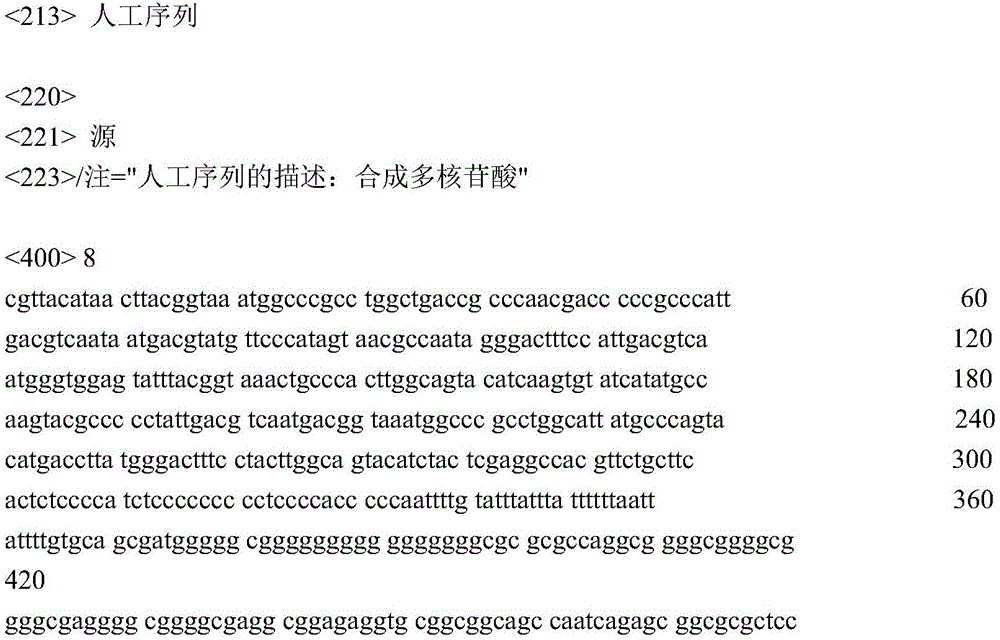


the SEQ ID N08CAG promoter: the underlined sequences represent three components of the promoter, respectively: cytomegalovirus early enhancer, chicken beta-actin promoter and SV40 intron insertion.
CGTTACATAACTTACGGTAAATGGCCCGCCTGGCTGACCGCCCAACGACCCCCGCCCATTGACGTCAAT AATGACGTATGTTCCCATAGTAACGCCAATAGGGACTTTCCATTGACGTCAATGGGTGGAGTATTTACGGTAAACTG CCCACTTGGCAGTACATCAAGTGTATCATATGCCAAGTACGCCCCCTATTGACGTCAATGACGGTAAATGGCCCGCC TGGCATTATGCCCAGTACATGACCTTATGGGACTTTCCTACTTGGCAGTACATCTACTCGAGGCCACGTTCTGCTTC ACTCTCCCCATCTCCCCCCCCCTCCCCACCCCCAATTTTGTATTTATTTATTTTTTAATTATTTTGTGCAGCGATGG GGGCGGGGGGGGGGGGGGGGCGCGCGCCAGGCGGGGCGGGGCGGGGCGAGGGGCGGGGCGAGGCGGAGAGGTGCGGC GGCAGCCAATCAGAGCGGCGCGCTCCGAAAGTTTCCTTTTATGGCGAGGCGGCGGCGGCGGCGGCCCTATAAAAAGC GAAGCGCGCGGCGGGCGGGAGCGGGATCAGCCACCGCGGTGGCGGCCTAGAGTCGACGAGGAACTGAAAAACCAGAA AGTTAACTGGTAAGTTTAGTCTTTTTGTCTTTTATTTCAGGTCCCGGATCCGGTGGTGGTGCAAATCAAAGAACTGCTCCTCAGTGGATGTTGCCTTTACTTCTAGGCCTGTACGGAAGTGTTACTTCTGCTCTAAAAGCTGCGGAATTGTACC CGCGGCCGATCCACCGGTCGC


Claims (25)
1. A light-sensitive ion channel polypeptide comprising a Chrismson protein fused to a Td-Tomato fluorescent protein, wherein the fused Chrismson protein is more effective in responding to light stimulation than the Chrismson protein alone,
wherein the Td-Tomato fluorescent protein comprises Td-Tomato fluorescent protein SEQ ID NO. 3;
the Chrisson protein comprises Chrisson 88 SEQ ID NO. 1 or Chrimson R SEQ ID NO:2.
2. The light-sensitive ion channel polypeptide of claim 1, wherein the light stimulus is red light.
3. The light-sensitive ion channel polypeptide of claim 1, wherein the light stimulus has a wavelength between 365 nm and 700 nm.
4. The light-sensitive ion channel polypeptide of claim 1, wherein the light stimulus has a wavelength of 590 nm.
5. The light sensitive ion channel polypeptide of claim 1, wherein the light sensitive ion channel polypeptide comprises a fusion protein of SEQ ID No. 5.
6. The light-sensitive ion channel polypeptide of claim 1, wherein an effective amount of chrismson protein fused to a Td-mamio fluorescent protein is expressed chronically, wherein the chrismson protein fused to a Td-mamio fluorescent protein is continuously expressed at least 2 months after administration.
7. The light-sensitive ion channel polypeptide of claim 6, wherein the chrismson protein fused to Td-rimano fluorescent protein is continuously expressed at least 11 months after administration.
8. A polynucleotide encoding a photosensitive ion channel polypeptide according to any one of claims 1 to 5.
9. The polynucleotide of claim 8, wherein the polynucleotide comprises SEQ ID No. 6.
10. A nucleic acid construct encoding a polypeptide comprising a light sensitive ion channel according to any one of claims 1 to 5 and/or a polynucleotide according to claim 8 or claim 9.
11. An expression vector comprising the photosensitive ion channel polypeptide of any one of claims 1 to 5, the polynucleotide of claim 8 or claim 9, or the nucleic acid construct of claim 10.
12. The expression vector of claim 11, wherein the vector is an adeno-associated viral AAV vector.
13. The expression vector of claim 12, wherein the AAV vector is selected from an AAV2 vector and/or an AAV2.7m8 vector.
14. The expression vector according to claim 11, comprising SEQ ID NO. 7 or SEQ ID NO. 8.
15. The expression vector of claim 11, wherein the vector is injected intravitreally.
16. A composition comprising the photosensitive ion channel polypeptide of any one of claims 1 to 5, the polynucleotide of claim 8 or claim 9, the nucleic acid construct of claim 10, and/or the expression vector of any one of claims 11 to 14.
17. The composition of claim 16, wherein the Td-mamfo fluorescent protein increases the expression level of the fused chrismson protein compared to the expression level of chrismson protein alone for a given number of cells.
18. The composition of claim 17, wherein the expression level of the fused chrisson protein is increased by enhanced chrisson protein solubility, transport capacity and/or protein conformation.
19. Use of a composition comprising a photosensitive ion channel polypeptide according to any one of claims 1 to 5, a polynucleotide according to claim 8 or claim 9, a nucleic acid construct according to claim 10 and/or an expression vector according to any one of claims 11 to 14 in the manufacture of a medicament for the treatment of an ocular disease.
20. The use of claim 19, wherein the disease comprises a retinal disease.
21. The use according to claim 19, wherein the disease is retinitis pigmentosa RP, macular degeneration MD, congenital stationary night blindness and/or congenital cone dystrophy.
22. The use of claim 21, wherein the macular degeneration MD is age-related macular degeneration.
23. Use of a composition comprising a photosensitive ion channel polypeptide according to any one of claims 1 to 5, a polynucleotide according to claim 8 or claim 9, a nucleic acid construct according to claim 10 and/or an expression vector according to any one of claims 11 to 14 for the preparation of a medicament, said use being as follows:
a) Reactivating mammalian retinal ganglion cell RGCs;
b) Treating or preventing a subject-targeted neuronal-mediated disorder;
c) Restoring the light sensitivity of the inner retinal cells;
d) Restoring vision of the subject;
e) Treating or preventing retinal degeneration in a subject, including identifying that the subject is retinal degeneration due to loss of photoreceptor function;
f) Restoring human eye photoreceptor function, including confirming that the subject is losing vision due to inadequate light perception or sensitivity;
g) Depolarization of electroactive cells;
characterized by comprising administering to the cell a composition comprising an effective amount of a chrismson protein fused to a Td-mamato fluorescent protein, or administering a vector expressing an effective amount of a chrismson protein fused to a Td-mamato fluorescent protein.
24. The use of claim 23, wherein the use is to restore vision in a subject, including confirming that the subject is vision-lost due to insufficient light perception or sensitivity.
25. The use of claim 23, wherein the level of photo-stimulation that induces an RGC response is below a radiation safety limit.
Applications Claiming Priority (3)
| Application Number | Priority Date | Filing Date | Title |
|---|---|---|---|
| US201662329692P | 2016-04-29 | 2016-04-29 | |
| US62/329,692 | 2016-04-29 | ||
| PCT/IB2017/000663 WO2017187272A1 (en) | 2016-04-29 | 2017-04-28 | Optogenetic visual restoration using chrimson |
Publications (2)
| Publication Number | Publication Date |
|---|---|
| CN110267673A CN110267673A (en) | 2019-09-20 |
| CN110267673B true CN110267673B (en) | 2023-05-16 |
Family
ID=59253830
Family Applications (1)
| Application Number | Title | Priority Date | Filing Date |
|---|---|---|---|
| CN201780041321.8A Active CN110267673B (en) | 2016-04-29 | 2017-04-28 | Optogenetic visual restoration using chrituson |
Country Status (8)
| Country | Link |
|---|---|
| US (2) | US20190269755A1 (en) |
| EP (1) | EP3448411A1 (en) |
| JP (1) | JP6942789B2 (en) |
| KR (1) | KR102466887B1 (en) |
| CN (1) | CN110267673B (en) |
| AU (2) | AU2017256910B2 (en) |
| CA (1) | CA3025975A1 (en) |
| WO (1) | WO2017187272A1 (en) |
Families Citing this family (8)
| Publication number | Priority date | Publication date | Assignee | Title |
|---|---|---|---|---|
| US20190126043A1 (en) | 2016-04-14 | 2019-05-02 | The Trustees Of The University Of Pennsylvania | Implantable living electrodes and methods for use thereof |
| EP3693060A1 (en) | 2019-02-05 | 2020-08-12 | Gensight Biologics | Method for controlling an optogenetic device using a command law for the radiant power of a light source and associated devices |
| EP3693061A1 (en) | 2019-02-05 | 2020-08-12 | Gensight Biologics | Method for controlling an optogenetic device using filtering and associated devices |
| EP3733139A1 (en) | 2019-05-02 | 2020-11-04 | Gensight Biologics | Viewing apparatus and method for projecting a light signal |
| US20220249864A1 (en) * | 2019-12-26 | 2022-08-11 | Korea University Research And Business Foundation | Method by which brain with alzheimer's disease recovers from brain wave damage by using optogenetics |
| US20250235585A1 (en) * | 2022-02-18 | 2025-07-24 | The Trustees Of The University Of Pennsylvania | Tissue Engineered Spinal Tracts For Functional Regeneration After Spinal Cord Injury |
| EP4568710A1 (en) | 2022-08-11 | 2025-06-18 | Institute Of Molecular And Clinical Ophthalmology Basel (Iob) | Human cone photoreceptor optogenetic constructs |
| CN119827471B (en) * | 2025-02-14 | 2025-10-03 | 中山大学中山眼科中心 | Method for tracking in vivo and in vitro protein transport |
Citations (2)
| Publication number | Priority date | Publication date | Assignee | Title |
|---|---|---|---|---|
| CN101484005A (en) * | 2006-05-04 | 2009-07-15 | 韦恩州立大学 | Restoration of visual responses by in vivo delivery of rhodopsin nucleic acids |
| WO2015161308A1 (en) * | 2014-04-18 | 2015-10-22 | Massachusetts Institute Of Technology | Mutant channelrhodopsins with altered ion selectivity |
Family Cites Families (10)
| Publication number | Priority date | Publication date | Assignee | Title |
|---|---|---|---|---|
| DE10216005A1 (en) | 2002-04-11 | 2003-10-30 | Max Planck Gesellschaft | Use of biological photoreceptors as direct light-controlled ion channels |
| EP2465925A1 (en) | 2005-07-22 | 2012-06-20 | The Board Of Trustees Of The Leland | Light-activated cation channel and uses thereof |
| EP1891976A1 (en) | 2006-08-23 | 2008-02-27 | Novartis Forschungsstiftung, Zweigniederlassung Friedrich Miescher Institute for Biomedical Research | Use of light sensitive genes |
| AU2009212309B2 (en) * | 2008-02-07 | 2014-04-03 | Ceregene, Inc. | Rescue of photoreceptors by intravitreal administration of an expression vector encoding a therapeutic protein |
| KR20100136550A (en) | 2008-04-18 | 2010-12-28 | 노파르티스 포르슝스티프퉁 쯔바이크니덜라쑹 프리드리히 미셔 인스티튜트 포 바이오메디칼 리서치 | New treatment tools and methods for treating blindness |
| SI3693025T1 (en) | 2011-04-22 | 2022-04-29 | The Regents Of The University Of California | Adeno-associated virus virions with capsid variant and procedures for their use |
| KR102084803B1 (en) * | 2011-11-12 | 2020-03-05 | 메사추세츠 인스티튜트 오브 테크놀로지 | Channelrhodopsins for optical control of cells |
| US10882892B2 (en) * | 2014-08-05 | 2021-01-05 | Massachusetts Institute Of Technology | Channelrhodopsin variants and uses thereof |
| CN108474736B (en) * | 2015-11-05 | 2022-01-25 | 英思克斯公司 | Systems and methods for optogenetic imaging |
| ES2872798T3 (en) * | 2015-12-04 | 2021-11-02 | Univ Sorbonne | Promoters and uses thereof |
-
2017
- 2017-04-28 CN CN201780041321.8A patent/CN110267673B/en active Active
- 2017-04-28 AU AU2017256910A patent/AU2017256910B2/en active Active
- 2017-04-28 US US16/097,204 patent/US20190269755A1/en not_active Abandoned
- 2017-04-28 WO PCT/IB2017/000663 patent/WO2017187272A1/en not_active Ceased
- 2017-04-28 EP EP17734136.9A patent/EP3448411A1/en active Pending
- 2017-04-28 CA CA3025975A patent/CA3025975A1/en active Pending
- 2017-04-28 JP JP2019508309A patent/JP6942789B2/en active Active
- 2017-04-28 KR KR1020187034650A patent/KR102466887B1/en active Active
-
2022
- 2022-07-07 AU AU2022204884A patent/AU2022204884A1/en not_active Abandoned
-
2023
- 2023-09-05 US US18/461,035 patent/US20240165198A1/en active Pending
Patent Citations (2)
| Publication number | Priority date | Publication date | Assignee | Title |
|---|---|---|---|---|
| CN101484005A (en) * | 2006-05-04 | 2009-07-15 | 韦恩州立大学 | Restoration of visual responses by in vivo delivery of rhodopsin nucleic acids |
| WO2015161308A1 (en) * | 2014-04-18 | 2015-10-22 | Massachusetts Institute Of Technology | Mutant channelrhodopsins with altered ion selectivity |
Non-Patent Citations (1)
| Title |
|---|
| Optogenetics;JENS DUEBEL;《CURRENT OPINION IN OPHTHALMOLOGY》;20150501;第26卷(第3期);第226-232页 * |
Also Published As
| Publication number | Publication date |
|---|---|
| CA3025975A1 (en) | 2017-11-02 |
| WO2017187272A8 (en) | 2018-12-13 |
| US20190269755A1 (en) | 2019-09-05 |
| KR102466887B1 (en) | 2022-11-11 |
| AU2017256910A1 (en) | 2018-11-22 |
| WO2017187272A1 (en) | 2017-11-02 |
| KR20190058383A (en) | 2019-05-29 |
| CN110267673A (en) | 2019-09-20 |
| AU2017256910B2 (en) | 2022-04-07 |
| EP3448411A1 (en) | 2019-03-06 |
| JP6942789B2 (en) | 2021-09-29 |
| AU2022204884A1 (en) | 2022-07-28 |
| US20240165198A1 (en) | 2024-05-23 |
| JP2019518073A (en) | 2019-06-27 |
Similar Documents
| Publication | Publication Date | Title |
|---|---|---|
| CN110267673B (en) | Optogenetic visual restoration using chrituson | |
| AU2021245195B2 (en) | Identification of Channelopsin-2 (Chop2) mutations and methods of use | |
| JP2019194228A (en) | Compositions and methods for controlling pain | |
| JP2016522794A (en) | Optogenetic control of behavioral state | |
| AU2017372351B2 (en) | Optogenetic modulation by multi-characteristic opsins for vision restoration and other applications thereof | |
| CN119855619A (en) | Human cone photoreceptor optogenetic constructs | |
| HK1205744B (en) | Identification of channelrhodopsin-2 (chop2) mutations and methods of use | |
| NZ727041B2 (en) | Identification of channelrhodopsin-2 (chop2) mutations and methods of use |
Legal Events
| Date | Code | Title | Description |
|---|---|---|---|
| PB01 | Publication | ||
| PB01 | Publication | ||
| SE01 | Entry into force of request for substantive examination | ||
| SE01 | Entry into force of request for substantive examination | ||
| GR01 | Patent grant | ||
| GR01 | Patent grant | ||
| TG01 | Patent term adjustment | ||
| TG01 | Patent term adjustment |