CN110251093B - Acoustic focusing endoscopic photoacoustic/ultrasonic probe and scanning imaging method - Google Patents
Acoustic focusing endoscopic photoacoustic/ultrasonic probe and scanning imaging method Download PDFInfo
- Publication number
- CN110251093B CN110251093B CN201910671640.8A CN201910671640A CN110251093B CN 110251093 B CN110251093 B CN 110251093B CN 201910671640 A CN201910671640 A CN 201910671640A CN 110251093 B CN110251093 B CN 110251093B
- Authority
- CN
- China
- Prior art keywords
- ultrasonic transducer
- ultrasonic
- photoacoustic
- endoscopic
- rotating shaft
- Prior art date
- Legal status (The legal status is an assumption and is not a legal conclusion. Google has not performed a legal analysis and makes no representation as to the accuracy of the status listed.)
- Active
Links
- 238000003384 imaging method Methods 0.000 title claims abstract description 66
- 239000000523 sample Substances 0.000 title claims abstract description 56
- 230000003287 optical effect Effects 0.000 claims abstract description 20
- 238000001514 detection method Methods 0.000 claims abstract description 7
- 238000002604 ultrasonography Methods 0.000 claims description 24
- 239000013307 optical fiber Substances 0.000 claims description 7
- 230000001681 protective effect Effects 0.000 claims description 7
- 238000012285 ultrasound imaging Methods 0.000 claims description 6
- 239000013078 crystal Substances 0.000 claims description 5
- 210000001519 tissue Anatomy 0.000 description 12
- 238000010586 diagram Methods 0.000 description 10
- 239000000243 solution Substances 0.000 description 10
- 230000003902 lesion Effects 0.000 description 6
- 230000000694 effects Effects 0.000 description 5
- 238000000034 method Methods 0.000 description 4
- 238000011160 research Methods 0.000 description 4
- 230000005284 excitation Effects 0.000 description 3
- 210000000664 rectum Anatomy 0.000 description 3
- 238000010521 absorption reaction Methods 0.000 description 2
- 230000001066 destructive effect Effects 0.000 description 2
- 238000011161 development Methods 0.000 description 2
- 238000003745 diagnosis Methods 0.000 description 2
- 210000003238 esophagus Anatomy 0.000 description 2
- 230000031700 light absorption Effects 0.000 description 2
- 238000004519 manufacturing process Methods 0.000 description 2
- 238000012986 modification Methods 0.000 description 2
- 230000004048 modification Effects 0.000 description 2
- 230000007170 pathology Effects 0.000 description 2
- 238000012216 screening Methods 0.000 description 2
- 230000035945 sensitivity Effects 0.000 description 2
- 238000004088 simulation Methods 0.000 description 2
- 239000013589 supplement Substances 0.000 description 2
- 229920001817 Agar Polymers 0.000 description 1
- 210000000577 adipose tissue Anatomy 0.000 description 1
- 239000008272 agar Substances 0.000 description 1
- 230000003321 amplification Effects 0.000 description 1
- 230000009286 beneficial effect Effects 0.000 description 1
- 230000008094 contradictory effect Effects 0.000 description 1
- 239000003814 drug Substances 0.000 description 1
- 238000002474 experimental method Methods 0.000 description 1
- 239000000835 fiber Substances 0.000 description 1
- 239000000499 gel Substances 0.000 description 1
- 210000000936 intestine Anatomy 0.000 description 1
- 210000000867 larynx Anatomy 0.000 description 1
- 239000002960 lipid emulsion Substances 0.000 description 1
- 238000003199 nucleic acid amplification method Methods 0.000 description 1
- 238000005070 sampling Methods 0.000 description 1
- 210000000813 small intestine Anatomy 0.000 description 1
- XLYOFNOQVPJJNP-UHFFFAOYSA-N water Substances O XLYOFNOQVPJJNP-UHFFFAOYSA-N 0.000 description 1
Images


Classifications
-
- A—HUMAN NECESSITIES
- A61—MEDICAL OR VETERINARY SCIENCE; HYGIENE
- A61B—DIAGNOSIS; SURGERY; IDENTIFICATION
- A61B1/00—Instruments for performing medical examinations of the interior of cavities or tubes of the body by visual or photographical inspection, e.g. endoscopes; Illuminating arrangements therefor
- A61B1/00163—Optical arrangements
- A61B1/00165—Optical arrangements with light-conductive means, e.g. fibre optics
-
- A—HUMAN NECESSITIES
- A61—MEDICAL OR VETERINARY SCIENCE; HYGIENE
- A61B—DIAGNOSIS; SURGERY; IDENTIFICATION
- A61B1/00—Instruments for performing medical examinations of the interior of cavities or tubes of the body by visual or photographical inspection, e.g. endoscopes; Illuminating arrangements therefor
- A61B1/06—Instruments for performing medical examinations of the interior of cavities or tubes of the body by visual or photographical inspection, e.g. endoscopes; Illuminating arrangements therefor with illuminating arrangements
- A61B1/07—Instruments for performing medical examinations of the interior of cavities or tubes of the body by visual or photographical inspection, e.g. endoscopes; Illuminating arrangements therefor with illuminating arrangements using light-conductive means, e.g. optical fibres
-
- A—HUMAN NECESSITIES
- A61—MEDICAL OR VETERINARY SCIENCE; HYGIENE
- A61B—DIAGNOSIS; SURGERY; IDENTIFICATION
- A61B5/00—Measuring for diagnostic purposes; Identification of persons
- A61B5/0033—Features or image-related aspects of imaging apparatus, e.g. for MRI, optical tomography or impedance tomography apparatus; Arrangements of imaging apparatus in a room
-
- A—HUMAN NECESSITIES
- A61—MEDICAL OR VETERINARY SCIENCE; HYGIENE
- A61B—DIAGNOSIS; SURGERY; IDENTIFICATION
- A61B5/00—Measuring for diagnostic purposes; Identification of persons
- A61B5/0059—Measuring for diagnostic purposes; Identification of persons using light, e.g. diagnosis by transillumination, diascopy, fluorescence
- A61B5/0082—Measuring for diagnostic purposes; Identification of persons using light, e.g. diagnosis by transillumination, diascopy, fluorescence adapted for particular medical purposes
- A61B5/0084—Measuring for diagnostic purposes; Identification of persons using light, e.g. diagnosis by transillumination, diascopy, fluorescence adapted for particular medical purposes for introduction into the body, e.g. by catheters
-
- A—HUMAN NECESSITIES
- A61—MEDICAL OR VETERINARY SCIENCE; HYGIENE
- A61B—DIAGNOSIS; SURGERY; IDENTIFICATION
- A61B5/00—Measuring for diagnostic purposes; Identification of persons
- A61B5/0093—Detecting, measuring or recording by applying one single type of energy and measuring its conversion into another type of energy
- A61B5/0095—Detecting, measuring or recording by applying one single type of energy and measuring its conversion into another type of energy by applying light and detecting acoustic waves, i.e. photoacoustic measurements
-
- A—HUMAN NECESSITIES
- A61—MEDICAL OR VETERINARY SCIENCE; HYGIENE
- A61B—DIAGNOSIS; SURGERY; IDENTIFICATION
- A61B8/00—Diagnosis using ultrasonic, sonic or infrasonic waves
- A61B8/12—Diagnosis using ultrasonic, sonic or infrasonic waves in body cavities or body tracts, e.g. by using catheters
-
- A—HUMAN NECESSITIES
- A61—MEDICAL OR VETERINARY SCIENCE; HYGIENE
- A61B—DIAGNOSIS; SURGERY; IDENTIFICATION
- A61B8/00—Diagnosis using ultrasonic, sonic or infrasonic waves
- A61B8/44—Constructional features of the ultrasonic, sonic or infrasonic diagnostic device
-
- A—HUMAN NECESSITIES
- A61—MEDICAL OR VETERINARY SCIENCE; HYGIENE
- A61B—DIAGNOSIS; SURGERY; IDENTIFICATION
- A61B8/00—Diagnosis using ultrasonic, sonic or infrasonic waves
- A61B8/52—Devices using data or image processing specially adapted for diagnosis using ultrasonic, sonic or infrasonic waves
- A61B8/5215—Devices using data or image processing specially adapted for diagnosis using ultrasonic, sonic or infrasonic waves involving processing of medical diagnostic data
- A61B8/5238—Devices using data or image processing specially adapted for diagnosis using ultrasonic, sonic or infrasonic waves involving processing of medical diagnostic data for combining image data of patient, e.g. merging several images from different acquisition modes into one image
- A61B8/5261—Devices using data or image processing specially adapted for diagnosis using ultrasonic, sonic or infrasonic waves involving processing of medical diagnostic data for combining image data of patient, e.g. merging several images from different acquisition modes into one image combining images from different diagnostic modalities, e.g. ultrasound and X-ray
Landscapes
- Health & Medical Sciences (AREA)
- Life Sciences & Earth Sciences (AREA)
- Surgery (AREA)
- Physics & Mathematics (AREA)
- Engineering & Computer Science (AREA)
- Medical Informatics (AREA)
- Biophysics (AREA)
- Pathology (AREA)
- Biomedical Technology (AREA)
- Heart & Thoracic Surgery (AREA)
- Veterinary Medicine (AREA)
- Molecular Biology (AREA)
- Public Health (AREA)
- Animal Behavior & Ethology (AREA)
- General Health & Medical Sciences (AREA)
- Radiology & Medical Imaging (AREA)
- Nuclear Medicine, Radiotherapy & Molecular Imaging (AREA)
- Optics & Photonics (AREA)
- Computer Vision & Pattern Recognition (AREA)
- Acoustics & Sound (AREA)
- Ultra Sonic Daignosis Equipment (AREA)
Abstract
本发明属于光声内窥成像技术领域,公开了一种声聚焦内窥光声/超声探头及扫描成像方法,其中所述探头包括导管和手持外壳;导管包括透明硬质外壳,透明硬质外壳内设有超声换能器、反射组件、光学组件和旋转组件;所述透明硬质外壳固定于所述手持外壳上;所述超声换能器设于所述透明硬质外壳的顶部,所述光学组件的一端置于所述反射组件内,所述超声换能器与所述反射组件对应设置;所述超声换能器在垂直于所述导管的主轴方向的x‑y平面上的接收面为平面。本发明将超声换能器在x‑y平面上的探测面改为平面,其声场更均匀,径向上前后目标的原始信号强度和信噪比更接近,并采用相应的扫描成像方法,来实现径向上各位置目标的聚焦,从而增加其成像景深。
The invention belongs to the technical field of photoacoustic endoscopic imaging, and discloses an acoustic focusing endoscopic photoacoustic/ultrasonic probe and a scanning imaging method, wherein the probe comprises a catheter and a hand-held casing; the catheter comprises a transparent hard casing, and the transparent rigid casing An ultrasonic transducer, a reflection component, an optical component and a rotating component are arranged inside; the transparent hard shell is fixed on the hand-held shell; the ultrasonic transducer is arranged on the top of the transparent hard shell, and the One end of the optical assembly is placed in the reflection assembly, and the ultrasonic transducer is arranged corresponding to the reflection assembly; the receiving surface of the ultrasonic transducer on the x-y plane perpendicular to the main axis direction of the catheter for the plane. The invention changes the detection surface of the ultrasonic transducer on the x-y plane to a plane, the sound field is more uniform, the original signal intensity and signal-to-noise ratio of the front and rear targets in the radial direction are closer, and a corresponding scanning imaging method is used to achieve The focus of the target at each position in the radial direction increases its imaging depth of field.
Description
技术领域technical field
本发明涉及光声内窥成像技术领域,具体地涉及一种声聚焦内窥光声/超声探头及扫描成像方法。The invention relates to the technical field of photoacoustic endoscopic imaging, in particular to an acoustic focusing endoscopic photoacoustic/ultrasonic probe and a scanning imaging method.
背景技术Background technique
光声成像是一种利用脉冲激光对生物组织进行激发,然后根据超声换能器收到的由于生物组织的光吸收造成瞬间热膨胀产生的脉冲声压,对生物体内的光吸收分布进行探测和成像的技术,其反映了生物体内光吸收系数的分布情况。声聚焦光声内窥成像采用组织散射较小的超声对生物组织进行内窥聚焦成像,克服了光聚焦成像中由于光在组织中的强散射,从而在组织深处无法聚焦且难以清晰成像的缺点,在直肠、喉管等内窥成像领域得到了广泛的研究和应用。Photoacoustic imaging is a method of using pulsed laser to excite biological tissue, and then detect and image the light absorption distribution in the living body according to the pulsed sound pressure generated by the instantaneous thermal expansion caused by the optical absorption of the biological tissue received by the ultrasonic transducer. technology, which reflects the distribution of light absorption coefficients in living organisms. Acoustic-focused photoacoustic endoscopic imaging uses ultrasound with less tissue scattering to perform endoscopic focused imaging of biological tissue, which overcomes the problem that light cannot be focused deep in the tissue and is difficult to image clearly due to the strong scattering of light in the tissue in optical focusing imaging. Due to its shortcomings, it has been widely studied and applied in the field of endoscopic imaging such as rectum and larynx.
然而,现有的声聚焦光声内窥成像技术一般采用的是定焦的超声换能器,所以只有在焦点区域附近才能很好的聚焦;远离焦点区域时,其横向分辨率就会严重的降低。这在很大程度上是由于在声聚焦光声内窥成像中,采用的定焦的超声换能器和采用的类似于超声成像中的扇形扫描径向反投影算法。However, the existing acoustic focusing photoacoustic endoscopic imaging technology generally uses a fixed-focus ultrasonic transducer, so it can only focus well near the focal area; when it is far away from the focal area, its lateral resolution will be serious reduce. This is largely due to the use of fixed-focus ultrasound transducers in acoustically focused photoacoustic endoscopic imaging and the use of a sector scan radial backprojection algorithm similar to that used in ultrasound imaging.
发明内容SUMMARY OF THE INVENTION
本发明所要解决的技术问题是提供一种声聚焦内窥光声/超声探头,该声聚焦内窥光声/超声探头采用在x-y平面上接收面为平面的超声换能器,其声场更为均匀,径向上前后目标的原始信号强度和信噪比更接近。The technical problem to be solved by the present invention is to provide an acoustically focused endoscopic photoacoustic/ultrasonic probe. The acoustically focused endoscopic photoacoustic/ultrasonic probe adopts an ultrasonic transducer whose receiving surface is a plane on the x-y plane, and its sound field is more Uniform, the raw signal strength and signal-to-noise ratio of the front and rear targets in the radial direction are closer.
进一步地,本发明所要解决的第二个技术问题是提供一种声聚焦内窥光声/超声扫描成像方法,该声聚焦内窥光声/超声探头采用相应的扫描成像方法后,更有利于其景深的增加。Further, the second technical problem to be solved by the present invention is to provide an acoustically focused endoscopic photoacoustic/ultrasonic scanning imaging method, which is more beneficial to the application of the corresponding scanning imaging method to the acoustically focused endoscopic photoacoustic/ultrasonic probe. Its depth of field increases.
为了解决上述技术问题,本发明第一方面提供了一种声聚焦内窥光声/超声探头,包括导管和手持外壳;所述导管包括透明硬质外壳,所述透明硬质外壳内设有超声换能器、反射组件、光学组件和旋转组件;所述透明硬质外壳固定于所述手持外壳上;所述超声换能器设于所述透明硬质外壳的顶部,所述光学组件的一端置于所述反射组件内,所述超声换能器与所述反射组件对应设置;所述超声换能器在垂直于所述导管的主轴方向的x-y平面上的接收面为平面;所述手持外壳内设有步进电机,所述步进电机的输出轴连接至所述旋转组件,所述旋转组件带动所述光学组件及所述反射组件旋转,从而使得所述反射组件对待探测对象内壁实现全视角扫描。In order to solve the above-mentioned technical problems, the first aspect of the present invention provides an acoustically focused endoscopic photoacoustic/ultrasonic probe, comprising a catheter and a hand-held housing; the catheter comprises a transparent hard housing, and the transparent hard housing is provided with an ultrasonic probe transducer, reflection component, optical component and rotating component; the transparent hard shell is fixed on the hand-held shell; the ultrasonic transducer is arranged on the top of the transparent hard shell, one end of the optical component The ultrasonic transducer is placed in the reflection assembly, and the ultrasonic transducer is arranged correspondingly to the reflection assembly; the receiving surface of the ultrasonic transducer on the x-y plane perpendicular to the main axis direction of the catheter is a plane; the hand-held A stepping motor is arranged in the casing, and the output shaft of the stepping motor is connected to the rotating component, and the rotating component drives the optical component and the reflecting component to rotate, so that the reflecting component realizes the inner wall of the object to be detected. Full-view scanning.
具体地,所述反射组件包括激光反射镜和光声/超声反射镜。Specifically, the reflection component includes a laser mirror and a photoacoustic/ultrasonic mirror.
作为一种具体实施方式,所述激光反射镜为镜面反射镜,光声/超声反射模块为透明晶体,所述激光反射镜以一定角度置于所述反射组件上方,以使得反射光能够以一定角度入射生物组织,所述反射光以一定角度入射生物组织的角度范围是85°~90°;所述光声/超声反射模块与所述超声换能器相对的一侧斜面为45°。As a specific embodiment, the laser mirror is a specular mirror, the photoacoustic/ultrasonic reflection module is a transparent crystal, and the laser mirror is placed above the reflection component at a certain angle, so that the reflected light can be reflected at a certain angle. The angle at which the reflected light is incident on the biological tissue at a certain angle ranges from 85° to 90°; the slope on the opposite side of the photoacoustic/ultrasonic reflection module and the ultrasonic transducer is 45°.
作为一种优选实施方式,所述超声换能器为单个单元线聚焦超声换能器,所述线聚焦超声换能器的接收面在平行于所述导管的主轴的z向是聚焦的。As a preferred embodiment, the ultrasonic transducer is a single-unit line-focused ultrasonic transducer, and the receiving surface of the line-focused ultrasonic transducer is focused in the z-direction parallel to the main axis of the catheter.
作为另一种优选实施方式,所述超声换能器为线性阵列超声换能器,所述线性阵列超声换能器包括多个沿平行于所述导管的主轴的z向排列的超声换能器。As another preferred embodiment, the ultrasonic transducer is a linear array ultrasonic transducer, and the linear array ultrasonic transducer includes a plurality of ultrasonic transducers arranged along the z-direction parallel to the main axis of the catheter .
进一步地,所述光学组件为单模光纤。Further, the optical component is a single-mode optical fiber.
优选地,所述光学组件为多模光纤。Preferably, the optical component is a multimode optical fiber.
作为一种具体实施方式,所述旋转组件包括固定轴、第一旋转轴、第二旋转轴、传送带、覆盖软胶管保护壳的柔性旋转轴以及硬质旋转轴;所述固定轴与所述第一旋转轴之间由两个滚珠轴承支撑,所述第一旋转轴与所述覆盖软胶管保护壳的柔性旋转轴以及所述硬质旋转轴相连接;所述第二旋转轴与所述步进电机连接;所述第一旋转轴与所述第二旋转轴通过所述传送带相互连接。As a specific embodiment, the rotating assembly includes a fixed shaft, a first rotating shaft, a second rotating shaft, a conveyor belt, a flexible rotating shaft covering the protective shell of the soft rubber tube, and a hard rotating shaft; the fixed shaft and the first rotating shaft A rotating shaft is supported by two ball bearings, the first rotating shaft is connected with the flexible rotating shaft covering the protective shell of the soft rubber tube and the hard rotating shaft; the second rotating shaft is connected with the step The feeding motor is connected; the first rotating shaft and the second rotating shaft are connected to each other through the conveyor belt.
在本发明上述技术方案的基础上,本发明第二方面提供了一种声聚焦内窥光声/超声扫描成像方法,包括如下步骤:On the basis of the above technical solutions of the present invention, a second aspect of the present invention provides an acoustic focusing endoscopic photoacoustic/ultrasonic scanning imaging method, comprising the following steps:
S1将超声换能器的平面接收面分割成多个单元,并将每个单元视为一个点探测器;S1 divides the planar receiving surface of the ultrasonic transducer into multiple units, and treats each unit as a point detector;
S2采用反投影叠加算法,对每个点探测器位置分割后的小单元进行信号反投影加权叠加,以得到最终的图像,S2 uses the back-projection stacking algorithm to perform signal back-projection weighted stacking on the small units segmented by the position of each point detector to obtain the final image,
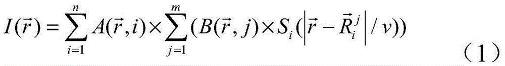
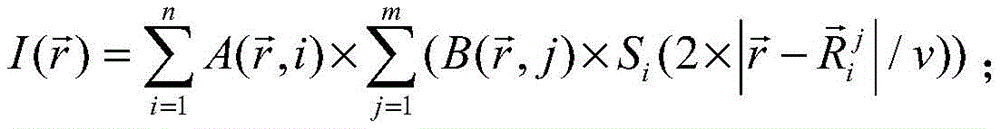
其中,光声成像通过如下的公式计算得到最终的图像:Among them, the photoacoustic imaging is calculated by the following formula to obtain the final image:

超声成像通过如下的公式计算得到最终的图像:Ultrasound imaging is calculated by the following formula to obtain the final image:

上式中,n为探测器的位置数;m为点探测器所分割的小单元数;I为图像


通过本发明的上述技术方案,本发明的声聚焦内窥光声/超声探头及扫描成像方法可以达到:Through the above technical solutions of the present invention, the acoustic focusing endoscopic photoacoustic/ultrasonic probe and the scanning imaging method of the present invention can achieve:
(1)本发明的声聚焦内窥光声/超声探头的超声换能器在垂直于导管的主轴方向的x-y平面上的接收面为平面,其声场更为均匀,径向上前后目标的原始信号强度和信噪比更接近,并且采用相应的扫描成像方法,实现了径向上各位置目标的聚焦,从而增加其成像景深;(1) The receiving surface of the ultrasonic transducer of the acoustic focusing endoscopic photoacoustic/ultrasonic probe of the present invention on the x-y plane perpendicular to the main axis direction of the catheter is a plane, and its sound field is more uniform, and the original signal of the front and rear targets in the radial direction is a plane. The intensity and the signal-to-noise ratio are closer, and the corresponding scanning imaging method is used to realize the focusing of the targets at each position in the radial direction, thereby increasing the imaging depth of field;
(2)本发明的声聚焦内窥光声/超声探头的超声换能器在x-y平面上的接收面为平面的超声换能器的制作难度比聚焦超声换能器低;(2) The manufacturing difficulty of the ultrasonic transducer whose receiving surface on the x-y plane of the ultrasonic transducer of the acoustically focused endoscopic photoacoustic/ultrasonic probe of the present invention is a plane is lower than that of the focused ultrasonic transducer;
(3)本发明的声聚焦内窥光声/超声探头使得高灵敏度、微米级分辨率、亚厘米级大成像深度的实时光声/超声内窥成像成为可能,从而能够对管腔内各类病变进行精确无损地诊断,使声聚焦光声/超声内窥成像技术成为现有管腔内病变筛诊方法的有力补充,并且推动了早期病变的病理学等相关研究的发展,具有重要的临床医学意义和科学价值;(3) The acoustic focusing endoscopic photoacoustic/ultrasonic probe of the present invention enables real-time photoacoustic/ultrasonic endoscopic imaging with high sensitivity, micron-level resolution, and sub-centimeter-level large imaging depth, so that various types of intraluminal imaging can be performed. Accurate and non-destructive diagnosis of lesions makes the acousto-focused photoacoustic/ultrasound endoscopic imaging technology a powerful supplement to the existing intraluminal lesion screening methods, and promotes the development of related researches on the pathology of early lesions. Medical significance and scientific value;
(4)本发明的声聚焦内窥光声/超声探头及扫描成像方法,不仅可以直接应用于食道、直肠等其他内窥成像的应用中,也应用在生物医学领域中;是光声成像理论方面的重要创新;(4) The acoustic focusing endoscopic photoacoustic/ultrasonic probe and the scanning imaging method of the present invention can not only be directly applied to other endoscopic imaging applications such as esophagus and rectum, but also be applied in the field of biomedicine; it is the theory of photoacoustic imaging. important innovations in
(5)本发明中的声聚焦内窥光声/超声探头及扫描成像方法还可以应用到其他类型的光声/超声成像中,以及其他影像学方法的研究中,具有重要的指导价值。(5) The acoustically focused endoscopic photoacoustic/ultrasonic probe and scanning imaging method in the present invention can also be applied to other types of photoacoustic/ultrasonic imaging and research of other imaging methods, and have important guiding value.
本发明实施例的其它特征和优点将在随后的具体实施方式部分予以详细说明。Other features and advantages of embodiments of the present invention will be described in detail in the detailed description section that follows.
附图说明Description of drawings
附图是用来提供对本发明实施例的进一步理解,并且构成说明书的一部分,与下面的具体实施方式一起用于解释本发明实施例,但并不构成对本发明实施例的限制。在附图中:The accompanying drawings are used to provide a further understanding of the embodiments of the present invention, and constitute a part of the specification, and are used to explain the embodiments of the present invention together with the following specific embodiments, but do not constitute limitations to the embodiments of the present invention. In the attached image:
图1为本发明的声聚焦内窥光声/超声探头的一种实施方式的结构示意图;1 is a schematic structural diagram of an embodiment of an acoustically focused endoscopic photoacoustic/ultrasonic probe of the present invention;
图2为本发明的声聚焦内窥光声/超声探头的一种实施方式的线聚焦超声换能器的x-y方向上的圆周旋转扫描的模拟效果图;Fig. 2 is the simulation effect diagram of the circular rotation scanning in the x-y direction of the line-focusing ultrasonic transducer of an embodiment of the acoustically focused endoscopic photoacoustic/ultrasonic probe of the present invention;
图3为本发明的声聚焦内窥光声/超声探头的一种实施方式的聚焦超声换能器的扫描示意图;3 is a schematic scanning diagram of a focused ultrasound transducer of an embodiment of the acoustically focused endoscopic photoacoustic/ultrasonic probe of the present invention;
图4为本发明的声聚焦内窥光声/超声探头的一种实施方式的线性阵列超声换能器的扫描示意图;Fig. 4 is a scanning schematic diagram of a linear array ultrasonic transducer of an embodiment of the acoustically focused endoscopic photoacoustic/ultrasonic probe of the present invention;
图5为本发明的声聚焦内窥光声/超声探头的另一种实施方式的结构示意图;5 is a schematic structural diagram of another embodiment of the acoustic focusing endoscopic photoacoustic/ultrasonic probe of the present invention;
图6为本发明的声聚焦内窥光声/超声探头对第一种样品(a)进行平面光声/超声成像的结果图;FIG. 6 is a result diagram of planar photoacoustic/ultrasonic imaging performed on the first sample (a) by the acoustically focused endoscopic photoacoustic/ultrasonic probe of the present invention;
图7为本发明的声聚焦内窥光声/超声探头对第二种样品(a)进行平面光声/超声成像的结果图;FIG. 7 is a result diagram of planar photoacoustic/ultrasonic imaging performed on the second sample (a) by the acoustically focused endoscopic photoacoustic/ultrasonic probe of the present invention;
图8为本发明的x-y方向的光声和超声的新旧算法对比图;Fig. 8 is the photoacoustic of the x-y direction of the present invention and the comparison diagram of old and new algorithms of ultrasound;
图9为本发明的x-z方向的光声和超声的新旧算法对比图。FIG. 9 is a comparison diagram of the old and new algorithms of photoacoustics and ultrasound in the x-z direction of the present invention.
附图标记说明Description of reference numerals
1 线聚焦超声换能器 2 超声换能器的镜像位置1 Line
3 激光反射镜 4 透明硬质外壳3
5 固定模块 6 覆盖软胶管保护壳的柔性旋转轴5 Fixed
7 固定轴 8 传送带7 Fixed
9 硬质胶管 10 步进电机9
11 第一旋转轴 12 硬质旋转轴11 The
13 集成光纤激光束 14 光声/超声反射镜13 Integrated Fiber Laser Beam 14 Photoacoustic/Ultrasonic Mirror
15 超声回波 16 成像目标15
17 手持外壳 18 第二旋转轴17
19 线性阵列超声换能器19 Linear Array Ultrasound Transducers
具体实施方式Detailed ways
以下结合附图对本发明实施例的具体实施方式进行详细说明。应当理解的是,此处所描述的具体实施方式仅用于说明和解释本发明实施例,并不用于限制本发明实施例。The specific implementations of the embodiments of the present invention will be described in detail below with reference to the accompanying drawings. It should be understood that the specific implementation manners described herein are only used to illustrate and explain the embodiments of the present invention, and are not used to limit the embodiments of the present invention.
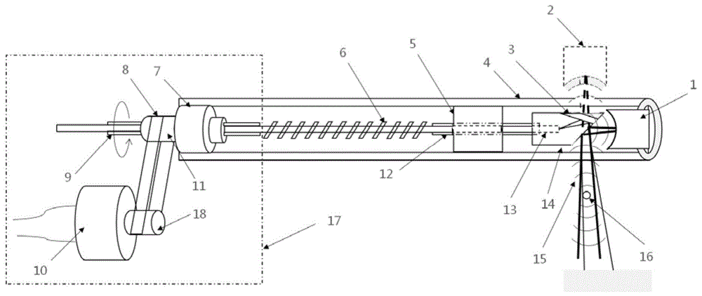
如图1所示,声聚焦内窥光声/超声探头包括导管和手持外壳17;导管包括透明硬质外壳4,透明硬质外壳4内设有超声换能器、反射组件、光学组件和旋转组件;透明硬质外壳4固定于手持外壳17上;超声换能器设于透明硬质外壳4的顶部,光学组件的一端置于反射组件内,超声换能器与反射组件对应设置;超声换能器在垂直于导管的主轴方向的x-y平面上的接收面为平面;手持外壳17内设有步进电机10,步进电机10的输出轴连接至旋转组件,旋转组件带动光学组件及反射组件旋转,从而使得反射组件对待探测对象内壁实现全视角扫描。As shown in FIG. 1, the acoustically focused endoscopic photoacoustic/ultrasonic probe includes a catheter and a hand-held
通过本发明上述基本技术方案的声聚焦内窥光声/超声探头,声聚焦内窥光声/超声探头进行探测时,超声换能器发出超声经反射组件进行反射,探测到成像目标16,会产生超声回波15;并且光学组件发出激光经反射组件进行反射;步进电机10提供扭矩,扭矩带动旋转组件进行运转,接着,旋转组件带动光学组件及反射组件进行旋转;最后,反射组件接收扭矩并执行侧视旋转扫描,以使得该声聚焦内窥光声/超声探头对待探测对象内壁实现全视角的扫描。Through the acoustic focusing endoscopic photoacoustic/ultrasonic probe of the above basic technical solution of the present invention, when the acoustic focusing endoscopic photoacoustic/ultrasonic probe performs detection, the ultrasonic wave emitted by the ultrasonic transducer is reflected by the reflection component, and the
在上述技术方案中,具体地,反射组件包括激光反射镜3和光声/超声反射镜14。优选地,激光反射镜3为镜面反射镜,激光反射镜3以一定角度置于反射组件上方,以使得反射光能够以一定角度入射生物组织,反射光以一定角度入射生物组织的角度范围是85°~90°,以使得反射光能够近似垂直的入射生物组织;光声/超声反射镜14为透明晶体,光声/超声反射镜14与超声换能器相对的一侧斜面为45°。激光经激光反射镜3以一定角度反射后透过透明晶体,使得反射光近似于垂直于导管,然后照射生物组织,超声经透明晶体反射面反射由探头接收。In the above technical solution, specifically, the reflection component includes a
在上述技术方案的优选方式中,线聚焦超声换能器1为单个单元线聚焦超声换能器1,线聚焦超声换能器1的接收面在平行于导管的主轴的z向是聚焦的。因为线聚焦超声换能器1的接受面在垂直于探头主轴的方向的x-y平面上是平面的,并且通过相应的光声内窥聚焦成像算法来实现在径向深度方向上各目标的聚焦,同时保证足够大的成像景深,有效地解决了点聚焦超声换能器中成像景深和成像分辨率不可调和矛盾的问题。In a preferred form of the above technical solution, the line-focused
在上技术方案的另一种优选方式中,超声换能器为线性阵列超声换能器19,线性阵列超声换能器19包括多个沿平行于导管的主轴的z向排列的超声换能器。其与线聚焦超声换能器1类似,能够达到类似的成像效果,保证成像景深和成像分辨率。In another preferred form of the above technical solution, the ultrasonic transducer is a linear array
在上述技术方案中,优选地,光学组件为单模光纤或多模光纤;更为优选地,多模光纤可以提供足够强的激光能量。In the above technical solution, preferably, the optical component is a single-mode optical fiber or a multi-mode optical fiber; more preferably, the multi-mode optical fiber can provide sufficiently strong laser energy.
在上述技术方案的一种具体实施方式中,旋转组件包括固定轴7、第一旋转轴11、第二旋转轴18、传送带8、覆盖软胶管保护壳的柔性旋转轴6以及硬质旋转轴12;固定轴7与第一旋转轴11之间由两个滚珠轴承支撑,第一旋转轴11与覆盖软胶管保护壳的柔性旋转轴6以及硬质旋转轴12相连接;第二旋转轴18与步进电机10连接;第一旋转轴11与第二旋转轴18通过传送带8相互连接。由步进电机10提供扭矩,扭矩通过传送带8和滑轮传递到有两个滚珠轴支撑的第一旋转轴11上,并通过覆盖软胶管保护壳的柔性旋转轴6和硬质旋转轴12进一步传递到反射组件上。最后,反射组件接收扭矩并执行侧视旋转扫描。In a specific embodiment of the above technical solution, the rotating assembly includes a fixed
参见图2所示,在本发明的声聚焦内窥光声/超声探头的一种优选实施方式中,将超声换能器在x-y平面上的探测面形状改为平面,从而极大的改善声场的不均匀性,再应用相应的动态聚焦内窥反投影算法,来实现目标在径向方向上的动态聚焦。Referring to Fig. 2, in a preferred embodiment of the acoustic focusing endoscopic photoacoustic/ultrasonic probe of the present invention, the shape of the detection surface of the ultrasonic transducer on the x-y plane is changed to a plane, thereby greatly improving the sound field The non-uniformity of the target in the radial direction is achieved by applying the corresponding dynamic focusing endoscopic back-projection algorithm.
在上述技术方案的一种实施方式中,以改进型内窥反投影算法为例:In an embodiment of the above technical solution, taking the improved endoscopic back-projection algorithm as an example:
由于探测面在导管的主轴方向的x-y平面上的接收面为平面的,结合超声成像的原理,可以采用如下的公式(2)进行成像Since the receiving surface of the detection surface on the x-y plane in the direction of the main axis of the catheter is a plane, the following formula (2) can be used for imaging combined with the principle of ultrasonic imaging

其中,


由于超声成像和光声成像的区别仅仅在于所得的超声是物体本身发出的还是换能器本身发出的,因此将公式(1)中的


其中,虽然公式(1)和(3)是可以应用于聚焦超声换能器的,但是也可以将其应用到基于平面的超声换能器当中,即当做一个特例来使用。Among them, although formulas (1) and (3) can be applied to focused ultrasonic transducers, they can also be applied to planar-based ultrasonic transducers, that is, used as a special case.
以上公式考虑的是探头的平面方向,没有考虑到z方向上的聚焦。因此,在z方向上可以采用聚焦超声换能器的形式,也可以采用线性阵列的方式进行成像。如图3所示,在垂直方向上采用单个聚焦换能器进行扫描;或者如图4所示,用阵列进行扫描。The above formula considers the plane orientation of the probe and does not take into account the focusing in the z-direction. Therefore, in the z direction, a focused ultrasound transducer can be used, or a linear array can be used for imaging. Scan with a single focused transducer in the vertical direction, as shown in Figure 3; or with an array, as shown in Figure 4.
最后,仿真结果x-y平面方向上达到的效果,新旧算法对比如图8所示;z方向上达到的效果,新旧算法对比如图9所示。Finally, the effect achieved in the x-y plane direction of the simulation results, the comparison between the new and the old algorithm is shown in Figure 8; the effect achieved in the z direction, the comparison between the new and the old algorithm is shown in Figure 9.
参见图5所示,在本发明声聚焦内窥光声/超声探头的另一个实施例中,提供了一种线性阵列超声换能器19的声聚焦内窥光声/超声探头。所述探头除了将线聚焦超声换能器1换为线性阵列超声换能器19以外,与图1中的探头的其他结完全一致,也能达到类似的成像效果。Referring to FIG. 5 , in another embodiment of the acoustically focused endoscopic photoacoustic/ultrasonic probe of the present invention, an acoustically focused endoscopic photoacoustic/ultrasonic probe of a linear array
参见图6和图7所示,利用图1中所示的声聚焦内窥光声/超声探头,对仿体进行成像,结果说明如下:第一种样品(a)、第二种样品(a)均为仿体实验所用样品的示意图。其中仿体主体为用来模拟生物组织的凝胶琼脂,掺有脂肪乳和水,使得其散射和吸收系数分别为1/mm和0.07/mm,和人体脂肪组织相近。仿体外径为40mm,内径为12mm。第一种样品(a)中在内径15mm处嵌入不规则形状的猪小肠,第二种样品(a)中在不同位置垂直插有十六根直径为0.5mm的铅芯。扫描采用图1所示的实施方式的探头进行采集。在进行光声成像时,激发光选用Nd:YAG激光器产生的532nm脉冲绿光,激发光重复频率为10Hz,脉冲宽度为8ns。放大器采用奥林巴斯5072PR进行光声和超声信号放大,同时5072PR还可以用来进行超声激发进行超声成像。采集到的信号用LDI400SE采集卡收集,采样频率为100MHz,扫描角度间隔为1度,共采集360个角度的数据。Referring to Figures 6 and 7, the phantom was imaged using the acoustically focused endophotoacoustic/ultrasonic probe shown in Figure 1, and the results were described as follows: the first sample (a), the second sample (a ) are schematic diagrams of the samples used in the phantom experiments. The main body of the phantom is gel agar used to simulate biological tissue, mixed with fat emulsion and water, so that its scattering and absorption coefficients are 1/mm and 0.07/mm, respectively, which are similar to human adipose tissue. The phantom has an outer diameter of 40mm and an inner diameter of 12mm. In the first sample (a), an irregularly shaped pig intestine was embedded at an inner diameter of 15 mm, and in the second sample (a), sixteen lead cores with a diameter of 0.5 mm were vertically inserted at different positions. Scans were acquired using the probe of the embodiment shown in FIG. 1 . In the photoacoustic imaging, the excitation light is 532nm pulsed green light generated by Nd:YAG laser, the excitation light repetition frequency is 10Hz, and the pulse width is 8ns. The amplifier uses Olympus 5072PR for photoacoustic and ultrasound signal amplification, and 5072PR can also be used for ultrasound excitation for ultrasound imaging. The collected signals are collected by the LDI400SE acquisition card, the sampling frequency is 100MHz, the scanning angle interval is 1 degree, and the data of 360 angles are collected in total.
如图6和图7所述,图6和图7分别是使用图1所示的探头对两种不同的样品(a)进项平面光声/超声成像的结果图。从中可以看出,猪小肠和铅芯在光声和超声图像中都能得到清晰的重建,与常规内窥算法相比,大大地提高了图像分辨率。As described in Figures 6 and 7, Figures 6 and 7 are graphs of the results of in-plane photoacoustic/ultrasonic imaging of two different samples (a) using the probe shown in Figure 1, respectively. It can be seen that the pig small intestine and lead core can be clearly reconstructed in both photoacoustic and ultrasound images, which greatly improves the image resolution compared with conventional endoscopic algorithms.
由上述描述可以看出,本发明的声聚焦内窥光声/超声探头及扫描成像方法的优点在于:本发明的声聚焦内窥光声/超声探头的超声换能器的在垂直于所述导管的主轴方向的x-y平面上的接收面为平面,使得其声场更为均匀,径向上前后目标的原始信号强度和信噪比更接近,并且采用相应的扫描成像方法,实现了径向上各位置目标的聚焦,从而增加其成像景深,同时,保证足够大的成像景深,有效地解决了点聚焦超声换能器中成像景深和成像分辨率不可调和矛盾的问题。并且,本发明的声聚焦内窥光声/超声探头的超声换能器在x-y平面上的接收面为平面的超声换能器的制作难度比聚焦超声换能器低。It can be seen from the above description that the advantages of the acoustically focused endoscopic photoacoustic/ultrasonic probe and the scanning imaging method of the present invention are: the ultrasonic transducer of the acoustically focused endoscopic photoacoustic/ultrasonic probe of the present invention is perpendicular to the The receiving surface on the x-y plane in the main axis direction of the catheter is a plane, which makes the sound field more uniform, and the original signal intensity and signal-to-noise ratio of the front and rear targets in the radial direction are closer, and the corresponding scanning imaging method is used. The focus of the target increases its imaging depth of field, and at the same time, a sufficiently large imaging depth of field is ensured, which effectively solves the problem of inconsistency and contradiction between the imaging depth of field and the imaging resolution in the point-focused ultrasound transducer. In addition, the ultrasonic transducer of the acoustically focused endoscopic photoacoustic/ultrasonic probe of the present invention has a lower receiving surface on the x-y plane than the focused ultrasonic transducer in manufacturing difficulty.
此外,本发明的声聚焦内窥光声/超声探头使得高灵敏度、微米级分辨率、亚厘米级大成像深度的实时光声/超声内窥成像成为可能,从而能够对管腔内各类病变进行精确无损地诊断,使声聚焦光声/超声内窥成像技术成为现有管腔内病变筛诊方法的有力补充,并且推动了早期病变的病理学等相关研究的发展,具有重要的临床医学意义和科学价值。并且,本发明的声聚焦内窥光声/超声探头及扫描成像方法,不仅可以直接应用于食道、直肠等其他内窥成像的应用中,也应用在生物医学领域中;是光声成像理论方面的重要创新;本发明中的声聚焦内窥光声/超声探头及扫描成像方法还可以应用到其他类型的光声/超声成像中,以及其他影像学方法的研究中,具有重要的指导价值。In addition, the acoustic focusing endoscopic photoacoustic/ultrasonic probe of the present invention enables real-time photoacoustic/ultrasonic endoscopic imaging with high sensitivity, micron-level resolution, and sub-centimeter-level large imaging depth, so that various lesions in the lumen can be detected. Accurate and non-destructive diagnosis makes the acousto-focused photoacoustic/ultrasonic endoscopic imaging technology a powerful supplement to the existing intraluminal lesions screening methods, and promotes the development of related researches on the pathology of early lesions, and has important clinical medicine. significance and scientific value. Moreover, the acoustic focusing endoscopic photoacoustic/ultrasonic probe and the scanning imaging method of the present invention can not only be directly applied to other endoscopic imaging applications such as esophagus and rectum, but also be applied in the field of biomedicine; it is a theoretical aspect of photoacoustic imaging. important innovation; the acoustic focusing endoscopic photoacoustic/ultrasonic probe and scanning imaging method in the present invention can also be applied to other types of photoacoustic/ultrasonic imaging and research of other imaging methods, and have important guiding value.
以上结合附图详细描述了本发明实施例的可选实施方式,但是,本发明实施例并不限于上述实施方式中的具体细节,在本发明实施例的技术构思范围内,可以对本发明实施例的技术方案进行多种简单变型,这些简单变型均属于本发明实施例的保护范围。The optional embodiments of the embodiments of the present invention have been described in detail above with reference to the accompanying drawings. However, the embodiments of the present invention are not limited to the specific details of the above-mentioned embodiments. A variety of simple modifications are made to the technical solution of the invention, and these simple modifications all belong to the protection scope of the embodiments of the present invention.
另外需要说明的是,在上述具体实施方式中所描述的各个具体技术特征,在不矛盾的情况下,可以通过任何合适的方式进行组合。为了避免不必要的重复,本发明实施例对各种可能的组合方式不再另行说明。In addition, it should be noted that each specific technical feature described in the above-mentioned specific implementation manner may be combined in any suitable manner under the circumstance that there is no contradiction. To avoid unnecessary repetition, various possible combinations are not further described in this embodiment of the present invention.
此外,本发明实施例的各种不同的实施方式之间也可以进行任意组合,只要其不违背本发明实施例的思想,其同样应当视为本发明实施例所公开的内容。In addition, various implementations of the embodiments of the present invention may also be combined arbitrarily, as long as they do not violate the ideas of the embodiments of the present invention, they should also be regarded as the contents disclosed in the embodiments of the present invention.
Claims (7)


Priority Applications (1)
| Application Number | Priority Date | Filing Date | Title |
|---|---|---|---|
| CN201910671640.8A CN110251093B (en) | 2019-07-24 | 2019-07-24 | Acoustic focusing endoscopic photoacoustic/ultrasonic probe and scanning imaging method |
Applications Claiming Priority (1)
| Application Number | Priority Date | Filing Date | Title |
|---|---|---|---|
| CN201910671640.8A CN110251093B (en) | 2019-07-24 | 2019-07-24 | Acoustic focusing endoscopic photoacoustic/ultrasonic probe and scanning imaging method |
Publications (2)
| Publication Number | Publication Date |
|---|---|
| CN110251093A CN110251093A (en) | 2019-09-20 |
| CN110251093B true CN110251093B (en) | 2021-02-19 |
Family
ID=67928053
Family Applications (1)
| Application Number | Title | Priority Date | Filing Date |
|---|---|---|---|
| CN201910671640.8A Active CN110251093B (en) | 2019-07-24 | 2019-07-24 | Acoustic focusing endoscopic photoacoustic/ultrasonic probe and scanning imaging method |
Country Status (1)
| Country | Link |
|---|---|
| CN (1) | CN110251093B (en) |
Families Citing this family (8)
| Publication number | Priority date | Publication date | Assignee | Title |
|---|---|---|---|---|
| CN111419285B (en) * | 2020-04-28 | 2023-07-28 | 深圳英美达医疗技术有限公司 | Ultrasonic three-dimensional imaging catheter and three-dimensional scanning method thereof |
| CN112515631B (en) * | 2020-10-22 | 2022-10-28 | 中国科学院深圳先进技术研究院 | Vascular Endoscopic Imaging Device |
| CN112493997B (en) * | 2020-11-30 | 2023-03-31 | 中国科学院深圳先进技术研究院 | Photoacoustic endoscopic imaging device and photoacoustic endoscopic imaging method based on same |
| CN113057675B (en) * | 2021-03-15 | 2024-10-25 | 慧威医疗科技(台州)有限公司 | Imaging device, imaging method thereof, detection probe and endoscope system |
| CN113080814B (en) * | 2021-04-12 | 2022-01-28 | 中南大学 | A transmission coaxial photoacoustic endoscope probe and its imaging method |
| CN113842158B (en) * | 2021-08-09 | 2023-11-21 | 中南大学 | Photoacoustic/ultrasound endoscopic probe and dynamic focus reconstruction algorithm based on fixed-focus sound field |
| CN113576534A (en) * | 2021-08-17 | 2021-11-02 | 陈智毅 | Endometrium imaging device, scoring system and scoring method based on multi-modal imaging |
| CN114732343A (en) * | 2022-06-08 | 2022-07-12 | 深圳市三平影像科技有限公司 | Multifunctional esophagus endoscope |
Citations (4)
| Publication number | Priority date | Publication date | Assignee | Title |
|---|---|---|---|---|
| CN1720006A (en) * | 2002-12-24 | 2006-01-11 | 松下电器产业株式会社 | Ultrasonic probe |
| CN102743191A (en) * | 2012-06-28 | 2012-10-24 | 华南师范大学 | Focusing rotary scanning photoacoustic ultrasonic blood vessel endoscope imaging device and focusing rotary scanning photoacoustic ultrasonic blood vessel endoscope imaging method |
| CN105662477A (en) * | 2016-04-05 | 2016-06-15 | 湖南致力工程科技有限公司 | Handheld full-view endoscopic opto-acoustic/ultrasonic probe |
| CN108261209A (en) * | 2018-01-23 | 2018-07-10 | 湖南大学 | The method of follow-on high-resolution sound focusing optoacoustic endoscopy imaging back projection imaging |
Family Cites Families (1)
| Publication number | Priority date | Publication date | Assignee | Title |
|---|---|---|---|---|
| JPH1071149A (en) * | 1996-08-29 | 1998-03-17 | Toshiba Corp | Ultrasonic probe |
-
2019
- 2019-07-24 CN CN201910671640.8A patent/CN110251093B/en active Active
Patent Citations (4)
| Publication number | Priority date | Publication date | Assignee | Title |
|---|---|---|---|---|
| CN1720006A (en) * | 2002-12-24 | 2006-01-11 | 松下电器产业株式会社 | Ultrasonic probe |
| CN102743191A (en) * | 2012-06-28 | 2012-10-24 | 华南师范大学 | Focusing rotary scanning photoacoustic ultrasonic blood vessel endoscope imaging device and focusing rotary scanning photoacoustic ultrasonic blood vessel endoscope imaging method |
| CN105662477A (en) * | 2016-04-05 | 2016-06-15 | 湖南致力工程科技有限公司 | Handheld full-view endoscopic opto-acoustic/ultrasonic probe |
| CN108261209A (en) * | 2018-01-23 | 2018-07-10 | 湖南大学 | The method of follow-on high-resolution sound focusing optoacoustic endoscopy imaging back projection imaging |
Also Published As
| Publication number | Publication date |
|---|---|
| CN110251093A (en) | 2019-09-20 |
Similar Documents
| Publication | Publication Date | Title |
|---|---|---|
| CN110251093B (en) | Acoustic focusing endoscopic photoacoustic/ultrasonic probe and scanning imaging method | |
| US8870770B2 (en) | Low-cost device for C-scan acoustic wave imaging | |
| Xu et al. | Photoacoustic imaging in biomedicine | |
| CN105769128B (en) | Integrated optoacoustic, ultrasound, optoacoustic elasticity based endoscopic imaging devices and methods therefor | |
| US10105062B2 (en) | Miniaturized photoacoustic imaging apparatus including a rotatable reflector | |
| CN101912250B (en) | Intravascular photoacoustic and ultrasonic double-mode imaging endoscope device and imaging method thereof | |
| JP3654309B2 (en) | Acicular ultrasonic probe | |
| CN105380586B (en) | A combined stereoscopic scanning optical and acoustic endoscopic imaging device and method thereof | |
| JP2015507947A5 (en) | ||
| US20130190594A1 (en) | Scanning Optoacoustic Imaging System with High Resolution and Improved Signal Collection Efficiency | |
| JP2010167258A (en) | Biological information acquisition apparatus | |
| CN105011906B (en) | A photoacoustic computed tomography system combined with a slip ring and its imaging method | |
| CN100446730C (en) | Photoacoustic imaging and tomographic imaging method and device based on acoustic lens | |
| CN106037663B (en) | A kind of continuous vari-focus ultrasonic probe and its Zooming method | |
| CN101828928A (en) | Three-dimensional optoacoustic mammary gland or brain non-destructive imaging system | |
| CN101828902B (en) | A photoacoustic sensor for 3D medical diagnosis of breast or brain | |
| Osman et al. | A novel matching layer design for improving the performance of transparent ultrasound transducers | |
| JP5725781B2 (en) | Subject information acquisition device | |
| CN107822595A (en) | The per urethra optoacoustic prostate developing method and device received based on ring battle array | |
| CN201624672U (en) | A photoacoustic sensor for three-dimensional medical diagnosis of biological tissue | |
| CN219000345U (en) | Miniature multi-frequency array ultrasonic transducer and multi-frequency ultrasonic three-dimensional imaging probe | |
| CN205006919U (en) | Through urethral prostate diasonograph and transducer | |
| CN205006920U (en) | Human internal diasonograph and transducer | |
| Valluru et al. | Probe design for photoacoustic imaging of prostate | |
| CN205006923U (en) | Blood vessel ultrasonic focus diagnostic equipment and focus transducer |
Legal Events
| Date | Code | Title | Description |
|---|---|---|---|
| PB01 | Publication | ||
| PB01 | Publication | ||
| SE01 | Entry into force of request for substantive examination | ||
| SE01 | Entry into force of request for substantive examination | ||
| CB03 | Change of inventor or designer information | ||
| CB03 | Change of inventor or designer information |
Inventor after: Wang Bo Inventor after: Xiao Jiaying Inventor after: Pang Weiran Inventor after: Peng Kuan Inventor after: Wang Congcong Inventor before: Wang Bo Inventor before: Pang Weiran Inventor before: Peng Kuan Inventor before: Wang Congcong |
|
| GR01 | Patent grant | ||
| GR01 | Patent grant |