CN109970852B - Hybridoma cell line secreting anti-rabies virus M protein monoclonal antibody and its application - Google Patents
Hybridoma cell line secreting anti-rabies virus M protein monoclonal antibody and its application Download PDFInfo
- Publication number
- CN109970852B CN109970852B CN201910258004.2A CN201910258004A CN109970852B CN 109970852 B CN109970852 B CN 109970852B CN 201910258004 A CN201910258004 A CN 201910258004A CN 109970852 B CN109970852 B CN 109970852B
- Authority
- CN
- China
- Prior art keywords
- monoclonal antibody
- rabv
- hybridoma cell
- protein
- rabies virus
- Prior art date
- Legal status (The legal status is an assumption and is not a legal conclusion. Google has not performed a legal analysis and makes no representation as to the accuracy of the status listed.)
- Active
Links
Images


Classifications
-
- G—PHYSICS
- G01—MEASURING; TESTING
- G01N—INVESTIGATING OR ANALYSING MATERIALS BY DETERMINING THEIR CHEMICAL OR PHYSICAL PROPERTIES
- G01N33/00—Investigating or analysing materials by specific methods not covered by groups G01N1/00 - G01N31/00
- G01N33/48—Biological material, e.g. blood, urine; Haemocytometers
- G01N33/50—Chemical analysis of biological material, e.g. blood, urine; Testing involving biospecific ligand binding methods; Immunological testing
- G01N33/53—Immunoassay; Biospecific binding assay; Materials therefor
- G01N33/569—Immunoassay; Biospecific binding assay; Materials therefor for microorganisms, e.g. protozoa, bacteria, viruses
- G01N33/56983—Viruses
-
- C—CHEMISTRY; METALLURGY
- C07—ORGANIC CHEMISTRY
- C07K—PEPTIDES
- C07K16/00—Immunoglobulins [IGs], e.g. monoclonal or polyclonal antibodies
- C07K16/08—Immunoglobulins [IGs], e.g. monoclonal or polyclonal antibodies against material from viruses
- C07K16/10—Immunoglobulins [IGs], e.g. monoclonal or polyclonal antibodies against material from viruses from RNA viruses
-
- G—PHYSICS
- G01—MEASURING; TESTING
- G01N—INVESTIGATING OR ANALYSING MATERIALS BY DETERMINING THEIR CHEMICAL OR PHYSICAL PROPERTIES
- G01N33/00—Investigating or analysing materials by specific methods not covered by groups G01N1/00 - G01N31/00
- G01N33/48—Biological material, e.g. blood, urine; Haemocytometers
- G01N33/50—Chemical analysis of biological material, e.g. blood, urine; Testing involving biospecific ligand binding methods; Immunological testing
- G01N33/53—Immunoassay; Biospecific binding assay; Materials therefor
- G01N33/577—Immunoassay; Biospecific binding assay; Materials therefor involving monoclonal antibodies binding reaction mechanisms characterised by the use of monoclonal antibodies; monoclonal antibodies per se are classified with their corresponding antigens
-
- C—CHEMISTRY; METALLURGY
- C07—ORGANIC CHEMISTRY
- C07K—PEPTIDES
- C07K2317/00—Immunoglobulins specific features
- C07K2317/30—Immunoglobulins specific features characterized by aspects of specificity or valency
- C07K2317/34—Identification of a linear epitope shorter than 20 amino acid residues or of a conformational epitope defined by amino acid residues
-
- G—PHYSICS
- G01—MEASURING; TESTING
- G01N—INVESTIGATING OR ANALYSING MATERIALS BY DETERMINING THEIR CHEMICAL OR PHYSICAL PROPERTIES
- G01N2333/00—Assays involving biological materials from specific organisms or of a specific nature
- G01N2333/005—Assays involving biological materials from specific organisms or of a specific nature from viruses
- G01N2333/08—RNA viruses
- G01N2333/145—Rhabdoviridae, e.g. rabies virus, Duvenhage virus, Mokola virus or vesicular stomatitis virus
Landscapes
- Health & Medical Sciences (AREA)
- Life Sciences & Earth Sciences (AREA)
- Chemical & Material Sciences (AREA)
- Immunology (AREA)
- Virology (AREA)
- Engineering & Computer Science (AREA)
- Molecular Biology (AREA)
- Urology & Nephrology (AREA)
- Hematology (AREA)
- Biomedical Technology (AREA)
- General Health & Medical Sciences (AREA)
- Medicinal Chemistry (AREA)
- Biochemistry (AREA)
- Organic Chemistry (AREA)
- Analytical Chemistry (AREA)
- General Physics & Mathematics (AREA)
- Biotechnology (AREA)
- Cell Biology (AREA)
- Pathology (AREA)
- Microbiology (AREA)
- Physics & Mathematics (AREA)
- Food Science & Technology (AREA)
- Tropical Medicine & Parasitology (AREA)
- Proteomics, Peptides & Aminoacids (AREA)
- Genetics & Genomics (AREA)
- Biophysics (AREA)
- Chemical Kinetics & Catalysis (AREA)
- Preparation Of Compounds By Using Micro-Organisms (AREA)
- Micro-Organisms Or Cultivation Processes Thereof (AREA)
- Peptides Or Proteins (AREA)
Abstract
Description
技术领域technical field
本发明涉及生物技术领域,具体涉及一种分泌抗狂犬病毒M蛋白单克隆抗体的杂交瘤细胞株及应用。The invention relates to the field of biotechnology, in particular to a hybridoma cell line that secretes an anti-rabies virus M protein monoclonal antibody and its application.
背景技术Background technique
狂犬病(Rabies)是由狂犬病毒(RABV)引起的一种高度致死性人畜共患传染病。RABV感染发病后,致死率几乎为100%,是迄今为止人类历史上死亡率最高的急性传染病。目前仍无任何治疗措施,只能通过疫苗免疫接种预防RABV感染。在全球范围内有150多个国家均有狂犬病病例报道,每年约有5~7万人死于狂犬病,其中95%的病例发生在非洲和亚洲。中国的人狂犬病感染病例仅次于印度,位于亚洲第二位。犬是RABV最常见的病毒储藏库,99%的人感染狂犬病是由犬传播介导的。Rabies is a highly lethal zoonotic infectious disease caused by rabies virus (RABV). After the onset of RABV infection, the fatality rate is almost 100%, which is the acute infectious disease with the highest mortality rate in human history so far. There is still no treatment available, and the only way to prevent RABV infection is through immunization. Rabies cases have been reported in more than 150 countries worldwide, and about 50,000 to 70,000 people die of rabies every year, 95% of which occur in Africa and Asia. The number of human rabies infections in China ranks second in Asia after India. Dogs are the most common virus reservoir for RABV, and 99% of human rabies infections are mediated by canine transmission.
RABV在病毒分类学上属于弹状病毒科(Rhabdoviridae)狂犬病毒属(Lyssavirus)的成员,是单股副链非节段RNA病毒,其基因组主要编码核蛋白(N)、磷蛋白(P)、基质蛋白(M)、糖蛋白(G)和RNA依赖的RNA聚合酶蛋白(L)。M蛋白,由202个氨基酸组成,约23kDa,是RABV病毒粒子最小、含量最丰富的一种非糖基化蛋白,其位于病毒囊膜和核衣壳之间,起到连接G蛋白和核衣壳的作用。M蛋白具有多种功能:参与病毒粒子的组装和出芽,对病毒粒子形态呈长杆状起到决定因素,调控病毒的转录和翻译过程,参与决定病毒的致病力,诱导细胞凋亡,抑制宿主相关基因的表达等。RABV is a member of the genus Lyssavirus in the Rhabdoviridae family (Rhabdoviridae) in virus taxonomy. It is a single-stranded parachain non-segmented RNA virus. Matrix protein (M), glycoprotein (G) and RNA-dependent RNA polymerase protein (L). M protein, consisting of 202 amino acids, about 23kDa, is the smallest and most abundant non-glycosylated protein in RABV virions. It is located between the viral envelope and the nucleocapsid and serves to connect the G protein and the nucleocapsid. role of the shell. M protein has multiple functions: participates in the assembly and budding of virions, plays a determinant for the shape of virions as long rods, regulates the transcription and translation process of viruses, participates in determining the pathogenicity of viruses, induces apoptosis, inhibits expression of host-related genes, etc.
目前已制备出的抗RABV蛋白的单克隆抗体主要是NP蛋白和G蛋白。NP蛋白单克隆抗体可用来区分狂犬病毒属不同血清型;G蛋白单克隆抗体则表现出较高的中和活性。RABVM蛋白的单克隆抗体报道很少,而且M蛋白单克隆抗体识别的精细表位还不清楚,缺乏对这些单克隆抗体生物学特性的了解,因此也限制了抗M蛋白单克隆抗体在病毒诊断中的应用,以及对M蛋白结构和抗原特性的研究。The monoclonal antibodies against RABV protein that have been prepared so far are mainly NP protein and G protein. NP protein monoclonal antibody can be used to distinguish different serotypes of rabies virus; G protein monoclonal antibody showed higher neutralizing activity. There are few reports of monoclonal antibodies against RABVM protein, and the fine epitopes recognized by M protein monoclonal antibodies are still unclear, and the lack of understanding of the biological characteristics of these monoclonal antibodies also limits the use of anti-M protein monoclonal antibodies in virus diagnosis. applications in , as well as studies on the structure and antigenic properties of the M protein.
发明内容SUMMARY OF THE INVENTION
本发明的目的在于提供一种能分泌特异性识别RABV M蛋白的单克隆单体的杂交瘤细胞株,其分泌的单克隆抗体能与RABV毒株发生特异性反应,不能与疫苗Flury毒株发生反应。The purpose of the present invention is to provide a hybridoma cell line capable of secreting a monoclonal monomer that specifically recognizes the RABV M protein, and the monoclonal antibody secreted by the monoclonal antibody can specifically react with the RABV strain, but cannot react with the vaccine Flury strain. reaction.
为实现上述目的,本发明采用如下技术方案:To achieve the above object, the present invention adopts the following technical solutions:
一种分泌抗狂犬病毒M蛋白单克隆抗体的杂交瘤细胞株,所述杂交瘤细胞株的分类命名为杂交瘤细胞株4A1,于2019年4月1日保藏于中国典型培养物保藏中心,地址:中国武汉、武汉大学,保藏编号为CCTCC NO:C201947。A hybridoma cell line that secretes anti-rabies virus M protein monoclonal antibody, the hybridoma cell line is classified as hybridoma cell line 4A1, and was deposited in the China Center for Type Culture Collection on April 1, 2019, address : Wuhan University, Wuhan, China, the deposit number is CCTCC NO: C201947.
本发明首先从建立的杂交瘤细胞库中得到12株稳定分泌抗狂犬病毒RABV M蛋白单克隆抗体的杂交瘤细胞,并最终筛选出杂交瘤细胞4A1株,其生物学特性优良。采用间接酶联免疫吸附分析法测得制备的小鼠腹水单克隆抗体4A1的效价为1×10⁵。The present invention firstly obtains 12 hybridoma cells stably secreting anti-rabies virus RABV M protein monoclonal antibody from the established hybridoma cell bank, and finally screen out the hybridoma cell 4A1, which has excellent biological characteristics. The titer of the prepared mouse ascites monoclonal antibody 4A1 was measured by indirect enzyme-linked immunosorbent assay method to be 1×10 5 .
本发明还提供了由杂交瘤细胞4A1株制备得到的抗狂犬病毒M蛋白的单克隆抗体。The present invention also provides the monoclonal antibody against rabies virus M protein prepared from the hybridoma cell 4A1 strain.
本发明首次鉴定了该单克隆抗体所识别的变异抗原表位,其氨基酸序列如SEQ IDNO:1和SEQ ID NO:2所示。该抗原表位在不同的RABV毒株M蛋白上相对保守,但在疫苗Flury毒株上存在抗原变异(Flury毒株氨基酸序列如SEQ ID NO:3所示),导致单克隆抗体不能识别。因此,本发明提供的单克隆抗体能与狂犬病毒毒株发生特异性反应,但是不能与疫苗Flury毒株反应。The present invention identifies the variant antigenic epitope recognized by the monoclonal antibody for the first time, and its amino acid sequence is shown in SEQ ID NO: 1 and SEQ ID NO: 2. The antigenic epitope is relatively conserved on the M protein of different RABV strains, but there is antigenic variation on the Flury strain of the vaccine (the amino acid sequence of the Flury strain is shown in SEQ ID NO: 3), which causes the monoclonal antibody to fail to recognize it. Therefore, the monoclonal antibody provided by the present invention can react specifically with the rabies virus strain, but cannot react with the vaccine Flury strain.
本发明提供了杂交瘤细胞4A1株在制备检测狂犬病毒RABV的试剂盒中的应用。所述杂交瘤细胞4A1株可以应用于ELISA、免疫荧光、Western Blot、免疫组化等方法,可以在制备临床诊断RABV感染试剂中得到应用,或在不以诊断为目的的RABV的病原检测中得到应用。The invention provides the application of the hybridoma cell 4A1 strain in preparing a kit for detecting rabies virus RABV. The hybridoma cell 4A1 strain can be applied to methods such as ELISA, immunofluorescence, Western Blot, immunohistochemistry, etc., and can be used in the preparation of clinical diagnosis reagents for RABV infection, or in the pathogen detection of RABV not for the purpose of diagnosis. application.
本发明提供了所述的单克隆抗体在制备检测狂犬病毒RABV的试剂盒中的应用。所述单克隆抗体可以应用于ELISA、免疫荧光、Western Blot、免疫组化等方法,可以在制备临床诊断RABV感染试剂中得到应用,或在不以诊断为目的的RABV的病原检测中得到应用。The present invention provides the application of the monoclonal antibody in preparing a kit for detecting rabies virus RABV. The monoclonal antibody can be applied to methods such as ELISA, immunofluorescence, Western Blot, immunohistochemistry, etc., and can be used in the preparation of reagents for clinical diagnosis of RABV infection, or in the pathogen detection of RABV not for the purpose of diagnosis.
本发明还提供了一种检测狂犬病毒RABV的试剂盒,包括所述的抗狂犬病毒M蛋白的单克隆抗体。The present invention also provides a kit for detecting rabies virus RABV, comprising the monoclonal antibody against rabies virus M protein.
本发明具备的有益效果:The beneficial effects that the present invention has:
本发明提供的杂交瘤细胞4A1株生物学特性优良,其分泌的单克隆抗体4A1能与RABV毒株发生特异性反应,但是不能与Flury毒株反应;本发明提供的单克隆抗体可应用于制备ELISA、免疫荧光、Western Blot、免疫组化等方法的试剂盒,可用于RABV的临床检测或者是Flury毒株和其他毒株的鉴别诊断。The hybridoma cell 4A1 strain provided by the present invention has excellent biological characteristics, and the monoclonal antibody 4A1 secreted by the hybridoma cell can specifically react with the RABV strain, but cannot react with the Flury strain; the monoclonal antibody provided by the present invention can be used for preparing Kits for ELISA, immunofluorescence, Western Blot, immunohistochemistry and other methods can be used for clinical detection of RABV or differential diagnosis of Flury strain and other strains.
附图说明Description of drawings
图1为单克隆抗体的特异性实验。Figure 1 shows the specificity experiments of monoclonal antibodies.
图2为单克隆抗体识别表位在不同RABV毒株的比对。Figure 2 shows the alignment of monoclonal antibody recognition epitopes in different RABV strains.
图3为单克隆抗体与不同毒株的反应性。Figure 3 shows the reactivity of monoclonal antibodies with different strains.
图4为单克隆抗体与不同突变表位的反应性。Figure 4 shows the reactivity of monoclonal antibodies with different mutated epitopes.
图5为单克隆抗体的免疫组化实验。Figure 5 is an immunohistochemical experiment of monoclonal antibodies.
具体实施方式Detailed ways
下面结合实施例对本发明作进一步说明。The present invention will be further described below in conjunction with the examples.
实施例1Example 1
(一)分泌单克隆抗体杂交瘤细胞株的建立(1) Establishment of monoclonal antibody-secreting hybridoma cell lines
1.CVS-11 M蛋白的原核表达及纯化1. Prokaryotic expression and purification of CVS-11 M protein
根据GenBank中注册的CVS-11株(Liu Juan,et al.BECN1-dependent CASP2incomplete autophagy induction by binding to rabies virusphosphoprotein.Autophagy,2017,13(4):739-753)M蛋白基因(序列号:ADJ29910.1)的核苷酸序列设计特异引物,以提取的病毒RNA反转录产物为模板,进行M蛋白基因的扩增,然后将M蛋白的基因克隆至pET-28a(+)载体,测序验证正确后转化感受态细胞E.coli BL21(DE3),在37℃条件下用IPTG(1mM)诱导约5h后,用Ni-NTA柱(购自QiAGEN)纯化重组M蛋白。重组M蛋白的表达和纯化用十二烷基硫酸钠-聚丙烯酰胺凝胶电泳(SDS-PAGE)和蛋白免疫印迹(Western blot)分析。According to the CVS-11 strain registered in GenBank (Liu Juan, et al. BECN1-dependent CASP2 incomplete autophagy induction by binding to rabies virusphosphoprotein. Autophagy, 2017, 13(4): 739-753) M protein gene (SEQ ID NO: ADJ29910. 1) Design specific primers for the nucleotide sequence of the nucleotide sequence, and use the extracted viral RNA reverse transcription product as a template to amplify the M protein gene, and then clone the M protein gene into the pET-28a(+) carrier, and the sequencing is verified to be correct After transformation of competent cells E. coli BL21 (DE3), the recombinant M protein was purified by Ni-NTA column (purchased from QiAGEN) after induction with IPTG (1 mM) at 37°C for about 5 h. Expression and purification of recombinant M protein were analyzed by sodium dodecyl sulfate-polyacrylamide gel electrophoresis (SDS-PAGE) and Western blot.
2.动物免疫2. Animal Immunization
用以上纯化的M蛋白作为免疫原,背部皮下注射免疫6~8周龄雌性BALB/c小鼠(购自上海西普尔-必凯实验动物有限公司),100μg/只。首免用等体积弗氏完全佐剂(购自Sigma)乳化抗原,以后每隔14d用弗氏不完全佐剂(购自Sigma)乳化抗原,加强免疫2次。第3次免疫一周后断尾采血,测定小鼠血清抗体效价。选取血清抗体效价>106的小鼠,细胞融合前3d用不加佐剂的抗原以腹腔注射的方式再次加强免疫(200μg/只)。Using the purified M protein as the immunogen, 6-8 week-old female BALB/c mice (purchased from Shanghai Sipple-Bike Laboratory Animal Co., Ltd.) were immunized by subcutaneous injection on the back, 100 μg/mice. The antigen was emulsified with an equal volume of Freund's complete adjuvant (purchased from Sigma) for the first immunization, and then the antigen was emulsified with incomplete Freund's adjuvant (purchased from Sigma) every 14 days, and the immunization was boosted twice. One week after the third immunization, blood was collected from the tail of the mice, and the serum antibody titers of the mice were determined. Mice with serum antibody titer>10 6 were selected, and 3 days before cell fusion, the antigen without adjuvant was injected again by intraperitoneal injection (200 μg/mice).
3.细胞融合3. Cell fusion
采用PEG细胞融合方法。取骨髓瘤细胞SP2/0与完成免疫的BALB/c小鼠脾细胞按1:10的比例充分混匀,在1min内加完预热至37℃的50%PEG-4000(购自Sigma)1mL,边加边轻轻振荡,1min内加完,再静置1min。沿管壁加入已预热至37℃的无血清RPMI-1640(购自Hyclone)培养基,终止融合反应。要求在第1min内加完4mL,前30s内加入1mL,后30s内加入3mL;在第2min内加完11mL,最后3min内加至总体积40mL。然后1000rpm低速离心5min,弃去上清,再沿管壁加入30mLRPMI-1640不完全培养基,再次离心、弃上清,以尽可能地去除残留的融合剂PEG。沿50mL离心管壁加入40mL预热至37℃的含HAT(购自Sigma)和10%FBS(购自Gibco)完全培养基,轻柔地吹打以重悬细胞。将重悬的细胞转移至10cm细胞培养皿内,将混匀的细胞以100μL/孔接种至已铺有饲养层细胞的96孔细胞培养板中,然后将细胞培养板放置于37℃、5%CO2的细胞培养箱(购自Thermo)中静置培养。5d后改用含HT(购自Sigma)和10%FBS(购自Gibco)的RPMI-1640(购自Hyclone)培养基,10d后换成含10%FBS(购自Gibco)的RPMI-1640(购自Hyclone)培养基培养,当融合的细胞生长至96孔板孔底1/10-1/5时,取培养上清检测抗体。PEG cell fusion method was used. Take myeloma cells SP2/0 and immunized BALB/c mouse spleen cells at a ratio of 1:10 and mix thoroughly, and add 1 mL of 50% PEG-4000 (purchased from Sigma) preheated to 37°C within 1 min. , Gently shake while adding, finish adding within 1min, and then let stand for 1min. The fusion reaction was terminated by adding serum-free RPMI-1640 (purchased from Hyclone) medium pre-warmed to 37°C along the tube wall. It is required to add 4mL in the first minute, add 1mL in the first 30s, and add 3mL in the last 30s; add 11mL in the second minute, and add it to a total volume of 40mL in the last 3 minutes. Then centrifuge at low speed at 1000rpm for 5min, discard the supernatant, add 30mL RPMI-1640 incomplete medium along the tube wall, centrifuge again, and discard the supernatant to remove the residual fusion agent PEG as much as possible. 40 mL of complete medium containing HAT (purchased from Sigma) and 10% FBS (purchased from Gibco) pre-warmed to 37° C. was added along the wall of the 50 mL centrifuge tube, and the cells were resuspended by gentle pipetting. Transfer the resuspended cells to a 10cm cell culture dish, inoculate 100 μL/well of the mixed cells into a 96-well cell culture plate that has been plated with feeder cells, and then place the cell culture plate at 37°C, 5% The cells were cultured statically in a CO2 incubator (purchased from Thermo). After 5d, it was changed to RPMI-1640 (Hyclone) medium containing HT (purchased from Sigma) and 10% FBS (purchased from Gibco), and after 10d, it was changed to RPMI-1640 (purchased from Gibco) containing 10% FBS (purchased from Gibco). Purchased from Hyclone) culture medium, when the fused cells grow to 1/10-1/5 of the bottom of the 96-well plate, take the culture supernatant to detect antibodies.
4.杂交瘤细胞的筛选4. Screening of Hybridoma Cells
用0.05mol/L碳酸盐缓冲液(pH9.6)对纯化的M蛋白进行稀释,并以0.02mg/mL蛋白浓度包被ELISA板(100μL/孔),置4℃包被过夜(不少于12h),PBST洗涤3次,每次5min,第3次后拍干;以含10%脱脂奶粉的PBS进行封闭(200μL/孔),37℃放置1h,PBST洗涤3次,每次5min,第3次后拍干;将融合后14d的细胞上清、1:1000稀释的免疫小鼠阳性血清和1:1000稀释的小鼠阴性血清分别加入相应孔内(100μL/孔),37℃作用1h,PBST洗涤3次,每次5min,第3次后拍干;加入1:10000稀释的辣根过氧化物酶(HRP)标记的羊抗鼠IgG(购自KPL)的脱脂奶粉(100μL/孔),37℃作用1h,PBST洗涤3次,每次5min,第3次后拍干;加入底物TMB-H2O2(100μL/孔),37℃避光作用10min;每孔加入50μL硫酸(2mol/L)终止反应。以空白对照调零,酶标仪测定各孔的OD450nm值,P为各待测孔中样品的OD450nm值,N为阴性参考血清的OD450nm值,将N≤0.1,阳性参考血清的OD450nm值与阴性参考血清的OD450nm值的比值≥2.1,即阴、阳性对照成立的前提下,将P/N≥2.1的检测孔判为阳性,2.1>P/N≥1.5的检测孔杂交瘤细胞株判为可疑,P/N<1.5的检测孔判为阴性。隔3d后再检测一次,选择两次检测结果均为阳性的杂交瘤细胞株进行克隆化。The purified M protein was diluted with 0.05mol/L carbonate buffer (pH 9.6), and the ELISA plate (100μL/well) was coated at a concentration of 0.02mg/mL protein, and was coated overnight at 4°C (a lot of 12h), washed 3 times with PBST, 5 min each time, patted dry after the 3rd time; blocked with PBS containing 10% nonfat dry milk (200 μL/well), placed at 37°C for 1 h, washed 3 times with PBST, 5 min each time, Pat dry after the third time; add the cell supernatant 14d after fusion, the immunized mouse positive serum diluted 1:1000 and the mouse negative serum diluted 1:1000 into the corresponding wells (100 μL/well), and treated at 37°C. 1 h, washed 3 times with PBST for 5 min each, patted dry after the 3rd time; added 1:10000 diluted horseradish peroxidase (HRP)-labeled goat anti-mouse IgG (purchased from KPL) skim milk powder (100 μL/ well), treated at 37°C for 1 h, washed 3 times with PBST for 5 min each, patted dry after the third time; added the substrate TMB-H 2 O 2 (100 μL/well), protected from light at 37 °C for 10 min; added 50 μL to each well Sulfuric acid (2 mol/L) was used to terminate the reaction. Set to zero with the blank control, measure the OD 450nm value of each well with the microplate reader, P is the OD 450nm value of the sample in each well to be tested, N is the OD 450nm value of the negative reference serum, N≤0.1, the OD of the positive reference serum The ratio of the 450nm value to the OD 450nm value of the negative reference serum is ≥2.1, that is, on the premise that the negative and positive controls are established, the test wells with P/N≥2.1 are judged as positive, and the test wells with 2.1>P/N≥1.5 Hybridoma The cell line was judged as suspicious, and the test wells with P/N < 1.5 were judged as negative. After 3 days, the cells were tested again, and the hybridoma cell lines with both positive test results were selected for cloning.
5.杂交瘤细胞的克隆化5. Cloning of Hybridoma Cells
首先将阳性孔活细胞用台盼兰进行染色和计数,用含10%FBS(购自Gibco)的RPMI-1640培养基(购自Hyclone)稀释成100个细胞/10mL培养基的细胞悬液,将稀释的细胞悬液加入已铺有饲养层细胞的96孔细胞培养板(100μL/孔),置于37℃、5%CO2培养箱中培养,每天于光学显微镜下观察克隆细胞的形成情况,记录只有单个克隆的生长孔,当细胞生长至96孔板孔底1/10-1/5时取细胞上清用于ELISA检测。选择检测为阳性的单克隆细胞再进行同样的克隆,直至克隆后所有细胞孔上清检测均为阳性,取OD450nm值最大的孔进行扩大培养,最终从杂交瘤细胞库中筛选到了12株稳定分泌RABV特异性单克隆抗体的杂交瘤细胞。First, the live cells in the positive wells were stained and counted with trypan blue, and diluted with RPMI-1640 medium (purchased from Hyclone) containing 10% FBS (purchased from Gibco) into a cell suspension of 100 cells/10 mL of culture medium. The diluted cell suspension was added to a 96-well cell culture plate (100 μL/well) plated with feeder cells, placed in a 37°C, 5% CO 2 incubator, and the formation of cloned cells was observed under a light microscope every day. , record the growth well of only a single clone, when the cells grow to 1/10-1/5 of the bottom of the 96-well plate, take the cell supernatant for ELISA detection. The positive monoclonal cells were selected and the same clone was performed until the supernatant of all cell wells were positive after cloning, and the well with the largest OD 450nm value was used to expand the culture. Finally, 12 strains of RABV stably secreting RABV were screened from the hybridoma cell bank. Specific monoclonal antibodies to hybridoma cells.
6.单克隆抗体腹水的制备6. Preparation of Monoclonal Antibody Ascites
将灭菌的液体石蜡腹腔注射6~8周龄的雌性BALB/c小鼠(购自上海西普尔-必凯实验动物有限公司),0.5mL/只,7d后小鼠腹腔注射杂交瘤细胞株(0.5mL/只,含2×106~5×106个杂交瘤细胞),7~10d后采取腹部明显鼓起小鼠的腹水,5000rpm离心10min,收集上清,即为RABV单克隆抗体,分装后于-20℃保存备用。The sterilized liquid paraffin was intraperitoneally injected into female BALB/c mice aged 6-8 weeks (purchased from Shanghai Sipple-Bikai Laboratory Animal Co., Ltd.), 0.5 mL/mice, and the mice were intraperitoneally injected with hybridoma cell lines 7 days later (0.5mL/mouse, containing 2×10 6 ~5×10 6 hybridoma cells), after 7-10 days, the ascites of mice with obviously bulging abdomens was collected, centrifuged at 5000 rpm for 10 min, and the supernatant was collected, which is RABV monoclonal antibody , store at -20°C after aliquoting.
(二)单克隆抗体4A1的生物学特性(2) Biological characteristics of monoclonal antibody 4A1
生物保藏biological preservation
杂交瘤细胞4A1株,于2019年4月1日保藏于中国典型培养物保藏中心,地址:中国武汉武汉大学,保藏编号为CCTCC NO:C201947,分类命名:杂交瘤细胞株4A1。Hybridoma cell strain 4A1 was deposited in the China Center for Type Culture Collection on April 1, 2019, address: Wuhan University, Wuhan, China, with the deposit number CCTCC NO: C201947, and the classification name: hybridoma cell strain 4A1.
1.单克隆抗体的类型测定1. Type determination of monoclonal antibodies
亚类测定按照单克隆抗体亚类鉴定试剂盒(购自Thermo)操作说明书进行,结果显示,单克隆抗体4A1亚型为IgG2bκ。The subclass determination was performed according to the operation instructions of the monoclonal antibody subclass identification kit (purchased from Thermo), and the results showed that the subtype of the monoclonal antibody 4A1 was IgG2bκ.
2.单克隆抗体效价和稳定性测定2. Monoclonal antibody titer and stability assay
将分泌单克隆抗体的杂交瘤细胞4A1株进行连续培养传代20次、液氮冻存与复苏试验,以纯化的原核表达的RABV M蛋白为检测抗原,通过间接ELISA方法连续检测单克隆抗体4A1的效价,结果各代次杂交瘤细胞分泌的抗体ELISA效价基本一致,达到103-104。The hybridoma cell 4A1 strain secreting monoclonal antibody was continuously cultured for 20 times, cryopreserved in liquid nitrogen and resuscitated. The purified prokaryotic expressed RABV M protein was used as the detection antigen, and the expression of monoclonal antibody 4A1 was continuously detected by indirect ELISA method. The results showed that the ELISA titers of antibodies secreted by hybridoma cells of each generation were basically the same, reaching 10 3 -10 4 .
3.单克隆抗体的特异性鉴定3. Specificity Identification of Monoclonal Antibodies
采用间接免疫荧光试验验证单克隆抗体4A1与RABV的反应性。用CVS-11毒株感染BHK-21细胞(购自ATCC),培养48h后吸弃细胞培养液,加入-20℃预冷的甲醇-丙酮(1:1)固定液,200uL/孔,-20℃固定15min后,弃去固定液,用PBS洗涤3次后拍干;加入5%脱脂奶粉(200uL/孔),37℃封闭1h后,PBST洗涤3次,加入单克隆抗体4A1(1:500),37℃孵育1h,PBS洗涤3次,拍干后加入1:500稀释的FITC标记的羊抗鼠IgG抗体(购自KPL),37℃孵育1h,PBS洗涤3次,拍干后每孔加入50uL PBS,置于荧光显微镜下观察。单克隆抗体4A1与CVS-11感染的BHK-21细胞反应,产生绿色荧光,而正常未感染CVS-11毒株的BHK-21细胞无绿色荧光(见图1)。The reactivity of monoclonal antibody 4A1 with RABV was verified by indirect immunofluorescence assay. BHK-21 cells (purchased from ATCC) were infected with CVS-11 strain, and after culturing for 48 hours, the cell culture medium was discarded, and pre-cooled methanol-acetone (1:1) fixative solution at -20°C was added, 200uL/well, -20 After fixing at ℃ for 15 min, discard the fixative, wash 3 times with PBS and pat dry; add 5% nonfat milk powder (200uL/well), block at 37 ℃ for 1 h, wash 3 times with PBST, add monoclonal antibody 4A1 (1:500 ), incubated at 37°C for 1 h, washed 3 times with PBS, patted dry, added FITC-labeled goat anti-mouse IgG antibody (purchased from KPL) at a dilution of 1:500, incubated at 37°C for 1 h, washed 3 times with PBS, patted dry for each well Add 50uL of PBS and observe under a fluorescence microscope. Monoclonal antibody 4A1 reacted with CVS-11-infected BHK-21 cells to produce green fluorescence, while normal uninfected BHK-21 cells with CVS-11 strain had no green fluorescence (see Figure 1).
4.单克隆抗体识别抗原表位的鉴定4. Identification of epitopes recognized by monoclonal antibodies
将M蛋白基因609bp序列(aa1-aa203)分为相互重叠的片段,设计引物(表1),上、下游分别引入HindⅢ/KpnⅠ酶切位点,将扩增纯化的目的片段克隆于表达载体pET-28a(+)中构建重组质粒,转化感受态细胞E.coli BL21(DE3),诱导表达的重组His标签多肽与单克隆抗体进行Western blot验证,对阳性区段逐步进行截短表达,完成抗原表位初步定位。为精确鉴定抗原表位,从初步定位的肽段的N端和C端分别以1个氨基酸残基为单位逐步递减设计引物,上、下游分别引入BamHⅠ/XhoⅠ酶切位点(表2),通过上、下游引物退火得到目的片段,将目的片段克隆于表达载体pGEX-4T-1中构建重组质粒,转化感受态细胞E.coli BL21(DE3),用Western blot检测诱导表达的重组GST多肽与单克隆抗体的反应性,确定单克隆抗体识别的抗原表位为25PPYDDD30。Divide the 609bp sequence (aa1-aa203) of the M protein gene into overlapping fragments, design primers (Table 1), and introduce HindIII/KpnI restriction sites upstream and downstream respectively, and clone the amplified and purified target fragment into the expression vector pET The recombinant plasmid was constructed in -28a(+), transformed into competent cells E.coli BL21(DE3), the induced expression of recombinant His-tag polypeptide and monoclonal antibody were verified by Western blot, and the positive segment was gradually truncated and expressed to complete the antigen. Preliminary localization of epitopes. In order to accurately identify the antigenic epitope, primers were gradually designed from the N-terminus and C-terminus of the preliminary positioned peptide in units of 1 amino acid residue, respectively, and BamHI/XhoI restriction sites were introduced upstream and downstream (Table 2). The target fragment was obtained by annealing the upstream and downstream primers. The target fragment was cloned into the expression vector pGEX-4T-1 to construct a recombinant plasmid, which was transformed into competent cells E.coli BL21 (DE3). The reactivity of the monoclonal antibody determined that the epitope recognized by the monoclonal antibody was 25 PPYDDD 30 .
表1鉴定单克隆抗体抗原表位所在区段引物Table 1 Identify the primers of the segment where the monoclonal antibody epitope is located
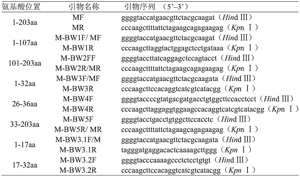
表2鉴定单克隆抗体精细表位引物Table 2 Identification of monoclonal antibody fine epitope primers

5.单克隆抗体表位序列分析5. Monoclonal Antibody Epitope Sequence Analysis
将鉴定的抗原表位序列25PPYDDD30与NCBI注册的RABV毒株M蛋白氨基酸序列进行比对分析(图2),结果表明该抗原表位在RABV毒株中主要是25PPDDDD30序列。对氨基酸P26L和Y27D分别突变后进行Western blot检测,结果发现P26L和Y27D突变后,该单克隆抗体仍然能够与之反应,说明单克隆抗体4A1识别CVS-11毒株的抗原表位发生Y27D置换,但是并不影响表位的抗原性(图4)。同时,Western blot实验结果发现本发明的M蛋白单克隆抗体可与CVS-11、WT型毒株反应,不能与HEP-Flury毒株(购自中国兽医微生物菌种保藏管理中心,保藏号:CVCC AV2013)反应(图3),同时RABV毒株的N蛋白单克隆抗体可分别与3个毒株发生特异性反应,说明病毒感染细胞是成功的。RABV序列比对发现HEP-Flury毒株的抗原表位序列是25PPDGDD30,进一步将25PPYDDD30表位进行氨基酸D28G突变,Western blot实验发现单克隆抗体4A1无法与之识别(图4)。然而本发明鉴定的抗原表位与Flury及变体毒株(图2:HEP-Flury、rHEP5.0-CVSG、LEP-Flury、Flury-LEP-C)发生氨基酸D28G突变,影响了表位的抗原性,因此本发明的单克隆抗体也可用于将Flury及变体毒株与其它流行RABV毒株进行区分。The identified epitope sequence 25 PPYDDD 30 was compared with the amino acid sequence of the RABV strain M protein registered by NCBI (Figure 2), and the results showed that the epitope in the RABV strain was mainly the 25 PPDDDD 30 sequence. The amino acid P26L and Y27D were mutated and detected by Western blot respectively. The results showed that the monoclonal antibody could still react with the mutation of P26L and Y27D, indicating that the monoclonal antibody 4A1 recognized the antigenic epitope of the CVS-11 strain and was replaced by Y27D. However, the antigenicity of the epitope was not affected (Figure 4). At the same time, the results of Western blot experiment found that the M protein monoclonal antibody of the present invention can react with CVS-11 and WT strains, but cannot react with HEP-Flury strains (purchased from China Veterinary Microorganisms Collection and Management Center, deposit number: CVCC AV2013) reaction (Figure 3), and the N protein monoclonal antibody of the RABV strain could react specifically with the three strains, indicating that the virus infected cells successfully. The RABV sequence alignment found that the epitope sequence of the HEP-Flury strain was 25 PPDGDD 30 , and the 25 PPYDDD 30 epitope was further mutated at amino acid D28G, and Western blot experiments found that the monoclonal antibody 4A1 could not recognize it (Figure 4). However, the antigenic epitope identified by the present invention has amino acid D28G mutation with Flury and variant strains (Fig. 2: HEP-Flury, rHEP5.0-CVSG, LEP-Flury, Flury-LEP-C), which affects the antigen of the epitope Therefore, the monoclonal antibodies of the present invention can also be used to distinguish Flury and variant strains from other prevalent RABV strains.
6.单克隆抗体的免疫组化实验6. Immunohistochemical Experiments of Monoclonal Antibodies
颅内注射感染6-8周龄C57BL/6小鼠(购自上海西普尔-必凯实验动物有限公司)30uL CVS-11毒株(105TCID50/mL)和HEP-Flury毒株,待小鼠发病症状明显时,用2.5%avertin(购自sigma)麻醉小鼠,取脑。以颅内注射等体积的DMEM为空白对照。6-8 week old C57BL/6 mice (purchased from Shanghai Sipple-Bike Laboratory Animal Co., Ltd.) were intracranially injected with 30uL CVS-11 strain (10 5 TCID 50 /mL) and HEP-Flury strain. When the onset symptoms of the mice were obvious, the mice were anesthetized with 2.5% avertin (purchased from sigma), and the brains were collected. The intracranial injection of an equal volume of DMEM was used as a blank control.
(1)固定与包埋:4%多聚甲醛固定24h,经酒精脱水与透明,浸石蜡包埋,切片。(1) Fixation and embedding: 4% paraformaldehyde was fixed for 24 hours, dehydrated and transparentized with alcohol, embedded in paraffin, and sliced.
(2)石蜡切片脱蜡至水:依次将切片放入二甲苯Ⅰ15min-二甲苯Ⅱ15min-二甲苯III 15min-无水乙醇Ⅰ5min-无水乙醇Ⅱ5min-85%酒精5min-75%酒精5min-蒸馏水洗。(2) Dewaxing the paraffin sections to water: put the sections into xylene I for 15 minutes, xylene II for 15 minutes, xylene III for 15 minutes, anhydrous ethanol I for 5 minutes, anhydrous ethanol II for 5 minutes, 85% alcohol for 5 minutes, 75% alcohol for 5 minutes, and distilled water for washing. .
(3)抗原修复:组织切片置于盛满柠檬酸抗原修复缓冲液(pH6.0)(购自福州迈新生物技术开发公司)的修复盒中于微波炉内中火5min,停火5min,转低火10min进行抗原修复,此过程中应防止缓冲液过度蒸发,切勿干片。自然冷却后将玻片置于PBS(pH7.4)中洗涤3次,每次5min。(3) Antigen retrieval: The tissue sections were placed in a repair box filled with citric acid antigen retrieval buffer (pH 6.0) (purchased from Fuzhou Maixin Biotechnology Development Co., Ltd.) in a microwave oven on medium heat for 5 minutes, ceased fire for 5 minutes, and turned to low Antigen retrieval was carried out for 10 minutes. During this process, the buffer should be prevented from over-evaporating, and the slices should not be dried. After natural cooling, the slides were washed three times in PBS (pH 7.4) for 5 min each time.
(4)阻断内源性过氧化物酶:切片放入3%双氧水溶液,室温避光孵育25min,将玻片置于PBS(pH7.4)中洗涤3次,每次5min。(4) Block endogenous peroxidase: The slices were placed in 3% hydrogen peroxide solution, incubated in the dark at room temperature for 25 minutes, and washed 3 times in PBS (pH 7.4) for 5 minutes each time.
(5)血清封闭:在组化圈内滴加3%BSA(购自Servicebio)均匀覆盖组织,室温封闭30min。(5) Serum blocking: 3% BSA (purchased from Servicebio) was added dropwise in the histochemical circle to cover the tissue evenly, and the tissue was blocked for 30 minutes at room temperature.
(6)加一抗:轻轻甩掉封闭液,在CVS-11毒株、HEP-Flury毒株和DMEM感染脑组织切片上滴加1:500稀释的一抗(本发明单克隆抗体),切片平放于湿盒内4℃孵育过夜(湿盒内加少量水防止抗体蒸发)。其中HEP-Flury毒株感染脑组织切片再做一个阳性对照,该切片以HEP-Flury感染小鼠的血清为一抗。(6) Add primary antibody: gently shake off the blocking solution, drop the primary antibody (monoclonal antibody of the present invention) diluted 1:500 on the CVS-11 strain, HEP-Flury strain and DMEM-infected brain tissue sections, The sections were placed flat in a wet box and incubated at 4°C overnight (a small amount of water was added to the wet box to prevent the antibodies from evaporating). The brain tissue section infected with HEP-Flury strain was used as a positive control, and the serum of the HEP-Flury-infected mouse was used as the primary antibody for this section.
(7)加二抗:玻片置于PBS(pH7.4)中洗涤3次,每次5min。切片稍甩干后在圈内滴加HRP标记的羊抗鼠IgG(购自KPL)覆盖组织,室温孵育50min。(7) Adding secondary antibody: The slides were washed three times in PBS (pH 7.4) for 5 min each time. After the sections were slightly dried, HRP-labeled goat anti-mouse IgG (purchased from KPL) was added dropwise in the circle to cover the tissue, and incubated at room temperature for 50 min.
(8)DAB显色:玻片置于PBS(pH7.4)中洗涤3次,每次5min。切片稍甩干后在圈内滴加新鲜配制的DAB显色液(购自福州迈新生物技术开发公司),显微镜下控制显色时间,阳性为棕黄色,自来水冲洗切片终止显色。(8) DAB color development: The slides were washed in PBS (pH 7.4) for 3 times, 5 min each time. After the slices were slightly dried, freshly prepared DAB color developing solution (purchased from Fuzhou Maixin Biotechnology Development Co., Ltd.) was added dropwise in the circle, and the color developing time was controlled under the microscope.
(9)复染细胞核:苏木素复染3min左右,自来水洗,苏木素分化液分化数秒,自来水冲洗,苏木素返蓝液返蓝,流水冲洗。(9) Counterstaining nuclei: counterstaining with hematoxylin for about 3 minutes, washing with tap water, differentiation with hematoxylin differentiation solution for several seconds, rinsing with tap water, returning to blue with hematoxylin solution, and rinsing with running water.
(10)脱水封片:将切片依次放入75%酒精5min-85%酒精5min--无水乙醇Ⅰ5min-无水乙醇Ⅱ5min-二甲苯Ⅰ5min中脱水透明,将切片从二甲苯拿出来稍晾干,中性树胶封片。(10) Dehydration and sealing: put the slices into 75% alcohol for 5min-85% alcohol for 5min--anhydrous ethanol I 5min-anhydrous ethanol II 5min-xylene I 5min in order to dehydrate and transparent, take out the slices from xylene and air dry for a while , Neutral gum sealant.
(11)显微镜镜检,图像采集分析显示苏木素染细胞核为蓝色,DAB显出的阳性表达为棕黄色,即该单克隆抗体能对感染的脑组织中的RABV病毒进行标记(图5)。(11) Microscopic examination, image acquisition and analysis showed that the hematoxylin-stained nuclei were blue, and the positive expression of DAB was brown, that is, the monoclonal antibody could label the RABV virus in the infected brain tissue (Figure 5).
序列表sequence listing
<110> 浙江大学<110> Zhejiang University
<120> 分泌抗狂犬病毒M蛋白单克隆抗体的杂交瘤细胞株及应用<120> Hybridoma cell line secreting anti-rabies virus M protein monoclonal antibody and its application
<160> 3<160> 3
<170> SIPOSequenceListing 1.0<170> SIPOSequenceListing 1.0
<210> 1<210> 1
<211> 6<211> 6
<212> PRT<212> PRT
<213> 人工序列(Artificial Sequence)<213> Artificial Sequence
<400> 1<400> 1
Pro Pro Tyr Asp Asp AspPro Pro Tyr Asp Asp Asp
1 51 5
<210> 2<210> 2
<211> 6<211> 6
<212> PRT<212> PRT
<213> 人工序列(Artificial Sequence)<213> Artificial Sequence
<400> 2<400> 2
Pro Pro Asp Asp Asp AspPro Pro Asp Asp Asp Asp
1 51 5
<210> 3<210> 3
<211> 6<211> 6
<212> PRT<212> PRT
<213> 人工序列(Artificial Sequence)<213> Artificial Sequence
<400> 3<400> 3
Pro Pro Asp Gly Asp AspPro Pro Asp Gly Asp Asp
1 51 5
Claims (6)
Priority Applications (3)
| Application Number | Priority Date | Filing Date | Title |
|---|---|---|---|
| CN201910258004.2A CN109970852B (en) | 2019-04-01 | 2019-04-01 | Hybridoma cell line secreting anti-rabies virus M protein monoclonal antibody and its application |
| PCT/CN2020/081974 WO2020200143A1 (en) | 2019-04-01 | 2020-03-30 | Hybridoma cell line for secreting anti-rabies virus m protein monoclonal antibody and use thereof |
| US17/294,414 US20240262891A1 (en) | 2019-04-01 | 2020-03-30 | Hybridoma cell line for secreting anti-rabies virus m protein monoclonal antibody and application thereof |
Applications Claiming Priority (1)
| Application Number | Priority Date | Filing Date | Title |
|---|---|---|---|
| CN201910258004.2A CN109970852B (en) | 2019-04-01 | 2019-04-01 | Hybridoma cell line secreting anti-rabies virus M protein monoclonal antibody and its application |
Publications (2)
| Publication Number | Publication Date |
|---|---|
| CN109970852A CN109970852A (en) | 2019-07-05 |
| CN109970852B true CN109970852B (en) | 2020-10-13 |
Family
ID=67082220
Family Applications (1)
| Application Number | Title | Priority Date | Filing Date |
|---|---|---|---|
| CN201910258004.2A Active CN109970852B (en) | 2019-04-01 | 2019-04-01 | Hybridoma cell line secreting anti-rabies virus M protein monoclonal antibody and its application |
Country Status (3)
| Country | Link |
|---|---|
| US (1) | US20240262891A1 (en) |
| CN (1) | CN109970852B (en) |
| WO (1) | WO2020200143A1 (en) |
Families Citing this family (6)
| Publication number | Priority date | Publication date | Assignee | Title |
|---|---|---|---|---|
| CN109970852B (en) * | 2019-04-01 | 2020-10-13 | 浙江大学 | Hybridoma cell line secreting anti-rabies virus M protein monoclonal antibody and its application |
| CN113403339B (en) * | 2021-06-16 | 2022-04-01 | 武汉大学 | Expression vector with epitope tag M at C end and construction method and application thereof |
| CN113481236B (en) * | 2021-06-16 | 2022-04-01 | 武汉大学 | Expression vector with epitope tag M at N end and construction method and application thereof |
| CN114057867B (en) * | 2021-12-14 | 2023-05-12 | 河南联科物联网科技有限公司 | Monoclonal antibody for resisting egg drop syndrome and application thereof |
| CN114921481B (en) * | 2022-02-25 | 2024-01-30 | 上海赛伦生物技术股份有限公司 | Rabies virus modified mRNA vaccine and preparation method thereof |
| CN114958774B (en) * | 2022-05-08 | 2023-10-27 | 中国医学科学院医学生物学研究所 | Anti-rabies virus monoclonal antibody, hybridoma cell strain secreting antibody and application |
Citations (5)
| Publication number | Priority date | Publication date | Assignee | Title |
|---|---|---|---|---|
| CN1431222A (en) * | 2003-01-23 | 2003-07-23 | 浙江大学 | Monoclone antibody of rice blast germ and detection peridium as well as method for locating antigen surface |
| CN1787837A (en) * | 2002-11-15 | 2006-06-14 | 希龙公司 | Methods for preventing and treating cancer metastasis and bone loss associated with cancer metastasis |
| WO2009093998A1 (en) * | 2008-01-23 | 2009-07-30 | Lobo Peter I | NATURALLY OCCURING IgM ANTIBODIES THAT BIND LYMPHOCYTES |
| CN104603149A (en) * | 2012-05-24 | 2015-05-06 | 万机集团有限公司(英国) | Compositions and methods related to prevention and treatment of rabies infection |
| WO2016078761A1 (en) * | 2014-11-18 | 2016-05-26 | Humabs Biomed Sa | Antibodies that potently neutralize rabies virus and other lyssaviruses and uses thereof |
Family Cites Families (7)
| Publication number | Priority date | Publication date | Assignee | Title |
|---|---|---|---|---|
| EP0404836A1 (en) * | 1988-04-07 | 1991-01-02 | FARMITALIA CARLO ERBA S.r.l. | Human monoclonal antibodies against rabies virus |
| DE4006630A1 (en) * | 1990-03-03 | 1991-09-12 | Behringwerke Ag | HUMANE MONOCLONAL ANTIBODIES AGAINST RABBIT VIRUSES, THEIR PRODUCTION AND USE |
| EP2630242B1 (en) * | 2010-10-19 | 2017-08-02 | The Government of the United States of America as Represented by the Secretary of the Department of Health and Human Services, | Identification of antibodies specific for lyssaviruses |
| CN103954777A (en) * | 2014-05-20 | 2014-07-30 | 北京凯思百奥科技发展有限公司 | Rabies virus monoclonal antibody and application thereof |
| CN104911195A (en) * | 2015-05-22 | 2015-09-16 | 华南农业大学 | Modified rabies virus resisting HEP-Flury strain M protein and preparing method and application of monoclonal antibody thereof |
| CN107815441B (en) * | 2017-08-31 | 2020-05-12 | 浙江大学 | Type II pseudorabies virus attenuated strain and preparation method and application thereof |
| CN109970852B (en) * | 2019-04-01 | 2020-10-13 | 浙江大学 | Hybridoma cell line secreting anti-rabies virus M protein monoclonal antibody and its application |
-
2019
- 2019-04-01 CN CN201910258004.2A patent/CN109970852B/en active Active
-
2020
- 2020-03-30 WO PCT/CN2020/081974 patent/WO2020200143A1/en not_active Ceased
- 2020-03-30 US US17/294,414 patent/US20240262891A1/en active Pending
Patent Citations (5)
| Publication number | Priority date | Publication date | Assignee | Title |
|---|---|---|---|---|
| CN1787837A (en) * | 2002-11-15 | 2006-06-14 | 希龙公司 | Methods for preventing and treating cancer metastasis and bone loss associated with cancer metastasis |
| CN1431222A (en) * | 2003-01-23 | 2003-07-23 | 浙江大学 | Monoclone antibody of rice blast germ and detection peridium as well as method for locating antigen surface |
| WO2009093998A1 (en) * | 2008-01-23 | 2009-07-30 | Lobo Peter I | NATURALLY OCCURING IgM ANTIBODIES THAT BIND LYMPHOCYTES |
| CN104603149A (en) * | 2012-05-24 | 2015-05-06 | 万机集团有限公司(英国) | Compositions and methods related to prevention and treatment of rabies infection |
| WO2016078761A1 (en) * | 2014-11-18 | 2016-05-26 | Humabs Biomed Sa | Antibodies that potently neutralize rabies virus and other lyssaviruses and uses thereof |
Also Published As
| Publication number | Publication date |
|---|---|
| US20240262891A1 (en) | 2024-08-08 |
| WO2020200143A1 (en) | 2020-10-08 |
| CN109970852A (en) | 2019-07-05 |
Similar Documents
| Publication | Publication Date | Title |
|---|---|---|
| CN109970852B (en) | Hybridoma cell line secreting anti-rabies virus M protein monoclonal antibody and its application | |
| CN108586607B (en) | Preparation method and application of anti-HPV16 L1 protein monoclonal antibody | |
| CN104877027B (en) | Anti- pig SC protein monoclonal antibodies and its application in terms of mycoplasma hyopneumoniae SIgA antibody ELISA detection kit is prepared | |
| CN107058239B (en) | Anti-classical swine fever virus E2 protein monoclonal antibody cell strain and application thereof | |
| CN101560255A (en) | Anti-rabies virus monoclonal antibody and preparation method and application | |
| CN112980802B (en) | Hybridoma cell secreting novel duck reovirus sigma B protein monoclonal antibody, monoclonal antibody and application | |
| CN113150124B (en) | Double-antibody sandwich ELISA based on African swine fever virus p72 gene and application thereof | |
| CN113527475B (en) | Hybridoma cell secreting novel duck reovirus sigma C protein monoclonal antibody, monoclonal antibody and application | |
| CN102023217A (en) | Double-antibody biotin-Avidin ELISA (enzyme-linked immuno sorbent assay) detection kit for cattle viral diarrhea virus and application method thereof | |
| CN107586783A (en) | Anti- PPR virus N protein monoclonal antibody and its application | |
| CN111440228A (en) | Common epitope, antibody, identification method and application of HA2 protein of multiple subtypes of influenza viruses | |
| CN113150079B (en) | Eukaryotic expression African swine fever virus p72 antigen and application thereof | |
| CN118530350A (en) | A monoclonal antibody capable of identifying novel duck reovirus σC protein, preparation method and application thereof | |
| Zhang et al. | Development of a potential diagnostic monoclonal antibody against capsid spike protein VP27 of the novel goose astrovirus | |
| CN108752471B (en) | Preparation method and application of anti-PCV 2 monoclonal antibody | |
| CN111041000B (en) | Hybridoma cell strain secreting anti-rift valley fever virus NSs protein monoclonal antibody and application thereof | |
| CN116769019B (en) | ASFVp30 protein monoclonal antibody and application thereof | |
| CN116970074A (en) | A monoclonal antibody that binds to the CD2v intracellular protein of African swine fever virus and its application | |
| CN105925537B (en) | A kind of anti-HSV glycoprotein gD monoclonal antibody and hybridoma cell producing the antibody | |
| CN110016466B (en) | Monoclonal antibody for specifically detecting bluetongue virus, hybridoma cell strain and application thereof | |
| CN109295002B (en) | Hybridoma cell, monoclonal antibody, preparation method and application thereof | |
| CN102304180A (en) | Monoclonal antibody of avian reticuloendotheliosis virus envelope protein and preparation method thereof | |
| CN118725095A (en) | A monoclonal antibody for NS2 protein and its application | |
| CN116731163A (en) | anti-ASFV pK205R protein monoclonal antibody, preparation and application thereof | |
| CN115057925A (en) | Anti-akabane virus monoclonal antibody and application thereof |
Legal Events
| Date | Code | Title | Description |
|---|---|---|---|
| PB01 | Publication | ||
| PB01 | Publication | ||
| SE01 | Entry into force of request for substantive examination | ||
| SE01 | Entry into force of request for substantive examination | ||
| GR01 | Patent grant | ||
| GR01 | Patent grant |