Detailed Description
The technical solution of the present invention will be explained in more detail below. It is to be understood, however, that within the scope of the present invention, the above-described features of the present invention and those specifically described below (e.g., in the examples) may be combined with one another to form new or preferred embodiments. Not to be reiterated herein, but to the extent of space.
In view of the deficiencies in the prior art, the inventors of the present invention have made extensive studies and extensive practices to provide technical solutions of the present invention. The technical solution, its implementation and principles, etc. will be further explained as follows.
An aspect of the embodiments of the present invention provides a brain targeting exosome, which is characterized by comprising an exosome capable of penetrating a blood brain barrier and a fluorescent material with an imaging function, wherein the fluorescent material is distributed in an inner cavity of the exosome.
In some embodiments, the fluorescent material comprises quantum dots.
In some preferred embodiments, the fluorescence emission of the quantum dots is located in the near infrared region II, wherein the emission wavelength is between 1050-1700 nm.
In some more preferred embodiments, the quantum dots are selected from glutathione-modified near-infrared region II quantum dots.
In some more preferred embodiments, the quantum dots comprise Ag2S、Ag2Se、Ag2Te, InAs, InSb, GbSb and Ag2SexS1-xAny one or a combination of more than two of the above, wherein x is 0.1-0.9.
Another aspect of the embodiments of the present invention also provides a method for preparing a brain-targeted exosome, comprising:
allowing neural stem cells with brain targeting to secrete exosomes with brain targeting peptides;
collecting and purifying the exosomes;
and (3) enabling a fluorescent material to enter the inner cavity of the exosome, thereby obtaining the brain-targeted exosome.
The exosome is derived from a neural stem cell which is modified by genetic engineering, and the neural stem cell can be modified by the genetic engineering method, so that the cell membrane surface of the neural stem cell carries a targeting peptide targeting the brain. The exosome with the brain targeting peptide secreted by the neural stem cells with the brain targeting modification has the function of passing through a blood brain barrier and can treat traumatic brain injury, and the natural nano vesicle can realize low toxic and side effects, high efficiency and accurate treatment.
Fluorescent materials are used for visualization and imaging. The fluorescent material marks exosomes, enters a hydrophilic inner cavity of the exosomes with the targeting peptide, realizes the visual treatment of monitoring traumatic brain injury in real time, improves the defect of the traditional biomedical image, and is in-vivo fluorescence imaging.
In some embodiments, the method for preparing neural stem cells with brain targeting comprises:
and (3) infecting the nerve stem cells with the lentivirus with the targeting peptide to obtain the nerve stem cells with brain targeting.
In some preferred embodiments, the method specifically comprises the following steps: and (3) constructing a three-plasmid system of the lentivirus, and infecting 293T-17 cells with the three-plasmid system of the lentivirus to obtain the lentivirus with the targeting peptide.
In some specific embodiments, the method for preparing neural stem cells with brain targeting comprises:
extracting neural stem cells;
constructing a three-plasmid system of the lentivirus, and infecting 293T-17 cells by the three-plasmid system of the lentivirus to obtain the lentivirus with the targeting peptide;
and infecting the lentivirus with the targeting peptide into the neural stem cell to obtain the neural stem cell with the brain targeting function.
The method comprises the steps of utilizing a genetic engineering method to modify neural stem cells, constructing a three-plasmid system of lentiviruses, infecting 293T-17 cells to obtain the lentiviruses with target genes, and infecting the lentiviruses with the neural stem cells to enable the neural stem cells to have targeting properties, namely the neural stem cells with the brain targeting property can secrete exosomes with brain targeting peptides.
Further, the composition of the lentiviral three-plasmid system included pmd2.g, psPAX2, and the plasmid of interest brain targeting peptide NFL.
Wherein, the lentivirus three-plasmid system infects 293T-17 cells, and can pack more lentiviruses with targeting peptides. The packaged lentivirus infects neural stem cells, so that the neural stem cells express target genes, and the neural stem cells with brain targeting can secrete exosomes with brain targeting peptides.
In some embodiments, the brain-targeting peptide comprises any one or a combination of two or more of Angiopep-2, ApoE, RVG, cell-penetrating peptide, and NFL.
The brain targeting peptides are selected from various kinds, such as Angiopep-2, ApoE, Rabies Virus Glycoprotein (RVG) which is widely used, cell penetrating peptide CPP and targeting peptide NFL of neural stem cells;
preferably, RVG serves as a targeting peptide for neurons and neuroepithelial cells and NFL serves as a targeting peptide for neural stem cells.
Preferably, the NFL peptide that targets neural stem cells is selected to target neural stem cells of the brain, such that the neural stem cells of the brain function at the site of the traumatic brain injury.
In some embodiments, the fluorescent material comprises quantum dots whose fluorescent emission is in the near infrared region.
In some preferred embodiments, the fluorescence emission of the quantum dots is located in the near infrared region II, wherein the emission wavelength is between 1050-1700 nm.
In some more preferred embodiments, the quantum dots are selected from glutathione-modified near-infrared region II quantum dots.
In some more preferred embodiments, the quantum dots comprise Ag2S、Ag2Se、Ag2Te, InAs, InSb, GbSb and Ag2SexS1-xAny one or combination of more than two of the above, wherein x is the value0.1-0.9。
In some embodiments, exosomes are labeled by using novel near-infrared region II Ag2S fluorescence quantum dot mark, research shows that the near infrared II region has higher spatial resolution and better tissue penetration depth, compared with the traditional visible light region (400-.
Preferably, the selected marker is a near-infrared fluorescent quantum dot. The near infrared fluorescence quantum dots are selected to be in a near infrared II region (NIR-II 1000-1700nm), and can be 1000, 1100, 1200 and the like. The near-infrared fluorescent quantum dots have Ag2S,Ag2Se,Ag2Te, CrSe, and the like.
Preferably, Ag which is positioned in a near infrared II region and has a glutathione-modified ligand is selected2And the quantum dots have the characteristic of near-infrared II-region fluorescence imaging.
Wherein the purified exosomes are combined with Ag2Mixing S quantum dots, and allowing Ag to pass through the way of electroporation with the quantum dots in excess2And the S quantum dots enter the inside of the exosome to realize the quantum dot marking of the exosome.
In some embodiments, the fluorescent material enters the lumen of the exosome by any one of co-incubation, sonication, and electroporation.
In some embodiments, comprises: the culture solution with brain-targeted neural stem cells is collected, followed by centrifugation to collect exosomes in the culture solution.
In some preferred embodiments, the method specifically comprises the following steps: sequentially adopting the rotation speeds of 750-.
In some specific embodiments, the method comprises: exosomes are collected by collecting culture solution of neural stem cells with brain targeting, the separation mode of exosomes is very various, and exosomes are mainly collected by an ultra-high speed centrifugation mode.
For example, firstly, the dead cells and cell debris are removed by differential centrifugation at three different rotation speeds of 750-.
Wherein the exosomes are purified by sucrose density gradient centrifugation.
In yet another aspect of the embodiments of the present invention, there is provided a use of the brain targeting exosome or the brain targeting exosome prepared by the preparation method in treating a brain disease.
Provides visual and precise nano-drug therapy for treating brain diseases such as traumatic brain injury.
The technical solutions of the present invention will be described in further detail with reference to several preferred embodiments, and it should be apparent that the described embodiments are only a part of the embodiments of the present invention, and not all embodiments. All other embodiments, which can be obtained by a person skilled in the art without any inventive step based on the embodiments of the present invention, are within the scope of the present invention. The conditions used in the following examples may be further adjusted as necessary, and the conditions used in the conventional experiments are not generally indicated.
Example (b):
(1) extraction of neural stem cells
Step 1: taking Balb/C mice about 4 weeks, killing the mice by a neck-breaking method, cutting off the head, taking out the complete brains, putting the brains on a DMEM-F12 culture medium (containing 1% double antibody) precooled on ice, carefully peeling off the hypothalamic (SVZ) area, putting the areas on ice, and peeling off meninges and blood vessels by using forceps; the hypothalamus (SVZ) was minced with a scalpel, placed into a 15mL centrifuge tube containing 5mL DMEM-F12 serum free medium, and digested with 0.5mL neural stem cell pancreatin Accutase at 37 ℃ for 20min with shaking every 5 min. Centrifuging at 200g for 5min to remove supernatant; add 1mL DMEM-F12, suspend cells by media, add 20. mu.l DNase, gently blow the tissue 10 times with a pipette gun without air bubbles during the blow. And slowly blowing 1mL of fresh culture medium and a small amount of DNase on ice for 15 times, standing for 1-2min, sucking the supernatant into a new 15mL centrifuge tube, adding 1mL of fresh culture medium into the old centrifuge tube, and repeating the operation steps for more than 2 times.
Step 2: filtering the blown cell suspension by a 400-mesh filter membrane to remove large tissue blocks and cell clusters; centrifuging the filtered cell suspension at the rotating speed of 200g for 5min, discarding the supernatant, re-suspending the cell precipitate by using a complete culture medium of neural stem cells, counting cells, and inoculating the appropriate number of cells into a 6-well plate coated by matrigel for adherent culture; after 24 hours of adherent culture, cell supernatant was removed, washed once with a small amount of PBS, and cultured in 2mL of complete medium for neural stem cells. The culture can be continued for 1 week, and the culture medium is changed half every two days. Subculture is carried out after 80% of the total length.
(2) Preparation of Lentiviral targeting group Lamp2b-NFL
Designing a sequence of the brain targeting peptide, and synthesizing a plasmid of Lamp2 b-NFL; one day before transfection, 293T-17 cells were digested, 2X 10 cells were taken66 well plates were plated per cell/well; transfection can be carried out by observing the cell density to be 80-90%; taking LipofectamineTM2000 transfection reagent, DMEM-High medium (without antibiotics) was preheated for 2min in a 37 deg.C constant temperature water bath, LipofectamineTM2000 transfection reagents need to be returned to room temperature for use; adding 1.5mL of preheated DMEM-High culture medium into a new EP tube, and then respectively adding 4 mu G of plasmid Pmd2.G, 8 mu G of plasmid psPAX2 and 12 mu G of plasmid Lamp2b-NFL into the EP tube for uniformly mixing; 1.5mL of preheated fresh DMEM-High medium and 50. mu.L of Lipofectamine were takenTM2000 transfection reagent, mixing well, and incubating for 5min at room temperature; all plasmids and Lipofectamine were addedTM2000, uniformly mixing the transfection reagents, incubating at room temperature for 20min, adding the incubated mixture into 293T-17 cells, and replacing with a fresh culture medium after 5-6h after transfection; collecting cell supernatants containing lentivirus twice at 48h and 72h after transfection; lentivirus was concentrated and titer was determined. Lentivirus infection 293T-17 FIGS. 1a, 1b,1c, lentivirus titer assay is shown in FIG. 2.
(3) Preparation of Lentiviral targeting group Lamp2b-RVG
Synthesizing a targeting sequence of a targeting peptide lamp2b-RVG in a company to obtain a target sequence; 293-17 cells are digested before transfection, the density of the cells in the next day can reach 80-90%, and a proper amount of Lipofectamine is taken firstlyTM2000 transfection reagent and DMEM-High culture medium without antibiotic, preheating for 2min in a water bath kettle at 37 ℃, firstly taking 1.5mL of the preheated DMEM-High culture medium, and then respectively taking 4 mu G of plasmid Pmd2.G, 8 mu G of plasmid psPAX2 and 12 mu G of plasmid Lamp2b-RVG, adding into an EP tube and uniformly mixing; then taking 1.5mL of fresh DMEM-High culture medium, adding 50 mu L of transfection reagent into the High-sugar culture medium, uniformly mixing, and incubating for 5min at room temperature; all plasmids and Lipofectamine were addedTM2000 mixing, incubating at room temperature for 20min, adding the incubated mixture into 293T-17 cells, transfecting for 5-6h, and replacing with fresh culture medium; collecting cell supernatants containing lentivirus twice at 48h and 72h after transfection; lentivirus was concentrated and titer was determined.
(4) Preparation of neural stem cells with targeting
Laying Neural Stem Cells (NSCs) in a 24-well plate, wherein the number density of the cells is about 70%, and culturing overnight in an incubator at 37 ℃; preparing a neural stem cell complete medium and a Polybrene mixture in advance, removing the old medium and adding 0.4mL of Polybrene and medium mixture to each well; before infection, the virus is taken out of an ultra-low temperature refrigerator, rapidly melted in water bath at 37 ℃, added into cells in proper dosage, and lightly blown and uniformly mixed; infecting at 37 deg.C for 4h with small volume, and replenishing the complete culture medium to normal volume; after the lentivirus is infected for 24 hours, changing the culture solution, changing a fresh complete culture solution, and continuing to culture at 37 ℃; observing the expression efficiency of the mCherry by a fluorescence microscope for the virus with the mCherry reporter gene 48h after infection; and (4) selecting the successfully transfected neural stem cells by flow screening, and amplifying and preserving the cells. Fluorescence patterns of lentivirus-infected neural stem cells are shown in FIGS. 3a, 3b, and 3 c.
(5) Preparation of exosomes of brain targeting peptides
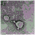
Culturing a large amount of successfully transfected neural stem cells, and collecting a culture solution of the neural stem cells; 750-; 80000-120000g, collecting exosome by ultra-high speed centrifugation for 2 h; the exosome is centrifugally purified by setting sucrose density gradient, the purified exosome is collected, and the sucrose is dialyzed clean by a dialysis method. An electron micrograph of the exosomes is shown in figure 4.
(6) Exosome loaded Ag2S quantum dot
Taking 100 mu g of purified exosome and 100 mu g of glutathione-modified Ag2After S QD mixing; performing electrotransformation under the conditions of 200V and 350 mu F by using a cell electrotransformation machine; after electrotransformation, incubating in an incubator at 37 ℃ for 30min to automatically fuse membranes of exosomes; carrying Ag2Exosomes of the S QDs can mark N2a cells, and imaging of the exosomes can be seen on a near-infrared microscope; carrying Ag2Exosomes of the S QDs effect in vivo imaging as shown in fig. 5.
(7) Exosome-transfected GbSb quantum dot
Taking a proper amount of exosome with target and 100 mu g of GbSb quantum dots, fully and uniformly mixing, and then carrying out electrotransformation under the conditions of 200V and 350 mu F by using a cell electrotransformation instrument; after electrotransformation, incubating the cells in a cell culture box at 37 ℃ for 30min, and automatically fusing the double-layer membrane of the exosome; the exosome carrying the GbSb QDs can mark N2a cells, and the imaging of the exosome can be seen on a near-infrared microscope; exosomes carrying GbSb QDs enable in vivo imaging.
The flow of brain-targeted exosome preparation is shown in figure 6.
In summary, according to the present invention, the neural stem cells are first targeted to the brain by the genetic engineering method, and the neural stem cells with brain targeting secrete exosomes with targeting peptides, which can be loaded with fluorescent materials to realize in vivo imaging. The brain targeting exosome has the function of passing through the blood brain barrier, and can overcome the problem of low efficiency of the traditional nanoparticle in passing through the blood brain barrier. The invention not only provides a natural material passing through a blood brain barrier, but also overcomes some toxic and side effects of the traditional nanoparticles, overcomes the difficulty of brain targeting of the traditional particles, and provides a visual and precise treatment strategy for treating brain-related diseases such as traumatic brain injury, brain tumor and the like.
In addition, the present inventors also performed corresponding experiments using other raw materials listed above and other process conditions and the like in alternative examples, for example, Angiopep-2, ApoE, RVG, cell-penetrating peptide as brain targeting peptide, and Ag as brain targeting peptide2Se、Ag2Te, InAs, InSb, GbSb and Ag2SexS1-xThe quantum dots are respectively tested, and the performances of the obtained brain targeting exosomes are also ideal and basically similar to the embodiments. Therefore, the contents of the verification of the respective examples are not described herein one by one, and only the excellent points of the present invention will be described by taking the above examples as representative examples.
It should be understood that the above-mentioned embodiments are merely illustrative of the technical concepts and features of the present invention, which are intended to enable those skilled in the art to understand the contents of the present invention and implement the present invention, and therefore, the protection scope of the present invention is not limited thereby. All equivalent changes and modifications made according to the spirit of the present invention should be covered within the protection scope of the present invention.