CN109303919B - Application of Akt inhibitor in preparation of anti-liver cancer active drug for enhancing lycorine - Google Patents
Application of Akt inhibitor in preparation of anti-liver cancer active drug for enhancing lycorine Download PDFInfo
- Publication number
- CN109303919B CN109303919B CN201710618320.7A CN201710618320A CN109303919B CN 109303919 B CN109303919 B CN 109303919B CN 201710618320 A CN201710618320 A CN 201710618320A CN 109303919 B CN109303919 B CN 109303919B
- Authority
- CN
- China
- Prior art keywords
- lycorine
- cells
- liver cancer
- treated
- alone
- Prior art date
- Legal status (The legal status is an assumption and is not a legal conclusion. Google has not performed a legal analysis and makes no representation as to the accuracy of the status listed.)
- Active
Links
Images


Classifications
-
- A—HUMAN NECESSITIES
- A61—MEDICAL OR VETERINARY SCIENCE; HYGIENE
- A61K—PREPARATIONS FOR MEDICAL, DENTAL OR TOILETRY PURPOSES
- A61K45/00—Medicinal preparations containing active ingredients not provided for in groups A61K31/00 - A61K41/00
- A61K45/06—Mixtures of active ingredients without chemical characterisation, e.g. antiphlogistics and cardiaca
-
- A—HUMAN NECESSITIES
- A61—MEDICAL OR VETERINARY SCIENCE; HYGIENE
- A61K—PREPARATIONS FOR MEDICAL, DENTAL OR TOILETRY PURPOSES
- A61K31/00—Medicinal preparations containing organic active ingredients
- A61K31/33—Heterocyclic compounds
- A61K31/395—Heterocyclic compounds having nitrogen as a ring hetero atom, e.g. guanethidine or rifamycins
- A61K31/435—Heterocyclic compounds having nitrogen as a ring hetero atom, e.g. guanethidine or rifamycins having six-membered rings with one nitrogen as the only ring hetero atom
- A61K31/4353—Heterocyclic compounds having nitrogen as a ring hetero atom, e.g. guanethidine or rifamycins having six-membered rings with one nitrogen as the only ring hetero atom ortho- or peri-condensed with heterocyclic ring systems
- A61K31/437—Heterocyclic compounds having nitrogen as a ring hetero atom, e.g. guanethidine or rifamycins having six-membered rings with one nitrogen as the only ring hetero atom ortho- or peri-condensed with heterocyclic ring systems the heterocyclic ring system containing a five-membered ring having nitrogen as a ring hetero atom, e.g. indolizine, beta-carboline
-
- A—HUMAN NECESSITIES
- A61—MEDICAL OR VETERINARY SCIENCE; HYGIENE
- A61K—PREPARATIONS FOR MEDICAL, DENTAL OR TOILETRY PURPOSES
- A61K31/00—Medicinal preparations containing organic active ingredients
- A61K31/33—Heterocyclic compounds
- A61K31/395—Heterocyclic compounds having nitrogen as a ring hetero atom, e.g. guanethidine or rifamycins
- A61K31/535—Heterocyclic compounds having nitrogen as a ring hetero atom, e.g. guanethidine or rifamycins having six-membered rings with at least one nitrogen and one oxygen as the ring hetero atoms, e.g. 1,2-oxazines
- A61K31/5375—1,4-Oxazines, e.g. morpholine
- A61K31/5377—1,4-Oxazines, e.g. morpholine not condensed and containing further heterocyclic rings, e.g. timolol
Landscapes
- Health & Medical Sciences (AREA)
- Chemical & Material Sciences (AREA)
- Medicinal Chemistry (AREA)
- Pharmacology & Pharmacy (AREA)
- Epidemiology (AREA)
- Life Sciences & Earth Sciences (AREA)
- Animal Behavior & Ethology (AREA)
- General Health & Medical Sciences (AREA)
- Public Health (AREA)
- Veterinary Medicine (AREA)
- Pharmaceuticals Containing Other Organic And Inorganic Compounds (AREA)
Abstract
本发明公开了Akt抑制剂在制备增强石蒜碱的抗肝癌活性药物中的应用。本发明提供了Akt抑制剂在制备增强石蒜碱的抗肝癌活性或抗肝癌细胞活性产品中的应用。实验证明,Akt抑制剂和石蒜碱联合用药的肝癌细胞凋亡率比Akt抑制剂单独用药的肝癌细胞凋亡率与石蒜碱单独用药的肝癌细胞凋亡率之和还要高,Akt抑制剂和石蒜碱在抗肝癌方面产生了协同作用,Akt抑制剂增强了石蒜碱的抗肝癌活性。The invention discloses the application of an Akt inhibitor in the preparation of a drug for enhancing the anti-hepatocellular carcinoma activity of lycorine. The invention provides the application of Akt inhibitor in the preparation of a product that enhances the anti-liver cancer activity of lycorine or the anti-liver cancer cell activity. Experiments have shown that the apoptosis rate of liver cancer cells treated with Akt inhibitor and lycorine combined is higher than the sum of the apoptosis rate of liver cancer cells treated with Akt inhibitor alone and the apoptosis rate of liver cancer cells treated with lycorine alone. Akt inhibitor enhanced the anti-hepatocellular carcinoma activity of lycorine.
Description
技术领域technical field
本发明涉及Akt抑制剂的用途,特别涉及Akt制剂在制备增强石蒜碱的抗肝癌活性药物中的应用。The invention relates to the use of Akt inhibitors, in particular to the application of Akt preparations in the preparation of lycorine-enhancing anti-hepatocellular carcinoma active drugs.
背景技术Background technique
肝细胞癌(HCC)是全球最常见的侵袭性肿瘤之一。肝癌是一种高度致命的肿瘤,其中位生存率在诊断后仍保持在1年以下。肝癌是源于慢性肝脏疾病如慢性炎症和肝硬化的异质性疾病。迄今为止,针对肝癌的治疗策略主要包括切除术,移植术,局部消融术和化疗。由于肝癌的早期症状不明显,大多数肝癌患者被诊断为晚期阶段,失去手术机会,及时接受过手术的患者多数仍会发生复发和转移。在化学治疗剂中,索拉非尼是一种小分子多激酶抑制剂,被认为是一种积极的治疗方法,是延长晚期肝癌患者生存期延长的唯一批准的全身药物。然而,索拉非尼治疗晚期肝癌患者的预期寿命仅为8-11个月。在过去几十年中,尽管肝癌患者常规治疗方案取得了显着进展,但由于治疗有限,预后差,复发率高,仍然是全球最致命的恶性肿瘤之一。新的有效和有希望的治疗策略仍然需要在全球范围内进行深入的探索。Hepatocellular carcinoma (HCC) is one of the most common aggressive tumors worldwide. Liver cancer is a highly lethal tumor with median survival rates remaining below 1 year after diagnosis. Liver cancer is a heterogeneous disease arising from chronic liver diseases such as chronic inflammation and cirrhosis. To date, treatment strategies for liver cancer mainly include resection, transplantation, local ablation, and chemotherapy. Because the early symptoms of liver cancer are not obvious, most patients with liver cancer are diagnosed at an advanced stage and lose the opportunity for surgery. Most patients who receive surgery in time will still experience recurrence and metastasis. Among chemotherapeutic agents, sorafenib, a small-molecule multikinase inhibitor, is considered an aggressive treatment and the only approved systemic drug that prolongs survival in patients with advanced liver cancer. However, the life expectancy of patients with advanced liver cancer treated with sorafenib is only 8-11 months. In the past few decades, despite significant progress in conventional treatment options for patients with liver cancer, it remains one of the most lethal malignancies worldwide due to limited treatment, poor prognosis, and high recurrence rate. New effective and promising therapeutic strategies still require intensive exploration on a global scale.
具有多种生物活性和机制的天然产物通常作为癌症预防和抗癌药物发现的优良药物。已经探索了石蒜碱(结构如图1中a),一种普通民间药物的活性生物碱,其中包括抗癌,抗病毒,抗疟疾,抗菌和抗炎活性等多种生物活性。尽管石蒜碱的靶点或机制尚未定义,但是石蒜碱的主要生物学活性和低细胞毒性使其成为潜在的临床药物或潜在的潜在药物。例如,它作为有前途的抗癌剂被宫颈癌,白血病,前列腺癌和多发性骨髓瘤所广泛关注。然而,其明确的分子机制目前仍不清楚。Natural products with diverse biological activities and mechanisms often serve as excellent drugs for cancer prevention and anticancer drug discovery. Lycorine (structure shown in Fig. 1a), an active alkaloid of common folk medicines, has been explored with various biological activities including anticancer, antiviral, antimalarial, antibacterial and anti-inflammatory activities. Although the target or mechanism of lycorine has not been defined, the main biological activity and low cytotoxicity of lycorine make it a potential clinical drug or a potential potential drug. For example, it is of great interest in cervical cancer, leukemia, prostate cancer and multiple myeloma as a promising anticancer agent. However, its precise molecular mechanism remains unclear.
Akt,也被称为蛋白激酶B(PKB),是在葡萄糖代谢、凋亡、细胞增殖转录及细胞迁移等多种细胞过程中起到重要作用的一种丝氨酸/苏氨酸特异性蛋白激酶。Akt, also known as protein kinase B (PKB), is a serine/threonine-specific protein kinase that plays an important role in various cellular processes such as glucose metabolism, apoptosis, cell proliferation, transcription, and cell migration.
发明内容SUMMARY OF THE INVENTION
本发明所要解决的技术问题是如何增强石蒜碱的抗肝癌活性或抗肝癌细胞活性。The technical problem to be solved by the present invention is how to enhance the anti-hepatoma activity or anti-hepatoma cell activity of lycorine.
为了解决以上技术问题,本发明提供了Akt抑制剂在制备增强石蒜碱的抗肝癌活性或抗肝癌细胞活性产品中的应用。In order to solve the above technical problems, the present invention provides the application of Akt inhibitors in the preparation of products that enhance the anti-cancer activity of lycorine or the activity against liver cancer cells.
本发明还提供了Akt抑制剂和石蒜碱在制备抗肝癌或抗肝癌细胞产品(如药物、疫苗、保健品和/或食品)中的应用。The present invention also provides the application of Akt inhibitor and lycorine in the preparation of anti-liver cancer or anti-liver cancer cell products (such as medicines, vaccines, health products and/or food).
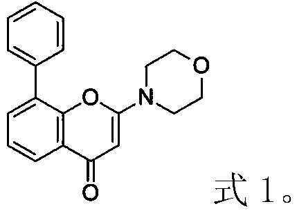
上述应用中,所述Akt抑制剂具体可为LY294002,其结构式如式1。In the above application, the Akt inhibitor may specifically be LY294002, whose structural formula is as shown in

上述应用中,所述Akt抑制剂和所述石蒜碱的配比本领域技术人员可根据抗肝癌或抗肝癌细胞的效果确定,如所述Akt抑制剂和所述石蒜碱的配比具体可为1μmol所述Akt抑制剂:2μmol石蒜碱。In the above-mentioned application, those skilled in the art can determine the ratio of the Akt inhibitor and the lycorine according to the effect of anti-liver cancer or anti-liver cancer cells, such as the specific ratio of the Akt inhibitor and the lycorine. The Akt inhibitor can be 1 μmol: 2 μmol Lycorine.
本发明还提供了抗肝癌或抗肝癌细胞的产品(如药物、疫苗、保健品和/或食品),所述产品含有Akt抑制剂和石蒜碱。The present invention also provides anti-liver cancer or anti-liver cancer cell products (such as medicines, vaccines, health products and/or food), which contain Akt inhibitors and lycorine.
上述抗肝癌或抗肝癌细胞的产品中,所述Akt抑制剂和所述石蒜碱均可单独包装。In the above product against liver cancer or liver cancer cells, both the Akt inhibitor and the lycorine can be packaged separately.
上述抗肝癌或抗肝癌细胞的产品中,所述Akt抑制剂和所述石蒜碱的配比本领域技术人员可根据抗肝癌或抗肝癌细胞的效果确定,如所述Akt抑制剂和所述石蒜碱的配比具体可为1μmol所述Akt抑制剂:2μmol石蒜碱。In the above-mentioned anti-liver cancer or anti-liver cancer cell products, the ratio of the Akt inhibitor and the lycorine can be determined by those skilled in the art according to the effect of anti-liver cancer or anti-liver cancer cells. Specifically, the proportion of lycorine can be 1 μmol of the Akt inhibitor: 2 μmol of lycorine.
上述抗肝癌或抗肝癌细胞的产品中,所述产品的活性成分可为所述Akt抑制剂和所述石蒜碱,所述产品的活性成分还可含有其它成分,所述产品的其它活性成分本领域技术人员可根据抗肝癌效果确定。In the above-mentioned anti-liver cancer or anti-liver cancer cell products, the active ingredients of the product may be the Akt inhibitor and the lycorine, the active ingredients of the product may also contain other ingredients, and the other active ingredients of the product may be Those skilled in the art can determine according to the anti-liver cancer effect.
上述抗肝癌或抗肝癌细胞的产品中,所述产品还可含有载体或赋形剂。所述载体材料包括但不限于水溶性载体材料(如聚乙二醇、聚乙烯吡咯烷酮、有机酸等)、难溶性载体材料(如乙基纤维素、胆固醇硬脂酸酯等)、肠溶性载体材料(如醋酸纤维素酞酸酯和羧甲基纤维素等)。In the above-mentioned anti-liver cancer or anti-liver cancer cell product, the product may further contain a carrier or an excipient. The carrier materials include but are not limited to water-soluble carrier materials (such as polyethylene glycol, polyvinylpyrrolidone, organic acids, etc.), insoluble carrier materials (such as ethyl cellulose, cholesterol stearate, etc.), enteric carriers Materials (such as cellulose acetate phthalate and carboxymethyl cellulose, etc.).
上述抗肝癌或抗肝癌细胞的产品中,所述Akt抑制剂可为LY294002。In the above anti-liver cancer or anti-liver cancer cell product, the Akt inhibitor may be LY294002.
上述应用中或上述抗肝癌或抗肝癌细胞的产品中,所述肝癌可为肝细胞肝癌。In the above application or the above anti-liver cancer or anti-liver cancer cell product, the liver cancer may be hepatocellular carcinoma.
本申请中,抗肝癌、抗肝癌细胞、抗肝癌活性或抗肝癌细胞活性具体可为促进肝癌细胞凋亡和/或促进肝癌细胞自噬和/或降低肝癌细胞活力。In the present application, the anti-hepatoma, anti-hepatoma cell, anti-hepatoma activity or anti-hepatoma cell activity may specifically be promoting liver cancer cell apoptosis and/or promoting liver cancer cell autophagy and/or reducing liver cancer cell viability.
实验证明,Akt抑制剂和石蒜碱联合用药的肝癌细胞凋亡率比Akt抑制剂单独用药的肝癌细胞凋亡率与石蒜碱单独用药的肝癌细胞凋亡率之和还要高,Akt抑制剂和石蒜碱在抗肝癌方面产生了协同作用,Akt抑制剂增强了石蒜碱的抗肝癌活性。Experiments have shown that the apoptosis rate of liver cancer cells treated with Akt inhibitor and lycorine combined is higher than the sum of the apoptosis rate of liver cancer cells treated with Akt inhibitor alone and the apoptosis rate of liver cancer cells treated with lycorine alone. Akt inhibitor enhanced the anti-hepatocellular carcinoma activity of lycorine.
附图说明Description of drawings
图1为石蒜碱抑制了肝细胞肝癌的生长。Figure 1 shows that lycorine inhibits the growth of hepatocellular carcinoma.
图1中,a为石蒜碱的结构式;b为细胞活力测定结果;c为单细胞克隆形成测定结果;d-f为异种移植瘤致瘤性检测结果,g为通过Ki67染色进行免疫组化检测肿瘤生长中增殖的相对变化,h为心、肝、脾、肺和肾的HE染色免疫组化检测结果。In Figure 1, a is the structural formula of lycorine; b is the cell viability assay result; c is the single cell clone formation assay result; d-f are the tumorigenicity test results of xenograft tumors, and g is the immunohistochemical detection of tumors by Ki67 staining Relative changes of proliferation during growth, h is the result of HE staining immunohistochemical detection of heart, liver, spleen, lung and kidney.
图2为石蒜碱促进了肝细胞肝癌的自噬。Figure 2 shows that lycorine promotes autophagy in hepatocellular carcinoma.
图2中,a为蛋白质印迹分析HepG2和SMMC-7721中p62和LC-3B的表达量;b蛋白质印迹分析HepG2和SMMC-7721中Atg5、Atg7和Atg12的表达量;c为透射电子显微镜分析,d和e为自噬通量分析;f为蛋白质印迹分析肝细胞肝癌中p62和LC-3B的表达量;g为免疫组化检测肝细胞肝癌中LC-3B的表达量。In Figure 2, a is the Western blot analysis of the expression levels of p62 and LC-3B in HepG2 and SMMC-7721; b is the Western blot analysis of the expression levels of Atg5, Atg7 and Atg12 in HepG2 and SMMC-7721; c is the transmission electron microscope analysis, d and e are the autophagic flux analysis; f is the expression of p62 and LC-3B in hepatocellular carcinoma by western blot analysis; g is the expression of LC-3B in hepatocellular carcinoma detected by immunohistochemistry.
图3为抑制自噬可以进一步促进石蒜碱诱导的肝癌细胞凋亡。Figure 3 shows that inhibition of autophagy can further promote lycorine-induced apoptosis of liver cancer cells.
图3中,a、c和f为蛋白质印迹分析各处理细胞中p62、LC-3B、Cleaved Caspase-3、PARP1/2和Actin表达量。b、d和e为各处理细胞的细胞活力和细胞凋亡率。In Figure 3, a, c and f are the expression levels of p62, LC-3B, Cleaved Caspase-3, PARP1/2 and Actin in each treated cell by western blot analysis. b, d and e are the cell viability and apoptosis rate of each treated cell.
图3中,a和b的各图片中,从左至右分别为对照处理细胞、石蒜碱单独处理细胞、3-MA单独处理细胞和石蒜碱和3-MA联合处理细胞。c和d的各图片中,从左至右分别为对照siRNA单独处理1细胞、石蒜碱和对照siRNA联合处理1细胞、LC-3B-1单独处理细胞、石蒜碱和LC-3B-1联合处理细胞。e和f的各图片中,从左至右分别为对照siRNA单独处理2细胞、石蒜碱和对照siRNA联合处理2细胞、LC-3B-2单独处理细胞、石蒜碱和LC-3B-2联合处理细胞。In Figure 3, in each picture of a and b, from left to right are control-treated cells, cells treated with lycorine alone, cells treated with 3-MA alone, and cells treated with lycorine and 3-MA in combination. In each picture of c and d, from left to right, control siRNA alone treated 1 cell, lycorine and control siRNA combined
图4为石蒜碱通过TCRP1/Akt/mTOR信号通路促进肝癌细胞凋亡和自噬。Figure 4 shows that lycorine promotes apoptosis and autophagy in liver cancer cells through the TCRP1/Akt/mTOR signaling pathway.
图4中,a为蛋白质印迹分析HepG2和SMMC-7721中TCRP1蛋白水平;b为实时定量PCR分析HepG2和SMMC-7721中的TCRP1的mRNA水平和蛋白质印迹分析HepG2和SMMC-7721中的TCRP1蛋白水平;c为蛋白质印迹分析结果表明过表达TCRP1显著阻止了石蒜碱对Akt/mTOR通路的抑制作用;d蛋白质印迹分析结果表明过表达TCRP1阻断了石蒜碱诱导的凋亡和自噬相关蛋白表达;e为细胞活力检测结果;f为自噬通量分析;g为TCRP1过表达强烈地阻断了石蒜碱对集落形成的抑制作用;h为石蒜碱处理减少HepG2异种移植肿瘤中TCRP1,Akt,4-EBP1和p70S6K的蛋白水平。In Figure 4, a is the Western blot analysis of TCRP1 protein levels in HepG2 and SMMC-7721; b is real-time quantitative PCR analysis of TCRP1 mRNA levels in HepG2 and SMMC-7721 and Western blot analysis of TCRP1 protein levels in HepG2 and SMMC-7721 ;c is the result of western blot analysis showing that overexpression of TCRP1 significantly prevented the inhibitory effect of lycorine on the Akt/mTOR pathway; d is the result of western blotting analysis that overexpression of TCRP1 blocked lycorine-induced apoptosis and autophagy-related proteins Expression; e is the result of cell viability assay; f is autophagy flux analysis; g is TCRP1 overexpression strongly blocks the inhibitory effect of lycorine on colony formation; h is lycorine treatment reduces TCRP1 in HepG2 xenograft tumors , protein levels of Akt, 4-EBP1 and p70S6K.
图5为免疫组化检测磷酸化的Akt和TCRP1水平。Figure 5 shows the levels of phosphorylated Akt and TCRP1 detected by immunohistochemistry.
a和b为在石蒜碱(10mg/kg)处理的异种移植肿瘤中,磷酸化的Akt和TCRP1的阳性染色强得多;c和d为免疫组化检测两个肝癌患者的磷酸化的Akt和TCRP1水平。a and b are much stronger positive staining for phosphorylated Akt and TCRP1 in lycorine (10 mg/kg)-treated xenograft tumors; c and d are immunohistochemical detection of phosphorylated Akt in two liver cancer patients and TCRP1 levels.
图6为Akt抑制剂增强石蒜碱的抗肝癌活性。两个图片中,从左至右均为对照处理细胞、石蒜碱单独处理细胞、LY294002单独处理细胞和石蒜碱和LY294002联合处理细胞。Figure 6 shows that Akt inhibitors enhance the anti-cancer activity of lycorine. In both images, from left to right are control-treated cells, lycorine-treated cells alone, LY294002-treated cells alone, and cells treated with lycorine and LY294002 in combination.
图7为靶向TCRP1的siRNA增强石蒜碱的抗肝癌活性。两个图片中,从左至右均为对照siRNA单独处理细胞、石蒜碱和对照siRNA联合处理细胞、siRNATCRP1单独处理细胞、石蒜碱和siRNATCRP1联合处理细胞。Figure 7 shows that siRNA targeting TCRP1 enhances the anti-cancer activity of lycorine. In both pictures, from left to right, cells treated with control siRNA alone, cells treated with lycorine and control siRNA in combination, cells treated with siRNA TCRP1 alone, and cells treated with lycorine and siRNA TCRP1 in combination.
具体实施方式Detailed ways
下面结合具体实施方式对本发明进行进一步的详细描述,给出的实施例仅为了阐明本发明,而不是为了限制本发明的范围。下述实施例中的实验方法,如无特殊说明,均为常规方法。下述实施例中所用的材料、试剂等,如无特殊说明,均可从商业途径得到。The present invention will be further described in detail below with reference to the specific embodiments, and the given examples are only for illustrating the present invention, rather than for limiting the scope of the present invention. The experimental methods in the following examples are conventional methods unless otherwise specified. The materials, reagents, etc. used in the following examples can be obtained from commercial sources unless otherwise specified.
下述实施例中的材料和方法如下:The materials and methods in the following examples are as follows:
细胞培养和细胞处理:人类HCC细胞系HepG2,SMMC-7721,HuH-7和Chang肝细胞于2015年4月从ATCC获得。用过的细胞在1个月内复苏。通过PCR扩增的短串联重复分析鉴定细胞系。在含有5%CO2的加湿气氛中,在补充有10%FBS和1%青霉素/链霉素的DMEM或RPMI-1640中将细胞保持在37℃。人类TCRP1和Akt的表达载体设计并购自ServicebioTechnologies(中国武汉)。石蒜碱(Lycorine)(纯度>98%)购自上海源叶生物科技(中国上海)。石蒜碱用二甲基亚砜(DMSO)溶解制备为20mM储备溶液,并以等分试样储存在-20℃,处理细胞时,培养基稀释后即用;给小鼠注射时,用PBS稀释至相应的浓度。3-MA,MG-132和LY294002购自Selleck(London,ON,Canada)。MTT(3-(4,5-二甲基-2-噻唑基)-2,5-二苯基-2-H-四唑溴化物)试剂购自Sigma-Aldrich(St.Louis,MO,USA)。3-MA用二甲基亚砜(DMSO)溶解制备为20mM储备溶液,处理细胞时,培养基稀释后即用。LY294002用二甲基亚砜(DMSO)溶解制备为20mM储备溶液,处理细胞时,培养基稀释后即用。Cell Culture and Cell Handling: Human HCC cell lines HepG2, SMMC-7721, HuH-7 and Chang hepatocytes were obtained from ATCC in April 2015. Spent cells were recovered within 1 month. Cell lines were identified by PCR-amplified short tandem repeat analysis. Cells were maintained at 37 °C in DMEM or RPMI-1640 supplemented with 10% FBS and 1% penicillin/streptomycin in a humidified atmosphere containing 5% CO . Expression vectors for human TCRP1 and Akt were designed and purchased from Servicebio Technologies (Wuhan, China). Lycorine (purity >98%) was purchased from Shanghai Yuanye Biotechnology (Shanghai, China). Lycorine was prepared as a 20 mM stock solution by dissolving in dimethyl sulfoxide (DMSO) and stored in aliquots at -20°C. When treating cells, the medium was diluted and used immediately; when injecting into mice, PBS was used. Dilute to the corresponding concentration. 3-MA, MG-132 and LY294002 were purchased from Selleck (London, ON, Canada). MTT (3-(4,5-dimethyl-2-thiazolyl)-2,5-diphenyl-2-H-tetrazolium bromide) reagent was purchased from Sigma-Aldrich (St. Louis, MO, USA ). 3-MA was prepared as a 20 mM stock solution by dissolving in dimethyl sulfoxide (DMSO), and when treating cells, the medium was diluted and used immediately. LY294002 was prepared by dissolving in dimethyl sulfoxide (DMSO) as a 20 mM stock solution, and when treating cells, the medium was diluted and used immediately.
组织样品:45对人肝癌及其匹配的相邻非肿瘤组织阵列均购自上海生物芯片有限公司(中国上海市)。所有这些样本都被取消识别,并且该队列具有临床结果信息。Tissue samples: 45 pairs of human liver cancer and their matched adjacent non-tumor tissue arrays were purchased from Shanghai Biochip Co., Ltd. (Shanghai, China). All of these samples were de-identified and the cohort had clinical outcome information.
通过MTT法测定细胞活力:向待测细胞中加入MTT溶液并在37℃下孵育4小时。然后取出培养基。加入100ul DMSO,用酶标仪测定570nm处的吸光度。Determination of cell viability by MTT method: Add MTT solution to the cells to be tested and incubate at 37°C for 4 hours. Then remove the medium. Add 100ul DMSO, and measure the absorbance at 570nm with a microplate reader.
细胞凋亡测定:通过使用Muse TM Annexin V和死细胞测定试剂盒(Millipore,Billerica,MA,USA)染色细胞来测量细胞凋亡,并且在台式Muse Cell Analyzer(Millipore,Billerica,MA,USA)中根据制造商的说明书进行细胞计数、细胞周期及细胞凋亡分析。Apoptosis Assay: Apoptosis was measured by staining cells using Muse™ Annexin V and Dead Cell Assay Kit (Millipore, Billerica, MA, USA) and in a benchtop Muse Cell Analyzer (Millipore, Billerica, MA, USA) Cell counts, cell cycle and apoptosis assays were performed according to the manufacturer's instructions.
单克隆形成实验(集落形成测定):将肿瘤细胞接种到6孔板中并培养过夜。然后用指定浓度的药剂处理细胞48小时。用新鲜培养基冲洗后,形成集落,用4%多聚甲醛固定,用0.1%结晶紫染色,然后在指定时间内计数。Monoclonal formation assay (colony formation assay): Tumor cells were seeded into 6-well plates and cultured overnight. Cells were then treated with the indicated concentrations of agents for 48 hours. After rinsing with fresh medium, colonies were formed, fixed with 4% paraformaldehyde, stained with 0.1% crystal violet, and then counted over the indicated time.
免疫荧光测定:用4%多聚甲醛固定细胞30分钟,并与0.5%Triton X-100在PBS中孵育,用5%BSA封闭30分钟。将载玻片在4℃下用抗LC-3B抗体染色过夜,随后在室温下用Alexa-Fluor 488缀合的山羊抗兔IgG抗体染色1小时。用2.5μg/mL DAPI(Invitrogen,Carlsbad,CA,USA)对核进行染色,并通过倒置荧光显微镜(Carl Zeiss,Oberkochen,Germany)显色。Immunofluorescence assay: Cells were fixed with 4% paraformaldehyde for 30 min and incubated with 0.5% Triton X-100 in PBS and blocked with 5% BSA for 30 min. Slides were stained with anti-LC-3B antibody overnight at 4 °C followed by Alexa-Fluor 488-conjugated goat anti-rabbit IgG antibody for 1 h at room temperature. Nuclei were stained with 2.5 μg/mL DAPI (Invitrogen, Carlsbad, CA, USA) and visualized by inverted fluorescence microscopy (Carl Zeiss, Oberkochen, Germany).
免疫组化检测:切除组织切片,福尔马林固定,石蜡包埋,然后与抗Ki67,抗切割的半胱天冬酶3,抗PARP,抗LC-3B和抗-TCRP1抗体在4℃过夜,随后是生物素化的二抗。使用Vectastain Elite ABC试剂盒(Vector Laboratories,Burlingame,CA,USA)可视化免疫反应性。在每个实验中包括已知的阳性对照,通过省略第一抗体获得阴性对照。Immunohistochemical detection: excised tissue sections, formalin-fixed, paraffin-embedded, then treated with anti-Ki67,
蛋白质印迹分析:使用的抗体如下:SQSTM1/p62(D5E2)抗体,Atg5(D5F5U)抗体,Atg7(D12B11)抗体,Atg12(D88H11)抗体,抗LC-3B(D11)抗体,磷酸-p70S6激酶(Thr-389)抗体,总p70S6K抗体,磷酸-Akt(Ser-473)抗体,总Akt抗体,磷酸4-EBP1(Thr-37/46)抗体,总4-EBP1抗体和Cleaved Caspase 3(D175)抗体为CST公司(Danvers,MA,USA)的产品。抗PARP1/2(H250)和TCRP1(E-13)抗体为Santa Cruz Biotechnology(Santa Cruz,CA,USA)产品。抗β-肌动蛋白(A5441)抗体为Sigma-Aldrich(St.Louis,MO,USA)产品。Western blot analysis: Antibodies used were as follows: SQSTM1/p62 (D5E2) antibody, Atg5 (D5F5U) antibody, Atg7 (D12B11) antibody, Atg12 (D88H11) antibody, anti-LC-3B (D11) antibody, phospho-p70S6 kinase (Thr -389) antibody, total p70S6K antibody, phospho-Akt (Ser-473) antibody, total Akt antibody, phospho-4-EBP1 (Thr-37/46) antibody, total 4-EBP1 antibody and Cleaved Caspase 3 (D175) antibody were A product of CST Corporation (Danvers, MA, USA). Anti-PARP1/2 (H250) and TCRP1 (E-13) antibodies were products of Santa Cruz Biotechnology (Santa Cruz, CA, USA). Anti-β-actin (A5441) antibody was a product of Sigma-Aldrich (St. Louis, MO, USA).
实施例1、自噬抑制剂增强石蒜碱的抗肝癌活性(自噬抑制剂和石蒜碱联用增强肝癌细胞的凋亡)Example 1. Autophagy inhibitor enhances the anti-cancer activity of lycorine (the combination of autophagy inhibitor and lycorine enhances the apoptosis of liver cancer cells)
1、石蒜碱抑制了肝细胞肝癌的生长1. Lycorine inhibits the growth of hepatocellular carcinoma
通过MTT法测定细胞活力,分析石蒜碱对三种典型的人类肝细胞肝癌细胞系Cell viability was assayed by MTT assay to analyze the effect of lycorine on three typical human hepatocellular carcinoma cell lines
(HepG2,SMMC-7721,HuH-7)的生长抑制作用,以及非致瘤性的人类肝细胞系(Chang Liver)。用分别含有0、10、20、30、40、50μM的石蒜碱的培养基分别培养HepG2,SMMC-7721,HuH-7和Chang Liver 48小时,用MTT法测定细胞活力,结果表明10、20、30、40、50μM的石蒜碱处理48小时,肝癌细胞存活能力显著下降,与此同时,石蒜碱并没有影响到相同剂量的正常肝细胞Chang Liver的细胞存活能力(图1中b)。取其中的0、10、20μM的石蒜碱处理48小时的HepG2,SMMC-7721,HuH-7进行单克隆形成实验检测,结果表明10和20μM石蒜碱处理HepG2,SMMC-7721,HuH-7 48小时,显著抑制HepG2,SMMC-7721,HuH-7的细胞增殖(图1中C)。异种移植瘤致瘤性检测结果表明,如图1中d所示,与对照组相比,用石蒜碱治疗后肿瘤的大小明显降低。与此相一致,正常组肿瘤的重量比用石蒜碱处理的肿瘤要大得多(图1中e)。与对照组相比,石蒜碱治疗的异种移植肿瘤的生长率明显下降(图1中f)。为了进一步评估肿瘤生长中增殖的相对变化,通过Ki67(标记细胞增殖状态的抗原)染色进行免疫组化检测,结果表明与正常组的肿瘤相比,Lycorine(10mg/kg)组和Lycorine(20mg/kg)组的肿瘤有明显的下降百分比(图1中g)。然而,主要目标器官(图1中h)的细胞形态和体重并没有明显的差异。总的来说,这些结果表明,石蒜碱在体外和体内抑制了肝癌的增殖。(HepG2, SMMC-7721, HuH-7), and a non-tumorigenic human hepatocyte cell line (Chang Liver). HepG2, SMMC-7721, HuH-7 and Chang Liver were cultured in medium containing 0, 10, 20, 30, 40, and 50 μM of lycorine for 48 hours, respectively, and the cell viability was measured by MTT method. The results showed that 10, 20 , 30, 40, and 50 μM of lycorine for 48 hours significantly decreased the viability of liver cancer cells. At the same time, lycorine did not affect the cell viability of normal hepatocyte Chang Liver at the same dose (b in Figure 1). . The HepG2, SMMC-7721, and HuH-7 treated with 0, 10, and 20 μM of lycorine for 48 hours were used for the detection of monoclonal formation. The results showed that 10 and 20 μM of lycorine treated HepG2, SMMC-7721, and HuH-7 At 48 hours, the cell proliferation of HepG2, SMMC-7721, and HuH-7 was significantly inhibited (C in Figure 1). The results of the tumorigenicity assay of the xenograft tumors showed that, as shown in d in Figure 1, the size of the tumors was significantly reduced after treatment with lycorine compared with the control group. Consistent with this, tumors in the normal group were much larger in weight than those treated with lycorine (Fig. 1, e). Compared with the control group, the growth rate of lycorine-treated xenograft tumors was significantly decreased (Fig. 1, f). To further evaluate the relative changes in proliferation in tumor growth, immunohistochemical detection was performed by Ki67 (an antigen that marks the state of cell proliferation) staining, and the results showed that compared with tumors in the normal group, Lycorine (10 mg/kg) and Lycorine (20 mg/kg) kg) group had a significant percentage decrease (g in Figure 1). However, there were no significant differences in cell morphology and body weight in the main target organ (h in Figure 1). Collectively, these results suggest that lycorine inhibits liver cancer proliferation in vitro and in vivo.
其中,异种移植瘤致瘤性检测方法如下:Among them, the tumorigenicity detection method of xenograft tumor is as follows:
将HepG2细胞(0.2mL PBS中的5×10⁶)(通过皮下注射)接种到7周龄的BALB/c雌性无胸腺裸鼠(Taconic)中。当肿瘤体积达到100mm3时,将小鼠随机分为3组:正常组,Lycorine(10mg/kg)组(Lycorine 10mg/kg)和Lycorine(20mg/kg)组(Lycorine20mg/kg),每组8只。并且每隔一天连续33天腹腔注射溶液,Lycorine(10mg/kg)组每只注射120μL的石蒜碱溶液,使石蒜碱的注射剂量为10mg/kg体重/天;Lycorine(20mg/kg)组每只注射120μL的石蒜碱溶液,使石蒜碱的注射剂量为20mg/kg体重/天;正常组注射等体积的PBS。每3天监测肿瘤体积和体重。从第一次给药起33天处死小鼠后,将其肿瘤称重,拍照并在4%多聚甲醛中固定,用于免疫组织化学测定。HepG2 cells ( 5 x 10⁶ in 0.2 mL PBS) were inoculated (by subcutaneous injection) into 7-week-old BALB/c female athymic nude mice (Taconic). When the tumor volume reached 100mm, the mice were randomly divided into 3 groups: normal group, Lycorine (10mg/kg) group (Lycorine 10mg/kg) and Lycorine (20mg/kg) group (Lycorine 20mg/kg), each group of 8 Only. And the solution was intraperitoneally injected every other day for 33 consecutive days. Lycorine (10mg/kg) group was injected with 120 μL of lycorine solution, so that the injection dose of lycorine was 10mg/kg body weight/day; Lycorine (20mg/kg) group Each animal was injected with 120 μL of lycorine solution, so that the injection dose of lycorine was 20 mg/kg body weight/day; the normal group was injected with an equal volume of PBS. Tumor volume and body weight were monitored every 3 days. After mice were sacrificed 33 days from the first dose, their tumors were weighed, photographed and fixed in 4% paraformaldehyde for immunohistochemical assays.
2、石蒜碱促进了肝细胞肝癌的自噬2. Lycorine promotes autophagy in hepatocellular carcinoma
除了细胞凋亡外,自噬是细胞应激反应的程序性细胞死亡的另一种模式。首先通过评价LC-3B和p62(两种经典的自噬标记物)来研究石蒜碱对自噬体形成的影响,用分别含有0、10、20、30和40μM的石蒜碱的培养基分别培养HepG2,SMMC-7721,48小时,取细胞分别用SQSTM1/p62(D5E2)抗体、LC-3B(D11)抗体、Atg5(D5F5U)抗体,Atg7(D12B11)抗体,Atg12(D88H11)抗体和抗β-肌动蛋白(A5441)抗体进行蛋白质印迹分析p62、LC-3B、Atg5、Atg7、Atg12和Actin(作为内参)表达量,结果表明石蒜碱可以上调LC-3B的表达并呈一定的剂量依赖型,而p62的表达显着降低(图2中a)。石蒜碱可以显著上调Atg5,Atg7和Atg12的蛋白表达(图2中b)。为了进一步验证石蒜碱诱导的自噬,通过透射电子显微镜研究了肝癌的细胞内形态学变化。方法如下:用分别含有0和40μM的石蒜碱的培养基分别培养HepG2,SMMC-772148小时,用4%戊二醛固定细胞。在超薄切片机中切割超薄(50nm)切片,并用1%乙酸双氧铀和柠檬酸铅染色。最后,通过电子显微镜Hitachi H-7650(Hitachi,Tokyo,Japan)测定切片。结果表明与正常组细胞(0μM石蒜碱处理的细胞)相比,40μM石蒜碱处理的细胞(图2中c用Lycorine表示的细胞)双膜囊泡有着显著的积累(含有亚细胞物质)(图2中c)。In addition to apoptosis, autophagy is another mode of programmed cell death in response to cellular stress. The effect of lycorine on autophagosome formation was first investigated by evaluating LC-3B and p62, two canonical markers of autophagy, using media containing 0, 10, 20, 30 and 40 μM of lycorine, respectively. HepG2 and SMMC-7721 were cultured for 48 hours, and the cells were harvested with SQSTM1/p62 (D5E2) antibody, LC-3B (D11) antibody, Atg5 (D5F5U) antibody, Atg7 (D12B11) antibody, Atg12 (D88H11) antibody and anti- The expression of p62, LC-3B, Atg5, Atg7, Atg12 and Actin (as an internal reference) was analyzed by western blotting with β-actin (A5441) antibody, and the results showed that lycorine could up-regulate the expression of LC-3B at a certain dose dependent, while the expression of p62 was significantly reduced (a in Figure 2). Lycorine can significantly up-regulate the protein expressions of Atg5, Atg7 and Atg12 (Fig. 2b). To further validate lycorine-induced autophagy, intracellular morphological changes in liver cancer were investigated by transmission electron microscopy. The method was as follows: HepG2, SMMC-772 were incubated with media containing 0 and 40 μM lycorine, respectively, for 48 hours, and the cells were fixed with 4% glutaraldehyde. Ultrathin (50 nm) sections were cut in an ultramicrotome and stained with 1% uranyl acetate and lead citrate. Finally, sections were determined by electron microscope Hitachi H-7650 (Hitachi, Tokyo, Japan). The results showed that double-membrane vesicles (containing subcellular material) were significantly accumulated in 40 μM lycorine-treated cells (cells represented by Lycorine in c in Figure 2) compared with normal cells (0 μM lycorine-treated cells). (c in Figure 2).
为了确定自噬通量,根据制造商(上海吉凯基因化学技术)的说明书用自噬双标腺病毒(mRFP-GFP-LC3)转染细胞HepG2,SMMC-7721。然后将转染的细胞分别用含有0和40μM的石蒜碱的培养基分别培养48小时。随后,将这些细胞用4%多聚甲醛固定10分钟,并用PBS洗涤。最后,用激光扫描共焦显微镜(Olympus FV1000,Tokyo,Japan)对GFP/mRFP图像进行显像。其中,mRFP用于标记及追踪LC3,GFP的减弱可指示溶酶体与自噬小体的融合形成自噬溶酶体,红绿荧光Merge后出现的黄色斑点即是自噬体,红色的斑点指示自噬溶酶体,通过不同颜色斑点的计数可以清晰的看出自噬流的强弱。另外,用分别含有0和40μM的石蒜碱的培养基分别培养HepG2,SMMC-772148小时,用DAPI对细胞进行细胞核染色,10分钟后,用抗LC-3B(D11)抗体对细胞进行免疫组化检测。结果表明使用串联mRFP-GFP标记的LC-3,石蒜碱处理后,导致LC-3-II转化增加,促进LC-3脂化和斑点,以及积累的黄色点状自噬体(图2中d和e)。图2中d和e,正常组和Control为0μM石蒜碱处理的细胞,Lycorine为40μM石蒜碱处理的细胞。To determine autophagic flux, cells HepG2, SMMC-7721, were transfected with autophagy double-labeled adenovirus (mRFP-GFP-LC3) according to the manufacturer's instructions (Shanghai Jikai Gene Chemical Technology). The transfected cells were then incubated with media containing 0 and 40 μM of lycorine for 48 hours, respectively. Subsequently, the cells were fixed with 4% paraformaldehyde for 10 minutes and washed with PBS. Finally, GFP/mRFP images were visualized with a laser scanning confocal microscope (Olympus FV1000, Tokyo, Japan). Among them, mRFP is used to label and track LC3, and the weakening of GFP can indicate the fusion of lysosomes and autophagosomes to form autolysosomes. Indicating autophagic lysosomes, the intensity of autophagic flux can be clearly seen through the counting of different colored spots. In addition, HepG2 and SMMC-772 were incubated with media containing 0 and 40 μM lycorine, respectively, for 48 hours, the cells were stained with DAPI for nuclei, and 10 minutes later, the cells were immunized with anti-LC-3B (D11) antibody. chemical detection. The results showed that the use of tandem mRFP-GFP-tagged LC-3, after lycorine treatment, resulted in increased LC-3-II conversion, promotion of LC-3 lipidation and puncta, and accumulation of yellow punctate autophagosomes (Fig. 2). d and e). In Figure 2 d and e, the normal group and Control are cells treated with 0 μM lycorine, and Lycorine is cells treated with 40 μM lycorine.
此外,通过蛋白质印迹测定和免疫组化染色评估了石蒜碱对体内肝癌自噬的影响。将HepG2细胞(0.2mL PBS中的5×10 6)(通过皮下注射)接种到7周龄的BALB/c雌性无胸腺裸鼠(Taconic)中。当肿瘤体积达到100mm3时,将小鼠随机分为3组:正常组,Lycorine(10mg/kg)组和Lycorine(20mg/kg)组,每组8只。并且每隔一天连续33天腹腔注射溶液,Lycorine(10mg/kg)组每只注射120μL的石蒜碱溶液,使石蒜碱的注射剂量为10mg/kg体重/天;Lycorine(20mg/kg)组每只注射120μL的石蒜碱溶液,使石蒜碱的注射剂量为20mg/kg体重/天;正常组注射等体积的PBS。每3天监测肿瘤体积和体重。从第一次给药起33天处死小鼠后,在4%多聚甲醛中固定肿瘤,用抗LC-3B(D11)抗体对肿瘤细胞进行免疫组化检测,并对肿瘤细胞分别用SQSTM1/p62(D5E2)抗体、LC-3B(D11)抗体和抗β-肌动蛋白(A5441)抗体进行蛋白质印迹分析p62、LC-3B和Actin(作为内参)表达量。结果表明与正常组相比,石蒜碱处理后明显增加了HepG2肿瘤中LC-3B-II水平,同时,p62的水平显着降低(图2中f)。图2中f中,Lycorine(mg/kg)行中的0列为正常组,Lycorine(mg/kg)行中的10列为Lycorine(10mg/kg)组,Lycorine(mg/kg)行中的20列为Lycorine(20mg/kg)组。Furthermore, the effect of lycorine on autophagy in liver cancer in vivo was assessed by western blot assay and immunohistochemical staining. HepG2 cells ( 5 x 10⁶ in 0.2 mL PBS) were inoculated (by subcutaneous injection) into 7-week-old BALB/c female athymic nude mice (Taconic). When the tumor volume reached 100 mm 3 , the mice were randomly divided into 3 groups: normal group, Lycorine (10 mg/kg) group and Lycorine (20 mg/kg) group, with 8 mice in each group. And the solution was intraperitoneally injected every other day for 33 consecutive days. Lycorine (10mg/kg) group was injected with 120 μL of lycorine solution, so that the injection dose of lycorine was 10mg/kg body weight/day; Lycorine (20mg/kg) group Each animal was injected with 120 μL of lycorine solution, so that the injection dose of lycorine was 20 mg/kg body weight/day; the normal group was injected with an equal volume of PBS. Tumor volume and body weight were monitored every 3 days. After mice were sacrificed 33 days from the first administration, tumors were fixed in 4% paraformaldehyde, and tumor cells were detected by immunohistochemistry with anti-LC-3B (D11) antibody, and tumor cells were treated with SQSTM1/ p62 (D5E2) antibody, LC-3B (D11) antibody and anti-β-actin (A5441) antibody were subjected to western blotting to analyze the expression levels of p62, LC-3B and Actin (as internal control). The results showed that compared with the normal group, lycorine treatment significantly increased the level of LC-3B-II in HepG2 tumors, and at the same time, the level of p62 was significantly decreased (f in Figure 2). In f in Figure 2, the 0 column in the Lycorine (mg/kg) row is in the normal group, the 10 column in the Lycorine (mg/kg) row is the Lycorine (10 mg/kg) group, and the Lycorine (mg/kg) row in the 20 columns were in the Lycorine (20 mg/kg) group.
与此一致地,通过LC-3B的免疫组化染色观察到类似的趋势。在石蒜碱治疗的肿瘤中,LC-3B阳性染色的自噬细胞数量明显高于正常组(图2中g)。总之,这些结果表明,石蒜碱促进了肝细胞肝癌的自噬。Consistent with this, a similar trend was observed by immunohistochemical staining for LC-3B. In the lycorine-treated tumors, the number of LC-3B-positively stained autophagic cells was significantly higher than that in the normal group (Fig. 2, g). Taken together, these results suggest that lycorine promotes autophagy in hepatocellular carcinoma.
3、抑制自噬可以进一步促进石蒜碱诱导的肝癌细胞凋亡3. Inhibition of autophagy can further promote lycorine-induced apoptosis of liver cancer cells
3.1自噬抑制剂3-MA增强石蒜碱的抗肝癌活性,3-MA和石蒜碱在抗肝癌方面产生了协同作用3.1 The autophagy inhibitor 3-MA enhances the anti-cancer activity of lycorine, and 3-MA and lycorine have a synergistic effect in anti-cancer
虽然细胞凋亡和自噬是程序性细胞死亡的两种不同模式,但它们具有复杂的互连,以维持细胞内环境平衡。为了研究石蒜碱对肝癌细胞细胞自噬和凋亡的影响,选择了3-甲基腺嘌呤(3-MA),一种自噬抑制剂(阻断自噬),分别用和不用3-MA处理的石蒜碱诱导的肝癌细胞。Although apoptosis and autophagy are two distinct modes of programmed cell death, they have complex interconnections to maintain intracellular homeostasis. To study the effect of lycorine on autophagy and apoptosis in hepatoma cells, 3-methyladenine (3-MA), an autophagy inhibitor (blocks autophagy), was selected with and without 3- MA-treated lycorine-induced hepatoma cells.
实验设4个处理,分别为对照处理、石蒜碱单独处理、3-MA单独处理、石蒜碱和3-MA联合处理。具体实验方法如下:Four treatments were set up in the experiment, which were control treatment, lycorine alone, 3-MA alone, and lycorine and 3-MA combined. The specific experimental methods are as follows:
对照处理:将HepG2,SMMC-7721用含有10%FBS和1%青霉素/链霉素的DMEM培养基在37℃分别培养50小时,收集细胞,得到对照处理细胞。Control treatment: HepG2, SMMC-7721 were cultured in DMEM medium containing 10% FBS and 1% penicillin/streptomycin at 37°C for 50 hours respectively, and the cells were collected to obtain control treated cells.
石蒜碱单独处理:将HepG2,SMMC-7721用含有10%FBS和1%青霉素/链霉素的DMEM培养基在37℃分别培养2小时后,再向培养基中加入石蒜碱至石蒜碱的含量为40μM在37℃分别继续培养48小时,收集细胞,得到石蒜碱单独处理细胞。Lycorine alone treatment: HepG2, SMMC-7721 were cultured in DMEM medium containing 10% FBS and 1% penicillin/streptomycin for 2 hours at 37°C, and then lycorine was added to the medium to lycolic acid. The content of alkali was 40 μM and the cells were further cultured at 37° C. for 48 hours respectively, and the cells were collected to obtain cells treated with lycorine alone.
3-MA单独处理:将HepG2,SMMC-7721用含有3-MA培养基(向含有10%FBS和1%青霉素/链霉素的DMEM培养基中加入3-MA至3-MA的含量为3mM得到的培养基)在37℃分别培养50小时,收集细胞,得到3-MA单独处理细胞。3-MA alone treatment: HepG2, SMMC-7721 were treated with 3-MA medium (3-MA was added to DMEM medium containing 10% FBS and 1% penicillin/streptomycin to 3 mM of 3-MA) The obtained medium) were cultured at 37° C. for 50 hours respectively, and the cells were collected to obtain cells treated with 3-MA alone.
石蒜碱和3-MA联合处理:将HepG2,SMMC-7721用含有3-MA培养基(向含有10%FBS和1%青霉素/链霉素的DMEM培养基中加入3-MA至3-MA的含量为3mM得到的培养基)在37℃分别培养2小时后,再向培养基中加入石蒜碱至石蒜碱的含量为40μM在37℃分别继续培养48小时,收集细胞,得到石蒜碱和3-MA联合处理细胞。Co-treatment with lycorine and 3-MA: HepG2, SMMC-7721 were treated with 3-MA medium (3-MA to 3-MA was added to DMEM medium containing 10% FBS and 1% penicillin/streptomycin The content is the medium obtained by 3mM) after culturing respectively for 2 hours at 37°C, then adding lycorine to the medium to the content of lycorine is 40 μM and continuing to culture for 48 hours at 37°C, collecting cells to obtain Lycoris Cells were treated in combination with base and 3-MA.
用SQSTM1/p62(D5E2)抗体、LC-3B(D11)抗体、Cleaved Caspase 3(D175)抗体、抗PARP1/2(H250)抗体和抗β-肌动蛋白(A5441)抗体进行蛋白质印迹分析对照处理细胞、石蒜碱单独处理细胞、3-MA单独处理细胞和石蒜碱和3-MA联合处理细胞中的p62、LC-3B、Cleaved Caspase-3、PARP和Actin(作为内参)表达量,结果表明3-MA显着减弱肝癌细胞(HepG2和SMMC-7721)中的石蒜碱诱导的自噬(图3中a)。有趣的是,3-MA通过激活CleavedCaspase 3和PARP进一步增加了石蒜碱诱导的细胞凋亡(图3中a)。Western blot analysis control treatment with SQSTM1/p62 (D5E2) antibody, LC-3B (D11) antibody, Cleaved Caspase 3 (D175) antibody, anti-PARP1/2 (H250) antibody and anti-β-actin (A5441) antibody The expression levels of p62, LC-3B, Cleaved Caspase-3, PARP and Actin (as an internal reference) in cells, cells treated with lycorine alone, cells treated with 3-MA alone, and cells treated with lycorine and 3-MA combined, the results showed that 3-MA significantly attenuated lycorine-induced autophagy in hepatoma cells (HepG2 and SMMC-7721) (Fig. 3a). Interestingly, 3-MA further increased lycorine-induced apoptosis by activating CleavedCaspase 3 and PARP (Fig. 3a).
PARP(poly ADP-ribose polymerase)是细胞凋亡核心成员胱天蛋白酶(caspase)的切割底物。PARP (poly ADP-ribose polymerase) is the cleavage substrate of caspase, a core member of apoptosis.
将对照处理细胞、石蒜碱单独处理细胞、3-MA单独处理细胞和石蒜碱和3-MA联合处理细胞通过MTT法测定细胞活力,通过使用Muse TM Annexin V和死细胞测定试剂盒(Millipore,Billerica,MA,USA)染色细胞来测量细胞凋亡,结果如图3中b所示,石蒜碱和3-MA联合处理的HepG2细胞的凋亡率(60.18%±1.02%)高于石蒜碱单独处理的HepG2细胞的凋亡率(40.85%±1.13%)和3-MA单独处理的HepG2细胞的凋亡率(7.53%±0.13%)之和;石蒜碱和3-MA联合处理的SMMC-7721细胞的凋亡率(63.52%±2.22%)高于石蒜碱单独处理的SMMC-7721细胞的凋亡率(46.33%±2.35%)和3-MA单独处理的SMMC-7721细胞的凋亡率(7.21%±0.11%)之和。说明3-MA增强石蒜碱的抗肝癌活性,3-MA和石蒜碱在抗肝癌方面产生了协同作用。Cell viability was measured by MTT method using the control-treated cells, lycorine alone, 3-MA alone, and lycorine and 3-MA combined treatment cells by using Muse™ Annexin V and a dead cell assay kit (Millipore , Billerica, MA, USA) stained cells to measure apoptosis, the results are shown in b in Figure 3, the apoptosis rate of HepG2 cells treated with lycorine and 3-MA combined (60.18% ± 1.02%) was higher than that of stone The sum of the apoptosis rate of HepG2 cells treated with alliline alone (40.85%±1.13%) and the apoptosis rate of HepG2 cells treated with 3-MA alone (7.53%±0.13%); the combined treatment of lycorine and 3-MA The apoptosis rate of SMMC-7721 cells (63.52% ± 2.22%) was higher than that of SMMC-7721 cells treated with lycorine alone (46.33% ± 2.35%) and SMMC-7721 cells treated with 3-MA alone The sum of the apoptosis rate (7.21%±0.11%). It indicated that 3-MA enhanced the anti-cancer activity of lycorine, and 3-MA and lycorine had a synergistic effect in anti-cancer.
实施例2、靶向LC-3B的siRNA增强石蒜碱的抗肝癌活性,靶向LC-3B的siRNA和石蒜碱在抗肝癌方面产生了协同作用Example 2. siRNA targeting LC-3B enhances the anti-liver cancer activity of lycorine, and siRNA targeting LC-3B and lycorine have a synergistic effect in anti-liver cancer
人微管相关蛋白1轻链3β(Homo sapiens microtubule associated protein1light chain 3beta,MAP1LC3B,简称LC-3B)基因的NCBI Reference Sequence:NM_022818.4(Update Date:10-JUL-2017)。NCBI Reference Sequence: NM_022818.4 (Update Date: 10-JUL-2017) of the human microtubule associated
本实验选择随机双链RNA作为阴性对照siRNA,该随机双链RNA的名称为siNC,siNC的一条链的从5’端至3’端的核苷酸序列为(5’-uucuccgaacgugucacguTT-3’),siNC的另一条链的从3’端至5’端的核苷酸序列为(3’-TTaagaggcuugcacagugca-5’)。In this experiment, random double-stranded RNA was selected as the negative control siRNA. The name of the random double-stranded RNA was siNC. The nucleotide sequence from the 5' end to the 3' end of one chain of siNC was (5'-uucuccgaacgugucacguTT-3') The nucleotide sequence from the 3' end to the 5' end of the other chain of siNC is (3'-TTaagaggcuugcacagugca-5').
本实验选择两个靶向人LC-3B的siRNA,分别命名为LC-3B-1和LC-3B-2,LC-3B-1的一条链的从5’端至3’端的核苷酸序列为(5’-gcucuucuagaauuguuuaTT-3’(序列表中序列1)),LC-3B-1的另一条链的从3’端至5’端的核苷酸序列为(3’-TTcgagaagaucuuaacaaau-5’(序列表中序列2))。LC-3B-2的一条链的从5’端至3’端的核苷酸序列为(5’-guagaagauguccgacuuaTT-3’(序列表中序列3)),LC-3B-2的另一条链的从3’端至5’端的核苷酸序列为(3’-TTcaucuucuacaggcugaau-5’(序列表中序列4))。In this experiment, two siRNAs targeting human LC-3B were selected, named LC-3B-1 and LC-3B-2, respectively. The nucleotide sequence of one strand of LC-3B-1 from the 5' end to the 3' end is (5'-gcucuucuagaauuguuuaTT-3' (
上述三个siRNA的序列中,u、c、g、a和T分别为尿嘧啶核糖核苷酸、胞嘧啶核糖核苷酸、鸟嘌呤核糖核苷酸、腺嘌呤核糖核苷酸和胸腺嘧啶脱氧核糖核苷酸。In the sequences of the above three siRNAs, u, c, g, a and T are uracil ribonucleotides, cytosine ribonucleotides, guanine ribonucleotides, adenine ribonucleotides and thymine deoxynucleotides, respectively. ribonucleotides.
实验设8个处理,分别为对照siRNA单独处理1、对照siRNA单独处理2、石蒜碱和对照siRNA联合处理1、石蒜碱和对照siRNA联合处理2、LC-3B-1单独处理、石蒜碱和LC-3B-1联合处理、LC-3B-2单独处理、石蒜碱和LC-3B-2联合处理。具体实验方法如下:Eight treatments were set up in the experiment, namely control siRNA
对照siRNA单独处理1和对照siRNA单独处理2的实验方法相同,石蒜碱和对照siRNA联合处理1与石蒜碱和对照siRNA联合处理2的实验方法相同,对照siRNA单独处理1与石蒜碱和对照siRNA联合处理1作为LC-3B-1的对照,对照siRNA单独处理2与石蒜碱和对照siRNA联合处理2作为LC-3B-2的对照。The experimental methods of control siRNA
对照siRNA单独处理1、对照siRNA单独处理2:将HepG2和SMMC-7721细胞分别接种于6孔板中,每孔接种4×10⁵个细胞,用含有10%FBS和1%青霉素/链霉素的DMEM培养基37℃培养12h后使用Lipofectamine RNAi Max转染siNC,12h后进行二次转染,二次转染siNC24h后换新鲜的含有10%FBS和1%青霉素/链霉素的DMEM培养基在37℃分别继续培养48小时,收集细胞,得到对照siRNA单独处理1细胞和对照siRNA单独处理2细胞。其中,每次转染的siNC浓度均为30nM。Control siRNA
石蒜碱和对照siRNA联合处理1、石蒜碱和对照siRNA联合处理2:将HepG2和SMMC-7721细胞分别接种于6孔板中,每孔接种4×10⁵个细胞,用含有10%FBS和1%青霉素/链霉素的DMEM培养基培养37℃12h后使用Lipofectamine RNAi Max转染siNC,12h后进行二次转染,二次转染siNC 24h后换含有40μM的石蒜碱的培养基(向含有10%FBS和1%青霉素/链霉素的DMEM培养基中加入石蒜碱至石蒜碱的含量为40μM)在37℃分别继续培养48小时,收集细胞,得到石蒜碱和对照siRNA联合处理1细胞、石蒜碱和对照siRNA联合处理2细胞。其中,每次转染的siNC浓度均为30nM。Combined treatment of lycorine and control
LC-3B-1单独处理:将HepG2和SMMC-7721细胞分别接种于6孔板中,每孔接种4×10⁵个细胞,用含有10%FBS和1%青霉素/链霉素的DMEM培养基37℃培养12h后使用Lipofectamine RNAi Max转染LC-3B-1,12h后进行二次转染,二次转染LC-3B-124h后换新鲜的含有10%FBS和1%青霉素/链霉素的DMEM培养基在37℃分别继续培养48小时,收集细胞,得到LC-3B-1单独处理细胞。其中,每次转染的LC-3B-1浓度均为30nM。LC-3B-1 treatment alone: HepG2 and SMMC-7721 cells were seeded in 6-well plates at 4 × 10 cells per well in DMEM medium containing 10% FBS and 1% penicillin/streptomycin LC-3B-1 was transfected with Lipofectamine RNAi Max after 12 hours of incubation at 37°C, and LC-3B-1 was transfected after 12 hours. After 124 hours of secondary transfection, LC-3B-1 was replaced with fresh water containing 10% FBS and 1% penicillin/streptomycin The DMEM medium was further cultured at 37 °C for 48 hours, and the cells were collected to obtain LC-3B-1-treated cells. The concentration of LC-3B-1 in each transfection was 30 nM.
石蒜碱和LC-3B-1联合处理:将HepG2和SMMC-7721细胞分别接种于6孔板中,每孔接种4×10⁵个细胞,用含有10%FBS和1%青霉素/链霉素的DMEM培养基37℃培养12h后使用Lipofectamine RNAi Max转染LC-3B-1,12h后进行二次转染,二次转染LC-3B-1 24h后换含有40μM的石蒜碱的培养基(向含有10%FBS和1%青霉素/链霉素的DMEM培养基中加入石蒜碱至石蒜碱的含量为40μM)在37℃分别继续培养48小时,收集细胞,得到石蒜碱和LC-3B-1联合处理细胞。其中,每次转染的LC-3B-1浓度均为30nM。Combined treatment with lycorine and LC-3B-1: HepG2 and SMMC-7721 cells were seeded in 6-well plates, 4 × 10⁵ cells per well, with 10% FBS and 1% penicillin/streptomycin LC-3B-1 was transfected with Lipofectamine RNAi Max after culturing in DMEM medium at 37°C for 12h, followed by secondary transfection after 12h, and the medium containing 40 μM lycorine was changed after secondary transfection of LC-3B-1 for 24h. (Add lycorine to the DMEM medium containing 10% FBS and 1% penicillin/streptomycin until the content of lycorine is 40 μM) at 37° C. for 48 hours, collect the cells to obtain lycorine and LC -3B-1 co-treated cells. The concentration of LC-3B-1 in each transfection was 30 nM.
LC-3B-2单独处理:将HepG2和SMMC-7721细胞分别接种于6孔板中,每孔接种4×10⁵个细胞,用含有10%FBS和1%青霉素/链霉素的DMEM培养基37℃培养12h后使用Lipofectamine RNAi Max转染LC-3B-2,12h后进行二次转染,二次转染LC-3B-224h后换新鲜的含有10%FBS和1%青霉素/链霉素的DMEM培养基在37℃分别继续培养48小时,收集细胞,得到LC-3B-2单独处理细胞。其中,每次转染的LC-3B-2浓度均为30nM。LC-3B-2 treatment alone: HepG2 and SMMC-7721 cells were seeded in 6-well plates at 4 x 10 cells per well in DMEM medium containing 10% FBS and 1% penicillin/streptomycin LC-3B-2 was transfected with Lipofectamine RNAi Max after 12 hours of incubation at 37°C, and the secondary transfection was performed after 12 hours. After 24 hours of secondary transfection, LC-3B-2 was replaced with fresh water containing 10% FBS and 1% penicillin/streptomycin The DMEM medium was further cultured at 37°C for 48 hours, and the cells were collected to obtain LC-3B-2 treated cells alone. The concentration of LC-3B-2 in each transfection was 30 nM.
石蒜碱和LC-3B-2联合处理:将HepG2和SMMC-7721细胞分别接种于6孔板中,每孔接种4×10⁵个细胞,用含有10%FBS和1%青霉素/链霉素的DMEM培养基37℃培养12h后使用Lipofectamine RNAi Max转染LC-3B-2,12h后进行二次转染,二次转染LC-3B-2 24h后换含有40μM的石蒜碱的培养基(向含有10%FBS和1%青霉素/链霉素的DMEM培养基中加入石蒜碱至石蒜碱的含量为40μM)在37℃分别继续培养48小时,收集细胞,得到石蒜碱和LC-3B-2联合处理细胞。其中,每次转染的LC-3B-2浓度均为30nM。Combined treatment with lycorine and LC-3B-2: HepG2 and SMMC-7721 cells were seeded in 6-well plates at 4 × 10 cells per well, and treated with 10% FBS and 1% penicillin/streptomycin. LC-3B-2 was transfected with Lipofectamine RNAi Max after culturing in DMEM medium at 37°C for 12h, followed by secondary transfection after 12h, and 24h after secondary transfection of LC-3B-2, the medium containing 40 μM lycorine was changed (Add lycorine to the DMEM medium containing 10% FBS and 1% penicillin/streptomycin until the content of lycorine is 40 μM) at 37° C. for 48 hours, collect the cells to obtain lycorine and LC -3B-2 co-treated cells. The concentration of LC-3B-2 in each transfection was 30 nM.
用SQSTM1/p62(D5E2)抗体、LC-3B(D11)抗体、Cleaved Caspase 3(D175)、抗PARP1/2(H250)抗体和抗β-肌动蛋白(A5441)抗体进行蛋白质印迹分析对照siRNA单独处理1细胞、对照siRNA单独处理2细胞、石蒜碱和对照siRNA联合处理1细胞、石蒜碱和对照siRNA联合处理2细胞、LC-3B-1单独处理细胞、石蒜碱和LC-3B-1联合处理细胞、LC-3B-2单独处理细胞、石蒜碱和LC-3B-2联合处理细胞中的p62、LC-3B、Cleaved Caspase-3、PARP和Actin(作为内参)表达量,结果表明LC-3B-1和LC-3B-2这两个靶向LC-3B的siRNA显着减弱肝癌细胞(HepG2和SMMC-7721)中的石蒜碱诱导的自噬(图3中c和f)。LC-3B-1和LC-3B-2这两个靶向LC-3B的siRNA均降低了石蒜碱诱导的HepG2和SMMC-7721细胞中的LC-3B的含量。说明siRNA敲低内源性LC-3B显著降低了石蒜碱诱导的自噬。Western blot analysis with SQSTM1/p62 (D5E2) antibody, LC-3B (D11) antibody, Cleaved Caspase 3 (D175), anti-PARP1/2 (H250) antibody and anti-β-actin (A5441) antibody Control siRNA alone 1 cell treated, 2 cells treated with control siRNA alone, 1 cell treated with lycorine and control siRNA, 2 cells treated with lycorine and control siRNA, LC-3B-1 treated cells alone, lycorine and LC-3B- 1 The expression levels of p62, LC-3B, Cleaved Caspase-3, PARP and Actin (as an internal reference) in cells treated with combined treatment, cells treated with LC-3B-2 alone, and cells treated with lycorine and LC-3B-2 combined, the results showed that two LC-3B-targeting siRNAs, LC-3B-1 and LC-3B-2, significantly attenuated lycorine-induced autophagy in hepatoma cells (HepG2 and SMMC-7721) (c and f in Figure 3). ). Both LC-3B-1 and LC-3B-2, two siRNA targeting LC-3B, reduced lycorine-induced LC-3B content in HepG2 and SMMC-7721 cells. This indicated that siRNA knockdown of endogenous LC-3B significantly reduced lycorine-induced autophagy.
将对照siRNA单独处理1细胞、对照siRNA单独处理2细胞、石蒜碱和对照siRNA联合处理1细胞、石蒜碱和对照siRNA联合处理2细胞、LC-3B-1单独处理细胞、石蒜碱和LC-3B-1联合处理细胞、LC-3B-2单独处理细胞、石蒜碱和LC-3B-2联合处理细胞通过MTT法测定细胞活力,通过使用Muse TM Annexin V和死细胞测定试剂盒(Millipore,Billerica,MA,USA)染色细胞来测量细胞凋亡,结果如图3中d和e所示,石蒜碱和LC-3B-1联合处理的HepG2细胞的凋亡率(68.32%±1.53%)高于石蒜碱和对照siRNA联合处理1的HepG2细胞的凋亡率(47.73%±1.81%)和LC-3B-1单独处理的HepG2细胞的凋亡率(3.12%±0.38%)之和;石蒜碱和LC-3B-2联合处理的HepG2细胞的凋亡率(57.62%±4.28%)高于石蒜碱和对照siRNA联合处理2的HepG2细胞的凋亡率(34.25%±2.92%)和LC-3B-2单独处理的HepG2细胞的凋亡率(6.23%±2.35%)之和。石蒜碱和LC-3B-1联合处理的SMMC-7721细胞的凋亡率(64.39%±1.62%)高于石蒜碱和对照siRNA联合处理1的SMMC-7721细胞的凋亡率(42.33%±3.85%)和LC-3B-1单独处理的SMMC-7721细胞的凋亡率(4.04%±0.29%)之和;石蒜碱和LC-3B-2联合处理的SMMC-7721细胞的凋亡率(58.98%±0.33%)高于石蒜碱和对照siRNA联合处理2的SMMC-7721细胞的凋亡率(38.66%±4.30%)和LC-3B-2单独处理的SMMC-7721细胞的凋亡率(3.14%±0.32%)之和。说明LC-3B-1和LC-3B-2这两个靶向LC-3B的siRNA增强石蒜碱的抗肝癌活性,靶向LC-3B的siRNA和石蒜碱在抗肝癌方面产生了协同作用。这些结果强烈地表明,抑制自噬可以进一步促进石蒜碱诱导的肝癌细胞凋亡。1 cell was treated with control siRNA alone, 2 cells were treated with control siRNA alone, 1 cell was treated with lycorine and control siRNA in combination, 2 cells were treated with lycorine and control siRNA in combination, cells were treated with LC-3B-1 alone, lycorine and LC-3B-1 combined treatment of cells, LC-3B-2 alone treatment of cells, lycorine and LC-3B-2 combined treatment of cells were determined by MTT assay cell viability, by using Muse™ Annexin V and dead cell assay kit ( Millipore, Billerica, MA, USA) stained cells to measure apoptosis, the results are shown in Figure 3 d and e, the apoptosis rate of HepG2 cells treated with lycorine and LC-3B-1 combined (68.32%±1.53 %) was higher than the apoptosis rate of HepG2 cells treated with lycorine and control siRNA combined 1 (47.73%±1.81%) and the apoptotic rate of HepG2 cells treated with LC-3B-1 alone (3.12%±0.38%). and; the apoptosis rate of HepG2 cells treated with lycorine and LC-3B-2 (57.62%±4.28%) was higher than that of HepG2 cells treated with lycorine and control siRNA (34.25%±2.92%). %) and the sum of the apoptosis rates (6.23%±2.35%) of HepG2 cells treated with LC-3B-2 alone. The apoptosis rate of SMMC-7721 cells treated with lycorine and LC-3B-1 (64.39%±1.62%) was higher than that of SMMC-7721 cells treated with lycorine and control siRNA (42.33%). ±3.85%) and the sum of the apoptosis rates of SMMC-7721 cells treated with LC-3B-1 alone (4.04% ± 0.29%); the apoptosis of SMMC-7721 cells treated with lycorine and LC-3B-2 combined The rate of apoptosis (58.98%±0.33%) was higher than the apoptosis rate of SMMC-7721 cells treated with lycorine and control siRNA combined 2 (38.66%±4.30%) and the apoptosis rate of SMMC-7721 cells treated with LC-3B-2 alone. The sum of mortality (3.14%±0.32%). This indicates that LC-3B-1 and LC-3B-2, two siRNAs targeting LC-3B, enhance the anti-cancer activity of lycorine, and the siRNA targeting LC-3B and lycorine have a synergistic effect in anti-liver cancer. . These results strongly suggest that inhibition of autophagy can further promote lycorine-induced apoptosis in HCC cells.
实施例3、Akt抑制剂增强石蒜碱的抗肝癌活性(Akt抑制剂和石蒜碱联用增强肝癌细胞的凋亡)Example 3. Akt inhibitor enhances the anti-hepatoma activity of lycorine (Akt inhibitor and lycorine in combination enhance the apoptosis of liver cancer cells)
1、石蒜碱通过TCRP1/Akt/mTOR信号通路促进肝癌细胞凋亡和自噬1. Lycorine promotes apoptosis and autophagy of liver cancer cells through TCRP1/Akt/mTOR signaling pathway
参照实施例1中相应的实验方法进行实验。Experiments were carried out with reference to the corresponding experimental methods in Example 1.
据报道,舌癌耐药相关蛋白1(Tongue cancer resistance-associatedprotein1,TCRP1)能够通过选择性活化PI3K/Akt和NF-KB通路在抗辐射性口腔鳞状细胞癌中促进肿瘤发生。如图4中a所示,石蒜碱治疗显着降低了肝癌细胞TCRP1蛋白水平的表达。然而,石蒜碱治疗并没有明显改变TCRP1的mRNA水平(图4中b),这表明TCRP1蛋白质通过蛋白酶体降解大部分被转移。随后,用MG-132(一种蛋白酶体抑制剂)治疗明显地消除了石蒜碱对肝癌细胞TCRP1蛋白水平的抑制作用(图4中b)。总之,这些结果表明,石蒜碱可能通过促进TCRP1蛋白降解途径降低TCRP1蛋白水平。Tongue cancer resistance-associated protein 1 (TCRP1) has been reported to promote tumorigenesis in radiation-resistant oral squamous cell carcinoma by selectively activating PI3K/Akt and NF-KB pathways. As shown in a in Figure 4, lycorine treatment significantly reduced the expression of TCRP1 protein level in hepatoma cells. However, lycorine treatment did not significantly alter the mRNA level of TCRP1 (Fig. 4, b), suggesting that TCRP1 protein was largely transferred through proteasomal degradation. Subsequently, treatment with MG-132, a proteasome inhibitor, significantly abolished the inhibitory effect of lycorine on TCRP1 protein levels in hepatoma cells (Fig. 4, b). Taken together, these results suggest that lycorine may reduce TCRP1 protein levels by promoting the TCRP1 protein degradation pathway.
接下来,发现肝癌细胞中过表达TCRP1显著阻止了石蒜碱对Akt/mTOR通路的抑制作用(图4中c)。此外,TCRP1过表达也阻断了石蒜碱诱导的凋亡和自噬相关蛋白表达,细胞死亡(图4中d-f)。此外,TCRP1过表达强烈地阻断了石蒜碱对集落形成的抑制作用(图4中g)。与此相一致的,石蒜碱处理减少HepG2异种移植肿瘤中TCRP1,Akt,4-EBP1和p70S6K的蛋白水平(图4中h)。因此,免疫组化染色证明,与对照异种移植物肿瘤相比,在石蒜碱处理的异种移植肿瘤中,磷酸化的Akt和TCRP1的阳性染色强得多(图5中a和b)。接下来,评价了肝癌患者组织中Akt和TCRP1之间的临床相关性。如预期的那样,TCRP1表达与p-Akt呈正相关(图5中c和d),其支持在培养的细胞和小鼠肿瘤模型中的结果。总之,该研究结果表明,石蒜碱通过TCRP1/Akt/mTOR信号通路促进肝癌细胞凋亡和自噬并起关键性的作用。Next, it was found that overexpression of TCRP1 in hepatoma cells significantly prevented the inhibitory effect of lycorine on the Akt/mTOR pathway (Fig. 4c). In addition, TCRP1 overexpression also blocked lycorine-induced apoptosis and autophagy-related protein expression, and cell death (d-f in Figure 4). In addition, TCRP1 overexpression strongly blocked the inhibitory effect of lycorine on colony formation (Fig. 4g). Consistent with this, lycorine treatment reduced the protein levels of TCRP1, Akt, 4-EBP1 and p70S6K in HepG2 xenograft tumors (h in Figure 4). Thus, immunohistochemical staining demonstrated much stronger positive staining for phosphorylated Akt and TCRP1 in lycorine-treated xenograft tumors compared to control xenograft tumors (Figure 5, a and b). Next, the clinical correlation between Akt and TCRP1 in liver cancer patient tissues was evaluated. As expected, TCRP1 expression positively correlated with p-Akt (c and d in Figure 5), supporting the results in cultured cells and mouse tumor models. In conclusion, the results of this study suggest that lycorine plays a key role in promoting apoptosis and autophagy in liver cancer cells through the TCRP1/Akt/mTOR signaling pathway.
2、Akt抑制剂增强石蒜碱的抗肝癌活性(Akt抑制剂和石蒜碱联用增强肝癌细胞的凋亡)2. Akt inhibitor enhances the anti-cancer activity of lycorine (the combination of Akt inhibitor and lycorine enhances the apoptosis of liver cancer cells)
本实验采用的Akt抑制剂是LY294002,其结构式如式1。The Akt inhibitor used in this experiment is LY294002, and its structural formula is shown in
实验设4个处理,分别为对照处理、石蒜碱单独处理、LY294002单独处理、石蒜碱和LY294002联合处理。具体实验方法如下:Four treatments were set up in the experiment, namely control treatment, lycorine alone, LY294002 alone, and lycorine and LY294002 combined. The specific experimental methods are as follows:
对照处理:将HepG2,SMMC-7721用含有10%FBS和1%青霉素/链霉素的DMEM培养基在37℃分别培养50小时,收集细胞,得到对照处理细胞。Control treatment: HepG2, SMMC-7721 were cultured in DMEM medium containing 10% FBS and 1% penicillin/streptomycin at 37°C for 50 hours respectively, and the cells were collected to obtain control treated cells.
石蒜碱单独处理:将HepG2,SMMC-7721用含有10%FBS和1%青霉素/链霉素的DMEM培养基在37℃分别培养2小时后,再向培养基中加入石蒜碱至石蒜碱的含量为40μM在37℃分别继续培养48小时,收集细胞,得到石蒜碱单独处理细胞。Lycorine alone treatment: HepG2, SMMC-7721 were cultured in DMEM medium containing 10% FBS and 1% penicillin/streptomycin for 2 hours at 37°C, and then lycorine was added to the medium to lycolic acid. The content of alkali was 40 μM and the cells were further cultured at 37° C. for 48 hours respectively, and the cells were collected to obtain cells treated with lycorine alone.
LY294002单独处理:将HepG2,SMMC-7721用含有LY294002培养基(向含有10%FBS和1%青霉素/链霉素的DMEM培养基中加入LY294002至LY294002的含量为20μM得到的培养基)在37℃分别培养50小时,收集细胞,得到LY294002单独处理细胞。LY294002 treatment alone: HepG2, SMMC-7721 were treated with LY294002 medium (a medium obtained by adding LY294002 to 20 μM LY294002 to DMEM medium containing 10% FBS and 1% penicillin/streptomycin) at 37°C After culturing for 50 hours, the cells were collected to obtain cells treated with LY294002 alone.
石蒜碱和LY294002联合处理:将HepG2,SMMC-7721用含有LY294002培养基(向含有10%FBS和1%青霉素/链霉素的DMEM培养基中加入LY294002至LY294002的含量为20μM得到的培养基)在37℃分别培养2小时后,再向培养基中加入石蒜碱至石蒜碱的含量为40μM在37℃分别继续培养48小时,收集细胞,得到石蒜碱和LY294002联合处理细胞。Combined treatment of lycorine and LY294002: HepG2, SMMC-7721 were treated with LY294002 medium (LY294002 was added to DMEM medium containing 10% FBS and 1% penicillin/streptomycin to a concentration of 20 μM of LY294002) ) After culturing for 2 hours at 37° C., add lycorine to the culture medium until the content of lycorine is 40 μM, and continue to culture at 37° C. for 48 hours, respectively, to collect cells to obtain cells treated with lycorine and LY294002.
将对照处理细胞、石蒜碱单独处理细胞、LY294002单独处理细胞与石蒜碱和LY294002联合处理细胞通过使用Muse TM Annexin V和死细胞测定试剂盒(Millipore,Billerica,MA,USA)染色细胞来测量细胞凋亡,结果表明,石蒜碱和LY294002联合处理的HepG2细胞的凋亡率(62.82%±3.82%)高于石蒜碱单独处理的HepG2细胞的凋亡率(37.66%±3.35%)和LY294002单独处理的HepG2细胞的凋亡率(4.85%±0.34%)之和;石蒜碱和LY294002联合处理的SMMC-7721细胞的凋亡率(60.59%±4.68%)高于石蒜碱单独处理的SMMC-7721细胞的凋亡率(39.19%±4.16%)和LY294002单独处理的SMMC-7721细胞的凋亡率(3.99%±0.28%)之和(图6)。说明LY294002增强石蒜碱的抗肝癌活性,LY294002和石蒜碱在抗肝癌方面产生了协同作用。Control-treated cells, lycorine alone, LY294002 alone and combined lycorine and LY294002 were measured by staining cells using Muse™ Annexin V and Dead Cell Assay Kit (Millipore, Billerica, MA, USA). The results showed that the apoptosis rate of HepG2 cells treated with lycorine and LY294002 (62.82%±3.82%) was higher than that of HepG2 cells treated with lycorine alone (37.66%±3.35%) and The sum of the apoptosis rates of HepG2 cells treated with LY294002 alone (4.85%±0.34%); the apoptotic rates of SMMC-7721 cells treated with lycorine and LY294002 combined (60.59%±4.68%) were higher than those treated with lycorine alone The sum of the apoptosis rate of SMMC-7721 cells (39.19% ± 4.16%) and that of LY294002-treated SMMC-7721 cells alone (3.99% ± 0.28%) (Figure 6). It indicated that LY294002 enhanced the anti-cancer activity of lycorine, and LY294002 and lycorine had a synergistic effect in anti-cancer.
实施例4、靶向TCRP1的siRNA增强石蒜碱的抗肝癌活性(抑制TCRP1表达的试剂和石蒜碱联用增强肝癌细胞的凋亡)Example 4. siRNA targeting TCRP1 enhances the anti-hepatocellular carcinoma activity of lycorine (the combination of a reagent that inhibits the expression of TCRP1 and lycorine enhances the apoptosis of hepatoma cells)
本实验选择随机双链RNA作为阴性对照siRNA,该随机双链RNA的名称为siNC,siNC的一条链的从5’端至3’端的核苷酸序列为(5’-uagaccuugguacucgacgaucuuuTT-3’),siNC的另一条链的从3’端至5’端的核苷酸序列为(3’-TTaucuggaaccaugagcugcuagaaa-5’)。In this experiment, random double-stranded RNA was selected as the negative control siRNA. The name of the random double-stranded RNA was siNC. The nucleotide sequence from the 3' end to the 5' end of the other chain of siNC is (3'-TTaucuggaaccaugagcugcuagaaa-5').
本实验选择一个靶向TCRP1的siRNA,命名为siRNATCRP1的一条链的从5’端至3’端的核苷酸序列为(5’-uagucccaguuaugcuccagaguuuTT-3’),siRNATCRP1的另一条链的从3’端至5’端的核苷酸序列为(3’-TTaucagggucaauacgaggucucaaa-5’。In this experiment, an siRNA targeting TCRP1 was selected and named as siRNA. The nucleotide sequence from the 5' end to the 3' end of one strand of siRNA TCRP1 is (5'-uagucccaguuaugcuccagaguuuuTT-3'), and the nucleotide sequence of the other strand of siRNA TCRP1 from 3' The nucleotide sequence from 'end to 5' end is (3'-TTaucagggucaauacgaggucucaaa-5'.
上述两个siRNA的序列中,u、c、g、a和T分别为尿嘧啶核糖核苷酸、胞嘧啶核糖核苷酸、鸟嘌呤核糖核苷酸、腺嘌呤核糖核苷酸和胸腺嘧啶脱氧核糖核苷酸。In the sequences of the above two siRNAs, u, c, g, a and T are uracil ribonucleotides, cytosine ribonucleotides, guanine ribonucleotides, adenine ribonucleotides and thymine deoxynucleotides, respectively. ribonucleotides.
实验设4个处理,分别为对照siRNA单独处理、石蒜碱和对照siRNA联合处理、siRNATCRP1单独处理、石蒜碱和siRNATCRP1联合处理。具体实验方法如下:Four treatments were set up in the experiment, namely control siRNA alone, lycorine and control siRNA combined, siRNA TCRP1 alone, and lycorine and siRNA TCRP1 combined. The specific experimental methods are as follows:
对照siRNA单独处理:将HepG2和SMMC-7721细胞分别接种于6孔板中,每孔接种4×10⁵个细胞,用含有10%FBS和1%青霉素/链霉素的DMEM培养基37℃培养12h后使用Lipofectamine RNAi Max转染siNC,12h后进行二次转染,二次转染siNC 24h后换新鲜的含有10%FBS和1%青霉素/链霉素的DMEM培养基在37℃分别继续培养48小时,收集细胞,得到对照siRNA单独处理细胞。其中,每次转染的siNC浓度均为30nM。Control siRNA treatment alone: HepG2 and SMMC-7721 cells were seeded in 6-well plates, 4 × 10 cells per well, and cultured at 37°C in DMEM medium containing 10% FBS and 1% penicillin/streptomycin After 12 h, Lipofectamine RNAi Max was used to transfect siNC, and 12 h later, secondary transfection was performed. After 24 h of secondary transfection, siNC was replaced with fresh DMEM medium containing 10% FBS and 1% penicillin/streptomycin, and the culture was continued at 37 °C. At 48 hours, cells were harvested and cells treated with control siRNA alone were obtained. The concentration of siNC in each transfection was 30 nM.
石蒜碱和对照siRNA联合处理:将HepG2和SMMC-7721细胞分别接种于6孔板中,每孔接种4×10⁵个细胞,用含有10%FBS和1%青霉素/链霉素的DMEM培养基37℃培养12h后使用Lipofectamine RNAi Max转染siNC,12h后进行二次转染,二次转染siNC 24h后换含有40μM的石蒜碱的培养基(向含有10%FBS和1%青霉素/链霉素的DMEM培养基中加入石蒜碱至石蒜碱的含量为40μM)在37℃分别继续培养48小时,收集细胞,得到石蒜碱和对照siRNA联合处理细胞。其中,每次转染的siNC浓度均为30nM。Combined treatment with lycorine and control siRNA: HepG2 and SMMC-7721 cells were seeded in 6-well plates at 4 × 10 cells per well, and cultured in DMEM containing 10% FBS and 1% penicillin/streptomycin After culturing at 37 °C for 12 h, siNCs were transfected with Lipofectamine RNAi Max, and 12 h later, secondary transfections were performed. Add lycorine to the DMEM medium of streptomycin until the content of lycorine is 40 μM) and continue to culture at 37° C. for 48 hours respectively, collect the cells, and obtain the cells treated with lycorine and control siRNA. The concentration of siNC in each transfection was 30 nM.
siRNATCRP1单独处理:将HepG2和SMMC-7721细胞分别接种于6孔板中,每孔接种4×10⁵个细胞,用含有10%FBS和1%青霉素/链霉素的DMEM培养基37℃培养12h后使用Lipofectamine RNAi Max转染siRNATCRP1,12h后进行二次转染,二次转染siRNATCRP124h后换新鲜的含有10%FBS和1%青霉素/链霉素的DMEM培养基在37℃分别继续培养48小时,收集细胞,得到siRNATCRP1单独处理细胞。其中,每次转染的siRNATCRP1浓度均为30nM。siRNA TCRP1 treatment alone: HepG2 and SMMC-7721 cells were seeded in 6-well plates, 4 × 10 cells per well, and cultured at 37°C in DMEM medium containing 10% FBS and 1% penicillin/streptomycin After 12h, Lipofectamine RNAi Max was used to transfect siRNA TCRP1 . After 12h, secondary transfection was performed. After 24h of secondary transfection, siRNA TCRP1 was replaced with fresh DMEM medium containing 10% FBS and 1% penicillin/streptomycin at 37°C, respectively. The culture was continued for 48 hours, and the cells were harvested to obtain cells treated with siRNA TCRP1 alone. Among them, the siRNA TCRP1 concentration of each transfection was 30 nM.
石蒜碱和siRNATCRP1联合处理:将HepG2和SMMC-7721细胞分别接种于6孔板中,每孔接种4×10⁵个细胞,用含有10%FBS和1%青霉素/链霉素的DMEM培养基37℃培养12h后使用Lipofectamine RNAi Max转染siRNATCRP1,12h后进行二次转染,二次转染siRNATCRP1 24h后换含有40μM的石蒜碱的培养基(向含有10%FBS和1%青霉素/链霉素的DMEM培养基中加入石蒜碱至石蒜碱的含量为40μM)在37℃分别继续培养48小时,收集细胞,得到石蒜碱和siRNATCRP1联合处理细胞。其中,每次转染的siRNATCRP1浓度均为30nM。Combined treatment with lycorine and siRNA TCRP1 : HepG2 and SMMC-7721 cells were seeded in 6-well plates at 4 × 10 cells per well, and cultured in DMEM containing 10% FBS and 1% penicillin/streptomycin After culturing at 37°C for 12 h, siRNA TCRP1 was transfected with Lipofectamine RNAi Max. After 12 h, secondary transfection was performed. After 24 h of secondary transfection of siRNA TCRP1 , the medium containing 40 μM lycorine was changed (to 10% FBS and 1% lycorine). Add lycorine to the DMEM medium of penicillin/streptomycin until the content of lycorine is 40 μM) and continue to culture at 37°C for 48 hours respectively, collect the cells, and obtain the cells treated with lycorine and siRNA TCRP1 . Among them, the siRNA TCRP1 concentration of each transfection was 30 nM.
将对照siRNA单独处理细胞、石蒜碱和对照siRNA联合处理细胞、siRNATCRP1单独处理细胞、石蒜碱和siRNATCRP1联合处理细胞通过使用Muse TM Annexin V和死细胞测定试剂盒(Millipore,Billerica,MA,USA)染色细胞来测量细胞凋亡,结果表明石蒜碱和siRNATCRP1联合处理的HepG2细胞的凋亡率(61.22%±1.49%)高于石蒜碱和对照siRNA联合处理的HepG2细胞的凋亡率(36.90%±3.88%)和siRNATCRP1单独处理的HepG2细胞的凋亡率(5.19%±0.31%)之和;石蒜碱和siRNATCRP1联合处理的SMMC-7721细胞的凋亡率(58.24%±4.37%)高于石蒜碱和对照siRNA联合处理的SMMC-7721细胞的凋亡率(35.55%±2.96%)和siRNATCRP1单独处理的SMMC-7721细胞的凋亡率(4.04%±0.35%)之和(图7)。说明靶向TCRP1的siRNA增强石蒜碱的抗肝癌活性,靶向TCRP1的siRNA和石蒜碱在抗肝癌方面产生了协同作用。Cells were treated with control siRNA alone, cells treated with lycorine and control siRNA in combination, cells treated with siRNA TCRP1 alone, and cells treated with lycorine and siRNA TCRP1 in combination by using Muse™ Annexin V and a dead cell assay kit (Millipore, Billerica, MA. , USA) stained cells to measure apoptosis, the results showed that the apoptosis rate (61.22%±1.49%) of HepG2 cells treated with lycorine and siRNA TCRP1 was higher than that of HepG2 cells treated with lycorine and control siRNA. The sum of the apoptosis rate (36.90%±3.88%) and the apoptosis rate of HepG2 cells treated with siRNA TCRP1 alone (5.19%±0.31%); the apoptosis rate of SMMC-7721 cells treated with lycorine and siRNA TCRP1 combined (58.24 %±4.37%) was higher than the apoptosis rate of SMMC-7721 cells treated with lycorine and control siRNA combined (35.55%±2.96%) and the apoptotic rate of SMMC-7721 cells treated with siRNA TCRP1 alone (4.04%±0.35 %) (Fig. 7). It indicated that siRNA targeting TCRP1 enhanced the anti-hepatocellular carcinoma activity of lycorine, and siRNA targeting TCRP1 and lycorine had a synergistic effect in anti-liver cancer.
<110> 天津中医药大学<110> Tianjin University of Traditional Chinese Medicine
<160> 4<160> 4
<170> PatentIn version 3.5<170> PatentIn version 3.5
<210> 1<210> 1
<211> 21<211> 21
<212> DNA/RNA<212> DNA/RNA
<213> 人工序列<213> Artificial sequences
<220><220>
<230><230>
<400> 1<400> 1
gcucuucuag aauuguuuat t 21gcucuucuag aauuguuuat t 21
<210> 2<210> 2
<211> 21<211> 21
<212> DNA/RNA<212> DNA/RNA
<213> 人工序列<213> Artificial sequences
<220><220>
<230><230>
<400> 2<400> 2
ttcgagaaga ucuuaacaaa u 21ttcgagaaga ucuuaacaaa u 21
<210> 3<210> 3
<211> 21<211> 21
<212> DNA/RNA<212> DNA/RNA
<213> 人工序列<213> Artificial sequences
<220><220>
<230><230>
<400> 3<400> 3
guagaagaug uccgacuuat t 21guagaagaug uccgacuuat t 21
<210> 4<210> 4
<211> 21<211> 21
<212> DNA/RNA<212> DNA/RNA
<213> 人工序列<213> Artificial sequences
<220><220>
<230><230>
<400> 4<400> 4
ttcaucuucu acaggcugaa u 21ttcaucuucu acaggcugaa u 21
Claims (9)
- The application of the Akt inhibitor in preparing the product for enhancing the anti-liver cancer activity of lycorine is LY 294002.
- 2. Use according to claim 1, characterized in that: the liver cancer is hepatocellular carcinoma.
- The application of the Akt inhibitor in preparing the product for enhancing the anti-hepatoma cell activity of lycorine is LY 294002.
- 4. Use according to claim 3, characterized in that: the liver cancer is hepatocellular carcinoma.
- The application of the Akt inhibitor and lycorine in preparing the anti-liver cancer product is LY 294002.
- 6. Use according to claim 5, characterized in that: the anti-liver cancer product is an anti-liver cancer cell product.
- 7. An anti-hepatoma product, characterized in that: the product contains an Akt inhibitor and lycorine, wherein the Akt inhibitor is LY 294002.
- 8. The product of claim 7, wherein: the product for resisting liver cancer is a product for resisting liver cancer cells.
- 9. The product according to claim 7 or 8, characterized in that: the liver cancer is hepatocellular carcinoma.
Priority Applications (1)
| Application Number | Priority Date | Filing Date | Title |
|---|---|---|---|
| CN201710618320.7A CN109303919B (en) | 2017-07-26 | 2017-07-26 | Application of Akt inhibitor in preparation of anti-liver cancer active drug for enhancing lycorine |
Applications Claiming Priority (1)
| Application Number | Priority Date | Filing Date | Title |
|---|---|---|---|
| CN201710618320.7A CN109303919B (en) | 2017-07-26 | 2017-07-26 | Application of Akt inhibitor in preparation of anti-liver cancer active drug for enhancing lycorine |
Publications (2)
| Publication Number | Publication Date |
|---|---|
| CN109303919A CN109303919A (en) | 2019-02-05 |
| CN109303919B true CN109303919B (en) | 2020-09-04 |
Family
ID=65202466
Family Applications (1)
| Application Number | Title | Priority Date | Filing Date |
|---|---|---|---|
| CN201710618320.7A Active CN109303919B (en) | 2017-07-26 | 2017-07-26 | Application of Akt inhibitor in preparation of anti-liver cancer active drug for enhancing lycorine |
Country Status (1)
| Country | Link |
|---|---|
| CN (1) | CN109303919B (en) |
Citations (7)
| Publication number | Priority date | Publication date | Assignee | Title |
|---|---|---|---|---|
| CN1562022A (en) * | 2004-03-24 | 2005-01-12 | 中南大学 | Compound for treating leukemia and multiple myeloma |
| CN1611504A (en) * | 2003-10-30 | 2005-05-04 | 广东天普生化医药股份有限公司 | Method for separating lycorine from plant extract |
| CN1634586A (en) * | 2004-11-22 | 2005-07-06 | 山东蓝金生物工程有限公司 | Anti-cancer medicine composition |
| WO2009020601A2 (en) * | 2007-08-03 | 2009-02-12 | Cornell University | Atf4 inhibitors and their use for neural protection, repair, regeneration, and plasticity |
| WO2012015023A1 (en) * | 2010-07-29 | 2012-02-02 | 国立大学法人京都大学 | Method for screening anticancer agents |
| CN105193806A (en) * | 2015-09-17 | 2015-12-30 | 中南大学 | New application of lycorine serving as HMGB1 protein inhibitor |
| US20160184278A1 (en) * | 2011-07-26 | 2016-06-30 | Children's Medical Center Corporation | Methods and compositions for the treatment of proliferative vascular disorders |
-
2017
- 2017-07-26 CN CN201710618320.7A patent/CN109303919B/en active Active
Patent Citations (7)
| Publication number | Priority date | Publication date | Assignee | Title |
|---|---|---|---|---|
| CN1611504A (en) * | 2003-10-30 | 2005-05-04 | 广东天普生化医药股份有限公司 | Method for separating lycorine from plant extract |
| CN1562022A (en) * | 2004-03-24 | 2005-01-12 | 中南大学 | Compound for treating leukemia and multiple myeloma |
| CN1634586A (en) * | 2004-11-22 | 2005-07-06 | 山东蓝金生物工程有限公司 | Anti-cancer medicine composition |
| WO2009020601A2 (en) * | 2007-08-03 | 2009-02-12 | Cornell University | Atf4 inhibitors and their use for neural protection, repair, regeneration, and plasticity |
| WO2012015023A1 (en) * | 2010-07-29 | 2012-02-02 | 国立大学法人京都大学 | Method for screening anticancer agents |
| US20160184278A1 (en) * | 2011-07-26 | 2016-06-30 | Children's Medical Center Corporation | Methods and compositions for the treatment of proliferative vascular disorders |
| CN105193806A (en) * | 2015-09-17 | 2015-12-30 | 中南大学 | New application of lycorine serving as HMGB1 protein inhibitor |
Non-Patent Citations (1)
| Title |
|---|
| Lycorine Promotes Autophagy and Apoptosis via TCRP1/Akt/mTOR Axis Inactivation in Human Hepatocellular Carcinoma;Haiyang Yu等;《Molecular Cancer Therapeutics》;20170928;第16卷(第12期);第2711-2723页 * |
Also Published As
| Publication number | Publication date |
|---|---|
| CN109303919A (en) | 2019-02-05 |
Similar Documents
| Publication | Publication Date | Title |
|---|---|---|
| Wen et al. | Cuproptosis enhances docetaxel chemosensitivity by inhibiting autophagy via the DLAT/mTOR pathway in prostate cancer | |
| JP6132833B2 (en) | Small molecule TRAIL gene induction of normal and tumor cells as anticancer therapy | |
| Long et al. | Downregulation of MCT 4 for lactate exchange promotes the cytotoxicity of NK cells in breast carcinoma | |
| Zhang et al. | Cell membrane-camouflaged bufalin targets NOD2 and overcomes multidrug resistance in pancreatic cancer | |
| US11723947B2 (en) | Anti-senescence compounds and uses thereof | |
| JP2014514326A5 (en) | ||
| Li et al. | Oxaliplatin induces the PARP1-mediated parthanatos in oral squamous cell carcinoma by increasing production of ROS | |
| BR112019017851A2 (en) | method for treating cancer in an individual who needs it, method for identifying an individual who has cancer as a candidate for treatment with a smarca2 antagonist, method for identifying a cancer cell as sensitive to treatment with a smarca2 antagonist, antagonist of smarca2 for use in the treatment of cancer in an individual who needs it, antagonist of smarca2 for use as a medicine in the treatment of cancer in an individual who needs it and use of the antagonist of smarca2 in the manufacture of a medicine in the treatment of cancer in an individual who needs the same | |
| US11524009B2 (en) | Combination comprising at least one spliceosome modulator and at least one inhibitor chosen from BCL2 inhibitors, BCL2/BCLxL inhibitors, and BCLxL inhibitors and methods of use | |
| Song et al. | Magnolin targeting of ERK1/2 inhibits cell proliferation and colony growth by induction of cellular senescence in ovarian cancer cells | |
| US10526662B2 (en) | FALZ for use as a target for therapies to treat cancer | |
| HK1248539A1 (en) | Combination treatments with seribantumab | |
| Li et al. | Mitochondrial fission factor promotes cisplatin resistancein hepatocellular carcinoma: Mff promotes cisplatin resistance in HCC | |
| Yu et al. | Chamaejasmenin E from Stellera chamaejasme induces apoptosis of hepatocellular carcinoma cells by targeting c-Met in vitro and in vivo | |
| Wang et al. | CD39 inhibitor (POM-1) enhances radiosensitivity of esophageal squamous cell carcinoma (ESCC) cells by promoting apoptosis through the Bax/Bcl-2/Caspase 9/Caspase 3 pathway | |
| US9901594B2 (en) | Pharmaceutical composition and uses thereof | |
| Xu et al. | Isovalerylspiramycin I suppresses small cell lung cancer proliferation via ATR/CHK1 mediated DNA damage response and PERK/eIF2α/ATF4/CHOP mediated ER stress | |
| CN109303783B (en) | Application of autophagy inhibitors in the preparation of drugs for enhancing the anti-hepatocellular carcinoma activity of lycorine | |
| CN109303919B (en) | Application of Akt inhibitor in preparation of anti-liver cancer active drug for enhancing lycorine | |
| Ma et al. | 20-hydroxyecdysone suppresses bladder cancer progression via inhibiting USP21: a mechanism associated with deubiquitination and degradation of p65 | |
| CN109303920B (en) | Application of a reagent that reduces the expression or activity of LC-3B in the preparation of a drug that enhances the anti-hepatocellular carcinoma activity of lycorine | |
| KR102732648B1 (en) | Phamaceutical composition for preventing or treating non small cell lung cancer comprising evodiamine as an active ingredient | |
| JP2019501959A (en) | Use of Akt2 in tumor diagnosis and treatment | |
| Wu et al. | 3'-epi-12β-hydroxyfroside induces autophagic degradation of ABCG2 to overcome drug resistance in lung cancer cells | |
| CN113633631B (en) | Application of miboplatin liposome in drug-resistant tumor resistance |
Legal Events
| Date | Code | Title | Description |
|---|---|---|---|
| PB01 | Publication | ||
| PB01 | Publication | ||
| SE01 | Entry into force of request for substantive examination | ||
| SE01 | Entry into force of request for substantive examination | ||
| GR01 | Patent grant | ||
| GR01 | Patent grant | ||
| OL01 | Intention to license declared | ||
| OL01 | Intention to license declared |