CN109166161B - A low-dose CT image processing system based on noise artifact suppression convolutional neural network - Google Patents
A low-dose CT image processing system based on noise artifact suppression convolutional neural network Download PDFInfo
- Publication number
- CN109166161B CN109166161B CN201810722915.1A CN201810722915A CN109166161B CN 109166161 B CN109166161 B CN 109166161B CN 201810722915 A CN201810722915 A CN 201810722915A CN 109166161 B CN109166161 B CN 109166161B
- Authority
- CN
- China
- Prior art keywords
- image
- dose
- low
- neural network
- noise artifact
- Prior art date
- Legal status (The legal status is an assumption and is not a legal conclusion. Google has not performed a legal analysis and makes no representation as to the accuracy of the status listed.)
- Active
Links
Images


Classifications
-
- G06T12/30—
-
- G—PHYSICS
- G06—COMPUTING OR CALCULATING; COUNTING
- G06N—COMPUTING ARRANGEMENTS BASED ON SPECIFIC COMPUTATIONAL MODELS
- G06N3/00—Computing arrangements based on biological models
- G06N3/02—Neural networks
- G06N3/04—Architecture, e.g. interconnection topology
- G06N3/045—Combinations of networks
-
- G—PHYSICS
- G06—COMPUTING OR CALCULATING; COUNTING
- G06T—IMAGE DATA PROCESSING OR GENERATION, IN GENERAL
- G06T5/00—Image enhancement or restoration
- G06T5/70—Denoising; Smoothing
-
- G—PHYSICS
- G06—COMPUTING OR CALCULATING; COUNTING
- G06T—IMAGE DATA PROCESSING OR GENERATION, IN GENERAL
- G06T5/00—Image enhancement or restoration
- G06T5/73—Deblurring; Sharpening
-
- G—PHYSICS
- G06—COMPUTING OR CALCULATING; COUNTING
- G06T—IMAGE DATA PROCESSING OR GENERATION, IN GENERAL
- G06T2207/00—Indexing scheme for image analysis or image enhancement
- G06T2207/10—Image acquisition modality
- G06T2207/10072—Tomographic images
- G06T2207/10081—Computed x-ray tomography [CT]
-
- G—PHYSICS
- G06—COMPUTING OR CALCULATING; COUNTING
- G06T—IMAGE DATA PROCESSING OR GENERATION, IN GENERAL
- G06T2207/00—Indexing scheme for image analysis or image enhancement
- G06T2207/20—Special algorithmic details
- G06T2207/20081—Training; Learning
-
- G—PHYSICS
- G06—COMPUTING OR CALCULATING; COUNTING
- G06T—IMAGE DATA PROCESSING OR GENERATION, IN GENERAL
- G06T2207/00—Indexing scheme for image analysis or image enhancement
- G06T2207/20—Special algorithmic details
- G06T2207/20084—Artificial neural networks [ANN]
Landscapes
- Engineering & Computer Science (AREA)
- Physics & Mathematics (AREA)
- Theoretical Computer Science (AREA)
- General Physics & Mathematics (AREA)
- Data Mining & Analysis (AREA)
- Molecular Biology (AREA)
- Biophysics (AREA)
- Computational Linguistics (AREA)
- Artificial Intelligence (AREA)
- Evolutionary Computation (AREA)
- General Health & Medical Sciences (AREA)
- Biomedical Technology (AREA)
- Computing Systems (AREA)
- General Engineering & Computer Science (AREA)
- Life Sciences & Earth Sciences (AREA)
- Mathematical Physics (AREA)
- Software Systems (AREA)
- Health & Medical Sciences (AREA)
- Apparatus For Radiation Diagnosis (AREA)
Abstract
本发明公开了一种基于噪声伪影抑制卷积神经网络的低剂量CT图像处理系统,包括:图像预处理模块,用于获得多组匹配的低剂量CT图像


The invention discloses a low-dose CT image processing system based on a noise artifact suppression convolutional neural network, including: an image preprocessing module for obtaining multiple groups of matched low-dose CT images
Description
Technical Field
The invention relates to an image processing technology, in particular to a low-dose CT image processing system based on a noise artifact suppression convolutional neural network.
Background
The X-ray computed tomography (X-ray Computer Tomography, CT) technology is an imaging technology for obtaining accurate and nondestructive cross-section attenuation information of an object by performing ray projection measurement on the object, is one of the conventional and effective clinical medical diagnosis tools at present, provides abundant three-dimensional human organ tissue information for diagnosis and prevention of clinicians, and becomes an indispensable examination and diagnosis method in the field of medical imaging. However, with the popularization of CT tomography in clinical diagnosis, especially in routine examination, the radiation dose problem in CT scanning has attracted more and more attention, and a large number of clinical studies indicate that CT radiation doses exceeding the normal range are liable to induce diseases such as abnormal metabolism of human body and even cancer, and increase the risk of cancer in human body. On the other hand, in order to obtain a clearer CT image, the diagnosis rate is improved, and the current value or the voltage value or the projection angle during scanning is increased conventionally, but the dose of the X-ray radiation to the patient is increased significantly. The control of the X-ray dose and the clinical need for CT image quality have been inevitable contradictions. It has become industry consensus how to obtain the best CT diagnostic images with the lowest radiation dose without significant degradation of image quality.
The current methods for improving the quality of low-dose CT images are mainly divided into two main categories: projection space data processing based and image space data processing based. The method based on projection space data mainly provides more accurate and less noise projection data for reconstruction by correcting, recovering and denoising low-dose CT projection data so as to improve the quality of reconstruction, for example, a structure self-adaptive filter and a bilateral filter are two effective filters. Still other students and companies improve imaging quality by studying the data model of the projection space and building different algorithms based thereon to suppress noise in the low dose projection data. Another type of method is to directly improve the quality of the reconstructed low-dose image by using an image space processing technology, which has the advantages of not depending on the original projection data and having high processing speed, and is usually performed by using a nonlinear processing method, such as a Wavelet transform method, to remove artifacts and noise by maintaining image edge information, however, such a method is mainly based on local information of the image, ignoring important non-local properties in the image, and is difficult to achieve a satisfactory effect. And then, as a sparse representation image processing algorithm based on dictionary learning, the method obtains a group of overcomplete dictionary (basis) through training, and in the low-dose CT image processing process based on dictionary representation, artifacts and noise can not be well represented through controlling parameters, so that the purpose of removing the artifacts and the noise is achieved. The sparse representation method based on dictionary learning has proved to have a certain effect in the processing of the low-dose abdomen CT image, and the abdomen low-dose CT image can obtain a better effect after being processed by training the dictionary, so that the dosage of a patient can be reduced to one fifth of the original dosage in abdomen CT scanning. However, the method needs to train high-frequency detail images in different directions, lacks correlation between tomographic images, is difficult to expand to processing of three-dimensional volume data, has overlarge calculated amount and time consumption, and is difficult to be widely applied to an actual three-dimensional medical image processing system.
With the popularity of large data sets and large samples, deep learning has received extensive attention in both industry and academia, and is also gradually applied to the field of CT images. For example, chen et al adopts a RED-CNN coding network, which greatly reduces noise artifact in CT image and improves the recognition rate of tumor lesion tissue. Based on the deep learning method, modeling is carried out on the image space, and artifacts, noise and human anatomy are distinguished through the strong characteristic representation capability of the convolutional neural network. The method based on deep learning has short test time and good processing effect, and is an algorithm which is considered firstly when the data volume is sufficient. Therefore, the invention provides a method based on the convolutional neural network, which fully utilizes the strong representation capability of the convolutional network to realize the decomposition of low-dose CT image noise artifact and anatomical structure.
Although RED-CNN is optimal in subjective visual effect, objective evaluation indexes such as peak signal-to-noise ratio, structural similarity and root mean square error and the like, and the denoising effect on a low-dose CT image reaches the current advanced level, RED-CNN has higher network complexity and longer calculation time.
Disclosure of Invention
The invention aims to: aiming at the problems existing in the prior art, the invention provides a low-dose CT image processing system based on a noise artifact suppression convolutional neural network, which can greatly reduce the number of network parameters and has better denoising and artifact removing effects compared with RED-CNN.
The technical scheme is as follows: the low-dose CT image processing system based on the noise artifact suppression convolutional neural network comprises:
an image preprocessing module for obtaining multiple groups of matched low-dose CT images V s ld And a conventional dose CT image V s rd And subtracting the low dose CT image from the conventional dose CT image to obtain a noise artifact image N s =V s ld -V s rd ;
Noise artifact suppression convolutional neural network building module for generating low-dose CT image V s ld As training images, noise artifact image N s As a label image, a low dose CT image V is created s ld And noise artifact image N s A mapping convolutional neural network therebetween as a noise artifact suppression convolutional neural network;
network trainingA module for imaging a plurality of low dose CT images V s ld As input, a corresponding noise artifact image as output, training and learning parameters in the noise artifact suppression convolutional neural network by reducing the neural network loss function;
the network processing module is used for processing the low-dose CT image V to be processed t ld Inputting the trained mapping convolutional neural network for processing to obtain a predicted noise artifact image ;
;
Noise artifact suppression module for suppressing low-dose CT image V to be processed t ld Subtracting predicted noise artifact images Obtaining an image V with suppressed noise artifacts t p 。
Further, the image preprocessing module specifically includes:
the low-dose CT image processing unit is used for obtaining a low-dose CT image V by analyzing CT projection data under low-dose scanning through an FBP reconstruction algorithm s ld ;
The conventional dose CT image processing unit is used for obtaining a conventional dose CT image V by using the CT projection data under conventional dose scanning through a GDSIR iterative reconstruction algorithm s rd 。
Further, the conventional dose CT image processing unit is specifically configured to perform the following steps:
CT projection data under conventional dose scanning is acquired;
the reconstruction objective function is established as follows:

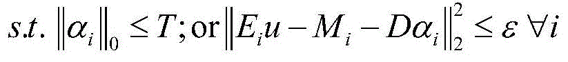
wherein G is a projection matrix, u is a reconstructed CT image, W is the statistical weight of CT projection data, and the statistical weight is calculated according to the variance of CT projection data p; I.I w To weight L 2 The norm of the sample is calculated, I.I 0 Is L 0 Norms, lambda is regularization parameter, E i For selecting three-dimensional image blocks at I positions of reconstructed image u, I is the total number of image blocks, D is a feature dictionary, α i Sparse representation vector for ith image block, M i The mean value of the ith image block is represented by T, sparsity and epsilon, and the error limit;
iterating the reconstruction objective function to obtain conventional dose CT image data V s rd 。
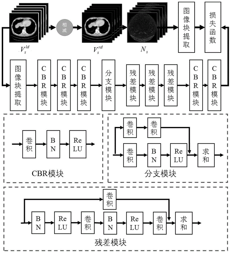
Further, the noise artifact suppression convolutional neural network comprises a CBR module, a branch module and a residual module.
Further, the CBR module is configured to convolve, scale-change, and ReLU-activate the input extracted low-dose CT image to extract low-level feature information of the low-dose CT image.
Further, the branching module is used for mixing the characteristic information extracted by the CBR module by increasing the network width so as to improve the representation capability of the network.
Furthermore, the residual error module shortens training time through convolution, scale change and ReLU activation, reduces redundancy of characteristic convolution kernels under the same representation capability, and avoids gradient dispersion in training.
Further, the Loss function Loss is:

in the method, in the process of the invention, for low dose CT image V s ld Image block obtained after blocking, P s N Is a noise artifact image N s The image blocks obtained after the segmentation are processed,omega is the image area.
The beneficial effects are that: compared with the prior art, the invention has the remarkable advantages that: the invention can effectively treat star-shaped artifacts, noise and characteristic structural components in the low-dose CT image under high noise pollution, the treatment effect is better than that of the traditional image space neural network method, the quality of the treated low-dose CT image can better meet the requirements of clinical analysis and diagnosis, and the invention contributes to the reduction of radiation dose injury of scanners in CT scanning.
Drawings
FIG. 1 is a flow chart of a method embodying the present invention;
FIG. 2 is a diagram of five exemplary axial training patterns (first row: conventional dose pattern; second row: low dose pattern) in an embodiment of the present invention;
FIG. 3 is an axial conventional dose CT image and a low dose CT image (a: conventional dose; b: low dose) in an embodiment of the present invention;
FIG. 4 shows results of axial clinical low-dose CT images using RED-CNN method and NAS-CNN treatment using the method of the present invention (a: RED-CNN; b: NAS-CNN) in the examples of the present invention;
FIG. 5 is a partial magnified view of results from a contrast experiment of axial clinical low-dose CT images in an embodiment of the present invention (a, b, c, d is the result after conventional dose CT images, low-dose CT images, RED-CNN treatment and NAS-CNN treatment, respectively);
FIG. 6 is a sagittal conventional dose CT image and coronal low dose CT image (a: conventional dose; b: low dose) of an embodiment of the present invention;
FIG. 7 is a graph showing the results of sagittal clinical low dose CT images using RED-CNN method and NAS-CNN treatment using the inventive method (a: RED-CNN; b: NAS-CNN) in the examples of the present invention;
fig. 8 is a partial magnified view of the results of a sagittal clinical low-dose CT image contrast experiment in an embodiment of the present invention (a, b, c, d is the results after conventional dose CT image, low dose CT image, RED-CNN treatment and NAS-CNN treatment, respectively).
Detailed Description
The embodiment provides a low-dose CT image processing system based on a noise artifact suppression convolutional neural network, as shown in fig. 1, including an image preprocessing module, a noise artifact suppression convolutional neural network building module, a network training module, a network processing module, and a noise artifact suppression module, which are described in detail below.
The image preprocessing module specifically comprises a low-dose CT image processing unit and a conventional-dose CT image processing unit, wherein the low-dose CT image processing unit is used for obtaining a low-dose CT image V by analyzing CT projection data under low-dose scanning through an FBP reconstruction algorithm s ld The method comprises the steps of carrying out a first treatment on the surface of the The conventional dose CT image processing unit is used for obtaining a conventional dose CT image V by performing GDSIR (Global Dictionary Based Statistical Iterative Reconstruction) iterative reconstruction algorithm on CT projection data under conventional dose scanning s rd The method specifically comprises the following steps of:
A. CT projection data under conventional dose scanning is acquired;
B. the reconstruction objective function is established as follows:

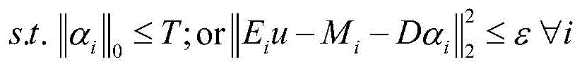
wherein G is a projection matrix, u is a reconstructed CT image, W is the statistical weight of CT projection data, and the statistical weight is calculated according to the variance of CT projection data p; I.I w In order to weight the L2 norm, I.I 0 Is L 0 Norms, lambda is regularization parameter, E i For selecting three-dimensional image blocks at I positions of reconstructed image u, I is the total number of image blocks, D is a feature dictionary, α i Sparse representation vector for ith image block, M i The mean value of the ith image block is represented by T, sparsity and epsilon, and the error limit;
C. iterating the reconstruction objective function to obtain conventional dose CT image data V s rd 。
The noise artifact suppression convolutional neural network building module is used for building the low-dose CT image V s ld As training images, noise artifact image N s As a label image, a low dose CT image V is created s ld And noise artifact image N s The mapping convolutional neural network therebetween acts as a noise artifact suppression convolutional neural network. The noise artifact suppression convolutional neural network comprises a CBR module, a branch module and a residual module. The CBR module is used for carrying out convolution, scale change and ReLU activation on the input extracted low-dose CT image so as to extract low-layer characteristic information of the low-dose CT image. The branching module is used for mixing the characteristic information extracted by the CBR module through increasing the network width so as to improve the representation capability of the network. The residual error module shortens training time through convolution, scale change and ReLU activation, reduces redundancy of characteristic convolution kernels under the same representation capability, and avoids gradient dispersion in training.
The network training module is used for integrating a plurality of low-dose CT images V s ld As input, the corresponding noise artifact image is used as output to train and learn parameters in the noise artifact suppression convolutional neural network by reducing the neural network loss function. Specifically, a low dose CT image V in the training set s ld And noise artifact image N s According to a certain size n×n×t and pixel interval l 1 ×l 2 ×l 3 The partitioning is performed (e.g.: the tile size is 65 x 32 pixels, the block interval is 12 x 12 pixels), respectively obtaining image block sets P s ld And P s N In particular, the two image blocks herein are selected to be exactly matched in position. Collecting image blocks P s ld And P s N Put into the network by reducing the neural network Loss function Loss, i.e. the set of predicted noise artifact image blocks Image block set P with actual noise artifact s N Training and learning parameters in NAS-CNN, and finally obtaining the neural network with stronger generalization capability.The Loss function Loss is defined as:

the network processing module is used for processing the low-dose CT image V to be processed t ld Inputting the trained mapping convolutional neural network for processing to obtain a predicted noise artifact image . Specifically, first, a low-dose CT image V to be processed is obtained t ld According to the size of n multiplied by t and the pixel interval of l 1 ×l 2 ×l 3 Partitioning to obtain an image block set P t ld The method comprises the steps of carrying out a first treatment on the surface of the Then, P is t ld Inputting into NAS-CNN with training to obtain predicted noise artifact image block set +.>
 Next, at a pixel interval l 1 ×l 2 ×l 3 Image block set +.>
 Combined noise artifact image +.>
Noise artifact suppression module for suppressing low-dose CT image V to be processed t ld Subtracting predicted noise artifact images Obtaining an image V with suppressed noise artifacts t p . The relation can be expressed as:
The method of the present invention is evaluated as follows.
1. Effect evaluation criterion
Firstly, a plurality of groups of abdomen data are obtained, the data published by Low Dose Challenge games used in experiments come from Somatom Definition AS +CT equipment, and specific scanning parameters are as follows: tube voltage 100KVP, tube current 360mAs (conventional dose)/85 mAs (low dose), detector size 736×64, and detector cell size 1.2856 × 1.0947mm each 2 The distance from the ray source to the center of the object and the center of the detector are 59.5cm and 108.56cm respectively, 1152 projection data are acquired every circle in a full-angle mode, the pitch is 0.6, and other parameters adopt machine default values. Reconstructed images were obtained by reconstruction with FDK (Feldkamp, davis, kress Algorithm) and GDSIR, respectively, the reconstructed image size was 512X 512, and the pixel size was 0.8X0.8 mm 2 The thickness of the layer is 1mm, and the three-dimensional continuity is good.
Nine groups of scanning data are selected as training data, and five typical axial training diagrams are shown in fig. 2; a set of scan data is used as test data, with the selected axial CT image shown in fig. 3 (normal dose and low dose) and the selected sagittal CT image shown in fig. 6 (normal dose and low dose). The low dose CT image, the normal dose CT image, and the processed image have a window width of 300HU (Housfield Units, HU) and a window level of 50HU.
2. Visual assessment
By observing the conventional dose and low dose CT images of FIGS. 3, 4, 5, and the images processed by the RED-CNN method and the NAS-CNN method of the present invention, it can be seen that the RED-CNN method can remove noise and streak artifacts in the low dose CT images, but during the processing, the anatomical components lose part of the tissue details, and part of the area has a certain blur, like liver, spleen vein and blood vessel cyst area; the visual effect of the CT image processed by the method is obviously improved, the image tissue can be well preserved, the noise artifact is basically and completely removed, and the visual texture of the processed image is more similar to that of a CT image under the conventional dose.
3. Quantitative evaluation
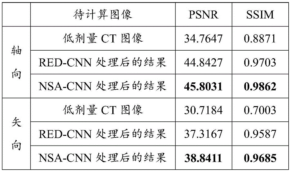
To quantitatively verify the effectiveness of the method of the present invention, we compare the peak signal-to-noise ratio and structural similarity of the whole image (low dose CT image, RED-CNN processed image and NAS-CNN processed image of the present invention) to the conventional dose CT image by calculation, where the peak signal-to-noise ratio PSNR is defined as:
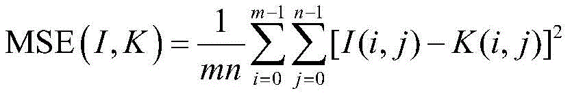

wherein I represents a normal dose CT image, K represents a CT image containing the image to be calculated, L I The maximum image pixel value that the representative image I can represent, I, j, is the pixel index of the image, respectively. The definition of structural similarity SSIM is:
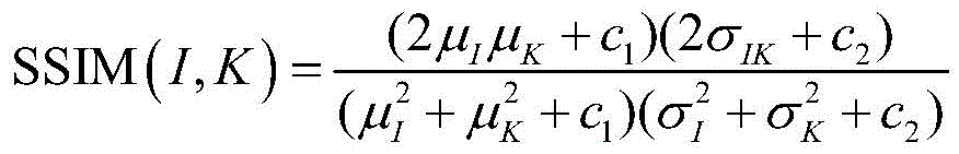
wherein mu I 、μ K Mean value, sigma of images I, K, respectively I 、σ K Standard deviation of I, K, sigma, respectively IK Is the covariance of images I and K, C 1 And C 2 Is two constants, wherein C 1 =(0.01×L) 2 ,C 2 =(0.03×L) 2 . From table 1 below, it can be seen that the decomposition method of the present invention can greatly reduce noise in the decomposed anatomical components, improve the signal-to-noise ratio, and obtain CT images closer to normal dose.
TABLE 1
In order to compare the calculation time consumption of RED-CNN and NAS-CNN network provided by the invention, we calculate the test chart with the size of 512×512×200 by using a TensorFlow platform under the environment that GPU is NVIDIA GTX1080, and find that RED-CNN takes 3.72 seconds on average, and NAS-CNN takes 1.08 seconds on average.
From the above experiments, it can be seen that the method of the present invention can effectively and rapidly process the low dose CT image, and can obtain the attenuation image of human anatomy tissue of CT information close to the normal dose level. In addition, in the method, once the network is constructed, repeated training is not needed, the actual processing time is short, the speed is high, and the method has a wide application range.
The above disclosure is only a preferred embodiment of the present invention and should not be construed as limiting the scope of the invention, which is defined by the appended claims.
Claims (2)
1. A low dose CT image processing system based on a noise artifact-suppressed convolutional neural network, comprising:
an image preprocessing module for obtaining multiple groups of matched low-dose CT images V s ld And a conventional dose CT image V s rd And subtracting the low dose CT image from the conventional dose CT image to obtain a noise artifact image N s =V s ld -V s rd The method comprises the steps of carrying out a first treatment on the surface of the The method specifically comprises the following steps:
the low-dose CT image processing unit is used for obtaining a low-dose CT image V by analyzing CT projection data under low-dose scanning through an FBP reconstruction algorithm s ld ;
The conventional dose CT image processing unit is used for obtaining a conventional dose CT image V by using the CT projection data under conventional dose scanning through a GDSIR iterative reconstruction algorithm s rd The method is specifically used for executing the following steps:
CT projection data under conventional dose scanning is acquired;
the reconstruction objective function is established as follows:


wherein G is a projection matrix, u is a reconstructed CT image, W is the statistical weight of CT projection data, and the statistical weight is calculated according to the variance of CT projection data p; I.I w To weight L 2 The norm of the sample is calculated, I.I 0 Is L 0 Norms, lambda is regularization parameter, E i For selecting three-dimensional image blocks at I positions of reconstructed image u, I is the total number of image blocks, D is a feature dictionary, α i Sparse representation vector for ith image block, M i The mean value of the ith image block is represented by T, sparsity and epsilon, and the error limit;
iterating the reconstruction objective function to obtain conventional dose CT image data V s rd ;
Noise artifact suppression convolutional neural network building module for generating low-dose CT image V s ld As training images, noise artifact image N s As a label image, a low dose CT image V is created s ld And noise artifact image N s The mapping convolutional neural network is used as a noise artifact suppression convolutional neural network, the noise artifact suppression convolutional neural network comprises a CBR module, a branch module and a residual module, the CBR module is used for convoluting an input extracted low-dose CT image, performing scale change and ReLU activation to extract low-level characteristic information of the low-dose CT image, the branch module is used for mixing the characteristic information extracted by the CBR module by increasing the network width so as to improve the representation capability of the network, and the residual module shortens the training time through convolution, scale change and ReLU activation, reduces the redundancy of characteristic convolution kernels under the same representation capability and avoids gradient dispersion in training;
a network training module for training a plurality of low-dose CT images V s ld As input, a corresponding noise artifact image as output, training and learning parameters in the noise artifact suppression convolutional neural network by reducing the neural network loss function;
a network processing module for processing the low dose C to be processedT image V t ld Inputting the trained mapping convolutional neural network for processing to obtain a predicted noise artifact image
Noise artifact suppression module for suppressing low-dose CT image V to be processed t ld Subtracting predicted noise artifact image N t p Obtaining an image V with suppressed noise artifacts t p 。
2. A low dose CT image processing system based on a noise artifact suppressing convolutional neural network as recited in claim 1, wherein: the Loss function Loss is:

in the method, in the process of the invention, for low dose CT image V s ld Image block obtained after blocking, P s N Is a noise artifact image N s And dividing the image block into blocks, wherein omega is an image area.
Priority Applications (1)
| Application Number | Priority Date | Filing Date | Title |
|---|---|---|---|
| CN201810722915.1A CN109166161B (en) | 2018-07-04 | 2018-07-04 | A low-dose CT image processing system based on noise artifact suppression convolutional neural network |
Applications Claiming Priority (1)
| Application Number | Priority Date | Filing Date | Title |
|---|---|---|---|
| CN201810722915.1A CN109166161B (en) | 2018-07-04 | 2018-07-04 | A low-dose CT image processing system based on noise artifact suppression convolutional neural network |
Publications (2)
| Publication Number | Publication Date |
|---|---|
| CN109166161A CN109166161A (en) | 2019-01-08 |
| CN109166161B true CN109166161B (en) | 2023-06-30 |
Family
ID=64897375
Family Applications (1)
| Application Number | Title | Priority Date | Filing Date |
|---|---|---|---|
| CN201810722915.1A Active CN109166161B (en) | 2018-07-04 | 2018-07-04 | A low-dose CT image processing system based on noise artifact suppression convolutional neural network |
Country Status (1)
| Country | Link |
|---|---|
| CN (1) | CN109166161B (en) |
Families Citing this family (36)
| Publication number | Priority date | Publication date | Assignee | Title |
|---|---|---|---|---|
| JP7237624B2 (en) | 2019-02-07 | 2023-03-13 | 浜松ホトニクス株式会社 | Image processing device and image processing method |
| CN110070510A (en) * | 2019-04-26 | 2019-07-30 | 东北大学 | A kind of CNN medical image denoising method for extracting feature based on VGG-19 |
| CN110378982B (en) * | 2019-07-23 | 2023-09-12 | 上海联影医疗科技股份有限公司 | Reconstructed image processing method, device, equipment and storage medium |
| JP7245740B2 (en) * | 2019-07-25 | 2023-03-24 | 富士フイルムヘルスケア株式会社 | Image processing device, image processing method and X-ray CT device |
| CN110570492B (en) * | 2019-09-11 | 2021-09-03 | 清华大学 | CT artifact suppression method, device and medium based on neural network |
| CN111009019B (en) * | 2019-09-27 | 2021-07-16 | 北京航空航天大学 | Incomplete data reconstruction method of differential phase contrast CT based on deep learning |
| CN112581554B (en) * | 2019-09-30 | 2024-02-27 | 中国科学院深圳先进技术研究院 | A CT imaging method, device, storage device and medical imaging system |
| CN110677649B (en) | 2019-10-16 | 2021-09-28 | 腾讯科技(深圳)有限公司 | Artifact removing method based on machine learning, artifact removing model training method and device |
| CN111080736B (en) * | 2019-12-11 | 2023-09-08 | 电子科技大学 | A low-dose CT image reconstruction method based on sparse transformation |
| CN111179366B (en) * | 2019-12-18 | 2023-04-25 | 深圳先进技术研究院 | Low-dose image reconstruction method and system based on anatomical difference prior |
| CN110992295B (en) * | 2019-12-20 | 2022-04-19 | 电子科技大学 | Low-dose CT reconstruction method based on wavelet-RED convolution neural network |
| CN111325686B (en) | 2020-02-11 | 2021-03-30 | 之江实验室 | A low-dose PET 3D reconstruction method based on deep learning |
| CN111325737B (en) * | 2020-02-28 | 2024-03-15 | 上海志唐健康科技有限公司 | Low-dose CT image processing method, device and computer equipment |
| CN111325695B (en) * | 2020-02-29 | 2023-04-07 | 深圳先进技术研究院 | Low-dose image enhancement method and system based on multi-dose grade and storage medium |
| CN111445406B (en) * | 2020-03-24 | 2023-05-05 | 广东工业大学 | A method, system and device for improving the quality of low-dose CT images |
| CN111968195B (en) * | 2020-08-20 | 2022-09-02 | 太原科技大学 | Dual-attention generation countermeasure network for low-dose CT image denoising and artifact removal |
| CN112116677B (en) * | 2020-09-23 | 2024-01-23 | 赣南师范大学 | A low-dose CT reconstruction method based on low-dimensional manifold prior |
| US20220130079A1 (en) * | 2020-10-23 | 2022-04-28 | Siemens Medical Solutions Usa, Inc. | Systems and methods for simultaneous attenuation correction, scatter correction, and de-noising of low-dose pet images with a neural network |
| CN112330575B (en) * | 2020-12-03 | 2022-10-14 | 华北理工大学 | Convolution neural network medical CT image denoising method |
| CN112446840B (en) * | 2020-12-07 | 2024-01-19 | 明峰医疗系统股份有限公司 | CT image black band artifact eliminating method and system based on deep learning |
| CN112598759B (en) * | 2020-12-15 | 2022-09-13 | 太原科技大学 | Multi-scale feature generation countermeasure network for suppressing artifact noise in low-dose CT images |
| CN116685999A (en) * | 2020-12-18 | 2023-09-01 | 皇家飞利浦有限公司 | Method and system for flexible denoising of images using disentangled feature representation domains |
| CN112767273B (en) * | 2021-01-21 | 2023-10-20 | 中山大学 | Low-dose CT image restoration method and system applying feature decoupling |
| CN112927318B (en) * | 2021-02-22 | 2022-04-22 | 明峰医疗系统股份有限公司 | Noise reduction reconstruction method of low-dose PET image and computer readable storage medium |
| CN112927230B (en) * | 2021-04-26 | 2024-08-16 | 常州市第一人民医院 | Image processing and analyzing system |
| CN113298167B (en) * | 2021-06-01 | 2024-10-15 | 北京思特奇信息技术股份有限公司 | Text detection method and system based on lightweight neural network model |
| CN113256529B (en) * | 2021-06-09 | 2021-10-15 | 腾讯科技(深圳)有限公司 | Image processing method, image processing device, computer equipment and storage medium |
| CN113450427B (en) * | 2021-06-29 | 2023-09-01 | 深圳高性能医疗器械国家研究院有限公司 | PET image reconstruction method based on joint dictionary learning and depth network |
| CN113379868A (en) * | 2021-07-08 | 2021-09-10 | 安徽工程大学 | Low-dose CT image noise artifact decomposition method based on convolution sparse coding network |
| CN113570705B (en) * | 2021-07-28 | 2024-04-30 | 广州瑞多思医疗科技有限公司 | Three-dimensional dose reconstruction method, device, computer equipment and storage medium |
| CN113436118B (en) * | 2021-08-10 | 2022-09-27 | 安徽工程大学 | Low-dose CT image restoration method based on multi-scale convolutional coding network |
| CN116433785A (en) * | 2021-12-31 | 2023-07-14 | 中国石油天然气股份有限公司 | Method, device, system and storage medium for processing ring artifacts in CT images |
| CN114494057B (en) * | 2022-01-23 | 2025-04-25 | 东南大学 | A digital X-ray image denoising method based on trainable joint bilateral filter |
| CN115828396B (en) * | 2023-01-03 | 2023-04-28 | 哈尔滨工业大学(深圳)(哈尔滨工业大学深圳科技创新研究院) | Automatic generation method and system for civil plane based on mixed constraint condition |
| CN117315063A (en) * | 2023-09-07 | 2023-12-29 | 先进能源科学与技术广东省实验室 | Low-dose CT image reconstruction method and system based on deep learning |
| CN119151817B (en) * | 2024-08-27 | 2025-10-03 | 东南大学 | Universal imaging physics-driven CT data simulation and CT image noise artifact suppression method |
Citations (3)
| Publication number | Priority date | Publication date | Assignee | Title |
|---|---|---|---|---|
| CN103473745A (en) * | 2013-09-16 | 2013-12-25 | 东南大学 | Low-dosage CT image processing method based on distinctive dictionaries |
| CN105118066A (en) * | 2015-09-16 | 2015-12-02 | 东南大学 | Low-dosage CT image decomposition method based on three-dimensional distinctive feature representation |
| CN108122265A (en) * | 2017-11-13 | 2018-06-05 | 深圳先进技术研究院 | A kind of CT reconstruction images optimization method and system |
-
2018
- 2018-07-04 CN CN201810722915.1A patent/CN109166161B/en active Active
Patent Citations (3)
| Publication number | Priority date | Publication date | Assignee | Title |
|---|---|---|---|---|
| CN103473745A (en) * | 2013-09-16 | 2013-12-25 | 东南大学 | Low-dosage CT image processing method based on distinctive dictionaries |
| CN105118066A (en) * | 2015-09-16 | 2015-12-02 | 东南大学 | Low-dosage CT image decomposition method based on three-dimensional distinctive feature representation |
| CN108122265A (en) * | 2017-11-13 | 2018-06-05 | 深圳先进技术研究院 | A kind of CT reconstruction images optimization method and system |
Also Published As
| Publication number | Publication date |
|---|---|
| CN109166161A (en) | 2019-01-08 |
Similar Documents
| Publication | Publication Date | Title |
|---|---|---|
| CN109166161B (en) | A low-dose CT image processing system based on noise artifact suppression convolutional neural network | |
| Lu et al. | Iterative reconstruction of low-dose CT based on differential sparse | |
| CN108961237B (en) | A low-dose CT image decomposition method based on convolutional neural network | |
| CN109102550B (en) | Full-network low-dose CT imaging method and device based on convolution residual error network | |
| Kuanar et al. | Low dose abdominal CT image reconstruction: an unsupervised learning based approach | |
| CN105118066B (en) | A kind of low-dose CT picture breakdown method based on three-dimensional distinctiveness character representation | |
| CN106373163B (en) | A low-dose CT imaging method based on the distinctive feature representation of 3D projection images | |
| CN110009613A (en) | Low-dose CT imaging method, apparatus and system based on the dense network of depth | |
| Wu et al. | Unsharp structure guided filtering for self-supervised low-dose CT imaging | |
| CN113706643B (en) | Head CT metal artifact correction method based on homomorphic adaptation learning | |
| Al-Kadi | Assessment of texture measures susceptibility to noise in conventional and contrast enhanced computed tomography lung tumour images | |
| CN116167929B (en) | Low-dose CT image denoising network based on residual error multi-scale feature extraction | |
| CN102024267A (en) | Low-dose computed tomography (CT) image processing method based on wavelet space directional filtering | |
| CN117876261A (en) | CBCT scattering correction imaging method based on deep learning | |
| CN103226815A (en) | Low dose CT image filtering method | |
| CN115731158B (en) | A low-dose CT reconstruction method based on residual domain iterative optimization network | |
| Wang et al. | Ring artifacts correction for computed tomography image using unsupervised contrastive learning | |
| Zhao et al. | Dual-domain neural networks for clinical and low-dose CBCT reconstruction | |
| CN113744356B (en) | A method for low-dose SPECT chordogram recovery and scatter correction | |
| Xia et al. | CMC-diffusion: curve matching correction diffusion model for LDCT denoising | |
| Chen et al. | Low-dose dental CT image enhancement using a multiscale feature sensing network | |
| CN119048614A (en) | Sparse angle CT artifact removal method based on differentiated convolution dictionary network | |
| Liu et al. | Cascade resunet with noise power spectrum loss for low dose ct imaging | |
| Zhao et al. | A dual-channel network based GAN for low-dose CT image denoising | |
| Xie et al. | Metal artifact correction in head computed tomography based on a homographic adaptation convolution neural network |
Legal Events
| Date | Code | Title | Description |
|---|---|---|---|
| PB01 | Publication | ||
| PB01 | Publication | ||
| SE01 | Entry into force of request for substantive examination | ||
| SE01 | Entry into force of request for substantive examination | ||
| GR01 | Patent grant | ||
| GR01 | Patent grant |