CN108471975B - Method and system for statistically analyzing electrograms of and mapping locally abnormal ventricular activity - Google Patents
Method and system for statistically analyzing electrograms of and mapping locally abnormal ventricular activity Download PDFInfo
- Publication number
- CN108471975B CN108471975B CN201680068127.4A CN201680068127A CN108471975B CN 108471975 B CN108471975 B CN 108471975B CN 201680068127 A CN201680068127 A CN 201680068127A CN 108471975 B CN108471975 B CN 108471975B
- Authority
- CN
- China
- Prior art keywords
- lava
- peak
- peaks
- function
- electrogram signal
- Prior art date
- Legal status (The legal status is an assumption and is not a legal conclusion. Google has not performed a legal analysis and makes no representation as to the accuracy of the status listed.)
- Active
Links
Images


Classifications
-
- A—HUMAN NECESSITIES
- A61—MEDICAL OR VETERINARY SCIENCE; HYGIENE
- A61B—DIAGNOSIS; SURGERY; IDENTIFICATION
- A61B5/00—Measuring for diagnostic purposes; Identification of persons
- A61B5/72—Signal processing specially adapted for physiological signals or for diagnostic purposes
- A61B5/7235—Details of waveform analysis
- A61B5/7253—Details of waveform analysis characterised by using transforms
- A61B5/726—Details of waveform analysis characterised by using transforms using Wavelet transforms
-
- A—HUMAN NECESSITIES
- A61—MEDICAL OR VETERINARY SCIENCE; HYGIENE
- A61B—DIAGNOSIS; SURGERY; IDENTIFICATION
- A61B5/00—Measuring for diagnostic purposes; Identification of persons
- A61B5/24—Detecting, measuring or recording bioelectric or biomagnetic signals of the body or parts thereof
- A61B5/316—Modalities, i.e. specific diagnostic methods
- A61B5/318—Heart-related electrical modalities, e.g. electrocardiography [ECG]
- A61B5/346—Analysis of electrocardiograms
- A61B5/349—Detecting specific parameters of the electrocardiograph cycle
-
- A—HUMAN NECESSITIES
- A61—MEDICAL OR VETERINARY SCIENCE; HYGIENE
- A61B—DIAGNOSIS; SURGERY; IDENTIFICATION
- A61B5/00—Measuring for diagnostic purposes; Identification of persons
- A61B5/24—Detecting, measuring or recording bioelectric or biomagnetic signals of the body or parts thereof
- A61B5/316—Modalities, i.e. specific diagnostic methods
- A61B5/318—Heart-related electrical modalities, e.g. electrocardiography [ECG]
- A61B5/339—Displays specially adapted therefor
- A61B5/343—Potential distribution indication
-
- A—HUMAN NECESSITIES
- A61—MEDICAL OR VETERINARY SCIENCE; HYGIENE
- A61B—DIAGNOSIS; SURGERY; IDENTIFICATION
- A61B5/00—Measuring for diagnostic purposes; Identification of persons
- A61B5/24—Detecting, measuring or recording bioelectric or biomagnetic signals of the body or parts thereof
- A61B5/316—Modalities, i.e. specific diagnostic methods
- A61B5/318—Heart-related electrical modalities, e.g. electrocardiography [ECG]
- A61B5/346—Analysis of electrocardiograms
- A61B5/347—Detecting the frequency distribution of signals
-
- A—HUMAN NECESSITIES
- A61—MEDICAL OR VETERINARY SCIENCE; HYGIENE
- A61B—DIAGNOSIS; SURGERY; IDENTIFICATION
- A61B5/00—Measuring for diagnostic purposes; Identification of persons
- A61B5/24—Detecting, measuring or recording bioelectric or biomagnetic signals of the body or parts thereof
- A61B5/316—Modalities, i.e. specific diagnostic methods
- A61B5/318—Heart-related electrical modalities, e.g. electrocardiography [ECG]
- A61B5/346—Analysis of electrocardiograms
- A61B5/349—Detecting specific parameters of the electrocardiograph cycle
- A61B5/352—Detecting R peaks, e.g. for synchronising diagnostic apparatus; Estimating R-R interval
-
- A—HUMAN NECESSITIES
- A61—MEDICAL OR VETERINARY SCIENCE; HYGIENE
- A61B—DIAGNOSIS; SURGERY; IDENTIFICATION
- A61B5/00—Measuring for diagnostic purposes; Identification of persons
- A61B5/24—Detecting, measuring or recording bioelectric or biomagnetic signals of the body or parts thereof
- A61B5/316—Modalities, i.e. specific diagnostic methods
- A61B5/318—Heart-related electrical modalities, e.g. electrocardiography [ECG]
- A61B5/346—Analysis of electrocardiograms
- A61B5/349—Detecting specific parameters of the electrocardiograph cycle
- A61B5/361—Detecting fibrillation
-
- A—HUMAN NECESSITIES
- A61—MEDICAL OR VETERINARY SCIENCE; HYGIENE
- A61B—DIAGNOSIS; SURGERY; IDENTIFICATION
- A61B5/00—Measuring for diagnostic purposes; Identification of persons
- A61B5/24—Detecting, measuring or recording bioelectric or biomagnetic signals of the body or parts thereof
- A61B5/316—Modalities, i.e. specific diagnostic methods
- A61B5/318—Heart-related electrical modalities, e.g. electrocardiography [ECG]
- A61B5/346—Analysis of electrocardiograms
- A61B5/349—Detecting specific parameters of the electrocardiograph cycle
- A61B5/363—Detecting tachycardia or bradycardia
-
- A—HUMAN NECESSITIES
- A61—MEDICAL OR VETERINARY SCIENCE; HYGIENE
- A61B—DIAGNOSIS; SURGERY; IDENTIFICATION
- A61B5/00—Measuring for diagnostic purposes; Identification of persons
- A61B5/24—Detecting, measuring or recording bioelectric or biomagnetic signals of the body or parts thereof
- A61B5/316—Modalities, i.e. specific diagnostic methods
- A61B5/318—Heart-related electrical modalities, e.g. electrocardiography [ECG]
- A61B5/346—Analysis of electrocardiograms
- A61B5/349—Detecting specific parameters of the electrocardiograph cycle
- A61B5/364—Detecting abnormal ECG interval, e.g. extrasystoles, ectopic heartbeats
-
- A—HUMAN NECESSITIES
- A61—MEDICAL OR VETERINARY SCIENCE; HYGIENE
- A61B—DIAGNOSIS; SURGERY; IDENTIFICATION
- A61B5/00—Measuring for diagnostic purposes; Identification of persons
- A61B5/24—Detecting, measuring or recording bioelectric or biomagnetic signals of the body or parts thereof
- A61B5/316—Modalities, i.e. specific diagnostic methods
- A61B5/318—Heart-related electrical modalities, e.g. electrocardiography [ECG]
- A61B5/346—Analysis of electrocardiograms
- A61B5/349—Detecting specific parameters of the electrocardiograph cycle
- A61B5/366—Detecting abnormal QRS complex, e.g. widening
-
- A—HUMAN NECESSITIES
- A61—MEDICAL OR VETERINARY SCIENCE; HYGIENE
- A61B—DIAGNOSIS; SURGERY; IDENTIFICATION
- A61B5/00—Measuring for diagnostic purposes; Identification of persons
- A61B5/72—Signal processing specially adapted for physiological signals or for diagnostic purposes
- A61B5/7235—Details of waveform analysis
- A61B5/7264—Classification of physiological signals or data, e.g. using neural networks, statistical classifiers, expert systems or fuzzy systems
-
- G—PHYSICS
- G16—INFORMATION AND COMMUNICATION TECHNOLOGY [ICT] SPECIALLY ADAPTED FOR SPECIFIC APPLICATION FIELDS
- G16H—HEALTHCARE INFORMATICS, i.e. INFORMATION AND COMMUNICATION TECHNOLOGY [ICT] SPECIALLY ADAPTED FOR THE HANDLING OR PROCESSING OF MEDICAL OR HEALTHCARE DATA
- G16H50/00—ICT specially adapted for medical diagnosis, medical simulation or medical data mining; ICT specially adapted for detecting, monitoring or modelling epidemics or pandemics
- G16H50/20—ICT specially adapted for medical diagnosis, medical simulation or medical data mining; ICT specially adapted for detecting, monitoring or modelling epidemics or pandemics for computer-aided diagnosis, e.g. based on medical expert systems
-
- A—HUMAN NECESSITIES
- A61—MEDICAL OR VETERINARY SCIENCE; HYGIENE
- A61B—DIAGNOSIS; SURGERY; IDENTIFICATION
- A61B2505/00—Evaluating, monitoring or diagnosing in the context of a particular type of medical care
- A61B2505/05—Surgical care
-
- A—HUMAN NECESSITIES
- A61—MEDICAL OR VETERINARY SCIENCE; HYGIENE
- A61B—DIAGNOSIS; SURGERY; IDENTIFICATION
- A61B5/00—Measuring for diagnostic purposes; Identification of persons
- A61B5/24—Detecting, measuring or recording bioelectric or biomagnetic signals of the body or parts thereof
- A61B5/25—Bioelectric electrodes therefor
- A61B5/279—Bioelectric electrodes therefor specially adapted for particular uses
- A61B5/28—Bioelectric electrodes therefor specially adapted for particular uses for electrocardiography [ECG]
- A61B5/283—Invasive
- A61B5/287—Holders for multiple electrodes, e.g. electrode catheters for electrophysiological study [EPS]
-
- A—HUMAN NECESSITIES
- A61—MEDICAL OR VETERINARY SCIENCE; HYGIENE
- A61B—DIAGNOSIS; SURGERY; IDENTIFICATION
- A61B5/00—Measuring for diagnostic purposes; Identification of persons
- A61B5/68—Arrangements of detecting, measuring or recording means, e.g. sensors, in relation to patient
- A61B5/6846—Arrangements of detecting, measuring or recording means, e.g. sensors, in relation to patient specially adapted to be brought in contact with an internal body part, i.e. invasive
- A61B5/6847—Arrangements of detecting, measuring or recording means, e.g. sensors, in relation to patient specially adapted to be brought in contact with an internal body part, i.e. invasive mounted on an invasive device
- A61B5/6852—Catheters
-
- A—HUMAN NECESSITIES
- A61—MEDICAL OR VETERINARY SCIENCE; HYGIENE
- A61B—DIAGNOSIS; SURGERY; IDENTIFICATION
- A61B5/00—Measuring for diagnostic purposes; Identification of persons
- A61B5/72—Signal processing specially adapted for physiological signals or for diagnostic purposes
- A61B5/7271—Specific aspects of physiological measurement analysis
- A61B5/7275—Determining trends in physiological measurement data; Predicting development of a medical condition based on physiological measurements, e.g. determining a risk factor
Landscapes
- Health & Medical Sciences (AREA)
- Life Sciences & Earth Sciences (AREA)
- Cardiology (AREA)
- Engineering & Computer Science (AREA)
- Public Health (AREA)
- Medical Informatics (AREA)
- Biomedical Technology (AREA)
- Pathology (AREA)
- General Health & Medical Sciences (AREA)
- Physics & Mathematics (AREA)
- Molecular Biology (AREA)
- Veterinary Medicine (AREA)
- Biophysics (AREA)
- Heart & Thoracic Surgery (AREA)
- Surgery (AREA)
- Animal Behavior & Ethology (AREA)
- Artificial Intelligence (AREA)
- Signal Processing (AREA)
- Psychiatry (AREA)
- Physiology (AREA)
- Computer Vision & Pattern Recognition (AREA)
- Evolutionary Computation (AREA)
- Mathematical Physics (AREA)
- Data Mining & Analysis (AREA)
- Databases & Information Systems (AREA)
- Epidemiology (AREA)
- Primary Health Care (AREA)
- Fuzzy Systems (AREA)
- Measurement And Recording Of Electrical Phenomena And Electrical Characteristics Of The Living Body (AREA)
- Measuring And Recording Apparatus For Diagnosis (AREA)
Abstract
Description
相关申请的交叉引用CROSS-REFERENCE TO RELATED APPLICATIONS
本申请要求现在未决的于2015年12月4日提交的美国临时申请no.62/263,136和现在未决的于2016年5月3日提交的美国临时申请no.62/330,886的权益。上述内容通过引用包含于此,如同在本文中完全阐述一样。This application claims the benefit of now pending US Provisional Application no. 62/263,136, filed December 4, 2015, and now pending US Provisional Application no. 62/330,886, filed May 3, 2016. The foregoing is incorporated herein by reference as if fully set forth herein.
背景技术Background technique
本公开一般涉及诸如可以在心脏诊断和治疗程序中执行的电描记图检测和分析。更具体地,本公开涉及用于从电描记图数据检测、分析和标测局部异常心室活动(LAVA)的系统和方法。The present disclosure generally relates to electrogram detection and analysis, such as may be performed in cardiac diagnostic and therapeutic procedures. More particularly, the present disclosure relates to systems and methods for detecting, analyzing, and mapping localized abnormal ventricular activity (LAVA) from electrogram data.
在室性心动过速(VT)形成中,局部异常心室活动(LAVA)代表疤痕内慢速传导通道的存活电位。由于沿着致密疤痕内存在的缓慢、高度纤维化的传导通道的去极化波的捕获,所以存在这些LAVA。这些被困的LAVA通常具有多个入口和出口点以触发宏观重返,从而导致VT。In ventricular tachycardia (VT) development, local abnormal ventricular activity (LAVA) represents the survival potential of slow conduction channels within the scar. These LAVAs exist due to the capture of depolarizing waves along slow, highly fibrotic conduction channels present within dense scars. These trapped LAVAs often have multiple entry and exit points to trigger macro-reentry, leading to VT.
用于检测LAVA的基于衬底的方法针对信号内相对于检测到的QRS复合波的延迟信号,其通常被定义为晚电位。然而,这种方法可能会漏掉很大一部分LAVA,特别是在隔膜和其他早期激活的区域中。这可以在图1A-1E中示出的示例性电描记图信号中看出。例如,图1A-1C示出了融合并隐藏在信号的EGM QRS部分内的早期LAVA的存在,而图1D-1E示出了在心脏的外侧或心外膜侧更易观察到的晚期LAVA。假设在窦性心律或心室起搏期间记录的LAVA的消除作为VT消融的终点将是可行的,并且完全消除LAVA将导致无心律失常存活的增加。目前在EP研究期间,这些LAVA是由EP医生基于某些双极电描记图(EGM)特征手动标记的。医生执行这种手动注释过程可能非常繁琐且困难,特别是对于高密度电描记图。Substrate-based methods for detecting LAVA target delayed signals within the signal relative to the detected QRS complexes, which are generally defined as late potentials. However, this approach may miss a large fraction of LAVA, especially in the septum and other areas that are activated early. This can be seen in the exemplary electrogram signals shown in Figures 1A-1E. For example, Figures 1A-1C show the presence of early LAVA fused and hidden within the EGM QRS portion of the signal, while Figures 1D-1E show late LAVA more readily observed on the lateral or epicardial side of the heart. It is hypothesized that elimination of LAVA recorded during sinus rhythm or ventricular pacing would be feasible as an endpoint for VT ablation, and that complete elimination of LAVA would result in an increase in arrhythmia-free survival. Currently during the EP study, these LAVAs were manually labeled by EP physicians based on certain bipolar electrogram (EGM) features. Physicians performing this manual annotation process can be very tedious and difficult, especially for high-density electrograms.
发明内容SUMMARY OF THE INVENTION
本文公开了一种针对局部异常心室活动(LAVA)分析心脏活动的方法,包括以下步骤:在信号处理器处接收电描记图信号;以及使用所述信号处理器:将电描记图信号变换至小波域,由此计算尺度图;计算所述尺度图的一维LAVA函数;检测LAVA函数中的一个或多个峰值;计算电描记图信号的峰峰振幅,并且如果峰峰振幅不超过预设的振幅阈值:使用在所述LAVA函数中检测到的所述一个或多个峰值中的一个来计算所述电描记图信号的LAVA迟滞参数;以及计算电描记图信号的LAVA概率参数。通过对电描记图信号应用连续小波变换,可以将电描记图信号变换至小波域,以计算尺度图。Disclosed herein is a method of analyzing cardiac activity for localized abnormal ventricular activity (LAVA), comprising the steps of: receiving an electrogram signal at a signal processor; and using the signal processor: transforming the electrogram signal to a wavelet domain, from which a scale map is calculated; a one-dimensional LAVA function of said scale map is calculated; one or more peaks in the LAVA function are detected; the peak-to-peak amplitude of the electrogram signal is calculated, and if the peak-to-peak amplitude does not exceed a preset Amplitude Threshold: use one of the one or more peaks detected in the LAVA function to calculate a LAVA hysteresis parameter for the electrogram signal; and calculate a LAVA probability parameter for the electrogram signal. By applying a continuous wavelet transform to the electrogram signal, the electrogram signal can be transformed to the wavelet domain to compute a scale map.
可以设想的是,小于预设噪声阈值(例如约0.2)的尺度图的值可以被设置为零。It is contemplated that the value of the scale map that is less than a preset noise threshold (eg, about 0.2) may be set to zero.
根据本公开的各方面,计算该尺度图的一维LAVA函数的步骤可以包括以预设的心脏活动频率(诸如约300Hz)计算该尺度图的一维LAVA函数。According to aspects of the present disclosure, calculating the one-dimensional LAVA function of the scale map may include calculating the one-dimensional LAVA function of the scale map at a preset cardiac activity frequency, such as about 300 Hz.
在实施例中,检测LAVA函数中的一个或多个峰值的步骤包括:检测LAVA函数中的一个或多个主要活动峰值;以及将一个或多个主要活动峰值中的每个主要活动峰值分类为近场峰值、远场峰值或噪声峰值。通过检测LAVA函数中的一个或多个局部最大峰值,可以在LAVA函数中检测主要活动峰值,其中电描记图信号的斜率的最大值超过在围绕局部最大峰值的预设不应窗口内的预设离散活动阈值(诸如约0.2mV/msec)。如果LAVA函数在主要活动峰值处超过预设近场阈值,则可以将主要峰值分类为近场峰值,如果LAVA函数在主要活动峰值处超过预设远场阈值而不是预设近场阈值,则将其分类为远场峰值,否则分类为噪声峰值。In an embodiment, the step of detecting one or more peaks in the LAVA function comprises: detecting one or more main activity peaks in the LAVA function; and classifying each of the one or more main activity peaks as a Near-field peaks, far-field peaks, or noise peaks. A major activity peak can be detected in the LAVA function by detecting one or more local maximum peaks in the LAVA function where the maximum value of the slope of the electrogram signal exceeds a preset value within a preset should not be window surrounding the local maximum peak Discrete activity threshold (such as about 0.2 mV/msec). If the LAVA function exceeds a preset near-field threshold at the main activity peak, then the main peak can be classified as a near-field peak, and if the LAVA function exceeds a preset far-field threshold at the main activity peak but not the preset near-field threshold, it will be classified as a near-field peak. It is classified as a far field peak, otherwise it is classified as a noise peak.
LAVA迟滞参数可以被计算为以下之间的时间差:如果在LAVA函数中检测到的一个或多个峰值不包括任何近场峰值,则在LAVA函数中检测到的一个或多个峰值中的最近远场峰值和参考ECG QRS之间;以及如果在LAVA函数中检测到的一个或多个峰确实包括至少一个近场峰值,则在LAVA函数中检测到的一个或多个峰值中的最近近场峰值和参考ECG QRS之间。The LAVA hysteresis parameter can be calculated as the time difference between: if the peak or peaks detected in the LAVA function do not include any near-field peaks, then the nearest farthest of the peak or peaks detected in the LAVA function between the field peak and the reference ECG QRS; and if the peak or peaks detected in the LAVA function do include at least one near-field peak, the nearest near-field peak among the peak or peaks detected in the LAVA function and the reference ECG QRS.
可以使用以下中的一个或多个计算LAVA概率参数:所述电描记信号的LAVA迟滞参数;电描记图信号的分割参数;在LAVA函数中检测到的峰值的数量;在LAVA函数中检测到的远场峰值的数量;电描记图信号的近场活动跨度;电描记图信号的孤立的QRS活动的数量;以及电描记图信号的QRS活动持续时间。根据本公开的方面,所述电描记图信号的所述分割参数被计算为在所述LAVA函数中检测到的最早的近场峰值与在LAVA函数中检测到的最近的近场峰值之间的所述电描记图信号的斜率的符号变化的次数。根据本公开的其他方面,所述电描记图信号的所述分割参数被计算为在所述LAVA函数中检测到的最早的近场峰值与在LAVA函数中检测到的最近的近场峰值之间的、在所述电描记图信号已经被过滤以去除噪声之后的、所述电描记图信号的斜率的符号变化的次数。还可以设想,可以使用在所述LAVA函数中检测到的所述一个或多个峰值来计算所述电描记图信号的所述QRS活动持续时间。The LAVA probability parameter can be calculated using one or more of the following: LAVA hysteresis parameter of the electrogram signal; segmentation parameter of the electrogram signal; number of peaks detected in the LAVA function; The number of far-field peaks; the near-field activity span of the electrogram signal; the number of isolated QRS activity of the electrogram signal; and the duration of QRS activity of the electrogram signal. According to an aspect of the present disclosure, the segmentation parameter of the electrogram signal is calculated as the difference between the earliest near-field peak detected in the LAVA function and the most recent near-field peak detected in the LAVA function The number of sign changes of the slope of the electrogram signal. According to other aspects of the present disclosure, the segmentation parameter of the electrogram signal is calculated as being between the earliest near-field peak detected in the LAVA function and the most recent near-field peak detected in the LAVA function is the number of sign changes of the slope of the electrogram signal after the electrogram signal has been filtered to remove noise. It is also contemplated that the one or more peaks detected in the LAVA function may be used to calculate the QRS activity duration of the electrogram signal.
该方法还可以包括在心脏模型上生成所述LAVA迟滞参数和所述LAVA概率参数中的一个或多个的图形表示。The method may also include generating a graphical representation of one or more of the LAVA delay parameter and the LAVA probability parameter on a cardiac model.
本文还公开了一种在电解剖标测系统中针对局部异常心室活动(LAVA)分析心电图的方法。所述方法包括电解剖标测系统:将心电图变换至小波域,由此计算尺度图;计算所述尺度图的一维LAVA函数;检测LAVA函数中的一个或多个峰值;计算电描记图信号的峰峰振幅,并且如果峰峰振幅不超过预设的振幅阈值,计算以下中至少之一:使用在所述LAVA函数中检测到的所述一个或多个峰值中的一个来计算所述电描记图信号的LAVA迟滞参数;以及计算电描记图信号的LAVA概率参数。Also disclosed herein is a method of analyzing an electrocardiogram for localized abnormal ventricular activity (LAVA) in an electroanatomical mapping system. The method includes an electroanatomical mapping system: transforming an electrocardiogram into the wavelet domain, thereby calculating a scale map; calculating a one-dimensional LAVA function of the scale map; detecting one or more peaks in the LAVA function; calculating an electrogram signal and if the peak-to-peak amplitude does not exceed a preset amplitude threshold, calculate at least one of: using one of the one or more peaks detected in the LAVA function to calculate the electrical A LAVA hysteresis parameter for the tracerogram signal; and a LAVA probability parameter for calculating the electrogrammogram signal.
根据本公开的方面,检测所述LAVA函数中的一个或多个峰值的步骤包括检测所述LAVA函数中的一个或多个局部最大峰值,其中,所述电描记图信号的斜率的最大值超过在围绕所述局部最大峰值的预设不应窗口内的预设离散活动阈值;以及LAVA函数在局部最大峰值处超过至少预设远场阈值。According to an aspect of the present disclosure, the step of detecting one or more peaks in the LAVA function includes detecting one or more local maximum peaks in the LAVA function, wherein the maximum value of the slope of the electrogram signal exceeds a predetermined discrete activity threshold within a predetermined undesired window around the local maximum peak; and the LAVA function exceeds at least a predetermined far-field threshold at the local maximum peak.
该方法还可以包括在心脏模型上生成LAVA迟滞参数和LAVA概率参数中的至少一个的图形表示。The method may also include generating a graphical representation of at least one of the LAVA delay parameter and the LAVA probability parameter on the cardiac model.
本公开还提供了一种电解剖标测系统,其被配置为分析局部异常心室活动(LAVA)的电描记图信号。所述系统包括:LAVA分析处理器,其被配置为:将电描记图信号变换至小波域,由此计算尺度图;计算所述尺度图的一维LAVA函数;检测LAVA函数中的一个或多个峰值;以及计算所述电描记图信号的LAVA迟滞参数和所述电描记图信号的LAVA概率参数中的一个或多个,其中,使用在所述LAVA函数中检测到的一个或多个峰值中的一个峰值来计算所述电描记图信号的LAVA迟滞参数。所述LAVA分析处理器可以进一步被配置成当所述电描记图信号的峰峰振幅不超过预设振幅阈值时计算所述LAVA迟滞参数和所述LAVA概率参数中的一个或多个。系统还可以包括标测处理器,所述标测处理器被配置为在心脏模型上生成所述LAVA迟滞参数和所述LAVA概率参数中的所述一个或多个的图形表示。The present disclosure also provides an electroanatomical mapping system configured to analyze electrogram signals of local abnormal ventricular activity (LAVA). The system includes: a LAVA analysis processor configured to: transform the electrogram signal into the wavelet domain, thereby calculating a scale map; calculate a one-dimensional LAVA function of the scale map; detect one or more of the LAVA functions and calculating one or more of a LAVA hysteresis parameter of the electrogram signal and a LAVA probability parameter of the electrogram signal, wherein one or more peaks detected in the LAVA function are used A peak in the LAVA hysteresis parameter of the electrogram signal was calculated. The LAVA analysis processor may be further configured to calculate one or more of the LAVA hysteresis parameter and the LAVA probability parameter when the peak-to-peak amplitude of the electrogram signal does not exceed a preset amplitude threshold. The system may also include a mapping processor configured to generate a graphical representation of the one or more of the LAVA hysteresis parameter and the LAVA probability parameter on a cardiac model.
通过阅读下面的说明书和权利要求,并且通过查看附图,本发明的前述和其他方面、特征、细节、效用和优点将变得显而易见。The foregoing and other aspects, features, details, utilities and advantages of the present invention will become apparent from a reading of the following specification and claims, and from a review of the accompanying drawings.
附图说明Description of drawings
图1A-1E是示出早期和晚期LAVA的存在的示例性电描记图信号的几个视图。1A-1E are several views of exemplary electrogram signals showing the presence of early and late LAVA.
图2是示例性电解剖标测系统的示意图,例如可用于检测和分析包含LAVA的电描记图信号。2 is a schematic diagram of an exemplary electroanatomical mapping system, eg, that may be used to detect and analyze electrogram signals including LAVA.
图3示出可用于电生理研究的示例性导管。Figure 3 shows an exemplary catheter that can be used for electrophysiological studies.
图4是根据本文公开的示例性实施例可以遵循的代表性步骤的流程图。4 is a flowchart of representative steps that may be followed in accordance with exemplary embodiments disclosed herein.
图5是示出在RAI内分析的示例性双极电描记图及其相应的连续小波变换尺度图和LAVA函数的视图。5 is a diagram illustrating an exemplary bipolar electrogram and its corresponding continuous wavelet transform scale map and LAVA function analyzed within the RAI.
图6是示出LAVA概率标测图、正在分析中的特定双极电描记图、与双极电描记图相关联的1-D LAVA函数的表示和峰峰电压标测图的示例性显示屏。6 is an exemplary display screen showing a LAVA probability map, a specific bipolar electrogram under analysis, a representation of a 1-D LAVA function associated with the bipolar electrogram, and a peak-to-peak voltage map .
图7以盒形图和表格形式示出了LAVA概率参数值与LAVA等级之间的关系。Figure 7 shows the relationship between the LAVA probability parameter value and the LAVA rank in box-plot and tabular form.
图8示出了包含非LAVA和LAVA电位的电描记图波形的几个示例性绘图。Figure 8 shows several exemplary plots of electrogram waveforms including non-LAVA and LAVA potentials.
图9示出了针对非LAVA与LAVA的LAVA概率参数值的各种截止电平的ROC曲线。Figure 9 shows ROC curves for various cutoff levels of LAVA probability parameter values for non-LAVA and LAVA.
虽然公开了多个实施例,但是本公开的其他实施例对于本领域技术人员而言从示出和描述说明性实施例的以下详细描述中将变得显而易见。因此,附图和详细描述在本质上被认为是说明性的而不是限制性的。While several embodiments have been disclosed, other embodiments of the present disclosure will become apparent to those skilled in the art from the following detailed description, which shows and describes illustrative embodiments. Accordingly, the drawings and detailed description are to be regarded as illustrative in nature and not restrictive.
具体实施方式Detailed ways
本公开涉及用于自动检测双极电描记图(EGM)内的尖锐分割的近场活动峰值和相对较不尖锐的远场活动峰值的系统和方法,并且根据该信息将各种度量分配给EGM,包括信号包括一个或多个局部异常心室活动(LAVA)的概率以及相对于表面ECG QRS检测到的最近近场活动的迟滞。根据这些度量,可以使用EP标测系统(例如,使用电解剖标测系统,诸如来自St.Jude Medical的ENSITE VELOCITY系统)来生成和显示LAVA概率标测图和LAVA迟滞标测图。The present disclosure relates to systems and methods for automatically detecting sharply segmented near-field activity peaks and relatively less sharp far-field activity peaks within bipolar electrograms (EGMs), and assigning various metrics to EGMs based on this information , including the probability that the signal includes one or more localized abnormal ventricular activity (LAVA) and hysteresis relative to the most recent near-field activity detected by the surface ECG QRS. From these metrics, LAVA probability maps and LAVA hysteresis maps can be generated and displayed using an EP mapping system (eg, using an electroanatomical mapping system, such as the ENSITE VELOCITY system from St. Jude Medical).
LAVA可以被定义为在远场EGM期间或之后的任何时间出现的尖锐分割的双极电位(具有表示局部近场活动的显著斜率(dv/dt))。虽然这些电位可以通过等电位线与信号的远场部分分离并且通常延伸超过表面QRS的末端,但是它们也可以看起来融合或隐藏在QRS内,如可以在例如图1A-1C中看到。LAVA的迟滞可以被定义为表面QRS的起始与最近检测到的LAVA电位之间的时间差。LAVA的迟滞在很大程度上受到其空间位置的影响。当电描记图起始时间较晚时,检测晚期LAVA的机会增加。LAVA can be defined as a sharply segmented bipolar potential (with a significant slope (dv/dt) indicative of local near-field activity) occurring at any time during or after far-field EGM. While these potentials can be separated from the far-field portion of the signal by equipotential lines and often extend beyond the ends of the surface QRS, they can also appear to merge or be hidden within the QRS, as can be seen, for example, in Figures 1A-1C. The hysteresis of LAVA can be defined as the time difference between the onset of surface QRS and the most recently detected LAVA potential. The hysteresis of LAVA is largely affected by its spatial location. The chance of detecting late LAVA increased when the electrogram was initiated later.
图2示出用于通过导航心脏导管以及测量在患者11的心脏10中出现的电活动并三维地标测电活动和/或关于或表示这样测量的电活动的信息来进行心脏电生理研究的示例性系统8的示意图。系统8可用于例如使用一个或多个电极来创建患者的心脏10的解剖模型。系统8也可用于在沿着心脏表面的多个点处测量电生理数据,并与电生理数据被测量所在的每个测量点的位置信息相关联地存储所测量的数据,例如以创建患者的心脏10的诊断数据标测图。在一些实施例中,并且如本文进一步讨论的,系统8可以用于生成包含与晚期激活相关的各种度量的电生理标测图。在一些实施例中,例如,系统8包括各种软件和硬件功能以帮助检测双极EGM信号中存在的LAVA的概率。如在此进一步讨论的,系统8还可以用于计算与LAVA相关联的各种其他度量。Figure 2 shows an example for conducting a cardiac electrophysiological study by navigating a cardiac catheter and measuring the electrical activity occurring in the
如本领域的普通技术人员将认识到的,并且如下面将进一步描述的,系统8确定通常在三维空间内的物体的位置以及在一些方面中的物体的定向,并且将那些位置表示为相对于至少一个参考确定的位置信息。As will be appreciated by those of ordinary skill in the art, and as will be described further below,
为了简化说明,患者11被示意性地描绘为椭圆形。在图2所示的实施例中,三组表面电极(例如贴片电极)被示为施加到患者11的表面,定义在本文被称为x轴、y轴和z轴的三个通常正交的轴。在其它实施例中,电极可以其它布置定位,例如在特定的身体表面上的多个电极。作为另一替换,电极不需要在身体表面上,但可在内部被定位到身体。To simplify the illustration, the patient 11 is schematically depicted as an oval. In the embodiment shown in FIG. 2, three sets of surface electrodes (eg, patch electrodes) are shown applied to the surface of the patient 11, defining three generally orthogonal axes referred to herein as the x-axis, the y-axis, and the z-axis axis. In other embodiments, the electrodes may be positioned in other arrangements, such as multiple electrodes on a particular body surface. As another alternative, the electrodes need not be on the surface of the body, but can be positioned internally to the body.
在图2中,x轴表面电极12、14沿着第一轴,例如在患者的胸部区域的侧面上施加到患者(例如施加到在每个臂下面的患者的皮肤),并且可被称为左和右电极。y轴电极18、19沿着通常正交于x轴的第二轴,例如沿着患者的大腿内侧和颈部区域施加到患者,并且可被称为左腿和颈部电极。z轴电极16、22沿着通常正交于x轴和y轴的第三轴,例如沿着在胸部区域中的患者的胸骨和脊柱被施加,并且可被称为胸部和背部电极。心脏10位于这些表面电极对12/14、18/19和16/22之间。In Figure 2, the
附加的表面参考电极(例如,“腹部贴片”)21为系统8提供参考和/或接地电极。腹部贴片电极21可以是下面更详细描述的固定心脏内电极31的替换物。还将认识到,此外,患者11可具有在适当位置上的大部分或全部常规心电图(“ECG”或“EKG”)系统导联。在某些实施例中,例如,标准的一组12个ECG导联可用于感测在患者的心脏10上的心电图。该ECG信息对于系统8是可用的(例如,它可作为输入被提供到计算机系统20)。在ECG导联被很好地理解的限度内且为了在附图中清楚起见,图2中未示出导联及其与计算机20的连接。Additional surface reference electrodes (eg, "abdominal patches") 21 provide
还示出了具有至少一个电极17(例如,远侧电极)的代表性导管13。该代表性导管电极17在整个说明书中被称为“巡回电极”、“移动电极”或“测量电极”。典型地,将使用导管13上或多个这种导管上的多个电极。在一个实施例中,例如,系统8可以包括设置在患者的心脏和/或脉管系统内的十二根导管上的64个电极。当然,该实施例仅仅是示例性的,并且可以使用任何数量的电极和导管。Also shown is a
同样,应当理解,导管13(或多个这样的导管)通常经由一个或多个导引器并使用熟悉的程序被引入患者的心脏和/或脉管系统中。为了本公开的目的,在图3中示出示例性多电极导管13的片段。在图3中,导管13通过经中隔鞘35延伸到患者心脏10的左心室50内。到左心室的经中隔通路的使用是公知的,且将是本领域中的普通技术人员熟悉的,且不需要在本文被进一步描述。当然,导管13也可以用任何其它适当的方式引入心脏10内。Likewise, it should be appreciated that catheter 13 (or such catheters) is typically introduced into the patient's heart and/or vasculature via one or more introducers and using familiar procedures. For the purposes of this disclosure, a fragment of an exemplary
在所示的实施例中,导管13包括在它的远侧尖端上的电极17以及沿着的它的长度间隔开的多个另外的测量电极52、54、56。通常,在相邻电极之间的间隔是已知的,但是应理解,电极可以不沿着导管13均匀地间隔开或具有彼此相等的尺寸。因为这些电极17、52、54、56中的每个位于患者体内,位置数据可由系统8同时为每个电极收集。In the embodiment shown, the
类似地,电极17、52、54、56中的每个可用于从心脏表面收集电生理数据。普通技术人员将熟悉用于获取并处理电生理数据点的各种方式(包括例如接触和非接触电生理标测),以使得其进一步的讨论对本文公开的技术的理解是不必要的。同样,在本领域中熟悉的各种技术可用于从多个电生理数据点生成图形表示。在普通技术人员将认识到如何从电生理数据点创建电生理标测图的限度内,其方面将仅在理解本公开所必需的程度上在本文被描述。Similarly, each of
现在返回到图2,在一些实施例中,在第二导管29上示出可选的固定参考电极31(例如附接到心脏10的壁)。为了校准目的,该电极31可以是静止的(例如附接到心脏的壁或在心脏的壁附近)或以与巡回电极(例如电极17、52、54、56)的固定空间关系布置,且因此可被称为“导航参考”或“局部参考”。固定参考电极31可另外或可替换地用于上面所述的表面参考电极21。在很多情况下,在心脏10中的冠状窦电极或其它固定电极可用作用于测量电压和位移的参考;也就是说,如下所述,固定参考电极31可定义坐标系的原点。Returning now to FIG. 2 , in some embodiments, an optional fixed reference electrode 31 (eg, attached to the wall of the heart 10 ) is shown on the
每个表面电极耦合到多路传输转换开关24,且表面电极对由在计算机20上运行的软件选择,计算机20将表面电极耦合到信号发生器25。可替换地,可省略开关24,且可提供信号发生器25的多个(例如三个)实例,一个实例针对每个测量轴(即,每个表面电极对)。Each surface electrode is coupled to a
计算机20例如可包括常规通用计算机、专用计算机、分布式计算机或任何其它类型的计算机。计算机20可包括可执行指令以实施本文描述的各种方面的一个或多个处理器28,例如单个中央处理单元(CPU)或通常被称为并行处理环境的多个处理单元。
通常,三个名义上正交的电场由一系列驱动和感测的电偶极子(例如表面电极对12/14、18/19和16/22)生成,以便实现在生物导体中的导管导航。可替换地,这些正交场可被分解,且任何表面电极对可作为偶极子被驱动以提供有效电极三角测量。同样,电极12、14、18、19、16和22(或任何数量的电极)可以任何其它有效布置定位以用于将电流驱动到心脏中的电极或感测来自心脏中的电极的电流。例如,多个电极可放置在患者11的背部、侧面和/或腹部上。另外,这种非正交方法增加系统的灵活性。针对任何期望轴,从一组预定的驱动(源-汇)配置产生的在巡回电极两端测量的电位可代数地组合以产生与将通过简单地沿着正交轴驱动均匀的电流得到的相同的有效电位。Typically, three nominally orthogonal electric fields are generated by a series of driven and sensed electric dipoles (eg, surface electrode pairs 12/14, 18/19, and 16/22) to enable catheter navigation in bioconductors . Alternatively, these orthogonal fields can be decomposed and any surface electrode pairs can be driven as dipoles to provide efficient electrode triangulation. Likewise,
因此,表面电极12、14、16、18、19、22中的任意两个可被选择为相对于接地参考(例如腹部贴片21)的偶极子源极和漏极,同时未激励电极测量相对于接地参考的电压。放置在心脏10中的巡回电极17、52、54、56被暴露于来自电流脉冲的场,并相对于地(例如腹部贴片21)被测量。实际上,在心脏10内的导管可包含比所示出的四个更多或更少的电极,且每个电极电位可被测量。如前面提到的,至少一个电极可固定到心脏的内表面以形成固定参考电极31,其也相对于地(例如腹部贴片21)被测量且可被定义为坐标系的原点,定位系统8相对于该原点来测量位置。来自表面电极、内部电极和虚拟电极中的每个的数据集都可用于确定在心脏10内的巡回电极17、52、54、56的位置。Thus, any two of the
所测量的电压可由系统8使用来确定相对于参考位置(例如参考电极31)的在心脏内部的电极(例如巡回电极17、52、54、56)在三维空间中的位置。也就是说,在参考电极31处测量的电压可用于定义坐标系的原点,而在巡回电极17、52、54、56处测量的电压可用于表示巡回电极17、52、54、56相对于原点的位置。在一些实施例中,坐标系是三维(x,y,z)笛卡尔坐标系,但是设想其它坐标系,例如极、球和柱面坐标系。The measured voltages may be used by
如从前述讨论应清楚的,当表面电极对在心脏上施加电场时,用于确定在心脏内的电极的位置的数据被测量。电极数据也可用于创建用于改善电极位置的原始位置数据的呼吸补偿值,如例如在通过引用被全部包含于此的美国专利No.7,263,397中所述的。电极数据也可用于补偿患者的身体的阻抗的变化,如例如在也通过引用被全部包含于此的美国专利No.7,885,707中所述的。As should be clear from the foregoing discussion, when a surface electrode pair applies an electric field on the heart, the data used to determine the position of the electrodes within the heart is measured. The electrode data can also be used to create respiration compensation values for the raw position data used to improve the electrode position, as described, for example, in US Patent No. 7,263,397, which is incorporated herein by reference in its entirety. Electrode data can also be used to compensate for changes in the impedance of the patient's body, as described, for example, in US Patent No. 7,885,707, also incorporated herein by reference in its entirety.
因此,在一个代表性实施例中,系统8首先选择一组表面电极并接着用电流脉冲驱动它们。当电流脉冲被输送时,电活动(例如用剩余表面电极和体内电极中的至少一个测量的电压)被测量并存储。可以如上所述执行对例如呼吸和/或阻抗漂移的伪影的补偿。Thus, in one representative embodiment,
在一些实施例中,系统8是如上所述生成电场的St.Jude Medical有限公司的EnSiteTMVelocityTM心脏标测和可视化系统或依赖于电场的另一定位系统。然而,其它定位系统可结合当前的教导来使用,包括例如利用代替电场或除了电场之外的磁场来用于定位的系统。这样的系统的示例没有限制地包括Biosense Webster有限公司的CARTO导航和定位系统、Northern Digital有限公司的

在下面的专利(所有专利通过引用被全部包含于此)中所述的定位和标测系统也可与本发明一起使用:美国专利No.6,990,370、6,978,168、6,947,785、6,939,309、6,728,562、6,640,119、5,983,126和5,697,377。The positioning and mapping systems described in the following patents, all of which are hereby incorporated by reference in their entirety, may also be used with the present invention: US Patent Nos. 6,990,370; 6,978,168; 6,947,785; 6,939,309; 5,697,377.
系统8还包括LAVA检测和分析模块58,其可用于分析与晚期激活相关联的一个或多个度量,包括包含LAVA的EGM信号的概率以及与激活相关联的迟滞的计算。基于该信息并且如本文进一步讨论的,这些度量可以用于生成可以在显示屏上显示以供医生进一步分析的各种标测图。
将参考如图4所呈现的代表性步骤的流程图400来解释用于检测EGM信号内的LAVA的存在的一种示例性方法。在一些实施例中,例如,流程图400可代表几个示例性步骤,其可由图2的计算机20(例如由包括LAVA检测和分析模块58的处理器28)执行。应该理解,下面所述的代表性步骤可以是硬件或软件实现的。为了解释起见,术语“信号处理器”在本文用于描述本文的教导的基于硬件和软件的实现。An exemplary method for detecting the presence of LAVA within an EGM signal will be explained with reference to a
在步骤402中,在信号处理器处(例如,由计算机20内的一个或多个处理器28,其包括LAVA检测和分析模块58)接收表示为S(t)的电描记图信号(并且在图5的面板A被示出为迹线502)。根据本公开的各方面,电描记图信号S(t)是双极信号(例如,从图3中的导管13接收的双极EGM)。然而,可以预期,本文的教导还可以应用于单极电描记图信号和/或单相动作电位(“MAP”)信号。In
在框404中,将电描记图信号S(t)变换至小波域,其计算尺度图G(f,t)(在图5的面板B中示出为尺度图504)。更具体地,可以针对预设窗口计算G(f,t),该窗口在此被称为关于参考时间点的“巡回激活间隔”(“RAI”),在此被称为Tref。根据本文公开的方面,Tref对应于使用用户定义的参考心脏信号(诸如来自EKG导联的信号或来自体内参考电极(例如,电极31,其可被定位在冠状窦内)的信号)检测到的QRS活动。In
类似地,RAI的宽度可以是用户定义的。根据本公开的各方面,RAI在大约100ms和大约300ms之间宽。Similarly, the width of the RAI can be user-defined. According to aspects of the present disclosure, the RAI is between about 100 ms and about 300 ms wide.
为了说明的目的,这里的图以关于示例性Tref为中心的示例性RAI的全宽度来显示。For illustrative purposes, the figures here are shown with the full width of an exemplary RAI centered about an exemplary Tref .
在实施例中,框404将连续小波变换应用于电描记图信号S(t)。连续小波变换在时间和频率两方面都提供了非常多的、但也非常精细的信号描述。连续小波变换特别有助于解决涉及隐藏瞬态(例如,难以检测,信号的短期元素)的信号识别和检测的问题。In an embodiment, block 404 applies a continuous wavelet transform to the electrogram signal S(t). The continuous wavelet transform provides a very large but also very fine signal description in both time and frequency. Continuous wavelet transforms are particularly helpful in solving signal identification and detection problems involving hidden transients (eg, hard-to-detect, short-term elements of the signal).
在小波变换中使用的母小波可以是高时间分辨率的母小波,诸如Paul小波、或高频分辨率母小波,诸如Morlet小波,这两者对于本领域普通技术人员来说是熟悉的。当然,在不脱离本教导的范围的情况下,也可以使用其他母小波。本文的教导也可以使用离散小波变换来应用。The mother wavelet used in the wavelet transform may be a high temporal resolution mother wavelet, such as a Paul wavelet, or a high frequency resolution mother wavelet, such as a Morlet wavelet, both of which are familiar to those of ordinary skill in the art. Of course, other mother wavelets may also be used without departing from the scope of the present teachings. The teachings herein can also be applied using discrete wavelet transforms.
根据需要,可以在框406中从尺度图G(f,t)中去除噪声。例如,可以将RAI内的能量振幅归一化为0和1之间的值,RAI内的最高能量振幅对应于1。一旦能量振幅被如此归一化,可以将小于预设的以及可选地用户定义的噪声阈值(诸如约0.2)的G(f,t)的值设置为零,并且因此从尺度图G(f,t)中去除。Noise may be removed from the scale map G(f,t) in
如下面更详细讨论的,对于每个心脏触发器,分析RAI内的电描记图信号S(t)以检测各种近场和远场峰值。在框408中,从尺度图G(f,t)计算一维LAVA函数L(t)。LAVA函数可以是L(t)=G(f*,t)的形式,其中f*是预设的以及可选地用户定义的心脏活动频率,诸如约300Hz。图5的面板C描绘了对应于电描记图信号502和关联的尺度图504的LAVA函数506。As discussed in more detail below, for each cardiac trigger, the electrogram signal S(t) within the RAI is analyzed to detect various near-field and far-field peaks. In
在其他实施例中,LAVA函数可以是L(t)=fmax(t)的形式,其中fmax被定义为在时间t处的最大频率,其中G(fmax,t)>0。In other embodiments, the LAVA function may be of the form L(t)= fmax (t), where fmax is defined as the maximum frequency at time t, where G( fmax ,t)>0.
在框410中,在LAVA函数L(t)中检测到一个或多个峰值。在实施例中,一阶分析检测LAVA函数L(t)中的所有局部最大峰值。In
然后分析检测到的局部最大峰值以确定它们是否是主要活动峰值。根据本公开的各方面,如果电描记图信号S(t)的斜率的最大值(在局部最大峰值附近的预设不应窗口内)超过预设以及可选地用户定义的离散活动阈值,则局部最大峰值被定性为主要活动峰值。The detected local maximum peaks are then analyzed to determine if they are the main activity peaks. According to aspects of the present disclosure, if the maximum value of the slope of the electrogram signal S(t) (within a preset undesired window around the local maximum peak) exceeds a preset and optionally user-defined discrete activity threshold, then The local maximum peak was characterized as the main activity peak.
可以将电描记图信号S(t)的斜率计算为电描记图信号S(t)的一阶导数S'(t)。在本公开的实施例中,一阶导数可以被计算为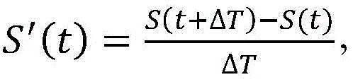
在其中分析电描记图信号S(t)的最大斜率的不应窗口可以在局部最大峰值的任一侧约2.5ms。The refractory window in which the maximum slope of the electrogram signal S(t) is analyzed can be about 2.5 ms either side of the local maximum peak.
离散活动阈值可以是大约0.2mV/msec。The discrete activity threshold may be approximately 0.2 mV/msec.
图5的面板C描绘了LAVA函数506中的多个主要活动峰值(为了清楚起见,在参考数字508中仅显示了两个具有导联线),其中的每一个可以分类为近场峰值(由三角形图标表示)、远场峰值(由圆形图标表示)、或噪声峰值(不由任何特殊图标表示)。在实施例中,使用以下逻辑来对主要活动峰值进行分类:Panel C of Figure 5 depicts a number of major activity peaks in the LAVA function 506 (for clarity, only two with lead lines are shown in reference numeral 508), each of which can be classified as a near-field peak (by triangle icon), far-field peaks (represented by circle icons), or noise peaks (not represented by any special icons). In an embodiment, the following logic is used to classify major activity peaks:
·如果主要活动峰值处的L(t)超过预设的以及可选地用户定义的近场阈值(510),则峰值可以被分类为近场峰值;- If L(t) at the main activity peak exceeds a preset and optionally user-defined near-field threshold (510), the peak may be classified as a near-field peak;
·如果主要活动峰值处的L(t)超过预设的以及可选地用户定义的远场阈值(512),但不超过上述的近场阈值(510),则峰值可以被分类为远场峰值;以及If L(t) at the main activity peak exceeds a preset and optionally user-defined far-field threshold (512), but does not exceed the above-mentioned near-field threshold (510), the peak may be classified as a far-field peak ;as well as
·如果主要活动峰值处的L(t)不超过上述远场阈值(512),则该峰值可被分类为噪声峰值并被丢弃。- If the L(t) at the main activity peak does not exceed the above-mentioned far-field threshold (512), the peak may be classified as a noise peak and discarded.
在一些实施例中,预设的远场阈值(512)约为电描记图信号S(t)的长度的2.5倍,并且预设的近场阈值(510)约为电描记图信号S(t)的长度的5倍。In some embodiments, the predetermined far-field threshold (512) is approximately 2.5 times the length of the electrogram signal S(t), and the predetermined near-field threshold (510) is approximately the length of the electrogram signal S(t) ) 5 times the length.
在框412中,将LAVA的一阶分析应用于电描记图信号S(t)。根据本公开的各方面,该一阶测试将电描记图信号S(t)的峰峰振幅与预设的以及可选地用户定义的振幅阈值(诸如约5mV)进行比较。如果电描记图信号S(t)的峰峰振幅确实超过振幅阈值,则电描记图S(t)可能来自健康组织。另一方面,如果电描记图信号S(t)的峰峰振幅不超过振幅阈值,则可以进行如本文所述的进一步的LAVA分析In
在框414中,使用在框410中检测到的LAVA功能峰值之一来计算电描记图信号S(t)的LAVA迟滞参数。根据本公开的各方面,将LAVA迟滞参数计算为一方面在LAVA函数L(t)中检测到的最近时间的近场峰值或在LAVA函数L(t)中检测到的最近时间的远场峰值与另一方面参考ECG QRS信号之间的时间差。更特别地,如果在LAVA函数中检测到任何近场峰值,则使用最近时间的近场峰值来计算LAVA迟滞参数;否则,使用最近时间的远场峰值来计算LAVA迟滞参数。In
在框416中,针对电描记图信号S(t)计算LAVA概率参数。LAVA概率参数可以考虑电描记图信号S(t)的LAVA迟滞参数、电描记图信号S(t)的分割参数、在LAVA函数L(t)中检测到的峰值的总数、在LAVA函数L(t)中检测到的远场峰值的数量、近场活动宽度、孤立的QRS活动的数量、和/或电描记图信号S(t)的QRS活动持续时间。LAVA概率参数、峰值的数量和远场峰值的数量都在上面描述。本领域普通技术人员将熟悉近场活动宽度的计算和QRS活动的识别。下面描述分割参数和QRS活动持续时间。In
分割参数。分割参数是电描记图信号S(t)的分割的量度。在本公开的实施例中,分割参数被计算为电描记图信号S(t)(或电描记图信号的噪声滤波版本,表示为S*(t))的斜率在LAVA函数L(t)中检测到的最早时间的近场峰值和在LAVA函数L(t)中检测到的最近时间的近场峰值之间改变符号的次数。Split parameter. The segmentation parameter is a measure of the segmentation of the electrogram signal S(t). In an embodiment of the present disclosure, the segmentation parameter is calculated as the slope of the electrogram signal S(t) (or a noise filtered version of the electrogram signal, denoted as S*(t)) in the LAVA function L(t) Number of sign changes between the earliest detected near-field peak and the most recent detected near-field peak in the LAVA function L(t).
通过进一步解释,计算分割参数的第一步骤可以是从电描记图信号S(t)中去除噪声(即,生成S*(t))。为了生成S*(t),可以将LAVA函数L(t)(其中已经去除了噪声,如上面结合尺度图G(f,t)的讨论所述)转换成脉冲波LPulse(t),其中
一旦生成S*(t),其斜率可以在LAVA函数L(t)中检测到的最早时间的近场峰值与LAVA函数L(t)中检测到的最近时间的近场峰值之间被分析。根据本公开的各方面,可以将斜率定义为
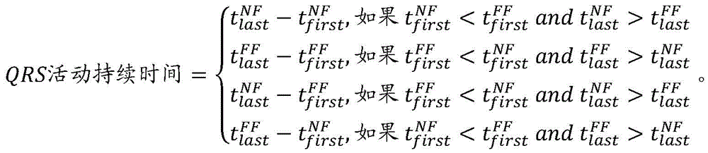
QRS活动持续时间。电描记图信号S(t)的QRS活动持续时间可以使用在LAVA函数中检测到的近场和远场峰值来计算。在本公开的一些实施例中,可以使用在LAVA函数中检测到的最早和最近时间的近场峰值(分别表示为tfirst NF和tlast NF)以及在LAVA函数中检测到的最早和最近时间的远场峰值(分别表示为tfirst FF和tlast FF)来计算QRS活动持续时间,如下所示:QRS activity duration. The QRS activity duration of the electrogram signal S(t) can be calculated using the near-field and far-field peaks detected in the LAVA function. In some embodiments of the present disclosure, near-field peaks (denoted t first NF and t last NF , respectively) detected at the earliest and latest times in the LAVA function may be used as well as the earliest and most recent times detected in the LAVA function of the far-field peaks (denoted t first FF and t last FF , respectively) to calculate the QRS activity duration as follows:
总LAVA概率参数可基于上述一个或多个参数计算为加权值。根据本公开的各方面,LAVA概率参数是累积加权值,其可以如示例性表1和/或示例性表2中所示进行计算,其中仅在前一步骤不引起LAVA概率超过100%时才采取后续步骤。The overall LAVA probability parameter may be calculated as a weighted value based on one or more of the above parameters. According to aspects of the present disclosure, the LAVA probability parameter is a cumulative weighted value that can be calculated as shown in Exemplary Table 1 and/or Exemplary Table 2, where only if the previous step did not cause the LAVA probability to exceed 100% Take next steps.

表1:用于计算LAVA概率的示例性步骤Table 1: Exemplary steps for calculating LAVA probabilities
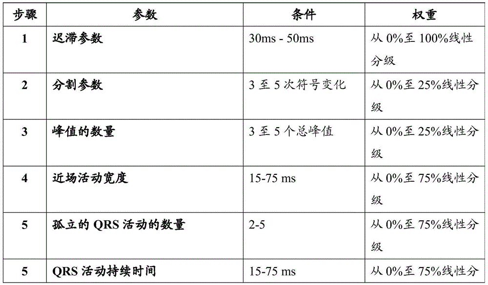

表2:用于计算LAVA概率的示例性步骤Table 2: Exemplary steps for calculating LAVA probabilities
一旦计算了LAVA迟滞和LAVA概率参数,就可以在框418中显示它们。例如,它们可以作为显示屏上的数值提供给用户(例如,作为在产生LAVA的电描记图信号旁边显示的注释或注解。参数也可以被保存用于进一步分析,例如用于随后的查阅或离线分析)。在电生理研究期间,这些指标可用于对观察到的广泛范围的早期至晚期LAVA进行分级,允许医生确定是否执行额外的诊断和/或治疗行动。例如,在某些情况下,疤痕中的存活纤维可以被检测为远场电描记图(LAVA)期间或之后的任何时间发生的高频信号。Once the LAVA hysteresis and LAVA probability parameters are calculated, they may be displayed in
在一些实施例中,并且如图6中可以进一步看到的,系统8被配置为使用计算的LAVA迟滞和/或概率参数来生成一个或多个标测图。图6可以表示由系统8生成的示例性显示屏78,其中屏幕78的第一窗格80示出了在地图82上具有不同颜色的样本LAVA概率标测图82(以%表示),不同颜色表示不同百分比LAVA概率值(例如,红色/橙色表示相对较高的LAVA概率值,而蓝色/紫色表示相对较低的LAVA概率值)。正在被分析的特定双极EGM信号84也可以在显示屏78上的第二窗格86中显示。用于LAVA计算的LAVA函数88也可以在显示屏78上的第三窗格90中显示。如所示,也可以在其他窗格94上显示其他标测图,例如峰峰电压标测图92。如上所述,可使用本领域中熟悉的各种技术来从多个电生理数据点(包括本文所述的参数)生成图形表示,以使得对前述标测图的创建的详细讨论对本公开的理解来说是不必要的。In some embodiments, and as can be further seen in FIG. 6 , the
图7-9示出了使用统计分析以及由经验丰富的从业人员将电描记图信号分类为不同等级的上述教导的验证。从图7上部的绘图中可以看出,例如,根据以下示例性分级方案,从业人员分析和分类了多个接触电描记图(示例性波形如图8所示):7-9 illustrate validation of the above teachings using statistical analysis and classification of electrogram signals into different classes by experienced practitioners. As can be seen from the plot in the upper part of Figure 7, for example, the practitioner analyzed and classified a number of contact electrograms (exemplary waveforms are shown in Figure 8) according to the following exemplary grading scheme:
·0级:非LAVA/健康电位;Level 0: non-LAVA/health potential;
·1级:具有融合的多个电位的早期心脏舒张LAVA;Grade 1: Early diastolic LAVA with fused multiple potentials;
·2级:具有碎裂电位的中期心脏舒张LAVA;Grade 2: mid-diastolic LAVA with fragmentation potential;
·3级:具有晚电位的晚期心脏舒张LAVA;以及Grade 3: Late diastolic LAVA with late potentials; and
·噪声。·noise.
图7还以盒形图和数字形式示出了根据本文的教导由系统8针对每个等级计算的LAVA概率的分布。Figure 7 also shows, in box-plot and numerical form, the distribution of LAVA probabilities calculated by
图9描绘了用于非LAVA与LAVA的LAVA概率参数的各种截止电平的示例性ROC曲线。ROC曲线以图形方式显示LAVA识别的灵敏度和特异性之间的折衷,并指定最佳截止值,诸如18%,如图所示。从图7和图9可以理解,大多数晚期(3级)或中期心脏舒张(2级)LAVA在70%的LAVA概率或更高处被观察到,而早期心脏舒张LAVA(1级)具有相对较低的概率。这些信息可以被医生用作反馈来确定哪些部位是消融的良好候选部位。9 depicts exemplary ROC curves for various cutoff levels of the LAVA probability parameter for non-LAVA and LAVA. The ROC curve graphically shows the trade-off between sensitivity and specificity of LAVA recognition, and assigns an optimal cutoff, such as 18%, as shown. As can be understood from Figures 7 and 9, most late (grade 3) or mid-diastolic (grade 2) LAVAs were observed at 70% LAVA probability or higher, whereas early diastolic LAVA (grade 1) had relative lower probability. This information can be used by physicians as feedback to determine which sites are good candidates for ablation.
可以使用统计方法针对一致性来分析不同等级信号的LAVA概率参数。LAVA概率参数也可以在视觉上与LAVA概率标测图进行比较,以确定计算的概率与LAVA概率标测图内的区域之间的视觉一致性,该区域围绕消融损伤的位置和/或在研究期间标记的其他位置具有较高值。The LAVA probability parameter of different levels of signals can be analyzed for consistency using statistical methods. The LAVA probability parameter can also be visually compared to the LAVA probability map to determine visual agreement between the calculated probability and the area within the LAVA probability map surrounding the location of the ablation lesion and/or during the study. Other locations marked during the period have higher values.
虽然以上已经以一定程度的特殊性描述了若干实施例,但是本领域技术人员可以在不脱离本发明的精神或范围的情况下对所公开的实施例进行许多改变。Although several embodiments have been described above with a certain degree of particularity, many changes to the disclosed embodiments can be made by those skilled in the art without departing from the spirit or scope of the invention.
例如,尽管前文描述了实时发生的代表性过程(例如,当从体内导管接收到电描计图时处理电描记图),但是本文的教导同样适用于后处理期间(例如,处理之前收集的一组电描记图)。For example, although the foregoing describes representative processes that occur in real-time (eg, processing electrograms as they are received from an in vivo catheter), the teachings herein are equally applicable during post-processing (eg, processing a previously collected group electrogram).
所有方向参考(例如,上部、下部、向上、向下、左、右、向左、向右、顶部、底部、在...之上、在...之下、垂直、水平、顺时针和逆时针)仅用于标识目的以帮助读者理解本发明,且并不产生特别是关于位置、定向或本发明的使用的限制。结合参考(例如,附接、耦合、连接等)应被宽泛地解释并可包括在元件的连接之间的中间构件和元件之间的相对移动。因此,结合参考不一定推断出两个元件直接连接并彼此处于固定关系中。All orientation references (eg, top, bottom, up, down, left, right, left, right, top, bottom, above, below, vertical, horizontal, clockwise and (counterclockwise) is for identification purposes only to aid the reader in understanding the invention, and does not create limitations, particularly with regard to location, orientation, or use of the invention. Incorporation references (eg, attached, coupled, connected, etc.) are to be construed broadly and may include intermediate members between connections of elements and relative movement between elements. Thus, incorporation of reference does not necessarily infer that two elements are directly connected and in a fixed relationship to each other.
意图是在上面的描述中包含或在附图中所示的所有事物应被解释为仅仅示例性的而不是限制性的。可在不偏离如在所附权利要求中限定的本发明的精神的情况下做出细节或结构上的变化。It is intended that all matter contained in the above description or shown in the accompanying drawings shall be interpreted as illustrative only and not in a limiting sense. Changes in detail or structure may be made without departing from the spirit of the invention as defined in the appended claims.
Claims (21)
Applications Claiming Priority (5)
| Application Number | Priority Date | Filing Date | Title |
|---|---|---|---|
| US201562263136P | 2015-12-04 | 2015-12-04 | |
| US62/263136 | 2015-12-04 | ||
| US201662330886P | 2016-05-03 | 2016-05-03 | |
| US62/330886 | 2016-05-03 | ||
| PCT/US2016/064671 WO2017096198A1 (en) | 2015-12-04 | 2016-12-02 | Methods and systems for statistically analyzing electrograms for local abnormal ventricular activities and mapping the same |
Publications (2)
| Publication Number | Publication Date |
|---|---|
| CN108471975A CN108471975A (en) | 2018-08-31 |
| CN108471975B true CN108471975B (en) | 2020-11-17 |
Family
ID=57610405
Family Applications (1)
| Application Number | Title | Priority Date | Filing Date |
|---|---|---|---|
| CN201680068127.4A Active CN108471975B (en) | 2015-12-04 | 2016-12-02 | Method and system for statistically analyzing electrograms of and mapping locally abnormal ventricular activity |
Country Status (5)
| Country | Link |
|---|---|
| US (2) | US10398331B2 (en) |
| EP (1) | EP3352663B1 (en) |
| JP (1) | JP6741776B2 (en) |
| CN (1) | CN108471975B (en) |
| WO (1) | WO2017096198A1 (en) |
Families Citing this family (30)
| Publication number | Priority date | Publication date | Assignee | Title |
|---|---|---|---|---|
| EP3352663B1 (en) * | 2015-12-04 | 2020-10-28 | St. Jude Medical, Cardiology Division, Inc. | Methods and systems for statistically analyzing electrograms for local abnormal ventricular activities and mapping the same |
| US10758147B2 (en) * | 2016-03-01 | 2020-09-01 | St. Jude Medical, Cardiology Division, Inc. | Methods and systems for mapping cardiac activity |
| US11065060B2 (en) * | 2018-04-26 | 2021-07-20 | Vektor Medical, Inc. | Identify ablation pattern for use in an ablation |
| US11259871B2 (en) | 2018-04-26 | 2022-03-01 | Vektor Medical, Inc. | Identify ablation pattern for use in an ablation |
| US12076119B2 (en) | 2018-04-26 | 2024-09-03 | Vektor Medical, Inc. | Bootstrapping a simulation-based electromagnetic output of a different anatomy |
| US10952794B2 (en) | 2018-11-13 | 2021-03-23 | Vektor Medical, Inc. | Augmentation of images with source locations |
| US10813612B2 (en) | 2019-01-25 | 2020-10-27 | Cleerly, Inc. | Systems and method of characterizing high risk plaques |
| US12167917B2 (en) * | 2019-05-09 | 2024-12-17 | St. Jude Medical, Cardiology Division, Inc. | System and method for detection and mapping of near field conduction in scar tissue |
| US12369840B2 (en) | 2019-05-24 | 2025-07-29 | St. Jude Medical, Cardiology Division, Inc. | System and method for cardiac mapping |
| US10709347B1 (en) | 2019-06-10 | 2020-07-14 | Vektor Medical, Inc. | Heart graphic display system |
| US10595736B1 (en) | 2019-06-10 | 2020-03-24 | Vektor Medical, Inc. | Heart graphic display system |
| US11278233B2 (en) | 2019-11-15 | 2022-03-22 | Biosense Webster (Israel) Ltd. | Method and apparatus to find abnormal activations in intra-cardiac electrocardiograms based on specificity and sensitivity |
| US11969280B2 (en) | 2020-01-07 | 2024-04-30 | Cleerly, Inc. | Systems, methods, and devices for medical image analysis, diagnosis, risk stratification, decision making and/or disease tracking |
| US20220392065A1 (en) | 2020-01-07 | 2022-12-08 | Cleerly, Inc. | Systems, methods, and devices for medical image analysis, diagnosis, risk stratification, decision making and/or disease tracking |
| KR20220124217A (en) | 2020-01-07 | 2022-09-13 | 클리어리, 인크. | Systems, methods and devices for medical image analysis, diagnosis, risk stratification, decision-making and/or disease tracking |
| US11497427B2 (en) * | 2020-03-12 | 2022-11-15 | Biosense Webster (Israel) Ltd. | Adjusting annotation points in real time |
| CN115243616B (en) * | 2020-03-16 | 2025-05-02 | 圣犹达医疗用品心脏病学部门有限公司 | Systems, methods and devices for mapping local activation time |
| JP7585474B2 (en) * | 2020-09-30 | 2024-11-18 | ボストン サイエンティフィック サイムド,インコーポレイテッド | Electrophysiology system and method for determining local cycle length and local duty cycle - Patents.com |
| US11439339B1 (en) * | 2020-10-16 | 2022-09-13 | Neucures Inc. | Method and system for measuring cardiac tissue health based on DV/DTmin of a depolarization wave within a cardiac electrogram |
| US12029570B1 (en) | 2020-10-16 | 2024-07-09 | Neutrace Inc. | Method and system for measuring unipolar and bipolar cardiac electrogram fractionation |
| WO2022094425A1 (en) | 2020-10-30 | 2022-05-05 | Vektor Medical, Inc. | Heart graphic display system |
| US11589795B2 (en) | 2020-12-22 | 2023-02-28 | Biosense Webster (Israel) Ltd. | Annotation of late potentials comprising local abnormal ventricular activation (LAVA) signals |
| US11338131B1 (en) | 2021-05-05 | 2022-05-24 | Vektor Medical, Inc. | Guiding implantation of an energy delivery component in a body |
| CN118103924A (en) | 2021-08-09 | 2024-05-28 | 维克多医疗股份有限公司 | Organization status graphic display system |
| US11534224B1 (en) | 2021-12-02 | 2022-12-27 | Vektor Medical, Inc. | Interactive ablation workflow system |
| US20250143657A1 (en) | 2022-03-10 | 2025-05-08 | Cleerly, Inc. | Systems, devices, and methods for non-invasive image-based plaque analysis and risk determination |
| US20250217981A1 (en) | 2022-03-10 | 2025-07-03 | Cleerly, Inc. | Systems, methods, and devices for image-based plaque analysis and risk determination |
| US12440180B2 (en) | 2022-03-10 | 2025-10-14 | Cleerly, Inc. | Systems, devices, and methods for non-invasive image-based plaque analysis and risk determination |
| US12406365B2 (en) | 2022-03-10 | 2025-09-02 | Cleerly, Inc. | Systems, devices, and methods for non-invasive image-based plaque analysis and risk determination |
| US12526434B2 (en) | 2022-05-27 | 2026-01-13 | The Vektor Group, Inc. | Encoding electrocardiographic data |
Citations (5)
| Publication number | Priority date | Publication date | Assignee | Title |
|---|---|---|---|---|
| CN101257841A (en) * | 2005-05-13 | 2008-09-03 | 卡迪欧扩实验室公司 | Method and device for rapid interpretation and analysis of electrocardiogram waveform |
| WO2010054409A1 (en) * | 2008-11-10 | 2010-05-14 | Cardioinsight Technologies, Inc. | Visualization of electrophysiology data |
| CN102988042A (en) * | 2012-08-30 | 2013-03-27 | 重庆电子工程职业学院 | Method for identifying sino atrial node electrogram based on integration of wavelet transform and support vector machine |
| US8812091B1 (en) * | 2013-03-15 | 2014-08-19 | Apn Health, Llc | Multi-channel cardiac measurements |
| CN104367317A (en) * | 2014-10-15 | 2015-02-25 | 北京理工大学 | Electrocardiogram electrocardiosignal classification method with multi-scale characteristics combined |
Family Cites Families (24)
| Publication number | Priority date | Publication date | Assignee | Title |
|---|---|---|---|---|
| US5662108A (en) | 1992-09-23 | 1997-09-02 | Endocardial Solutions, Inc. | Electrophysiology mapping system |
| CA2144973C (en) | 1992-09-23 | 2010-02-09 | Graydon Ernest Beatty | Endocardial mapping system |
| US6939309B1 (en) | 1993-09-23 | 2005-09-06 | Endocardial Solutions, Inc. | Electrophysiology mapping system |
| US5697377A (en) | 1995-11-22 | 1997-12-16 | Medtronic, Inc. | Catheter mapping system and method |
| US7263397B2 (en) | 1998-06-30 | 2007-08-28 | St. Jude Medical, Atrial Fibrillation Division, Inc. | Method and apparatus for catheter navigation and location and mapping in the heart |
| US7099714B2 (en) * | 2003-03-31 | 2006-08-29 | Medtronic, Inc. | Biomedical signal denoising techniques |
| DE10340546B4 (en) * | 2003-09-01 | 2006-04-20 | Siemens Ag | Method and apparatus for visually assisting electrophysiology catheter application in the heart |
| DE102005042329A1 (en) * | 2005-09-06 | 2007-03-08 | Siemens Ag | Electro-physiological catheter application assistance providing method, involves detecting contour of areas relevant for catheter application, and showing areas as simple line in representations of mapping and/or image data |
| US7885707B2 (en) | 2005-09-15 | 2011-02-08 | St. Jude Medical, Atrial Fibrillation Division, Inc. | Method of scaling navigation signals to account for impedance drift in tissue |
| US8301233B2 (en) * | 2009-03-31 | 2012-10-30 | Medtronic, Inc. | Detecting a condition of a patient using a probability-correlation based model |
| WO2010132311A1 (en) * | 2009-05-13 | 2010-11-18 | Monitoring Information Technologies, Inc. | Systems and methods for heart and activity monitoring |
| DE102009034245A1 (en) * | 2009-07-22 | 2011-03-03 | Siemens Aktiengesellschaft | Method and apparatus for visually assisting electrophysiology catheter application |
| US8948837B2 (en) * | 2011-01-13 | 2015-02-03 | Rhythmia Medical, Inc. | Electroanatomical mapping |
| CN104883969B (en) * | 2012-12-20 | 2018-01-12 | 波士顿科学医学有限公司 | The rotor matched using ordered mode is identified |
| US8880158B2 (en) * | 2013-01-16 | 2014-11-04 | University Of Vermont | Methods and systems for determining spatiotemporal variability for mapping cardiac fibrillation |
| US9662178B2 (en) * | 2013-05-17 | 2017-05-30 | University Health Network | System and method for Decrement EvokEd Potential (DEEP) mapping to identify components of the arrythmogenic circuit in cardiac arrhythmias |
| US9374323B2 (en) | 2013-07-08 | 2016-06-21 | Futurewei Technologies, Inc. | Communication between endpoints in different VXLAN networks |
| US9282907B2 (en) * | 2013-07-23 | 2016-03-15 | Medtronic, Inc. | Identification of healthy versus unhealthy substrate for pacing from a multipolar lead |
| US9220435B2 (en) * | 2013-10-09 | 2015-12-29 | St. Jude Medical, Cardiology Division, Inc. | System and method for generating electrophysiology maps |
| US9572535B2 (en) * | 2013-12-05 | 2017-02-21 | Biosense Webster (Israel) Ltd. | Dynamic mapping point filtering using a pre-acquired image |
| US9554718B2 (en) | 2014-01-29 | 2017-01-31 | Biosense Webster (Israel) Ltd. | Double bipolar configuration for atrial fibrillation annotation |
| US20150228254A1 (en) | 2014-02-07 | 2015-08-13 | St. Jude Medical, Cardiology Division, Inc. | Systems and Methods for Generating, Storing, and Displaying Anatomical Maps |
| EP3352663B1 (en) * | 2015-12-04 | 2020-10-28 | St. Jude Medical, Cardiology Division, Inc. | Methods and systems for statistically analyzing electrograms for local abnormal ventricular activities and mapping the same |
| US10403053B2 (en) * | 2016-11-15 | 2019-09-03 | Biosense Webster (Israel) Ltd. | Marking sparse areas on maps |
-
2016
- 2016-12-02 EP EP16816811.0A patent/EP3352663B1/en active Active
- 2016-12-02 JP JP2018548651A patent/JP6741776B2/en active Active
- 2016-12-02 CN CN201680068127.4A patent/CN108471975B/en active Active
- 2016-12-02 WO PCT/US2016/064671 patent/WO2017096198A1/en not_active Ceased
- 2016-12-02 US US15/367,895 patent/US10398331B2/en active Active
-
2019
- 2019-08-20 US US16/545,233 patent/US11229393B2/en active Active
Patent Citations (5)
| Publication number | Priority date | Publication date | Assignee | Title |
|---|---|---|---|---|
| CN101257841A (en) * | 2005-05-13 | 2008-09-03 | 卡迪欧扩实验室公司 | Method and device for rapid interpretation and analysis of electrocardiogram waveform |
| WO2010054409A1 (en) * | 2008-11-10 | 2010-05-14 | Cardioinsight Technologies, Inc. | Visualization of electrophysiology data |
| CN102988042A (en) * | 2012-08-30 | 2013-03-27 | 重庆电子工程职业学院 | Method for identifying sino atrial node electrogram based on integration of wavelet transform and support vector machine |
| US8812091B1 (en) * | 2013-03-15 | 2014-08-19 | Apn Health, Llc | Multi-channel cardiac measurements |
| CN104367317A (en) * | 2014-10-15 | 2015-02-25 | 北京理工大学 | Electrocardiogram electrocardiosignal classification method with multi-scale characteristics combined |
Also Published As
| Publication number | Publication date |
|---|---|
| JP2019503264A (en) | 2019-02-07 |
| US10398331B2 (en) | 2019-09-03 |
| WO2017096198A1 (en) | 2017-06-08 |
| EP3352663A1 (en) | 2018-08-01 |
| EP3352663B1 (en) | 2020-10-28 |
| US20200046238A1 (en) | 2020-02-13 |
| JP6741776B2 (en) | 2020-08-19 |
| CN108471975A (en) | 2018-08-31 |
| US20170156612A1 (en) | 2017-06-08 |
| US11229393B2 (en) | 2022-01-25 |
Similar Documents
| Publication | Publication Date | Title |
|---|---|---|
| CN108471975B (en) | Method and system for statistically analyzing electrograms of and mapping locally abnormal ventricular activity | |
| JP6200590B2 (en) | System and method for generating an electrophysiological map | |
| JP6646755B2 (en) | Method and system for operating a system for mapping cardiac activity | |
| JP7093776B2 (en) | Systems and Methods for Generating Premature Ventricular Contraction Electrophysiological Maps | |
| JP2017532140A (en) | Method and system for mapping local conduction velocities | |
| CN108348186B (en) | Method and system for mapping cardiac recovery | |
| CN105592787A (en) | Cardiac mapping system and method for bi-directional activation detection of electrograms | |
| CN108289631B (en) | Method and system for mapping cardiac repolarization | |
| JP2016531639A (en) | System and method for generating an electrophysiological map | |
| CN110381813B (en) | System and method for distinguishing adipose tissue and scar tissue during electrophysiology mapping | |
| JP6273355B2 (en) | Cardiac mapping system and method for voltage-based assessment of electrograms | |
| US11103177B2 (en) | System and method for mapping cardiac activity | |
| JP2021079099A (en) | Method for finding abnormal activation in intra-cardiac electrocardiograms | |
| US20200275851A1 (en) | Systems and methods for displaying ep maps using confidence metrics |
Legal Events
| Date | Code | Title | Description |
|---|---|---|---|
| PB01 | Publication | ||
| PB01 | Publication | ||
| SE01 | Entry into force of request for substantive examination | ||
| SE01 | Entry into force of request for substantive examination | ||
| TA01 | Transfer of patent application right |
Effective date of registration: 20200526 Address after: American Minnesota Applicant after: ST. JUDE MEDICAL, CARDIOLOGY DIVISION, Inc. Address before: American Minnesota Applicant before: ST. JUDE MEDICAL, CARDIOLOGY DIVISION, Inc. Applicant before: Pi Aier.jiayi |
|
| TA01 | Transfer of patent application right | ||
| GR01 | Patent grant | ||
| GR01 | Patent grant |