CN108414768B - Gold-labeled detection test strip for glyphosate-resistant GAT transgenic crops - Google Patents
Gold-labeled detection test strip for glyphosate-resistant GAT transgenic crops Download PDFInfo
- Publication number
- CN108414768B CN108414768B CN201810119151.7A CN201810119151A CN108414768B CN 108414768 B CN108414768 B CN 108414768B CN 201810119151 A CN201810119151 A CN 201810119151A CN 108414768 B CN108414768 B CN 108414768B
- Authority
- CN
- China
- Prior art keywords
- monoclonal antibody
- gold
- hybridoma cell
- line
- gat
- Prior art date
- Legal status (The legal status is an assumption and is not a legal conclusion. Google has not performed a legal analysis and makes no representation as to the accuracy of the status listed.)
- Active
Links
Images


Classifications
-
- G—PHYSICS
- G01—MEASURING; TESTING
- G01N—INVESTIGATING OR ANALYSING MATERIALS BY DETERMINING THEIR CHEMICAL OR PHYSICAL PROPERTIES
- G01N33/00—Investigating or analysing materials by specific methods not covered by groups G01N1/00 - G01N31/00
- G01N33/48—Biological material, e.g. blood, urine; Haemocytometers
- G01N33/50—Chemical analysis of biological material, e.g. blood, urine; Testing involving biospecific ligand binding methods; Immunological testing
- G01N33/68—Chemical analysis of biological material, e.g. blood, urine; Testing involving biospecific ligand binding methods; Immunological testing involving proteins, peptides or amino acids
- G01N33/6803—General methods of protein analysis not limited to specific proteins or families of proteins
-
- G—PHYSICS
- G01—MEASURING; TESTING
- G01N—INVESTIGATING OR ANALYSING MATERIALS BY DETERMINING THEIR CHEMICAL OR PHYSICAL PROPERTIES
- G01N33/00—Investigating or analysing materials by specific methods not covered by groups G01N1/00 - G01N31/00
- G01N33/48—Biological material, e.g. blood, urine; Haemocytometers
- G01N33/50—Chemical analysis of biological material, e.g. blood, urine; Testing involving biospecific ligand binding methods; Immunological testing
- G01N33/53—Immunoassay; Biospecific binding assay; Materials therefor
- G01N33/558—Immunoassay; Biospecific binding assay; Materials therefor using diffusion or migration of antigen or antibody
-
- G—PHYSICS
- G01—MEASURING; TESTING
- G01N—INVESTIGATING OR ANALYSING MATERIALS BY DETERMINING THEIR CHEMICAL OR PHYSICAL PROPERTIES
- G01N33/00—Investigating or analysing materials by specific methods not covered by groups G01N1/00 - G01N31/00
- G01N33/48—Biological material, e.g. blood, urine; Haemocytometers
- G01N33/50—Chemical analysis of biological material, e.g. blood, urine; Testing involving biospecific ligand binding methods; Immunological testing
- G01N33/53—Immunoassay; Biospecific binding assay; Materials therefor
- G01N33/577—Immunoassay; Biospecific binding assay; Materials therefor involving monoclonal antibodies binding reaction mechanisms characterised by the use of monoclonal antibodies; monoclonal antibodies per se are classified with their corresponding antigens
-
- G—PHYSICS
- G01—MEASURING; TESTING
- G01N—INVESTIGATING OR ANALYSING MATERIALS BY DETERMINING THEIR CHEMICAL OR PHYSICAL PROPERTIES
- G01N2333/00—Assays involving biological materials from specific organisms or of a specific nature
- G01N2333/435—Assays involving biological materials from specific organisms or of a specific nature from animals; from humans
- G01N2333/46—Assays involving biological materials from specific organisms or of a specific nature from animals; from humans from vertebrates
- G01N2333/47—Assays involving proteins of known structure or function as defined in the subgroups
Landscapes
- Health & Medical Sciences (AREA)
- Life Sciences & Earth Sciences (AREA)
- Immunology (AREA)
- Engineering & Computer Science (AREA)
- Molecular Biology (AREA)
- Chemical & Material Sciences (AREA)
- Urology & Nephrology (AREA)
- Biomedical Technology (AREA)
- Hematology (AREA)
- Physics & Mathematics (AREA)
- Food Science & Technology (AREA)
- Microbiology (AREA)
- Biotechnology (AREA)
- Pathology (AREA)
- Medicinal Chemistry (AREA)
- Cell Biology (AREA)
- Analytical Chemistry (AREA)
- Biochemistry (AREA)
- General Health & Medical Sciences (AREA)
- General Physics & Mathematics (AREA)
- Chemical Kinetics & Catalysis (AREA)
- Bioinformatics & Cheminformatics (AREA)
- Bioinformatics & Computational Biology (AREA)
- Biophysics (AREA)
- Proteomics, Peptides & Aminoacids (AREA)
- Preparation Of Compounds By Using Micro-Organisms (AREA)
- Peptides Or Proteins (AREA)
Abstract
本发明提供了一对杂交瘤细胞株,该对杂交瘤细胞株包括独立存放的第一杂交瘤细胞株和第二杂交瘤细胞株;所述第一杂交瘤细胞株的保藏编号为CGMCC NO.12277;所述第二杂交瘤细胞株的保藏编号为CGMCC NO.12278。本发明还提供了由这两株杂交瘤细胞分泌的一对配对单克隆抗体和它们在检测转GAT的转基因农作物中的用途以及一种免疫胶体金试纸条和检测转GAT的转基因农作物的方法。通过上述技术方案,本发明能够通过免疫胶体金试纸条,对GAT蛋白的检测取得1ng/mL的灵敏度,对28种非转基因作物和17种不同来源的各种非GAT转基因作物也不呈现交叉反应,具有较高的灵敏度和特异性。

The present invention provides a pair of hybridoma cell strains, the pair of hybridoma cell strains comprises a first hybridoma cell strain and a second hybridoma cell strain that are independently deposited; the deposit number of the first hybridoma cell strain is CGMCC NO. 12277; the deposit number of the second hybridoma cell line is CGMCC NO.12278. The invention also provides a pair of paired monoclonal antibodies secreted by the two hybridoma cells and their use in detecting GAT-transformed transgenic crops, as well as an immunocolloidal gold test strip and a method for detecting GAT-transformed transgenic crops . Through the above technical solution, the present invention can obtain a sensitivity of 1 ng/mL for the detection of GAT protein through the immunocolloidal gold test strip, and does not show crossover to 28 kinds of non-transgenic crops and 17 kinds of non-GAT transgenic crops from different sources. reaction with high sensitivity and specificity.
Description
技术领域technical field
本公开涉及农业生物技术领域,具体地,涉及一种杂交瘤细胞对、一种单克隆抗体对、该单克隆抗体对在检测转GAT的转基因农作物中的用途以及一种免疫胶体金试纸条和检测转GAT的转基因农作物的方法。The present disclosure relates to the field of agricultural biotechnology, in particular, to a hybridoma cell pair, a monoclonal antibody pair, the use of the monoclonal antibody pair in detecting GAT-transgenic transgenic crops, and an immunocolloidal gold test strip and methods for detecting GAT-transformed transgenic crops.
背景技术Background technique
草甘膦(N-phosphonomethyl-glycine,glyphosate)是美国孟山都公司于20世纪70年代开发,目前在全世界广泛使用的一种非选择性、内吸传导型的除草剂。草甘膦的特点是低成本,对大多数植物和微生物都有很高的毒性,但是对人畜低毒、低残留,具有较高的商业价值。但由于草甘膦的光谱灭生性,对农作物也会造成危害,其作用机理是竞争抑制莽草酸合成途径中5-烯醇丙酮莽草酸-3-磷酸合成酶(EPSPS)的活性,该酶是真菌、细菌、藻类、高等植物体内芳香族氨基酸(包括色氨酸、酪氨酸、苯丙氨酸)生物合成过程中的一个关键酶,是植物和微生物共有。研发对草甘膦抗性的农作物,对农业生产发展和草甘膦工业都有一定的促进作用。EPSPS合成酶不受草甘膦抑制,具有草甘膦抗性,是当今商业化草甘膦耐受农作物最广泛的作用模式。但是,转EPSPS作物并不能对草甘膦起到解毒的效果,而是过量产生EPSPS来耐受高浓度的草甘膦,因此存在草甘膦在分生组织积累的情况,影响农作物的生长发育和产量。更新换代转基因产品,用转基因技术克服转基因作物产生的问题是目前研究的热点之一。研究发现了一种新的草甘膦代谢失活机制,应用草甘膦N-乙酰转移酶(glyphosate N-acetyhransferase,GAT)将草甘膦转变为对植物无毒的N-乙酰草甘膦(NAG),失去除草剂活性。这一发现揭示了新的草甘膦耐受机制,为培育草甘膦耐受农作物提供了另一种可选模式。杜邦先锋公司于2010年已经获得农业部转基因生物安全证书,可进口耐除草剂转基因大豆356043作为食品与饲料加工原料。该转基因大豆利用地衣芽孢杆菌(Bacillus licheniformis)优化获得的gat基因,具有对除草剂草甘膦的耐性。2006年以来,已在美国、加拿大等17个国家和地区获得作为食品与饲料加工原料进口的许可;在美国、加拿大等11个国家和地区获得商业化种植许可。Glyphosate (N-phosphonomethyl-glycine, glyphosate) is a non-selective, systemic herbicide developed by Monsanto Company in the 1970s and is currently widely used around the world. Glyphosate is characterized by low cost and high toxicity to most plants and microorganisms, but it has low toxicity and low residue to humans and animals, and has high commercial value. However, due to the spectral sterilization of glyphosate, it will also cause harm to crops. Its mechanism of action is to competitively inhibit the activity of 5-enolpyruvateshikimate-3-phosphate synthase (EPSPS) in the shikimate synthesis pathway. A key enzyme in the biosynthesis of aromatic amino acids (including tryptophan, tyrosine, and phenylalanine) in fungi, bacteria, algae, and higher plants, which is shared by plants and microorganisms. The research and development of crops that are resistant to glyphosate has a certain role in promoting the development of agricultural production and the glyphosate industry. EPSPS synthase is not inhibited by glyphosate and has glyphosate resistance, which is the most widespread mode of action in commercial glyphosate-tolerant crops today. However, EPSPS-transformed crops cannot detoxify glyphosate, but overproduce EPSPS to tolerate high concentrations of glyphosate. Therefore, glyphosate accumulates in meristems, which affects the growth and development of crops. and yield. Replacing transgenic products and using transgenic technology to overcome the problems of transgenic crops is one of the current research hotspots. The study discovered a new metabolic inactivation mechanism of glyphosate, using glyphosate N-acetyltransferase (glyphosate N-acetyhransferase, GAT) to convert glyphosate into N-acetylglyphosate, which is non-toxic to plants ( NAG), loss of herbicidal activity. This discovery reveals a new mechanism of glyphosate tolerance and provides an alternative model for breeding glyphosate-tolerant crops. DuPont Pioneer has obtained the GMO safety certificate from the Ministry of Agriculture in 2010, and can import herbicide-tolerant genetically modified soybean 356043 as a raw material for food and feed processing. The transgenic soybean is tolerant to the herbicide glyphosate by using the gat gene optimized by Bacillus licheniformis. Since 2006, it has obtained licenses for import as food and feed processing raw materials in 17 countries and regions including the United States and Canada; commercial cultivation licenses have been obtained in 11 countries and regions including the United States and Canada.
根据国际农业生物技术应用服务组织ISAAA的数据,截止2015年,全世界已有28个国家种植转基因作物,全球转基因作物种植面积达1.797亿公顷。一方面转基因作物的种植面积逐渐增大,另一方面,社会对转基因安全性的争论愈演愈烈。转基因作物具有品质好、耐病虫害等优点,但是不排除转基因作物对生态环境和食品安全存在的潜在风险。因此,需要对转基因产品实行有效的监管、筛选和安全评估,行之有效的检测技术/工具是实现这一目标的有力支持。现有的检测技术,如酶免法、放免法等,检测时间长(酶免检测时间需2h、放免检测需3h左右),需要特殊的检测设备,如酶标仪、放免闪烁仪、离心机等,检测专用的场地等,检测成本较高,需要专业的操作人员进行检测,实践推广应用的空间存在一定局限性。因此,需要有一种操作简便、结果准确可靠、可快速检测转基因植物中GAT蛋白的装置。According to data from ISAAA, an international service organization for agricultural biotechnology applications, as of 2015, 28 countries around the world have planted genetically modified crops, and the global area of genetically modified crops has reached 179.7 million hectares. On the one hand, the planting area of GM crops is gradually increasing, on the other hand, the debate on the safety of GM crops has intensified. Genetically modified crops have the advantages of good quality and resistance to pests and diseases, but the potential risks of genetically modified crops to the ecological environment and food safety cannot be ruled out. Therefore, effective supervision, screening and safety assessment of genetically modified products are required, and effective detection technologies/tools are strong support to achieve this goal. Existing detection technologies, such as enzyme immunoassay, radioimmunoassay, etc., have long detection times (enzyme immunoassay detection takes 2 hours, and radioimmunoassay detection takes about 3 hours), requiring special detection equipment, such as microplate readers, radioimmunoassay scintillation instruments, and centrifuges. etc., special testing sites, etc., the testing cost is high, professional operators are required for testing, and there are certain limitations in the space for practical promotion and application. Therefore, there is a need for a device with simple operation, accurate and reliable results, and rapid detection of GAT protein in transgenic plants.
双抗体夹心法是应用最广泛的一种免疫结合胶体金层析法。利用抗原抗体反应相结合的原理,用免疫金标记技术,以微孔膜为固相载体,实现对样品的定性、半定量等检测。The double-antibody sandwich method is the most widely used immuno-binding colloidal gold chromatography method. Using the principle of combining antigen-antibody reactions, using immunogold labeling technology, and using microporous membranes as solid-phase carriers, qualitative and semi-quantitative detection of samples can be realized.
双抗体夹心法是将首先将已知的特异性单抗或多抗包被于膜上作为检测带(T线),可与金标物结合的二抗包被于膜上作为质控带(C线)。与包被抗体相配对的另一单抗与胶体金结合,吸附于金标垫。干燥的金标垫一端与膜相接,一端与样品垫相连。膜的另一侧粘贴吸水垫。检测时,加入样品垫上液体样品通过毛细管作用向吸水垫的方向移动,经过金标垫时,如标本中有待测抗原,则发生抗原抗体反应,形成金颗粒-抗体-抗原复合物;样本继续移动到达检测带位置,则与针对不同检测位点的包被抗体再次发生抗原抗体反应,形成金颗粒-抗体-抗原-包被抗体复合物,形成肉眼可见的T线,如样本中无待检抗原或待检抗原的浓度低于检测范围,则不能形成肉眼可见的红色条带。游离的金标结合物或金颗粒-抗体-抗原复合物越过检测带到达质控带,与二抗发生反应,聚集并产生肉眼可见的红色条带(C线)。无论样本中是否含有待检物质,质控带都会出现C线。The double-antibody sandwich method is to first coat a known specific monoclonal antibody or polyclonal antibody on the membrane as a detection band (T line), and a secondary antibody that can bind to a gold-labeled substance is coated on the membrane as a quality control band ( C line). Another monoclonal antibody paired with the coated antibody binds to colloidal gold and adsorbs to the gold-labeled pad. The dry gold-labeled pad was attached to the membrane at one end and the sample pad at the other end. Stick an absorbent pad on the other side of the membrane. During detection, the liquid sample added to the sample pad moves toward the direction of the water-absorbing pad by capillary action. When passing through the gold label pad, if there is an antigen to be tested in the sample, an antigen-antibody reaction will occur to form a gold particle-antibody-antigen complex; the sample continues When it moves to the position of the detection zone, the antigen-antibody reaction occurs again with the coated antibodies against different detection sites, forming a gold particle-antibody-antigen-coated antibody complex, forming a T line visible to the naked eye. If the concentration of the antigen or the antigen to be detected is lower than the detection range, a red band visible to the naked eye cannot be formed. The free gold conjugate or gold particle-antibody-antigen complex crosses the detection zone to the quality control zone, reacts with the secondary antibody, aggregates and produces a red band (C line) visible to the naked eye. No matter whether the sample contains the substance to be tested or not, the C line will appear in the quality control zone.
免疫结合胶体金层析法可对样品进行单份检测,具有操作便捷、不需要特殊设备和场地的优点。但目前免疫结合胶体金层析法普遍存在一定缺陷,如对样本的敏感度低,质量控制能力不够,批间、批内和不同试剂间变异系数过大等。因此,提高抗单克隆抗体对的敏感度和特异性,是弥补免疫结合胶体金层析法缺陷的重要手段。The immunoconjugated colloidal gold chromatography method can detect a single sample, and has the advantages of convenient operation and no need for special equipment and sites. However, the current immunobinding colloidal gold chromatography generally has certain defects, such as low sensitivity to samples, insufficient quality control ability, and excessive coefficient of variation between batches, within batches and between different reagents. Therefore, improving the sensitivity and specificity of anti-monoclonal antibody pairs is an important means to make up for the defects of immunoconjugation colloidal gold chromatography.
发明内容SUMMARY OF THE INVENTION
本发明的目的是提供一种灵敏特异的免疫结合胶体金层析法检测GAT蛋白的方法。The purpose of the present invention is to provide a sensitive and specific method for detecting GAT protein by immunobinding colloidal gold chromatography.
为了实现上述目的,一方面,本发明提供了一种杂交瘤细胞对,其中,该杂交瘤细胞对包括独立存放的第一杂交瘤细胞株和第二杂交瘤细胞株;所述第一杂交瘤细胞株的保藏编号为CGMCC NO.12277;所述第二杂交瘤细胞株的保藏编号为CGMCC NO.12278。In order to achieve the above object, on the one hand, the present invention provides a hybridoma cell pair, wherein the hybridoma cell pair comprises a first hybridoma cell line and a second hybridoma cell line stored independently; the first hybridoma cell line The deposit number of the cell line is CGMCC NO.12277; the deposit number of the second hybridoma cell line is CGMCC NO.12278.
再一方面,本发明还提供了一种单克隆抗体对,该单克隆抗体对包括独立存放的第一单克隆抗体和第二单克隆抗体;所述第一单克隆抗体由第一杂交瘤细胞株产生;所述第二单克隆抗体由第二杂交瘤细胞株产生;所述第一杂交瘤细胞株的保藏编号为CGMCCNO.12277;所述第二杂交瘤细胞株的保藏编号为CGMCC NO.12278。In another aspect, the present invention also provides a monoclonal antibody pair, the monoclonal antibody pair comprising a first monoclonal antibody and a second monoclonal antibody stored independently; the first monoclonal antibody is produced by a first hybridoma cell The second monoclonal antibody is produced by the second hybridoma cell line; the deposit number of the first hybridoma cell line is CGMCC NO.12277; the deposit number of the second hybridoma cell line is CGMCC NO. 12278.
再一方面,本发明还提供了如上所述的单克隆抗体对在检测转GAT的转基因农作物中的用途。In yet another aspect, the present invention also provides the use of the monoclonal antibody pair described above in detecting GAT-transformed transgenic crops.
再一方面,本发明还提供了一种免疫胶体金试纸,该免疫胶体金试纸包括依次相连接的吸水垫、基膜、金标垫和样品垫;所述基膜上设置有质控线(C线)和检测线(T线),所述C线上包被有抗鼠IgG的抗体,所述T线上包被有第一单克隆抗体,所述金标垫中含有胶体金标记的第二单克隆抗体,所述第一单克隆抗体和所述第二单克隆抗体构成单克隆抗体对,其中,所述单克隆抗体对为如上所述的单克隆抗体对。In another aspect, the present invention also provides an immunocolloidal gold test paper, which comprises a water absorbing pad, a base film, a gold label pad and a sample pad connected in sequence; the base film is provided with a quality control line ( C line) and detection line (T line), the C line is coated with anti-mouse IgG antibody, the T line is coated with the first monoclonal antibody, and the gold label pad contains colloidal gold labeled The second monoclonal antibody, the first monoclonal antibody and the second monoclonal antibody constitute a monoclonal antibody pair, wherein the monoclonal antibody pair is the monoclonal antibody pair described above.
再一方面,本发明还提供了一种检测转GAT的转基因农作物的方法,其中,该方法包括如下步骤:S1、从待检测的农作物中提取全蛋白;S2、对所述全蛋白使用如上所述的单克隆抗体对进行免疫胶体金试纸检测或双抗夹心酶连免疫检测;S3、如果免疫胶体金试纸检测或双抗夹心酶连免疫检测的结果中出现GAT的阳性结果,则指示待检测的农作物为转GAT的转基因农作物。In another aspect, the present invention also provides a method for detecting GAT-transformed transgenic crops, wherein the method comprises the following steps: S1, extracting whole protein from the crops to be detected; S2, using the above-mentioned whole protein for the whole protein The monoclonal antibody described above is tested by immunocolloidal gold test paper or double-antibody sandwich enzyme-linked immunoassay; S3. If there is a positive result of GAT in the result of the immunocolloidal gold test paper or double-antibody sandwich enzyme-linked immunoassay, it indicates that the test is to be detected. The crops are GAT transgenic crops.
通过上述技术方案,本发明能够通过免疫胶体金试纸,对GAT蛋白的检测取得1ng/mL的灵敏度,对28种非转基因作物和17种不同来源的各种非GAT转基因作物也不呈现交叉反应,具有较高的敏感度和特异性。Through the above technical solution, the present invention can obtain a sensitivity of 1 ng/mL for the detection of GAT protein through the immune colloidal gold test paper, and does not show cross-reaction to 28 kinds of non-transgenic crops and 17 kinds of various non-GAT transgenic crops from different sources, Has high sensitivity and specificity.
本发明的其他特征和优点将在随后的具体实施方式部分予以详细说明。Other features and advantages of the present invention will be described in detail in the detailed description that follows.
生物材料保藏biological material preservation
本发明的第一杂交瘤细胞株是本发明的发明人自行融合筛选得到的,其保藏编号为CGMCC NO.12277,保藏日期为2016年04月07日,保藏单位为中国微生物菌种保藏管理委员会普通微生物中心,地址位于北京市朝阳区北辰西路1号院3号中国科学院微生物研究所,分类命名为抗GAT单克隆抗体杂交瘤细胞株。The first hybridoma cell line of the present invention was obtained by the inventor of the present invention through self-fusion screening, the deposit number is CGMCC NO.12277, the deposit date is April 7, 2016, and the deposit unit is China Microorganism Culture Collection Management Committee The General Microbiology Center is located at the Institute of Microbiology, Chinese Academy of Sciences, No. 3, No. 1, Beichen West Road, Chaoyang District, Beijing. It is classified as an anti-GAT monoclonal antibody hybridoma cell line.
本发明的第二杂交瘤细胞株是本发明的发明人自行融合筛选得到的,其保藏编号为CGMCC NO.12278,保藏日期为2016年04月07日,保藏单位为中国微生物菌种保藏管理委员会普通微生物中心,地址位于北京市朝阳区北辰西路1号院3号中国科学院微生物研究所,分类命名为抗GAT单克隆抗体杂交瘤细胞株。The second hybridoma cell line of the present invention was obtained by the inventor of the present invention through self-fusion screening, the deposit number is CGMCC NO.12278, the deposit date is April 7, 2016, and the deposit unit is China Microorganism Culture Collection Management Committee The General Microbiology Center is located at the Institute of Microbiology, Chinese Academy of Sciences, No. 3, No. 1, Beichen West Road, Chaoyang District, Beijing. It is classified as an anti-GAT monoclonal antibody hybridoma cell line.
附图说明Description of drawings
附图是用来提供对本公开的进一步理解,并且构成说明书的一部分,与下面的具体实施方式一起用于解释本公开,但并不构成对本公开的限制。在附图中:The accompanying drawings are used to provide a further understanding of the present disclosure, and constitute a part of the specification, and together with the following detailed description, are used to explain the present disclosure, but not to limit the present disclosure. In the attached image:
图1是实施例1中His-Tag GAT融合蛋白的表达与纯化结果图。FIG. 1 is a graph showing the expression and purification results of the His-Tag GAT fusion protein in Example 1. FIG.
图2是实施例2中利用胶体金产生的信号颜色深浅,进行定性或半定量检测的结果图。FIG. 2 is a graph of the results of qualitative or semi-quantitative detection of the color depth of the signal generated by colloidal gold in Example 2. FIG.
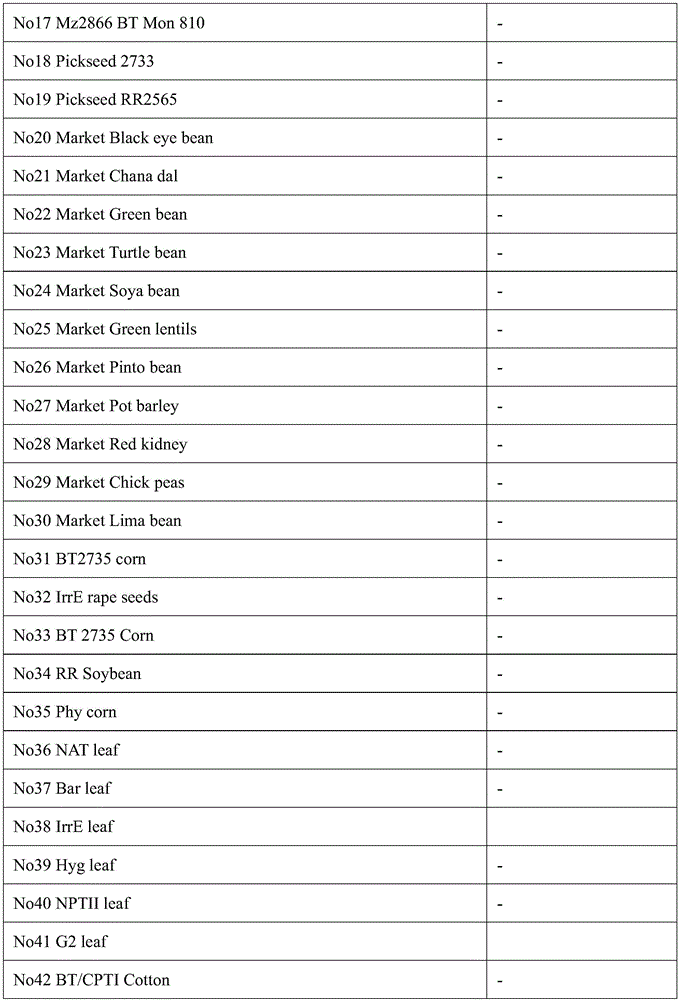
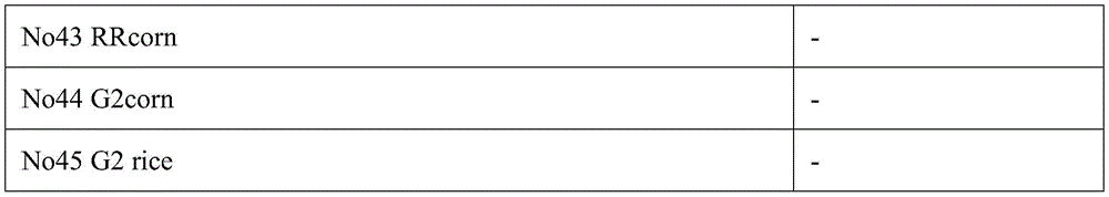
图3是实施例2中利用胶体金产生的信号颜色深浅,对28种非转基因作物和17种不同来源的各种转基因作物(表1)做了特异性检测实验的结果图。FIG. 3 is a graph showing the result of a specific detection experiment on 28 non-transgenic crops and 17 various transgenic crops from different sources (Table 1) using the color shades of the signals generated by colloidal gold in Example 2.
图4是免疫胶体金试纸的组装结构示意图。Figure 4 is a schematic diagram of the assembly structure of the immunocolloidal gold test paper.
附图标记说明Description of reference numerals
1 样品垫 2 被衬底板 3 胶体金垫1
4 T线 5 C线 6 纤维素膜4 T-line 5 C-
7 吸收垫7 Absorbent pads
具体实施方式Detailed ways
以下结合附图对本公开的具体实施方式进行详细说明。应当理解的是,此处所描述的具体实施方式仅用于说明和解释本公开,并不用于限制本公开。The specific embodiments of the present disclosure will be described in detail below with reference to the accompanying drawings. It should be understood that the specific embodiments described herein are only used to illustrate and explain the present disclosure, but not to limit the present disclosure.
一方面,本发明提供了一种杂交瘤细胞对,其中,该杂交瘤细胞对包括独立存放的第一杂交瘤细胞株和第二杂交瘤细胞株;所述第一杂交瘤细胞株的保藏编号为CGMCCNO.12277;所述第二杂交瘤细胞株的保藏编号为CGMCC NO.12278。In one aspect, the present invention provides a hybridoma cell pair, wherein the hybridoma cell pair comprises a first hybridoma cell line and a second hybridoma cell line stored independently; the deposit number of the first hybridoma cell line It is CGMCC NO.12277; the deposit number of the second hybridoma cell line is CGMCC NO.12278.
再一方面,本发明还提供了一种单克隆抗体对,该单克隆抗体对包括独立存放的第一单克隆抗体和第二单克隆抗体;所述第一单克隆抗体由第一杂交瘤细胞株产生;所述第二单克隆抗体由第二杂交瘤细胞株产生;所述第一杂交瘤细胞株的保藏编号为CGMCCNO.12277;所述第二杂交瘤细胞株的保藏编号为CGMCC NO.12278。In another aspect, the present invention also provides a monoclonal antibody pair, the monoclonal antibody pair comprising a first monoclonal antibody and a second monoclonal antibody stored independently; the first monoclonal antibody is produced by a first hybridoma cell The second monoclonal antibody is produced by the second hybridoma cell line; the deposit number of the first hybridoma cell line is CGMCC NO.12277; the deposit number of the second hybridoma cell line is CGMCC NO. 12278.
再一方面,本发明还提供了如上所述的单克隆抗体和如上所述的单克隆抗体对在检测转GAT的转基因农作物中的用途。In still another aspect, the present invention also provides the use of the above-mentioned monoclonal antibody and the above-mentioned monoclonal antibody pair in detecting GAT-transgenic transgenic crops.
可选地,其中,所述农作物为棉花、玉米、水稻或大豆。Optionally, the crop is cotton, corn, rice or soybean.
其中,所述GAT蛋白是指GAT基因所编码的蛋白,GAT基因是利用免培养技术提取草甘膦污染土壤的总DNA,构建了约含16,000个重组子的宏基因组文库。用20mM的草甘膦筛库,得到一株具有草甘膦抗性的重组菌株。提取质粒测序分析发现此片段中含有一个441bp的ORF序列。网上BLAST结果表明,此基因与已知N-乙酰转移酶基因具有较高的同源性。利用原核表达系统对其功能进行了分析验证,发现含有gat基因的菌株EGAT在300mM草甘膦浓度下仍能够生长;而且在37℃条件下,EGAT菌株经0.75mM IPTG诱导后,GAT蛋白也得到了高效表达,说明获得的gat基因具有高抗草甘膦特性,为N-乙酰转移酶基因的同源基因,将其命名为gat。GAT蛋白由146个氨基酸组成,分子量约16kDa。GAT蛋白质氨基酸序列在NCBI数据库中进行同源比对。结果显示,一致性最高的序列信息为来自Bacillus licheniformis菌株的glyphosate N-acetyltransferase蛋白(草甘膦N-乙酰基转移酶,蛋白ID:2JDC_A),比对序列一致性为98%。具体地,GAT基因的编码序列如SEQ ID NO.3所示,其编码的蛋白氨基酸序列如SEQ ID NO.4所示。The GAT protein refers to the protein encoded by the GAT gene. The GAT gene is a metagenomic library containing about 16,000 recombinants constructed by extracting the total DNA of the glyphosate-contaminated soil using a culture-free technique. A glyphosate-resistant recombinant strain was obtained by screening the library with 20 mM glyphosate. Extracted plasmid sequencing analysis found that this fragment contains a 441bp ORF sequence. The online BLAST results showed that this gene had high homology with known N-acetyltransferase genes. The prokaryotic expression system was used to analyze and verify its function. It was found that the strain EGAT containing the gat gene could still grow at a concentration of 300 mM glyphosate; and at 37 °C, after the EGAT strain was induced by 0.75 mM IPTG, the GAT protein was also obtained. The obtained gat gene is highly resistant to glyphosate, and it is the homologous gene of N-acetyltransferase gene, which is named gat. GAT protein consists of 146 amino acids and has a molecular weight of about 16kDa. The amino acid sequences of GAT proteins were homologously aligned in the NCBI database. The results showed that the sequence information with the highest identity was the glyphosate N-acetyltransferase protein (glyphosate N-acetyltransferase, protein ID: 2JDC_A) from Bacillus licheniformis strain, and the alignment sequence identity was 98%. Specifically, the coding sequence of the GAT gene is shown in SEQ ID NO.3, and the amino acid sequence of the encoded protein is shown in SEQ ID NO.4.
再一方面,本发明还提供了一种免疫胶体金试纸,该免疫胶体金试纸包括依次相连接的吸水垫、基膜、金标垫和样品垫;所述基膜上设置有C线和T线,所述C线上包被有抗鼠IgG的抗体,所述T线上包被有第一单克隆抗体,所述金标垫中含有胶体金标记的第二单克隆抗体,所述第一单克隆抗体和所述第二单克隆抗体构成配对单克隆抗体。其中,所述配对单克隆抗体为如上所述的单克隆抗体对。In another aspect, the present invention also provides an immunocolloidal gold test paper, which includes a water absorbing pad, a base film, a gold label pad and a sample pad connected in sequence; the base film is provided with a C line and a T line. The C line is coated with an anti-mouse IgG antibody, the T line is coated with a first monoclonal antibody, the gold label pad contains a second monoclonal antibody labeled with colloidal gold, and the first monoclonal antibody is coated on the T line. One monoclonal antibody and the second monoclonal antibody constitute paired monoclonal antibodies. Wherein, the paired monoclonal antibody is the monoclonal antibody pair described above.
其中,所述第一单克隆抗体由第一杂交瘤细胞株产生;所述第二单克隆抗体由第二杂交瘤细胞株产生;所述第一杂交瘤细胞株的保藏编号为CGMCC NO.12277;所述第二杂交瘤细胞株的保藏编号为CGMCC NO.12278。Wherein, the first monoclonal antibody is produced by the first hybridoma cell line; the second monoclonal antibody is produced by the second hybridoma cell line; the deposit number of the first hybridoma cell line is CGMCC NO.12277 ; The deposit number of the second hybridoma cell line is CGMCC NO.12278.
其中,免疫胶体金试纸的检测原理可能包括:将待检样品滴入样品垫中,若样品中有GAT蛋白,待检样品中GAT蛋白先和胶体金标记的第二单克隆抗体结合,由于毛细管作用,GAT蛋白和胶体金标记的第二单克隆抗体形成的复合体沿基膜向前泳动,到达检测线时遇到包被在硝酸纤维素基膜上的第一单克隆抗体,由于第一单克隆抗体和第二单克隆抗体分别能结合GAT蛋白的不同抗原表位结合,从而形成“胶体金标记的第二单克隆抗体-GAT蛋白-第一单克隆抗体”复合体,从而使得胶体金富集在检测线上,形成特异性的红色层析线;没有和GAT蛋白结合的胶体金标记的第二单克隆抗体则会直接通过检测线,达到质控线后被质控线上的羊抗鼠IgG捕获,从而使得胶体金富集在质控线上形成红色层析线,即判为阳性结果;若样品中无GAT蛋白,由于无法在检测线上形成“胶体金标记的第二单克隆抗体-GAT蛋白-第一单克隆抗体”复合体,从而使得检测线上不会产生肉眼可见的颜色反应,没有和GAT蛋白结合的胶体金标记的第二单克隆抗体则会直接通过检测线,达到质控线后被质控线上的羊抗鼠IgG捕获,从而使得胶体金富集在质控线上形成红色层析线,即判为阴性结果。Among them, the detection principle of the immunocolloidal gold test paper may include: dropping the sample to be tested into the sample pad, if there is GAT protein in the sample, the GAT protein in the sample to be tested is first bound to the second monoclonal antibody labeled with colloidal gold, and due to the capillary tube The complex formed by GAT protein and colloidal gold-labeled second monoclonal antibody migrates forward along the basement membrane, and when it reaches the detection line, it encounters the first monoclonal antibody coated on the nitrocellulose basement membrane. A monoclonal antibody and a second monoclonal antibody can bind to different epitopes of the GAT protein, thereby forming a complex of "colloidal gold-labeled second monoclonal antibody-GAT protein-first monoclonal antibody", thereby making the colloid Gold is enriched on the detection line to form a specific red chromatographic line; the second monoclonal antibody labeled with colloidal gold that is not bound to GAT protein will directly pass through the detection line, and after reaching the quality control line, it will be detected on the quality control line. Goat anti-mouse IgG is captured, so that colloidal gold is enriched on the quality control line to form a red chromatographic line, which is a positive result; if there is no GAT protein in the sample, it is impossible to form a "colloidal gold-labeled second" on the detection line. Monoclonal antibody-GAT protein-first monoclonal antibody "complex, so that there is no visible color reaction on the detection line, and the colloidal gold-labeled second monoclonal antibody that is not bound to GAT protein will directly pass the detection. After reaching the quality control line, it was captured by goat anti-mouse IgG on the quality control line, so that the colloidal gold was enriched on the quality control line to form a red chromatographic line, which was judged as a negative result.
本发明中,使用保藏编号为CGMCC NO.12277的杂交瘤细胞株和保藏编号为CGMCCNO.12278的杂交瘤细胞株分别制备和纯化第一单克隆抗体和第二单克隆抗体的方法可以为本领域常规的方法,例如小鼠腹水法和亲和层析法。In the present invention, the method for preparing and purifying the first monoclonal antibody and the second monoclonal antibody respectively by using the hybridoma cell line with the deposit number CGMCC NO.12277 and the hybridoma cell line with the deposit number CGMCC NO.12278 can be in the art Conventional methods such as mouse ascites and affinity chromatography.
本发明中,将单克隆抗体进行胶体金标记的方法可以为本领域常规的方法,例如可以包括:将0.01重量%的HAuCl4溶液加热煮沸后加入1重量%的柠檬酸三钠溶液直至溶液的颜色完全变为透明的红色时,继续回流10min后停止加热,冷却至室温,即得到胶体金;取1mL制取好的胶体金,用1重量%的K2CO3调节pH值到8.0,加入20μg单克隆抗体,混合均匀,室温反应40min;加入5重量%的BSA至终浓度为0.1重量%,静置30min;先用低速(1500×g)离心15分钟,弃去由凝聚的金胶粒形成的沉淀;然后用10000×g离心30分钟;仔细吸出上清,沉淀物用0.1mL含1%BSA的0.1M PBS(pH 7.4)复溶,加入5%叠氮钠至终浓度为0.05%,4℃保存。In the present invention, the method for labeling the monoclonal antibody with colloidal gold can be a conventional method in the field, for example, it can include: heating and boiling 0.01% by weight of HAuCl 4 solution and then adding 1% by weight of trisodium citrate solution until the solution reaches When the color becomes transparent red completely, continue to reflux for 10 minutes, then stop heating and cool to room temperature to obtain colloidal gold; take 1 mL of the prepared colloidal gold, adjust the pH to 8.0 with 1 wt% K 2 CO 3 , add 20 μg of monoclonal antibody, mixed evenly, and reacted at room temperature for 40 min; add 5 wt% BSA to a final concentration of 0.1 wt%, let stand for 30 min; first centrifuge at low speed (1500×g) for 15 min, discard the agglomerated gold particles The resulting pellet was then centrifuged at 10,000 x g for 30 minutes; the supernatant was carefully aspirated, the pellet was reconstituted with 0.1 mL of 0.1 M PBS (pH 7.4) containing 1% BSA, and 5% sodium azide was added to a final concentration of 0.05% , 4 ℃ preservation.
本发明中,制备免疫胶体金试纸的方法可以为本领域常规的方法,例如可以包括:用喷金点膜机将第一单克隆抗体和羊抗鼠IgG喷于硝酸纤维素膜上,形成相互平行的检测T线和质控C线,然后烘干;将胶体金标记的第二单克隆抗体用喷金点膜机均匀的喷在金标垫上;然后将样品垫、金标垫、喷有T线和C线的基膜(硝酸纤维素膜)和吸水纸依次连接组装,而后裁切包装备用。In the present invention, the method for preparing the immunocolloidal gold test paper can be a conventional method in the field, for example, it can include: spraying the first monoclonal antibody and goat anti-mouse IgG on a nitrocellulose membrane with a gold spraying machine to form mutual Parallel detection T line and quality control C line, and then drying; spray the second monoclonal antibody labeled with colloidal gold on the gold label pad evenly with a gold spray dot film machine; then spray the sample pad, gold label pad, and The base film (nitrocellulose film) and absorbent paper of T line and C line are connected and assembled in sequence, and then cut and packaged for later use.
可选地,其中,所述金标垫中所含有胶体金标记的第二单克隆抗体是由5-100μg/mL胶体金标记的第二单克隆抗体溶液以0.5-5μL/cm的喷涂速度通过喷金点膜机喷涂在金标垫上干燥后得到的。Optionally, wherein, the colloidal gold-labeled second monoclonal antibody contained in the gold-labeled pad is passed through a 5-100 μg/mL colloidal gold-labeled second monoclonal antibody solution at a spraying speed of 0.5-5 μL/cm. It is obtained by spraying gold dot film machine on the gold label pad and drying.
可选地,其中,所述T线上包被的第一单克隆抗体是由0.2-5mg/mL的第一单克隆抗体溶液以0.2-4μL/cm的喷涂速度通过喷金点膜机喷涂在T线上干燥后得到的。Optionally, wherein, the first monoclonal antibody coated on the T line is sprayed by a gold-spraying dot film machine with a 0.2-5 mg/mL first monoclonal antibody solution at a spraying speed of 0.2-4 μL/cm. Obtained after drying on the T line.
可选地,其中,所述C线上包被的羊抗鼠IgG的抗体是由0.2-5mg/mL的羊抗鼠IgG的抗体溶液以0.2-4μL/cm的喷涂速度通过喷金点膜机喷涂在C线上干燥后得到的。Optionally, wherein, the goat anti-mouse IgG antibody coated on the C line is made of 0.2-5 mg/mL goat anti-mouse IgG antibody solution at a spraying speed of 0.2-4 μL/cm through a gold spraying dot film machine. Obtained after spray drying on the C line.
再一方面,本发明还提供了一种检测转GAT的转基因农作物的方法,其中,该方法包括如下步骤:S1、从待检测的农作物中提取全蛋白;S2、对所述全蛋白使用如上所述的单克隆抗体对进行免疫胶体金试纸检测或双抗夹心酶连免疫检测;S3、如果免疫胶体金试纸检测或双抗夹心酶连免疫检测的结果中出现GAT的阳性结果,则指示待检测的农作物为转GAT的转基因农作物。In another aspect, the present invention also provides a method for detecting GAT-transformed transgenic crops, wherein the method comprises the following steps: S1, extracting whole protein from the crops to be detected; S2, using the above-mentioned whole protein for the whole protein The monoclonal antibody described above is tested by immunocolloidal gold test paper or double-antibody sandwich enzyme-linked immunoassay; S3. If there is a positive result of GAT in the result of the immunocolloidal gold test paper or double-antibody sandwich enzyme-linked immunoassay, it indicates that the test is to be detected. The crops are GAT transgenic crops.
根据如上所述的方法,可选地,其中,所述农作物为棉花、玉米、水稻或大豆。According to the method as described above, optionally, the crop is cotton, corn, rice or soybean.
以下通过实施例进一步详细说明本发明:The present invention is described in further detail below by embodiment:
实施例1Example 1
gat基因的PCR克隆:根据gat的ORF序列设计引物(两端带有BamHI与SacI位点);Fgat:CGCGGATCCATGATTGACGTGAACCCAAT(SEQ ID NO.1)和Rgat:CGCGAGCTCTTATGCGATCCTCTTGTACA(SEQ ID NO.2);以Fgat和Rgat为引物,抗性重组子质粒DNA为模板进行PCR扩增,PCR产物连入pGEM-Tvector载体,转化大肠杆菌DH5α;挑选阳性克隆并测序,测序结果显示GAT基因的编码序列如SEQ ID NO.3所示,其编码的蛋白氨基酸序列如SEQ ID NO.4所示。PCR cloning of gat gene: primers were designed according to the ORF sequence of gat (with BamHI and SacI sites at both ends); Fgat: CGCGGATCCATGATTGACGTGAACCCAAT (SEQ ID NO. 1) and Rgat: CGCGAGCTCTTATGCGATCCTCTTGTACA (SEQ ID NO. 2); Rgat was used as a primer, and the resistant recombinant plasmid DNA was used as a template for PCR amplification. The PCR product was connected to the pGEM-Tvector vector and transformed into Escherichia coli DH5α; positive clones were selected and sequenced. The sequencing results showed that the coding sequence of the GAT gene is as SEQ ID NO. 3, the encoded protein amino acid sequence is shown in SEQ ID NO.4.
SEQ ID NO.4为:MIDVNPINAEDTYELRHRILRPNQPIEACMFESDLLRGAFHLGGYYGGKLISIASFHQAEHSELQGQKQYQLRGMATLEGYREQKAGSSLIKHAEEILRKRGADLLWCNARTSASGYYKKLGFSEQGEVFDTPPVGPHILMYKRIA;具体参见序列表。SEQ ID NO. 4 is: MIDVNPINAEDTYELRHRILRPNQPIEACMFESDLLRGAFHLGGYYGGKLISIASFHQAEHSELQGQKQYQLRGMATLEGYREQKAGSSLIKHAEEILRKRGADLLWCNARTSASGYYKKLGFSEQGEVFDTPPVGPHILMYKRIA; see the sequence table for details.
SEQ ID NO.3为:atgattgacgtgaacccaattaacgctgaggatacttacgagcttagacatagaattcttagaccaaaccaaccaatcgaggcttgcatgttcgagtctgatcttcttagaggagctttccatcttggaggttactacggaggtaagcttatttctattgcttctttccatcaagctgagcattctgagcttcaaggacaaaagcaataccaacttaggggaatggctactcttgagggatacagagagcaaaaggctggttcttctcttatcaagcatgccgaggagatcctcaggaagaggggcgccgaccttctttggtgcaacgctaggacttccgcctctggatactacaagaagcttggcttctctgagcagggagaggtgttcgacactccacctgtgggaccccatatccttatgtacaagaggatcgcataa;具体参见序列表。SEQ ID NO.3为:atgattgacgtgaacccaattaacgctgaggatacttacgagcttagacatagaattcttagaccaaaccaaccaatcgaggcttgcatgttcgagtctgatcttcttagaggagctttccatcttggaggttactacggaggtaagcttatttctattgcttctttccatcaagctgagcattctgagcttcaaggacaaaagcaataccaacttaggggaatggctactcttgagggatacagagagcaaaaggctggttcttctcttatcaagcatgccgaggagatcctcaggaagaggggcgccgaccttctttggtgcaacgctaggacttccgcctctggatactacaagaagcttggcttctctgagcagggagaggtgttcgacactccacctgtgggaccccatatccttatgtacaagaggatcgcataa;具体参见序列表。
gat基因大肠杆菌表达载体的构建:用BamHI与SacI酶切表达载体pET-28a(+)和pGAT,分别回收5.6kb和0.45kb的DNA片段;将回收得到的表达载体pET-28a酶切片段和gat回收片段连接;连接产物利用氯化钙转化法转化入大肠杆菌BL21感受态细胞中,挑选阳性克隆,命名为pEGAT。Construction of E. coli expression vector for gat gene: The expression vectors pET-28a(+) and pGAT were digested with BamHI and SacI, and 5.6kb and 0.45kb DNA fragments were recovered respectively; the recovered expression vector pET-28a digested fragments and The fragments were recovered by gat and ligated; the ligated products were transformed into E. coli BL21 competent cells by calcium chloride transformation, and positive clones were selected and named pEGAT.
gat基因在原核中的表达、蛋白纯化及多克隆抗体制备:诱导表达:EGAT接种于3mL含卡那霉素100μg/mL的LB液体培养基中,37℃过夜培养,按1%接菌量加入到6mL含卡那霉素100μg/mL的LB液体培养基中,振荡培养至OD600值为0.6,加入IPTG至终浓度为0.75mM,继续于37℃摇床培养,对其进行诱导表达,以IPTG诱导的含pET28a(+)空载体菌株、未经IPTG诱导的EGAT菌株以及BL21菌株为对照。离心收集菌体后加入100μL上样缓冲液,煮沸10min,12000×g离心10min,取上清置4℃备用,诱导产物经12%的SDS-PAGE电泳分析。Expression of gat gene in prokaryotic cells, protein purification and polyclonal antibody preparation: Induction of expression: EGAT was inoculated into 3 mL of LB liquid medium containing 100 μg/mL of kanamycin, cultured at 37°C overnight, and added at 1% inoculum volume. To 6 mL of LB liquid
蛋白纯化:样品的准备:准备细胞,在细胞OD600≈0.5-0.8时,用0.75mM IPTG诱导4h,收集细胞,加入1/20细胞生长体积的NTA-O Buffer(20mM Tris-HCl pH7.9,0.5M NaCl,PMSF的工作浓度为1mM。将细胞悬浮起来,加入溶菌酶(工作浓度为0.2-0.4mg/mL),混匀,冰上放置30min,超声或匀浆破碎细胞,该步骤冰上操作;加入10%Triton X-100,使Triton终浓度为0.05%,充分混匀,冰上放置15min,间或混匀,冰上超声破碎细胞;加入1M MgCl2,使MgCl2终浓度为1mM。加入DNase,使DNase的终浓度为10μg/mL,混匀,室温放置10min;20,000×g,4℃离心15min以上。取上清,置于冰上备用或-20℃保存。层析:将NTA树脂装入合适的层析柱,用10倍NTA体积的NTA-0Buffer洗;将步骤1)准备好的样品加到NTA层析柱中,流速控制在15mL/h,收集穿透部分,用SDS-PAGE分析蛋白质的结合情况;层析用5倍NTA体积的NTA-0Buffer洗,流速控制在30mL/h左右;用5倍NTA体积的NTA-20Buffer(20mM Tris-HClpH 7.9,0.5M NaCl,10%Glycerol,20mM Imidazole),NTA-40Buffer(20mM Tris-HCl pH7.9,0.5M NaCl,10%Glycerol,40mM Imidazole),NTA-60Buffer(20mM Tris-HCl pH 7.9,0.5M NaCl,10%Glycerol,60mM Imidazole),NTA-80Buffer(20mM Tris-HCl pH 7.9,0.5MNaCl,10%Glycerol,80mM Imidazole),NTA-100 Buffer(20mM Tris-HCl pH 7.9,0.5MNaCl,10%Glycerol,100mM Imidazole),NTA-200Buffer(20mM Tris-HCl pH 7.9,0.5MNaCl,10%Glycerol,200mM Imidazole),NTA-1000Buffer(20mM Tris-HCl pH 7.9,0.5MNaCl,10%Glycerol,1000mM Imidazole)洗脱,流速控制在15mL/h左右,收集洗脱液,每管收集一个NTA体积;SDS-PAGE确定目标蛋白在洗脱液中的分布情况。GAT蛋白片段的理论大小为16.5kDa,但由于表达时在其N端加了His·Tag和T7·Tag,所以融合蛋白大小分别为20.5kDa与GAT分子量的理论值相符,如图1所示。图1中,泳道2为分子量标记,泳道3为诱导表达后的蛋白,泳道4-8为纯化后的His-Tag GAT融合蛋白,可见纯度较高。纯化的GAT蛋白可用于制备其单克隆抗体。Protein purification: sample preparation: prepare cells, induce 4h with 0.75mM IPTG when cell OD 600 ≈ 0.5-0.8, collect cells, add 1/20 cell growth volume NTA-O Buffer (20mM Tris-HCl pH7.9 , 0.5M NaCl, the working concentration of PMSF is 1mM. Suspend the cells, add lysozyme (working concentration is 0.2-0.4mg/mL), mix well, place on ice for 30min, sonicate or homogenize to disrupt cells, this step is ice-
实施例2Example 2
GAT蛋白单克隆抗体的制备及鉴定Preparation and identification of GAT protein monoclonal antibody
将6-8W+的BALB/c雌小鼠按剂量分为四组,按剂量50、100、150、200μg/只(即实施例1得到的GAT蛋白)进行免疫,共免疫3次。初次免疫前取眼血作为阴性对照,初次免疫时加等体积福氏完全佐剂皮下多点注射;以后隔四周进行一次免疫,以相同剂量重组抗原加等体积福氏不完全佐剂皮下多点注射。第三次免疫结束后10天左右用重组抗原包被ELISA板,用间接ELISA测定小鼠血清的抗体效价;融合前三天对抗体效价最高的(1:105以上)小鼠尾静脉注射50μg重组GAT蛋白加强免疫。加强免疫三天后进行细胞融合。The 6-8W + BALB/c female mice were divided into four groups according to their doses, and were immunized at doses of 50, 100, 150, and 200 μg/mice (ie, the GAT protein obtained in Example 1), and were immunized three times in total. Eye blood was taken as a negative control before the initial immunization, and an equal volume of Freund's complete adjuvant was added subcutaneously at multiple points during the initial immunization; after immunization every four weeks, the same dose of recombinant antigen plus an equal volume of incomplete Freund's adjuvant was subcutaneously administered at multiple points. injection. About 10 days after the third immunization, the ELISA plate was coated with the recombinant antigen, and the antibody titer of the mouse serum was measured by indirect ELISA; the tail vein of the mouse with the highest antibody titer (above 1 :105) three days before fusion Immunization was boosted by injection of 50 μg recombinant GAT protein. Cell fusion was performed three days after the booster immunization.
细胞融合前一天,按照以下方法制备饲养层细胞:1)将8W+健康雄性BALB/c小鼠,拉颈处死后,于75%乙醇浸泡3-5min;2)移至超净台内,用无菌剪刀剪开皮肤,暴露腹膜,并用75%乙醇消毒其腹膜;3)用止血钳轻轻拉起腹膜,将10mL预温的1640液体培养基用注射器注入腹腔,用棉球轻揉腹腔1-2min,吸出细胞悬液,放入离心管中;4)离心:1000×g,5min,弃上清;5)用10mL含血清HAT培养基将细胞混匀,细胞计数,调整细胞密度于2×105/mL;6)将此细胞悬液按100μL/孔加入96孔细胞培养板,则细胞密度为2×104/孔;7)置37℃,5%CO2孵箱培养,供次日融合实验用。One day before cell fusion, prepare feeder cells according to the following methods: 1) 8W + healthy male BALB/c mice were sacrificed by pulling their necks, and then soaked in 75% ethanol for 3-5 min; Cut the skin with sterile scissors, expose the peritoneum, and disinfect the peritoneum with 75% ethanol; 3) Gently pull up the peritoneum with hemostatic forceps, inject 10 mL of pre-warmed 1640 liquid medium into the abdominal cavity with a syringe, and gently rub the abdominal cavity with a cotton ball 1 -2min, aspirate the cell suspension and put it into a centrifuge tube; 4) Centrifuge: 1000×g, 5min, discard the supernatant; 5) Mix the cells with 10 mL of serum-containing HAT medium, count the cells, and adjust the cell density to 2 ×10 5 /mL; 6) Add 100 μL/well of this cell suspension to a 96-well cell culture plate, and the cell density is 2 × 10 4 /well; 7) Incubate at 37°C, 5% CO 2 incubator for The next day for fusion experiments.
骨髓瘤细胞SP2/0的制备:1)收获对数生长期SP2/0细胞,用1640液体培养基洗涤3次,1000×g离心5min,弃上清;2)用1640液体培养基重悬细胞。取100μL细胞悬液,以0.2%台盼蓝染色,进行细胞计数,要求细胞活力>95%,并调整细胞密度待用。Preparation of myeloma cell SP2/0: 1) Harvest SP2/0 cells in logarithmic growth phase, wash three times with 1640 liquid medium, centrifuge at 1000 × g for 5 min, and discard the supernatant; 2) Resuspend cells in 1640 liquid medium . Take 100 μL of cell suspension, stain with 0.2% trypan blue, and count the cells. The cell viability is required to be >95%, and the cell density is adjusted for later use.
脾细胞悬液的制备:1)将3天前经过冲击免疫的BALB/c小鼠摘除眼球放血,断颈处死,于75%乙醇浸泡2min;2)移至超净台内,剪开腹部皮肤向两侧剥离,暴露腹壁;换剪刀、镊子,剪开腹膜,取出脾脏,去掉脂肪和结缔组织,用1640培养基冲洗;3)将脾脏置于200目的筛网上,一边用注射器芯轻轻地研磨;一边以培养液冲洗,收集脾细胞悬液,离心1000×g,5min,弃上清;4)用1640培养基重悬细胞,离心洗涤2次:1000×g,5min,弃上清;5)用10mL不完全培养基重悬。取100μL细胞悬液,以0.2%台盼蓝染色,要求细胞活力>95%,并计数脾细胞。剩余细胞调整细胞密度待用。Preparation of spleen cell suspension: 1) BALB/c mice that had undergone
细胞融合及培养:1)将脾细胞与SP2/0骨髓瘤细胞以5:1比例混合于50mL离心管中,1000×g离心5min,弃上清,轻弹离心管底部,使细胞沉淀松散;2)一边均匀转动离心管,一边用吸管滴加37℃预温的50%PEG 4000 1mL,于1min内完成;3)加37℃预热的1640培养基1mL,在1min内完成;4)加37℃预热的1640培养基10mL,在5min内完成;5)800×g离心8min;用100mL HAT培养基重悬细胞沉淀;6)按100μL/孔将细胞悬液转移到接种了饲养细胞的细胞培养板中,同时留2孔加未经融合的SP2/0细胞作对照,观察细胞对HAT的敏感性。培养板置于37℃、5%CO2孵箱中培养;7)融合3天后,每孔补加100μL新鲜的HAT培养液。以后每3-5天半量更换HAT培养液一次;8)2周后,HT培养基半量换液;9)3-4周后,换完全培养基维持培养。Cell fusion and culture: 1) Mix spleen cells and SP2/0 myeloma cells in a 5:1 ratio in a 50 mL centrifuge tube, centrifuge at 1000 × g for 5 min, discard the supernatant, and flick the bottom of the centrifuge tube to loosen the cell pellet; 2) While rotating the centrifuge tube evenly, add 1 mL of 50% PEG 4000 pre-warmed at 37 °C dropwise with a pipette, and complete within 1 min; 3) Add 1 mL of pre-warmed 1640 medium at 37 ° C and complete within 1 min; 4) Add 10 mL of 1640 medium preheated at 37°C, completed within 5 min; 5) Centrifuge at 800 × g for 8 min; resuspend the cell pellet with 100 mL of HAT medium; 6) Transfer the cell suspension to feeder cells inoculated with 100 μL/well. In the cell culture plate, 2 wells were added with unfused SP2/0 cells as a control to observe the sensitivity of cells to HAT. The culture plate was cultured in a 37°C, 5% CO 2 incubator; 7) After 3 days of fusion, add 100 μL of fresh HAT medium to each well. After that, the HAT medium was replaced by half amount every 3-5 days; 8) After 2 weeks, half of the HT medium was changed; 9) After 3-4 weeks, the complete medium was changed to maintain the culture.
ELISA筛选阳性杂交瘤细胞:1)融合后的细胞生长到培养孔底面积的1/4时(培养约12-15天左右),取上清用间接ELISA法检测特异性反应和交叉反应,对杂交瘤细胞进行筛选。重组蛋白GAT为包被抗原,包被浓度5μg/mL常规包被ELISA板。往包被ELISA板中加100μL/孔的细胞培养上清液,用PBS 1:100稀释的免疫鼠血清为阳性对照,SP2/0细胞孔培养上清为阴性对照。细胞上清与包被ELISA板37℃孵育30min,充分洗涤后加入HRP标记的羊抗鼠IgG抗体(1:10000稀释)100μL/孔,37℃孵育30min,弃二抗。充分洗板后每孔加TMB 100μL显色15min,每孔再加入50μL 1N H2SO4终止反应。测定OD450值。2)ELISA筛选获得98株分泌GAT单抗的细胞株;上述43株GAT单抗的细胞株所分泌的单克隆抗体均对重组GAT蛋白具有阳性反应。选择其中阳性反应最强的15株细胞进一步亚克隆培养,其余细胞株直接扩大培养,冻存和少量生产腹水。ELISA screening of positive hybridoma cells: 1) When the fused cells grow to 1/4 of the bottom area of the culture well (about 12-15 days of culture), take the supernatant and use indirect ELISA to detect specific reactions and cross-reactions. Hybridoma cells were screened. Recombinant protein GAT is the coating antigen, and the coating concentration is 5μg/mL for conventional coating ELISA plate. 100 μL/well of cell culture supernatant was added to the coated ELISA plate, the immunized mouse serum diluted 1:100 with PBS was used as a positive control, and the culture supernatant of SP2/0 cell well was used as a negative control. The cell supernatant was incubated with the coated ELISA plate at 37°C for 30min. After thorough washing, 100 μL/well of HRP-labeled goat anti-mouse IgG antibody (1:10000 dilution) was added, incubated at 37°C for 30min, and the secondary antibody was discarded. After fully washing the plate, add 100 μL of TMB to each well for 15 min, and then add 50 μL of 1N H 2 SO 4 to each well to stop the reaction. OD 450 values were determined. 2) 98 GAT monoclonal antibody-secreting cell lines were obtained by ELISA screening; the monoclonal antibodies secreted by the above-mentioned 43 GAT monoclonal antibody-secreting cell lines were all positive for recombinant GAT protein. The 15 cell lines with the strongest positive reaction were selected for further subcloning culture, and the remaining cell lines were directly expanded for culture, cryopreserved and a small amount of ascites was produced.
有限稀释法进行克隆化培养:1)用移液器将待克隆的细胞吹打混匀,用含20%血清的HT选择培养液稀释至1个细胞/孔的密度,加入已有饲养细胞的细胞板,置5%CO2、37℃的培养箱中进行培养;2)培养至第4天时,在倒置显微镜下观察并记录细胞单克隆生长孔;培养1周左右,吸取100μL已变黄的细胞培养液上清,用上述ELISA法检测细胞培养上清;3)检测为强阳性的孔内细胞进行2-3次亚克隆,直到最后一次所有仅一个细胞集落生长的培养孔上清ELISA检测结果均为阳性为止;4)将最后一次有限稀释后ELISA检测结果最好的杂交瘤细胞克隆扩大培养,并冻存。Limit dilution method for cloning culture: 1) Mix the cells to be cloned with a pipette, dilute to a density of 1 cell/well with HT selection medium containing 20% serum, and add cells with feeder cells 2 ) On the 4th day of culture, observe and record the cell monoclonal growth wells under an inverted microscope; after culturing for about 1 week, aspirate 100 μL of cells that have turned yellow The culture medium supernatant was detected by the above-mentioned ELISA method; 3) The cells in the wells that were detected as strong positive were subcloned 2-3 times, until the last time all the culture wells with only one cell colony growth were detected by ELISA. 4) The hybridoma cell clone with the best ELISA test result after the last limiting dilution is expanded and cultured, and frozen.
腹水的制备:1)选取10周龄BALB/c小鼠,腹腔注射液体石蜡0.5mL/只;2)7天后,腹腔接种经PBS稀释的培养至对数期的阳性杂交瘤细胞,每只小鼠5×105/mL杂交瘤细胞;3)5天后观察,当小鼠腹部明显膨胀时,用12号注射针头收集腹水,每隔3天收集一次,直到小鼠死亡;4)将腹水以7 000×g,离心10min;留上清分装后-80℃冰箱保存。Preparation of ascites: 1) 10-week-old BALB/c mice were selected, and 0.5 mL/mouse of liquid paraffin was intraperitoneally injected; 2) 7 days later, positive hybridoma cells diluted with PBS and cultured to log phase were intraperitoneally inoculated.
单克隆抗体的纯化(protein A亲和层析):1)装柱:用Equilibration Buffer(50mM Tris-HCl,150mM NaCl,pH8.6)湿润柱子,检查柱子是否堵塞,加5mL Protein AAgarose于柱中;2)加入10倍柱体积的Equilibration Buffer平衡柱子;3)缓慢将腹水上样;4)加入10倍柱体积的Equilibration Buffer,按4-5mL/管收集穿透液直至OD280<0.1;5)准备收集管,收集洗脱液的试管按500μL/管加入中和缓冲液(20mM磷酸盐缓冲液,pH 7.7);6)用5倍柱体积的Elution Buffer(50mM glycine,0.5M NaCl,pH 2.3)洗脱,按1.5mL/管收集洗脱液直至OD280<0.1;7)用5倍柱体积的Equilibration Buffer洗柱及平衡柱子。Purification of monoclonal antibody (protein A affinity chromatography): 1) Column loading: Wet the column with Equilibration Buffer (50mM Tris-HCl, 150mM NaCl, pH8.6), check whether the column is blocked, add 5mL Protein AAgarose to the column ; 2) Add 10 column volumes of Equilibration Buffer to equilibrate the column; 3) Slowly sample the ascites; 4) Add 10 column volumes of Equilibration Buffer, collect the permeate at 4-5 mL/tube until OD 280 <0.1; 5 ) Prepare a collection tube, add neutralization buffer (20 mM phosphate buffer, pH 7.7) to the test tube for collecting the eluate at 500 μL/tube; 6)
单克隆抗体配对筛选:将所获得的43株纯化单抗两两组合,共得到(43×43)对组合,每对组合中的两个抗体分别包被硝酸纤维素膜和标记胶体金,制备胶体金试纸条,通过对GAT重组抗原,GAT转基因作物的灵敏性筛选,对非转基因作物,非GAT转基因作物的特异性筛选,最后筛选出只针对GAT高特异性高灵敏性的配对单抗。具体如下:Monoclonal antibody paired screening: The 43 purified monoclonal antibodies obtained were combined in pairs to obtain (43 × 43) pairs of combinations. The two antibodies in each combination were coated with nitrocellulose membrane and labeled colloidal gold, respectively. Colloidal gold test strips, through the sensitivity screening of GAT recombinant antigen, GAT transgenic crops, specific screening of non-transgenic crops, non-GAT transgenic crops, and finally screened out the paired monoclonal antibodies with high specificity and high sensitivity only for GAT . details as follows:
制备胶体金颗粒:采用柠檬酸三钠还原法制备胶体金颗粒。具体步骤为:用超纯水将氯金酸配制成0.01%水溶液,取100mL与1.2mL 1%的柠檬酸三钠水溶液混合,加热至持续沸腾5min。冷却后用超纯水恢复至原体积,制成颗粒直径约为30nm的胶体金颗粒。Preparation of colloidal gold particles: The colloidal gold particles were prepared by trisodium citrate reduction method. The specific steps are as follows: prepare chloroauric acid into a 0.01% aqueous solution with ultrapure water, mix 100 mL with 1.2 mL of 1% trisodium citrate aqueous solution, and heat to continuous boiling for 5 min. After cooling, it was restored to its original volume with ultrapure water to prepare colloidal gold particles with a particle diameter of about 30 nm.
确定胶体金标记的抗体最适稳定量:采用经典MEY法确定标记1mL胶体金所需要的抗体用量为20μg。Determining the optimal stable amount of colloidal gold-labeled antibody: using the classical MEY method to determine the amount of antibody required to label 1 mL of colloidal gold is 20 μg.
硝酸纤维素膜(NC)的包被:用0.0l M pH 7.2的PBS缓冲液稀释纯化单抗至2mg/mL,用于包被T线,用0.0l M pH 7.2的PBS缓冲液稀释羊抗鼠IgG至l mg/mL,用于包被C线。用BIODOT公司XYZ3050工作系统以30mm/s速度喷于硝酸纤维素膜上,形成相互平行的检测T线和质控C线,37℃烘干。Coating of nitrocellulose membrane (NC): Dilute purified mAb to 2 mg/mL with 0.01 M PBS buffer pH 7.2 for coating T-lines, and dilute goat antibody with 0.01 M PBS buffer pH 7.2 Murine IgG to 1 mg/mL was used to coat C lines. Use XYZ3050 working system of BIODOT company to spray on nitrocellulose membrane at a speed of 30mm/s to form parallel detection T line and quality control C line, and dry at 37 °C.
免疫胶体金试纸的组装:将包被了C线5和T线4的硝酸纤维素膜6,吸收垫7,胶体金垫3以及样品垫1依次粘附于不吸水的PVC被衬底板2上,如图4组装成免疫胶体金试纸。The assembly of the immunocolloidal gold test paper: the
单克隆抗体的配对筛选:43株单克隆抗体,一共获得43×43(1849)组配对单克隆抗体,组合成1849种胶体金试纸条。配对筛选时以ddH2O作为阴性样本,重组GAT蛋白1μg/mL,转GAT棉花种子抽提物0.2g/mL作为阳性样本,转CP4-EPSPS大豆RR Soybean及转BT玉米BT2836种子抽提物各0.2g/mL作为交叉反应检测样本,对每一种试纸条进行测试。将针对重组GAT蛋白,转GAT棉花种子抽提物呈阳性反应,而对其它样本呈阴性反应的12株,9对配对单克隆抗体筛选出来作为候选的配对单抗。对于候选的9对配对单抗,进一步筛选检测其对重组GAT及转GAT作物的检测灵敏度和对其他常见的非转基因作物和非GAT的转基因作物的特异性。选择对重组GAT蛋白检测灵敏度达到1ng/mL水平,对转GAT作物种子检测达到0.2μg/mL水平,与非转基因及非GAT转基因作物没有交叉反应的配对单克隆抗体作为制备双抗体夹心法检测试剂盒的配对单克隆抗体。实验最终得到第一单克隆抗体FH6和第二单克隆抗体JG7a。Paired screening of monoclonal antibodies: 43 strains of monoclonal antibodies, a total of 43 × 43 (1849) pairs of monoclonal antibodies were obtained, which were combined into 1849 kinds of colloidal gold test strips. In paired screening, ddH 2 O was used as negative sample,
单克隆抗体亚类的鉴定:利用免疫球蛋白标准亚类鉴定试剂盒(Sigma公司)对各株单抗进行亚类鉴定,具体试验方法如下:1)在酶标板中分别加入1:1000倍PBS稀释的羊抗鼠IgG(IgM、IgA、IgG1、IgG2a、IgG2b和IgG3),100μL/孔,37℃放置1h;2)弃去酶标板中液体,用PBST洗涤3次;3)100μL/孔加入PBS稀释后的纯化单克隆抗体(抗体浓度2-5μg/mL),室温孵育1h;PBST洗涤3次;4)加入HRP标记的羊抗鼠IgG抗体(1:10000稀释)100μL/孔,室温孵育30min。5)充分洗板后每孔加100μL TMB,显色15min,每孔再加入50μL 1N H2SO4终止反应。测定OD450值。最终鉴定得到第一单克隆抗体FH6的抗体亚类为IgG1,第二单克隆抗体JG7a的抗体亚类均为IgG2b。Identification of monoclonal antibody subclasses: Use the immunoglobulin standard subclass identification kit (Sigma) to identify the subclasses of each monoclonal antibody. The specific test methods are as follows: 1) Add 1:1000 times to the ELISA plate respectively Goat anti-mouse IgG (IgM, IgA, IgG 1 , IgG 2a , IgG 2b and IgG 3 ) diluted in PBS, 100 μL/well, placed at 37°C for 1 h; 2) Discard the liquid in the ELISA plate and wash it three times with PBST; 3) Add 100 μL/well of purified monoclonal antibody diluted in PBS (antibody concentration 2-5 μg/mL), incubate at room temperature for 1 h; wash 3 times with PBST; 4) Add HRP-labeled goat anti-mouse IgG antibody (1:10000 dilution) 100 μL/well, incubated at room temperature for 30 min. 5) After fully washing the plate, add 100 μL TMB to each well, develop color for 15 min, and add 50 μL 1N H 2 SO 4 to each well to stop the reaction. OD 450 values were determined. Finally, the antibody subclass of the first monoclonal antibody FH6 was identified as IgG1, and the antibody subclass of the second monoclonal antibody JG7a was IgG2b.
配对抗体灵敏度评价:筛选到的配对单克隆抗体:第一单克隆抗体FH6和第二单克隆抗体JG7a,对重组GAT蛋白检测灵敏度达1ng/mL;对转GAT棉花种子的检测灵敏度可达0.2μg/mL;如表1,图2所示。Sensitivity evaluation of paired antibodies: The screened paired monoclonal antibodies: the first monoclonal antibody FH6 and the second monoclonal antibody JG7a, have a detection sensitivity of 1 ng/mL for recombinant GAT protein; the detection sensitivity of GAT cotton seeds can reach 0.2 μg /mL; as shown in Table 1, Figure 2.
表1Table 1
配对抗体特异性评价:本实验筛选到的配对单克隆抗体:第一单克隆抗体FH6和第二单克隆抗体JG7a,对28种非转基因作物和17种不同来源的各种非GAT转基因作物(表2)做了特异性检测实验。检测结果如表2及图3示。配对单克隆抗体FH6-JG7a对所有检测非转基因和非GAT转基因作物没有交叉反应。综上所述,配对单克隆抗体FH6-JG7a制备的检测试纸条对大部分非转基因和非GAT转基因作物没有交叉反应。将产FH6单克隆抗体的杂交瘤细胞株进行保藏,保藏编号为CGMCC NO.12277;将产JG7a单克隆抗体的杂交瘤细胞株进行保藏,保藏编号为CGMCC NO.12278。Specificity evaluation of paired antibodies: The paired monoclonal antibodies screened in this experiment: the first monoclonal antibody FH6 and the second monoclonal antibody JG7a, against 28 non-transgenic crops and 17 different non-GAT transgenic crops (Table 1). 2) A specific detection experiment was done. The test results are shown in Table 2 and Figure 3. The paired monoclonal antibody FH6-JG7a did not cross-react on all tested non-transgenic and non-GAT transgenic crops. In conclusion, the test strips prepared by paired monoclonal antibody FH6-JG7a did not cross-react to most non-transgenic and non-GAT transgenic crops. The FH6 monoclonal antibody-producing hybridoma cell line was deposited under the deposit number CGMCC No. 12277; the JG7a monoclonal antibody-producing hybridoma cell line was deposited under the deposit number CGMCC NO. 12278.
表2Table 2

根据实施例2可以看出,本发明筛选到了一对单克隆抗体FH6-JG7a,能够通过免疫胶体金试纸,对重组GAT蛋白检测灵敏度达1ng/mL,对转GAT作物种子的检测灵敏度可达2μg/mL,对28种非转基因作物和17种不同来源的各种非GAT转基因作物也不呈现交叉反应,具有较高的敏感度和特异性。According to Example 2, it can be seen that a pair of monoclonal antibodies FH6-JG7a has been screened in the present invention, and the detection sensitivity of recombinant GAT protein can reach 1 ng/mL by immunocolloidal gold test paper, and the detection sensitivity of trans-GAT crop seeds can reach 2 μg /mL, it does not show cross-reaction to 28 non-GM crops and 17 kinds of non-GAT GM crops from different sources, with high sensitivity and specificity.
以上结合附图详细描述了本公开的优选实施方式,但是,本公开并不限于上述实施方式中的具体细节,在本公开的技术构思范围内,可以对本公开的技术方案进行多种简单变型,这些简单变型均属于本公开的保护范围。The preferred embodiments of the present disclosure have been described above in detail with reference to the accompanying drawings. However, the present disclosure is not limited to the specific details of the above-mentioned embodiments. Various simple modifications can be made to the technical solutions of the present disclosure within the scope of the technical concept of the present disclosure. These simple modifications all fall within the protection scope of the present disclosure.
另外需要说明的是,在上述具体实施方式中所描述的各个具体技术特征,在不矛盾的情况下,可以通过任何合适的方式进行组合,为了避免不必要的重复,本公开对各种可能的组合方式不再另行说明。In addition, it should be noted that the various specific technical features described in the above-mentioned specific embodiments can be combined in any suitable manner unless they are inconsistent. In order to avoid unnecessary repetition, the present disclosure provides The combination method will not be specified otherwise.
此外,本公开的各种不同的实施方式之间也可以进行任意组合,只要其不违背本公开的思想,其同样应当视为本公开所公开的内容。In addition, the various embodiments of the present disclosure can also be arbitrarily combined, as long as they do not violate the spirit of the present disclosure, they should also be regarded as the contents disclosed in the present disclosure.
序列表sequence listing
<110> 中国农业科学院生物技术研究所<110> Institute of Biotechnology, Chinese Academy of Agricultural Sciences
<120> 一种抗草甘膦GAT转基因农作物的金标检测试纸条<120> A gold-labeled test strip for glyphosate-resistant GAT transgenic crops
<130> 8989CAAS_B<130> 8989CAAS_B
<160> 4<160> 4
<170> SIPOSequenceListing 1.0<170> SIPOSequenceListing 1.0
<210> 1<210> 1
<211> 29<211> 29
<212> DNA<212> DNA
<213> 人工序列(Artificial Sequence)<213> Artificial Sequence
<400> 1<400> 1
cgcggatcca tgattgacgt gaacccaat 29cgcggatcca tgattgacgt gaacccaat 29
<210> 2<210> 2
<211> 29<211> 29
<212> DNA<212> DNA
<213> 人工序列(Artificial Sequence)<213> Artificial Sequence
<400> 2<400> 2
cgcgagctct tatgcgatcc tcttgtaca 29cgcgagctct tatgcgatcc tcttgtaca 29
<210> 3<210> 3
<211> 441<211> 441
<212> DNA<212> DNA
<213> 人工序列(Artificial Sequence)<213> Artificial Sequence
<400> 3<400> 3
atgattgacg tgaacccaat taacgctgag gatacttacg agcttagaca tagaattctt 60atgattgacg tgaacccaat taacgctgag gatacttacg agcttagaca tagaattctt 60
agaccaaacc aaccaatcga ggcttgcatg ttcgagtctg atcttcttag aggagctttc 120aaccaaacc aaccaatcga ggcttgcatg ttcgagtctg atcttcttag aggagctttc 120
catcttggag gttactacgg aggtaagctt atttctattg cttctttcca tcaagctgag 180catcttggag gttactacgg aggtaagctt atttctattg cttctttcca tcaagctgag 180
cattctgagc ttcaaggaca aaagcaatac caacttaggg gaatggctac tcttgaggga 240cattctgagc ttcaaggaca aaagcaatac caacttaggg gaatggctac tcttgaggga 240
tacagagagc aaaaggctgg ttcttctctt atcaagcatg ccgaggagat cctcaggaag 300tacagagagc aaaaggctgg ttcttctctt atcaagcatg ccgaggagat cctcaggaag 300
aggggcgccg accttctttg gtgcaacgct aggacttccg cctctggata ctacaagaag 360aggggcgccg accttctttg gtgcaacgct aggacttccg cctctggata ctacaagaag 360
cttggcttct ctgagcaggg agaggtgttc gacactccac ctgtgggacc ccatatcctt 420cttggcttct ctgagcaggg agaggtgttc gacactccac ctgtgggacc ccatatcctt 420
atgtacaaga ggatcgcata a 441atgtacaaga ggatcgcata a 441
<210> 4<210> 4
<211> 146<211> 146
<212> PRT<212> PRT
<213> 人工序列(Artificial Sequence)<213> Artificial Sequence
<400> 4<400> 4
Met Ile Asp Val Asn Pro Ile Asn Ala Glu Asp Thr Tyr Glu Leu ArgMet Ile Asp Val Asn Pro Ile Asn Ala Glu Asp Thr Tyr Glu Leu Arg
1 5 10 151 5 10 15
His Arg Ile Leu Arg Pro Asn Gln Pro Ile Glu Ala Cys Met Phe GluHis Arg Ile Leu Arg Pro Asn Gln Pro Ile Glu Ala Cys Met Phe Glu
20 25 30 20 25 30
Ser Asp Leu Leu Arg Gly Ala Phe His Leu Gly Gly Tyr Tyr Gly GlySer Asp Leu Leu Arg Gly Ala Phe His Leu Gly Gly Tyr Tyr Gly Gly
35 40 45 35 40 45
Lys Leu Ile Ser Ile Ala Ser Phe His Gln Ala Glu His Ser Glu LeuLys Leu Ile Ser Ile Ala Ser Phe His Gln Ala Glu His Ser Glu Leu
50 55 60 50 55 60
Gln Gly Gln Lys Gln Tyr Gln Leu Arg Gly Met Ala Thr Leu Glu GlyGln Gly Gln Lys Gln Tyr Gln Leu Arg Gly Met Ala Thr Leu Glu Gly
65 70 75 8065 70 75 80
Tyr Arg Glu Gln Lys Ala Gly Ser Ser Leu Ile Lys His Ala Glu GluTyr Arg Glu Gln Lys Ala Gly Ser Ser Leu Ile Lys His Ala Glu Glu
85 90 95 85 90 95
Ile Leu Arg Lys Arg Gly Ala Asp Leu Leu Trp Cys Asn Ala Arg ThrIle Leu Arg Lys Arg Gly Ala Asp Leu Leu Trp Cys Asn Ala Arg Thr
100 105 110 100 105 110
Ser Ala Ser Gly Tyr Tyr Lys Lys Leu Gly Phe Ser Glu Gln Gly GluSer Ala Ser Gly Tyr Tyr Lys Lys Leu Gly Phe Ser Glu Gln Gly Glu
115 120 125 115 120 125
Val Phe Asp Thr Pro Pro Val Gly Pro His Ile Leu Met Tyr Lys ArgVal Phe Asp Thr Pro Pro Val Gly Pro His Ile Leu Met Tyr Lys Arg
130 135 140 130 135 140
Ile AlaIle Ala
145145
Claims (10)
Priority Applications (1)
| Application Number | Priority Date | Filing Date | Title |
|---|---|---|---|
| CN201810119151.7A CN108414768B (en) | 2018-02-06 | 2018-02-06 | Gold-labeled detection test strip for glyphosate-resistant GAT transgenic crops |
Applications Claiming Priority (1)
| Application Number | Priority Date | Filing Date | Title |
|---|---|---|---|
| CN201810119151.7A CN108414768B (en) | 2018-02-06 | 2018-02-06 | Gold-labeled detection test strip for glyphosate-resistant GAT transgenic crops |
Publications (2)
| Publication Number | Publication Date |
|---|---|
| CN108414768A CN108414768A (en) | 2018-08-17 |
| CN108414768B true CN108414768B (en) | 2020-10-30 |
Family
ID=63127799
Family Applications (1)
| Application Number | Title | Priority Date | Filing Date |
|---|---|---|---|
| CN201810119151.7A Active CN108414768B (en) | 2018-02-06 | 2018-02-06 | Gold-labeled detection test strip for glyphosate-resistant GAT transgenic crops |
Country Status (1)
| Country | Link |
|---|---|
| CN (1) | CN108414768B (en) |
Families Citing this family (3)
| Publication number | Priority date | Publication date | Assignee | Title |
|---|---|---|---|---|
| CN112014579A (en) * | 2020-08-05 | 2020-12-01 | 右江民族医学院 | Hemoglobin immunochromatography detection test strip |
| CN120490488B (en) * | 2025-07-18 | 2025-09-19 | 中国农业科学院生物技术研究所 | An enzyme-linked immunosorbent assay kit for quantitatively detecting GAT protein and its detection method |
| CN120485127B (en) * | 2025-07-18 | 2025-10-28 | 中国农业科学院生物技术研究所 | Hybridoma cell line and antibody produced therefrom and application thereof |
Citations (4)
| Publication number | Priority date | Publication date | Assignee | Title |
|---|---|---|---|---|
| CN1772908A (en) * | 2005-10-17 | 2006-05-17 | 中国农业科学院生物技术研究所 | Glyphosate acetyl transferase gene and its application |
| CN101684458A (en) * | 2000-10-30 | 2010-03-31 | 弗迪亚股份有限公司 | Novel glyphosate N-acetyltransferase (gat) genes |
| CN103740670A (en) * | 2014-01-13 | 2014-04-23 | 中国农业大学 | Kit for screening glyphosate N-acetyltransferase antiserum |
| CN105154409A (en) * | 2015-09-18 | 2015-12-16 | 中国农业科学院生物技术研究所 | Hybridoma cell strains and monoclonal antibodies generated by same and application of hybridoma cell strain and monoclonal antibody in detecting G2-EPSPS protein |
Family Cites Families (1)
| Publication number | Priority date | Publication date | Assignee | Title |
|---|---|---|---|---|
| US8581046B2 (en) * | 2010-11-24 | 2013-11-12 | Pioneer Hi-Bred International, Inc. | Brassica gat event DP-073496-4 and compositions and methods for the identification and/or detection thereof |
-
2018
- 2018-02-06 CN CN201810119151.7A patent/CN108414768B/en active Active
Patent Citations (4)
| Publication number | Priority date | Publication date | Assignee | Title |
|---|---|---|---|---|
| CN101684458A (en) * | 2000-10-30 | 2010-03-31 | 弗迪亚股份有限公司 | Novel glyphosate N-acetyltransferase (gat) genes |
| CN1772908A (en) * | 2005-10-17 | 2006-05-17 | 中国农业科学院生物技术研究所 | Glyphosate acetyl transferase gene and its application |
| CN103740670A (en) * | 2014-01-13 | 2014-04-23 | 中国农业大学 | Kit for screening glyphosate N-acetyltransferase antiserum |
| CN105154409A (en) * | 2015-09-18 | 2015-12-16 | 中国农业科学院生物技术研究所 | Hybridoma cell strains and monoclonal antibodies generated by same and application of hybridoma cell strain and monoclonal antibody in detecting G2-EPSPS protein |
Also Published As
| Publication number | Publication date |
|---|---|
| CN108414768A (en) | 2018-08-17 |
Similar Documents
| Publication | Publication Date | Title |
|---|---|---|
| AU2009285624B2 (en) | Novel Hemipteran and coleopteran active toxin proteins from Bacillus thuringiensis | |
| CN105154409B (en) | Hybridoma cell strain and its generate monoclonal antibody and they detection G2-EPSPS albumen in application | |
| CN107513521A (en) | Hybridoma cell strain, secreted monoclonal antibody and its application in bar/PAT albumen is detected | |
| KR100943302B1 (en) | Antibody specific to methicillin resistant staphylococcus aureus, detection method and kit for methicillin resistant staphylococcus aureus using the same | |
| BRPI0822484B1 (en) | IMMUNORREACTIVE ANTIBODY WITH AN EPSPS POLYPEPTIDE (5-ENOLPYRUVYL-3-PHOSPHOSHICHEMIC ACID SYNTASE) MUTANT, HYBRIDOMAS CELL LINEAGE AND METHOD FOR DETECTING THE PRESENCE OF AN EPSPS POLYPEPTIDE (5-ENOLPHYRUVYL 3-PHOSPHYCHEMICAL ACID) WITH A MUTANT 5-ENOLPHYLOPHYSRUVIL ACID | |
| BR9713219B1 (en) | recombinant vector, recombinant microorganism host cell, method of using a nucleic acid segment, method for detecting a nucleic acid sequence encoding a crystal protein, composition and method for preparing a crystal protein. | |
| CN108414768B (en) | Gold-labeled detection test strip for glyphosate-resistant GAT transgenic crops | |
| CN108486064A (en) | The monoclonal antibody and application of hybridoma cell strain Anti-CLasMcAb1 and its secretion | |
| CN108486063A (en) | The monoclonal antibody and application of hybridoma cell strain Anti-CLas McAb2 and its secretion | |
| CN108034637B (en) | Monoclonal antibody test strip of GR79-EPSPS and its application | |
| CN104497142B (en) | The monoclonal antibody of CP4 EPSPS albumen | |
| CN105154408B (en) | A kind of monoclonal antibody and application thereof of detection antiweed glyphosate albumen | |
| CN103848916B (en) | Preparation method, coded sequence and the application thereof of a kind of anti-CP4 EPSPS monoclonal antibody | |
| CN108395477B (en) | Application of monoclonal antibody FB9b in detection of GAT transgenic crops | |
| CN105158475B (en) | For detect the monoclonal antibody of transgenic crop to and Double-antibody sandwich enzymelinked immunosorbent detection kit | |
| CN106596976B (en) | Human C-reactiveprotein colloidal gold quantitative test card | |
| CN101550186B (en) | Sheep recombinant prion protein, its monoclonal antibody and application | |
| CN108396012B (en) | Application of monoclonal antibody 1DB4 in detection of IrrE transgenic crops | |
| CN114280306B (en) | ELISA detection kit and detection method for eleusine indica EPSPS protein | |
| CN108396013B (en) | A gold-labeled test strip for detecting the global regulatory factor IrrE protein and its transgenic crops | |
| CN110294803B (en) | Monoclonal antibody against Cry1Ah1 protein and its application | |
| CN110702913A (en) | A kind of monoclonal antibody composition for quantitative detection of C. beriberi phase I strain | |
| CN113109562B (en) | ELISA quantitative detection method of exogenous EPSPS protein in plant | |
| CN107964537B (en) | Monoclonal antibody for detecting GR79 transgenic plants and its application | |
| US20090247422A1 (en) | In SITU Induced Antigen Technology (ISIAT) for Identification of Polynucleotides Expressed during Infection or Colonization |
Legal Events
| Date | Code | Title | Description |
|---|---|---|---|
| PB01 | Publication | ||
| PB01 | Publication | ||
| SE01 | Entry into force of request for substantive examination | ||
| SE01 | Entry into force of request for substantive examination | ||
| GR01 | Patent grant | ||
| GR01 | Patent grant | ||
| TR01 | Transfer of patent right | ||
| TR01 | Transfer of patent right |
Effective date of registration: 20220323 Address after: 572024 building 3, nuclear energy R & D and Industrial Park, Yiju Road, Yazhou District, Sanya City, Hainan Province Patentee after: Longping Biotechnology (Hainan) Co.,Ltd. Address before: 100081 No. 12 South Main Street, Haidian District, Beijing, Zhongguancun Patentee before: BIOTECHNOLOGY Research Institute CHINESE ACADEMY OF AGRICULTURAL SCIENCES |