CN108205806B - Automatic analysis method for three-dimensional craniofacial structure of cone beam CT image - Google Patents
Automatic analysis method for three-dimensional craniofacial structure of cone beam CT image Download PDFInfo
- Publication number
- CN108205806B CN108205806B CN201611185771.8A CN201611185771A CN108205806B CN 108205806 B CN108205806 B CN 108205806B CN 201611185771 A CN201611185771 A CN 201611185771A CN 108205806 B CN108205806 B CN 108205806B
- Authority
- CN
- China
- Prior art keywords
- image
- cone beam
- neural network
- structures
- images
- Prior art date
- Legal status (The legal status is an assumption and is not a legal conclusion. Google has not performed a legal analysis and makes no representation as to the accuracy of the status listed.)
- Active
Links
Images

Classifications
-
- G—PHYSICS
- G06—COMPUTING OR CALCULATING; COUNTING
- G06T—IMAGE DATA PROCESSING OR GENERATION, IN GENERAL
- G06T2207/00—Indexing scheme for image analysis or image enhancement
- G06T2207/10—Image acquisition modality
- G06T2207/10004—Still image; Photographic image
- G06T2207/10012—Stereo images
-
- G—PHYSICS
- G06—COMPUTING OR CALCULATING; COUNTING
- G06T—IMAGE DATA PROCESSING OR GENERATION, IN GENERAL
- G06T2207/00—Indexing scheme for image analysis or image enhancement
- G06T2207/10—Image acquisition modality
- G06T2207/10072—Tomographic images
- G06T2207/10081—Computed x-ray tomography [CT]
-
- G—PHYSICS
- G06—COMPUTING OR CALCULATING; COUNTING
- G06T—IMAGE DATA PROCESSING OR GENERATION, IN GENERAL
- G06T2207/00—Indexing scheme for image analysis or image enhancement
- G06T2207/30—Subject of image; Context of image processing
- G06T2207/30004—Biomedical image processing
- G06T2207/30016—Brain
Landscapes
- Image Analysis (AREA)
- Apparatus For Radiation Diagnosis (AREA)
Abstract
本发明公布了一种锥束CT图像三维颅面结构的自动解析方法,基于图画模型和全卷积神经网络,训练图画模型用于解剖结构的自动检测与定位,利用全卷积神经网络构造解剖结构所在的锥束CT图像与对应的标注图像之间的映射。在测试阶段,对输入的三维锥束CT图像,首先利用图画模型检测感兴趣的解剖结构的空间位置,再利用全卷积深度网络估计该结构所在的图像子块的自动标注,实现三维颅面结构自动解析。本发明能够对锥束CT图像中感兴趣的三维颅面结构进行自动分割与标注,获得稳定结构的自动解析与分割,可用于口腔正畸临床治疗方案的制定与疗效的评价。
The invention discloses an automatic analysis method for three-dimensional craniofacial structure of cone beam CT images. Based on a picture model and a full convolutional neural network, the picture model is trained for automatic detection and positioning of anatomical structures, and the full convolutional neural network is used to construct the anatomy. The mapping between the cone beam CT image where the structure is located and the corresponding annotated image. In the testing phase, for the input 3D cone beam CT images, the pictorial model is used to detect the spatial position of the anatomical structure of interest, and then the fully convolutional deep network is used to estimate the automatic labeling of the image sub-block where the structure is located, so as to realize the 3D craniofacial structure. The structure is automatically resolved. The invention can automatically segment and label the three-dimensional craniofacial structures of interest in cone beam CT images, obtain automatic analysis and segmentation of stable structures, and can be used for formulation of orthodontic clinical treatment plans and evaluation of curative effects.
Description
技术领域technical field
本发明涉及计算机视觉与口腔临床医学领域,具体涉及一种锥束CT图像中三维颅面结构自动解析的方法。The invention relates to the fields of computer vision and oral clinical medicine, in particular to a method for automatic analysis of three-dimensional craniofacial structures in cone beam CT images.
背景技术Background technique
颅面结构的自动解析是进行临床口腔正畸治疗评价以及手术预测的基础,例如重叠治疗前后的锥束CT图像以可视化并评价治疗与生长造成的颅面形态的变化,就依赖从锥束CT图像中分割的局部稳定结构。考虑到局部细小骨结构,对颅面部口腔正畸治疗中相对稳定的结构,例如颧弓、前颅底进行自动分割仍存在困难。手工交互分割费时并依赖于相关人员的经验。锥束CT图像具有相对较低的信噪比,软组织与近邻细小结构之间对比度较低,一些稳定结构例如颧弓甚至与相邻接骨结构没有明显的界限,这些都造成锥束CT图像自动分割的困难。传统的锥束CT图像解析方法包含基于阈值及形态学算子的方法与基于三维统计模型的方法。基于阈值及形态学算子的方法对图像伪影敏感,对于信噪比较低的锥束CT图像不能有效地进行结构解析。基于统计模型的分割方法从预先标注的训练数据集获取三维骨骼表面或灰度统计模型,并将该模型与锥束CT图像进行非刚性配准以获取标注。但是基于统计表面或者灰度模型的方法难以在缩减的子空间中处理细微结构。使用图像配准或者模板变形的方法进行标注迁移与融合常常具有较高的在线计算代价。近年来,卷积神经网络被用于进行医学图像的特征检测与分割,但至今还缺乏对大规模体数据进行端到端的细小骨结构的分割与标注的有效技术方案。The automatic analysis of craniofacial structure is the basis for clinical orthodontic treatment evaluation and surgical prediction. For example, by overlapping cone beam CT images before and after treatment to visualize and evaluate changes in craniofacial morphology caused by treatment and growth, it relies on cone beam CT. Locally stable structures segmented in an image. Considering the local small bone structure, it is still difficult to automatically segment relatively stable structures in craniofacial orthodontic treatment, such as the zygomatic arch and the anterior skull base. Manual interaction segmentation is time-consuming and depends on the experience of the people involved. Cone beam CT images have a relatively low signal-to-noise ratio, low contrast between soft tissue and adjacent small structures, and some stable structures such as the zygomatic arch even have no obvious boundary with adjacent bone structures, which all result in automatic segmentation of cone beam CT images. Difficulties. Traditional cone beam CT image analysis methods include methods based on threshold and morphological operators and methods based on three-dimensional statistical models. The methods based on threshold and morphological operators are sensitive to image artifacts, and cannot effectively analyze the structure of cone beam CT images with low signal-to-noise ratio. Statistical model-based segmentation methods obtain a 3D bone surface or grayscale statistical model from a pre-annotated training dataset, and non-rigidly register the model with cone-beam CT images to obtain annotations. However, methods based on statistical surfaces or grayscale models are difficult to deal with fine structures in the reduced subspace. Using image registration or template deformation methods for annotation transfer and fusion often has a high online computational cost. In recent years, convolutional neural networks have been used for feature detection and segmentation of medical images, but there is still no effective technical solution for end-to-end segmentation and labeling of small bone structures for large-scale volume data.
发明内容SUMMARY OF THE INVENTION
为了克服上述现有技术的不足,本发明提供一种锥束CT图像三维颅面结构的自动解析方法,从输入的锥束CT图像中,对感兴趣的颅面结构进行自动分割与标注,获得临床正畸治疗中稳定结构的自动解析与分割,可用于口腔正畸临床治疗方案的制定与疗效的评价。In order to overcome the above-mentioned deficiencies of the prior art, the present invention provides an automatic analysis method for the three-dimensional craniofacial structure of cone beam CT images. The automatic analysis and segmentation of stable structures in clinical orthodontic treatment can be used for the formulation of clinical orthodontic treatment plans and the evaluation of curative effects.
为了达到上述目的,本发明提供了一种基于图画(pictorial)模型以及全卷积神经网络的三维颅面结构自动解析方法。首先,利用预先标注的三维颅面图像作为训练样本学习图画模型用于解剖结构的自动检测与定位,其中利用SVM分类器生成图像表观所对应的一元势能项,解剖结构之间的关系势能项则由描述近邻结构在三维空间中的相对位置关系的高斯分布定义。然后,利用全卷积神经网络构造解剖结构所在的锥束CT图像与对应的标注图像之间的映射。不同于传统卷积网络中利用卷积以及池化操作获取图像的特征图,该网络将去卷积(deconvolution)以及去池化(unpooling)操作叠加在由卷积以及池化获取的抽象图层之后,多层的去卷积以及去池化操作可以从锥束CT图像的抽象特征图重建对应的稠密分割图。该网络基于重建分割图像与目标标注图像间的差异构造代价函数,以有监督的方式获取网络参数。在测试阶段对输入的三维锥束CT图像,首先利用图画模型检测感兴趣的解剖结构的空间位置,再利用全卷积深度网络估计该结构所在的图像子块的自动标注,实现三维颅面结构自动解析。In order to achieve the above object, the present invention provides an automatic analysis method of three-dimensional craniofacial structure based on a pictorial model and a fully convolutional neural network. First, use the pre-annotated 3D craniofacial images as training samples to learn a picture model for automatic detection and localization of anatomical structures, in which the SVM classifier is used to generate the unary potential energy term corresponding to the image appearance, the relationship potential energy term between the anatomical structures Then it is defined by a Gaussian distribution that describes the relative positional relationship of neighboring structures in three-dimensional space. Then, a fully convolutional neural network is used to construct a mapping between the cone beam CT images where the anatomical structures are located and the corresponding annotated images. Unlike traditional convolutional networks that use convolution and pooling operations to obtain image feature maps, this network superimposes deconvolution and unpooling operations on abstract layers obtained by convolution and pooling. Afterwards, multi-layer deconvolution and depooling operations can reconstruct the corresponding dense segmentation maps from the abstract feature maps of the cone beam CT images. The network constructs a cost function based on the difference between the reconstructed segmented image and the target annotated image, and obtains network parameters in a supervised manner. In the testing phase, for the input 3D cone beam CT images, the pictorial model is used to detect the spatial position of the anatomical structure of interest, and then the fully convolutional deep network is used to estimate the automatic labeling of the image sub-block where the structure is located, so as to realize the 3D craniofacial structure. Automatically parsed.
本发明提供的技术方案是:The technical scheme provided by the present invention is:
一种锥束CT图像三维颅面结构的自动解析方法,基于图画模型和全卷积神经网络,对锥束CT图像中感兴趣的三维颅面结构进行自动分割与标注,从而获得稳定结构的自动解析与分割;包括以下步骤:An automatic analysis method for 3D craniofacial structures in cone beam CT images. Based on a picture model and a fully convolutional neural network, it can automatically segment and label the 3D craniofacial structures of interest in cone beam CT images, so as to obtain an automatic analysis of stable structures. Parsing and segmentation; includes the following steps:
步骤1:训练图画模型用于解剖结构的自动检测:Step 1: Train a picture model for automatic detection of anatomical structures:
11)建立三维分层图画模型,所述图画模型描述锥束CT图像中所有感兴趣结构之间的空间相互关系,从所述图画模型推出结构锚点和包围盒,用于确定感兴趣结构对应的图像子块;所述图画模型中,感兴趣结构以三元组si=(xi,zi,li)表示,其中xi表示结构锚点,zi表示结构的包围盒的大小,li为结构的类别;并采用分层图画模型表示不同感兴趣结构之间的空间关系,所述分层图画模型包含层内连接和层间连接;定义后验概率为式1:11) Establish a three-dimensional layered picture model, the picture model describes the spatial relationship between all structures of interest in the cone beam CT image, and derives structural anchors and bounding boxes from the picture model to determine the corresponding structures of interest. In the picture model, the structure of interest is represented by a triple s i =( xi , zi , li ), where xi represents the anchor point of the structure, and zi represents the size of the bounding box of the structure , l i is the category of the structure; and a hierarchical graphic model is used to represent the spatial relationship between different structures of interest, the hierarchical graphic model includes intra-layer connections and inter-layer connections; the posterior probability is defined as formula 1:
P(S|V,Ψ)∝P(V|S,Ψ)P(S|Ψ) (式1)P(S|V,Ψ)∝P(V|S,Ψ)P(S|Ψ) (Equation 1)
式1中,S表示感兴趣结构;V表示输入锥束CT图像;Ψ表示图画模型参数;In Equation 1, S represents the structure of interest; V represents the input cone beam CT image; Ψ represents the picture model parameters;
12)对每类结构分别训练表观分类器,通过式2定义结构si对应的一元势能函数φ(si);12) Train the apparent classifiers for each type of structure respectively, and define the unary potential energy function φ(s i ) corresponding to the structure s i by formula 2;
φ(si)=(1+exp(aifi(si)+bi))-1 (式2)φ(s i )=(1+exp(a i f i (s i )+b i )) -1 (Equation 2)
其中,φ(si)为结构si对应的一元势能函数;fi是表观分类器的输出;ai与bi是对应第i类结构的预定义参数;Among them, φ(s i ) is the unary potential energy function corresponding to the structure si ; f i is the output of the apparent classifier; a i and b i are the predefined parameters corresponding to the i-th type of structure;
13)将在分层模型上的似然概率P(V|S,Ψ)定义为式3,得到最大似然函数lnP(V|S,Ψ),用于对输入的锥束CT图像解剖结构的自动检测:13) Define the likelihood probability P(V|S,Ψ) on the hierarchical model as Equation 3, and obtain the maximum likelihood function lnP(V|S,Ψ), which is used to analyze the anatomical structure of the input cone beam CT image Automatic detection of:

式3中,lnP(V|S,Ψ)为最大似然对数函数;φ(si)为结构si对应的一元势能函数;由
步骤2:训练全卷积神经网络,对感兴趣结构进行自动分割与标注;Step 2: Train a fully convolutional neural network to automatically segment and label structures of interest;
21)利用深度神经网络构造锥束CT图像与其对应的二值分割图像之间的回归,所述深度神经网络是一个全卷积神经网络,包括去卷积操作层和去池化操作层,将所述去卷积操作层和所述去池化操作层连到由卷积网络所获取的抽象特征图后构成;21) Construct the regression between the cone beam CT image and its corresponding binary segmentation image by using a deep neural network. The deep neural network is a fully convolutional neural network, including a deconvolution operation layer and a depooling operation layer. The deconvolution operation layer and the de-pooling operation layer are connected to the abstract feature map obtained by the convolutional network;
22)从所述全卷积神经网络中获取抽象特征图,从抽象特征图中重建对应结构的二值分割图像,实现从三维锥束CT图像中稳定结构所在的局部图像子块估计得到其对应的二值分割图像;所述分割图像与原始的锥束CT图像具有相同的分辨率;22) Obtaining an abstract feature map from the fully convolutional neural network, reconstructing a binary segmentation image of the corresponding structure from the abstract feature map, and estimating the corresponding structure from the local image sub-block where the stable structure is located in the three-dimensional cone beam CT image. The binary segmented image; the segmented image has the same resolution as the original cone beam CT image;
23)采用成对的锥束CT图像与预先定义的分割图像作为训练数据集,训练全卷积神经网络;训练数据集包括预先交互标注的强标注数据和对成对训练数据进行三维空间位置扰动产生的弱标注数据;训练完成后的网络可用于从输入的锥束CT图像通过抽象与重建得到与输入图像相同大小的分割图像;23) Using pairs of cone beam CT images and pre-defined segmentation images as training data sets to train a fully convolutional neural network; the training data sets include pre-interactively annotated strongly labeled data and three-dimensional spatial position perturbation of paired training data The generated weakly labeled data; the network after training can be used to obtain a segmented image of the same size as the input image through abstraction and reconstruction from the input cone beam CT image;
231)在训练过程中,首先使用强标注数据优化网络参数;231) In the training process, first use strong labeling data to optimize network parameters;
232)随后在训练数据中加入弱标注数据训练网络,以使得网络具有处理结构图像块偏移的能力;232) then adding weakly labeled data to the training data to train the network, so that the network has the ability to process the offset of structural image blocks;
233)定义基于该重建图像和预先定义的分割图像之间的均方差的损失函数如式4,通过最小化式4的损失函数获取最优的网络参数:233) Define a loss function based on the mean square error between the reconstructed image and the pre-defined segmented image as Equation 4, and obtain optimal network parameters by minimizing the loss function of Equation 4:

其中,
步骤3:在线的三维颅面结构自动解析过程,包括:Step 3: Online automatic analysis process of 3D craniofacial structure, including:
31)基于训练好的图画模型,从输入的锥束CT图像中获取感兴趣的三维局部解剖结构所在的图像子块;将似然概率最高的位置作为解剖结构的锚点;31) Based on the trained picture model, obtain the image sub-block where the three-dimensional local anatomical structure of interest is located from the input cone beam CT image; use the position with the highest likelihood probability as the anchor point of the anatomical structure;
32)将每类解剖结构对应的图像子块输入到对应解剖结构的全卷积神经网络中,获得每个解剖结构的自动标注与分割。32) Input the image sub-blocks corresponding to each type of anatomical structure into the fully convolutional neural network corresponding to the anatomical structure to obtain automatic labeling and segmentation of each anatomical structure.
针对上述锥束CT图像三维颅面结构的自动解析方法,进一步地,步骤11)所述图画模型建立在颅面部各个感兴趣结构上,所述感兴趣结构包括左右颧弓、前颅底、上颌和下颌。Aiming at the automatic analysis method of the three-dimensional craniofacial structure of the above-mentioned cone beam CT image, further, the picture model in step 11) is established on each interesting structure of the craniofacial face, and the interesting structure includes the left and right zygomatic arches, the anterior skull base, the maxilla and jaw.
针对上述锥束CT图像三维颅面结构的自动解析方法,进一步地,步骤12)所述对每类结构分别训练表观分类器,具体将图像子块划分为多个三维单元,并将每个单元中的体素灰度直方图化,再将所有单元的直方图连接作为图像子块的表观特征。Aiming at the automatic analysis method of the three-dimensional craniofacial structure of the above-mentioned cone beam CT image, further, in step 12), the apparent classifier is separately trained for each type of structure, and the image sub-block is divided into a plurality of three-dimensional units, and each The voxel gray levels in the cells are histogramized, and then the histograms of all cells are connected as the apparent features of the image sub-blocks.
针对上述锥束CT图像三维颅面结构的自动解析方法,进一步地,步骤22)在卷积网络中,经过多层卷积和池化得到不同分辨率图像的特征图,所述特征图可作为原始锥束CT图像的抽象特征;再通过多层的去卷积操作与去池化操作,从锥束CT图像的抽象特征图重建具有原始图像分辨率的二值分割图像。For the automatic analysis method of the three-dimensional craniofacial structure of the above-mentioned cone beam CT image, further, step 22) in the convolutional network, through multi-layer convolution and pooling to obtain feature maps of images with different resolutions, the feature maps can be used as The abstract features of the original cone beam CT images; then through the multi-layer deconvolution and depooling operations, the binary segmentation images with the original image resolution are reconstructed from the abstract feature maps of the cone beam CT images.
针对上述锥束CT图像三维颅面结构的自动解析方法,进一步地,所述全卷积神经网络的网络参数包括:全卷积神经网络中卷积核大小;卷积层、池化层、去卷积层、去池化层的数量;每层特征图的数量。For the automatic analysis method for the above-mentioned three-dimensional craniofacial structure of cone beam CT images, further, the network parameters of the fully convolutional neural network include: the size of the convolution kernel in the fully convolutional neural network; Number of convolutional layers, de-pooling layers; number of feature maps per layer.
针对上述锥束CT图像三维颅面结构的自动解析方法,进一步地,步骤31)具体通过贪婪搜索算法从输入的锥束CT图像中搜索得到感兴趣的三维局部解剖结构。For the automatic analysis method of the three-dimensional craniofacial structure of the cone beam CT image, step 31) specifically searches for the three-dimensional local anatomical structure of interest from the input cone beam CT image through a greedy search algorithm.
与现有技术相比,本发明的有益效果是:Compared with the prior art, the beneficial effects of the present invention are:
本发明提供一种锥束CT图像三维颅面结构的自动解析方法,基于图画(pictorial)模型和全卷积神经网络,首先利用预先标注的三维颅面图像作为训练样本学习图画模型,用于解剖结构的自动检测与定位,其中利用SVM分类器生成图像表观所对应的一元势能项,由描述近邻结构在三维空间中的相对位置关系的高斯分布定义解剖结构之间的关系势能项;然后利用全卷积神经网络构造解剖结构所在的锥束CT图像与对应的标注图像之间的映射。在测试阶段对输入的三维锥束CT图像,首先利用图画模型检测感兴趣的解剖结构的空间位置,再利用全卷积深度网络估计该结构所在的图像子块的自动标注,实现三维颅面结构的自动解析。The invention provides an automatic analysis method for the three-dimensional craniofacial structure of cone beam CT images. Based on a pictorial model and a full convolutional neural network, the pre-labeled three-dimensional craniofacial images are used as training samples to learn a picture model for anatomy. Automatic detection and localization of structures, in which the SVM classifier is used to generate the unary potential energy term corresponding to the appearance of the image, and the relationship potential energy term between the anatomical structures is defined by the Gaussian distribution that describes the relative position relationship of the neighboring structures in the three-dimensional space; A fully convolutional neural network constructs a mapping between cone beam CT images where anatomical structures are located and the corresponding annotated images. In the testing phase, for the input 3D cone beam CT images, the pictorial model is used to detect the spatial position of the anatomical structure of interest, and then the fully convolutional deep network is used to estimate the automatic labeling of the image sub-block where the structure is located, so as to realize the 3D craniofacial structure. automatic parsing.
本发明可以实现从输入的锥束CT图像中,对锥束CT图像中感兴趣的三维颅面结构进行自动分割与标注,获得稳定结构的自动解析与分割,可用于口腔正畸临床治疗方案的制定与疗效的评价。The invention can realize automatic segmentation and labeling of the three-dimensional craniofacial structure of interest in the cone beam CT image from the input cone beam CT image, obtain automatic analysis and segmentation of the stable structure, and can be used for the clinical treatment plan of orthodontics. formulation and evaluation of efficacy.
附图说明Description of drawings
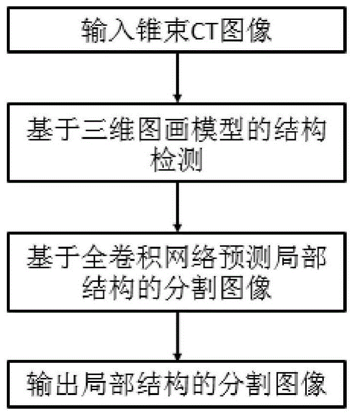
图1是本发明提供的锥束CT图像中三维颅面结构自动解析方法的流程框图。FIG. 1 is a flow chart of a method for automatic analysis of three-dimensional craniofacial structures in cone beam CT images provided by the present invention.
具体实施方式Detailed ways
下面结合附图,通过实施例进一步描述本发明,但不以任何方式限制本发明的范围。Below in conjunction with the accompanying drawings, the present invention is further described by means of embodiments, but the scope of the present invention is not limited in any way.
本发明提供一种锥束CT图像三维颅面结构的自动解析方法,基于图画(pictorial)模型和全卷积神经网络,可以实现从输入的锥束CT图像中,对锥束CT图像中感兴趣的三维颅面结构进行自动分割与标注,获得稳定结构的自动解析与分割。The present invention provides an automatic analysis method for the three-dimensional craniofacial structure of cone beam CT images. Based on a pictorial model and a fully convolutional neural network, it is possible to realize interest in the cone beam CT images from the input cone beam CT images. The 3D craniofacial structure can be automatically segmented and labeled, and the automatic analysis and segmentation of stable structures can be obtained.
本发明自动检测并标注锥束CT图像中的颌骨、颧弓、以及前颅底等结构,其中利用图画模型在三维锥束CT图像上检测感兴趣解剖结构并获取该结构所在的三维图像子块。利用全卷积神经网络对该图像子块进行自动分割与标注。图1是本发明提供的锥束CT图像中三维颅面结构自动解析方法的流程框图,主要包括步骤:训练图画模型用于解剖结构的自动检测;训练全卷积神经网络用于体素的自动标注;在线进行三维颅面结构自动解析。The present invention automatically detects and annotates structures such as jawbone, zygomatic arch, and anterior skull base in the cone beam CT image, wherein the anatomical structure of interest is detected on the three-dimensional cone beam CT image by using a picture model, and the three-dimensional image subsection where the structure is located is obtained. piece. The fully convolutional neural network is used to automatically segment and label the image sub-blocks. 1 is a flowchart of a method for automatic analysis of three-dimensional craniofacial structures in cone beam CT images provided by the present invention, which mainly includes steps: training a picture model for automatic detection of anatomical structures; training a fully convolutional neural network for automatic voxel analysis Annotation; online automatic analysis of 3D craniofacial structures.
以下实施例为实现了本发明提供的锥束CT图像中三维颅面结构自动解析方法的自动解析系统,系统采用包含1000个交互标注的锥束CT图像作为训练图画模型的训练集,该训练集中的每个锥束CT图像具有标注的颌骨、颧弓和前颅底的包围盒。系统中全卷积神经网络的训练数据来自成对的锥束CT图像与预先定义的分割图像。为了包含上下文信息,局部稳定结构的包围盒略大于实际结构所处的区域。在分割图像中稳定结构包括颧弓、前颅底、下颌被标注为前景,其它部分标为背景。全卷积神经网络的训练集中预先交互标注的数据被称为强标注数据。同时为了克服在线分割时结构图像子块可能出现的偏移,对成对的训练数据进行三维空间中位置的扰动,并将这部分数据作为弱标注数据。锥束CT图像中三维颅面结构的自动解析具体实施如下:The following embodiment is an automatic analysis system that realizes the automatic analysis method of the three-dimensional craniofacial structure in the cone beam CT image provided by the present invention. Each cone beam CT image has annotated bounding boxes for the jaw, zygomatic arch, and anterior skull base. The training data for the fully convolutional neural network in the system comes from pairs of cone-beam CT images and pre-defined segmentation images. To contain contextual information, the bounding box of the locally stable structure is slightly larger than the area where the actual structure is located. In the segmented images, stable structures including the zygomatic arch, the anterior skull base, and the mandible are marked as foreground, and other parts are marked as background. The pre-interactively annotated data in the training set of a fully convolutional neural network is called strongly annotated data. At the same time, in order to overcome the possible offset of structural image sub-blocks during online segmentation, the position of the paired training data is perturbed in the three-dimensional space, and this part of the data is used as weakly labeled data. The specific implementation of automatic analysis of 3D craniofacial structures in cone beam CT images is as follows:
步骤1:训练三维图画模型用于解剖结构的自动检测Step 1: Train a 3D picture model for automatic detection of anatomical structures
11)建立三维分层图画模型,该图画模型描述锥束CT图像中所有感兴趣结构之间的空间相互关系,从该图画模型推出结构锚点以及包围盒,用于确定感兴趣结构对应的图像子块;11) Establish a three-dimensional layered picture model, which describes the spatial relationship between all structures of interest in the cone beam CT image, and derives structural anchors and bounding boxes from the picture model to determine the image corresponding to the structure of interest subblock;
利用三维图画模型描述颅面部各结构之间的相对关系。图画模型建立在颅面部各个感兴趣结构上,包括左右颧弓、前颅底、上颌、下颌。图画模型中,感兴趣结构以三元组si=(xi,zi,li)表示,其中xi表示结构锚点即三维结构所在的位置,zi表示对应结构包围盒的大小,li对应该结构的类别。在不同感兴趣结构的包围盒之间可能会有明显的重叠,例如上颌与下颌的包围盒重叠,前颅底与颧弓的包围盒甚至完全在上颌的包围盒内部。为了处理结构之间的重叠关系,本发明采用分层图画模型,其中根节点对应颅骨中心,第二层包含上颌与下颌,第三层包含前颅底与左右颧弓。该分层图中不仅包含层内连接,还包含层间的连接。根据贝叶斯法则定义后验概率为式1:A three-dimensional picture model is used to describe the relative relationship between the structures of the craniofacial region. The pictorial model is built on various craniofacial structures of interest, including the left and right zygomatic arches, the anterior skull base, the upper jaw, and the lower jaw. In the pictorial model, the structure of interest is represented by a triple si = ( xi , zi , li ), where xi represents the location of the structural anchor point, that is, the location of the three-dimensional structure, zi represents the size of the corresponding structure bounding box, l i corresponds to the category of the structure. There may be significant overlap between the bounding boxes of different structures of interest, for example the bounding boxes of the upper and lower jaws overlap, the bounding boxes of the anterior skull base and the zygomatic arch even completely inside the bounding boxes of the upper jaw. In order to deal with the overlapping relationship between the structures, the present invention adopts a layered pictorial model, in which the root node corresponds to the center of the skull, the second layer includes the upper jaw and the lower jaw, and the third layer includes the anterior skull base and the left and right zygomatic arches. This hierarchical graph contains not only intra-layer connections, but also inter-layer connections. The posterior probability is defined according to Bayes' rule as Equation 1:
P(S|V,Ψ)∝P(V|S,Ψ)P(S|Ψ) (式1)P(S|V,Ψ)∝P(V|S,Ψ)P(S|Ψ) (Equation 1)
式1中,S表示感兴趣结构,V表示输入锥束CT图像,Ψ表示图画模型参数。In Equation 1, S represents the structure of interest, V represents the input cone beam CT image, and Ψ represents the picture model parameters.
12)对每类结构分别训练表观分类器(SVM),定义结构si对应的一元势能函数φ(si);12) Train the apparent classifier (SVM) for each type of structure separately, and define the unary potential energy function φ(s i ) corresponding to the structure si ;
其中可将图像子块划分为10×10×10的单元,并将每个单元中的体素灰度直方图化,然后将所有单元的直方图连接作为图像子块的表观特征。通过式2定义结构si对应的一元势能函数φ(si):The image sub-block can be divided into 10×10×10 units, and the voxel gray level in each unit can be histogrammed, and then the histograms of all units can be connected as the apparent feature of the image sub-block. The univariate potential energy function φ(s i ) corresponding to the structure si is defined by formula 2:
φ(si)=(1+exp(aifi(si)+bi))-1 (式2)φ(s i )=(1+exp(a i f i (s i )+b i )) -1 (Equation 2)
其中,φ(si)为结构si对应的一元势能函数,fi是表观分类器的输出,ai与bi是对应第i类结构的预定义参数。Among them, φ(s i ) is the unary potential energy function corresponding to the structure si , f i is the output of the apparent classifier, and a i and b i are the predefined parameters corresponding to the i-th type of structure.
13)定义在分层图画模型上的似然概率,得到最大似然函数lnP(V|S,Ψ),用于对输入的锥束CT图像解剖结构的自动检测;13) Define the likelihood probability on the layered picture model to obtain the maximum likelihood function lnP(V|S,Ψ), which is used for automatic detection of the anatomical structure of the input cone beam CT image;
通过最大化似然函数(式3)获取锥束CT图像中稳定结构所在的图像子块。将在分层模型上的似然概率P(V|S,Ψ)定义为式3:The image sub-block where the stable structure is located in the cone beam CT image is obtained by maximizing the likelihood function (equation 3). The likelihood probability P(V|S,Ψ) on the hierarchical model is defined as Equation 3:

式3中,lnP(V|S,Ψ)为最大似然对数函数;φ(si)为结构si对应的一元势能函数;由
步骤2:训练全卷积神经网络进行感兴趣结构的自动分割与标注;Step 2: Train a fully convolutional neural network for automatic segmentation and labeling of structures of interest;
21)利用深度神经网络构造锥束CT图像与其对应的二值分割图像之间的回归,该深度神经网络是一个全卷积神经网络;21) Construct the regression between cone beam CT images and their corresponding binary segmentation images using a deep neural network, which is a fully convolutional neural network;
经过多层卷积以及池化得到不同分辨率图像的特征图。本发明不同于传统的卷积网络,其引入去卷积操作层与去池化操作层,并将这两种操作层连到由一般的卷积网络所获取的抽象特征图后,构成一个全卷积神经网络。After multi-layer convolution and pooling, feature maps of different resolution images are obtained. The present invention is different from the traditional convolutional network in that it introduces a deconvolution operation layer and a depooling operation layer, and connects these two operation layers to the abstract feature map obtained by the general convolutional network to form a complete Convolutional Neural Networks.
22)在上述全卷积神经网络中获取三维锥束CT图像中稳定结构所在的图像子块的抽象特征图,并从抽象特征图中重建对应结构的二值分割图像,实现从三维锥束CT图像中稳定结构所在的图像子块估计其对应的二值分割图像;22) In the above-mentioned fully convolutional neural network, the abstract feature map of the image sub-block where the stable structure is located in the 3D cone beam CT image is obtained, and the binary segmentation image of the corresponding structure is reconstructed from the abstract feature map, so as to realize the transformation from the 3D cone beam CT image. The image sub-block where the stable structure is located in the image estimates its corresponding binary segmentation image;
多层的去卷积与去池化操作的目的是从卷积神经网络中所获取的抽象特征图中重建对应结构的二值分割图像,实现从三维锥束CT图像中稳定结构所在的局部图像子块估计其对应的二值分割图像,并要求该分割图像与原始的锥束CT图像具有相同的分辨率,实现端到端的结构分割与解析。在卷积网络中,经过多层卷积以及池化得到的不同分辨率图像的特征图,该特征图可看作是原始锥束CT图像的抽象特征,其中低层的特征图具有图像局部细节特征,而高层的特征图通常可以反映形状的全局信息。网络结构的后半部分中的去卷积与去池化操作从锥束CT图像的抽象特征图重建具有原始图像分辨率的二值分割图像。和卷积网络类似,多层去卷积与去池化操作可看作是从低分辨率的分割预测逐步获取高分辨率的分割图像,其中低层包含全局分割的特征图,而高层逐步包含结构分割的细节。The purpose of the multi-layer deconvolution and depooling operations is to reconstruct the binary segmentation image of the corresponding structure from the abstract feature map obtained from the convolutional neural network, and realize the local image of the stable structure from the 3D cone beam CT image. The sub-block estimates its corresponding binary segmented image, and requires the segmented image to have the same resolution as the original cone-beam CT image to achieve end-to-end structural segmentation and analysis. In the convolutional network, the feature maps of images of different resolutions obtained by multi-layer convolution and pooling can be regarded as the abstract features of the original cone beam CT images, and the feature maps of the lower layers have the local details of the image. , and high-level feature maps can usually reflect the global information of the shape. The deconvolution and depooling operations in the second half of the network structure reconstruct binary segmented images with original image resolution from abstract feature maps of cone beam CT images. Similar to the convolutional network, the multi-layer deconvolution and de-pooling operations can be regarded as gradually obtaining high-resolution segmentation images from low-resolution segmentation predictions, where the lower layers contain the feature maps of global segmentation, and the higher layers gradually contain the structure. Segmentation details.
23)采用成对的锥束CT图像与预先定义的分割图像作为训练数据集,训练全卷积神经网络;训练数据集包括强标注数据和弱标注数据;23) Using pairs of cone beam CT images and predefined segmentation images as training data sets to train a fully convolutional neural network; the training data sets include strongly labeled data and weakly labeled data;
系统中全卷积神经网络的训练数据来自成对的锥束CT图像与预先标注、分割的分割图像。为了包含上下文信息,局部稳定结构的包围盒略大于实际结构所处的区域。在分割图像中稳定结构包括颧弓、前颅底、下颌被标注为前景,其它部分标为背景。训练集中预先交互标注的数据被称为强标注数据。同时为了克服在线分割时结构图像子块可能出现的偏移,对成对的训练数据进行三维空间中位置的扰动,并将这部分数据作为弱标注数据。The training data for the fully convolutional neural network in the system comes from pairs of cone beam CT images and pre-annotated and segmented segmented images. To contain contextual information, the bounding box of the locally stable structure is slightly larger than the area where the actual structure is located. In the segmented images, stable structures including the zygomatic arch, the anterior skull base, and the mandible are marked as foreground, and other parts are marked as background. Data pre-labeled interactively in the training set is called strongly labeled data. At the same time, in order to overcome the possible offset of structural image sub-blocks during online segmentation, the position of the paired training data in the three-dimensional space is perturbed, and this part of the data is used as weakly labeled data.
231)在训练过程中,首先使用强标注数据优化网络参数;231) In the training process, first use strong labeling data to optimize network parameters;
网络参数包括:全卷积神经网络中卷积核大小;卷积层、池化层、去卷积层、去池化层的数量、每层特征图的数量;The network parameters include: the size of the convolution kernel in the fully convolutional neural network; the number of convolution layers, pooling layers, deconvolution layers, and de-pooling layers, and the number of feature maps per layer;
232)随后在训练数据中加入弱标注数据训练网络,以使得网络具有处理结构图像块偏移的能力。232) Then add weakly labeled data to the training data to train the network, so that the network has the ability to process the offset of structural image blocks.
训练完成后的网络可用于从输入的锥束CT图像通过抽象与重建得到与输入图像相同大小的分割图像。The trained network can be used to obtain segmented images of the same size as the input images through abstraction and reconstruction from the input cone beam CT images.
233)定义基于该重建图像和预先定义的分割图像之间的均方差的损失函数如式4:233) Define a loss function based on the mean square error between the reconstructed image and the pre-defined segmented image as in Equation 4:

其中,
步骤3:在线的三维颅面结构自动解析过程Step 3: Online automatic analysis process of 3D craniofacial structures
31)基于训练好的图画模型,从输入的锥束CT图像中获取感兴趣的三维局部解剖结构所在的图像子块;将似然概率最高的位置作为解剖结构的锚点;31) Based on the trained picture model, obtain the image sub-block where the three-dimensional local anatomical structure of interest is located from the input cone beam CT image; use the position with the highest likelihood probability as the anchor point of the anatomical structure;
在线自动解析过程利用了贪婪搜索算法,在锥束CT图像中搜索感兴趣结构;首先每个结构的初始位置由图画模型中与根节点的相对位置的高斯分布均值确定。对于所有感兴趣解剖结构估计似然概率,并将似然概率最高的位置作为结构的锚点。The online automatic parsing process utilizes a greedy search algorithm to search for structures of interest in cone beam CT images; first, the initial position of each structure is determined by the mean value of the Gaussian distribution relative to the root node in the picture model. Likelihood probabilities are estimated for all anatomical structures of interest, and the locations with the highest likelihood probabilities are used as anchors for the structures.
32)将每类解剖结构对应的图像子块输入到对应解剖结构的全卷积神经网络中,获得每个解剖结构的自动标注与分割。32) Input the image sub-blocks corresponding to each type of anatomical structure into the fully convolutional neural network corresponding to the anatomical structure to obtain automatic labeling and segmentation of each anatomical structure.
为了验证基于图画模型与全卷积网络的三维颅面结构的自动解析,计算了实施例中各解剖结构的自动标注与手工标注之间的Dice相似度,其中在上颌、前颅底、颧弓、下颌的相似度可以分别达到87%、76%、96%、92%。In order to verify the automatic analysis of the 3D craniofacial structure based on the picture model and the fully convolutional network, the Dice similarity between the automatic annotation and manual annotation of each anatomical structure in the embodiment was calculated. , the similarity of the mandible can reach 87%, 76%, 96%, 92%, respectively.
利用本发明的方法,可由输入的锥束CT图像获得解剖结构自动解析与分割,该方法基于图画模型进行解剖结构的自动检测并确定感兴趣结构所在的图像子块,随后从全卷积网络估计与原始图像相同分辨率的分割图。其中图画模型构造了多类结构之间的相对关系,并利用层内与层间的连接描述不同结构的图像子块之间的相对关系。在全卷积网络中多层卷积与池化所获取的锥束CT图像的特征图被用于重建该结构对应的分割图,其中去卷积与去池化操作从抽象的特征图中重建该结构稠密的分割图。该系统实现了锥束CT图像中颅面结构的自动标注与分割。Using the method of the present invention, the automatic analysis and segmentation of anatomical structures can be obtained from the input cone beam CT images. The method automatically detects the anatomical structures based on the picture model, determines the image sub-block where the structure of interest is located, and then estimates from the fully convolutional network. Segmentation map at the same resolution as the original image. The pictorial model constructs the relative relationship between multiple types of structures, and uses intra-layer and inter-layer connections to describe the relative relationship between image sub-blocks with different structures. The feature maps of cone beam CT images obtained by multi-layer convolution and pooling in a fully convolutional network are used to reconstruct the segmentation map corresponding to the structure, where the deconvolution and depooling operations are reconstructed from the abstract feature maps. The densely structured segmentation map. The system realizes automatic labeling and segmentation of craniofacial structures in cone beam CT images.
需要注意的是,公布实施例的目的在于帮助进一步理解本发明,但是本领域的技术人员可以理解:在不脱离本发明及所附权利要求的精神和范围内,各种替换和修改都是可能的。因此,本发明不应局限于实施例所公开的内容,本发明要求保护的范围以权利要求书界定的范围为准。It should be noted that the purpose of publishing the embodiments is to help further understanding of the present invention, but those skilled in the art can understand that various replacements and modifications are possible without departing from the spirit and scope of the present invention and the appended claims of. Therefore, the present invention should not be limited to the contents disclosed in the embodiments, and the scope of protection of the present invention shall be subject to the scope defined by the claims.
Claims (6)


Priority Applications (1)
| Application Number | Priority Date | Filing Date | Title |
|---|---|---|---|
| CN201611185771.8A CN108205806B (en) | 2016-12-20 | 2016-12-20 | Automatic analysis method for three-dimensional craniofacial structure of cone beam CT image |
Applications Claiming Priority (1)
| Application Number | Priority Date | Filing Date | Title |
|---|---|---|---|
| CN201611185771.8A CN108205806B (en) | 2016-12-20 | 2016-12-20 | Automatic analysis method for three-dimensional craniofacial structure of cone beam CT image |
Publications (2)
| Publication Number | Publication Date |
|---|---|
| CN108205806A CN108205806A (en) | 2018-06-26 |
| CN108205806B true CN108205806B (en) | 2020-10-09 |
Family
ID=62603532
Family Applications (1)
| Application Number | Title | Priority Date | Filing Date |
|---|---|---|---|
| CN201611185771.8A Active CN108205806B (en) | 2016-12-20 | 2016-12-20 | Automatic analysis method for three-dimensional craniofacial structure of cone beam CT image |
Country Status (1)
| Country | Link |
|---|---|
| CN (1) | CN108205806B (en) |
Families Citing this family (14)
| Publication number | Priority date | Publication date | Assignee | Title |
|---|---|---|---|---|
| IL271743B2 (en) | 2017-06-30 | 2024-08-01 | Promaton Holding Bv | Classification and 3D model of dental structures - 3D maxilla using deep learning methods |
| EP3462373A1 (en) | 2017-10-02 | 2019-04-03 | Promaton Holding B.V. | Automated classification and taxonomy of 3d teeth data using deep learning methods |
| EP3503038A1 (en) | 2017-12-22 | 2019-06-26 | Promaton Holding B.V. | Automated 3d root shape prediction using deep learning methods |
| EP3561778A1 (en) | 2018-04-26 | 2019-10-30 | Promaton Holding B.V. | Automated correction of metal affected voxel representations of x-ray data using deep learning techniques |
| EP3591616A1 (en) | 2018-07-03 | 2020-01-08 | Promaton Holding B.V. | Automated determination of a canonical pose of a 3d dental structure and superimposition of 3d dental structures using deep learning |
| EP3620130A1 (en) * | 2018-09-04 | 2020-03-11 | Promaton Holding B.V. | Automated orthodontic treatment planning using deep learning |
| CN109493325B (en) * | 2018-10-23 | 2021-02-26 | 清华大学 | Tumor Heterogeneity Analysis System Based on CT Image |
| EP3671531A1 (en) | 2018-12-17 | 2020-06-24 | Promaton Holding B.V. | Semantic segmentation of non-euclidean 3d data sets using deep learning |
| CN110287965A (en) * | 2019-06-18 | 2019-09-27 | 成都玻尔兹曼智贝科技有限公司 | The method that multilayer neural network is automatically separated root of the tooth and alveolar bone in CBCT image |
| CN112581513B (en) * | 2019-09-29 | 2022-10-21 | 北京大学 | Feature Extraction and Corresponding Method of Cone Beam Computed Tomography Image |
| CN111127488B (en) * | 2019-12-29 | 2022-10-14 | 兰州理工大学 | Method for automatically constructing patient anatomical structure model based on statistical shape model |
| CN111265317B (en) * | 2020-02-10 | 2022-06-17 | 上海牙典医疗器械有限公司 | A method for predicting the orthodontic process |
| CN111599432B (en) * | 2020-05-29 | 2024-04-02 | 上海优医基医疗影像设备有限公司 | Three-dimensional craniofacial image feature point marking analysis system and method |
| CN111968124B (en) * | 2020-10-26 | 2020-12-22 | 四川省肿瘤医院 | Structural segmentation of shoulder musculoskeletal ultrasound based on semi-supervised semantic segmentation |
Citations (6)
| Publication number | Priority date | Publication date | Assignee | Title |
|---|---|---|---|---|
| CN101533466A (en) * | 2009-04-09 | 2009-09-16 | 南京壹进制信息技术有限公司 | Image processing method for positioning eyes |
| CN101639895A (en) * | 2009-08-14 | 2010-02-03 | 浙江工业大学 | Method for extracting and matching features of computer visual image based on Similarity-Pictorial structural model |
| CN101882326A (en) * | 2010-05-18 | 2010-11-10 | 广州市刑事科学技术研究所 | Three-dimensional craniofacial reconstruction method based on overall facial structure shape data of Chinese people |
| CN103860191A (en) * | 2012-12-14 | 2014-06-18 | 奥姆科公司 | Integration of intra-oral imagery and volumetric imagery |
| WO2016036516A1 (en) * | 2014-09-02 | 2016-03-10 | Impac Medical Systems, Inc. | Systems and methods for segmenting medical images based on anatomical landmark-based features |
| CN105761252A (en) * | 2016-02-02 | 2016-07-13 | 北京正齐口腔医疗技术有限公司 | Image segmentation method and device |
Family Cites Families (2)
| Publication number | Priority date | Publication date | Assignee | Title |
|---|---|---|---|---|
| US8761493B2 (en) * | 2011-07-21 | 2014-06-24 | Carestream Health, Inc. | Method and system for tooth segmentation in dental images |
| US9437011B2 (en) * | 2012-06-11 | 2016-09-06 | Samsung Electronics Co., Ltd. | Method and apparatus for estimating a pose of a head for a person |
-
2016
- 2016-12-20 CN CN201611185771.8A patent/CN108205806B/en active Active
Patent Citations (6)
| Publication number | Priority date | Publication date | Assignee | Title |
|---|---|---|---|---|
| CN101533466A (en) * | 2009-04-09 | 2009-09-16 | 南京壹进制信息技术有限公司 | Image processing method for positioning eyes |
| CN101639895A (en) * | 2009-08-14 | 2010-02-03 | 浙江工业大学 | Method for extracting and matching features of computer visual image based on Similarity-Pictorial structural model |
| CN101882326A (en) * | 2010-05-18 | 2010-11-10 | 广州市刑事科学技术研究所 | Three-dimensional craniofacial reconstruction method based on overall facial structure shape data of Chinese people |
| CN103860191A (en) * | 2012-12-14 | 2014-06-18 | 奥姆科公司 | Integration of intra-oral imagery and volumetric imagery |
| WO2016036516A1 (en) * | 2014-09-02 | 2016-03-10 | Impac Medical Systems, Inc. | Systems and methods for segmenting medical images based on anatomical landmark-based features |
| CN105761252A (en) * | 2016-02-02 | 2016-07-13 | 北京正齐口腔医疗技术有限公司 | Image segmentation method and device |
Non-Patent Citations (5)
| Title |
|---|
| 3D exemplar-based random walks for tooth segmentation from cone-beam computed tomography images;Yuru Pei 等;《Med. Phys》;20160919;5040-5050 * |
| Better appearance models for pictorial structures;Marcin Eichner 等;《https://www.research.ed.ac.uk》;20091231;1-11 * |
| Three-Dimensional CT Image Segmentation by Combining 2D Fully Convolutional Network with 3D Majority Voting;Xiangrong Zhou 等;《DLMIA 2016》;20160927;1-10 * |
| 一种基于图结构模型的人体姿态估计算法;韩贵金 等;《计算机工程与应用》;20130427;第49卷(第14期);46-54 * |
| 启发式牙颌CT影像自动分割;黄志伟 等;《中国生物医学工程学报》;20150220;第34卷(第1期);30-33 * |
Also Published As
| Publication number | Publication date |
|---|---|
| CN108205806A (en) | 2018-06-26 |
Similar Documents
| Publication | Publication Date | Title |
|---|---|---|
| CN108205806B (en) | Automatic analysis method for three-dimensional craniofacial structure of cone beam CT image | |
| Zanjani et al. | Mask-MCNet: Tooth instance segmentation in 3D point clouds of intra-oral scans | |
| CN108053417B (en) | A Lung Segmentation Device Based on 3D U-Net Network with Hybrid Coarse Segmentation Features | |
| CN107203998B (en) | A method for segmenting dentition in cone beam CT images | |
| CN113902761A (en) | Unsupervised segmentation of lung disease lesions based on knowledge distillation | |
| CN107403201A (en) | Tumour radiotherapy target area and jeopardize that organ is intelligent, automation delineation method | |
| WO2024021523A1 (en) | Graph network-based method and system for fully automatic segmentation of cerebral cortex surface | |
| CN105957066A (en) | CT image liver segmentation method and system based on automatic context model | |
| CN107730542B (en) | Cone beam computed tomography image correspondence and registration method | |
| CN112150472A (en) | CBCT-based 3D jaw image segmentation method, device and terminal equipment | |
| CN102622750A (en) | Stomach computed tomography (CT) sequence image segmentation method based on interactive region growth | |
| Pei et al. | 3D exemplar‐based random walks for tooth segmentation from cone‐beam computed tomography images | |
| CN106600621A (en) | Space-time cooperation segmentation method based on infant brain tumor multi-modal MRI graph | |
| CN107203988A (en) | A kind of method and its application that three-dimensional volumetric image is rebuild by two dimensional x-ray image | |
| CN114638852A (en) | Jaw bone and soft tissue identification and reconstruction method, device and medium based on CBCT image | |
| Liu et al. | Tracking-based deep learning method for temporomandibular joint segmentation | |
| Oguz et al. | Combining deep learning and multi-atlas label fusion for automated placenta segmentation from 3DUS | |
| Wu et al. | Auto-contouring via automatic anatomy recognition of organs at risk in head and neck cancer on CT images | |
| Sharafeldeen et al. | Accurate segmentation for pathological lung based on integration of 3d appearance and surface models | |
| Arjmand et al. | Artificial intelligence–based modeling can predict face shape based on underlying craniomaxillofacial bone | |
| CN110378910A (en) | Abdominal cavity multiple organ dividing method and device based on map fusion | |
| CN112581513B (en) | Feature Extraction and Corresponding Method of Cone Beam Computed Tomography Image | |
| US20250029260A1 (en) | Natural and artificial intelligence for robust automatic anatomy segmentation | |
| CN106570880B (en) | Brain Tissue MRI Image Segmentation Method Combining Fuzzy Clustering and Markov Random Field | |
| CN111369662A (en) | Method and system for reconstructing 3D model of blood vessels in CT images |
Legal Events
| Date | Code | Title | Description |
|---|---|---|---|
| PB01 | Publication | ||
| PB01 | Publication | ||
| SE01 | Entry into force of request for substantive examination | ||
| SE01 | Entry into force of request for substantive examination | ||
| GR01 | Patent grant | ||
| GR01 | Patent grant |