CN107849112B - Chimeric Antigen Receptors (CAR), compositions and methods of use thereof - Google Patents
Chimeric Antigen Receptors (CAR), compositions and methods of use thereof Download PDFInfo
- Publication number
- CN107849112B CN107849112B CN201680036459.4A CN201680036459A CN107849112B CN 107849112 B CN107849112 B CN 107849112B CN 201680036459 A CN201680036459 A CN 201680036459A CN 107849112 B CN107849112 B CN 107849112B
- Authority
- CN
- China
- Prior art keywords
- cells
- cell
- car
- target
- antigen recognition
- Prior art date
- Legal status (The legal status is an assumption and is not a legal conclusion. Google has not performed a legal analysis and makes no representation as to the accuracy of the status listed.)
- Active
Links
Images


Classifications
-
- C—CHEMISTRY; METALLURGY
- C07—ORGANIC CHEMISTRY
- C07K—PEPTIDES
- C07K14/00—Peptides having more than 20 amino acids; Gastrins; Somatostatins; Melanotropins; Derivatives thereof
- C07K14/435—Peptides having more than 20 amino acids; Gastrins; Somatostatins; Melanotropins; Derivatives thereof from animals; from humans
- C07K14/705—Receptors; Cell surface antigens; Cell surface determinants
- C07K14/70578—NGF-receptor/TNF-receptor superfamily, e.g. CD27, CD30, CD40, CD95
-
- C—CHEMISTRY; METALLURGY
- C12—BIOCHEMISTRY; BEER; SPIRITS; WINE; VINEGAR; MICROBIOLOGY; ENZYMOLOGY; MUTATION OR GENETIC ENGINEERING
- C12N—MICROORGANISMS OR ENZYMES; COMPOSITIONS THEREOF; PROPAGATING, PRESERVING, OR MAINTAINING MICROORGANISMS; MUTATION OR GENETIC ENGINEERING; CULTURE MEDIA
- C12N5/00—Undifferentiated human, animal or plant cells, e.g. cell lines; Tissues; Cultivation or maintenance thereof; Culture media therefor
- C12N5/06—Animal cells or tissues; Human cells or tissues
- C12N5/0602—Vertebrate cells
- C12N5/0634—Cells from the blood or the immune system
- C12N5/0636—T lymphocytes
-
- A—HUMAN NECESSITIES
- A61—MEDICAL OR VETERINARY SCIENCE; HYGIENE
- A61K—PREPARATIONS FOR MEDICAL, DENTAL OR TOILETRY PURPOSES
- A61K40/00—Cellular immunotherapy
- A61K40/10—Cellular immunotherapy characterised by the cell type used
- A61K40/11—T-cells, e.g. tumour infiltrating lymphocytes [TIL] or regulatory T [Treg] cells; Lymphokine-activated killer [LAK] cells
-
- A—HUMAN NECESSITIES
- A61—MEDICAL OR VETERINARY SCIENCE; HYGIENE
- A61K—PREPARATIONS FOR MEDICAL, DENTAL OR TOILETRY PURPOSES
- A61K40/00—Cellular immunotherapy
- A61K40/10—Cellular immunotherapy characterised by the cell type used
- A61K40/15—Natural-killer [NK] cells; Natural-killer T [NKT] cells
-
- A—HUMAN NECESSITIES
- A61—MEDICAL OR VETERINARY SCIENCE; HYGIENE
- A61K—PREPARATIONS FOR MEDICAL, DENTAL OR TOILETRY PURPOSES
- A61K40/00—Cellular immunotherapy
- A61K40/30—Cellular immunotherapy characterised by the recombinant expression of specific molecules in the cells of the immune system
- A61K40/31—Chimeric antigen receptors [CAR]
-
- A—HUMAN NECESSITIES
- A61—MEDICAL OR VETERINARY SCIENCE; HYGIENE
- A61K—PREPARATIONS FOR MEDICAL, DENTAL OR TOILETRY PURPOSES
- A61K40/00—Cellular immunotherapy
- A61K40/40—Cellular immunotherapy characterised by antigens that are targeted or presented by cells of the immune system
- A61K40/41—Vertebrate antigens
- A61K40/42—Cancer antigens
- A61K40/4202—Receptors, cell surface antigens or cell surface determinants
- A61K40/421—Immunoglobulin superfamily
-
- A—HUMAN NECESSITIES
- A61—MEDICAL OR VETERINARY SCIENCE; HYGIENE
- A61K—PREPARATIONS FOR MEDICAL, DENTAL OR TOILETRY PURPOSES
- A61K40/00—Cellular immunotherapy
- A61K40/40—Cellular immunotherapy characterised by antigens that are targeted or presented by cells of the immune system
- A61K40/41—Vertebrate antigens
- A61K40/42—Cancer antigens
- A61K40/4202—Receptors, cell surface antigens or cell surface determinants
- A61K40/4214—Receptors for cytokines
- A61K40/4215—Receptors for tumor necrosis factors [TNF], e.g. lymphotoxin receptor [LTR], CD30
-
- A—HUMAN NECESSITIES
- A61—MEDICAL OR VETERINARY SCIENCE; HYGIENE
- A61K—PREPARATIONS FOR MEDICAL, DENTAL OR TOILETRY PURPOSES
- A61K40/00—Cellular immunotherapy
- A61K40/40—Cellular immunotherapy characterised by antigens that are targeted or presented by cells of the immune system
- A61K40/41—Vertebrate antigens
- A61K40/42—Cancer antigens
- A61K40/4202—Receptors, cell surface antigens or cell surface determinants
- A61K40/4214—Receptors for cytokines
- A61K40/4217—Receptors for interleukins [IL]
-
- A—HUMAN NECESSITIES
- A61—MEDICAL OR VETERINARY SCIENCE; HYGIENE
- A61K—PREPARATIONS FOR MEDICAL, DENTAL OR TOILETRY PURPOSES
- A61K40/00—Cellular immunotherapy
- A61K40/40—Cellular immunotherapy characterised by antigens that are targeted or presented by cells of the immune system
- A61K40/41—Vertebrate antigens
- A61K40/42—Cancer antigens
- A61K40/4202—Receptors, cell surface antigens or cell surface determinants
- A61K40/4224—Molecules with a "CD" designation not provided for elsewhere
-
- A—HUMAN NECESSITIES
- A61—MEDICAL OR VETERINARY SCIENCE; HYGIENE
- A61P—SPECIFIC THERAPEUTIC ACTIVITY OF CHEMICAL COMPOUNDS OR MEDICINAL PREPARATIONS
- A61P35/00—Antineoplastic agents
-
- A—HUMAN NECESSITIES
- A61—MEDICAL OR VETERINARY SCIENCE; HYGIENE
- A61P—SPECIFIC THERAPEUTIC ACTIVITY OF CHEMICAL COMPOUNDS OR MEDICINAL PREPARATIONS
- A61P35/00—Antineoplastic agents
- A61P35/02—Antineoplastic agents specific for leukemia
-
- A—HUMAN NECESSITIES
- A61—MEDICAL OR VETERINARY SCIENCE; HYGIENE
- A61P—SPECIFIC THERAPEUTIC ACTIVITY OF CHEMICAL COMPOUNDS OR MEDICINAL PREPARATIONS
- A61P43/00—Drugs for specific purposes, not provided for in groups A61P1/00-A61P41/00
-
- C—CHEMISTRY; METALLURGY
- C07—ORGANIC CHEMISTRY
- C07K—PEPTIDES
- C07K14/00—Peptides having more than 20 amino acids; Gastrins; Somatostatins; Melanotropins; Derivatives thereof
- C07K14/435—Peptides having more than 20 amino acids; Gastrins; Somatostatins; Melanotropins; Derivatives thereof from animals; from humans
- C07K14/52—Cytokines; Lymphokines; Interferons
- C07K14/54—Interleukins [IL]
- C07K14/5443—IL-15
-
- C—CHEMISTRY; METALLURGY
- C07—ORGANIC CHEMISTRY
- C07K—PEPTIDES
- C07K14/00—Peptides having more than 20 amino acids; Gastrins; Somatostatins; Melanotropins; Derivatives thereof
- C07K14/435—Peptides having more than 20 amino acids; Gastrins; Somatostatins; Melanotropins; Derivatives thereof from animals; from humans
- C07K14/705—Receptors; Cell surface antigens; Cell surface determinants
- C07K14/70503—Immunoglobulin superfamily
- C07K14/7051—T-cell receptor (TcR)-CD3 complex
-
- C—CHEMISTRY; METALLURGY
- C07—ORGANIC CHEMISTRY
- C07K—PEPTIDES
- C07K14/00—Peptides having more than 20 amino acids; Gastrins; Somatostatins; Melanotropins; Derivatives thereof
- C07K14/435—Peptides having more than 20 amino acids; Gastrins; Somatostatins; Melanotropins; Derivatives thereof from animals; from humans
- C07K14/705—Receptors; Cell surface antigens; Cell surface determinants
- C07K14/70503—Immunoglobulin superfamily
- C07K14/70514—CD4
-
- C—CHEMISTRY; METALLURGY
- C07—ORGANIC CHEMISTRY
- C07K—PEPTIDES
- C07K14/00—Peptides having more than 20 amino acids; Gastrins; Somatostatins; Melanotropins; Derivatives thereof
- C07K14/435—Peptides having more than 20 amino acids; Gastrins; Somatostatins; Melanotropins; Derivatives thereof from animals; from humans
- C07K14/705—Receptors; Cell surface antigens; Cell surface determinants
- C07K14/70503—Immunoglobulin superfamily
- C07K14/70517—CD8
-
- C—CHEMISTRY; METALLURGY
- C07—ORGANIC CHEMISTRY
- C07K—PEPTIDES
- C07K14/00—Peptides having more than 20 amino acids; Gastrins; Somatostatins; Melanotropins; Derivatives thereof
- C07K14/435—Peptides having more than 20 amino acids; Gastrins; Somatostatins; Melanotropins; Derivatives thereof from animals; from humans
- C07K14/705—Receptors; Cell surface antigens; Cell surface determinants
- C07K14/70503—Immunoglobulin superfamily
- C07K14/70521—CD28, CD152
-
- C—CHEMISTRY; METALLURGY
- C07—ORGANIC CHEMISTRY
- C07K—PEPTIDES
- C07K16/00—Immunoglobulins [IGs], e.g. monoclonal or polyclonal antibodies
- C07K16/18—Immunoglobulins [IGs], e.g. monoclonal or polyclonal antibodies against material from animals or humans
- C07K16/28—Immunoglobulins [IGs], e.g. monoclonal or polyclonal antibodies against material from animals or humans against receptors, cell surface antigens or cell surface determinants
- C07K16/2803—Immunoglobulins [IGs], e.g. monoclonal or polyclonal antibodies against material from animals or humans against receptors, cell surface antigens or cell surface determinants against the immunoglobulin superfamily
-
- C—CHEMISTRY; METALLURGY
- C07—ORGANIC CHEMISTRY
- C07K—PEPTIDES
- C07K16/00—Immunoglobulins [IGs], e.g. monoclonal or polyclonal antibodies
- C07K16/18—Immunoglobulins [IGs], e.g. monoclonal or polyclonal antibodies against material from animals or humans
- C07K16/28—Immunoglobulins [IGs], e.g. monoclonal or polyclonal antibodies against material from animals or humans against receptors, cell surface antigens or cell surface determinants
- C07K16/2866—Immunoglobulins [IGs], e.g. monoclonal or polyclonal antibodies against material from animals or humans against receptors, cell surface antigens or cell surface determinants against receptors for cytokines, lymphokines, interferons
-
- C—CHEMISTRY; METALLURGY
- C07—ORGANIC CHEMISTRY
- C07K—PEPTIDES
- C07K16/00—Immunoglobulins [IGs], e.g. monoclonal or polyclonal antibodies
- C07K16/18—Immunoglobulins [IGs], e.g. monoclonal or polyclonal antibodies against material from animals or humans
- C07K16/28—Immunoglobulins [IGs], e.g. monoclonal or polyclonal antibodies against material from animals or humans against receptors, cell surface antigens or cell surface determinants
- C07K16/2878—Immunoglobulins [IGs], e.g. monoclonal or polyclonal antibodies against material from animals or humans against receptors, cell surface antigens or cell surface determinants against the NGF-receptor/TNF-receptor superfamily, e.g. CD27, CD30, CD40, CD95
-
- C—CHEMISTRY; METALLURGY
- C07—ORGANIC CHEMISTRY
- C07K—PEPTIDES
- C07K16/00—Immunoglobulins [IGs], e.g. monoclonal or polyclonal antibodies
- C07K16/18—Immunoglobulins [IGs], e.g. monoclonal or polyclonal antibodies against material from animals or humans
- C07K16/28—Immunoglobulins [IGs], e.g. monoclonal or polyclonal antibodies against material from animals or humans against receptors, cell surface antigens or cell surface determinants
- C07K16/2887—Immunoglobulins [IGs], e.g. monoclonal or polyclonal antibodies against material from animals or humans against receptors, cell surface antigens or cell surface determinants against CD20
-
- C—CHEMISTRY; METALLURGY
- C07—ORGANIC CHEMISTRY
- C07K—PEPTIDES
- C07K16/00—Immunoglobulins [IGs], e.g. monoclonal or polyclonal antibodies
- C07K16/18—Immunoglobulins [IGs], e.g. monoclonal or polyclonal antibodies against material from animals or humans
- C07K16/28—Immunoglobulins [IGs], e.g. monoclonal or polyclonal antibodies against material from animals or humans against receptors, cell surface antigens or cell surface determinants
- C07K16/289—Immunoglobulins [IGs], e.g. monoclonal or polyclonal antibodies against material from animals or humans against receptors, cell surface antigens or cell surface determinants against CD45
-
- C—CHEMISTRY; METALLURGY
- C07—ORGANIC CHEMISTRY
- C07K—PEPTIDES
- C07K16/00—Immunoglobulins [IGs], e.g. monoclonal or polyclonal antibodies
- C07K16/18—Immunoglobulins [IGs], e.g. monoclonal or polyclonal antibodies against material from animals or humans
- C07K16/28—Immunoglobulins [IGs], e.g. monoclonal or polyclonal antibodies against material from animals or humans against receptors, cell surface antigens or cell surface determinants
- C07K16/2896—Immunoglobulins [IGs], e.g. monoclonal or polyclonal antibodies against material from animals or humans against receptors, cell surface antigens or cell surface determinants against molecules with a "CD"-designation, not provided for elsewhere
-
- C—CHEMISTRY; METALLURGY
- C12—BIOCHEMISTRY; BEER; SPIRITS; WINE; VINEGAR; MICROBIOLOGY; ENZYMOLOGY; MUTATION OR GENETIC ENGINEERING
- C12N—MICROORGANISMS OR ENZYMES; COMPOSITIONS THEREOF; PROPAGATING, PRESERVING, OR MAINTAINING MICROORGANISMS; MUTATION OR GENETIC ENGINEERING; CULTURE MEDIA
- C12N5/00—Undifferentiated human, animal or plant cells, e.g. cell lines; Tissues; Cultivation or maintenance thereof; Culture media therefor
- C12N5/06—Animal cells or tissues; Human cells or tissues
- C12N5/0602—Vertebrate cells
- C12N5/0634—Cells from the blood or the immune system
- C12N5/0646—Natural killers cells [NK], NKT cells
-
- A—HUMAN NECESSITIES
- A61—MEDICAL OR VETERINARY SCIENCE; HYGIENE
- A61K—PREPARATIONS FOR MEDICAL, DENTAL OR TOILETRY PURPOSES
- A61K2121/00—Preparations for use in therapy
-
- A—HUMAN NECESSITIES
- A61—MEDICAL OR VETERINARY SCIENCE; HYGIENE
- A61K—PREPARATIONS FOR MEDICAL, DENTAL OR TOILETRY PURPOSES
- A61K2239/00—Indexing codes associated with cellular immunotherapy of group A61K40/00
- A61K2239/31—Indexing codes associated with cellular immunotherapy of group A61K40/00 characterized by the route of administration
-
- A—HUMAN NECESSITIES
- A61—MEDICAL OR VETERINARY SCIENCE; HYGIENE
- A61K—PREPARATIONS FOR MEDICAL, DENTAL OR TOILETRY PURPOSES
- A61K2239/00—Indexing codes associated with cellular immunotherapy of group A61K40/00
- A61K2239/38—Indexing codes associated with cellular immunotherapy of group A61K40/00 characterised by the dose, timing or administration schedule
-
- A—HUMAN NECESSITIES
- A61—MEDICAL OR VETERINARY SCIENCE; HYGIENE
- A61K—PREPARATIONS FOR MEDICAL, DENTAL OR TOILETRY PURPOSES
- A61K2239/00—Indexing codes associated with cellular immunotherapy of group A61K40/00
- A61K2239/46—Indexing codes associated with cellular immunotherapy of group A61K40/00 characterised by the cancer treated
- A61K2239/48—Blood cells, e.g. leukemia or lymphoma
-
- A—HUMAN NECESSITIES
- A61—MEDICAL OR VETERINARY SCIENCE; HYGIENE
- A61K—PREPARATIONS FOR MEDICAL, DENTAL OR TOILETRY PURPOSES
- A61K2300/00—Mixtures or combinations of active ingredients, wherein at least one active ingredient is fully defined in groups A61K31/00 - A61K41/00
-
- C—CHEMISTRY; METALLURGY
- C07—ORGANIC CHEMISTRY
- C07K—PEPTIDES
- C07K2319/00—Fusion polypeptide
-
- C—CHEMISTRY; METALLURGY
- C07—ORGANIC CHEMISTRY
- C07K—PEPTIDES
- C07K2319/00—Fusion polypeptide
- C07K2319/01—Fusion polypeptide containing a localisation/targetting motif
- C07K2319/02—Fusion polypeptide containing a localisation/targetting motif containing a signal sequence
-
- C—CHEMISTRY; METALLURGY
- C07—ORGANIC CHEMISTRY
- C07K—PEPTIDES
- C07K2319/00—Fusion polypeptide
- C07K2319/01—Fusion polypeptide containing a localisation/targetting motif
- C07K2319/03—Fusion polypeptide containing a localisation/targetting motif containing a transmembrane segment
-
- C—CHEMISTRY; METALLURGY
- C07—ORGANIC CHEMISTRY
- C07K—PEPTIDES
- C07K2319/00—Fusion polypeptide
- C07K2319/50—Fusion polypeptide containing protease site
-
- C—CHEMISTRY; METALLURGY
- C07—ORGANIC CHEMISTRY
- C07K—PEPTIDES
- C07K2319/00—Fusion polypeptide
- C07K2319/95—Fusion polypeptide containing a motif/fusion for degradation (ubiquitin fusions, PEST sequence)
-
- C—CHEMISTRY; METALLURGY
- C12—BIOCHEMISTRY; BEER; SPIRITS; WINE; VINEGAR; MICROBIOLOGY; ENZYMOLOGY; MUTATION OR GENETIC ENGINEERING
- C12N—MICROORGANISMS OR ENZYMES; COMPOSITIONS THEREOF; PROPAGATING, PRESERVING, OR MAINTAINING MICROORGANISMS; MUTATION OR GENETIC ENGINEERING; CULTURE MEDIA
- C12N2510/00—Genetically modified cells
Landscapes
- Health & Medical Sciences (AREA)
- Life Sciences & Earth Sciences (AREA)
- Chemical & Material Sciences (AREA)
- Organic Chemistry (AREA)
- Immunology (AREA)
- General Health & Medical Sciences (AREA)
- Genetics & Genomics (AREA)
- Biochemistry (AREA)
- Medicinal Chemistry (AREA)
- Engineering & Computer Science (AREA)
- Zoology (AREA)
- Proteomics, Peptides & Aminoacids (AREA)
- Biophysics (AREA)
- Molecular Biology (AREA)
- Animal Behavior & Ethology (AREA)
- Public Health (AREA)
- Veterinary Medicine (AREA)
- Biomedical Technology (AREA)
- Cell Biology (AREA)
- Epidemiology (AREA)
- Bioinformatics & Cheminformatics (AREA)
- Gastroenterology & Hepatology (AREA)
- Toxicology (AREA)
- Wood Science & Technology (AREA)
- Biotechnology (AREA)
- Hematology (AREA)
- General Engineering & Computer Science (AREA)
- Microbiology (AREA)
- Chemical Kinetics & Catalysis (AREA)
- General Chemical & Material Sciences (AREA)
- Nuclear Medicine, Radiotherapy & Molecular Imaging (AREA)
- Pharmacology & Pharmacy (AREA)
- Oncology (AREA)
- Micro-Organisms Or Cultivation Processes Thereof (AREA)
- Peptides Or Proteins (AREA)
- Medicines Containing Material From Animals Or Micro-Organisms (AREA)
- Measuring Or Testing Involving Enzymes Or Micro-Organisms (AREA)
- Medicines Containing Antibodies Or Antigens For Use As Internal Diagnostic Agents (AREA)
- Medicines That Contain Protein Lipid Enzymes And Other Medicines (AREA)
Abstract
本发明是关于与嵌合抗原受体(CAR)多肽相关之组合物及方法,以及与其有关之方法。在一个实施例中,本发明是关于经工程改造之细胞,其具有针对至少两个标靶之嵌合抗原受体多肽。在另一实施例中,本发明是关于经工程改造之细胞,其具有嵌合抗原受体多肽及强化子部分。

The present invention relates to compositions and methods related to chimeric antigen receptor (CAR) polypeptides, and methods related thereto. In one embodiment, the invention relates to engineered cells having chimeric antigen receptor polypeptides directed against at least two targets. In another embodiment, the invention relates to engineered cells having a chimeric antigen receptor polypeptide and an enhancer moiety.
Description
Cross reference to related applications
This application is an international PCT application claiming us provisional application No. 62/184,321 filed on 25/6/2015; 62/235,840, filed on 10/1/2015; and 62/244,435, filed on 21/10/2015, which are incorporated herein by reference in their entirety.
Background
T cells, a type of lymphocyte, play an important role in cell-mediated immunity. It differs from other lymphocytes, such as B cells and Natural Killer (NK) cells, in the presence of a T Cell Receptor (TCR) on the cell surface. T helper cells, also known as CD4+ T or CD 4T cells, express the CD4 glycoprotein on their surface. Helper T cells are activated when exposed to peptide antigens presented by MHC (major histocompatibility complex) class II molecules. Once activated, these cells rapidly proliferate and secrete cytokines that regulate the immune response. Cytotoxic T cells, also known as CD8+ T cells or CD8T cells, express the CD8 glycoprotein on the cell surface. CD8+ T cells are activated when exposed to peptide antigens presented by MHC class I molecules. Memory T cells, a subset of T cells, persist long and respond to their cognate antigen, thus providing the immune system with "memory" against past infections and/or tumor cells.
After genetic engineering, T cells can produce specific receptors on their surface, called Chimeric Antigen Receptors (CARs). CARs are proteins that allow T cells to recognize specific proteins (antigens) on tumor cells. These engineered CAR T cells are then grown in the laboratory until their number reaches billions. The expanded CAR T cell population is then infused into the patient.
To date, clinical trials have demonstrated that Chimeric Antigen Receptor (CAR) T cells hold great promise in hematologic malignancies that are resistant to standard chemotherapy. Most notably, certain CD19CAR (CD19CAR) T cell therapies have significant effects, including long-term remission of B cell malignancies (Kochenderfer, Wilson et al 2010, Kalos, Levine et al 2011, Porter, Levine et al 2011, Davila, Riviere et al 2013, Grupp, Frey et al 2013, Grupp, Kalos et al 2013, Kalos, Nazimuddin et al 2013, Kochenderfer, Dudley et al 2013, Kochenderfer, riey et al 2013, Lee, Shah et al 2013, Park, Riviere et al 2013, Maude, Frey et al 2014).
Despite the success of CAR therapy in B cell leukemias and lymphomas, there is no clear application of CAR therapy to T cell malignancies. Given that therapy of T-cell malignancies is significantly less effective than therapy of B-cell malignancies (Abramson, Feldman et al 2014), CAR therapy has the potential to further address the enormous clinical need.
To date, current efforts have focused on CAR T cells that show efficacy in treating a variety of B cell malignancies. Although an initial remission rate of about 90% is common in B-ALL using CD19CAR, most relapse within one year. At least part of the cause of relapse is due to antigen escape. Thus, there is an ongoing urgent need for more effective CAR T cell therapy to prevent relapse. Target exploration and selection is an initial step because there is no general rule that can ensure or guide efficient CAR design.
There are several obstacles that currently prevent the broader use of CAR therapeutic approaches. The most common of these challenges are: (1) selection of antigen targets and chimeric antigen receptors; (2) CAR design; (3) tumor heterogeneity, in particular differences in surface expression of tumor antigens. Targeting a single antigen carries the risk of immunological escape, which can be addressed by targeting multiple desired antigens.
Most CAR chimeric antigen receptors are scFv derived from monoclonal antibodies and some of these monoclonal antibodies have been used in clinical trials or disease treatment. However, monoclonal antibodies have limited efficacy, suggesting a need for alternative and more potent targeting approaches, such as CARs. scFvs are the most commonly used chimeric antigen receptor for CARs. However, CAR affinity binding and location of epitopes recognized on the antigen can affect function. In addition, the amount of CAR expression on the surface of T cells or NK cells is influenced by the appropriate leader sequence and promoter. In addition, over-expressed CAR proteins can be toxic to cells.
Thus, there remains a need for improved chimeric antigen receptor-based therapies that can more effectively, safely and effectively target and T cell-related malignancies.
Disclosure of Invention
In one embodiment, the invention provides an engineered cell having a first chimeric antigen receptor polypeptide comprising a first antigen recognition domain, a first signal peptide, a first hinge region, a first transmembrane domain, a first co-stimulatory domain, and a first signaling domain; and a second chimeric antigen receptor polypeptide comprising a second antigen recognition domain, a second signal peptide, a second hinge region, a second transmembrane domain, a second costimulatory domain, and a second signaling domain; wherein the first antigen recognition domain is different from the second antigen recognition domain.
In another embodiment, the invention provides an engineered polypeptide comprising a chimeric antigen receptor and an enhancer.
In another embodiment, the invention provides an engineered polypeptide comprising a chimeric antigen receptor polypeptide and an enhancer.
In another embodiment, the present invention provides an engineered chimeric antigen receptor polypeptide comprising: a signal peptide, a CD45 antigen recognition domain, a hinge region, a transmembrane domain, at least one co-stimulatory domain, and a signaling domain. In another embodiment, the invention provides polynucleotides encoding the aforementioned polypeptides.
In another embodiment, the invention provides an engineered cell having an engineered polypeptide or polynucleotide as described above.
In another embodiment, the invention provides a method of reducing the number of target cells comprising the steps of (i.) contacting the target cells with an effective amount of an engineered cell having at least one chimeric antigen receptor polypeptide, the engineered cell having a plurality of chimeric antigen receptor polypeptides, each chimeric antigen receptor polypeptide being independent; and (ii) optionally analyzing the reduction in the number of the cells. The target cell comprises at least one cell surface antigen selected from the group consisting of: interleukin 6 receptor, NY-ESO-1, alpha-fetoprotein (AFP), glypican-3 (GPC3), BAFF-R, BCMA, TACI, LeY, CD5, CD13, CD14, CD15CD19, CD20, CD22, CD33, CD41, CD45, CD61, CD64, CD68, CD117, CD123, CD138, CD267, CD269, CD38, Flt3 receptor, and CS 1.
In another embodiment, the invention provides a method of treating B-cell lymphoma, T-cell lymphoma, multiple myeloma, chronic myelogenous leukemia, B-cell acute lymphocytic leukemia (B-ALL), and cell proliferative disorders by administering any one of the above engineered cells to a patient in need thereof.
Brief description of the drawings
FIG. 1 is a schematic diagram of a cCAR construct (hereinafter, "CARs or compound CARs"). Multiple or complexed CARs target multiple antigens (e.g., cell type 1 or cell type 2 or the same cell type). Diverse or cCAR T cell immunotherapies comprise individual component CARs comprising different or identical antigen recognition domains, hinge regions, transmembrane domains, multiple co-stimulatory domains, and intracellular signaling domains.
FIG. 2A schematic representation of the cCAR-T construct. The constructs comprise the SFFV promoter that drives expression of multiple modular units of CAR linked by the P2A peptide. Upon linker cleavage, the cCAR cleaves and joins when the target expresses CD33 and/or CD 123. As novel cCAR constructs, the activation domain of the constructs can include, but is not limited to, the 4-1BB on the CD33CAR segment and the CD28 region on the CD123 CAR.
Figure 2b western blot depicting expression of transduced CD33CD123 cCAR-T cells. The figure depicts the expression of two different CAR proteins, namely CD33CAR and CD123 CAR. The cCAR-T cells expressing CD33 and CD123 CARs produced two distinct and consistently strong protein bands upon linker cleavage. A Green Fluorescent Protein (GFP) was included as a negative control.
FIG. 2C flow cytometry for expression of transduction efficiency. The upper panel shows lentiviral titers of CD33CD123cCAR (also known as CD33CD123-2G-CAR) tested on 293FT HEK (human embryonic kidney) cells to test for maximum transduction efficiency prior to use on UCB (cord blood) and PB (peripheral blood) T cells. The lower panel shows CD33CD123cCAR (also referred to as CD33CD123-2G-CAR) T cells transduced with a lentiviral vector comprising a CD33CD123cCAR construct and GFP-transduced cells as controls. The percentage indicated by the yellow ring is a surrogate for transduction efficiency.
FIG. 3 shows a schematic diagram of a method for generating a high efficiency composite CAR (cCAR).
FIG. 4 Co-culture analysis of CD33CD123-2G CAR-T cells (cCAR) incubated with the promyelocytic leukemia cell line HL 60. The cCAR-T cells (lower panel) were compared to control GFP-transduced T cells (upper panel). Lethality was measured by the CD33+ cell population remaining after about 24 hours of incubation (surrounded in yellow rings).
FIG. 5 Co-culture analysis of cCAR-T cells incubated with myeloid leukemia cell line KG-1a, expressing about 100% CD33 and about 50-80% CD 123. The cCAR-T cells (lower panel) were compared to control GFP-transduced T cells (upper panel). Lethality was measured by the CD33+ cell population remaining after approximately 24 hours of incubation.
FIG. 6 Co-culture analysis of cCAR-T cells incubated with AML patient samples (referred to herein as AML-9). Patient cells include mixed cell populations such as leukemia cells, monocytes and other types of primitive cells. CD33 serves as a marker of CAR-T action as well as a marker for CD34, a specific marker for leukemia cells. CAR-T plots (right) were compared to control GFP-transduced T cells (middle). Lethality was measured by the CD33+/CD34+ cell population remaining after at least 24 hours of incubation.
FIG. 7 Co-culture assay of cCAR-T cells incubated with B-ALL patient samples (referred to herein as Sp-BM-B6). Patient cells include mixed cell populations such as leukemia cells, monocytes and other types of primitive cells. CD34 serves as a specific marker for leukemia cells. CAR-T plots (right) were compared to control GFP-transduced T cells (middle). Lethality was measured by the CD34+ cell population remaining after at least 24 hours of incubation.
FIG. 8 CD33CD123cCAR expression in NK-92 cells. Detection of CD33CD123cCAR expression using goat anti-mouse antibody f (ab) 2.
FIG. 9 Co-culture analysis of cCAR NK-92 cells incubated with HL-60. The cCAR NK-92 cells were compared to GFP transduced NK-92 cells. Lethality was measured by the CD33+ cell population remaining after approximately 24 hours of incubation.
FIG. 10 Co-culture analysis of cCAR NK-92 cells incubated with KG1 a. Compare the cCAR NK cell map to GFP-transduced NK-92 cells. Lethality was measured by the CD33+ cell population remaining after approximately 24 hours of incubation.
FIG. 11 dose response of CD33CD123cCAR (CAR-CD33/123) NK-92 cells with HL-60 or KG1 a. Lethality was measured by the CD33+ cell population remaining after approximately 24 hours of incubation.
FIG. 12 comparison of CD33CD123cCAR NK-92 cell killing in two KG11 cell populations with controls. The analysis was performed at different ratios of CAR-CD33/123(CD33 CD123cCAR NK-92 cells) and target cell kG1 a. Lethality was measured by the CD33+ CD123+ or CD33+ CD 123-cell population remaining after about 24 hours of incubation.
FIG. 13 is a schematic representation of cCAR. The construct comprises an SFFV promoter that drives expression of multiple modular units of CAR linked by a linker. When the linker is cleaved, the cCAR cleaves and engages when the target expresses a combination of multiple target antigens (CD19 and/or CD20, and/or CD22 and/or 138). Multiple ccars use the same or different co-stimulatory domains, such as, but not limited to, 4-1BB (also labeled 4-BB) and/or CD 28.
bcma-CS1cCAR construct combinations (BC1 cCAR). (A) The construct consists of the SFFV promoter driving expression of the two modular units of CAR linked by the P2A peptide. When this P2A peptide is cleaved, the cCAR cleaves and engages when the target expresses BCMA and/or CS 1. The two unit CARs use the same co-stimulatory domain 4-1 BB. (B) Flow cytometry analysis of BC1cCAR expression on the surface of T cells with vector (left) and exhibiting BC1cCAR 15.3% positive for f (ab)2 (right, highlighted by squares). Gating was performed against isotype controls. (C) Basic functional validation of BC1cCAR-T cells was performed by co-culturing BCMA cDNA (BCMA-K562) -transduced K562 cells (obtained from Kochenderfer, NIH). The histogram shows lysis of a BCMA-K562 cell line versus a control T cell and wild-type K562(wt-K562) versus a control.
FIG. 14D BCMACS1-2G constructs using two different co-stimulatory domains (4-1BB or CD28) for each cell. The constructs include the SFFV promoter driving expression of the two modular units of CAR linked by the P2A peptide. When this P2A peptide is cleaved, the cCAR cleaves and binds to BCMA and/or CS1 expressing targets. The two units CAR use different co-stimulatory domains, namely 4-1BB or CD 28. Flow cytometry analysis of BC1cCAR expression on the surface of T cells with respect to vector (left) and BC1cCAR (right, highlighted by squares) displaying rare positive cells to f (ab) 2. Gating was performed against isotype controls.
FIG. 14E protein expression of BC1cCAR and BCMA-CS1-2G in HEK-293FT cells. HEK-293FT cells were transfected with lentivirus plasmids of GFP (lane 1), BC1cCAR (lane 2), CD269-CS1-2G (lane 3) for 48 hours, the supernatant was removed after transfection, and the cells were also removed. Cells were lysed with mouse anti-human CD3z antibody for western blotting and investigation.
FIG. 15A-B. MM1S cell line coculture. Co-culture was performed at 24 hours followed by collection and analysis via flow cytometry. Target MM1S cells (myeloma cells) were labeled with cytotracker (cmtmr) dye to distinguish them from effector T cells. The population was gated by anti-BCMA (CD269) and anti-CS 1(CD319) antibodies. FIG. 15A: flow cytometry mapping of co-cultures. FIG. 15B: right side: summary of dissolution versus E: T ratio.
FIG. 16A-B.RPMI-8226 cell line coculture. Co-culture was performed at 24 hours followed by collection and analysis via flow cytometry. Target RPMI-8226 cells were labeled with cytotracker (cmtmr) dye to distinguish them from effector T cells. The population was gated by anti-BCMA (CD269) and anti-CS 1(CD319) antibodies. FIG. 16A: flow cytometry mapping of co-cultures. FIG. 16B: summary of dissolution versus E: T ratio.
Co-culture of the u266 cell line fig. 17A-b. Co-culture and collection were performed at 24 hours and analyzed via flow cytometry. Target U266 cells were labeled with a cytotracker (cmtmr) dye to distinguish them from effector T cells. The population was gated by anti-BCMA (CD269) and anti-CS 1(CD319) antibodies. Left side: flow cytometry plots of co-cultures, right: summary of dissolution versus E: T ratio.
Figure 18A-b. mm10-G primary patient samples co-culture and specific lysis rates. Co-culture was performed at 24 hours and collected and analyzed via flow cytometry. Target MM10-G cells were labeled with cytotracker (cmtmr) dye to distinguish them from effector T cells. The population was gated by anti-BCMA (CD269) and anti-CS 1(CD319) antibodies. Notably, gating shows MM10-G presenting different BCMA + and CS1+ populations. FIG. 18A: flow cytometry mapping of co-cultures. FIG. 18B: summary of dissolution versus E: T ratio.
Figure 19A-b. mm7-G primary patient samples co-culture and specific lysis rates. Co-culture was performed at 24 hours and collected and analyzed via flow cytometry. Target MM7-G cells were labeled with cytotracker (cmtmr) dye to distinguish them from effector T cells. The population was gated by anti-BCMA (CD269) and anti-CS 1(CD319) antibodies. FIG. 19A: flow cytometry mapping of co-cultures. FIG. 19B: summary of dissolution versus E: T ratio.
Figure 20A-b. mm11-G primary patient samples co-culture and specific lysis rates. Co-culture was performed at 24 hours and collected and analyzed via flow cytometry. Target MM11-G cells were labeled with cytotracker (cmtmr) dye to distinguish them from effector T cells. The population was gated by anti-BCMA (CD269) and anti-CS 1(CD319) antibodies. FIG. 20A: flow cytometry mapping of co-cultures. FIG. 20B: summary of dissolution versus E: T ratio.
FIG. 21 CD269CS1-BBCAR NK cells exhibit in vivo anti-leukemic effects. NSG mice were irradiated with a sublethal dose and on the following day mm.1s multiple myeloma cells expressing luciferase were injected intravenously to induce measurable tumor formation. After 3 days, mice were injected intravenously with 8 × 10⁶ CD269-CS1-BBCAR NK cells or vector-controlled NK control cells. On days 3, 6 and 8, mice were injected subcutaneously with redifect D-luciferin and subjected to IVIS imaging. The mean light intensity of CD269-CS1-BBCAR NK injected mice was compared to vehicle control NK injected mice.
Figure 22. percent survival of mice was measured and compared between the two groups based on the study from figure 21.
Figure 23 CRISPR/Cas9 interference system. Expression of sgRNA and Cas9 puromycin was driven by U6 and SFFV promoters, respectively. Cas9 is linked to the puromycin resistance gene by an E2A self-cleaving sequence.
Figure 24 provides a schematic diagram of an example of steps for generating CAR T or NK cells targeted to hematological malignancies.
Figure 25 expression using CRISPR/Cas9 lentiviral system stabilizes the production and sorting of CD45 blocker NK-92 cells. Flow cytometry analysis indicated the amount of CD45 expression on the surface of NK-92 cells (left panel). After CRISPR transduction of sgCD45B into NK-92 cells, the transduced cells were cultured in puromycin-containing medium for several weeks. CD45 negative NK-92 cells were assayed using CD45 antibody and sorted. The purity of stable NK45i-92 (expressing CD45 blocking gene) NK-92 cells was determined by flow cytometry analysis (right panel). This data confirmed the successful generation and acquisition of NK45i-92 cells.
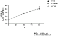
FIG. 26 cell growth curves of wild type, GFP transduced NK-92 or NK45i-92NK cells. To assess the cell proliferation effect in NK-92 cells caused by the CD45 blocking gene expression (KD), the number of NK-92(●), GFP-transduced-92 (■) and NK45i-92 (. tangle-solidup.) cells were counted at 48 and 96 hours after inoculation into 24-well plates. IL-2 was added at the 48 hour time point (n-3 independent experiments were performed in duplicate). Data are mean ± s.d. These data indicate that the blocking gene expression of CD45 receptor on NK-92 shows similar cell growth curves compared to untransduced NK-92 or GFP transduced NK-92 cells.
FIGS. 27A-B Co-culture analysis with CCRF-CEM (target: T) and GFP NK-92 or GFP NK45i-92 cells (effector: E), 5:1(E: T) ratio. Incubation was carried out for 16 hours. (A) Flow cytometry analysis of CCRF-CEM alone (blue dots in left panel), in co-culture with CCRF-CEM and control GFP-transduced NK-92 cells (middle panel) or GFP NK45i-92 cells (right panel). The blue dots in all figures indicate residual target CCRF-CEM cells and the red dots show effector cells of the co-culture assay. The total incubation time was 16 hours and the ratio of effector T cells to target cells was 5: 1. All experiments were performed in duplicate. (B) The histogram indicates the percent of cytolysis of GFP-transduced NK45i-92 cells compared to control GFP-transduced NK92 cells in a co-culture assay with CCRF-CEM. These data indicate that the blocking gene for CD45 in NK-92 cells shows no significant difference in killing activity against CCRF-CEM cells compared to GFP control NK-92 cells in vitro co-culture assay. The blue dot is located in the upper left portion.
FIGS. 28A-B Co-culture assays with CCRF-CEM (target: T) and GFP NK-92, CD5CAR NK-92 or CD5CAR NK45i-92 cells (Effector: E). 5:1(E: T) ratio. 16 hour incubation (A) from right to left, flow cytometry analysis of CCRF-CEM alone (left panel), in co-culture with CCRF-CEM and control GFP NK-92 cells (middle left panel), CD5CAR NK-92 cells (middle right panel), CD5CAR NK45i-92 cells (right panel). The blue dots in all figures indicate residual target CCRF-CEM cells and the red dots show effector cells of the co-culture assay. The total incubation time was 16 hours and the ratio of effector T cells to target cells was 5: 1. All experiments were performed in duplicate. (B) The histogram indicates the percentage of cytolysis of CD5CAR NK-92 cells or CD5CAR NK45i-92 cells compared to control GFP NK92 cells in a co-culture assay with CCRF-CEM. Data are mean ± s.d. Both CD5CAR NK cells and CD5CAR NK45i-92 cells demonstrated about 100% cell killing activity against CD5 positive CCRF-CEM compared to control GFP NK-92 cells. These data indicate that CD5CAR NK cells and CD5CAR NK45i-92 cells can effectively lyse CD 5-expressing CCRF-CEM cells compared to GFP-control NK-92 cells in vitro co-culture assays, and provide evidence that blocking gene expression of CD45 does not affect cell function in NK-92 cells with respect to killing activity. The blue spot is located in the upper left portion of the two previous figures from the left.
FIGS. 29A-B.CD45CAR construct organization and expression thereof. (A) Schematic representation of CD45CAR lentiviral vector (lentivirus vector). The CD45CAR construct is a modular signaling domain containing: leader sequence, anti-CD 45scFv, hinge domain (H), transmembrane domain (TM), two costimulatory domains defining the construct 3 rd generation CAR (CD28 and 4-1BB), and intracellular signaling domain CD3 ζ. (B) HEK-293FT cells were transfected with lentiviral plasmids of GFP (lane 1) and CD45CAR (lane 2). At 48 hours after transfection, the supernatant was removed, and the cells were also removed. Cells were lysed with mouse anti-human CD3z antibody for western blotting and investigation.
Figures 30A-b. transduction of CD45CAR into NK45i-92 cells and cell sorting of CD45 CAR-transduced cells. (A) The amount of CD45CAR expression on NK45i-92 (blue circles in the middle panel) was determined by flow cytometry analysis after lentiviral transduction of CD45CAR into NK45i-92 cells, compared to NK45i-92 cells (left panel). NK45i-92 cells expressing CD45CAR were sorted and the amount of CD45 expression on the cell surface was determined by flow cytometry analysis (right panel). (B) About 87% CD45CAR expression on the cell surface was detected by flow cytometry analysis.
FIGS. 31A-31B Co-culture assays were performed with CCRF-CEM (target: T) and GFP NK-92 or CD45CAR NK45i-92 cells (Effector: E). 5:1(E: T) ratio. Incubation was carried out for 16 hours. (A) Flow cytometry analysis of co-cultures with CCRF-CEM and control GFP-transduced NK-92 cells (left panel) or CD45CAR NK45i-92 cells (right panel). The blue dots in all figures indicate remnant target CCRF-CEM cells and the red dots show effector NK-92 cells of the co-culture assay. The total incubation time was 16 hours and the ratio of effector T cells to target cells was 5: 1. All experiments were performed in duplicate. (B) The histogram indicates the percentage of cytolysis of CD45CAR NK45i-92 cells compared to control GFP NK92 cells in a co-culture assay with CCRF-CEM. Data are mean ± s.d. CD45CAR NK45i-92 cells displayed about 70% cell lysis against CCRF-CEM cells compared to control GFP NK-92 cells. These data indicate that CD45CAR NK45i-92 cells efficiently lyse CD 45-expressing CCRF-CEM cells compared to GFP control NK-92 cells in vitro co-culture assays.
FIGS. 32A-C, co-culture analysis with Jurkat cells (target: T) and GFP control or CD45CAR NK45i-92 cells (Effector: E). 5:1 or 2:1(E: T) ratio. Incubation was carried out for 6 hours. (A) After staining Jurkat cells with CMTMR cell tracer dye, flow cytometry analysis was performed. These data confirm that Jurkat cells are CD45 positive (left panel) and mostly CD56 negative (right panel). (B) Flow cytometric analysis of co-cultures with Jurkat cells (target: T) and control or CD45CAR NK45i-92 cells (effector: E). Co-culture analysis was performed at a ratio of 5:1 or 2:1(E: T). The left panel shows co-culture with control GFP or CD45CAR/CD45KD NK-92 cells at a 5:1(E: T) ratio and the right panel indicates co-culture with control GFP or CD45CAR NK45i-92 cells at a 2:1(E: T) ratio. The blue dots in the figure indicate residual target Jurkat cells and the red dots indicate effector cells of the co-culture assay. The total incubation time was 6 hours. All experiments were performed in duplicate. (C) Histogram shows the percentage of cytolysis of CD45CAR NK45i-92 cells compared to control GFP NK92 cells at 5:1 or 2:1(E: T) ratio. Data are mean ± s.d. Under both conditions, CD45CAR NK45i-92 cells exhibited about 60% cell lysis against Jurkat cells compared to control GFP NK-92 cells. This data demonstrates that CD45CAR NK45i-92 cells efficiently lyse CD45 expressing Jurkat cells on the cell surface compared to GFP control NK-92 cells in vitro co-culture assays.
FIGS. 33A-C Co-culture assays with GFP-NK-92 cells (target: T) and untransduced NK-92 cells or CD45CAR NK45i-92 cells (effector: E). 5:1 or 2:1(E: T) ratio. 6 hours of incubation (A) flow cytometry analysis was performed using GFP control NK-92 cells. These data confirm that GFP control NK-92 cells are about 99% GFP positive cells (green dots). (B) Flow cytometry analysis of coculture assays with GFP control NK-92 cells (target: T) and untransduced or CD45CAR NK45i-92 cells (effector: E). Ratio of co-culture assays performed at 5:1 or 2:1(E: T). The left panel shows co-culture with non-transduced or CD45CAR NK45i-92 cells at a 5:1(E: T) ratio and the right panel indicates co-culture with non-transduced or CD45CAR NK45i-92 cells at a 2:1(E: T) ratio. The green dots in the figure indicate residual target GFP NK-92 cells and the red dots represent effector cells of the co-culture assay. The incubation time was 6 hours. All experiments were performed in duplicate. (C) Histogram shows the percentage of cytolysis of GFP NK-92 cells by CD45CAR NK45i-92 cells compared to non-transduced NK-92 cells at 5:1 or 2:1(E: T) ratio. Data are mean ± s.d. Compared to non-transduced NK-92 cells, CD45CAR NK45i-92 cells showed about 20% cell lysis at a 2:1(E: T) ratio and about 55% cell lysis at a 5:1(E: T) ratio for GFP NK-92 cells. This data demonstrates that CD45CAR NK45i-92 cells efficiently lyse GFP NK-92 cells expressing CD45 on the cell surface compared to the in vitro co-culture assay of untransduced NK-92 cells. The green dot is located in the upper right portion of each figure.
FIGS. 33D-E transduction of CD45b-BB or CD45b-28 into NK45i-92 cells and cell sorting of CD45b-BB or CD45b-28 transduced NK45i-92 cells. (D) After lentiviral transduction of CD45b-BB or CD45b-28 into NK45i-92 cells, the surface expression of CD45b-BB CAR or CD45b-28CAR on NK45i-92 (blue circles in middle panel) was determined by flow cytometry analysis compared to NK45i-92 cells (left panel). (E) NK45i-92 cells expressing CD45b-BB or CD45b-28CAR were sorted by flow cytometry analysis. Detection of approximately 74% CD45b-BB CAR or 82% CD45b-28CAR expression on the cell surface was detected by flow cytometry analysis.
FIG. 33F-G co-culture assays with REH cells (target: T) and GFP NK-92 cells or CD45CAR NK45i-92 cells or CD45b-BB NK45i-92 cells or CD45b-28NK45i-92 cells (effector: E). 5:1(E: T) ratio. Incubation was carried out for 20 hours. (F) Flow cytometry analysis of coculture of REH cells alone (left panel), with REH cells and control GFP-transduced NK-92 cells (left panel 2), CD45CAR NK45i-92 cells (middle panel), CD45b-BB NK45i-92 cells (left panel 4) or CD45b-28NK45i-92 cells (right panel). The blue dots in all figures indicate residual target REH cells and the red dots display effector GFP or CAR-NK-92 cells of the co-culture assay. REH is a B acute lymphoblastic leukemia cell line. The total incubation time was 20 hours and the ratio of effector NK-cells to target cells was 5: 1. All experiments were performed in duplicate. (G) The histogram indicates the percentage of cytolysis of CD45CAR NK45i-92 cells, CD45b-BB NK45i-92 cells, or CD45b-28NK45i-92 cells, compared to control GFP NK92 cells, in a co-culture assay with REH cells. Data are mean ± s.d. Compared to control GFP NK-92 cells, CD45CARNK45i-92 cells displayed about 76% lysis, CD45b-BB NK45i-92 cells displayed about 79% lysis and CD45b-28NK45i-92 cells displayed 100% lysis against REH cells. These data indicate that all three CD45 CARs were effective in lysing REH cells.
FIGS. 34A-B, schematic diagrams illustrating constructs and their expression in T or NK cells. (A) The CAR (third generation), sushi/IL-15 combination was assembled on an expression vector and its expression was driven by the SFFV promoter. The CAR with sushi/IL-15 is linked to the P2A cleavage sequence. The sushi/IL-15 moiety consists of an IL-2 signal peptide fused to the sushi domain and is linked to IL-5 via a 26 amino acid poly-proline linker. (B) CAR and sushi/IL15 are present on T or NK cells.
FIG. 35A-B.CD4IL15RA-CAR expression. (a) HEK-293FT cells were transfected with lentivirus plasmids of GFP (lane 1) and CD4IL15RA CAR (lane 2) and a positive control CD4CAR (lane 3). At 48 hours after transfection, the supernatant was removed, and the cells were also removed and subjected to western blotting with mouse anti-human CD3z antibody. (b) HEK-293 cells were transduced with GFP (left) or CD4IL15RA CAR (right) viral supernatants from transfected HEK-293FT cells. After 3 days incubation, cells were collected, stained with goat anti-mouse F (Ab')2 and analyzed by flow cytometry.
Figure 36 transduction of NK cells with CD4IL15RA CAR. NK-92 cells were transduced with GFP (left) or CD4IL15RA CAR (right) viral supernatants from transfected HEK-293FT cells. A second transduction was performed 24 hours after the first transduction. 24 hours after the second transduction, cells were harvested, washed and transferred to tissue culture dishes with fresh medium and IL-2. After 3 days incubation, cells were collected and stained with goat anti-mouse F (Ab')2 antibody or goat IgG (control) at 1:250 for 30 minutes. Cells were washed and stained with streptavidin-PE conjugate at 1:500, washed, suspended in 2% formalin (formalin) and analyzed by flow cytometry.
Figure 37 transduction of T cells with CD4IL15RA CAR. The left side is the western blot. HEK-293FT cells were transfected with lentivirus plasmids of GFP (lane 1) and CD4IL15RA-CAR (lane 2). At 48 hours after transfection, the supernatant was removed and the cells were also harvested and subjected to western blotting with mouse anti-human CD3 ζ antibody. On the right is CD4IL15RA CAR expression. Activated T cells from cord blood leukocyte layers were transduced with GFP (left) or CD4IL15RA CAR (right) viral supernatants from transfected HEK-293FT cells. A second transduction was performed 24 hours after the first transduction. 24 hours after the second transduction, cells were harvested, washed and transferred to tissue culture plates with fresh medium and IL-2. After 3 days incubation, cells were collected and stained with goat anti-mouse F (Ab')2 or isotype control for 30 minutes. Transduction with GFP (left) or CD4IL15RA (right). Cells were washed and stained with streptavidin-PE conjugate at 1:250, washed, suspended in 2% formalin (formalin) and analyzed by flow cytometry.
Figure 38A-b.cd4car NK-92 cells and CD4IL15RA CAR NK-92 cells abrogated KARPAS 299T leukemia cells in co-culture. NK-92 cells transduced with GFP control (upper right), CD4CAR (lower left) or CD4IL15RA (lower right) lentiviral supernatants were incubated with KARPAS299 cells at a ratio of 5: 1. After 4 hours of co-culture, cells were stained with mouse anti-human CD4(APC) and CD3(PerCp) antibodies and analyzed by flow cytometry (N ═ 2). The top left panel shows labeled Karpas299 cells alone. The percentage of lysed target cells is shown in the figure.
FIG. 39 CD4CAR NK-92 cells and CD4IL15RA CAR NK-92 cells abrogate CD 4-expressing MOLT4T leukemia cells in co-culture. NK-92 cells transduced with GFP control (left), CD4CAR (middle), or CD4IL15RA (second from right) lentiviral supernatants were incubated with MOLT4 cells at an effector: target ratio of 1:1 or 2: 1. After overnight co-culture, cells were stained with mouse anti-human CD4(APC) and CD56(PerCp) antibodies and analyzed by flow cytometry (N ═ 2). The top right panel shows labeled MOLT4 cells alone. The percentage of lysed target cells is shown in the figure.
Figure 40 CD4IL15RA CAR T cells showed more potent in vivo anti-leukemic effects than CD4 CARs. NSG mice were irradiated with a sublethal dose and luciferase-expressing MOLM13 cells were injected intravenously (tail vein) the following day to induce measurable tumor formation. After 3 days, mice were injected intravenously with a course of 8 × 10⁶ CD4CAR, or CD4IL15RA CAR T cells or vector control T control cells. On days 3, 6, 9 and 11, mice were injected subcutaneously with RediJect D-luciferin and subjected to IVIS imaging.
Figure 41. percent tumor reduction in mice was measured and compared between the three groups based on the study from figure 40. The measured mean light intensity of mice injected with CD4CAR and CD4IL15RA CAR T was compared to mice injected with vector control T and correlated with residual tumor burden. In each group consisting of two injection cases, CD4CAR T was located on the left and CD4IL15RA CAR T was located on the right.
FIG. 42 HEK293 cells were transduced with EF1-GFP or SFFV-GFP virus supernatant in 6-well tissue culture plates in DMEM with 10% FBS, using the volumes indicated. The medium was changed the next morning. After forty-eight hours, the transduced cells were observed on an EVOS fluorescence microscope at 10 ℃ using GFP.
FIG. 43 HEK293 cells transduced with EF1-GFP or SFFV-GFP viral supernatant were trypsinized using volumes from the previous figures, suspended in formalin and analyzed by flow cytometry, using the FITC channel to determine the percentage of GFP + cells.
FIGS. 44A-44B activated cord blood leukocyte layer T cells transduced with EF1-GFP or SFFV-GFP virus supernatants were trypsinized at days 7, 14, 21, and 28 after transduction with little or large amounts of virus supernatant, suspended in formalin and analyzed by flow cytometry using the FITC channel to determine the percentage of GFP + cells.
(A) Percentage of GFP + T cells of transduced cells in the presence of small or large amounts of supernatant.
(B) The percentage of transduced GFP + T cells in the case of the large amount of EF1-GFP supernatant relative to the percentage of GFP + cells in the case of the small amount of SFFV-GFP supernatant. (50. mu.L of SFFV-GFP and 1mLEF1-GFP supernatant was used). (N ═ 2).
FIG. 45 ligand receptor interactions in malignant plasma cells. APRIL ligand binds TAC1 or BCMA. The BAFF ligand is combined with TAC1, BCMA or BAFF-R.
Detailed description of the preferred embodiments
The present invention provides Chimeric Antigen Receptor (CAR) compositions, methods of making and using the same.
A Chimeric Antigen Receptor (CAR) polypeptide includes a signal peptide, an antigen recognition domain, a hinge region, a transmembrane domain, at least one co-stimulatory domain, and a signaling domain.
The first generation CARs included CD3z as intracellular signaling domains, while the second generation CARs included at least one single co-stimulatory domain derived from multiple proteins. Examples of co-stimulatory domains include, but are not limited to, CD28, CD2, 4-1BB (CD137, also referred to as "4-BB"), and OX-40(CD 124). Third generation CARs include two co-stimulatory domains such as, but not limited to, CD28, 4-1BB, CD134(OX-40), CD2, and/or CD137(4-1 BB).
As used herein, the terms "peptide," "polypeptide," and "protein" are used interchangeably and refer to a compound having amino acid residues covalently linked by peptide bonds. The protein or peptide must contain at least two amino acids, and there is no limit to the maximum number of amino acids that can comprise the protein or peptide sequence. Polypeptides include any peptide or protein having two or more amino acids joined to each other by peptide bonds. The term as used herein refers to both short chains (which are also commonly referred to in the art as, for example, peptides, oligopeptides, and oligomers) and long chains (which are commonly referred to in the art as proteins, of which there are a variety of types). "polypeptide" includes, for example, biologically active fragments, substantially homologous polypeptides, oligopeptides, homodimers, heterodimers, polypeptide variants, modified polypeptides, derivatives, analogs, fusion proteins, among others. The polypeptide includes a natural peptide, a recombinant peptide, a synthetic peptide, or a combination thereof.
A "signal peptide" includes a peptide sequence that directs the delivery and localization of intracellular peptides and any linked polypeptides, for example to a certain organelle (such as the endoplasmic reticulum) and/or cell surface.
Signal peptides are peptides of any secreted or transmembrane protein that direct the delivery of the disclosed polypeptide to the cell membrane and cell surface and provide for the correct localization of the polypeptide of the invention. In particular, the signal peptide of the invention directs the polypeptide of the invention to the cell membrane, wherein the extracellular portion of the polypeptide is displayed on the cell surface, the transmembrane portion spans the plasma membrane, and the active domain is located in the cytoplasmic portion or the interior of the cell.
In one embodiment, the signal peptide is cleaved after passage through the Endoplasmic Reticulum (ER), i.e., is a cleavable signal peptide. In one embodiment, the signal peptide is a human protein of type I, II, III or IV. In one embodiment, the signal peptide comprises an immunoglobulin heavy chain signal peptide.
An "antigen recognition domain" includes polypeptides that are selective for or target: an antigen, receptor, peptide ligand or protein ligand of a target; or a polypeptide of interest.
The antigen recognition domain may be obtained from any of a wide variety of extracellular domains or secreted proteins associated with ligand binding and/or signal transduction. The antigen recognition domain may comprise a portion of an Ig heavy chain linked to a portion of an Ig light chain, which constitutes a variable single chain fragment (scFv) that specifically binds to a target antigen. The antibody can be a monoclonal or polyclonal antibody or can be of any type that specifically binds to the target antigen. In another embodiment, the antigen recognition domain can be a receptor or a ligand. In particular embodiments, the target antigen is specific for a particular disease condition and the disease condition can be of any type so long as it has a cell surface antigen that is recognized by at least one of the chimeric receptor constructs present in the CAR architecture of the compound. In particular embodiments, the chimeric receptor can be used in any cancer in which a specific monoclonal or polyclonal antibody is present or is capable of producing a specific monoclonal or polyclonal antibody. In particular, cancers such as neuroblastoma, small cell lung cancer, melanoma, ovarian cancer, renal cell carcinoma, colon cancer, Hodgkin's lymphoma, and childhood acute lymphoblastic leukemia have antigens for chimeric receptors.
The target-specific antigen recognition domain preferably includes an antigen binding domain derived from an antibody directed against an antigen of the target, or a peptide that binds to an antigen of the target, or a peptide or protein that binds to an antibody that binds to an antigen of the target, or a peptide or protein ligand that binds to a receptor on the target, including but not limited to a growth factor, cytokine, or hormone, or a domain derived from a receptor that binds to a peptide or protein ligand on the target, including but not limited to a growth factor receptor, cytokine receptor, or hormone receptor.
In one embodiment, the antigen recognition domain comprises a binding portion or variable region of a monoclonal or polyclonal antibody directed against (selective for) a target.
In another embodiment, the antigen recognition domain comprises a Camelid (Camelid) single domain antibody, or a portion thereof. In one embodiment, the camelid single domain antibody comprises a heavy chain antibody, or VHH antibody, found in a camelid. VHH antibodies in the camelidae family (e.g. camel, dromedary, llama and alpaca) refer to variable fragments of camel single chain antibodies (see Nguyen et al, 2001; Muydermans, 2001) and also include isolated camel VHH antibodies, recombinant camel VHH antibodies or synthetic camel VHH antibodies.
In another embodiment, the antigen recognition domain includes a ligand that binds its cognate receptor. By way of example, APRIL is a ligand that binds to the TAC1 receptor or the BCMA receptor. According to the invention disclosed herein, the antigen recognition domain comprises APRIL, or a fragment thereof. As another example, BAFF is a ligand that binds to the BAFF-R receptor or the BCMA receptor. According to the invention disclosed herein, the antigen recognition domain comprises BAFF, or a fragment thereof. In another embodiment, the antigen recognition domain is humanized.
Antigen recognition domains may include some variability within their sequence and still be selective for the targets disclosed herein. Thus, it is contemplated that polypeptides of the antigen recognition domain may be at least 95%, at least 90%, at least 80%, or at least 70% identical to the antigen recognition domain polypeptides disclosed herein, and still be selective for the targets described herein and are within the scope of the invention.
Targets include interleukin 6 receptor, NY-ESO-1, Alpha Fetoprotein (AFP), phosphatidylinositolglycan-3 (GPC3), BCMA, BAFF-R, TACI, LeY, CD5, CD13, CD14, CD15CD19, CD20, CD22, CD33, CD41, CD61, CD64, CD68, CD117, CD123, CD138, CD267, CD269, CD38, Flt 38 receptor, CS 38, CD38, ROR 38, PSMA, MAGE A38, glycolipids, phosphatidylinositolglycan 3, F38, GD-2, WT 38, CEA-2/neu, MAGE-3, MAGE-4, MAGE-5, MAGE-6, alpha-fetoprotein, CA19-9, ESCA-4, NY-ESO, FAP, HER-1, HER-3, CD38, CD 36III, CD38, CD 36III, CD38, CD 36III, CD38, CD 36III and CD 36138.
In another embodiment, the target includes any portion of the interleukin 6 receptor, NY-ESO-1, Alpha Fetoprotein (AFP), phosphatidylinositolglycan-3 (GPC3), BCMA, BAFF-R, TACI, LeY, CD5, CD13, CD14, CD15CD19, CD20, CD22, CD33, CD41, CD61, CD64, CD68, CD117, CD123, CD138, CD267, CD269, CD68, Flt 68 receptor, CS 68, CD68, TACI, ROR 68, PSMA, MAGE A68, glycolipid, phosphatidylinositolglycan 3, F68, GD-2, 68, CEA, HER-2/neu, MAGE-3, MAGE-4, MAGE-5, MAGE-6, EGFP-fetoprotein, NY 19-9, NY-72-WT-72, CD68, CD 36III, CD68, CD 36III, CD68, CD 36III, CD68, CD2 and CD 138.
In one embodiment, the target includes surface exposed portions of: interleukin 6 receptor, NY-ESO-1, Alpha Fetoprotein (AFP), phosphatidylinositolglycan-3 (GPC3), BCMA, BAFF-R, TACI, LeY, CD5, CD13, CD14, CD15CD19, CD20, CD22, CD33, CD41, CD61, CD64, CD68, CD117, CD123, CD138, CD267, CD38, Flt 38 receptor, CS 38, CD38, TACI, ROR 38, PSMA, MAGE A38, glycolipid, phosphatidylinositolglycan 3, F38, GD-2, WT 38, CEA, HER-2/neu, MAGE-3, MAGE-4, MAGE-5, MAGE-6, alpha-fetoprotein, CA19-9, ESCA-4, NY-ESO, FAP, HER-1, CD38, CD 36III, CD38, CD 36III, CD38, CD 36III, CD38, CD 36III, CD38, CD 36III, CD38, CD 36138, CD38, CD 36III, CD 36138, CD 36III, CD 36.
In another embodiment, the target antigens include viral or fungal antigens, such as E6 and E7 from Human Papilloma Virus (HPV), or EBV (Epstein Barr virus)) antigens; a portion thereof; or a surface exposed region thereof.
In one embodiment, the TACI antigen recognition domain comprises SEQ ID NO 24.
In one embodiment, the BCMA antigen recognition domain comprises SEQ ID NO 25.
In one embodiment, the CS1 antigen recognition domain comprises SEQ ID NO 26.
In one embodiment, the BAFF-R antigen recognition domain comprises SEQ ID NO 27.
In one embodiment, the CD33 antigen recognition domain comprises SEQ ID NO 28.
In one embodiment, the CD123 antigen recognition domain comprises SEQ ID NO. 29.
In one embodiment, the CD19 antigen recognition domain comprises SEQ ID NO 30.
In one embodiment, the CD20 antigen recognition domain comprises SEQ ID NO 31. At another place
In an embodiment, the CD20 antigen recognition domain comprises SEQ ID NO 32.
In one embodiment, the CD22 antigen recognition domain comprises SEQ ID NO 33.
In one embodiment, the CD45 antigen recognition domain comprises SEQ ID NO 34.
The hinge region is a sequence disposed, for example, including but not limited to, between a chimeric antigen receptor and at least one costimulatory and signaling domain. Hinge sequences are available, including, for example, any suitable sequence from any genus, including humans or parts thereof. Such hinge regions are known in the art. In one embodiment, the hinge region comprises a hinge region of a human protein, including CD-8 α, CD28, 4-1BB, OX40, CD 3-zeta, T cell receptor α or β chain, CD3 zeta chain, CD28, CD3 ε, CD45, CD4, CD5, CD8, CD8a, CD9, CD16, CD22, CD33, CD37, CD64, CD80, CD86, CD134, CD137, ICOS, CD154, functional derivatives thereof, and combinations thereof.
In one embodiment, the hinge region comprises a CD8a hinge region.
In some embodiments, the hinge region comprises one selected from, but not limited to, immunoglobulins such as IgG1, IgG2, IgG3, IgG4, and IgD.
The transmembrane domain comprises a hydrophobic polypeptide that spans the cell membrane. In particular, the transmembrane domain spans from one side of the cell membrane (extracellular) across the other side of the cell membrane (intracellular or cytoplasmic).
The transmembrane domain may be in the form of an alpha helix or a beta barrel, or a combination thereof. The transmembrane domain may comprise an alloplastic, homogeneous protein having multiple transmembrane segments, each in the form of an alpha-helix, a beta sheet, or a combination thereof.
In one embodiment, a transmembrane domain is used that is naturally associated with one of the domains in the CAR. In another embodiment, transmembrane domains are selected or modified by amino acid substitutions to avoid binding of such domains to transmembrane domains of the same or different surface membrane proteins, thereby minimizing interaction with other members of the receptor complex.
For example, transmembrane domains include those of: t cell receptor alpha or beta chain, CD3 zeta chain, CD28, CD3 epsilon, CD45, CD4, CD5, CD7, CD8, CD9, CD16, CD22, CD33, CD37, CD64, CD80, CD86, CD68, CD134, CD137, ICOS, CD41, CD154, functional derivatives thereof, and combinations thereof.
In one embodiment, the transmembrane domain is engineered such that more than 25%, more than 50%, or more than 75% of the amino acid residues in the domain are hydrophobic residues, such as leucine and valine. In one embodiment, a triplet of phenylalanine, tryptophan and valine is found at each end of the synthetic transmembrane domain.
In one embodiment, the transmembrane domain is a CD8 transmembrane domain. In another embodiment, the transmembrane domain is a CD28 transmembrane domain. Such transmembrane domains are known in the art.
The signaling domain and co-stimulatory domain comprise polypeptides that activate immune cells to stimulate or activate at least some aspects of the immune cell signaling pathway.
In one embodiment, the signaling domain comprises a polypeptide of a functional signaling domain of: CD3 ζ, common FcR γ (FCER1G), fcγ Rlla, FcR β (fcepsilon Rib), CD3 γ, CD3 δ, CD3 epsilon, CD79a, CD79b, DNAX-activating protein 10(DAP10), DNAX-activating protein 12(DAP12), active fragments thereof, functional derivatives thereof, and combinations thereof. Such signaling domains are known in the art.
In one embodiment, the CAR polypeptide further comprises one or more co-stimulatory domains. In one embodiment, the co-stimulatory domain is a functional signaling domain from a protein comprising: OX 40; CD 27; CD 28; CD 30; CD 40; PD-1; CD 2; CD 7; CD 258; natural killer group 2 member C (NKG 2C); natural killer group 2, group D (NKG2D), B7-H3; a ligand that binds to at least one of CD83, ICAM-1, LFA-1(CDl la/CD18), ICOS and 4-1BB (CD 137); CDS; ICAM-1; LFA-1(CD1a/CD 18); CD 40; CD 27; CD 7; B7-H3; NKG 2C; PD-1; ICOS; an active fragment thereof; a functional derivative thereof; and combinations thereof.
As used herein, at least one co-stimulatory domain and signaling domain may be collectively referred to as an endodomain. As used herein, the hinge region and antigen recognition may be collectively referred to as the extracellular domain.
The invention also provides polynucleotides encoding the chimeric antigen receptor polypeptides described above.
The term "polynucleotide" as used herein is defined as a chain of nucleotides. Polynucleotides include DNA and RNA. In addition, nucleic acids are polymers of nucleotides. Thus, nucleic acids and polynucleotides as used herein are interchangeable. Those skilled in the art have the common knowledge that nucleic acids are polynucleotides which can be hydrolyzed to monomeric "nucleotides". Monomeric nucleotides can be hydrolyzed to nucleosides. As used herein, polynucleotides include, but are not limited to, all nucleic acid sequences obtained by any means available in the art, including, but not limited to, recombinant means, i.e., the cloning of nucleic acid sequences from recombinant libraries or cell genomes using general cloning techniques, and the Polymerase Chain Reaction (PCR), and the like, and by synthetic fragmentation.
The polynucleotide encoding the CAR can be readily prepared from the amino acid sequence of the given CAR by any conventional method. With respect to the amino acid sequence of each domain, the base sequence encoding the amino acid sequence can be obtained from the aforementioned accession numbers of NCBI RefSeq ID or GenBenk, and the nucleic acids of the invention can be prepared using standard molecular biological and/or chemical procedures. For example, based on the substrate sequence, polynucleotides can be synthesized, and polynucleotides of the invention can be prepared by combining DNA fragments obtained from a cDNA library using Polymerase Chain Reaction (PCR).
In one embodiment, the polynucleotides disclosed herein are part of a gene, or express or colonize a cassette.
The polynucleotides described above may be cloned into a vector. A "vector" is a composition of matter that includes an isolated polynucleotide and that can be used to deliver the isolated polynucleotide to the interior of a cell. A large number of vectors are known in the art including, but not limited to, linear polynucleotides, polynucleotides related to ionic or amphoteric compounds, plasmids, phagemids, cosmids and viruses. Viruses include bacteriophages, bacteriophage derivatives. Thus, the term "vector" includes autonomously replicating plastids or viruses. The term should also be considered to include non-plastids and non-viral compounds that facilitate the transfer of nucleic acids into cells, such as poly-lysine compounds, liposomes, and the like. Examples of viral vectors include, but are not limited to, adenoviral vectors, adeno-associated viral vectors, retroviral vectors, lentiviral vectors, and the like. In one embodiment, the vector includes a cloning vector, an expression vector, a replication vector, a probe generation vector, an integration vector, and a sequencing vector.
In one embodiment, the vector is a viral vector. In one embodiment, the viral vector is a retroviral vector or a lentiviral vector. In one embodiment, the engineered cell is virally transduced to express the polynucleotide sequence.
Many virus-based systems have been developed for gene transfer into mammalian cells. For example, retroviruses provide a suitable platform for gene delivery systems. The selected gene can be inserted into a vector and encapsulated in retroviral particles using techniques known in the art. The recombinant virus can then be isolated and delivered to the cells of a patient in vivo or ex vivo. Many retroviral systems are known in the art. In some embodiments, an adenoviral vector is used. Many adenoviral vectors are known in the art. In one embodiment, a lentiviral vector is used.
Viral vector technology is well known in the art and is described, for example, in Sambrook et al (2001, Molecular Cloning: A Laboratory Manual, Cold Spring Harbor Laboratory, New York) and other virology and Molecular biology manuals. Viruses suitable for use as vectors include, but are not limited to, retroviruses, adenoviruses, adeno-associated viruses, herpes viruses, and lentiviruses. Generally, suitable vectors contain an origin of replication that is functional in at least one organism, a promoter sequence, a suitable restriction endonuclease site, and one or more selectable markers (e.g., WO 01/96584; WO 01/29058; and U.S. Pat. No. 6,326,193).
The ability of lentiviral vectors to transfer genes efficiently into human T cells is well known, but expression of the gene encoded by the vector depends on an internal promoter that drives its expression. For third or fourth generation CARs with other co-stimulatory domains or genes encoding proliferative cytokines, a strong promoter is particularly important because the increased body size of the CAR does not ensure equivalent expression levels. There are a variety of promoters with different strengths and cell type specificities. Gene therapy using CAR T cells relies on the ability of the T cells to express sufficient CAR bodies and maintain expression for long periods of time. The EF-1. alpha. promoter is typically selected for CAR expression.
The present invention relates to an expression vector containing a strong promoter for high gene expression level in T cells or NK cells. In other embodiments, the inventors disclose strong promoters suitable for high CAR expression levels in T cells or NK cells. In a particular embodiment, the strong promoter is associated with the SFFV promoter, which is selectively introduced into an expression vector to achieve high expression levels and maintain expression for a long period of time in T cells or NK cells. The expressed genes favor CARs, T cell co-stimulators, and cytokines for immunotherapy.
One example of a suitable promoter is the pre-early Cytomegalovirus (CMV) promoter sequence. The promoter sequence is a strong constitutive promoter sequence capable of driving high expression levels of any polynucleotide sequence to which it is operably linked. Another example of a suitable promoter is elongation growth factor-1 a (EF-1 a). However, other constitutive promoter sequences may also be used, including, but not limited to, monkey virus 40(SV40) early promoter, Mouse Mammary Tumor Virus (MMTV), Human Immunodeficiency Virus (HIV) Long Terminal Repeat (LTR) promoter, MoMuLV promoter, avian leukemia virus promoter, epstein barr virus pre-early promoter, Rous sarcoma virus promoter (Rous sarcoma virus promoter), and human gene promoters such as, but not limited to, actin promoter, myosin promoter, hemoglobin promoter, and creatine kinase promoter. Furthermore, the invention should not be limited to the use of constitutive promoters, but also encompasses inducible promoters as part of the invention. The use of an inducible promoter provides a molecular switch that is capable of turning on the expression of its operably linked polynucleotide sequence when such expression is desired, or turning off the expression when not desired. Examples of inducible promoters include, but are not limited to, the metallothionein promoter, the glucocorticoid promoter, the progesterone promoter, and the tetracycline promoter.
Expression of chimeric antigen receptor polynucleotides can be obtained using, for example, expression vectors, including, but not limited to, SFFV (spleen focus forming virus) (e.g., SEQ ID NO:23) or at least one of the human elongation factor 11 α (EF) promoter, CAG (chicken β -actin promoter with CMV enhancer) promoter, human elongation factor 1 α (EF) promoter. Examples of weaker/less expressed promoters that may be used include, but are not limited to, the simian virus 40(SV40) early promoter, the Cytomegalovirus (CMV) pre-early promoter, the ubiquitin C (UBC) promoter, and the phosphoglycerate kinase 1(PGK) promoter or portions thereof. Inducible expression of the chimeric antigen receptor can be obtained using, for example, a tetracycline-responsive promoter, including, but not limited to, TRE3GV (Tet responsive element, including all generations and preferably generation 3), an inducible promoter (Clontech Laboratories, Mountain View, Calif.), or a portion or combination thereof.
In a preferred embodiment, the promoter is the SFFV promoter or a derivative thereof. And it was unexpectedly found that the SFFV promoter provides stronger expression and a greater degree of persistence in transduced cells according to the invention.
An "expression vector" refers to a vector comprising a recombinant polynucleotide comprising an expression control sequence operably linked to a nucleotide sequence to be expressed. The expression vector includes sufficient cis-acting elements for expression; other elements for expression may be supplied by the host cell or in an in vitro expression system. Expression vectors include all expression vectors known in the art, such as cosmids, plastids (e.g., naked or contained in liposomes) and viruses (e.g., lentiviruses, retroviruses, adenoviruses, and adeno-associated viruses) that incorporate recombinant polynucleotides. The expression vector may be a bicistronic or polycistronic expression vector. A bicistronic or polycistronic expression vector, which may comprise (1) multiple promoters fused to each open reading frame; (2) inserting a splicing signal between genes; the fusion of genes driven by a single promoter; (3) insertion of proteolytic cleavage sites between genes (self-cleaving peptides); and (iv) inserting an Internal Ribosome Entry Site (IRES) between the genes.
In one embodiment, the invention provides an engineered cell having at least one chimeric antigen receptor polypeptide or polynucleotide.
An "engineered cell" means any cell of any organism that is modified, transformed, or manipulated by the addition or modification of genes, DNA or RNA sequences, or proteins or polypeptides. Isolated cells, host cells, and genetically engineered cells of the invention include isolated immune cells, such as NK cells and T cells that contain a DNA or RNA sequence encoding a chimeric antigen receptor or a chimeric antigen receptor complex and express the chimeric receptor on the cell surface. The isolated host cells and engineered cells are useful, for example, in enhancing NK cell activity or T lymphocyte activity, treating cancer, and treating infectious diseases.
In one embodiment, the engineered cells comprise immunoregulatory cells. Immunoregulatory cells include T cells such as CD 4T cells (helper T cells), CD8T cells (cytotoxic T cells, CTLs), and memory T cells or memory stem cell T cells. In another embodiment, the T cells comprise natural killer T cells (NK T cells).
In one embodiment, the engineered cell comprises a natural killer cell. Natural killer cells are well known in the art. In one embodiment, the NK cells include cell lines, such as NK-92 cells. Other examples of NK cell lines include NKG, YT, NK-YS, HANK-1, YTS cells and NKL cells.
NK cells mediate anti-tumor effects without the risk of GvHD and are short lived relative to T cells. Thus, NK cells will be depleted shortly after destruction of the cancer cells, reducing the need for inducible suicide genes on the CAR construct that will ablate the modified cells.
In accordance with the present invention, we have unexpectedly found that NK cells provide readily available cells engineered to contain and express the chimeric antigen receptor polypeptides disclosed herein.
Allogeneic or autologous NK cells induce a rapid immune response, but the self-circulation disappears relatively rapidly due to their limited life span. Thus, applicants have unexpectedly found that the use of CAR cell-based therapies can reduce the problem of persistent side effects.
The invention includes methods of generating a cCAR. In some embodiments, the cCAR is produced using T cells. In other embodiments, the cCAR is the use of primary NK cells isolated from peripheral or umbilical cord blood and NK-92 cells such that it is administered "off-the-shelf" to any mammal with a disease or cancer.
According to one aspect of the invention, NK cells can be amplified and transfected with the CAR polynucleotides according to the invention, and NK cells can be derived from cord blood, peripheral blood, iPS cells and embryonic stem cells. According to one aspect of the invention, NK-92 cells are expanded and transfected with a CAR. NK-92 is a continuous growth cell line with the properties and characteristics of Natural Killer (NK) cells (Arai, Meagher et al 2008). The NK-92 cell line is IL-2 dependent and has been shown to be safe (Arai, Meagher et al 2008) and viable. NK-92 cells expressing the CAR can be expanded in serum-free media with or without co-culture with feeder cells. Pure populations of NK-92 carrying relevant CAR can be obtained by sorting.
In one embodiment, the engineered cells include allogeneic T cells obtained from donors that are modified to not activate components of the TCR (T cell receptor) involved in MHC recognition. Therefore, TCR-depleted T cells will not cause the graft to fight host disease (GVHD).
In some embodiments, the engineered cell may be modified to prevent expression of cell surface antigens. For example, the engineered cell may be genetically modified to delete the native CD45 gene to prevent its expression and cell surface presentation.
In some embodiments, the engineered cell includes an inducible suicide gene ("safety switch") or a combination of safety switches, which can be assembled on a vector, such as, but not limited to, a retroviral vector, a lentiviral vector, an adenoviral vector, or a plastid. The introduction of a "safety switch" can greatly increase the safety profile and limit targeting of the compound CAR or deviation from tumor toxicity. A "safety switch" can be an inducible suicide gene such as, but not limited to, a caspase 9 gene, thymidine kinase, Cytosine Deaminase (CD), or cytochrome P450. Other safety switches for eliminating undesirable modified T cells involve the expression of CD20 or CD19 or truncated epidermal growth factor receptors in T cells. All possible safety switches have been covered and implemented in the present invention.
In some embodiments, the suicide gene is integrated into the engineered cell genome.
In one embodiment, the invention provides an engineered cell having a CD45 chimeric antigen receptor polynucleotide.
In one embodiment, the CD45CAR polypeptide comprises SEQ ID NO 13 and the corresponding polynucleotide sequence of SEQ ID NO 14. In another embodiment, the CD45CAR polypeptide comprises SEQ ID NO. 15, and the corresponding polynucleotide sequence SEQ ID NO. 16. In another embodiment, the CD45CAR polypeptide comprises SEQ ID NO 17, and the corresponding polynucleotide sequence SEQ ID NO 18.
Multiple CAR units
The invention provides engineered cells having at least two different CAR polypeptides.
As used herein, the compound CAR (ccar) or CARs refers to an engineered cell having at least two different chimeric antigen receptor polypeptides. As used herein, a "different chimeric antigen receptor polypeptide" has a unique antigen recognition domain, a signal peptide, a hinge region, a transmembrane domain, at least one co-stimulatory domain, and a signaling domain. Thus, two distinct chimeric antigen receptor polypeptides will have different antigen recognition domains. The signal peptide, hinge region, transmembrane domain, at least one co-stimulatory domain, and signaling domain may be the same or different between two different chimeric antigen receptor polypeptides. As used herein, a Chimeric Antigen Receptor (CAR) unit refers to a distinct chimeric antigen receptor polypeptide, or a polynucleotide encoding a distinct chimeric antigen receptor polypeptide.
As used herein, a unique antigen recognition domain is an antigen recognition domain that is specific for or targeted to a single target or a single epitope of a target.
In some embodiments, the compound CAR targets the same antigen. For example, the cCAR targets different epitopes or portions of a single antigen. In some embodiments, each of the CAR units in the compound CAR targets a different antigen specific for the same or a different disease condition or side effects caused by a disease condition.
In some embodiments, the compound CAR targets two different antigens.
Generating a compound CAR carrying different CAR units can be very challenging: (1) the interaction between CAR and CAR can have an adverse effect and a suitable CAR design is critical to counteract this effect; (2) the compound CAR in a single construct can increase the length of the expression cassette, which can lead to a reduction in viral titer and protein expression; (3) suitable designs are required to include multiple CAR body elements, especially strategies are chosen to express multiple CARs in a single vector; (4) a strong promoter is especially important for compound CARs carrying other CAR units; (5) the hinge region in the CAR needs to be designed so that interaction of the hinge region between CAR units is preferably avoided; (6) two or more CAR units expressed in a cell can cause toxic effects (CAR-CAR interactions). Applicants herein provide novel and unexpected CAR compositions and methods that address these obstacles.
In one embodiment, the invention provides an engineered cell having a plurality of CAR units. This allows a single engineered cell to target multiple antigens. Targeting multiple surface markers or antigens simultaneously with one or more CAR units can prevent selection of resistant clones and reduce tumor recurrence. Multiple CAR T cell immunotherapies for any malignancy have not been developed, in which each individual component CAR comprises multiple domains and activation sites.
In one aspect of the invention, the cCAR comprises a plurality of CAR units. In some embodiments, the cCAR comprises at least two CAR units. In another embodiment, the cCAR comprises at least three CAR units. In another embodiment, the cCAR comprises at least four cells.
In one embodiment, the invention provides an engineered cell having at least two different chimeric antigen receptor polypeptides, each having a different antigen recognition domain.
In a preferred embodiment, the engineered cells having at least two different chimeric antigen receptor polypeptides are primary NK cells isolated from peripheral or umbilical cord blood and NK-92 cells such that they are administered "ready for use" to any mammal having a disease or cancer.
In one embodiment, the engineered cell comprises (i) a first chimeric antigen receptor polypeptide comprising a first antigen recognition domain, a first signal peptide, a first hinge region, a first transmembrane domain, a first co-stimulatory domain, and a first signaling domain; and (ii) a second chimeric antigen receptor polypeptide comprising a second antigen recognition domain, a second signal peptide, a second hinge region, a second transmembrane domain, a second costimulatory domain, and a second signaling domain. The first antigen recognition domain is different from the second antigen recognition domain.
In a preferred embodiment, each of the engineered CAR unit polynucleotides has a different nucleotide sequence to avoid homologous recombination.
In one embodiment, the target of the first antigen recognition domain is selected from the group consisting of: interleukin 6 receptor, NY-ESO-1, Alpha Fetoprotein (AFP), phosphatidylinositolglycan-3 (GPC3), BAFF-R, BCMA, TACI, LeY, CD5, CD13, CD14, CD15, CD19, CD20, CD22, CD33, CD41, CD61, CD64, CD68, CD117, CD123, CD138, CD267, CD269, CD38, Flt3 receptor and CS 1; and the target of the second recognition domain is selected from the group consisting of: IL-6 receptor, NY-ESO-1, alpha-fetoprotein (AFP), glypican-3 (GPC3), BAFF-R, BCMA, TACI, LeY, CD5, CD13, CD14, CD15CD19, CD20, CD22, CD33, CD41, CD61, CD64, CD68, CD117, CD123, CD269, CD267, CD38, Flt3 receptor, and CS 1.
In one embodiment, the engineered cell comprises a first chimeric antigen receptor polypeptide having a CD19 antigen recognition domain and a second chimeric antigen receptor polypeptide having a CD20 recognition domain. In one embodiment, the engineered cell comprises a polypeptide of SEQ ID NO. 3 and a corresponding polynucleotide of SEQ ID NO. 4.
In one embodiment, the engineered cell comprises a first chimeric antigen receptor polypeptide having a CD19 antigen recognition domain and a second chimeric antigen receptor polypeptide having a CD22 recognition domain. In one embodiment, the engineered cell comprises a polypeptide of SEQ ID NO. 5 and a corresponding polynucleotide of SEQ ID NO. 6.
In one embodiment, the engineered cell comprises a first chimeric antigen receptor polypeptide having a CD19 antigen recognition domain and a second chimeric antigen receptor polypeptide having a CD123 recognition domain. In one embodiment, the engineered cell comprises a polypeptide of SEQ ID NO. 7 and a corresponding polynucleotide of SEQ ID NO. 8.
In one embodiment, the engineered cell comprises a first chimeric antigen receptor polypeptide having a CD33 antigen recognition domain and a second chimeric antigen receptor polypeptide having a CD123 antigen recognition domain. In one embodiment, the engineered cell comprises a polypeptide of SEQ ID NO. 9 and a corresponding polynucleotide of SEQ ID NO. 10. In another embodiment, the engineered cell comprises a polypeptide of SEQ ID NO. 11 and a corresponding polynucleotide of SEQ ID NO. 12.
In one embodiment, the engineered cell comprises a first chimeric antigen receptor polypeptide having a BAFF-R antigen recognition domain and a second chimeric antigen receptor polypeptide having a CS1 antigen recognition domain.
In one embodiment, the engineered cell comprises a first chimeric antigen receptor polypeptide having a CD269 antigen recognition domain and a second chimeric antigen receptor polypeptide having a CS1 recognition domain. In one embodiment, the engineered cell comprises a polypeptide comprising SEQ ID NO 19 and the corresponding polynucleotide SEQ ID NO 20. In one embodiment, the engineered cell comprises a polypeptide comprising SEQ ID NO 21 and the corresponding polynucleotide SEQ ID NO 22.
In one embodiment, the engineered cell comprises a first chimeric antigen receptor polypeptide having a CD33 antigen recognition domain and a second chimeric antigen receptor polypeptide having a CD123 recognition domain.
In one embodiment, each CAR unit includes the same or different hinge region. In another embodiment, each CAR unit comprises the same or different transmembrane region. In another embodiment, each CAR unit comprises the same or different endodomains.
In one embodiment, each CAR unit comprises a CD3 zeta chain signaling domain.
In one embodiment, each different CAR unit includes a different co-stimulatory domain to avoid mutual use. For example, the first chimeric antigen receptor polypeptide comprises a 4-BB co-stimulatory domain; and the second chimeric antigen receptor polypeptide comprises a CD28 co-stimulatory domain.
In another embodiment, the hinge region is designed to exclude amino acids that may cause undesirable intramolecular or intermolecular interactions. For example, the hinge region may be designed to exclude or minimize cysteine residues to prevent disulfide bond formation. In another embodiment, the hinge region may be designed to exclude or minimize hydrophobic residues to prevent undesirable hydrophobic interactions.
The compound CAR can kill cells independently or in combination. The multiplex or compound CAR comprises the same or different hinge region, the same or different transmembrane, the same or different costimulatory, and the same or different intracellular domain. The hinge region is preferably selected to avoid sites of interaction.
The compound CAR of the invention can target the same or different tumor populations in T or NK cells. The first CAR can, for example, target a large tumor population and then, or the second CAR can, for example, eradicate cancer or leukemic stem cells to avoid cancer recurrence.
According to the present invention, it was surprisingly found that a compound CAR targeting different or the same tumor populations in T or NK cells antagonizes tumor factors of cancer cells that can cause resistance to CAR killing activity, thereby resulting in down-regulation of target antigens from the surface of cancer cells. It has also been unexpectedly found that this can cause cancer cells to "evade" CAR therapy, known as "antigen escape", and tumor heterogeneity, whereby different tumor cells may exhibit different surface antigen expression profiles.
Engineered cells with CAR polypeptides and enhancers
In another embodiment, the invention provides an engineered cell having at least one chimeric antigen receptor polypeptide and an enhancer.
In one embodiment, the invention provides an engineered cell having at least two different chimeric antigen receptor polypeptides and an enhancer.
As used herein, enhancers include biomolecules that promote or enhance the activity of engineered cells having a chimeric antigen receptor polypeptide. Enhancers include cytokines. In another embodiment, enhancers include IL-2, IL-7, IL-12, IL-15, IL-21, PD-1, PD-L1, CSF1R, CTAL-4, TIM-3, and TGFR β, receptors thereof, and functional fragments thereof.
The enhancer may be expressed by the engineered cells described herein and presented on the surface of the engineered cells, or the enhancer may be secreted by the engineered cells into the surrounding extracellular space. Methods for surface presentation and secretion are well known in the art. For example, the enhancer may be a fusion protein having a peptide that provides surface presentation or secretion to the extracellular space.
The action of the enhancer may be supplemented by other factors, such as enhancer receptors and functional fragments thereof. Other factors may be co-expressed with the enhancer as fusion proteins or as separate peptides and secreted into the extracellular space.
In one embodiment, the enhancer is IL-15. In this case, the other factor is the IL-15 receptor, and functional fragments thereof. Functional fragments include the IL-15 receptor, IL-15RA and the sushi domain of IL-15 RA. Examples of suitable domains include SEQ ID NO 35. According to the present invention, any of the chimeric antigen receptor polypeptides disclosed herein include human interleukin 15 having the human interleukin 2 signal peptide SEQ ID NO: 36.
Interleukin (IL) -15 and its specific receptor chain IL-15Ra (IL-15-RA) play a key functional role in a variety of effector cells, including NK and CD8T cells. CD8+ T cells can be modified to express autocrine growth factors, including but not limited to IL-2, IL-7, IL21, or IL-15, to maintain survival following in vivo transfer. Without wishing to be bound by theory, IL-15 may address CD4 deficiency to induce primary and recall memory CD8T cells. Overexpression of IL-15-RA or IL-15IL-RA fusions on CD8T cells significantly enhanced their survival and proliferation in vitro and in vivo. In some embodiments, the CD4CAR or any CAR may include expression of portions, any one or more of IL-15, IL15RA, and IL-15/IL-15R or IL-15-RA/IL-15, or a portion or combination thereof, to enhance survival or proliferation of CAR T or NK, and improve expansion of memory CAR CD8+ T cells.
The invention relates to engineered cells having any one or more of CAR and portions of IL-15, IL15RA and IL-15/IL-15R or IL15-RA/IL-15 as described herein, or a portion or combination thereof, to enhance the survival or persistence or proliferation of CAR T or NK for use in treating cancer in a patient.
In one embodiment, the engineered cell comprises a CD4 chimeric antigen receptor polypeptide and IL-15RA (SEQ ID NO:1), and the corresponding polynucleotide (SEQ ID NO: 2).
Method for producing engineered cells
Any of the polynucleotides disclosed herein can be introduced into an engineered cell by any method known in the art.
In one embodiment, the CAR polynucleotide is delivered into the engineered cell by any of the viral vectors as disclosed herein.
In one embodiment, any of the engineered cells disclosed herein are constructed as a transiently modified "biodegradable" version or derivative of RNA, or a combination thereof, in order to obtain an enhanced safety profile or therapeutic index. The RNA-modified CARs of the invention can be electroporated into T cells or NK cells. The expression of the compound CAR can be gradually attenuated over several days.
In some embodiments of the invention, any of the engineered cells disclosed herein can be constructed in a transposon system (also known as "Sleeping Beauty") that integrates CAR DNA into the host genome without a viral vector.
Methods of generating engineered cells having multiple CAR units
In another embodiment, the invention provides a method of making an engineered cell having at least two CAR units.
In some embodiments, a bicistronic or polycistronic expression vector is used to express multiple CAR units in T or NK cells. There are several strategies that can be used to construct a bicistronic or polycistronic vector, including, but not limited to, (1) fusion of multiple promoters to the open reading frame of a CAR; (2) inserting a splicing signal between CAR units; expression of fusion of CARs driven by a single promoter; (3) insertion of proteolytic cleavage sites between CAR units (self-cleaving peptides); and (iv) insertion of an Internal Ribosome Entry Site (IRES).
In a preferred embodiment, multiple CAR units are expressed in a single Open Reading Frame (ORF), thereby generating a single polypeptide having multiple CAR units. In this embodiment, amino acid sequences or linkers containing efficient cleavage sites are disposed between the CAR units.
As used herein, high cleavage efficacy is defined as more than 50%, more than 70%, more than 80%, or more than 90% of the translated protein is cleaved. Cleavage efficacy can be measured by western blot analysis as described by Kim 2011.
Furthermore, in a preferred embodiment, equal amounts of cleavage product are present, as shown by western blot analysis.
Examples of highly effective cleavage sites include porcine teschovirus-12A (P2A), FMDV 2A (abbreviated herein as F2A); equine Rhinitis A Virus (ERAV)2A (E2A); and Spodoptera litura beta-tetrad Virus (Thosaagigna Virus)2A (T2A), cytoplasmic polyhedrosis Virus 2A (BmCPV2A), and molliserie Virus (flacherie Virus)2A (BmIFV2A), or combinations thereof. In a preferred embodiment, the efficient cleavage site is P2A. Efficient Cleavage sites are described in Kim JH, Lee S-R, Li L-H, Park H-J, Park J-H, Lee KY et al, (2011) High Cleavage Efficiency of a 2AP Derived from Port Teschovirus-1 in Human Cell Lines, Zebraphish and Mice.
In embodiments in which multiple CAR units are expressed in a single Open Reading Frame (ORF), expression is under the control of a strong promoter. Examples of strong promoters include the SFFV promoter, and derivatives thereof.
Engineered cells with CAR polypeptides and enhancers
In another embodiment, the invention provides a method of making an engineered cell expressing at least one CAR unit and an enhancer.
In some embodiments, at least one CAR unit and enhancer is expressed in T or NK cells using a bicistronic or polycistronic expression vector. There are several strategies that can be used to construct a bicistronic or polycistronic vector, including, but not limited to, (1) fusion of multiple promoters to the open reading frame of a CAR; (2) inserting a splicing signal between CAR units; expression of fusion of CARs driven by a single promoter; (3) insertion of proteolytic cleavage sites between CAR units (self-cleaving peptides); and (iv) insertion of an Internal Ribosome Entry Site (IRES).
In a preferred embodiment, at least one CAR unit and enhancer are expressed in a single Open Reading Frame (ORF), thereby generating a single polypeptide having at least one CAR unit and enhancer. In this embodiment, amino acid sequences or linkers containing efficient cleavage sites are disposed between CAR units and enhancers. In this example, the ORF is under the control of a strong promoter. Examples of strong promoters include the SFFV promoter, and derivatives thereof.
Furthermore, in a preferred embodiment, equal amounts of cleavage product are present, as shown by western blot analysis.
Methods of treatment using the compositions disclosed herein
In another embodiment, the invention provides a method of targeting CD45 for modulation prior to allogeneic transplantation in the treatment of cancer. CD45 is also known as Leukocyte Common Antigen (LCA) and is a tyrosine phosphatase expressed on almost all cells of hematopoietic origin except erythrocytes and platelets. Most hematological malignancies express CD 45. For example, 85% to 90% of acute lymphoblastic and myelogenous leukemias express CD 45. CD45 was not found in non-hematopoietic sources. In addition, CD45 is expressed on malignant and white blood cells in high density average copies of about 200,000 molecules per cell. CD45 presents an ideal target for a variety of hematological malignancies. However, CAR T and NK cells also express CD 45. Without the inactivation of endogenous CD45, CAR T or NK cells with a CD45CAR targeted can cause suicide.
Binding of CD45 to the TCR complex is necessary in modulating T cell activation in response to antigen. The unlikely presentation of antigen by CD 45-depleted T cells is due to reduced signaling through the T Cell Receptor (TCR). TCRs are cell surface receptors that play an important role in the activation of T cells in response to antigen presentation. TCRs are typically made from two chains, α and β, which are associated with the transducer unit CD3 to form a T cell receptor complex presented on the cell surface.
It was unexpectedly found that a number of CARs (compound CARs, ccars) of the invention are important mechanisms for use against cancer cells for combating CAR activity, i.e. down-regulation or heterogeneous expression of target antigens from the surface of cancer cells. This mechanism allows cancer cells to "" evade "" CAR therapy, a phenomenon known as antigen escape. The present invention preempts cancer antigen escape by identifying a combination of two or more antigens that can rapidly eliminate a tumor.
The present invention provides methods of simultaneously targeting multiple antigens using a cCAR that results in improved tumor control by minimizing the likelihood of tumor selection based on loss or downregulation of the target antigen.
The disclosed invention includes compound(s) (multiplex or compound (s)) cCAR in T or NK cells that target different or the same surface antigens present in tumor cells. The chimeric antigen receptor of the compounds of the present invention comprises at least a plurality of chimeric receptor constructs linked by linkers and targeting the same or different antigens. For example, each CAR construct in the compound CAR (ccar) construct includes an antigen recognition domain, an extracellular domain, a transmembrane domain, and/or a cytoplasmic domain. The extracellular domain and transmembrane domain may be derived from any desired source for such domains. Multiple CAR constructs are linked by a linker. Expression of the compound CAR construct is driven by a promoter. The linker may be a peptide or a portion of a protein that is self-cleaving after production of the protein or peptide (also known as a self-cleaving peptide).
In one embodiment, the compound of the invention CAR targets myelodysplastic syndrome and Acute Myeloid Leukemia (AML) population. Myelodysplastic syndrome (MDS), still an incurable hematopoietic stem cell malignancy, occurs most commonly in the elderly, with approximately 14,000 new cases occurring annually in the united states. About 30-40% of MDS cases progress to AML. The incidence of MDS continues to increase as the population ages. Although MDS and AML have been studied extensively, satisfactory treatments have not been developed.
The compositions and methods of the invention are useful for generating T lymphocytes or NK cell populations that deliver primary and co-stimulatory signals for immunotherapy in cancer therapy, particularly the treatment of lung cancer, melanoma, breast cancer, prostate cancer, colon cancer, renal cell carcinoma, ovarian cancer, brain cancer, sarcoma, leukemia, and lymphoma.
Immunotherapeutics generally rely on the use of immune effector cells and molecules to target and destroy cancer cells. The effector may be a lymphocyte carrying a surface molecule that interacts directly or indirectly with a target of the tumor cell. Various effector cells include cytotoxic T cells, NK cells, and NK-92 cells. The compositions and methods described in the present invention can be used in conjunction with other types of cancer therapies, such as chemotherapy, surgery, radiation, gene therapy, and the like. The compositions and methods described in this invention are useful for other disease conditions that depend on immune responses, such as inflammation, immune diseases, and infectious diseases.
In some embodiments, the compound CAR of the present invention may act as a bridge to bone marrow transplantation by achieving complete remission in patients with minimal residual disease and no longer responding to chemotherapy. In other embodiments, the compound CAR depletes leukemia cells followed by the rescue of bone marrow stem cells to support leukopenia.
In some embodiments, the compound CARs of the invention can be used to antagonize important mechanisms of CAR activity in anti-cancer cells by down-regulating target antigens. In another embodiment, the compound CARs of the invention may also address heterogeneity of anti-cancer cells that creates significant challenges in conventional CAR T/NK cell therapies. In another embodiment, the disclosed compound CARs are designed such that the first CAR targets a large tumor population and the other eradicates cancer or leukemic stem cells to avoid cancer recurrence.
In one embodiment, the invention provides methods of destroying cells having CD33 antigen or CD123 antigen, or by contacting the cells with engineered cells containing at least one of a chimeric antigen receptor polypeptide having a CD33 antigen recognition domain and a chimeric antigen receptor polypeptide having a CD23 antigen recognition domain. The engineered cell may be a T or NK cell.
Cells having at least one of the CD33 antigen and the CD123 antigen include acute myeloid leukemia, precursor acute lymphoblastic leukemia, chronic myeloproliferative neoplasm, chronic myelogenous leukemia, myelodysplastic syndrome, blastic plasmacytoid dendritic neoplasms (BPDCN), hodgkin's lymphoma, mastocytosis, and hairy cell leukemia cells.
In another embodiment, the present invention provides methods for providing a myelosuppressive modulation regimen for hematopoietic stem cell transplantation. In this embodiment, T or NK engineered cells having CD33 units and CD123 units are administered to a patient in need thereof.
In other embodiments, the invention provides methods of eradicating or killing Leukemic Stem Cells (LSCs) or host leukemic cells that express CD123 or CD33, or both. In this embodiment, T or NK engineered cells having CD33 units and CD123 units are administered to a patient in need thereof.
In other embodiments, the compound CAR in T or NK cells can be used to eradicate or kill CD34+ CD 38-leukemic stem cells or host leukemic cells expressing CD123 or CD33, or both.
In some embodiments, the compound CAR targets cells expressing CD19 or CD20 antigen, or both. In another embodiment, the compound CAR targets cells expressing CD19 or CD22 antigen, or both. The targeted cells may be cancer cells such as, but not limited to, B-cell lymphoma or leukemia. In other embodiments, the target antigen may include (but is not limited to) at least one of the group: ROR1, PSMA, MAGE A3, glycolipids, glypican 3, F77, GD-2, WT1, CEA, HER-2/neu, MAGE-3, MAGE-4, MAGE-5, MAGE-6, alpha-fetoprotein, CA19-9, CA72-4, NY-ESO, FAP, ErbB, c-Met, MART-1, CD30, EGFRvIII, immunoglobulins kappa and lambda, CD38, CD52, CD3, CD4, CD8, CD5, CD7, CD2 and CD 138. Target antigens may also include viral or fungal antigens such as E6 and E7 from Human Papilloma Virus (HPV) or EBV (epstein-barr virus) antigens.
In some embodiments, the compound CAR targets cells expressing CD19 or CD123 antigen, or both. The cells targeted are cancer cells such as, but not limited to, B-cell lymphoma or leukemia.
In other embodiments, the compound CAR targets cells expressing CS1 and/or B Cell Maturation Antigen (BCMA), or both. In another embodiment, the target cell is a malignant plasma cell, such as (but not limited to) multiple myeloma.
In some embodiments, the compound CAR targets cells expressing multiple antigens, including (but not limited to) CS1, BCMA, CD267, BAFF-R, CD38, CD138, CD52, CD19, CD20, interleukin 6 receptor, and NY-ESO-1 antigen. In another embodiment, the target cell is a malignant plasma cell, such as (but not limited to) multiple myeloma.
In some embodiments, the compound CAR targets cells expressing multiple antigens, including but not limited to Alpha Fetoprotein (AFP) and phosphatidylinositolglycan-3 (GPC 3). In another embodiment, the target cells are hepatocellular carcinoma, fibrolamellar carcinoma, hepatoblastoma, undifferentiated embryonal sarcoma and hepatic metakaryotic hamartoma, lung squamous cell carcinoma, testicular non-seminiferous germ cell tumors, liposarcoma, ovarian and extragonadal yolk sac tumors, ovarian choriocarcinoma, teratoma, ovarian clear cell carcinoma, and placental site trophoblastic tumors.
According to the invention, T or NK cells comprising the compound CAR targeting different or the same antigens can counteract tumor escape and at the same time are able to target tumor cells.
T or NK host cells comprising the compound CAR disclosed herein are practiced in the present invention. The nucleotide and polypeptide constructs, sequences, host cells, vectors of the compound CAR are considered part of the invention and are embodied herein.
In some embodiments, the compound CAR is administered in combination with any chemotherapeutic agent currently being studied or available on the market. In some embodiments, the compound CAR is administered as a first line therapy for diseases including, but not limited to, hematological malignancies, cancer, non-hematological carcinomas, inflammatory diseases, infectious diseases such as HIV and HTLV, and the like. In one embodiment, T cells expressing the compound CAR are co-administered with NK cells expressing the same or a different compound CAR as an adaptive immunotherapy. The compound CAR NK cells provide rapid, innate activity targeting cells, while the compound T cells provide relatively durable adaptive immune activity.
In one embodiment, the cells expressing the compound CAR are administered as a bridge for bone marrow stem transplantation in mammals, such as patients who are resistant to chemotherapy and not suitable for bone marrow stem cell transplantation.
In some embodiments, the compound CAR co-expresses a transgene in the target tumor lesion and releases a transgene product (such as IL-12) and further modulates the tumor microenvironment.
In one embodiment, part of the treatment of the disease is the elimination of bone marrow in a mammal with cells expressing the compound CAR.
In a particular embodiment, a mammalian (e.g., human) cell (which can be a T cell or NK cell) expressing the compound CAR is administered. The invention includes methods of treating a mammal having a disorder or disease by administering the compound CAR. The target cells may be, for example, cancer cells, or cells affected by any other disease condition, such as infectious diseases, inflammation, and autoimmune disorders.
The invention is intended to include the use of a fragment, mutant or variant (e.g. modified form) of the compound CAR or antigen that retains the ability to induce stimulation and proliferation of T/NK cells. By "form of a protein" is intended a protein that shares significant homology with at least one CAR or antigen and is capable of achieving stimulation and proliferation of T/NK cells. As used herein, the term "biologically active" or "biologically active form of a protein" is meant to include the form of the protein or variant that is capable of effecting the anti-tumor activity of a cell.
The compositions and methods of the invention are useful in the treatment of T/NK cell populations that produce primary and co-stimulatory signals for immunotherapy for the treatment of cancer, particularly lung cancer, melanoma, breast cancer, prostate cancer, colon cancer, renal cell carcinoma, ovarian cancer, neuroblastoma, rhabdomyosarcoma, leukemia and lymphoma. The compositions and methods described in the present invention can be used in conjunction with other types of cancer therapies, such as chemotherapy, surgery, radiation, gene therapy, and the like.
In some embodiments, the invention discloses methods of depleting B cells, immature B cells, memory B cells, plasmablasts, long-lived plasma cells or plasma cells in a patient with an autoimmune disease by administering a patient CAR or compound CAR T cells or NK cells. The CAR-targeted cells are B or plasma cells expressing one or both of antigen, BCMA, TACI, and BAFF-R. Autoimmune diseases include systemic scleroderma, multiple sclerosis, psoriasis, dermatitis, inflammatory bowel disease (such as Crohn's disease and ulcerative colitis), generalized lupus erythematosus, vasculitis, rheumatoid arthritis, Sjorgen's syndrome, polymyositis, granuloma and vasculitis, Addison's disease, antigen-antibody complex mediated diseases, and anti-glomerular basement membrane diseases.
A variety of extracellular cell markers are currently being investigated for their value as tumor-associated antigens and thus as potential targets for CAR T/NK cell therapy. However, in addition to off-target toxicity, expression of these antigens on healthy tissues that cause targeting, off-set tumor adverse events remains a major safety concern. Furthermore, a major limitation of CAR T/NK cell therapy is the possibility to select antigen escape variants when targeting molecules essential for non-tumorigenesis. Thus, malignant cells that retain little or no expression of the target antigen can evade CAR T/NK cells even though they have high affinity.
According to the invention, Natural Killer (NK) cells represent an alternative cytotoxic effector to CAR-driven killing. Unlike T cells, NK cells do not need to be pre-activated and constitutively exhibit cytolytic function. Further expression of the cCAR in NK cells makes NK cells effective in killing cancer, particularly cancer cells that are resistant to NK cell therapy.
In addition, NK cells are known to mediate anticancer effects without the risk of inducing graft versus host disease (GvHD).
Studies have shown abnormal over-expression of CD123 on CD34+ CD38-AML cells, while the normal bone marrow counterpart, CD34+ CD38-, does not express CD123(Jordan, Upchurch et al 2000). This CD123+, CD34+ CD 38-population was considered LSCs because these cells were able to initiate and sustain the leukemic process in immunodeficient mice.
The number of CD34+/CD38-/CD123+ LSCs can be used to predict clinical outcome of AML patients.
CD34+/CD38-/CD123+ cells (greater than 15% in AML patients) were associated with incomplete remission and an adverse cytogenetic profile. Furthermore, the presence of more than 1% CD34+/CD38-/CD123+ cells may also have an adverse effect on disease-free survival and overall survival.
Currently, therapies for MDS and AML focus on leukemic blasts because they represent very well and definitively the problem facing a large proportion of patients. Importantly, Leukemic Stem Cells (LSCs) are significantly different from other leukemic cells ("blast" cells) and constitute a rare subpopulation. While killing the blast cells may provide short-term remission, if not destroyed, LSCs will consistently regrow, causing the patient to relapse. LSCs must be destroyed to achieve a durable cure for MDS disease. Unfortunately, standard drug regimens are not effective for MDS or AML LSC. Therefore, it is important to develop new therapies that can specifically target leukemia stem cell populations as well as large leukemia populations. The compounds disclosed in the present invention, CARs, target these populations and are practiced herein.
According to the present invention, it has been unexpectedly found that NK cells provide a ready-to-use product which can be used as an allogeneic product for therapy. Thus, according to the present invention, there is a need for patient-specific based cCAR cell therapy, as required by the state of the art. Applicants of the present invention have discovered novel immunotherapies in which effective CAR cell-based therapy can be achieved without isolating lymphocytes or tumor-infiltrating lymphocytes in the patient.
Allogeneic or autologous NK cells are expected to induce a rapid immune response, but the self-circulation disappears relatively rapidly due to their limited life span. Thus, applicants have unexpectedly found that the use of cCAR cell-based therapies can reduce the problem of persistent side effects.
According to one aspect of the invention, NK cells may be expanded and transfected with a cCAR according to the invention. The NK cells can be derived from umbilical cord blood, peripheral blood, iPS cells and embryonic stem cells. According to one aspect of the invention, NK-92 cells are expanded and transfected with a cCAR. NK-92 is a continuously growing cell line with the properties and characteristics of Natural Killer (NK) cells. NK-92 cell lines are IL-2 dependent and have been shown to be safe and viable. NK-92 cells expressing the cCAR can be expanded in serum-free medium with or without co-culture with feeder cells. A pure population of NK-92 carrying the relevant cCAR can be obtained by sorting.
The identification of suitable surface target antigens is a prerequisite for the generation of CART/NK cells in adaptive immunotherapy.
In one aspect of the invention, the CD123 antigen is one of the targets of the cCAR therapy. CD123 (alpha chain of the interleukin 3 receptor) is overexpressed in a variety of hematologic malignancies, including Acute Myeloid Leukemia (AML), B-cell acute lymphoblastic leukemia (B-ALL), hairy cell leukemia, and blastic plasmacytoid dendritic neoplasms. CD123 is absent or minimally expressed on normal hematopoietic stem cells. More importantly, CD123 is expressed on a subset of leukemic cells associated with Leukemic Stem Cells (LSCs), the elimination of which is necessary to prevent disease refractory and relapse.
In one aspect of the invention, the CD33 antigen is one of the targets of the cCAR therapy. CD33 is a transmembrane receptor expressed on 90% of malignant cells in acute myeloid leukemia. Thus, according to the present invention, CD123 and CD33 target antigens are particularly attractive from a safety standpoint.
According to the present invention, the compound CD33CD123CAR may be extremely effective in the therapeutic treatment of the Chronic Myelogenous Leukemia (CML) population. In Chronic Myelogenous Leukemia (CML), there is a rare subset of cells, which is CD34+ CD 38-. This population is considered to contain LSCs. Increased numbers of LSCs are associated with the progression of the disease. It was demonstrated that small molecule Bcr-Abl Tyrosine Kinase Inhibitors (TKIs) significantly improve the overall survival of CP-CML patients. However, LSCs are considered resistant to TKI therapy. There is a pressing need for novel therapies targeting CML-resistant LSCs for the treatment of CML, and the novel therapies are implemented in the form of the compound CD33CD123 CARs disclosed in the present invention. CD123 expression was higher in the CD34+ CD 38-population. According to the invention, the compound CD33CD123CAR is very effective in the therapeutic treatment of this population.
In one embodiment of the invention, leukemia cells expressing CD123 and CD33 in cCAR are used as therapeutic treatments. CD33 is expressed on myeloid lineage cells, myeloid leukemia blast cells, and mature monocytes but not on normal pluripotent hematopoietic stem cells (Griffin, Linch et al 1984). CD33 is widely expressed in CML, myeloproliferative neoplasms, and leukemia cells in MDS.
Since a large number of Acute Myeloid Leukemia (AML) patients are difficult to treat with standard chemotherapy regimens or experience disease relapse after treatment (Burnett2012), the study of CART cell immunotherapy of AML has the potential to address a huge clinical need. In most of these patients, leukemia cells express CD123 and CD33, providing broad clinical applicability of the compound CD33CD123 CARs disclosed herein. Accordingly, the present invention discloses novel multicart/NK cell constructs comprising multiple CARs targeting multiple leukemia associated antigens, thereby counteracting the antigen escape mechanism, targeting leukemia cells, including leukemia stem cells, by a synergistic effect of co-stimulatory domain activation, and thereby providing more potent, safe and effective therapies.
The invention also discloses compound CAR constructs that have enhanced potency for anti-tumor activity against cells co-expressing a target antigen, and yet retain sensitivity to tumor cells expressing only one antigen. Furthermore, each of the compound CARs includes one or two co-stimulatory domains and a potent lethality in the presence of a specific target.
In preclinical studies of dual specific, reverse signaling CARs targeting solid tumors, including breast cancer and epithelial ovarian cancer, the CD3 ζ intracellular signaling domain was isolated from costimulatory domains from second generation CARs. In other words, one CAR contains the first generation CAR without any co-stimulatory domain, and the other does not have the CD3 ζ endodomain. Thus, the presence of two target antigens is required for T cell activation and potent killing. Thus, it was proposed to increase target specificity as a way to reduce the off-tumor toxicity caused by healthy tissue expression of one of the two target antigens, but at the expense of sensitivity. In one embodiment, the compound CAR is a compound CD123CD19 CAR. It was demonstrated that more than 90% of the population subsets expressed CD123 for B-ALL. Similar to AML and MDS, a rare population of LSCs is thought to be present in B-ALL. Thus, targeting leukemic stem cells and large leukemic populations can be applied to B-ALL according to the present invention. According to the present invention, CD123 and CD19 surface antigens expressed in B-ALL can be targets, since CD19 is abundantly expressed in different stages of the B-cell lymphoid population.
Multiple Myeloma (MM) is the second most common hematological malignancy in the united states and is caused by the accumulation of pure plasma cells in the bone marrow or extramedullary sites. MM is an incurable disease with a value survival of about 4.5 years (Kumar, Rajkumar et al 2008). Anti-myeloma CARs in preclinical studies have been studied and CAR targets include CD38, CS1, B Cell Maturation Antigen (BCMA), and CD 38. However, heterogeneity of surface antigen expression is often present in malignant plasma cells (Ruiz-Arguelles and SanMiguel1994), making it difficult to target CAR. Malignant plasma cells also express small amounts of CD 19. Myeloma stem cells have also previously been shown to express several B cell markers, including CD 19. In combination with standard and other myeloma CAR therapies, targeting this population can be effective in the treatment of myeloma.
Multiple Myeloma (MM) is a hematologic malignancy with clonal expansion of plasma cells. Despite the important advances in treatment, myeloma has not yet been a curable disease; novel methods of treatment are therefore urgently needed.
CS1 (also known as CD319 or SLAMF7) is a protein encoded by the SLAMF7 gene. The surface antigen CS1 is a stable marker for normal plasma cells and myeloma cells (malignant plasma cells).
The tumor necrosis factor receptor superfamily, member 17(TNFRSF17), also known as B Cell Maturation Antigen (BCMA) or CD269, is almost completely expressed at the end of plasma cells and malignant plasma cells. The absence of its expression in other tissues suggests potential as a target for CART or NK cells.
Malignant plasma cells show variable degrees of antigenic heterogeneity of CD269 and CS 1. A single CAR unit product targeting CD269 or CS1 can target a large fraction of the cells in a subject tumor, resulting in an initial stable anti-tumor response. Then, the remaining few untargeted cells expand and cause disease recurrence. Although multiple myeloma is particularly heterogeneous, this phenomenon must be applicable to other leukemias or tumors. Current clinical trials using bcmaart cells in NIH showed promising results with complete responses in some multiple myeloma patients. However, these patients relapsed after 17 weeks, which could be attributed to antigen escape. Antigen escape was also found in CD19CAR and NY-ESO1CART cell therapy. Therefore, there is an urgent need for more effective CART cell therapy to prevent relapse.
In one aspect of the invention, BCMA and CS1 are targets for bcmcs 1CAR therapy.
In some embodiments, the compound CAR targets cells expressing BCMA or CS1 antigens, or both. The target cell may be a cancer cell, such as (but not limited to) a lymphoma, or a leukemia or plasma cell neoplasm. In other embodiments, the plasma cell neoplasm is selected from the group consisting of plasma cell leukemia, multiple myeloma, plasmacytoma, heavy chain disease, amyloidosis, waldesrom's macroglobulinemia, heavy chain disease, solitary plasmacytoma, undefined individual globulinemia (MGUS), and smoldering multiple myeloma.
BAFF (B cell activating factor) and APRIL (proliferation-inducing ligand) are two TNF homologs that specifically bind TACI (also known as TNFRSF13B or CD267) and BCMA with high affinity. BAFF (also known as BLyS) binds to BAFF-R and is functionally involved in the promotion of survival and late proliferation of B cells. BAFF has been shown to be associated with several autoimmune disorders. APRIL plays an important role in promoting class switching of antibodies. BAFF and APRIL have been shown to be growth and survival factors for malignant plasma cells.
Ligand-receptor interactions in malignant plasma cells are described below:
ligand-receptor interaction in malignant plasma cells

In some embodiments, the compound CAR targets cells expressing TACI or CS1 antigens, or both. In another embodiment, the compound CAR targets cells that express TACI or CS1 antigens, or both. The target cell may be a cancer cell, such as (but not limited to) a lymphoma, or a leukemia or plasma cell neoplasm. In other embodiments, the plasma cell neoplasm is selected from the group consisting of plasma cell leukemia, multiple myeloma, plasmacytoma, heavy chain disease, amyloidosis, waldenstrom's macroglobulinemia, heavy chain disease, solitary plasmacytoma, ambiguous Monoclonal Globulinemia (MGUS), and smoldering multiple myeloma. The target cell can also be a B cell, immature B cell, primary B cell, central blast, central cell, memory B cell, plasmablast, long-lived plasma cell, plasma cell of one or two or more different cell types. These cells are associated with autoimmune diseases, including systemic scleroderma, multiple sclerosis, psoriasis, dermatitis, inflammatory bowel disease (such as Crohn's disease and ulcerative colitis), generalized lupus erythematosus, vasculitis, rheumatoid arthritis, Sjogren's syndrome, polymyositis, granuloma and vasculitis, Edison's disease, antigen-antibody complex mediated diseases, and anti-mesangial disease.
In some embodiments, the compound CAR targets cells expressing BAFF-R or CS1 antigens, or both. In another embodiment, the compound CAR targets cells expressing either BAFF-R or CS1 antigen or both. The target cell may be a cancer cell, such as (but not limited to) a lymphoma, or a leukemia or plasma cell neoplasm. In other embodiments, the plasma cell neoplasm is selected from the group consisting of plasma cell leukemia, multiple myeloma, plasmacytoma, heavy chain disease, amyloidosis, waldenstrom's macroglobulinemia, heavy chain disease, solitary plasmacytoma, ambiguous Monoclonal Globulinemia (MGUS), and smoldering multiple myeloma.
In some embodiments, the compound car (ccar) targets cells expressing one or both or all of BAFF-R, BCMA, TACI, and CS1 antigens.
In some embodiments, the unit of CAR in the cCAR may comprise: 1) scFv against BAFF-R, BCMA, TACI and CS 1; 2) a hinge region; 3) a co-stimulatory domain and an intracellular signaling domain.
In some embodiments, the unit of CAR in the cCAR may comprise 1) a BCMA or TACI or BAFF-R binding domain, or an APRIL binding domain; 2) a hinge region; 3) a co-stimulatory domain and an intracellular signaling domain.
In another embodiment, the BCMA or TAC1 or BAFF-R binding domain may be the entire APRIL and BAFF molecule or a portion thereof.
In some embodiments, the unit of CAR in the cCAR can comprise 1) an scFv to BCMA or CS 1; 2) a hinge region; 3) a co-stimulatory domain and an intracellular signaling domain.
In other embodiments, the cCAR may comprise one or two or more elements of the CAR. Each unit CAR may have the same or different hinge region and co-stimulatory domain.
In other embodiments, the target antigen may include (but is not limited to) at least one of the group: ROR1, PSMA, MAGEA3, glycolipids, phosphatidylinositolglycan 3, F77, GD-2, WT1, CEA, HER-2/neu, MAGE-3, MAGE-4, MAGE-5, MAGE-6, alpha-fetoprotein, CA19-9, CA72-4, NY-ESO, FAP, ErbB, c-Met, MART-1, CD30, EGFRvIII, immunoglobulins kappa and lambda, CD38, CD52, CD3, CD4, CD8, CD5, CD7, CD2 and CD 138. Target antigens may also include viral or fungal antigens such as E6 and E7 from Human Papilloma Virus (HPV) or EBV (epstein-barr virus) antigens.
In some embodiments, the cCAR targets cells expressing CD19 or CD20 antigen, or both. In another embodiment, the cCAR targets cells expressing CD19 or CD22 antigen, or both. The target cell is a cancer cell, such as a B-cell lymphoma or leukemia.
Acute Graft Versus Host Disease (GVHD) remains the most important cause of morbidity and mortality following allogeneic hematopoietic stem cell transplantation. In the effector phase of GVHD, T Cell Receptors (TCRs), heterodimers of the α and β chains, are expressed on the surface of T cells, the TCRs recognize some antigens on HLA molecules on host cells, enhance T cell proliferation, and release cytotoxic agents that cause damage to the host cells. TCR genes are effective in preventing potential graft versus host responses. Inactivation of the TCR may lead to TCR recognition against alloantigens and thus GVHD.
The effect of CD45 on NK cells is significantly different from T cells. NK cells from CD 45-deficient mice have normal cytotoxic activity against the prototype tumor cell line Yac-1. In addition, CD 45-deficient NK cells proliferated normally and responded to IL15 and IL-21. Thus, disruption or deletion of CD45 will not affect NK cell killing and proliferation.
The invention includes methods of permanently deleting CD45 followed by stable introduction of a CD 45-specific CAR in T or NK cells. Thus, engineered T cells exhibit the desired property of redirecting specificity for CD45 without causing suicide and response to antigen presentation. In another embodiment, the engineered T cells may have efficacy as a ready-to-use therapy for the treatment of malignant or other diseases.
The present invention relates to a method in which T cells are engineered to allow proliferation while reducing or losing TCR signaling through inactivation or deletion of endogenous CD 45. GVHD can be prevented as a result of reduced or lost TCR signaling.
In another embodiment, T cells that have reduced or lost TCR signaling by the inactivation of CD45 may be used as "off-the-shelf therapeutic products.
The present invention includes a method of modifying a T or NK cell comprising: (a) modifying T or NK cells by not activating CD 45; (b) expanding the modified cells; (c) sorting modified T or NK cells that do not express CD 45; (d) CD45CAR was introduced.
In embodiments, the CD45CAR gene encodes a Chimeric Antigen Receptor (CAR), wherein the CAR comprises at least one of an antigen recognition domain, a hinge region, a transmembrane domain, and a T cell activation domain, and the antigen recognition domain is redirected against a CD45 surface antigen presented on a cell. The antigen recognition domain includes a monoclonal antibody or a polyclonal antibody directed against the CD45 antigen. The antigen recognition domain includes the binding portion or variable region of a monoclonal or polyclonal antibody.
In some embodiments, modified T cells are obtained from allogeneic donors and used as "off-the-shelf products".
Targeting CD45 with CART or NK cells can cause suicide because T and NK cells express this surface antigen. To overcome this drawback, the inventors propose to not activate the CD45 gene using engineered CRISPR/Cas9 systems, Zinc Finger Nucleases (ZFNs), and TALE nucleases (TALENs), and meganucleases. Loss of CD45 in T or NK cells is further transduced by CARs targeting CD45 expressing neoplasms.
The present invention includes methods for eliminating or reducing abnormal or malignant cells in bone marrow, blood, and organs. In some embodiments, the CD 45-expressing malignant cell is present in a patient with: acute leukemia, chronic leukemia, B and T cell lymphoma, myeloid leukemia, acute lymphoblastic lymphoma or leukemia, primary effusion lymphoma, reticulocytoma, transient myeloproliferative disorders of the Down's syndrome, lymphocyte predominant Hodgkin's lymphoma, myeloid leukemia or sarcoma, dendritic cell tumor, histiocytic sarcoma, giant cell tumor of the tendon sheath, engaged dendritic cell sarcoma, post-transplant lymphoproliferative disorders, and the like.
In some embodiments, CD45CAR cells can be used to create space in the bone marrow for bone marrow stem cell transplantation by removing hematopoietic cells, while removing leukemia/lymphoma cells or immune cells capable of transplant rejection.
In another embodiment, the CD45CAR cells can be used to pre-treat a patient before the patient undergoes a bone marrow transplant to receive stem cells. In another embodiment, the CD45CAR can be used as a spinal cord inhibition modulation regimen for hematopoietic stem cell transplantation.
In some embodiments, the CD45CAR cells are used to treat or prevent residual disease following stem cell transplantation and/or chemotherapy.
In some embodiments, the CD45CAR is part of an expression gene or cassette. In a preferred embodiment, the expression gene or cassette may comprise an accessory gene or tag or portion thereof in addition to the CD45 CAR. The accessory gene may be an inducible suicide gene or a portion thereof, including but not limited to a caspase 9 gene, thymidine kinase, Cytosine Deaminase (CD), or cytochrome P450. The "" suicide gene "" elimination approach can improve the safety of gene therapy and kill cells only when initiated by a specific compound or molecule. In some embodiments, the suicide gene is inducible and is activated using a specific dimerization Chemical Inducer (CID).
In some embodiments, the safety switch may comprise an attachment tag that is a c-myc tag, CD20, CD52(Campath), truncated EGFR gene (EGFRt), or a portion or combination thereof. The accessory tag can be used as a non-immune gene selection tool or for a tracking marker.
In some embodiments, the safety switch may comprise a 24-residue peptide corresponding to residue 254-277 (nselllslindlpitntdqkklmsnn) of the rsv f glycoprotein a2 strain. In some embodiments, the safety switch may include an amino acid sequence of TNF α bound by a single strain of anti-TNF α drug.
The use of any of the engineered cells described herein can be used with a CAR enhancer. Examples of CAR enhancers include immunomodulatory drugs that enhance CAR activity, such as (but not limited to) agents that target the immunodetection pathway, inhibitors of colony stimulating factor-1 receptor (CSF1R) for better therapeutic outcomes. Agents that target the immunocheckpoint pathway include small molecules, proteins or antibodies that bind to the inhibitory immune receptors CTLA-4, PD-1 and PD-L1 and cause blockade of CTLA-4 and PD-1/PD-L1. As used herein, enhancers include enhancers as described above.
As used herein, a "patient" includes a mammal. The mammal referred to herein may be any mammal. As used herein, the term "mammal" refers to any mammal, including, but not limited to, mammals of the order Rodentia (such as mice and hamsters) and mammals of the order lagomorpha (such as rabbits). Mammals can be from the order Carnivora (Carnivora) including Felines (cats) and Canines (Canines). Mammals may be from the order Artiodactyla, including Bovines (cows) and Swines (pigs) or perissodactyla, including Equines (Equines) (horses). The mammal may be of the order primates, quadrupeodactyla (Ceboids) or simiales (Simoids) (monkeys) or simians (Anthropoids) (humans and apes). The mammal is preferably a human. The patient includes an individual.
In certain embodiments the patient is a human with an age of 0 to 6 months, 6 to 12 months, 1 to 5 years, 5 to 10 years, 5 to 12 years, 10 to 15 years, 15 to 20 years, 13 to 19 years, 20 to 25 years, 25 to 30 years, 20 to 65 years, 30 to 35 years, 35 to 40 years, 40 to 45 years, 45 to 50 years, 50 to 55 years, 55 to 60 years, 60 to 65 years, 65 to 70 years, 70 to 75 years, 75 to 80 years, 80 to 85 years, 85 to 90 years, 90 to 95 years, or 95 to 100 years.
As used herein, the terms "effective amount" and "therapeutically effective amount" of an engineered cell mean an amount of the engineered cell sufficient to provide a desired therapeutic or physiological or effect or result. Such effects or results include reduction or amelioration of symptoms of the cellular disease. Undesirable effects, such as side effects, are sometimes manifested with the desired therapeutic effect; thus, the physician balances the potential benefits against the potential risks in determining what is the appropriate "effective amount". The precise amount required will vary from patient to patient depending on the species, age and general health of the patient, mode of administration and the like. Therefore, the precise "effective amount" may not be specified. However, an appropriate "effective amount" in any individual case can be determined by one of ordinary skill using no more than routine experimentation. Typically, the engineered cells are administered in an amount and under conditions sufficient to reduce proliferation of the target cells.
Following administration of the delivery system for treating, inhibiting or preventing cancer, the efficacy of the therapeutically engineered cells can be assessed in a variety of ways well known to those skilled in the art. For example, it will be understood by those of ordinary skill in the art that a therapeutically engineered cell delivered in conjunction with a chemical adjuvant can be effective to treat or inhibit cancer in a patient by observing that the therapeutically engineered cell reduces cancer cell burden or prevents further increases in cancer cell burden. Cancer cell burden can be measured by methods known in the art, such as using polymerase chain reaction analysis to detect the presence of certain cancer cell nucleic acids or to identify certain cancer cell markers in blood, using, for example, antibody analysis to detect the presence of markers in a sample (such as, but not limited to, blood) from an individual or patient, or by the amount of circulating cancer cell antibody content in a patient.
Throughout this specification, amounts are defined by the range and the lower and upper limits of the range. Each lower limit may be combined with each upper line to define a range. The lower and upper limits should each be considered as separate elements. Reference throughout this specification to "one embodiment" or "an example" means that a particular feature, structure, or characteristic described in connection with the embodiment or example is included in at least one embodiment of the present invention. Thus, appearances of the phrases "in one embodiment" or "an example" in various places throughout this specification are not necessarily all referring to the same embodiment or example. Furthermore, the particular features, structures, or characteristics may be combined in any suitable combination and/or subcombination in one or more embodiments or examples. Further, it should be understood that the drawings provided herewith are for illustrative purposes to those of ordinary skill in the art and are not necessarily drawn to scale.
As used herein, the terms "comprises," "comprising," "includes," "including," "has/having," or any other variation thereof, are intended to cover a non-exclusive inclusion. For example, a process, article, or apparatus that comprises a list of elements is not necessarily limited to only those elements but may include other elements not expressly listed or inherent to such process, article, or apparatus.
Furthermore, unless expressly stated to the contrary, "or" refers to an inclusive "or" and not to an exclusive "or". For example, condition a or condition B is satisfied by any one of: a is true (or present) and B is false (or not present); a is false (or not present) and B is true (or present); and both A and B are true (or present).
Furthermore, any examples or illustrations provided herein should not be construed as imposing limitations, or a representational definition on any term used therein in any way. Rather, such examples or illustrations are to be regarded as being described with respect to one particular embodiment and are illustrative only. Those of ordinary skill in the art will understand that any term used in connection with these examples or descriptions will encompass other embodiments that may or may not be provided therewith or elsewhere in the specification, and all such embodiments are intended to be included within the scope of the term or terms. Language designating such non-limiting examples and descriptions includes (but is not limited to): for example, the terms "for example", "for example" and "in one embodiment".
In this specification, groups of various parameters containing multiple members are described. Within a parameter group, each member may be combined with any one or more other members to form other subgroups. For example, if the group members are a, b, c, d, and e, other subgroups of a particular coverage include any one, two, three, or four of such members, e.g., a and c; a. d and e; b. c, d, e, etc.
As used herein, a XXXX antigen recognition domain is a polypeptide that is selective for XXXX. Thus, XXXX is the target. For example, the CD38 antigen recognition domain is a polypeptide specific for CD 38.
As used herein, CDXCAR refers to a chimeric antigen receptor having a CDX antigen recognition domain.
The invention may be better understood by reference to the examples described below. The following examples are put forth so as to provide those of ordinary skill in the art with a complete disclosure and description of how the compounds, compositions, articles, devices, and/or methods claimed herein are made and evaluated, and are intended to be merely exemplary and not intended to limit the present invention.
Examples of the invention
Generation of the Compound CAR (cCAR)
The construction of CD33CD123cCAR follows the schematic in fig. 1A. It comprises an SFFV (spleen focus forming virus) promoter driving the expression of the functional compound CAR (ccar) with two different CAR units. Antigen receptor orientation, scFv (single-chain variable fragment) nucleotide sequences against CD33 and CD 123. The P2A peptide derived from a small RNA virus was used due to the efficient mechanism of self-cleavage kinetics for the bicistronic gene construct. Self-cleaving P2A peptide was used to link together two independent units of CAR, CD33CAR and CD123CAR during expression. Advantages of this approach over the Internal Ribosome Entry Site (IRES) commonly used in the literature include its small size and high cleavage efficiency between the two upstream and downstream unit proteins of the 2A peptide. Furthermore, when an IRES is used, the use of self-cleaving P2A peptide can avoid the problem of the difference in expression amount between genes before and after the IRES.
The modular unit, CD33CAR, comprises a CD33scFv domain, a CD8a hinge region, a CD8a transmembrane domain, a 4-BB costimulatory domain, and an endodomain of the CD3 zeta chain. The second, CD123CAR, has the same hinge, transmembrane and intracellular signaling domains as the CD33CAR, but a different scFv and co-stimulatory domains. CD33 CARs recognize their respective antigens and CD123 CARs bind to their respective antigens. The hinge region is designed such that disulfide interactions are avoided in the sequence. Different co-stimulatory domains 4-BB and CD28 were used. The CD33CD123 compound CAR was sub-cloned into lentiviral plastids.
Generation of the highly potent Compound CAR (cCAR)
Compound CAR lentiviruses were generated by transfecting HEK-293FT cells with Lipofectamine2000 (Lipofectamine2000) according to the manufacturer's instructions, but 2-fold vector DNA was used to increase titer due to the larger size of the insert, as shown in figure 2. After about 12-16 hours incubation, the lipofectamine-containing medium was removed and replaced with DMEM containing 10% FBS, 20mM HEPES, 1mM sodium pyruvate and 1mM sodium butyrate. After about 24 hours, the supernatant was collected and refrigerated and replaced with fresh medium. After about 24 hours, the supernatant was collected and combined with the previous supernatant and filtered through a 0.45 μ M filter. The supernatant was divided into aliquots, flash frozen with liquid nitrogen and stored at-80 ℃. HEK-293FT cells were collected, stored frozen, and lysed for subsequent electrophoresis and western blotting.
PB (peripheral blood) or CB (human umbilical cord blood) leukocyte layer cells were activated with anti-CD 3 antibody and IL-2 for 2 days. The cCAR lentiviral supernatant was spin-coated onto a recombinant human fibrin fragment-coated multi-well culture dish. Activated T cells were transduced with lentiviral supernatant in multiple wells at low concentrations of about 0.3 x 10⁶ cells/ml to increase transduction efficacy (figure 2).
After the first overnight transduction, cells were added directly to a second virus-coated disc without washing for a second transduction unless the cells appeared unhealthy. After the second overnight transduction, the cells were washed, pooled and incubated in tissue culture-treated culture plates. CART cells were expanded for up to about 5 days, followed by co-culture killing assays. After about 3 days of incubation, the cells were incubated with biotin-bound goat anti-mouse F (ab')2 or goat IgG (isotype) antibodies, washed and then incubated with streptavidin-PE and bound anti-human CD 3. After washing and suspension in 2% formalin, cells were then analyzed by flow cytometry to determine percent transduction efficacy.
Characterization of CD33CD123cCAR
Transfected CD33CD123cCARHEK293T cells were subjected to Western blot analysis to confirm the compound constructs. Immunoblotting with anti-CD 3 ζ monoclonal antibody showed bands of predicted size for the compound CARCD3 ζ fusion protein (FIG. 1B). Importantly, as expected, two distinct bands with similar intensities were observed on the ink spot for successful high cleavage of the signaling P2A peptide. As expected, no CD3 ζ expression was found for the GFP control vector. Surface expression of scFv was also tested on HEK293 cells (fig. 1C) and primary T cells (fig. 1C).
The transduction efficacy of compound CD33CD23CAR lentivirus was tested in HEK293 cell line and analyzed by flow cytometry (beckmanccoulter) (fig. 1C). Flow cytometry indicated that about 67% of HEK cells expressed CD33CD123 CAR. Human Peripheral Blood (PB) is commonly used for autologous T cell therapy. Human PB buffy coat cells were activated with anti-CD 3 antibody and IL-2 and transduced with CD4CAR or control (GFP) lentivirus. After transduction, flow cytometry analysis indicated that about 22% of T cells expressed CD33CD123CAR (fig. 1C).
Results
CD33CD123cCART cells from Umbilical Cord Blood (UCB) and Peripheral Blood (PB) specifically kill CD33 expressing tumor cells
CD33CD123cCART cells or GFPT cells (controls) are incubated with target cells in about 1-2mLT cell culture medium in the absence of IL-2 at a ratio in the range of 0.5:1, 50:1, preferably about 2:1, 5:1, 10:1, 20:1, 50:1, at about 100,000, 200,000, 500,000, about 1 million or 2 million effector cells to about 50,000, 100,000, 200,000 target cells, respectively, for about 24 hours. The target cells are leukemia cell lines and leukemia cells from leukemia patients. After approximately 24 hours of co-culture, cells were stained with mouse anti-human CD33, CD123, CD34, and CD3 antibodies.
CD33CD123cCART cells expressing CD33CAR and CD123CAR were generated and tested for anti-leukemia function using HL60 and KG-1a cell lines. The HL60 cell line is a highly enriched CD33 pre-myelocytic leukemia cell line. About 100% of its cell population is CD33+, with a small subset (< 10%) of it being dimCD123 +. This cell line was tested in culture to determine the utility of CD33CD123CAR, with emphasis on targeting CD33 expressing leukemic cells. Furthermore, CD33CD123cCAR effect can be significant due to strong CD33 expression in HL 60. Indeed, CD33CD123cCAR exhibited significant leukemia cell killing properties during 24 hour co-culture conditions with multiple effector to target cell ratios (fig. 3). CB-derived CD33CD123CART cells were first tested for their ability to kill HL60 cells. CD33CD123CAR cells eliminated about 55% of CD33 expressing HL60 cells when compared to GFP control at about 24 hours incubation and at low effector to target (E: T) ratios ranging from about 0.5:1 to 50:1, preferably 1:1 to about 5:1, more preferably about 2:1 to 4: 1. At a ratio of about 5:1, killing is promoted to about 82%.
CD33CD123CAR derived from Peripheral Blood Mononuclear Cells (PBMC) was co-cultured with the myeloid leukemia cell line KG1a, and KG1a also moderately expressed about 100% CD33 compared to HL60 and 50-80% CD 123. Thus, KG1a is a relatively dual target cell population that is double positive for the antigen targeted by the CD33CD123 CAR. An incubation of about 24 hours and a low effector to target (E: T) ratio in the range of about 0.5:1 to 50:1 was used. Although CD33CD123CAR still exhibited modest anti-leukemic activity of about 26% at low E: T ratios of about 2:1, an increase in E: T ratio of 10:1 compared to GFP control could result in killing of about 62% KG1a (fig. 4), suggesting that the intensity of the CD33 marker may be an indicator of lethality, with HL60 being strongly presented and more CAR effect being utilized compared to KG1 a. These experiments provide evidence of the function of the complete CD33CD123CAR against its relevant antigen presenting cell population.
The other compound CAR, CD33CD123-BBcCAR, has been generated. This compound CAR comprises two independent CAR units, CD33 and CD 123. The first CAR comprises a scFv that binds to CD33 and the second CAR has a different scFv that recognizes CD 123. Both CARs contain the same hinge region, transmembrane, costimulatory, and intracellular domains. CD33CD123-BBcCAR lentivirus was generated and tested for lethality in KG-1a cells. As shown in fig. 5, there was substantial killing at a ratio of about 10:1, but it was less intense compared to CD33CD123 cCAR.
CD33CD123cCAR has activity against patient samples expressing CD33 and/or CD123
In addition to cell line experiments, patient samples were also studied to test the function of each individual CAR unit. The efficacy of CD33CD123cCAR was tested using aggressive Acute Myeloid Leukemia (AML), AML-9. Due to the heterogeneity of the patient's cell population, which includes multiple cell types in the AML-9 sample, leukemic blast cells were gated with CD34 and CD33 as they were positive for both markers. At a certain ratio of CART cells to target cells, a 48% higher depletion of this CD33+ CD34+ population of leukemic cells was observed than the GFP control (fig. 6).
Leukemia cells positive for CD123 and negative for CD33 were also tested. For this purpose, a human B-cell acute lymphoblastic leukemia (B-ALL) sample, Sp-BM-B6, was selected. All leukemic blast cells in this sample were CD34+ CD33-, and more than about 50% were positive for CD 123. The depletion rate of the CD34+ leukemia cell population of CD33CD123cCART cells was about 86% compared to the GFP control (fig. 7). Based on cell line and human sample studies, our data clearly indicate that the compound CD33CD123CAR is capable of targeting leukemic cells expressing CD33 or CD123, or both.
Targeting CD33CD123cCARNK cells to leukemia cells expressing CD33 or CD23 or both
Natural Killer (NK) cells are CD56+ CD 3-and are effective at killing infected and tumor cell class CD8+ T cells. Unlike CD8+ T cells, NK cells initiate cytotoxicity against tumors without the need for activation to kill the cells. NK cells are safer than effector cells because they avoid potentially lethal complications of cytokine storms (cytokines). However, the use of CD33 or CD123 or both CARNK cells to kill leukemias was not explored at all.
Generation of CD33CD123cCARNK cells
NK-92 cells were transduced with CD33CD123CAR lentiviral supernatant for two consecutive overnight periods, with the change between transduction between recombinant human fibrin fragment-coated and virus-coated plates. The transduced cells were expanded for 3 or 4 days and then analyzed for CAR expression by flow cytometry. Cells were collected and incubated with goat anti-mouse F (Ab')2 at about 1:250 for about 30 minutes. Cells were washed, suspended and stained with streptavidin-PE for about 30 minutes. Cells were washed and suspended in 2% formalin and analyzed by flow cytometry. NK-92 cells expressing CD33CD123cCAR were then labeled as described above and sorted on FACSAria, with top 0.2% F (Ab')2 expressing cells collected and cultured. Subsequent labeling of sorted, expanded cells showed that about 89% of NK-92 cells were positive for anti-mouse F (ab')2 (FIG. 8).
Effective lysis or elimination of leukemia cells by CD33CD123cCARNK cells
First, we tested their function by assessing the ability of CD33CD123cCARNK-92 cells to kill the HL-60 cancer cell line in coculture. Almost all HL-60 cells highly expressed CD33, but CD123 expression in this cell line was only less than 10% (weak). Thus, the lethality of CD33CD123cCAR may depend on the ability of the cCAR to properly target CD 33.
CD33CD123cCARNK-92 cells were co-cultured with HL-60 cells in IL-2-free NK cell medium for about 24 hours. After incubation, CD33CD123cCARNK-92 cells were labeled and compared to non-CAR control GFPNK-92 cells. Significant killing of HL-60 cells by CD33CD123cCARNK-92 cells was observed compared to control GFPNK-92 cells. In addition, the lethality of CD33CD123cCARNK-92 cells was dose-dependent, with an approximately 10:1 ratio of approximately 100% compared to controls (fig. 9 and 11).
A second co-culture experiment was performed using KG1a, using a myeloid leukemia cell line, and KG1a expressed CD33 in all cells, but in moderate amounts compared to HL-60. The CD123 antigen is expressed in approximately 50-80% KG1a cells. The experimental design was similar to the first experiment described above for the HL-60 kill assay, with the same incubation time, effector-to-cancer cell ratio, and GFPNK-92 cell control. The results show that CD33CD123cCARNK-92 cells significantly killed KG1a cells in a dose-dependent manner compared to the GFPNK-92 cell control. CD33CD123cCARNK-92 cells killed approximately 85% KG1a cells compared to GFP control at a 10:1 effector to target ratio (fig. 10 and 11).
Analysis of KG1a cells showed two different populations of CD33+ CD 123-and CD33+ CD 123-. Figure 11 shows the dose-dependent increase in cell killing found in both populations. Surprisingly, the double positive population showed a higher effective kill at each increased rate, indicating a possible synergy of the two modular CARs of CD33 and CD123 (fig. 12).
Production of CD19CD20, CD19CD22, CD19CD138cCAR
Three ccars have been generated using a similar strategy to the CD33CD123cCAR described above (fig. 13).
Generating a cCAR for treating multiple myeloma comprising BCMACS1cCAR and BCMACD19cCAR
Preclinical studies of ccars have been performed to target surface antigens, including CD38, CS1, CD138, B Cell Maturation Antigen (BCMA), and CD 38. CD19 CARs also showed some efficacy in phase I clinical trials for the treatment of multiple myeloma. However, given the heterogeneity of surface antigen expression that is commonly present in malignant plasma cells (Ruiz-Arguelles and SanMiguel1994), a single target is unlikely to be sufficient to eliminate the disease. BCMACS1cCAR, BCMACD19cCAR, BCMACD38cCAR, and BCMACD138cCAR were generated and the experimental design was similar to CD33CD123cCAR as described above.
Generating a cCAR for treating multiple myeloma comprising BCMACS1cCAR (BC1cCAR)
Generation and characterization of BCMA-CS1cCAR (BC1cCAR) constructs
The modular design of BC1 ccars consisted of an anti-CD 269(BCMA, B cell maturation antigen) single-chain variable fragment (scFv) region fused to anti-CD 319(CS1) by a cleavage P2A peptide, a CD 8-derived hinge (H) and Transmembrane (TM) region, and a tandem 4-1BB coactivator domain linked to a CD3 zeta signaling domain (fig. 14A). Robust spleen foci forming viral promoter (SFFV) and CD8 leader sequence for efficient expression of BC1cCAR) CAR molecules on the surface of T cells. The two unit CARs use the same co-stimulatory domain 4-1 BB. Transfected BC1cCARHEK293T cells were subjected to Western blot analysis to confirm the compound constructs. Immunoblotting with anti-CD 3 ζ monoclonal antibody showed bands of predicted size for the compound CARCD3 ζ fusion protein (FIG. 14E). Importantly, as expected, two distinct bands with similar intensities were observed on the ink spot for successful high cleavage of the signaling P2A peptide. As expected, no CD3 ζ expression was found for the GFP control vector.
Generating BC1cCAR (cCAR) T cells
After 2 days of activation, T cells isolated from the cord blood (UCB) leukocyte layer were transduced with BC1cCAR lentiviruses. The two unit CARs use the same co-stimulatory domain 4-1 BB. Transduction efficacy of BC1cCAR was determined to be about 15% as determined by flow cytometry (fig. 14B). The BC1cCART cells were first tested on CML (chronic myelogenous leukemia) cell lines negative for the myeloma markers BCMA and CS 1. As expected, there was no lysis caused by control T cells or BC1cCART cells against wild-type K562 (fig. 14C). BCMA-K562(Kochenderfer, NIH) is a K562 cell transduced with cDNA-expressing BCMA to express BCMA in > 80% of the cell population. BC1cCART cells were co-cultured with this cell line at E: T ratios of 2:1 and 5:1 and showed more than 30% lysis (undetectable) compared to the control (fig. 14C). These results are compatible with performing other cultures on cell lines of other CARs transduced with the antigen, such as CS1CART cells.
However, when BCMA-CS1-2g (ccar) used different co-stimulatory domains, rare surface CAR expression was detected for each unit using 4-BB or CD28, indicating that appropriate co-stimulatory domain selection may be important to ensure surface CAR expression on T cells (fig. 14D).
Although protein was detected by western blotting in HEK cells (fig. 14E), we were unable to detect surface expression in activated T cells transduced with CD269-CS1-2G lentiviral supernatant. This is attributable to the inability to export the expressed protein to the cell membrane. In the future, we may need to optimize the sequence of this construct to allow for greater cell surface expression.
BC1cCART cell specific lysis BCMA + and CS1+ cell lines
To assess the cytotoxic capacity of BC1cCART cells, we used myeloma cell lines: MM1S (BMCA + CS1+), RPMI-8226(BCMA + CS1-), and U266(BCMA + CS1dim) were subjected to co-culture analysis. The capacity of BC1cCART cells to lyse target cells was quantified by flow cytometry analysis, and target cells were stained with Cytotracker dye (CMTMR). In 24-hour co-culture, BC1cCAR exhibited almost complete lysis of MM1S cells, with over 90% of target cells consumed at an E: T ratio of 2:1 and over 95% consumed at an E: T of 5:1 (fig. 15). In RPMI-8226 cells, BC1cCAR lysed more than 70% BCMA + target cells at an E: T ratio of 2:1, and lysed more than 75% target cells at an E: T of 5:1 (fig. 16). BC1cCAR lysed 80% BCMA + U266 cells to saturation at an E: T ratio of 2:1 in 24 hour co-culture with U266 target cells (fig. 17).
BC1cCART cells in myeloma samples from naive patients specifically target BCMA + and CS1+ populations
Flow cytometric analysis of MM10-G patient samples revealed different and invariant BCMA + and CS1+ population subsets (fig. 18). The MM7-G sample exhibited a complete BCMA + CS1+ expression profile, while MM11-G exhibited a contaminating BCMAdimCS1dim expression profile, possibly attributable to its properties as a bone marrow aspirate. After 24 hours, BC1cCART cells displayed stable elimination of MM7-G naive patient samples, with lysis exceeding 75% at an E: T ratio of 5:1, increasing to over 85% at 10:1 (fig. 19). For MM11-G (fig. 20), BC1cCART cells were able to lyse more than 45% of BCMA + CS1+ population at E: T of 10: 1.
BC1cCAR demonstrated targeting and specific lysis capacity by significantly eliminating the BCMA + CS1+ and BCMA-CS1+ subset of the population in MM10-G co-culture within 24 hours. At an E: T ratio of 2:1, BC1cCART cells eliminated more than 60% of BCMA + CS1+ population and 70% of CS1+ population alone. At a 5:1 ratio of E: T, the elimination of CS1+ population alone increased to 80% (fig. 18).
BC1cCART cells exhibit significant control and reduction of tumors in vivo
To evaluate the in vivo anti-tumor activity of BC1cCART cells, we studied a xenogeneic mouse model of NSG mice using mm.1s cells expressing luciferase (a multiple myeloma cell line) irradiated with a sub-lethal dose and injected intravenously to induce measurable leukemia formation. On the third day after tumor cell injection, mice were injected intravenously with 8 × 10⁶ BC1cCART cells or vector control cells in a single dose. On days 3, 6 and 8, mice were injected subcutaneously with redifectd-luciferin (perkinelmer) and subjected to IVIS imaging to measure tumor burden (fig. 21). The measured mean light intensity of the mice injected with BC1cCART cells was compared to the mean light intensity of the mice injected with the vehicle control to determine the percentage of tumor cells in the treated mice versus the control mice (fig. 21 and 22). Unpaired T-test analysis showed a very significant difference between the two groups at day 8 (P ═ 0.0001), with the BC1cCART cell injected group having lower light intensity and therefore lower tumor burden compared to the control group (P < 0.0001). On day 1 and then every other day, tumor size areas were measured and the average tumor size between the two groups was compared (fig. 21). Taken together, these in vivo data indicate that CD269-CS1-BBCART cells significantly reduced tumor burden in mm.1s-injected NSG mice when compared to vector-controlled NK control cells.
CD45CAR therapy
Three pairs of sgrnas were designed with chopchopchop to target the relevant genes. Gene-specific sgRNAs were then cloned into a lentiviral vector expressing the human Cas9 and puromycin-resistant gene (LentiU6-sgRNA-SFFV-Cas9-puro-wpre) linked to an E2A self-cleaving linker. The U6-sgRNA cassette was located before the Cas9 element. Expression of sgRNA and Cas9puro was driven by the U6 promoter and SFFV, respectively (fig. 23).
The following gene-specific sgRNA sequences were used and constructed.
In a non-limiting embodiment of the invention, exemplary gene-specific sgrnas have been designed and constructed as set forth below:
CD45sgRNA constructs:
Lenti-U6-sgCD45a-SFFV-Cas9-puroGTGGTGTGAGTAGGTAA
Lenti-U6-sgCD45b-SFFV-Cas9-puroGAGTTTTGCATTGGCGG
Lenti-U6-sgCD45c-SFFV-Cas9-puroGAGGGTGGTTGTCAATG
FIG. 24 shows the steps of generating CD45CART or NK cells targeted to hematological malignancies.
CRISPR/Cas nuclease targeting CD45 on NK cells
NK-92 cells were transduced with lentiviruses with gene-specific sgrnas. The reduction in CD45 expression on NK-92 cells was determined by flow cytometry analysis. Sorting and expanding a CD45 negative population of NK-92 cells (FIG. 25). CD45CARNK cells were generated using sorted and expanded CD45 negative NK-92 cells. The resulting CD45CARNK cells were used to test their ability to kill CD45+ cells.
Functional characterization of CD 45-inactivated NK-92 cells (NK45i-92) following CRISPR/Cas nuclease targeting. We demonstrated that NK45i-92 cells grew similarly to wild-type NK-92 cells after CRISPR/Cas nuclease inactivation of CD45 (FIG. 26).
The inactivation of CD45 did not significantly affect NK-92 cell proliferation. Furthermore, we demonstrated that the lytic capacity of NK45i-92 cells was compatible with wild-type NK-92 when the cells were co-cultured with leukemia cells CCRF (FIG. 27).
To demonstrate that CD 45-inactivated NK-92 is compatible with CAR solubilization, NK45i-92 cells and their wild-type NK-92 cells were transduced with lentiviruses expressing CD5CAR or GFP. The resulting CD5CARNK45i-92 cells and GFPNK45i-92 cells were sorted by FACS and used to compare their ability to kill target cells. CD5CARNK45i-92 cells showed the ability to stably kill CD5 target leukemia cells at a ratio of 2:1 and 5:1(E: T) when they were co-cultured with CCRF-CEM cells. Similar in vitro CCRF-CEM cell depletion efficacy was present between our CD5CARNK45i-92 and CD5CARNK-92 cells (FIG. 28). This indicates that a decrease in CD45 expression does not decrease the antitumor activity of the CARNK cells.
Generating CD45CAR constructs
We subsequently investigated the response of CD45CAR to CD45 antigen in NK45i-92 cells in leukemia cells-we generated CD45 CAR. The CD45CAR consisted of an anti-CD 45 single-chain variable fragment (scFv) region, a CD 8-derived hinge (H) and transmembrane TM) region, and tandem CD28 and 4-1BB coactivation domains linked to a CD3 zeta signaling domain (fig. 29A). The strong spleen foci forming viral promoter (SFFV) and CD8 leader sequence were used. CD45CAR protein was characterized by western blotting of HEK293-FT cells transfected with CD45CAR lentiviral plasmids using appropriate vector controls. In addition, anti-CD 3 ζ monoclonal antibody immunoblotting revealed bands of predicted size for the CD45CAR protein, with no bands observed in the loading control (fig. 29B).
CD45CARNK45i-92NK cells
Following Fluorescence Activated Cell Sorting (FACS) to enrich NK45i-92 cells, the CD45CARNK-92 transduction efficacy was determined to be 87%, as determined by flow cytometry after sorting (fig. 30). CD45CAR expression remained consistently stable for at least 10 passages following FACS collection of NK45i-92 cells.
CD45CARNK45i-92 cell-specific lysis of CD45+ leukemia cells
To evaluate the antileukemic activity of CD45CARNK45i-92, we performed co-culture assays using T-ALL cell lines, CCRF-CEM and Jurkat as well as NK cell lines and NK-92 cells, since they all expressed CD45 (fig. 31,32 and their demonstration that CD45CARNK45i-92 cells consistently showed stable lysis of leukemic cells, incubation for 6 hours at low efficacy against target cells (E: after the T ratio is 5: 1), CD45CARNK45i-92 cells efficiently lysed more than 60% CCRF-CEM cells (figure 31), after 6 hours of co-culture, CD45CARNK45i-92 cells were between 2:1 or 5: 1E: also, approximately 60% of Jurkat cells were eliminated at T ratio (figure 32), after 6 hours of co-culture, CD45CARNK45i-92 cells were between 2: 1E: effectively lysing 20% CD45 positive NK-92 cells at T rate, wherein 5:1 of E: approximately 60% will dissolve at T (FIGS. 33A-33C).
To further analyze the CD45 target of hematologic malignancies, we also generated two additional CARs: CD45-28 and CD45-BB, and NK45i-92 cells were transduced using lentiviruses between tables CD45-28 or CD45-BBCAR CD45-28 and CD45-BBCAR contain a novel anti-CD 45scFv, which is different from the above CD45CAR CD45-28CAR uses a CD28 co-stimulatory domain, whereas CD45-BB has a 4-BB co-stimulatory domain. CARs use a CD 8-derived hinge (H) Transmembrane (TM) region and a CD3 zeta signaling domain. CD45CAR showed stable lysis of the B acute lymphocytic cell line REH. CD45CARNK45i-92 cell lysis approximately 76% REH cells compared to control GFPNK-92 cells, CD45b-BBCARNK45i-92 cells and CD45b-28CARNK45i-92 cells lysis demonstrated approximately 79% and 100% REH cell lysis (fig. 33D-G). CD45b-28CARNK45i-92 cells exhibited the highest REH cytolytic capacity.
IL15 and its receptor enhance CART and NK cell function
Recent studies indicate that T cell persistence correlates well with CART cell therapeutic efficacy. Current experiments indicate that potent and sustained anti-tumor activity can be produced by importing a small number of CART cells, indicating that the quality, but not the quantity, of the imported product is more important in promoting anti-tumor activity. Interleukin (IL) -15 is a cytokine that promotes the development of lymphocytes and stops bleeding. Increased levels of IL-15 promote T cell proliferation and enhance T cell effector responses. Data from recent studies demonstrate that IL-15 is important for the production and maintenance of memory CD8T cells, an important factor associated with anti-tumor activity IL-15 binds to the IL-15 receptor alpha chain (also known as IL15RA or RA), promoting IL-15 mediated effects such as T cell survival, proliferation and memory T cell production.
IL-15Ra binds to the betagamma complex in the surface of T cells and IL15 signals by binding to the/betagamma complex of IL-15Ra on the cell surface of T cells and other types of cells.
Current data indicate that IL-15 transfection alone did not appear to affect T cell function, but IL-15/1IL-15RA caused T cells to spontaneously survive and proliferate.
All functions of IL-15 administered alone can be limited by the availability of free IL-15RA and its short half-life, administration of soluble IL-15/RA complexes greatly enhances II-15 half-life and bioavailability in vivo and therefore treatment of mice with such complexes, but not IL-15 alone, can result in stable proliferation and maintenance of memory CD8T cells and NK cells. Recent studies have shown that a portion of the extracellular domain of IL-15RA, known as the sushi domain, is required for its binding to IL15 (WEI et al, J.Immunol., Vol.167 (Vol.1), p.277-282, 2001). The linker-containing IL-15/RA fusion protein or IL-15/sushi fusion protein is more potent than IL-15 alone and soluble IL-15RA, the IL-15/RA or IL-15/sushi combination maximizes IL-15 activity. However, it is not clear whether the design incorporating CAR and IL-15/RA or IL15/sushi in the same construct can maintain its desired biological properties in T or NK cells, since the length of the insert sequence can affect transfection function and gene expression.
The invention provides engineered cells having CAR and IL15/RA or IL15/sushi in a single construct. In some embodiments, the invention includes methods of generating higher viral titers and using stronger promoters to drive the CAR and IL15/RA or IL-15/sushi therein.
In some embodiments, the invention provides an engineered cell having: (1) CARs that target antigens including, but not limited to, CD4, CD2, CD3, CD7, CD5, CD45, CD20, CD19, CD33, CD123, CS1, and B Cell Maturation Antigen (BCMA); and (2) 15; (3) IL15RA (RA) or sushi. In other embodiments, the CAR comprises a chimeric antigen receptor, co-stimulatory endodomain or domains, such as CD28, CD2, 4-1BB and OX40 and CD3 ζ in other embodiments, the strong promoter can be, but is not limited to, SFFV, CAR, IL-15/RA or sushi and an inducible suicide gene ("safety switch"), or can be assembled in combination on a vector, such as a lentiviral vector, an adenoviral vector, and a retroviral vector or plastid. The introduction of a "safety switch" can be shown to increase the safety profile and limit targeting of CARs or deviation from tumor toxicity.
Characterisation of CD4IL15RA-CAR
CD4IL15 RA-CARs containing a third generation CD4CAR linked to IL15RA have been produced (figure 34). The sushi/IL-15 combination was assembled on an expression vector (third generation) and its expression was driven by the SFFV promoter to link the CAR with sushi/IL-15 to the P2A cleavage sequence. The sushi/IL-15 portion consists of an IL-2 signal peptide fused to the sushi domain and is linked to IL-5 via a 26 amino acid poly-proline linker (FIG. 34).
To validate the CD4IL15RA construct, HEK293FT cells were transfected with GFP (control) or a viral plasmid preceding CD4IL15 RA. HEK-293FT cells and supernatant were collected approximately 60 hours after transfection. The cells were lysed in RIPA buffer containing a protease inhibitor cocktail and subjected to electrophoresis. The gel was transferred to an immiolon fl blot membrane, blocked and read at 1: at 500, the mouse anti-human CD3z antibody was used for probing. After washing, the membrane was probed with goat anti-mouse HRP conjugate, washed, and exposed to the membrane after treatment with hygrohrp substrate. CD4IL15RA-CAR was successfully expressed in HEK293 cells (lane 2, fig. 35a, as indicated by the recombinant IL-15 protein in lane 3 (arrows)). CD4IL15RA-CAR lentivirus supernatant was further examined by transduction of fresh HEK-293 cells (FIG. 35 a). HEK-293 cell derived transfected HEK-293FT cell GFP or CD4IL15RA-CAR virus supernatant was transduced with the addition of coagulant polyamine to 4. mu.L/mL. After 16 hours the medium was changed and replaced with medium without virus supernatant or coagulated polyamine. Three days after transduction, cells were harvested and purified with goat anti-mouse F (ab')2 antibody at 1: cells were washed with 250 staining for 30 min and the streptavidin-PE conjugate was used to wash cells at 1:500 staining, washing, suspension in 2% formalin and analysis by flow cytometry figure 34B shows that HEK-293 cells transduced with CD4IL15RA-CAR lentivirus are 80% positive for f (ab)2-PE (circles, figure 35B), while cells transduced with GFP control lentivirus are minimal for f (ab)2-PE (figure 35B, left).
Production of CD4IL15RA-CARNK cells
NK-92 cells were transduced with CD4IL15RA-CAR lentiviral supernatant. After 5 days of incubation, cells were harvested and incubated with goat anti-mouse F (ab')2 at 1:250 for 30 minutes. Cells were washed, suspended and stained with streptavidin-PE for 30 min. Cells were washed and suspended in 2% formalin and, as analyzed by flow cytometry, caused approximately 70% of the transduced cells to express CD4IL15RA-CAR (circles, fig. 36). Other experimental tests of CD4IL15RA-CAR will include in vitro and in vivo leukemia/lymphoma killing assays, and comparison of target killing and proliferation rates of cells transduced with CD4 CAR. The inventors also generated CD19IL15RA-CAR using the same strategy as described above.
Production of CD4IL15RA-CART cells
Human umbilical cord leukocyte layer cells were transduced with CD4IL15RA-CAR lentiviral supernatant. After 5 days of incubation, cells were harvested and incubated with goat anti-mouse F (Ab')2 at 1:250 for 30 minutes. Cells were washed, suspended and stained with streptavidin-PE for 30 min. Cells were washed and suspended in 2% formalin and, by flow cytometry analysis, caused 63% of the transduced cells to express CD4IL15RA-CAR (circles, fig. 37). Other experimental tests of CD4IL15RA-CAR will include in vitro and in vivo leukemia/lymphoma killing assays, and comparison of target killing and proliferation rates of cells transduced with CD4 CAR.
In vitro anti-leukemic activity relative to CD4CARNK cells was tested by co-culturing CD4IL15 racarnnk cells with the following CD4 positive cell line: karpas299 and MOLT4
The Karpas299 cell line is derived from patients with pleomorphic large T-cell lymphoma. A MOLT4 cell line expressing CD4 was generated from the peripheral blood of a19 year old patient with acute lymphoblastic leukemia (T-ALL). During the 4 hour co-culture experiment, CD4IL15RACARNK cells showed significant killing of Karpas299 cells at an effector: target ratio of 5:1 at even higher ratios than CD4CARNK cells (95%) (82%, fig. 38). Similarly, CD4IL15RACARNK cells lysed target cells at a higher rate (84% to 65%) than CD4CARNK cells in the overnight assay when co-cultured with MOLT4 cells at 1:1 (fig. 39). These results demonstrate that CD4IL15CARNK cells can eliminate tumor cells at least as well as CD4CARNK cells.
CD4CAR and CD4IL15RACART cells exhibit more potent in vivo antitumor activity than CD4CAR
To evaluate the in vivo anti-tumor activity of CD4CAR and CD4IL15RACART cells and to determine the likely increase in persistence of CD4IL15RACART cells relative to CD4CART cells, we studied a xenogeneic mouse model of NSG mice using luciferase-expressing MOLM13 cells (an acute myeloid leukemia cell line (M5), which is 100% CD4) irradiated with sub-lethal doses and injected intravenously to induce measurable tumor formation. Three days after tumor cell injection, 6 mice were each injected intravenously with a course of 8 × 10⁶ CD4CAR, CD4IL15RACART cells or vehicle-controlled T cells. On days 3, 6, 9 and 11, mice were injected subcutaneously with redifectd-luciferin (perkinelmer) and subjected to IVIS imaging to measure tumor burden (fig. 40). On day 6, mice treated with CD4CART cells had a 52% reduction in tumor burden relative to control, while mice treated with CD4IL15RACART cells had a 74% reduction in tumor burden (figure 41). On day 11, almost all tumor cells were lysed in both of these groups. On day 9, unpaired T-test analysis showed a very significant difference between the control and the two groups (P ═ 0.0045), where there was lower light intensity and therefore lower tumor burden in the groups treated with CD4CAR and CD4IL15RACART cells compared to the control.
Promoter assay using GFP reporter
HEK293FT cells were transfected with GFP expressing lentiviral plasmids under SFFV, EF1 or CAG promoters. Approximately 60 hours after transfection, supernatants were collected from each sample. Relative viral titers were determined by first transducing HEK293 cells with supernatants from each of the 3 promoters. HEK-293 cells were transduced with GFP virus supernatant from each of 3 transfected HEK-293FT cells. Polyamine was added to 4. mu.L/mL. After 16 hours the medium was changed and replaced with medium without virus supernatant or coagulated polyamine. Three days after transduction, cells were collected and washed, suspended in 2% formalin, and analyzed for GFP expression by flow cytometry (FITC). GFP expression was found in each sample, but was highest in cells transduced with viruses made using the SFFV promoter.
Activated human umbilical cord leukocyte layer cells were transduced with GFP lentiviral supernatant (in amounts based on the results of HEK293 transduction efficacy) from each promoter. After 5 days of incubation, cells were collected, washed and suspended in 2% formalin, and analyzed for GFP expression by flow cytometry. GFP was expressed in large quantities (>103) in 43% of cells, whereas GFP expression was significantly lower in cells transduced with viruses using promoters EF1 (15%) and CAG (3%). Five days later, cells analyzed in the same manner showed nearly identical percentages (46%, 15% and 3%, respectively; FIG. 23). These results indicate that the SFFV promoter causes stronger expression than the EF1 or CAG promoters, and that the expression remains high for at least 10 days after transduction. Other experimental tests will include longer incubation times of transduced cells beyond the 10 day window.
Methods of generating a CAR gene comprising at least one of a T antigen recognition portion (at least one of CD4, CD8, CD3, CD5, CD7, and CD2, or a portion or combination thereof), a hinge region, and a T cell activation domain are provided.
Methods are provided for generating a plurality of car (ccar) units targeting an antigen including at least one of CD33, CD123, CD19, CD20, CD22, CD269, CS1, CD38, CD52, ROR1, PSMA, CD138 and GPC3, or a portion or combination of the hinge region and T cell activation domain. All references cited and/or disclosed herein are incorporated by reference in their entirety.
The provided method also includes: 1) generating CART or NK cells that target CD 45-expressing leukemias and lymphomas and avoid suicide; 2) generate "" Toba "" CART or NK cells designed to overcome inhibitory tumor microenvironments and exhibit enhanced antitumor activity and long-term persistence. The invention is not limited to the embodiments described and illustrated above, and variations and modifications can be made within the scope of the appended claims. Throughout this specification, various publications are referenced, including patents, published applications, technical papers, and academic papers. Each of the cited publications is incorporated herein by reference in its entirety and for all purposes. Various terms relating to aspects of the present invention are used throughout the specification and claims. Unless otherwise indicated, such terms shall have their ordinary meaning in the art. Other specifically defined terms are to be construed in a manner consistent with the definitions provided herein.
Functional titer of viral vector particles in supernatant (% GFP cells as determined by flow cytometry allow proxy virus titer modulation since higher titer viruses infiltrate more cells, resulting in a higher GFP cell population%).
To determine the functional potency of the viral vector particles in each of the our supernatants, HEK293 cells were transduced with EF1-GFP or SFFV-GFP virus supernatants at 30 μ L (low), 125 μ L (medium), or 500 μ L (high) per well in 12-well tissue culture treated plates. The following morning the media was changed to DMEM plus 10% FBS.
The transduced cells are then trypsinized, washed and suspended in formalin and subjected to flow analysis. The percentage of GFP + cells under each condition was determined by flow cytometry using the FITC channel (figure 43). In each case, the percentage of GFP + in cells transduced with SFFV-GFP was higher than in cells transduced with the corresponding volume of EF1-GFP virus supernatant (50% vs. 18% (low), 80% vs. 40% (medium) and 82% vs. 70% (high)). Thus, we determined that the use of the highest volume of EF1 promoter virus was similar to the use of the lowest volume of SFFV promoter virus in terms of potency, and the relative promoter concentrations of the following transduction experiments will be compared.
Transduced cells were also observed on an EVOS fluorescence microscope at 20X using GFP under the same exposure conditions for each well (FIG. 42). Cells transduced with the supernatant of SFFV-GFP virus were significantly brighter than those transduced with EF 1-GFP. Furthermore, comparing images of the EF1 promoter at high virus volume loading with images of the SFFV promoter using low virus volume loading may show similar fluorescence intensity. This indicates that the SFFV promoter is a stronger gene expression driver.
Comparison of surface Addi and persistence of different promoters in native T cells (% of T cell transduced GFP cells as determined by flow cytometry exhibit expected differences in GFP cell populations as expected from previous experiments with HEK293 cells)
To determine promoter transduction efficacy and surface expression persistence in the primary T cells, activated T cells from cord blood leukocyte layer were transduced with 50 μ LSFFV-GFP or 1mLEF1-GFPEF1-GFP virus supernatant in 12-well tissue culture-treated culture plates precoated with recombinant human fibrin fragment (Clontech). After two overnight transductions, the cells were cultured on T cell culture medium with 300IU/mLIL-2(Peprotech) and maintained at a concentration of 1.0-4.0X 10⁶ cells/ml. At days 7, 14, 21 and 28 after transduction, cells were washed, suspended in formalin and subjected to flow cytometry analysis using the FITC channel to determine the percentage of GFP + cells. The percentage of GFP + cells was consistently higher in SFFV-GFP-transduced T cells compared to EF 1-GFP-transduced T cells (fig. 44A), even though the total GFP + cell percentage was reduced during this cycle. Another comparison demonstrated that between day 7 and day 28, the percentage of T cells transduced with a higher (1mL) amount of EF1-GFP supernatant actually decreased from over 60% to under 40% relative to the percentage of GFP + cells transduced with a lower amount (50 μ Ι _ or 20-fold lower) of SFFV-GFP (fig. 44B). This indicates that transduction using the SFFV promoter results in greater persistence of the transduced cells.
Targeting BCMA or TACI or BAFF-RCARNK cells or T cells expressing at least one of BCMA or TACI or BAFF-RCAR antigens
To assess the cytotoxic ability of a CAR targeting at least one of BCMA or TACI or BAFF-RNK cells or T cells, a co-culture assay is performed with cell lines or naive human cells of at least one of BCMA or TACI or BAFF-R. The ability of the aforementioned CARNK cells or T cells to lyse target cells is quantified by flow cytometry analysis, and the target cells are stained with a Cytotracker dye (CMTMR). Lysis was observed in cultures lasting 24 hours.
A BAFF or APRILCARNK or T cell that targets cells expressing at least one of BCMA or TACI or BAFF-R antigens.
The chimeric antigen receptor in the CAR is a ligand for BCMA or TACI or BAFF-R.
To assess the cytotoxic capacity of the CAR after targeting at least one of BCMA or TACI or BAFF-RNK or T cells, a co-culture assay is performed with cell lines or human protocells of at least one of BCMA or TACI or BAFF-R. The ability of the aforementioned CARNK or T cells to lyse target cells is quantified by flow cytometry analysis, and the target cells are stained with a Cytotracker dye (CMTMR). Lysis was observed in cultures lasting 24 hours.
Reference to the literature
Arai,S.,R.Meagher,M.Swearingen,H.Myint,E.Rich,J.Martinson and H.Klingemann (2008)."Infusion of the allogeneic cell line NK-92 in patients with advanced renal cell cancer or melanoma:a phase I trial."Cytotherapy 10(6):625-632.
Boissel,L.,M.Betancur-Boissel,W.Lu,D.S.Krause,R.A.Van Etten,W.S.Wels and H.Klingemann(2013)."Retargeting NK-92cells by means of CD19-and CD20-specific chimeric antigen receptors compares favorably with antibody-dependent cellular cytotoxicity."
Oncoimmunology 2(10):e26527.
Burnett,A.K.(2012)."Treatment of acute myeloid leukemia:are we making progress?"Hematology-American Society Hematology Education Program:1-6.
Chu,J.,Y.Deng,D.M.Benson,S.He,T.Hughes,J.Zhang,Y.Peng,H.Mao,L.Yi,K.Ghoshal,X.He,S.M.Devine,X.Zhang,M.A.Caligiuri,C.C.Hofmeister and J.Yu(2014)."CS1-specific chimeric antigen receptor(CAR)-engineered natural killer cells enhance in vitro and in vivo antitumor activity against human multiple myeloma."Leukemia 28(4):917-927.
Corbin,A.S.,A.Agarwal,M.Loriaux,J.Cortes,M.W.Deininger and B.J.Druker(2011)."Human chronic myeloid leukemia stem cells are insensitive to imatinib despite inhibition of BCR-ABL activity."J Clin Invest 121(1):396-409.
Dinndorf,P.A.,R.G.Andrews,D.Benjamin,D.Ridgway,L.Wolff and I.D.Bernstein(1986)."Expression of normal myeloid-associated antigens by acute leukemia cells."Blood 67(4):1048-1053.
Djokic,M.,E.Bjorklund,E.Blennow,J.Mazur,S.Soderhall and A.Porwit(2009)."Overexpression of CD123 correlates with the hyperdiploid genotype in acute lymphoblastic leukemia."Haematologica 94(7):1016-1019.
Ehninger,A.,M.Kramer,C.Rollig,C.Thiede,M.Bornhauser,M.von Bonin,M.Wermke,A.Feldmann,M.Bachmann,G.Ehninger and U.Oelschlagel(2014)."Distribution and levels of cell surface expression of CD33 and CD123 in acute myeloid leukemia."Blood Cancer J 4:e218.
Firor,A.E.,A.Jares and Y.Ma(2015)."From humble beginnings to success in the clinic:Chimeric antigen receptor-modified T-cells and implications for immunotherapy."Exp Biol Med(Maywood).
Garfall,A.L.,M.V.Maus,W.T.Hwang,S.F.Lacey,Y.D.Mahnke,J.J.Melenhorst,Z.Zheng,D.T.Vogl,A.D.Cohen,B.M.Weiss,K.Dengel,N.D.Kerr,A.Bagg,B.L.Levine,C.H.June and E.A.Stadtmauer(2015)."Chimeric Antigen Receptor T Cells against CD19 for Multiple Myeloma."N Engl J Med 373(11):1040-1047.
Ghosh,N.and W.Matsui(2009)."Cancer stem cells in multiple myeloma."Cancer Lett 277(1):1-7.
Griffin,J.D.,D.Linch,K.Sabbath,P.Larcom and S.F.Schlossman(1984)."A monoclonal antibody reactive with normal and leukemic human myeloid progenitor cells."Leuk Res 8(4):521-534.
Jilani,I.,E.Estey,Y.Huh,Y.Joe,T.Manshouri,M.Yared,F.Giles,H.Kantarjian,J.Cortes,D.Thomas,M.Keating,E.Freireich and M.Albitar(2002)."Differences in CD33 intensity between va rious myeloid neoplasms."Am J Clin Pathol 118(4):560-566.
Jordan,C.T.,D.Upchurch,S.J.Szilvassy,M.L.Guzman,D.S.Howard,A.L.Pettigrew,T.Meyerrose,R.Rossi,B.Grimes,D.A.Rizzieri,S.M.Luger and G.L.Phillips(2000)."The interleukin-3 receptor alpha chain is a unique marker for human acute myelogenous leukemia stem cells."Leukemia 14(10):1777-1784.
Klingemann,H.(2014)."Are natural killer cells superior CAR drivers?"Oncoimmunology 3:e28147.
Kumar,S.K.,S.V.Rajkumar,A.Dispenzieri,M.Q.Lacy,S.R.Hayman,F.K.Buadi,S.R.Zeldenrust,D.Dingli,S.J.Russell,J.A.Lust,P.R.Greipp,R.A.Kyle and M.A.Gertz(2008)."Improved survival in multiple myeloma and the impact of novel therapies."Blood 111(5):2516-2520.
Lang,S.,N.L.Vujanovic,B.Wollenberg and T.L.Whiteside(1998)."Absence of B7.1-CD28/CTLA-4-mediated co-stimulation in human NK cells."Eur J Immunol 28(3):780-786.
Lanitis,E.,M.Poussin,A.W.Klattenhoff,D.Song,R.Sandaltzopoulos,C.H.June and D.J.Powell,Jr.(2013)."Chimeric antigen receptor T Cells with dissociated signaling domains exhibit focused antitumor activity with reduced potential for toxicity in vivo."Cancer Immunol Res 1(1):43-53.
Loke,J.,J.N.Khan,J.S.Wilson,C.Craddoc k and K.Wheatley(2015)."Mylotarg has potent anti-leukaemic effect:a systematic review and meta-analysis of anti-CD33 antibody treatment in acute myeloid leukaemia."Annals of Hematology 94(3):361-373.
Maus,M.V.,J.A.Fraietta,B.L.Levine,M.Kalo s,Y.Zhao and C.H.June(2014)."Adoptive immunotherapy for cancer or viruses."Annu Rev Immunol 32:189-225.
Olson,J.A.,D.B.Leveson-Gower,S.Gill,J.Baker,A.Beilhack and R.S.Negrin(2010)."NK cells mediate reduction of GVHD by inhibiting activated,alloreactive T cells while retaining GVT effects."Blood 115(21):4293-4301.
Ruiz-Arguelles,G.J.and J.F.San Miguel(1994)."Cell surface markers in multiple myeloma."Mayo Clin Proc 69(7):684-690.
Testa,U.,E.Pelosi and A.Frankel(2014)."CD 123is a membrane biomarker and a therapeutic target in hematologic malignancies."Biomark Res 2(1):4.
Vergez,F.,A.S.Green,J.Tamburini,J.E.Sarry,B.Gaillard,P.Cornillet-Lefebvre,M.Pannetier,A.Neyret,N.Chapuis,N.Ifrah,F.Dreyfus,S.Manenti,C.Demur,E.Delabesse,C.Lacombe,P.Mayeux,D.Bouscary,C.Recher and V.Bardet(2011)."High leve ls of CD34+CD38low/-CD123+blasts are predictive of an adverse outcome in acute myeloid leukemia:a Groupe Ouest-Est des Leucemies Aigues et Maladies du Sang(GOELAMS)study."Haematologica 96(12):1792-1798.
Wilkie,S.,M.C.van Schalkwyk,S.Hobbs,D.M.Davies,S.J.van der Stegen,A.C.Pereira,S.E.Burbridge,C.Box,S.A.Eccles and J.Maher(2012)."Dual targeting of ErbB2 and MUC1in breast cancer using chimeric antigen receptors engineered to provide complementary signaling."JClin Immunol 32(5):1059-1070.
[ notation ] to show
None.


Claims (23)
1. An engineered cell that is a T cell or NK cell that co-expresses two different Chimeric Antigen Receptor (CAR) units on the cell surface, wherein the T cell or NK cell comprises a nucleotide sequence comprising from 5 'to 3' a first polynucleotide encoding a first chimeric antigen receptor polypeptide (CAR), a second polynucleotide encoding a second chimeric antigen receptor polypeptide (CAR), nucleotides encoding a self-cleaving peptide between the first CAR and the second CAR under the transcriptional control of a single promoter, comprising:
(i.) a first chimeric antigen receptor polypeptide comprising a first antigen recognition domain, a first signal peptide, a first hinge region, a first transmembrane domain, a first co-stimulatory domain, and a first signaling domain; and
(ii) a second chimeric antigen receptor polypeptide comprising a second antigen recognition domain, a second signal peptide, a second hinge region, a second transmembrane domain, a second costimulatory domain, and a second signaling domain;
wherein the first and second chimeric antigen receptor polypeptides are on a single polypeptide molecule, and wherein an amino acid sequence comprising a cleavage site is disposed between the first and second chimeric antigen receptor polypeptides, the first antigen recognition domain being different from the second antigen recognition domain, the first antigen recognition domain binding to a different target than the second antigen recognition domain, the target for which the first antigen recognition domain is different from the second antigen recognition domain being selected from the group consisting of CD19 and CD20, CD123 and CD33, BCMA (CD269) and CD19, BCMA (CD269) and CD38, BCMA (CD269) and CD20, BCMA (CD269) and CS1 (SLAMF7, CD319), the first and second co-stimulatory domains being intracellular domains, wherein the cleavage site is selected from the group consisting of: P2A, T2A, E2A and F2A.
2. The engineered cell of claim 1, wherein the first co-stimulatory domain and the second co-stimulatory domain are the same or different.
3. The engineered cell of claim 1 or 2, wherein the first co-stimulatory domain is CD28 and the second co-stimulatory domain is 4-1 BB.
4. The engineered cell of claim 1, wherein the T cell is a CD 4T cell or a CD8T cell.
5. The engineered cell of claim 1, wherein the NK cell is an NK T cell or an NK-92 cell.
6. The engineered cell of claim 1, wherein the first antigen recognition domain is targeted to CD123, the second antigen recognition domain is targeted to CD33, the first co-stimulatory domain is CD28, the second co-stimulatory domain is 4-1BB, and the cleavage site is P2A.
7. The engineered cell of claim 1 or 2, wherein the target of the first antigen recognition domain is BCMA (CD 269); and the second antigen recognition domain is CS 1.
8. The engineered cell of claim 1 or 2, wherein the target of the first antigen recognition domain is BCMA (CD 269); and the second antigen recognition domain is CD 19.
9. The engineered cell of any one of claims 1 or 2, wherein the target of the first antigen recognition domain is CD 19; and the target of the second antigen recognition domain is CD 20.
10. The engineered cell of claim 1 or 2, wherein the target of the first antigen recognition domain is CD 33; and the target of the second antigen recognition domain is CD 123.
11. The engineered cell of claim 1 or 2, wherein the target of the first antigen recognition domain is BCMA (CD 269); and the target of the second antigen recognition domain is CS 1.
12. The engineered cell of claim 1 or 2, wherein the target of the first antigen recognition domain is BCMA (CD 269); and the target of the second antigen recognition domain is CD 20.
13. The engineered cell of claim 1 or 2, wherein the target of the first antigen recognition domain is BCMA (CD 269); and the target of the second antigen recognition domain is CD 38.
14. Use of an engineered cell comprising the cell of claim 1 in the manufacture of a medicament for the treatment of B-cell lymphoma.
15. Use of an engineered cell comprising the cell of claim 1 in the manufacture of a medicament for the treatment of T-cell lymphoma.
16. Use of an engineered cell comprising claim 1 in the manufacture of a medicament for treating multiple myeloma.
17. Use of an engineered cell comprising the cell of claim 1 in the preparation of a medicament for treating an autoimmune disease.
18. Use of an engineered cell comprising the first antigen recognition domain of claim 1, wherein the target of the first antigen recognition domain is CD 33; and the target of the second antigen recognition domain is CD 123.
19. Use of an engineered cell comprising the antibody of claim 1, wherein the target of the first antigen recognition domain and the target of the second antigen recognition domain are selected from BCMA (CD269) and CD38, BCMA (CD269) and CD19, BCMA (CD269) and CS1, respectively, for the preparation of a medicament for treating multiple myeloma.
20. Use of an engineered cell comprising the antibody of claim 1, wherein the target of the first antigen recognition domain and the target of the second antigen recognition domain are selected from BCMA (CD269) and CS1, respectively, for the preparation of a medicament for the treatment of a cell proliferative disease.
21. The use of claim 20, wherein the cell proliferative disorder is selected from the group consisting of: lymphoma, leukemia, and plasma cell neoplasm.
22. The use of claim 21, wherein the plasmacytoid neoplasm is selected from the group consisting of plasma cell leukemia, multiple myeloma, plasmacytoma, amyloidosis, waldenstrom's macroglobulinemia, heavy chain disease, solitary plasmacytoma, undefined individual globulinemia (MGUS), and smoldering multiple myeloma.
23. Use of a composition comprising an engineered cell according to claim 1, wherein the target of the first antigen recognition domain and the target of the second antigen recognition domain are selected from BCMA (CD269) and CD38, BCMA (CD269) and CD19, BCMA (CD269) and CD20, respectively, for the manufacture of a medicament for the treatment of an autoimmune disease, including systemic scleroderma, multiple sclerosis, neuromyelitis optica spectrum disorders, psoriasis, dermatitis, inflammatory bowel disease, systemic lupus erythematosus, vasculitis, rheumatoid arthritis, sjogren syndrome, polymyositis, granulomatous disease, addison's vasculitis, and antigen-antibody complex mediated diseases and anti-glomerular basement membrane diseases.
Priority Applications (1)
| Application Number | Priority Date | Filing Date | Title |
|---|---|---|---|
| CN202210523997.3A CN115058395B (en) | 2015-06-25 | 2016-06-24 | Chimeric Antigen Receptor (CAR), compositions, and methods of use thereof |
Applications Claiming Priority (7)
| Application Number | Priority Date | Filing Date | Title |
|---|---|---|---|
| US201562184321P | 2015-06-25 | 2015-06-25 | |
| US62/184,321 | 2015-06-25 | ||
| US201562235840P | 2015-10-01 | 2015-10-01 | |
| US62/235,840 | 2015-10-01 | ||
| US201562244435P | 2015-10-21 | 2015-10-21 | |
| US62/244,435 | 2015-10-21 | ||
| PCT/US2016/039306 WO2016210293A1 (en) | 2015-06-25 | 2016-06-24 | CHIMERIC ANTIGEN RECEPTORS (CARs), COMPOSITIONS AND METHODS OF USE THEREOF |
Related Child Applications (1)
| Application Number | Title | Priority Date | Filing Date |
|---|---|---|---|
| CN202210523997.3A Division CN115058395B (en) | 2015-06-25 | 2016-06-24 | Chimeric Antigen Receptor (CAR), compositions, and methods of use thereof |
Publications (2)
| Publication Number | Publication Date |
|---|---|
| CN107849112A CN107849112A (en) | 2018-03-27 |
| CN107849112B true CN107849112B (en) | 2022-04-01 |
Family
ID=57586695
Family Applications (2)
| Application Number | Title | Priority Date | Filing Date |
|---|---|---|---|
| CN201680036459.4A Active CN107849112B (en) | 2015-06-25 | 2016-06-24 | Chimeric Antigen Receptors (CAR), compositions and methods of use thereof |
| CN202210523997.3A Active CN115058395B (en) | 2015-06-25 | 2016-06-24 | Chimeric Antigen Receptor (CAR), compositions, and methods of use thereof |
Family Applications After (1)
| Application Number | Title | Priority Date | Filing Date |
|---|---|---|---|
| CN202210523997.3A Active CN115058395B (en) | 2015-06-25 | 2016-06-24 | Chimeric Antigen Receptor (CAR), compositions, and methods of use thereof |
Country Status (11)
| Country | Link |
|---|---|
| US (1) | US11655452B2 (en) |
| EP (1) | EP3313872A4 (en) |
| JP (4) | JP6961497B2 (en) |
| KR (1) | KR20180021137A (en) |
| CN (2) | CN107849112B (en) |
| AU (3) | AU2016283102B2 (en) |
| CA (1) | CA2990177A1 (en) |
| IL (2) | IL287718B2 (en) |
| SG (1) | SG10201913682QA (en) |
| TW (2) | TWI833684B (en) |
| WO (1) | WO2016210293A1 (en) |
Families Citing this family (168)
| Publication number | Priority date | Publication date | Assignee | Title |
|---|---|---|---|---|
| SG11201505858VA (en) | 2013-01-28 | 2015-09-29 | St Jude Childrens Res Hospital | A chimeric receptor with nkg2d specificity for use in cell therapy against cancer and infectious disease |
| KR102211120B1 (en) | 2014-05-15 | 2021-02-03 | 내셔널 유니버시티 오브 싱가포르 | Modified natural killer cells and uses thereof |
| HRP20240357T1 (en) | 2014-12-24 | 2024-06-07 | Autolus Limited | Cell |
| IL254817B2 (en) | 2015-04-08 | 2023-12-01 | Novartis Ag | CD20 treatments, CD22 treatments and combined treatments with CD19 chimeric antigen receptor expressing cells |
| CN107849112B (en) * | 2015-06-25 | 2022-04-01 | 美商生物细胞基因治疗有限公司 | Chimeric Antigen Receptors (CAR), compositions and methods of use thereof |
| US20220241327A1 (en) * | 2015-06-25 | 2022-08-04 | Icell Gene Therapeutics Llc | Chimeric Antigen Receptor (CAR) Targeting Multiple Antigens, Compositions and Methods of Use Thereof |
| US11173179B2 (en) | 2015-06-25 | 2021-11-16 | Icell Gene Therapeutics Llc | Chimeric antigen receptor (CAR) targeting multiple antigens, compositions and methods of use thereof |
| WO2017030370A1 (en) | 2015-08-17 | 2017-02-23 | 서울대학교산학협력단 | Chimeric antibody receptor to which anti-cotinine antibody is linked, and use thereof |
| WO2017180587A2 (en) | 2016-04-11 | 2017-10-19 | Obsidian Therapeutics, Inc. | Regulated biocircuit systems |
| US11293021B1 (en) | 2016-06-23 | 2022-04-05 | Inscripta, Inc. | Automated cell processing methods, modules, instruments, and systems |
| JP7114490B2 (en) | 2016-06-24 | 2022-08-08 | アイセル・ジーン・セラピューティクス・エルエルシー | Construction of chimeric antibody receptors (CARs) and methods of their use |
| US11078481B1 (en) | 2016-08-03 | 2021-08-03 | KSQ Therapeutics, Inc. | Methods for screening for cancer targets |
| WO2018027036A1 (en) | 2016-08-03 | 2018-02-08 | Dipersio John F | Gene editing of car-t cells for the treatment of t cell malignancies with chimeric antigen receptors |
| JP7160482B2 (en) | 2016-09-02 | 2022-10-25 | レンティジェン・テクノロジー・インコーポレイテッド | Compositions and methods for treating cancer with DUOCAR |
| CA3042613A1 (en) * | 2016-11-11 | 2018-05-17 | Autolus Limited | Chimeric antigen receptor |
| CA3044593A1 (en) * | 2016-11-22 | 2018-05-31 | TCR2 Therapeutics Inc. | Compositions and methods for tcr reprogramming using fusion proteins |
| KR20190114966A (en) | 2016-12-09 | 2019-10-10 | 온키뮨 리미티드 | Engineered natural killer cells and uses thereof |
| CN110475857B (en) | 2017-01-05 | 2023-07-18 | 韩国生命工学研究院 | Natural killer cells expressing anti-cotinine chimeric antigen receptor |
| EP3577134A1 (en) * | 2017-01-31 | 2019-12-11 | Novartis AG | Treatment of cancer using chimeric t cell receptor proteins having multiple specificities |
| AU2018214558B2 (en) * | 2017-02-01 | 2020-08-27 | Immunitybio, Inc. | Calreticulin-mediated cancer treatment |
| US12012602B2 (en) * | 2017-03-10 | 2024-06-18 | The Medical College Of Wisconsin, Inc. | Riboswitch modulated gene therapy for retinal diseases |
| US11744861B2 (en) * | 2017-03-13 | 2023-09-05 | Poseida Therapeutics, Inc. | Compositions and methods for selective elimination and replacement of hematopoietic stem cells |
| US11896616B2 (en) | 2017-03-27 | 2024-02-13 | National University Of Singapore | Stimulatory cell lines for ex vivo expansion and activation of natural killer cells |
| KR20240057444A (en) | 2017-03-27 | 2024-05-02 | 내셔널 유니버시티 오브 싱가포르 | Truncated nkg2d chimeric receptors and uses thereof in natural killer cell immunotherapy |
| ES2905377T3 (en) * | 2017-04-18 | 2022-04-08 | Autolus Ltd | Cell |
| US11344578B2 (en) * | 2017-04-19 | 2022-05-31 | Board Of Regents, The University Of Texas System | Immune cells expressing engineered antigen receptors |
| EP3615068A1 (en) | 2017-04-28 | 2020-03-04 | Novartis AG | Bcma-targeting agent, and combination therapy with a gamma secretase inhibitor |
| EP3615055A1 (en) | 2017-04-28 | 2020-03-04 | Novartis AG | Cells expressing a bcma-targeting chimeric antigen receptor, and combination therapy with a gamma secretase inhibitor |
| CA3062433A1 (en) * | 2017-05-15 | 2018-11-22 | The United States Of America, As Represented By The Secretary, Department Of Health And Human Services | Bicistronic chimeric antigen receptors and their uses |
| WO2018222935A1 (en) | 2017-06-02 | 2018-12-06 | Pfizer Inc. | Chimeric antigen receptors targeting flt3 |
| AU2018279085B2 (en) * | 2017-06-07 | 2025-02-20 | Region Of Southern Denmark | T cells expressing a chimeric antigen receptor |
| MX2019014960A (en) * | 2017-06-12 | 2020-08-06 | Obsidian Therapeutics Inc | Pde5 compositions and methods for immunotherapy. |
| CN118853581A (en) | 2017-06-21 | 2024-10-29 | 美商生物细胞基因治疗有限公司 | Chimeric antigen receptors (CARs), compositions and methods of use thereof |
| US10011849B1 (en) | 2017-06-23 | 2018-07-03 | Inscripta, Inc. | Nucleic acid-guided nucleases |
| US9982279B1 (en) | 2017-06-23 | 2018-05-29 | Inscripta, Inc. | Nucleic acid-guided nucleases |
| WO2019006464A1 (en) * | 2017-06-30 | 2019-01-03 | Memorial Sloan Kettering Cancer Center | Compositions and methods for adoptive cell therapy for cancer |
| US12496310B2 (en) | 2017-06-30 | 2025-12-16 | Memorial Sloan Kettering Cancer Center | Compositions and methods for adoptive cell therapy |
| US20190002874A1 (en) | 2017-06-30 | 2019-01-03 | Inscripta, Inc. | Cell libraries created using rationally designed nucleic acids |
| CA3068636A1 (en) | 2017-06-30 | 2019-01-03 | Memorial Sloan Kettering Cancer Center | Compositions and methods for adoptive cell therapy for cancer |
| US11471490B2 (en) * | 2017-07-03 | 2022-10-18 | Torque Therapeutics, Inc. | T cells surface-loaded with immunostimulatory fusion molecules and uses thereof |
| US11976121B2 (en) * | 2017-07-20 | 2024-05-07 | H. Lee Moffitt Cancer Center And Research Institute, Inc. | CD123-binding chimeric antigen receptors |
| AU2018338874A1 (en) * | 2017-09-29 | 2020-04-02 | Nantcell, Inc. | Cd1d and tcr-nkt cells |
| CN111247168B (en) * | 2017-10-12 | 2023-12-29 | 美商生物细胞基因治疗有限公司 | Composite chimeric antigen receptors (cCAR) targeting multiple antigens and their composition and methods of use |
| JP7585034B2 (en) | 2017-10-18 | 2024-11-18 | ノバルティス アーゲー | Compositions and methods for selective protein degradation |
| WO2019089813A1 (en) * | 2017-11-01 | 2019-05-09 | Nantkwest, Inc. | Nk-92 cells to stimulate anti-cancer vaccine |
| PT3703750T (en) | 2017-11-01 | 2025-01-17 | Memorial Sloan Kettering Cancer Center | Chimeric antigen receptors specific for b-cell maturation antigen and encoding polynucleotides |
| JP2021502979A (en) | 2017-11-15 | 2021-02-04 | ノバルティス アーゲー | BCMA targeting chimeric antigen receptor, CD19 targeting chimeric antigen receptor and combination therapy |
| KR20200096253A (en) | 2017-11-30 | 2020-08-11 | 노파르티스 아게 | BCMA-targeting chimeric antigen receptor, and uses thereof |
| EP3720882A4 (en) | 2017-12-05 | 2021-10-27 | The Medical Research, Infrastructure and Health Services Fund of the Tel Aviv Medical Center | T LYMPHOCYTES INCLUDING TWO DIFFERENT ANTIGENIC CHEMERICAL RECEPTORS AND THEIR USES |
| DK3720883T5 (en) * | 2017-12-05 | 2024-08-26 | Medical Res Infrastructure & Health Services Fund Tel Aviv Medical Ct | T CELL COMPREHENSIVE CHIMERIC ANTI-CD38 AND ANTI-CD138 ANTIGEN RECEPTORS AND USES THEREOF |
| US12539308B2 (en) | 2018-01-08 | 2026-02-03 | The Trustees Of The University Of Pennsylvania | Immune-enhancing RNAs for combination with chimeric antigen receptor therapy |
| US20210038659A1 (en) | 2018-01-31 | 2021-02-11 | Novartis Ag | Combination therapy using a chimeric antigen receptor |
| SG11202007156QA (en) | 2018-02-09 | 2020-08-28 | Nat Univ Singapore | Activating chimeric receptors and uses thereof in natural killer cell immunotherapy |
| WO2019160956A1 (en) | 2018-02-13 | 2019-08-22 | Novartis Ag | Chimeric antigen receptor therapy in combination with il-15r and il15 |
| JP7273421B2 (en) | 2018-02-21 | 2023-05-15 | ボード オブ リージェンツ,ザ ユニバーシティ オブ テキサス システム | Methods and uses thereof for activation and expansion of natural killer cells |
| WO2019165156A1 (en) | 2018-02-23 | 2019-08-29 | H. Lee Moffitt Cancer Center And Research Institute Inc. | Cd83-binding chimeric antigen receptors |
| KR20200130709A (en) | 2018-03-06 | 2020-11-19 | 더 트러스티스 오브 더 유니버시티 오브 펜실바니아 | Prostate-specific membrane antigen CAR and methods of use thereof |
| SG11202008976YA (en) | 2018-04-02 | 2020-10-29 | Nat Univ Singapore | Neutralization of human cytokines with membrane-bound anti-cytokine non-signaling binders expressed in immune cells |
| US20210145881A1 (en) * | 2018-04-09 | 2021-05-20 | Mayo Foundation For Medical Education And Research | Methods and materials for treating graft-versus-host disease |
| US10869888B2 (en) | 2018-04-17 | 2020-12-22 | Innovative Cellular Therapeutics CO., LTD. | Modified cell expansion and uses thereof |
| US10557216B2 (en) | 2018-04-24 | 2020-02-11 | Inscripta, Inc. | Automated instrumentation for production of T-cell receptor peptide libraries |
| US10858761B2 (en) | 2018-04-24 | 2020-12-08 | Inscripta, Inc. | Nucleic acid-guided editing of exogenous polynucleotides in heterologous cells |
| EP3561053A1 (en) * | 2018-04-26 | 2019-10-30 | Baylor College of Medicine | Immune effector cells and molecular adaptors with an antigen cytokine complex for effective cancer immunotherapy |
| SG11202010233XA (en) * | 2018-04-27 | 2020-11-27 | Crispr Therapeutics Ag | Anti-bcma car-t-cells for plasma cell depletion |
| CA3101510A1 (en) * | 2018-06-01 | 2019-12-05 | University Of Southern California | Diverse antigen binding domains, novel platforms and other enhancements for cellular therapy |
| CN110577605A (en) * | 2018-06-11 | 2019-12-17 | 浙江启新生物技术有限公司 | Construction method and application of chimeric antigen receptor T (CAR-T) cell targeting multiple myeloma multiple antigens |
| AU2019292919A1 (en) | 2018-06-30 | 2021-03-11 | Inscripta, Inc. | Instruments, modules, and methods for improved detection of edited sequences in live cells |
| CN110305847B (en) * | 2018-07-27 | 2021-05-18 | 赛诺生(深圳)基因产业发展有限公司 | Genetically engineered cell for treating tumor |
| US20210299223A1 (en) * | 2018-07-31 | 2021-09-30 | Washington University | Use of interleukin-7 and chimeric antigen receptor (car)-bearing immune effector cells for treating tumor |
| IL314189A (en) * | 2018-08-01 | 2024-09-01 | Immunitybio Inc | A quadricistronic system containing a receptor or cytokine and a chimeric antigen receptor for genetic modification of immunotherapies |
| WO2020033585A1 (en) * | 2018-08-07 | 2020-02-13 | The Broad Institute, Inc. | Methods for combinatorial screening and use of therapeutic targets thereof |
| GB201813178D0 (en) * | 2018-08-13 | 2018-09-26 | Autolus Ltd | Cell |
| US11142740B2 (en) | 2018-08-14 | 2021-10-12 | Inscripta, Inc. | Detection of nuclease edited sequences in automated modules and instruments |
| CN109306016B (en) * | 2018-08-15 | 2021-10-22 | 华东师范大学 | NKG2D-CAR-T cells co-expressing cytokine IL-7 and uses thereof |
| WO2020044239A1 (en) | 2018-08-29 | 2020-03-05 | National University Of Singapore | A method to specifically stimulate survival and expansion of genetically-modified immune cells |
| US12240915B2 (en) | 2018-08-30 | 2025-03-04 | Innovative Cellular Therapeutics Holdings, Ltd. | Chimeric antigen receptor cells for treating solid tumor |
| EP3844265A2 (en) | 2018-08-31 | 2021-07-07 | Novartis AG | Methods of making chimeric antigen receptor-expressing cells |
| EP3844267B1 (en) | 2018-08-31 | 2025-06-25 | Novartis AG | Methods of making chimeric antigen receptor-expressing cells |
| AU2019344795A1 (en) * | 2018-09-17 | 2021-03-25 | The United States Of America, As Represented By The Secretary, Department Of Health And Human Services | Bicistronic chimeric antigen receptors targeting CD19 and CD20 and their uses |
| WO2020061796A1 (en) * | 2018-09-26 | 2020-04-02 | Hrain Biotechnology Co., Ltd. | Bcma-and-cd19-targeting chimeric antigen receptor and uses thereof |
| US12331320B2 (en) | 2018-10-10 | 2025-06-17 | The Research Foundation For The State University Of New York | Genome edited cancer cell vaccines |
| EP3864142A4 (en) * | 2018-10-12 | 2023-07-19 | iCell Gene Therapeutics LLC | Chimeric antigen receptors (cars) compositions and methods of use thereof |
| AU2019359890B2 (en) * | 2018-10-17 | 2024-10-24 | Senti Biosciences, Inc. | Combinatorial cancer immunotherapy |
| US11214781B2 (en) | 2018-10-22 | 2022-01-04 | Inscripta, Inc. | Engineered enzyme |
| WO2020092839A1 (en) | 2018-10-31 | 2020-05-07 | Mayo Foundation For Medical Education And Research | Methods and materials for treating cancer |
| EP3873540A4 (en) | 2018-10-31 | 2022-07-27 | Mayo Foundation for Medical Education and Research | METHODS AND MATERIALS FOR THE TREATMENT OF CANCER |
| KR102533540B1 (en) | 2018-10-31 | 2023-05-17 | 난트퀘스트, 인크. | ELIMINATION OF PD-L1-POSITIVE MALIGNANCIES BY PD-L1 CHIMERIC ANTIGEN RECEPTOR-EXPRESSING NK CELLS |
| WO2020091869A1 (en) * | 2018-10-31 | 2020-05-07 | Nantkwest, Inc. | Elimination of cd19-positive lymphoid malignancies by cd19-car expressing nk cells |
| EP3873943A2 (en) | 2018-11-01 | 2021-09-08 | Juno Therapeutics, Inc. | Methods for treatment using chimeric antigen receptors specific for b-cell maturation antigen |
| CN113286813A (en) * | 2018-11-19 | 2021-08-20 | 得克萨斯大学体系董事会 | Modular polycistronic vectors for CAR and TCR transduction |
| US10918667B2 (en) | 2018-11-20 | 2021-02-16 | Innovative Cellular Therapeutics CO., LTD. | Modified cell expressing therapeutic agent and uses thereof |
| US20220047635A1 (en) * | 2018-11-26 | 2022-02-17 | Nkarta, Inc. | Methods for the simultaneous expansion of multiple immune cell types, related compositions and uses of same in cancer immunotherapy |
| WO2020112669A1 (en) * | 2018-11-26 | 2020-06-04 | Nantkwest, Inc. | Il-2 dependent nk-92 cells with stable fc receptor expression |
| CN120025452A (en) * | 2018-12-12 | 2025-05-23 | 凯德药业股份有限公司 | Chimeric antigens and T cell receptors and methods of use |
| JP2022524178A (en) * | 2019-01-07 | 2022-04-28 | セルリス パルティシパソエス リミターダ | How to Modulate Tumor Microenvironment with Bispecificity in Tandem Receptor CAR |
| CA3130489A1 (en) * | 2019-02-18 | 2020-08-27 | Memorial Sloan-Kettering Cancer Center | Combinations of multiple chimeric antigen receptors for immunotherapy |
| CA3126087A1 (en) | 2019-02-25 | 2020-09-03 | Novartis Ag | Mesoporous silica particles compositions for viral delivery |
| CA3131533A1 (en) | 2019-03-05 | 2020-09-10 | Nkarta, Inc. | Cd19-directed chimeric antigen receptors and uses thereof in immunotherapy |
| US12018061B2 (en) * | 2019-03-08 | 2024-06-25 | St Phi Therapeutics Co., Ltd. | Chimeric endocytic receptors and method of use thereof |
| US20220088073A1 (en) * | 2019-03-19 | 2022-03-24 | H. Lee Moffitt Cancer Center And Research Institute, Inc. | Chimeric antigen receptors with enhanced tumor infiltration |
| WO2020190902A1 (en) * | 2019-03-19 | 2020-09-24 | H. Lee Moffitt Cancer Center And Research Institute Inc. | Chimeric antigen receptors with enhanced tumor infiltration |
| EP3942025A1 (en) | 2019-03-21 | 2022-01-26 | Novartis AG | Car-t cell therapies with enhanced efficacy |
| US10815467B2 (en) | 2019-03-25 | 2020-10-27 | Inscripta, Inc. | Simultaneous multiplex genome editing in yeast |
| US11001831B2 (en) | 2019-03-25 | 2021-05-11 | Inscripta, Inc. | Simultaneous multiplex genome editing in yeast |
| WO2020210678A1 (en) | 2019-04-12 | 2020-10-15 | Novartis Ag | Methods of making chimeric antigen receptor-expressing cells |
| WO2020219742A1 (en) | 2019-04-24 | 2020-10-29 | Novartis Ag | Compositions and methods for selective protein degradation |
| CN114007626A (en) * | 2019-04-26 | 2022-02-01 | 艾丽塔生物治疗剂公司 | Compositions and methods for treating cancer |
| SG11202111130SA (en) * | 2019-04-30 | 2021-11-29 | Senti Biosciences Inc | Chimeric receptors and methods of use thereof |
| US12023354B2 (en) * | 2019-05-31 | 2024-07-02 | Case Western Reserve University | Targeting B cell activating factor receptor (BAFF-R) using ligand-based chimeric antigen receptor (CAR)-T cells |
| EP3953477A4 (en) | 2019-06-06 | 2022-06-22 | Inscripta, Inc. | Curing for recursive nucleic acid-guided cell editing |
| AU2020286471B2 (en) * | 2019-06-07 | 2025-07-31 | The Trustees Of The University Of Pennsylvania | Dual CAR expressing T cells individually linked to CD28 and 4-1BB |
| AU2020315796A1 (en) * | 2019-07-19 | 2022-02-24 | Memorial Hospital For Cancer And Allied Diseases | Fusion polypeptide for immunotherapy |
| CN110452294B (en) | 2019-08-06 | 2020-08-07 | 复旦大学 | Five hinge regions and their chimeric antigen receptors and immune cells |
| JP2022544592A (en) | 2019-08-16 | 2022-10-19 | ヤンセン バイオテツク,インコーポレーテツド | THERAPEUTIC IMMUNE CELLS WITH IMPROVED FUNCTIONS AND METHOD FOR PRODUCING SAME |
| US20210128617A1 (en) * | 2019-08-27 | 2021-05-06 | The Trustees Of The University Of Pennsylvania | SYNTHETIC CARS TO TREAT IL13R-alpha-2 POSITIVE HUMAN AND CANINE TUMORS |
| CN112442509A (en) * | 2019-08-29 | 2021-03-05 | 爱博赛特生物治疗公司 | CD19-CD20 bispecific and dual channel CAR-T and methods of use thereof |
| US11058725B2 (en) | 2019-09-10 | 2021-07-13 | Obsidian Therapeutics, Inc. | CA2 compositions and methods for tunable regulation |
| EP4031577A4 (en) * | 2019-09-18 | 2023-12-20 | Board of Regents, The University of Texas System | METHOD FOR CULTURING NATURAL KILLER CELLS AGAINST BCMA-POSITIVE TUMORS |
| WO2021102059A1 (en) | 2019-11-19 | 2021-05-27 | Inscripta, Inc. | Methods for increasing observed editing in bacteria |
| CA3163104A1 (en) * | 2019-11-26 | 2021-06-03 | Novartis Ag | Chimeric antigen receptors and uses thereof |
| KR20220104217A (en) | 2019-11-26 | 2022-07-26 | 노파르티스 아게 | CD19 and CD22 chimeric antigen receptors and uses thereof |
| JP2023504884A (en) * | 2019-12-05 | 2023-02-07 | ヴィセリックス・インコーポレーティッド | Modulators of immune escape mechanisms for universal cell therapy |
| KR20220118498A (en) | 2019-12-18 | 2022-08-25 | 인스크립타 인코포레이티드 | Cascade/DCAS3 Complementary Assay for In Vivo Detection of Nucleic Acid-Guided Nuclease Edited Cells |
| US20230028399A1 (en) * | 2020-01-13 | 2023-01-26 | Nkarta, Inc. | Bcma-directed cellular immunotherapy compositions and methods |
| US20230390391A1 (en) * | 2020-01-22 | 2023-12-07 | Regents Of The University Of Minnesota | Bi-specific chimeric antigen receptor t cells targeting cd83 and interleukin 6 receptor |
| KR20220133257A (en) | 2020-01-27 | 2022-10-04 | 인스크립타 인코포레이티드 | Electroporation modules and instruments |
| US11230699B2 (en) | 2020-01-28 | 2022-01-25 | Immunitybio, Inc. | Chimeric antigen receptor-modified NK-92 cells targeting EGFR super-family receptors |
| US20210238535A1 (en) * | 2020-02-02 | 2021-08-05 | Inscripta, Inc. | Automated production of car-expressing cells |
| JP2023512657A (en) * | 2020-02-05 | 2023-03-28 | ワシントン・ユニバーシティ | Method for treating solid tumors with a combination of IL-7 protein and CAR-bearing immune cells |
| US12076343B2 (en) | 2020-02-19 | 2024-09-03 | Innovative Cellular Therapeutics Holdings, Ltd. | Engineered safety in cell therapy |
| MX2022010685A (en) | 2020-02-27 | 2022-09-23 | Novartis Ag | Methods of making chimeric antigen receptor-expressing cells. |
| US20230256017A1 (en) | 2020-02-27 | 2023-08-17 | Jennifer Brogdon | Methods of making chimeric antigen receptor-expressing cells |
| KR20220150353A (en) | 2020-03-05 | 2022-11-10 | 네오티엑스 테라퓨틱스 엘티디. | Methods and compositions for treating cancer using immune cells |
| US20210332388A1 (en) | 2020-04-24 | 2021-10-28 | Inscripta, Inc. | Compositions, methods, modules and instruments for automated nucleic acid-guided nuclease editing in mammalian cells |
| CN115777017A (en) * | 2020-05-06 | 2023-03-10 | 亘喜生物科技(上海)有限公司 | Compositions and methods for T cell engineering |
| CN117736297A (en) | 2020-05-08 | 2024-03-22 | 高山免疫科学股份有限公司 | APRIL and BAFF inhibitory immunomodulatory proteins and methods of use thereof |
| KR102777035B1 (en) * | 2020-05-08 | 2025-03-11 | (주)에스엠티바이오 | Chimera antigen receptors for treating cancer |
| US11787841B2 (en) | 2020-05-19 | 2023-10-17 | Inscripta, Inc. | Rationally-designed mutations to the thrA gene for enhanced lysine production in E. coli |
| US20240261401A1 (en) * | 2020-05-29 | 2024-08-08 | Icell Gene Therapeutics Inc. | Engineered immune cells, compositions and methods thereof |
| US12043654B2 (en) | 2020-06-02 | 2024-07-23 | Innovative Cellular Therapeutics Holdings, Ltd. | Anti-GCC antibody and CAR thereof for treating digestive system cancer |
| BR112022024228A2 (en) * | 2020-06-04 | 2023-02-07 | Carisma Therapeutics Inc | CONSTRUCTS FOR CHIMERIC ANTIGEN RECEPTORS |
| AU2021288224A1 (en) | 2020-06-11 | 2023-01-05 | Novartis Ag | ZBTB32 inhibitors and uses thereof |
| TW202227124A (en) | 2020-08-21 | 2022-07-16 | 瑞士商諾華公司 | Compositions and methods for in vivo generation of car expressing cells |
| GB202013477D0 (en) * | 2020-08-27 | 2020-10-14 | Quell Therapeutics Ltd | Nucleic acid constructs for expressing polypeptides in cells |
| EP4211230A4 (en) * | 2020-09-08 | 2025-01-15 | Gracell Biotechnologies (Shanghai) Co., Ltd. | Compositions and methods for t cell engineering |
| EP4214314A4 (en) | 2020-09-15 | 2024-10-16 | Inscripta, Inc. | CRISPR EDITING TO INCORPORATE NUCLEIC ACID DOCKING PLATES INTO LIVING CELL GENOMES |
| WO2022076256A1 (en) * | 2020-10-05 | 2022-04-14 | Icell Gene Therapeutics Llc | Engineered immune cells for immunotherapy using endoplasmic retention techniques |
| US11512297B2 (en) | 2020-11-09 | 2022-11-29 | Inscripta, Inc. | Affinity tag for recombination protein recruitment |
| CN116685602A (en) * | 2020-11-30 | 2023-09-01 | 先声创新公司 | Universal chimeric antigen receptor-expressing immune cells for allogeneic cell therapy |
| CN116648457A (en) * | 2020-12-31 | 2023-08-25 | 亘喜生物科技(上海)有限公司 | Membrane fusion protein and its application in immune cells |
| WO2022146497A1 (en) | 2021-01-04 | 2022-07-07 | Inscripta, Inc. | Mad nucleases |
| US20240376451A1 (en) | 2021-01-07 | 2024-11-14 | Inscripta, Inc. | Mad nucleases |
| CN112778427B (en) * | 2021-01-29 | 2022-03-15 | 武汉思安医疗技术有限公司 | Bispecific CS1-BCMA CAR-T cells and their applications |
| US11884924B2 (en) | 2021-02-16 | 2024-01-30 | Inscripta, Inc. | Dual strand nucleic acid-guided nickase editing |
| CN112698037B (en) * | 2021-03-25 | 2021-06-25 | 北京积水潭医院 | An antibody composition for detecting the therapeutic effect of multiple myeloma and its kit and application |
| EP4330381A1 (en) | 2021-04-27 | 2024-03-06 | Novartis AG | Viral vector production system |
| AU2022330406A1 (en) | 2021-08-20 | 2024-03-07 | Novartis Ag | Methods of making chimeric antigen receptor–expressing cells |
| WO2023091954A2 (en) * | 2021-11-19 | 2023-05-25 | The Trustees Of The University Of Pennsylvania | Engineered pan-leukocyte antigen cd45 to facilitate car t cell therapy |
| CN114835820B (en) * | 2022-04-14 | 2023-01-06 | 呈诺再生医学科技(珠海横琴新区)有限公司 | Chimeric Fc receptor for genetically modified pluripotent stem cells and natural killer cells |
| CN116574194A (en) * | 2022-06-02 | 2023-08-11 | 深圳普瑞金生物药业股份有限公司 | Chimeric antigen receptors and uses thereof |
| CN115232217B (en) * | 2022-07-04 | 2023-06-06 | 深圳市先康达生命科学有限公司 | A SynNotch Structure and Its Application |
| GB202209920D0 (en) * | 2022-07-06 | 2022-08-17 | Autolus Ltd | Cell |
| WO2024089639A1 (en) | 2022-10-26 | 2024-05-02 | Novartis Ag | Lentiviral formulations |
| KR20240072334A (en) * | 2022-11-11 | 2024-05-24 | 주식회사 유씨아이테라퓨틱스 | Genetically engineered cells and the use thereof |
| KR20240081499A (en) * | 2022-11-11 | 2024-06-10 | 주식회사 유씨아이테라퓨틱스 | Genetically engineered cells and the use thereof |
| WO2025059162A1 (en) | 2023-09-11 | 2025-03-20 | Dana-Farber Cancer Institute, Inc. | Car-engager containing il-2 variants to enhance the functionality of car t cells |
| EP4574838A1 (en) | 2023-12-21 | 2025-06-25 | Vilnius University | Chimeric antigen receptor (car) with enhanced recruitment of signaling partners |
| WO2026016944A1 (en) * | 2024-07-15 | 2026-01-22 | 南京元迈细胞生物科技有限公司 | Adenovirus vector and use thereof |
Citations (2)
| Publication number | Priority date | Publication date | Assignee | Title |
|---|---|---|---|---|
| WO2013123061A1 (en) * | 2012-02-13 | 2013-08-22 | Seattle Children's Hospital D/B/A Seattle Children's Research Institute | Bispecific chimeric antigen receptors and therapeutic uses thereof |
| WO2014124143A1 (en) * | 2013-02-06 | 2014-08-14 | Anthrogenesis Corporation | Modified t lymphocytes having improved specificity |
Family Cites Families (63)
| Publication number | Priority date | Publication date | Assignee | Title |
|---|---|---|---|---|
| US6764681B2 (en) | 1991-10-07 | 2004-07-20 | Biogen, Inc. | Method of prophylaxis or treatment of antigen presenting cell driven skin conditions using inhibitors of the CD2/LFA-3 interaction |
| WO2001029058A1 (en) | 1999-10-15 | 2001-04-26 | University Of Massachusetts | Rna interference pathway genes as tools for targeted genetic interference |
| US6326193B1 (en) | 1999-11-05 | 2001-12-04 | Cambria Biosciences, Llc | Insect control agent |
| WO2001096584A2 (en) | 2000-06-12 | 2001-12-20 | Akkadix Corporation | Materials and methods for the control of nematodes |
| US20030147865A1 (en) | 2002-02-07 | 2003-08-07 | Benoit Salomon | Cell therapy using immunoregulatory T-cells |
| US20080254027A1 (en) | 2002-03-01 | 2008-10-16 | Bernett Matthew J | Optimized CD5 antibodies and methods of using the same |
| JP4596916B2 (en) | 2002-09-05 | 2010-12-15 | メディミューン,エルエルシー | Method for preventing or treating T cell malignancies by administering a CD2 antagonist |
| US7435596B2 (en) | 2004-11-04 | 2008-10-14 | St. Jude Children's Research Hospital, Inc. | Modified cell line and method for expansion of NK cell |
| WO2005108415A2 (en) | 2004-04-30 | 2005-11-17 | Biogen Idec Ma Inc. | Membrane associated molecules |
| US20050277587A1 (en) | 2004-06-10 | 2005-12-15 | Academia Sinica | CD7 as biomarker and therapeutic target for psoriasis |
| EP1777294A1 (en) * | 2005-10-20 | 2007-04-25 | Institut National De La Sante Et De La Recherche Medicale (Inserm) | IL-15Ralpha sushi domain as a selective and potent enhancer of IL-15 action through IL-15Rbeta/gamma, and hyperagonist (IL15Ralpha sushi -IL15) fusion proteins |
| WO2007120368A2 (en) | 2006-01-09 | 2007-10-25 | The Regents Of The University Of California | Immunostimulatory combinations for vaccine adjuvants |
| CA2655471C (en) | 2006-06-12 | 2017-02-14 | Centocor, Inc. | Method for detecting il-16 activity and modulation of il-16 activity based on rantes proxy levels |
| NZ576445A (en) | 2006-11-02 | 2012-03-30 | Daniel J Capon | Hybrid immunoglobulins with moving parts |
| WO2009091826A2 (en) | 2008-01-14 | 2009-07-23 | The Board Of Regents Of The University Of Texas System | Compositions and methods related to a human cd19-specific chimeric antigen receptor (h-car) |
| US9206440B2 (en) | 2009-01-23 | 2015-12-08 | Roger Williams Hospital | Viral vectors encoding multiple highly homologus non-viral polypeptides and the use of same |
| WO2010132697A2 (en) | 2009-05-13 | 2010-11-18 | Genzyme Corporation | Methods and compositions for treatment |
| RU2607022C2 (en) | 2009-05-13 | 2017-01-10 | Джензим Корпорейшн | Methods and compositions for treating lupus |
| EP2325322A1 (en) | 2009-11-23 | 2011-05-25 | 4-Antibody AG | Retroviral vector particles and methods for their generation and use |
| PH12013501201A1 (en) | 2010-12-09 | 2013-07-29 | Univ Pennsylvania | Use of chimeric antigen receptor-modified t cells to treat cancer |
| CA2821080C (en) | 2010-12-14 | 2021-02-02 | University Of Maryland, Baltimore | Universal anti-tag chimeric antigen receptor-expressing t cells and methods of treating cancer |
| WO2012154344A1 (en) | 2011-04-06 | 2012-11-15 | Sanbio, Inc. | Methods and compositions for modulating peripheral immune function |
| CA2844540C (en) | 2011-08-23 | 2018-10-16 | Roche Glycart Ag | Bispecific antibodies specific for t-cell activating antigens and a tumor antigen and methods of use |
| BR112014020499A2 (en) | 2012-02-22 | 2019-09-24 | Univ Pennsylvania | isolated nucleic acid sequence, t-cell, vector, and persistent t-cell population |
| TWI619729B (en) | 2012-04-02 | 2018-04-01 | 再生元醫藥公司 | Anti-hla-b*27 antibodies and uses thereof |
| SG11201408398UA (en) * | 2012-07-13 | 2015-02-27 | Univ Pennsylvania | Compositions and methods for regulating car t cells |
| NZ746914A (en) | 2012-10-02 | 2020-03-27 | Memorial Sloan Kettering Cancer Center | Compositions and methods for immunotherapy |
| US10117896B2 (en) * | 2012-10-05 | 2018-11-06 | The Trustees Of The University Of Pennsylvania | Use of a trans-signaling approach in chimeric antigen receptors |
| AU2013204922B2 (en) | 2012-12-20 | 2015-05-14 | Celgene Corporation | Chimeric antigen receptors |
| KR102332790B1 (en) * | 2013-02-15 | 2021-12-01 | 더 리젠츠 오브 더 유니버시티 오브 캘리포니아 | Chimeric Antigen Receptor and Methods of Use Thereof |
| EP2958942B1 (en) * | 2013-02-20 | 2020-06-03 | Novartis AG | Effective targeting of primary human leukemia using anti-cd123 chimeric antigen receptor engineered t cells |
| EP3623380B1 (en) * | 2013-03-15 | 2025-10-22 | Novartis AG | Targeting cytotoxic cells with chimeric receptors for adoptive immunotherapy |
| RS63798B1 (en) | 2013-05-13 | 2022-12-30 | Cellectis | Cd19 specific chimeric antigen receptor and uses thereof |
| EP3783098A1 (en) * | 2013-05-14 | 2021-02-24 | Board Of Regents, The University Of Texas System | Human application of engineered chimeric antigen receptor (car) t-cells |
| EP3030262B1 (en) | 2013-08-08 | 2019-10-09 | Cytune Pharma | Combined pharmaceutical composition |
| RU2733652C2 (en) | 2013-10-31 | 2020-10-06 | Фред Хатчинсон Кэнсер Рисерч Сентер | Modified hematopoietic stem cells/progenitor cells and non-t effector cells, and use thereof |
| KR20160090308A (en) | 2013-11-04 | 2016-07-29 | 그렌마크 파머수티칼스 에스. 아. | Production of t cell retargeting hetero-dimeric immunoglobulins |
| WO2015075470A1 (en) | 2013-11-21 | 2015-05-28 | Ucl Business Plc | Cell |
| EP4303229A3 (en) | 2014-01-21 | 2024-04-17 | Novartis AG | Enhanced antigen presenting ability of car t cells by co-introduction of costimulatory molecules |
| US20170145108A1 (en) | 2014-02-05 | 2017-05-25 | The University Of Chicago | Chimeric antigen receptors recognizing cancer-specific tn glycopeptide variants |
| PL3105317T3 (en) | 2014-02-14 | 2019-02-28 | Cellectis | IMMUNOTHERAPY CELLS DESIGNED FOR TARGETING PRESENT ANTIGENES BOTH ON IMMUNE CELLS AND PATHOLOGICAL CELLS |
| PH12016502010B1 (en) | 2014-04-10 | 2023-08-16 | Seattle Childrens Hospital Dba Seattle Childrens Res Inst | Drug related transgene expression |
| US10301370B2 (en) | 2014-05-02 | 2019-05-28 | The Trustees Of The University Of Pennsylvania | Compositions and methods of chimeric autoantibody receptor T cells |
| DE112014006656B4 (en) | 2014-05-14 | 2018-05-17 | Carsgen Therapeutics Limited | Nucleic acid for encoding a chimeric antigen receptor protein and T lymphocyte for expression of a chimeric antigen receptor protein |
| WO2016014553A1 (en) | 2014-07-21 | 2016-01-28 | Novartis Ag | Sortase synthesized chimeric antigen receptors |
| TW202140557A (en) * | 2014-08-19 | 2021-11-01 | 瑞士商諾華公司 | Treatment of cancer using a cd123 chimeric antigen receptor |
| MX2017002605A (en) | 2014-08-28 | 2017-05-19 | Bioatla Llc | Conditionally active chimeric antigen receptors for modified t-cells. |
| KR20170068539A (en) * | 2014-10-07 | 2017-06-19 | 셀렉티스 | Method for modulating car-induced immune cells activity |
| HRP20240357T1 (en) | 2014-12-24 | 2024-06-07 | Autolus Limited | Cell |
| CN107249602B (en) * | 2015-02-27 | 2021-09-28 | 美商生物细胞基因治疗有限公司 | Chimeric Antigen Receptors (CARs) targeting hematologic malignancies, compositions thereof, and methods of use |
| IL254817B2 (en) * | 2015-04-08 | 2023-12-01 | Novartis Ag | CD20 treatments, CD22 treatments and combined treatments with CD19 chimeric antigen receptor expressing cells |
| US11173179B2 (en) * | 2015-06-25 | 2021-11-16 | Icell Gene Therapeutics Llc | Chimeric antigen receptor (CAR) targeting multiple antigens, compositions and methods of use thereof |
| US20220241327A1 (en) * | 2015-06-25 | 2022-08-04 | Icell Gene Therapeutics Llc | Chimeric Antigen Receptor (CAR) Targeting Multiple Antigens, Compositions and Methods of Use Thereof |
| CN107849112B (en) * | 2015-06-25 | 2022-04-01 | 美商生物细胞基因治疗有限公司 | Chimeric Antigen Receptors (CAR), compositions and methods of use thereof |
| US20190135894A1 (en) * | 2015-06-25 | 2019-05-09 | iCell Gene Therapeuticics LLC | COMPOUND CHIMERIC ANTIGEN RECEPTOR (cCAR) TARGETING MULTIPLE ANTIGENS, COMPOSITIONS AND METHODS OF USE THEREOF |
| CN105384825B (en) * | 2015-08-11 | 2018-06-01 | 南京传奇生物科技有限公司 | A kind of bispecific chimeric antigen receptor and its application based on single domain antibody |
| CN105087495B (en) * | 2015-08-21 | 2016-04-27 | 深圳市茵冠生物科技有限公司 | T lymphocyte that two Chimeric antigen receptor is modified and preparation method thereof |
| GB201518817D0 (en) | 2015-10-23 | 2015-12-09 | Autolus Ltd | Cell |
| US20180371052A1 (en) * | 2015-12-22 | 2018-12-27 | Icell Gene Therapeutics Llc | Chimeric antigen receptors and enhancement of anti-tumor activity |
| JP7114490B2 (en) * | 2016-06-24 | 2022-08-08 | アイセル・ジーン・セラピューティクス・エルエルシー | Construction of chimeric antibody receptors (CARs) and methods of their use |
| CN118853581A (en) * | 2017-06-21 | 2024-10-29 | 美商生物细胞基因治疗有限公司 | Chimeric antigen receptors (CARs), compositions and methods of use thereof |
| CN111247168B (en) * | 2017-10-12 | 2023-12-29 | 美商生物细胞基因治疗有限公司 | Composite chimeric antigen receptors (cCAR) targeting multiple antigens and their composition and methods of use |
| KR20200096253A (en) * | 2017-11-30 | 2020-08-11 | 노파르티스 아게 | BCMA-targeting chimeric antigen receptor, and uses thereof |
-
2016
- 2016-06-24 CN CN201680036459.4A patent/CN107849112B/en active Active
- 2016-06-24 TW TW105120100A patent/TWI833684B/en active
- 2016-06-24 TW TW113103577A patent/TW202444897A/en unknown
- 2016-06-24 SG SG10201913682QA patent/SG10201913682QA/en unknown
- 2016-06-24 EP EP16815399.7A patent/EP3313872A4/en active Pending
- 2016-06-24 CN CN202210523997.3A patent/CN115058395B/en active Active
- 2016-06-24 WO PCT/US2016/039306 patent/WO2016210293A1/en not_active Ceased
- 2016-06-24 KR KR1020187002389A patent/KR20180021137A/en active Pending
- 2016-06-24 AU AU2016283102A patent/AU2016283102B2/en active Active
- 2016-06-24 CA CA2990177A patent/CA2990177A1/en active Pending
- 2016-06-24 JP JP2017567178A patent/JP6961497B2/en active Active
- 2016-06-24 IL IL287718A patent/IL287718B2/en unknown
- 2016-06-24 US US15/739,596 patent/US11655452B2/en active Active
-
2017
- 2017-12-19 IL IL256410A patent/IL256410B/en unknown
-
2021
- 2021-05-28 AU AU2021203481A patent/AU2021203481B2/en active Active
- 2021-10-12 JP JP2021167360A patent/JP7254870B2/en active Active
-
2023
- 2023-03-29 JP JP2023053851A patent/JP7502515B2/en active Active
- 2023-09-22 AU AU2023233207A patent/AU2023233207B2/en active Active
-
2024
- 2024-06-06 JP JP2024092213A patent/JP2024116259A/en active Pending
Patent Citations (2)
| Publication number | Priority date | Publication date | Assignee | Title |
|---|---|---|---|---|
| WO2013123061A1 (en) * | 2012-02-13 | 2013-08-22 | Seattle Children's Hospital D/B/A Seattle Children's Research Institute | Bispecific chimeric antigen receptors and therapeutic uses thereof |
| WO2014124143A1 (en) * | 2013-02-06 | 2014-08-14 | Anthrogenesis Corporation | Modified t lymphocytes having improved specificity |
Non-Patent Citations (1)
| Title |
|---|
| Dual Targeting of ErbB2 and MUC1 in Breast Cancer Using Chimeric Antigen Receptors Engineered to Provide Complementary Signaling.;Wilkie et al.;《J Clin Immunol》;20121231;1059-1070 * |
Also Published As
| Publication number | Publication date |
|---|---|
| TW202444897A (en) | 2024-11-16 |
| AU2016283102B2 (en) | 2021-03-11 |
| IL287718B1 (en) | 2025-01-01 |
| IL287718B2 (en) | 2025-05-01 |
| TWI833684B (en) | 2024-03-01 |
| CN115058395B (en) | 2025-07-18 |
| SG10201913682QA (en) | 2020-03-30 |
| AU2021203481B2 (en) | 2023-08-03 |
| KR20180021137A (en) | 2018-02-28 |
| EP3313872A1 (en) | 2018-05-02 |
| IL256410B (en) | 2022-01-01 |
| US11655452B2 (en) | 2023-05-23 |
| CN107849112A (en) | 2018-03-27 |
| AU2023233207A1 (en) | 2023-10-12 |
| JP7502515B2 (en) | 2024-06-18 |
| JP2022001067A (en) | 2022-01-06 |
| JP2018518974A (en) | 2018-07-19 |
| WO2016210293A1 (en) | 2016-12-29 |
| AU2021203481A1 (en) | 2021-07-01 |
| AU2016283102A1 (en) | 2018-01-18 |
| JP2023076570A (en) | 2023-06-01 |
| EP3313872A4 (en) | 2019-03-20 |
| US20180187149A1 (en) | 2018-07-05 |
| JP6961497B2 (en) | 2021-11-05 |
| IL287718A (en) | 2021-12-01 |
| IL256410A (en) | 2018-02-28 |
| AU2023233207B2 (en) | 2025-11-06 |
| CN115058395A (en) | 2022-09-16 |
| JP2024116259A (en) | 2024-08-27 |
| TW201713768A (en) | 2017-04-16 |
| JP7254870B2 (en) | 2023-04-10 |
| CA2990177A1 (en) | 2016-12-29 |
Similar Documents
| Publication | Publication Date | Title |
|---|---|---|
| CN107849112B (en) | Chimeric Antigen Receptors (CAR), compositions and methods of use thereof | |
| US20230340113A1 (en) | CHIMERIC ANTIGEN RECEPTORS (CARs), COMPOSITIONS AND METHODS THEREOF | |
| US20240059754A1 (en) | Chimeric antigen receptors and enhancement of anti-tumor activity | |
| TWI823829B (en) | CHIMERIC ANTIGEN RECEPTORS (CARs) TARGETING HEMATOLOGIC MALIGNANCIES, COMPOSITIONS AND METHODS OF USE THEREOF | |
| EP3954708A1 (en) | Transgene genetic tags and methods of use | |
| CN111247242A (en) | Chimeric Antigen Receptors (CARs), compositions and methods of use thereof | |
| US20230277622A1 (en) | CHIMERIC ANTIGEN RECEPTORS (CARs) COMPOSITIONS AND METHODS THEREOF | |
| KR20240111754A (en) | Chimeric adapter polypeptide | |
| HK40069668A (en) | Transgene genetic tags and methods of use | |
| HK1233273B (en) | Transgene genetic tags and methods of use |
Legal Events
| Date | Code | Title | Description |
|---|---|---|---|
| PB01 | Publication | ||
| PB01 | Publication | ||
| SE01 | Entry into force of request for substantive examination | ||
| SE01 | Entry into force of request for substantive examination | ||
| GR01 | Patent grant | ||
| GR01 | Patent grant |