CN107429252B - Optimized RPE65 promoter and coding sequence - Google Patents
Optimized RPE65 promoter and coding sequence Download PDFInfo
- Publication number
- CN107429252B CN107429252B CN201680020425.6A CN201680020425A CN107429252B CN 107429252 B CN107429252 B CN 107429252B CN 201680020425 A CN201680020425 A CN 201680020425A CN 107429252 B CN107429252 B CN 107429252B
- Authority
- CN
- China
- Prior art keywords
- sequence
- aav
- seq
- vector
- promoter
- Prior art date
- Legal status (The legal status is an assumption and is not a legal conclusion. Google has not performed a legal analysis and makes no representation as to the accuracy of the status listed.)
- Active
Links
Images


Classifications
-
- A—HUMAN NECESSITIES
- A61—MEDICAL OR VETERINARY SCIENCE; HYGIENE
- A61K—PREPARATIONS FOR MEDICAL, DENTAL OR TOILETRY PURPOSES
- A61K48/00—Medicinal preparations containing genetic material which is inserted into cells of the living body to treat genetic diseases; Gene therapy
- A61K48/005—Medicinal preparations containing genetic material which is inserted into cells of the living body to treat genetic diseases; Gene therapy characterised by an aspect of the 'active' part of the composition delivered, i.e. the nucleic acid delivered
- A61K48/0058—Nucleic acids adapted for tissue specific expression, e.g. having tissue specific promoters as part of a contruct
-
- A—HUMAN NECESSITIES
- A61—MEDICAL OR VETERINARY SCIENCE; HYGIENE
- A61P—SPECIFIC THERAPEUTIC ACTIVITY OF CHEMICAL COMPOUNDS OR MEDICINAL PREPARATIONS
- A61P17/00—Drugs for dermatological disorders
-
- A—HUMAN NECESSITIES
- A61—MEDICAL OR VETERINARY SCIENCE; HYGIENE
- A61P—SPECIFIC THERAPEUTIC ACTIVITY OF CHEMICAL COMPOUNDS OR MEDICINAL PREPARATIONS
- A61P27/00—Drugs for disorders of the senses
- A61P27/02—Ophthalmic agents
-
- A—HUMAN NECESSITIES
- A61—MEDICAL OR VETERINARY SCIENCE; HYGIENE
- A61P—SPECIFIC THERAPEUTIC ACTIVITY OF CHEMICAL COMPOUNDS OR MEDICINAL PREPARATIONS
- A61P9/00—Drugs for disorders of the cardiovascular system
- A61P9/10—Drugs for disorders of the cardiovascular system for treating ischaemic or atherosclerotic diseases, e.g. antianginal drugs, coronary vasodilators, drugs for myocardial infarction, retinopathy, cerebrovascula insufficiency, renal arteriosclerosis
-
- C—CHEMISTRY; METALLURGY
- C12—BIOCHEMISTRY; BEER; SPIRITS; WINE; VINEGAR; MICROBIOLOGY; ENZYMOLOGY; MUTATION OR GENETIC ENGINEERING
- C12N—MICROORGANISMS OR ENZYMES; COMPOSITIONS THEREOF; PROPAGATING, PRESERVING, OR MAINTAINING MICROORGANISMS; MUTATION OR GENETIC ENGINEERING; CULTURE MEDIA
- C12N15/00—Mutation or genetic engineering; DNA or RNA concerning genetic engineering, vectors, e.g. plasmids, or their isolation, preparation or purification; Use of hosts therefor
- C12N15/09—Recombinant DNA-technology
- C12N15/63—Introduction of foreign genetic material using vectors; Vectors; Use of hosts therefor; Regulation of expression
- C12N15/79—Vectors or expression systems specially adapted for eukaryotic hosts
- C12N15/85—Vectors or expression systems specially adapted for eukaryotic hosts for animal cells
- C12N15/8509—Vectors or expression systems specially adapted for eukaryotic hosts for animal cells for producing genetically modified animals, e.g. transgenic
-
- C—CHEMISTRY; METALLURGY
- C12—BIOCHEMISTRY; BEER; SPIRITS; WINE; VINEGAR; MICROBIOLOGY; ENZYMOLOGY; MUTATION OR GENETIC ENGINEERING
- C12N—MICROORGANISMS OR ENZYMES; COMPOSITIONS THEREOF; PROPAGATING, PRESERVING, OR MAINTAINING MICROORGANISMS; MUTATION OR GENETIC ENGINEERING; CULTURE MEDIA
- C12N15/00—Mutation or genetic engineering; DNA or RNA concerning genetic engineering, vectors, e.g. plasmids, or their isolation, preparation or purification; Use of hosts therefor
- C12N15/09—Recombinant DNA-technology
- C12N15/63—Introduction of foreign genetic material using vectors; Vectors; Use of hosts therefor; Regulation of expression
- C12N15/79—Vectors or expression systems specially adapted for eukaryotic hosts
- C12N15/85—Vectors or expression systems specially adapted for eukaryotic hosts for animal cells
- C12N15/86—Viral vectors
-
- C—CHEMISTRY; METALLURGY
- C12—BIOCHEMISTRY; BEER; SPIRITS; WINE; VINEGAR; MICROBIOLOGY; ENZYMOLOGY; MUTATION OR GENETIC ENGINEERING
- C12N—MICROORGANISMS OR ENZYMES; COMPOSITIONS THEREOF; PROPAGATING, PRESERVING, OR MAINTAINING MICROORGANISMS; MUTATION OR GENETIC ENGINEERING; CULTURE MEDIA
- C12N9/00—Enzymes; Proenzymes; Compositions thereof; Processes for preparing, activating, inhibiting, separating or purifying enzymes
- C12N9/14—Hydrolases (3)
- C12N9/16—Hydrolases (3) acting on ester bonds (3.1)
- C12N9/18—Carboxylic ester hydrolases (3.1.1)
-
- C—CHEMISTRY; METALLURGY
- C12—BIOCHEMISTRY; BEER; SPIRITS; WINE; VINEGAR; MICROBIOLOGY; ENZYMOLOGY; MUTATION OR GENETIC ENGINEERING
- C12Y—ENZYMES
- C12Y301/00—Hydrolases acting on ester bonds (3.1)
- C12Y301/01—Carboxylic ester hydrolases (3.1.1)
- C12Y301/01064—Retinoid isomerohydrolase (3.1.1.64)
-
- C—CHEMISTRY; METALLURGY
- C12—BIOCHEMISTRY; BEER; SPIRITS; WINE; VINEGAR; MICROBIOLOGY; ENZYMOLOGY; MUTATION OR GENETIC ENGINEERING
- C12N—MICROORGANISMS OR ENZYMES; COMPOSITIONS THEREOF; PROPAGATING, PRESERVING, OR MAINTAINING MICROORGANISMS; MUTATION OR GENETIC ENGINEERING; CULTURE MEDIA
- C12N15/00—Mutation or genetic engineering; DNA or RNA concerning genetic engineering, vectors, e.g. plasmids, or their isolation, preparation or purification; Use of hosts therefor
- C12N15/09—Recombinant DNA-technology
- C12N15/63—Introduction of foreign genetic material using vectors; Vectors; Use of hosts therefor; Regulation of expression
- C12N15/79—Vectors or expression systems specially adapted for eukaryotic hosts
- C12N15/85—Vectors or expression systems specially adapted for eukaryotic hosts for animal cells
- C12N15/86—Viral vectors
- C12N15/861—Adenoviral vectors
-
- C—CHEMISTRY; METALLURGY
- C12—BIOCHEMISTRY; BEER; SPIRITS; WINE; VINEGAR; MICROBIOLOGY; ENZYMOLOGY; MUTATION OR GENETIC ENGINEERING
- C12N—MICROORGANISMS OR ENZYMES; COMPOSITIONS THEREOF; PROPAGATING, PRESERVING, OR MAINTAINING MICROORGANISMS; MUTATION OR GENETIC ENGINEERING; CULTURE MEDIA
- C12N15/00—Mutation or genetic engineering; DNA or RNA concerning genetic engineering, vectors, e.g. plasmids, or their isolation, preparation or purification; Use of hosts therefor
- C12N15/09—Recombinant DNA-technology
- C12N15/63—Introduction of foreign genetic material using vectors; Vectors; Use of hosts therefor; Regulation of expression
- C12N15/79—Vectors or expression systems specially adapted for eukaryotic hosts
- C12N15/85—Vectors or expression systems specially adapted for eukaryotic hosts for animal cells
- C12N15/8509—Vectors or expression systems specially adapted for eukaryotic hosts for animal cells for producing genetically modified animals, e.g. transgenic
- C12N2015/8518—Vectors or expression systems specially adapted for eukaryotic hosts for animal cells for producing genetically modified animals, e.g. transgenic expressing industrially exogenous proteins, e.g. for pharmaceutical use, human insulin, blood factors, immunoglobulins, pseudoparticles
-
- C—CHEMISTRY; METALLURGY
- C12—BIOCHEMISTRY; BEER; SPIRITS; WINE; VINEGAR; MICROBIOLOGY; ENZYMOLOGY; MUTATION OR GENETIC ENGINEERING
- C12N—MICROORGANISMS OR ENZYMES; COMPOSITIONS THEREOF; PROPAGATING, PRESERVING, OR MAINTAINING MICROORGANISMS; MUTATION OR GENETIC ENGINEERING; CULTURE MEDIA
- C12N2750/00—MICROORGANISMS OR ENZYMES; COMPOSITIONS THEREOF; PROPAGATING, PRESERVING, OR MAINTAINING MICROORGANISMS; MUTATION OR GENETIC ENGINEERING; CULTURE MEDIA ssDNA viruses
- C12N2750/00011—Details
- C12N2750/14011—Parvoviridae
- C12N2750/14111—Dependovirus, e.g. adenoassociated viruses
- C12N2750/14141—Use of virus, viral particle or viral elements as a vector
- C12N2750/14143—Use of virus, viral particle or viral elements as a vector viral genome or elements thereof as genetic vector
-
- C—CHEMISTRY; METALLURGY
- C12—BIOCHEMISTRY; BEER; SPIRITS; WINE; VINEGAR; MICROBIOLOGY; ENZYMOLOGY; MUTATION OR GENETIC ENGINEERING
- C12N—MICROORGANISMS OR ENZYMES; COMPOSITIONS THEREOF; PROPAGATING, PRESERVING, OR MAINTAINING MICROORGANISMS; MUTATION OR GENETIC ENGINEERING; CULTURE MEDIA
- C12N2830/00—Vector systems having a special element relevant for transcription
- C12N2830/008—Vector systems having a special element relevant for transcription cell type or tissue specific enhancer/promoter combination
Landscapes
- Health & Medical Sciences (AREA)
- Life Sciences & Earth Sciences (AREA)
- Engineering & Computer Science (AREA)
- Chemical & Material Sciences (AREA)
- Genetics & Genomics (AREA)
- Organic Chemistry (AREA)
- Bioinformatics & Cheminformatics (AREA)
- Zoology (AREA)
- Wood Science & Technology (AREA)
- General Health & Medical Sciences (AREA)
- Biotechnology (AREA)
- Biomedical Technology (AREA)
- General Engineering & Computer Science (AREA)
- Biochemistry (AREA)
- Molecular Biology (AREA)
- Medicinal Chemistry (AREA)
- Veterinary Medicine (AREA)
- Microbiology (AREA)
- Pharmacology & Pharmacy (AREA)
- Animal Behavior & Ethology (AREA)
- Public Health (AREA)
- Plant Pathology (AREA)
- Biophysics (AREA)
- Physics & Mathematics (AREA)
- General Chemical & Material Sciences (AREA)
- Nuclear Medicine, Radiotherapy & Molecular Imaging (AREA)
- Chemical Kinetics & Catalysis (AREA)
- Epidemiology (AREA)
- Dermatology (AREA)
- Cardiology (AREA)
- Urology & Nephrology (AREA)
- Heart & Thoracic Surgery (AREA)
- Ophthalmology & Optometry (AREA)
- Virology (AREA)
- Vascular Medicine (AREA)
- Medicines That Contain Protein Lipid Enzymes And Other Medicines (AREA)
- Medicines Containing Material From Animals Or Micro-Organisms (AREA)
- Preparation Of Compounds By Using Micro-Organisms (AREA)
- Eye Examination Apparatus (AREA)
- Micro-Organisms Or Cultivation Processes Thereof (AREA)
Abstract
本发明涉及在患者中预防和/或治疗视网膜营养不良,包括利伯先天性黑矇(LCA)。

The present invention relates to the prevention and/or treatment of retinal dystrophies, including Leber congenital amaurosis (LCA), in patients.
Description
Technical Field
The present invention relates to gene therapy for the treatment and/or prevention of retinal dystrophies, in particular diseases of the retinal pigment epithelium, such as Leber congenital amaurosis.
Background
Retinal dystrophies, including hereditary retinal dystrophy (IRD), constitute a large group of genetically and phenotypically heterogeneous diseases characterized by a gradual depletion of photoreceptor cells and concomitant loss of vision. In europe and the united states, IRDs affect about 1 out of every 3000. To date, about 200 genes and an additional 50 loci associated with retinal dystrophy have been identified. Most of these diseases are caused by loss-of-function mutations, which are acquired by recessive inheritance or X-linked inheritance.
There are substantial differences with respect to the onset, speed and major cell types affected in visual loss. The most severe form of inherited retinal degeneration is the various types of Leber Congenital Amaurosis (LCA), in which there is severe visual impairment from birth and, usually, complete loss of vision in the first 20 years. Although the primary cell type most commonly affected in retinal degeneration is photoreceptor cells, defects in other cell types, such as the Retinal Pigment Epithelium (RPE), can also lead to decreased photoreceptor function and their subsequent loss.
An example of genetic retinal dystrophy due to defects in the RPE is the form of LCA caused by defects in the RPE-dominated iron-dependent retinoid isomerohydrolase RPE65, which accounts for 6% to 16% of LCA cases. Loss of RPE65 results in disruption of the visual cycle, which results in loss of rod function, leading to degeneration of photoreceptors.
Several clinical and preclinical gene replacement therapy studies have shown that subretinal delivery of adenoviral AAV2 vectors is safe and capable of increasing visual function (Bainbridge et al 2008, Maguire et al 2008, 2009, haudwirth et al 2008, cidan et al 2008, 2009) and cortical activity (Ashtari et al 2011).
However, in a previous study on RPE65 gene replacement therapy in RPE (Bainbridge et al 2008), the authors reported that despite improvements in retinal sensitivity and mobility of visual guidance in the latter patient after treatment, there was no significant improvement in visual acuity and peripheral vision (field vision). Furthermore, all measured parameters did not improve significantly in the other treated patients. The improvement in electroretinogram response has not been reported in any study. It has also been observed that RPE 65-/-dogs of 30 months of age were treated with AAV2/4 vector-mediated therapy without remedying (rescue) vision or retinal function (Le heart et al 2007).
Therefore, there is a need for improved gene replacement therapies for diseases of retinal dystrophy, in particular hereditary retinal dystrophy, in particular Retinal Pigment Epithelium (RPE), such as leber congenital amaurosis.
Disclosure of Invention
The present invention is based on the generation of optimized promoters for expressing genes in RPE. The optimized promoter is shown as SEQ ID NO. 2. The promoter comprises nucleotides 865 to 1614 of the human RPE65 promoter used in Bainbridge et al (2008), and the human RPE65 promoter is shown in SEQ ID NO: 1. The use of this promoter in a vector to drive expression of regulatory genes in RPE is effective at levels of about 20-fold higher than those using the original RPE65 promoter. The optimized RPE65 promoters were all more efficient than the original RPE65 promoter, while driving expression more strictly in RPE cells relative to photoreceptor cells.
In addition, the native coding sequence of RPE65 as shown in SEQ ID NO. 3 has been optimized to produce an optimized sequence as shown in SEQ ID NO. 4. After transfection of the AAV2/8 expression plasmid carrying the ubiquitous CMV promoter, the optimized RPE65 sequence and the original RPE65 sequence were tested simultaneously in (human) 293T cells in vitro to determine the effect on the level of RPE65 protein production. In vitro protein production in 293T cells after optimization of the RPE65 coding sequence showed a 7-fold increase in the amount of RPE65 protein produced by the vector carrying the optimized coding sequence compared to the wild type coding sequence.
The optimized promoter and optimized RPE65 sequences were also combined in vectors to test their ability to salvage retinal function in vivo in RPE65 deficient mice. The remedial efficacy was compared to the clinical grade vector previously used in Bainbridge et al (2008). Lower vector doses were given to allow comparison of treatment efficacy in limited cases. The b-wave amplitude is used as a measure of remediation. Unexpectedly, the amplitude of b-waves from eyes treated with the optimized vector was equal to or higher than the amplitude from eyes injected with 300 times higher dose of the original vector. Thus, optimization of the promoter and/or coding sequence of the invention is highly advantageous compared to the use of the native sequence.
Accordingly, the present invention provides a Retinal Pigment Epithelium (RPE) -specific promoter comprising: (a) 1, which confers RPE-specific expression to an operably linked polynucleotide sequence; or (b) a sequence having at least 90% sequence identity to the sequence of (a) and retaining RPE-specific promoter activity.
In another related aspect, the invention provides an expression construct comprising a promoter of the invention operably linked to a sequence to be expressed in an RPE-specific manner.
In another related aspect, the invention provides a vector comprising a promoter of the invention or an expression cassette of the invention.
In another related aspect, the invention provides a host cell comprising a vector of the invention or producing a viral vector of the invention.
In another related aspect, the present invention provides a pharmaceutical composition comprising a carrier of the present invention and a pharmaceutically acceptable carrier (carrier).
In another related aspect, the invention provides a vector of the invention for use in a method of preventing or treating retinal dystrophy.
In another related aspect, the invention provides the use of a vector of the invention for the preparation of a medicament for the treatment or prevention of retinal dystrophy.
In another related aspect, the present invention provides a method of treating or preventing retinal dystrophy in a patient in need thereof comprising administering to said patient a therapeutically effective amount of a vector of the present invention.
The invention also provides expression constructs and vectors comprising the promoter of the invention, as well as pharmaceutical compositions comprising the vector and the use of the vector for the treatment or prevention of retinal dystrophy, in particular retinal pigment epithelial diseases (e.g. leber congenital amaurosis).
Drawings
FIG. 1: the level and pattern of GFP expression in murine retinas driven by the original RPE65 promoter and the new RPE65 promoter configuration following subretinal injection. Quantitative pcr (a) and western blot (B) were used to assess promoter activity. (C) Frozen sections of the eyes 4 weeks after subretinal injection of AAV-RPE65-eGFP (top) or AAV-NA-eGFP (bottom).
FIG. 2: following optimization of the RPE65 coding sequence, western blots generated of RPE65 protein in vitro in 293T cells were evaluated (a) and subsequently quantified (B). White bars indicate production of RPE65 protein using the non-optimized RPE65 coding sequence, and black bars indicate production of RPE65 protein using the optimized RPE65 coding sequence (SEQ ID NO: 4).
FIG. 3: 4 weeks after treatment, comparison of therapeutic efficacy of the optimized vector (AAV 5-optimized RPE65) and the original vector (AAV2-hRPE65, Bainbridge et al 2008). The graph shows the mean scotopic b-wave amplitude (mean ± SD) at 4 weeks post-treatment when the highest expression was achieved for both vectors. The optimized vector contains a Novel (NA) RPE65 promoter configuration and an optimized RPE65 coding sequence.
Brief description of the sequences
SEQ ID NO:4 shows the cDNA sequence (Kozak sequence and coding sequence) of the optimized RPE65 gene.
SEQ ID NO. 5 shows the cDNA sequence of the human MERK gene.
SEQ ID NO 6 shows the cDNA sequence of the human LRAT gene.
SEQ ID NO. 7 shows the cDNA sequence of the human TYR gene.
SEQ ID NO. 8 shows the cDNA sequence of the human GRP143 gene.
11 and 12 show primer sequences that hybridize to eGFP.
SEQ ID NOS 13 and 14 show primer sequences that hybridize to β -actin.
Detailed Description
It is understood that different applications of the disclosed polynucleotide sequences may be tailored to the specific needs of the art. It is also to be understood that the terminology used herein is for the purpose of describing particular embodiments of the invention only and is not intended to be limiting.
In addition, as used in this specification and the appended claims, the singular forms "a," "an," and "the" include plural referents unless the context clearly dictates otherwise. Thus, for example, reference to "a polynucleotide" includes "polynucleotides", reference to "a promoter" includes "promoters", reference to "a vector" includes two or more such vectors, and the like.
All publications, patents, and patent applications cited herein, whether supra or infra, are hereby incorporated by reference in their entirety.
The present invention relates to gene therapy for the treatment and/or prevention of retinal dystrophy, in particular retinal pigment epithelial diseases (e.g. leber congenital amaurosis) in a patient.
The patient is preferably a mammal. The mammal may be a commercially farmed animal, such as a horse, cow, sheep or pig; experimental animals, such as mice or rats; or pets, such as cats, dogs, rabbits or guinea pigs. More preferably, the patient is a human.
The promoter of the invention can be used for treating retinal dystrophy. The retinal dystrophy may be hereditary retinal dystrophy. Retinal dystrophy can be defined as a disease of the retina characterized by a gradual decrease in photoreceptor cells and a concomitant loss of vision.
Retina
The retina consists of a layer of Retinal Pigment Epithelium (RPE) cells and 3 layers of sensory nerve cells; i.e. (from outside to inside), the outer nuclear layer (containing photoreceptor cells), the inner nuclear layer (containing bipolar cells) and the ganglion cell layer.
Retinal Pigment Epithelium (RPE)
RPE cells are interlaced with the outer photoreceptor segment. The space between the photoreceptors and RPE cells contains the matrix through which the retinoid circulating compounds migrate. RPE has several significant contributions to retinal function and retinoid circulation. These include phagocytosis of the photoreceptor outer segment disc, reduction of light scattering, contribution to the external blood-retinal barrier, metabolism of vitamin a and maintenance of the immunosuppressive microenvironment (ocular immune privilege).
Retinal dystrophy or degeneration can be associated with abnormalities in the retinoid cycle. The retinoid cycle is the process by which the visual chromophore is regenerated. Photoisomerization of the chromophore 11-cis-retinal produces all-trans retinal, which in turn dissociates from the purple-red. All-trans retinal is then reduced to all-trans retinol by NADPH dependent enzyme all-trans retinol dehydrogenase (Baehr et al 2003). All-trans retinol then leaves the photoreceptor cells, passes through the intercellular matrix and enters the RPE, where the final phase of pigment regeneration occurs. Lecithin Retinol Acyltransferase (LRAT) esterifies all-trans retinyl to all-trans retinyl ester. This ester is then converted to 11-cis-retinol by the RPE-dominated iron-dependent retinoid isomerohydrolase RPE65 (Jin et al 2005; Moiseyev et al 2005; Redmond et al 2005). NAD-and NADP-dependent enzyme 11-cis-retinol dehydrogenase finally regenerates 11-cis-retinal by oxidation of 11-cis-retinol. Thus, the RPE65 protein is an essential component of the retinoid cycle.
Genetic retinal dystrophy of RPE
Inherited retinal dystrophies of the RPE may include leber congenital amaurosis, ocular albinism, and MER proto-oncogene tyrosine kinase (merk) deficiency, among others.
Leber Congenital Amaurosis (LCA)
Leber Congenital Amaurosis (LCA), one of the most severe forms of inherited retinal degenerations, is caused by autosomal recessive mutations in multiple genes, one of which is RPE65(Gu et al, 1997; Marlhens et al 1997; Morimura et al, 1998).
Leber Congenital Amaurosis (LCA) was first described in 1869 by the Theodor Karl Gustav Leber. It is a rare form of retinal degeneration which accounts for a considerable proportion of blindness in children. Different estimates of LCA incidence and prevalence can be obtained from current data. For example, Alstrom and Olson estimate the worldwide prevalence of LCA to be 3 out of 100000 neonates (1957). More recent analyses estimated that the prevalence of LCA was lower, 1/80000(Stone 2007). LCA is said to account for more than 5% of all inherited retinopathies, and about 20% of children cared for by blindness schools worldwide (Schappert-Kimmijser et al 1959). These statistics are used to illustrate the heavy burden on individuals and the entire society of morbidity caused by LCA.
The clinical features of LCA (first described by Leber in 1869) are still the major criteria for diagnosis to date; namely the following four features: severe vision loss at or near birth, migratory nystagmus (involuntary eye movement pattern), amaurous pupils (non-reactive pupils on the same side of the affected eye if the affected eye is photostimulated) and pigmentary retinopathy (Ahmed and Loewenstein 2008; Koenekoop 2004; Leber 1869); furthermore, the lack of Electroretinogram (ERG) signal evidence represents an absolute criterion for LCA diagnosis (den Hollander et al.2008). Although it is generally recognized in retrospect (due to delayed diagnosis) that one of the first clinical signs of LCA occurs in infants, at which time the infant cannot be visually tracked. This is of course a non-specific behavioral sign of severe visual impairment.
RPE-specific genes involved in LCA
RPE65
RPE65 is a retinyl ester binding protein that is predominantly located in RPE cells. RPE65 is highly preferentially localized in the smooth endoplasmic reticulum of RPE cells, where the 11-cis-retinal chromophore is formed. Although expression of RPE65 is relatively tissue specific, RPE65 is also expressed in the conphotoreceptors (Znoiko et al 2002). RPE65 was originally demonstrated to be an essential component of the pathway for regeneration of 11-cis-retinol from all-trans retinyl esters (Gollapalli et al 2003; Mata et al 2004). RPE65 is assumed to function as a substrate chaperone in this reaction.
However, subsequent studies have confirmed that RPE65 has an enzymatic role and represents a crucial isomerohydrolase that recycles all-trans retinoids to 11-cis-retinoids (Jin et al 2005; Moiseyev et al 2005; Redmond et al).2005). It was also found that RPE65 isomerohydrolase activity was dependent on Fe2+Due to Fe2+Mutations in the binding residue abolish its enzymatic activity (Moiseyev et al 2006; Redmond et al 2005). Mutations in key enzymes and iron binding residues abolish the activity of this isomerohydrolase, allowing retinyl esters to accumulate in the RPE and block the retinoid cycle (Redmond et al 1998; Redmond et al 2005). In the murine RPE 65-/-knockout model, there was also a massive accumulation of all-trans retinyl esters (enzyme substrates for RPE65) shown as lipid droplets (Katz and Redmond 2001).
Mutations in RPE65 are responsible for the LCA subtype (Gu et al, 1997; Marlhens et al, 1997; Morimura et al, 1998). Mutations in RPE65 were responsible for 6 to 16% of LCA cases, and in addition for 2% of recessive Retinitis Pigmentosa (RP) cases (Morimura et al 1998; Hanein et al 2004; Simonelli et al 2007). Several studies have reported that the LCA-associated RPE65 mutation is more prevalent in the Mediterranean population than in the rest of Europe and the U.S. population (Hanein et al 2004; Simonelli et al 2007; Yzer et al 2006).
Mutations in RPE65 are associated with several phenotypic traits, including nyctalopia and minimal preservation of visual function in the first 10 years of life (Simonelli et al 2007). RPE65 mutations are also associated with a particular fundus appearance; namely salt and pepper-and-pepper-like (pepper) retinal dystrophies. Unlike other LCA-related mutations (e.g., in CRB 1), the RPE65 mutation is associated with normal retinal thickness and a detectable autofluorescence signal (Simonelli et al 2007; Van hoser et al 2000).
The human RPE65 promoter region used in Bainbridge et al 2008 is shown in SEQ ID NO 1. The cDNA sequence of human RPE65 is shown in SEQ ID NO. 3.
Lecithin Retinol Acyltransferase (LRAT)
In addition to RPE65, there are three other forms of severe retinal dystrophy caused by mutations in genes encoding proteins that play a role in the visual cycle, a set of biochemical reactions that regenerate visual pigments after exposure to light. One of these proteins, Lecithin Retinol Acyltransferase (LRAT), is RPE specific like RPE65. LRAT is a visual cycle enzyme that produces a substrate for RPE65, and defects in either of these lead to virtually indistinguishable conditions. However, although the RPE65 gene accounts for about 6% of all LCA cases, mutations in LRAT only lead to independent LCA cases. The cDNA sequence of human LRAT is shown in SEQ ID NO 6.
Albinism of the eye
Genes involved in ocular albinism
Tyrosinase (TYR)
Tyrosinase (TYR) is the rate-limiting enzyme responsible for melanin biosynthesis in RPE. Melanin plays an important role in retinal development, function, and protection against light-induced oxidative stress, and melanin levels are associated with AMD. In addition to being involved in AMD, mutations in tyrosinase can also lead to ocular skin albinism type 1 (OCA1), which is characterized by congenital hypopigmentation.
Melanin functions in several ways to protect cells expressing tyrosinase. First, melanin protects these cells from damage induced by sunlight and ultraviolet radiation. Second, melanin can counteract oxidative stress caused by free radicals from lipid peroxidation products and iron accumulated in RPE. Such pro-oxidants may promote age-related degeneration of the tissue. Third, the high binding capacity of melanin to metal ions and exogenous chemicals also provides support for the protective role of melanin in the eye. Consistent with these findings, melanin and its precursors are essential for proper retinal development in mammals. Dysfunction in the normal expression of tyrosine, its post-translational modification or transport into melanosomes may reduce pigmentation, stability of melanosomes and normal function of the RPE. Researchers have demonstrated that the content of RPE cells decreases with age, which may be due in part to oxidative degradation. In addition, several age-related changes occur in melanin, resulting in its hypofunction. The cDNA sequence of human TYR is shown in SEQ ID NO. 7.
G protein-coupled receptor 143(GRP143)
GRP143 is expressed in RPE. More than 60 mutations of G protein-coupled receptor 143(GPR143) have been identified in humans with the most common form of albinism, which is called Nettleship-Falls type or type 1. The cDNA sequence of human GRP143 is shown in SEQ ID NO 8.
MER proto oncogene tyrosine kinase (MERK) deficiency
Merks are membrane tyrosine kinases expressed in RPE cells and are essential for normal phagocytosis of the light-sensitive extracellular segment. Lack of functionality of merks results in the accumulation of debris (debris) between the RPE and photoreceptor cells, which adversely affects essential metabolic pathways.
Unlike photoreceptor cells, RPE can be efficiently transduced with a variety of viral vectors, and many studies have demonstrated improvements following gene supplementation of merks in Royal surgical institute (Royal College of surgens) rats, which are a natural model of merk deficiency. The first of these studies used adenoviral vectors to transfer the Mertk gene into the RPE, resulting in short-term improvement in photoreceptor structure and function as assessed by ERG. Subsequent studies have demonstrated that gene supplementation with AAV2 vector and HIV 1-based lentiviral vector can reduce debris deposition, prolong photoreceptor survival and maintain EGR response for up to 3 months and 7 months, respectively, in royal surgery institute rats. However, even lentiviral vector-mediated remediation (the most effective of the three test vectors) failed to prevent photoreceptor cell loss for long periods of time.
In the royal surgery institute rats, defects in merks impair critical metabolic support, resulting in faster cell loss. The cDNA sequence of human MERK is shown in SEQ ID NO: 5.
Age-related macular degeneration (AMD)
In addition to inherited retinal dystrophies, the present invention is also useful for treating AMD. Progressive retinal degenerative diseases, such as age-related macular degeneration (AMD) and Retinitis Pigmentosa (RP), are the leading cause of untreatable blindness and place a tremendous social and economic burden on society. As many as 3000 million people worldwide suffer from AMD, and due to the ever-aging population, this diagnosis is expected to increase dramatically in the coming decades. AMD is an age-related multifactorial disease affecting the photoreceptor-RPE-choroidal interface in the macula (macula) and results from the interaction of genetically susceptible factors and the environment. RPE is the source and target of many retinal degenerative diseases, and defects in RPE function can affect the integrity and viability of neighboring cells (primarily photoreceptors).
For the purpose of treating AMD, the coding sequence linked to the promoter of the invention typically encodes an anti-angiogenic polypeptide, such as sFlt1, sFlt-4, VEGF sequestering protein (sequencing protein), such as an antibody or antibody fragment that binds VEGF, soluble receptors for VEGF, angiostatin (angiostatin) or endostatin (endostatin); or polypeptides having anti-apoptotic effects in the RPE, such as Bcl2 and other Bcl2 family members, XIAP (also known as BIRC4), and other IAP/BIRC family members.
Other genes suitable for expression from the vectors of the invention
For the purpose of correcting a range of eye diseases, the sequences that can be expressed by the vectors of the present invention also include genes encoding: neurotrophic factors that support neuronal survival, such as GDNF, CNTF, PEDF, VEGF, EPO, IGF1, and RdCVF 1; anti-angiogenic polypeptides, such as sFlt1, sFlt-4; VEGF-sequestering proteins, such as antibodies or antibody fragments that bind VEGF, soluble receptors for VEGF, angiogenesis inhibitors or endostatin; and sequences encoding polypeptides having anti-apoptotic effects in the RPE, such as Bcl2 and other Bcl2 family members, XIAP (also known as BIRC4), and other IAP/BIRC family members.
Neurotrophic factors that support neuronal survival, such as GDNF, CNTF, PEDF, VEGF, EPO, IGF1 and RdCVF1, can be used to treat fundus macular disease (Stargardt disease).
Anti-angiogenic polypeptides, such as sFlt1, sFlt-4; VEGF-sequestering proteins, such as antibodies or antibody fragments that bind VEGF, soluble receptors for VEGF, angiogenesis inhibitors or endostatin; and sequences encoding polypeptides having anti-apoptotic effects in the RPE, such as Bcl2 and other Bcl2 family members, XIAP (also known as BIRC4), and other IAP/BIRC family members, useful for treating diabetic retinopathy.
Another gene that may be expressed from the vectors of the present invention is MYO7A, which is associated with the disease Usher 1B, which is believed to be due in part to the absence in the RPE of the protein encoded by MYO 7A.
Promoters of the invention
The promoters of the invention are fragments and/or variants of the human RPE65 promoter and have RPE-specific promoter activity. They may be in isolated form.
The promoters of the present invention may comprise a nucleotide (typically contiguous nucleotides) sequence from SEQ ID NO. 1 that confers RPE-specific expression to an operably linked polynucleotide sequence. The sequence of SEQ ID NO. 1 is 1614 nucleotides in length and has NO RPE-specific activity. Any truncation of SEQ ID NO 1 that does have RPE-specific activity is a sequence of the present invention. Thus, the promoter sequences of the present invention may for example comprise up to 1500 or 1600 nucleotides of SEQ ID NO. 1, but preferably they comprise NO more than 1300, NO more than 1200, NO more than 1100, NO more than 1000, NO more than 900, NO more than 800, NO more than 775, NO more than 750, NO more than 700, NO more than 650, NO more than 600 or NO more than 500 nucleotides of SEQ ID NO. 1. Preferably, however, the sequences of the invention comprise at least 500, 550, 600, 650, 700, 750, 800, 900, 1000, 1100 or 1200 nucleotides of SEQ ID NO 1. Preferably, the sequence of the invention is derived from the 3 'end of SEQ ID NO. 1 and comprises 3' 500, 600, 650, 700, 750, 800, 900, 1000, 1100 or 1200 consecutive nucleotides of SEQ ID NO. 1, or lacks only up to 10, 20, 30, 40, 50, 60, 70, 80, 90 or 100 nucleotides of SEQ ID NO. 1.
Preferred promoters of the invention comprise the sequence of SEQ ID NO:2 or the sequence of nucleotides 12 to 761 of SEQ ID NO:2 (nucleotides 1 to 11 of SEQ ID NO:2 differ from the corresponding sequence of SEQ ID NO: 1; this is a cloned artifact whose presence does not impair RPE-specific activity, but is not essential for this activity), typically within a sequence of NO more than 800, NO more than 850, NO more than 900, NO more than 1000, NO more than 1100 or NO more than 1200 consecutive nucleotides of SEQ ID NO: 1. Other preferred promoters comprise at least 750, at least 700, at least 650, at least 600, at least 550 or at least 500 consecutive nucleotides of SEQ ID NO. 2, preferably at least 500, 550, 600, 650, 700 or 750 nucleotides at the 3' end of SEQ ID NO. 2 or at least 550, 600, 650, 700 or 750 nucleotides starting at nucleotide position 12 of SEQ ID NO. 2.
Other promoters of the invention are promoters that differ in sequence from the above sequences but retain RPE-specific promoter activity. Such a sequence has at least 75%, at least 80%, at least 85%, at least 90%, at least 95%, at least 98% or at least 99% sequence identity to the sequence of contiguous nucleotides of SEQ ID NO. 1 as defined above.
The percentage of sequence identity of the variant is preferably measured over the full length of the corresponding part of SEQ ID NO. 1 or over a 500, 600, 700, 800, 900, 1000, 1100 or 1200 nucleotide part of SEQ ID NO. 1 aligned with the variant sequence.
Any suitable algorithm may be used to calculate sequence identity. For example, the PILEUP and BLAST algorithms can be used to calculate identity or align sequences (e.g., identify equivalent or corresponding sequences (typically on their default settings)), such as Altschul S.F, (1993) J Mol Evol 36: 290-; altschul, S, F et al (1990) J Mol Biol 215: 403-10. Software for performing BLAST analyses is publicly available through the national center for Biotechnology information (http:// www.ncbi.nlm.nih.gov /). The algorithm comprises the following steps: high scoring sequence pairs (HSPs) are first determined by identifying short fields of length W in the query sequence (word) that match or satisfy some positive-valued threshold score T when aligned with a field of the same length in a database sequence. T refers to the neighbor field score threshold (Altschul et al, above). These initial neighborhood hits act as seeds for starting a search to find HSPs containing them. The field hits extend in both directions along each sequence until the cumulative alignment score increases. Stopping extension of field hits in each direction when: the cumulative alignment score is decreased by an amount X from its maximum acquisition value; a cumulative score of zero or below due to accumulation of one or more negative-scoring residue alignments; or to the end of either sequence. The BLAST algorithm parameters W, T and X determine the sensitivity and speed of the alignment. The BLAST program defaults to using a word length (W) of 11, a BLOSUM62 scoring matrix (see Henikoff and Henikoff (1992) Proc. Natl. Acad. Sci. USA 89: 10915-.
The BLAST algorithm performs a statistical analysis of the similarity between two sequences; see, e.g., Karlin and Altschul (1993) Proc. Natl. Acad. Sci. USA 90: 5873. sup. 5787. One measure of similarity provided by the BLAST algorithm is the smallest sum probability (P (N)), which provides an indication (indication) of the probability by which a match between two polynucleotide or amino acid sequences would occur by chance. For example, a first sequence is considered similar to a second sequence if the smallest sum probability of a comparison between the first sequence and the second sequence is less than about 1, preferably less than about 0.1, more preferably less than about 0.01, and most preferably less than about 0.001. Alternatively, the UWGCG Package provides the BESTFIT program, which can be used to calculate identity (e.g., using its default parameters) (Devereux et al (1984) Nucleic Acids Research 12, 387-395).
The promoters of the present invention may also comprise additional nucleotide sequences not naturally found in the RPE65 promoter region. Thus, the promoter sequences of the present invention can be located anywhere in the longer sequence, so long as RPE 65-specific promoter activity is retained. The additional sequences may be 5 'or 3' or both of the sequences defined above.
The promoters of the invention may also be used in tandem with other regulatory elements, such as one or more other promoters or enhancers or locus regulatory regions (LCR).
The promoters of the invention can drive gene expression in RPE in a manner that is RPE specific. RPE-specific expression can be defined as expression that is present only in RPE and not in other cell types. RPE-specific expression may be defined as expression that is about 10-fold, 20-fold, 50-fold, or 100-fold or more higher in RPE than in other cell types, particularly photoreceptor cells. Expression in RPE and other cell types can be measured by any suitable standard technique known to those skilled in the art. For example, RNA expression levels can be measured by quantitative real-time PCR. Protein expression can be measured by western blotting or immunohistochemistry.
The promoters of the invention may be used to drive significantly increased expression of genes in RPE. Significantly increased expression can be defined as about 10-fold, 20-fold, 50-fold, 100-fold, 200-fold, or 300-fold more expression of the gene in RPE when compared to expression driven by the original RPE65 promoter (Bainbridge et al 2008). Expression in RPE and other cell types can be measured by any suitable standard technique known to those skilled in the art. For example, RNA expression levels can be measured by quantitative real-time PCR. Protein expression can be measured by western blotting or immunohistochemistry.
The promoters of the present invention may be used to drive expression of any protein in the RPE. The promoters of the present invention are useful for driving expression in RPE of proteins not normally expressed in RPE, such as GFP.
Expression constructs
The invention also provides expression constructs comprising a promoter of the invention operably linked to a sequence to be expressed in an RPE-specific manner.
An expression construct may be defined as a polynucleotide sequence capable of driving the expression of a protein from a polynucleotide sequence comprising a coding sequence.
Thus, the expression construct may for example comprise an RPE65, merk, LRAT, TYR or GRP143 coding sequence, such as a polynucleotide selected from SEQ ID NOs 3 to 8, or a variant of SEQ ID NOs 3 to 8 that retains the function of a protein translated from a sequence selected from SEQ ID NOs 3 to 8.
A variant of a polynucleotide selected from SEQ ID NO 3 to 8 may be defined as any variant of the sequence of SEQ ID NO 3 to 8, including naturally occurring variants in the nucleic acid sequence. The variant may be defined as having at least about 60%, 70%, 80%, 90%, 95%, 96%, 97%, 98%, or 99% sequence identity to any one of SEQ ID NOs 3 to 8, wherein the polypeptide translated from the variant sequence retains its function. A variant may be defined as having at least about 60%, 70%, 80%, 90%, 95%, 96%, 97%, 98%, or 99% sequence identity to any one of SEQ ID NOs 3 to 8, wherein the polypeptide translated from the variant sequence has the ability to salvage RPE function. Remedial RPE function may be defined as remedial RPE function of at least about 50%, 60%, 70%, 80%, 90%, 95%, 96%, 97%, 98%, 99%, or 100%. RPE function can be analyzed by any suitable standard technique known to those skilled in the art, such as electroretinogram analysis of retinal responses.
The variant may be defined as having at least about 60%, 70%, 80%, 90%, 95%, 96%, 97%, 98%, or 99% sequence identity to any one of SEQ ID NOs 3 to 8, wherein the polypeptide resulting from translation of the variant sequence is identical to the polypeptide translated from SEQ ID NOs 3 to 8.
The term "operably linked" refers to a juxtaposition (juxtaposition) in which the components are in a relationship permitting them to function in their intended manner. A regulatory sequence "operably linked" to a coding sequence is ligated in such a way that: i.e., expression of the coding sequence is achieved under conditions compatible with the control sequences. Multiple copies of the same or different polynucleotides may be introduced into the expression construct.
The expression construct may comprise a promoter of the present invention operably linked to SEQ ID No. 4.
"codon optimization" refers to the process of altering a naturally occurring polynucleotide sequence to enhance expression in a target organism (e.g., a human). In one embodiment of the invention, the human RPE65 gene, SEQ ID NO 3, has been optimized to produce SEQ ID NO 4. In the optimized RPE65 of SEQ ID NO:4, 7 rare codons (including tandem pairs) were replaced by those that occur more frequently and/or those that are frequently present in highly expressed human genes. In addition to 1 cryptic splice site, 4 cryptic premature polyadenylation sites and 150 base pair direct repeat were removed.
Carrier
The invention provides vectors comprising the promoters and expression constructs of the invention. The vector may be of any type, and may be, for example, a plasmid vector or a minicircle DNA.
However, the vectors of the present invention are typically viral vectors. Viral vectors may be based on herpes simplex virus, adenovirus or lentivirus. The viral vector may be an adeno-associated virus (AAV) vector or a derivative thereof.
The viral vector derivative may be a chimeric, shuffled or capsid-modified derivative.
The viral vector may comprise an AAV genome from a serotype, isolate, or clade (clade) of the AAV's natural source.
The serotype may be, for example, AAV2, AAV5 or AAV 8.
The efficacy of gene therapy generally depends on adequate and efficient delivery of the DNA provided. This process is typically mediated by viral vectors. Adeno-associated viruses (AAV), a member of the parvovirus family, are commonly used in gene therapy. Wild-type AAVs containing viral genes insert their genomic material into chromosome 19 of the host cell (Kotin, et al 1990). The AAV single stranded DNA genome comprises two Inverted Terminal Repeats (ITRs) and two open reading frames, including the construct (cap) and the packaging (rep) gene (Hermonat et al 1984).
For therapeutic purposes, the only cis sequence required in addition to the therapeutic gene is the ITR. AAV viruses are thus modified: viral genes are removed from the genome to produce recombinant aav (raav). Recombinant AAV contains only the therapeutic gene, two ITRs. The removal of viral genes renders rAAV unable to actively insert its genome into host cell DNA. In contrast, rAAV genomes are fused by ITRs, form circular, episomal structures, or are inserted into pre-existing chromosomal breaks. For virus production, the structure and packaging genes that are now removed from the rAAV are provided in helper plasmid form, in trans.
AAV is a particularly attractive vector because it is generally nonpathogenic; most people are already infected by the virus during their life, but the virus does not produce harmful effects (Erles et al 1999). Nevertheless, the use of rAAV in gene therapy has several disadvantages, although most of these disadvantages are applicable only to systemic administration of rAAV. However, it is important to recognize these possible limitations even though they are not directly related to ocular administration of rAAV. Infection can elicit the following immune responses:
because most of the population are seropositive for AAV, neutralizing antibodies to rAAV are able to impair gene delivery (Moskalenko et al 2000; Sun et al 2003).
Systemically delivered rAAV are able to trigger capsid protein-directed T cell responses, leading to apoptosis of transduced cells (mann et al 2006).
rAAV vectors are capable of triggering complement activation (Zaiss et al.2008).
Because rAAV delivery is generally non-specific, vectors can accumulate in the liver (Michelfelder et al 2009).
The immune privilege of ocular tissues (caused by the anatomic barriers and immune regulators) largely spares the eye from the harmful immune responses listed above (Taylor 2009).
AAV vectors are limited by a relatively small packaging capacity (about 4.8kb) and a slow onset of expression following transduction (Dong et al 1996). Despite these minor drawbacks, AAV has become the most commonly used viral vector for retinal gene therapy.
Most vector constructs are based on AAV serotype 2(AAV 2). AAV2 binds to target cells through heparin sulfate proteoglycan receptors (summermerd and Samulski 1998). The AAV genome, like those of all AAV serotypes, can be encapsulated in many different capsid proteins. AAV2 can be packaged in its native AAV2 capsid (AAV2/2), or can form pseudotyped viruses with other capsids (e.g., AAV2 genome in AAV1 capsid; AAV2/1, AAV2 genome in AAV5 capsid; AAV2/5 and AAV2 genome in AAV8 capsid; AAV 2/8).
rAAV transduces cells through serotype-specific receptor-mediated endocytosis. The major factor affecting the kinetics of rAAV transgene expression is the rate at which virions uncoat in the endosome (Thomas et al 2004). This in turn depends on the type of capsid that encapsulates the genetic material (supra). After dehulling, a double-stranded molecule is formed by de novo synthesis via the complementary strand, thereby stabilizing the linear single-stranded rAAV genome (Vincent-lacize et al 1999). The use of self-complementary DNA avoids this stage by producing double-stranded transgenic DNA. Natkunarajah et al found that expression of the self-complementary AAV2/8 gene started faster and was expressed in higher amounts compared to single stranded AAV2/8 (2008). Thus, gene expression levels can be increased by avoiding the time delay associated with second strand synthesis when compared to transgene expression from a standard single stranded construct. Subsequent studies investigating the effect of self-complementary DNA in other AAV pseudotypes (e.g., AAV2/5) have produced similar results (Kong et al 2010; Petersen-Jones et al 2009). One limitation of this technique is that, because the packaging capacity of AAV is about 4.8kb, the self-complementary recombinant genome must be of appropriate size (i.e., 2.3kb or less).
In addition to altering packaging capacity, AAV2 genomes pseudotyped with other AAV capsids can alter the cell specificity and kinetics of transgene expression. For example, when AAV2 is pseudotyped with AAV4 capsid, transgene expression is specifically directed to RPE cells (Le Meur et al 2007). Furthermore, AAV2/8 was reported to transduce photoreceptors more efficiently than AAV2/2 or AAV2/5 (Natkunajah et al.2008).
AAV genome
The vectors of the invention may comprise an adeno-associated virus (AAV) genome or a derivative thereof.
The AAV genome is a polynucleotide sequence that encodes functions required for production of AAV virions. These functions include those that manipulate the replication and packaging cycle of AAV in a host cell, including encapsidation of the AAV genome into AAV virions. Naturally occurring AAV viruses are replication-defective and rely on the provision of reverse helper functions to complete the replication and packaging cycle. Thus, the AAV genome of the vectors of the invention is replication-defective after additional removal of AAV rep and cap genes.
The AAV genome may be in single-stranded form, either sense or antisense, or may be in double-stranded form. The use of a double stranded form allows bypassing the DNA replication step in the target cell and thus enables accelerating transgene expression.
The AAV genome may be from any serotype or isolate or clade of natural origin of AAV. Naturally occurring AAV viruses can be classified according to a variety of biological systems, as known to the skilled artisan.
They are often referred to in terms of the serotype of the AAV virus. The serotypes correspond to variant subspecies of AAV, which have different reactivities due to the expression characteristics of the capsid surface antigen, which reactivity can be used to distinguish the variant subspecies from other variant subspecies. In general, viruses with a particular AAV serotype do not cross-react efficiently with neutralizing antibodies specific for any other AAV serotype. AAV serotypes include AAV1, AAV2, AAV3, AAV4, AAV5, AAV6, AAV7, AAV8, AAV9, AAV10, and AAV11, as well as recombinant serotypes such as Rec2 and Rec3 recently identified from primate brains. In the vectors of the invention, the genome may be from any AAV serotype. The capsid may also be from any AAV serotype. The genome and capsid may be from the same serotype or different serotypes.
In the vector of the present invention, preferably, the genome is from AAV serotype 2(AAV2), AAV serotype 4(AAV4), AAV serotype 5(AAV5) or AAV serotype 8(AAV 8). Most preferably, the genome is from AAV2, but other serotypes particularly suitable for use in the invention include AAV4, AAV5 and AAV8, which efficiently transduce intraocular tissues such as the retinal pigment epithelium. Preferably, the capsid is from AAV5 or AAV 8.
An overview of AAV serotypes can be found in Choi et al (Curr Gene ther.2005; 5 (3); 299-. The AAV genomic sequences or AAV genomic elements (including ITR sequences, rep or cap genes) for use in the present invention may be from AAV whole genome sequences of the following accession numbers: adeno-associated virus 1NC _002077, AF 063497; adeno-associated virus 2NC _ 001401; adeno-associated virus 3 NC _ 001729; adeno-associated virus 3B NC _ 001863; adeno-associated virus 4NC _ 001829; adeno-associated virus 5Y18065, AF 085716; adeno-associated virus 6NC _ 001862; birds AAV ATCC VR-865AY186198, AY629583, NC _ 004828; avian AAV strain DA-1NC _006263, AY 629583; bovine AAV NC _005889, AY 388617.
AAV viruses may also be referred to in terms of clades or clones. This refers to the phylogenetic relationship of naturally-derived AAV viruses, and generally refers to the phylogenetic group of AAV viruses that can be traced back to a common ancestor, and includes all progeny thereof. In addition, AAV viruses may be referred to in terms of a particular isolate (i.e., a genetic isolate of a particular AAV virus that occurs naturally). The term genetic isolate describes a group of AAV viruses that has undergone limited genetic mixing with other naturally occurring AAV viruses, thereby defining identifiable distinct populations at the genetic level.
Examples of clades and isolates of AAV that can be used in the present invention include:
clade a: AAV 1NC _002077, AF063497, AAV 6NC _001862, hu.48 AY530611, Hu 43 AY530606, Hu 44 AY530607, Hu 46 AY 530609;
clade B Hu.19 AY530584, Hu.20 AY530586, Hu 23 AY530589, Hu22 AY530588, Hu24 AY530590, Hu21 AY530587, Hu27 AY530592, Hu28 AY530593, Hu 29 AY530594, Hu63 AY530624, Hu64 AY530625, Hu13 AY 5306957, Hu56 AY530618, Hu57 AY 619 530695, Hu49 AY530612, Hu58 AY530620, Hu34 AY530598, Hu34 AY530599, AAV 34 NC _ 34, Hu34 AY 530695, Hu34 AY530610, Hu 53072 AY530613, Hu34 AY 530376, Hu 53072 AY 53072, HuT 34 AY 34, HuT 34 AY 36695 34, HuT 36695 34, Hu 36695 34, Hu34 AY 36695 34, Hu 36695 34, Hu 36695;
clade C, Hu9 AY530629, Hu10 AY530576, Hu11 AY530577, Hu53 AY530615, Hu55 AY530617, Hu54 AY530616, Hu7 AY530628, Hu18 AY530583, Hu15 AY530580, Hu16 AY530581, Hu25 AY530591, Hu60 AY530622, Ch 5AY 243021, Hu3 AY530595, Hu1 AY530575, Hu4 AY530602 Hu2, AY530585 and Hu61 AY 530623;
clade D, Rh62 AY530573, Rh48 AY530561, Rh54 AY530567, Rh55 AY530568, Cy2 AY243020, AAV7 AF513851, Rh35 AY243000, Rh37 AY242998, Rh36 AY242999, Cy6 AY243016, Cy4 AY243018, Cy3 AY243019, Cy 5AY 243017, Rh13 AY 243013;
clade E, Rh38 AY530558, Hu66 AY530626, Hu42 AY530605, Hu67 AY530627, Hu67 AY530603, Hu67 AY530604, Hu67 AY530600, Rh 67 AY530559, Rh 67 AY243007, Bb 67 AY243023, Bb 67 AY243022, Rh 67 AY243015, Hu67 AY530582, Hu67 AY530621, Rh 67 AY530557, Pi 67 AY530554, Pi 67 AY530553, Pi 67 AY530555, Rh 67 AY 5309, Rh 67 AY530563, Rh 67 AY530562, Rh 67 AY530601, Rh 67 AY530570, Rh 67 AY530564, Rh 67 AY 530530564, Rh 67 AY 53072 AY530565, Rh 67 AY 53072, Rh 3653072 AY 36530564, Rh 67 AY 3653072, Rh 365153072, Rh 3653072 AY 36530564, Rh 67 AY 3653072, Rh 3653072, Ay3653072, Ay36530564, Rh 363636993672 AY 3653072, Ay3653072, Rh 363653072, Ay363636530564;
clade F, Hu14(AAV9) AY530579, Hu31 AY530596, Hu32 AY530597, clonal isolate AAV 5Y18065, AF085716, AAV3 NC _001729, AAV 3B NC _001863, AAV 4NC _001829, Rh34 AY243001, Rh33 AY243002, Rh32 AY 243003-
The skilled person is able to select serotypes, clades, clones or isolates of AAV suitable for use in the invention based on their common general knowledge.
It is to be understood, however, that the invention also encompasses the use of AAV genomes of other serotypes that may not have been identified or characterized. The AAV serotype determines the tissue specificity (or tropism) of AAV viral infection. Thus, preferred AAV serotypes of AAV viruses for administration to patients according to the present invention are those that have a natural tropism or high infection efficiency for infection of target cells within the RPE.
Typically, the AAV genome of a serotype or isolate or clade of natural origin of AAV comprises at least one Inverted Terminal Repeat (ITR). The vectors of the invention typically comprise two ITRs, preferably one at each end of the genome. The ITR sequences function in cis to provide a functional origin of replication and allow vector integration into and excision from the cellular genome. Preferred ITR sequences are those of AAV2 and variants thereof. AAV genomes typically comprise packaging genes, such as rep and/or cap genes, that encode the packaging function of AAV virions. The Rep genes encode one or more of the proteins Rep78, Rep68, Rep52 and Rep40, or variants thereof. The Cap gene encodes one or more capsid proteins, such as VP1, VP2, and VP3, or variants thereof. These proteins constitute the capsid of the AAV virion. Capsid variants are discussed below.
Preferably, the AAV genome is derivatized for the purpose of administering to a patient. Such derivatization is standard in the art, and the present invention encompasses the use of any known derivative of the AAV genome and derivatives that can be produced by applying techniques known in the art. Derivatization of the AAV genome and of the AAV capsid is reviewed in Coura and Nardi (Virology Journal, 2007, 4:99) cited above, as well as in Choi et al and Wu et al.
Derivatives of the AAV genome include any truncated or modified form of the AAV genome that allows for in vivo expression of the Rep-1 transgene from the vector of the invention. Typically, the AAV genome can be significantly truncated to contain minimal viral sequences while retaining the above-described functions. This is preferably for safety reasons to reduce the risk of recombination of the vector with the wild-type virus and to avoid the initiation of a cellular immune response by viral gene proteins present in the target cell.
Typically, the derivative will comprise at least one Inverted Terminal Repeat (ITR), preferably more than one ITR, for example two ITRs or more. One or more ITRs may be derived from an AAV genome of a different serotype, or may be chimeric or mutant ITRs. Preferred mutant ITRs have a deletion of trs (terminal resolution site). This deletion allows the genome to continue to replicate to produce a single-stranded genome (i.e., a self-complementary AAV genome) that comprises the coding sequence and a complementary sequence. This allows bypassing of DNA replication in the target cell and thus faster transgene expression.
The one or more ITRs are preferably flanked by expression construct cassettes comprising a promoter and a transgene of the invention. Preferably one or more ITRs are included to facilitate packaging of the vectors of the invention into viral particles. In a preferred embodiment, the ITR element is the only sequence in the derivative that is retained from the native AAV genome. Thus, the derivative will preferably not comprise the rep and/or cap genes of the native genome as well as any other sequences of the native genome. This is preferred for the reasons stated above, and in order to reduce the likelihood of the vector being integrated into the host cell genome. In addition, reducing the size of the AAV genome allows for increased flexibility in incorporating other sequence elements (e.g., regulatory elements) in addition to the transgene into the vector.
For the AAV2 genome, the following parts can therefore be removed in the derivatives of the invention: an Inverted Terminal Repeat (ITR) sequence, the replication (rep) gene and the capsid (cap) gene. However, in some embodiments (including in vitro embodiments), the derivative may additionally comprise one or more rep and/or cap genes, or other viral sequences of the AAV genome.
The derivative may be a chimeric, shuffled or capsid-modified derivative of one or more naturally occurring AAV viruses. The invention includes providing capsid protein sequences from different AAV serotypes, clades, clones, or isolates in the same vector. The invention involves packaging the genome of one serotype into the capsid of another serotype, i.e., pseudotyping.
The chimeric, shuffled or capsid-modified derivatives are typically selected to provide one or more desired functions of the viral vector. Thus, these derivatives may exhibit increased gene delivery efficiency, reduced immunogenicity (humoral or cellular), altered tropism spectrum, and/or improved targeting to specific cell types, as compared to AAV viral vectors comprising naturally occurring AAV genomes (e.g., the genome of AAV 2). Increased gene delivery efficiency can be affected by: improved receptor or co-receptor binding at the cell surface, improved internalization, improved transport into the cell and into the nucleus, improved uncoating of the virion, and improved conversion of the single-stranded genome to double-stranded form. Increased efficiency may also involve an altered tropism profile or targeting of specific cell populations so that the vector dose is not diluted by administration to tissues where the vector is not required.
Chimeric capsid proteins include those produced by recombination between two or more capsid coding sequences of naturally occurring AAV serotypes. This can be done, for example, by marker-salvage methods in which non-infectious capsid sequences of one serotype are co-transfected with capsid sequences of a different serotype, and directed selection is used to select capsid sequences having the desired properties. Capsid sequences of different serotypes can be altered by homologous recombination within the cell to produce novel chimeric capsid proteins.
Chimeric capsid proteins also include those produced by the following alterations: capsid protein sequences are engineered to transfer specific capsid protein domains, surface loops, or specific amino acid residues between two or more capsid proteins (e.g., between two or more capsid proteins of different serotypes).
Shuffled or chimeric capsid proteins can also be produced by DNA shuffling or by error-prone PCR. Hybrid AAV capsid genes can be produced by: random fragmentation of the sequences of the relevant AAV genes (e.g. those encoding capsid proteins of multiple different serotypes) followed by recombination of the fragments in a self-priming polymerase reaction can also result in exchange in regions of sequence homology. Libraries of hybrid AAV genes produced by shuffling the capsid genes of several serotypes can be screened to identify viral clones with desired function. Similarly, error-prone PCR can be used to randomly mutate AAV capsid genes to generate a diverse library of variants that can then be used to select for desired properties.
Genetic modifications may also be made to the capsid gene sequence to introduce specific deletions, substitutions or insertions relative to the native wild-type sequence. In particular, the capsid gene may be modified by inserting a sequence of a non-related protein or peptide into the open reading frame of the capsid coding sequence or into the N-and/or C-terminus of the capsid coding sequence.
The non-related protein or peptide may advantageously be one that functions as a ligand for a particular cell type, thereby conferring improved binding to target cells or improved specificity of the vector to target a particular cell population.
The non-related protein may also be a protein (i.e., an epitope or affinity tag) that facilitates purification of the virion as part of the production process. The insertion site is generally chosen so as not to interfere with other functions of the virion, such as internalization, transport of the virion. The skilled person is able to identify suitable insertion sites based on their common general knowledge. Specific sites are disclosed in Choi et al, cited above.
The invention also includes sequences of the AAV genome that provide a different order and configuration than the native AAV genome. The invention also encompasses the replacement of one or more AAV sequences or genes with sequences from another virus or a chimeric gene consisting of sequences from more than one virus. The chimeric gene may consist of sequences of two or more related viral proteins from different viral species.
The vectors of the invention are in the form of viral vectors, which include the promoters and expression constructs of the invention.
For the avoidance of doubt, the present invention also provides AAV virions comprising a vector of the invention. AAV particles of the invention include a trans-encapsidated (transcapsidated) form in which an AAV genome or derivative having an ITR of one serotype is packaged into a capsid of a different serotype. AAV particles of the invention also include chimeric forms (mosaic forms) in which a mixture of unmodified capsid proteins from two or more different serotypes make up the viral envelope. AAV particles also include chemically modified forms that carry ligands adsorbed to the capsid surface. For example, such ligands may include antibodies that target specific cell surface receptors.
The invention also provides host cells comprising a vector or AAV virion of the invention.
Preparation of the support
The vectors of the present invention can be prepared by standard methods known in the art to provide vectors for gene therapy. Thus, established public domain (public domain) transfection, packaging and purification methods can be used to prepare suitable vector preparations.
As discussed above, the vectors of the invention may comprise the entire genome of a naturally occurring AAV virus in addition to the promoter of the invention or a variant thereof. However, typically derivative genomes will be used, e.g., having at least one Inverted Terminal Repeat (ITR), but may lack derivatives of any AAV gene (e.g., rep or cap).
In such embodiments, in order to assemble the derived genome into an AAV virion, additional genetic constructs (which provide AAV and/or helper virus functions) will be provided, along with the derived genome, in the host cell. These additional constructs typically comprise genes encoding the structural AAV capsid proteins (i.e., cap, VP1, VP2, VP3) and genes encoding other functions required for the AAV life cycle (e.g., rep). The choice of structural capsid proteins provided on the additional construct will determine the serotype of the packaged viral vector.
Particularly preferred packaging viral vectors for use in the present invention include the derived genome of AAV2 in combination with AAV5 or AAV8 capsid proteins.
As mentioned above, AAV viruses are non-replicative, and thus helper viral functions (preferably adenoviral helper functions) are typically also provided on one or more additional constructs to allow AAV replication.
All of the above additional constructs may be provided as plasmids or other episomal elements in the host cell, or one or more of the constructs may be integrated into the genome of the host cell.
The promoter sequences of the present invention have the ability to remedy loss of RPE function, which may occur, for example, by mutation in the RPE65 gene. By "remediate" is generally meant any improvement or slowing of the progression of the retinal dystrophy phenotype, such as restoring the presence of RPE65 protein in the RPE, improving ERG activity or slowing the loss of ERG activity, increasing retinal sensitivity or slowing/preventing the gradual loss of retinal sensitivity, slowing or preventing the loss of photoreceptor cells, improving vision or slowing/preventing vision loss.
The characteristics of the promoters of the invention can also be tested using techniques based on those in the examples. In particular, the sequences of the invention can be assembled into vectors of the invention and delivered to the retina of RPE 65-deficient test animals (e.g., mice), and the effect observed and compared to controls. Preferably, the control is the other eye of the same animal, which eye is untreated or treated with a control vector (e.g., a vector comprising a reporter gene as compared to the sequence of the invention). Electroretinographic analysis of the retinal response to light can then be used to confirm that photoreceptor cells in eyes treated with the vehicle of the present invention are more sensitive to light than photoreceptor cells in eyes untreated or treated with the control vehicle. For example, the treated eye is at least 1.1, 1.2, 1.5, 2, 5, 10, 20, 50, 100, 200, 500, or 1000 times more sensitive to light than the untreated or control-treated eye.
Therapeutic methods and medical uses
The promoters of the invention are useful for treating retinal dystrophies, particularly LCA. This provides a method useful for treating, preventing, alleviating or preventing a degenerative process of a disease.
The invention therefore provides a pharmaceutical composition comprising a carrier of the invention and a pharmaceutically acceptable carrier.
The invention also provides a vector for use in a method of preventing or treating retinal dystrophy.
The invention also provides the use of a vector of the invention for the manufacture of a medicament for the treatment or prevention of retinal dystrophy.
The present invention also provides a method of treating or preventing retinal dystrophy in a patient in need thereof comprising administering to the patient a therapeutically effective amount of a vector of the present invention.
The invention further provides a method of treating or preventing a retinal dystrophy, wherein the retinal dystrophy is Leber Congenital Amaurosis (LCA), age-related macular degeneration (AMD), ocular dermatosis type I, Nettleship-Falls type ocular albinism or a merk deficiency in a patient in need thereof.
General treatment with RPE65 is preferred according to the present invention. More particularly, vectors expressing the coding sequence of RPE65 or LRAT are preferred for treatment of LCA, vectors expressing genes whose expressed proteins inhibit vascular growth or reduce or prevent RPE apoptosis for treatment of AMD, tyrosinase or GRP143 coding sequences for treatment of ocular albinism, and merk coding sequences for treatment of merk deficiencies.
Generally, direct retinal delivery, subretinal delivery, or intravitreal delivery of the vectors of the invention is preferred, typically by injection. Thus, delivery to the retina, subretinal space, or intravitreal space is preferred.
The invention therefore also provides a method of treating or preventing retinal dystrophy in a patient in need thereof comprising administering to the patient a therapeutically effective amount of a vector of the invention by direct retinal injection, subretinal injection or intravitreal injection. Thus, thereby treating or preventing retinal dystrophy in said patient.
In a related aspect, the invention provides the use of a vector of the invention for a method of treating or preventing retinal dystrophy by administering the vector to a patient by direct retinal, subretinal or intravitreal injection. Furthermore, the present invention provides the use of a vector of the invention for the preparation of a medicament for the treatment or prevention of retinal dystrophy by direct retinal, subretinal or intravitreal injection.
The invention also provides a vector for use wherein the vector is administered directly into the retina, the subretinal space or the intravitreal space.
In all of these embodiments, the vectors of the present invention may be administered to prevent the appearance of one or more symptoms of retinal dystrophy. The patient may be asymptomatic. The subject may have a predisposition to the disease. The method or use may comprise the step of identifying whether the subject is at risk of developing retinal dystrophy or has retinal dystrophy. Administering a prophylactically effective amount of the carrier to the subject. A prophylactically effective amount is an amount that prevents the appearance of one or more symptoms of a disease.
Alternatively, the vector is administered once symptoms of the disease have occurred in the subject, i.e., to treat the symptoms of the presence of the disease. Administering a therapeutically effective amount of an antagonist to the subject. A therapeutically effective amount is an amount effective to alleviate one or more symptoms of a disease.
The subject may be male or female. The subject is preferably identified as being at risk for or suffering from a disease.
The vector is typically administered by direct retinal injection or subretinal injection. This includes direct delivery to RPE cells. The delivery is typically directly to the degenerated retina or subretinal space of a patient suffering from retinal dystrophy. The vector can transduce the target cell without entering any other cell population. Intravitreal injection may also be used to deliver the vectors of the invention. The delivery may or may not be subretinal or by subretinal injection. The delivery may not be intravitreal.
The dosage of the vector of the invention can be determined according to a number of parameters, in particular according to the age, weight and condition of the patient to be treated; the route of administration; and the required protocol. Likewise, the physician will be able to determine the route of administration and the dosage required for any particular patient.
A typical single dose is 1010To 1012Individual genomic particles, depending on the amount of remaining retinal tissue that needs to be transduced. A genomic particle is defined herein as an AAV capsid that comprises a single-stranded DNA molecule that can be quantified using sequence-specific methods (e.g., real-time PCR). The dose may be provided as a single dose, but may be repeated for the other eye or in cases where the vector may not be targeted to the correct area of the retina for any reason (e.g., surgical complications). The treatment is preferably a single permanent treatment for each eye, but repeated injections, for example in the coming years and/or with different AAV serotypes, are contemplated.
Host cell
Any suitable host cell may be used to produce the vectors of the invention. Typically, such cells are transfected mammalian cells, but other cell types, such as insect cells, can also be used. HEK293 and HEK293T are preferred for AAV vectors for mammalian production systems. BHK or CHO cells may also be used.
Pharmaceutical compositions and dosages
The carriers of the present invention may be formulated into pharmaceutical compositions. In addition to carriers, these compositions may include pharmaceutically acceptable excipients, carriers, buffers, stabilizers or other materials known to those skilled in the art. Such materials should be non-toxic and should not interfere with the efficacy of the active ingredient. The exact nature of the carrier or other material can be determined by the skilled artisan according to the route of administration (i.e., herein, direct retinal injection, subretinal injection, or intravitreal injection).
Pharmaceutical compositions are typically in liquid form. Liquid pharmaceutical compositions typically include a liquid carrier such as water, petroleum, animal or vegetable oil, mineral oil, or synthetic oil. Physiological saline solution, magnesium chloride, dextrose (dextrose) or other saccharide solution, or glycols (e.g., ethylene glycol, propylene glycol or polyethylene glycol) may be included. In some cases, a surfactant, such as pluronic acid (PF68) 0.001% may be used.
For injection at the site of disease, the active ingredient is in the form of an aqueous solution, which is pyrogen-free and has suitable pH, isotonicity and stability. The person skilled in the art is fully enabled to prepare suitable solutions using, for example, isotonic vehicles (e.g. sodium chloride injection, ringer's injection, lactated ringer's injection, hartmann's solution). Preservatives, stabilizers, buffers, antioxidants and/or other additives may be included as desired.
For delayed release, the carrier may be included in a pharmaceutical composition formulated for sustained release, for example in microcapsules formed from a biocompatible polymer or in a liposomal carrier system according to methods known in the art.
Dosages and dosage regimens can be determined within the normal abilities of the medical practitioner responsible for administering the compositions.
Combination therapy
The promoter, expression construct, vector and/or pharmaceutical composition may be used in combination with any other therapy for treating or preventing retinal dystrophy.
Reagent kit
The promoter, expression construct, vector and/or pharmaceutical composition may be packaged into a kit.
Examples
Materials and methods
Plasmid construction
To generate a fragment of the RPE65 promoter, the "full-length" human RPE65 promoter (bp 1556 to + 23-Nicolletti et al 1998, LeMeur et al 2007) into the parental plasmid pD10.CMV. eGFP, resulting in the pD10/RPE65prom. eGFP plasmid construct. Use of Restriction sites were identified. Three restriction modifications were selected, including: nsiI, AccI and BglII. pD10/RPE65prom. eGFP was digested in an appropriate buffer at an appropriate temperature for at least 1 hour. The product was then run on a 0.8% gel for 40-60 minutes, and used
 Spin column gel extraction kit (NBS Biological Ltd, cambridge, UK) extracts the correct bands and ligates (after blunt end generation if necessary). The ligation products were transformed into E.coli (competent cells-Bioline), incubated in Soc medium (Invitrogen) for 30-60 min, then plated on LB/agar ampicillin plates and incubated overnight at 37 ℃. Clones were picked and grown overnight in 12.5% LB medium (1/1000 ampicillin). Using GenEluteTMPlasmid miniprep kit (Sigma Aldrich) extracted DNA from these bacterial preparations. The DNA was digested at least twice to determine the correct plasmid construct. The enzymes used to generate the novel plasmid constructs are shown below: nsi1 and Acc1 for "NA-RPE65. eGFP", BglII for "BglII-RPE65. eGFP".
For the optimized gene construct studies, pD10/CMV. sv40.Kozak. RPE65opti was generated by cloning the codon-, intron-and Kozak-optimized human RPE65 sequences from the pUC57 plasmid (prepared by GenScript) into the CMV promoter-containing pD10 plasmid pD10.CMV. egfp (unpublished studies). The "full-length" RPE65 promoter was then cloned into a plasmid carrying the optimized construct.
Codon optimization
OptimumGene specific for GenScript' sTMCodon optimization tools to achieve codon optimization.
Virus production protocol AAV2/8
Using the 293T cell described previouslyThree transfections of the cells produced recombinant AAV2 serotype 8 virus (Gao et el. 2002). After 10 min incubation, 145cm of transfected mixture comprising the plasmid of interest, the viral capsid plasmid, the helper plasmid DNA, polyethyleneimine (PEI-Polysciences Inc., Eppelheim, Germany) and DMEM in a ratio of 1:1:32293T cell plates (20 plates per virus batch). Transfected cells were placed on a culture bed (bedded) for 24 hours. 48 hours after transfection, cells were harvested and concentrated by centrifugation and resuspended in TD buffer (140mM NaCl, 5mM KCl, 0.7mM K)2HPO4、3.5mM MgCl2、25mM Tris Base[pH=7.5]) In (1). The solution was then lysed by 3-4 freeze-thaw cycles, followed by treatment with a totipotent nuclease (Benzonase, Sigma Aldrich, Dorset, UK), and then cell debris was removed by successive centrifugation and syringe filtration steps.
Purification was performed by ion exchange chromatography (using a method based on Davidoff et al 2004). The eluate was concentrated in a Vivaspin 410 kDa concentration tube (Sartorius Stedim Biotech, Fisher Scientific, Loughborough, UK), washed in PBS-MK, and then concentrated to a volume of 150. mu.l in 100-.
Transfection of 293T
To be 150cm3The HEK-293T cell plate was divided into 16-well plates. Each well was transfected with 0.5. mu.g of the desired plasmid DNA and 2. mu.g of PEI and placed on a culture bed for about 60 hours. Cells were then collected using a syringe plunger (syringe plunger) and then centrifuged at 14,000rpm to pellet.
Immunohistochemistry
The eye was prepared for fixation by corneal puncture and then immersed in 1% paraformaldehyde (PFA, pH 7.4, using a trace volume of 0.07M sodium cacodylate-HCl). The eyes were fixed at room temperature for up to 1 hour and then removed from solution and completely submerged in Optimal Cutting Temperature (OCT) embedding medium, with the anterior-posterior part of the eye suspended in the horizontal-vertical axis inside the embedding tube. These were then frozen and stored at-20 ℃ until sectioning was required.
Use of OTF5000 cryostat (Bright Instrument Co Ltd, cambridge, UK) to prepare 13.5 micron coronal sections to enable visualization of the top and bottom of the retina. Immediately after sectioning on a polylysine coated microscope slide, sections were collected and allowed to air dry at room temperature. Sections were stored at-20 ℃ or prepared for mounting in fluorescent mounting medium (DAKO). Sections were stained with DAPI as an adjunct to the mounting agent (0.1% DAPI in mounting agent) or by immersion in 0.2% DAPI in TBS and washed with PBS prior to mounting. The mounted sections were stored at 4 ℃.
Using Zeiss AxioObserver Z1(Carl Zeiss Inc, germany) to image the mounted sections. Photographs were taken at 2.5x, 10x and 20x magnification using appropriate fluorescence filters. GFP images were exposed at 200ms and 5000 ms at 10x and 20x magnification and 9000ms at 2.5x magnification.
Tissue dissection and RNA/protein extraction
Mice were sacrificed by cervical dislocation. The eye was removed by stretching at the optic nerve and then washed in PBS. The retina and RPE-choroid were carefully removed by peeling and immediately stored in dry collection tubes on ice for no more than 1 hour. Use of The All-Prep DNA/RNA/protein kit treats the sample. Note that: the homogenization step was performed using a pestle and mortar technique.
RNA extraction and quantitative real-time PCR
RNA was incubated in 40. mu.l RNase-free H2Eluted in O and stored immediately on ice or-20 ℃. Use of The RNA concentration was quantified by ND-1000 spectrophotometer. Use of
 Reverse transcription kit, up to 1. mu.g of RNA (in each study, the amount of RNA used corresponds to the sample containing the lowest amount of RNA in a given set of samples for comparison) was processed into cDNA. Mu.l cDNA and 29. mu.l volume of RT-PCR master mix containing 50% 2X was loaded into each well
 Sensimix, soluble in dH21.67% primer mix in O (forward and reverse primers). Each sample was loaded in triplicate. The standard logarithmic gradients of the plasmid constructs containing each gene of interest were also loaded in parallel for absolute quantification. PCR was run using standard conditions. The primers used included:
RPE65:5’-AATTACCAAATATTGTAAACGGTTCCATC-3’(SEQ ID NO:9),
5’-TGTTTGAAACTGTGGAGGAACTGTC-3’(SEQ ID NO:10),
eGFP: 5'-GAAGCGCGATCACATGGT-3' (SEQ ID NO:11) and
5’-CCATGCCGAGAGTGATCC-3’(SEQ ID NO:12);
beta-actin: 5'-GTGGTACGACCAGAGGCATAC-3' (SEQ ID NO:13) and 5'-AAGGCCAACCGTGAAAAGAT-3' (SEQ ID NO: 14).
In all relevant expression experiments, β -actin was used as loading control. Data were analyzed using one-way ANOVA using statistical software (GraphPad, PRISM).
Protein extraction and western blotting
Use of The All-Prep DNA/RNA/protein kit was used to obtain protein extracts, but the extracts were resuspended in 100. mu.l of 5% SDS (containing protease inhibitor cocktail (Cocktail) (Sigma Aldrich, Gillingham, UK)) in PBS and heat-treated at 95 ℃ before storage at-20 ℃. Prior to SDS-PAGE, use was made
 Protein assay (DC protein assay kit, Bio-Rad, Hemel Hempstead UK) to quantify protein concentration. Protein samples were brought up to 20. mu.l with diluent, heat shocked at 95 ℃ with 4. mu.l loading dye, then loaded and run SDS-PAGE at 120V for approximately 70 min on gels immersed in 1 Xtank buffer (1.64% Tris base, 7.82% glycine, 0.54% SDS). For RPE65 and GFP blots, 9% and 12% gels were used, respectively. The electrophoresed gel was then semi-dried and transferred to a PVDF membrane (Millipore Watford UK), after which the membrane was blocked in 5% skim milk/1% BSA + 0.05% Tween in PBS for 1 hour. The membranes were then blocked in primary antibody (. alpha. -GFP or. alpha. -RPE65), washed with PBS + 0.05% Tween, and then blocked with 1:5,000-phase 10,000 dilution of secondary (HRP-conjugated) antibody (Pierce Immunopure goat anti-rabbit and goat anti-mouse IgG, Perbio Science UK Ltd., Northumberland UK). The washed blots were then immersed in ECL luminescence reagent (ECL plus GE Healthcare UK ltd. amersham, UK) before detection using chemiluminescence: (
 LAS-1000 luminescence image analyzer). Use of
 The software performs density analysis and statistical analysis using a nonparametric paired T-test.
Subretinal injection
Subretinal injections were performed in Rd12(Pang et al 2005) and C57BL/6 mice at least 4 weeks after birth. The surgical microscope is used throughout the eye surgery. A1.5 cm, 34-gauge hypodermic needle (Hamilton, Switzerland) was inserted tangentially through the sclera, creating a self-sealing scleral tunnel wound (Tan et al 2009). Injection of 1.5-2.0 μ l of viral suspension into both the upper and lower hemispheres of the subretinal space produced bullous retinal detachment visible by ophthalmoscopy. C57BL/6 was used for promoter studies and used at 1X10¹²Viral titre injectionAnd (4) shooting. Rd12 mice were used for RPE65 remediation studies. The RPE65 virus construct was injected in the designated eye of the mice and the RPE65opti virus construct in the contralateral eye. The titer of the "Low dose" (LD) experiment was 1X10⁹vg/mL and 1X10¹⁰vg/mL (viral genome/mL) (two mice were injected with each titer), all other remedial experiments had titers of 1 × 10¹¹vg/mL. All mice were injected bilaterally.
In vivo therapeutic efficacy
To compare the therapeutic efficacy of the optimized vector (AAV 2/5-optimized RPE65) and the original vector (AAV2/2-hRPE65), titers ranging from 3X10⁷To 1x10⁹vg/mL (optimized construct) and from 1X10¹⁰To 1x10¹²Vg/mL (original construct) of AAV2/2-hRPE65 or AAV 2/5-optimized RPE65 to inject Rpe65-/-A mouse.
Recovery of retinal function was assessed by electroretinography. Figure 3 shows the mean scotopic b-wave amplitude (mean SD) at 4 weeks post-treatment when both vectors have reached maximum expression. The amplitude of b-waves from eyes treated with optimized vehicle was equal to or higher than the amplitude from eyes injected with 300 times higher dose of the original vehicle. This demonstrates that the new vector is at least 300 times more effective than the original vector. This evaluation did not take into account the effect of codon optimization, which did not result in more efficient protein translation in mice.
Example 1: optimization of promoters driving RPE expression
The "full-length" RPE65 promoter was digested with NsiI and AccI restriction enzymes to generate the "NA" RPE65 promoter fragment as described above in the "plasmid construct" section. The fragment is shown as SEQ ID NO. 2. The "full-length" RPE65 promoter was also digested with the BglII restriction enzyme to generate a "Bgl" promoter fragment. These promoter configurations (fragments of genomic DNA near the transcription start site of RPE65) were tested with "full-length" promoters to determine relative expression levels and tissue specificity of expression. The promoters driving GFP expression to facilitate localization of transgene expression were used for evaluation (fig. 1). FIG. 1A shows real-time PCR analysis of GFP mRNA expression from AAV2/8 vector containing the full-length RPE65 promoter "RPE 65" or "NA" fragment or "Bgl" fragment in RPE-choroidal extract. The "NA" fragment of RPE65 promoter was effective at about 20-fold higher expression levels than the original RPE65 promoter (fig. 1A). The "Bgl" fragment had no effect. For all values, p < 0.05.
Fig. 1B shows a representative western blot of GFP expression. In lane 2, the "NA" sample was diluted 1/20.
FIG. 1C shows thin cryosection fluorescence imaging of eyes injected with AAV2/8 vector containing different promoters driving eGFP. Left panel shows eGFP expression at 20x magnification, middle panel shows co-staining with DAPI, right panel shows eGFP expression at 2.5x magnification. Fluorescence images show that the optimized hRPE65 promoter is more efficient than the normal hRPE65 promoter, while being more stringent in driving expression in RPE cells, as evidenced by eGFP intensity in RPE and absence from photoreceptors.
Example 2: optimization of RPE65cDNA
In an attempt to improve the efficacy of post-transcriptional processing (RNA stability, nuclear export and translation) of human RPE65mRNA, various modifications were made to the coding sequence of the RPE65 cDNA. This results in the sequence shown in SEQ ID NO 4. The Kozak sequence was optimized to "CCACCATG" (see nucleotides 1 to 8 of SEQ ID NO:4) in an attempt to achieve better recognition of the start codon and subsequent more efficient translation. The natural Kozak sequence of the human RPE65 gene differed considerably from the resulting optimized consensus sequence.
In addition, the coding sequence of RPE65 was codon optimized in an attempt to increase codon usage bias and CG content and to remove any hidden processing sites and possible stem-loop structures in the mRNA. During optimization of the RPE65 coding sequence, 7 rare codons (including tandem pairs), 1 cryptic splice site, 4 cryptic prematurely occurring polyadenylation sites, and 150 base pair direct repeat were replaced. These changes significantly increase codon usage frequency. These changes were co-tested in vitro in 293T cells following transfection of an AAV2/8 expression plasmid carrying the ubiquitous CMV promoter to determine their effect on the level of RPE65 protein production (fig. 2). Fig. 2A shows a western blot of RPE65 expression. Figure 2B shows quantification of western blots. Following optimization of the RPE65 coding sequence, in vitro protein production in 293T cells showed a 7-fold increase in the amount of RPE65 protein produced by the vector carrying the optimized coding sequence (AAV2/8.Optim) compared to the wild type coding sequence (AAV2/8.RPE65) (p < 0.05).
Example 3: in vivo assessment of treatment efficacy
The expression vector will be composed of the promoter (the "NA" fragment shown as SEQ ID NO: 2) that results in the highest expression level and the promoter sequence shown as SEQ ID NO:4 into the capsid of AAV5 and AAV8, and tested for its ability to salvage retinal function in vivo in RPE65 deficient mice. The remedial efficacy was compared to the clinical grade vector "AAV 2-hRPE 65" previously used in Bainbridge et al (2008). The vector contains the human RPE65 coding sequence, which is driven by a 1400bp fragment of the human RPE65 promoter. Since the previous vehicle had led to remediation in the animals, lower vehicle doses were given to allow comparison of treatment efficacy in limited cases (figure 3). The b-wave amplitude is used as a measure of remediation. Unexpectedly, the amplitude of b-waves from eyes treated with optimized carrier was equal to or higher than the amplitude from eyes injected with 300 times higher dose of the original carrier. This indicates that the effect of the new vector is at least 300 times that of the original vector.
Reference to the literature
Ahmed,E.and Loewenstein,J.,Leber congenital amaurosis:disease,genetics and therapy, Semin.Ophthalmol.23,39-43(2008).
Alstrom,C.H.and Olson O.,Heredo-retinopathia cengetalis monohybrida recessive autosomalis.A genetical statistical study,Hereditas,43,178(1957).
Altschul S.F.A protein alignment scoring system sensitive at all evolutionary distances.J. Mol.Evol.36,290-300(1993).
Altschul,S.F.et al.,Basic local alignment search tool J.Mol.Biol.,215,403-410(1990).
Ashtari,M.et al.,The human visual cortex responds to gene therapy-mediated recovery of retinal function,J.Clin.Invest.121,2160-2168(2011).
Baehr,W.et al.,The visual cycle and retina disease,Vision Res.43,2957-2958(2003).
Bainbridge,J.W.B.et al.,Effect of gene therapy on visual function in Leber′s congenital amaurosis,N.Engl.J.Med.358,2231-2239(2008).
Choi,V.W.et al.,AAV hybrid serotypes:improved vectors for gene delivery,Curr.Gene Ther.,5,299-310(2005).
Cideciyan,A.V.et al.,Human gene therapy for RPE65 isomerase deficiency activates the retinoid cycle of vision but with slow rod kinetics,Proc.Natl.Acad.Sci.U.S.A 105,15112- 15117(2008).
Cideciyan,A.V.et al.,Vision 1 year after gene therapy for Leber’s congenital amaurosis,N. Engl.J.Med.361,725-727(2009).
Coura Rdos S.and Nardi,N.B.,The state of the art of adeno-associated virus-based vectors in gene therapy,Virol.J.,4,99(2007).
Davidoff,A.M.et al.,Purification of recombinant adeno-associated virus type 8vectors by ion exchange chromatography generates clinical grade vector stock J.Virol.Meth.121,209- 215(2004).
den Hollander,A.I.et al.,Leber congenital amaurosis:genes,proteins and disease mechanisms,Prog.Retin.Eye Res.27,391-419(2008).
Devereux,J.et al.A comprehensive set of sequence analysis programmes for the VAX.Nucl. Acids Res.12,387-395(1984).
Dong,J.Y.et al.,Quantitative analysis of the packaging capacity of recombinant adeno- associated virus,Hum.Gene Ther.,7,2101-2112(1996).
Erles,K.,Sebokova,P.and Schlehofer,J.R.,Update on the prevalence of serum antibodies (IgG and IgM)to adeno-associated virus(AAV),JMed.Virol.59,406-411(1999).
Gao,G.P.et al.,Novel adeno-associated viruses from rhesus monkeys as vectors for human gene therapy,Proc.Natl.Acad.Sci U.S.A 99,11854-11859(2002).
Gollapalli,D.R.et al.,RPE65 operates in the vertebrate visual cycle by stereospecifically binding all-trans-retinyl esters,Biochemistry,42,11824-11 830(2003).
Gu,S.M.et al.,Mutations in RPE65 cause autosomal recessive childhood-onset severe retinal dystrophy,Nat.Genet.17,194-197(1997).
Hanein,S.et al.,Leber congenital amaurosis:comprehensive survey of the genetic heterogeneity,refinement of the clinical definition,and genotype-phenotype correlations as a strategy for molecular diagnosis,Hum.Mutat.23,306-317(2004).
Hauswirth,W.et al.,Phase I Trial of Leber Congenital Amaurosis due to RPE65Mutations by Ocular Subretinal Injection of Adeno-Associated Virus Gene Vector:Short-Term Results, Hum.Gene Ther.19,979-990(2008).
Henikoff,S.and Henikoff,J.G.,Proc.Natl.Acad.Sci.USA 89,10915-10919(1992).
Hermonat,P.L.et al.,Genetics ofadeno-associated virus:isolation and preliminary characterization of adeno-associated virus type 2 mutants,J.Virol.,51,329-339(1984).
Jin,M.et al.,Rpe65 is the retinoid isomerase in bovine retinal pigment epithelium,Cell,122, 449-459(2005).
Karlin,S.and Altschul,S.F.,Applications and statistics for multiple high-scoring segments in molecular sequences Proc.Natl.Acad.Sci.USA 90:5873-5787(1993)..
Katz,M.L.and Redmond,T.M.,Effect of Rpe65 knockout on accumulation oflipofuscin fiuorophores in the retinal pigment epithelium,Invest.Ophthalmol.Vis.Sci.,42.,3023-3030 (2001).
Koenekoop,R.K.,An overview of Leber congenital amaurosis:a model to understand human retinal development,Surv.Ophthalmol 49,379-398(2004).
Kong,F.et al.,Self-complementary AAV5 vector facilitates quicker transgene expression in photoreceptor and retinal pigment epithelial cells of normal mouse,Exp.Eye Res.,90,546- 554(2010).
Kotin,R.M.et al.,Site-specific integration by adeno-associated virus,Proc.Natl.Acad.Sci U.S.A 87,2211-2215(1990).
Leber,T., retinitis pigmentosa und angeborene Amaurose,Graefes Arch.Augenheilk. 15,1(1859).
Le Meur,G.et al.,Restoration of vision in RPE65-deficient Briard dogs using an AAV serotype4vector that specifically targets the retinal pigmented epithelium,14,292-303 (2007).
Maguire,A.M.et al.,Safety and efficacy of gene transfer for Leber′s congenital amaurosis, N.Engl.J.Med..358,2240-2248(2008).
Maguire,A.M.et al.,Age-dependent effects of RPE65 gene therapy for Leber′s congenital amaurosis;a phase 1 dose-escalation trial.The Lancet374,1597-1605(2009).
Manno,C.S.et al.,Successful transduction of liver in hemophilia by AAV-Factor IX and limitations imposed by the host immune response,Nat.Med.(2006).
Marlhens,F.et al.,Mutations in RPE65 cause Leber′s congenital amaurosis,Nat.Genet.17, 139-141(1997).
Mata,N.L.et al.,Rpe65is a retinyl ester binding protein that presents insoluble substrate to the isomerase in retinal pigment epithelial cells,J.Biol.Chem.,279,635-643(2004).
Michelfelder,S.and Trepel,M.,Adeno-associated viral vectors and their redirection to cell- type specific receptors,Adv.Genet.,67,29-60(2009).
Moiseyev,G.et al.,RPE65 is the isomerohydrolase in the retinoid visual cycle,Proc.Natl. Acad.Sci.U.S.A.102,12413-12418(2005).
Morimura,H.et al.,Mutations in the RPE65 gene in patients with autosomal recessive retinitis pigmentosa or leber congenital amaurosis,Proc.Natl.Acad.Sci.U.S.A 95,3088- 3093(1998).
Moskalenko,M.et al.,Epitope mapping of human anti-adeno-associated virus type 2 neutralizing antibodies:implications for gene therapy and virus structure,J.Virol.,74,1761- 1766(2000).
Natkunarajah,M.et al.,Assessment of ocular transduction using single-stranded and self- complementary recombinant adeno-associated virus serotype 2/8,Gene Ther.15,463-467 (2008).
Nicolletti,A.etal.,Promoter analysis of RPE65,the gene encoding a 61-kDa retinal pigment epithelium-specific protein,Invest.Ophthalmol.Vis.Sci.,39,637-644(1998).
Pang,J.J.et al.,Retinal degeneration 12(rd12):a new,spontaneously arising mouse model for human Leber congenital amaurosis(LCA),Mol.Vis 11,152-162(2005).
Petersen-Jones,S.M.et al.,AAV retinal transduction in a large animal model species: comparison of a self-complementary AAV2/5 with a single-stranded AAV2/5 vector,Mol. Vis.,15,1835-1842(2009).
Redmond,T.M.et al.,Mutation of key residues of RPE65 abolishes its enzymatic role as isomerohydrolase in the visual cycle,Proc.Natl.Acad.Sci.U.S.A 102,13658-13663 (2005).
Redmond,T.M.et al.,Rpe65 is necessary for production of 11-cis-vitamin a in the retinal visual cycle.Nat Genet.20,344-351(1998).
Schappert-Kimmijser,J.et al.,Amaurosis congenita(Leber),AMA Arch.Ophthalmol.61, 211-218(1959).
Simonelli,F.et al.,Clinical and molecular genetics of Leber′s congenital amaurosis:a multicenter study of Italian patients,Invest.Ophthalmol.Vis.Sci.,48,4284-4290(2007).
Stone,E.M.,Genetic testing for inherited eye disease,Arch.Ophthalmol.,125,205-212 (2007).
Summerford,C.and Samulski,R.J.,Membrane-associated heparan sulfate proteoglycan is a receptor for adeno-associated virus type 2virions,J.Virol.,72,1438-1445(1998).
Sun,J.Y.et al.,Immune responses to adeno-associated virus and its recombinant vectors,10, 964-976(2003).
Tan,M.H.et al.,Gene therapy for retinitis pigmentosa and Leber congenital amaurosis caused by defects in AIPL1:effective rescue of mouse models of partial and complete Aipl 1 deficiency using AAV2/2and AAV2/8vectors,Hum.Mol.Genet.18,2099-2114(2009).
Taylor,A.W.,Ocular immune privilege,Eye,23,1885-1889(2009).
Thomas,C.E.et al.,Rapid uncoating of vector genomes is the key to efficient liver transduction with pseudotyped adeno-associated virus vectors,J.Virol.78,3110-3122 (2004).
Van Hooser,J.P.et al.,Rapid restoration of visual pigment and function with oral retinoid in a mouse model of childhood blindness,Proc.Natl.Acad.Sci.U.S.A 97,8623-g628(2000).
Vincent-Lacaze,N.et al.,Structure of adeno-associated virus vector DNA following transduction of the skeletal muscle,J Virol.73,1949-1955(1999).
Wu,Z.et al.,Adeno-associated virus serotypes:vector toolkit for human gene therapy,Mol. Ther.,14,316-327(2006).
Yzer,S.et al.,Microarray-based mutation detection and phenotypic characterization of patients with Leber congenital amaurosis,Invest Ophthalmol.Vis.Sci.47,1167-1176(2006).
Zaiss,A.K.and Muruve,D.A.,Immunity to adeno-associated virus vectors in animals and humans:a continued challenge.Gene Ther 15,808-816(2008).
Znoiko,S.L.et al.,Identification of the RPE65protein in mammalian cone photoreceptors, Invest.Ophthalmol.Vis.Sci.,43,1604-1609(2002).
Sequence information
SEQ ID NO:1 shows the boxed sequence in SEQ ID NO:2, deleted SEQ ID NO:1 and their positions. SEQ ID NO: the boxed sequence in 2 shows that the substitutions added are from SEQ ID NO:1, the nucleotides of those nucleotides removed in the boxes indicated in figure 1. SEQ ID NO: the bolded and underlined text in 1 and 2 shows SEQ ID NO:2 relative position at the beginning of the image.
SEQ ID NO: 1RPE65 promoter region Genbank No. NG _008472.1
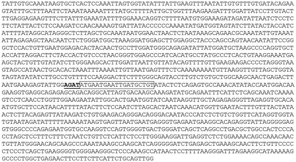
SEQ ID NO:2 optimized RPE65 promoter fragment

SEQ ID NO: cDNA Genbank No. NM-000329.2 of 3 human RPE65
ATGTCTATCCAGGTTGAGCATCCTGCTGGTGGTTACAAGAAACTGTTTGAAACTGTGGAGGAACTGTCCTCGCCG CTCACAGCTCATGTAACAGGCAGGATCCCCCTCTGGCTCACCGGCAGTCTCCTTCGATGTGGGCCAGGACTCTTT GAAGTTGGATCTGAGCCATTTTACCACCTGTTTGATGGGCAAGCCCTCCTGCACAAGTTTGACTTTAAAGAAGGA CATGTCACATACCACAGAAGGTTCATCCGCACTGATGCTTACGTACGGGCAATGACTGAGAAAAGGATCGTCATA ACAGAATTTGGCACCTGTGCTTTCCCAGATCCCTGCAAGAATATATTTTCCAGGTTTTTTTCTTACTTTCGAGGA GTAGAGGTTACTGACAATGCCCTTGTTAATGTCTACCCAGTGGGGGAAGATTACTACGCTTGCACAGAGACCAAC TTTATTACAAAGATTAATCCAGAGACCTTGGAGACAATTAAGCAGGTTGATCTTTGCAACTATGTCTCTGTCAAT GGGGCCACTGCTCACCCCCACATTGAAAATGATGGAACCGTTTACAATATTGGTAATTGCTTTGGAAAAAATTTT TCAATTGCCTACAACATTGTAAAGATCCCACCACTGCAAGCAGACAAGGAAGATCCAATAAGCAAGTCAGAGATC GTTGTACAATTCCCCTGCAGTGACCGATTCAAGCCATCTTACGTTCATAGTTTTGGTCTGACTCCCAACTATATC GTTTTTGTGGAGACACCAGTCAAAATTAACCTGTTCAAGTTCCTTTCTTCATGGAGTCTTTGGGGAGCCAACTAC ATGGATTGTTTTGAGTCCAATGAAACCATGGGGGTTTGGCTTCATATTGCTGACAAAAAAAGGAAAAAGTACCTC AATAATAAATACAGAACTTCTCCTTTCAACCTCTTCCATCACATCAACACCTATGAAGACAATGGGTTTCTGATT GTGGATCTCTGCTGCTGGAAAGGATTTGAGTTTGTTTATAATTACTTATATTTAGCCAATTTACGTGAGAACTGG GAAGAGGTGAAAAAAAATGCCAGAAAGGCTCCCCAACCTGAAGTTAGGAGATATGTACTTCCTTTGAATATTGAC AAGGCTGACACAGGCAAGAATTTAGTCACGCTCCCCAATACAACTGCCACTGCAATTCTGTGCAGTGACGAGACT ATCTGGCTGGAGCCTGAAGTTCTCTTTTCAGGGCCTCGTCAAGCATTTGAGTTTCCTCAAATCAATTACCAGAAG TATTGTGGGAAACCTTACACATATGCGTATGGACTTGGCTTGAATCACTTTGTTCCAGATAGGCTCTGTAAGCTG AATGTCAAAACTAAAGAAACTTGGGTTTGGCAAGAGCCTGATTCATACCCATCAGAACCCATCTTTGTTTCTCAC CCAGATGCCTTGGAAGAAGATGATGGTGTAGTTCTGAGTGTGGTGGTGAGCCCAGGAGCAGGACAAAAGCCTGCT TATCTCCTGATTCTGAATGCCAAGGACTTAAGTGAAGTTGCCCGGGCTGAAGTGGAGATTAACATCCCTGTCACC TTTCATGGACTGTTCAAAAAATCTTGA
SEQ ID NO:4 optimized RPE65cDNA (Kozak sequence and coding sequence)
CCACCATGAGTATCCAGGTGGAACATCCCGCAGGGGGGTATAAGAAACTGTTTGAGACCGTCGAAGAACTGAGCA GCCCTCTGACCGCACATGTCACCGGAAGAATCCCCCTGTGGCTGACAGGATCACTGCTGAGATGCGGACCAGGAC TGTTCGAAGTGGGAAGCGAACCTTTCTACCACCTGTTTGACGGACAGGCCCTGCTGCATAAGTTCGACTTCAAGG AGGGGCACGTGACTTACCATCGGCGGTTCATCCGAACCGACGCCTATGTCCGGGCTATGACAGAGAAGAGAATCG TGATTACTGAGTTCGGCACCTGCGCCTTTCCAGATCCCTGTAAGAACATTTTCTCCAGGTTCTTTTCTTACTTTC GCGGCGTCGAGGTGACAGACAACGCACTGGTCAACGTGTACCCTGTGGGGGAGGATTACTATGCCTGCACTGAAA CCAACTTCATCACCAAGATTAATCCAGAGACACTGGAAACTATCAAACAGGTGGACCTGTGCAACTACGTCAGTG TGAATGGCGCCACCGCTCACCCCCATATCGAGAACGATGGGACAGTCTACAACATTGGCAATTGCTTCGGGAAGA ACTTTAGCATCGCCTACAACATCGTGAAGATCCCCCCTCTGCAGGCTGACAAGGAGGATCCTATCTCTAAAAGTG AAATTGTGGTCCAGTTCCCTTGTTCTGACCGGTTTAAGCCAAGTTACGTCCACTCATTCGGCCTGACACCAAACT ATATCGTCTTTGTGGAGACTCCCGTGAAGATTAATCTGTTCAAATTTCTGAGCTCCTGGTCTCTGTGGGGGGCTA ACTACATGGACTGCTTCGAGAGTAATGAAACAATGGGAGTGTGGCTGCACATCGCAGATAAGAAACGAAAGAAAT ACCTGAACAATAAGTACCGGACTAGCCCCTTCAACCTGTTTCACCATATCAACACCTATGAGGACAATGGATTTC TGATTGTCGATCTGTGCTGTTGGAAGGGCTTCGAGTTCGTGTACAACTATCTGTACCTGGCAAACCTGCGCGAAA ATTGGGAGGAAGTGAAGAAAAATGCTCGAAAAGCACCTCAGCCAGAAGTCAGGCGCTACGTGCTGCCACTGAACA TCGACAAGGCTGATACAGGCAAAAACCTGGTGACTCTGCCCAATACCACAGCAACTGCCATCCTGTGCTCCGACG AGACCATTTGGCTGGAGCCCGAAGTGCTGTTCTCTGGACCTCGCCAGGCCTTCGAATTTCCACAGATTAATTACC AGAAGTACTGCGGCAAACCCTATACCTACGCTTATGGACTGGGCCTGAACCACTTCGTGCCTGATAGACTGTGCA AGCTGAATGTCAAGACCAAAGAGACATGGGTGTGGCAGGAACCTGACTCATACCCCAGCGAGCCTATCTTTGTGA GCCATCCAGATGCCCTGGAGGAAGACGATGGCGTGGTCCTGAGCGTGGTCGTGTCCCCAGGAGCAGGACAGAAGC CAGCCTATCTGCTGATTCTGAACGCTAAAGATCTGTCCGAAGTGGCAAGAGCAGAGGTGGAGATCAATATCCCAG TCACATTTCACGGGCTGTTCAAAAAGTCCTAA
SEQ ID NO: cDNA Genbank No. NM-006343.2 of 5 human MERKs
ATGGGGCCGGCCCCGCTGCCGCTGCTGCTGGGCCTCTTCCTCCCCGCGCTCTGGCGTAGAGCTATCACTGAGGCA AGGGAAGAAGCCAAGCCTTACCCGCTATTCCCGGGACCTTTTCCAGGGAGCCTGCAAACTGACCACACACCGCTG TTATCCCTTCCTCACGCCAGTGGGTACCAGCCTGCCTTGATGTTTTCACCAACCCAGCCTGGAAGACCACATACA GGAAACGTAGCCATTCCCCAGGTGACCTCTGTCGAATCAAAGCCCCTACCGCCTCTTGCCTTCAAACACACAGTT GGACACATAATACTTTCTGAACATAAAGGTGTCAAATTTAATTGCTCAATCAGTGTACCTAATATATACCAGGAC ACCACAATTTCTTGGTGGAAAGATGGGAAGGAATTGCTTGGGGCACATCATGCAATTACACAG TTTTATCCAGATGATGAAGTTACAGCAATAATCGCTTCCTTCAGCATAACCAGTGTGCAGCGTTCAGACAATGGG TCGTATATCTGTAAGATGAAAATAAACAATGAAGAGATCGTGTCTGATCCCATCTACATCGAAGTACAAGGACTT CCTCACTTTACTAAGCAGCCTGAGAGCATGAATGTCACCAGAAACACAGCCTTCAACCTCACCTGTCAGGCTGTG GGCCCGCCTGAGCCCGTCAACATTTTCTGGGTTCAAAACAGTAGCCGTGTTAACGAACAGCCTGAAAAATCCCCC TCCGTGCTAACTGTTCCAGGCCTGACGGAGATGGCGGTCTTCAGTTGTGAGGCCCACAATGACAAAGGGCTGACC GTGTCCAAGGGAGTGCAGATCAACATCAAAGCAATTCCCTCCCCACCAACTGAAGTCAGCATCCGTAACAGCACT GCACACAGCATTCTGATCTCCTGGGTTCCTGGTTTTGATGGATACTCCCCGTTCAGGAATTGCAGCATTCAGGTC AAGGAAGCTGATCCGCTGAGTAATGGCTCAGTCATGATTTTTAACACCTCTGCCTTACCACATCTGTACCAAATC AAGCAGCTGCAAGCCCTGGCTAATTACAGCATTGGTGTTTCCTGCATGAATGAAATAGGCTGGTCTGCAGTGAGC CCTTGGATTCTAGCCAGCACGACTGAAGGAGCCCCATCAGTAGCACCTTTAAATGTCACTGTGTTTCTGAATGAA TCTAGTGATAATGTGGACATCAGATGGATGAAGCCTCCGACTAAGCAGCAGGATGGAGAACTGGTGGGCTACCGG ATATCCCACGTGTGGCAGAGTGCAGGGATTTCCAAAGAGCTCTTGGAGGAAGTTGGCCAGAATGGCAGCCGAGCT CGGATCTCTGTTCAAGTCCACAATGCTACGTGCACAGTGAGGATTGCAGCCGTCACCAGAGGGGGAGTTGGGCCC TTCAGTGATCCAGTGAAAATATTTATCCCTGCACACGGTTGGGTAGATTATGCCCCCTCTTCAACTCCGGCGCCT GGCAACGCAGATCCTGTGCTCATCATCTTTGGCTGCTTTTGTGGATTTATTTTGATTGGGTTGATTTTATACATC TCCTTGGCCATCAGAAAAAGAGTCCAGGAGACAAAGTTTGGGAATGCATTCACAGAGGAGGATTCTGAATTAGTG GTGAATTATATAGCAAAGAAATCCTTCTGTCGGCGAGCCATTGAACTTACCTTACATAGCTTGGGAGTCAGTGAG GAACTACAAAATAAACTAGAAGATGTTGTGATTGACAGGAATCTTCTAATTCTTGGAAAAATTCTGGGTGAAGGA GAGTTTGGGTCTGTAATGGAAGGAAATCTTAAGCAGGAAGATGGGACCTCTCTGAAAGTGGCAGTGAAGACCATG AAGTTGGACAACTCTTCACAGCGGGAGATCGAGGAGTTTCTCAGTGAGGCAGCGTGCATGAAAGACTTCAGCCAC CCAAATGTCATTCGACTTCTAGGTGTGTGTATAGAAATGAGCTCTCAAGGCATCCCAAAGCCCATGGTAATTTTA CCCTTCATGAAATACGGGGACCTGCATACTTACTTACTTTATTCCCGATTGGAGACAGGACCAAAGCATATTCCT CTGCAGACACTATTGAAGTTCATGGTGGATATTGCCCTGGGAATGGAGTATCTGAGCAACAGGAATTTTCTTCAT CGAGATTTAGCTGCTCGAAACTGCATGTTGCGAGATGACATGACTGTCTGTGTTGCGGACTTCGGCCTCTCTAAG AAGATTTACAGTGGCGATTATTACCGCCAAGGCCGCATTGCTAAGATGCCTGTTAAATGGATCGCCATAGAAAGT CTTGCAGACCGAGTCTACACAAGTAAAAGTGATGTGTGGGCATTTGGCGTGACCATGTGGGAAATAGCTACGCGG GGAATGACTCCCTATCCTGGGGTCCAGAACCATGAGATGTATGACTATCTTCTCCATGGCCACAGGTTGAAGCAG CCCGAAGACTGCCTGGATGAACTGTATGAAATAATGTACTCTTGCTGGAGAACCGATCCCTTAGACCGCCCCACC TTTTCAGTATTGAGGCTGCAGCTAGAAAAACTCTTAGAAAGTTTGCCTGACGTTCGGAACCAAGCAGACGTTATT TACGTCAATACACAGTTGCTGGAGAGCTCTGAGGGCCTGGCCCAGGGCTCCACCCTTGCTCCACTGGACTTGAAC ATCGACCCTGACTCTATAATTGCCTCCTGCACTCCCCGCGCTGCCATCAGTGTGGTCACAGCAGAAGTTCATGAC AGCAAACCTCATGAAGGACGGTACATCCTGAATGGGGGCAGTGAGGAATGGGAAGATCTGACTTCTGCCCCCTCT GCTGCAGTCACAGCTGAAAAGAACAGTGTTTTACCGGGGGAGAGACTTGTTAGGAATGGGGTCTCCTGGTCCCAT TCGAGCATGCTGCCCTTGGGAAGCTCATTGCCCGATGAACTTTTGTTTGCTGACGACTCCTCAGAAGGCTCAGAA GTCCTGATGTGA
SEQ ID NO:6 human LRAT cDNA Genbank No. NM-004744.3
ATGAAGAACCCCATGCTGGAGGTGGTGTCTTTACTACTGGAGAAGCTGCTCCTCATCTCCAACTTCACGCTCTTT AGTTCGGGCGCCGCGGGCGAAGACAAAGGGAGGAACAGTTTTTATGAAACCAGCTCTTTCCACCGAGGCGACGTG CTGGAGGTGCCCCGGACCCACCTGACCCACTATGGCATCTACCTAGGAGACAACCGTGTTGCCCACATGATGCCC GACATCCTGTTGGCCCTGACAGACGACATGGGGCGCACGCAGAAGGTGGTCTCCAACAAGCGTCTCATCCTGGGC GTTATTGTCAAAGTGGCCAGCATCCGCGTGGACACAGTGGAGGACTTCGCCTACGGAGCTAACATCCTGGTCAAT CACCTGGACGAGTCCCTCCAGAAAAAGGCACTGCTCAACGAGGAGGTGGCGCGGAGGGCTGAAAAGCTGCTGGGC TTTACCCCCTACAGCCTGCTGTGGAACAACTGCGAGCACTTCGTGACCTACTGCAGATATGGCACCCCGATCAGT CCCCAGTCCGACAAGTTTTGTGAGACTGTGAAGATAATTATTCGTGATCAGAGAAGTGTTCTTGCTTCAGCAGTC TTGGGATTGGCGTCTATAGTCTGTACGGGCTTGGTATCATACACTACCCTTCCTGCAATTTTTATTCCATTCTTC CTATGGATGGCTGGCTAA
SEQ ID NO: cDNA Genbank No. NM-000372.4 of 7 human TYR
ATGCTCCTGGCTGTTTTGTACTGCCTGCTGTGGAGTTTCCAGACCTCCGCTGGCCATTTCCCTAGAGCCTGTGTC TCCTCTAAGAACCTGATGGAGAAGGAATGCTGTCCACCGTGGAGCGGGGACAGGAGTCCCTGTGGCCAGCTTTCA GGCAGAGGTTCCTGTCAGAATATCCTTCTGTCCAATGCACCACTTGGGCCTCAATTTCCCTTCACAGGGGTGGAT GACCGGGAGTCGTGGCCTTCCGTCTTTTATAATAGGACCTGCCAGTGCTCTGGCAACTTCATGGGATTCAACTGT GGAAACTGCAAGTTTGGCTTTTGGGGACCAAACTGCACAGAGAGACGACTCTTGGTGAGAAGAAACATCTTCGAT TTGAGTGCCCCAGAGAAGGACAAATTTTTTGCCTACCTCACTTTAGCAAAGCATACCATCAGCTCAGACTATGTC ATCCCCATAGGGACCTATGGCCAAATGAAAAATGGATCAACACCCATGTTTAACGACATCAATATTTATGACCTC TTTGTCTGGATGCATTATTATGTGTCAATGGATGCACTGCTTGGGGGATCTGAAATCTGGAGAGACATTGATTTT GCCCATGAAGCACCAGCTTTTCTGCCTTGGCATAGACTCTTCTTGTTGCGGTGGGAACAAGAAATCCAGAAGCTG ACAGGAGATGAAAACTTCACTATTCCATATTGGGACTGGCGGGATGCAGAAAAGTGTGACATTTGCACAGATGAG TACATGGGAGGTCAGCACCCCACAAATCCTAACTTACTCAGCCCAGCATCATTCTTCTCCTCTTGGCAGATTGTC TGTAGCCGATTGGAGGAGTACAACAGCCATCAGTCTTTATGCAATGGAACGCCCGAGGGACCTTTACGGCGTAAT CCTGGAAACCATGACAAATCCAGAACCCCAAGGCTCCCCTCTTCAGCTGATGTAGAATTTTGCCTGAGTTTGACC CAATATGAATCTGGTTCCATGGATAAAGCTGCCAATTTCAGCTTTAGAAATACACTGGAAGGATTTGCTAGTCCA CTTACTGGGATAGCGGATGCCTCTCAAAGCAGCATGCACAATGCCTTGCACATCTATATGAATGGAACAATGTCC CAGGTACAGGGATCTGCCAACGATCCTATCTTCCTTCTTCACCATGCATTTGTTGACAGTATTTTTGAGCAGTGG CTCCGAAGGCACCGTCCTCTTCAAGAAGTTTATCCAGAAGCCAATGCACCCATTGGACATAACCGGGAATCCTAC ATGGTTCCTTTTATACCACTGTACAGAAATGGTGATTTCTTTATTTCATCCAAAGATCTGGGCTATGACTATAGC TATCTACAAGATTCAGACCCAGACTCTTTTCAAGACTACATTAAGTCCTATTTGGAACAAGCGAGTCGGATCTGG TCATGGCTCCTTGGGGCGGCGATGGTAGGGGCCGTCCTCACTGCCCTGCTGGCAGGGCTTGTGAGCTTGCTGTGT CGTCACAAGAGAAAGCAGCTTCCTGAAGAAAAGCAGCCACTCCTCATGGAGAAAGAGGATTACCACAGCTTGTAT CAGAGCCATTTATAA
SEQ ID NO: cDNA Genbank No. NM-000273.2 of 8-human GRP143
ATGACCCAGGCAGGCCGGCGGGGTCCTGGCACACCCGAGCCGCGTCCGCGAACACAGCCCATGGCCTCCCCGCGC CTAGGGACCTTCTGCTGCCCCACGCGGGACGCAGCCACGCAGCTCGTGCTGAGCTTCCAGCCGCGGGCCTTCCAC GCGCTCTGCCTGGGCAGCGGCGGGCTCCGCTTGGCGCTGGGCCTTCTGCAGCTGCTGCCCGGCCGCCGGCCCGCG GGCCCCGGGTCCCCCGCGACGTCCCCGCCGGCCTCGGTCCGCATCCTGCGCGCTGCCGCTGCCTGCGACCTTCTC GGCTGCCTGGGTATGGTGATCCGGTCCACCGTGTGGTTAGGATTCCCAAATTTTGTTGACAGCGTCTCGGATATG AACCACACGGAAATTTGGCCTGCTGCTTTCTGCGTGGGGAGTGCGATGTGGATCCAGCTGTTGTACAGTGCCTGC TTCTGGTGGCTGTTTTGCTATGCAGTGGATGCTTATCTGGTGATCCGGAGATCGGCAGGACTGAGCACCATCCTG CTGTATCACATCATGGCGTGGGGCCTGGCCACCCTGCTCTGTGTGGAGGGAGCCGCCATGCTCTACTACCCTTCC GTGTCCAGGTGTGAGCGGGGCCTGGACCACGCCATCCCCCACTATGTCACCATGTACCTGCCCCTGCTGCTGGTT CTCGTGGCGAACCCCATCCTGTTCCAAAAGACAGTGACTGCAGTGGCCTCTTTACTTAAAGGAAGACAAGGCATT TACACGGAGAACGAGAGGAGGATGGGAGCCGTGATCAAGATCCGATTTTTCAAAATCATGCTGGTTTTAATTATT TGTTGGTTGTCGAATATCATCAATGAAAGCCTTTTATTCTATCTTGAGATGCAAACAGATATCAATGGAGGTTCT TTGAAACCTGTCAGAACTGCAGCCAAGACCACATGGTTTATTATGGGAATCCTGAATCCAGCCCAGGGATTTCTC TTGTCTTTGGCCTTCTACGGCTGGACAGGATGCAGCCTGGGTTTTCAGTCTCCCAGGAAGGAGATCCAGTGGGAA TCACTGACCACCTCGGCTGCTGAGGGGGCTCACCCATCCCCACTGATGCCCCATGAAAACCCTGCTTCCGGGAAG GTGTCTCAAGTGGGTGGGCAGACTTCTGACGAAGCCCTGAGCATGCTGTCTGAAGGTTCTGATGCCAGCACAATT GAAATTCACACTGCAAGTGAATCCTGCAACAAAAATGAGGGTGACCCTGCTCTCCCAACCCATGGAGACCTATGA


Claims (19)
Applications Claiming Priority (3)
| Application Number | Priority Date | Filing Date | Title |
|---|---|---|---|
| GBGB1502137.1A GB201502137D0 (en) | 2015-02-09 | 2015-02-09 | Treatment |
| GB1502137.1 | 2015-02-09 | ||
| PCT/GB2016/050289 WO2016128722A1 (en) | 2015-02-09 | 2016-02-08 | Optimized rpe65 promoter and coding sequences |
Publications (2)
| Publication Number | Publication Date |
|---|---|
| CN107429252A CN107429252A (en) | 2017-12-01 |
| CN107429252B true CN107429252B (en) | 2022-03-29 |
Family
ID=52746363
Family Applications (1)
| Application Number | Title | Priority Date | Filing Date |
|---|---|---|---|
| CN201680020425.6A Active CN107429252B (en) | 2015-02-09 | 2016-02-08 | Optimized RPE65 promoter and coding sequence |
Country Status (24)
| Country | Link |
|---|---|
| US (3) | US10568973B2 (en) |
| EP (2) | EP3256169B1 (en) |
| JP (1) | JP6963503B2 (en) |
| CN (1) | CN107429252B (en) |
| AU (1) | AU2016217654B2 (en) |
| BR (1) | BR112017017060A2 (en) |
| CA (1) | CA2975850C (en) |
| CY (1) | CY1123947T1 (en) |
| DK (1) | DK3256169T3 (en) |
| ES (1) | ES2830030T3 (en) |
| GB (1) | GB201502137D0 (en) |
| HR (1) | HRP20201831T1 (en) |
| HU (1) | HUE052407T2 (en) |
| IL (2) | IL287142B2 (en) |
| LT (1) | LT3256169T (en) |
| MX (2) | MX383316B (en) |
| PH (1) | PH12017501430A1 (en) |
| PL (1) | PL3256169T3 (en) |
| PT (1) | PT3256169T (en) |
| RS (1) | RS61079B1 (en) |
| SG (1) | SG11201706520UA (en) |
| SI (1) | SI3256169T1 (en) |
| SM (1) | SMT202000655T1 (en) |
| WO (1) | WO2016128722A1 (en) |
Families Citing this family (16)
| Publication number | Priority date | Publication date | Assignee | Title |
|---|---|---|---|---|
| GB201502137D0 (en) * | 2015-02-09 | 2015-03-25 | Ucl Business Plc | Treatment |
| GB201508026D0 (en) | 2015-05-11 | 2015-06-24 | Ucl Business Plc | Capsid |
| IL262583B2 (en) * | 2016-04-28 | 2023-10-01 | Spark Therapeutics Inc | Relative potency assay for viral vector encoding isomerohydrolases |
| WO2019106035A1 (en) | 2017-11-30 | 2019-06-06 | Friedrich Miescher Institute For Biomedical Research | Synpiii, a promoter for the specific expression of genes in retinal pigment epithelium |
| CN111118017B (en) * | 2018-11-01 | 2023-10-03 | 上海朗昇生物科技有限公司 | Carrier for treating Leber congenital amaurosis and application thereof |
| IL318756A (en) | 2019-02-04 | 2025-04-01 | Spur Therapeutics Ltd | Polynucleotides |
| CN115066255A (en) * | 2019-09-18 | 2022-09-16 | 星际治疗有限公司 | Synthetic DNA vectors and methods of use |
| GB202002202D0 (en) | 2020-02-18 | 2020-04-01 | Ucl Business Plc | Gene therapy |
| CN113952471A (en) * | 2020-07-21 | 2022-01-21 | 英斯培瑞有限公司 | Compositions and methods for treating ocular diseases |
| WO2022017363A1 (en) * | 2020-07-21 | 2022-01-27 | Inspirar Limited | Compositions and methods for the treatment of ocular diseases |
| CN111826378B (en) * | 2020-08-05 | 2022-05-27 | 武汉纽福斯生物科技有限公司 | Nucleotide sequence for coding human receptor tyrosine kinase Mer and application thereof |
| CN112522292B (en) * | 2020-10-29 | 2023-05-02 | 南京启真基因工程有限公司 | CRISPR/Cas9 system for constructing congenital amaranth clone pig nuclear donor cells and application thereof |
| AU2022340595A1 (en) * | 2021-09-06 | 2024-04-18 | Huidagene Therapeutics (Singapore) Pte. Ltd. | Treatment of rpe65-associated eye diseases and disorders |
| CN120882416A (en) * | 2023-01-05 | 2025-10-31 | 欧普斯遗传学股份有限公司 | Gene therapy for ocular diseases |
| CN117018235A (en) * | 2023-06-20 | 2023-11-10 | 浙江大学 | A method and tool viral vector that can be used to visualize neural circuits in vivo |
| CN116898991A (en) * | 2023-06-20 | 2023-10-20 | 浙江大学 | A method and tool viral vector for visualizing neural activity status assessment in vivo |
Family Cites Families (9)
| Publication number | Priority date | Publication date | Assignee | Title |
|---|---|---|---|---|
| EP1788084A4 (en) | 2004-08-06 | 2007-11-07 | Chemo Sero Therapeut Res Inst | Yeast promoter |
| US7740836B2 (en) | 2006-05-03 | 2010-06-22 | Fondazione Telethon | Methods and compositions for recovering or improving visual function |
| US20100081707A1 (en) * | 2008-02-21 | 2010-04-01 | Ali Robin R | Devices and methods for delivering polynucleotides into retinal cells of the macula and fovea |
| SI3418300T1 (en) | 2011-07-18 | 2021-03-31 | Institute For Research In Biomedicine | Neutralizing antibodies against influenza A virus and their uses |
| US9163259B2 (en) * | 2012-05-04 | 2015-10-20 | Novartis Ag | Viral vectors for the treatment of retinal dystrophy |
| TWI698240B (en) * | 2012-05-15 | 2020-07-11 | 澳大利亞商艾佛蘭屈澳洲私營有限公司 | Treatment of amd using aav sflt-1 |
| GB201502137D0 (en) * | 2015-02-09 | 2015-03-25 | Ucl Business Plc | Treatment |
| GB202002073D0 (en) * | 2020-02-14 | 2020-04-01 | Ucl Business Ltd | Horizontal cells |
| GB202002202D0 (en) * | 2020-02-18 | 2020-04-01 | Ucl Business Plc | Gene therapy |
-
2015
- 2015-02-09 GB GBGB1502137.1A patent/GB201502137D0/en not_active Ceased
-
2016
- 2016-02-08 EP EP16703850.4A patent/EP3256169B1/en active Active
- 2016-02-08 AU AU2016217654A patent/AU2016217654B2/en active Active
- 2016-02-08 WO PCT/GB2016/050289 patent/WO2016128722A1/en not_active Ceased
- 2016-02-08 HU HUE16703850A patent/HUE052407T2/en unknown
- 2016-02-08 SG SG11201706520UA patent/SG11201706520UA/en unknown
- 2016-02-08 CA CA2975850A patent/CA2975850C/en active Active
- 2016-02-08 PL PL16703850T patent/PL3256169T3/en unknown
- 2016-02-08 ES ES16703850T patent/ES2830030T3/en active Active
- 2016-02-08 US US15/549,549 patent/US10568973B2/en active Active
- 2016-02-08 MX MX2017010292A patent/MX383316B/en unknown
- 2016-02-08 CN CN201680020425.6A patent/CN107429252B/en active Active
- 2016-02-08 LT LTEP16703850.4T patent/LT3256169T/en unknown
- 2016-02-08 RS RS20201400A patent/RS61079B1/en unknown
- 2016-02-08 DK DK16703850.4T patent/DK3256169T3/en active
- 2016-02-08 BR BR112017017060-4A patent/BR112017017060A2/en active Search and Examination
- 2016-02-08 SM SM20200655T patent/SMT202000655T1/en unknown
- 2016-02-08 EP EP20193488.2A patent/EP3769790A1/en active Pending
- 2016-02-08 HR HRP20201831TT patent/HRP20201831T1/en unknown
- 2016-02-08 PT PT167038504T patent/PT3256169T/en unknown
- 2016-02-08 SI SI201630984T patent/SI3256169T1/en unknown
- 2016-02-08 IL IL287142A patent/IL287142B2/en unknown
- 2016-02-08 JP JP2017541749A patent/JP6963503B2/en active Active
-
2017
- 2017-08-08 IL IL253915A patent/IL253915B/en unknown
- 2017-08-09 MX MX2021006762A patent/MX2021006762A/en unknown
- 2017-08-09 PH PH12017501430A patent/PH12017501430A1/en unknown
-
2020
- 2020-02-20 US US16/796,439 patent/US11969478B2/en active Active
- 2020-11-26 CY CY20201101129T patent/CY1123947T1/en unknown
-
2024
- 2024-04-25 US US18/646,126 patent/US20240293580A1/en active Pending
Non-Patent Citations (4)
| Title |
|---|
| Effect of gene therapy on visual function in Leber"s congenital amaurosis;James W B Bainbridge et al.;《The new england journal of medicine》;20080427;第358卷(第21期);2231-2239 * |
| Genbank:NG_008472.1;Genbank;《Genbank》;20140518;全文 * |
| Genbank:NP_000320.1;Genbank;《Genbank》;20140515;全文 * |
| Restoration of vision in RPE65-deficient Briard dogs using an AAV serotype 4 vector that specifically targets the retinal pigmented epithelium;G Le Meur et al.;《Gene Therapy》;20061105;第14卷(第4期);292-303 * |
Also Published As
Similar Documents
| Publication | Publication Date | Title |
|---|---|---|
| CN107429252B (en) | Optimized RPE65 promoter and coding sequence | |
| Ramlogan‐Steel et al. | Gene therapy and the adeno‐associated virus in the treatment of genetic and acquired ophthalmic diseases in humans: trials, future directions and safety considerations | |
| JP7572990B2 (en) | Methods and compositions for treating diseases and conditions in which RDH12 is involved | |
| JP2019523648A (en) | Dual overlapping adeno-associated virus vector system for expressing ABC4A | |
| US20250290096A1 (en) | Gene therapy to improve vision | |
| CA3096088A1 (en) | Compositions and methods for treating macular dystrophy | |
| JP7289306B2 (en) | Compositions and methods for treating retinal disorders | |
| WO2020167770A1 (en) | Txnip and ldhb compositions and methods for the treatment of degenerative ocular diseases | |
| EP3630986A1 (en) | Gene therapy for treating peroxisomal disorders | |
| HK40045081A (en) | Optimized rpe65 promoter and coding sequences | |
| JP2021520231A (en) | Compositions and methods for the treatment of Stargart's disease | |
| US20240191257A1 (en) | Gene Therapy for Retinal Disease | |
| HK1248525B (en) | Optimized rpe65 promoter and coding sequences | |
| Gupte | Development of an AAV-Based Gene Therapy for the Ocular Phenotype of Friedreich’s Ataxia | |
| CN117980489A (en) | RETGC gene therapy | |
| HK40013460A (en) | Methods and compositions for treatment of disorders and diseases involving rdh12 | |
| HK1248595B (en) | Gene therapy to improve vision |
Legal Events
| Date | Code | Title | Description |
|---|---|---|---|
| PB01 | Publication | ||
| PB01 | Publication | ||
| SE01 | Entry into force of request for substantive examination | ||
| SE01 | Entry into force of request for substantive examination | ||
| CB02 | Change of applicant information |
Address after: London, England Applicant after: UCL Business Co.,Ltd. Address before: London, England Applicant before: UCL BUSINESS PLC |
|
| CB02 | Change of applicant information | ||
| GR01 | Patent grant | ||
| GR01 | Patent grant | ||
| TG01 | Patent term adjustment | ||
| TG01 | Patent term adjustment |