CN106999577B - Oncolytic virus and immune checkpoint modulator combinations - Google Patents
Oncolytic virus and immune checkpoint modulator combinations Download PDFInfo
- Publication number
- CN106999577B CN106999577B CN201580050021.7A CN201580050021A CN106999577B CN 106999577 B CN106999577 B CN 106999577B CN 201580050021 A CN201580050021 A CN 201580050021A CN 106999577 B CN106999577 B CN 106999577B
- Authority
- CN
- China
- Prior art keywords
- immune checkpoint
- virus
- cancer
- checkpoint modulator
- oncolytic
- Prior art date
- Legal status (The legal status is an assumption and is not a legal conclusion. Google has not performed a legal analysis and makes no representation as to the accuracy of the status listed.)
- Active
Links
Images


Classifications
-
- A—HUMAN NECESSITIES
- A61—MEDICAL OR VETERINARY SCIENCE; HYGIENE
- A61K—PREPARATIONS FOR MEDICAL, DENTAL OR TOILETRY PURPOSES
- A61K35/00—Medicinal preparations containing materials or reaction products thereof with undetermined constitution
- A61K35/66—Microorganisms or materials therefrom
- A61K35/76—Viruses; Subviral particles; Bacteriophages
-
- A—HUMAN NECESSITIES
- A61—MEDICAL OR VETERINARY SCIENCE; HYGIENE
- A61K—PREPARATIONS FOR MEDICAL, DENTAL OR TOILETRY PURPOSES
- A61K35/00—Medicinal preparations containing materials or reaction products thereof with undetermined constitution
- A61K35/12—Materials from mammals; Compositions comprising non-specified tissues or cells; Compositions comprising non-embryonic stem cells; Genetically modified cells
- A61K35/28—Bone marrow; Haematopoietic stem cells; Mesenchymal stem cells of any origin, e.g. adipose-derived stem cells
-
- A—HUMAN NECESSITIES
- A61—MEDICAL OR VETERINARY SCIENCE; HYGIENE
- A61K—PREPARATIONS FOR MEDICAL, DENTAL OR TOILETRY PURPOSES
- A61K35/00—Medicinal preparations containing materials or reaction products thereof with undetermined constitution
- A61K35/66—Microorganisms or materials therefrom
- A61K35/76—Viruses; Subviral particles; Bacteriophages
- A61K35/768—Oncolytic viruses not provided for in groups A61K35/761 - A61K35/766
-
- A—HUMAN NECESSITIES
- A61—MEDICAL OR VETERINARY SCIENCE; HYGIENE
- A61K—PREPARATIONS FOR MEDICAL, DENTAL OR TOILETRY PURPOSES
- A61K38/00—Medicinal preparations containing peptides
- A61K38/16—Peptides having more than 20 amino acids; Gastrins; Somatostatins; Melanotropins; Derivatives thereof
- A61K38/17—Peptides having more than 20 amino acids; Gastrins; Somatostatins; Melanotropins; Derivatives thereof from animals; from humans
- A61K38/19—Cytokines; Lymphokines; Interferons
- A61K38/193—Colony stimulating factors [CSF]
-
- A—HUMAN NECESSITIES
- A61—MEDICAL OR VETERINARY SCIENCE; HYGIENE
- A61K—PREPARATIONS FOR MEDICAL, DENTAL OR TOILETRY PURPOSES
- A61K39/00—Medicinal preparations containing antigens or antibodies
-
- A—HUMAN NECESSITIES
- A61—MEDICAL OR VETERINARY SCIENCE; HYGIENE
- A61K—PREPARATIONS FOR MEDICAL, DENTAL OR TOILETRY PURPOSES
- A61K39/00—Medicinal preparations containing antigens or antibodies
- A61K39/395—Antibodies; Immunoglobulins; Immune serum, e.g. antilymphocytic serum
- A61K39/39533—Antibodies; Immunoglobulins; Immune serum, e.g. antilymphocytic serum against materials from animals
- A61K39/3955—Antibodies; Immunoglobulins; Immune serum, e.g. antilymphocytic serum against materials from animals against proteinaceous materials, e.g. enzymes, hormones, lymphokines
-
- A—HUMAN NECESSITIES
- A61—MEDICAL OR VETERINARY SCIENCE; HYGIENE
- A61K—PREPARATIONS FOR MEDICAL, DENTAL OR TOILETRY PURPOSES
- A61K9/00—Medicinal preparations characterised by special physical form
- A61K9/0012—Galenical forms characterised by the site of application
- A61K9/0019—Injectable compositions; Intramuscular, intravenous, arterial, subcutaneous administration; Compositions to be administered through the skin in an invasive manner
-
- A—HUMAN NECESSITIES
- A61—MEDICAL OR VETERINARY SCIENCE; HYGIENE
- A61P—SPECIFIC THERAPEUTIC ACTIVITY OF CHEMICAL COMPOUNDS OR MEDICINAL PREPARATIONS
- A61P35/00—Antineoplastic agents
-
- C—CHEMISTRY; METALLURGY
- C07—ORGANIC CHEMISTRY
- C07K—PEPTIDES
- C07K16/00—Immunoglobulins [IGs], e.g. monoclonal or polyclonal antibodies
- C07K16/18—Immunoglobulins [IGs], e.g. monoclonal or polyclonal antibodies against material from animals or humans
- C07K16/28—Immunoglobulins [IGs], e.g. monoclonal or polyclonal antibodies against material from animals or humans against receptors, cell surface antigens or cell surface determinants
- C07K16/2803—Immunoglobulins [IGs], e.g. monoclonal or polyclonal antibodies against material from animals or humans against receptors, cell surface antigens or cell surface determinants against the immunoglobulin superfamily
- C07K16/2818—Immunoglobulins [IGs], e.g. monoclonal or polyclonal antibodies against material from animals or humans against receptors, cell surface antigens or cell surface determinants against the immunoglobulin superfamily against CD28 or CD152
-
- C—CHEMISTRY; METALLURGY
- C07—ORGANIC CHEMISTRY
- C07K—PEPTIDES
- C07K16/00—Immunoglobulins [IGs], e.g. monoclonal or polyclonal antibodies
- C07K16/18—Immunoglobulins [IGs], e.g. monoclonal or polyclonal antibodies against material from animals or humans
- C07K16/28—Immunoglobulins [IGs], e.g. monoclonal or polyclonal antibodies against material from animals or humans against receptors, cell surface antigens or cell surface determinants
- C07K16/2803—Immunoglobulins [IGs], e.g. monoclonal or polyclonal antibodies against material from animals or humans against receptors, cell surface antigens or cell surface determinants against the immunoglobulin superfamily
- C07K16/2827—Immunoglobulins [IGs], e.g. monoclonal or polyclonal antibodies against material from animals or humans against receptors, cell surface antigens or cell surface determinants against the immunoglobulin superfamily against B7 molecules, e.g. CD80, CD86
-
- C—CHEMISTRY; METALLURGY
- C12—BIOCHEMISTRY; BEER; SPIRITS; WINE; VINEGAR; MICROBIOLOGY; ENZYMOLOGY; MUTATION OR GENETIC ENGINEERING
- C12N—MICROORGANISMS OR ENZYMES; COMPOSITIONS THEREOF; PROPAGATING, PRESERVING, OR MAINTAINING MICROORGANISMS; MUTATION OR GENETIC ENGINEERING; CULTURE MEDIA
- C12N15/00—Mutation or genetic engineering; DNA or RNA concerning genetic engineering, vectors, e.g. plasmids, or their isolation, preparation or purification; Use of hosts therefor
- C12N15/09—Recombinant DNA-technology
- C12N15/63—Introduction of foreign genetic material using vectors; Vectors; Use of hosts therefor; Regulation of expression
- C12N15/79—Vectors or expression systems specially adapted for eukaryotic hosts
- C12N15/85—Vectors or expression systems specially adapted for eukaryotic hosts for animal cells
- C12N15/86—Viral vectors
- C12N15/863—Poxviral vectors, e.g. entomopoxvirus
-
- C—CHEMISTRY; METALLURGY
- C12—BIOCHEMISTRY; BEER; SPIRITS; WINE; VINEGAR; MICROBIOLOGY; ENZYMOLOGY; MUTATION OR GENETIC ENGINEERING
- C12N—MICROORGANISMS OR ENZYMES; COMPOSITIONS THEREOF; PROPAGATING, PRESERVING, OR MAINTAINING MICROORGANISMS; MUTATION OR GENETIC ENGINEERING; CULTURE MEDIA
- C12N5/00—Undifferentiated human, animal or plant cells, e.g. cell lines; Tissues; Cultivation or maintenance thereof; Culture media therefor
-
- C—CHEMISTRY; METALLURGY
- C12—BIOCHEMISTRY; BEER; SPIRITS; WINE; VINEGAR; MICROBIOLOGY; ENZYMOLOGY; MUTATION OR GENETIC ENGINEERING
- C12N—MICROORGANISMS OR ENZYMES; COMPOSITIONS THEREOF; PROPAGATING, PRESERVING, OR MAINTAINING MICROORGANISMS; MUTATION OR GENETIC ENGINEERING; CULTURE MEDIA
- C12N7/00—Viruses; Bacteriophages; Compositions thereof; Preparation or purification thereof
-
- A—HUMAN NECESSITIES
- A61—MEDICAL OR VETERINARY SCIENCE; HYGIENE
- A61K—PREPARATIONS FOR MEDICAL, DENTAL OR TOILETRY PURPOSES
- A61K39/00—Medicinal preparations containing antigens or antibodies
- A61K2039/505—Medicinal preparations containing antigens or antibodies comprising antibodies
-
- A—HUMAN NECESSITIES
- A61—MEDICAL OR VETERINARY SCIENCE; HYGIENE
- A61K—PREPARATIONS FOR MEDICAL, DENTAL OR TOILETRY PURPOSES
- A61K39/00—Medicinal preparations containing antigens or antibodies
- A61K2039/51—Medicinal preparations containing antigens or antibodies comprising whole cells, viruses or DNA/RNA
- A61K2039/525—Virus
- A61K2039/5256—Virus expressing foreign proteins
-
- A—HUMAN NECESSITIES
- A61—MEDICAL OR VETERINARY SCIENCE; HYGIENE
- A61K—PREPARATIONS FOR MEDICAL, DENTAL OR TOILETRY PURPOSES
- A61K39/00—Medicinal preparations containing antigens or antibodies
- A61K2039/545—Medicinal preparations containing antigens or antibodies characterised by the dose, timing or administration schedule
-
- A—HUMAN NECESSITIES
- A61—MEDICAL OR VETERINARY SCIENCE; HYGIENE
- A61K—PREPARATIONS FOR MEDICAL, DENTAL OR TOILETRY PURPOSES
- A61K39/00—Medicinal preparations containing antigens or antibodies
- A61K2039/58—Medicinal preparations containing antigens or antibodies raising an immune response against a target which is not the antigen used for immunisation
- A61K2039/585—Medicinal preparations containing antigens or antibodies raising an immune response against a target which is not the antigen used for immunisation wherein the target is cancer
-
- A—HUMAN NECESSITIES
- A61—MEDICAL OR VETERINARY SCIENCE; HYGIENE
- A61K—PREPARATIONS FOR MEDICAL, DENTAL OR TOILETRY PURPOSES
- A61K2300/00—Mixtures or combinations of active ingredients, wherein at least one active ingredient is fully defined in groups A61K31/00 - A61K41/00
-
- C—CHEMISTRY; METALLURGY
- C07—ORGANIC CHEMISTRY
- C07K—PEPTIDES
- C07K2317/00—Immunoglobulins specific features
- C07K2317/70—Immunoglobulins specific features characterized by effect upon binding to a cell or to an antigen
- C07K2317/76—Antagonist effect on antigen, e.g. neutralization or inhibition of binding
-
- C—CHEMISTRY; METALLURGY
- C12—BIOCHEMISTRY; BEER; SPIRITS; WINE; VINEGAR; MICROBIOLOGY; ENZYMOLOGY; MUTATION OR GENETIC ENGINEERING
- C12N—MICROORGANISMS OR ENZYMES; COMPOSITIONS THEREOF; PROPAGATING, PRESERVING, OR MAINTAINING MICROORGANISMS; MUTATION OR GENETIC ENGINEERING; CULTURE MEDIA
- C12N2710/00—MICROORGANISMS OR ENZYMES; COMPOSITIONS THEREOF; PROPAGATING, PRESERVING, OR MAINTAINING MICROORGANISMS; MUTATION OR GENETIC ENGINEERING; CULTURE MEDIA dsDNA viruses
- C12N2710/00011—Details
- C12N2710/24011—Poxviridae
- C12N2710/24111—Orthopoxvirus, e.g. vaccinia virus, variola
- C12N2710/24132—Use of virus as therapeutic agent, other than vaccine, e.g. as cytolytic agent
Landscapes
- Health & Medical Sciences (AREA)
- Life Sciences & Earth Sciences (AREA)
- Chemical & Material Sciences (AREA)
- Medicinal Chemistry (AREA)
- General Health & Medical Sciences (AREA)
- Engineering & Computer Science (AREA)
- Immunology (AREA)
- Microbiology (AREA)
- Pharmacology & Pharmacy (AREA)
- Animal Behavior & Ethology (AREA)
- Public Health (AREA)
- Veterinary Medicine (AREA)
- Epidemiology (AREA)
- Bioinformatics & Cheminformatics (AREA)
- Genetics & Genomics (AREA)
- Organic Chemistry (AREA)
- Virology (AREA)
- Zoology (AREA)
- Mycology (AREA)
- Wood Science & Technology (AREA)
- Biomedical Technology (AREA)
- Biotechnology (AREA)
- General Engineering & Computer Science (AREA)
- Biochemistry (AREA)
- Cell Biology (AREA)
- Oncology (AREA)
- Molecular Biology (AREA)
- Biophysics (AREA)
- Endocrinology (AREA)
- Proteomics, Peptides & Aminoacids (AREA)
- Developmental Biology & Embryology (AREA)
- Plant Pathology (AREA)
- Physics & Mathematics (AREA)
- Gastroenterology & Hepatology (AREA)
- Hematology (AREA)
- Dermatology (AREA)
- Chemical Kinetics & Catalysis (AREA)
- General Chemical & Material Sciences (AREA)
- Nuclear Medicine, Radiotherapy & Molecular Imaging (AREA)
- Medicines Containing Material From Animals Or Micro-Organisms (AREA)
Abstract
The present invention provides a combination comprising at least an oncolytic virus and one or more immune checkpoint modulator(s), for use in the treatment of a proliferative disease, such as cancer. Also relates to a kit comprising said oncolytic virus and said one or more immune checkpoint modulator(s) in separate containers. Also relates to pharmaceutical compositions comprising an effective amount of the oncolytic virus and the one or more immune checkpoint modulator(s).
Description
Technical Field
The present invention relates to oncolytic viral therapy, and more particularly to compositions and methods for treating, preventing or inhibiting proliferative diseases, particularly cancer. Embodiments include oncolytic viruses for use in combination with one or more immune checkpoint modulators for treating cancer. Embodiments also include a kit comprising such components and methods of treatment using the oncolytic virus and the one or more immune checkpoint modulator(s).
Background
Every year 1200 million subjects worldwide are diagnosed with cancer. In industrialized countries, one of about five people dies from cancer. Although a large number of chemotherapies exist, they are often not effective, particularly against malignant and metastatic tumors that are established in the very early stages of the disease. Furthermore, anti-tumor immunity is often ineffective due to the mechanism by which tumor cells evolve to evade host defenses. One of the major mechanisms of immunosuppression is a process known as "T cell depletion", which is caused by continued exposure to antigen and is characterized by the upregulation of inhibitory receptors. These inhibitory receptors act as immune checkpoints to prevent uncontrolled immune responses. Various immune checkpoints have been described in the literature to play a role at different levels of T cell immunity, including programmed cell death protein 1(PD-1) and its ligands PD-L1 and PD-L2, CTLA-4 (cytotoxic T lymphocyte-associated protein 4), LAG3 (lymphocyte activation gene 3), B and T lymphocyte attenuation factors, T cell immunoglobulin, mucin 3(TIM-3), and T cell activated immunoglobulin suppressor type V domains.
Whatever the mechanism of action, these immune checkpoints are able to inhibit the development of a potent anti-tumor immune response. There is increasing interest in blocking such immune checkpoints as a possible therapeutic benefit of a means of suppressing the tolerance of the immune system to tumors and thus rescuing depleted anti-tumor T cells (Leach et al, 1996, Science 271: 1734-6). Over the past decade, a number of antagonistic antibodies (e.g., anti-Tim 3, anti-PD-L1, anti-CTLA-4, anti-PD 1, etc.) have been developed, and more importantly, some of them have been associated with objective clinical responses in cancer patients. Antibodies against CTLA-4 have been marketed (e.g., Yipima, Yervoy, Bezishi, BMS) for metastatic melanoma. BMS reports show that 22% of the 1800 melanoma patients treated with leprima survive three years later. anti-PD-L1 (e.g., MPDL3280A, Roche) and anti-PD-1 (e.g., Nivolumab, BMS) antibody therapies are also in progress.
Another emerging therapeutic modality in the cancer field is oncolytic virus (Hermiston,2006, curr. opin. mol. ther.8: 322-30). Oncolytic viruses are able to selectively replicate in dividing cells (e.g., cancer cells) without harming non-dividing cells (e.g., normal cells). When infected dividing cells are lysed, they release new infectious viral particles to infect surrounding dividing cells. Cancer cells are ideal hosts for many viruses because their antiviral interferon pathway is turned off or tumor suppressor genes are mutated so that viral replication can proceed without restriction (Chernajovsky et al, 2006, British med.j.332: 170-2). Many viruses including adenovirus (adenovirus), reovirus (reovirus), measles virus (measles), herpes simplex virus (herpes simplex), Newcastle disease virus (Newcastle disease virus), and vaccinia virus (vaccinia) have been clinically tested as oncolytic agents.
Some viruses naturally oncolytic, such as reovirus and Seneca valley picornavirus, others are engineered for tumor selectivity by modifying the viral genome. Such modifications include functional deletions in key viral genes, the use of tumor or tissue specific promoters to control viral gene expression, and tropism modifications to redirect the virus to the surface of cancer cells.
The first oncolytic virus approved by regulatory agencies was a genetically modified adenovirus designated H101 (shanghai three-dimensional biotechnology), which was approved by the chinese food and drug administration for the treatment of head and neck cancer in 2005. Another oncolytic adenovirus, ONYX-015, is in the clinical trial phase for the treatment of various solid tumors (in a third phase clinical trial for the treatment of recurrent head and neck cancer) (Cohen et al, 2001, Curr. Opin. Investig. drugs 2: 1770-5). Another example is that oncolytic herpes simplex 1(T-VEC) has been genetically engineered to attenuate viral virulence and increase selectivity for cancer cells as well as enhance the anti-tumor immune response (through the expression of GM-CSF (granulocyte-macrophage colony stimulating factor)). Clinical efficacy against surgically unresectable melanoma has been demonstrated in phase two and phase three clinical trials (Senzer et al,2009, j. clin. oncol.27: 5763-71).
Vaccinia Virus (VV) has many key properties essential for oncolytic virus therapy, such as natural tropism for tumors, strong lytic capacity, short life cycle and rapid cell-cell spread, efficient gene expression and large cloning capacity. In addition, they have been delivered to millions of individuals in an exercise that eliminates smallpox without significant safety concerns. In this regard, VV (designated JX-963) double-deleted for TK (thymidine kinase) and VGF (vaccinia virus growth factor) and expressing GM-CSF showed significant cancer selectivity in tumor-bearing mice (Thorne et al, 2007, J Clin invest.117: 3350-8). Likewise, JX-594, VV with GM-CSF and thymidine kinase deletion (Wyeth strain), exhibits promising clinical data, and its randomized three-phase clinical trial in hepatocellular carcinoma is expected to be imminent.
Combination therapies have also been described in the literature. WO2010/014784 describes the use of anti-CTLA 4 antibodies in combination with chemotherapeutic drugs such as gleevec, taxol, etc. to treat cancer. WO2014/047350 contemplates recombinant oncolytic viruses having a gene encoding an anti-PD-1 antibody inserted in the viral genome.
Technical problem
Cancer is considered to continue to be a serious global health threat for many years, as a number of disease-causing agents may interact or initiate or contribute individually to the development of cancer. Furthermore, malignant, in particular metastatic, tumors are often resistant to conventional therapeutic approaches resulting in a high incidence of some cancers.
Therefore, there is an urgent need to develop more effective methods, particularly combination methods, for the prevention and treatment of such proliferative diseases.
Combination therapy with concurrent administration of an oncolytic virus and one or more immune checkpoint modulator(s) provides a synergistic immune response compared to either method alone. Surprisingly, as has been demonstrated in suitable model animals, administration of a combination therapy of oncolytic vaccinia virus prior to administration of an anti-checkpoint antibody such as anti-PD-1 or anti-CTLA-4 improves the anti-tumor response and thus may provide an effective and robust cancer therapy. Thus, the embodiments provided herein provide significant advantages in the treatment and prevention of proliferative diseases, such as cancer.
This technical problem is solved by providing the embodiments defined in the claims.
Other and further aspects, features and advantages of the present invention will be apparent from the following description of the presently preferred embodiments of the invention. These embodiments are provided for purposes of disclosure.
Summary of The Invention
The present invention relates to synergistic combinations of an oncolytic virus and one or more immune checkpoint modulator(s) for use in the treatment of a proliferative disease, such as cancer. The oncolytic virus is preferably selected from the group consisting of reovirus, Newcastle Disease Virus (NDV), Vesicular Stomatitis Virus (VSV), measles virus, influenza virus, Sinbis virus, adenovirus, poxvirus, herpes virus (HSV), and the like. In one embodiment, the oncolytic virus is a vaccinia virus. In a preferred embodiment, the vaccinia virus is engineered to lack Thymidine Kinase (TK) activity (e.g., the VV genome has an inactivating mutation or gene deletion in the J2R gene to produce a TK-deficient phenotype). Alternatively or in combination, the vaccinia virus is engineered to lack Ribonucleotide Reductase (RR) activity (e.g., the VV genome has an inactivating mutation or gene deletion in the I4L gene and/or F4L gene to produce an RR-deficient phenotype).
In one embodiment, the vaccinia virus further expresses at least one therapeutic gene, particularly a gene encoding a suicide gene product and/or an immunostimulatory protein.
In one embodiment, the one or more immune checkpoint modulator is an antagonist molecule that antagonizes the activity of PD-1, PD-L1 or CTLA4, particularly preferably an anti-PD-1 antibody and/or an anti-CTLA 4 antibody.
In one embodiment, the oncolytic virus is preferably formulated for intravenous or intratumoral administration and/or the one or more immune checkpoint modulator(s) is preferably formulated for intravenous or intraperitoneal or intratumoral administration.
The invention further provides a method of treating proliferative diseases, including cancer, comprising administering to a mammal in need of treatment a synergistically effective amount of an oncolytic virus as described herein and one or more immune checkpoint modulator(s) as described herein. In one embodiment, the proliferative disease to be treated by the method of the invention is cancer, and in particular melanoma, renal cancer, prostate cancer, breast cancer, colorectal cancer, lung cancer and liver cancer. In one embodiment, the method comprises the additional step wherein a pharmaceutically acceptable amount of the prodrug is administered to the mammal. Preferably, administration of the prodrug occurs at least three days after administration of the oncolytic virus or viral composition.
The invention further provides a kit comprising an oncolytic virus as described herein and one or more immune checkpoint modulator(s), preferably in separate containers.
Detailed Description
The present invention relates to a composition comprising at least one oncolytic virus and one or more immune checkpoint modulator(s), for use in the treatment of a proliferative disease, such as cancer.
Definition of
Throughout this specification, unless otherwise indicated, the terms "a" and "an" mean "at least one", "at least a first", "one or more", or "a plurality" of the indicated component or step. For example, the term "a cell" encompasses a plurality of cells and mixtures thereof.
The term "one or more" means one or more number (e.g., 2, 3, 4, 5, etc.).
The term "and/or" as used anywhere herein is used in the sense of "including" and "," or ", and" all or any other combination of elements associated by the term ".
The term "about" or "approximately" as used herein means within 20%, preferably within 10%, and more preferably within 5% of a given value or range.
As used herein, the terms "comprising" (and any form comprising such as "comprise" and "comprise"), "having" (and any form having such as "have" and "has"), "including" (and any form including such as "include" and "include") or "containing" (and any form containing such as "contain" and "contain") are open-ended and do not exclude additional unrecited elements or process steps when used to define products, compositions and processes. Thus, a polypeptide "comprising" an amino acid sequence means that the amino acid sequence may be part of the final amino acid sequence of the polypeptide. The polypeptide may comprise up to several hundred additional amino acid residues. "consisting essentially of … …" means that other components or steps of significance are excluded. Therefore, a composition consisting essentially of the recited components does not exclude trace amounts of contaminants and pharmaceutically acceptable carriers. A polypeptide consists essentially of an amino acid sequence meaning that this amino acid sequence will eventually have a few additional amino acid residues. "consisting of … …" means that not only trace amounts of other ingredients or steps are excluded. For example, a polypeptide consisting of an amino acid sequence means that the polypeptide does not contain any amino acid other than the amino acid sequence.
The terms "polypeptide", "peptide" and "protein" refer to a polymer of amino acid residues comprising at least nine or more amino acids joined by peptide bonds. The polymer may be linear, branched or cyclic, and may comprise natural amino acids and/or amino acid analogues, and it may be interrupted by non-amino acids. Generally, if the amino acid polymer is more than 50 amino acid residues, it is preferably referred to as a polypeptide or protein, and if it is 50 amino acids or less in length, it is referred to as a "peptide".
In the context of the present invention, the terms "nucleic acid", "nucleic acid molecule", "polynucleotide" and "nucleotide sequence" are used interchangeably and define a polymer of polydeoxyribonucleotides (DNA) (e.g. cDNA, genomic DNA, plasmids, vectors, viral genomes, isolated DNA, probes, primers and any mixtures thereof) or polyribonucleotides (RNA) (e.g. mRNA, antisense RNA, SiRNA) or mixed polyribonucleotides of any length. They encompass single-or double-stranded, linear or circular, natural or synthetic, modified or unmodified polynucleotides. In addition, the polynucleotide may comprise non-naturally occurring nucleotides and may be interrupted by non-nucleotide components.
As used herein, the term "analog" means that a molecule (polypeptide or nucleic acid) exhibits one or more modifications relative to its natural counterpart. Any modification, including substitution, insertion and/or deletion of one or more nucleotides or amino acid residues, is envisaged. Preferred are analogs that retain at least 80%, preferably at least 85%, more preferably at least 90% and even more preferably at least 98% sequence identity to the natural counterpart.
Generally, the term "identity" denotes the amino acid to amino acid or nucleotide to nucleotide correspondence in two polypeptide or nucleotide sequences. The percent identity of two sequences is a function of the number of identical positions shared by the sequences, taking into account the number of gaps that need to be introduced to optimize alignment and the length of each gap. There are a variety of computer programs and mathematical algorithms in the art to determine percent identity between amino acid sequences, such as the Blast program of NCBI or ALIGN in Atlas of Protein Sequence and Structure (Dayhoffset, 1981, suppl.,3: 482-9). Programs for determining identity between nucleotide sequences are also available on professional databases (e.g., Genbank, the Wisconsin Sequence Analysis Package, BESTFIT, FASTA and GAP programs). For purposes of this specification, "at least 80% identity" means 80%, 81%, 82%, 83%, 84%, 85%, 86%, 87%, 88%, 89%, 90%, 91%, 92%, 93%, 94%, 95%, 96%, 97%, 98%, 99% or 100%.
The term "isolated" as used herein refers to a protein, polypeptide, peptide, polynucleotide, vector, or the like that is removed from its natural environment (i.e., separated from at least one other component with which it naturally accompanies or is found in nature). For example, when a nucleotide sequence is separated from the sequence to which it is normally associated in nature (e.g., dissociated from the genome), the nucleotide sequence is isolated, but it may be associated with a heterologous sequence.
The terms "obtained from", "derived from" or "derived from" are used to indicate the original source of the components (e.g. polypeptides and nucleotide molecules), but are not meant to limit the method of preparing the components, which may be, for example, chemical synthesis or recombinant means.
The term "host cell" as used herein is to be understood broadly and is not limited in any way to the particular structure of a tissue, organ or isolated cell. Such cells may be a unique type of cell or a group of different types of cells, such as cultured cell lines, primary cells, and dividing cells. In the context of the present invention, the term "host cell" includes prokaryotic cells, lower eukaryotic cells such as yeast, other eukaryotic cells such as insect cells, plant and mammalian (e.g., human or non-human) cells, and cells capable of producing oncolytic viruses and/or immune checkpoint regulators for use in the present invention. The term also includes cells that may or may not have been the recipient of the vectors described herein, as well as progeny of such cells.
The term "oncolytic virus" as used herein refers to a virus that selectively replicates in dividing cells (e.g., proliferating cells, such as cancer cells) in vitro or in vivo to slow the growth of and/or lyse the dividing cells, while non-dividing cells show no or minimal replication. Typically, an oncolytic virus comprises a viral genome packaged into a viral particle (or virion) and is infectious (i.e., capable of infecting and entering a host cell or subject).
The term "treatment" as used herein encompasses prophylaxis (e.g. prophylactic measures in a subject at risk of developing the pathological condition to be treated) and/or treatment (e.g. in a subject already diagnosed as having the pathological condition), ultimately in association with conventional treatment modalities. The result of treatment is to slow, cure, improve or control the progression of the targeted pathological state. For example, a subject is successfully treated for cancer if the subject exhibits an observable improvement in clinical status following administration of an oncolytic virus as described herein and one or more immune checkpoint modulators.
The term "administering" herein means delivering a therapeutic agent, such as an oncolytic virus and/or immune checkpoint modulator as described herein, to a subject.
The term "proliferative disease" as used herein encompasses any disease or condition resulting from uncontrolled cell growth and spread, including cancer, as well as diseases associated with increased osteoclast activity (e.g., rheumatoid arthritis, osteoporosis, etc.) and cardiovascular disease (restenosis due to vascular wall smooth muscle cell proliferation, etc.). The term "cancer" may be used interchangeably with any of the following terms: "tumor", "malignant tumor", "neoplasms (neoplasms)", and the like. These terms are intended to encompass any type of tissue, organ or cell, as well as any stage of malignancy (e.g., from premalignant lesions to stage IV).
The term "subject" generally refers to an organism that requires or can benefit from any of the products and methods of the present invention. Typically, the organism is a mammal, in particular a mammal selected from the group consisting of domestic animals, farm animals, sport animals and primates. Preferably, the subject is a human that has been diagnosed as having or at risk of developing a proliferative disease, such as cancer. The terms "subject" and "patient" are used interchangeably when referring to a human organism and encompass both males and females. The subject of treatment may be a neonate, an infant, a young adult or an adult.
The term "combination" herein denotes any possible permutation and combination of components (e.g. oncolytic virus and immune checkpoint modulator). Such a permutation and combination comprises at least one oncolytic virus and a mixture of one or more immune checkpoint modulators in the form of a polypeptide (e.g., a recombinant antibody or a mixed recombinant antibody) or one or more immune checkpoint modulators in the form of a nucleotide molecule (e.g., carried by one or more vectors engineered to express the one or more immune checkpoint modulators), and a mixture of polypeptides and nucleotide molecules (e.g., a recombinant antibody and an expression vector). The present invention encompasses combinations comprising equivalent quantitative concentrations of each immune checkpoint modulator (when more than one is used) or combinations with very different concentrations. It is understood that one skilled in the art can determine the optimum concentration of each component in the combination. Preferably, the combination is synergistic, providing greater efficacy than each entity alone.
The term "immune checkpoint modulator" refers to a molecule (in particular, an interaction between an Antigen Presenting Cell (APC) such as a cancer cell and an immune T effector cell) that is capable of modulating the potency of an immune checkpoint protein in a positive or negative manner. The term "immune checkpoint" denotes a protein that is directly or indirectly involved in the immune pathway, which is crucial for preventing uncontrolled immune reactions under normal physiological conditions and thus for maintaining self-tolerance and/or tissue protection. The one or more immune checkpoint modulators described herein may act independently at any step of T cell-mediated immunity, including clonal selection of antigen-specific cells, T cell activation, proliferation, trafficking to antigen and inflammatory sites, performing direct effector functions, and signaling through cytokines and membrane ligands. Each of these steps is adjusted to fine-tune the response by countering the stimulatory and inhibitory signals. In the context of the present invention, this term encompasses immune checkpoint modulators (antagonists) capable of at least partially down-regulating inhibitory immune checkpoint functions and/or immune checkpoint modulators (agonists) capable of at least partially up-regulating stimulatory immune checkpoint functions.
Oncolytic virus
The oncolytic virus used in the present invention may be obtained from any member of the currently identified viruses, as long as it is oncolytic due to its property of selectively replicating and killing dividing cells compared to non-dividing cells. It may be a naturally oncolytic virus or it may be engineered by modifying one or more viral genes, such as those involved in DNA replication, nucleic acid metabolism, host tropism, surface attachment, virulence, cell lysis and spread, to increase tumor selectivity and/or preferential replication of dividing cells (see, e.g., Kirn et al, 2001, nat. med.7: 781; Wong et al, 2010, Viruses 2: 78-106). It is also contemplated that one or more viral genes may be placed under the control of event or tissue specific regulatory elements (e.g., promoters).
Exemplary oncolytic viruses include, but are not limited to, reovirus (reovirus), Seneca Valley Virus (SVV), Vesicular Stomatitis Virus (VSV), Newcastle Disease Virus (NDV), Herpes Simplex Virus (HSV), morbillivirus virus (morbillivirus), retrovirus (retrovirus), influenza virus (influenza virus), Sindbis virus (Sinbis virus), poxvirus (poxvirus), adenovirus (adenvirus), and the like.
In one embodiment, the oncolytic virus used in the present invention is obtained from reovirus (reovirus). Representative examples include Reolysin (Oncolytics Biotech is under development; NCT 01166542).
In one embodiment, the oncolytic virus used in the present invention is obtained from Seneca Valley virus (Seneca Valley virus). Representative examples include NTX-010(Rudin et al, 2011, Clin. cancer. Res.17(4): 888-95).
In one embodiment, the oncolytic virus for use in the present invention is obtained from Vesicular Stomatitis Virus (VSV). Representative examples for use in the present invention have been described in the literature (e.g., Stojdl et al, 2000, nat. Med.6(7): 821-5; Stojdl et al, 2003, Cancer Cell 4(4): 263-75).
In one embodiment, the oncolytic virus for use in the present invention is obtained from newcastle disease virus. Representative examples of the present invention include, but are not limited to, strains 73-T PV701 and HDV-HUJ and those described in the literature (e.g., Phuangsab et al, 2001, Cancer Lett.172(1): 27-36; Lorence et al, 2007, Curr. Cancer Drug Targets 7(2): 157-67; Freeman et al, 2006, mol. Ther.13(1): 221-8).
In one embodiment, the oncolytic virus used in the present invention is obtained from Herpes Simplex Virus (HSV). The herpesviridae family is a large family of DNA viruses, all of which share a common structure and comprise a relatively large double-stranded linear DNA genome encoding 100-200 genes encapsidated in an icosahedral capsid that is enveloped in a lipid bilayer membrane. Although the oncolytic herpes viruses may be derived from different types of HSV, HSV1 and HSV2 are particularly preferred. The herpesvirus may be genetically engineered to limit replication of the virus within a tumor or reduce its cytotoxicity within a non-dividing cell. For example, any viral Gene involved in nucleic acid metabolism may be inactivated, such as thymidine kinase (Martuza et al, 1991, Science 252:854-6), Ribonucleotide Reductase (RR) (Boviatisis et al, 1994, Gene Ther.1: 323-31; Mineta et al, 1994, Cancer Res.54:3363-66) or uracil-N-glycosylase (Pyres et al, 1994, J.Virol.68: 4963-72). Another aspect relates to viral mutants having a defect in the function of a gene encoding a virulence factor, such as the ICP34.5 gene (Chambers et al, 1995, Proc. Natl. Acad. Sci. USA 92: 1411-5). Representative examples of oncolytic herpes viruses include NV1020 (e.g., Geevarghese et al, 2010, hum. Gene. Ther.21(9):1119-28) and T-VEC (Andtbacka et al, 2013, J. Clin. Oncol.31, abstrat number LBA 9008).
In one embodiment, the oncolytic virus used in the present invention is obtained from morbillivirus, which may be obtained from the paramyxoviridae family, with morbillivirus being particularly preferred. Representative examples of suitable oncolytic measles viruses include, but are not limited to, MV-Edm (McDonald et al, 2006; Breast Cancer Treat.99(2):177-84) and HMWMAA (Kaufmann et al, 2013, J.Invest. Dermatol.133(4): 1034-42).
In one embodiment, the oncolytic virus of the invention is obtained from an adenovirus. Methods for engineering oncolytic adenoviruses are known in the art. Advantageous strategies include replacing the viral promoter with a tumor-selective promoter or modifying the E1 adenovirus gene product to inactivate its binding function to the p53 or retinoblastoma (Rb) protein which is altered in tumor cells. In its natural environment, the adenovirus E1B55kDa gene, in cooperation with other adenovirus products, inactivates p53 (p 53 is normally deregulated in cancer cells), thus preventing apoptosis. Representative examples of oncolytic adenoviruses include ONYX-015 (e.g., Khuri et al, 2000, nat. Med 6(8):879-85) and H101, also known as Oncorine (Xia et al, 2004, Ai Zheng 23(12): 1666-70).
In one embodiment, the oncolytic virus used in the present invention is a poxvirus. The term "poxvirus" as used herein denotes a virus belonging to the family poxviridae, particularly preferably a poxvirus belonging to the subfamily Chordopoxviridae, and more preferably belonging to the genus Orthopoxvirus (Orthopoxvirus). Genomic sequences of various poxviruses, such as Vaccinia virus (Vaccinia virus), Vaccinia virus (cowpox virus), Canarypox virus (Canarypox virus), mousepox virus (ecromelia virus) and Myxoma virus (Myxoma virus) are available in the art and in professional databases, such as Genbank (accession numbers NC _006998, NC _003663, NC _005309, NC _004105, NC _001132, respectively).
Desirably, the oncolytic poxvirus is an oncolytic vaccinia virus. Vaccinia virus is a member of the poxviridae family and is characterized by a 200kb double-stranded DNA genome that encodes a large number of viral enzymes and factors that enable the virus to replicate independently of the host cell structure. Most vaccinia virus particles are intracellular (IMV means intracellular mature virion), have a monolayer lipid envelope and remain in the cytoplasm of infected cells until lysed. Another infectious form is a double-layered envelope particle (EEV means virion enveloped by a cell) that buds from an infected cell without lysing the cell.
More preferred strains are the Elsterry (Elstree), Whitman (Wyeth), Copenhagen (Copenhagen) and Western reservoir (Western Reserve) strains, although they may be derived from any vaccinia virus strain. The gene nomenclature used herein is that of the Copenhagen vaccinia virus strain. Unless otherwise indicated, this method is also applied to homologous genes of other poxviridae herein. However, gene nomenclature may vary depending on the pox strain, but correspondences between Copenhagen (Copenhagen) and other vaccinia virus strains are generally available in the literature.
Preferably, the oncolytic vaccinia virus of the invention is modified by altering one or more viral genes. Preferably, the modification results in the synthesis (or lack thereof) of a defective protein that does not possess the activity of the protein produced by the unmodified gene under normal conditions. Modifications encompass deletions, mutations and/or substitutions of one or more nucleotides (continuous or discontinuous) within the viral gene or its regulatory elements. Modification can be carried out by a number of means known to those skilled in the art using conventional recombinant techniques. Exemplary modifications are disclosed in the literature, particularly preferred are those that alter viral genes involved in DNA metabolism, host virulence and the IFN pathway (see, e.g., Guse et al, 2011, Expert Opinion biol. Ther.11(5): 595-608).
More preferably, the oncolytic poxvirus of the invention is modified by altering the gene encoding thymidine kinase (locus J2R). The thymidine kinase enzyme is involved in the synthesis of deoxyribonucleotides. Thymokinase is essential for viral replication in normal cells because these cells usually have low concentrations of nucleotides, whereas thymokinase is dispensable in dividing cells because of the high nucleotide concentrations of dividing cells.
Alternatively or in combination, the oncolytic poxvirus of the invention is modified by altering at least one or two genes encoding Ribonucleotide Reductase (RR). In the natural environment, the enzyme catalyzes the reduction of ribonucleotides to deoxyribonucleotides, a key step in the biosynthesis of DNA. The viral enzymes are similar in subunit structure to mammalian enzymes and each comprise two heterologous subunits, designated R1 and R2, encoded by the I4L and F4L loci, respectively. The sequences of the I4L and F4L genes in different poxviruses and the positions on the genome are available on public databases, for example by accession numbers DQ437594, DQ437593, DQ377804, AH015635, AY313847, AY313848, NC _003391, NC _003389, NC _003310, M-35027, AY243312, DQ011157, DQ011156, DQ011155, DQ011154, DQ011153, Y16780, X71982, AF438165, U60315, AF410153, AF380138, U86916, L22579, NC _006998, DQ121394 and NC _ 008291. In the present invention, either or both of the I4L gene (encoding the R1 large subunit) or the F4L gene (encoding the R2 small subunit) may be inactivated.
Alternatively or in combination, other strategies may also be used to further increase viral tumor specificity. Representative examples of suitable modifications include disruption of the VGF-encoding gene from the viral genome. VGF (vaccinia virus growth factor) is a secreted protein that is expressed early after cell infection and whose function appears to be important for virus spread between normal cells. Another example is disruption of the A56R gene encoding hemagglutinin (hemagglutinin), eventually in combination with a thymidine kinase deletion (Zhang et al, 2007, cancer Res.67: 10038-46). It may also be advantageous to disrupt an interferon regulatory gene (e.g., the B8R or B18R gene) or the caspase-1 inhibitor B13R gene.
In a preferred embodiment, the oncolytic virus used in the present invention is a vaccinia virus that is defective in TK due to an inactivating mutation in the J2R gene. In another preferred embodiment, the oncolytic virus used in the present invention is a vaccinia virus with a deficiency in both TK and RR activity caused by inactivating mutations in both said J2R Gene and said I4L and/or F4L Gene carried by the viral genome (as described in WO2009/065546 and Foloppe et al, 2008, Gene ther, 15: 1361-71).
Therapeutic genes
In one embodiment, the oncolytic virus used in the present invention further expresses at least one therapeutic gene inserted into the viral genome. When properly administered to a subject, a "therapeutic gene" encodes a product capable of providing a biological activity that is expected to have a beneficial effect on the course and symptoms of the pathological condition to be treated, either by potentiating anti-tumor efficacy or by potentiating the oncolytic properties of the virus. In the present invention, the therapeutic gene may be of human origin or non-human origin (e.g. of bacterial, yeast or viral origin). Preferably, the therapeutic gene is not a gene or nucleotide sequence encoding an immune checkpoint modulator as described herein.
In the context of the present invention, a large number of therapeutic genes are envisaged, for example those encoding polypeptides capable of compensating for a protein deficiency or deficiency in a subject, or those that limit or remove harmful cells from the body by toxic effects, or those encoding polypeptides for immune effects. They may be native genes or genes obtained by subsequent mutation, deletion, substitution and/or addition of one or more nucleotides.
Advantageously, the oncolytic viruses used in the present invention carry a therapeutic gene selected from the group consisting of genes encoding suicide gene products and immunostimulatory proteins.
Suicide gene
The term "suicide gene" denotes a gene encoding a protein that can convert a prodrug into a cytotoxic compound. Suicide genes include, but are not limited to, genes encoding proteins having cytosine deaminase activity, thymidine kinase activity, uracil phosphoribosyltransferase activity, purine nucleoside phosphorylase activity, and thymidylate kinase activity. Examples of suicide genes and corresponding prodrugs comprising a nucleobase moiety are listed in the following table.
TABLE 1
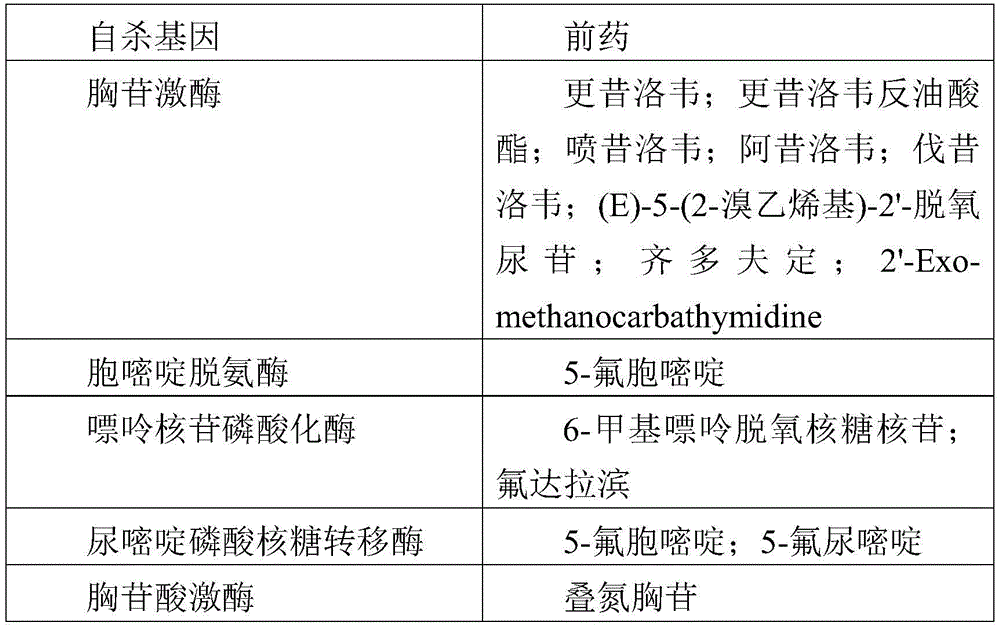
Desirably, the suicide gene encodes a protein having at least cytosine deaminase (CDase) activity. In prokaryotes and lower eukaryotes (which do not exist in mammals), cdases are involved in the pyrimidine metabolic pathway through which exogenous cytosines are converted to uracils by hydrolytic deamination. CDase is also capable of cleaving the amino group of the cytosine analogue, 5-fluorocytosine (5-FC), thereby forming 5-fluorodioxypyrimidine (5-FU), which is a compound having strong cytotoxicity after being converted into 5-fluoro-UMP (5-FUMP). The nucleotide molecules encoded by the CDase can be obtained from any prokaryotic and lower eukaryote, such as Saccharomyces cerevisiae (FCY1 gene), Candida Albicans (Candida Albicans) (FCA1 gene), and Escherichia coli (codA gene). The gene sequence and the encoded CDase protein have been published and available in specialized databases (SWISSPROT EMBL, Genbank, Medline, etc.). Functional analogs of these genes may also be used. Preferably, the analogue has a nucleic acid sequence which has a degree of identity of at least 70%, advantageously at least 80%, preferably at least 90%, most preferably 95% to the nucleic acid sequence of the native gene.
Alternatively or in combination, the oncolytic virus of the invention carries in its viral genome a suicide gene encoding a polypeptide having uracil phosphoribosyltransferase (UPRTase) activity. In prokaryotes and lower eukaryotes, uracil is converted to UMP by the action of UPRTase. This enzyme converts 5-FU to 5-FUMP. By way of example, nucleic acid sequences encoding UPRTases obtained from Escherichia coli (Andersen et al, 1992, European J.biochem.204:51-56), from Lactococcus lactis (Lactococcus lactis) (Martinussen et al, 1994, J.Bacteriol.176:6457-63), from Mycobacterium bovis (Mycobacterium bovis) (Kim et al,1997, biochem.mol.biol.Internat.41:1117-24) and from Bacillus subtilis (Martinussen et al, 1995, J.Bacteriol.177:271-4) can be used in the present invention. However, most particularly preferred is the use of the yeast UPRTase, particularly that encoded by Saccharomyces cerevisiae (FUR1 Gene), the sequence of which is disclosed in Kern et al (1990, Gene 88: 149-57). Functional UPRTase analogs may also be used, such as the nitrogen-terminally truncated FUR1 mutant described in EP998568 (deletion of the first 35 residues up to the second Met residue present at position 36 of the native protein) which exhibits a higher UPRTase activity compared to the native enzyme.
Preferably, the suicide gene inserted into the viral genome of the oncolytic virus used according to the present invention encodes a polypeptide having CDase and UPRTase activity. This polypeptide can be engineered by fusing two enzymatic domains, one of which has CDase activity and the other having UPRTase activity. Exemplary polypeptides include, but are not limited to, the fusion polypeptides codA:: upp, FCY1:: FUR1 and FCY1:: FUR1[ Delta ]105(FCU1) and FCU1-8 described in WO96/16183, EP998568 and WO 2005/07857. Of particular interest is the FCU1 suicide gene (or FCY1:: FUR1[ Delta ]105 fusion) which encodes a polypeptide comprising the amino acid sequence shown in SEQ ID NO:1 in WO 2009/065546. The invention encompasses analogs of the polypeptides as long as they retain CDase and/or UPRTase activity. It is within the ability of the person skilled in the art to isolate nucleotide molecules encoding CDase and/or UPRTase from published data, to finally engineer their analogues and to test the enzymatic activity in cell-free or cellular systems using conventional techniques (see e.g. EP 998568).
Immunostimulatory therapeutic genes
In this context, the term "immunostimulatory protein" means a protein having the ability to stimulate the immune system in a specific or non-specific manner. A large number of proteins are known in the art for their ability to achieve an immunostimulatory effect. In the present invention, examples of suitable immunostimulatory proteins include, but are not limited to, cytokines, particularly interleukins (e.g., IL-2, IL-6, IL-12, IL-15, and IL-24), chemokines (e.g., CXCL10, CXCL9, and CXCL11), interferons (e.g., IFNg, and IFNalpha), Tumor Necrosis Factor (TNF), colony stimulating factors (e.g., GM-CSF, C-CSF, and M-CSF … …), APC (antigen presenting cell) -exposed proteins (e.g., B7.1, B7.2, etc.), growth factors (transforming growth factor TGF, fibroblast growth factor FGF, vascular endothelial growth factor VEGF, etc.), Major Histocompatibility Complex (MHC) antigens of type I or type II, apoptosis inducers or inhibitors (e.g., Bax, Bcl2, BcIX … …), cytostatins (p21, p16, BcJV 2, BcIX … …), cytostatins (p 21), Rb, etc.), immunotoxins (immunotoxins), antigens (antigenic polypeptides, epitopes, etc.), and markers (β -galactosidase, luciferase … …). Preferably, the immunostimulatory protein is an interleukin or a colony stimulating factor, particularly preferably GM-CSF.
Expression of therapeutic genes
The therapeutic gene can be readily obtained by cloning, by PCR or by chemical synthesis according to conventional techniques. In addition, the therapeutic gene may be optimized to provide high expression levels in a particular host cell or subject. It has indeed been observed that the codon usage pattern in organisms is highly non-random and that codon usage can vary significantly between different hosts. Because the therapeutic gene may be derived from a bacterium or a lower eukaryote (e.g., a suicide gene), it may have a codon usage pattern that is inappropriate for efficient expression in higher eukaryotes (e.g., humans). In general, codon optimization is achieved by replacing one or more "native" codons (e.g., bacterial or yeast) corresponding to codons that are not commonly used in the target host organism with one or more commonly used codons that encode the same amino acid. It is not necessary to replace all of the native codons corresponding to the infrequently used codons, since even partial substitutions can achieve enhanced expression.
In addition to optimizing codon usage, expression in a host cell or subject may be further enhanced by additional modifications of the gene sequence. For example, the therapeutic gene sequence may be modified to prevent clustering of rare, non-optimized codons in a localized region, and/or to suppress or modify "negative" sequence elements that are expected to negatively impact expression. These negative sequence elements include, but are not limited to, regions with very high (> 80%) or very low (< 30%) GC content; AT-rich or GC-rich sequence extension; unstable forward or inverted repeat sequences; RNA secondary structure; and/or internal hidden regulatory elements, such as internal TATA boxes, chi-sites, ribosomal entry sites, and/or splice donor/acceptor sites.
According to the present invention, the therapeutic gene inserted into the genome of the oncolytic virus for use in the present invention is operably linked to appropriate regulatory elements for expression in a host cell or subject. As used herein, the term "regulatory element" or "regulatory sequence" denotes any element that allows, contributes or regulates the expression of the therapeutic gene in a given host cell or subject, including replication, amplification, transcription, splicing, translation, stabilization and/or transport of nucleotides or derivatives thereof (i.e., mRNA). As used herein, "operably linked" means that the elements being linked are arranged such that they are capable of functioning in accordance with their intended purpose. For example, a promoter is operably linked to a nucleotide molecule if the promoter effects transcription from the transcription initiation codon to the transcription terminator of the nucleotide molecule in a permissive host cell.
One skilled in the art will appreciate that the choice of regulatory sequences may depend on the following factors: the gene itself, the virus into which it is inserted, the host cell or subject, the level of expression desired, etc. Promoters are of particular importance. In the context of the present invention, it may constitutively direct the expression of the therapeutic gene in various types of host cells or be specific for certain host cells (e.g., liver-specific regulatory sequences), or be regulated in response to specific events or exogenous factors (e.g., by temperature, nutritional supplements, hormones, etc.) or according to the stage (e.g., late or early) of the viral cycle. Promoters that are repressed during the production step in response to a particular event or exogenous factors may also be used in order to optimize viral production and avoid potential toxicity of the expressed polypeptide.
Promoters suitable for constitutive expression in mammalian cells include, but are not limited to, the Cytomegalovirus (CMV) immediate early promoter (U.S. Pat. No. 5,168,062), the RSV promoter, the adenovirus major late promoter, the phosphoglycerate kinase (PGK) promoter (Adra et al, 1987, Gene 60:65-74), the Thymidine Kinase (TK) promoter of Herpes Simplex Virus (HSV) -1, and the T7 polymerase promoter (WO 98/10088). Vaccinia virus promoters are particularly suitable for expression in oncolytic poxviruses. Representative examples include, but are not limited to, vaccinia 7.5K, H5R, 11K7.5(Erbs et al, 2008, Cancer Gene ther.15(1):18-28), TK, p28, p11, and K1L promoters, as well as synthetic promoters such as described in Chakrabarti et al (1997, Biotechniques 23: 1094-7; Hammond et al,1997, J.Virol Methods 66: 135-8; and Kumar and Boyle,1990, Virology 179:151-8), and early/late chimeric promoters. Promoters suitable for oncolytic measles viruses include, but are not limited to, any promoter that directs the expression of the measles transcription unit (brander and Tangy,2008, CIMID 31: 271).
One skilled in the art will appreciate that the regulatory elements controlling expression of the therapeutic gene may further include additional elements for proper initiation, regulation, and/or termination of transcription (e.g., PolyA transcription termination sequences), mRNA transport (e.g., nuclear localization signal sequences), processing (e.g., cleavage signals), stabilization (e.g., introns and non-coding 5 'and 3' end sequences), and translation (e.g., initiation of Met, triplet leader sequences, IRES ribosome binding sites, signal peptides, etc.).
The therapeutic gene may be inserted anywhere in the viral genome, with non-essential loci being particularly preferred. For example, the TK gene is particularly suited for insertion into oncolytic vaccinia virus.
In a preferred embodiment, the oncolytic virus used in the present invention is a vaccinia virus (preferably from the copenhagen strain) lacking both TK and RR activity (e.g. due to inactivating mutations in both viral J2R and I4L genes). More preferably, the vaccinia virus is equipped with a suicide gene, particularly preferably the FCU1 suicide gene described herein. More preferably, the suicide gene (e.g., FCU1) is under the transcriptional control of the p11k7.5 vaccinia virus promoter. Still more preferably, said FCU1 placed under the control of a vaccinia virus promoter is inserted into the TK locus of the viral genome.
In an alternative and also preferred embodiment, the oncolytic virus used according to the invention is a vaccinia virus (preferably from the Huwlett-packard) lacking TK activity (caused by an inactivating mutation in the viral J2R gene). More preferably, the vaccinia virus is equipped with an immunostimulatory therapeutic gene, particularly preferably the human GM-CSF gene described herein. More preferably, the therapeutic gene (e.g., GM-CSF) is under the transcriptional control of a synthetic early-late promoter vaccinia virus promoter, and is preferably inserted within the TK locus.
Typically, the oncolytic virus for use according to the present invention is produced in a suitable host cell line using conventional techniques comprising culturing transfected or infected host cells under suitable conditions to allow production of infectious viral particles and recovering the produced infectious viral particles from the culture of the cells, and optionally purifying the recovered infectious viral particles. Suitable host cells for producing the oncolytic virus include, but are not limited to, Human cell lines such as hela (ATCC), 293 cells (Graham et al,1997, j.gen.virol.36:59-72), HER96, PER-C6(Fallaux et al, 1998, Human Gene ther.9:1909-17), avian cells (such as those described in WO2005/042728, WO2006/108846, WO2008/129058, WO2010/130756, WO2012/001075, etc.), hamster cell lines such as BHK-21(ATCC CCL-10), and primary Chicken Embryo Fibroblasts (CEF) prepared from chicken embryos obtained from fertilized eggs. The oncolytic virus may be at least partially isolated prior to use in the present invention. A variety of purification steps are envisaged, including clarification, enzymatic treatment (e.g. Benzonase, protease), chromatography steps and filtration steps. Suitable methods have been described in the art (e.g. WO2007/147528, WO2008/138533, WO2009/100521, WO2010/130753, WO 2013/022764).
Immune checkpoint modulators
Immune checkpoints and modulators thereof, as well as methods of using such compounds, are described in the literature. According to the invention, the one or more immune checkpoint modulator(s) may independently be a polypeptide or a nucleic acid molecule encoding a polypeptide; the polypeptide comprises a domain capable of binding to a target immune checkpoint and/or inhibiting the binding of a ligand to said target immune checkpoint to exert an antagonistic function (i.e. capable of antagonizing an immune checkpoint-mediated inhibitory signal) or an agonist function (i.e. capable of potentiating an immune checkpoint-mediated stimulatory signal). Such one or more immune checkpoint modulator(s) can be independently selected from the group consisting of a peptide (e.g., a peptide ligand), a soluble domain of a natural receptor, an RNAi, an antisense molecule, an antibody, and a protein scaffold.
In a preferred embodiment, the immune checkpoint modulator is an antibody. In the context of the present invention, an "antibody" ("Ab") has the broadest meaning and encompasses naturally occurring and artificially produced as well as full-length antibodies or functional fragments or analogs thereof that are capable of binding to a target immune checkpoint or epitope (thus retaining the target binding portion). The antibodies used in the present invention may be of any origin, such as human antibodies, humanized antibodies, animal antibodies (e.g., rodent or camelid) or chimeric antibodies. Any isotype is possible, particular preference being given to the isotypes of IgG1 or IgG 4. Furthermore, it may be glycosylated or non-glycosylated. The term antibody also encompasses bispecific or multispecific antibodies so long as they exhibit the binding specificities described above.
For purposes of illustration, a full-length antibody is a glycoprotein comprising at least two heavy chains (H) and two light chains (L) that are linked to each other by disulfide bonds. Each heavy chain comprises a heavy chain variable region (VH) consisting of three CH1, CH2 and CH3 domains (eventually with a hinge between CH1 and CH 2) and a heavy chain constant region. Each light chain comprises a light chain variable region (VL) and a light chain constant region comprising a CL domain. The VH and VL regions comprise hypervariable regions, termed Complementarity Determining Regions (CDRs), separated by more conserved regions, termed Framework Regions (FRs). Each VH and VL comprises three CDRs and four FRs in the following order: FR1-CDR1-FR2-CDR2-FR3-CDR3-FR 4. The CDR regions of the heavy and light chains are the determinants of binding specificity.
As used herein, "humanized antibody" means a non-human (e.g., mouse, camel, rat, etc.) antibody whose protein sequence has been altered to increase similarity to a human antibody (i.e., a human naturally-occurring antibody). Humanization methods are well known in the art (see, for example, Presta et al,1997, Cancer Res.57(20): 4593-9; U.S. Pat. No. 5,225,539; U.S. Pat. No. 5,530,101; U.S. Pat. No. 6,180,370; WO 2012/110360). For example, monoclonal antibodies developed for human use can be humanized by substituting residues of one or more FR regions to make them appear as human immunoglobulin sequences, while the vast majority of residues of the variable regions are unchanged and correspond to those of non-human immunoglobulins. As a general rule, the number of such amino acid substitutions in the FR region will generally not exceed 20 per variable region of the VH or VL.
As used herein, "chimeric antibody" means an antibody comprising one or more elements of one species and one or more elements of another species, e.g., a non-human antibody comprising at least a portion of a constant region (Fc) of a human immunoglobulin.
Many forms of antibodies can be engineered for use in the combinations of the invention. Typical examples include, but are not limited to, Fab ', F (ab')2, dAb, Fd, Fv, scFv, bis-scFv, diabody (diabody), and the like. More specifically:
(i) a Fab fragment represented as a monovalent fragment consisting of the VL, VH, CL and CH1 domains;
(ii) a F (ab')2 fragment represented as a bivalent fragment comprising two Fab fragments linked by at least one disulfide bond at the hinge region;
(iii) an Fd fragment consisting of the VH and CH1 domains;
(iv) (ii) an Fv fragment consisting of the VL and VH domains of a single arm of an antibody;
(v) a dAb fragment consisting of a single variable domain fragment (VH or VL domain);
(vi) single chain Fv (scFv) comprising the two domains of the Fv fragment, VL and VH, eventually fused together by a linker to form a single protein chain (see, for example, Bird et al, 1988, Science 242: 423-6; Huston et al, 1988, Proc. Natl. Acad. Sci. USA 85: 5879-83; U.S. Pat. No. 4,946,778; U.S. Pat. No. 5,258,498); and
(vii) any other artificial antibody.
Methods for making Antibodies, fragments and analogs thereof are known in the art (e.g., Harlow and Lane,1988, Antibodies-A Laboratory manual; Cold Spring Harbor Laboratory, Cold Spring Harbor NY). For example, hybridoma techniques (such as those described by Kohler and Milstein,1975, Nature 256: 495-7; Cote et al, 1983, Proc. Natl. Acad. Sci. USA 80: 2026-30; Cole et al. in Monoclonal antibodies and Cancer Therapy; Alan Liss pp 77-96), recombinant techniques (such as those using phage display technology), peptide synthesis and enzymatic cleavage can be used. Antibody fragments can be produced by recombinant techniques as described herein. Antibody fragments may also be produced by proteolytic cleavage using enzymes, for example papain to produce Fab fragments or pepsin to produce F (ab')2 fragments as described herein (e.g.Wahl et al, 1983, J.Nucl.Med.24: 316-25). Analogs (or fragments thereof) can be prepared by conventional molecular biological methods (PCR, mutagenesis techniques). If desired, these fragments and analogs can be screened for functionality in the same manner as the original antibody (e.g., by standard ELISA assays).
In a preferred embodiment, at least one of the one or more immune checkpoint modulator(s) used in the present invention is a monoclonal antibody, particularly preferably a human antibody (wherein both framework regions are derived from human germline immunoglobulin sequences) or a humanized antibody according to well-known humanization methods.
Desirably, the one or more immune checkpoint modulator(s) used in the present invention at least partially antagonize (e.g. greater than 50%) the activity of the inhibitory immune checkpoint, in particular mediated by either: PD-1, PD-L1, PD-L2, LAG3, Tim3, KIR, BTLA and CTLA4, with monoclonal antibodies specifically binding any such target protein being particularly preferred. The term "specific binding" refers to the magnitude of binding specificity and affinity for a particular target or epitope even in the presence of other heterologous proteins and biologics. Thus, under the specified assay conditions, the antibody used in the present invention preferentially binds its target and does not bind in significant amounts to other components present in the test sample or subject. Preferably, the antibody exhibits high affinity for its target binding with an equilibrium dissociation constant equal to or less than 1x10⁻⁶M (e.g., at least 0.5x10⁻⁶、1x10⁻⁷、1x10⁻⁸、1x10⁻⁹、1x10⁻¹⁰Etc.). Alternatively, one or more immune checkpoint modulator(s) of the invention perform an agonist function, capable of stimulating or potentiating a stimulatory signal, particularly those mediated by CD28, preferably any of ICOS, CD137(4-1BB), OX40, CD27, CD40 and GITR immune checkpoints. Standard assays to assess the binding capacity of antibodies to immune checkpoints are known in the art and include, for example, ELISA, Western blot (Western blot), RIA and flow cytometry (flow cytometry). The binding kinetics (e.g., binding affinity) of an antibody can also be assessed by standard assays known in the art, e.g., by Biacore analysis.
In a preferred embodiment, at least one of the one or more checkpoint modulator(s) used in the present invention comprises a human or humanized antibody that is capable of antagonizing an immune checkpoint involving a T cell-mediated response. Examples of preferred immune checkpoint modulators are modulators capable of at least partially antagonizing programmed death protein (PD-1), and in particular antibodies that specifically bind to human PD-1. PD-1 is part of the immunoglobulin (Ig) gene superfamily, a CD28 family member. It is a 55kDa type 1 transmembrane protein expressed in antigen stimulated cells (e.g., activated B cells, T cells, and bone marrow cells) (Agata et al, 1996, int. Immunol.8: 765-72; Okazaki et al, 2002, curr. Opin. Immunol.14: 391779-82; Bennett et al, 2003, J. Immunol 170: 711-8). In the normal environment, it acts by limiting the activity of T cells in the inflammatory response, thereby protecting normal tissues from destruction (Topalian,2012, curr. opin. immunol.24: 207-12). Two ligands for PD-1 have been identified, PD-L1 (programmed death ligand 1) and PD-L2 (programmed death ligand 2) (Freeman et al, 2000, J.Exp.Med.192: 1027-34; Carter et al, 2002, Eur.J.Immunol.32:634-43), respectively. PD-L1 is found in 20-50% of human cancers (Dong et al, 2002, nat. Med.8: 787-9). The interaction between PD-1 and PD-L1 results in a reduction in tumor infiltrating lymphocytes, a reduction in T-cell receptor mediated proliferation, and immune evasion of Cancer cells (Dong et al, 2003, J.MoI.Med.81: 281-7; Blank et al, 2005, Cancer Immunol.Immunother.54: 307-314). The complete nucleotide and amino acid sequence of PD-1 can be found in GenBank accession No. U64863 and NP-005009.2. Some anti-PD 1 antibodies are available in the art (e.g., as described in WO 2004/004771; WO 2004/056875; WO 2006/121168; WO 2008/156712; WO 2009/014708; WO 2009/114335; WO 2013/043569; and WO 2014/047350). Preferred anti-PD-1 antibodies in the context of the present invention are FDA approved or developed in advanced clinics, and the following anti-PD-1 antibodies may be used in particular: nivolumab (developed by Bristol Myer Squibb, also known as BMS-936558), Pembrolizumab (developed by Merck, also known as Lanbrolizumab or MK-3475), and Pidilizumab (developed by CureTech, also known as CT-011).
Another example of a preferred immune checkpoint modulator is a modulator capable of at least partially antagonizing the PD-1 ligand known as PD-L1, in particular an antibody capable of recognizing human PD-L1. Some anti-PD-L1 antibodies are available in the art (see for example those described in EP 1907000). Preferred anti-PD-L1 antibodies are FDA-approved or developed in advanced clinics (e.g., MPDL3280A developed by Genentech/Roche and BMS-936559 developed by Bristol Myer Squibb).
In another preferred embodiment of the immune checkpoint modulator is a modulator capable of at least partially antagonizing CTLA-4 protein, in particular an antibody recognizing human CTLA-4. CTLA4 (i.e., cytotoxic T-lymphocyte corresponding antigen 4), also known as CD152, was identified in 1987 (Brunet et al, 1987, Nature 328:267-70), encoded by the CTLA4 gene (Dariavach et al, Eur. J. Immunol.18: 1901-5). CTLA4 is a member of the immunoglobulin superfamily of receptors. It is expressed on the surface of helper T cells, where it primarily regulates the magnitude of early T cell activation. Recent studies suggest that CTLA-4 may act in organisms by capturing and removing B7-1 and B7-2 from the cell membrane of antigen presenting cells, and thus these cannot activate CD28(Qureshi et al, Science,2011,332: 600-3). The complete CTLA-4 nucleotide sequence is available under GenBank accession number Ll 5006. Some anti-CTLA-4 antibodies are available in the art (e.g., as described in US 8,491,895). Preferred anti-CTLA-4 antibodies in the context of the present invention are FDA approved or developed in advanced clinics. Mention may in particular be made of ipilimumab (ipilimumab), which is marketed as Yervoy by Bristol Myer Squibb (see, for example, US 6,984,720; US 8,017,114), tremelimumab (tremelimumab) developed by feverfew (see, for example, US 7,109,003 and US 8,143,379) and single-chain anti-CTLA 4 antibodies (see, for example, WO97/20574 and WO 2007/123737).
Immune checkpoint modulators that antagonize the LAG3 receptor may also be used in the combinations of the invention (see e.g. US 5,773,578).
Another example of a suitable immune checkpoint modulator is an OX40 agonist, such as an agonist ligand of OX40 (OX40L) (see, e.g., US 5,457,035, US 7,622,444; WO03/082919) or an antibody against the OX40 receptor (see, e.g., US 7,291,331 and WO 03/106498).
Another example of an immune checkpoint modulator is anti-KIR or anti-CD 96 antibodies targeted to inhibitory receptors contained by CD8+ T cells and NK cells.
The invention encompasses a combination comprising more than one immune checkpoint modulator. Preferred examples include, but are not limited to, the use of anti-CTLA-4 antibodies and anti-PD-1 antibodies in combination with oncolytic viruses as described herein.
Preparation of immune checkpoint modulators
The one or more immune checkpoint modulator(s) used in the present invention may be prepared by recombinant means using suitable expression vectors and host cells.
Nucleotide molecules encoding the corresponding portions of the desired immune checkpoint modulator can be obtained using sequence data available in the art and the information provided by using conventional molecular biology techniques (e.g., PCR amplification, cDNA cloning, chemical synthesis). For example, cdnas encoding the light and heavy chains of an antibody or their CDRs can be isolated from hybridomas, immunoglobulin gene libraries, or any available source.
In one embodiment, the nucleotide molecule encoding the immune checkpoint modulator can be cloned in a suitable vector and expressed in a host cell by recombinant means to produce the immune checkpoint modulator. Insertion of the expression vector can be performed by conventional Molecular biology techniques, such as those described in Sambrook et al (2001, Molecular Cloning-A Laboratory Manual, Cold Spring Harbor Laboratory). Insertion into an adenoviral vector or a poxviral vector can be performed by homologous recombination as described in Chartier et al (1996, J.Virol.70:4805-10) and Paul et al (2002, Cancer gene ther.9:470-7), respectively. Nucleic acid molecules encoding immune checkpoint modulators may also be optimized to increase expression levels as described herein in relation to the therapeutic genes.
It is possible to construct or use various host-vector systems for expression of the one or more immune checkpoint modulators for use in the present invention, including prokaryotes such as bacteria (e.g. escherichia coli) or Bacillus subtilis); yeasts (e.g., Saccharomyces cerevisiae, Schizosaccharomyces pombe, Pichia pastoris); insect cell systems (e.g., Sf 9 cells and baculovirus); plant cell systems (e.g., cauliflower mosaic virus, CaMV; tobacco mosaic virus, TMV) and mammalian cell systems (e.g., cultured cells). Typically, these vectors are commercially available (e.g., in Invitrogen, Stratagene, Amersham Biosciences, Promega, etc.) or available from a depository, such as the American Type Culture Collection (ATCC, Rockville, Md.) or as the subject of a large number of publications, describing their sequence, organization and methods of preparation to allow those skilled in the art to apply them.
Suitable vectors for use in prokaryotic systems include, but are not limited to, pBR322(Gibco BRL), pUC (Gibco BRL), pbluescript (Stratagene), p Poly (Lathe et al, 1987, Gene 57:193- "201), pTrc (Amann et al, 1988, Gene 69: 301-15); pET lid (student et al, 1990, Gene Expression Technology: Methods in Enzymology 185: 60-89); pIN (Inouye et al, 1985, Nucleic Acids Res.13: 3101-9; Van Heeke et al, 1989, J.biol.chem.264: 5503-9); and a pGEX vector, wherein the nucleotide molecule is capable of being expressed in fusion with glutathione S-transferase (GST) (product of Amersham Biosciences).
Suitable vectors for expression in yeast, such as Saccharomyces cerevisiae (S.cerevisiae), include, but are not limited to, but are found in but are pYepsec1(Baldari et al, 1987, EMBO J.6:229-34), pMFa (Kujan et al, 1982, Cell 30:933-43), pJRY88(Schultz et al, 1987, Gene 54:113-23), pYES2(Invitrogen Corporation) and pTEF-MF (Dual systems Biotech product).
Suitable vectors for expression in mammalian host cells include, but are not limited to, pREP4, pCEP4(Invitrogene), pCI (Promega), pCDM8(Seed,1987, Nature 329:840), and pMT2PC (Kaufman et al, 1987, EMBO J.6:187-95), pVAX, and pgWiz (Gene Therapy System Inc; Himoudi et al, 2002, J.Virol.76: 12735-46).
Virus-based expression systems from a variety of different viruses may also be used in the context of the present invention (e.g., baculovirus (baculovirus), retrovirus (retrovirus), adenovirus (adeno virus), adeno-associated virus (AAV), poxvirus (poxvirus), measles virus (measles virus), and the like). The term "viral vector" as used herein comprises vector DNA as well as viral particles produced thereof. The viral vector is preferably replication-defective or replication-impaired.
Furthermore, the expression vector used in the context of the present invention may also comprise one or more additional elements that enable maintenance, transmission or expression of the nucleotide sequence encoding said immune checkpoint modulator in the host cell. These additional elements include, but are not limited to, marker genes (e.g., by complementation with auxotrophic cells or by antibiotic resistance), stabilizing elements (e.g., cer sequences as described in Summers and Sherry, 1984, Cell 36:1097-103 and DAP system as described in U.S.5,198, 343), and binding elements (e.g., LTR viral sequences and transposons) to facilitate recognition and isolation from recombinant host cells.
Suitable marker genes for expression in prokaryotic host cells include the tetracycline and ampicillin resistance genes. Also, resistance genes can be used to express in mammalian host cells, such as dihydrofolate reductase (dhfr), which confers resistance to aminomethane (Wigler et al, 1980, Proc. Natl. Acad. Sci. USA 77: 3567; O' Hare et al,1981, Proc. Natl. Acad. Sci. USA 78: 1527); gpt confers mycophenolic acid resistance (Mullgan and Berg,1981, Proc. Natl. Acad. Sci. USA 78: 2072); neo confers resistance to the aminoglycoside antibiotic G418(aminoglycoside G-418) (Colberre-Garapin et al,1981, J.mol.biol.150: 1); zeo confers resistance to zeomycin; kana confers kanamycin resistance and hygro confers hygromycin resistance (Santerre et al, 1984, Gene 30: 147). The URA3 and LEU2 genes can be used for expression in a yeast system that provides complementation of URA3 or LEU2 yeast mutations.
The expression vector may, suitably be combined with one or more components, to improve transfection efficiency and/or stability of the vector. These ingredients are well documented in the literature. Typical examples of transfection reagents that aid in the introduction of the vector into the host cell include, but are not limited to, polycationic polymers (e.g., chitosan (chitosan), polymethacrylate (polymethacrylate), Polyetherimide (PEI), etc.), cationic liposomes (cationic lipids) (e.g., DC-Chol/DOPE, Transfectam lipofectin, now sold by Promega), and liposomes (lipomes).
DNA recombination techniques can also be used to enhance expression of nucleotide molecules in host cells, for example by using high copy number vectors, substituting or modifying one or more transcriptional regulatory sequences (e.g., promoters, enhancers, and the like), optimizing codon usage in host cells, and suppressing negative sequences (negative sequences) that may destabilize transcripts.
Preferably, the nucleotide molecule encoding an immune checkpoint modulator is in a form suitable for expression in its host cell, which means that the nucleotide molecule is under the control of one or more regulatory sequences, suitable for the vector, host cell and/or the desired level of expression in the description relating to the therapeutic gene.
Promoters suitable for expression in an E.coli (E.coli) host cell include, but are not limited to, the lambda phage pL promoter, the lac, the TRP, and the IPTG-indelible pTAC promoter. Promoters suitable for expression in yeast include the TEF (Mumberg et al, 1995, Gene 156:119-22), PGK (Hitemman et al, 1983, Science 219:620-5), MF alpha (Inokuchi et al, 1987, mol. cell. biol.7:3185-93), CYC-1(Guarente et al,1981, Proc. Natl. Acad. Sci. USA 78:2199), GAL-1, GAL4, GAL10, PHO5, glyceraldehyde-3-phosphate dehydrogenase (GAP or GAPDH), and alcohol dehydrogenase (alcohol dehydrogenase) (ADH) (Denis et al, 1983, J.biol. chem.25:1165) promoters. Inducible eukaryotic promoters regulated by exogenous compounds may also be used, including, but not limited to, the zinc-induced Metallothionein (MT) promoter (Mc Ivor et al, 1987, mol. cell biol.7:838-48), dexamethasone-inducible (dexamethosone (Dex) -indelible) Mouse Mammary Tumor Virus (MMTV) promoter, ecdysone insect promoter (No. et al, 1996, Proc. Natl. Acad. Sci.USA 93:3346-51), tetracycline-repressible promoter (Gossen et al, 1992, Proc. Natl. Acad. Sci.USA 89:5547-51), tetracycline-inducible promoter (Kim et al, 1995, J.Virol.69:2565-73), RU 486-inducible promoter (Wang et al,1997, nat. Bioh.15: 239-43), and rapamycin-inducible promoter (Magari et al, Clin, Cl65: 72: 10 J.72). Finally, the promoters described for the expression of the therapeutic genes are also particularly suitable for the expression of one or more immune checkpoint regulators, in particular in mammalian cells.
According to the invention, the immune checkpoint modulator may be modified. Various modifications are contemplated, such as those modifying the amino acid sequence and those directed to increasing its biological half-life, its affinity, or its stability.
For example, a signal peptide may be added to facilitate secretion of immune checkpoint regulators in cell culture. The signal peptide is typically inserted at the N-terminus of the protein, immediately after the Met promoter. The choice of signal peptide is wide and readily available to those skilled in the art.
As an additional example, a tag protein (typically a short peptide sequence that can be recognized by available antisera or compositions) can also be added to facilitate purification of recombinant immune checkpoint regulation. A variety of tag proteins may be used in the context of the present invention, including but not limited to PK tags, FLAG octapeptides, MYC tags, HIS tags (typically an extension of 4 to 10 histidine residues), and e-tags (US 6,686,152). The tag polypeptide may be placed at the N-terminus of the protein alone or alternatively at the C-terminus or in between or at any of these positions when multiple tags are introduced. The tag polypeptide may be detected by an immunoassay using an anti-tag antibody.
As an additional example, glycosylation of an immune checkpoint modulator can be modified to increase its affinity for a target. Such deaths can be achieved by mutating residues at one or more glycosylation sites. Alternatively, the type of glycosylation may be modified, for example by expression in a host cell with an altered glycosylation machinery. For illustration purposes, non-glycosylated proteins may be expressed in E.coli (E.coli), whereas regulatory factors lacking trehalose in carbohydrates may be prepared in other cells, such as those described in US 2004-0110704 (lacking alpha (1,6) fucosyltransferase activity). This engineered glycosylation pattern is described as enhancing ADCC activity of the antibody.
Another modification is pegylation, for example, increasing the biological half-life of the antibody. Methods of pegylation are known in the art (e.g., EP 154316; EP 401384; WO98/15293, WO01/23001, etc.).
In the context of the present invention, another approach may be pursued, coupling an immune checkpoint modulator to an external agent, such as a radiosensitizer, a cytotoxic agent and/or a labeling agent. The coupling may be covalent or non-covalent. As used herein, the term "radiosensitizer" refers to a molecule that makes cells more sensitive to radiation therapy. Radiation sensitizers include, but are not limited to, metronidazole (metronidazole), misonidazole (misonidazole), desmethyl etherol (desmethyisonidazole), pimonidazole (pimonidazole), etanidazole (etanidazole), nimorazole (nimorazole), mitomycin c (mitomycin c), RSU 1069, SR 4233, E09, RB 6145, niacinamide (nicotinamide), 5-bromodeoxyuridine (5-bromodeoxyuridine) (BUdR), 5-iodo-2-deoxyuridine (5-iododeoxyuridine) (IUdR), 5-bromodeoxycytidine (bromodeoxycytidine), fluorodeoxyuridine (fluorodeoxyuridine) (dr), hydroxyurea (furazane), and cisplatin (cillatin).
As used herein, the term "cytotoxic agent" refers to a compound that is directly toxic to cells preventing their proliferation or growth, such as a toxin (e.g., an enzymatically active toxin of bacterial, fungal, plant or animal origin or a fragment thereof). As used herein, "labeling agent" means a detectable compound. The label may be detectable by itself (e.g., radioisotope labels or fluorescent labels) or, in the case of an enzymatic label, may catalyze chemical alteration of a substrate composition that is detectable.
Methods for recombinant production of immune checkpoint modulators are common in the art. Typically, these methods comprise (a) introducing an expression vector described herein into a suitable producer cell to prepare a transfected or infected producer cell, (b) culturing the transfected or infected producer cell in vitro under conditions suitable for its growth, (c) recovering the immune checkpoint modulator from the cell culture, and (d) optionally, purifying the recovered immune checkpoint modulator. In the context of the present invention, producer cells include prokaryotic cells, lower eukaryotic cells such as yeast, and other eukaryotic cells such as insect cells, plant and mammalian (e.g., human and non-human) cells. Preferred E.coli cells include, but are not limited to, E.coli BL21(Amersham Biosciences). Preferred yeast producing cells include, but are not limited to, Saccharomyces cerevisiae (S.cerevisiae), Schizosaccharomyces pombe (S.pombe), Pichia pastoris (Pichia pastoris). Preferred mammalian producer cells include, but are not limited to, BHK-21 (hamster baby kidney), CV-1 (African monkey kidney cell line), COS (e.g., COS-7) cells, Chinese hamster ovary (Chinese hamster ovary) (CHO) cells, mouse NIH/3T3 cells, mouse NSO myeloma cells, Hela cells (HeLa cells), Wello cells (Vero cells), HEK293 cells, and PERC.6 cells, as well as corresponding hybridoma cells.
The producer cells can be cultured in fermentation bioreactors, flasks, and petri dishes. Culturing may be performed under conditions of temperature, pH and oxygen content appropriate for a given host cell. The various methods known for the production of proteins in prokaryotic and eukaryotic cells are not described in detail here. Production of the immune checkpoint modulator may be periplasmic, intracellular or preferably secreted outside the producing cell (e.g. in culture).
If necessary, particularly when the immune checkpoint modulator is not secreted outside the producer cell or is not fully secreted, it can be recovered by conventional cell lysis including freeze-thawing, sonication, mechanical lysis, use of lysing agents, and the like. If secreted, it can be recovered from the culture medium.
The immune checkpoint modulator may then be purified using well known purification methods including ammonium sulfate precipitation, acid extraction, gel electrophoresis, filtration, and chromatography (e.g., reverse phase chromatography, size exclusion, ion exchange, affinity, phosphocellulose, hydrophobic reaction, or hydroxyapatite chromatography). The conditions and techniques used to purify a particular protein depend on factors such as net charge, molecular weight, hydrophobicity, hydrophilicity, and are apparent to those skilled in the art. Furthermore, the level of purification depends on the purpose of use. It will also be appreciated that, depending on the production cell, the immune checkpoint regulatory factor protein may have multiple glycosylated forms or may be non-glycosylated as described herein (e.g. when produced in bacteria).
Preferably, the immune checkpoint modulator used in the present invention is at least partially purified with respect to being substantially free of other antibodies and/or other cellular material having different antigenic specificities. Further, immune checkpoint modulators may be synthesized according to conventional conditions used in the art (e.g., WO 2009/073569).
Another aspect of the invention is the use described herein in relation to a nucleotide molecule encoding an immune checkpoint modulator. For example, the immune checkpoint modulator may be delivered to a subject in the form of a vector expressing one or more immune checkpoint modulators. Any of the vectors described herein may be used.
Combination therapy
The oncolytic virus and one or more immune checkpoint modulator(s) may be administered together in a single composition or simultaneously in different compositions, optionally comprising a pharmaceutically acceptable vehicle in addition to a therapeutically effective dose of these active agents. A single composition comprises a mixture of an oncolytic virus and the one or more immune checkpoint modulator(s) (e.g., a mixture of an oncolytic virus and one or more antibody(s) or a mixture of an oncolytic virus and one or more vector(s) that are expressing one or more antibody (s)). The different compositions of oncolytic virus and the one or more immune checkpoint modulator(s) may be administered at the same time or sequentially, each one or more times (separately or by interspersed means) and by the same or different routes.
An "effective therapeutic dose" corresponds to the dose of each active agent (oncolytic virus and one or more immune checkpoint modulator(s) thereof) comprised in a combination or composition of the invention which is sufficient to produce one or more beneficial effects. The effective therapeutic dose may vary as a function of different variables, particularly the mode of administration, the disease state, the age and weight of the subject, the ability of the subject to respond to treatment, the nature of concurrent therapy, the frequency of treatment and/or the need for treatment or prophylaxis. When referring to the use for preventing disease, the composition is administered in vivo, particularly in a subject at risk of developing the disease, at a dosage sufficient to prevent or delay the onset and/or establishment and/or recurrence of a pathological condition (e.g., a proliferative disease such as cancer). For "therapeutic" use, a combination of a virus and an immune checkpoint modulator is administered in a subject diagnosed with a pathological condition (e.g., a proliferative disease such as cancer) for the purpose of treating the disease, ultimately in combination with one or more conventional therapeutic modalities. A particularly effective therapeutic dose may be one that results in an improvement in the clinical state seen compared to the baseline or expected state without treatment, e.g., a reduction in the number of tumors, a reduction in tumor size, a reduction in the number or extent of metastases, an extension of remission, stabilization (i.e., no worsening) of the disease state, a delay or slowing in the progression or severity of the disease, an improvement or remission of the disease state, an extension of survival, a better response to conventional therapy, an increase in quality of life, a reduction in mortality, etc. An effective therapeutic dose may also be the necessary dose to cause the development of an effective non-specific (innate) and/or specific anti-tumor response. Typically, the development of an immune response in a particular T cell response can be assessed in vitro, in a suitable animal model, or using a biological sample collected from the subject. For example, technical means commonly used in the laboratory (e.g. flow cytometry, histology) may be used to monitor tumors. One may also use multiple existing antibodies to identify different immune cell populations present in a subject involved in an anti-tumor response, such as cytotoxic T cells, activated cytotoxic T cells, natural killer cells, and activated natural killer cells. The improvement in clinical condition can be readily assessed by any relevant clinical measurement means used by a physician or other healthcare technician.
The term "pharmaceutically acceptable vehicle" is intended to encompass any and all carriers, solvents, diluents, excipients, adjuvants, dispersion media, coatings, antibacterial and antifungal agents, absorbents and the like that are compatible with administration in mammalian and particularly human subjects.
Each oncolytic virus and one or more immune checkpoint modulator or a combination of both may be placed in a solvent or diluent suitable for human or animal use. The solvent or diluent is preferably isotonic, hypotonic or weakly hypotonic and has a relatively low ionic strength. Typical examples include sterile water, physiological saline (e.g., sodium chloride), Ringer's solution, dextrose, trehalose or sucrose solutions, Hank's solution, and other aqueous physiological balanced salt solutions (e.g., Remington's latest: The Science and Practice of Pharmacy, A.Gennaro, Lippincott, Williams & Wilkins).
In another embodiment, each oncolytic virus and one or more immune checkpoint modulator compositions are suitably buffered for human use. Suitable buffers include, but are not limited to, phosphate buffers (e.g., PBS), bicarbonate buffers, and/or Tris buffers capable of maintaining a physiologically or slightly basic pH (e.g., from about pH7 to about pH 9).
Each oncolytic virus and/or immune checkpoint modulator composition may further comprise other pharmaceutically acceptable excipients to provide desirable drug or pharmacodynamic properties, including, for example, osmolarity, viscosity, clarity, color, sterility, stability, formulation dissolution rate, modifying or maintaining release or absorption in a human or animal subject, facilitating transport across blood barriers, or facilitating osmotic forces in specific organs.
Each oncolytic virus and immune checkpoint modulator composition may further comprise one or more adjuvants capable of stimulating immunity, particularly T cell mediated immunity, or facilitating infection of tumor cells upon administration, for example by toll-like receptors (TLRs) such as TLR-7, TLR-8 and TLR-9, including but not limited to alum, mineral oil gels such as Freund' S complete and incomplete adjuvant (IFA), lipopolysaccharides or derivatives thereof (Ribi et al, 1986, Immunology and Immunopharmacology of Bacterial endoxins, Plenum bl. Corp., NY, p407-419), sapogenins such as 21 (Suminio et al, 1998, J.virol.72: 4931; WO98/56415), imidazole-quinoline compounds (amaquinoline-56415), and immune checkpoint modulators such as imiquizaloqu.8624 (Suminio et al, Desmodiqui.2752, Smallkol.2752, Smallkol.43: 493.43, 2005, Gene ther.12:1324) and related compounds such as those described in WO2007/147529, CpG oligodeoxynucleotides (cysteine phosphate guanosyne oligodeoxynucleotides) such as CpG (Chu et al,1997, J.exp.Med.186: 1623; tritel et al, 2003, J.Immunol.171:2358) and cationic peptides (cationic peptides) such as IC-31(Kritsch et al, 2005, J.Chromatogr Anal.technol.biomed.Life Sci.822: 263-70).
In one embodiment, the oncolytic virus and one or more immune checkpoint modulator(s) comprised in the composition of the invention may be prepared for improving its stability, in particular its stability under production conditions and long term (i.e. at least 6 months, preferably at least 2 years) freezing (e.g. -70 ℃, -20 ℃), cold storage (e.g. 4 ℃), ambient storage at room temperature. Various viral formulations exist in the art either in frozen liquid form or in lyophilized form (e.g., WO98/02522, WO01/66137, WO03/053463, WO2007/056847, and WO2008/114021, among others). Solid (e.g., dry powder or lyophilized) compositions can be obtained by steps involving vacuum drying and lyophilization. For illustrative purposes, sterile histidine, citrate acetate or phosphate buffered saline solutions containing surfactants such as octal polysorbate 80 and stabilizers such as sucrose or mannitol are suitable for the preservation of recombinant antibodies, and buffer formulations with added NaCl and/or sugar are particularly suitable for the preservation of viruses (e.g., Tris 10mM pH 8 with sucrose 5% (W/V), sodium glutamate 10mM and NaCl 50mM, or phosphate buffered saline solution with glycerol (10%) and NaCl).
In particular embodiments, the immune checkpoint modulator is prepared to ensure normal distribution or delayed release in an organism. For example, it can be prepared in liposomes. Biodegradable, biocompatible polymers such as ethylene vinyl acetate (polyanhydrides), polyglycolic acid (polyglycolic acid), collagen (collagen), polyorthoesters (polyorthoesters), and polylactic acid (polylactic acid) may be used. A number of methods for preparing such formulations are described, for example, in "stable and Controlled Release Drug Delivery Systems", ed., Marcel Dekker, inc., New York,1978, by j.r.
Suitable dosages of oncolytic viruses and immune checkpoint modulators may vary according to a variety of parameters and can be routinely determined by practitioners according to relevant circumstances. Suitable dosages of the immune checkpoint modulator may range from about 0.01mg/kg to about 50mg/kg, desirably from about 0.1mg/kg to about 30mg/kg, more desirably from about 0.5mg/kg to about 25mg/kg, and preferably from about 1mg/kg to about 20 mg/kgmg/kg, more preferably from about 2mg/kg to about 15mg/kg, and particularly preferably at a dose of from about 3mg/kg to about 10mg/kg (e.g., 3mg/kg, 4mg/kg, 5mg/kg, 6mg/kg, 7mg/kg, 8mg/kg, 9mg/kg or 10mg/kg) when used systemically. However, doses that are reduced by a factor of 10-100 may be considered for intratumoral injection of immune checkpoint regulators. Suitable dosages of oncolytic virus are from about 105 to about 1013vp (viral particles), infectious units (iu) or plaque forming units (pfu) depending on the virus and quantification technique used. As a general guide, the dose of vaccinia virus is about 105To about 1013pfu is suitable, preferably from about 106pfu to about 1011pfu, more preferably from about 107pfu to about 5x10⁹pfu; particularly preferred dosages are from about 108pfu to about 109pfu (e.g. dose 10)8、2x10⁸、3x10⁸、4x10⁸、5x10⁸、6x10⁸、7x10⁸、8x10⁸、9x10⁸Or 109pfu), in particular for human use. Similarly, doses that are reduced by a factor of 10-100 may be considered for intratumoral injection of oncolytic viruses. 106To 5x10¹²Individual doses of vp are particularly suitable for oncolytic adenoviruses, preferably 107To 1012vp, more preferably 108To 5x10¹¹vp. The amount of virus present in a sample may be determined by conventional titration means, for example the number of plaques after infection of the recipient cells with the recipient cells (e.g.BHK-21 or CEF), immunostaining (e.g.with an anti-viral antibody; Caroll et al,1997, Virology 238: 198-.
Administration of
The oncolytic virus and/or immune checkpoint modulator may be administered to the subject together or separately in a single dose or in multiple doses. Administration can be by the same or different routes and can be at the same site or at different sites.
For administration of each active agent contained in the compositions of the present invention, any conventional route of administration may be used in the context of the present invention, including parenteral, topical or mucosal routes. Parenteral routes are administration by injection or infusion, including systemic as well as local routes. Typical parenteral injection types are intravenous (into a vein), intraarterial (into an artery), cutaneous (into the dermis), subcutaneous (below the epidermis), intramuscular (into a muscle), and intratumoral (into a tumor or a site in close proximity to a tumor). Infusion is typically by the intravenous route. Mucosal administration includes, but is not limited to, oral/esophageal, intranasal, tracheal, intrapulmonary, intravaginal, or intrarectal routes. Topical application may also be accomplished by transdermal means (e.g., patches and the like, and so forth). Conventional syringes and needles may be used to administer (e.g., Quadrafuse needles) or any compound or device known in the art that facilitates or enhances the delivery of an active agent within a subject. Preferred routes of administration of immune checkpoint modulators include intravenous (e.g., intravenous injection or infusion), intratumoral, and intraperitoneal routes. Skin patches are also envisaged. Preferred routes of administration of the oncolytic virus include intravenous and intratumoral routes. Local intratumoral inoculation of oncolytic viruses can be advantageously combined with local intratumoral injection of immune checkpoint modulators to optimize the distal effect of distant metastatic tumors or tumor foci, simultaneously or non-simultaneously. This can reduce the effective dose per product and can also reduce deleterious side effects.
In a preferred embodiment, the oncolytic virus and the one or more immune checkpoint modulator(s) can be administered separately, e.g., the oncolytic virus is administered first, the immune checkpoint modulator(s) is administered later, or vice versa (the immune checkpoint modulator(s) is administered first, the oncolytic virus is administered later). If more than one immune checkpoint modulator is used (e.g., anti-PD-1 and anti-CTLA-4 antibodies), they may be administered simultaneously or sequentially, in which case the dosage of each administered antibody falls within the ranges indicated herein. Furthermore, if more than one dose of the combination therapy is administered sequentially, the order of sequential administration may be reversed or the same order maintained for each administration. Furthermore, sequential administration may be combined with simultaneous administration. Repeated periodic sequential administration may also be continued after a rest period. The interval between each administration can be from a few hours to a year (e.g., 24h, 48h, 72h, weekly, biweekly, monthly, or yearly). Intervals may also be irregular (e.g., after measuring the level of monoclonal antibody in the patient's blood). The dosage per administration may vary within the above ranges.
In the context of the present invention, the oncolytic virus may be administered in one or more doses (e.g., 2, 3, 4, 5, 6,7 or 8 etc.) at a dose of 107To 5x10⁹Range of pfu. The interval between each viral administration may range from about 1 day to about 8 weeks, advantageously from about 2 days to about 6 weeks, preferably from about 3 days to about 4 weeks, more preferably from about 1 week to about 3 weeks. In combination, the immune checkpoint modulator is administered one or more times (e.g., 2, 3, 4, 5, 6,7, or 8 times, etc.) at a dose in the range of 2mg/kg to 15 mg/kg. The time interval between each administration of the immune checkpoint modulator can vary from about 1 day to about 8 weeks, advantageously from about 2 days to about 6 weeks, preferably from about 3 days to about 4 weeks, more preferably from about 3 days to about 3 weeks. For illustrative purposes, the preferred schedule of administration of ipilimumab is 3mg/kg, with intravenous infusion of a total of 4 doses every 3 weeks. In some embodiments, two or more monoclonal antibodies having different binding specificities are administered together, in which case the dosage of each antibody falls within the ranges described. In a preferred embodiment, the oncolytic virus and the immune checkpoint modulator are administered sequentially (separately or interspersed), with virus followed by immune checkpoint modulator being particularly preferred. The time period for the first administration of the oncolytic virus and the first administration of the immune checkpoint modulator may vary from about a few hours (at least 6 hours) to several weeks. In a preferred embodiment, the method of the invention comprises at least 1 administration of an oncolytic virus before starting the administration of an immune checkpoint modulator, particularly preferably 2-5 administrations of an immune checkpoint modulator after at least 2 viral administrations (e.g. 1, 2, 3, 4, 5, 6,7, 8, 9, 10, 11, 12, 13 or 14 days between the 2 nd virus administration and the 1 st immune checkpoint modulator administration). Another preferred treatment regimen involves 108Or 1092 to 5 (e.g., 3) intravenous or intratumoral administrations of pfu oncolytic vaccinia virus, approximately 1 or 2 week apart, or interspersed 2-5 (e.g., 3 or 4) intravenous administrations of 3 to 10mg/kg every 2 or 3 weeksThe anti-immune checkpoint antibody of (a).
The invention also relates to methods of treating a proliferative disease, such as cancer, comprising administering to a subject in need thereof an oncolytic virus and one or more immune checkpoint modulator(s). In particular an oncolytic virus as described herein and one or more immune checkpoint modulator(s), for use in the treatment of a proliferative disease, in particular in a subject having or at risk of having cancer.
The present invention also relates to a method of inhibiting the growth of tumor cells in an organism comprising administering to a subject in need thereof an oncolytic virus and one or more immune checkpoint modulator(s). In particular the use of a combination of an oncolytic virus as described herein and one or more immune checkpoint modulator(s) for increasing cytolysis of dividing cells.
The invention also relates to a method of enhancing an immune response to a tumor cell comprising administering to a subject in need thereof an oncolytic virus and one or more immune checkpoint modulator(s). In particular, the use of a combination of an oncolytic virus as described herein and one or more immune checkpoint modulator(s) for increasing the number and/or function of CD8+ T lymphocytes, in particular tumor infiltrating CD8+ T lymphocytes.
The invention also relates to an allogeneic tumor cell line infected in vitro with a combination of an oncolytic virus described herein and one or more immune checkpoint modulator(s) described herein and a method of treating a proliferative disease, such as cancer, in vitro comprising administering to a subject in need thereof said allogeneic tumor cell line infected with an oncolytic virus followed by administration of said one or more immune checkpoint modulator(s) to activate immunity induced by said infected allogeneic tumor cell line in said subject.
Examples of proliferative diseases that can be treated by using the combinations and methods of the invention include bone cancer, liver cancer, pancreatic cancer, stomach cancer, colon cancer, esophageal cancer, oropharyngeal cancer, lung cancer, head and neck cancer, skin cancer, melanoma, uterine cancer, cervical cancer, ovarian cancer, breast cancer, rectal cancer, anal cancer, prostate cancer, lymphoma, cancer of the endocrine system, thyroid cancer, soft tissue tumor, chronic or acute leukemia, bladder cancer, renal cancer, Central Nervous System (CNS) tumor, glioma, and the like. The invention is also useful for treating metastatic cancer, particularly metastatic cancer expressing PD-L1 (Iwai et al, 2005, int. Immunol.17: 133-44). Preferred cancers that can be treated by the combination therapies of the invention include cancers that typically respond to immunotherapy. Non-limiting examples of preferred cancers to treat include melanoma (e.g., metastatic malignant melanoma), renal cancer (e.g., clear cell carcinoma), prostate cancer (e.g., hormone refractory prostate adenocarcinoma), breast cancer, colon cancer, lung cancer (e.g., non-small cell lung cancer), and liver cancer (e.g., liver cancer).
According to an advantageous embodiment, in particular when the oncolytic virus is equipped with a suicide gene, the combination therapy or method according to the present invention may comprise an additional step wherein a pharmaceutically acceptable amount of a prodrug, preferably an analogue of cytosine, in particular 5-FC, is administered to the subject. By way of example, a dosage of 50-500 mg/kg/day, preferably 200 mg/kg/day or 100 mg/kg/day, may be used. In the context of the present invention, the prodrug is administered in the same manner as is commonly done (e.g., orally, systemically, etc.). Preferably, the administration occurs after administration of the oncolytic virus, preferably at least 3 days, more preferably at least 4 days and more preferably at least 7 days after administration of the virus. The oral route is preferred. A single dose of the prodrug can be administered or a dose can be repeatedly administered over a period of time sufficient to allow production of the toxic metabolite in the host tissue or cells.
The combinations or methods of the invention are related to one or more substances that are effective anticancer therapy. Among the drug substances effective in anticancer therapy, which can be used in combination or combination with the composition of the present invention, can be more specifically described as follows:
alkylating agents, such as mitomycin C (mitomycin C), cyclophosphamide (cyclophosphamide), busulfan (busulfan), ifosfamide (ifosfamide), ifosfamide (isosfamide), melphalan (melphalan), hexamethocyanolamine (hexametholamine), thiotepa (thiotepa), chlorambucil (chlorembucil) or dacarbazine (dacarbazine);
a √ antimetabolite, such as gemcitabine (gemcitabine), capecitabine (capecitabine), 5-fluorouracil (5-fluorouricil), cytarabine (cytarabine), 2-fluorodeoxycytidine (2-fluorodeoxycytidine), methotrexate (methotrexate), idatraxate (idatrexate), tomodulex (tomoudex), or trimetrexate (trimetrexate);
an inhibitor of topoisomerase Il, such as doxorubicin (doxorubicin), epirubicin (epirubicin), etoposide (etoposide), teniposide (teniposide) or mitoxantrone (mitoxantrone);
a topoisomerase I inhibitor, for example irinotecan (CPT-11)), 7-ethyl-10-hydroxy-camptothecin (SN-38)) or topotecan (topotecan);
a √ antimitotic drug, such as paclitaxel (paclitaxel), docetaxel (docetaxel), vinblastine (vinblastine), vincristine (vincristine), or vinorelbine (vinorelbine);
platinum derivatives such as platinum compounds (cissplatin), oxaliplatin (oxaliplatin), cisplatin (spiroplatin) or carboplatin (carboplatin);
inhibitors of receptors for the tyrosine kinase activity, such as sunitinib (Pfizer) and sorafenib (sorafenib) (Bayer); and
an antitumor antibody.
The combinations or methods of the invention may also be used in combination with one or more other agents, including but not limited to an immunomodulator, such as an alpha, beta or gamma interferon, an interleukin (in particular IL-2, IL-6, IL-10 or IL-12) or a tumor necrosis factor; agents that affect cell surface receptors, such as epidermal growth factor receptor inhibitors (in particular cetuximab (cetuximab), panitumumab (panitumumab), zalutumumab (zalutumumab), nimotuzumab (nimotuzumab), matuzumab (matuzumab), gefitinib (gefitinib), erlotinib (erlotinib) or lapatinib (lapatinib)) or human epidermal growth factor receptor-2 inhibitors (in particular trastuzumab (trastuzumab)); and agents that affect angiogenesis, such as vascular endothelial cell growth factor inhibitors (particularly bevacizumab or ranibizumab).
These effective anti-cancer therapeutic substances may be administered to a subject simultaneously or subsequently with a combination or method of the invention.
Alternatively or in combination, the combination or method of the invention can also be used in combination with radiotherapy.
The invention also provides a kit comprising the active agents of the combination of the invention in kit form. In one embodiment, a kit comprises at least one oncolytic virus as discussed herein in one container (e.g., in a sterile glass or plastic vial), and one or more immune checkpoint modulator as described herein in another container (e.g., in a sterile glass or plastic vial). Preferably the kit comprises an oncolytic vaccinia virus (e.g. a vaccinia virus deficient in TK and RR activity, equipped with a suicide gene) and an immune checkpoint modulator that specifically binds CTLA-4 (e.g. an anti-CTLA-4 antibody, such as ipilimumab). Another preferred kit comprises an oncolytic vaccinia virus (e.g., a vaccinia virus deficient in TK and RR activity, equipped with a suicide gene) and an immune checkpoint modulator that specifically binds PD-1 (e.g., an anti-PD-1 antibody, such as nivolumab or lanbrolizumab). Another preferred kit comprises an oncolytic vaccinia virus (e.g., a vaccinia virus deficient in TK and RR activity, equipped with a suicide gene) and an immune checkpoint modulator that specifically binds PD-L1 (e.g., an anti-PD-L1 antibody, such as MPDL3280A or BMS 936559). Optionally, the kit can comprise a device for administering the active agent. The kit can also contain instructions including information regarding the components or parts of the kit and the dosage form.
Brief description of the drawings
FIGS. 1A, B and C illustrate MCA205 tumors implanted at different time points (D2, D13 and D20, respectively) in mice with 2 intratumoral injections 10 on days 0 and 37pfu WRTG 17137; growth of the treatment was performed by intraperitoneal injection of 250 μ g of anti-PD-1 antibody on days 6,9, and 12.
FIG. 2 illustrates the implantation of 8x10⁵MCA205 tumor cells and 2 intratumoral injections 10 on days 0 and 37pfu WRTG 17137; on days 6,9 and 12, 3 times of abdomenSurvival of mice treated by intramembrane injection of 250 μ g of anti-PD-1 antibody.
FIGS. 3A, B and C illustrate MCA205 tumors implanted at different time points (D3, D8 and D13, respectively) in mice injected 2 intratumoral injections 10 on days 0,37pfu WRTG 17137; growth of the treated animals was performed by 3 intraperitoneal injections of 100. mu.g of anti-CTLA-4 antibody on days 6,9 and 12.
FIG. 4 illustrates the implantation of 8x10⁵Tumor cells and at days 0,3, 2 intratumoral injections of WRTG 17137; survival of mice treated with 3 i.p. injections of anti-CTLA-4 antibody on days 6,9, and 12.
FIGS. 5A and B illustrate MCA205 tumors implanted in mice injected intratumorally with 2 WRTG17137 injections (10) on days 0 and 35、106Or 107pfu); growth of the treatment was performed by intraperitoneal injection of 250 μ g of anti-PD-1 antibody on days 6,9, and 12. Tumor progression was measured on days 12 and 14. Black circular dots indicate the development of tumors treated only by virus in mice; grey squares indicate the development of tumors treated by virus and anti-PD 1 antibody in mice.
FIG. 6 illustrates MCA205 tumors implanted in mice injected intratumorally 2 times 10 on days 0 and 37pfu WRTG 17137; intraperitoneal injection of 250 μ g of anti-PD-1 antibody or isotype control, the first antibody injection occurred on day 1,3, 5 or 7 after the second virus injection for the growth cases treated. Tumor development was measured over time. Black circular dots indicate the development of tumors treated by virus and isotype control in mice; gray square, triangle, pentagon, hexagon dots indicate the development of tumors in mice treated by addition of virus and anti-PD 1 antibody 1,3, 5,7 days after the second virus injection.
FIG. 7 illustrates MCA205 tumors implanted in mice injected intratumorally 2 times 10 on days 0 and 37pfu WRTG 17137; on days 6,9, and 12, 3 intraperitoneal injections of 100 μ g of anti-CTLA-4 antibody or isotype control occurred, with the first antibody injection occurring on days 1,3, 5, or 7 following the second virus injection for growth of the treatment. With followingThe progression of the tumor was measured over time. Black circular dots indicate the development of tumors treated by virus and isotype control in mice; gray square, triangle, pentagon, hexagon dots indicate the development of tumors in mice treated by addition of virus and anti-CTLA-4 antibody 1,3, 5,7 days after the second virus injection.
Examples
The present invention combines an immune checkpoint blockade approach with an oncolytic vaccinia virus vector. Replication of the virus in the tumor results in cell death, destruction of the tumor, and release of tumor antigens. The combination of oncolytic virus and anti-immune checkpoint inhibitors will brake T cell production, resulting in tumor-specific T-cells. Preclinical evidence for the synergistic effect of immune checkpoint blockade in combination with viral vectors is shown in murine tumor models. This implies the use of i) murine specific anti-immune checkpoint antibodies, and ii) oncolytic poxviruses that are highly efficient at infecting murine cells.
The oncolytic virus (WRTG17137) selected for these experiments was Vaccinia Virus (VV) WR strain (Western Reserve strain) for Thymidine Kinase (TK)-) (position J2R) and RR-The defect (I4L site) is such that the virus does not replicate in healthy (non-dividing) cells. In contrast, VV TK-RR-Should selectively and efficiently replicate in tumor cells. It is equipped with a chimeric yeast-derived gene, FCU-1, an enzyme for converting the prodrug 5-fluorocytosine (5-FC) into the toxic anabolic products 5-fluorodioxypyrimidine (5-FU) and 5-fluorouridine-5' monophosphate (Erbs et al, 2000, Cancer Res, 60(14): 3813-22).
Two immune checkpoint regulators, anti-PD-1 and anti-CTLA 4 monoclonal antibodies, were tested in combination with WRTG17137, respectively.
Oncolytic VV in combination with anti-PD-1 MAb
First, an appropriate antibody directed against an immune checkpoint blockade murine PD-1(mPD-1) is selected. The rat anti-mPD-1 antibody RMP1-14(BioXcell) was selected as anti-mPD-1. This antibody can block the interaction between mPD1 and its ligand (Yamazaki et al, 2005, J.Immunol.175(3): 1586-92).
Combinations of mPD-1 inhibitors (commercial clone RMP1-14) and oncolytic virus WRTG17137 were tested in vivo in a murine model of MCA205(Shu and Rosenberg,1985, Cancer Res.45(4): 1657-62). Various administration regimens were tested.
In a first setting, C57BL/6 mice were injected subcutaneously with 8x10⁵MCA205 tumor cells. On day 7 after tumor cell implantation, 250 μ g of anti-mPD 1 antibody RMP1-14 or its isotype control 2A3 was injected intraperitoneally (ip) at days 0,3, and 6. Two intratumoral (it) injections of virus WRTG17137(1X 10) were then performed on days 7 and 107pfu). 4 groups of 13 mice were tested, a control group injected with isotype control (3 ip on days 0,3, 6), one group of mice treated with anti-PD-1 mAb (3 ip on days 0,3, 6), one group of mice treated with oncolytic virus (2 it on days 7, 10) and a fourth group injected with anti-PD-1 mAb 1 day after the last antibody injection of 2 WRTG17137 (2 it on days 7, 10) (3 ip on days 0,3, 6). Tumor development and survival of mice were followed for 40 days.
As expected, the tumor increased very rapidly in the control group, whereas the tumor growth was delayed to the same extent in the other three groups, although a slight improvement was seen in the groups receiving the antibody and the oncolytic virus. Survival results were 50% survival obtained on days 16, 23, 24 and 26 on the same line, control, antibody, WRTG 17137-treated and antibody + WRTG 17137-treated groups, respectively.
In a second setting, C57BL/6 mice were injected subcutaneously with 8x10 as before5MCA205 tumor cells, animals were divided into 4 groups of 13 mice, each control group receiving isotype control (3 ip injection on days 6,9, and 12), and one group of mice received oncolytic virus treatment (2 it injection 1x10 on days 0 and 3)7pfu WRTG17137), one group of mice received anti-PD-1 mAb treatment (3 ip injection of 250. mu.g anti-mPD 1 antibody RMP1-14 on days 6,9, 12), and the fourth group of mice received antibody (3 ip injection every three days, i.e., on days 6,9, 12) and then received virus treatment (on days 0, 3). Tumor development and survival rate were followed for 40 days.
As expected, the tumor increased very rapidly in the control group, whereas the tumor growth was retarded in all three other groups. However, as shown in FIG. 1, the tumor growth was delayed to the same extent in the group treated with one of the components (mPD-1 antibody or oncolytic virus), and the delay was more pronounced in the group treated with mPD-1 antibody and oncolytic virus, particularly on days 13 and 20.
As shown in fig. 2, in the control group, 50% survival was obtained on day 16. An increase in survival (50% survival on day 22) was observed due to injection of WRTG17137 or an increase in survival (50% survival on day 20) was observed due to injection of mPD-1 antibody. Administration of WRTG17137 after antibody administration resulted in further improvement in survival (50% survival on day 28).
Combinations of anti-CTLA 4 inhibitors
anti-CTLA-4 antibody (commercial clone 9D9) in combination with oncolytic virus WRTG17137 was tested in vivo in MCA205 mouse model. Various administration regimens were tested.
In a first setting, C57BL/6 mice were injected subcutaneously with 8x10⁵Tumor cells (MCA 205). On day 7 after tumor cell implantation, 100 μ g of anti-CTLA-4 antibody 9D9(BioXcell) was ip-injected on days 0,3, and 6. On days 7 and 10, virus WRTG17137(1X 10)7pfu) was injected subcutaneously 2 times. Test 4 groups of 6 mice, a control group receiving isotype control MCP-11 (3 ip injection on days 0,3, 6), one group of mice treated with anti-CTLA-4 mAb (3 ip injection on days 0,3, 6), one group of mice treated with oncolytic virus (2 it injection on days 7, 10), a fourth group of mice injected with anti-CTLA-4 antibody (3 ip injection on days 0,3, 6), and 2 WRTG17137 injections (2 it injections on days 7, 10) 1 day after the last antibody injection, respectively. Tumor development and survival tracking of mice were monitored for 35 days.
As expected, the tumor increased very rapidly in the control group, whereas the tumor growth was delayed to the same extent in all three other groups.
In a second setting, mice were injected subcutaneously with 8x10 as before5MCA tumor cells. 4 groups of 6 mice were tested, one group of mice received a control group of isotype control MCP-11 (3 ip injection on days 0,3, 6) and one group of mice was oncolyticVirus therapy (2 it injections 1X10 on days 0 and 37pfuWRTG17137), one group of mice was treated with 100 μ g anti-CTLA-4 mAb 9D9 (3 ip injection on days 6,9, 12), and the fourth group received anti-CTLA-4 antibody treatment (3 ip injection every 3 days, i.e., on days 6,9, 12) 3 days later for virus treatment (on days 0, 3). Tumor development and survival tracking of mice were monitored for 35 days.
As expected, the tumor increased very rapidly in the control group. Tumor growth was delayed in the other three groups. However, as shown in figure 3, the tumor volume decreased in the group receiving one component (anti-CTLA-4 antibody or oncolytic virus), and the delay in tumor progression was more pronounced in the group treated with anti-CTLA 4 antibody and oncolytic virus.
As shown in fig. 4, in the control group, 50% survival was obtained on day 18. An increase in survival was observed due to injection of WRTG17137 (50% survival on day 21) or due to injection of anti-CTLA-4 antibody (50% survival on day 20). Survival was even further increased in the group administered WRTG17137 after antibody administration (50% survival measured on day 32).
Dose effect
The same experiment as before was performed using different doses of virus. 4 groups of 6 mice were implanted with tumors (8X 10)5MCA tumor cells) followed by treatment. The control group received the formulation buffer instead of virus and isotype control instead of antibody. Second group 105、106Or 107pfu WRTG17137 treatment (2 it injections on days 0, 3) and the third group with anti-PD-1 mAb treatment (250. mu.g anti-mPD 1 antibody RMP1-14 ip injection on days 6,9, 12). The fourth group received the antibody treatment (3 ip injections every 3 days, i.e. on days 6,9, 12) 3 days later and received the virus (10 on days 0,35、106Or 107pfu). Tumor development was followed for 15 days.
Figure 5 shows the progression of tumors observed in mice receiving the same dose of virus alone (black dots) or in combination with anti-PD-1 treatment (grey squares) on days 12 and 14 after the first virus injection. Regardless of the dose of virus injected into the tumor, tumor growth was delayed in mice treated with the combination of virus + anti-PD-1 compared to mice treated with virus alone.
These results indicate therapeutic and synergistic antitumor activity of the compositions of the invention, particularly when the virus is administered first prior to administration of the immune checkpoint modulator.
Variation of time intervals between viral and antibody administration
Combination of anti-PD 1 antibodies
6-week-old female C57BL/6 mice were injected subcutaneously (sc) in the right flank with 8X10⁵MCA205 tumor cells. On day 0 (D0), when the tumor volume reached 40-60mm2, the animals were randomly drawn and divided into 11 groups of 6. Control group received buffer (2 it injections on days 0, 3) and one group of mice was treated with oncolytic virus (2 it injections on days 0, 31 x 10)7pfu WRTG17137), one group received virus (at days 0, 3) and isotype control (3 ip injection at days 6,9, 12), one group of mice treated with virus (at days 0, 3) and anti-PD-1 mAb (250 μ g anti-mPD 1 antibody clone RMP1-14 injected at 3ip injection at days 4,7, 10), one group of mice treated with virus (at days 0, 3) and anti-PD-1 mAb (250 μ g anti-mPD 1 antibody clone RMP1-14 injected at 3ip injection at days 6,9, 12), one group of mice treated with virus (at days 0, 3) and anti-PD-1 mAb (250 μ g anti-mPD 1 antibody clone RMP1-14 injected at 3ip injection at days 8, 11, 14), one group of mice treated with virus (at days 0, 3) and anti-PD-1 mAb (250 μ g anti-mPD 1 antibody clone RMP 38714 injected at days 10, 13, 16), one group of mice received treatment with virus (on days 0, 3) and anti-PD-1 mAb (3 ip injection of 100. mu.g anti-mPD 1 antibody clone RMP1-14 on days 4,7, 10), one group of mice received treatment with virus (on days 0, 3) and anti-PD-1 mAb (3 ip injection of 100. mu.g anti-mPD 1 antibody clone RMP1-14 on days 6,9, 12), one group of mice received treatment with virus (on days 0, 3) and anti-PD-1 mAb (3 ip injection of 100. mu.g anti-mPD 1 antibody clone RMP1-14 on days 8, 11, 14), and one group of mice received treatment with virus (on days 0, 3) and anti-PD-1 mAb (3 ip injection of 100. mu.g anti-mPD 1 antibody clone RMP1-14 on days 10, 13, 16). Tumor progression and survival were followed over time.
As shown in FIG. 6, treatment was compared to receiving oncolytic virus and isotype controlTumor growth was prolonged in mice receiving virus and anti-PD-1 (250. mu.g/mouse) treatment compared to treated mice. The longer the interval between administration of virus and antibody, the more the tumor growth can be controlled. Statistical differences between the control and virus + anti-PD-1 groups (D +7, i.e., 3ip injection of 250. mu.g of anti-mPD 1 antibody clone RMP1-14 on days 10, 13, 16) were seen on days 9, 13 post-treatment. The same trend can be found in WRTG17137(1x 10)7pfu/mouse) and anti-PD-1 (100. mu.g/mouse), although not as good as the control group (1X 10)7pfu/mouse +100 μ g isotype control/mouse) were not statistically different.
Combinations of anti-CTLA-4 antibodies
6-week-old female C57BL/6 mice were injected subcutaneously (sc) in the right flank with 8X10⁵MCA205 tumor cells. On day 0 (D0), when the tumor volume reached 40-60mm2The animals were randomly drawn and divided into 11 groups of 6 animals. Control group received buffer (2 it injections on days 0, 3) and one group of mice was treated with oncolytic virus (2 it injections on days 0, 31 x 10)7pfu WRTG17137), one group received virus (at days 0, 3) and isotype control (3 ip injection at days 6,9, 12), one group of mice treated with virus (at days 0, 3) and anti-CTLA 4mAb (100 μ g anti-mCTLA 4 antibody clone 9D9 injected at 3ip injection at days 4,7, 10), one group of mice treated with virus (at days 0, 3) and anti-CTLA 4mAb (100 μ g anti-mCTLA 4 antibody clone 9D9 injected at 3ip injection at days 6,9, 12), one group of mice treated with virus (at days 0, 3) and anti-CTLA 4mAb (100 μ g anti-CTLA 4 antibody clone 9D9 injected at 3ip injection at days 8, 11, 14), one group of mice treated with virus (at days 0, 3) and anti-CTLA 4(100 μ g anti-TLA 4 antibody clone 9D 3875 injected at days 3ip injection at days 10, 13, 16), one group of mice treated with virus (100 μ g anti-CTLA 9D 3875) and anti-CTLA 4mAb at days 0,3 mAb (3) injected at days 3, 3D 3, one group of mice treated with virus (anti-CTLA 4 mAb) and anti-CTLA 4mAb injected at days 10, 3D 3, 3D, 7. 10 days 3ip injection of 50 μ g anti-mCTLA 4 antibody clone 9D9), one group of mice received virus (on days 0, 3) and anti-CTLA 4mAb (on days 6,9, 123 ip injection of 50 μ g anti-mCTLA 4 antibody clone 9D9), one group of mice received virus (on days 0, 3) and anti-CTLA 4mAb (on days 8, 11, 14 ip injection of 50 μ g anti-mCTLA 4 antibody clone 9D9), and one group of mice received virus (on days 0, 3) and anti-CTLA 4mAb (on days 10, 13, 16 ip injection of 5 ip injection of 30 μ g anti-mCTLA 4 antibody clone 9D 9). Tumor progression and survival were followed over time.
As shown in FIG. 7, tumor growth was prolonged in mice treated with virus and anti-CTLA-4 (100 μ g/mouse) as compared to mice treated with oncolytic virus and isotype controls. Considering the statistical differences, the shorter the interval between virus and antibody administration (1, 3,5 days), the more tumor growth control these three groups compared to the control group after 6,9, 13 days of treatment. The same trend can be followed for WRTG17137(1x 10)7pfu/mouse) and anti-CTLA-4 (50. mu.g/mouse) were observed in the D +1, D +3 and D +5 groups, although compared to the control group (1X 10)7pfu/mouse +100 μ g isotype control/mouse) were not statistically different.
Those skilled in the art will recognize, or be able to ascertain using no more than routine experimentation, many equivalents to the specific methods and reagents described herein, including alternatives, variants, additions, deletions, modifications, and substitutions. Such equivalents are also considered to be encompassed within the scope of the present invention and are covered by the following claims.
Claims (14)
1. Use of a combination comprising an oncolytic poxvirus and one or more immune checkpoint modulator(s) that antagonizes at least part of an inhibitory immune checkpoint activity in the preparation of a medicament for the treatment of cancer, wherein:
a) the oncolytic poxvirus is an oncolytic vaccinia virus defective for Thymidine Kinase (TK) activity due to inactivating mutations in the J2R viral gene and further defective for Ribonucleotide Reductase (RR) activity due to inactivating mutations in the viral I4L and/or F4L genes;
b) the one or more immune checkpoint modulator(s) is selected from monoclonal antibodies that specifically bind to PD-1, PD-L1 and PD-L2; and is
c) The oncolytic vaccinia virus and the one or more immune checkpoint modulator(s) are administered sequentially, the oncolytic vaccinia virus being administered first, the immune checkpoint modulator(s) being administered later,
wherein the cancer is selected from melanoma, renal cancer, prostate cancer, breast cancer, colorectal cancer, lung cancer, liver cancer, and soft tissue sarcoma.
2. The use of claim 1, wherein the oncolytic vaccinia virus further expresses at least one therapeutic gene inserted into the viral genome, wherein the therapeutic gene is selected from the group consisting of a gene encoding a suicide gene product and a gene encoding an immunostimulatory protein.
3. The use of claim 2, wherein the suicide gene is selected from the group consisting of genes encoding proteins having cytosine deaminase (CDase) activity, thymidine kinase activity, uracil phosphoribosyl transferase (UPRTase) activity, purine nucleoside phosphorylase activity and thymidylate kinase activity.
4. Use according to claim 3, wherein the suicide gene product has CDase and UPRTase activity.
5. The use of claim 4 wherein the suicide gene product is selected from the group consisting of codA: upp, FCY1: FUR1 and FCY1: FUR1 Δ 105(FCU1) and FCU1-8 polypeptides.
6. The use of claim 3, wherein the oncolytic vaccinia virus is deficient in TK and RR activity and comprises a therapeutic FCU1 suicide gene inserted into its genome.
7. The use of claim 2, wherein the immunostimulatory protein is an interleukin or colony stimulating factor.
8. The use of claim 7, wherein the immunostimulatory protein is GM-CSF.
9. Use according to claim 1, wherein the combination comprises 107pfu to 5x10⁹pfu of said oncolytic vaccinia virus.
10. The use of claim 1, wherein the one or more immune checkpoint modulator(s) comprises an anti-PD-1 antibody selected from Nivolumab, Lanbrolizumab and pidilizumab.
11. The use of claim 1, wherein the one or more immune checkpoint modulator comprises an anti-PD-L1 antibody selected from MPDL3280A and BMS-936559.
12. The use of claim 1, wherein the combination comprises from 2mg/kg to 15mg/kg of the one or more immune checkpoint modulator.
13. The use of claim 1, wherein the immune checkpoint modulator is administered by an intravenous, intratumoral, or intraperitoneal route, and wherein the oncolytic vaccinia virus is administered by an intravenous or intratumoral route.
14. Use of a kit comprising in one container at least one oncolytic vaccinia virus as defined in any of claims 1-9 and in another container one or more immune checkpoint modulator(s) as defined in any of claims 10-12, wherein the oncolytic vaccinia virus and the one or more immune checkpoint modulator(s) are administered sequentially, the oncolytic vaccinia virus being administered first and the immune checkpoint modulator(s) being administered later,
wherein the cancer is selected from melanoma, renal cancer, prostate cancer, breast cancer, colorectal cancer, lung cancer, liver cancer, and soft tissue sarcoma.
Applications Claiming Priority (3)
| Application Number | Priority Date | Filing Date | Title |
|---|---|---|---|
| EP14306155 | 2014-07-16 | ||
| EP14306155.4 | 2014-07-16 | ||
| PCT/EP2015/066353 WO2016009017A1 (en) | 2014-07-16 | 2015-07-16 | Combination of oncolytic virus with immune checkpoint modulators |
Publications (2)
| Publication Number | Publication Date |
|---|---|
| CN106999577A CN106999577A (en) | 2017-08-01 |
| CN106999577B true CN106999577B (en) | 2021-12-21 |
Family
ID=51224898
Family Applications (1)
| Application Number | Title | Priority Date | Filing Date |
|---|---|---|---|
| CN201580050021.7A Active CN106999577B (en) | 2014-07-16 | 2015-07-16 | Oncolytic virus and immune checkpoint modulator combinations |
Country Status (12)
| Country | Link |
|---|---|
| US (2) | US10765710B2 (en) |
| EP (1) | EP3169340B1 (en) |
| JP (2) | JP6843736B2 (en) |
| KR (1) | KR102504758B1 (en) |
| CN (1) | CN106999577B (en) |
| AU (1) | AU2015289081B2 (en) |
| CA (1) | CA2955084C (en) |
| DK (1) | DK3169340T3 (en) |
| ES (1) | ES2833425T3 (en) |
| IL (1) | IL250103B (en) |
| RU (1) | RU2705780C2 (en) |
| WO (1) | WO2016009017A1 (en) |
Families Citing this family (68)
| Publication number | Priority date | Publication date | Assignee | Title |
|---|---|---|---|---|
| MX390311B (en) | 2013-12-05 | 2025-03-20 | Rfemb Holdings Llc | PRESENTATION OF ENHANCED CANCER ANTIGEN TO CELLS PRESENTING BY ELECTRICAL MEMBRANE BREAKDOWN BY RADIOFREQUENCY (RF-RMB) AS AN ADJUVANT MECHANISM FOR IMMUNOTHERAPY. |
| CN106999577B (en) * | 2014-07-16 | 2021-12-21 | 特兰斯吉恩股份有限公司 | Oncolytic virus and immune checkpoint modulator combinations |
| US12465624B2 (en) * | 2018-05-08 | 2025-11-11 | Case Western Reserve University | Cancer immunotherapy using virus particles and immune checkpoint therapy |
| CA2975123A1 (en) | 2015-01-30 | 2016-08-04 | Rfemb Holdings, Llc | Radio frequency electrical membrane breakdown for the treatment of high risk and recurrent prostate cancer, unresectable pancreatic cancer, tumors of the breast, melanoma or other skin malignancies, sarcoma, soft tissue tumors, ductal carcinoma, neoplasia, and intra and extra luminal abnormal tissue |
| US20180028626A1 (en) * | 2015-02-13 | 2018-02-01 | Transgene Sa | Immunotherapeutic vaccine and antibody combination therapy |
| JP2018510143A (en) | 2015-02-25 | 2018-04-12 | メモリアル スローン ケタリング キャンサー センター | Use of inactivated non-replicating modified vaccinia virus Ankara (MVA) in monotherapy for solid tumors or in combination with immune checkpoint blockers |
| SG11201707383PA (en) | 2015-03-13 | 2017-10-30 | Cytomx Therapeutics Inc | Anti-pdl1 antibodies, activatable anti-pdl1 antibodies, and methods of use thereof |
| US10548930B2 (en) | 2015-04-17 | 2020-02-04 | Memorial Sloan Kettering Cancer Center | Use of MVA or MVAΔE3L as immunotherapeutic agents against solid tumors |
| US20200046671A1 (en) * | 2015-06-29 | 2020-02-13 | Regents Of The University Of Minnesota | Apobec3b mutagenesis and immunotherapy |
| JP2018527895A (en) | 2015-06-29 | 2018-09-27 | リージェンツ オブ ザ ユニバーシティ オブ ミネソタ | Anti-APOBEC3 antibody and method for producing and using the same |
| MA42447A (en) | 2015-07-13 | 2018-05-23 | Cytomx Therapeutics Inc | ANTI-PD-1 ANTIBODIES, ACTIVABLE ANTI-PD-1 ANTIBODIES, AND METHODS OF USE THEREOF |
| EP3324988B1 (en) * | 2015-07-20 | 2021-03-03 | Virttu Biologics Limited | Use of oncolytic herpes simplex virus in combination with immune checkpoint inhibitor, in the treatment of cancer |
| JP2018532810A (en) | 2015-11-07 | 2018-11-08 | マルチビア インコーポレイテッド | Composition comprising tumor suppressor gene therapy and immune checkpoint therapy for the treatment of cancer |
| ES3041885T3 (en) | 2016-01-08 | 2025-11-17 | Replimune Ltd | Engineered virus |
| CN109069624A (en) | 2016-01-15 | 2018-12-21 | 瑞美控股有限责任公司 | The immunization therapy of cancer |
| BR112018016948A2 (en) | 2016-02-25 | 2019-01-08 | Memorial Sloan Kettering Cancer Center | recombinant mva or mva¿e3l expressing human flt3l and use thereof as an immunotherapeutic agent against solid tumors |
| BR112018016949A2 (en) | 2016-02-25 | 2019-01-08 | Memorial Sloan Kettering Cancer Center | replication-competent attenuated vaccinia virus as deletion of thymidine kinase with or without expression of human flt3l or gm-csf for cancer immunotherapy |
| EP3426271B1 (en) * | 2016-03-10 | 2025-08-27 | CG Oncology, Inc. | Methods of treating solid tumors by combination therapy |
| EP3225253A1 (en) * | 2016-04-01 | 2017-10-04 | Deutsches Krebsforschungszentrum Stiftung des Öffentlichen Rechts | Cancer therapy with an oncolytic virus combined with a checkpoint inhibitor |
| SG11201808314QA (en) * | 2016-04-08 | 2018-10-30 | Krystal Biotech Inc | Compositions and methods for the treatment of wounds, disorders, and diseases of the skin |
| WO2017201281A1 (en) | 2016-05-18 | 2017-11-23 | Mayo Foundation For Medical Education And Research | Targeting pd-l1 on tumor cells |
| CA3024508A1 (en) | 2016-05-27 | 2017-11-30 | Agenus Inc. | Anti-tim-3 antibodies and methods of use thereof |
| US20190367886A1 (en) * | 2016-07-13 | 2019-12-05 | National University Corporation Tottori Tottori University | Method for producing vaccinia virus expressing foreign gene |
| TWI773694B (en) | 2016-10-11 | 2022-08-11 | 美商艾吉納斯公司 | Anti-lag-3 antibodies and methods of use thereof |
| CN110381997A (en) | 2016-12-12 | 2019-10-25 | 茂体外尔公司 | For treating and preventing the method and composition comprising gene-virus therapy and immunologic test point inhibitor of cancer and infectious diseases |
| CN108261426B (en) * | 2017-01-04 | 2019-04-05 | 杭州康万达医药科技有限公司 | Pharmaceutical composition and its application in the drug for the treatment of tumour and/or cancer |
| GB201700350D0 (en) | 2017-01-09 | 2017-02-22 | Replimune Ltd | Altered virus |
| JP2020510050A (en) * | 2017-03-15 | 2020-04-02 | アムジエン・インコーポレーテツド | Use of an oncolytic virus alone or in combination with a checkpoint inhibitor to treat cancer |
| KR102228055B1 (en) | 2017-03-23 | 2021-03-16 | 큐바이오틱스 피티와이 리미티드 | Combination therapy for the treatment or prevention of tumors |
| AU2018251987B2 (en) | 2017-04-14 | 2024-11-28 | Cg Oncology, Inc. | Methods of treating bladder cancer |
| WO2018195552A1 (en) * | 2017-04-21 | 2018-10-25 | Sillajen, Inc. | Oncolytic vaccinia virus and checkpoint inhibitor combination therapy |
| TW201900193A (en) | 2017-04-28 | 2019-01-01 | 美商默沙東藥廠 | Biomarkers for cancer treatment |
| CN111107872A (en) | 2017-05-12 | 2020-05-05 | 纪念斯隆-凯特林癌症中心 | Vaccinia virus mutants available for cancer immunotherapy |
| WO2018218137A1 (en) | 2017-05-25 | 2018-11-29 | Leidos, Inc. | Pd-1 and ctla-4 dual inhibitor peptides |
| US11168144B2 (en) | 2017-06-01 | 2021-11-09 | Cytomx Therapeutics, Inc. | Activatable anti-PDL1 antibodies, and methods of use thereof |
| TW201912173A (en) * | 2017-08-07 | 2019-04-01 | 美商安進公司 | Treatment of triple-negative breast cancer or colorectal cancer with liver metastasis |
| CN111163803B (en) | 2017-08-11 | 2025-05-09 | 希望之城公司 | Oncolytic viruses expressing CAR T cell targets and uses thereof |
| CN109576231B (en) | 2017-09-28 | 2022-03-25 | 北京康万达医药科技有限公司 | Isolated recombinant oncolytic adenoviruses, pharmaceutical compositions and their use in medicaments for the treatment of tumors and/or cancers |
| SG11202002123XA (en) * | 2017-10-27 | 2020-04-29 | Merck Sharp & Dohme | Compositions and methods for treating liver cancer |
| CN108165536A (en) * | 2017-12-11 | 2018-06-15 | 浙江大学 | A kind of recombination oncolytic vaccinia virus and preparation method and application |
| JP7304558B2 (en) * | 2017-12-12 | 2023-07-07 | 地方独立行政法人神奈川県立病院機構 | Biomarkers for prognostic prediction of cancer immunotherapy |
| WO2019122941A1 (en) | 2017-12-21 | 2019-06-27 | Debiopharm International Sa | Combination anti cancer therapy with an iap antagonist and an anti pd-1 molecule |
| KR20200103020A (en) * | 2017-12-22 | 2020-09-01 | 코다제닉스 인코포레이티드 | Recombinant viruses with codon pair deoptimization regions and their use for cancer treatment |
| WO2019151836A1 (en) * | 2018-02-05 | 2019-08-08 | Sillajen, Inc. | Oncolytic vaccinia virus expressing an immune checkpoint protein antagonist to treat cancer |
| CA3091508A1 (en) | 2018-03-08 | 2019-09-12 | Codagenix Inc. | Attenuated flaviviruses |
| CA3094329A1 (en) | 2018-03-19 | 2020-02-20 | Multivir Inc. | Methods and compositions comprising tumor suppressor gene therapy and cd122/cd132 agonists for the treatment of cancer |
| CN108635380A (en) | 2018-04-13 | 2018-10-12 | 北京唯源立康生物科技有限公司 | Recombination oncolytic virus composition and its application in preparing the drug for treating tumour |
| CN110387353B (en) * | 2018-04-16 | 2023-07-18 | 厦门大学 | A Coxsackie group B virus for the treatment of tumors |
| US20210085736A1 (en) * | 2018-04-20 | 2021-03-25 | The University Of Hong Kong | Immuno-oncolytic modified vaccinia tian tan virus and methods of treating cancer |
| WO2020056424A1 (en) | 2018-09-15 | 2020-03-19 | Memorial Sloan Kettering Cancer Center | Recombinant poxviruses for cancer immunotherapy |
| AU2019426340A1 (en) * | 2019-01-29 | 2021-06-17 | Nerlich, Linda Libuska | Recombinant oncolytic Newcastle Disease Viruses with increased activity |
| CN111494432A (en) * | 2019-01-31 | 2020-08-07 | 惠君生物医药科技(杭州)有限公司 | Pharmaceutical composition for treating tumor or cancer and application thereof |
| JP7737141B2 (en) * | 2019-05-14 | 2025-09-10 | 国立大学法人鳥取大学 | Vaccinia virus inducing cell fusion and its use |
| AU2020279371A1 (en) | 2019-05-22 | 2021-12-23 | Leidos, Inc. | LAG 3 binding peptides |
| JP7655566B2 (en) * | 2019-07-08 | 2025-04-02 | ニューヨーク ユニバーシティ | Tumor immunotherapy using sindbis virus vectors and agonistic monoclonal antibodies |
| US20220275347A1 (en) * | 2019-08-09 | 2022-09-01 | Km Biologics Co., Ltd. | Oncolytic vaccinia virus |
| JP7326663B2 (en) * | 2019-08-26 | 2023-08-16 | バイオノックス インコーポレイテッド | Pharmaceutical composition containing vaccinia virus and hydroxyurea as active ingredients for the treatment of cancer |
| JP7485888B2 (en) * | 2019-08-26 | 2024-05-17 | バイオノックス インコーポレイテッド | Pharmaceutical composition for treating cancer comprising an anticancer virus, an immune checkpoint inhibitor and hydroxyurea as active ingredients |
| BR112022001881A2 (en) * | 2019-08-29 | 2022-06-21 | Astellas Pharma Inc | Genetically manipulated oncolytic vaccinia virus and methods of using it |
| EP4031165A4 (en) * | 2019-09-20 | 2023-10-11 | University of Pittsburgh - Of the Commonwealth System of Higher Education | ONCOLYTIC VIRUSES EXPRESSING CYTOKINE IL-36 FOR THE TREATMENT OF CANCER |
| CN110747174A (en) * | 2019-10-30 | 2020-02-04 | 青岛宁逸生物科技有限公司 | Recombinant virus for tumor treatment |
| WO2021113644A1 (en) | 2019-12-05 | 2021-06-10 | Multivir Inc. | Combinations comprising a cd8+ t cell enhancer, an immune checkpoint inhibitor and radiotherapy for targeted and abscopal effects for the treatment of cancer |
| US11338040B2 (en) | 2020-06-04 | 2022-05-24 | Leidos, Inc. | Immunomodulatory compounds |
| US11578102B2 (en) | 2020-07-31 | 2023-02-14 | Leidos, Inc. | LAG3 binding peptides |
| WO2022081426A1 (en) | 2020-10-12 | 2022-04-21 | Leidos, Inc. | Immunomodulatory peptides |
| CN112641798A (en) * | 2020-12-29 | 2021-04-13 | 武汉博威德生物技术有限公司 | Method for improving recombinant coxsackie virus oncolytic effect |
| CN114404600B (en) * | 2022-01-21 | 2023-06-27 | 首都医科大学附属北京同仁医院 | Pharmaceutical composition for treating tumors and application thereof |
| CN120648657A (en) * | 2024-03-08 | 2025-09-16 | 上海锦斯生物技术有限公司 | Modified oncolytic virus and compositions thereof |
Citations (4)
| Publication number | Priority date | Publication date | Assignee | Title |
|---|---|---|---|---|
| CN101868474A (en) * | 2007-11-19 | 2010-10-20 | 特朗斯吉有限公司 | Poxvirus oncolytic vectors |
| CN103221544A (en) * | 2010-09-24 | 2013-07-24 | 昂克斯治疗有限公司 | Oncolytic adenoviral vectors coding for monoclonal anti-CTLA-4 antibodies |
| WO2014036412A2 (en) * | 2012-08-30 | 2014-03-06 | Amgen Inc. | A method for treating melanoma using a herpes simplex virus and an immune checkpoint inhibitor |
| WO2014047350A1 (en) * | 2012-09-20 | 2014-03-27 | Morningside Technology Ventures Ltd. | Oncolytic virus encoding pd-1 binding agents and uses of the same |
Family Cites Families (43)
| Publication number | Priority date | Publication date | Assignee | Title |
|---|---|---|---|---|
| US3124773A (en) * | 1964-03-10 | crowley | ||
| US5225539A (en) | 1986-03-27 | 1993-07-06 | Medical Research Council | Recombinant altered antibodies and methods of making altered antibodies |
| US5530101A (en) | 1988-12-28 | 1996-06-25 | Protein Design Labs, Inc. | Humanized immunoglobulins |
| FR2656800B1 (en) | 1990-01-08 | 1992-05-15 | Roussy Inst Gustave | NEW PROTEINS PRODUCED BY HUMAN LYMPHOCYTES, DNA SEQUENCE ENCODING THESE PROTEINS AND PHARMACEUTICAL AND BIOLOGICAL APPLICATIONS. |
| US5457035A (en) | 1993-07-23 | 1995-10-10 | Immunex Corporation | Cytokine which is a ligand for OX40 |
| US5811097A (en) | 1995-07-25 | 1998-09-22 | The Regents Of The University Of California | Blockade of T lymphocyte down-regulation associated with CTLA-4 signaling |
| EP0954593A1 (en) | 1996-07-25 | 1999-11-10 | Therion Biologics Corporation | Recombinant pox virus for immunization against tumor-associated antigens |
| US6312700B1 (en) | 1998-02-24 | 2001-11-06 | Andrew D. Weinberg | Method for enhancing an antigen specific immune response with OX-40L |
| US7109003B2 (en) | 1998-12-23 | 2006-09-19 | Abgenix, Inc. | Methods for expressing and recovering human monoclonal antibodies to CTLA-4 |
| ATE354655T1 (en) | 1999-08-24 | 2007-03-15 | Medarex Inc | HUMAN ANTIBODIES TO CTLA-4 AND THEIR USES |
| US7605238B2 (en) | 1999-08-24 | 2009-10-20 | Medarex, Inc. | Human CTLA-4 antibodies and their uses |
| GB0128288D0 (en) | 2001-11-27 | 2002-01-16 | Siddall & Hilton Ltd | Spring location means |
| IL164376A0 (en) | 2002-04-03 | 2005-12-18 | Applied Research Systems | Ox4or binding agents, their preparation and pharmaceutical compositions containing them |
| US7550140B2 (en) | 2002-06-13 | 2009-06-23 | Crucell Holland B.V. | Antibody to the human OX40 receptor |
| EP2206517B1 (en) | 2002-07-03 | 2023-08-02 | Ono Pharmaceutical Co., Ltd. | Immunopotentiating compositions comprising anti-PD-L1 antibodies |
| US7291331B1 (en) | 2002-09-11 | 2007-11-06 | La Jolla Institute For Allergy And Immunology | Methods of treating OX40 medicated recall immune responses |
| JP4511943B2 (en) | 2002-12-23 | 2010-07-28 | ワイス エルエルシー | Antibody against PD-1 and use thereof |
| PL2161336T5 (en) | 2005-05-09 | 2017-10-31 | Ono Pharmaceutical Co | Human monoclonal antibodies to programmed death 1(PD-1) and methods for treating cancer using anti-PD-1 antibodies alone or in combination with other immunotherapeutics |
| DK1907000T4 (en) | 2005-06-08 | 2020-03-30 | The President And Fellows Of Harvard College | Methods and compositions for treating persistent HIV infections by inhibiting the pathway of programmed cell death 1 (PD-1). |
| WO2007123737A2 (en) | 2006-03-30 | 2007-11-01 | University Of California | Methods and compositions for localized secretion of anti-ctla-4 antibodies |
| KR20080084528A (en) * | 2007-03-15 | 2008-09-19 | 제네렉스 바이오테라퓨틱스 인크. | Oncolytic Vaccinia Virus Cancer Treatment |
| CA2691357C (en) | 2007-06-18 | 2014-09-23 | N.V. Organon | Antibodies to human programmed death receptor pd-1 |
| US20090028857A1 (en) | 2007-07-23 | 2009-01-29 | Cell Genesys, Inc. | Pd-1 antibodies in combination with a cytokine-secreting cell and methods of use thereof |
| ES2535633T3 (en) | 2007-11-19 | 2015-05-13 | Transgene Sa | Poxviral oncolytic vectors |
| US8168757B2 (en) | 2008-03-12 | 2012-05-01 | Merck Sharp & Dohme Corp. | PD-1 binding proteins |
| US8470977B2 (en) | 2008-03-14 | 2013-06-25 | Transgene S.A. | Antibody against the CSF-1R |
| WO2010014784A2 (en) | 2008-08-01 | 2010-02-04 | Bristol-Myers Squibb Company | Combination of anti-ctla4 antibody with diverse therapeutic regimens for the synergistic treatment of proliferative diseases |
| PH12013500974A1 (en) * | 2010-11-17 | 2013-07-08 | Chugai Pharmaceutical Co Ltd | Multi-specific antigen-binding molecule having alternative function to function of blood coagulation factor viii |
| US8790651B2 (en) * | 2011-07-21 | 2014-07-29 | Zoetis Llc | Interleukin-31 monoclonal antibody |
| AU2013201121A1 (en) | 2011-09-20 | 2013-04-04 | Vical Incorporated | Synergistic anti-tumor efficacy using alloantigen combination immunotherapy |
| KR20170039774A (en) | 2012-07-30 | 2017-04-11 | 알렉스 와 힌 영 | Cancer vaccine comprises tumor cells, an oncolytic virus vector and/or an immune checkpoint modulator |
| WO2014055960A1 (en) * | 2012-10-05 | 2014-04-10 | Genelux Corporation | Energy absorbing-based diagnostic and therapeutic methods employing nucleic acid molecules encoding chromophore-producing enzymes |
| RU2714142C2 (en) * | 2013-11-05 | 2020-02-12 | Бавариан Нордик А/С | Combination therapy for treating cancer with poxvirus expressing tumour antigen and antagonist and/or agonist of immune checkpoint inhibitor |
| US20170106065A1 (en) * | 2013-12-31 | 2017-04-20 | Bavarian Nordic A/S | Combination Therapy for Treating Cancer with a Poxvirus Expressing a Tumor Antigen and an Antagonist of TIM-3 |
| US20170266270A1 (en) * | 2014-05-13 | 2017-09-21 | Bavarian Nordic A/S | Combination Therapy for Treating Cancer with a Poxvirus Expressing a Tumor Antigen and an Antagonist and/or Agonist of an Immune Checkpoint Inhibitor |
| CN106999577B (en) * | 2014-07-16 | 2021-12-21 | 特兰斯吉恩股份有限公司 | Oncolytic virus and immune checkpoint modulator combinations |
| PT3169341T (en) * | 2014-07-16 | 2019-09-09 | Transgene Sa | Oncolytic virus for expression of immune checkpoint modulators |
| US20180028626A1 (en) * | 2015-02-13 | 2018-02-01 | Transgene Sa | Immunotherapeutic vaccine and antibody combination therapy |
| US10548930B2 (en) * | 2015-04-17 | 2020-02-04 | Memorial Sloan Kettering Cancer Center | Use of MVA or MVAΔE3L as immunotherapeutic agents against solid tumors |
| CN111094977B (en) * | 2017-07-13 | 2024-02-13 | 古斯塔夫·鲁西研究所 | A radiomics-based imaging tool for monitoring tumor lymphocyte infiltration and prognosis in anti-PD-1/PD-L1-treated cancer patients |
| HUE063891T2 (en) * | 2019-07-19 | 2024-02-28 | Roussy Inst Gustave | P21 expressing monocytes for cancer cell therapy |
| US20230302031A1 (en) * | 2020-06-02 | 2023-09-28 | Institut Gustave-Roussy | Modulators Of Purinergic Receptors and Related Immune Checkpoint For Treating Acute Respiratory Distress Syndrome |
| TW202321458A (en) * | 2021-09-22 | 2023-06-01 | 瑞典商生物創新國際公司 | Novel combinations of antibodies and uses thereof |
-
2015
- 2015-07-16 CN CN201580050021.7A patent/CN106999577B/en active Active
- 2015-07-16 US US15/325,576 patent/US10765710B2/en active Active
- 2015-07-16 JP JP2017502209A patent/JP6843736B2/en active Active
- 2015-07-16 DK DK15738652.5T patent/DK3169340T3/en active
- 2015-07-16 CA CA2955084A patent/CA2955084C/en active Active
- 2015-07-16 RU RU2017103151A patent/RU2705780C2/en active
- 2015-07-16 EP EP15738652.5A patent/EP3169340B1/en active Active
- 2015-07-16 KR KR1020177004132A patent/KR102504758B1/en active Active
- 2015-07-16 AU AU2015289081A patent/AU2015289081B2/en active Active
- 2015-07-16 ES ES15738652T patent/ES2833425T3/en active Active
- 2015-07-16 WO PCT/EP2015/066353 patent/WO2016009017A1/en not_active Ceased
-
2017
- 2017-01-15 IL IL250103A patent/IL250103B/en active IP Right Grant
-
2020
- 2020-06-29 JP JP2020112036A patent/JP7146852B2/en active Active
- 2020-08-05 US US16/985,607 patent/US20200376052A1/en active Pending
Patent Citations (4)
| Publication number | Priority date | Publication date | Assignee | Title |
|---|---|---|---|---|
| CN101868474A (en) * | 2007-11-19 | 2010-10-20 | 特朗斯吉有限公司 | Poxvirus oncolytic vectors |
| CN103221544A (en) * | 2010-09-24 | 2013-07-24 | 昂克斯治疗有限公司 | Oncolytic adenoviral vectors coding for monoclonal anti-CTLA-4 antibodies |
| WO2014036412A2 (en) * | 2012-08-30 | 2014-03-06 | Amgen Inc. | A method for treating melanoma using a herpes simplex virus and an immune checkpoint inhibitor |
| WO2014047350A1 (en) * | 2012-09-20 | 2014-03-27 | Morningside Technology Ventures Ltd. | Oncolytic virus encoding pd-1 binding agents and uses of the same |
Non-Patent Citations (4)
| Title |
|---|
| Ipilimumab and a Poxviral Vaccine Targeting Prostate-Specific Antigen in Metastatic Castration-Resistant Prostate Cancer: A Phase 1 Dose-Escalation Trial;Ravi A Madan等;《Lancet Oncol》;20120531;第13卷(第5期);第501-508页 * |
| Optimizing oncolytic vaccinia virus and anti-CTLA4 combination therapy to treat cancer(VAC8P.997);Rojas,J等;《The Journal of Immunology》;20140501;第192卷(第1(增刊)期);摘要第1-2、8-17行 * |
| Rojas,J等.Optimizing oncolytic vaccinia virus and anti-CTLA4 combination therapy to treat cancer(VAC8P.997).《The Journal of Immunology》.2014,第192卷(第1(增刊)期),第1页. * |
| 溶瘤病毒抗肿瘤临床试验的回顾与展望;何志旭等;《中国肿瘤生物治疗杂志》;20140430;第21卷(第2期);第119-124页 * |
Also Published As
| Publication number | Publication date |
|---|---|
| US20200376052A1 (en) | 2020-12-03 |
| JP6843736B2 (en) | 2021-03-17 |
| EP3169340A1 (en) | 2017-05-24 |
| JP2017524693A (en) | 2017-08-31 |
| RU2017103151A (en) | 2018-08-16 |
| KR102504758B1 (en) | 2023-02-28 |
| KR20170033364A (en) | 2017-03-24 |
| RU2705780C2 (en) | 2019-11-11 |
| CA2955084C (en) | 2023-08-29 |
| IL250103A0 (en) | 2017-03-30 |
| AU2015289081A1 (en) | 2017-02-02 |
| WO2016009017A1 (en) | 2016-01-21 |
| RU2017103151A3 (en) | 2018-12-25 |
| US10765710B2 (en) | 2020-09-08 |
| IL250103B (en) | 2020-06-30 |
| US20170143780A1 (en) | 2017-05-25 |
| JP2020183392A (en) | 2020-11-12 |
| CN106999577A (en) | 2017-08-01 |
| AU2015289081B2 (en) | 2020-02-06 |
| JP7146852B2 (en) | 2022-10-04 |
| DK3169340T3 (en) | 2020-11-09 |
| CA2955084A1 (en) | 2016-01-21 |
| ES2833425T3 (en) | 2021-06-15 |
| EP3169340B1 (en) | 2020-09-02 |
Similar Documents
| Publication | Publication Date | Title |
|---|---|---|
| CN106999577B (en) | Oncolytic virus and immune checkpoint modulator combinations | |
| US11779619B2 (en) | Oncolytic virus for expression of immune checkpoint modulators | |
| WO2016128542A1 (en) | Immunotherapeutic vaccine and antibody combination therapy | |
| HK1241743A1 (en) | Combination of oncolytic virus with immune checkpoint modulators | |
| HK1241743B (en) | Combination of oncolytic virus with immune checkpoint modulators | |
| HK1237666B (en) | Oncolytic virus for expression of immune checkpoint modulators | |
| HK1237666A1 (en) | Oncolytic virus for expression of immune checkpoint modulators |
Legal Events
| Date | Code | Title | Description |
|---|---|---|---|
| PB01 | Publication | ||
| PB01 | Publication | ||
| SE01 | Entry into force of request for substantive examination | ||
| SE01 | Entry into force of request for substantive examination | ||
| REG | Reference to a national code |
Ref country code: HK Ref legal event code: DE Ref document number: 1241743 Country of ref document: HK |
|
| GR01 | Patent grant | ||
| GR01 | Patent grant |