This application claims the benefit of U.S. provisional application entitled "METHODS AND COMPOSITIONS related TO exomes", serial No. 61/994,974 filed 5/18/2014 under 35u.s.c. § 119(e), the contents of which are incorporated herein by reference in their entirety.
Summary of The Invention
The present disclosure provides compositions comprising exosomes and methods of their use in treating and/or preventing a variety of diseases or disorders.
Accordingly, one aspect of the present disclosure provides an isolated exosome. In some embodiments, the isolated exosome comprises one or more markers selected from the group consisting of ALIX, TSG101, TGFBR2, SMAD1, SMAD2, SMAD3, SMAD 5and CD 105; and/or the isolated exosome does not comprise one or more markers selected from the group consisting of float 1, CD9, CD81, CAV1, EGFR, AKT1, and AKT 2. In some embodiments, the isolated exosome comprises 2, 3, 4, 5, 6, 7 or 8 markers selected from ALIX, TSG101, TGFBR2, SMAD1, SMAD2, SMAD3, SMAD 5and CD 105. In some embodiments, the isolated exosome comprises markers ALIX, TSG101, TGFBR2, SMAD1, SMAD2, SMAD3, SMAD 5and CD 105. In some embodiments, the isolated exosome does not comprise 2, 3, 4, 5, 6 or 7 markers selected from the group consisting of flo 1, CD9, CD81, CAV1, EGFR, AKT 1and AKT 2. In some embodiments, the isolated exosome does not comprise the markers flo t1, CD9, CD81, CAV1, EGFR, AKT 1and AKT 2. In some embodiments, the isolated exosomes have a spherical morphology and appear radiolucent (radiolucent) after negative staining in transmission electron microscopy, and/or the isolated exosomes do not have a cup-shaped (cup) morphology in negative staining transmission electron microscopy. The isolated exosomes have a diameter of about 10 to 150 nm. In some embodiments, the isolated exosomes have a diameter of about 30 to 100 nm. In some embodiments, the isolated exosomes are isolated from Mesenchymal Stem Cells (MSCs), fibroblasts, or macrophages. In some embodiments, the MSC, fibroblast or macrophage is a human MSC, human fibroblast or human macrophage. In some embodiments, the MSCs are isolated from Wharton's jelly, cord blood, placenta, peripheral blood, bone marrow, or adipose tissue. In some embodiments, the isolated exosome is comprised in a composition. In some embodiments, the composition is a pharmaceutical composition.
According to another aspect of the present disclosure, an isolated exosome is provided. In some embodiments, the isolated exosome comprises one or more markers selected from the group consisting of float 1, CD9, CD81, CAV1, EGFR, AKT1, and AKT 2; and/or the isolated exosome does not comprise one or more markers selected from the group consisting of ALIX, TSG101, TGFBR2, SMAD1, SMAD2, SMAD3, SMAD5, and CD 105. In some embodiments, the isolated exosome comprises 2, 3, 4, 5, 6 or 7 markers selected from the group consisting of flo 1, CD9, CD81, CAV1, EGFR, AKT 1and AKT 2. In some embodiments, the isolated exosome comprises the markers flo t1, CD9, CD81, CAV1, EGFR, AKT 1and AKT 2. In some embodiments, the isolated exosome does not comprise 2, 3, 4, 5, 6, 7 or 8 markers selected from ALIX, TSG101, TGFBR2, SMAD1, SMAD2, SMAD3, SMAD 5and CD 105. In some embodiments, the isolated exosome does not comprise the markers ALIX, TSG101, TGFBR2, SMAD1, SMAD2, SMAD3, SMAD 5and CD 105. In some embodiments, the isolated exosomes have a cup-shaped morphology in negative staining transmission electron microscopy (negatively staining transmission electron microscopy), and/or do not have a spherical morphology in negative staining transmission electron microscopy. In some embodiments, the isolated exosomes have a diameter of about 10 to 250 nm. In some embodiments, the isolated exosomes have a diameter of about 30 to 200 nm.
According to another aspect, a method for treating a pulmonary disease, a cardiovascular disease, a renal disease, or an ischemic neuropathy (ischemical neurological disorder) is provided. In some embodiments, the method comprises administering to a subject having or at risk of having a lung disease, a cardiovascular disease, a renal disease, or an ischemic neuropathy a therapeutically effective amount of an isolated exosome. In some embodiments, the isolated exosome comprises one or more markers selected from the group consisting of ALIX, TSG101, TGFBR2, SMAD1, SMAD2, SMAD3, SMAD 5and CD 105; and/or the isolated exosome does not comprise one or more markers selected from the group consisting of float 1, CD9, CD81, CAV1, EGFR, AKT1, and AKT 2. In some embodiments, the isolated exosome comprises 2, 3, 4, 5, 6, 7 or 8 markers selected from ALIX, TSG101, TGFBR2, SMAD1, SMAD2, SMAD3, SMAD 5and CD 105. In some embodiments, the isolated exosome comprises markers ALIX, TSG101, TGFBR2, SMAD1, SMAD2, SMAD3, SMAD 5and CD 105. In some embodiments, the isolated exosome does not comprise 2, 3, 4, 5, 6 or 7 markers selected from the group consisting of flo 1, CD9, CD81, CAV1, EGFR, AKT 1and AKT 2. In some embodiments, the isolated exosome does not comprise the markers flo t1, CD9, CD81, CAV1, EGFR, AKT 1and AKT 2. In some embodiments, the isolated exosomes have a spherical morphology and appear radiolucent after negative staining in transmission electron microscopy, and/or the isolated exosomes do not have a cup-shaped morphology in negative staining transmission electron microscopy. In some embodiments, the isolated exosomes have a diameter of about 10 to 150 nm. In some embodiments, the isolated exosomes have a diameter of about 30 to 100 nm. In some embodiments, the isolated exosome is isolated from a Mesenchymal Stem Cell (MSC), a fibroblast, or a macrophage. In some embodiments, the MSC, fibroblast or macrophage is a human MSC, human fibroblast or human macrophage. In some embodiments, the MSCs are isolated from Wharton's jelly, cord blood, placenta, peripheral blood, bone marrow, or adipose tissue. In some embodiments, the lung disease treated is an inflammatory lung disease, pulmonary vascular disease, or acute lung injury. In some embodiments, the inflammatory lung disease is hypoxia-induced lung inflammation, pulmonary arterial hypertension (pulmony hypertension), asthma, bronchopulmonary dysplasia (BPD), allergy, or idiopathic pulmonary fibrosis (idiophatic pulmony fibrosis). In some embodiments, the acute lung injury is associated with sepsis (sepsis), or is ventilator induced Acute Respiratory Distress Syndrome (ARDS). In some embodiments, the cardiovascular disease treated according to the method is myocardial infarction, cardiovascular disease, hypertension, atherosclerosis, or heart failure. In some embodiments directed to treating a renal disease, the renal disease is ischemic kidney injury, acute renal failure, or renal fibrosis. In some embodiments related to methods of treating ischemic neuropathy, the disorder is hypoxic ischemic encephalopathy (hypoxic ischemic encephalopathy) or ischemic stroke (ischemic stroke).
According to yet another aspect of the present disclosure, there is provided a use of an isolated exosome for treating a pulmonary disease, a cardiovascular disease, a renal disease, or an ischemic neuropathy. In some embodiments, the isolated exosome comprises one or more markers selected from the group consisting of ALIX, TSG101, TGFBR2, SMAD1, SMAD2, SMAD3, SMAD 5and CD 105; and/or the isolated exosome does not comprise one or more markers selected from the group consisting of float 1, CD9, CD81, CAV1, EGFR, AKT1, and AKT 2. In some embodiments, the isolated exosome comprises 2, 3, 4, 5, 6, 7 or 8 markers selected from ALIX, TSG101, TGFBR2, SMAD1, SMAD2, SMAD3, SMAD 5and CD 105. In some embodiments, the isolated exosome comprises markers ALIX, TSG101, TGFBR2, SMAD1, SMAD2, SMAD3, SMAD 5and CD 105. In some embodiments, the isolated exosome does not comprise 2, 3, 4, 5, 6 or 7 markers selected from the group consisting of flo 1, CD9, CD81, CAV1, EGFR, AKT 1and AKT 2. In some embodiments, the isolated exosome does not comprise the markers flo t1, CD9, CD81, CAV1, EGFR, AKT 1and AKT 2. In some embodiments, the isolated exosomes have a spherical morphology and appear radiolucent after negative staining in transmission electron microscopy, and/or the isolated exosomes do not have a cup-shaped morphology in negative staining transmission electron microscopy. In some embodiments, the isolated exosomes have a diameter of about 10 to 150 nm. In some embodiments, the isolated exosomes have a diameter of about 30 to 100 nm. In some embodiments, the isolated exosome is isolated from a Mesenchymal Stem Cell (MSC), a fibroblast, or a macrophage. In some embodiments, the MSC, fibroblast or macrophage is a human MSC, human fibroblast or human macrophage. In some embodiments, the MSCs are isolated from Wharton's jelly, cord blood, placenta, peripheral blood, bone marrow, or adipose tissue. In some embodiments, the lung disease is an inflammatory lung disease, pulmonary vascular disease, or acute lung injury. In some embodiments, the inflammatory lung disease is hypoxia-induced lung inflammation, pulmonary arterial hypertension, asthma, bronchopulmonary dysplasia (BPD), allergy, or idiopathic pulmonary fibrosis. In some embodiments, the acute lung injury is associated with sepsis, or is ventilator induced Acute Respiratory Distress Syndrome (ARDS). In some embodiments, the cardiovascular disease is myocardial infarction, cardiovascular disease, hypertension, atherosclerosis, or heart failure. In some embodiments, the renal disease is ischemic kidney injury, acute renal failure, or renal fibrosis. In some embodiments, the ischemic neuropathy is hypoxic ischemic encephalopathy or ischemic stroke.
According to another aspect, there is provided a use of an isolated exosome in the preparation of a medicament for treating a pulmonary disease, a cardiovascular disease, a renal disease or an ischemic neuropathy. In some embodiments, the isolated exosome comprises one or more markers selected from the group consisting of ALIX, TSG101, TGFBR2, SMAD1, SMAD2, SMAD3, SMAD 5and CD 105; and/or the isolated exosome does not comprise one or more markers selected from the group consisting of float 1, CD9, CD81, CAV1, EGFR, AKT1, and AKT 2. In some embodiments, the isolated exosome comprises 2, 3, 4, 5, 6, 7 or 8 markers selected from ALIX, TSG101, TGFBR2, SMAD1, SMAD2, SMAD3, SMAD 5and CD 105. In some embodiments, the isolated exosome comprises markers ALIX, TSG101, TGFBR2, SMAD1, SMAD2, SMAD3, SMAD 5and CD 105. In some embodiments, the isolated exosome does not comprise 2, 3, 4, 5, 6 or 7 markers selected from the group consisting of flo 1, CD9, CD81, CAV1, EGFR, AKT 1and AKT 2. In some embodiments, the isolated exosome does not comprise the markers flo t1, CD9, CD81, CAV1, EGFR, AKT 1and AKT 2. In some embodiments, the isolated exosomes have a spherical morphology and appear radiolucent after negative staining in transmission electron microscopy, and/or the isolated exosomes do not have a cup-shaped morphology in negative staining transmission electron microscopy. In some embodiments, the isolated exosomes have a diameter of about 10 to 150 nm. In some embodiments, the isolated exosomes have a diameter of about 30 to 100 nm. In some embodiments, the isolated exosome is isolated from a Mesenchymal Stem Cell (MSC), a fibroblast, or a macrophage. In some embodiments, the MSC, fibroblast or macrophage is a human MSC, human fibroblast or human macrophage. In some embodiments, the MSCs are isolated from Wharton's jelly, cord blood, placenta, peripheral blood, bone marrow, or adipose tissue. In some embodiments involving the use of exosomes for the preparation of a medicament for treating a lung disease, the disorder is an inflammatory lung disease, a pulmonary vascular disease, or an acute lung injury. In some embodiments, the inflammatory lung disease is hypoxia-induced lung inflammation, pulmonary arterial hypertension, asthma, bronchopulmonary dysplasia (BPD), allergy, or idiopathic pulmonary fibrosis. In some embodiments, the acute lung injury is associated with sepsis, or is ventilator induced Acute Respiratory Distress Syndrome (ARDS). In some embodiments, there is provided a use of an exosome for the preparation of a medicament for treating a cardiovascular disease, the disorder being myocardial infarction, cardiovascular disease, hypertension, atherosclerosis, or heart failure. In some embodiments relating to the use of exosomes for the preparation of a medicament for treating a renal disease, the renal disease is ischemic kidney injury, acute renal failure, or renal fibrosis. In some embodiments involving the use of exosomes for the preparation of a medicament for treating ischemic neuropathy, the condition is hypoxic ischemic encephalopathy or ischemic stroke.
According to yet another aspect of the present disclosure, a method for producing exosomes is provided. In some embodiments, the method comprises culturing a cell to produce a conditioned medium and isolating exosomes from the conditioned medium. In some embodiments, the isolated exosome comprises one or more markers selected from the group consisting of ALIX, TSG101, TGFBR2, SMAD1, SMAD2, SMAD3, SMAD 5and CD 105; and/or the isolated exosome does not comprise one or more markers selected from the group consisting of float 1, CD9, CD81, CAV1, EGFR, AKT1, and AKT 2. In some embodiments, the isolated exosome comprises 2, 3, 4, 5, 6, 7 or 8 markers selected from ALIX, TSG101, TGFBR2, SMAD1, SMAD2, SMAD3, SMAD 5and CD 105. In some embodiments, the isolated exosome comprises markers ALIX, TSG101, TGFBR2, SMAD1, SMAD2, SMAD3, SMAD 5and CD 105. In some embodiments, the isolated exosome does not comprise 2, 3, 4, 5, 6 or 7 markers selected from the group consisting of flo 1, CD9, CD81, CAV1, EGFR, AKT 1and AKT 2. In some embodiments, the isolated exosome does not comprise the markers flo t1, CD9, CD81, CAV1, EGFR, AKT 1and AKT 2. In some embodiments, the isolated exosomes have a spherical morphology and appear radiolucent after negative staining in transmission electron microscopy, and/or the isolated exosomes do not have a cup-shaped morphology in negative staining transmission electron microscopy. In some embodiments, the isolated exosomes have a diameter of about 10 to 150 nm. In some embodiments, the isolated exosomes have a diameter of about 30 to 100 nm. In some embodiments, the isolated exosome is isolated from a Mesenchymal Stem Cell (MSC), a fibroblast, or a macrophage. In some embodiments, the MSC, fibroblast or macrophage is a human MSC, human fibroblast or human macrophage. In some embodiments, the MSCs are isolated from Wharton's jelly, cord blood, placenta, peripheral blood, bone marrow, or adipose tissue. In some embodiments, the culturing comprises a two-dimensional (2D) culture or a three-dimensional (3D) culture. In some embodiments, the 3D culture comprises hanging drop culture (hanging drop culture), on-substrate culture (culturing on substrate), culturing on microcarriers (culturing on microcarriers), culturing on synthetic extracellular scaffolds (culturing on synthetic extracellular scaffold), culturing on chitosan membranes (culturing on chitosan membranes), culturing under magnetic suspension (culturing under magnetic suspension), culturing in suspension in a rotary bioreactor (culturing under non-contact inhibition conditions). In some embodiments, the culturing comprises use of one or more growth factors selected from the TGF β superfamily (TGF β 1, Activin, BMP, GDF, GDNF, Inhibin (Inhibin), Nodal, Lefty, MIS), EGF, PDGF and FGF. In some embodiments, the method enhances production of exosomes comprising one or more markers selected from the group consisting of ALIX, TSG101, TGFBR2, SMAD1, SMAD2, SMAD3, SMAD5, and CD105, relative to exosomes comprising one or more markers selected from the group consisting of lot1, CD9, CD81, CAV1, EGFR, AKT1, and AKT 2. In some embodiments, the enhancement comprises a 1.5-fold, 2.0-fold, 2.5-fold, 3.0-fold, 3.5-fold, 4.0-fold, 4.5-fold, or 5.0-fold, 6.0-fold, 7.0-fold, 8.0-fold, 9.0-fold, or 10.0-fold increase or more in production of an exosome comprising one or more markers selected from the group consisting of float 1, CD9, CD81, CAV1, EGFR, AKT1, and AKT2 relative to an exosome comprising one or more markers selected from the group consisting of ALIX, TSG101, TGFBR2, SMAD1, SMAD2, SMAD3, SMAD5, and CD 105.
These and other aspects and embodiments of the disclosure are described in more detail herein.
Detailed Description
The present disclosure is based, in part, on the following unexpected findings: two types of exosomes are obtained from cells (e.g., mesenchymal stem cells) and can be distinguished based on molecular markers, size, morphology, and function. For example, one of the two subpopulations comprises a unique marker and has therapeutic efficacy in treating certain disorders, while the other subpopulation comprises a separate and unique set of markers and lacks therapeutic efficacy in treating certain disorders.
The present disclosure relates broadly to compositions of isolated exosomes and methods of their use in treating and/or preventing certain diseases or disorders, including but not limited to pulmonary diseases, cardiovascular diseases, renal diseases, and ischemic neuropathy.
Exosomes and exosome preparations
Exosomes of the present disclosure are membrane (e.g., lipid bilayer) vesicles released from cells, such as Mesenchymal Stem Cells (MSCs), fibroblasts, and macrophages. By electron microscopy, exosomes are generally described as having a cup-shaped morphology. However, some aspects of the present disclosure relate to the following new findings: in contrast to the cup shape, some exosomes (e.g., those with therapeutic efficacy as described herein) exhibit spherical morphology in a given preparation and are also radiolucent (e.g., translucent) as determined by negative stain transmission electron microscopy. The exosomes are deposited at about 100,000x g and have a buoyant density in sucrose (buoyant density) of about 1.10 to about 1.21 g/ml. Exosomes may be referred to as microvesicles or nanovesicles.
Some aspects of the present disclosure relate to isolated exosomes. An isolated exosome as used herein is an exosome physically separated from its natural environment. An isolated exosome may be physically separated in whole or in part from the tissue or cells in which it naturally occurs (the cells including MSCs, fibroblasts and macrophages). In some embodiments of the disclosure, the composition of the isolated exosomes may be free of cells (e.g., MSCs, fibroblasts, and macrophages), or it may be free or substantially free of conditioned medium. Generally, the isolated exosomes are provided in higher concentration than exosomes present in the untreated conditioned medium.
Exosomes may be isolated from conditioned media from cultures of cells, including but not limited to MSCs, fibroblasts, and macrophages. In embodiments, methods for harvesting exosomes from MSCs are provided. Briefly, such methods involve first culturing MSCs under standard conditions until they reach about 70% confluence (confluency), then culturing the cells in serum-free medium for 24 hours, after which the conditioned medium is collected and subjected to differential centrifugation (400xg for 10 minutes and 12000xg for 10 minutes) to remove cells and cell debris. The clarified conditioned media was then concentrated by ultracentrifugation using a 100kDa MWCO filter (Millipore) followed by another 10 min centrifugation at 12000 Xg. The exosomes were then separated using size exclusion chromatography as follows: the concentrated conditioned medium was loaded onto a Chroma S-200 column (Clontech) equilibrated with PBS, eluted with PBS and fractions of 350 to 550 microliters were collected. Fractions containing exosomes were identified and may be pooled. Protein concentrations were measured using a standard Bradford assay (Bio-Rad). An aliquot of the enriched exosome preparation may be stored at-80 ℃.
Exosomes can also be purified by ultracentrifugation of clarified conditioned media at 100,000x g. Exosomes may also be purified by ultracentrifugation on a sucrose pad (sucrose cushion). J Immunol methods.2002; 270: 211-226 has described a GMP method for purification of exosomes from dendritic cells.
The exosomes may also be purified via differential filtration through a nylon membrane filter of defined pore size. The first filtration through a large pore size will retain the cell fragments and debris. Subsequent filtration through a smaller pore size will retain the exosomes and purify them from smaller sized contaminants.
In some embodiments, exosomes are fractionated into two subpopulations enriched for certain markers described herein. Methods for fractionating two subpopulations are described in the examples and include, for example, speed ultracentrifugation in a step gradient of sucrose (5% to 60%), iodixanol (iodixanol, optiprep m, 0% to 60%) or similar separation media.
In some embodiments, the present disclosure provides two different types of exosomes differentiated based on molecular markers, size, morphology and function. Throughout this disclosure, these two different types are referred to as "a-type" and "f-type" and are interchangeably referred to as "a-MEX" when coming from an MSC (for "a" type coming from)MEfflux of SC (exosome)) Or "f-MEX" (for the "f" type fromMEfflux of SC (exosome)). Surprisingly, type a exosomes exhibit therapeutic efficacy in the treatment of certain disorders (e.g. lung diseases); whereas the type f exosomes did not show any therapeutic efficacy in the same treatment example. While not bound by any particular mechanism, it is believed that the molecular signature (molecular signature) of each type of exosome specifies its effect or function on the target cell or tissue.
For example, in some embodiments, an isolated a-type exosome comprises one or more markers (e.g., proteins) selected from the group consisting of: ALIX (also known as "programmed cell death 6-interacting protein" or PDCD6 IP; homologGene: 22614; e.g., the NCBI reference sequence: NP-001155901.1), TSG101 (tumor susceptibility gene 101; homologGene: 4584; e.g., the NCBI reference sequence: NP-006283.1), TGFBR2 (transforming growth factor beta receptor II; homologGene: 2435; e.g., the NCBI reference sequence: NP-001020018.1), SMAD1(SMAD family member 1; homologGene: 21196; e.g., the NCBI reference sequence: NP-001003688.1), SMAD2(SMAD family member 2; homologGene: 21197; e.g., the NCBI reference sequence: NP-001003652.1), SMAD3(SMAD family member 3; HomologGene: 55937; e.g., the NCAD family member NP-001138574.1), SMAD5(SMAD family member 5; Homolog: 4313; e.g., the HomologNP; e.g., the NCAD family member P-3959648; e.g., the NCNEP 2; and the NCBI reference sequence: 000109.1; e.g., the NCBI reference sequence: NCNP-3959648; e.g., the NCBI reference sequence: NCBI protein). In some embodiments, type a comprises 2, 3, 4, 5, 6, 7, or 8 of these markers. In some embodiments, when an exosome "comprises" a particular marker, it is meant that the exosome comprises a detectable level (e.g., as determined by Western blot) of that marker and/or a level sufficient to elicit a particular response in a target cell or tissue or in the context of the treatment methods described herein in a subject. Some of these markers (e.g., proteins) that can be found in a-type exosomes comprise a portion of the TGF/BMP growth factor superfamily that is believed to contribute to their function and therapeutic effect. In some embodiments, the isolated a-type exosomes do not comprise one or more markers selected from the group consisting of: FLOT1(flotillin 1; homologGene: 31337; e.g., NCBI reference sequence: NP-005794.1), CD9(CD9 molecule; homologGene: 20420; e.g., NCBI reference sequence: NP-001760.1), CD81(CD81 molecule; homologGene: 20915; e.g., NCBI reference sequence: NP-004347.1), CAV1 (crypt protein 1; homologGene: 1330; e.g., NCBI reference sequence: NP-001166366.1), EGFR (epidermal growth factor receptor; homologGene: 74545; e.g., NCBI reference sequences: NP-005219.2, NP-958439.1, NP-958440.1, thymosin-958441.1), AKT1(v-AKT murine thymoma virus oncogene homolog 1; Homolog: 3785; e.g., NCBI reference sequence: NP-001014431.1) and HOakt 2(v-AKT murine thymoma virus oncogene homolog 73; e.g., NCBI reference sequence: 4872). In some embodiments, form a does not comprise 2, 3, 4, 5, 6, or 7 of these markers. In some embodiments, when an exosome "does not comprise" a particular marker, it is meant that the exosome does not comprise the particular marker, or only comprises an insignificant amount of the particular marker. For example, an insignificant amount may be an undetectable amount or an amount detectable only in trace amounts.
In some embodiments, a-type exosomes may be distinguished from f-type exosomes based on morphology. The outer discharge is generally described as having a cup-shaped morphology. Surprisingly, the present disclosure provides exosomes (a-forms) having spherical shapes, as opposed to cup-shaped morphologies. Methods for assessing exosome morphology are known in the art and include transmission electron microscopy and negative staining in transmission electron microscopy. In addition, the a-type exosomes were found to be radiolucent (e.g., translucent) using negative staining in transmission electron microscopy. In contrast, the f-type exosomes exhibited a cup-shaped morphology and were not radiolucent as determined by negative staining in transmission electron microscopy.
In some embodiments, the a-type exosomes are distinguished from the f-type exosomes on the basis of size. For example, in some embodiments, the a-type exosomes have a diameter of about 10 to 150nm, about 20 to 120nm, or about 30 to 100 nm.
In other aspects, the disclosure provides an isolated f-type exosome. In some embodiments, an isolated f-type exosome comprises one or more markers (e.g., proteins) selected from the group consisting of float 1, CD9, CD81, CAV1, EGFR, AKT1, and AKT 2. In some embodiments, the isolated f-type exosomes comprise 2, 3, 4, 5, 6 or 7 of these markers. In some embodiments, when an exosome "comprises" a particular marker, it is meant that the exosome comprises a detectable level (e.g., as determined by Western blot) of the marker and/or a level sufficient to elicit a particular response in a target cell or tissue or in a subject in the context of the treatment methods described herein. Some of these markers (e.g., proteins) found in f-type exosomes comprise a portion of the FGF/PDGF growth factor superfamily. The FGF/PDGF signaling pathway is involved in angiogenesis. Thus, it is believed that f-type exosomes (e.g., compositions thereof) may be used to enhance angiogenesis. In some embodiments, the isolated f-type exosome does not comprise one or more markers selected from the group consisting of ALIX, TSG101, TGFBR2, SMAD1, SMAD2, SMAD3, SMAD5, and CD 105. In some embodiments, the f-type exosomes do not comprise 2, 3, 4, 5, 6, 7 or 8 of these markers. In some embodiments, when an exosome "does not comprise" a particular marker, it means that the exosome does not comprise the particular marker, or only comprises an insignificant amount of the particular marker. For example, an insignificant amount may be an undetectable amount or an amount detectable only in trace amounts.
As described above and in the examples, the f-type exosomes exhibited a cupped morphology as determined by negative staining transmission electron microscopy. In some embodiments, the f-type exosomes have a diameter of about 10 to 250nm, about 20 to 230nm, or about 30 to 200 nm. In some embodiments, the f-type exosomes have a diameter of no less than 100 nm.
Exosomes (including both a-type exosomes and f-type exosomes) are produced by a variety of different cell types, including but not limited to MSCs, fibroblasts, and macrophages. Methods for obtaining such cells are well known in the art. The source of the MSCs is described in more detail herein.
The present disclosure also contemplates the use of synthetic exosomes having some or all of the features of the isolated exosomes described herein. These synthetic exosomes may be synthesized in vitro (rather than obtained and isolated from cells or conditioned media). It can be a synthetic liposome having one or more of the proteins provided herein, including 2, 3, 4, 5, 6, 7, 8 or more. Which may or may not comprise nucleic acids encoding one or more of these proteins, including 2, 3, 4, 5, 6, 7, 8 or more. Liposome synthesis is known in the art, and liposomes are commercially available. It is to be understood that the various compositions, formulations, methods and uses described herein in relation to exosomes obtained and isolated from cells (or from conditioned medium of cells) are also contemplated in the context of synthetic exosomes.
The present disclosure contemplates immediate use of the exosomes, or alternatively short-term and/or long-term storage of the exosomes, e.g., in a cryopreserved state, prior to use. Protease inhibitors are typically included in the freezing medium because they provide exosome integrity during long-term storage. Freezing at-20 ℃ is not preferred as it is associated with an increased loss of exosome activity. Because of its preservative activity, rapid freezing at-80 ℃ is more preferred. (see, e.g., Kidney International (2006)69, 1471-. Such additives are similar to those used for cryopreservation of intact cells and may include, but are not limited to DMSO, glycerol, and polyethylene glycol.
Cells
Mesenchymal Stem Cells (MSCs) are progenitor cells with the ability to differentiate into neuronal cells, adipocytes, chondrocytes, osteoblasts, myocytes, cardiac tissue, and other endothelial and epithelial cells. (see, e.g., Wang, Stem Cells 2004; 22 (7); 1330-7; McElreaavey; 1991Biochem Soc Trans (1); 29 s; Takechi, Placenta 1993, 3/4 months; 14 (2); 235-45; Takechi, 1993; Kobayashi; Early Human Development; 1998, 7/10 months; 51 (3); 223-33; Yen; Stem Cells; 2005; 23 (1): 3-9.). These cells can be phenotypically defined by gene or protein expression. These cells have been characterized as expressing (and thus being positive for) one or more of the following: CD 13; CD 29; CD 44; CD49a, b, c, e, f; CD 51; CD 54; CD 58; CD 71; CD 73; CD 90; a CD 102; CD 105; CD 106; CDw 119; CD120 a; CD120 b; CD 123; CD 124; CD 126; CD 127; CD140 a; CD 166; p75; TGF-bIR; TGF-bIIR; HLA-A, B, C; SSEA-3, SSEA-4, D7, and PD-L1. These cells have also been characterized as not expressing CD 3; CD 5; CD 6; CD 9; CD 10; CD11 a; CD 14; CD 15; CD 18; CD 21; CD 25; CD 31; CD 34; CD 36; CD 38; CD 45; CD49 d; CD 50; CD62E, L, S; CD 80; CD 86; CD 95; CD 117; CD 133; SSEA-1 and ABO (and thus negative for them). Thus, MSCs can be characterized phenotypically and/or functionally based on their differentiation potential.
MSCs can be harvested from a variety of sources including, but not limited to, bone marrow, blood, periosteum (periosteum), dermis, cord blood, and/or matrix (e.g., Wharton's jelly), and placenta. Methods for harvesting MSCs are described in more detail in the examples. For additional harvesting methods that may be used in the present disclosure, reference may also be made to U.S. patent No. 5486359.
Fibroblasts are a cell type that synthesizes extracellular matrix and collagen (the structural framework (e.g., interstitium) of animal tissue) and plays a key role in wound healing. Fibroblasts are the most common cells in the connective tissue of animals. Fibroblasts typically have a branched cytoplasm surrounding an oval, spotted nucleus with two or more nucleoli. Active fibroblasts are identified by their rich rough endoplasmic reticulum. Inactive fibroblasts (which are also referred to as fibroblasts) are small and spindle-shaped. It has a reduced coarse endoplasmic reticulum.
Sources of fibroblasts include connective tissue, such as loose connective tissue, dense connective tissue, elastic connective tissue, reticulated connective tissue, and fatty connective tissue. In addition, there are embryonic connective tissues, as well as specialized connective tissues, which include bone, cartilage, and blood. Other sources include skin. Methods for isolating and culturing Fibroblasts are well known in the art (see, e.g., Weber et al, "Isolation and Culture of Fibroblasts, Vascular Smooth Muscle, and intrinsic Cells From the Fetal Rat Ductus organisms," Pediatric research.2011; 70, 236. Buchschtscha et al, "Enhanced Isolation of Fibroblasts From human skin explants," biotechniques.2012; 53 (4): 239-44; the entire contents of which are incorporated herein by reference).
In some embodiments, fibroblasts or fibroblast conditioned media are used to produce and isolate exosomes described herein. Methods for producing and isolating Exosomes from fibroblasts are known in the art (see, e.g., Luga et al, "Exosomes medium biological synthesis of autocrine Wnt-PCP signaling in branched cell migration," cell.2012; 151 (7): 1542-56; Bang et al, "cardio-fibrous-derived microRNA sensor strand-derived Exosomes medium Cardiac hybridization," J clean invest.2014; 124 (5): 2136-46; Hoffman, "molecular-cell and Cancer-cell amplification the metallic substances for the mixed polypeptide, culture Research, 310; 20115: each of which is incorporated herein by reference).
Macrophages are cells produced by the differentiation of monocytes in tissue. Macrophages function in nonspecific defense (innate immunity) and contribute to the initiation of specific defense mechanisms in vertebrates (adaptive immunity). It is a specialized phagocytic cell that attacks foreign substances, infectious microorganisms, and cancer cells by destruction and uptake. It is present in all living tissues and has a function in regeneration. Macrophages can be identified by flow cytometry, immunohistochemical staining, or other suitable methods by the specific expression of a variety of proteins, including CD14, CD40, CD11b, CD64, F4/80 (mouse)/EMR 1 (human), lysozyme M, MAC-1/MAC-3, and CD 68.
Sources of macrophages include almost any tissue, and can be readily derived from blood and bone marrow. Methods for isolating and culturing Macrophages are well known in the art (see, e.g., Bennet, "Isolation and accumulation in the vision of vibrations from the sources in the mouse." Am J Pathol.1966 for 1 month; 48 (1): 165-181; Davies and Gordon, "Isolation and accumulation of music vibrations" ("Methods Mol. Biol. 2005; 290: 91-103; Weischedfert and Port," Bone Marrow-Derived macromolecules (BMM): Isolation applications. "CSH Protoc. 2008: pdb. prot5080. doi: 10.1101/pdb. prot5080; the entire contents of which are each incorporated herein by reference).
In some embodiments, macrophages or macrophage conditioned media are used to produce and isolate exosomes described herein. Methods for producing and isolating Exosomes from macrophages are known in the art (see, e.g., Lee et al, "Exosomes derived from human macrophages supplemented by endogenous recombinant cell migration," Eur J Immunol.2014; 44 (4): 1156-69; Yang et al, "microorganisms isolated by monoclonal antibodies derived from particulate microorganisms, 10: 117; Lee et al," Exosomes derived from particulate microorganisms derived from ADAM15and particulate microorganisms derived from particulate microorganisms, FAS J.26; 95-7; incorporated herein by reference, respectively).
MSCs, fibroblasts, and/or macrophages and resulting exosomes contemplated for use in the methods of the present disclosure may be from the same subject to be treated (and are therefore referred to as autologous to the subject), or they may be from different subjects, preferably different subjects of the same species (and are therefore referred to as allogeneic to the subject).
As used herein, it is understood that, unless otherwise indicated, some aspects and embodiments of the present disclosure relate to cells and cell populations. Thus, unless otherwise indicated, when reference is made to cells, it is understood that cell populations are also contemplated.
An isolated cell (e.g., an MSC, fibroblast, and/or macrophage) as used herein is a cell that has been physically separated from its natural environment, including physical separation of one or more components of its natural environment. Thus, an isolated cell or population of cells encompasses a cell or population of cells that has been manipulated in vitro or ex vivo. As an example, an isolated cell (e.g., MSC, fibroblast, and/or macrophage) may be a cell that has been physically separated from at least 50%, preferably at least 60%, more preferably at least 70%, and even more preferably at least 80% of the cells in the tissue from which the cell was harvested. In some cases, an isolated cell is present in a population that is at least 40%, at least 50%, at least 60%, at least 70%, at least 80%, at least 85%, at least 90%, at least 95%, at least 96%, at least 97%, at least 98%, at least 99%, or 100% cells, as defined herein in terms of phenotype and/or function. Preferably, the ratio of MSCs, fibroblasts and/or macrophages to other cells is increased in the isolated preparation compared to the starting cell population.
MSCs can be isolated from, for example, bone marrow mononuclear cells, umbilical cord blood, adipose tissue, placental tissue, based on their adherence to tissue culture plastic using methods known in the art. For example, MSCs can be isolated from commercially available bone marrow aspirate (bone marrow asperate). Enrichment of MSCs in a cell population can be achieved using methods known in the art, including but not limited to FACS.
Commercially available media can be used to grow, culture and maintain MSCs, fibroblasts and macrophages. Such media include, but are not limited to, Dulbecco's Modified Eagle's Medium (DMEM). Components of such media that may be used to grow, culture and maintain MSCs, fibroblasts and macrophages include, but are not limited to, amino acids, vitamins, carbon sources (natural and non-natural), salts, sugars, plant-derived hydrolysates, sodium pyruvate, surfactants, ammonia, lipids, hormones or growth factors, buffers, non-natural amino acids, sugar precursors, indicators, nucleosides and/or nucleotides, butyrate or organics, DMSO, animal-derived products, gene inducers, non-natural sugars, intracellular pH regulators, betaines or osmoprotectants (osmoprotectants), trace elements, amplification substances, non-natural vitamins. Other components that can be used to supplement commercially available tissue culture media include, for example, animal serum (e.g., Fetal Bovine Serum (FBS), Fetal Calf Serum (FCS), Horse Serum (HS)), antibiotics (e.g., including, but not limited to, penicillin, streptomycin, neomycin sulfate, amphotericin B, blasticidin, chloramphenicol, amoxicillin, bacitracin, bleomycin, cephalosporin, chlortetracycline (chlorotetracycline), giycetin (zeocin), and puromycin), and glutamine (e.g., L-glutamine). Survival and growth of mesenchymal stem cells also depends on maintenance of a suitable aerobic environment, pH and temperature. MSCs can be maintained using methods known in the art (see, e.g., Pittenger et al, Science, 284: 143-147 (1999)).
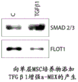
Certain aspects of the present disclosure relate to the following unexpected findings: culture conditions may be biased toward production of exosome types. For example, as described in the examples, culture conditions may bias the production of a-type exosomes versus f-type exosomes. In some embodiments, a three-dimensional (3D) culture enhances production of a-type exosomes versus f-type exosomes compared to conventional two-dimensional (e.g., monolayer) cultures. Methods for 3D culture are well known in the art and include, but are not limited to, hanging drop culture, culture on a substrate, culture on a microcarrier, culture on a synthetic extracellular scaffold, culture on a chitosan membrane, culture under magnetic suspension, culture in suspension in a rotating bioreactor, or culture under non-contact inhibitory conditions. See, e.g., Haycock JW. (2011), "3D cell culture: a review of current approaches and techniques ". Methods Mol biol.695: 1 to 15; lee, J; cuddihy MJ, Kotov NA. (3.14.2008.). Three-dimensional cell culture substrates: state of the art. 10.1089/teb.2007.0150; pampaloni, francisco (10.2007), "The third dimension bridges The gate beta cell culture and live tissue". Nature Reviews 8: 839-845; and Souza, Glauco (3/14 2010), "Three-dimensional tissue culture based on magnetic cell navigation". Nature Nanotechnology: 291-296; the entire contents of each are hereby incorporated by reference, respectively. In addition, it has also been unexpectedly found that the addition of certain growth factors enhances the production of a-type exosomes versus f-type exosomes. For example, the addition of more growth factors selected from the TGF β superfamily (TGFB1, activin, BMP, GDF, GDNF, inhibin, Nodal, Lefty, MIS) EGF, PDGF or FGF may enhance the production of a-type exosomes versus f-type exosomes.
In some embodiments, the enhancement (e.g., in the case of 3D culture and/or growth factor addition) comprises a 1.1-fold, 1.2-fold, 1.3-fold, 1.5-fold, 1.6-fold, 1.7-fold, 1.8-fold, 1.9-fold, 2.0-fold, 2.5-fold, 3.0-fold, 3.5-fold, 4.0-fold, 4.5-fold, 5.0-fold, 5.5-fold, 6.0-fold, 6.5-fold, 7.0-fold, 7.5-fold, 8.5-fold, 9.0-fold, 9.5-fold, 10.0-fold, 12.0-fold, 15.0-fold, or 20.0-fold increase or more of an a-type exosome relative to an f-type exosome.
Object
The methods of the present disclosure may be performed on any subject that may benefit from the methods, including human subjects, agricultural livestock (e.g., cattle, pigs, etc.), valuable animals (e.g., horses), companion animals (e.g., dogs, cats, etc.), and the like. In aspects of the present disclosure, human subjects are preferred. In some aspects, human subjects and human MSC exosomes are used.
The subjects may be those having a disease (or condition) described herein that are capable of being treated using exosomes described in the present disclosure, or the subjects may be those at risk of developing such a disease (or condition). Such subjects include neonates, particularly neonates born at low gestational age. Human neonates, as used herein, refers to a person from birth to about 4 weeks of age. A human infant as used herein refers to a human from about 4 weeks of age to about 3 steps of age. Low gestational age, as used herein, refers to birth (or delivery) occurring prior to the normal pregnancy of a given species. In humans, the full gestation period is about 40 weeks, and may range from 37 weeks to over 40 weeks. Low gestational age in humans similar to preterm birth is defined as birth occurring before 37 weeks of gestation. Accordingly, the present disclosure contemplates preventing and/or treating subjects born prior to 37 weeks of gestation, including those born at even shorter gestation periods (e.g., prior to 36 weeks of gestation, prior to 35 weeks of gestation, prior to 34 weeks of gestation, prior to 33 weeks of gestation, prior to 32 weeks of gestation, prior to 31 weeks of gestation, prior to 30 weeks of gestation, prior to 29 weeks of gestation, prior to 28 weeks of gestation, prior to 27 weeks of gestation, prior to 26 weeks of gestation, or prior to 25 weeks of gestation). Typically, such preterm infants are treated in neonates, however, the present disclosure contemplates treatment thereof even beyond the neonatal period and into childhood and/or adulthood. Certain subjects may have a genetic predisposition to certain forms of disease (or disorder) described herein, such as pulmonary hypertension, and these subjects may also be treated according to the present disclosure.
Methods for preventing and treating diseases
The present disclosure contemplates the prevention and treatment of certain diseases or conditions. Preventing a disease means reducing the likelihood of the disease manifesting itself and/or delaying the onset of the disease. Treating a disease means alleviating or eliminating the symptoms of the disease. The a-type exosomes described herein comprise a functional signaling component of the TGF/BMP pathway and have therapeutic efficacy in treating conditions in which this pathway is involved. Such disorders include certain Pulmonary and Vascular diseases, as described herein (see, e.g., Cai et al, "BMP Signaling in Vascular diseases" FEBS Lett.2012 (14): 1993: 2002; "Davies et al," TGF- β/BMP Signaling in cellular diseases, "Vascular compositions in Human diseases. Springer, 2008, pages 46-59; Alejardre-Alc zar et al," Hyperoxia models TGF-beta/Signaling in a motor Model of Vascular diseases. Springer, 2008, pages 46-59; Alejandre-Alc zar; Alexa Model of Vascular diseases. 2007; 292; L-49; Stumm et al, "Lung Model of Vascular diseases. beta. Involution of Vascular cells.9584; each of which is incorporated by reference herein as BMP No. 3: 357). Indeed, as demonstrated in the examples, treatment of mice by i.v. injection of a-type exosome preparations down-regulates hypoxic activation of signaling associated with vascular remodeling and pulmonary arterial hypertension and ameliorates hypoxia-induced pulmonary inflammation.
Accordingly, some aspects of the present disclosure provide compositions and methods for preventing and/or treating various lung (or pulmonary) diseases. These diseases include inflammatory lung diseases such as, but not limited to, pulmonary arterial hypertension (PH), which is also known as Pulmonary Arterial Hypertension (PAH), asthma, bronchopulmonary dysplasia (BPD), allergies, sarcoidosis and idiopathic pulmonary fibrosis. These diseases also include pulmonary vascular diseases that may not have an inflammatory component. Other lung disorders that may be treated according to the present disclosure include acute lung injury that may be associated with sepsis or with ventilation (ventilation). An example of this latter condition is acute respiratory distress syndrome occurring in older children and adults.
Pulmonary hypertension is a pulmonary disease characterized by blood pressure in the pulmonary artery that is much higher than normal. Symptoms include shortness of breath (breath of breath); chest pain, particularly during physical activity; frailty; fatigue; syncope; light headedness (light headedness), particularly during exercise; vertigo; abnormal heart sounds and murmurs; the jugular vein is full; fluid retention in the abdomen, legs and ankles; and bed bluing (liquid colouring in the nail bed).
Bronchopulmonary dysplasia is a condition that afflicts newborns that have been given oxygen or that have been on the ventilator or that are born prematurely, particularly those born extremely prematurely (e.g., those born before 32 weeks gestation). It is also known as chronic lung disease in neonates. Causes of BPD include mechanical injury (e.g., caused by ventilation), oxygen toxicity (e.g., caused by oxygen therapy), and infection. The disease may progress from non-inflammatory to inflammatory over time. Symptoms include cyan skin (bluish skin), chronic cough (chronic cough), rapid breathing (rapid breathing), and short breath. Subjects with BPD are more susceptible to infection, such as respiratory syncytial virus (respiratory syncytial virus). Subjects with BPD may develop pulmonary hypertension.
Acute Respiratory Distress Syndrome (ARDS), also known as Respiratory Distress Syndrome (RDS) or adult respiratory distress syndrome, is a condition that occurs as a result of lung injury or an acute illness. The lung injury may be caused by ventilation, trauma, burns, and/or inhalation. The acute disease may be infectious pneumonia or sepsis. It is considered a severe form of acute lung injury and it is often fatal. It is characterized by pulmonary inflammation, impaired gas exchange and release of inflammatory mediators, hypoxemia and multiple organ failure. ARDS may also be defined as arterial partial oxygen tension (PaO) in the presence of bilateral infiltration on chest X-ray2) And Fractional Inspired Oxygen (FiO)2) Ratio of (A to (B)Values below 200 mmHg. Less than 300mmHg PaO on both sides2/FiO2The ratio is indicative of acute lung injury, which is usually a precursor of ARDS. Symptoms of ARDS include shortness of breath, tachypnea (tachypnea) and confusion caused by low oxygen levels.
Idiopathic pulmonary fibrosis is characterized by an unclear occurrence of scarring or thickening of the lung. It usually occurs in people between 50 and 70 years of age. Symptoms include shortness of breath, frequent coughing (usually dry coughing), chest pain, and reduced activity levels.
Allergies are hypersensitivity disorders of the immune system, in which symptoms include conjunctival congestion (red eye), itching and runny nose (running nose), eczema, urticaria (hives) or asthma attacks. Allergies play a major role in disorders such as asthma. Severe allergic reactions to environmental or dietary allergens or to drugs can lead to life threatening reactions known as anaphylaxis. The allergic reaction can occur when the individual's immune system reacts to substances that are typically conventional harmless substances in the environment. Allergy is one of four forms of hypersensitivity and is sometimes referred to as type I (or immediate) hypersensitivity. Allergy is unique in that certain leukocytes (mast cells and basophils) are overactivated by immunoglobulin e (ige). This reaction results in an inflammatory response that can range from mild to dangerous. There are a number of tests for diagnosing allergic conditions. The tests include placing a possible allergen on the skin and observing reactions such as swelling, as well as blood tests looking for allergen specific IgE.
Hypoxia-induced pneumonia is a condition commonly caused by acute lung injury and/or ARDS, where the inflammatory response is caused by chronic exposure to hypoxic conditions. Such inflammatory responses include increased macrophages, basophils and inflammatory cytokines (including IL-1B, IL-6, IL-8 and TNF- α) in bronchoalveolar lavage fluid of humans exposed to hypoxia (hypobaric hypoxia).
Other conditions that can be treated using a-type exosomes (e.g., by enhancing The TGF/BMP pathway) include Cardiovascular diseases such as Myocardial Infarction, Cardiovascular Disease, hypertension, atherosclerosis, and Heart failure (see, e.g., Pardali et al, "TGF β Signaling and Cardiovascular diseases," Int J Biol Sci 2012; 8 (2): 195. sub., "Garside et al," coding Notch, BMP, and TGF- β Signaling along with vascular Disease, "Cell Mol Life scien Sci.2013; 70 (16): 2899. sub.," Wang et al, "Bmp Signaling in genetic Heart Disease," New elasticity and molecular diseases, "TGF failure" 2007, "TGF characteristics of biological diseases A. sub. molecular Teol 91, 91. sub.," TGF-400. sub., "TGF-molecular tissue research, and molecular research, 2, and molecular research, 2007, and molecular diagnosis, TGF-beta. sub.74, and molecular research, 2. sub.74. sub. 2): 184-; chang et al, "Impact of biochemical proteins and differentiation on the differentiation of metabolic stem cells: effect of ingredient myocarpial inactivation on Stem cell differentiation, "Stem cells.2008; 26(7): 1901-12; koitabashi et al, "Pivotal roll of cardiac cell TGF- β signaling in the muscle pathology response to supplemented compression overload," J Clin invest.2011; 121(6): 2301 and 2312; and Blann et al, "Serum levels of the TGF-beta receptor area amplified in atherospermis," atherospermis.1996; 120(1-2): 221-6; the entire contents of each are hereby incorporated by reference, respectively).
Myocardial infarction is a medical term for an event commonly referred to as a heart attack (heart attack). Myocardial infarction occurs when blood stops flowing properly to a portion of the heart and the myocardium is damaged by not receiving sufficient oxygen. This is often caused when one of the coronary arteries supplying blood to the heart becomes occluded due to an unstable accumulation of leukocytes, cholesterol and fat. An event is said to be "acute" if it is sudden and severe. Symptoms of acute myocardial infarction include sudden chest pain felt behind the sternum and sometimes progressing to the left arm or neck. In addition, the individual may have shortness of breath, sweating, nausea, vomiting, abnormal heartbeat, and anxiety. Women experience fewer of these symptoms than men, but often have shortness of breath, weakness, feeling of dyspepsia (feelings of fatigue), and fatigue.
Cardiovascular disease refers to any disease affecting the cardiovascular system, mainly heart disease, vascular diseases of the brain and kidneys, and peripheral artery disease. The causes of cardiovascular disease vary, but atherosclerosis and/or hypertension are the most common. In addition, with age, a variety of physiological and morphological changes occur that alter cardiovascular function and lead to an increased risk of cardiovascular disease, even in healthy asymptomatic individuals.
Atherosclerosis is a specific form of arteriosclerosis in which the arterial wall is thickened due to the invasion and accumulation of leukocytes and contains both live, active WBCs (inflammation) and residual, dead cells, which contain cholesterol and triglycerides, eventually calcium and other crystalline materials, in the outermost and oldest plaque. These changes reduce the elasticity of the arterial wall but do not affect blood flow over decades as the arterial muscle wall expands at the site of the plaque. However, wall stiffening may eventually increase pulse pressure; widening of the pulse pressure is a possible cause of advanced disease in the aorta. Symptoms may be caused by a significant narrowing of the coronary arteries, which are responsible for providing oxygenated blood to the heart, giving rise to, for example, the following symptoms: angina pectoris, chest pain and shortness of breath, sweating, nausea, dizziness or lightheadedness, dyspnea (breathlessness) or palpitations. Significant narrowing of the carotid artery can also be accompanied by symptoms such as: feelings of weakness; the inability to think clearly; difficulty in speaking; become stunned and difficult to walk and or erect; blurred vision; facial, arm and leg numbness; severe headache and loss of consciousness. These symptoms are also associated with stroke (i.e., death of brain cells). Stroke is caused by a significant narrowing/closure of the arteries that enter the brain; the lack of adequate blood supply leads to cell death in the affected tissue. Peripheral arteries that supply blood to the legs, arms and pelvis also experience significant narrowing due to plaque rupture and clotting. Symptoms of significant narrowing are numbness in the arms or legs, and pain. Another significant site of plaque formation is the renal arteries that supply blood to the kidneys. Plaque development and accumulation lead to reduced renal blood flow and chronic kidney disease, which is, like all other sites, often asymptomatic until late.
Hypertension (sometimes referred to as arterial hypertension) is a chronic medical condition in which blood pressure in the arteries is elevated. Hypertension is classified as primary (idiopathic) hypertension or secondary hypertension; about 90% to 95% of cases are classified as "essential hypertension", which means hypertension without obvious underlying medical cause. The remaining 5% to 10% of cases (secondary hypertension) are caused by other disorders affecting the kidney, arteries, heart or endocrine system. Hypertension stresses the heart, resulting in hypertensive heart disease and coronary artery disease if left untreated. Hypertension is also a major risk factor for stroke, aneurysms (e.g., aortic aneurysms), peripheral arterial disease, and is a cause of chronic kidney disease. Hypertension is rarely accompanied by any symptoms, and its identification is usually by screening, or when medical care is sought because of an unrelated problem. Some people with hypertension report headache (especially in the back of the head and in the morning) as well as dizziness, vertigo, tinnitus (buzzing or hissing in the ear), changes in vision or episodes of syncope.
Heart failure (often used to mean chronic heart failure) occurs when the heart is unable to provide sufficient pumping action to maintain blood flow to meet the body's needs. When edema is present in addition to the above, it is called Congestive Heart Failure (CHF) or congestive heart failure (CCF). Heart failure can cause a variety of symptoms including shortness of breath, swelling of the legs, and exercise intolerance (exercise intulerance). Common causes of heart failure include myocardial infarction (heart attack) and other forms of coronary artery disease, hypertension, valvular heart disease, and cardiomyopathy. The term heart failure is sometimes used incorrectly for myocardial infarction (which may cause heart failure but is not heart failure itself) or for cardiac arrest (in which blood flow effectively stops altogether).
Still other conditions that can be treated using a-type exosomes (e.g., by enhancing the TGF/BMP pathway) include kidney diseases such as ischemic kidney injury, acute renal failure, and renal fibrosis (see, e.g., Meng et al, "Role of the TGF- β/BMP-7/Smad pathways in renal disorders," Clin Sci (Lond): 2013; 124 (4): 243-54; zeberg et al, "BMP-7 cores TGF-beta1-induced temporal-sensory mutation and recovery of renal intrinsic output," Nat med.2003; 9 (7): 964-8; and Yanagita, "inhibition/aggregation of beta systems in kit" novel surgery trap transfer; 91.91; incorporated by reference herein in its entirety; 3686).
Ischemic kidney injury or ischemic kidney disease occurs when blood flow to the kidney is insufficient (hypoperfusion). Inadequate perfusion can lead to loss of kidney function and renal atrophy (shrinkage). However, this process damages both kidneys, resulting in renal failure. One of the following clinical situations is commonly present in ischemic renal disease: bilateral renal artery stenosis (RAS; narrowing of the aorta supplying both kidneys); unilateral RAS in humans with only one functional kidney; or unilateral RAS with hypertensive injury to another kidney. Symptoms of ischemic kidney injury include uremia (high blood levels of protein by-products (e.g., urea)); acute episodes of dyspnea (labourious or difficult breathing) caused by the sudden accumulation of fluid in the lungs; and based on the severity of the injury, hypertension may be present. Murmurs (sounds or murmurs heard with a stethoscope) caused by blood turbulence in the arteries can be detected in the neck (carotid mur), abdomen (which may reflect narrowing of the renal arteries), and groin (femoral murs).
Acute renal failure or Acute Kidney Injury (AKI) is a sudden loss of kidney function that usually occurs within 7 days. Its occurrence is generally caused by injury to renal tissue as follows: reduced renal blood flow (renal ischemia) due to any cause (e.g. hypotension), exposure to substances harmful to the kidney, inflammatory processes in the kidney, or urethral obstruction that impedes urine flow. Acute renal failure is diagnosed based on, for example, the following characteristic laboratory results: blood urea nitrogen and creatinine rise, or the kidney is unable to produce sufficient amounts of urine. Acute renal failure can lead to a number of complications, including metabolic acidosis, high potassium levels, uremia, altered fluid balance, and effects on other organ systems. Symptoms of acute kidney injury include urea accumulation, and other nitrogenous substances in the bloodstream cause many symptoms, such as fatigue, loss of appetite (loss of appetite), headache, nausea, and vomiting. Elevated potassium levels can lead to irregular heartbeats, which can be severe and life threatening. Fluid balance is usually affected, but hypertension is rare. Flank pain in some conditions (e.g., thrombosis of the renal vasculature or renal inflammation); this is a result of the extension of the fibrous tissue envelope around the kidney. If the kidney injury is caused by dehydration, there may be signs of thirst and fluid consumption at the time of physical examination. Physical examination may also provide other clues about the underlying cause of renal problems, such as an interstitial nephritis burst and palpable bladder (palpable). The reduced ability to drain sufficient fluid from the body can lead to fluid accumulation in the limbs (peripheral edema) and lungs (pulmonary edema), and cardiac tamponade (cardiac tamponade) caused by fluid exudates.
Renal fibrosis is caused by the excessive accumulation of extracellular matrix that occurs in almost every type of chronic kidney disease. The pathogenesis of renal fibrosis is a progressive process that often leads to end-stage renal failure. In some aspects, renal fibrosis represents a failure of the wound healing process of renal tissue following chronic, persistent injury. Many cellular pathways (e.g., mesenteric and fibroblast activation and tubular epithelial-mesenchymal transition) have been identified as the major cause of matrix-producing cells in diseased states. Fibrogenic factors that modulate the process of renal fibrosis, such as Transforming Growth Factor (TGF), contribute to the condition. Recent findings with endogenous anti-fibrotic factors (anti-fibrotic factors) have developed new strategies aimed at antagonizing the fibrogenic effects of TGF-/Smad signaling.
Still other conditions that can be treated using a-type exosomes (e.g., by enhancing the TGF/BMP pathway) include ischemic neurological diseases such as hypoxic ischemic encephalopathy or ischemic Stroke (see, e.g., Harvey et al, "linkage and TGF-beta proteins: neurological Cell line-derived neurological factor and bone morpholinogenic protein," pharmaceutical theory 2005; 105 (2): 113-25; Krampert et al, "Small 7 ligands the adaptive nerve/genetic Cell pore in a transforming factor beta-and bone morpholinogenic protein index," Mol Cell biol 2010; 30 (14): 3685-94; and YIn et al, "effective of viral expression of formula man. fig. 2016, incorporated by reference in patent publication J-66).
Hypoxic Ischemic Encephalopathy (HIE) or perinatal asphyxia is characterized by clinical and laboratory evidence of acute or subacute brain injury caused by asphyxia. The main causes of this condition are systemic hypoxemia and/or reduced cerebral blood flow. Worldwide, birth asphyxia causes 840,000 or 23% of all neonatal deaths. Signs and symptoms of mild hypoxic-ischemic encephalopathy include: muscle tone was slightly increased; and transient behavioral abnormalities (e.g., poor feeding, irritability, or excessive crying or sleep) may be observed. Symptoms of moderately severe hypoxic-ischemic encephalopathy include: infant lethargy and significant hypotonia (hypotonia) and a decrease in deep tendon reflex; the grip, morlo (Moro) and suck reflexes may be retarded or absent; infants may experience occasional short periods of breathing; and seizures (seizure) can occur early within the first 24 hours after birth. Symptoms of severe hypoxic-ischemic encephalopathy include seizures that are delayed and severe and that may initially develop resistance to conventional treatment. Seizures are usually systemic and their frequency can increase during the 24 to 48 hours after the seizure, which is associated with the period of reperfusion injury. Other symptoms include: stupor (stupor) or coma (infants may not respond to any physical stimulus other than the most harmful); breathing irregularity; low systemic tension and reduced deep tendon reflexes; absence of neonatal reflexes (e.g., sucking, swallowing, grasping, moro reflexes); ocular movement disorders (e.g., eye deviation, nystagmus, unsteadiness of the eye (bobbing)); mydriasis, immobilization, or poor reactivity to light; as well as heart rate and blood pressure irregularities.
Ischemic stroke is the loss of brain function caused by the disturbance of blood supply to the brain. Ischemia is caused by blockage of blood vessels by thrombosis or arterial embolism, or by inadequate cerebral perfusion. As a result, the affected brain region does not function properly, which can result in an inability to move one or more limbs on one side of the body, an inability to understand or express speech, or impaired vision on one side of the field of view.
In some cases, prevention and/or treatment may involve the use of type a exosomes, either alone or with one or more second agents. Mechanical intervention, such as ventilation, may also be performed on the subject in the presence or absence of exogenous oxygen administration.
For newborns, and in particular, for low gestational age newborns, the present disclosure contemplates administration of type a exosomes within 4 weeks, 3 weeks, 2 weeks, 1 week, 6 days, 5 days, 4 days, 3 days, 2 days, 1 day, 12 hours, 6 hours, 3 hours, or 1 hour of birth. In some critical cases, type a exosomes were administered within 1 hour of birth.
The present disclosure also contemplates administration of type a exosomes even in the absence of symptoms indicative of the diseases or disorders described herein.
The present disclosure also contemplates repeated administration of a-type exosomes, including two, three, four, five or more administrations of a-type exosomes. In some cases, the a-type exosomes may be administered continuously. Based on the severity of the condition being treated, repeated administration or continuous administration may occur over a period of hours (e.g., 1 to 2, 1 to 3, 1 to 6, 1 to 12, 1 to 18, or 1 to 24 hours), days (e.g., 1 to 2 days, 1 to 3 days, 1 to 4 days, 1 to 5 days, 1 to 6 days, or 1 to 7 days), or weeks (e.g., 1 to 2 weeks, 1 to 3 weeks, or 1 to 4 weeks). If administration is repeated but not continuous, the time between administrations can be hours (e.g., 4 hours, 6 hours, or 12 hours), days (e.g., 1 day, 2 days, 3 days, 4 days, 5 days, or 6 days), or weeks (e.g., 1 week, 2 weeks, 3 weeks, or 4 weeks). The time between administrations may be the same or it may be different. As one example, type a exosomes may be administered more frequently if symptoms of the disease show worsening, and then less frequently once the symptoms are stabilized or alleviated.
In some critical cases, type a exosomes are administered at least once within 24 hours of birth and then at least once more within 1 week of birth. Even more preferably, the a-type exosomes are administered at least once within 1 hour of birth and then at least once more within 3 to 4 days of birth.
In some cases, low doses of type a exosomes may be administered intravenously reproducibly. Thus, the present disclosure contemplates repeated administration of a low dose form of a-type exosomes as well as a single administration of a high dose form of a-type exosomes. The low dose form may range, but is not limited to, 1 to 50 micrograms/kg, while the high dose form may range, but is not limited to, 51 to 1000 micrograms/kg. It is understood that single or repeated administration of low-dose or high-dose a-type exosomes, based on the severity of the disease, the health condition of the subject, and the route of administration, among others, is also contemplated by the present disclosure.
Administration, pharmaceutical composition, effective amount
The type a exosomes may be used (e.g., administered) in a pharmaceutically acceptable formulation (or pharmaceutically acceptable composition) (typically when combined with a pharmaceutically acceptable carrier). Such formulations may routinely contain pharmaceutically acceptable concentrations of salt, buffering agents, preservatives, compatible carriers, and may optionally contain other (e.g., second) therapeutic agents.
A pharmaceutically acceptable carrier is a pharmaceutically acceptable material, composition or vehicle, such as a liquid or solid filler, diluent, excipient, solvent or encapsulating material, involved in the transport or delivery of a prophylactically or therapeutically active agent. Each carrier must be "acceptable" in the sense of being compatible with the other ingredients of the formulation and not deleterious to the subject. Some examples of materials that can serve as pharmaceutically acceptable carriers include: sugars such as lactose, glucose and sucrose; glycols, such as propylene glycol; polyols such as glycerol, sorbitol, mannitol and polyethylene glycol; esters such as ethyl oleate and ethyl laurate; buffering agents such as magnesium hydroxide and aluminum hydroxide; pyrogen-free water; isotonic saline (isotonic saline); ringer's solution; ethanol; phosphoric acid buffer solution; and other non-toxic compatible substances used in pharmaceutical formulations.
A second therapeutic agent. The exosomes may be administered with one or more second therapeutic agents. A therapeutic agent, as used herein, refers to any agent useful in the prevention, treatment and/or management of pulmonary diseases, such as those described herein. These include, but are not limited to, surfactants, inhalation nitric oxide, amitrazine dimesylate, immunomodulators and antioxidants. Examples of immunomodulators include steroids and corticosteroids, such as, but not limited to, methylprednisolone. Examples of antioxidants include, but are not limited to, superoxide dismutase.
Certain second therapeutic agents useful in the treatment or management of certain pulmonary and vascular diseases (including but not limited to pulmonary arterial hypertension) include oxygen; anticoagulants, such as warfarin (coumadin); diuretics, e.g. furosemide
Or Antipodium androsaceum
A calcium channel blocker; potassium, e.g.
Inotropic agents, such as digoxin; vasodilators, e.g. nifedipine
Or diltiazem
Endothelin receptor antagonists, e.g. bosentan
And ambrisentan
Prostacyclin analogues, e.g. epoprostenol
Treprostinil sodium
And iloprost
And PDE-5 inhibitors, such as sildenafil
And tadalafil
A surfactant. The a-type exosomes may be administered with a pulmonary surfactant. Lung surfactants are mixtures of lipoproteins that can be used to keep the lung airways open (e.g., by preventing alveolar walls from adhering to each other). The pulmonary surfactant may comprise: phospholipids, such as Dipalmitoylphosphatidylcholine (DPPC), Phosphatidylcholine (PC), Phosphatidylglycerol (PG); cholesterol; and proteins such as SP-A, B, C and D. The pulmonary surfactant may be obtained from a natural source (e.g., bovine or porcine lung tissue). Examples include AlveofactTM(Lung lavage fluid from cow), CurosurfTM(from minced pig lungs), InfasurfTM(Lung lavage from calf) and SurvantaTM(from minced cow lungs, with additional ingredients including DPPC, palmitic acid and tripalmitin). The pulmonary surfactant may also be synthetic. Examples include ExosurfTM(including DPPC with cetyl alcohol and tyloxapol), PumactantTMOr the Artificial Lung Expansion Compound (ALEC) (comprising DPPC and PG), KL-4 (comprising DPPC, palmitoyl-oleoyl phosphatidylglycerol, palmitic acid and synthetic peptides mimicking SP-B), VenticuteTM(including DPPC, PG, palmitic acid and recombinant SP-C). Pulmonary surfactants are available from commercial suppliers.
An effective amount. The formulations of the present disclosure are administered in an effective amount. An effective amount is the amount of the agent that alone stimulates the desired result. The absolute amount will depend on a number of factors, including the material selected for administration, whether administration is carried out in single or multiple doses, and the parameters of the individual patient, including age, physical condition, size, weight and disease stage. These factors are well known to those of ordinary skill in the art and can be addressed with only routine experimentation.
The route of administration. Type a exosomes may be administered by any route that achieves delivery to the lung or other tissue. Suitable are systemic routes of administration, such as intravenous bolus injection or continuous infusion. More direct routes, such as intranasal administration, intratracheal administration (e.g., by intubation), and inhalation (e.g., via mouth or nose by aerosol) are also within the contemplation of the present disclosure, and may be more suitable in some cases, particularly where rapid onset of action is necessary. As used herein, an aerosol is a suspension of a liquid dispersed in a gas as small particles (suspension), and it includes a fine mist (fine mist) or a spray (spray) containing such particles. Aerosolization (aerosolization), as used herein, is the process of generating an aerosol by converting a liquid suspension into small particles or droplets. This can be done using an aerosol delivery system (e.g., pressurized pack or nebulizer). Nebulizers include air jet nebulizers (i.e., pneumatic nebulizers), ultrasonic nebulizers, and vibrating mesh nebulizers, such as by use of a suitable propellant, for example, but not limited to, dichlorodifluoromethane, trichlorofluoromethane, dichlorotetrafluoroethane, carbon dioxide, or other suitable gas. In addition to nebulizers, other devices for pulmonary delivery include, but are not limited to, Metered Dose Inhalers (MDIs) and Dry Powder Inhalers (DPIs). Capsules and cartridges (e.g., gelatin capsules or cartridges) for use in an inhaler or insufflator may be formulated to contain a lyophilized exosome and a suitable powder base, such as lactose or starch.
Exosomes may be formulated for parenteral administration by injection (including, for example, by bolus injection or continuous infusion) when systemic delivery thereof is desired. Formulations for injection may be presented in unit dosage form, e.g., in ampoules or in multi-dose containers, with or without an added preservative.
The compositions may take such forms as aqueous suspensions, solutions or emulsions in oily or aqueous vehicles, and may contain formulatory agents such as suspending, stabilizing and/or dispersing agents. Suitable lipophilic solvents or carriers include fatty oils, such as sesame oil; or synthetic fatty acid esters, such as ethyl oleate or triglycerides. Aqueous injection suspensions may contain substances that increase the viscosity of the suspension, such as sodium carboxymethyl cellulose, sorbitol, or dextran. Optionally, the suspension may also contain suitable stabilizers or solubility enhancing substances. Alternatively, the exosomes may be in lyophilized or other powder or solid form for reconstitution with a suitable carrier (e.g., sterile pyrogen-free water) prior to use.
It is to be understood that other agents administered to a subject being treated according to the present disclosure may be administered by any suitable route, including oral administration, intranasal administration, intratracheal administration, inhalation, intravenous administration, and the like. One of ordinary skill in the art will know the conventional routes of administration for such second agents.
Medicine box
The present disclosure also encompasses packaged and labeled pharmaceutical products. Such articles (articles of manufacture) or kits (kits) comprise a suitable unit dosage form in a suitable vessel or container, for example a glass vial or a plastic ampoule or other container hermetically sealed. The unit dosage form should be suitable for pulmonary delivery, for example by aerosol. Preferably, the article of manufacture or kit further comprises instructions on how to use (including how to administer) the pharmaceutical product. The instructions may also contain informational material suggesting to a medical practitioner, technician, or subject how to suitably prevent or treat the disease or condition in question. In other words, the article of manufacture contains instructions indicating or suggesting a dosing regimen for use (including but not limited to actual dosages, monitoring procedures, and other monitoring information).
For any pharmaceutical product, the packaging material and containers are designed to preserve the stability of the product during storage and transport.
The kit may comprise the exosomes in a sterile aqueous suspension which may be used directly or may be diluted with physiological saline for intravenous injection or for nebuliser, or diluted with or combined with a surfactant for intratracheal administration. Thus, the kit may further comprise a diluent solution or diluent, such as saline or a surfactant. Thus, the kit may also contain a transpulmonary delivery device, such as a nebulizer or a disposable component (mask), such as a mouthpiece (mouthpiece), a nose piece (nosepiece), or a face mask (mask).
Examples
Materials and methods
Human MSCs were isolated from human umbilical cord collagen. MSCs derived from Wharton's jelly of human umbilical cord (hUC-MSCs) were isolated according to the published methods (Mitchell, K.E. et al, 2003, Stem Cells 21: 50-60; and Penolazzi, L. et al, 2011, J Cell Physiol) with minor modifications. The umbilical cord was washed twice with ice-cold sterile PBS, dissected longitudinally and the arteries and veins removed. Scraping the soft gel tissue and finely cutting (2 to 3 mm)2) And placed directly on 100mM dishes (15 blocks/dish) with DMEM/F12 (1: 1) (Invitrogen) supplemented with 10% fetal bovine serum (Hyclone), 2mM L-glutamine and penicillin/streptomycin, at 5% CO2Was incubated at 37 ℃ for 5 days in a humid atmosphere. After removing tissue and media, plates were washed 3 times with PBS, adherent cells were cultured and fresh media was changed 3 times per week. At 70% to 80% confluence, cells were collected and stained with PE conjugated antibodies against CD34(Miltenybiotec) and CD45 (Miltenybiotec). The immunoblotting was performed using anti-PE microbeads (Miltenybiotec) and MSCS columns (Miltenybiotec) according to the manufacturer's instructions. The CD34 and CD45 negative populations were further propagated and selected using MoFlo flow cytometry (Beckman Coulter) by using a specific fluorescence labelled antibody panel (BD Biosciences) for characterization of human MSCs according to the expression of MSC markers (CD105, CD90, CD44 and CD73) and the absence of CD11b, CD19 and HLA-DR.
And (4) preparing a conditioned medium. To exclude contamination from serum-derived microvesicles, serum used to proliferate the cell culture and collect the conditioned medium was clarified by ultracentrifugation at 100,000x g for 18 hours. MSCs were cultured in α -MEM medium supplemented with 10% (v/v) fetal bovine serum (FBS, Hyclone), 10% (v/v) horse serum (Hyclone), and 5mM L-glutamine (Gibco). At 70% confluence, the cultures were washed twice with PBS and incubated with serum-free medium supplemented with 2mM L-glutamine for 24 hours under standard culture conditions. Conditioned media (in microparticle-depleted FBS cultures) was collected from nearly confluent cultures of human MSCs over a period of 24 to 48 hours, or 3 to 4 days, and cells and debris were removed by differential centrifugation: 5 minutes at 400Xg, 10 minutes at 2,000x g and 30 minutes at 13,000x g. Subsequently, the clarified conditioned medium is filtered through a 0.2 μm filter unit and concentrated at least 10-fold using a tangential flow filtration system with a cut-off of 100kD or 300kD or 500 kD. Protein levels in conditioned media were determined by Bradford assay (Bio-Rad).
And (5) separating an efflux body. MSC exosomes were isolated from concentrated conditioned media by centrifugation at 100k × g for 3.5 hours on a 10% to 60% step gradient of iodixanol. Exosomes were further separated from conditioned media by differential centrifugation, sometimes followed by a 30% sucrose pad or sucrose velocity gradient. The particle number of the isolated exosome preparation was determined by Nanosight.
Western blot of exosomes. The exosome preparations were separated on 12% polyacrylamide gels and then transferred onto 0.45 μm PVDF membranes (Millipore). Goat polyclonal anti-CD 63 (1: 1,000; Santa Cruz) antibody, polyclonal rabbit anti-CD 81 (1: 1,000, Santa Cruz) and monoclonal anti-Dicer (1: 1,000, Abcam) were used. Primary antibodies against ALIX, flo-1, TSG101, TGF β R2, SMAD, CD105, CD9, CD81, CAV1, EFGR and AKT were also used to identify exosome subpopulations. Immunoreactive bands were visualized by enhanced chemiluminescence reagent (Pierce) or Lumi-LightPLUS (Roche) using peroxidase conjugated anti-rabbit secondary antibody (Santa Cruz) at a dilution of 1: 20,000. Quantification was performed by densitometric analysis (densitometric analysis) using NIH's ImageJ program after appropriate background subtraction.
And (4) quantifying micro RNA. Total exosome RNAs were extracted by the method of Chomczynski and Sacchi (1987Anal Biochem 162: 156-. Each reverse transcription reaction also included primers for small nuclear rna (small nuclear) sno202, which served as an internal control. 37.5ng cDNA was used for each 20. mu.l qPCR reaction with TaqMan Universal Master mix II (Universal Master mix II) (Applied Biosystems) without UNG in the presence of the indicated microRNAs and probes specific for the internal controls (TaqMan microRNA assay, Applied Biosystems). Amplification was performed on a StepOne Plus platform (Applied Biosystems) as follows: 2 minutes at 50 ℃; 10 minutes at 95 ℃; this is followed by 40 cycles: 15 seconds at 95 ℃ and 1 minute at 60 ℃.
Animals and hypoxic exposure. FVB male mice, 8 weeks old, were obtained from Charles River Laboratories (Wilmington, Mass.) or housed in the animal facilities of Boston's Children Hospital, Children's Hospital Boston. Mice from each group were exposed to 8.5% oxygen in Plexiglas chambers (OxyCycler, BioSpherix, Redfield, NY) for different experimental times. Adjusting aeration to remove CO2Such that it does not exceed 5,000ppm (0.5%) (average range 1,000 to 3,000 ppm). Ammonia was removed by air purifier via aeration and activated carbon filtration. All Animal protocols were approved by the Children Hospital Animal Care and Use Committee (Children's Hospital Animal Care and Use Committee).
Example 1
Identification and characterization of two different exosome populations derived from mesenchymal stem cells
Accumulated evidence supports a key role for MSCs in lung homeostasis and repair. It has been previously reported that MSC Exosomes (MEX) mediate the cytoprotective effect of mesenchymal stem cells on hypoxia-induced pulmonary hypertension. Intravenous mouse MEX delivery inhibits hypoxic lung influx of macrophages and induction of pro-inflammatory and pro-proliferative mediators, and ultimately vascular remodeling and hypoxic pulmonary hypertension in the murine PH model.
The purpose of this study was to investigate whether MSCs isolated from human umbilical cord interstitium secrete microvesicles exhibiting similar protective properties, as well as to characterize the biochemical properties of therapeutic MSC exosomes and study their effect on the phenotypic changes of vascular cells triggered by signals initiating pulmonary vascular remodeling.
Separation of MEX
Exosomes (MEX) were isolated from tissue culture media conditioned by human Mesenchymal Stem Cells (MSC). Here, the source of human MSCs is Wharton's Jelly (WJ), but other possible sources are cord blood, bone marrow, fat or other tissues. MSCs were isolated, immunoselected, cultured and evaluated for differentiation potential. Negative selections for human WJ-MSC included CD34 and CD 45. MSCs were positive for CD90, CD73, CD105 and CD 44.
Treatment of mice with MEX
One dose of MEX was delivered to animals by tail vein injection and recipient mice were exposed to normotensive hypoxia (8% to 10% O)2) For 2.5 days. Reduction of hypoxia-induced inflammatory markers (e.g., CCL2, IL6) was used as a measure of the effectiveness of the preparation. The dose used corresponds to about 100 to 200 billion particles, or approximately to the total exosomes produced by 1000 to 2000 million MSCs in monolayer culture over 24 hours, as measured by NanoSight.
Results
Mice treated by i.v. injection of MEX preparations down-regulated hypoxia activation of signaling associated with vascular remodeling and pulmonary arterial hypertension (fig. 1A) and ameliorated hypoxia-induced lung inflammation (fig. 1B). Figure 1A shows that WJ-MEX treatment reduces hypoxia-induced phosphorylation of AKT and its downstream target mTOR. As shown in fig. 1B, lung protein levels of DNA binding/differentiation protein inhibitor ID1 (direct SMAD targeting gene and downstream signaling of BMPR 2) were inhibited by hypoxia, but increased under MEX. Alpha tubulin served as a normalization control.
The presence of more than one MEX subtype population was investigated by density and speed ultracentrifugation. Analysis of MEX preparations by density and speed ultracentrifugation revealed that the extracellular microvesicle population produced by MSCs was composed of two types based on molecular markers (fig. 2 and 3). These two types are referred to as a-MEX and f-MEX. Efflux biomarkers such as ALIX and TSG101 are preferentially enriched in fractions 12 to 14(a-MEX), while tetraspanin CD9 and CD81 and lipid raft markers such as flotilin 1 (float 1) and crater 1(CAV1) are enriched in fractions 5 to 8 (f-MEX). Table 1 shows specific markers for a-MEX and f-MEX:
TABLE 1
| a-MEX specific markers
|
f-MEX specific markers
|
| ALIX
|
FLOT1
|
| TSG101
|
CD9
|
| TGFβR2
|
CD81
|
| SMAD(1,2,3,5)
|
CAV1
|
| CD105
|
EFGR
|
| |
AKT(2,3) |
Preparations of a-MEX or f-MEX by EM showed different morphologies (FIG. 4). f-MEX has a diameter of 30 to 200nm and exhibits the typical deep cup-shaped morphology of exosomes, while a-MEX is 30 to 100nm with a translucent and spherical morphology.
To prepare an exosome subpopulation highly enriched with a-MEX or f-MEX from a bulk (bulk) exosome preparation obtained from MSCs grown in monolayer culture, the bulk exosome preparation was adjusted to 45% sucrose, layered on a 60% sucrose pad and overlayed with 35% to 5% sucrose of the distribution gradient. The preparation was centrifuged at 180k Xg for 20 hours to isolate a-MEX and f-MEX subsets. Gradient fractions containing a-MEX or f-MEX were concentrated by ultracentrifugation (100k Xg, 3 hours) and resuspended in PBS or filtered by tangential flow.
Administration of isolated a-MEX or f-MEX to mice exposed to hypoxia revealed immunosuppressive properties of MEX residues present only in a-MEX types (fig. 5). Treatment with a-MEX caused a significant reduction in mRNA levels of hypoxia-induced inflammatory markers including CCL2, IL6, and ADM.
Enhanced production of a-MEX in 3-D cultures
MSCs and certain other cell types can form aggregates of cells called spheroids. Figure 6 shows microscopy images depicting MSC spheroid formation under 2-D and 3D culture conditions. Spheroid formation is considered to represent a more physiological condition than two-dimensional monolayer cultures. For MSCs, the spheroid state is believed to represent a more undifferentiated stem cell-like state. Three-dimensional culture systems that enhance the propensity to form spheroids are commercially available (special membranes). MSCs can spontaneously grow from monolayer cultures at high density to form spheroids and can be induced to form spheroids if suspended in the culture medium (hanging drop technique).
Hanging drop technology is a well-known culture method for many cell types. Briefly, MSCs grown in monolayer culture were trypsinized to produce single cell suspensions. 30 μ L of this suspension containing 500,000 cells was applied to the inner surface of a petri dish (petri dish) lid and the lid was placed on a dish containing a small amount of PBS to maintain humidity. The dish with the hanging drop was placed in a tissue incubator for 1 to 2 days, during which time the MSCs aggregated into spheroids. A typical spheroid preparation consists of 1500 ten thousand cells divided into 30 drops.
The data show that MSCs cultured in 3D cultures (spheroids) produced significantly more a-MEX than the same MSC clone cultured in the same generation in 2D cultures (monolayers) (figure 7). The increase in a-MEX production was demonstrated by the strong immunoreactivity of the samples to SMAD2/3 and ALIX antibodies but not to flo 1, as shown in figure 7.
It was also found that the production of a-MEX was variable between different MSC clones and that the producing cells could be enriched by 3D culture and/or growth factor supplementation. Relative a-MEX production of a variety of independent source MSC clones was characterized (the ratio of flo 1 relative to ALIX (or SMAD) marker in their conditioned media during 2D culture was determined and substantial changes were observed) (fig. 8A). Some clones (e.g., clone D) did not produce significant amounts of SMAD (a-MEX marker compared to flo 1(f-MEX marker)). This property of clone D correlates with the very inefficient sphere formation using hanging drop technology (fig. 8B).
Notably, when few spheroids formed from clonal D cells are expanded as a monolayer, the resulting 2D culture produces a-MEX at a high rate and forms spheroids with high efficiency. These observations indicate that selection by spheroid formation can be used as a tool to enrich for inefficient MSC clones to obtain cell subsets that retain their sternness (stemness) and produce a-MEX in high proportion. Towards this goal, it was observed that controlling the concentration of growth factors (e.g., TGF β 1) can accelerate spheroid formation and enhance the production of a-MEX (fig. 9). TGF-beta1 stimulation was achieved at a concentration of 10 ng/ml.
a-MEX and f-MEX carry distinct signal transduction modules for specific growth factors
a-MEX has TGF receptor and downstream transcription factor SMAD (fig. 2 and 3). Addition of TGF β 1 to isolated a-MEX preparations revealed that this signaling module was functional, as the SMAD module within a-MEX was efficiently and specifically phosphorylated in vitro (fig. 10A). This property was specific for a-MEX (FIG. 10B). f-MEX contains the EGF receptor and phosphorylates endogenous AKT in vitro (not shown).
The data indicate that MSCs produce two different types of exosomes: the a-MEX type carries the functional signaling module of the TGF/BMP growth factor superfamily, while the f-MEX carries the corresponding FGF/PDGF superfamily module. These exosomes transfer modules to target cells and enhance the corresponding signaling pathways, and their relative secretion ratios depend on the programming of the MSC population that produces them.
Furthermore, a-MEX, but not f-MEX, was found to inhibit TGF-induced transformation of fibroblasts into myofibroblasts in human lung cells, as shown in FIG. 11. Human lung fibroblasts after 3 days serum starvation (0.1% FBS) were stimulated with TGF β to undergo myofibroblast differentiation, which is marked by an increase in α -Smooth Muscle Actin (SMA) expression. FIG. 11A shows that pretreatment with hMEX (2. mu.g/ml) abolished the effect of TGF β on α -SMA protein levels. To further test this finding, human lung fibroblasts after 24 hour serum starvation were stimulated with TGF β to induce the myofibroblast marker PAI 1. Treatment with a-MEX prevented PAI1 upregulation, which was not observed with f-MEX (FIG. 11B). It was also found that a-MEX treatment down-regulated baseline MCP-1 and IL1 β mRNA levels in human lung fibroblasts in vitro (fig. 11C).
Equivalent scheme
This disclosure is not limited in its application to the details of construction and the arrangement of components set forth in the following description or illustrated in the drawings. The disclosure is capable of other embodiments and of being practiced or of being carried out in various ways. Also, the phraseology and terminology used herein is for the purpose of description and should not be regarded as limiting. The use of "including," "comprising," or "having," "containing," "involving," and variations thereof herein, is meant to encompass the items listed thereafter and equivalents thereof as well as additional items.
Having thus described several aspects of at least one embodiment of this disclosure, it is to be appreciated various alterations, modifications, and improvements will readily occur to those skilled in the art. Such alterations, modifications, and improvements are intended to be part of this disclosure, and are intended to be within the spirit and scope of the disclosure. Accordingly, the foregoing description and drawings are by way of example only.