CN106687589B - DNA amplification technology - Google Patents
DNA amplification technology Download PDFInfo
- Publication number
- CN106687589B CN106687589B CN201580048495.8A CN201580048495A CN106687589B CN 106687589 B CN106687589 B CN 106687589B CN 201580048495 A CN201580048495 A CN 201580048495A CN 106687589 B CN106687589 B CN 106687589B
- Authority
- CN
- China
- Prior art keywords
- nucleic acid
- target nucleic
- primer
- sequence
- amplification
- Prior art date
- Legal status (The legal status is an assumption and is not a legal conclusion. Google has not performed a legal analysis and makes no representation as to the accuracy of the status listed.)
- Expired - Fee Related
Links
Images


Classifications
-
- C—CHEMISTRY; METALLURGY
- C12—BIOCHEMISTRY; BEER; SPIRITS; WINE; VINEGAR; MICROBIOLOGY; ENZYMOLOGY; MUTATION OR GENETIC ENGINEERING
- C12Q—MEASURING OR TESTING PROCESSES INVOLVING ENZYMES, NUCLEIC ACIDS OR MICROORGANISMS; COMPOSITIONS OR TEST PAPERS THEREFOR; PROCESSES OF PREPARING SUCH COMPOSITIONS; CONDITION-RESPONSIVE CONTROL IN MICROBIOLOGICAL OR ENZYMOLOGICAL PROCESSES
- C12Q1/00—Measuring or testing processes involving enzymes, nucleic acids or microorganisms; Compositions therefor; Processes of preparing such compositions
- C12Q1/68—Measuring or testing processes involving enzymes, nucleic acids or microorganisms; Compositions therefor; Processes of preparing such compositions involving nucleic acids
- C12Q1/6844—Nucleic acid amplification reactions
- C12Q1/6853—Nucleic acid amplification reactions using modified primers or templates
-
- C—CHEMISTRY; METALLURGY
- C12—BIOCHEMISTRY; BEER; SPIRITS; WINE; VINEGAR; MICROBIOLOGY; ENZYMOLOGY; MUTATION OR GENETIC ENGINEERING
- C12Q—MEASURING OR TESTING PROCESSES INVOLVING ENZYMES, NUCLEIC ACIDS OR MICROORGANISMS; COMPOSITIONS OR TEST PAPERS THEREFOR; PROCESSES OF PREPARING SUCH COMPOSITIONS; CONDITION-RESPONSIVE CONTROL IN MICROBIOLOGICAL OR ENZYMOLOGICAL PROCESSES
- C12Q1/00—Measuring or testing processes involving enzymes, nucleic acids or microorganisms; Compositions therefor; Processes of preparing such compositions
- C12Q1/68—Measuring or testing processes involving enzymes, nucleic acids or microorganisms; Compositions therefor; Processes of preparing such compositions involving nucleic acids
- C12Q1/6844—Nucleic acid amplification reactions
- C12Q1/6846—Common amplification features
-
- C—CHEMISTRY; METALLURGY
- C12—BIOCHEMISTRY; BEER; SPIRITS; WINE; VINEGAR; MICROBIOLOGY; ENZYMOLOGY; MUTATION OR GENETIC ENGINEERING
- C12Q—MEASURING OR TESTING PROCESSES INVOLVING ENZYMES, NUCLEIC ACIDS OR MICROORGANISMS; COMPOSITIONS OR TEST PAPERS THEREFOR; PROCESSES OF PREPARING SUCH COMPOSITIONS; CONDITION-RESPONSIVE CONTROL IN MICROBIOLOGICAL OR ENZYMOLOGICAL PROCESSES
- C12Q1/00—Measuring or testing processes involving enzymes, nucleic acids or microorganisms; Compositions therefor; Processes of preparing such compositions
- C12Q1/68—Measuring or testing processes involving enzymes, nucleic acids or microorganisms; Compositions therefor; Processes of preparing such compositions involving nucleic acids
- C12Q1/6811—Selection methods for production or design of target specific oligonucleotides or binding molecules
-
- C—CHEMISTRY; METALLURGY
- C12—BIOCHEMISTRY; BEER; SPIRITS; WINE; VINEGAR; MICROBIOLOGY; ENZYMOLOGY; MUTATION OR GENETIC ENGINEERING
- C12Q—MEASURING OR TESTING PROCESSES INVOLVING ENZYMES, NUCLEIC ACIDS OR MICROORGANISMS; COMPOSITIONS OR TEST PAPERS THEREFOR; PROCESSES OF PREPARING SUCH COMPOSITIONS; CONDITION-RESPONSIVE CONTROL IN MICROBIOLOGICAL OR ENZYMOLOGICAL PROCESSES
- C12Q1/00—Measuring or testing processes involving enzymes, nucleic acids or microorganisms; Compositions therefor; Processes of preparing such compositions
- C12Q1/68—Measuring or testing processes involving enzymes, nucleic acids or microorganisms; Compositions therefor; Processes of preparing such compositions involving nucleic acids
- C12Q1/6844—Nucleic acid amplification reactions
- C12Q1/686—Polymerase chain reaction [PCR]
-
- C—CHEMISTRY; METALLURGY
- C12—BIOCHEMISTRY; BEER; SPIRITS; WINE; VINEGAR; MICROBIOLOGY; ENZYMOLOGY; MUTATION OR GENETIC ENGINEERING
- C12Q—MEASURING OR TESTING PROCESSES INVOLVING ENZYMES, NUCLEIC ACIDS OR MICROORGANISMS; COMPOSITIONS OR TEST PAPERS THEREFOR; PROCESSES OF PREPARING SUCH COMPOSITIONS; CONDITION-RESPONSIVE CONTROL IN MICROBIOLOGICAL OR ENZYMOLOGICAL PROCESSES
- C12Q1/00—Measuring or testing processes involving enzymes, nucleic acids or microorganisms; Compositions therefor; Processes of preparing such compositions
- C12Q1/68—Measuring or testing processes involving enzymes, nucleic acids or microorganisms; Compositions therefor; Processes of preparing such compositions involving nucleic acids
- C12Q1/6876—Nucleic acid products used in the analysis of nucleic acids, e.g. primers or probes
- C12Q1/6888—Nucleic acid products used in the analysis of nucleic acids, e.g. primers or probes for detection or identification of organisms
- C12Q1/689—Nucleic acid products used in the analysis of nucleic acids, e.g. primers or probes for detection or identification of organisms for bacteria
-
- C—CHEMISTRY; METALLURGY
- C12—BIOCHEMISTRY; BEER; SPIRITS; WINE; VINEGAR; MICROBIOLOGY; ENZYMOLOGY; MUTATION OR GENETIC ENGINEERING
- C12Q—MEASURING OR TESTING PROCESSES INVOLVING ENZYMES, NUCLEIC ACIDS OR MICROORGANISMS; COMPOSITIONS OR TEST PAPERS THEREFOR; PROCESSES OF PREPARING SUCH COMPOSITIONS; CONDITION-RESPONSIVE CONTROL IN MICROBIOLOGICAL OR ENZYMOLOGICAL PROCESSES
- C12Q2525/00—Reactions involving modified oligonucleotides, nucleic acids, or nucleotides
- C12Q2525/30—Oligonucleotides characterised by their secondary structure
- C12Q2525/301—Hairpin oligonucleotides
-
- C—CHEMISTRY; METALLURGY
- C12—BIOCHEMISTRY; BEER; SPIRITS; WINE; VINEGAR; MICROBIOLOGY; ENZYMOLOGY; MUTATION OR GENETIC ENGINEERING
- C12Q—MEASURING OR TESTING PROCESSES INVOLVING ENZYMES, NUCLEIC ACIDS OR MICROORGANISMS; COMPOSITIONS OR TEST PAPERS THEREFOR; PROCESSES OF PREPARING SUCH COMPOSITIONS; CONDITION-RESPONSIVE CONTROL IN MICROBIOLOGICAL OR ENZYMOLOGICAL PROCESSES
- C12Q2527/00—Reactions demanding special reaction conditions
- C12Q2527/107—Temperature of melting, i.e. Tm
-
- C—CHEMISTRY; METALLURGY
- C12—BIOCHEMISTRY; BEER; SPIRITS; WINE; VINEGAR; MICROBIOLOGY; ENZYMOLOGY; MUTATION OR GENETIC ENGINEERING
- C12Q—MEASURING OR TESTING PROCESSES INVOLVING ENZYMES, NUCLEIC ACIDS OR MICROORGANISMS; COMPOSITIONS OR TEST PAPERS THEREFOR; PROCESSES OF PREPARING SUCH COMPOSITIONS; CONDITION-RESPONSIVE CONTROL IN MICROBIOLOGICAL OR ENZYMOLOGICAL PROCESSES
- C12Q2565/00—Nucleic acid analysis characterised by mode or means of detection
- C12Q2565/10—Detection mode being characterised by the assay principle
- C12Q2565/101—Interaction between at least two labels
-
- C—CHEMISTRY; METALLURGY
- C12—BIOCHEMISTRY; BEER; SPIRITS; WINE; VINEGAR; MICROBIOLOGY; ENZYMOLOGY; MUTATION OR GENETIC ENGINEERING
- C12Q—MEASURING OR TESTING PROCESSES INVOLVING ENZYMES, NUCLEIC ACIDS OR MICROORGANISMS; COMPOSITIONS OR TEST PAPERS THEREFOR; PROCESSES OF PREPARING SUCH COMPOSITIONS; CONDITION-RESPONSIVE CONTROL IN MICROBIOLOGICAL OR ENZYMOLOGICAL PROCESSES
- C12Q2600/00—Oligonucleotides characterized by their use
- C12Q2600/158—Expression markers
Landscapes
- Chemical & Material Sciences (AREA)
- Life Sciences & Earth Sciences (AREA)
- Organic Chemistry (AREA)
- Proteomics, Peptides & Aminoacids (AREA)
- Zoology (AREA)
- Wood Science & Technology (AREA)
- Health & Medical Sciences (AREA)
- Engineering & Computer Science (AREA)
- Analytical Chemistry (AREA)
- Biophysics (AREA)
- Immunology (AREA)
- Microbiology (AREA)
- Molecular Biology (AREA)
- Biotechnology (AREA)
- Physics & Mathematics (AREA)
- Biochemistry (AREA)
- Bioinformatics & Cheminformatics (AREA)
- General Engineering & Computer Science (AREA)
- General Health & Medical Sciences (AREA)
- Genetics & Genomics (AREA)
- Chemical Kinetics & Catalysis (AREA)
- Measuring Or Testing Involving Enzymes Or Micro-Organisms (AREA)
Abstract
本发明描述了适合用于进行聚合酶链式反应的方法和试剂。具体而言,公开了允许有效扩增多个核酸靶并且具有改进的灵敏度的核酸扩增设计策略和热循环曲线。本公开还描述了用于提高引物的解链温度(Tm)的方法和设备。

The present invention describes methods and reagents suitable for performing the polymerase chain reaction. Specifically, nucleic acid amplification design strategies and thermocycling profiles are disclosed that allow for efficient amplification of multiple nucleic acid targets with improved sensitivity. The present disclosure also describes methods and apparatus for increasing the melting temperature (Tm) of primers.
Description
Cross Reference to Related Applications
This application claims priority to U.S. provisional application No. 62/023,123 filed on 10/7/2014, U.S. provisional application No. 62/075,769 filed on 5/11/2014, and U.S. provisional application No. 62/115,559 filed on 12/2/2015, each of which is hereby incorporated by reference in its entirety.
Statement regarding sequence listing
The sequence listing accompanying this application is provided in text form instead of a paper copy and is hereby incorporated into the specification by reference. The name of the text file containing the sequence listing is FLUO-004_03WO _ ST25. txt. The text file is about 15KB, created on 7 months and 9 days 2015, and submitted electronically via EFS-Web.
Technical Field
The present disclosure relates to methods and materials useful for performing PCR amplification. In particular, nucleic acid amplification design strategies and thermal cycling profiles (thermal cycling profiles) are disclosed that allow for efficient amplification of multiple nucleic acid targets and have improved sensitivity.
The present disclosure also describes methods and apparatus for increasing the melting temperature (Tm) of a primer. Specifically, primers with synthetic tags attached to them are used to reduce the range between the Tm of the amplicon and the Tm of the primer (range).
Background
PCR amplification is traditionally achieved via several amplification cycles, where each cycle includes the steps of initial denaturation, annealing, polymerization, and final extension. These cycles are typically performed in a reaction chamber, wherein the necessary PCR reagents are provided, including a biological sample containing the target nucleotide sequence (typically DNA or RNA), a DNA polymerase (e.g., Taq polymerase), nucleoside triphosphates, an RT enzyme, and first and second primers (comprising a primer pair) that hybridize to the target DNA and flank the amplified DNA product sequence ("amplicon"). PCR devices will typically include means for cycling the reaction chamber temperature as required for each step of the amplification cycle, including, for example, "melting" of double-stranded DNA to produce single-stranded DNA; annealing the primer to the single-stranded DNA template; and the amplified DNA is extended by a polymerase.
The precise conditions for amplifying a particular target DNA sequence may vary depending on a number of factors, which are within the knowledge of one of ordinary skill in the art. In some embodiments of conventional DNA amplification, denaturation is performed at about 90-95 ℃ for about 10-30 seconds, and annealing is performed at about 45-65 ℃ for about 10-30 seconds; the extension is carried out at a temperature of between about 70 and 75 ℃ for about 10 to 90 seconds; and final extension was performed at 72 ℃ for about 5 minutes. In some embodiments, the reaction mixture comprises genomic DNA, MgCl2And other physiological salts (e.g., NaCl), PCR buffer, 0.1-1.0mM dNTPs, 0.04-1.5 μ M of each primer, and 0.5-5.0 units of thermostable polymerase (e.g., taq. polymerase).
Other amplification methods known in the art may also be used, including, for example, self-sustained sequence replication (3SR), Strand Displacement Amplification (SDA); "branched" DNA amplification (Chiron Corp.); ligase Chain Reaction (LCR), QB replicase amplification (QBR), Ligation Activated Transcription (LAT), Nucleic Acid Sequence Based Amplification (NASBA), Repair Chain Reaction (RCR) and Cycling Probe Reaction (CPR) (reviewed, for example, in The genes Report, DX; Vol.3(4), pages 2-7 (2 months 1994)).
Real-time PCR typically relies on the use of fluorescent molecules that allow for real-time quantification or detection of PCR products, while other detection/quantification chemistries, such as electrochemistry, are also suitable.
The fluorescent molecule may be a DNA binding dye such as Green or fluorescently labeled primers or probes. There are many fluorescent dyes and probe designs available for different applications. The most commonly used DNA binding dyes for real-time PCR are
 Green I, which binds preferentially to double stranded DNA (dsdna) over single stranded DNA. SYBR Green I fluorescence increases up to 1000-fold when it binds to dsDNA. Thus, the fluorescence signal is proportional to the amount of dsDNA present.
The major drawback of DNA binding dyes is that they lack specificity, that is, the DNA binding dyes bind to any dsDNA. As such, the presence of any non-specific products in a real-time or end-point PCR reaction contributes to overall fluorescence and affects the accuracy of the quantification or detection. In addition, DNA binding dyes cannot be used for quantification or detection in multiplex reactions because the fluorescent signals from different products cannot be distinguished without post-PCR melting curve analysis involving the formation of distinct products.
In contrast, primer-based and probe-based detection chemistry ensures that a signal is only produced when a product of interest is amplified. The primer or target-specific oligonucleotide probe is typically labeled with a reporter fluorophore, but in most cases, fluorescence is quenched when the particular target has not been amplified or is not present in the sample. Typically this is achieved by attaching a quencher molecule to the primer or probe and designing some mechanism by which the reporter and quencher are separated when the primer or probe binds to its particular target.
The main primer/probe detection chemistries used today are as follows:
hydrolysis (TaqMan) probes
Hydrolysis assays include sequence-specific, fluorescently labeled oligonucleotide probes in addition to sequence-specific primers. Hydrolysis assays utilize the 5' exonuclease activity of certain thermostable polymerases such as Taq or Tth. The hydrolysis probe is labeled with a fluorescent reporter at one end and a quencher at the opposite end, although a number of variations on this particular design are common. When the probe is intact, the fluorescence is quenched because the fluorophore is close to the quencher. Common fluorescent reporter-Quencher pairs are Fluorescein (FAM), which emits green fluorescence, and Black Hole Quencher 1 dye, although this is only one of the many dye/Quencher combinations used.
The amplification reaction includes a combined annealing/extension step during which the probe hybridizes to the target and the dsDNA-specific 5 'to 3' exonuclease activity of Taq or Tth cleaves the oligonucleotide, separating the fluorophore from the quencher, producing a fluorescent signal proportional to the amount of amplified product in the sample. Properly designed hydrolysis probes can be used in combination with other probes of similar design to determine sequence variations, i.e., genotypes, within the amplified target.
Molecular beacons
Molecular beacons are dye-labeled oligonucleotides (25-40 nt) that form hairpin structures. The 5 'and 3' ends have complementary sequences of 5-6 nucleotides forming a stem, while the loop is designed to specifically hybridize to a 15-30 nucleotide portion of the target sequence. A fluorescent reporter is attached to one end of the molecular beacon and a quencher is attached to the other end. When the probe is not bound, hairpin formation occurs such that the reporter and quencher are in proximity and fluorescence is quenched.
If the target sequence is present during the annealing step of the amplification reaction, the loop portion of the molecular beacon binds to its target sequence, causing stem denaturation. Thereby separating the reporter and quencher, quenching is reduced and the reporter is fluorescently detectable. Since fluorescence is emitted from the probe only when the probe is bound to the target, the amount of fluorescence detected is directly proportional to the amount of target in the reaction. Again, properly designed molecular beacons can be used to distinguish potential sequence variations, i.e., genotypes, within the amplified sequence. Typically, this is achieved using melting curve analysis after PCR.
Two-hybrid probe
These assays use two sequence-specific oligonucleotide probes that bind to adjacent sequences in the target. The probes are labeled with a dye pair capable of participating in Fluorescence Resonance Energy Transfer (FRET). The donor dye is attached to the 3 'end of the first probe, while the acceptor dye is attached to the 5' end of the second probe. The order may be reversed, so long as the binding of the two oligonucleotides to the target is such that the fluorophore is in the FRET range (Forster radius).
In a real-time PCR process, excitation is performed at a wavelength specific to the donor dye, and the reaction is monitored at the emission wavelength of the acceptor dye. In the annealing step, probes hybridize to their target sequences in a head-to-tail (head-to-tail) arrangement. This brings the donor and acceptor dyes into proximity, allowing FRET to occur. The amount of acceptor fluorescence is directly proportional to the amount of PCR product present. Hybridization probes enable a variety of genetic detection and quantitative readings.
Primer/probe combination
These detection agents use sequence-specific oligonucleotide primers and sequence-specific oligonucleotide probes. The primers and probes are designed to bind to adjacent sequences in the target, usually the probes are complementary to the strand formed by the primers. The probes and primers are labeled with a dye pair capable of participating in (FRET). Typically, the donor dye is attached near the 3 'end of the primer, while the acceptor dye is attached to the 3' end of the probe, which anneals to the complementary strand synthesized by primer extension.
If a two-hybrid probe is used, excitation is performed at a wavelength specific to the donor dye during DNA amplification, and the reaction is monitored at the emission wavelength of the acceptor dye. In the annealing step, probes and primers hybridize to their target sequences in a head-to-tail arrangement. This brings the donor and acceptor dyes into proximity, allowing FRET to occur. The amount of increase in acceptor fluorescence is directly proportional to the amount of PCR product present.
Dynamic flow Amplification (Dynamic Flux Amplification)
Amplification methods described in the art include determining the melting temperature of the target sequence and setting the upper limit of the thermal cycling temperature to maximize denaturation of the target sequence while minimizing denaturation of non-target sequences (dynamic flow amplification or DFA). This method encourages the generation of bubbles because the reaction is heated to a temperature near the denaturation temperature of the target sequence. Assuming that the denaturation temperature of the target sequence is lower than that of the adjacent sequences, the adjacent sequences will remain annealed, resulting in the formation of bubbles in the DNA strand as the target sequence is denatured. Of course, it is possible to form multiple bubbles along the DNA sequence at different points that possess similar denaturation temperatures to the target sequence. In any case, the total amount of undenatured sequences will still be less than if the upper temperature limit was raised to 95 ℃ or higher.
One advantage of manipulating the denaturation temperature to create nucleic acid bubbles is that it significantly limits the formation of non-specific products by preventing primer binding to sites other than the target sequence, by rendering these sites unavailable for hybridization. This is a consequence of the greater propensity of the target sequence to denature relative to non-target regions of the target genome, and thus significantly reduces the available sequence that can serve as a non-specific binding site during amplification.
One disadvantage of the aforementioned conventional probe chemistries is that they are not compatible with dynamic flow amplification ("DFA") techniques. This is due in part to the difference in melting temperatures required for probes used in PCR compared to DFA. PCR utilizes probes that are typically in the 20-30 base pair range and typically possess a Tm that is at least 20 ℃ lower than the Tm of the sequence of interest. In contrast, DFA requires probes within 20 ℃ or less of the Tm of the sequence of interest. Because DFA generally operates outside of the annealing temperatures used in probe technology for PCR, such probes are generally not compatible with DFA technology in current practice.
It would be desirable if the PCR primers present could be modified to take advantage of the narrow temperature ranges used in DFAs or at least narrower thermal cycling ranges than those used in conventional PCR, and thus avoid the need to completely redesign the primers, thereby achieving an increase in speed. A narrow temperature range can be used as a target temperature range to identify, design, and/or generate specific primers with sufficiently high Tm values upon hybridization to the target nucleic acid.
It would be desirable to have an amplification method that significantly eliminates the formation of unwanted products by inhibiting the extension of the reaction beyond the amplification bubble.
In general, primers with the necessary Tm range must be redesigned. Thus, while users of conventional PCR assays may desire increased speed, the cost of designing, evaluating, and optimizing primers for DFA necessary to obtain a narrower cycling range often becomes a constraint, tying users to slower conventional PCR rather than utilizing increased speed that may result from dynamic flow amplification.
Thus, there is a need in the art to develop primers and probes, other reagents, and methodologies that are compatible with DFA. In particular, there is an unmet need in the art to develop primers and probes that can be used in DFA protocols.
In some aspects, the terms "extreme chain reaction (XCR)" or "XCR" will be used in this specification. The inventors use the term XCR as a synonym for DFA. Thus, the two terms may be used interchangeably.
Multiplex assays
The need for the ability to at least detect two or more different amplified targets within a single reaction is an essential aspect of modern diagnostic testing. While some assays are available in separate reaction vessels containing the necessary assay performance controls, it is cost effective to incorporate the reaction controls within a single reaction vessel in terms of sample throughput and reagent usage. Ideally effective use of DFA would involve means of simultaneously detecting one or more amplified targets.
Another result of being able to custom design target denaturation and primer annealing temperatures while narrowing the thermal cycling range allows amplification of different targets to be performed in a single reaction vessel by sequentially thermal cycling the reaction vessels at different temperature ranges.
The probe technology used with both PCR primers and often longer primers commonly used in high Tm and DFA is disclosed in WO 2015/054516 (hereby incorporated in its entirety for all purposes).
Disclosure of Invention
In one aspect of the invention, the present disclosure provides oligonucleotide primers with increased melting temperatures for more specific amplification of a target nucleic acid.
In one embodiment, an oligonucleotide primer for amplifying a target nucleic acid sequence in a Polymerase Chain Reaction (PCR) comprises: a first region, wherein the first region is complementary to one strand of the target nucleic acid sequence and is located at the 3' end of the primer; and a second region, wherein the second region is located 5' to the primer; and wherein the Tm of the oligonucleotide primer is increased compared to the Tm of an oligonucleotide primer having only the first region.
In another embodiment, said oligonucleotide primer comprises a transition between said first and second regions. In yet another embodiment, the transition comprises a single nucleotide, a chain of carbons, a multifunctional moiety, a modified nucleotide, a modified backbone, or a combination thereof.
In one embodiment, the oligonucleotide primers have a melting temperature (Tm) that is within at least 15 ℃ of the Tm of the target nucleic acid sequence. In another embodiment, the Tm of the oligonucleotide primers is within at least 10 ℃ of the Tm of the target nucleic acid sequence. In another embodiment, the Tm of the oligonucleotide primers is within at least 5 ℃ of the Tm of the target nucleic acid sequence. In another embodiment, the Tm of the oligonucleotide primers is within at least 2.5 ℃ of the Tm of the target nucleic acid sequence. In another embodiment, the Tm of the oligonucleotide primer is equal to the Tm of the target nucleic acid sequence.
In one embodiment, the second region of the oligonucleotide primer comprises a nucleotide or backbone modification to optimize annealing of the oligonucleotide primer to the target nucleic acid region.
In one embodiment, the second region is any sequence that is not complementary to either strand of the target nucleic acid sequence.
In one embodiment, the second region is complementary to a strand of the target nucleic acid sequence opposite the strand of the target nucleic acid sequence to which the first region is complementary. In another embodiment, the second region comprises cleavable chemistry (cleavable chemistries) to inhibit cleavage by a polymerase.
In one embodiment, the oligonucleotide primer comprises a sequence of cytosine nucleotides adjacent to a first sequence of guanosine nucleotides. In another embodiment, the number of nucleotides between cytosine and guanosine nucleotides is less than 5. In another embodiment, the number of nucleotides between cytosine and guanosine nucleotides is less than 4. In another embodiment, the number of nucleotides between cytosine and guanosine nucleotides is less than 3. In another embodiment, the number of nucleotides between cytosine and guanosine nucleotides is less than 2. In another embodiment, the number of nucleotides between the cytosine and guanosine nucleotides is 0. In another embodiment, the primer is capable of forming a guanosine quadruplex structure (quadruplex structure).
In one embodiment, the oligonucleotide primer further comprises a second sequence of guanosine nucleotides adjacent to the first sequence of guanosine nucleotides. In another embodiment, the second sequence of guanosine nucleotides causes the primer to migrate (shift) and form a guanosine quadruplex structure.
In another aspect of the present invention, the present disclosure provides a method for increasing the melting temperature (Tm) of an oligonucleotide primer for amplification of a target nucleic acid sequence in a Polymerase Chain Reaction (PCR), comprising: identifying a target nucleic acid sequence from one or more segments of DNA; designing an oligonucleotide primer having a first region and a second region, wherein the first region is complementary to one strand of a target nucleic acid sequence and is located at the 3 'end of the primer, and the second region is located at the 5' end of the primer; and wherein the Tm of the oligonucleotide primer is increased compared to the Tm of an oligonucleotide primer having only the first region.
In another aspect of the invention, the present disclosure provides a method for nucleic acid sequence amplification comprising: identifying a target nucleic acid sequence from one or more segments of DNA comprising target and non-target nucleic acid sequences; obtaining a first oligonucleotide primer and a second oligonucleotide primer of the invention; and amplifying the target nucleic acid sequence by thermal cycling the target nucleic acid sequence and the first and second oligonucleotide primers, wherein thermal cycling comprises: (i) denaturing the target nucleic acid; (ii) hybridizing the first oligonucleotide primer to a first strand of the denatured target nucleic acid and the second oligonucleotide primer to a second strand of the denatured target nucleic acid; (iii) extending the first and second oligonucleotide primers by polymerization using a polymerase to create two new strands of the target nucleic acid; (iv) denaturing two new strands from the first and second strands of the target nucleic acid; (v) hybridizing the first oligonucleotide primer to the first strand of the target nucleic acid and to one new strand, and hybridizing the second oligonucleotide primer to the second strand of the target nucleic acid and to another new strand; (vi) extending the first and second oligonucleotide primers by polymerization using a polymerase to create four additional new strands of the target nucleic acid; (vii) repeating steps (i) to (vi) to create multiple strands of the target nucleic acid incorporating the second regions of the first and second oligonucleotide primers; and wherein an upper thermal cycling temperature (upper thermal cycling temperature) in the thermal cycling is selected to minimize non-target denaturation and maximize target denaturation.
In one embodiment of a method for nucleic acid sequence amplification, the thermal cycling creates a gas bubble comprising a (composed of) denatured target nucleic acid sequence and adjacent annealed non-target nucleic acid sequences. In another embodiment, the oligonucleotide primer prevents amplification of the target nucleic acid sequence outside of the bubble.
In another aspect of the invention, the present disclosure provides a method for amplifying and detecting two or more target nucleic acid sequences in a sample, comprising: identifying two or more target nucleic acid sequences from one or more segments of DNA; obtaining a pair of oligonucleotide primers specific for each target nucleic acid sequenceWherein each oligonucleotide primer pair has a denaturation curve (T) with its target nucleic acid sequenceD) Overlapping annealing curves (T)A) In this way, the temperature range between the higher of the melting temperatures of the oligonucleotide primer pairs and the melting temperature of their target nucleic acid sequences is minimized; amplifying each target nucleic acid sequence by thermocycling each oligonucleotide primer pair and its target nucleic acid sequence within a specific temperature range, wherein successive thermocycling at different temperature ranges results in amplification of the two or more target nucleic acid sequences; and detecting the two or more amplified target nucleic acid sequences.
In one embodiment of the method for amplifying and detecting two or more target nucleic acid sequences in a sample, each amplified target nucleic acid sequence is about 400bp or greater. In another embodiment, one or more temperature-compatible polymerases are selected for each temperature range.
In one embodiment, the one or more target nucleic acid sequences are internal controls. In another embodiment, the oligonucleotide primer pair specific for the internal control is the same as the oligonucleotide primer pair specific for the target nucleic acid sequence except for mismatches that allow the internal control to amplify at a different temperature range than the target nucleic acid sequence.
In one embodiment, each oligonucleotide primer pair is used only in its own thermal cycling temperature range. In another embodiment, the thermal cycling at each temperature range comprises as many cycles as are necessary for amplification of each target nucleic acid sequence.
In one embodiment, the thermal cycle includes cycling through successive temperature ranges, starting with the lowest temperature range and progressing to the (move to) highest temperature range. In another embodiment, the thermal cycle comprises cycling through successive temperature ranges, starting with the highest temperature range and progressing to the lowest temperature range.
In one embodiment, there is an overlap between one or more temperature ranges. In another embodiment, the thermal cycle comprises a temperature range from about 50 ℃ to about 65 ℃, from about 60 ℃ to about 95 ℃, and from about 90 ℃ to about 105 ℃. In another embodiment, the thermal cycle comprises a temperature range from about 45 ℃ to about 72 ℃ and from about 72 ℃ to about 99 ℃. In another embodiment, the thermal cycle comprises a temperature range from about 54 ℃ to about 63 ℃, from about 63 ℃ to about 81 ℃, and from about 81 ℃ to about 99 ℃.
In one embodiment of the method for amplifying and detecting two or more target nucleic acid sequences in a sample, the detecting comprises using a fluorescent dye, an electrochemical indicator, a target immobilization strategy, or any combination thereof.
In one embodiment, the amplifying step further comprises thermocycling each oligonucleotide primer pair, its target nucleic acid sequence and an oligonucleotide probe complementary to the target nucleic acid sequence and having a cleavable sequence. In another embodiment, each oligonucleotide probe comprises a fluorescent dye and a quencher interchangeably positioned at the 5 'or 3' end of each probe. In another embodiment, each oligonucleotide probe comprises the same fluorescent dye interchangeably positioned at the 5 'or 3' end of each probe. In another embodiment, the cleavable sequence of each oligonucleotide probe is cleaved by polymerization-independent cleavage or by polymerization-dependent cleavage by a polymerase. In another embodiment, the one or more oligonucleotide probes are mixed hairpin/cleavage probes.
In one embodiment of the method for amplifying and detecting two or more target nucleic acid sequences in a sample, the detecting step comprises detecting a signal resulting from cleavage of the probe.
In one embodiment of the method for amplifying and detecting two or more target nucleic acid sequences in a sample, the amplifying step further comprises thermocycling each oligonucleotide primer pair, its target nucleic acid sequence and an oligonucleotide probe complementary to the target nucleic acid sequence and having a cleavable sequence, the two or more target nucleic acid sequences comprising a Trichomonas (Trichomonas) sequence and a Xenorhabdus nematophila (xenophila) sequence. In another embodiment, the Xenorhabdus nematophila (Xenorhabdus nematophila) sequence is a control sequence. In another embodiment, the oligonucleotide primer pair specific for Trichomonas sequence comprises SEQ ID NO:56 and SEQ ID NO: 57. In another embodiment, the oligonucleotide probe complementary to a Trichomonas sequence comprises SEQ ID NO 59. In another embodiment, the oligonucleotide primer pair specific for Xenorhabdus nematophila (Xenorhabdus nematophila) sequences comprises SEQ ID NO:51 and SEQ ID NO: 52. In another embodiment, the oligonucleotide probe complementary to a Xenorhabdus nematophila (Xenorhabdus nematophila) sequence comprises SEQ ID NO 53. In another embodiment, the thermal cycle comprises a temperature range from about 89 ℃ to about 74 ℃ and from about 63 ℃ to about 78 ℃. In another embodiment, the thermocycling from about 89 ℃ to about 74 ℃ amplifies a Trichomonas (Trichomonas) sequence. In another embodiment, the thermocycling from about 63 ℃ to about 78 ℃ amplifies a Xenorhabdus nematophila (Xenorhabdus nematophila) sequence.
In one embodiment, a method of detecting trichomonas in a bovine comprises: obtaining an oligonucleotide primer pair specific for a trichomonas target nucleic acid sequence; obtaining an oligonucleotide primer pair specific for a xenorhabdus nematophilus control nucleic acid sequence; wherein each oligonucleotide primer pair has a denaturation curve (T) with its target nucleic acid sequenceD) Overlapping annealing curves (T)A) In this way, the temperature range between the higher of the melting temperatures of the oligonucleotide primer pairs and the melting temperature of their target nucleic acid sequences is minimized; amplifying each target nucleic acid sequence by thermocycling each oligonucleotide primer pair and its target nucleic acid sequence within a specific temperature range, wherein successive thermocycles within different temperature ranges result in amplification of the trichomonas and xenorhabdus nematophila sequences; and detecting the amplified target nucleic acid sequence.
In one embodiment of the method for amplifying and detecting two or more target nucleic acid sequences in a sample, one or both of each oligonucleotide primer pair comprises a Triplex Forming Region (TFR). In another embodiment, when the target nucleic acid sequence comprises a sequence having complementarity to the TFR primer sequence, the TFR primer creates a strand of triplex-forming DNA. In another embodiment, the target nucleic acid sequence includes a native triplex forming region.
In one embodiment of the method for amplifying and detecting two or more target nucleic acid sequences in a sample, one or both of each oligonucleotide primer pair further comprises a label.
In one embodiment, the method further comprises one or more Triplex Forming Oligonucleotide (TFO) probes that hybridize to the one or more TFRs, thereby forming triplexes in a double stranded DNA sequence created during amplification of the target nucleic acid sequence by the one or more TFR primers. In another embodiment, the TFO probe is designed to anneal at about the same or lower temperature than the Tm of the TFR primer. In another embodiment, the method further comprises one or more non-specific DNA binding dyes that bind to hybridized triplex DNA. In another embodiment, the method further comprises one or more quadruplex binding dyes.
In one embodiment, each TFO probe comprises a label (label) moiety selected from the group consisting of: one of a fluorescent moiety, a radioactive moiety, a colored moiety, a fluorescent reporter moiety, a fluorescent quencher moiety, a pair of fluorescent resonance energy transfer moieties, and combinations thereof. In another embodiment, the one or more TFO probes are Triplex Forming Fluorescent Probes (TFFP). In another embodiment, the one or more TFO probes are Triplex Forming Fluorescent Probes (TFFPs) and the double stranded DNA has a reporter dye. In another embodiment, the label moiety is the same for each TFO probe. In another embodiment, a cap at the 3 'end of the TFO probe inhibits extension of the probe from the 3' end.
In one embodiment, the double stranded DNA has a first label and the TFO probe has a second label, wherein the first label and the second label provide detectable emission when in close association.
In one embodiment, the TFO probe comprises a fluorescent dye and a quencher. In another embodiment, the TFO probe comprises a fluorescent dye in a hairpin configuration and a quencher.
Also provided herein are kits comprising any of the foregoing oligonucleotides, primers, probes, and reagents.
These and other features, aspects, and advantages of embodiments of the present disclosure will become better understood with regard to the following description, claims, and accompanying drawings, which are explained below.
Brief Description of Drawings
FIGS. 1A and 1B show schematic representations of the design for overlapping primer annealing temperature and template denaturation temperature (FIG. 1A) and the design for non-overlapping primer annealing temperature and template denaturation temperature (FIG. 1B).
FIG. 2 is a diagram of a conventional amplification product of real-time PCR.
FIG. 3 is a graph showing high temperature PCR amplification using the same template as in FIG. 2.
FIG. 4 is a graph showing HTPCR amplification of the same template material using different starting material concentrations.
FIG. 5 depicts that "bubbles" 16 are created as the reaction is heated to near the denaturation temperature of the target sequence.
Fig. 6 depicts a bubble 16, wherein a first primer 32 anneals to a DNA strand 24 at one end of the bubble. The primer 32 has a first blocking tag 42 that anneals to the complementary DNA strand 28. The second primer 38 anneals to the DNA strand 28 at the other end of the bubble. The primer 38 has a second blocking tag 46 that anneals to the complementary DNA strand 24.
FIG. 7 depicts the extension phase of amplification using primers with blocking tags. The first primer 32 extends in the direction of the second blocking label 46, resulting in the extension 50 not extending easily beyond the second blocking label 46. Similarly, the second primer 38 extends in the direction of the first blocking label 42, resulting in extension 54 not extending easily beyond the first blocking label 42.
FIGS. 8A-8C depict a second amplification cycle using primers with blocking tags. Figure 8A depicts two extension products from a first amplification cycle. The point at which the tag (tag 42 or tag 46) transitions to the primer (primer 32 or primer 38) is marked with a T to indicate the transition. Fig. 8B depicts amplification of extension 70, wherein fresh primer 58 comprising tag 66 anneals to the 3' end of extension 50. Fig. 8C depicts amplification of extension 79, wherein primer 32 comprising tag 42 anneals to the 3' end of extension 54.
Figure 9 depicts a third amplification cycle using primers with blocked tags. Using the extension 70 product as a template, primer 78 containing tag 74 anneals to the 3' end of extension 70 and amplification results in another complete copy of the target sequence plus tags 74 and 66 at both ends of the target sequence.
Figure 10 depicts the first cycle of amplification using primers comprising a tag and an arbitrary sequence. The label 80 is attached to the primer 84. The tag 80 does not correspond to any DNA strand adjacent to the target sequence 88 sequence, but rather represents a more or less arbitrary oligonucleotide sequence. In the first cycle, the primer 84 binds to the target sequence 88 and extends completely across the target sequence 88, creating an oligonucleotide 94 comprising the primer 84, the extension 90, and the tag 80. In the first cycle, the tag 80 does not bind to the target sequence 88.
Figure 11 depicts a second cycle of amplification using primers comprising a tag and an arbitrary sequence. In the second cycle, oligonucleotide 94 binds to fresh primer 96 and tag 98. Fresh tag 98 has no complementary sequence on oligonucleotide 94 for binding. The primer 96 extends all the way to the end of the oligonucleotide 94 creating a duplicate oligonucleotide 99 comprising the replicated tag 80, the primer 84 and the extension 90 of the oligonucleotide 94. Such a duplicate oligonucleotide comprises a duplicate of tag 80 at one end and its own tag 98 at the opposite end.
Figure 12 depicts a third cycle in amplification using primers comprising a tag and an arbitrary sequence. In the third cycle, the fresh tag 100 and primer 104 bind to the duplicate oligonucleotide 99 (note that the fresh primer 104 and tag 100 are identical in sequence to the primer 84 and tag 80). Primer extension 106 extends all the way to the end of tag 98, creating a complete copy.
FIG. 13 depicts the initial stages of one mechanism for G-quadruplex formation. In this mechanism, the primer 130 is designed to engage one end of a target bubble 134, where the bubble contains primarily GC sequences. Primer 130 is designed to have a GC sequence that complements the GC sequence of the target.
FIG. 14 depicts the non-conventional hybridization of multiple Gs to form Hoogsteen pairs in regions containing high GC content, whereby G quadruplexes are formed by the process of folding the strands to align the multiple Gs to the multiple Gs.
Fig. 15 depicts G quadruplex formation. The displaced (displaced) C sequence 138 does not bind to any complementary sequence in the target and is therefore distorted into a folded shape, which acts as a solid blocker for any primer extending beyond the bubble.
FIG. 16 depicts the sequence 140 of G added to the primer interior of primer 130, near the 3' end and adjacent to the quadruplex forming region. This sequence 140 of G is attracted to its adjacent sequence 144 of C on the first strand of the target 148. This attraction increases the kinetics of migration of the primers and thus the formation of the G quadruplexes.
Fig. 17 depicts G quadruplex formation. The sequence 140 of G is shifted to pair with the sequence 144 of C on the first strand of the target 148. The displaced C sequence 138 does not bind to any complementary sequence in the target and is therefore distorted into a folded shape, which acts as a solid blocker for any primer extending beyond the bubble.
FIG. 18 shows the hybridization of primers (SEQ ID NOs:22 and 23) to the Mycobacterium avium subsp.paratuberculosis strain k10 (SEQ ID NO:21) sequence to form a G-quadruplex structure that blocks extension outside the bubble.
FIG. 19 depicts first the thermal profile for amplification of the high AT nucleic acid region, then the second the normal nucleic acid region (region amplified by the traditional PCR temperature range), then the third the high GC nucleic acid region.
Fig. 20 depicts first the thermal curve for amplification of the high GC nucleic acid region, then second the normal nucleic acid region (region amplified by the traditional PCR temperature range), then third the high AT nucleic acid region.
FIG. 21 depicts a thermal profile for nucleic acid region amplification between about 45 ℃ and about 72 ℃, followed by a thermal profile for nucleic acid region amplification between about 72 ℃ and about 99 ℃.
FIG. 22 depicts a thermal profile for nucleic acid region amplification between about 54 ℃ to about 63 ℃; a thermal profile for nucleic acid region amplification between about 63 ℃ to about 81 ℃; and a thermal profile for nucleic acid region amplification between about 81 ℃ and about 99 ℃.
Figure 23 depicts thermal profiles for amplification of five different nucleic acid targets from five different organisms. There are five different temperature ranges, one for each of the five targets, from low to high.
Figure 24 depicts thermal profiles for amplification of five different nucleic acid targets from five different organisms. There are five different temperature ranges, one for each of the five targets, from high to low.
FIG. 25 depicts the fluorescence and temperature histories of the amplifications described in example 3.
FIG. 26 depicts the fluorescence and temperature histories of the amplifications described in example 4.
FIG. 27 depicts exemplary thermal curves for amplification of the Trichomonas folliculorum (Trichomonas foetus) target and the reaction control template, Xenorhabdus nematophila (Xenorhabdus nematophila).
FIG. 28 depicts exemplary thermal curves for amplification of the Trichomonas folliculorum (Trichomonas foetus) target and the reaction control template, Xenorhabdus nematophila (Xenorhabdus nematophila).
FIG. 29 depicts exemplary thermal curves for amplification of the Trichomonas folliculorum (Trichomonas foetus) target and the reaction control template, Xenorhabdus nematophila (Xenorhabdus nematophila).
FIG. 30 depicts exemplary thermal curves for amplification of the Trichomonas folliculorum (Trichomonas foetus) target and the reaction control template, Xenorhabdus nematophila (Xenorhabdus nematophila).
Fig. 31A and 31B depict exemplary thermal curves for amplification of the Trichomonas bovis (Trichomonas foetus) target and the reaction control template Xenorhabdus nematophila (Xenorhabdus nematophila).
FIG. 32 is a generalized embodiment of a cleavage probe technique according to the present disclosure.
FIG. 33 depicts the cleavage probe technique in an embodiment in which bathophenanthroline-RUII complex (bathophenanthrine-RUII complex) is used as the labeling molecule.
FIG. 34 depicts a two-hybrid probe and primer combination.
FIG. 35 depicts primer/probe combinations capable of participating in FRET.
FIG. 36 illustrates forward (top) and reverse (bottom) primers in which the dyes (squares) are spaced approximately 6-9 nucleotides apart along the length of the primer, but enough nucleotides without dyes are left at the 3' end. When the primers bind to their complement, fluorescence quenching is released and thus a detectable signal is created.
Fig. 37 illustrates a quenched forward primer dimer complex (top), a quenched reverse primer dimer complex (middle), and a primer dimer complex formed by the forward and reverse primers binding together (bottom), which can be detected via FRET signals. The squares represent the dye.
FIG. 38 illustrates the forward primer template formation signal (top), and the reverse primer template formation signal (middle), and the signal generated when both the forward and reverse primers produce a targeted template (bottom). The squares represent the dye.
Figure 39 illustrates that the correct product using two dye-labeled primers would show the formation of fluorescent signals from two different dyes with equal reaction formation efficiency, since they would be directly linked to each other in the formation of amplification products and could be monitored simultaneously in both fluorescent channels. The forward primer signal is on the left and the reverse primer signal is on the right.
Figure 40 illustrates the data evaluation advantages of the design strategies of the present disclosure in which amplification products are formed and two fluorescent signals are generated by the amplified products. Any primer dimer signal that causes FRET, since a similar primer will be quenched, can be subtracted from the signal formed to allow baseline normalization of the amplification signal. Forward and reverse primer signals forming a sigmoidal curve. The primer dimer signals to be subtracted are illustrated by the line at the bottom of the figure.
Figure 41 illustrates the Triplex Forming Region (TFR) primers participating in amplification of a target sequence, creating strands of triplex forming DNA along the length of and attached to the target sequence.
FIG. 42 illustrates a triplex forming oligonucleotide probe in which the 3' end of the Triplex Forming Oligonucleotide (TFO) probe is capped.
FIG. 43 depicts a double stranded DNA sequence comprising a triplex forming region. The double stranded DNA sequence has a reporter dye. The TFR of TFFP is attached to the triplex forming region of the double stranded DNA.
Figure 44 illustrates that the binding dye is covalently attached to be bound to a specific location on the TFO probe, in this case, to the end of the TFO probe, which can only bind to the hybridized DNA structure when bound to the TFO probe, and thus bring the TFO probe into proximity with the dye attached to the amplification sequence of interest. Thus, the fluorescent signal indicates that amplification has occurred.
Figure 45 depicts a TFO probe utilizing a hairpin dye and quencher configuration in this embodiment.
Figure 46 illustrates that two or more primers having the same TFR sequence can be used together with TFR primers comprising a sequence complementary to the TFR sequence.
FIG. 47 depicts an embodiment in which six primers are grouped into three groups, two in each group.
FIG. 48 depicts a donor dye attached near the 3 'end of a first primer, while an acceptor dye is attached near the 3' end of a second primer. In the annealing step, the primers hybridize to their target sequences in an approximate tail-to-tail arrangement, which brings the dyes close enough for FRET to occur.
Detailed Description
In the following description and tables, where a number of terms are used, the following definitions are provided in order to provide a clear and consistent understanding of the specification and claims, including the scope to which such terms are set forth.
Definition of
With respect to the use of substantially any plural and/or singular terms herein, those having skill in the art are able to translate from the plural to the singular and/or from the singular to the plural as is appropriate to the context and/or application. Various singular/plural permutations may be expressly set forth herein for clarity.
The terms "a" or "an" refer to one or more/one or more of the entity; for example, "a primer" refers to one or more/one or more primers or at least one/at least one primer. Likewise, the terms "a" (or "an"), "one or more" and "at least one" are used interchangeably herein. In addition, the use of the indefinite article "a" or "an" to refer to "an element" does not exclude the possibility that more than one and only one of the element is present, unless the context clearly requires that one and only one of the element is present.
The term "adjacent" is used herein to refer to the positioning of a probe on its complementary strand of a template nucleic acid, wherein the nucleotides may be directly adjacent to each other. Alternatively, for use in polymerization-dependent methods, such as when the method is used in PCR and DFA and detection methods taught herein, "adjacent" can be anywhere within the sequence to be amplified, anywhere downstream of the primer, whereby primer extension will localize the polymerase such that probe cleavage occurs.
The term "allele" as used herein refers to one or more alternative forms of a gene that are related to a trait or characteristic. In a diploid cell or organism, both alleles of a given gene occupy corresponding loci on a pair of homologous chromosomes.
The term "amino acid sequence" as used herein includes oligopeptides, peptides, polypeptides or proteins and fragments thereof, which are isolated from, native to or naturally occurring in plants, or are nucleic acid sequences prepared synthetically but contain the endogenous counterpart.
A "biological sample" as described herein may include any biological material taken from a subject, including, but not limited to, an expectoration (e.g., sputum), blood cells (e.g., lymphocytes), tissue, biopsy specimens, cultured cells, pleura, peritoneum or cerebrospinal fluid, sweat, stool, and urine. In some embodiments, a biological sample from a subject is treated, e.g., to culture an infectious microorganism and/or to amplify its genetic material, prior to performing an assay according to the methods provided herein.
The term "bioluminescence" refers to a chemiluminescent form in which the light-emitting compound is present in a living organism. Examples of bioluminescent compounds include bacterial luciferases and firefly luciferases.
The term "drug" as used herein may refer to any compound, agent, therapeutic modality (treatment modality), or combination thereof. In some preferred aspects, the drug is an antibiotic compound.
The term "potency" as used herein refers to the hallmark character (hallmark) of a real-time PCR assay. An ideal qPCR (quantitative PCR) reaction has 100% efficacy with a slope of-3.32, which correlates to perfect doubling of the PCR product during each cycle. However, slopes between-3.1 and-3.6 and potencies between 90 and 110% are generally considered acceptable (Commission, c.a. (2009). Definition of Minimum Performance Requirements for Analytical Methods of GMO Testing European Network of GMO Laboratories (ENGL), (10 months 2008), 1-8). The efficacy is established by copying standard curves (replicable standard curves). Amplification efficacy was determined from The slope of The log-linear portion of The standard curve and was calculated as E ═ (10 (-1/slope) -1) × 100 (burtin, s.a., et al (2009). The MIQE Guidelines: Minimum I informatio for Publication of Quantitative Real-time PCR experiments, 55(4), 1-12. doi: 10.1373/clinche.2008.112797).
The term "fluorophore" refers to a compound that is capable of fluorescing (i.e., absorbing light of one frequency and emitting light of another, usually lower, frequency).
The term "homogeneous" as used herein with respect to a multi-step process refers to a method for performing process steps in which the need for sample handling and manipulation between steps is minimized or eliminated. For example, a "homogeneous" amplification/detection assay refers to a coupled amplification and detection assay in which the need for sample handling and manipulation between amplification and detection is minimized or eliminated.
The term "intercalator" refers to an agent or moiety capable of non-covalent insertion between stacked base pairs in a nucleic acid duplex.
The term "label" as used herein refers to any atom or molecule that can be used to provide a detectable (preferably quantifiable) signal or that interacts with a second label to modify the detectable signal provided by the second label. Labels may be attached to nucleic acids or proteins. The label may be a luminescent compound that generates a detectable signal by fluorescence, chemiluminescence, phosphorescence, or bioluminescence. Alternatively, the label may provide a signal detectable by radioactivity, electrochemically, colorimetry, or by absorption of light, producing fluorescence, or may be used to immobilize the product to an array.
The term "linear" as used herein refers to the characteristic signature (hallmark) of an optimized real-time PCR assay, and is determined by the R2 value obtained by linear regression analysis, which should be ≧ 0.98(Bustin et al, 2009).
The term "microorganism" as used herein may refer to bacteria, comte, fungi, protozoa, parasites and/or viruses.
The terms "nucleic acid" and "oligonucleotide" refer to primers, probes, and oligomer fragments to be detected, and shall be generic to polydeoxyribonucleotides (containing 2-deoxy-D-ribose), polynucleotides (containing D-ribose), and any other type of polynucleotide containing purine or pyrimidine bases or N-glycosides of modified purine or pyrimidine bases. There is no intended distinction in length between the terms "nucleic acid" and "oligonucleotide", and these terms will be used interchangeably. These terms refer only to the primary structure of the molecule. Thus, these terms include double-and single-stranded DNA, as well as double-and single-stranded RNA.
Oligonucleotides are not necessarily physically derived from any existing or natural sequence, but may be generated in any manner, including chemical synthesis, DNA replication, reverse transcription, or a combination thereof. The term "oligonucleotide" means a polynucleotide of genomic DNA or RNA, cDNA, semisynthetic or synthetic origin which, by virtue of its origin or manipulation: (1) not related to all or part of a polynucleotide with which it is associated in nature; and/or (2) a polynucleotide other than the polynucleotide to which it is linked in nature; and (3) does not occur in nature.
Complement of nucleic acid sequences (complement) as used herein refers to oligonucleotides that are "antiparallel associated" when aligned with a nucleic acid sequence such that the 5 'end of one sequence is paired with the 3' end of the other. Certain bases not commonly found in natural nucleic acids can be included in the nucleic acids of the present disclosure, and include, for example, creatinine and 7-dislocated base (7-deasaguanine). The complementarity need not be perfect; stable duplexes may contain mismatched base pairs or unmatched bases. One skilled in the art of nucleic acid technology can empirically determine duplex stability in view of a variety of variables including, for example, the length of the oligonucleotide, the base composition and order of the oligonucleotide, ionic strength, and the incidence of mismatched base pairs.
The term "target nucleic acid" as used herein refers to a nucleic acid derived from an infectious microorganism, human, mammal, or plant. In some aspects, a target nucleic acid is a nucleic acid of an organism or microorganism determined according to the methods provided herein.
The terms "target region", "target sequence" and "target nucleic acid sequence" refer to a region of nucleic acid to be detected, quantified or genotyped.
The term "reference nucleic acid" as used herein refers to a nucleic acid corresponding to a target nucleic acid (e.g., representing the same portion of genomic DNA) that differs from the target nucleic acid by one or more sequence changes. For example, in some aspects, the reference nucleic acid has the sequence of a wild-type microorganism (e.g., in terms of responsiveness to a drug of interest). In other aspects, the reference nucleic acid has the sequence of a wild-type human cell, such as a diseased cell, including, for example, a human cancer cell.
The term "primer" can refer to more than one primer and refers to an oligonucleotide, whether naturally occurring, as in a purified restriction enzyme digest, or synthetically produced, that is capable of acting as a point of initiation of synthesis along a complementary strand when placed under conditions in which synthesis of a primer extension product that is complementary to a nucleic acid strand is catalyzed. These conditions include the presence of five different deoxyribonucleoside triphosphates and a polymerization initiator such as a DNA polymerase or reverse transcriptase at a suitable temperature. The primer is preferably single stranded for maximum efficiency in amplification.
The term "probe" refers to an oligonucleotide, typically a labeled oligonucleotide, that forms a duplex structure with a sequence of a target nucleic acid due to complementary base pairing. A probe will comprise a "hybridizing region", preferably consisting of 30 or more nucleotides, and in some cases, 50 or more nucleotides, which corresponds to a region of the target sequence. Ideally, the Tm of the probe will be within 30 degrees or less of the Tm of the sequence of interest. "corresponding" means identical to or complementary to a specified nucleic acid. Preferably, the probe does not contain a sequence complementary to the sequence used to prime the PCR. Typically, the 3' end of the probe will be "blocked" to inhibit incorporation of the probe into the primer extension product. The achievement of "blocking" can be by using non-complementary bases or by adding chemical moieties such as biotin, phosphate groups or fluorophores to the 3' hydroxyl group of the base nucleotide, which, depending on the moiety chosen, serve a dual purpose by also functioning as a label for subsequent detection or capture of nucleic acids attached to the label. Blocking can also be achieved by removing the 3 '-OH or by using nucleotides lacking the 3' -OH, such as dideoxynucleotides.
The term "quenching" refers to a decrease in fluorescence of a first compound caused by a second compound, regardless of mechanism. Quenching typically requires close proximity of the compounds. As used herein, it is said that either the compound or the fluorescence of the compound is quenched, and it is understood that both uses refer to the same phenomenon.
The terms "responsive" and "drug-responsive" as used herein may refer to resistance, sensitivity, susceptibility, tolerance and/or other phenotypic characteristics of a microorganism or diseased cell, such as a cancer cell, which are associated with the therapeutic effect of a drug, including non-responsiveness. Drug responsiveness can be assessed directly, based on the effect of the drug on the targeted microorganism or diseased cell, such as a cancer cell (e.g., bacterial mortality or cell mortality), and/or indirectly, based on the effect of the drug on one or more aspects of an infectious disease caused by the microorganism (e.g., prevention, amelioration, alleviation, and/or elimination of the disease or one or more symptoms of the disease). In some preferred aspects, systems and methods are provided herein for detecting resistance to one or more drugs, wherein resistance refers to heritable (genetic) resistance.
The terms "sequence-specific oligonucleotide" and "SSO" refer to an (exact complementary) oligonucleotide probe in which the hybridizing region is fully complementary to the sequence to be detected. This is called "stringent hybridization". Probes will only hybridize to a perfectly complementary target sequence under stringent hybridization conditions, and the use of stringent hybridization conditions allows the detection of a particular target sequence. Stringent hybridization conditions are well known in the art (see, e.g., Sambrook, et al, 1985, molecular cloning-A Laboratory Manual, Cold Springs Harbor, N.Y., incorporated herein by reference). Stringent conditions are sequence dependent and will be different in different circumstances.
The term "sequence variation" as used herein in relation to a nucleic acid refers to a change in the sequence of the nucleic acid relative to the corresponding nucleic acid sequence (e.g., a sequence representing some gene or other portion of genomic DNA). In some embodiments, the sequence change detected according to the various methods provided herein is a "single nucleotide polymorphism" ("SNPS") resulting from a difference in the identity of a single nucleotide between the target nucleic acid and the reference nucleic acid. In a further embodiment, the sequence changes detected according to the various methods provided herein include "polynucleotide polymorphisms (multiple nucleotide polymorphisms)". In some embodiments, the reference nucleic acid corresponds to a non-drug resistant phenotype and the drug resistant phenotype is detected according to the methods provided herein by identifying sequence variations between the reference nucleic acid and a target nucleic acid from a biological sample from a subject infected with a microorganism or a diseased cell, such as a drug resistant cancer cell.
Reference herein to a "subject" can be to any organism capable of serving as a host for a microorganism, including, but not limited to, experimental animals (e.g., mice, rats, rabbits, etc.) and humans. In various embodiments, the subject is a human patient having an infectious disease. In other embodiments, the subject is the organism itself, such as a human patient.
The term "subsequence" refers herein to a nucleotide sequence that is contained within another sequence.
The Tm is the temperature (e.g., under defined ionic strength and pH) at which 50% of the oligonucleotide dissociates. Relaxing the stringency of the hybridization conditions will allow tolerance to sequence mismatches; the degree of mismatch tolerated can be controlled by appropriate adjustment of the hybridization conditions.
The term "variable sequence element" refers to a nucleic acid region (e.g., DNA or RNA) comprising a contiguous stretch of nucleotides that includes at least one sequence change known to be associated with a phenotypic feature of interest, such as resistance, sensitivity, and/or other aspects of drug responsiveness or predisposition to a particular disease, such as cancer or heart disease, or a more subtle (mundane) phenotypic feature, such as eye color or hair color. For example, sequence changes associated with drug resistance typically occur in nucleic acid regions that encode sites in the corresponding protein that are structural and/or functional determinants of drug responsiveness, such as drug binding sites. Variable sequence elements that include known changes (and surrounding nucleotides) are likely to encode structurally and/or functionally related portions of the protein (e.g., pockets, folds, or other structures that contain drug binding sites), and additional, uncharacterized changes within the variable sequence elements are likely to be associated with the same phenotype as the known changes.
As defined herein, "5 '→ 3' nuclease activity" or "5 'to 3' nuclease activity" refers to those activities of the template-specific nucleic acid polymerase, or include 5 'to 3' exonuclease activity traditionally associated with some DNA polymerases, whereby nucleotides are removed from the 5 'end of the oligonucleotide in a sequential manner (i.e., e.coli DNA polymerase I has this activity, but the Klenow fragment does not), or include 5' to 3 'endonuclease activity, wherein cleavage occurs at more than one phosphodiester bond (nucleotide) from the 5' end, or both.
The term "reaction mixture" refers to a solution containing the reagents necessary to carry out a reaction. "amplification reaction mixture" refers to a solution containing the reagents necessary to carry out an amplification reaction, typically containing oligonucleotide primers and a DNA polymerase in a suitable buffer. Reaction mixtures for specific reactions are well known in the literature.
By "single-pass reaction" is meant a reaction in which only one product is detected in a single reaction vessel.
By "dual reaction" is meant a reaction in which both products are detected in a single reaction vessel.
By "multiplex reaction" is meant a reaction in which more than two products are detected in a single reaction vessel.
It will be understood by those within the art that, in general, terms used herein, and especially in the appended claims (e.g., bodies of the appended claims) are generally intended as "open" terms (e.g., the term "including" should be interpreted as "including but not limited to," the term "having" should be interpreted as "having at least," the term "includes" should be interpreted as "includes but is not limited to," etc.). It will be further understood by those within the art that if a specific number of an introduced claim recitation is intended, such an intent will be explicitly recited in the claim, and in the absence of such recitation no such intent is present. For example, as an aid to understanding, the following appended claims may contain usage of the introductory phrases "at least one" and "one or more" to introduce claim recitations. However, the use of such phrases should not be construed to imply that the introduction of a claim recitation by the indefinite articles "a" or "an" limits any particular claim containing such introduced claim recitation to embodiments containing only one such recitation, even when the same claim includes the introductory phrases "one or more" or "at least one" and indefinite articles such as "a" or "an" (e.g., "a" and/or "an" should be interpreted to mean "at least one" or "one or more"); the same holds true for the use of definite articles used to introduce claim recitations. Furthermore, even if a specific number of an introduced claim recitation is explicitly recited, those skilled in the art will recognize that such recitation should be interpreted to mean at least the recited number (e.g., the bare recitation of "two/two recitations," without other modifiers, means at least two/two recitations, or two or more/two or more recitations).
Additionally, in those instances where a convention (convention) analogous to "at least one of A, B and C/at least one of the like" is used, in general such a construction is intended to convey the meaning that one skilled in the art would understand the convention (e.g., "a system having at least one of A, B and C" would include, but not be limited to, systems that have a alone, B alone, C alone, a and B in common, a and C in common, B and C in common, and/or A, B and C in common, etc.). In those instances where a convention (convention) analogous to "A, B or at least one of C, etc." is used, in general such a construction is intended to convey the meaning of the convention to those skilled in the art (e.g., "a system having at least one of A, B or C" would include but not be limited to systems that have a alone, B alone, C alone, a and B in common, a and C in common, B and C in common, and/or A, B and C in common, etc.). It will be further understood by those within the art that virtually any disjunctive word and/or phrase presenting two or more alternative terms, whether in the description, claims, or drawings, should be understood to encompass the possibility of including one of the terms, either of the terms, or both terms. For example, the phrase "a or B" should be understood to include "a" or "B" or "a and B".
In addition, where features or aspects of the disclosure are recited in a Markush group, those skilled in the art will recognize that the disclosure is also thereby described in terms of any individual member or subgroup of members in the Markush group.
As will be understood by those skilled in the art, for any and all purposes, such as in providing a written description, all ranges disclosed herein also encompass any and all possible subranges and combinations of subranges thereof. Any listed range may be simply considered as fully descriptive and such that the same range is broken down into at least equal halves, thirds, quarters, fifths, tenths. As a non-limiting example, each range discussed herein can be readily broken down into a lower third, a middle third, and an upper third, etc. As will also be understood by those skilled in the art, all terms such as "up to," "at least," and the like include the recited number and refer to ranges that may be subsequently broken down into subranges as discussed above. Finally, as will be understood by those skilled in the art, a range includes each individual member. Thus, for example, a group having 1-3 cells refers to a group having 1, 2, or 3 cells. Similarly, a group having 1-5 cells refers to a group having 1, 2, 3, 4, or 5 cells, and so on.
Dynamic flow Amplification (Dynamic Flux Amplification)
In general, the present disclosure relates to devices, systems and methods of using the same that incorporate DNA amplification methods, hereinafter referred to as "dynamic flow amplification" or "DFA". DFA is disclosed in US7838235, which is incorporated herein in its entirety for all purposes. Methods for improved nucleic acid sequence amplification are described that include the combined use of oligonucleotide primer design and target sequence design to achieve an accurate temperature range for annealing primers to target nucleic acid, amplification of target nucleic acid, and denaturation of amplified target nucleic acid products.
In general, DFA refers to a specific technique for DNA and RNA amplification. DFA exploits the fact that DNA amplification can occur within a fairly narrow temperature range. Once the Tm of the sequence of interest is determined, the DNA sample can be heated to or 1 ℃ to 5 ℃ above that temperature. This defines the upper limit parameter for the heating and cooling cycles. The Tm of the primer or probe (the one with the lower Tm) defines the lower limit parameter for the heating and cooling cycles, within 1 ℃ to 5 ℃.
In the practice of DFA, it is generally preferred to use primers with a Tm as close as possible to the Tm of the sequence of interest, so that the temperature can be cycled through a narrow range. The result of this narrow cycling is the dynamic opening and closing of duplexes between complementary nucleic acids comprising the sequence of interest, relative to complete or near complete denaturation of the entire DNA strand. Existing primers (e.g., primers to be detected) target nucleic acid products that contain fewer non-specific products. In this manner, the amplified target nucleic acid products are more specific and sensitive overall for quantitative PCR and genotyping target detection applications as described herein.
Rational design of oligonucleotide primers may include selection of primers with a desired melting temperature (Tm) via calculation, experiment, or calculation. Rational design may include selection of specific primer sequences with appropriate% GC to achieve the desired Tm. Likewise, rational design may include modifications to the primers, including internucleotide, base, and nucleotide modifications.
DFA primer design methodology
In some embodiments, methods are provided for selecting primers for PCR that flank a variable sequence element of interest on a target nucleic acid.
In some embodiments, the primer is selected to have a Tm (primer: target Tm) with the target nucleic acid that is within a narrow range of the Tm of the target nucleic acid (target: target Tm). The particular narrow temperature range used for such amplification of the target nucleic acid depends on the melting curve (profile) of the target nucleic acid and thereby the sequence of the target nucleic acid is amplified. In this manner, the narrow temperature range can be used as the target temperature range to identify and/or generate specific primers having a sufficiently high Tm value upon hybridization to the target nucleic acid.
DFA primer design-overlapping annealing/denaturation curves
Thus, the Tm values of the primers can overlap within the annealing and/or denaturation temperature range of the target nucleic acid (see, fig. 1A). FIG. 1A can be compared with FIG. 1B to illustrate the design of primers that bring the Tm within the Tm range of the target nucleic acid. FIG. 1B shows that conventional amplification using primers and target nucleic acids lacks temperature overlap and requires extreme temperature changes during the amplification process, which correspond to denaturation, annealing, and extension cycles, to produce amplification products. This extreme temperature range allows the formation of unwanted products, as shown in fig. 2.
DFA primer design-iterative design
In some embodiments, providing iterations involves methods to select and/or optimize primers for a particular target nucleic acid sequence to be amplified and/or detected. Advantageously, the iterative method allows the formation of a particular target nucleic acid by using a narrow range of thermal conditions under which both the target nucleic acid and the oligonucleotide primer hybridized to the target nucleic acid are in dynamic flow of annealing and denaturation. This dynamic flow of annealing and denaturation can result in specific amplification of the target nucleic acid with a corresponding reduction in the formation of non-specific amplification products. The significance of this iterative approach to selecting and/or optimizing primers provides for the use of low cost dyes instead of more expensive custom oligonucleotide probes (such as those with fluorescent labels), which can allow quantitative PCR or high resolution denaturation to be used in analyzing the sequence of a target nucleic acid. Also, the iterative method can provide primers for the formation of amplification products that function in the absence of elaborate thermal control instruments.
That is, the primers can be operated within a narrow temperature range, thereby amplifying a target nucleic acid, allowing nucleic acid amplification to be used over a much wider range of uses. A variety of methods for calculating the theoretical Tm of DNA of known sequence have been described in the art, for example, by Rychlik and Rhoads, Nucleic Acids Res.17:8543-8551 (1989); sambrook, J.et al, Molecular Cloning: A Laboratory Manual, Cold Spring Harbor Laboratory Press, Cold Spring Harbor, N.Y. (1989); breslauer et al, Proc Natl Acad. Sci.83:3746-3750 (1986); santa Lucia, J Jr. (1998) "A unetified view of polymers, Dumbbell, and oligonucleotide DNA nearest-neighbor thermal dynamics," Proc. Natl. Acad. Sci. USA 95, 1460-.
Such iterative methods can include identifying an initial target nucleic acid sequence as a target replicon, wherein the target nucleic acid sequence can be associated with a particular biological activity, such as potential drug resistance. The target nucleic acid sequence is then amplified to produce amplified products, and the Tm values of the amplified products (e.g., amplicons) are determined using conventional melting curve analysis. The melting curve analysis is then used to determine or operate on new primers or primer sets for use in amplification of the target nucleic acid.
The determined or calculated primers are then designed to have primer Tm values within the range of melting peaks produced by melting of the amplification product. The primers are then prepared or synthesized to have the designed primer Tm values.
DFA primer design-oligonucleotide chemical modification
In some embodiments, by chemically modifying the oligonucleotide, the primer can be configured to have a Tm within a narrow range of the Tm of the target nucleic acid. Well known oligonucleotide synthesis chemistry can be used to increase the Tm of the primers so that they correspond to the temperature range of the Tm of the target nucleic acid. These chemistries may use modified bases (e.g., super G, A, T, C), LNA or PNA, or other such oligonucleotide stabilization chemistries. Likewise, other oligonucleotide hybridization stabilization chemistries can be developed that can be used for this application.
For example, primers synthesized with both conventional phosphodiester ligation chemistry and LNA chemistry have been used to provide primer Tm values that are close to the Tm value of the target nucleic acid sequence. However, it is possible that some target nucleic acids may have a Tm value that is lower than the Tm value of the primer, and it may be desirable to include hybridization stabilization chemistry to lower the Tm value of the primer so that the Tm value of the primer is within the range of the Tm value of the target nucleic acid sequence.
DFA primer design-melting curve analysis
In some embodiments, methods are provided for improving primer design to minimize the temperature range for specific amplification of a target nucleic acid sequence. In this manner, the target nucleic acid is amplified using standard reaction thermal cycling conditions to ensure that the target nucleic acid sequence is amplified. The amplification is monitored using real-time PCR with a double stranded DNA binding dye such as SYBR, LCGreen +, Eva dye or the like.
Melting curve analysis is performed on the amplified target nucleic acid to determine the actual Tm value of the target nucleic acid sequence. Melting peaks (which may be expressed as-dF/dT) are generated by melting the amplified target nucleic acid and may have a range similar to a distribution curve throughout a defined temperature range. At the low temperature end, the amplified target nucleic acid template is partially denatured. At the highest temperature, the entire sample of amplified target nucleic acid is denatured. The temperature necessary to denature the target nucleic acid during amplification is within this temperature profile.
Initially, the highest temperature is recommended to ensure more complete denaturation. Next, the lowest temperature of the distribution curve can be used as the initial Tm for a set of designed primers for use in amplification, and then any iterative changes to the primers are made.
The identification of the narrow temperature range that can be used for the initial primer can be performed in a series or parallel experiment with increasing annealing temperatures and decreasing annealing temperatures to identify the desired combination of annealing and denaturation temperatures for any particular nucleic acid target.
Alternatively, separate primers may be added to the amplified template, and additional melting curve analysis may be performed on the combined primer and template melting curve.
In any case, the Tm of the primer can be configured to overlap with a narrow temperature range containing the Tm of the target nucleic acid sequence. The highest melting temperature from these experiments in which the target nucleic acid sequence is specifically and efficiently amplified can be considered as the temperature that defines the optimal melting temperature for the existing primers (e.g., the primers being detected). These same primers or slightly modified primers can then be resynthesized with additional hybridization stabilization chemistry. The primers are modified to change Tm in a desired direction such that the primer Tm overlaps with a narrow temperature range containing the target nucleic acid sequence Tm. This can be achieved using an online design tool, such as LNA design tool, which is available from Integrated DNA Technologies. This design tool can be used to estimate the number of necessary LNA modifications required to raise the Tm of a primer to better overlap with the target nucleic acid sequence melting curve.
In the case where the primer Tm value is higher than the highest melting temperature of the target nucleic acid sequence, it may be necessary to redesign the primer to have a lower Tm. Alternatively, the amount of divalent and/or monovalent cation salts or other destabilizing agents (e.g., AgCI, DMSO, etc.) used in the amplification protocol (e.g., PCR) can be reduced to destabilize hybridization of the oligonucleotides to the template. In any case, a decrease in primer Tm may be desirable in some cases.
DFA primer design-GC content modification
In some embodiments, primer Tm can be modified by changing the GC content of the primer sequence. By varying the GC content, the Tm of the primers can be selectively varied. Generally, increasing the GC content can increase the Tm, while decreasing the GC content can decrease the Tm. However, there are cases where a high GC content is desired, which may excessively increase Tm. In these cases, destabilizers can be used to enable incorporation of high GC content primers or to use high GC content target nucleic acid sequences. The destabilizing agent may selectively lower the temperature of the amplification process. Examples of destabilizing agents include DMSO, AgCl, and others.
Range of DFA thermal cycling
In some embodiments, primers can be prepared such that a target nucleic acid amplification or enrichment protocol can be performed with minimal temperature differences during thermal cycling. This allows thermal cycling to be accomplished within a narrow temperature range, thereby promoting the formation of specific products.
One range of thermal cycling can be within about 15 ℃ of the target nucleic acid Tm, or within 10 ℃ of the target nucleic acid Tm, or within 5 ℃ of the target nucleic acid Tm, or within 2.5 ℃ of the target nucleic acid Tm, or within 1 ℃ of the target nucleic acid Tm, or even a Tm that is substantially the same as the target nucleic acid Tm.
In some embodiments, the thermal cycling conditions for amplification of the target nucleic acid span a range of +/-about 1 ℃ to 15 ℃ of the Tm peak of the target nucleic acid sequence.
In some embodiments, the thermal cycling conditions for amplification of the target nucleic acid span a range of +/-about 1 ℃ to 10 ℃ of the Tm peak of the target nucleic acid sequence.
Alternatively, in some embodiments, the thermal cycling conditions for amplification of the target nucleic acid span a range of +/-about 1 ℃ to 5 ℃ of the Tm peak of the target nucleic acid sequence.
In some embodiments, the thermal cycling conditions for amplification of the target nucleic acid span a range of +/-about 5 ℃ to 15 ℃ of the Tm peak of the target nucleic acid sequence.
In some embodiments, the thermal cycling conditions for amplification of the target nucleic acid span a range of +/-about 5 ℃ to 10 ℃ of the Tm peak of the target nucleic acid sequence.
In some embodiments, the thermal cycling conditions for amplification of the target nucleic acid span a range of +/-about 5 ℃ of the Tm peak of the target nucleic acid sequence.
In some embodiments, the thermal cycling conditions for amplification of the target nucleic acid span a range of +/-about 2.5 ℃ of the Tm peak of the target nucleic acid sequence.
These narrow temperature ranges make it possible to amplify a specific target nucleic acid without thermal cycling between temperatures corresponding to the normal stages of PCR amplification (denaturation, annealing and extension).
Also, it makes possible amplification and enrichment in commercial temperature-controlled instruments, which can be set at a selected temperature or varied within a narrow temperature range, such as ovens, heat blocks or the like.
Fig. 3 shows a diagram of a narrow temperature range PCR amplification using the same target nucleic acid sequence as shown in fig. 2, while fig. 3 shows more specific product formation and less undesired products are formed.
In some embodiments, the temperature of the thermal cycle can be selected at a narrow temperature range to substantially limit amplification to amplify a target nucleic acid sequence. As such, thermal cycling conditions can be modified to amplify a target nucleic acid sequence by modifying the annealing temperature to be substantially the same as the lower temperature basis for the melting peak of the amplicon. Also, thermal cycling conditions can be modified to amplify a target nucleic acid sequence by modifying the annealing temperature to be substantially the same as the higher temperature basis for the melting peak of the amplicon.
In some embodiments, the primer Tm can be selected such that amplification of the target nucleic acid can be performed at a temperature ranging from about 75 ℃ to about 90 ℃. Such a temperature range, or a narrowed range of 5 ℃ to 10 ℃ therein, can be used to amplify DNA and/or RNA target nucleic acid sequences to reduce the formation of non-specific products during amplification (e.g., PCR).
In some embodiments, the primer Tm can be selected to amplify within the Tm range of the target nucleic acid sequence under isothermal amplification conditions to ensure formation of the appropriate product.
In some embodiments, the disclosure includes methods of designing a primer set with a target nucleic acid having a Tm within a narrow range from the Tm of the target nucleic acid sequence. In this manner, the primer set can be designed such that the distribution curves of the primer Tm and the Tm of the target nucleic acid sequence overlap. For example, the primer set can be used in a real-time PCR assay such that the primer Tm overlaps with a distribution curve for the Tm of the target nucleic acid sequence, thereby allowing amplification of the target nucleic acid sequence using a narrow temperature range.
DFA pH modification
In some embodiments, the conditions of the protocol for amplifying a target nucleic acid sequence can be modified to an appropriate pH to increase the specificity of selectively amplifying a target nucleic acid relative to other nucleic acids. Thus, the use of an appropriate pH can improve the ability to selectively amplify a target nucleic acid sequence. This may include the use of an amplification buffer which enables activation of a chemically inactivated thermostable DNA polymerase. Also, adjusting the pH using a selected amplification buffer can allow amplification protocols to be performed at reduced temperatures, such as those temperature ranges already described herein.
In some embodiments, the pH of the amplification buffer can be adjusted to allow the chemically inactivated enzyme to convert to an activated state. Thus, the enzyme can be activated under slightly acidic conditions; however, alkaline pH values may be used for some enzymes. For acid-activated enzymes, standard Tris-based PCR buffers can have significant temperature dependence (e.g., 0.028pH units reduction per degree celsius). Complete activation of an enzyme (e.g., a chemically inactivated thermostable DNA polymerase) from an inactivated state may require a pH of less than about 7, more preferably less than about 6.75, and most preferably less than 6.5.
In some embodiments, the amplification protocol includes the use of a lower pH buffer, thereby enabling the amplification to be performed at a lower activation temperature. For example, the enzyme activation temperature can be lowered by 0.3pH units at every 10 ℃ below 95 ℃. However, the limitation of this approach is entirely a function of the dye chemistry used for real-time detection of amplified templates (e.g., fluorescein-based detection has significantly reduced fluorescence below pH 7.3).
DFA modulation of amplicon size
In some embodiments, the design of primers and/or amplification conditions can be adjusted to adjust the size of the target nucleic acid sequence to be amplified. This may include adjusting the design of the primers and/or amplification conditions so that the size of the amplicon is significantly larger than if only the primers were combined. This may include amplicons 1-3 nucleotides longer than the primers, or 2 times as large as the primers, or 5 times as large as the primers, or more preferably 10 times as large as the primers.
DFA array (DFA array)
In some embodiments, primers designed as described herein can be applied in an array of amplification processes (an array of amplification products) with different starting material concentrations. That is, the starting materials can be dispensed to the array at different concentrations, and the primers can be used therewith for a narrow temperature amplification protocol as described herein.
The use of primers and narrow temperature amplification protocols with arrays of different concentrations of starting materials can be used to quantify the amount of target nucleic acid in the starting materials.
FIG. 4 is a graph showing that primers and protocols can be used with different concentrations of starting material so that the amount of target material can be quantified.
Target nucleic acid amplification enrichment
In some embodiments, the methods provided herein comprise a step of amplifying or enriching a target nucleic acid. Such methods may include processes substantially similar to well-known whole genome amplification and whole transcriptome amplification.
This may involve amplifying the genome using a genomic library generation step (which may be followed by a library amplification step). Likewise, the library generation step can utilize a specific primer or a mixture of specific primers, as described herein, as well as a DNA polymerase or reverse transcriptase. The specific primer mixture may be designed with the primers, which may be designed as described herein, so as to eliminate the ability to self-hybridize and/or hybridize to other primers within the mixture, but allow the primers to prime (prime) the target nucleic acid sequence efficiently and frequently.
In some embodiments, methods are provided to simultaneously determine the genetic expression profile of individual members of a species relative to the entire standard genome of the species. The method can include dispensing a liquid sample of genomic material into an array of reaction chambers of a substrate. The array may comprise a primer set and a probe for each target nucleic acid sequence along the entire standard genome. The liquid sample may comprise substantially all genetic material of the member. Each reaction chamber may comprise a primer set and a probe for at least one of the target nucleic acid sequences and a polymerase. The method may further comprise amplifying the liquid sample in the array, detecting a signal emitted by at least one of the probes, and identifying a genetic expression profile responsive to the signal.
Using this directly lysed DNA sample and combining it with reaction components similar to those used in whole genome amplification processes enables dynamic opening and closing of nucleotides, which is referred to as "breathing" or "flow" of complementary nucleic acids. This flow allows only specific primers and probes to be accessed and bound because only those regions in the flow can be interrogated by the primers or probes. This in turn makes the amplification completely specific and subsequently substantially eliminates the formation of non-specific products (NSP).
DFA technology has been validated against a variety of DNA templates and has been determined to be effective in templates with a wide range of G + C content of 30-66%, where the DFA technology is comparable in sensitivity to PCR performance and without the formation of non-specific products (NSP). DFA has adapted to the following detection techniques: real-time PCR (dsDNA binding dyes), gel electrophoresis, chemiluminescence, colorimetry, and ELISA.
Primers and tags for specific amplification of target nucleic acids
The following relates to methods for rapid amplification of nucleic acid sequences that significantly inhibit the extension of the reaction beyond the target sequence and allow existing amplification primers to be modified, thereby narrowing the thermal cycling temperature range required to generate one or more specific target amplicons. As shown in FIG. 5, the method achieves the creation of "bubbles" 16 as the reaction is heated to a temperature near the denaturation temperature of the target sequence. Assuming that the denaturation temperature or the onset of denaturation of the target sequence at or near the Tm of the target sequence is less than the denaturation temperature of the sequence 20 adjacent to or near the end of the target sequence, the adjacent sequence 20 will remain double stranded, resulting in the formation of "bubbles 16" in the DNA strand as the target sequence is denatured. Such bubbles will include the target sequence and possible sequences outside of the target sequence (which depend on the denaturation temperature of sequences close to the target sequence). In the bubble 16, a length of one half strand of the DNA 24 is separated from a length of its complementary strand of DNA 28, while the adjacent sequences 20 remain annealed or hybridize substantially better than the target sequence.
FIG. 6 depicts the annealing of the first primer 32 to the first strand of DNA 24 in the bubble 16. The second primer 38 anneals to a second strand of the DNA 28. A first blocking tag 42 has been added to the first primer 32. The first blocking tag 42 comprises an oligonucleotide sequence that is complementary or substantially similar to a portion of the second DNA strand 28 that is complementary to the portion of the first DNA strand 24 that anneals to the first primer 32 (since such tag 42 designs will contain variable lengths and potential nucleotide or backbone modifications to optimize annealing of the tag 42 to the end of the target region of the opposite strand 28). This configuration causes the blocking tag 42 to anneal to the portion of the second DNA strand 28 that is complementary to the first DNA strand 24 that is annealed to the first primer 32. A second blocking tag 46 has been added to the second primer 38. The second blocking tag 46 comprises an oligonucleotide sequence that is complementary to a portion of the first DNA strand 24 that is complementary to a portion of the second DNA strand 28 annealed to the second primer 38. This configuration causes the second blocking tag 46 to anneal to the portion of the first DNA strand 24 that is complementary to the second DNA strand 28 that anneals to the second primer 38. The use of a blocking tag in this manner inhibits the reaction from extending beyond the bubble 16 by essentially blocking the bubble 16 from adjacent sequences 20.
FIG. 7 depicts the extension phase of the reaction. The first primer 32 is extended in the direction of the second blocking label 46. The first primer 32, which forms an extension sequence from the opposite strand primer 32 in the direction of the 5' end of the tag 46, cannot easily extend beyond the second blocking tag 46. This results in a shorter extension 50 than would otherwise be present in the absence of the closure label 46. Similarly, the second primer 38 cannot easily extend beyond the first blocking tag 42, also resulting in a shorter extension 54 than would otherwise be present. The blocking tags 42 and 46 may comprise a cleavable chemistry to inhibit cleavage of the tags 42 and 46 by a polymerase as they are extended.
Figure 8A depicts two extension products from a first amplification cycle. The point at which the tag (either tag 42 or tag 46) transitions to the primer (either primer 32 or primer 38) is indicated by T to indicate the transition. The transition is between the primer and the tag. The transition may be a single nucleotide, a chain of carbons, a multifunctional moiety (such as an Epochs trifunctional linker), or a modified nucleotide or backbone. The transition may also be a fluorophore, MGB, or any chemical or combination of chemicals known to those skilled in the art. The transition may be placed at a point where hydrogen bonds are held together at the end of the bubble. The newly formed product from the first cycle of the thermocycling reaction blocked by the tag will only have partial complementarity with any fresh primer annealed in the subsequent cycle until the complete product with the primer and tag is fully synthesized.
Fig. 8B depicts a fresh primer 58 annealed to the end of the first extension 50. Because the first extension 50 does not extend all the way to the end of the bubble, it does not replicate the full complement of fresh primer 58. As a result, the fresh primer 58 only partially anneals to the first extension 50, resulting in a "overhang 62" of the unannealed primer. The fresh primer 58 comprises a tag 66 (note that the fresh primer 58 and the tag 66 are identical in sequence to the primer 38 and the tag 46). The fresh primer 58 extends the length of the first primer 32 and its tag 42 to form a third extension 70 that forms a complete complementary copy of the first tag 42, the first primer 32 and the first extension 50. Because this second extension 70 is appended to the fresh primer 58 and its tag 66, it forms a complete copy of the target sequence plus the two tags appended to the ends of the primer. Similarly, the opposite strand with extension 54 will hybridize to the fresh primer (32-T-42), and extension of primer 32 and tag 42 creates an extension 79 of that strand (FIG. 8C).
FIG. 9 depicts a tag 74 and a primer 78 that are complementary to the sequence of the third extension 70. Because the tag 74 and the primer 78 have a complete complement in the third extension 70, the entire tag and primer 78 anneal to the third extension 70 (note that the primer 78 and the tag 74 are identical in sequence to the primer 32 and the tag 42). The primer 78 is then extended to the end of the tag 66 to complete another target sequence plus a complete copy of the tag.
Because the primer did not achieve complete extension in cycle 1, the primer did not have a complete binding site in cycle 2. This results in potentially different and most likely lower annealing temperatures for cycle 2. In the case of nucleotide modifications, the Tm of the first primer 32 may be adjusted to ensure that its 3' most portion anneals sufficiently to a newly formed partial product that is truncated in length due to the ' blocking ' function of the sequence brought about by the presence of the opposite strand tag, and which in turn will differ in temperature from the intact first primer (32). The low or high temperatures and the slopes between the temperatures can be adjusted to be quite different to achieve the design goal of the design test temperature for cycle 2, rather than the annealing temperature for cycle 1. Once complete extension of the primer, tag and target sequence is achieved in cycle 2, the annealing temperature for subsequent cycles may be higher or lower, or the rate of annealing (rate of annealing) may be modified to be different from that which would be if the unlabeled primer were used (although again with nucleotide modifications), the Tm of the fully annealed oligonucleotide may be adjusted to limit the effect of the longer annealing sequence. Thus, in order to match these characteristics of the product of the method, it is sufficient to vary the annealing temperature to allow for the formation of the amplification product and the matched portion of the primer with sufficient thermal stability to anneal and initiate the polymerization reaction in the first few cycles of the reaction. At a minimum, a single cycle with a reduced annealing temperature is necessary. Thus, the Tm of the primer can be adjusted with reference to the Tm of the (in relationship to) target nucleic acid sequence. The theoretical Tm of a primer can be determined in advance using an online design tool, such as the LNA design tool available from Integrated DNA Technologies. These design tools can be used to estimate the number of necessary LNA modifications required to increase the primer Tm to better overlap the melting curve of a target nucleic acid sequence. In the case where the primer Tm value is greater than the highest melting temperature of the target nucleic acid sequence, it may be necessary to redesign the primer to have a lower Tm. In some embodiments, the primer Tm can be modified by altering or selecting a specific% GC region in the target template as the primer sequence. By varying% GC, primer Tm can be selectively varied to better match within the thermal cycling range for optimal DFA performance. Generally, increasing% GC can increase Tm, while decreasing% GC can decrease% GC. However, there are cases where high% GC is desired, which may excessively increase Tm. In these cases, a destabilizing agent can be used to enable the high% GC content primer to be incorporated or used for the use of the high% GC target nucleic acid sequence. The destabilizing agent may selectively lower the temperature of the amplification process. Examples of destabilizing agents include DMSO, AgCl, and the like.
The concern with the design of additional tags attached to the template may increase the temperature of the recent target sequence, the denaturation temperature of the template, although it may be designed to decrease the temperature of the recent target sequence, the denaturation temperature of the template. Overall, the reaction should correspond to the relationship between Tm for the following template and Tm for the primer: the difference between the Tm of the tagged template and the Tm of the untagged template is less than the difference between the Tm of the tagged primer and the Tm of the untagged primer. This means that while the Tm of the template will tend to increase by some amount due to the addition of the tag, the Tm of the primer will tend to increase by a greater amount due to the addition of the tag. This results in a net narrowing of the thermal cycling conditions for the reaction. An example of a thermal profile is as follows:
1 or more cycles of 88 ℃ -75 ℃ (Δ 13 ℃)
1 or more cycles of 88 ℃ -70 ℃ (Δ 18 ℃)
1 or more cycles of 89 deg.C-78 deg.C (. DELTA.11 deg.C)
Thus, the assumed cycle temperature would look like the following:
| temperature of denaturation | | |
| Cycle | ||
| 1 | 88 |
75 |
| Cycle | ||
| 2 | 88 |
70 |
| Cycle | ||
| 3 | 89 |
78℃ |
By cycle 3, the denaturation temperature of the template was increased by 1 ℃ and the annealing temperature of the tagged primers was increased by 3 ℃ above the initial annealing temperature.
See example 1 for exemplary primers and tags for amplification of target sequences.
In another embodiment, shown in FIG. 10, the tag 80 is attached to a primer 84. In this embodiment, the tag 80 does not correspond to any DNA strand adjacent to the target sequence 88 sequence, but rather represents a more or less arbitrary oligonucleotide sequence. The arbitrary oligonucleotide sequence is designed such that it does not react with any other oligonucleotide sequence in the reaction. In the first cycle, the primer 84 binds to the target sequence 88 and extends through the entire target sequence 88, creating an oligonucleotide 94 comprising the primer 84, the extension 90, and the tag 80. In the first cycle, the tag 80 does not bind to the target sequence 88. In the second cycle, which is shown in fig. 11, oligonucleotide 94 binds to fresh primer 96 and tag 98. Fresh tag 98 has no complementary sequence on oligonucleotide 94 for binding. Primer 96 extends all the way to the end of oligonucleotide 94 creating a duplicate oligonucleotide 99 that contains tag 80, primer 84 and a copy of extension 90 of oligonucleotide 94. Such a duplicate oligonucleotide comprises a duplicate of tag 80 at one end and its own tag 98 at the opposite end.
Fig. 12 depicts a third cycle of this method. In the third cycle, the fresh tag 100 and primer 104 bind to the duplicate oligonucleotide 99 (note that the fresh primer 104 and tag 100 are identical in sequence to the primer 84 and tag 80). Primer extension 106 extends all the way to the end of tag 98, creating a complete copy.
In another embodiment, a tag is added to the primer to promote the formation of a bubble structure and at the same time serve the role of blocking the extension of the newly amplified strand beyond the bubble by incorporating fragments of naturally occurring 3 or more cysteine residues (stretches) adjacent to the guanosine residue or which may have 0 or a small number of bases in between.
These fragments can be used to form guanosine quadruplex structures that will hold the DFA bubbles together and prevent the newly amplified strands from extending outside the bubbles.
The G-quadruplexes can form multiple forms, either as parallel strands of separate G segments, as intermolecular dimers, or as intramolecular folds. FIG. 13 depicts the initial stage of one mechanism of G-quadruplex formation. In this mechanism, the primer 130 is designed to attach to one end of a target bubble 134, where the bubble contains primarily GC sequences. The primer 130 is designed with a GC sequence to complement the GC sequence of the target. However, because of the unconventional hybridization of G and G forming the Hoogsteen pair in the region containing high GC content, the region with high GC content tends to form G quadruplexes by the process of folding the strand to align G and G as shown in fig. 14. This results in the formation of the G quadruplexes shown in fig. 15. The displaced (displaced) C sequence 138 does not bind to any complementary sequence in the target and is therefore distorted into a folded shape which acts as a solid blocker for any primer extending through the bubble.
In another embodiment, shown in FIG. 16, the sequence 140 of G is added to the primer interior of primer 130, near the 3' end and adjacent to the quadruplex forming region. This sequence 140 of G is attracted to its adjacent sequence 144 of C on the first strand of the target 148. This attraction increases the kinetics of the primer migration and thus the formation of the G quadruplex as shown in fig. 17.
Methods of modifying existing PCR oligonucleotide primers based on those embodiments in FIGS. 6-17 can be readily accomplished by those skilled in the art of polymerase assay design.
Multiplex Amplification (Multiplex Amplification)
PCR has traditionally been performed over a single, fairly broad thermal cycling range. In general, the range is approximately between 60 ℃ and 90 ℃ -95 ℃. As a result of this practice, primer design in conventional PCR has focused only on designing a set of primers corresponding to a target sequence, and not on the melting or annealing temperature of a particular target or amplicon. However, it has been observed that primers corresponding to different targets may have different denaturation and annealing curves, depending on the relative GC and AT concentrations in the sequence in question. This observation in turn led to the ability to design primers that thermally cycled within a specific temperature range, which in turn allowed thermal cycling in a range narrower than the traditional range between 60 ℃ and 90 ℃ -95 ℃.
However, another consequence of being able to tailor target denaturation and primer annealing temperatures while narrowing the thermal cycling range allows amplification of multiple targets to be performed in a single reaction vessel by sequentially thermal cycling the reaction vessel at different temperature ranges. This capability has a number of potential applications, several of which are listed below.
Next generation sequencing
Next generation sequencing is a term referring to the process of sequencing an entire DNA strand. Next generation sequencing utilizes PCR to amplify sufficient sample DNA for subsequent sequencing at its initial stage. PCR methods for amplifying sample DNA are typically thermally cycled in a temperature range between 95 ℃ and 60 ℃. In order to perform next generation sequencing in an optimal manner, the initial PCR amplification process should ideally amplify the entire genomic nucleic acid equally. However, conventional PCR does not achieve this. PCR methods for amplifying sample DNA are typically thermally cycled in a temperature range between 95 ℃ and 60 ℃. Thus, for example, the high% AT (low% GC) region is typically most efficiently amplified with an annealing temperature substantially below 60 ℃. Since PCR is not typically performed below 60 ℃, the high% AT (low% GC) region experiences low amplification efficiency in conventional PCR. Further confounding target enrichment, the high% AT (low% GC) region also suffers from low recovery from gel electrophoretic size sorting processes. On the other hand, high% GC regions experience low amplification efficiency in conventional PCR because they tend to denature optimally at temperatures above 95 ℃. Conventional PCR is not usually performed at temperatures above 95 ℃. Differential amplification of different regions is called enrichment bias (enrichment bias). The enrichment bias problem cannot be solved by simply increasing the thermal cycling range to include temperatures above 95 ℃ and below 60 ℃. These extreme ranges may tend to produce unacceptably high levels of non-specific products and require substantially greater genome coverage and data analysis to assemble the sequence information therein.
The problem of enrichment bias in PCR enrichment is therefore usually solved by using molecular crowding agents (molecular crowding agents) which act to generate an average amplification efficiency for the entire template region. Enrichment bias can also be mitigated by using larger concentrations of high% AT (low% GC) and high% GC templates. Frequently, a stabilizer such as TMAC is used to stabilize the high% AT (low% GC) region. Destabilization of the high% GC region using destabilizing agents such as betaine. In addition, a polymerase having a higher reaction efficiency such as Kapa Biosystems HiFi can be usedTMThe polymerase facilitates smoothing out differences in amplification efficiency. These compensation methods all have some effect in promoting mitigation of enrichment bias. However, they also all suffer from undesirable side effects, including increased complexity and expense of the reaction. Template enrichment is best suited for approximately 200PCR of products of one base pair or less. Short amplicons, such as those ideally suited for enrichment with PCR, require a large number of reads to accurately cover the entire sequencing target region, which is a gene or an entire genome, and with such short read lengths, the large number of data streams (the high number of data streams) means that analysis of these sequencing results requires long data processing.
Therefore, it would be useful to have a method for compensating or eliminating enrichment bias without relying on additional chemistry to increase the amplification reaction.
It would also be useful to have a method for compensating or eliminating the enrichment bias, yet producing a larger amplicon length. For example, a method that creates an amplicon of 400 base pairs in length would require half the reading of a method that creates an amplicon of 200 base pairs in length.
Described below are methods for amplifying genomic sequences that reduce or eliminate offset bias without adding additional chemistry to the reaction. The method is suitable for generating amplicons of 400 base pairs or greater, reduces the number of reads required, and allows for shorter time to process and compile sequencing data.
The method divides the amplification process into two or more parts, each employing a different thermal profile. The methods disclosed herein allow for thermal cycling over a specific temperature range that is most effective for amplifying target sequences or representative% GC content regions, using the portion of the thermal curve specified for these target sequences or representative% GC content regions. Other portions of the thermal profile may be designed for amplification of other target sequences or other% GC content regions.
In one embodiment, shown in FIG. 19, primers corresponding to regions with high% AT (low% GC) content are designed to be thermocycled AT temperatures AT or below 60 ℃. Primers corresponding to regions with high% GC content were designed to be thermocycled at temperatures at or above 95 ℃. Finally, the remaining region is amplified using primers designed to thermocycle within the traditional PCR range.
The DNA will then be thermally cycled within each range in succession. An exemplary thermal cycling profile for such an experiment would be: performing as many cycles as necessary to produce results between 45 ℃ and 60 ℃; performing as many cycles between 60 ℃ and 95 ℃ as necessary to produce a result; and between 95 ℃ and 99 ℃ for as many cycles as necessary to produce a result. Such a curve can vary depending on temperature and cycle number, depending on primer design and the DNA being detected. For example, in one embodiment, the number of cycles varies for one temperature range and another. Or a polymerase suitable for selecting a specific temperature for each thermal cycling temperature range. The specific order of the thermal cycling curves from low to high or from high to low may vary depending on the target and the target region being enriched, amplified and ultimately sequenced. Furthermore, the method may also include some degree of overlap between thermal cycling ranges. For example, a set of thermal cycling parameters may include 50 ℃ to 65 ℃, 60 ℃ to 95 ℃, and 90 ℃ to 105 ℃.
An alternative thermal cycling profile for this test shown in fig. 29 would be: performing as many cycles as necessary to produce a result between 95 ℃ and 99 ℃; performing as many cycles between 60 ℃ and 95 ℃ as necessary to produce a result; and between 45 ℃ and 60 ℃ for as many cycles as necessary to produce a result. Such a curve can vary depending on temperature and cycle number, depending on primer design and the DNA being detected. For example, in one embodiment, the number of cycles varies for one temperature range and another. Or a polymerase suitable for selecting a specific temperature for each thermal cycling temperature range. The specific order of the thermal cycling curves from low to high or from high to low may vary depending on the target and the target region being enriched, amplified and ultimately sequenced. Furthermore, the method may also include some degree of overlap between thermal cycling ranges. For example, a set of thermal cycling parameters may include 50 ℃ to 65 ℃, 60 ℃ to 95 ℃, and 90 ℃ to 105 ℃.
In an alternative embodiment, shown in FIG. 21, one set of primers is designed to thermocycle between about 45 ℃ and about 72 ℃ and another set of primers is designed to thermocycle between about 72 ℃ and about 99 ℃. The target nucleic acid would then be thermally cycled between about 45 ℃ and about 72 ℃ for 40 cycles, or as many cycles as necessary, and then between about 72 ℃ and about 99 ℃ for 40 cycles, or as many cycles as necessary to produce a result.
In yet another embodiment, shown in FIG. 22, the primers are designed to be thermocycled between about 54 ℃ to about 63 ℃; thermal cycling between about 63 ℃ to about 81 ℃; and thermally cycled between about 81 ℃ to about 99 ℃.
Temperature-Dependent Multiplexing (Temperature Dependent Multiplexing)
Methods that use multiple temperature ranges for a particular target primer as part of the amplification process can also be used to create partial temperature-dependent methods of target detection and analysis. In one embodiment, shown in figure 23, primers corresponding to five different organisms were designed to thermocycle at five distinct temperature ranges, one for each of the five targets. A fluorescent dye, an electrochemical indicator, or any other detection chemistry or method (which may be a combination of multiple detection methods) may be used for each of the five targets. The mixture is then thermally cycled successively in each temperature range. The order in which the cycles are performed may be from low to high (fig. 23) or from high to low (fig. 24) or any other order. Readings can be taken at each temperature range during the amplification process. The temperature range at which a positive result is achieved reveals which targets are amplified. Of course, multiple detection chemistries may be combined, multiple fluorescent dyes, electrochemical indicators, target immobilization strategies, or any combination thereof are possible. Embodiments of detection chemistry that can be used with temperature-dependent multiplexing are described below.
This method allows for simpler multiplexing in terms of designing primers and probes, as well as simplifying the method of using a thermal cycler for reading results. It also provides an advantage over conventional multiplexing, in which it is sought to amplify all different targets simultaneously. This means that all primers are active at the same time and may potentially interact with each other. This phenomenon often causes design difficulties. In the case of multiple temperature thermocycles, each primer set is used only within its own thermocycling range and it does not compete or bind with other primers. One thermal cycling sequence that can be used to complete multiple temperature thermal cycles includes thermal cycling each temperature range in sequence, starting with the lowest and progressing sequentially to the highest. This thermal cycling mode ensures that nucleic acid polymerases or reverse transcriptases that are thermally labile at lower temperatures are not destroyed by high temperatures before they are needed for subsequent amplification protocols. Thermal cycling protocols and thermal profiles that start with a higher temperature target and run toward a lower temperature target may also be well suited to generating amplification of internal reaction controls after completion of a detection protocol for a target of interest.
In diagnostic assays, it is desirable and often desirable to include control amplifications that verify proper processing of the sample and/or lack of inhibition of DNA or RNA amplification. As an internal control, such amplification may use different primer pairs and templates, or may use the same primers for amplification of target sequences with distinct internal sequences. When control amplification occurs in the same reaction vessel as target amplification (internal control), competition between amplicons may occur, resulting in reduced sensitivity. Multiple temperature thermocycling solves this problem by first amplifying target species within the thermocycling temperature range, and after completion, altering the thermocycling temperature range to enable amplification of the internal control. This series of thermocycles is made possible by the nature of XCR in which discrete thermocycling parameters are optimized for each amplicon. Similarly, in multiplex amplification of different target species, sensitivity can be maintained using multiple thermocycling profiles, such that the amplicons do not compete with each other.
By using primers with higher Tm after the initial extension, narrow thermal cycling parameters can be maintained after the initial extension round, allowing for higher specificity and faster amplification speed. Further benefits of this approach are that a high concentration of control template or organism can be present in the amplification without loss of sensitivity, increased reproducibility and reduced time required for the second (control) thermocycling period. Finally, an unexpected effect of the GC-rich primer tail was noted (see example 4): increased amplification efficiency even when the annealing/extension temperature remains much higher than the expected Tm. This observation suggests the utility of the GC-rich primer tail added to the target and control amplicons: after the initial amplification run, the annealing/extension temperature can be raised to shorten the cycle time, speed up the time to Ct and reduce the turn-back for a given test using multiple temperature thermal cycling.
Probes and primers for DFA and temperature-dependent multiplexing
One unique feature of DFA probes and primers is the possession of a melting temperature (Tm) close to the Tm of the target sequence. To meet this operating parameter, the primers and probes must generally possess a higher Tm than those used in PCR amplification. As a result, general probe designs for PCR are often not available for DFA, particularly if real-time readings are desired. Thus, primer and probe designs, as well as probe and primer combinations, that can be used for DFA and temperature-dependent multiplexing are described below.
In one or more embodiments, the described techniques involve modification of existing probe and primer technologies to function in DFA and temperature-dependent multiplexing.
In other embodiments, the described techniques minimize the number of probes and primers required in DFA and temperature-dependent multiplexing procedures. The oligonucleotides can be configured and used to limit the number of different oligonucleotides present during the reaction.
Many specific probe designs and probe label combinations are discussed in the literature and are known to those having average skill in the art. This includes, but is not limited to, the techniques listed in the following patents: U.S.5,491,063; U.S.5,538,848; U.S.5,571,673; U.S.5,573,906 and 5,804,375, each incorporated in its entirety by specific reference.
Dfa cleavage probe technique-considerations
For exemplary reasons, one embodiment of the present disclosure is based on the cleavage probe technology (clean probe technology), generally shown in fig. 32 and described below. Cleavage probe technology refers to any of several strategies that can be used to distinguish uncleaved labeled oligonucleotides from their cleaved fragments. In this manner, the cleavage probe technique allows for the identification of those nucleic acid samples that contain sequences complementary to the upstream and downstream oligonucleotides.
The embodiments of the DFA cleavage probe technology herein are modifications of existing cleavage probe technologies. For background purposes, the cleavage probe technique is described below. The cleavage probe technology is based on 5 'to 3' nuclease activity whereby a nucleic acid polymerase is able to cleave a single nucleotide or a small oligonucleotide from an oligonucleotide (e.g., a downstream oligonucleotide) that is annealed to its target oligonucleotide. For efficient cleavage to occur, the upstream oligonucleotide must also anneal to the same target oligonucleotide.
The 3' end of this upstream oligonucleotide provides an initial binding site for a nucleic acid polymerase. Once the bound polymerase encounters the 5' end of the downstream oligonucleotide, the polymerase is able to cleave a single nucleotide or small oligonucleotide therefrom.
The two oligonucleotides can be designed such that they anneal in close proximity to complementary target nucleic acids such that binding of the nucleic acid polymerase to the 3 'end of the upstream oligonucleotide automatically places it in contact with the 5' end of the downstream oligonucleotide in a process called "polymerization-dependent cleavage".
Alternatively, if both oligonucleotides anneal to more distant regions of the template nucleic acid target, polymerization must occur before the nucleic acid polymerase encounters the 5' end of the downstream oligonucleotide. As polymerization continues, the polymerase progressively cleaves single nucleotides or small oligonucleotides from the 5' end of the downstream oligonucleotide. In a process called "polymerization-dependent cleavage", this cleavage will continue until the remainder of the downstream oligonucleotide is destabilized to the extent that it dissociates from the template molecule.
In practice, the upstream oligonucleotide comprises a primer and the downstream oligonucleotide comprises a probe.
The probe contains at least one label that is cleaved by nuclease activity. In some embodiments, the probe comprises an upstream label and a downstream label. The upstream label comprises a fluorescent dye or quencher and the downstream label comprises a fluorescent dye or quencher, such that when the probe is in solution, a signal from the fluorescent dye is inhibited by the quencher. Thus, when the top label comprises a fluorescent dye, the downstream label comprises a quencher, and vice versa.
When binding of the probe and primer to the target oligonucleotide occurs, the polymerase cleaves the fluorescent label or the quencher, or both, releasing them into solution, thereby rendering the dye no longer acted upon by the quencher and capable of fluorescing.
In designing a primer and probe combination to utilize a probe technology polymerase in the case of DFA, the following factors must be taken into account.
First, because one of the central features of DFA is the close proximity of the Tm of the primer and the Tm of the sequence of interest, the primer often must be longer than the primers used in PCR. In embodiments, the primer is often approximately 50 base pairs.
Second, because the probes must also anneal at approximately the same or slightly higher temperatures than the primers, they are often longer than the probes used in PCR. In embodiments, the probe is often also 50 base pairs or longer.
Third, to accommodate probes and primers of this length, the target sequence must be longer than that of PCR. These lengths may vary to some extent, depending on the GC content of the respective probe and primer.
The long probe length when using existing cleavage probe chemistry in DFAs creates a problem for the following reasons. Quenching generally follows the following equation: f is 1/r 3.
Thus, in the case of existing cleavage probe chemistries, the quencher is typically radially close enough to the fluorophore that quenching occurs efficiently when the probe is in solution. In the case of DFA probes, the quencher is typically not sufficiently radially close to the fluorophore to quench when the probe is in solution.
Thus, in the case of conventional cleavage probe chemistry, it is not possible to distinguish between cleavage probes and probes that are still in solution.
Solutions to this problem are referred to herein as "mixed hairpin/cleaved probes" or simply "mixed probes".
DFA cleavage Probe technique-Mixed hairpin/cleavage Probe
In particular, these mixed hairpin/cleavage probes are similar to conventional hairpin probes in that the oligonucleotide strand comprising the probe contains at least one pair of complementary sequences. When the probe is in solution, the complementary sequences hybridize intramolecularly to each other, causing the probe to form a hairpin-like shape and thereby bringing the quencher sufficiently radially close to the fluorophore to quench the signal from the fluorophore.
The following contains exemplary sequences for DFA oligonucleotide probes that will form hairpins:


mfold version 3.5
Zuker, Institute of Ronshler science (Rensselaer Polytechnic Institute)
However, the mixed probe differs from the conventional hairpin probe in the following manner. Unlike traditional hairpin probes, the mixed probe comprises at its ends sequences complementary to the relative sequences (opposition sequences) on the DNA strand to which the probe anneals. This causes the probe to anneal completely to the target sequence in the same or similar manner as the cleavage probe. Thus, like cleavage of a probe, the mixed probe is cleaved as the polymerase extends the sequence. This differs from conventional hairpin probes in that the ends of conventional hairpin probes are deliberately designed not to fill their relative target sequences. This is to allow the polymerase to move under the probe.
In another embodiment, the melting temperature of the primers and probes can be increased without significantly increasing the oligonucleotide length by a covalent coupling agent that binds to single or double stranded DNA, thereby increasing Tm. A class of reagents called minor groove binders (minor groove binders) bind and stabilize helical DNA and have been used as probes in a defined temperature range. By increasing Tm, shorter primers and probes can function within the DFA temperature range. An example of such a method is the use of minor groove binders for the PCR temperature range. Many other classes of agents, including those that bind to both single-stranded and double-stranded DNA, are contemplated in the examples below.
(SEQ ID NO:11)
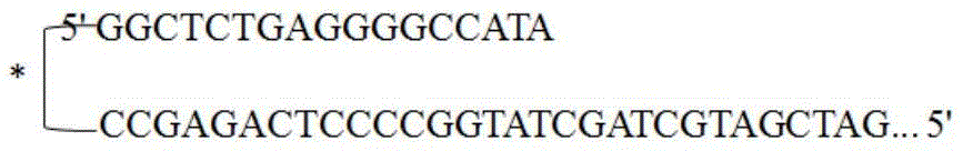
As indicated above, the primers or probes have covalently linked moieties (labeled "×") that bind to adjacent DNA and increase the Tm of the primer/probe. The stabilizing moiety can bind to ds or ssDNA.
In further embodiments, oligonucleotide backbone or base modifications that increase Tm can also be used to shift primer/probe Tm into the DFA range without increasing oligonucleotide length.
These modifications include, but are not limited to, LNA, PNA, phosphorodithioate linkages, 2' sugar modifications such as 2' -O-methyl, 2' -fluoro, base modifications such as 5-halopyridine, 5-methylpyrimidine, bases that create additional hydrogen bonds, other purine base modifications such as super G, 2-aminopurine, and the like.
DFA Probe technique-FRET Probe
In another embodiment of the cleavage probe technique, which is shown in fig. 33, a bathophenanthroline-RU II complex is used as the labeling molecule. These complexes may be part of an interacting pair of label molecules, allowing energy transfer from a suitable energy donor molecule to the Ru complex. Since the efficiency of energy transfer is highly dependent on the distance between the donor and acceptor molecules, this energy transfer system is useful in studying molecular interactions.
One suitable class of acceptor molecules for use as Ru complexes are the lumazine chromophore groups (lumazine chromophore groups) of the molecule. Using such a combination, energy transfer can be detected between the Ru bathophenanthroline complex and the lumazine chromophore, where the Ru complex is located at a suitable distance from the lumazine chromophore. When used in conjunction with polymerase cleavage techniques, wherein one of the two labels is cleaved from the probe, a change in luminescence can be detected, which is useful in determining whether amplification of the target sequence is achieved.
FIG. 34 depicts a two-hybrid probe and primer combination. This embodiment comprises two sequence-specific oligonucleotide probes in addition to two sequence-specific primers. The probes comprise a dye pair capable of participating in Fluorescence Resonance Energy Transfer (FRET), wherein a donor dye is attached to one probe and an acceptor dye is attached to the other probe, both the donor and acceptor dyes being positioned such that when the probes are attached to the target sequence they are in sufficient proximity to each other to participate in FRET. Both the probe and the primer meet the XCR temperature requirements.
FIG. 35 depicts primer/probe combinations capable of participating in FRET. Sequence specific oligonucleotide primers and sequence specific oligonucleotide probes are designed to bind to adjacent sequences of the target, typically the probe is complementary to the strand formed by the primer, thereby allowing the probe to anneal to the complementary strand synthesized from the extended labeled primer.
Designing assays containing probes and primers of these lengths yields unexpected results because it is generally believed that probes and primers of these lengths cannot be designed to possess a Tm within a narrow range of the Tm of the target sequence required for DFA. However, it has been found that probes and primers having an appropriate Tm range for DFA can be easily designed.
Primer/primer XCR detection chemistry
Observations from existing probe detection chemistries such as the HybBeacon and HyGlow probes show that the natural folding of the probe into a compact secondary structure and the consequent relative hydrophobicity of the fluorescent dyes allows these fluorescent moieties to become in close proximity if not in physical contact with each other. This is believed to occur due to the configuration that is advantageous in terms of entropy as a whole for the folded oligonucleotide and its attached dye.
XCR has demonstrated its ability to amplify DNA or RNA templates at nearly 10x the rate of traditional PCR techniques. A substantial limitation to performing amplification at these speeds is the need to incorporate probe-based detection during the amplification protocol. The main source of the additional time required is the hybridization required for the probe and, in the case of the 5' nuclease XCR probe, the additional time required for cleavage of the probe to release the dye quenching from its quencher.
The following describes a method for detecting fluorescence in real-time amplification, which utilizes the HybBeacon and HyGlow techniques, in which fluorescence quenching is released by binding of the oligonucleotide to its complement template.
According to this design, instead of fluorescent oligonucleotides as probes, fluorescent molecules are primers for generating amplifications.
The main advantage is that no additional time is required to allow binding of the fluorescent probe, since the primer inherently anneals on the template and "extends" out, thereby releasing the fluorescence quenching when the priming complex is initiated. Referring to FIG. 36, forward and reverse primers are shown with spaced dyes approximately 6-9 nucleotides apart along the length of the primer, but leaving enough nucleotides at the 3' end to be free of dyes. When the primer binds to its complement, fluorescence quenching is released and a detectable signal is thereby generated.
The fluorescent primers serve multiple purposes in this design. First, two fluorescent signals are generated, one for binding of the forward primer to the appropriate template and one for binding of the reverse primer to the opposite strand template.
If a primer forms an extension product, i.e., a primer dimer, with another similar primer, the dye on the primer should be close enough to prevent release of the quench, and thus remain quenched and not generate a fluorescent signal from such a primer dimer complex.
If two differently labeled primers form a primer dimer complex, their quenching will not be released, but a FRET complex will form, and it is suggested that the signal of primer dimer complex formation will be able to be monitored by excitation at higher energy wavelengths and absorption at lower energy wavelengths (standard FRET signal). Referring to fig. 37, a quenched forward primer dimer complex, a quenched reverse primer dimer complex, and a primer dimer complex formed from the binding together of forward and reverse primers is illustrated, which can be detected via FRET signals.
In some cases, it may be possible for a single primer to initiate priming of the template in the case of template-dependent non-specific products. These products will generate a single fluorescent dye signal. Referring to FIG. 38, the forward primer template formation signal, and the reverse primer template formation signal, are illustrated, as well as the signals generated when both the forward and reverse primers produce the target template.
However, the correct product with two dye-labeled primers will show the formation of fluorescent signals from two distinct dyes, and the reaction formation efficiency will be equal, since they will be directly linked to each other in the formation of the amplification product and can be monitored simultaneously in both fluorescence channels. See fig. 39.
Another data evaluation advantage of this design strategy is the formation of amplification products and the generation of two fluorescent signals from the amplified products. Because similar primers will be quenched, any primer dimer signal that causes FRET can be subtracted from the signal formed to allow baseline normalization of the amplification signal. See fig. 40.
Overall, this technique is advantageous in that it does not limit the XCR speed during amplification by eliminating the need to wait for probe hybridization or probe cleavage.
Besides XCR, this design is equally applicable to PCR assays; however, this chemistry has not been implemented and the non obvious reason for it is that PCR suffers from substantial non-specific product formation and neglects that the use of primers only (as in the case of double stranded DNA binding dyes such as SYBR green 1) is suitable for diagnostic detection methodologies.
Triplex forming region probe design
In another embodiment, the present disclosure provides multiplex probe technology suitable for DFA or temperature dependent multiplexing.
This embodiment minimizes the number of oligomers (e.g., primers and probes) required by eliminating the need for probes for the amplification portion of the reaction.
Most current probe technologies utilize single or multiple oligonucleotides to probe the amplified sequence. The oligonucleotide probes bind to the sequence of interest during the DNA amplification process.
In contrast, amplification can be detected using a Triplex Forming Region (TFR) attached to a sequence specific primer. The present disclosure uses triplex forming oligonucleotide probes designed to interact with each specific product at the TFR to generate a unique fluorescent color based on the specific product formed. This reduces the number of oligonucleotides present in the reaction.
Triplex forming probes do not participate in the amplification reaction and therefore do not slow the reaction as existing probe technology tends to do.
When two different, non-overlapping oligonucleotides anneal to different regions of the same linear complementary nucleic acid sequence, with the 3 'end of one oligonucleotide pointing to the 5' end of the other, the former may be referred to as an "upstream" oligonucleotide and the latter as a "downstream" oligonucleotide.
The practice of the present disclosure will employ, unless otherwise indicated, conventional techniques of molecular biology, microbiology, and recombinant DNA techniques, as fully set forth in the literature and as disclosed in U.S. patent No. US7,838,235, which is hereby incorporated by reference in its entirety.
In one embodiment, triplex forming oligonucleotide probe-based detection chemistry is used for nucleic acid amplification products. According to this method, triplex forming primers were synthesized according to the following method.
An artificial sequence Triplex Forming Region (TFR) was added to the designed oligonucleotide to create a triplex forming primer.
As used herein, a triplex forming region, or TFR, refers to a specific DNA sequence that contributes itself to Hoogsteen or a triplex base pair, where the third DNA strand binds to a double stranded TFR to form a triplex length of DNA, referred to as a triplex.
The following are illustrative examples of sequences that form triplex forming regions:
5’–GTGTGGGAAGAGGGGGAXGAGGGGGAGGAGC–3’(SEQ ID NO:32)
3’–CACACCCCTTCTCCCTXCTCCCCTCCGTCG–5’(SEQ ID NO:33)
5’–GTGTGGGAAGAGGGGGAXGAGGGGGAGGAGC–3’(SEQ ID NO:34)
3’–CACACCCCTTCTCCCTXCTCCCCTCCGTCG–5’(SEQ ID NO:35)
in one embodiment, the TFR is located at the 5' end of the designed primer.
In an alternative embodiment, the TFR is located near the 5' end of the designed primer.
In another embodiment, the TFR is located anywhere within the designed primer.
In another embodiment, the TFR is located at the 3' end of the designed primer.
In another embodiment, the TFR is located near the 3' end of the designed primer.
The triplex forming primer may be a segment of DNA or RNA that is complementary to a given DNA sequence and is required to initiate replication by a DNA polymerase.
The triplex forming oligonucleotide may comprise a triplex forming oligonucleotide probe (TFO probe). The TFO probe may be complexed to an appropriate sequence triplex forming region of a double stranded nucleic acid sequence and thus may be used to identify any nucleic acid sequence when the TFO probe is labeled in some manner, such as with a fluorophore.
In one embodiment, the primer comprising the triplex forming region (TFR primer) may further have a fluorescent dye.
As shown in fig. 41, the TFR primers participate in the amplification of the target sequence, creating triplex-forming DNA strands along the length of and attached to the target sequence.
The following detection method can then be used to determine whether the TFO probe has bound to the triplex forming region of the duplex in the amplified DNA, indicating that the DNA possesses the sequence of interest.
As shown in FIG. 42, triplex forming oligonucleotide probes were created in the following manner. Single-stranded TFOs are designed to bind to triplex forming regions in the target sequence created during amplification. A dye capable of participating in fluorescence energy transfer (FRET) is attached to the triplex TFO probe. In this case, the dye is a donor dye. The following are two examples of single-stranded DNA that forms triplexes with double-stranded DNA carrying TFRs:
5’–GGAGGGGGAGAAGGGAGAAGGG–3’(SEQ ID NO:36)
3’–CCTCCCCCTCTTCCCTCTTCCC–5’(SEQ ID NO:37)
5’–GGTGGGGGTGTTGGGTGTTGGG–3’TFO(SEQ ID NO:38)
3’-GGGTTGTGGGTTGTGGGGGTGG–5’TFO(SEQ ID NO:39)
5’–GTGTGGGAAGAGGGGGAXGAGGGGGAGGAGC–3’(SEQ ID NO:40)
3’–CACACCCCTTCTCCCTXCTCCCCTCCGTCG–5’(SEQ ID NO:41)
as shown in fig. 42, the 3' end of the TFO probe was capped with phosphate to prevent the TFO probe from participating in the amplification process. The TFO probe may also be capped by a fluorescent dye, a non-amplifiable linker, or any other suitable atom or molecule known to one of ordinary skill in the art that prevents extension of an oligonucleotide during an amplification reaction. The donor dye may be attached at or near the 3' end of the probe. The donor dye may also be attached at or near the 5' end of the probe. The donor dye may also be attached at any position between the 5 'end and the 3' end of the TFO probe. When the dye is attached to the TFO probe, it constitutes a Triplex Forming Fluorescent Probe (TFFP).
FIG. 43 depicts a double stranded DNA sequence comprising a triplex forming region. The double stranded DNA sequence possesses an acceptor dye. The TFR of TFFP is attached to the triplex forming region of double stranded DNA.
In one embodiment, TFFP anneals to amplified DNA only when the reaction temperature is at or below the annealing temperature of the primers. Therefore, the triplex forming fluorescent probe does not participate in the reaction. The donor dye on the TFFP resonates when the TFFP binds to the amplified sequence and light is emitted on the product. As the donor dye resonates, it transfers energy to an acceptor dye located on the double stranded DNA, causing the acceptor dye to fluoresce at a particular wavelength, emitting light of a color corresponding to that wavelength. This indicates that the sequence of interest is present in the test sample and has been amplified.
In an alternative embodiment, the acceptor dye is attached to the TFFP and the donor dye is attached to the amplified double stranded DNA product. In such embodiments, the acceptor dye fluoresces when the sequence of interest has been amplified.
In another embodiment, a plurality of primers can be used, each designed to bind to and specifically amplify a different sequence of interest. Each primer has a different acceptor dye attached to it, such that each acceptor dye fluoresces at a different wavelength. The triplex forming fluorescent probe binds to the triplex forming region of the amplification product. The donor dye attached to the triplex forming probe will cause the acceptor dye on the amplified product to fluoresce with a specific color, depending on which product is amplified. In this way, a plurality of different sequences can be detected at once. Such an embodiment would allow for the detection of a variety of different sequences of potential interest within one reaction vessel. The detection of multiple different sequences of potential interest in one reaction vessel is referred to as multiplexing.
In an alternative embodiment of the multiplex probe combination, an acceptor dye may be attached to the TFFP and a donor dye may be attached to the amplified double stranded DNA product. In such embodiments, each primer will have a donor dye of a different color, thereby causing the acceptor dye attached to the TFO probe to fluoresce in a different color, depending on which primer amplifies the sequence of interest.
In another embodiment, the TFO probe is designed to anneal at about the same temperature, or at a slightly higher or lower temperature, as compared to the Tm of the primers. This embodiment allows real-time reading of the amplification results.
Triplex forming region probe design with naturally occurring TFR
Another embodiment takes advantage of the naturally occurring Triplex Forming Region (TFR) located within or adjacent to the sequence of interest itself. The following are examples of TFR sequences naturally occurring in double stranded DNA.
5’TTTTTTCCCGTCC 3’(SEQ ID NO:42)
3’AAAAAGGGCAGG 5’(SEQ ID NO:43)
5’GGCGAGGGGGGAGCGGG 3’(SEQ ID NO:44)
3’CCGCTCCCCCCTCGCCC 5’(SEQ ID NO:45)
5’GGAGGTGGGGGAG 3’(SEQ ID NO:46)
3’CCTCCACCCCCTC 5’(SEQ ID NO:47)
5’GGAGGTGGGGGAG 3’(SEQ ID NO:48)
3’CCTCCACCCCCCTC 5’(SEQ ID NO:49)
5’GGAGAAGGTGAGGAAGAAGAAGAGGAAGAA 3’(SEQ ID NO:50)
In such embodiments, the primers are designed to bond to the naturally occurring triplex forming region and to the sequence of interest.
In this manner, the triplex forming region of the DNA is amplified to a detectable level as the sequence of interest is amplified. The primer has an acceptor dye attached thereto. Triplex forming probes are created to have a sequence complementary to the naturally occurring sequence of interest.
In another embodiment of the multiplexed method, each primer set designed to detect a particular sequence of interest contains its own unique TFR base pair sequence in addition to its own unique color dye. In one embodiment, the dye will be a donor dye. Multiple TFO probes were then designed, where each set contained a TFR that matched a specific TFR of one of the amplification products. Each set of probes also contained its own unique acceptor dye color. Which product is amplified determines which probe will bond to it. Which probe is attached to the amplified product, and thus which sequence of interest is present in the sample, is determined by the color of the fluorescence of the probe as it undergoes FRET with the dye of the TFO probe.
In another embodiment, the detection method may include the use of a specialized DNA binding dye that preferentially binds to triplex DNA structures. In one embodiment, the dye comprises Thiazole orange (Thiazole orange). In another embodiment, the dye comprises a cyanine 40 dye. In addition to the dyes listed herein, any other dye known to one of ordinary skill in the art that preferentially binds to triplex DNA structures may be used. These triplex binding dyes can be used in combination with dye-labeled TFRs, either on the primer or within the product itself, to generate a FRET-based signal that can also indicate the presence of a specifically formed target sequence.
An alternative method involves a TFO probe conjugated to a DNA binding dye. These would include, but are not limited to: sybr Green 1; sybr Gold; eva Green; LightCycler Green I; LightCycler Green II; Toto/Yoyo/Toyo; and other DNA binding dyes known to those of ordinary skill in the art that bind to hybridized DNA structures. As shown in fig. 44, the binding dye is constrained by covalent attachment to a specific location on the TFO probe, in which case the end of the TFO probe can only bind to the hybridized DNA structure when the TFO probe binds and thus brings the TFO probe into proximity with the dye attached to the amplified sequence of interest. Thus, the fluorescent signal indicates that amplification has occurred.
In yet another embodiment, Cy2 or other quadruplex-binding dyes known to those of ordinary skill in the art can be used.
In another embodiment, a synthetic TFO probe may have a fluorescent dye and a quencher located at any position along its length. As shown in fig. 45, this embodiment utilizes a hairpin dye and quencher configuration. When the TFO probe binds to the sequence of interest, the hairpin structure of the probe disappears, with the result that the quencher becomes far enough away from the dye that it can no longer inhibit the fluorescence of the dye. This results in the emission of a fluorescence of a particular color if the sequence of interest is amplified. The change in the specific fluorescent signal is irrelevant as long as it is distinguishable from any other product in the reaction mixture. The total number of reactions that can be detected can be differentiated depending only on the platform of the device on which the reaction is performed.
In another embodiment, shown in figure 46, two or more primers having the same TFR sequence may be used with a TFR primer comprising a sequence complementary to the TFR sequence. Figure 46 depicts three TFR primers, each with a different color dye. In step 2, primers containing a red dye bind to the sequence of interest and are amplified. In step 3, the TFR probe binds to the TFR in the presence of a binding dye that preferentially binds to the triplex. Such conjugated dyes may comprise thiazole orange, cyanine 40, or any other triplex conjugated dye known to one of ordinary skill in the art. The binding dye participates in FRET with the attached fluorophore, indicating that a sequence complementary to the primer containing the red dye has been amplified.
In alternative embodiments, the products may be distinguished by color, melting temperature, or a combination of both color and melting temperature of the triplex product. FIG. 47 depicts an embodiment in which the six primers are divided into three groups, two in each group. Each set of two primers comprises the same TFR sequence and each of the three sets comprises three different TFR sequences, thereby allowing each of the three sets of primers to be distinguished from the other two by virtue of having different melting temperatures. The two primers within each group sharing a TFR each have a dye of a different color. The method further includes using three sets of probes comprising sequences that bind to the TFR of one of the primer pairs. The products will then be able to be distinguished based on their unique combination of melting temperature and color. These different methods of differentiating products would make possible the use of triplex DNA formation in the detection of amplified products for use as a quantitative, genotypic, or simply target detection.
The following are alternative embodiments of primer and probe techniques designed to take advantage of the unique features of DFA. In this embodiment, each primer of a pair of primers is labeled with a dye capable of participating in Fluorescence Resonance Energy Transfer (FRET). As shown in FIG. 48, the donor dye is attached near the 3 'end of the first primer, while the acceptor dye is attached near the 3' end of the second primer. In the annealing step, the primers hybridize to their target sequences in a near tail-to-tail arrangement, which brings the dyes close enough for FRET to occur. The amount of acceptor fluorescence is directly proportional to the amount of DFA product present.
An assay kit of the present disclosure for amplifying and/or detecting a target sequence in a DNA sample may comprise: i) the primers and probes described herein, and ii) buffers, dntps, and enzymes. These reactants are present in an amount sufficient to perform multiple assays.
Other triplex forming region probe design
Triplex forming oligonucleotides provide a unique method of detecting amplification products from PCR, XCR, RAMP, HAD, NEAR, temperature dependent multiplexing, or any other amplification technique that results in the amplification of large quantities of double stranded DNA.
We have described the introduction of artificial TFRs, either attached somewhere along the length of the primer. More interestingly, however, the natural occurrence of the Triplex Forming Region (TFR) is surprisingly abundant. For example, Streptococcus agalactiae (Streptococcus agalactiae) has a 2 million base pair genome with up to 29 TFRs of 16 base pairs or longer. This relatively high abundance of these TFRs makes it plausible to use these TFRs (homopurine or homopyrimidine fragments) as potential diagnostic markers for amplification of desired nucleic acids.
TFO will bind to the amplified double stranded DNA at any point during the reaction, where complete double stranded extension occurs throughout the TFR portion of the product.
Advantageously, these binding events can be monitored at many different stages of amplification, and therefore unlike other hybridization chemistries, they do not force real-time readings to occur at the lowest temperature of the reaction at which unextended single-stranded DNA is exposed for probe binding.
One significant advantage of being able to use higher temperatures for fluorescence readings is that the reaction can be accelerated to maximum speed, and the commensurate advantage is that nonspecific product formation is not promoted by maintaining at relatively low reaction temperatures for extended periods of time.
Cycling probe technology
An alternative method of detecting amplification products utilizes cycling probe technology to take advantage of the speed of DFA by providing an end-point assay. Cycling probe technology, as described in U.S.5,660,988 (incorporated in its entirety for all purposes), is a technology used in the so-called signal amplification method. As described in u.s.5,660,988, the cycling probe binds to the target and is subsequently cleaved by an enzyme. Once the cycling probe is cleaved, another cycling probe binds to the target and is then cleaved. This process can be repeated until all probes are used. In the methods described herein, the cycling probe is designed to have a Tm that is lower than the lowest Tm of the thermal cycling curve for the reaction run. Once the reaction is run, the temperature of the reaction is lowered to a point at or below the Tm of the cycling probe. In the case of a positive result, the cycling probe is then bound successively and cleaved until a signal is generated. Alternatively, the ribonucleotide containing probe may function during the initial thermal cycling, being cleaved by an rnase or by the exonuclease activity of a polymerase.
Examples
Example 1: amplification of target sequences using primers with tags
The following primers were created for use in conjunction with DFA to amplify target sequences.
Salmonella (Salmonella) FOR without a tag (SEQ ID NO:12):
CGACGACCCTTCTTTTTCCTCAATACTGAGCGGCTG, Tm 75.8 ℃ @4mM Mg and 0.5. mu.M primer
Salmonella REV without a tag (SEQ ID NO:13):
CGCTGCCGGTATTTGTTATTTTATCGGTGGTTTTAAGCGTACTCTTCTATTTTAAATTCC, Tm 75.2 ℃ @4mM Mg and 0.5. mu.M primer
Salmonella FOR carries a tag (SEQ ID NO:14):CGTCGCGACGACCCTTCTTTTTCCTCAATACTGAGCGGCTG, the label is underlined
Salmonella REV carries a tag (SEQ ID NO:15):CAGCGCGCTGCCGGTATTTGTTATTTTATCGGTGGTTTTAAGCGTACTCTTCTATTTTAAATTCC, the label is underlined
Target for first primer binding (SEQ ID NO:16):
CGACGACCCTTCTTTTTCCTCAATACTGAGCGGCTGCTCGCCTTTGCTGGTTTTAGGTTTGGCGGCGCTACGTTTTGCTTCACGGAATTTAAAATAGAAGAGTACGCTTAAAACCACCGATAAAATAACAAATACCGGCAGCG,86.5℃@4mM Mg
annealing temperature after first extension:
ACCCTTCTTTTTCCTCAATACTGAGCGGCTG(SEQ ID NO:17),73.3℃
CCGGTATTTGTTATTTTATCGGTGGTTTTAAGCGTACTCTTCTATTTTAAATTCC(SEQ ID NO:18),73.3℃
target for second primer binding (SEQ ID NO:19):
ACCCTTCTTTTTCCTCAATACTGAGCGGCTGCTCGCCTTTGCTGGTTTTAGGTTTGGCGGCGCTACGTTTTGCTTCACGGAATTTAAAATAGAAGAGTACGCTTAAAACCACCGATAAAATAACAAATACCGG,85.2℃@4mM Mg
target for intact tagged primer binding (SEQ ID NO:20):
CGTCGCGACGACCCTTCTTTTTCCTCAATACTGAGCGGCTGCTCGCCTTTGCTGGTTTTAGGTTTGGCGGCGCTACGTTTTGCTTCACGGAATTTAAAATAGAAGAGTACGCTTAAAACCACCGATAAAATAACAAATACCGGCAGCGCGCTG,87.8℃@4mM Mg
example 2: amplification of target sequences using G-quadruplex-forming tagged primers
The following are examples of potential G-quadruplexes for maintaining DFA amplification bubbles and helping to block extension outside the bubbles.
Mycobacterium avium subspecies paratuberculosis strain k10, complete genome, sequence ID gb | AE016958.1| (SEQ ID NO:21):
5'-TCGAATCCCTCTCCCCGCCCGGGCGGTACGACGCGCCGAGGAAGCGGTGCACCAGGGCGC
GCTCGGCGGCCGGGTCCTTGAGCGGCCAGCCCCATAACGCCAGGAAGACGCGGATCAGCC
ACTGCGCCGCCAGCGGGTCGTCGTGGCCGGGCCCGAGCATCTCGGCGGCCAGGGCCGTCA-3'
(SEQ ID NO:22):
5'-TACCGCCCGGGCCCGGGCGGTACGACGCGCCGA-3'
(SEQ ID NO:23):
5'-GGCCGGGCCCGGGCCCGGCCACGACGACCCGCT-3'
FIG. 18 shows the formation of a G-quadruplex structure to block hybridization of the above primer extended out of the bubble to a Mycobacterium avium sequence.
Example 3: temperature-dependent multiplexing of target and control sequences
The internal control was developed for amplification of mycobacterial targets following a multiple temperature protocol. In this case, the control amplicon has similar thermal cycling properties as the target amplicon, 94 ℃ for denaturation and 84 ℃ for annealing/extension. However, the primers (Mfo1275fmut2, Mfo1490rmut2) introduce nucleotide mismatches that result in a predicted Tm for the target DNA, i.e., the Mycobacterium fortuitum sequence (Mfo template)<80 ℃. However, after an initial round of elongation to eliminate mismatches, the predicted Tm returns to 86DEG C. As shown in fig. 25, the initial low affinity of the primers for the template means that there is no significant amplification at 80 cycles of 94 ℃ -85 ℃ (thermocycling profile where the target mycobacterium species will be amplified). After the target cycle is completed (80 cycles), the annealing/extension temperature is adjusted down to 75 ℃ for five cycles to enable the control primers to prime, yet back to 77 ℃ to amplify the internal control species. In this case, 1x10 of the control template in the initial 80 cycles7To 12x109The individual copies were quiescent and then activated and amplified by the second phase of thermal cycling of the Ct of about 40 cycles.
The thermal cycling conditions were: 95 ℃ -84 ℃ x80 cycles, 93 ℃ -72 ℃ x5 cycles to initiate (to catch), 93 ℃ -77 ℃ x40 cycles.
Input is 1x10⁹、1x10⁷One copy of the Mycobacterium fortuitum synthetic template.
The method comprises the following steps: mutations are introduced that lower the initial Tm and return a high Tm after initial extension by the polymerase.
The primer sequence is as follows:
Mfo1275fmut2(SEQ ID NO:24):CGTGCACACCCGGCCAAGGTCGTTGCGGCCCAGAG(underlined bases are mismatched to the template), the effect of a pre-Tm of 80 ℃ plus a 3' mismatch is triggered (not predicted by the software); tm after initiation is 85 ℃; wild type Tm 86 deg.c
Mfo1490rmut2(SEQ ID NO:25):ACGGCGTTTTCGATTGTCGGATCCACCCCGGAGGCCCTGCTCACC (base underlined is mismatched to template), pre-priming Tm 80 ℃; tm after initiation is 86 ℃; wild type Tm 86 deg.c
Mfo template (SEQ ID NO:26): CGTGCACACCCGGCCGAGGTCGTTGCGGCCGAGATCGACTCGGTCGCCCCGCGCCAGCGAGTGCCCGCGATCGACGGTGACCAGGGCCTCCGGGCTGGATCCGACAATCGAAAACGCCGT, Tm 97 deg.C
As a result: the control template remained unamplified for 80 cycles, then after a temperature change, the control template was amplified in-40 cycles.
Example 4: temperature-dependent multiplexing using primers comprising a 5' tail
In this example, the internal control primer did not introduce a mismatch with the template, but rather had a lower predicted Tm of 70 ℃. Each of these primers (SPWXMF _ mut4 and SPWXMR _ mut4) carries a 5' GC-rich tail that does not bind to the target sequence, but when extended by polymerase in the first round of amplification, Tm becomes 76 ℃. As in example 3, these primers did not amplify the template throughout 50 cycles between 89 ℃ and 76 ℃. After 50 cycles, the annealing/extension temperature was lowered to 72 ℃ for 10 cycles, then back to 76 ℃, after which amplification of the control template was seen (see fig. 26). The term "priming" (catch) refers to the point at which the amplification product begins to achieve full extension.
The thermal cycling conditions were: 89-76 ℃ x50 cycles, 89-72 ℃ x10 cycles, 89-76 ℃ x60 cycles.
The input was Saflager W-34/70 yeast (Spw) genomic DNA, diluted 10X.
The method comprises the following steps: shortened primers with 5' tails without mismatched bases were prepared to increase Tm after priming.
The primer sequence is as follows:
SPWXMF_mut4(SEQ ID NO:27):
CCA GCCACC CAC CAA TTC CTG TGC CAG AT (underlined bases are 5' tails) with a Tm of 70 ℃ before priming and 77 ℃ after priming
SPWXMR_mut4(SEQ ID NO:28):
ACC GGAGGT CAC TTT TGA TGG CCA TGG GTC TAT (underlined bases are 5' tails) at 70 ℃ before priming and 76 ℃ after priming
Template (SEQ ID NO:29):
CTC GTT AGA GGG GCT AAA GCT AAC CCA CCA ATT CCT GTG CCA GAG AAT ATA TAG GGC GGT GCA TGA ACA ATA GCC GGT AGG TAT GTC AGA AAA CCT CCA ATG CCA AAC ATT ACT CCT TGA CAC CGC CTA TAT TTA GAC CCA TGG CCA TCA AAA GTG ACC CGA GCA CCA TCG TTT GTT G,Tm=87℃
as a result: the control template remained unamplified for 50 cycles, then after a temperature change, the control template was amplified in-30 cycles.
Example 5: temperature-dependent multiplexing of target and control sequences
The following contains a dual example using a trichomonas bovis target and a reaction control template, xenorhabdus nematophilus.
Methods for combining multiple targets in one reaction vessel are described, wherein each target is amplified at a different stage of the thermal profile. In this example, trichomonas bovis represents the desired diagnostic target organism, while the control template xenorhabdus nematophila serves as an extraction control or as an internal process control. As designed, trichomonas bovis is amplified at the initial stage of the thermal profile with the higher temperature design, and after a sufficient number of thermal cycles, the reaction is transitioned to the appropriate conditions to amplify the reaction control target.
Xenorhabdus nematophilus ATCC 19061(SEQ ID NO:30):
TTTATTTTTTAGTTATCAATATATCTGAGTTTTATTTTTTAGCTACAGTGTTTTTATTTGTTTTTTTAGCAGCCTTACTTAACAGTATTTTGTTAATATCAACATATATAAGATATATTATTATTTCTTTACTATGCTCAATAACTCTATCTTTACATTTAGATATATTACCATCATTTGATTTAATATTTTTCTTGCCTATATTTATTTTTGTTTTTATCTATAAATTTAATCTCGTCAAAAAAAGACTATAATTATATTGATTAATTTAAGTTTTCAGATGATATAATCAAATTTTATTCCAATAATACCAATCATCACTGAAATTGCTTAATTTATACTGAAGATTTGGTTATGTATTAAATAGTTAATTCTTATCATATACTCCCT
exemplary Forward primer sequences (SEQ ID NO:31):
TTTTTAGTTATCAATATATCTGAGTTTTATTTTTTAGCTACAGTGTTTTTATTTGTTTTTTTAGCAGCCTTACTTAACAGTATTTTGTTAATATCAACATATATAAGAT,Tm=74℃
exemplary Forward primer sequence (SEQ ID NO:51):
TTTTTAGTTATCAATATATCTGAGTTTTATTTTTTAGC,Tm=62.2℃
exemplary reverse primer sequence (SEQ ID NO:52):
ATCTTATATATGTTGATATTAACAAAATACTGTTAAGT,Tm=62.3℃
exemplary Probe sequence (SEQ ID NO:53):
CAGTGTTTTTATTTGTTTTTTTAGCAGCCT,Tm=65.2℃
trichomonas bovis 372(SEQ ID NO:54):
CGGTAGGTGAACCTGCCGTTGGATCAGTTTCGTTAATAATTACAAACATATTTTTTTAATGTCTATAACTATTTATACAAAATTAAACACATAATCTAAAAAATTTAGACCTTAGGCAATGGATGTCTTGGCTTCTTACACGATGAAGAACGTTGCATAATGCGATAAGCGGCTGGATTAGCTTTCTTTGCGACAAGTTCGATCTTTGAATGCACATTGCGCGCCGTTTTAGCTTGCTAGAACACGCATATATGTTACAGTAACCCATATTAATTTAATACCAAATTCTCTTTTTAAGCAAAAGAGCGAAAAACAAATATGTATTAACAAAAGGGTTCTGTCTCATATAGGAAGACCCGCTGAACTGAAGCA
trichomonas bovis 266(SEQ ID NO:55):
AGACCTTAGGCAATGGATGTCTTGGCTTCTTACACGATGAAGAACGTTGCATAATGCGATAAGCGGCTGGATTAGCTTTCTTTGCGACAAGTTCGATCTTTGAATGCACATTGCGCGCCGTTTTAGCTTGCTAGAACACGCATATATGTTACAGTAACCCATATTAATTTAATACCAAATTCTCTTTTTAAGCAAAAGAGCGAAAAACAAATATGTATTAACAAAAGGGTTCTGTCTCATATAGGAAGACCCGCTGAACTGAAGCA,Tm=85.3℃
exemplary Forward sequence (SEQ ID NO:56):
AGACCTTAGGCAATGGATGTCTTGGCTTCTTACACGATGAAGAACG,Tm=77.3℃
exemplary reverse primer sequences (SEQ ID NO:57):
TGCTTCAGTTCAGCGGGTCTTCCTATATGAGACAGAACCCTT,Tm=77.3℃
exemplary Probe sequence (SEQ ID NO:58):
AAGCGGCTGGATTAGCTTTCTTTGCGACAAGTTCGATCTTTGAATGCACATTGCGCGCCG,Tm=83.1℃
possible thermal profiles for amplification of the target and reaction control templates of M.nematophilus are shown in FIGS. 27-31.
Example 6: temperature-dependent multiplexing using Trichomonas primers to detect Trichomonas in cattle
The following primers and probes were used in temperature-dependent multiplexing to detect trichomonas in cattle.
TFXMF(SEQ ID NO:56):AGACCTTAGGCAATGGATGTCTTGGCTTCTTACACGATGAAGAACG
TFXMR(SEQ ID NO:57):
TGCTTCAGTTCAGCGGGTCTTCCTATATGAGACAGAACCCTT
TFXMP2(SEQ ID NO:59):AAGCGGCTGGATTAGCTTTCTTTGCGACAAGTTCGATCTTTGAATGCACATTGCGCGCGCCG
Samples for amplification were from Bull (Bull) tissue. Thermocycling from about 89 ℃ to about 74 ℃ to amplify the Trichomonas target. Thermocycling from about 63 ℃ to about 78 ℃ to amplify the xenorhabdus nematophila control.
Those skilled in the art will appreciate that for this and other processes and methods disclosed herein, the functions presented in the processes and methods may be performed in a different order. In addition, the listed steps and operations are provided as examples only, and some of the steps and operations may be optional, combined into fewer steps or operations, or expanded into additional steps and operations without detracting from the essence of the disclosed embodiments.
The present disclosure is not limited to the particular embodiments described in this application, which are intended to illustrate various aspects. Many modifications and variations may be made without departing from the spirit and scope thereof, as will be apparent to those skilled in the art. Functionally equivalent methods and devices within the scope of the disclosure, in addition to those enumerated herein, will be apparent to those skilled in the art from the foregoing description. Such modifications and variations are intended to fall within the scope of the appended claims. The disclosure is to be limited only by the terms of the appended claims, along with the full scope of equivalents to which such claims are entitled. It is to be understood that this disclosure is not limited to particular methods, reagents, compound compositions, or biological systems, which can, of course, vary. It is also to be understood that the terminology used herein is for the purpose of describing particular embodiments only, and is not intended to be limiting. Applicants hereby incorporate by reference, in their entirety, U.S. application No. 14/510,939 for all purposes, filed 10/9/2014 and entitled: a multiplex probe. Additionally, applicants hereby incorporate by reference U.S. application No. 12/951,710 in its entirety for all purposes, filed on 11/22/2010 and entitled: systems and methods for high resolution analysis of nucleic acids to detect sequence variations. Applicants further incorporate herein by reference in their entirety U.S. application No. 12/058,637 for all purposes, filed on 28/3/2008 and entitled: systems and methods for high resolution analysis of nucleic acids to detect sequence variations are granted on 23/11/2010 as U.S. patent No. 7,838,235.
By the incorporation of
All references, articles, publications, patents, patent publications and patent applications cited herein are incorporated by reference in their entirety for all purposes. However, the mention of any references, articles, publications, patents, patent publications and patent applications cited herein is not, and should not be taken as, an acknowledgment or any form of suggestion that they form part of the common general knowledge or the effective prior art in any country in the world.
Sequence listing
<110> Froreson-Telick Co
Caplin, Brian
Hicke, Brian
Green, Bryson
<120> DNA amplification technique
<130> FLUO-004_03WO
<150> 62/023,123
<151> 2014-07-10
<150> 62/075,769
<151> 2014-11-05
<150> 62/115,559
<151> 2015-02-12
<160> 59
<170> PatentIn version 3.5
<210> 1
<211> 72
<212> DNA
<213> Artificial
<220>
<223> example of folded hairpin-like probes
<400> 1
acctccaatg ccaaacatta ctccttgact caatgtttcc tgtgccagca tgcgcttatt 60
agacccatgg cc 72
<210> 2
<211> 67
<212> DNA
<213> Artificial
<220>
<223> example of folded hairpin-like probes
<400> 2
agcactcagt tattctgctg gtgcacttgc cagttgcatg ggcctcatat acaacaggat 60
gggggct 67
<210> 3
<211> 83
<212> DNA
<213> Artificial
<220>
<223> example of folded hairpin-like probes
<400> 3
atggacgtgg cttagcgtat atttatgctg atggaaaaat ggtaaacgaa gctttagttc 60
gtcaaggctt ggctaaagtt gct 83
<210> 4
<211> 83
<212> DNA
<213> Artificial
<220>
<223> example of folded hairpin-like probes
<400> 4
atggacgtgg cttagcgtat atttatgctg atggaaaaat ggtaaacgaa gctttagttc 60
gtcaaggctt ggctaaagtt gct 83
<210> 5
<211> 67
<212> DNA
<213> Artificial
<220>
<223> example of folded hairpin-like probes
<400> 5
agcactcagt tattctgctg gtgcacttgc cagttgcatg ggcctcatat acaacaggat 60
gggggct 67
<210> 6
<211> 38
<212> DNA
<213> Artificial
<220>
<223> example of folded hairpin-like probes
<400> 6
tggcaatccc aggttttctt ttctacctgt ttgctcaa 38
<210> 7
<211> 36
<212> DNA
<213> Artificial
<220>
<223> example of folded hairpin-like probes
<400> 7
cagacccatc ccccaggtga gggactatgg cctcct 36
<210> 8
<211> 30
<212> DNA
<213> Artificial
<220>
<223> example of folded hairpin-like probes
<400> 8
tcaatggctg aggtgaggta ccccgcaggg 30
<210> 9
<211> 42
<212> DNA
<213> Artificial
<220>
<223> example of folded hairpin-like probes
<400> 9
tttgctctga gagttccccc tgtcccctcc accttccctc ag 42
<210> 10
<211> 45
<212> DNA
<213> Artificial
<220>
<223> example of folded hairpin-like probes
<400> 10
tgcaagagtc accaaaattg ccgagaggcc ccagttagca tccca 45
<210> 11
<211> 47
<212> DNA
<213> Artificial
<220>
<223> preparation in laboratory
<400> 11
gatcgatgct agctatggcc cctcagagcc ggctctgagg ggccata 47
<210> 12
<211> 36
<212> DNA
<213> Artificial
<220>
<223> Salmonella (Salmonella) forward primer, tag-free
<400> 12
cgacgaccct tctttttcct caatactgag cggctg 36
<210> 13
<211> 60
<212> DNA
<213> Artificial
<220>
<223> Salmonella (Salmonella) reverse primer, tag-free
<400> 13
cgctgccggt atttgttatt ttatcggtgg ttttaagcgt actcttctat tttaaattcc 60
<210> 14
<211> 41
<212> DNA
<213> Artificial
<220>
<223> Salmonella (Salmonella) forward primer comprising tag
<400> 14
cgtcgcgacg acccttcttt ttcctcaata ctgagcggct g 41
<210> 15
<211> 65
<212> DNA
<213> Artificial
<220>
<223> Salmonella (Salmonella) reverse primer comprising tag
<400> 15
cagcgcgctg ccggtatttg ttattttatc ggtggtttta agcgtactct tctattttaa 60
<210> 16
<211> 143
<212> DNA
<213> Artificial
<220>
<223> target sequence for binding of first primer
<400> 16
cgacgaccct tctttttcct caatactgag cggctgctcg cctttgctgg ttttaggttt 60
ggcggcgcta cgttttgctt cacggaattt aaaatagaag agtacgctta aaaccaccga 120
taaaataaca aataccggca gcg 143
<210> 17
<211> 31
<212> DNA
<213> Artificial
<220>
<223> shorter Salmonella (Salmonella) forward primer, without tag
<400> 17
acccttcttt ttcctcaata ctgagcggct g 31
<210> 18
<211> 55
<212> DNA
<213> Artificial
<220>
<223> shorter Salmonella (Salmonella) reverse primer, not containing tag
<400> 18
ccggtatttg ttattttatc ggtggtttta agcgtactct tctattttaa attcc 55
<210> 19
<211> 133
<212> DNA
<213> Artificial
<220>
<223> target for second primer binding
<400> 19
acccttcttt ttcctcaata ctgagcggct gctcgccttt gctggtttta ggtttggcgg 60
cgctacgttt tgcttcacgg aatttaaaat agaagagtac gcttaaaacc accgataaaa 120
taacaaatac cgg 133
<210> 20
<211> 153
<212> DNA
<213> Artificial
<220>
<223> target for complete primer binding with tag
<400> 20
cgtcgcgacg acccttcttt ttcctcaata ctgagcggct gctcgccttt gctggtttta 60
ggtttggcgg cgctacgttt tgcttcacgg aatttaaaat agaagagtac gcttaaaacc 120
accgataaaa taacaaatac cggcagcgcg ctg 153
<210> 21
<211> 180
<212> DNA
<213> Mycobacterium avium subspecies paratuberculosis strain k10
(Mycobacterium avium subsp. paratuberculosis str. k10)
<400> 21
tcgaatccct ctccccgccc gggcggtacg acgcgccgag gaagcggtgc accagggcgc 60
gctcggcggc cgggtccttg agcggccagc cccataacgc caggaagacg cggatcagcc 120
actgcgccgc cagcgggtcg tcgtggccgg gcccgagcat ctcggcggcc agggccgtca 180
<210> 22
<211> 33
<212> DNA
<213> Artificial
<220>
<223> Forward primer for amplifying Mycobacterium avium target
<400> 22
taccgcccgg gcccgggcgg tacgacgcgc cga 33
<210> 23
<211> 33
<212> DNA
<213> Artificial
<220>
<223> reverse primer for amplifying Mycobacterium avium target
<400> 23
ggccgggccc gggcccggcc acgacgaccc gct 33
<210> 24
<211> 35
<212> DNA
<213> Artificial
<220>
<223> Mfo1275fmut2 primer
<400> 24
cgtgcacacc cggccaaggt cgttgcggcc cagag 35
<210> 25
<211> 45
<212> DNA
<213> Artificial
<220>
<223> Mfo1490rmut2 primer
<400> 25
acggcgtttt cgattgtcgg atccaccccg gaggccctgc tcacc 45
<210> 26
<211> 120
<212> DNA
<213> Artificial
<220>
<223> Mycobacterium fortuitum (M. fortuitum) synthetic template
<400> 26
cgtgcacacc cggccgaggt cgttgcggcc gagatcgact cggtcgcccc gcgccagcga 60
gtgcccgcga tcgacggtga ccagggcctc cgggctggat ccgacaatcg aaaacgccgt 120
<210> 27
<211> 29
<212> DNA
<213> Artificial
<220>
<223> SPWXMF _ mut4 primer
<400> 27
ccagccaccc accaattcct gtgccagat 29
<210> 28
<211> 33
<212> DNA
<213> Artificial
<220>
<223> SPWXMR _ mut4 primer
<400> 28
accggaggtc acttttgatg gccatgggtc tat 33
<210> 29
<211> 178
<212> DNA
<213> Artificial
<220>
<223> template for SPWXM primer
<400> 29
ctcgttagag gggctaaagc taacccacca attcctgtgc cagagaatat atagggcggt 60
gcatgaacaa tagccggtag gtatgtcaga aaacctccaa tgccaaacat tactccttga 120
caccgcctat atttagaccc atggccatca aaagtgaccc gagcaccatc gtttgttg 178
<210> 30
<211> 390
<212> DNA
<213> Xenorhabdus nematophila (Xenorhabdus nematophila)
<400> 30
tttatttttt agttatcaat atatctgagt tttatttttt agctacagtg tttttatttg 60
tttttttagc agccttactt aacagtattt tgttaatatc aacatatata agatatatta 120
ttatttcttt actatgctca ataactctat ctttacattt agatatatta ccatcatttg 180
atttaatatt tttcttgcct atatttattt ttgtttttat ctataaattt aatctcgtca 240
aaaaaagact ataattatat tgattaattt aagttttcag atgatataat caaattttat 300
tccaataata ccaatcatca ctgaaattgc ttaatttata ctgaagattt ggttatgtat 360
taaatagtta attcttatca tatactccct 390
<210> 31
<211> 109
<212> DNA
<213> Artificial
<220>
<223> exemplary Forward primers for Xenorhabdus nematophilus targets
<400> 31
tttttagtta tcaatatatc tgagttttat tttttagcta cagtgttttt atttgttttt 60
ttagcagcct tacttaacag tattttgtta atatcaacat atataagat 109
<210> 32
<211> 31
<212> DNA
<213> Artificial
<220>
<223> triplex forming region sequence
<220>
<221> misc_feature
<222> (18)..(18)
<220>
<221> misc_feature
<222> (18)..(18)
<223> n is a, c, g or t
<400> 32
gtgtgggaag aggggganga gggggaggag c 31
<210> 33
<211> 30
<212> DNA
<213> Artificial
<220>
<223> triplex forming region sequence
<220>
<221> misc_feature
<222> (14)..(14)
<220>
<221> misc_feature
<222> (14)..(14)
<223> n is a, c, g or t
<400> 33
gctgcctccc ctcntccctc ttccccacac 30
<210> 34
<211> 31
<212> DNA
<213> Artificial
<220>
<223> triplex forming region sequence
<220>
<221> misc_feature
<222> (18)..(18)
<220>
<221> misc_feature
<222> (18)..(18)
<223> n is a, c, g or t
<400> 34
gtgtgggaag aggggganga gggggaggag c 31
<210> 35
<211> 30
<212> DNA
<213> Artificial
<220>
<223> triplex forming region sequence
<220>
<221> misc_feature
<222> (14)..(14)
<220>
<221> misc_feature
<222> (14)..(14)
<223> n is a, c, g or t
<400> 35
gctgcctccc ctcntccctc ttccccacac 30
<210> 36
<211> 22
<212> DNA
<213> Artificial
<220>
<223> triplex forming sequence
<400> 36
ggagggggag aagggagaag gg 22
<210> 37
<211> 22
<212> DNA
<213> Artificial
<220>
<223> triplex forming sequence
<400> 37
cccttctccc ttctccccct cc 22
<210> 38
<211> 22
<212> DNA
<213> Artificial
<220>
<223> triplex forming sequence
<400> 38
ggtgggggtg ttgggtgttg gg 22
<210> 39
<211> 22
<212> DNA
<213> Artificial
<220>
<223> triplex forming sequence
<400> 39
ggtgggggtg ttgggtgttg gg 22
<210> 40
<211> 31
<212> DNA
<213> Artificial
<220>
<223> triplex forming sequence
<220>
<221> misc_feature
<222> (18)..(18)
<220>
<221> misc_feature
<222> (18)..(18)
<223> n is a, c, g or t
<400> 40
gtgtgggaag aggggganga gggggaggag c 31
<210> 41
<211> 30
<212> DNA
<213> Artificial
<220>
<223> triplex forming sequence
<220>
<221> misc_feature
<222> (14)..(14)
<220>
<221> misc_feature
<222> (14)..(14)
<223> n is a, c, g or t
<400> 41
gctgcctccc ctcntccctc ttccccacac 30
<210> 42
<211> 13
<212> DNA
<213> unknown
<220>
<223> naturally occurring TFR sequence in double-stranded DNA
<400> 42
ttttttcccg tcc 13
<210> 43
<211> 12
<212> DNA
<213> unknown
<220>
<223> naturally occurring TFR sequence in double-stranded DNA
<400> 43
<210> 44
<211> 17
<212> DNA
<213> Ergonococcus (Spiromtra erioceuriopaei)
<400> 44
ggcgaggggg gagcggg 17
<210> 45
<211> 17
<212> DNA
<213> dog Bow ascaris (Toxocara canis)
<400> 45
cccgctcccc cctcgcc 17
<210> 46
<211> 13
<212> DNA
<213> unknown
<220>
<223> naturally occurring TFR sequence in double-stranded DNA
<400> 46
ggaggtgggg gag 13
<210> 47
<211> 13
<212> DNA
<213> unknown
<220>
<223> naturally occurring TFR sequence in double-stranded DNA
<400> 47
ctcccccacc tcc 13
<210> 48
<211> 13
<212> DNA
<213> unknown
<220>
<223> naturally occurring TFR sequence in double-stranded DNA
<400> 48
ggaggtgggg gag 13
<210> 49
<211> 14
<212> DNA
<213> unknown
<220>
<223> naturally occurring TFR sequence in double-stranded DNA
<400> 49
ctccccccac ctcc 14
<210> 50
<211> 30
<212> DNA
<213> Streptococcus dysgalactiae
<400> 50
ggagaaggtg aggaagaaga agaggaagaa 30
<210> 51
<211> 38
<212> DNA
<213> Artificial
<220>
<223> exemplary Forward primers for Xenorhabdus nematophilus targets
<400> 51
tttttagtta tcaatatatc tgagttttat tttttagc 38
<210> 52
<211> 38
<212> DNA
<213> Artificial
<220>
<223> exemplary reverse primers for Xenorhabdus nematophilus targets
<400> 52
atcttatata tgttgatatt aacaaaatac tgttaagt 38
<210> 53
<211> 30
<212> DNA
<213> Artificial
<220>
<223> exemplary probes for Xenorhabdus nematophilus targets
<400> 53
cagtgttttt atttgttttt ttagcagcct 30
<210> 54
<211> 372
<212> DNA
<213> Trichomonas bovis (Trichomonas foetus)
<400> 54
cggtaggtga acctgccgtt ggatcagttt cgttaataat tacaaacata tttttttaat 60
gtctataact atttatacaa aattaaacac ataatctaaa aaatttagac cttaggcaat 120
ggatgtcttg gcttcttaca cgatgaagaa cgttgcataa tgcgataagc ggctggatta 180
gctttctttg cgacaagttc gatctttgaa tgcacattgc gcgccgtttt agcttgctag 240
aacacgcata tatgttacag taacccatat taatttaata ccaaattctc tttttaagca 300
aaagagcgaa aaacaaatat gtattaacaa aagggttctg tctcatatag gaagacccgc 360
tgaactgaag ca 372
<210> 55
<211> 266
<212> DNA
<213> Trichomonas bovis (Trichomonas foetus)
<400> 55
agaccttagg caatggatgt cttggcttct tacacgatga agaacgttgc ataatgcgat 60
aagcggctgg attagctttc tttgcgacaa gttcgatctt tgaatgcaca ttgcgcgccg 120
ttttagcttg ctagaacacg catatatgtt acagtaaccc atattaattt aataccaaat 180
tctcttttta agcaaaagag cgaaaaacaa atatgtatta acaaaagggt tctgtctcat 240
ataggaagac ccgctgaact gaagca 266
<210> 56
<211> 46
<212> DNA
<213> Artificial
<220>
<223> exemplary Forward primer for Trichomonas
<400> 56
agaccttagg caatggatgt cttggcttct tacacgatga agaacg 46
<210> 57
<211> 42
<212> DNA
<213> Artificial
<220>
<223> exemplary reverse primer for Trichomonas
<400> 57
tgcttcagtt cagcgggtct tcctatatga gacagaaccc tt 42
<210> 58
<211> 60
<212> DNA
<213> Artificial
<220>
<223> exemplary Probe for Trichomonas
<400> 58
aagcggctgg attagctttc tttgcgacaa gttcgatctt tgaatgcaca ttgcgcgccg 60
<210> 59
<211> 62
<212> DNA
<213> Artificial
<220>
<223> exemplary Probe for Trichomonas
<400> 59
aagcggctgg attagctttc tttgcgacaa gttcgatctt tgaatgcaca ttgcgcgcgc 60
Claims (17)
1. A bubble structure comprising a denatured target nucleic acid sequence and an adjacent annealed non-target nucleic acid sequence, the bubble structure having a first oligonucleotide primer that anneals to a first strand of the denatured target nucleic acid sequence and a second oligonucleotide primer that anneals to a second strand of the denatured target nucleic acid sequence, wherein:
a first blocking tag has been added to the first oligonucleotide primer, the first blocking tag comprising an oligonucleotide sequence complementary to a portion of the second strand that is complementary to the portion of the first strand annealed to the first oligonucleotide primer, such that the first blocking tag anneals to the portion of the second strand that is complementary to the first strand annealed to the first oligonucleotide primer; and
a second blocking tag has been added to the second oligonucleotide primer, the second blocking tag comprising an oligonucleotide sequence complementary to a portion of the first strand that is complementary to a portion of the second strand annealed to the second oligonucleotide primer, such that the second blocking tag anneals to the portion of the first strand that is complementary to the second strand annealed to the second oligonucleotide primer.
2. The bubble structure of claim 1, wherein the point at which the first blocking tag transitions to the first oligonucleotide primer comprises a transition.
3. The bubble structure of claim 2, wherein the transition comprises a single nucleotide, a chain of carbons, a multifunctional moiety, a modified nucleotide, a modified backbone, or a combination thereof.
4. The bubble structure of claim 1, wherein the point at which the second blocking tag transitions to the second oligonucleotide primer comprises a transition.
5. The bubble structure of claim 4, wherein the transition comprises a single nucleotide, a chain of carbons, a multifunctional moiety, a modified nucleotide, a modified backbone, or a combination thereof.
6. The bubble structure of claim 1, wherein the Tm of the first oligonucleotide primer and first blocking tag is within at least 15 ℃,10 ℃,5 ℃, or 2.5 ℃ of the Tm of the target nucleic acid sequence.
7. The bubble structure of claim 1, wherein the Tm of the second oligonucleotide primer and second blocking tag is within at least 15 ℃,10 ℃,5 ℃, or 2.5 ℃ of the Tm of the target nucleic acid sequence.
8. The bubble structure of claim 1, wherein the Tm of the first oligonucleotide primer and first blocking tag is equal to the Tm of the target nucleic acid sequence.
9. The bubble structure of claim 8, wherein the Tm of the second oligonucleotide primer and second blocking tag is equal to the Tm of the target nucleic acid sequence.
10. The bubble structure of claim 1, wherein the first blocking tag comprises a nucleotide or backbone modification to optimize annealing of the oligonucleotide primer and tag to the target nucleic acid region.
11. The bubble structure of claim 1, wherein the second blocking tag comprises a nucleotide or backbone modification to optimize annealing of the oligonucleotide primer and tag to the target nucleic acid region.
12. The bubble structure of any one of claims 1-11, wherein the second blocking tag is complementary to a strand of the target nucleic acid sequence that is opposite to the strand of the target nucleic acid sequence that is complementary to the first region, and wherein the second region comprises a cleavable chemistry to inhibit cleavage by a polymerase.
13. A method for nucleic acid sequence amplification comprising:
identifying a target nucleic acid sequence from one or more segments of DNA comprising target and non-target nucleic acid sequences;
obtaining a first oligonucleotide primer and a first blocking tag and a second oligonucleotide primer and a second blocking tag of claim 1; and is
Amplifying the target nucleic acid sequence by thermal cycling the target nucleic acid sequence and the first and second oligonucleotide primers, wherein the thermal cycling comprises:
i. denaturing the target nucleic acid;
hybridizing the first oligonucleotide primer to a first strand of the denatured target nucleic acid and the second oligonucleotide primer to a second strand of the denatured target nucleic acid;
extending the first and second oligonucleotide primers by polymerization using a polymerase to create two new strands of the target nucleic acid;
denaturing two new strands from the first and second strands of the target nucleic acid;
v. hybridizing the first oligonucleotide primer to the first strand of the target nucleic acid and the first blocking tag to one new strand and the second oligonucleotide primer to the second strand of the target nucleic acid and the second blocking tag to another new strand;
extending the first and second oligonucleotide primers by polymerization using a polymerase to create four additional new strands of the target nucleic acid;
repeating steps i to vi to create multiple strands of the target nucleic acid incorporating the second regions of the first and second oligonucleotide primers; and is
Wherein an upper thermocycling temperature in the thermocycling is selected to minimize non-target denaturation and maximize target denaturation.
14. The method of claim 13, wherein the thermal cycling creates gas bubbles comprising denatured target nucleic acid sequences and adjacent annealed non-target nucleic acid sequences.
15. The method of claim 14, wherein the first blocking tag prevents amplification of the target nucleic acid sequence out of the bubble.
16. The method of any one of claims 13-15, wherein the thermal cycling creates gas bubbles comprising denatured target nucleic acid sequences and adjacent annealed non-target nucleic acid sequences.
17. The method of claim 16, wherein the second blocking tag prevents amplification of the target nucleic acid sequence out of the bubble.
Applications Claiming Priority (7)
| Application Number | Priority Date | Filing Date | Title |
|---|---|---|---|
| US201462023123P | 2014-07-10 | 2014-07-10 | |
| US62/023,123 | 2014-07-10 | ||
| US201462075769P | 2014-11-05 | 2014-11-05 | |
| US62/075,769 | 2014-11-05 | ||
| US201562115559P | 2015-02-12 | 2015-02-12 | |
| US62/115,559 | 2015-02-12 | ||
| PCT/US2015/040035 WO2016007914A1 (en) | 2014-07-10 | 2015-07-10 | Dna amplification technology |
Publications (2)
| Publication Number | Publication Date |
|---|---|
| CN106687589A CN106687589A (en) | 2017-05-17 |
| CN106687589B true CN106687589B (en) | 2021-05-07 |
Family
ID=55064993
Family Applications (1)
| Application Number | Title | Priority Date | Filing Date |
|---|---|---|---|
| CN201580048495.8A Expired - Fee Related CN106687589B (en) | 2014-07-10 | 2015-07-10 | DNA amplification technology |
Country Status (9)
| Country | Link |
|---|---|
| US (1) | US20210071245A1 (en) |
| EP (2) | EP3167060B1 (en) |
| KR (1) | KR102295290B1 (en) |
| CN (1) | CN106687589B (en) |
| AU (1) | AU2015287576A1 (en) |
| CA (1) | CA2954420A1 (en) |
| MX (1) | MX2017000391A (en) |
| RU (1) | RU2017103505A (en) |
| WO (1) | WO2016007914A1 (en) |
Families Citing this family (9)
| Publication number | Priority date | Publication date | Assignee | Title |
|---|---|---|---|---|
| ES2582602T3 (en) | 2007-03-28 | 2016-09-14 | Signal Diagnostics | High resolution nucleic acid analysis system and method to detect sequence variations |
| US10669574B2 (en) | 2008-11-18 | 2020-06-02 | XCR Diagnostics, Inc. | DNA amplification technology |
| CN107262177B (en) | 2012-06-28 | 2020-07-10 | 弗洛雷森特里克公司 | Chemical indicator device |
| US10370707B2 (en) | 2013-10-09 | 2019-08-06 | Fluoresentric, Inc. | Multiplex probes |
| KR101979834B1 (en) * | 2017-08-11 | 2019-05-17 | 한국과학기술원 | Gene digital signal analyzing apparatus using microfluidic device and analysis method thereof |
| WO2019236978A1 (en) * | 2018-06-07 | 2019-12-12 | Biofire Diagnostics, Llc | Rapid thermocycling methods |
| EP4087941A4 (en) * | 2020-01-10 | 2023-06-28 | Siemens Healthcare Diagnostics, Inc. | Compositions, kits, and methods for performing rapid polymerase chain reactions |
| CN113832147B (en) * | 2021-09-08 | 2024-06-14 | 华南农业大学 | A PCR primer, method and application for efficient synthesis and amplification of large DNA fragments |
| CN116287358B (en) * | 2023-03-06 | 2025-01-03 | 北京纳百生物科技有限公司 | Real-time fluorescent primer probe set and detection kit for Tritrichomonas fetus from cats and their application |
Citations (4)
| Publication number | Priority date | Publication date | Assignee | Title |
|---|---|---|---|---|
| CN1650028A (en) * | 2002-02-26 | 2005-08-03 | 联邦科学技术研究组织 | Melting temperature dependent DNA amplification |
| CN101068937A (en) * | 2004-09-20 | 2007-11-07 | 匹兹堡大学 | Multiple mode multiplex reaction quenching method |
| CN101903535A (en) * | 2007-03-28 | 2010-12-01 | 信号诊断公司 | System and method for high resolution analysis of nucleic acids to detect sequence variations |
| CN102639712A (en) * | 2009-10-23 | 2012-08-15 | 卢米耐克斯公司 | Amplification primers with non-standard bases for increased reaction specificity |
Family Cites Families (20)
| Publication number | Priority date | Publication date | Assignee | Title |
|---|---|---|---|---|
| US5210015A (en) | 1990-08-06 | 1993-05-11 | Hoffman-La Roche Inc. | Homogeneous assay system using the nuclease activity of a nucleic acid polymerase |
| ES2082256T3 (en) | 1992-03-23 | 1996-03-16 | Hoffmann La Roche | DNA DETECTION METHOD. |
| US5538848A (en) | 1994-11-16 | 1996-07-23 | Applied Biosystems Division, Perkin-Elmer Corp. | Method for detecting nucleic acid amplification using self-quenching fluorescence probe |
| AU8102694A (en) | 1993-11-17 | 1995-06-06 | Id Biomedical Corporation | Cycling probe cleavage detection of nucleic acid sequences |
| CA2140081C (en) * | 1994-01-13 | 2008-04-01 | Dean L. Engelhardt | Process, construct and conjugate for producing multiple nucleic acid copies |
| US5491063A (en) | 1994-09-01 | 1996-02-13 | Hoffmann-La Roche Inc. | Methods for in-solution quenching of fluorescently labeled oligonucleotide probes |
| US5585069A (en) | 1994-11-10 | 1996-12-17 | David Sarnoff Research Center, Inc. | Partitioned microelectronic and fluidic device array for clinical diagnostics and chemical synthesis |
| US5571673A (en) | 1994-11-23 | 1996-11-05 | Hoffmann-La Roche Inc. | Methods for in-solution quenching of fluorescently labeled oligonucleotide probes |
| CA2905326C (en) * | 1999-09-28 | 2016-09-27 | Geneohm Sciences Canada Inc. | Nucleic acids and methods for the detection of klebsiella |
| US7291477B2 (en) * | 2001-07-03 | 2007-11-06 | Xenotope Diagnostics, Inc. | Method and device for trichomonas detection |
| US7198897B2 (en) * | 2001-12-19 | 2007-04-03 | Brandeis University | Late-PCR |
| US20070054276A1 (en) * | 2004-08-12 | 2007-03-08 | Sampson Jeffrey R | Polynucleotide analysis and methods of using nanopores |
| US8658608B2 (en) * | 2005-11-23 | 2014-02-25 | Yale University | Modified triple-helix forming oligonucleotides for targeted mutagenesis |
| US8455190B2 (en) * | 2007-08-01 | 2013-06-04 | Dana-Farber Cancer Institute, Inc. | Enrichment of a target sequence |
| US9234232B2 (en) * | 2008-07-31 | 2016-01-12 | Oxitec Limited | Multiplex amplification and detection |
| MX2017015093A (en) * | 2011-01-11 | 2023-03-10 | Seegene Inc | Detection of target nucleic acid sequences by pto cleavage and extension assay. |
| WO2012095639A2 (en) * | 2011-01-14 | 2012-07-19 | Genefirst Limited | Methods, compositions, and kits for determing the presence/absence of a varian nucleic acid sequence |
| US10308979B2 (en) * | 2012-06-01 | 2019-06-04 | Agilent Technologies, Inc. | Target enrichment and labeling for multi-kilobase DNA |
| US9273349B2 (en) * | 2013-03-14 | 2016-03-01 | Affymetrix, Inc. | Detection of nucleic acids |
| US10370707B2 (en) | 2013-10-09 | 2019-08-06 | Fluoresentric, Inc. | Multiplex probes |
-
2015
- 2015-07-10 CN CN201580048495.8A patent/CN106687589B/en not_active Expired - Fee Related
- 2015-07-10 MX MX2017000391A patent/MX2017000391A/en unknown
- 2015-07-10 RU RU2017103505A patent/RU2017103505A/en not_active Application Discontinuation
- 2015-07-10 EP EP15818265.9A patent/EP3167060B1/en active Active
- 2015-07-10 EP EP21209584.8A patent/EP4015649A1/en not_active Withdrawn
- 2015-07-10 KR KR1020177003574A patent/KR102295290B1/en not_active Expired - Fee Related
- 2015-07-10 CA CA2954420A patent/CA2954420A1/en active Pending
- 2015-07-10 AU AU2015287576A patent/AU2015287576A1/en not_active Abandoned
- 2015-07-10 WO PCT/US2015/040035 patent/WO2016007914A1/en not_active Ceased
-
2020
- 2020-04-22 US US16/855,776 patent/US20210071245A1/en not_active Abandoned
Patent Citations (4)
| Publication number | Priority date | Publication date | Assignee | Title |
|---|---|---|---|---|
| CN1650028A (en) * | 2002-02-26 | 2005-08-03 | 联邦科学技术研究组织 | Melting temperature dependent DNA amplification |
| CN101068937A (en) * | 2004-09-20 | 2007-11-07 | 匹兹堡大学 | Multiple mode multiplex reaction quenching method |
| CN101903535A (en) * | 2007-03-28 | 2010-12-01 | 信号诊断公司 | System and method for high resolution analysis of nucleic acids to detect sequence variations |
| CN102639712A (en) * | 2009-10-23 | 2012-08-15 | 卢米耐克斯公司 | Amplification primers with non-standard bases for increased reaction specificity |
Non-Patent Citations (2)
| Title |
|---|
| Basic principles of real-time quantitative PCR;Arya M等;《Expert review of Molecular Diagnostics》;20050301;第5卷(第2期);第209-219页 * |
| Visual DNA Detection and SNP Genotyping Using Asymmetric PCR and Split DNA Enzymes;Jia Ling Neo等;《Methods in molecular biology》;20130702;第1039卷;第141-151页 * |
Also Published As
| Publication number | Publication date |
|---|---|
| CA2954420A1 (en) | 2016-01-14 |
| CN106687589A (en) | 2017-05-17 |
| EP4015649A1 (en) | 2022-06-22 |
| EP3167060A4 (en) | 2018-06-20 |
| RU2017103505A3 (en) | 2018-08-15 |
| AU2015287576A1 (en) | 2017-02-02 |
| EP3167060A1 (en) | 2017-05-17 |
| MX2017000391A (en) | 2017-07-11 |
| US20210071245A1 (en) | 2021-03-11 |
| KR20170020929A (en) | 2017-02-24 |
| EP3167060B1 (en) | 2021-11-24 |
| RU2017103505A (en) | 2018-08-15 |
| WO2016007914A1 (en) | 2016-01-14 |
| KR102295290B1 (en) | 2021-08-30 |
Similar Documents
| Publication | Publication Date | Title |
|---|---|---|
| CN106687589B (en) | DNA amplification technology | |
| EP2756101B1 (en) | Probe: antiprobe compositions for high specificity dna or rna detection | |
| EP2449104B1 (en) | Chimeric primers with hairpin conformations and methods of using same | |
| US20100273159A1 (en) | Nested Multiplex Amplification Method for Identification of Multiple Biological Entities | |
| US9353406B2 (en) | Universal probe assay methods | |
| US20200172958A1 (en) | Multiplex probes | |
| WO2019166816A1 (en) | Means and methods for nucleic acid target detection | |
| CN103797132A (en) | Use of G-Clamps for Improved Allele-Specific PCR | |
| US10669574B2 (en) | DNA amplification technology | |
| US20070031869A1 (en) | Template specific inhibition of PCR | |
| WO2016087621A1 (en) | Highly specific and sensitive methods for detecting enterohemorrhagic escherichia coli serotypes o157:h7 and/or o145:h28 | |
| KR20240058024A (en) | Primer sets for rapid detection of Escherichia coli O115 serotype and a method of detecting a serotype of Escherichia coli using the same | |
| KR20240058020A (en) | Primer sets for rapid detection of Escherichia coli O146 or O51 serotype and a method of detecting a serotype of Escherichia coli using the same | |
| HK1227444B (en) | Method for the detection of target nucleic acid sequences | |
| HK1227444A1 (en) | Method for the detection of target nucleic acid sequences | |
| KR20240058025A (en) | Primer sets for rapid detection of Escherichia coli O110, O113, O167 or O116 serotype and a method of detecting a serotype of Escherichia coli using the same | |
| KR20240058023A (en) | Primer sets for rapid detection of Escherichia coli O104, O124, O55, O157, O128ab, O76, O185, O130, O81, O11 or O153 serotype and a method of detecting a serotype of Escherichia coli using the same | |
| KR20240058019A (en) | Primer sets for rapid detection of Escherichia coli O127, O128ac or O86 serotype and a method of detecting a serotype of Escherichia coli using the same | |
| KR20240058021A (en) | Primer sets for rapid detection of Escherichia coli O103, O121, O123, O136, O156, O168, O170, O174, O3, O40, O64, O66, O6, O78, O80, O82, O88, O8, O91, O181 or O111 serotype and a method of detecting a serotype of Escherichia coli using the same | |
| KR20240058022A (en) | Primer sets for rapid detection of Escherichia coli O105, O10, O119, O132, O150, O35, O25, O36, O177, O26 or O2 serotype and a method of detecting a serotype of Escherichia coli using the same | |
| IL203074A (en) | Chimeric primers for improved nucleic acid amplification reactions |
Legal Events
| Date | Code | Title | Description |
|---|---|---|---|
| PB01 | Publication | ||
| PB01 | Publication | ||
| SE01 | Entry into force of request for substantive examination | ||
| SE01 | Entry into force of request for substantive examination | ||
| GR01 | Patent grant | ||
| GR01 | Patent grant | ||
| CF01 | Termination of patent right due to non-payment of annual fee |
Granted publication date: 20210507 |
|
| CF01 | Termination of patent right due to non-payment of annual fee |