CN106413750B - Molecules with altered neonatal Fc receptor binding with enhanced therapeutic and diagnostic properties - Google Patents
Molecules with altered neonatal Fc receptor binding with enhanced therapeutic and diagnostic properties Download PDFInfo
- Publication number
- CN106413750B CN106413750B CN201580026594.6A CN201580026594A CN106413750B CN 106413750 B CN106413750 B CN 106413750B CN 201580026594 A CN201580026594 A CN 201580026594A CN 106413750 B CN106413750 B CN 106413750B
- Authority
- CN
- China
- Prior art keywords
- igg
- fcrn
- region
- molecule
- antibody
- Prior art date
- Legal status (The legal status is an assumption and is not a legal conclusion. Google has not performed a legal analysis and makes no representation as to the accuracy of the status listed.)
- Active
Links
Images


Classifications
-
- C—CHEMISTRY; METALLURGY
- C07—ORGANIC CHEMISTRY
- C07K—PEPTIDES
- C07K16/00—Immunoglobulins [IGs], e.g. monoclonal or polyclonal antibodies
- C07K16/40—Immunoglobulins [IGs], e.g. monoclonal or polyclonal antibodies against enzymes
-
- A—HUMAN NECESSITIES
- A61—MEDICAL OR VETERINARY SCIENCE; HYGIENE
- A61K—PREPARATIONS FOR MEDICAL, DENTAL OR TOILETRY PURPOSES
- A61K47/00—Medicinal preparations characterised by the non-active ingredients used, e.g. carriers or inert additives; Targeting or modifying agents chemically bound to the active ingredient
- A61K47/50—Medicinal preparations characterised by the non-active ingredients used, e.g. carriers or inert additives; Targeting or modifying agents chemically bound to the active ingredient the non-active ingredient being chemically bound to the active ingredient, e.g. polymer-drug conjugates
- A61K47/51—Medicinal preparations characterised by the non-active ingredients used, e.g. carriers or inert additives; Targeting or modifying agents chemically bound to the active ingredient the non-active ingredient being chemically bound to the active ingredient, e.g. polymer-drug conjugates the non-active ingredient being a modifying agent
- A61K47/68—Medicinal preparations characterised by the non-active ingredients used, e.g. carriers or inert additives; Targeting or modifying agents chemically bound to the active ingredient the non-active ingredient being chemically bound to the active ingredient, e.g. polymer-drug conjugates the non-active ingredient being a modifying agent the modifying agent being an antibody, an immunoglobulin or a fragment thereof, e.g. an Fc-fragment
-
- A—HUMAN NECESSITIES
- A61—MEDICAL OR VETERINARY SCIENCE; HYGIENE
- A61P—SPECIFIC THERAPEUTIC ACTIVITY OF CHEMICAL COMPOUNDS OR MEDICINAL PREPARATIONS
- A61P31/00—Antiinfectives, i.e. antibiotics, antiseptics, chemotherapeutics
-
- C—CHEMISTRY; METALLURGY
- C07—ORGANIC CHEMISTRY
- C07K—PEPTIDES
- C07K16/00—Immunoglobulins [IGs], e.g. monoclonal or polyclonal antibodies
-
- C—CHEMISTRY; METALLURGY
- C07—ORGANIC CHEMISTRY
- C07K—PEPTIDES
- C07K16/00—Immunoglobulins [IGs], e.g. monoclonal or polyclonal antibodies
- C07K16/005—Immunoglobulins [IGs], e.g. monoclonal or polyclonal antibodies constructed by phage libraries
-
- C07K16/11—
-
- C—CHEMISTRY; METALLURGY
- C07—ORGANIC CHEMISTRY
- C07K—PEPTIDES
- C07K16/00—Immunoglobulins [IGs], e.g. monoclonal or polyclonal antibodies
- C07K16/18—Immunoglobulins [IGs], e.g. monoclonal or polyclonal antibodies against material from animals or humans
- C07K16/28—Immunoglobulins [IGs], e.g. monoclonal or polyclonal antibodies against material from animals or humans against receptors, cell surface antigens or cell surface determinants
- C07K16/2887—Immunoglobulins [IGs], e.g. monoclonal or polyclonal antibodies against material from animals or humans against receptors, cell surface antigens or cell surface determinants against CD20
-
- A—HUMAN NECESSITIES
- A61—MEDICAL OR VETERINARY SCIENCE; HYGIENE
- A61K—PREPARATIONS FOR MEDICAL, DENTAL OR TOILETRY PURPOSES
- A61K39/00—Medicinal preparations containing antigens or antibodies
- A61K2039/505—Medicinal preparations containing antigens or antibodies comprising antibodies
-
- C—CHEMISTRY; METALLURGY
- C07—ORGANIC CHEMISTRY
- C07K—PEPTIDES
- C07K2317/00—Immunoglobulins specific features
- C07K2317/20—Immunoglobulins specific features characterized by taxonomic origin
- C07K2317/24—Immunoglobulins specific features characterized by taxonomic origin containing regions, domains or residues from different species, e.g. chimeric, humanized or veneered
-
- C—CHEMISTRY; METALLURGY
- C07—ORGANIC CHEMISTRY
- C07K—PEPTIDES
- C07K2317/00—Immunoglobulins specific features
- C07K2317/50—Immunoglobulins specific features characterized by immunoglobulin fragments
- C07K2317/52—Constant or Fc region; Isotype
-
- C—CHEMISTRY; METALLURGY
- C07—ORGANIC CHEMISTRY
- C07K—PEPTIDES
- C07K2317/00—Immunoglobulins specific features
- C07K2317/50—Immunoglobulins specific features characterized by immunoglobulin fragments
- C07K2317/52—Constant or Fc region; Isotype
- C07K2317/524—CH2 domain
-
- C—CHEMISTRY; METALLURGY
- C07—ORGANIC CHEMISTRY
- C07K—PEPTIDES
- C07K2317/00—Immunoglobulins specific features
- C07K2317/70—Immunoglobulins specific features characterized by effect upon binding to a cell or to an antigen
- C07K2317/77—Internalization into the cell
-
- C—CHEMISTRY; METALLURGY
- C07—ORGANIC CHEMISTRY
- C07K—PEPTIDES
- C07K2317/00—Immunoglobulins specific features
- C07K2317/90—Immunoglobulins specific features characterized by (pharmaco)kinetic aspects or by stability of the immunoglobulin
- C07K2317/92—Affinity (KD), association rate (Ka), dissociation rate (Kd) or EC50 value
-
- C—CHEMISTRY; METALLURGY
- C07—ORGANIC CHEMISTRY
- C07K—PEPTIDES
- C07K2317/00—Immunoglobulins specific features
- C07K2317/90—Immunoglobulins specific features characterized by (pharmaco)kinetic aspects or by stability of the immunoglobulin
- C07K2317/94—Stability, e.g. half-life, pH, temperature or enzyme-resistance
-
- C—CHEMISTRY; METALLURGY
- C07—ORGANIC CHEMISTRY
- C07K—PEPTIDES
- C07K2319/00—Fusion polypeptide
-
- C—CHEMISTRY; METALLURGY
- C07—ORGANIC CHEMISTRY
- C07K—PEPTIDES
- C07K2319/00—Fusion polypeptide
- C07K2319/30—Non-immunoglobulin-derived peptide or protein having an immunoglobulin constant or Fc region, or a fragment thereof, attached thereto
Landscapes
- Health & Medical Sciences (AREA)
- Chemical & Material Sciences (AREA)
- Organic Chemistry (AREA)
- Life Sciences & Earth Sciences (AREA)
- Immunology (AREA)
- General Health & Medical Sciences (AREA)
- Medicinal Chemistry (AREA)
- Biophysics (AREA)
- Genetics & Genomics (AREA)
- Molecular Biology (AREA)
- Biochemistry (AREA)
- Proteomics, Peptides & Aminoacids (AREA)
- Pharmacology & Pharmacy (AREA)
- Animal Behavior & Ethology (AREA)
- Public Health (AREA)
- Veterinary Medicine (AREA)
- Engineering & Computer Science (AREA)
- Bioinformatics & Cheminformatics (AREA)
- Epidemiology (AREA)
- Virology (AREA)
- Oncology (AREA)
- Communicable Diseases (AREA)
- Nuclear Medicine, Radiotherapy & Molecular Imaging (AREA)
- Chemical Kinetics & Catalysis (AREA)
- General Chemical & Material Sciences (AREA)
- Peptides Or Proteins (AREA)
- Micro-Organisms Or Cultivation Processes Thereof (AREA)
- Medicines Containing Antibodies Or Antigens For Use As Internal Diagnostic Agents (AREA)
- Pulmonology (AREA)
- Medicinal Preparation (AREA)
- Medicines That Contain Protein Lipid Enzymes And Other Medicines (AREA)
Abstract
本发明提供了这样的分子,这些分子包括蛋白质,更具体地免疫球蛋白:其体内半衰期通过在至少CH3结构域中具有一个或多个氨基酸残基的修饰的IgG恒定结构域或其FcRn结合片段(例如Fc区或铰链Fc区)(例如,来自人类IgG,例如人类IgG1)的存在而改变(增加或减少)。

The present invention provides molecules, including proteins, more particularly immunoglobulins, whose in vivo half-life is determined by a modified IgG constant domain or FcRn-binding fragment thereof having one or more amino acid residues in at least the CH3 domain. (eg, Fc region or hinge Fc region) (eg, from a human IgG, eg, human IgGl) is altered (increased or decreased).
Description
Background
The use of immunoglobulins as therapeutic agents has increased significantly in recent years and has expanded into different medical treatment areas. Such uses include the treatment of agammaglobulinemia and hypogammaglobulinemia, as immunosuppressants for the treatment of autoimmune diseases and graft-versus-host (GVH) disease, for the treatment of lymphoid malignancies and as passive immunotherapy for the treatment of various systemic and infectious diseases. Furthermore, immunoglobulins can be used as in vivo diagnostic tools, for example in diagnostic imaging procedures.
The efficacy of immunotherapy and immunodiagnostics can be influenced by the persistence of immunoglobulins in the circulation. For example, the rate of immunoglobulin clearance directly affects the amount and frequency of dosage of the immunoglobulin. Rapid clearance may require increased dosage and dosage frequency, which may in turn lead to adverse reactions by the patient and also increase medical costs. On the other hand, in certain other diagnostic and therapeutic procedures, rapid clearance of the immunoglobulin may be desirable.
IgG is the most prevalent class of immunoglobulins in humans and other mammals and is utilized in different types of immunotherapeutic and diagnostic procedures. The mechanism of IgG catabolism in the circulation has been explained by studies related to the transfer of IgG from mother to fetus/neonate by passive immunization in rodents through the placenta or yolk sac or by colostrum (maternal-fetal transfer of IgG via transcytosis) (Branbourl, lancets (Lancet), ii: 1087-.
High affinity Fc receptors neonatal Fc receptor (FcRn) is involved in this transfer mechanism. The FcRn receptor has been isolated from epithelial brush border cells of the duodenum of suckling mice (Rodweld et al, J.Cell.biol., 99:154s-164s, 1984; Simaster et al, European J.Immunol., 15:733-738, 1985) and the corresponding genes have been cloned (Semith et al, Nature, 337:184, 1989 and quantitative biology at the Cold Spring Harbor conference (Cold Spring Harbor Symp. Quant. biol.), LIV, 571-580, 1989). Later clones of the FcRn-encoding genes from mouse (Haus et al, J.Immunol.), 151: 6076-J6088, 1993) and human (Story et al, J.Exp.Med.), 180: 2377-J.2381, 1994) demonstrated high homology of these sequences to rat FcRn, thus demonstrating a similar maternal-fetal delivery mechanism for FcRn-related IgGs in these species.
Also, the mechanism of IgG catabolism has been proposed by the Blancobu group (Blancobu et al, Nature, 203: 1352-. They propose that a fraction of IgG molecules in circulation bind to certain cellular receptors that can be saturated (i.e., FcRn), whereby IgG is protected from degradation and eventually recycled into circulation; on the other hand, IgG that does not bind to the receptor is degraded. The proposed mechanism is consistent with IgG catabolism observed in patients with hypergammaglobulinemia or hypogammoproteinemia. Furthermore, Bromobel is based on his research and other studies (see, for example, Schweiberger (Spiegelberg) et al, journal of Experimental medicine 121: 323-. Indeed, it was later reported that mutations in the hinge Fc fragment lead to catabolism, maternal-fetal transfer, neonatal transcytosis and, in particular, to concomitant changes in binding to FcRn (Khatt (Ghetie) et al, Immunology Today, 18(12):592-598, 1997). FcRn has been shown to promote both maternal IgG transfer via placenta to the neonate and IgG and albumin levels in adults in vivo homeostasis (graheter et al (1996) european journal of Immunology 26, 690-.
These observations indicate that part of the IgG constant domain controls IgG metabolism, including the rate of IgG degradation in serum by interaction with FcRn. Indeed, increased FcRn binding affinity increases the serum half-life of the molecule (Kim et al, J. Eur. Immunol., 24:2429-2434, 1994; Popov et al, molecular immunology (mol. Immunol.), 33:493-502, 1996; Grheter et al, J. Eur. Immunol., 26:690-696, 1996; Ronghans (Junghans) et al, J. Natl. Acad. Sci., 93:5512-5516, 1996; Issaturel et al, immunology, 89:573-578, 1996).
Later it was found that the interaction between IgG Fc and neonatal Fc receptor (FcRn) is the basis for IgG homeostasis, resulting in a longer serum half-life of circulating IgG. Once the antibody is endocytosed, the FcRn receptor protects the antibody from lysogenic degradation by high affinity binding to the antibody in acidic (pH 6.0) endosomes and subsequent release of the antibody on the surface of neutrophils back into circulation.
Different site-specific mutagenesis experiments in the Fc region of mouse IgG have allowed the identification of certain amino acid residues involved in the interaction between IgG and FcRn (Kim et al, J. Eur. Immunol., 24:2429-2434, 1994; Medison (Medesan) et al, J. Eur. Immunol., 26:2533, 1996; Medison et al, J. Immunol., 158:2211-2217, 1997). These studies and sequence comparison studies found that isoleucine at position 253, histidine at position 310 and histidine at position 435 (according to Kabat numbering, Kabat et al, Sequences of Proteins of Immunological Interest, 5 th edition, 1991 NIH publication nos. 91-3242, incorporated herein by reference in their entirety) are highly conserved in human IgG and rodent IgG, suggesting their importance in IgG-FcRn binding. The scavenger mechanism responsible for recycling of IgG is highly pH dependent and is specifically achieved by histidines 310 and 435 in Fc of IgG at pH around 6. The imidazole side chain of histidine with a pKa of about 6-6.5 has a net neutral charge at pH 7.4, but acquires a positive charge in a lower pH environment. Thus, both the binding affinity and the pH dependence of the Fc/FcRn interaction are important regulators of pharmacokinetic function in vivo.
In addition, different publications describe methods for obtaining physiologically active molecules whose half-life is modified by: by introducing FcRn binding polypeptides into these molecules (WO 97/43316; U.S. Pat. No. 5,869,046; U.S. Pat. No. 5,747,035; WO 96/32478; WO 91/14438) or by fusing these molecules to antibodies whose FcRn binding affinity is retained but affinity to other Fc receptors is greatly reduced (WO 99/43713) or to the FcRn binding domain of antibodies (WO 00/09560; U.S. Pat. No. 4,703,039).
Researchers have identified mutations in the IgG constant domain that affect binding of IgG to FcRn and/or alter the in vivo half-life of IgG. For example, WO 93/22332 (wadd (Ward) et al) discloses different recombinant mouse iggs whose in vivo half-life is reduced due to mutations in the IgG constant domain. Also disclosed in WO 98/23289 is the modulation of IgG molecules by amino acid substitutions, additions or deletions to increase or decrease the affinity for FcRn. More recently, IgG Fc has been altered to obtain molecules that produce longer serum persistence in vivo via enhanced binding to FcRn (dalberga (Dall' Acqua) et al (2002) journal of immunology 169, 5171-; zalefsky et al (2010) Nature Biotechnology (nat. Biotechnol.)28, 157-159). IgG with an extended half-life in vivo due to modification of the IgG constant domain is also described in U.S. Pat. Nos. 7,083,784, 7670,600, 7,704,497, 8,012,476, 8,323,962, 8,475,792 and WO2002/060919 (Dalquara et al).
SUMMARY
The invention encompasses polypeptides and other molecules that include at least a portion of an immunoglobulin constant domain that binds neonatal Fc receptor (FcRn), such as an Fc region or a hinge Fc region, that contains one or more amino acid modifications (e.g., one or more substitutions, insertions, and/or deletions) relative to a wild-type immunoglobulin Fc region. The one or more amino acid modifications alter the affinity of the immunoglobulin constant domain, Fc region, or FcRn binding fragment thereof for FcRn and alter the serum half-life of the polypeptides or other molecules. The polypeptides and other molecules of the invention are useful in the treatment, prevention, diagnosis and prognosis of diseases, disorders or infections.
More specifically, the invention relates to molecules whose in vivo half-life is affected (increased or decreased) by modification of an IgG constant domain or FcRn binding fragment thereof (such as an Fc region or hinge Fc region) (e.g., from human IgG, e.g., human IgG 1). The modified molecules of the invention thus comprise at least an FcRn binding portion of the Fc region of an immunoglobulin molecule, which portion contains one or more amino acid modifications as described herein.
The molecules of the invention exhibit altered in vivo half-lives due to the presence of: an IgG constant domain or FcRn binding fragment thereof such as an Fc region or hinge Fc region, which is modified (e.g., by amino acid substitution, deletion, or insertion) to alter its binding affinity for FcRn. The molecules of the invention thus have an altered in vivo half-life and affinity for FcRn relative to a comparable unmodified molecule and/or a comparable wild-type molecule. Optionally, the molecules of the invention additionally exhibit a change (increase or decrease) in binding affinity for FcRn at a particular pH (e.g., pH 6.0 and/or pH 7.4) for an IgG constant domain or an FcRn binding fragment thereof, such as an Fc region or hinge Fc region, relative to a comparable unmodified molecule.
In one aspect, the invention relates to immunoglobulins, such as IgG antibodies, that contain an IgG constant domain having one or more of these amino acid modifications or an FcRn binding fragment thereof, such as an Fc region or hinge Fc region. In other aspects, the invention also relates to other types of immunoglobulins or fragments thereof (i.e., non-IgG immunoglobulins), non-immunoglobulin proteins and non-protein agents fused or conjugated to, or engineered to contain, an IgG constant domain having one or more such amino acid modifications, or an FcRn binding fragment thereof, such as an Fc region or hinge Fc region. Thus, in one aspect, the invention relates to a fusion protein comprising an IgG constant domain or FcRn binding fragment thereof, such as an Fc region or hinge Fc region, having one or more of these amino acid modifications, and a non-IgG polypeptide covalently linked to the modified IgG constant domain or FcRn binding fragment thereof, wherein the presence of the modified IgG constant domain or fragment thereof increases or decreases the in vivo half-life of the non-IgG protein or molecule. In one exemplary embodiment, the fusion protein comprises a non-IgG polypeptide covalently linked to at least an FcRn binding portion of an Fc region of an IgG molecule. In another aspect, the invention relates to molecules containing an IgG constant domain or FcRn binding fragment thereof, such as an Fc region or hinge Fc region, having one or more of these amino acid modifications, and a non-protein agent conjugated to this modified IgG constant domain or fragment thereof, wherein the presence of the modified IgG constant domain or fragment thereof increases or decreases the in vivo half-life of the non-protein agent compared to those conjugated to wild-type IgG1 constant domain or FcRn binding fragment thereof. In one exemplary embodiment, the molecule comprises a non-protein agent conjugated to at least the FcRn binding portion of the Fc region of an IgG molecule.
In some embodiments, amino acid mutations that reduce the in vivo half-life of a modified IgG molecule of the invention or an FcRn binding fragment thereof (such as an Fc region or hinge Fc region) as compared to a wild-type IgG molecule or fragment can still result in an increased in vivo half-life when the modified IgG molecule is conjugated to a non-IgG polypeptide or non-protein agent, resulting in a slower clearance rate.
In one embodiment of the molecule of the invention, the in vivo half-life is increased relative to a comparable unmodified molecule. Increasing the in vivo half-life of therapeutic and diagnostic IgG and other bioactive molecules using the methods of the invention has a number of benefits, including, for example, reducing the amount and/or frequency of administration of these molecules in vaccines, passive immunotherapy and other therapeutic and prophylactic methods.
In another embodiment of the molecule of the invention, the in vivo half-life is reduced relative to a comparable unmodified molecule. Reducing the in vivo half-life of therapeutic and diagnostic IgG and other bioactive molecules using the methods of the invention also has a number of benefits, including increasing the serum clearance rate for removing or neutralizing toxic antigens, providing IgG for use in autoimmune therapy or bioimaging.
The present invention is based on the identification by the inventors of several mutations in the Fc region of a human IgG molecule that alter the binding affinity of an IgG constant domain or FcRn binding fragment thereof, such as an Fc region or hinge Fc region, for FcRn at a particular pH (e.g., pH 6.0 and/or pH 7.4). Residues 432-437 of the CH3 domain of human IgG1 (containing His435 and referred to herein as the "His 435 loop region") were targeted for mutation. Accordingly, an IgG or other molecule containing a modified IgG constant domain or FcRn binding fragment thereof, such as an Fc region or hinge Fc region, according to the invention contains one or more mutations at one or more of amino acid residues 432, 434, 435, 436 and/or 437 in the CH3 domain of the Fc region of human IgG1, or similar residues in other iggs as determined by sequence alignment. The CH3 domain of human IgG1 is in the Fc region of the IgG constant domain and is shown in FIG. 1D (SEQ ID NO: 2); similar residues in the Fc region of other IgG molecules can be readily determined by sequence alignment and can likewise serve as mutation sites. In this regard, it should be noted that the amino acid sequence of the His435 loop region (position 432. sub.437) of human IgG1, IgG2 and IgG4 is Leu-His-Asn-His-Tyr-Thr (LHNHYT; SEQ ID NO:8), and the amino acid sequence of the human IgG3, analogous to the His435 loop region (position 432. sub.437), is Leu-His-Asn-Arg-Phe-Thr (LHNRFT; SEQ ID NO:7, FIG. 1D), which differs from the sequences of human IgG1, IgG2 and IgG3 in that it comprises arginine (R435) instead of histidine (H435) at position 435, and phenylalanine (F436) instead of tyrosine (Y436) at position 436. In addition, positions 435 and 436 in human IgG3 represent sites of known allelic variation (see fig. 1D, shaded boxes and asterisks). In view of the known variations, one skilled in the art will recognize that the histidine at position 435 represents the wild-type amino acid present in the CH3 domain of IgG1, IgG2 and IgG4, but also represents the R435H substitution in the CH3 domain of IgG 3. Similarly, the tyrosine at position 436 represents the wild-type amino acid present in the CH3 domain of IgG1, IgG2 and IgG4, but also represents the F436Y substitution in the CH3 domain of IgG 3.
Libraries of human IgG1 constant domains with random amino acid mutations introduced into the His435 loop region were screened for pH-dependent changes in binding affinity of IgG for FcRn. The basic structures of both native and mutant IgG are utilized. Exemplary modified iggs identified via the screening process are shown in table I below and described in more detail below. In one embodiment, the modified IgG constant domain or FcRn binding fragment thereof, such as an Fc region or hinge Fc region, contains a His435 loop region comprising amino acids selected from: glutamic acid at position 432, arginine or alanine at position 433, tryptophan, serine, or phenylalanine at position 434, histidine at position 435, arginine at position 436, and/or glutamine at position 437. In a particular embodiment, the molecule of the invention comprises an IgG constant domain having the sequence E (R/A) (W/S/F/Y) HRQ (SEQ ID NO:15) at residues 432-437 or an FcRn binding fragment thereof such as an Fc region or a hinge Fc region. In another embodiment, the modified IgG constant domain or FcRn binding fragment thereof, such as an Fc region or hinge Fc region, comprises a His435 loop region comprising amino acids selected from: cysteine at position 432, arginine, histidine, asparagine, proline or serine at position 433, arginine or tryptophan at position 434, histidine at position 435, arginine, isoleucine, leucine, methionine or serine at position 436 and/or cysteine at position 437. In a particular embodiment, the molecule of the invention comprises an IgG constant domain having the sequence CSWHLC (mutant "N3"; SEQ ID NO:20) at residues 432-437 or an FcRn-binding fragment thereof such as an Fc region or a hinge Fc region.
Changes in binding affinity for FcRn at one or more different phs have been shown to affect the in vivo half-life of IgG. The in vivo half-life or persistence of antibodies and other therapeutic agents and other biologically active molecules in the serum or other tissues of a subject is a major clinical parameter that determines the amount and frequency of antibody (or any other drug molecule) administration. Thus, such molecules with varying half-lives, including antibodies and reagents coupled to IgG constant domains or FcRn binding fragments thereof such as Fc regions or hinge Fc regions, are of significant pharmaceutical importance.
In certain embodiments, the molecules of the invention comprise an IgG constant domain or FcRn binding fragment thereof, such as an Fc region or hinge Fc region, with amino acid modifications (e.g., substitutions, insertions, and/or deletions) that increase the affinity of the IgG constant domain or FcRn binding fragment thereof for FcRn at pH 6 relative to the wild-type molecule or fragment. For example, the molecules of the invention may comprise the following IgG constant domains or FcRn binding fragments thereof such as the Fc region or hinge Fc region: it exhibits high affinity binding to FcRn at pH 6.0 characterized by a KD of less than about 500 nM. IgG or other molecules containing modified IgG constant domains or FcRn binding fragments thereof (e.g., Fc regions or hinge Fc regions) can exhibit longer or shorter in vivo half-lives than comparable unmodified molecules such as wild-type molecules or fragments.
In certain embodiments, the molecules of the invention comprise an IgG constant domain or an FcRn binding fragment thereof such as an Fc region or hinge Fc region with amino acid modifications (e.g., substitutions, insertions, and/or deletions) that alter the binding affinity for FcRn at pH 7.4 relative to the wild-type molecule or fragment. In one embodiment, for example, a molecule of the invention may comprise the following IgG constant domains or FcRn binding fragments thereof: it exhibits a binding affinity for FcRn at pH 7.4 characterized by a KD of less than about 1000 nM. In another embodiment, for example, a molecule of the invention may comprise the following IgG constant domains or FcRn binding fragments thereof: it exhibits a binding affinity for FcRn at pH 7.4 characterized by a KD of greater than about 1000 nM. Optionally, the binding affinity of the molecule of the invention to FcRn at pH 7.4 is less than the binding affinity of the wild-type molecule or fragment to FcRn at pH 7.4.
In certain embodiments, the molecules of the invention comprise an IgG constant domain or FcRn binding fragment thereof, such as an Fc region or hinge Fc region, with amino acid modifications (e.g., substitutions, insertions, and/or deletions) that increase binding affinity for FcRn at both pH 6.0 and pH 7.4 relative to a comparable unmodified molecule, such as a wild-type molecule or fragment.
In one embodiment, the molecule of the invention comprises the following IgG constant domains or FcRn binding fragments thereof such as an Fc region or hinge Fc region: it exhibits a binding affinity for FcRn at pH 6.0 characterized by a KD of less than about 500nM, and exhibits a binding affinity for FcRn at pH 7.4 characterized by a KD of less than about 1000 nM. Optionally, the molecules of this embodiment of the invention may, but need not, have "abdeg-like" properties as described in more detail below (see, e.g., fig. 2A and 5B, quadrant III). The molecules of this embodiment may, but need not, exhibit a shorter in vivo half-life as compared to unmodified molecules, such as molecules containing a wild-type IgG constant domain or FcRn binding fragment thereof.
In another embodiment, the molecule of the invention comprises the following IgG constant domains or FcRn binding fragments thereof such as an Fc region or hinge Fc region: it exhibits a binding affinity for FcRn at pH 6.0 characterized by a KD of less than about 500nM, and at pH 7.4 characterized by a KD of greater than about 1000 nM but less than that of a comparable wild-type molecule at pH 7.4. Optionally, the molecules of this embodiment of the invention may, but need not, have "YTE-like" properties (see, e.g., fig. 2A and 5B, quadrant I). The molecules of this embodiment may, but need not, exhibit a longer in vivo half-life than unmodified molecules, such as molecules containing a wild-type IgG constant domain or FcRn binding fragment thereof.
In one aspect, the invention relates to IgG and other molecules containing modified Fc regions or FcRn binding fragments thereof (e.g., Fc regions or hinge Fc regions) whose in vivo half-lives are extended by modification of IgG constant domains or FcRn binding fragments thereof, such as Fc regions or hinge Fc regions. Increasing the half-life of therapeutic and diagnostic IgG and other bioactive molecules using the methods of the invention has a number of benefits, including, for example, reducing the amount and/or frequency of administration of these molecules in vaccines, passive immunotherapy and other therapeutic and prophylactic methods. In some embodiments, the modified IgG and other modified molecules of this aspect of the invention that exhibit longer in vivo half-life are characterized by high affinity binding to FcRn at pH 6, and by relatively low affinity binding to FcRn at pH 7.4, and optionally increased pH dependence of binding to FcRn compared to wild-type IgG. It is expected that the half-lives of these modified IgG and other molecules may also be extended using additional molecular modifications such as pI (isoelectric point) engineering, pegylation, and Pas (Pasylation).
In another aspect, the invention relates to IgG and other molecules containing modified IgG constant domains or FcRn binding fragments thereof (e.g., Fc regions or hinge Fc regions) whose in vivo half-lives are shortened by modification of the IgG constant domains or FcRn binding fragments thereof, as compared to unmodified molecules such as those containing wild-type IgG1 constant domains or FcRn binding fragments thereof. Shortening the half-life of therapeutic and diagnostic IgG and other bioactive molecules comprising IgG constant regions or FcRn binding fragments thereof using the methods of the invention has a number of benefits, including facilitating rapid clearance of therapeutically or diagnostically useful but toxic antibodies, biologies, and other molecules, and facilitating clearance of therapeutic antibodies that exhibit pH-dependent antigen binding. The modified IgG and other molecules of the invention that exhibit a shortened half-life are useful for imaging, e.g., as in Positron Emission Tomography (PET), where rapid clearance and reduced toxicity are important. Some modified iggs of the invention with reduced half-lives can be used as abdeg with the ability to promote degradation of endogenous iggs, thereby improving certain autoimmune diseases characterized by destructive antibodies. In some embodiments, the modified IgG and other modified molecules of this aspect of the invention that exhibit a shorter in vivo half-life are characterized by high affinity binding to FcRn at pH 6, and by increased affinity to FcRn at pH 7.4, and optionally reduced pH dependence of binding to FcRn compared to wild-type IgG.
In some embodiments, the modified IgG or other molecules of the invention also exhibit low or even reduced binding affinity for FcRn at pH 7.4. Similar to wild-type IgG, the modified IgG or other molecule of the invention has a greater affinity for FcRn at pH 6.0 than for FcRn at pH 7.4. The observed difference in binding affinity for FcRn at pH 6.0 compared to that at pH 7.4 can be used to determine whether the pH dependence of FcRn binding is increased or decreased relative to a comparable unmodified molecule such as a wild-type molecule.
Term(s) for
Natural antibodies and immunoglobulins are typically heterotetrameric glycoproteins of about 150,000 daltons, composed of two identical light (L) chains and two identical heavy (H) chains. Each light chain is linked to a heavy chain by one covalent disulfide bond, and the heavy chains are linked to each other, although the number of disulfide bonds varies between heavy chains of different immunoglobulin isotypes. Each light chain comprises a light chain variable region (abbreviated herein as VL) and a light chain constant region (abbreviated herein as CL). Each heavy chain consists of a heavy chain variable region (VH) consisting of three domains, CH1, CH2 and CH3, and a heavy chain constant region (CH). The CH1 and CH2 of the heavy chain are separated from each other by a so-called hinge region. The hinge region typically comprises one or more cysteine residues that can form disulfide bridges with cysteine residues of the hinge region of other heavy chains in the antibody molecule. Antibodies have variable domains comprising an antigen-specific binding site and constant domains involved in effector functions.
The term "Fc region" (sometimes referred to as "Fc" or "Fc domain") as used herein refers to the portion of an IgG molecule that is associated with a crystallizable fragment obtained by papain digestion of an IgG molecule. The Fc region consists of the C-terminal halves of the two heavy chains of an IgG molecule that are linked by disulfide bonds. It has no antigen binding activity but contains carbohydrate moieties and binding sites for complement and Fc receptors, including the FcRn receptor (see below). The Fc region contained the entire second constant domain CH2 (residues 231-340 of human IgG1, according to the kabat numbering system) (e.g., SEQ ID NO: 1; FIG. 1C) and the third constant domain CH3 (residues 341-447) (e.g., SEQ ID NO: 2; FIG. 1D).
As used herein, the terms "hinge Fc region", "Fc hinge region", "hinge Fc domain" or "Fc hinge domain" are used interchangeably and refer to the region of an IgG molecule consisting of the Fc region (residues 231 and 447) and the hinge region ( residues 216 and 230; e.g., SEQ ID NO:3) extending from the N-terminus of the Fc region. An example of the amino acid sequence of the hinge Fc region of human IgG1 is SEQ ID NO 4 (FIG. 1B-FIG. 1D).
The term "constant domain" refers to the portion of an immunoglobulin molecule having a more conserved amino acid sequence relative to the other portions of the immunoglobulin, i.e., the variable domains containing the antigen binding site. The heavy chain constant domain contains the CH1, CH2, and CH3 domains and the light chain constant domain contains the CL domain.
"fusion protein" refers to a chimeric polypeptide comprising a first polypeptide linked to a second polypeptide to which it is not naturally linked in nature. For example, a fusion protein can comprise an amino acid sequence encoding an Fc region or at least a portion of an Fc region (e.g., a portion of the Fc region that binds to FcRn) and a nucleic acid sequence encoding a non-immunoglobulin polypeptide (e.g., a ligand binding domain of a receptor or a receptor binding domain of a ligand). These polypeptides may typically be present in separate proteins, they are combined together in a fusion polypeptide, or the polypeptides may typically be present in the same protein but placed in a new arrangement in a fusion polypeptide. Fusion proteins can be produced, for example, by chemical synthesis or by producing and translating polynucleotides in which peptide regions are encoded in a desired relationship.
"linked," "fused," or "fused" are used interchangeably. These terms refer to the joining together of two or more elements or components by any means including chemical conjugation or recombinant means. By "in-frame fusion" or "operably linked" is meant the joining of two or more Open Reading Frames (ORFs) to form a continuous longer ORF in a manner that maintains the correct reading frame of the original ORF. Thus, the resulting recombinant fusion protein is a single protein containing two or more segments corresponding to the polypeptide encoded by the original ORF (these segments are not normally so linked in nature). Although the reading frames are thus rendered contiguous throughout the fusion segment, these segments may be physically or spatially separated by, for example, in-frame linker sequences.
The term "FcRn receptor" or "FcRn" as used herein refers to an Fc receptor ("n" indicates neonatal): the Fc receptor is known to be involved in the transfer of maternal IgG to the fetus via the human or primate placenta or yolk sac (lagomorph), and from colostrum to the newborn via the small intestine. It is also known that FcRn is involved in the maintenance of constant serum IgG levels by binding IgG molecules and recycling them to the serum. Binding of FcRn to naturally occurring IgG1, IgG2, and IgG4 molecules is strictly pH dependent with optimal binding at pH 6. IgG3 has a known variation at position 435 (i.e.Human IgG has R435 instead of H435 found in human IgG1, IgG2, and IgG 4), which can result in reduced binding at pH 6. FcRn comprises a heterodimer of two polypeptides having molecular weights of approximately 50kD and 15kD, respectively. The extracellular domain of the 50kD polypeptide is associated with the Major Histocompatibility Complex (MHC) class I alpha chain and the 15kD polypeptide appears to be a non-polymorphic beta2-microglobulin (β)2-m). In addition to placenta and neonatal intestine, FcRn is expressed in different tissues across species as well as different types of endothelial cell lines. FcRn is also expressed in human adult vascular endothelium, muscle vasculature and hepatic sinusoids and suggests that endothelial cells may be most responsible for the maintenance of serum IgG levels in humans and mice. The amino acid sequences of human and murine FcRn are indicated by SEQ ID NOs 5 and 6, respectively. Homologues of these sequences having FcRn activity are also included.
The term "FcRn-binding fragment" of an IgG constant domain as used herein refers to a fragment of an IgG constant domain that binds to the FcRn receptor. The FcRn binding fragment of an IgG constant domain may include an Fc region or a hinge Fc region; it may therefore comprise a part of the heavy chain CH2-CH3 region or the hinge CH2-CH3 region involved in binding FcRn (see Ropelian et al, Nature review immunology 7:715-725 (2007)).
The term "KD" (also sometimes referred to as Kd, K) as used hereinDOr Kd) Is the equilibrium dissociation constant for the binding interaction between two molecules such as IgG and FcRn. KD can be determined by the observed association rate constant (k)Association of) And dissociation rate constant (k)Dissociation) Is calculated so that KD equals kDissociation/kAssociation ofThe ratio of (a) to (b).
The term "in vivo half-life" as used herein refers to the biological half-life of a particular type of IgG molecule or fragment thereof containing an FcRn binding site in the circulation of a given animal and is expressed as the time required for half of the administered amount in the animal to clear from the circulation and/or other tissues in the animal. When the clearance curve for a given IgG is constructed as a function of time, the curve is generally biphasic, with a rapid a-phase representing the equilibrium of the injected IgG molecules between the intravascular and extravascular spaces and determined in part by the size of the molecules, and a longer β -phase representing the catabolism of the IgG molecules in the intravascular space. The term "in vivo half-life" actually corresponds to the half-life of an IgG molecule in the β -phase.
An "isolated" or "purified" antibody or fusion protein is substantially free of cellular material or other contaminating proteins from the cell or tissue source from which the protein is derived, or substantially free of chemical precursors or other chemicals when chemically synthesized. The language "substantially free of cellular material" includes preparations of an antibody or fusion protein in which the antibody or fusion protein is isolated from cellular components of the cell from which it is isolated or recombinantly produced. Thus, an antibody or fusion protein that is substantially free of cellular material includes preparations of the antibody or fusion protein having less than about 30%, 20%, 10%, or 5% (by dry weight) of contaminating protein. In one embodiment, when the antibody or fusion protein is recombinantly produced, it may also be substantially free of culture medium, i.e., culture medium may comprise less than about 20%, 10%, or 5% of the volume of the protein preparation. In another example, when the antibody or fusion protein is produced by chemical synthesis, it may be substantially inert to the chemical precursor or other chemical, i.e., it is separated from the chemical precursor or other chemical involved in the synthesis of the protein. Thus, such preparations of antibodies or fusion proteins have less than about 30%, 20%, 10%, 5% (by dry weight) of chemical precursors or compounds other than the antibody or antibody fragment of interest. In one embodiment of the invention, the antibody is isolated or purified. Optionally, the fusion protein is isolated or purified.
An "isolated" nucleic acid molecule is one that is separated from other nucleic acid molecules present in the natural source of the nucleic acid molecule. In addition, an "isolated" nucleic acid molecule (such as a cDNA molecule) can be substantially free of other cellular material or culture medium when produced by recombinant techniques, or substantially free of chemical precursors or other chemicals when chemically synthesized. An "isolated" nucleic acid molecule does not include a cDNA molecule within a cDNA library. In one embodiment of the invention, the nucleic acid molecule encoding the antibody is isolated or purified. Optionally, the nucleic acid molecule encoding the fusion protein is isolated or purified.
The term "host cell" as used herein refers to a particular test cell transfected with a nucleic acid molecule or infected with a phagemid or phage, as well as to progeny or potential progeny of such a cell. The progeny of such a cell may not be identical to the parent cell transfected with the nucleic acid molecule due to mutations or environmental effects that may occur during passage or integration of the nucleic acid molecule into the genome of the host cell.
The amino acid residues of the IgG constant and variable domains referred to herein are numbered according to the EU numbering index of kabat et al (protein sequences of immunological significance, 5 th edition, 1991 NIH publication No. 91-3242, incorporated herein by reference in its entirety), and include the corresponding residues in other IgG constant domains as determined by sequence alignment. FIGS. 1A-1D show the corresponding residues in the human IgG1 heavy chain constant domain, as well as in other IgG constant domains. The names of amino acids referred to herein are abbreviated using three letters or one letter symbol.
To determine the percent identity of two amino acid sequences or two nucleic acid sequences, the sequences are aligned for optimal comparison purposes (e.g., gaps can be introduced in a first amino acid or nucleic acid sequence for optimal alignment with a second amino acid or nucleic acid sequence). The amino acid residues or nucleotides at the corresponding amino acid positions or nucleotide positions are then compared. When a position in the first sequence is occupied by the same amino acid residue or nucleotide as the corresponding position in the second sequence, the molecules are identical at that position. The percent identity between these two sequences is a function of the number of identical positions shared by these sequences (i.e.,% identity-the number of identical overlapping positions/total number of positions x 100%). In one embodiment, the two sequences are the same length.
The determination of percent identity between two sequences can also be accomplished using a mathematical algorithm. Non-limiting examples of mathematical algorithms for comparison of two sequences are the algorithms of Carlin (Karlin) and Aluchul (Altschul), 1990, Proc. Natl. Acad. Sci. USA 87: 2264-. This algorithm is integrated into the NBLAST and XBLAST programs of alchol et al, 1990, journal of molecular biology (j.mol.biol.) 215: 403. BLAST nucleotide searches are performed using NBLAST nucleotide program parameter settings, e.g., score 100, word length 12, to obtain nucleotide sequences homologous to the nucleic acid molecules of the present invention. BLAST protein searches are performed using XBLAST program parameter settings, such as a score of 50 and a word length of 3, to obtain amino acid sequences homologous to the protein molecules of the invention. To obtain gap alignments for comparison purposes, the gap BLAST programs described in Archuette et al, 1997, Nucleic Acids research (Nucleic Acids Res.)25: 3389-. Alternatively, an iterative search can be performed using PSI-BLAST to detect distant relationships between molecules (supra). When utilizing the BLAST program, the gapped BLAST program, and the PSI-BLAST program, the default parameters of the corresponding programs (e.g., XBLAST and NBLAST) can be used (see, e.g., http:// www.ncbi.nlm.nih.gov). Another non-limiting example of a mathematical algorithm for sequence comparison is the algorithm of Meiers (Myers) and Miller (Miller), 1988, CABIOS 4: 11-17. This algorithm is incorporated into the ALIGN program (version 2.0), which is part of the GCG sequence alignment software package. When comparing amino acid sequences using the ALIGN program, a PAM120 weight residue table, a gap length penalty of 12, and a gap penalty of 4 can be used.
Techniques similar to those described above can be used to determine percent identity between two sequences with or without allowing gaps. In calculating percent identity, only exact matches are typically counted.
The term "about" as used herein with respect to numerical values can include a range of values of +/-20%, +/-10%, or +/-5%.
It is to be understood that this invention is not limited to particular compositions or process steps, as these may vary. It must be noted that, as used in this specification and the claims, the singular forms "a," "an," and "the" include plural referents unless the context clearly dictates otherwise.
Brief description of the drawings
FIGS. 1A-1D show the amino acid sequences (SEQ ID NOS: 56-59) and numbering of IgG heavy chain constant regions (IgG1(SEQ ID NO:56), IgG2(SEQ ID NO:57), IgG3(SEQ ID NO:58), and IgG4(SEQ ID NO:59)) numbered according to the EU index as set forth in kabat. "EU index as set forth in kabat" refers to the residue numbering of the human IgG1EU antibody as described in kabat et al, protein sequences of immunological interest, 5 th edition, 1991 NIH publication Nos. 91-3242. FIGS. 1A-1B show the amino acid sequence and numbering of CH1 and the hinge region. The amino acid sequence and numbering of the CH2 region is shown in figure 1C. The amino acid sequence and numbering of the CH3 region is shown in figure 1D. Residues that differ between IgG1, IgG2, IgG3, and/or IgG4 are shaded and the sites of known allelic variation are designated by the asterisk ( *) And (4) indicating. FIG. 1E shows the human neonatal Fc receptor (FcRn) large subunit p51 amino acid sequence (SEQ ID NO:5) which forms a complex with beta-2-microglobulin (also known as p 14). FIG. 1F shows a representative human β -2-microglobulin amino acid sequence (SEQ ID NO: 6). Due to known allelic variations, there may be minor differences between the sequences shown and those of the prior art.
Figure 2A shows the space defined by the binding affinity (KD values) of IgG Fc fragments to human fcrn (hfcrn) at pH 6.0 (x-axis) and pH 7.4 (y-axis). The plane is divided into quadrants that may be associated with different pharmacokinetic properties. Figure 2B shows a scatter plot showing binding of hFcRn to selected anti-CD 20 variants at pH 6.0 and 7.4. Figure 2C shows a scatter plot showing the insert boxes from figure 2B showing binding of hFcRn to selected anti-CD 20 variants.
Figure 3 shows Differential Scanning Calorimetry (DSC) analysis of hb20.3igg and different Fc variants. All antibodies showed a Fab unfolding temperature of about 73 ℃. HB20.3 (black dashed line) has a C of 83.3 deg.CH3Tm;HB20.3CHThe denaturing transition of 2 (usually about 69 ℃) is likely to be buried within the Fab transition. YTE (dark gray line) C having a temperature of 83.3 DEG CH3TmAnd C at 65.1 DEG C H2. N3-YTE (black line) and CwtC-YTE (light grey dashed line) showed similar C H2 and C H3 transformation, C of two variants thereofH3TmAll at 87.1 ℃ and C of N3-YTE H2 conversion was reduced to 62.7 ℃ and C of CwtC-YTEHThe 2 transition was reduced to 62.3 ℃. SerN3-YTE (light grey line) has the lowest C of the antibodies shown at 58.7 ℃ H2 transition, wherein it is CHThe 3 transition is masked by Fab unfolding at 73 ℃.
FIG. 4 shows the sequence analysis of the CXXXXCE (SEQ ID NO:10) and ZXXHXZ (SEQ ID NO:14) libraries, which graphically represents the relative incidence of amino acid occurrences at specific ones of the positions shown in all the sequences investigated. The larger the letter, the more frequently the amino acid is found at that position in the examined sequence. The sequence diagram for amino acids 432-. The sequence scheme representation of amino acids 432-437(SEQ ID NO:23) is shown for 68 phage clones isolated after 4 rounds of phage panning on the ZXXHXZ library (FIG. 4C).
FIG. 5 shows phage ELISA data for CXXXXCE (SEQ ID NO:10) and ZXXHXZ (SEQ ID NO:14) libraries. Representative phage ELISA data comparing pH 7.4 and pH 6.0hFcRn binding and sequence of variants from the CXXXXCE (SEQ ID NO:10) library are shown, with NO grouping for N3E-YTE (FIG. 5A). Also shown are representative phage ELISA results for individual clones isolated after 4 rounds of panning on the ZXXHXZ (SEQ ID NO:14) library (FIG. 5B). Phage clones that exhibit pH-dependent binding in the preferred region are circular. Some of these clones were converted to full-length IgG and reassessed for FcRn binding.
Figure 6 shows a Pharmacokinetic (PK) analysis of the mevizumab variant in hFcRn transgenic mice. IgG levels of the wild-type (●), YTE (■), Y31-YTE (t), and N3E-YTE (a) versions of motavizumab in hFcRn mice are shown (fig. 6A). The clearance rates of all antibody variants tested in table 2 were plotted against their pH 6.0hFcRn binding affinity (fig. 6B).
Figure 7 shows the results of immunogenicity (T cell proliferation assay) using PBMCs from 202 donors. Donors were selected to reflect the diversity of the white HLA and represent more than twelve HLA-DRB 1 allotypes. As antigens, mevizumab (wild type (wt)), N3 mevizumab, buffer (PBS) and keyhole limpet hemocyanin (KLH, a known immunogenic protein) were used. Antigens were administered to PBMCs and allowed to incubate for 14 days. T cell proliferation was measured by FAC. Data are recorded as Stimulation Index (SI). A table showing SI and significance of the four groups tested is shown (fig. 7A). The SI of each donor can be seen (fig. 7B).
FIG. 8 shows opsonophagocytic killing of Pseudomonas aeruginosa (strain PA 01: lux containing luciferase gene) by patient-derived polymorphonuclear cells. Cam004 wild type (black circles) effectively promoted OPK, while Cam004 YTE had reduced OPK (black triangles). Cam 440N 3 (grey squares) has an OPK similar to that of the parent Cam 004. Non-specific antibodies (R347; grey triangles) are also shown as negative controls.
Figure 9 depicts the results of a one month stability stress test using 50mg/ml of N3 variant antibody (N3 mabs) incubated at 4 ℃ (black square), 25 ℃ (black diamond), or 40 ℃ (black triangle) for one month.
Figure 10 is a schematic representation of antigen clearance using antibodies exhibiting pH-dependent antigen binding. Classical pinocytosis (fig. 10A) was compared to possible endocytosis (fig. 10B) using an antibody that showed high affinity for FcRn at 7.4 and pH 6.0.
Detailed description of exemplary embodiments
Therapeutic antibodies, diagnostic antibodies, and Fc fusion biologics employ FcRn-mediated recycling to achieve a serum half-life that may be similar to or different from that of endogenous IgG, depending on the desired properties. By further imparting improved pharmacokinetic properties to the biotherapeutic and diagnostic agents, the present invention provides the opportunity for more desirable dosages, reduced dosing frequency, or improved clearance while maintaining efficacy.
The present invention provides such molecules, in particular proteins, more particularly immunoglobulins: its in vivo half-life is altered (increased or decreased) by the presence of a modified IgG constant domain or FcRn binding fragment thereof (e.g., Fc region or hinge Fc region) (e.g., from a human IgG, e.g., human IgG1) having one or more amino acid residues in at least the CH3 domain. These modifications may include amino acid substitutions, insertions, deletions, or any combination thereof. It will be understood that all references to amino acid residues of the IgG constant and variable domains presented herein are numbered according to the EU numbering index of kabat et al (protein sequences of immunological significance, 5 th edition, 1991 NIH publication No. 91-3242, incorporated herein by reference in its entirety), and include corresponding residues in other IgG constant domains as determined by sequence alignment.
More specifically, the invention provides such molecules, in particular proteins, more specifically immunoglobulins: its in vivo half-life is altered (increased or decreased) by the presence of an IgG constant domain or an FcRn binding fragment thereof (e.g., from human IgG, e.g., human IgG1) having a modification of one or more of amino acid residues 432, 433, 434, 435, 436 or 437 in the His435 loop region of the CH3 domain and/or having a single amino acid insertion between amino acids 437 and 438 (herein this insertion is referred to as 437) that alters (increases or decreases) the binding affinity of the IgG constant domain or FcRn binding fragment thereof to FcRn at a particular pH (e.g., pH 6.0 or pH 7.4). Such modifications, including the insertion between residues 437 and 438, are commonly referred to as modifications within the His435 loop region, i.e., at amino acid residues 432 and 437. In certain embodiments, these modifications may exclude residue 435, such that the modified IgG constant domain or FcRn binding portion thereof (e.g., Fc region or hinge Fc region) contains His435 present in wild-type human IgG1, IgG2, and IgG 4. In certain embodiments, for example, modifications of the His 435-like loop region in human IgG3 that include arginine at position (R435) in the wild-type molecule instead of IgG1, IgG2, and IgG4, and histidine (H435) as found in others, are known allelic variation sites (see fig. 1D, shaded boxes and asterisks), including substitutions of wild-type non-histidine residues 435 with histidine to produce H435. In one embodiment, the modified IgG constant domain or FcRn binding portion thereof (e.g., Fc region or hinge Fc region) is a human or humanized IgG constant domain or FcRn binding portion thereof, although it may be murine. The human or humanized IgG constant domain may be a constant domain from IgG1, IgG2, IgG3 or IgG4 domain or any subtype thereof.
In one aspect, the invention addresses the pharmaceutical importance of increasing the in vivo half-life of immunoglobulins and other biologically active molecules. To this end, the invention provides IgG and other molecules that: it contains modified IgG constant domains or FcRn binding fragments thereof (e.g., Fc region or hinge Fc region) (e.g., from human IgG, such as human IgG1) that result in immunoglobulins and other biologically active molecules with increased half-lives in vivo. In this aspect, the invention relates to an IgG or other molecule (e.g., a protein, but may be a non-protein agent) having increased in vivo half-life due to the presence of a modified IgG constant domain or FcRn binding fragment thereof (e.g., Fc region or hinge Fc region) (e.g., from a human IgG, such as human IgG1), wherein the IgG constant domain or fragment thereof is modified (e.g., by amino acid substitution, deletion, or insertion) to alter (increase or decrease) the binding affinity of the IgG constant domain or FcRn binding fragment for FcRn at a particular pH (e.g., pH 6.0 or pH 7.4). In one embodiment, the IgG constant domain or FcRn binding fragment thereof is modified to increase binding affinity for FcRn at pH 6.0 relative to binding affinity for FcRn at pH 7.4. The in vivo half-life of the modified IgG of the invention can be conveniently evaluated in a human transgenic mouse model or a cynomolgus primate model, as described in more detail in the examples below.
In another aspect, the invention addresses the pharmaceutical importance of shortening the in vivo half-life of immunoglobulins and other biologically active molecules covalently attached to an IgG constant domain or FcRn-binding fragment thereof (e.g., an Fc region or hinge Fc region). To this end, the invention provides IgG and other molecules that: it contains a modified IgG constant domain or FcRn binding fragment thereof (e.g., from a human IgG, e.g., human IgG1) that results in reduced in vivo half-life of immunoglobulins and other biologically active molecules covalently linked to the IgG constant domain or FcRn binding fragment thereof. In this aspect, the invention relates to an IgG or other molecule (e.g., a protein or non-protein agent) covalently linked to an IgG constant domain or fragment thereof (e.g., Fc region or hinge Fc region) (e.g., from a human IgG, e.g., human IgG1) that binds FcRn, the IgG or other molecule having a reduced in vivo half-life due to the presence of a modified IgG constant domain or FcRn binding fragment thereof, wherein the IgG constant domain or fragment thereof is modified (e.g., by amino acid substitution, deletion, or insertion) to alter (increase or decrease) the binding affinity of the IgG constant domain or fragment thereof for FcRn at one or more phs; for example, and not by way of limitation, the alteration is made in a manner that maintains or increases the binding affinity for FcRn at pH 6.0 and at the same time increases the binding affinity for FcRn at pH 7.4.
Most of the modified iggs of the invention (whether they exhibit an increased or decreased half-life in vivo compared to each other or to their unmodified or wild-type counterparts) contain an IgG constant domain or an FcRn binding fragment thereof which exhibits a greater binding affinity for FcRn than the wild-type IgG constant domain at pH 6.0.
More generally, it will be appreciated by those skilled in the art that Fc variants of the invention (whether or not they exhibit increased or decreased in vivo half-life as compared to each other or to their unmodified or wild-type counterparts) may have altered FcRn binding characteristics. Examples of binding characteristics include, but are not limited to, binding specificity, equilibrium dissociation constant (KD), dissociation rate, and association rate (respectivelyK association and K dissociation), binding affinity and/or antibody avidity. It is well known in the art that the equilibrium dissociation constant (KD) is defined as kDissociation/kAssociation of. It is understood that higher affinity interactions have a lower KD and conversely, lower affinity interactions have a higher KD. However, in some cases, k is compared to the value of KDAssociation ofOr kDissociationThe values of (c) may be more relevant.
While the relationship between binding affinity of IgG to FcRn, the pH dependence of this binding affinity, and in vivo half-life is complex, it has been found that for those IgG constant domains that exhibit high affinity binding (e.g., KD less than about 500nM) to FcRn at pH 6.0, the result can be a shorter in vivo half-life of the modified IgG in some cases when the binding affinity to FcRn is increased at pH 7.4 (generally reflecting a decrease in pH dependence of FcRn binding), e.g., if the KD at pH 7.4 falls below about 1 μ Μ to nanomolar (see, e.g., quadrant III in fig. 2A and 5B, discussed in more detail below).
In contrast, a reduced FcRn binding affinity at pH 7.4 (e.g., KD above about 1 μ M at pH 7.4) plus a high binding affinity at pH 6.0 (e.g., KD less than about 500nM), which generally reflects greater pH dependence of FcRn binding, may in some cases result in a longer in vivo half-life (see, e.g., quadrant I in fig. 2A and 5B, discussed in more detail below).
In some embodiments, the modified IgG and other molecules of the invention contain such modified IgG constant domains or FcRn binding fragments thereof (e.g., Fc regions or hinge Fc regions): it exhibits a KD for binding to FcRn of less than 100nM, less than 200nM, less than 300nM, less than 400nM, less than 500nM, or less than 1000nM at pH 6.0. The modified IgG of the invention may, for example, be characterized by a KD value for FcRn binding of 10nM to 500nM, 50nM to 500nM at pH 6.0. In some embodiments, the modified IgG of the invention exhibits at least a 10-fold increase, at least a 20-fold increase, or at least a 50-fold increase in binding affinity for FcRn at pH 6.0 as compared to a wild-type IgG constant domain or FcRn binding fragment thereof.
Additionally or alternatively, the modified IgG may exhibit a binding affinity for FcRn between 10nM and 50 μ Μ at pH 7.4. Without intending to be bound by any theory, it is observed that the following thresholds may exist: a KD of about 1 μ M or higher at pH 7.4 (e.g., a KD of 1 μ M, 5 μ M, 10 μ M, 20 μ M, 30 μ M, 40 μ M, 50 μ M or higher; i.e., a KD of micromolar or millimolar binding affinity demonstrating lower binding affinity for FcRn) may be associated with a modified IgG or other molecule having an enhanced half-life (slower clearance), while a KD of less than 1 μ M at pH 7.4 (e.g., a KD of 50nM, 100nM, 200nM, 500nM, 800nM up to about 1000 nM; i.e., a KD of nanomolar binding affinity demonstrating higher binding affinity for FcRn) may be associated with a modified IgG or other molecule having a shortened half-life (faster clearance). The increased half-life of modified IgG or other molecules is typically (but not always) associated with a pH dependence of binding FcRn characterized by: the KD for binding is between 50nM and 400nM or 500nM at pH 6 and is greater than 1. mu.M at pH 7.4.
Fig. 2A shows the coordinate plane defined by the binding KD values of IgG to FcRn at pH 6.0 (x-axis) and pH 7.4 (y-axis). Although four quadrants are shown, it should be understood that these quadrants may overlap each other because the relationship between pH dependence of FcRn binding and different pharmacokinetic properties such as half-life and serum clearance is complex.
IgG falling in quadrant I shows high binding affinity for FcRn (higher than wild-type) at pH 6.0 and relatively low binding affinity at pH 7.4. The result is generally a greater pH dependence of FcRn binding, which may be associated with a longer in vivo half-life of these iggs. Figure 2B shows exemplary mutant IgG falling in quadrant I. One such mutant is referred to herein as "YTE". YTE has in the CH2 domain has mutations (i.e., M252Y/S254T/T256E) of IgG constant domain. YTE mutants have enhanced binding affinity for FcRn at pH 6.0 and relatively low binding affinity at 7.4 and exhibit 3-4 fold increased in vivo half-life and clearance compared to wild-type IgG constant domains. This mutation is the best among the identified ones to prolong IgG half-life and is described in more detail in U.S. patent No. 7,083,784 issued on 8/1 of 2006 and PCT publication WO 2002/060919 issued on 8/8 of 2002. Thus, the modified iggs of the invention falling into quadrant I are sometimes referred to herein as "YTE-like" or have "YTE-like" properties. Exemplary modified iggs of the invention with "YTE-like" properties (the structures of which are described in table I) are shown in quadrant I in fig. 2B and 2C and include, but are not limited to, N3, Y31, Y12, YC37-YTE, YC56-YTE, Y3-YTE, Y31-YTE, Y12-YTE, Y83-YTE, Y37-YTE and Y9-YTE.
The modified iggs of the invention falling into quadrant II show no high binding affinity for FcRn at pH 6.0 (i.e., more similar to wild-type IgG binding affinity) and also very high binding affinity at pH 7.4. Wild-type IgG is exemplary IgG in quadrant II. YC59-YTE (table I) also falls in quadrant II and is expected to have wild-type properties.
Quadrant III includes such modified IgG: it exhibits a binding affinity for FcRn that is not only higher at pH 6.0, but also significantly higher than wild-type levels at pH 7.4. For these molecules, a smaller pH dependence of their binding affinity for FcRn is generally observed. High binding affinity at pH 7.4 may indicate short IgG half-life in vivo and fast clearance rate. Without intending to be bound by any theory, it is believed that these pharmacokinetic properties may result from: immunoglobulins bind more tightly to receptors on their cell surface and can inhibit the release of IgG into serum or tissue, thereby losing the benefits of FcRn-mediated recycling, and can also prevent endogenous IgG from utilizing FcRn for recycling, thereby generally resulting in a reduction in circulating IgG levels. It is expected that modified IgGs with shortened half-lives and those with abdeg-like properties (Barcarlo (Vaccaro) et al, Nature Biotechnology, 2005, 10(23): 1283) 1288) are likely to fall within quadrant III. Abdeg, which has enhanced binding affinity for FcRn at both pH 6.0 and pH 7.4, can potentially be used as an autoimmune drug in view of its ability to reduce IgG levels (bacaro et al, natural biotechnology, 2005, 10(23): 1283-. Modified iggs with enhanced binding affinity for FcRn at both pH 6.0 and pH 7.4 may also have utility in rapidly sweeping soluble antigens from serum and tissues. Examples of modified IgGs that fall in quadrant III and exhibit abdeg properties include N3-YTE and N3E-YTE (Table 1). The modifications IgGY 54-YTE, SerN3-YTE and Y8-YTE also showed increased FcRn binding at pH 7.4 and were located in quadrant III of the graph in FIG. 2B.
Since FcRn binding in acidic endosomes may be compromised, modified IgG falling into quadrant IV may be less efficiently recycled. In addition, high binding affinity for FcRn at pH 7.4 may prevent its release into serum or tissue.
In addition to modifications within residues 432 and 437 of the His435 loop region, the modified IgG constant domain or FcRn binding fragment thereof (e.g., Fc region or hinge Fc region) may comprise one or more modifications at other sites in the IgG constant domain or FcRn binding fragment thereof. In other words, the mutations in the His435 region described herein may be incorporated into or superimposed on such an IgG constant domain or FcRn binding fragment thereof (e.g., Fc region or hinge Fc region) or other molecule: which have been engineered to have other desirable characteristics related to stability, specificity, binding affinity, and the like. For example, the constant domain or fragment thereof may be further modified by substitution of one or more of amino acid residues 251-256, 285-290, 308-314, 385-389 and 428-431 that increase the affinity of the constant domain or FcRn binding fragment thereof for FcRn, as described in U.S. patent No. 7,083,784 issued on 8/1 2006 and PCT publication WO 2002/060919 issued on 8/8 2002. The one or more additional modifications may include amino acid substitutions, insertions, deletions, or any combination thereof. Modifications may include, for example, modifications at one or more surface exposed residues, and such modifications may be substitutions using residues having similar charge, polarity, or hydrophobicity to the residue being substituted.
The structure of the IgG constant domain (or FcRn-binding fragment thereof, e.g., Fc region or hinge Fc region) outside of the His435 loop region may be referred to herein as the IgG "base structure" or "background" of the molecule, and these two terms may be used interchangeably. The invention thus encompasses modified iggs with mutations integrated into the His435 loop region in the wild-type IgG basic structure or mutant IgG basic structure. Any mutant IgG basic structure can be used; described herein are exemplary but non-limiting mutant IgG basic structures. "YTE" as described in more detail below is an example of a mutant IgG basic structure.
One embodiment of an immunoglobulin or other biologically active molecule of the invention contains an IgG constant domain or FcRn binding fragment thereof (e.g., and an Fc region or hinge Fc region) (e.g., from a human IgG, such as human IgG 1): having amino acid modifications at one or more of positions 251, 252, 254, 255 and 256 in the CH2 domain; more specifically, there is substitution at one or more of these positions. In particular embodiments, residue 251 is substituted with leucine or arginine; residue 252 is substituted with tyrosine, phenylalanine, serine, tryptophan, or threonine, residue 254 is substituted with threonine or serine, residue 255 is substituted with leucine, glycine, isoleucine, or arginine, and/or residue 256 is substituted with serine, arginine, glutamine, glutamic acid, aspartic acid, alanine, asparagine, or threonine. In more particular embodiments, residue 251 is substituted with leucine, residue 252 is substituted with tyrosine, residue 254 is substituted with threonine or serine, and/or residue 255 is substituted with arginine. In yet another specific embodiment, residue 252 is substituted with phenylalanine and/or residue 256 is substituted with aspartic acid. In particular embodiments, residue 251 is substituted with leucine, residue 252 is substituted with tyrosine, residue 254 is substituted with threonine or serine; and/or residue 255 is substituted with arginine. In another specific example, residue 252 is substituted with tyrosine, residue 254 is substituted with threonine and residue 256 is substituted with glutamic acid (M252Y/S254T/T256E, referred to herein as "YTE"). Many of the modified IgG constant regions or FcRn binding fragments thereof described herein comprise the basic structure "YTE". Any combination of these substitutions can be used in the basic structure.
Some embodiments of the immunoglobulins or other biologically active molecules of the present invention contain an IgG constant domain or FcRn binding fragment thereof (e.g., Fc region or hinge Fc region) (e.g., from a human IgG, such as human IgG 1): having amino acid modifications at one or more of positions 308, 309, 311, 312 and 314; more specifically, there are substitutions at one or more of positions 308, 309, 311, 312 and 314 using threonine, proline, serine, aspartic acid and leucine, respectively. In one embodiment, the residue at one or more of positions 308, 309 and 311 is substituted with isoleucine, proline and glutamic acid, respectively. In one embodiment, the residues at one or more of positions 308, 309, 311, 312 and 314 are substituted with threonine, proline, serine, aspartic acid and leucine, respectively. Any combination of these substitutions can be used in the basic structure.
Some embodiments of the immunoglobulins or other biologically active molecules of the present invention contain an IgG constant domain or FcRn binding fragment thereof (e.g., Fc region or hinge Fc region) (e.g., from a human IgG, such as human IgG 1): having one or more amino acid modifications at positions 385, 386, 387, and 389; more specifically, there is substitution at one or more of these positions. In particular embodiments, residue 385 is substituted with arginine, aspartic acid, serine, threonine, histidine, lysine, or alanine, residue 386 is substituted with threonine, proline, aspartic acid, serine, lysine, arginine, isoleucine, or methionine, residue 387 is substituted with arginine, histidine, serine, threonine, alanine, or proline and/or residue 389 is substituted with proline or serine. In a more specific embodiment, one or more of the residues at positions 385, 386, 387, and 389 are substituted with arginine, threonine, arginine, and proline, respectively. In yet another specific embodiment, the residues at one or more of positions 385, 386, and 389 are substituted with aspartic acid, proline, and serine, respectively. Any combination of these substitutions can be used in the basic structure.
Some embodiments of the immunoglobulins or other biologically active molecules of the present invention contain an IgG constant domain or FcRn binding fragment thereof (e.g., Fc region or hinge Fc region) (e.g., from a human IgG, such as human IgG1) having an amino acid modification at position 428. In particular embodiments, residue 428 is substituted with methionine, threonine, leucine, phenylalanine, or serine. In one embodiment, residue 428 is substituted with methionine.
The molecules of the invention thus contain at least one modification in the amino acid residues in the His435 loop region (i.e., residues 432, 433, 434, 435, 436 or 437), and/or an amino acid insertion between amino acids 437 and 438, and may also optionally include any combination of the above-described substitutions, including but not limited to substitutions at one or more of residues 251, 252, 254, 255, 256, 308, 309, 311, 312, 385, 386, 387, 389 and/or 428.
Pharmaceutical compositions and prophylactic and therapeutic methods using the modified immunoglobulins, proteins and other bioactive molecules with extended half-lives of the present invention are included in the present invention. Diagnostic methods using the modified immunoglobulins, proteins and other biologically active molecules of the invention having extended half-lives are also included.
Mutations associated with altered in vivo half-life
The present invention relates to amino acid modifications (e.g., substitutions, insertions, or deletions) in an IgG constant domain or FcRn binding fragment thereof (e.g., an Fc region or hinge Fc region), which have been found to increase the affinity of the IgG constant domain or fragment thereof for FcRn at pH 6, and optionally alter the affinity of the IgG or fragment thereof for FcRn at pH 7.4, thereby altering the pH dependence of the binding affinity of the IgG constant domain or fragment thereof (e.g., an Fc region or hinge Fc region) for FcRn. In addition, these modifications may increase or decrease the in vivo half-life of the molecule.
The present invention is based on the identification of amino acid modifications in the His435 loop region of the CH3 domain of an Fc fragment of human IgG (SEQ ID NO:7 or 8) that affect the binding affinity of the modified IgG or fragment thereof to FcRn at one or more pHs. These modifications may result in an altered pH dependence of IgG or fragments thereof binding to FcRn. In some embodiments, the amino acid modification in the His435 loop region may be such that the binding affinity of the IgG constant domain or FcRn binding fragment thereof to FcRn at pH 6, at pH 7.4, or at both pH 6 and pH 7.4 is higher than that exhibited by the wild-type IgG constant domain. Additionally or alternatively, these modifications may affect the in vivo half-life of the molecule.
The loop region of His435 comprises amino acid residues 432, 433, 434, 435, 436 and 437. The wild-type amino acid sequence of the His435 loop region (residues 432 to 437) of the CH3 domain of the Fc fragment of human IgG1, IgG2 and IgG4 was Leu-His-Asn-His-Tyr-Thr (SEQ ID NO:8) and the wild-type amino acid sequence of the His435 loop region (residues 432 to 437) of the CH3 domain of the Fc fragment of human IgG3 was Leu-His-Asn-Arg-Phe-Thr (SEQ ID NO: 7). In some embodiments, one or more amino acid modifications are made at or near residues 432, 433, 434, 435, 436 and 437 in a human IgG constant domain or FcRn binding fragment thereof (e.g., Fc region or hinge Fc region) or at one or more of its analogous residues in other iggs as determined by amino acid sequence alignment. Such mutations include amino acid substitutions as well as deletions and insertions. An exemplary site of amino acid insertion is between residues 437 and 438, the addition position being referred to herein as 437*。
In one embodiment of modifying an IgG constant domain or FcRn binding fragment thereof (e.g. an Fc-region or hinge Fc-region), residue 435 remains histidine (His435) (as in wild-type human IgG1, IgG2 and IgG 4) or is mutated to histidine (as in IgG3, which naturally contains R435 and is thereby mutated to R435H), while at least one of residues 432, 433, 434, 436 or 437 is substituted and/or is at position 437 *Is inserted. In one embodiment, neither residue 435(His435) nor residue 433(His 433) is mutated (except that for human IgG3, residue 435 has the R435H mutation such that it is His435), while at least one of residues 432, 434, 436 or 437 is substituted, and/or at position 437*Is inserted. In one embodiment, the FcRn binding domain has a substitution at 1, 2, 3, 4 or all 5 of residues 432, 433, 434, 436, 437 in the His435 loop region, and/or position 437*Is inserted. In another embodiment, the FcRn binding domain has positions 432, 433, 434, 435, 436 or 437, or a substitution at three or more of them. In another embodiment, the FcRn binding domain has substitutions at four or more of positions 432, 433, 434, 435, 436 or 437.
In one embodiment, at least one of positions 432 and 437 is substituted with cysteine, and residues 433, 434, 435, and 436 are each independently substituted or unsubstituted. In certain embodiments, both residues 432 and 437 are substituted with cysteine, and each of residues 433, 434, 435, and 436 is independently substituted or unsubstituted.
In one embodiment, at least one of positions 432 and 437 is substituted with an amino acid selected from the group consisting of: glutamine, glutamic acid, aspartic acid, lysine, arginine, and histidine, and residues 433, 434, 435, and 436 are each independently substituted or unsubstituted. In certain embodiments, both positions 432 and 437 are substituted with an amino acid independently selected from the group consisting of: glutamine, glutamic acid, aspartic acid, lysine, arginine, and histidine, and residues 433, 434, 435, and 436 are each independently substituted or unsubstituted.
In one embodiment, the modified IgG constant domain or FcRn-binding fragment thereof (e.g., Fc region or hinge Fc region) contains at least three mutations in the His435 loop region and has a histidine at position 435 (the histidine at position 435 can be a wild-type residue or a mutation). The present invention encompasses any of a variety of permutations of three or more mutations (excluding the mutation at 435, if present), including but not limited to mutations at the following sites:
position: 432. 433 and 434, 436, 437 and 437*Any one of them
Position: 432. 434 and 436, 437 and 437*Any one of them
Position: 432. 436 and 437*Any one of them
Position: 432. 437 and 437*
Position: 433. 434 and 436, 437 and 437*Any one of them
Position: 433. 436 and 437*Any one of them
Position: 433. 437 and 437*
Position: 434. 436 and 437*Any one of them
Position: 434. 437 and 437*
Position: 436. 437 and 437*
Position: 432. 433, 434 and 436, 437 and 437*Any one of them
Position: 432. 433, 436 and 437*Any one of them
Position: 432. 433, 437 and 437*
Position: 432. 434, 436 and 437*Any one of them
Position: 432. 434, 437 and 437*
Position: 432. 436, 437 and 437*
Position: 433. 434, 436 and 437*Any one of them
Position: 433. 434, 437 and 437*
Position: 434. 436, 437 and 437*
Position: 432. 433, 434, 436 and 437*Any one of them
Position: 432. 433, 434, 437 and 437
Position: 432. 433, 436, 437 and 437
Position: 432. 434, 436, 437 and 437
Position: 433. 434, 436, 437 and 437
Position: 432. 433, 434, 436, 437 and 437
In one embodiment of modifying an IgG constant domain or an FcRn binding fragment thereof (e.g., an Fc region or a hinge Fc region), the His435 loop region of the CH3 domain of the Fc fragment has the amino acid sequence CXXXXC (residues 432-437; SEQ ID NO:9) or CXXXXCE (residues 432-437) *Wherein 437*Is an insertion; SEQ ID NO: 10). Generation from the CXXXXCE (SEQ ID NO:10) libraryAn exemplary His435 loop amino acid sequence of the different hb20.3igg mutants of (a) is shown in table 1 (example 1). Binding data for different hb20.3igg mutants to human FcRn are also shown. Without intending to be bound by any theory, it may be possible for the two cysteine residues to exert a stabilizing effect on the loop region by forming the disulfide cystine. The predicted distance between two cysteines is about 6.7A, which is in the range suitable for cystine formation (4.6-7A). In certain embodiments based on the His435 loop sequence CXXXXC (SEQ ID NO:9), the amino acid modification that modifies the IgG constant domain or FcRn binding fragment thereof (e.g., the Fc region or hinge Fc region) is a substitution at one or more of positions 432-437, wherein both positions 432 and 437 are substituted with cysteine. Position 435 is histidine. Position 433 may be substituted with arginine, proline, threonine, lysine, serine, alanine, methionine, or asparagine; in one embodiment, position 433 is histidine. Position 434 can be substituted with arginine, tryptophan, histidine, phenylalanine, tyrosine, serine, methionine, or threonine. Position 436 may be substituted with leucine, arginine, isoleucine, lysine, methionine, valine, histidine, serine, or threonine. Optionally, the mutated His435 loop region is contained in 437 *Glutamic acid insertion at position. In one embodiment, positions 432 and 437 are cysteines, position 433 is arginine, proline, threonine, lysine, or histidine, position 434 is arginine, tryptophan, histidine, phenylalanine, or tyrosine, position 435 is histidine, and position 436 is leucine, arginine, isoleucine, lysine, methionine, valine, or histidine (see, e.g., fig. 4A). In one embodiment, the His435 loop region is CXRHXC (SEQ ID NO:11) wherein position 433 is arginine, histidine, asparagine, proline or serine and position 436 is arginine, isoleucine, methionine or serine (see, e.g., fig. 4B). In one embodiment, the His435 loop region is CRRHXC (SEQ ID NO:12) wherein position 436 is substituted with leucine, arginine, isoleucine, lysine, methionine, valine, histidine, serine or threonine; for example, it can be substituted with leucine, isoleucineSerine or threonine. In one embodiment, the His435 loop region is CXRHRRC (SEQ ID NO:13) wherein position 433 is arginine, proline, threonine, lysine, serine, alanine, methionine or asparagine. Optionally, in any of these embodiments, the mutated His435 loop region is contained at 437 *Glutamic acid insertion at position.
In another embodiment of modifying an IgG constant domain or an FcRn binding fragment thereof (e.g., an Fc region or a hinge Fc region), the His435 loop region of the CH3 domain of the Fc fragment has the amino acid sequence ZXXHXZ, wherein "Z" is glutamine, glutamic acid, histidine or aspartic acid (residue 432. sup. sub.437; SEQ ID NO: 14). In this example, the amino acid identified with a "Z" at position 432 can be the same as the amino acid identified with a "Z" at position 437, or it can be different. Table 1 refers to such a library as mutants based on the amino acid sequence ZXXHXZ (SEQ ID NO: 14).
Without intending to be bound by any theory, it is hypothesized that the introduction of charged residues (e.g., glutamic acid, aspartic acid, histidine) into the His435 loop region, particularly at positions 432 and/or 437 (Z residues in the ZXXHXZ motif; SEQ ID NO:14), may alter the pH-dependent recycling of IgG within the cell. In one embodiment, at least one of positions 432 or 437 is substituted with a negatively charged residue, which can facilitate the formation of a salt bridge between the histidine at position 435 and the negatively charged residue at positions 432 and/or 437. In one embodiment, one of residues 432 and 437 is substituted with a negatively charged residue and the other with a positively charged residue, which can facilitate the formation of a salt bond between residue 432 and residue 437. In one embodiment, at least one of positions 432 or 437 is substituted with a charged residue; for example, position 432 may be substituted with a charged residue such as glutamic acid or histidine, and position 437 may be substituted with glutamine, glutamic acid, aspartic acid, or histidine. In an exemplary embodiment, position 432 is substituted with glutamic acid and position 437 is substituted with glutamine. Additionally or alternatively, position 433 is substituted with arginine, alanine, lysine, threonine, leucine, serine, proline, or glutamine; in another embodiment, position 433 is histidine. Additionally or alternatively, position 434 is substituted with tyrosine, phenylalanine, histidine, serine or tryptophan. Additionally or alternatively, position 436 is substituted with arginine, histidine, asparagine, lysine, leucine, methionine, threonine, or valine. In certain embodiments, position 432 is substituted with glutamic acid or histidine, position 433 is substituted with arginine, alanine, lysine, threonine, or leucine, position 434 is substituted with phenylalanine or tyrosine, position 436 is substituted with arginine, histidine, asparagine, or lysine, and position 437 is substituted with glutamine or glutamic acid (see, e.g., fig. 4C). In one embodiment, positions 433 and/or 436 are substituted with a positively charged amino acid. Additionally or alternatively, position 434 is substituted with a hydrophobic amino acid.
In one embodiment, the mutated His435 loop region of the CH3 domain of the Fc fragment has the consensus sequence of E (R/A) (W/S/F) HRQ (SEQ ID NO: 15). Although it is hypothesized that the introduction of a salt bridge between positions 432 and 437 may increase the flexibility of the His435 loop (compared to cystine at these positions) and result in greater pH dependence (and longer in vivo half-life), it was surprisingly found that substitution with glutamine at position 437, rather than with a charged amino acid, produces the most consistent gain in modifying the pH dependence of the binding of the IgG constant domain or FcRn binding fragment thereof (e.g., Fc region or hinge Fc region) to FcRn.
Table 1 shows the amino acid sequences of a number of exemplary mutations within the His435 loop region of the IgG1 Fc region. These modified IgG constant domains were evaluated in an anti-CD 20 IgG (referred to as hb20.3) (table 1). Some of these modified IgG constant domains were further evaluated in the mevizumab (NuMax) IgG (tables 2 and 3), and pharmacokinetic properties were evaluated in mice and/or cynomolgus monkeys as described in more detail in the examples.
An exemplary embodiment of the modified IgG of the invention is herein referred to as "N3E-YTE", which N3E-YTE contains at position 432-437 integrated into the basic structure of the constant domain of "YTE" IgG1 (IgG 1 with Tyr252, Thr254 and Glu 256) *The sequence Cys-Ser-Trp-His-Leu-Cys-Glu (CSWHLCE; SEQ ID NO:16) (wherein Glu is position 437*Between positions 437 and 478). The histidine at position 435 is the only IgG1 wild-type residue in the His435 loop region in N3E-YTE. Both wild-type IgG1 and "YTE" (US 7,083,784 published on 1/8/2006; PCT publication WO 2002/060919 published on 8/2002) showed relatively low binding affinity for FcRn at pH 7.4, with YTE having a binding affinity for FcRn at pH 6.0 that is significantly higher than wild-type and has a longer in vivo half-life (thus falling in quadrant I of the graph in fig. 2B). On the other hand, N3E-YTE appearing in quadrant III of the graph in fig. 2B showed high binding affinity for FcRn at both pH 6.0 and pH 7.4 (overall with lower pH dependence) and short in vivo half-life in a human FcRn mouse model. Thus N3E-YTE can be very suitable for use in IgG intended for removal or neutralization of toxic antigens, autoimmune therapy or bioimaging applications. Except that it does not contain position 437*N3-YTE, which is identical to N3E-YTE except for the additional glutamic acid, has similar pharmacokinetic properties as N3E-YTE and appears also in quadrant III in the coordinate plane shown in fig. 2B. The modified iggs of the invention, which are similar to N3E-YTE and N3-YTE and which are characterized by high binding affinity for FcRn at both pH 6.0 and pH 7.4 (those with lower pH dependence overall) and short in vivo half-life, may optionally exhibit abdeg-like pharmacokinetic properties; they may have the ability to promote degradation of endogenous IgG, thereby ameliorating certain autoimmune diseases characterized by destructive antibodies.
In addition to being useful for applications requiring short in vivo half-lives, N3E-YTE and N3-YTE serve as convenient platforms for the development of other IgG constant domains that retain high binding affinity for FcRn at pH 6.0, but exhibit lower levels of binding affinity and prolonged in vivo half-lives at pH 7.4.
Thus, in some embodiments of modifying an IgG constant domain or FcRn binding fragment thereof (e.g., an Fc region or hinge Fc region), the His435 loop region is analogous to the His435 loop region of N3E-YTE or N3-YTESimilarly, except that position 434 can be substituted with a different hydrophobic residue. Examples of His435 sequences that can be integrated into YTE basic structure include Cys-Ser-Phe-His-Leu-Cys (CSFHLC; SEQ ID NO:17), Cys-Ser-Ile-His-Leu-Cys (CSIHLC; SEQ ID NO:18), Cys-Ser-Leu-His-Leu-Cys-Glu (CSLHLC; SEQ ID NO:19), with or without position 437*Glutamic acid insertion of (c). Without intending to be bound by any theory, it is observed that hydrophobic residues at either or both of positions 434 and 436 can lead to reduced pH sensitivity, which in turn can reduce half-life in vivo.
Another exemplary embodiment of a modified IgG constant domain or FcRn binding fragment thereof (e.g., an Fc region or hinge Fc region) is referred to herein as "N3", the N3 comprising the sequence Cys-Ser-Trp-His-Leu-Cys (CSWHLC; SEQ ID NO:20) at position 432-437 integrated into the YTE base structure of the wild-type IgG1 constant domain but not the N3E-YTE (NO insertion between positions 437 and 438). Surprisingly, removal of the mutant YTE base structure restored pH dependence of binding to FcRn, mainly by reducing the binding affinity of the molecule to FcRn at pH 7.4, compared to N3E-YTE and N3-YTE. N3 showed significantly longer in vivo half-life and slow clearance rate, which is comparable to those observed for YTE.
However, half-life extension is not merely a clinically relevant consequence of modulating Fc-FcRn interactions; it may also be important to maintain or enhance, or at other times inhibit, other biological activities such as effector function, depending on the intended therapeutic application. YTE mutants exhibit reduced effector functions (ADCC and opsonophagocytic killing) due to slightly reduced Fc γ (gamma) receptor binding. Molecules with YTE-like pharmacokinetics but with enhanced effector function are useful in certain therapeutic applications. Surprisingly and advantageously, in addition to the observed increased serum half-life, N3 exhibited additional (compared to YTE) biological properties of great significance. For example, N3 is still fully capable of binding Fc ligands such as Fc γ RIIa and C1 q. Potentially, N3 could exhibit even better effector function than wild-type IgG 1. N3 further exhibited complete opsonophagocytic killing (OPK) activity and antibody-dependent cell-mediated cytotoxicity (ADCC). More generally, N3 appeared to exhibit similar or improved pharmacological properties as wild-type IgG compared to wild-type IgG in experiments performed in HuFcRn mice and cynomolgus monkeys, while exhibiting a substantially longer in vivo half-life (see example I). Thus, N3 may be a useful alternative to YTE in clinical settings where normal effector function is desired, such as in antibacterial monoclonal antibodies that rely on opsonophagocytic killing efficacy.
Several other exemplary embodiments of modifying an IgG constant domain or FcRn binding fragment thereof (e.g., an Fc region or hinge Fc region) incorporate the CXXHXC motif (SEQ ID NO:21) in the His435 loop region at residues 432-437 of IgG1, or at residues 432-437*Integrated with CXXHXCE motifs (SEQ ID NO:22) which are characteristic of N3E-YTE and N3-YTE, respectively. These examples generally show a greater pH dependence of binding affinity for FcRn and/or a longer in vivo half-life than N3E-YTE and N3-YTE and typically fall within quadrant I of the graph shown in fig. 2B. Increased pH dependence is generally manifested by a decrease in binding affinity for FcRn at pH 7.4 (i.e., KD for binding to FcRnpH7Higher) and higher binding affinity for FcRn at pH 6.0 (i.e., KD for binding to FcRn)pH6Lower). As suggested above, the His435 loop region (Cys-Ser-Trp-His-Leu-Cys-Glu; SEQ ID NO:16) in N3E-YTE has hydrophobic substitutions at residues 434(Trp) and 436 (Leu). In some embodiments exhibiting greater pH dependence, at least one of residues 433, 434, and 436 is substituted with a positively charged residue. At residues 432 to 437*Exemplary modified IgGs (e.g., IgG1) having a CXXHXCE motif (SEQ ID NO:22) substituted with a positively charged residue at least one of positions 433, 434 and 436 and incorporating the YTE basic structure are shown in Table 1 and include embodiments herein designated YC33-YTE, YC83-YTE, YC37-YTE, YC56-YTE and YC 59-YTE. The motif for the His435 loop region at position 432-437 is CXRHRRC (SEQ ID NO:13), which, like all motifs described herein, can be integrated into the wild type base structure, YTE base structure or any other mutant IgG constant domain or FcRn binding fragment thereof.
Other exemplary embodiments of modifying an IgG constant domain or FcRn binding fragment thereof (e.g., an Fc region or hinge Fc region) incorporate the ZXXHXZ (SEQ ID NO:14) motif and lack a cystine moiety characteristic of the CXXXXC (E) motif (SEQ ID NO: 9/10). One such embodiment shown in Table 1 (e.g., IgG1) is referred to herein as Y31-YTE. Y31-YTE has a modified His435 loop at position 432-437 with the sequence Glu-Arg-Phe-His-Arg-Gln (ERFHRQ; SEQ ID NO:25) and an extended half-life compared to wild-type IgG 1. Other examples shown in Table 1 that exhibit increased pH dependence include Y3-YTE and Y12-YTE, which are characterized by a modified His435 loop at position 432-437 with Glu-Arg-Tyr-His-Thr-Gln (ERYHTQ; SEQ ID NO:26) and Glu-Ala-Trp-His-Arg-Gln (EAWHRQ; SEQ ID NO:27), respectively. Still other examples shown in Table 1 with increased pH dependence include Y37-YTE, Y9-YTE and Y83-YTE, which are characterized by a modified His435 loop at position 432-437 with His-Arg-Phe-His-Leu-Gln (HRFHLQ; SEQ ID NO:28), Glu-Ala-Phe-His-Arg-Glu (EAFHAR; SEQ ID NO:29) and Glu-Pro-Tyr-His-Arg-Glu (EPYHRE; SEQ ID NO:37), respectively.
As described in detail in the examples below, some of the modified IgG constant domains evaluated in anti-CD 20(HB20.3) IgG were further tested in mevizumab to evaluate binding to FcRn in the context of different variable domains. The modified IgG constant domains present in certain mutants based on the basic structure of YTE IgG constant domain, N3E-YTE, Y3-YTE, Y12-YTE, Y31-YTE, and certain mutants based on the basic structure of the wild-type IgG1 constant domain, N3, Y12, and Y31, were tested in mevinozumab. The binding affinity of the IgG constant domain for FcRn at pH 6.0 binding was found to be as great as in anti-CD 20 IgG. The effect of "YTE" basic structure was also tested in mevizumab. The pharmacokinetic properties of the mevinolizumab-Y12-YTE and mevinolizumab-Y31-YTE were compared with the pharmacokinetic properties of the mevinolizumab-Y12-wild type and mevinolizumab-Y31-wild type, respectively (mevinolizumab provides the variable domain). An increase in binding affinity to FcRn and pH dependence of binding affinity at pH 6.0 was observed, indicating that both YTE and H435 loop regions affect the pH dependence of binding to FcRn. Serum clearance rates were also evaluated in a mouse model using mevinolizumab. Most of the IgG evaluated was cleared relatively slowly from the serum, indicating enhanced recycling via FcRn binding. However, N3E-YTE mutated IgG with a pH-dependent large reduction in its FcRn binding affinity was rapidly cleared from serum. For many therapeutic applications, reduced (slower) serum clearance is beneficial, whereas for some therapeutic applications and many diagnostic applications, faster clearance is preferred, as described in more detail elsewhere herein. These data indicate that the binding affinity of the IgG constant domain to FcRn at pH 7.4 may be an important determinant of serum half-life and clearance rate of IgG. For N3E-YTE, tight binding to FcRn at pH 7.4 (e.g., KD of 25 nM) correlates with rapid serum clearance rates. However, when the KD at pH 7.4 decreased below 1uM at pH 7.4, IgG began to exhibit enhanced half-life and slower clearance rate.
Amino acid modifications can be made by any method known in the art and many such methods are well known and routine to the skilled artisan. For example, but not by way of limitation, amino acid substitutions, deletions and insertions may be made using any of the well-known PCR-based techniques. Amino acid substitutions can be made by site-directed mutagenesis (see, e.g., Leylor (Zoller) and Smith (Smith), nucleic acids research 10: 6487-6500, 1982; Kockel (Kunkel), Proc. Natl. Acad. Sci. USA 82:488, 1985, which are incorporated herein by reference in their entirety). Mutants that produce increased FcRn affinity and increased in vivo half-life can be readily screened using well-known and routine assays such as those described herein. Amino acid substitutions may be introduced at one or more residues in an IgG constant domain or FcRn binding fragment thereof (e.g., Fc region or hinge Fc region), and the mutated constant domain or fragment may be expressed on the surface of a bacteriophage and then screened for increased FcRn binding affinity.
Once produced, the mutated IgG constant domains or fragments thereof (e.g., Fc regions or hinge Fc regions) can be used in the construction of IgG (e.g., by fusion to a variable portion of an antibody of interest) or Fc fusion molecules (e.g., by fusion/conjugation of a heterologous portion). The modified IgG or Fc fusion molecules of the invention can be produced by methods well known to those skilled in the art. Briefly, such methods include, but are not limited to: variable regions having the desired specificity (e.g., variable regions isolated from phage display or expression libraries or derived from human or non-human antibodies) are combined with the modified IgG constant regions provided herein having altered half-lives or FcRn binding fragments thereof (e.g., Fc regions or hinge Fc regions). Alternatively, one skilled in the art can generate the modified IgG or Fc fusion molecule of the invention by substituting at least one amino acid residue in the Fc region of the antibody or Fc fusion molecule.
In addition to affecting half-life, the amino acid modifications described herein can alter (i.e., increase or decrease) the bioavailability of the molecule (e.g., transport to a mucosal surface or other target tissue), and in particular, alter (i.e., increase or decrease) the transport of the molecule (or the concentration or half-life of the molecule) to a mucosal surface (e.g., pulmonary) or other portion of a target tissue. In some embodiments, the amino acid modification alters (e.g., increases or decreases) transport of the molecule to the lung or the concentration or half-life of the molecule. In some embodiments, amino acid modifications alter (e.g., increase or decrease) the transport (or concentration or half-life of the molecule) of the molecule to the heart, pancreas, liver, kidney, bladder, stomach, large or small intestine, respiratory tract, lymph node, neural tissue (central and/or peripheral neural tissue), muscle, epidermis, bone, cartilage, joint, blood vessel, bone marrow, prostate, ovary, uterus, tumor or cancer tissue, and the like.
In some embodiments, the amino acid modification does not abolish or alter one or more other immune effector or receptor binding functions of the constant domain, such as, but not limited to, complement binding, antibody-dependent cellular cytotoxicity (ADCC), complement-dependent cytotoxicity (CDC), antibody-dependent cellular phagocytosis (ADCP), and/or binding to one or more Fc γ receptors such as Fc γ RI, Fc γ RII, and Fc γ RIII. The effector functions of the modified IgG and other molecules of the invention can be evaluated using methods well known and routine in the art.
In some embodiments, the modified FcRn binding fragment of the constant domain does not contain sequences that mediate immune effector functions or other receptor binding. Such fragments may be particularly useful for conjugation to non-IgG or non-immunoglobulin molecules to increase their half-life in vivo. In some embodiments, effector function is selectively altered (e.g., to reduce or diminish effector function).
More generally, the present invention provides modified iggs that are well suited for a variety of diagnostic and therapeutic purposes. The present invention provides modified iggs that exhibit different levels of pH dependence of FcRn binding. Different levels of pH dependence may result in or be associated with different pharmacokinetic properties, which in turn results in a modified IgG that is better suited for some purposes than others.
For example, some of the modified iggs described herein exhibit high affinity binding to FcRn at pH 6.0 while exhibiting high levels of pH dependence on FcRn binding, and an increase in vivo half-life is observed. The modified IgG of this aspect of the invention has utility, for example, when used as a therapeutic agent in applications where a longer half-life in vivo is desired. Optionally, the modified IgG also exhibits other improved pharmacokinetic properties such as maintaining or enhancing interaction with Fc ligands such as Fc γ receptor and C1q, potent opsonophagocytic killing (OPA) activity, and the ability to mediate Fc effector functions (e.g., CDC, ADCC).
In contrast, some of the modified iggs described herein exhibit high affinity binding to FcRn at pH 6.0 while their binding to FcRn exhibits low levels of pH dependence (typically as a result of enhanced affinity for FcRn at pH 7.4), and a reduction in vivo half-life is observed. The modified IgG of this aspect of the invention has utility, for example, when used as a therapeutic agent in applications where a shorter in vivo half-life is desired, such as in the treatment of certain autoimmune conditions. They may also be well suited for diagnostic applications where rapid clearance from body fluids or tissues is desired, such as when used as a biological imaging agent.
Another aspect of the invention aims to improve the effectiveness of therapeutic and diagnostic IgG engineered to undergo pH-dependent binding to its antigen. Antibodies can be used to inactivate targets or antigens by binding them with high affinity. The antibody-antigen complex is then cleared from the circulation. Antibody-mediated clearance can be improved by: pH-dependent antigen binding is engineered such that the antigen binds more tightly at higher pH (pH 7.4) and less tightly at lower pH (pH 6), such that the antigen is released and undergoes degradation in the acidic environment of the endosome.
Engineering the pH dependence of IgG constant domain binding to FcRn may be advantageous for immunoglobulins that naturally exhibit or are engineered to exhibit pH dependence of antigen binding. Immunoglobulins engineered to more rapidly clear antigen from serum or tissue by altering the pH dependence of the antigen binding site of the antigen binding immunoglobulin may be further improved by incorporating a pH binding modified IgG constant domain or FcRn binding fragment thereof (e.g., an Fc region or hinge Fc region) (e.g., the pH binding engineered Fc shown in quadrant I of fig. 2A and 2B). Without intending to be bound by any theory, it is suggested that modified iggs exhibiting pH-dependent antigen binding of the variable region, wherein binding to antigen is significantly reduced at acidic pH (e.g., 6) relative to binding at neutral pH (e.g., 7.4), and pH-binding engineered Fc, wherein binding affinity to FcRn is increased at both pH 7.4 and pH 6.0 relative to the wild-type IgG1, may scavenge antigen in a faster and more durable manner as compared to wild-type IgG 1. The improved Fc and FcRn binding at neutral pH enhances the presence of the antibody at the cell surface and thereby allows the antigen to bind IgG at the cell surface. Thus, the antibody pulls the antigen into the endosome more efficiently; (FIG. 10B), where the binding affinity to the antigen is reduced, resulting in faster release and degradation. The high affinity binding of Fc to FcRn at acidic pH allows recycling of antigen-free IgG back to the cell surface or serum to bind and scavenge more antigen. . This strategy combines pH-dependent IgG antigen binding (variable region) with the pH binding of an engineered IgG constant domain to FcRn. Examples of modified IgG of this aspect of the invention are IgG: it is engineered to have pH-dependent binding to its antigen and to contain either N3E-YTE or N3E-YTE IgG constant domains (table 1).
Modified immunoglobulin molecules of the invention include IgG molecules that naturally contain an FcRn binding domain, as well as other non-IgG immunoglobulins (e.g., IgE, IgM, IgD, IgA, and IgY) or fragments of immunoglobulins (i.e., fusion proteins comprising a non-IgG immunoglobulin or fragment thereof and an FcRn binding domain such as an Fc region or Fc hinge region) that are engineered to contain an FcRn binding fragment. In both cases, the FcRn binding domain has one or more amino acid modifications that increase the affinity of the constant domain fragment for FcRn at pH 6.0, and optionally affect (increase or decrease) the pH dependence of binding to FcRn.
The modified IgG constant domains of the invention were tested in different immunoglobulins such as hb20.3 anti-CD 20 IgG and mevizumab as described in more detail in the examples. One skilled in the art will recognize that the modified IgG constant domains or FcRn binding fragments thereof (e.g., Fc regions or hinge Fc regions) of the present invention can thus be readily incorporated into any immunoglobulin or other molecule in order to confer enhanced pharmacokinetic properties to the modified immunoglobulin or other molecule.
Modified immunoglobulins include any immunoglobulin molecule that binds (preferably immunospecifically, i.e., competes for non-specific binding as determined by immunoassays well known in the art for determining specific antigen-antibody binding) to an antigen and contains an FcRn binding fragment. Such antibodies include, but are not limited to, polyclonal, monoclonal, bispecific, multispecific, human, humanized, chimeric, single chain antibody, Fab fragment, F (ab') 2Fragments, disulfide-linked fvs, and fragments containing VL or VH domains or even Complementarity Determining Regions (CDRs), which in some cases specifically bind to antigens engineered to contain or fused to an FcRn binding domain.
The IgG molecules of the invention and FcRn binding fragments thereof are in certain embodiments the IgG1 subclass of IgG, but can also be any other IgG subclass of a given animal. For example, in humans, IgG classes include IgG1, IgG2, IgG3, and IgG 4. In the human IgG 3-based example of the invention, it should be noted that the amino acid sequence of the His435 loop region (position 432-437) of human IgG3 is not completely conserved with respect to human IgG1, IgG2 and IgG4, has non-conserved residues of arginine (R) and phenylalanine (F) at positions 435 and 436, respectively (LHNRFT; SEQ ID NO:7, FIG. 1D), and furthermore, the amino acid sequence is known to exhibit allelic variation at positions 435 and 436 (FIG. 1D, asterisks). In view of known variations, the skilled person will recognise that it may be advantageous to replace the histidine at position 435 (R435H) and/or the tyrosine at position 436 (F436Y) in the CH3 domain of IgG3 in the molecule of the invention.
The immunoglobulins (and other proteins used herein) may be from any animal source, including birds and mammals. The antibody may be, for example, human, rodent (e.g., mouse and rat), donkey, sheep, rabbit, goat, guinea pig, camel, horse or chicken. As used herein, "human" antibodies include antibodies having the amino acid sequence of a human immunoglobulin, and include antibodies isolated from a human immunoglobulin library or from an animal that is transgenic for one or more human immunoglobulins and does not express endogenous immunoglobulins, as described below and in, for example, U.S. patent No. 5,939,598 to Kucherlapati (Kucherlapati) et al.
The antibodies of the invention may be monospecific, bispecific, trispecific or have greater multispecific character. Multispecific antibodies may be specific for different epitopes of a polypeptide or may be specific for a heterologous epitope, such as a heterologous polypeptide or a solid support material. See, e.g., PCT publications WO 93/17715; WO 92/08802; WO 91/00360; WO 92/05793; tatt (Tutt) et al, journal of immunology 147:60-69, 1991; U.S. patent nos. 4,474,893; 4,714,681, respectively; 4,925,648; 5,573,920, respectively; 5,601,819, respectively; constenib (Kostelny) et al, J Immunity 148: 1547-.
The antibodies of the invention include derivatives that are modified in other forms, i.e., by covalently attaching any type of molecule to the antibody such that the covalent attachment does not prevent the antibody from binding to an antigen and/or generating an anti-idiotypic response. For example, but not by way of limitation, antibody derivatives include antibodies that are modified, e.g., by glycosylation, acetylation, pegylation, phosphorylation, amidation, derivatization with known protecting/blocking groups, proteolytic cleavage, attachment to cellular ligands or other proteins, and the like. Any of a variety of chemical modifications can be made by known techniques including, but not limited to, specific chemical cleavage, acetylation, formylation, metabolic synthesis of tunicamycin, and the like. In addition, the derivative may contain one or more non-canonical amino acids.
Monoclonal antibodies can be prepared using a wide variety of techniques known in the art, including the use of hybridoma, recombinant, and phage display technologies, or a combination thereof. For example, monoclonal antibodies can be produced using hybridoma techniques, including those known in the art and taught, for example, in the following references: harlow et al, antibody: a Laboratory Manual (Antibodies: A Laboratory Manual) (Cold spring harbor Laboratory Press, 2 nd edition 1988); hammerling et al, Monoclonal Antibodies and T-Cell hybrids, p. 563-681 (Elsevier, N.Y., 1981); ancient delmin (Goding), monoclonal antibody: principles and practices (Monoclonal Antibodies: Principles and Practice), pages 59-103 (Academic Press, 1986); kozbor (Kozbor), J.Immunol, 133:3001 (1984); and Broder (Brodeur), et al, Monoclonal Antibody Production Techniques and Applications, pp 51-63 (Marcel Dekker, Inc.), New York, 1987) (all incorporated herein by reference in their entirety). The term "monoclonal antibody" as used herein is not limited to antibodies produced by hybridoma technology. The term "monoclonal antibody" refers to an antibody that is derived from a single clone (including any eukaryotic, prokaryotic, or phage clone) and not from the method by which the monoclonal antibody is produced.
Methods for generating and screening specific antibodies using hybridoma technology are routine and well known in the art. In a non-limiting example, a mouse can be immunized with an antigen of interest or a cell expressing such an antigen. Once an immune response is detected, e.g., antibodies specific for an antigen are detected in mouse serum, mouse spleens are harvested and splenocytes isolated. The spleen cells are then fused to any suitable myeloma cells by well-known techniques. Hybridomas were selected and cloned by limiting dilution. Hybridoma clones are then assayed for cells secreting antibodies capable of binding the antigen by methods known in the art. Ascites fluids, which typically contain high levels of antibodies, can be generated by inoculating mice intraperitoneally with positive hybridoma clones.
Antibody fragments that recognize specific epitopes can be generated by known techniques. For example, Fab and F (ab')2Fragments can be generated by using enzymes such as papain (for the production of Fab fragments) or pepsin (for the production of F (ab')2Fragments) of the immunoglobulin molecule. F (ab')2Fragments contain the complete light and heavy chain variable regions, the CH1 region, and the hinge region.
For example, antibodies can also be produced using different phage display methods known in the art. In the phage display method, functional antibody domains are displayed on the surface of phage particles, which carry the polynucleotide sequences encoding them. In particular embodiments, the phage can be used to display antigen binding domains, such as Fab and Fv or disulfide-stabilized Fv, expressed from a full-range or combinatorial antibody library (e.g., human or murine). Phage expressing an antigen binding domain that binds an antigen of interest can be selected or identified with the antigen, for example, using a labeled antigen or an antigen bound or captured on a solid surface or bead. The phage used in these methods are typically filamentous phage, including fd and M13. These antigen binding domains are expressed as proteins recombinantly fused to phage gene III or gene VIII proteins. Alternatively, the modified FcRn binding portion of the immunoglobulin of the invention may also be expressed in a phage display system.
Methods for producing monoclonal antibodies using antibody phage libraries are routine in the art andare well known. See, e.g., McCafferty et al, Nature 348:552- > 554 (1990); and Clakson (Clackson) et al, Nature 352: 624-. In addition to commercially available kits for generating phage display libraries (e.g., the Pharmacia recombinant phage antibody System, catalog No. 27-9400-01; and Stratagene SurfZAP TMPhage display kit, catalog No. 240612), examples of methods and reagents for generating and screening antibody display libraries can be found, for example, in the following patents: U.S. patent nos. 6,248,516; US 6,545,142; 6,291,158, respectively; 6,291,159 l; 6,291,160, respectively; 6,291,161, respectively; 6,680,192, respectively; 5,969,108, respectively; 6,172,197, respectively; 6,806,079, respectively; 5,885,793, respectively; 6,521,404; 6,544,731, respectively; 6,555,313, respectively; 6,593,081, respectively; 6,582,915, respectively; 7,195,866. Additional examples of phage display methods that can be used to prepare the immunoglobulins or fragments thereof of the present invention include those disclosed in the following references: brinkman (Brinkman) et al, J.Immunol.methods 182:41-50, 1995; ames et al, J.Immunol.A.184: 177-186, 1995; kettleburg (Kettleborough) et al, Eur J Immunol, 24: 952-; pelusch (Persic) et al, Gene (Gene), 187:9-18, 1997; berton (Burton) et al, immunological Advances, 57: 191-280, 1994; PCT application No. PCT/GB 91/01134; PCT publications WO 90/02809; WO 91/10737; WO 92/01047; WO 92/18619; WO 93/11236; WO 95/15982; WO 95/20401; and U.S. Pat. nos. 5,698,426; 5,223,409; 5,403,484; 5,580,717; 5,427,908; 5,750,753, respectively; 5,821,047, respectively; 5,571,698; 5,427,908; 5,516,637; 5,780,225, respectively; 5,658,727, respectively; 5,733,743 and 5,969,108; each of which is incorporated herein by reference in its entirety.
As described in the references above, following phage selection, the antibody coding regions from the phage can be isolated and used to produce whole antibodies, including human antibodies, or any other desired fragments, and expressed in any desired host, including mammalian cells, insect cells, plant cells, yeast, and bacteria, for example, as described in detail belowThe method is described. For example, Fab 'and F (ab')2Techniques for fragmentation such as those disclosed in the following references: PCT publications WO 92/22324; marinerx et al, BioTechniques 12(6) 864-8691992; and Sanhuai (Sawai) et al, J.Gen.Immunity (AJRI), 34:26-34, 1995; and fibrate (Better) et al, science, 240: 1041-. Examples of techniques that can be used to produce single chain Fv's and antibodies include those described in the following references: U.S. Pat. nos. 4,946,778 and 5,258,498; huston et al, Methods in Enzymology, 203:46-88, 1991; shu (Shu) et al, Proc. Natl. Acad. Sci. USA (PNAS), 90: 7995-; and Scherla (Skerra) et al, science 240: 1038-.
For some uses, including in vivo use of antibodies in humans and in vitro detection assays, it may be useful to use chimeric, humanized or human antibodies. Chimeric antibodies are molecules in which different portions of the antibody are from different animal species, such as antibodies having variable regions derived from murine monoclonal antibodies and constant regions derived from human immunoglobulins. Methods for producing chimeric antibodies are known in the art. See, e.g., Morrison (Morrison), science 229:1202, 1985; vitamin (Oi) et al, biotech, 4: 2141986; gilles (Gillies) et al, J.Immunol. methods 125:191-202, 1989; U.S. patent nos. 5,807,715; 4,816,567; and 4,816,397, which are hereby incorporated by reference in their entirety. Humanized antibodies are antibody molecules from non-human species that bind to a desired antigen, having one or more Complementarity Determining Regions (CDRs) from the non-human species and framework regions from a human immunoglobulin molecule. Framework residues in the human framework regions will often be substituted with corresponding residues from a CDR donor antibody in order to alter, preferably improve, antigen binding. These framework substitutions are identified by methods well known in the art, for example by modeling the interaction of the CDRs and framework residues to identify framework residues important for antigen binding, and by sequence comparison to identify unusual framework residues at specific positions. See, e.g., quinine (Queen) et al, U.S. Pat. nos. 5,585,089; richmann et al, Nature, 332:323, 1988, which is incorporated herein by reference in its entirety. Antibodies can be humanized using a variety of techniques known in the art, including, for example, CDR grafting (EP 239,400; PCT publication WO 91/09967; U.S. Pat. No. 5,225,539; 5,530,101 and 5,585,089), veneering or resurfacing (EP 592,106; EP 519,596; Pasdlan, Molecular Immunology, 28(4/5):489-498, 1991; Studickika et al, protein engineering, 7(6):805-814, 1994; Roguska (Roguska) et al, Proc. Natl. Acad. Sci. USA, 91:969-973, 1994), and chain shuffling (U.S. Pat. No. 5,565,332), all of which are incorporated herein by reference in their entirety. Humanization can be performed by substituting the hypervariable region sequences for the corresponding sequences of a human antibody according to the methods of Wentt (Winter) and co-workers (Jones et al, Nature 321:522-525 (1986); Leichmann (Reichmann) et al, supra; Weiwinen (Verhoeyen) et al, science 239:1534-1536 (1988)). Specifically, humanized antibodies can be prepared by methods known in the art, including CDR-grafting methods (see, e.g., U.S. Pat. No. 6,548,640), veneering or resurfacing (U.S. Pat. Nos. 5,639,641 and 6,797,492; Strongyland et al, protein engineering, 7(6):805-814 (1994); Rogesch et al, Proc. Natl. Acad. Sci. USA 91:969-973(1994)), chain shuffling strategies (see, e.g., U.S. Pat. No. 5,565,332; Rede (Rader) et al, Proc. Natl. Acad. Sci. USA (1998)95: 8910-8915), molecular modeling strategies (U.S. Pat. No. 5,639,641), and the like. These general methods can be combined with standard mutagenesis and recombinant synthesis techniques to produce a monomeric humanized antibody with the desired properties.
Fully human antibodies are particularly desirable for therapeutic treatment of human patients. Human antibodies can be made by a variety of methods known in the art, including the phage display methods described above using antibody libraries derived from human immunoglobulin sequences. See U.S. Pat. nos. 4,444,887 and 4,716,111; and PCT publications WO 98/46645; WO 98/50433; WO 98/24893; WO 98/16654; WO 96/34096; WO 96/33735; and WO 91/10741, each of which is incorporated herein by reference in its entirety.
Human antibodies can also be produced using transgenic mice that are incapable of expressing functional endogenous immunoglobulins, but that express human immunoglobulin genes. For a review of this technique for the production of human antibodies, see Ronberg (Lonberg) and Saxar (Huszar), International review of immunology (int. Rev. Immunol.)13:65-93, 1995. For a more detailed discussion of this technique for producing human antibodies and human monoclonal antibodies and protocols for producing such antibodies, see, e.g., PCT publications WO 98/24893; WO 92/01047; WO 96/34096; WO 96/33735; european patent No. 0598877; U.S. patent nos. 5,413,923; 5,625,126, respectively; 5,633,425, respectively; 5,569,825; 5,661,016, respectively; 5,545,806; 5,814, 318; 5,885,793, respectively; 5,916,771, respectively; and 5,939,598, which are incorporated herein by reference in their entirety. See also Jacoberwitz (Jakobovits) et al, Proc. Natl. Acad. Sci. USA, 90:2551 (1993); jeckbergz et al, Nature, 362:255-258 (1993); bruggemann et al, modern immunology (Yeast in immunology), 7:33 (1993); U.S. Pat. Nos. 5,545,806, 5,569,825, 5,591,669 (all by Genpharm); U.S. patent nos. 5,545,807; and WO 97/17852. Has described the use  Mouse strains to produce human antibodies. See Mondez (Mendez) et al Nature Genetics (Nature Genetics)15: 146-.
 Strains are commercially available from ampen, Inc (Fremont, Calif).
 Of mouse strainsAntibodies produced and produced in those mice are further discussed in the following patents: U.S. patent nos. 6,673,986; 7,049,426, respectively; 6,833,268, respectively; 6,162,963, 6,150,584,6,114,598,6,075,181,6,657,103; 6,713,610 and 5,939,598; U.S. publication No. 2004/0010810; 2003/0229905, respectively; 2004/0093622, respectively; 2005/0054055, respectively; 2005/0076395, respectively; and 2006/0040363. In an alternative approach, other companies, including true drug International (GenPharm International, Inc.) utilize the "minilocus" approach. The process is described in U.S. patent nos. 5,545,807; 5,545,806; 5,625,825, respectively; 5,625,126, respectively; 5,633,425, respectively; 5,661,016, respectively; 5,770,429, respectively; 5,789,650, respectively; 5,814, 318; 5,877,397, respectively; 5,874,299, respectively; 6,255,458, respectively; 5,591,669, respectively; 6,023,010; 5,612,205; 5,721,367, respectively; 5,789,215; 5,643,763, respectively; and 5,981,175. Kenrey (Kirin) also demonstrated the production of human antibodies from mice in which large segments of chromosomes or whole chromosomes were introduced by minicell fusion. See patent No. 6,632,976. In addition, KM has been produced TMMice, which were the result of cross breeding kennel Tc mice with Medarex minilocus (Humab) mice. These mice had human IgH transchromosomes of Kenrey mice (Transchromosome) and kappa chain transgenes of eupharma International mice (Ishida et al, cloned stem cells, (2002)4: 91-102). Human antibodies can also be derived by in vitro methods. Suitable examples include, but are not limited to, phage display (medimany (formerly CAT), morfesi bio (Morphosys), dyx (Dyax), bosch (Biosite)/medarax, soma (Xoma), Symphogen, yali brother (Alexion) (formerly phillon), ribosome display (formerly madumoni (formerly CAT)), yeast display, and the like. Human antibodies and antibody fragments can be produced in vitro from immunoglobulin variable (V) domain gene lineages from unimmunized donors using phage display technology (see, e.g., U.S. patent No. 5,969,108). Phage display can be performed in a variety of formats as reviewed, for example, in Johnson Kelvin S. (Johnson, Kevin S.) and Kissweierdonwed J. (Chiswell, David J.), New in Structural Biology (Current Opinion in Structural Biology)3:564-571 (1993). Several V radicals Since the segment source can be used for phage display. See, e.g., claisen (Clackson), et al, Nature 352: 624-; marx et al, journal of molecular biology 222:581-597 (1991); geriftz et al, J.European molecular Biol.Soc. (EMBO J.)12: 725-; and U.S. Pat. nos. 5,565,332 and 5,573,905. As discussed above, human antibodies can also be produced from B cells activated in vitro (see U.S. Pat. nos. 5,567,610 and 5,229,275). Additionally, companies such as amnix corporation (Abgenix, Inc.), virmont, ca, medarex corporation (NJ), and true pharmaceuticals corporation (San Jose, ca) can participate in providing human antibodies to selected antigens using techniques similar to those described above.
Fully human antibodies that recognize selected epitopes can be generated using a technique known as "guided selection". In this method, selected non-human monoclonal antibodies, such as mouse antibodies, are used to guide the selection of fully human antibodies that recognize the same epitope. (Yasberss (Jespers) et al, Biotechnology, 12: 899-.
In particular embodiments, the modified antibodies have in vivo therapeutic and/or prophylactic uses. Examples of therapeutic and prophylactic antibodies that can be so modified include, but are not limited to  (midmuir, MD), a humanized anti-Respiratory Syncytial Virus (RSV) monoclonal antibody for the treatment of RSV infected patients;
 (trastuzumab) (Genentech, ca), a humanized anti-HER 2 monoclonal antibody for the treatment of metastatic breast cancer patients;
 (infliximab) (Centocor, Pa.), which is used to treat Crohn's diseasease) a chimeric anti-TNF α monoclonal antibody from the patient;
 (abciximab) (senduoco), which is a receptor against glycoprotein IIb/IIIa on platelets used to prevent clot formation;
 (daclizumab) (Roche Pharmaceuticals, Switzerland (Switzerland)), a humanized anti-CD 25 monoclonal antibody for immunosuppression to prevent acute renal allograft rejection. Other examples are humanized anti-CD 18F (ab')2(GeneTak Co.); CDP860, which is humanized anti-CD 18F (ab')2(Seeltach (Celltech), UK); PRO542, which is an anti-HIV gp120 antibody fused to CD4 (Progenics/Genzyme Transgenics), Ostavir, which is a human anti-hepatitis B virus antibody (Protein Design laboratory/Novartis), PROTOVIR TMA humanized anti-CMV IgG1 antibody (protein design laboratory/nova); MAK-195(SEGARD), which is murine anti-TNF-. alpha.F (ab')2(Knoop pharmaceutical/BASF); IC14, which is an anti-CD 14 antibody (ICOS Pharm); humanized anti-VEGF IgG1 antibody (gene tag); OVAREXTMIt is a
 murine anti-CA 125 antibody (Altarex); panorex (R) D. C. A. C. A. B. A. C. A. B. C. A. C. B. A. C. A. C. B. C. A. C. A. C. A. C. B. A. C. A. B. A. C. A. B. C. B. A. C. A. C. A. C. A. C. A. C. A. B. A. B. A. B. A. BTMA murine anti-17-IA cell surface antigen IgG2a antibody (Glaxo Wellcome)/Sentolacca); BEC2, which is a murine anti-idiotype (GD3 epitope) IgG antibody (immunoconclone (ImClone) system); IMC-C225, which is a chimeric anti-EGFR IgG antibody (immune cloning system); VITAXIN (vitamin A)TMHumanized anti- α V β 3 integrin antibody (Applied Molecular Evolution)/mdimeir); kanapus (Campath)1H/LDP-03, a humanized anti-CD 52IgG1 antibody (Liuxester, Leukosite)); smart M195, a humanized anti-CD 33IgG antibody (protein design laboratory/Splenders Co.) (Kanebo));RITUXANTMThe chimeric anti-CD 20 IgG1 antibody (IDEC pharmaceutical company/Gene Tak, Roche/Jack (Zettyaku); LYMPHOCIDE) TMIt is a humanized anti-CD 22IgG antibody (immunology) antibody; smart ID10, which is a humanized anti-HLA antibody (protein design laboratory); ONCOLYMTM(Lym-1), which is a radiolabeled murine anti-HLA diagnostic reagent antibody (Techniclone); ABX-1L8, which is a human anti-IL 8 antibody (Angenix (Abgenix)); anti-CDlla, which is a humanized IgGl antibody (gene tack/soymar); ICM3, humanized anti-ICAM 3 antibody, iecks pharmaceutical); IDEC-114, which is a primatized anti-CD 80 antibody (IDEC pharmaceuticals/Mitsubishi); ZEVALINTMA radiolabeled murine anti-CD 20 antibody (IDEC/pioneer company (Schering AG)); IDEC-131, which is a humanized anti-CD 40L antibody (IDEC/wadales (Eisai)); IDEC-151, which is a primatized anti-CD 4 antibody (IDEC); IDEC-152, which is a primatized anti-CD 23 antibody (IDEC/SKK, Inc. (Seikagaku)); SMART anti-CD 3, which is a humanized anti-CD 3IgG (protein design laboratory); 5G1.1, which is a humanized anti-complement factor 5(C5) antibody (Yalix pharmaceutical Co.); D2E7, which is a humanized anti-TNF- α antibody (CAT/Pasteur); CDP870, which is a humanized anti-TNF- α Fab fragment (selttack); IDEC-151, which is a primatized anti-CD 4IgGl antibody (IDEC pharmaceuticals/SmithKline Beecham); MDX-CD4, which is a human anti-CD 4IgG antibody (Medarex/Ware/Gimby (Genmab)); CDP571, which is a humanized anti-TNF- α IgG4 antibody (seiltik); LDP-02, which is a humanized anti- α 4 β 7 antibody (liuxett/genethak); ondolone (OrthoClone) OKT4A, which is a humanized anti-CD 4IgG antibody (orthobiotech); ANTOVA TMIt is a humanized anti-CD 40L IgG antibody (Biogen); ANTEGRENTMA humanized anti-VLA-4 IgG antibody (Elan, ilan); MDX-33, which is a human anti-CD 64(Fc γ R) antibody (Merdarex/Berlin (Centeon)); SCH55700, which is of human originanti-IL-5 IgG4 antibody (Sertolacc/pioneer); SB-240563 and SB-240683, which are humanized anti-IL-5 and anti-IL-4 antibodies (Shikubikoku Co., Ltd.), respectively; rhuMab-E25, which is a humanized anti-IgE IgGl antibody (genethak/novain/tanino Biosystems); IDEC-152, which is a primatized anti-CD 23 antibody (IDEC pharmaceuticals); ABX-CBL, which is a murine anti-CD 147IgM antibody (Annenx corporation); BTI-322, which is a rat anti-CD 2IgG antibody (mdimani/Bio-Transplant (Bio Transplant)); ondolone/OKT 3, which is a murine anti-CD 3IgG2a antibody (optobio technologies); SMULECTTMA chimeric anti-CD 25IgGl antibody (nova pharmaceutical); LDP-01 which is humanized anti-beta2Integrin IgG antibodies (liuclett); anti-LFA-1, which is murine anti-CD 18F (ab')2(Pasteur-Merieux)/Immunotech); CAT-152 which is human anti-TGF-beta 2Antibodies (Cambridge antibody technology (Cambridge Ab Tech)); vitaxin, an anti- α v β 3 antibody; and corevin μm, which is a chimeric anti-factor VII antibody (motocladaceae).
The modified IgG of the present invention may include IgG: the biologically active site thereof such as an antigen binding site, Fc receptor binding site or complement binding site is modified by genetic engineering to increase or decrease such activity compared to the wild type.
The invention also provides a composition comprising a biologically active molecule and a modified IgG constant domain or FcRn binding fragment thereof (e.g., an Fc region or hinge Fc region) as described below: which has one or more modifications (i.e., substitutions, deletions or insertions) at or near amino acid residues in the His435 region (residues 432-437) as identified herein. The biologically active molecule may be recombinantly fused or chemically conjugated (including covalent and non-covalent conjugation) to a modified IgG constant domain or FcRn binding fragment thereof. Optionally, the modified IgG constant domain or FcRn binding fragment thereof may contain additional modifications, such as modifications at one or more of amino acid residues 251-256, 285-290 and/or amino acid residues 308-314 in the CH2 domain, and/or at one or more of amino acid residues 385-389 and/or 428-431 in the CH3 domain. Fusion of a biomolecule with a constant domain or fragment thereof having one or more of such modifications increases the in vivo half-life of the biomolecule.
The biomolecule may be any polypeptide or synthetic drug known to those skilled in the art. In exemplary embodiments, the biomolecule is a polypeptide consisting of at least 5, at least 10, at least 20, at least 30, at least 40, at least 50, at least 60, at least 70, at least 80, at least 90, or at least 100 amino acid residues. Biologically active polypeptides include, but are not limited to, different types of antibodies, cytokines (e.g., IL-2, IL-3, IL-4, IL-5, IL-6, IL-7, IL-10, IL-12, IL-15, IFN- γ, IFN- α, and IFN- β), cell adhesion molecules (e.g., CTLA4, CD2, and CD28), ligands (e.g., TNF- α, TNF- β, and anti-angiogenic factors such as endothelial somatostatin), receptors, antibodies, and growth factors (e.g., PDGF, EGF, NGF, and KGF). The biomolecule may also be a therapeutic moiety, such as a cytotoxin (e.g., a cytostatic or cytocidal agent), a therapeutic agent, or a radioactive element (e.g., an alpha emitter, a gamma emitter, etc.). Examples of cytostatics or cytocides include, but are not limited to, paclitaxel (paclitaxel), cytochalasin B, gramicidin D, ethidium bromide, emetine, mitomycin, etoposide, teniposide, vincristine, vinblastine, colchicine, doxorubicin, daunorubicin, dihydroxyanthrax dione, mitoxantrone, mithramycin, actinomycin D, 1-dehydrotestosterone, glucocorticoids, procaine, tetracaine, lidocaine, propranolol, and puromycin, and analogs or homologs thereof. Therapeutic agents include, but are not limited to, antimetabolites (e.g., methotrexate, 6-mercaptopurine, 6-thioguanine, cytarabine, 5-fluorouracil aminoimidamide (decarbazine)), alkylating agents (e.g., nitrogen mustard, thiotepa (thioepa) chlorambucil, melphalan, carmustine (BSNU) and lomustine (CCNU), cyclophosphamide, busulfan, dibromomannitol, streptozocin, mitomycin C, and cis-diaminedichloroplatinum (II) (DDP) cisplatin), anthracyclines (e.g., daunorubicin (formerly daunomycin) and doxorubicin), antibiotics (e.g., actinomycin D (formerly actinomycin), bleomycin, mithramycin, and Atramycin (AMC)), and antimitotics (e.g., vincristine and vinblastine).
In some aspects, TNF superfamily members (receptors or ligands) as well as subunits, domains, motifs and epitopes of proteins belonging to the protein family bind or are fused to a modified IgG domain or FcRn binding fragment thereof (e.g., an Fc region or hinge Fc region) of the invention. The TNF superfamily includes a number of molecules including, but not limited to, tumor necrosis factor-alpha ("TNF-alpha"), tumor necrosis factor-beta ("TNF-beta"), lymphotoxin-alpha ("LT-alpha"), CD30 ligand, CD27 ligand, CD40 ligand, 4-1BB ligand, Apo-1 ligand (also known as Fas ligand or CD95 ligand), Apo-2 ligand (also known as TRAIL), Apo-3 ligand (also known as TWEAK), Osteoprotegerin (OPG), APRIL, RANK ligand (also known as TRANCE), TALL-1 (also known as BlyS, BAFF or THANK), DR4, DR5 (also known as TRAIL-2, TRAIL-ApoR 2, TR6, Tango-63, hAPO8, TRICK2 or KILLER), DR6, DCR1, DcR2, DcR3 (also known as 6 or M68), CAR1, HVEM (also known as HVAR 2) or ATER 2, GITR, ZTFR-5, NTR-1, TNFL1, CD30, LTBr, 4-1BB receptor, and TR 9.
Additional examples of binding or fusion to a modified IgG constant region or FcRn binding fragment thereof (e.g., an Fc region or hinge Fc region) that can bind to or be fused to a modified IgG of the invention include, but are not limited to: 5T, ABL, ABCF, ACVR1, ACVR2, ACVRL, ADORA2, Aggrecan, AGR, AICDA, AIF, AIGI, AKAP, AMH, AMHR, ANGPT, ANGPTL, ANPEP, APC, APOCl, AR, aromatase, ATX, AXl, AZGP (zinc-a-glycoprotein), B7.1, B7.2, B-H, BAD, BAFF, BAG, BAIl, BCR, BCL, BDNF, BLNK, BLR (MDR), BlyS, BMP3 (GDFIO), BMP, BMPR1, BMPR (reticulin), BRCA, C19 orflNO (IL 27), C4, C, CABP, CCL (CCL), CCL-CCL (CCL-MCL), CCL-MCL (CCL-A), CCL-MCL, CCL-4 CCL (CCL-MCL), CCL-MCL), CCL-4 CCL-MCL (CCL), CCL-4 CCL-MCL), CCL-MCL), CCL-4-MCL (CCL), CCL-MCL), CCL-4-MCL), CCL-MCL, CCL (CCL), CCL-C (CCL), CCL-4-MCL), CCL-4-MCL), CCL-MCL, CCL (CCL-A-4-C (CCL), CCL-C-4-C (CCL, CCL-4-C, CCL, and CCL, and CCL, and CCL, and CCL, CCL, MCAF, CCL (MIP-3a), CCL (MEP-2), SLC, leaving protein-2 (exodus-2), CCL (MDC/STC-I), CCL (MPIF-2/eotaxin-2), CCL (TECK), CCL (eotaxin-3), CCL (CTACK/ILC), CCL (MIP-la), CCL (MIPIb), CCL (RANR), CCL (MCP-3), CCL (MCP-2), CCNA, CCND, CCNE, CCRI (CKR/HM 145), CCR (MCP-IRB/RA), CCR (CKR/CMKBR), CCR, CMBR/ChemR), CCR (CMBR/CCBR/L/STRL/DRY), CCR (CKR/EBI), CCR (KBR/TERI/TERL), CCR9(GPR-9-6), CCRL1(VSHK1), CCRL2(L-CCR), CD164, CD19, CDIC, CD20, CD200, CD22, CD24, CD28, CD3, CD33, CD35, CD37, CD38, CD3E, CD3G, CD3Z, CD4, CD40, CD40L, CD 45L, CD 79L, CD137, CDH L (E-cadherin), CDH L, CDLFK L, CDK 363636363672, CDK 363636363636363672, CDK L, CDK 3636363672, CDK 36363636363672, CDK 363636363636363636363672, CDK 3636363636363636363672, CDK 363636363672, CDK L, CDK 3636363672, CDK 363672, CDK L, CDK 363636363672, CDK L, CDK 36363636363672, CDK 363672, CDK L, CDK 3636363636363672, CDK L, CDK 36363672, CDK 363636363636363636363636363636363636363636363636363636363636363636363636363636363636363636363636363672, CDK 363636363672, CDK L, CDK 36363672, CDK 363672, CDK L, CDK 363672, CDK L, CDK 36363672, CDK 3636363672, CDK L, CDK 363672, CDK L, CDK 363636363672, CDK 36363672, CDK L, CDK 3636363636363636363672, CDK 363672, CDK 363636363636363636363636363636363636363636363672, CDK L, CDK 36363636363636363672, CDK 3636363672, CDK L, CDK 36363636363636363636363636363636363636363672, CDK L, CDK 363672, CDK L, CDK 36363672, CDK 3636363672, CDK L, CDK 36363636363672, CDK L, CDK 3636363672, CDK L, CRP, CSF (M-CSF), CSF (GM-CSF), CSF (GCSF), CTLA, CTL, CTNNB (B-catenin), CTSB (cathepsin B), CX3CL (SCYD), CX3CR (V), CXCL (GRO), CXCL (IP-IO), CXCLI (I-TAC/IP-9), CXCL (SDF), CXCL (GRO), CXCL (ENA-78/LIX), CXCL (GCP-2), CXCL (MIG), CXCR (GPR/CKR-L), CXCR (TYLR/STRL/Bonzo), CYB, CYCl, CYSLTR, DAB21, DES, FZp 0118, DNCL, DPP, E2F, Engel, Edge, Fennel, EGFR, EFNB, EGF, ELAC, ENG, EnDKA, EPHA-9-I-9-OCF-9-CxCL, CXCL, CXCR, EPHRIN-A, EPHRINA, EPHRIN-A, EPHRIN-B, EPHB, EPG, ERBB (Her-2), EREG, ERK, estrogen receptor, Earl, ESR, F (TF), FADD, farnesyltransferase, FasL, FASNf, FCER1, FCER, FCGR3, FGF (aFGF), FGF12, FGF (bFGF), FGF (int-2), FGF (HST), FGF, HST-2), FGF (KGF), FGF, FGFR, VEGFD, FILI SILON), FLL (ZETA), FLJ12584, FLJ25530, FLRT (FLT), FOFLT-3, FOGFS-1, FGFR, GFG, GAFVFA-4, GAFVFA, GAFG-4, GAFG, GFAG, FGFR, GAFG, GAFGFR, FG, FGFR, FG-2, FG-2, FG-2, FG, GAFTG, FG-2, FG-2, FG-2, FG-2, FG-2, FG-2, FG-2, FG-2, FG, GM-CSF, GNAS, GNRH, GPR (CCR), GPR (FKSG), GRCC (C), GRP, GSN (gelsolin), GSTP, HAVCR, HDAC7, HDAC, Hedgehog, HGF, HIF1, HIP, histamine and histamine receptors, HLA-DRA, HM, HMOXl, HSP, HUMCYT2, ICEBERG, ICOSL, ID, IFN-a, IFNA, 1 IFNA, EFNA, BFNA, IFNB, IFNg γ, IFNW, IGBP, IGF, IGFIR, IGF, IGFBP, 1GFBP, IGFBP, DL-1, ILIO, ILIORA, ILIORB, IL-1, IL1R (CD 121), ILIRA, IL-2, IL2 (CD), IL2 (CD122), IL2 (IL 4, IL 4-R (CD123), IL-5 (CD 7), IL-5, IL-125), IL 6-CD 7, IL-7 (CD 11, CD-7), IL-7, CD-CD 11, IL-7, IL-6, IL-7, CD-7, IL-CD-1, IL-CD-1, IFNg, IFNy, HfNy, Hf, IL-8, CXCR1(IL8RA), CXCR2, (IL8RB/CD128), IL-9, IL9R (CD129), IL-10, IL10RA (CD210), IL10RB (CDW210B), IL-11, IL11RA, IL-12A, IL-12B, IL-12RB1, IL-12RB2, IL-13, IL13RA1, IL13RA2, IL14, IL15, IL15RA, 1L16, IL17, IL17A, IL17B, IL17C, IL17R, IL18, IL18BP, IL18R1, IL18RAP, IL19, ILIA, ILIB, IL1F 19, IL 72, IL 36IL 19, IL19, 36IL 19, 36IL 19, 36IL 19, 36IL 19, 36IL 19, 36IL 19, 36IL 19, 36IL 19, 36IL 19, 36IL 19, 36IL 19, 36IL 19, 36IL 19, 36IL 19, 36IL 19, 36IL 19, 36IL 19, 36IL 19, 36IL 19, 36IL 19, 36IL 19, 36IL 19, 36IL 19, 36IL 19, 36IL 19, 36IL 19, 36IL 19, 36IL 19, 36IL 19, 36IL 19, 36IL, ITGAV, ITGB3, ITGB4(134 integrin), JAG1, JAK1, JAK3, JTB, JUN, K6HF, KAI1, KDR, KITLG, KLF5(GC Box BP), KLF6, KLK10, KLK12, KLK13, KLK14, KLK15, KLK3, KLK4, KLK5, KLK6, KLK9, KRT1 (keratin 19), KRT 21, KRTHB 1 (hair-specific type II keratin), LAMA 1, LEP (leptin), Lingo-p 1, Lingo-Troy, LPS, LTA (TNF-b), LTB4 (GPR 1), LTB4R 1, LTBR, MACGB, MAG 2 (MAG), MAG-MAG), MAG-MAG 2 (MAG), MAG-MAG), MY-MAG-72, MY-MAG, MY-MAG-I, MY-BIN 1, MY-binding protein, MY, MYK 1, MY-I-binding protein, MY-1, MY-I, MYK 1, MY-I-binding protein, MY-1, MY-I, MY-binding protein, MY-1, MYK 1, MY-I, MY-binding protein, MAG-I, MY-I, MAG-binding protein, MAG-I, MYK 1, MAG-I, MAG-I, MAG-I, MAG-I, MAG-I, MAG-I, MAG-I, MAG-I, MAG-I, MAG-I, MAG, NGFR, NgR-Lingo, NgRNogo, (Nogo), NgR-P, NgR-Troy, NMEI (NMNM 23), NOTCH, NOX, NPPB, NROB, NRID, NR1D, NR1H, NRll, NR2C, NR2E, NR2F, NR3C, NR4A, NR5A, NR6A, NRP, NT5, NTN, ODZL, OPRDI, P2RX, PAP, PART, PATE, PAWR, PCA, PCDGF, PCNA, PDGFA, PDGFB, FRA, FRB, CAPERAR, PDGMI, CXPF (CXPF), PGAS, mucin, PRACRP, PRACP, PRACK, PRACP, PRACK, PRACR, SCGB 1D (lipophilic protein B (lipophilin B)), SCGB2A (mammaglobin 2), SCGB2A (mammaglobin 1), SCYE (endothelial monocyte-activating cytokine), SDF, SERPENA, SERPINA, SERPINB (maspin), SERPINEI (PA-I), SERPINFI, SHIP-1, SHIP-2, SHB, SHBG, SfcAZ, SLC2A, SLC33A, SLC43A, SLIT, SPP, SPRR1 (Sprl), ST6GAL, STAB, STAT, STEAP, TB4R, TBX, TCP, TDGF, TEK, TGFA, TGFB, TGFBII, TGFB I, TGFBR, THIL, THBS (platelet-reactive protein-1), TIPO, THFB, TRAIL, TNFRSF (TNFRSF, TNFRS 1, TNFRSF, TNFRS 1, TNFRS, TNFSF 11 (TRANCE), TNFSF12 (APO3L), TNFSF13(April), TNFSF13B, TNFSF14(HVEM-L), TNFSF15(VEGI), TNFSF18, TNFSF4(OX40 ligand), TNFSF5(CD40 ligand), TNFSF6(FasL), TNFSF7(CD27 ligand), TNFSF8(CD30 ligand), TNFSF9(4-1BB ligand), TOLLIP, Tolll-like receptor, TOP2A (topoisomerase lia), TP53, TPM1, TPM2, TRADD, TRAF1, TRAF2, TRKA, TREM2, TRPC 36K, tyrosinase, VEuWF, VEGF, VLXCB, VEXC 2, TRFC, WolF 2, Wnt 36RI-2, Wnt 36IL 2, Wnt 3613, Wnt 36X 36III, Wnt 3613, Wnt 36III, Wnt 2, Wnt 36III, Wnt 36X 36III, Wnt 36III, Wnt 2, Wnt 36III, Wnt, Wnt 36III, Wnt 36il-X36il, Wnt 2, Wnt 36il-X36il, Wnt 2, Wnt 36il, Wnt, Wnt 36il, Wnt 2, Wnt 36il, Wnt 2, Wnt 36il, Wnt 2, Wnt 2, Wnt 2, Wnt 36il, Wnt 2, Wnt 36il, Wnt 2, Wnt 36il, Wnt, Wnt 36il, Wnt 2, Wnt 2, Wnt 2, Wnt 36il.
In some aspects, one or more non-protein molecules, such as nucleic acids (e.g., DNA or RNA), lipids, glycolipids, polysaccharides, and the like, bind to or are fused to a modified IgG constant region or FcRn binding fragment thereof (e.g., an Fc region or hinge Fc region) of the invention. In some aspects, the tumor-associated glycolipid antigen (as well as subunits, domains, motifs, and epitopes thereof; see U.S. Pat. No. 5,091,178) binds or is fused to a modified IgG constant region or an FcRn binding fragment thereof (e.g., an Fc region or hinge Fc region) of the present invention.
In some aspects, a modified IgG or modified IgG constant domain or FcRn binding fragment thereof (e.g., an Fc region or hinge Fc region) comprises a domain (e.g., an epitope binding domain or ligand domain) that competes with ligand for binding to: PDGFR alpha, PDGFR beta, PDGF, VEGF-A, VEGF-B, VEGF-C, VEGF-D, VEGFE, VEGFF, VEGFR-1, VEGFR-2, VEGFR-3, FGF2, HGF, KDR, fit-l, FLK-1Ang-2, Ang-1, PLGF, CEA, CXCL13, Baff, IL-21, CCL21, TNF-alpha, CXCL12, SDF-1, bFGF, MAC-1, IL23p19, FPR, IGFBP4, CXCR3, TLR4, CXCR2, EphA2, EphA4, ephrin B2(EphrinB2), EGFR (2), HER2 (Erb3672 or p185neu), HER2 (ErbGF), HER2(ErbB2 or Tb3672), ErbB 2(ErbB2, Ha-2, Ha, hMPV, PIV-1, PIV-2, OX40L, IGFBP3, cMet, PD-1, PLGF, renin (Neprolysin), CTD, IL-18, IL-6, CXCL-13, IL-1R1, IL-15, IL-4R, IgE, PAl-l, NGF, EphA2, CEA, uPART, DLL-4, av136, a5131, type I and type II interferon receptors, CD19, ICOS, IL-17, factor II, Hsp90, IGF, CD19, GM-CSFR, PIV-3, CMV, IL-13, IL-9 and EBV.
The invention also provides polynucleotides comprising a nucleotide sequence encoding a modified IgG constant domain of the invention or an FcRn binding fragment thereof (e.g., an Fc region or hinge Fc region) and vectors comprising the polynucleotides. In certain embodiments, the polynucleotide encodes an immunoglobulin (e.g., IgG) comprising a modified IgG constant domain or FcRn binding fragment thereof. In alternative embodiments, the polynucleotide encodes a fusion protein comprising a biologically active molecule and a modified IgG constant domain or FcRn binding fragment thereof.
In addition, the invention includes polynucleotides that hybridize under stringent or less stringent hybridization conditions to polynucleotides encoding the modified IgG and fusion proteins of the invention.
The nucleotide sequences of the modified IgG and the polynucleotide encoding the modified IgG may be obtained by any method known in the art, including general DNA sequencing methods, such as dideoxy chain termination (Sanger) sequencing and oligonucleotide priming, respectively, in combination with PCR.
Identification of mutations
Amino acid modifications of the His435 region of the IgG constant domain as described herein can be introduced using any technique known to those skilled in the art. Constant domains or fragments thereof having one or more modifications in amino acid residues 432-437 (or elsewhere in the IgG constant domain or FcRn binding fragment thereof) can be screened, e.g., by a binding assay, to identify constant domains or fragments thereof having increased affinity for the FcRn receptor (as described in more detail below). Those modifications in the hinge Fc domain or fragment thereof that increase the affinity of the constant domain or fragment thereof for the FcRn receptor may be introduced into an antibody to increase the in vivo half-life of the antibody. Furthermore, those modifications of the constant domain or fragment thereof that increase the affinity of the constant domain or fragment thereof for FcRn can be fused to a bioactive molecule to increase the in vivo half-life of the bioactive molecule and optionally alter (e.g., increase or decrease) the bioavailability of the molecule, e.g., to increase or decrease transport to mucosal surfaces (or other target tissues), such as the lung.
Mutagenesis
Mutagenesis may be performed according to any technique known in the art, including, but not limited to, synthesis of oligonucleotides having one or more modifications within the sequence of the constant domain of the antibody to be modified or a fragment thereof (e.g., the CH2 or CH3 domain). Site-specific mutagenesis allows the generation of mutants by the formation of a stable duplex on both sides of the crossing deletion junction using a specific oligonucleotide sequence encoding the DNA sequence carrying the desired mutation, and a sufficient number of adjacent nucleotides to provide a primer sequence of sufficient size and sequence complexity. Exemplary primers are about 17 to about 75 nucleotides or more in length, with about 10 to about 25 or more residues flanking the junction of the altered sequences. Many such primers introducing a variety of different mutations at one or more positions can be used to generate a library of mutants. Techniques for site-specific mutagenesis are well known in the art, as exemplified by various publications (see, e.g., Kockel et al, methods in enzymology, 154:367-82, 1987, which is incorporated herein by reference in its entirety). Generally, site-directed mutagenesis is performed by: first, a single-stranded vector is obtained, or both strands of a double-stranded vector are melted, and the sequence of the vector contains a DNA sequence encoding a desired peptide. Oligonucleotide primers having the desired mutated sequence are generally prepared synthetically. The primer is then annealed to the single stranded vector and subjected to a DNA polymerase such as T7DNA polymerase to complete synthesis of the strand having the mutation. Thus, a heteroduplex is formed in which one strand encodes the original non-mutated sequence and the second strand has the desired mutation. This heteroduplex vector is then used to transform or transfect appropriate cells such as E.coli cells, and clones are selected which contain the recombinant vector with the mutated sequence arrangement. As should be appreciated, this technique typically employs phage vectors that exist in both single-stranded and double-stranded forms. Typical vectors that can be used for site-directed mutagenesis include vectors such as bacteriophage M13. These bacteriophages are readily commercially available and their use is generally well known to those skilled in the art. Double-stranded plasmids are also routinely used in site-directed mutagenesis, which eliminates the step of transferring the gene of interest from the plasmid to the phage.
Alternatively, PCR using a commercially available thermostable enzyme such as Taq DNA polymerase is usedTMCan be used to incorporate mutagenic oligonucleotide primers into amplified DNA fragments which can then be cloned into appropriate cloning or expression vectors. For PCRTMMediated mutagenesis procedures are described, for example, in Tomicky (Tomic) et al, nucleic acids research, 18(6):1656, 1987, and Upendra (Upender) et al, Biotechnology, 18(1):29-30, 32, 1995, which are incorporated herein in their entirety. PCR using thermostable ligase other than thermostable polymeraseTMCan also be used to incorporate phosphorylated mutagenic oligonucleotides into amplified DNA fragments that can then be cloned into appropriate cloning or expression vectors (see, e.g., Michael, Biotechnology, 16(3):410-2, 1994, which is incorporated herein by reference in its entirety).
Other methods known to those skilled in the art for generating sequence variants of the Fc domain of an antibody or fragment thereof may be used. For example, a recombinant vector encoding the amino acid sequence of the constant domain of an antibody or fragment thereof can be treated with a mutagen such as hydroxylamine to obtain sequence variants.
Elutriation
Vectors (specifically, phage) expressing constant domains or fragments thereof with one or more modifications in amino acid residues 432-437 or other positions can be screened to identify constant domains or fragments thereof with increased affinity for FcRn in order to select the highest affinity binders from the phage population. Immunoassays which can be used to analyze the binding of a constant domain or fragment thereof having one or more modifications in amino acid residues 432-437 or other positions to FcRn include, but are not limited to, radioimmunoassays, ELISA (enzyme linked immunosorbent assay), "sandwich" immunoassays and fluorescent immunoassays. Such assays are conventional and well known in the art (see, e.g., authored by austobel et al, 1994, modern Molecular Biology techniques (Current Protocols in Molecular Biology), volume 1, John Wiley father, Inc, new york, which is hereby incorporated by reference in its entirety). Exemplary immunoassays are briefly described below (but are not intended to be limiting). BIAcore kinetic analysis can also be used to determine the association and dissociation rates of binding to FcRn of a constant domain or fragment thereof having one or more modifications in amino acid residues 432-437 or other positions. BIAcore kinetic analysis involves analysis of the binding and dissociation of a constant domain or fragment thereof from the chip with one or more modifications in amino acid residues 432-437 or other positions to the immobilized FcRn on its surface.
Sequencing
Any of a variety of sequencing reactions known in the art can be used to directly sequence a nucleotide sequence encoding a constant domain or fragment thereof having one or more modifications in amino acid residues 432-437 or other positions. Examples of sequencing reactions include those based on techniques developed by Marksimm (Maxim) and Gilbert (Gilbert) (Proc. Natl. Acad. Sci. USA, 74:560,1977) or Sanger (Proc. Natl. Acad. Sci. USA, 74:5463,1977). It is also contemplated that any of a variety of automated sequencing procedures (Bio/technology, 19:448, 1995) can be utilized, including sequencing by mass spectrometry (see, e.g., PCT publication No. WO 94/16101; Kohen (Cohen) et al, advanced chromatography (adv. Chromatogr.), 36: 127-.
Recombinant methods for producing antibodies
The antibodies or fragments thereof of the invention may be produced by any method known in the art for antibody synthesis, in particular by chemical synthesis or by recombinant expression techniques.
The nucleotide sequence encoding the antibody may be obtained from any information available to those skilled in the art (i.e., from gene banks (Genbank), the literature, or by routine cloning). If clones containing nucleic acid encoding a particular antibody or epitope-binding fragment thereof are not available, but the sequence of the antibody molecule or epitope-binding fragment thereof is known, the nucleic acid encoding the immunoglobulin may be chemically synthesized or isolated from a suitable source (e.g., an antibody cDNA library, or a cDNA library generated from any tissue or cell in which the antibody is expressed, such as poly A nucleic acid, e.g., from a cDNA library encoding the antibody) by PCR amplification using synthetic primers that hybridize to the 3 'and 5' ends of the sequence or by cloning (e.g., a cDNA clone from a cDNA library encoding the antibody) using an oligonucleotide probe specific for a particular gene sequence to identify antibody-encoding cDNA clones from the cDNA library +RNA, such as hybridoma cells selected to express the antibody). The amplified nucleic acid produced by PCR may then be cloned into a replicable cloning vector using any method well known in the art.
Once the nucleotide sequence of an antibody is determined, the nucleotide sequence of the antibody can be manipulated using methods well known in the art for manipulating nucleotide sequences, such as recombinant DNA techniques, site-directed mutagenesis, PCR, and the like (see, e.g., Sambruk (Sambrook) et al, 1990, Molecular Cloning, A Laboratory Manual, 2 nd edition, Cold Spring Harbor Laboratory, Cold Spring Harbor, New York; and Otsubel et al, 1998, modern Molecular biology Laboratory techniques, John Willi-parent, New York, all of which are incorporated herein by reference in their entirety), to introduce, for example, amino acid substitutions, deletions and/or insertions into the epitope-binding domain region of an antibody, for example, into the hinge Fc region of an antibody involved in the interaction with FcRn to produce an antibody with a different amino acid sequence. Antibodies can be generated with one or more modifications in amino acid residues 432-437 or other positions.
Recombinant expression of antibodies requires the construction of an expression vector containing the nucleotide sequence encoding the antibody. Once the nucleotide sequence encoding the antibody molecule, or the heavy or light chain of an antibody, or a portion thereof (optionally, but not necessarily, containing a heavy or light chain variable domain) has been obtained, the vector used to produce the antibody molecule can be produced by recombinant DNA techniques using techniques well known in the art. Thus, described herein are methods for producing proteins by expressing polynucleotides comprising antibody-encoding nucleotide sequences. Methods well known to those skilled in the art can be used to construct expression vectors containing antibody coding sequences and appropriate transcriptional and translational control signals. These methods include, for example, in vitro recombinant DNA techniques, synthetic techniques, and in vivo gene recombination. The invention therefore provides the following replicable vectors: comprising a nucleotide sequence encoding a constant region of an antibody molecule having one or more modifications in amino acid residues involved in interaction with FcRn (see, e.g., PCT publication WO 86/05807, PCT publication WO 89/01036, and U.S. Pat. No. 5,122,464). Nucleotide sequences encoding the heavy chain variable region, the light chain variable region, both the heavy chain variable region and the light chain variable region of an antibody, epitope-binding fragments of the heavy chain variable region and/or the light chain variable region, or one or more Complementarity Determining Regions (CDRs) can be cloned into this expression vector.
The expression vector is transferred into a host cell by conventional techniques, and the transfected cells are then cultured by conventional techniques in order to produce antibodies with increased FcRn affinity and increased in vivo half-life. Accordingly, the invention includes a host cell comprising a polynucleotide optionally operably linked to a heterologous promoter as follows: the polynucleotide encodes an antibody, a constant domain thereof, or an FcRn binding fragment thereof having one or more modifications in amino acid residues 432-437 or elsewhere.
A variety of host expression vector systems can be utilized to express the antibody molecules of the invention. Such host expression systems represent vehicles by which the coding sequences of interest can be produced and subsequently purified, but also represent cells that, when transformed or transfected with the appropriate nucleotide coding sequences, express the antibody molecules of the invention in situ. These host expression systems include, but are not limited to: microorganisms such as bacteria transformed with recombinant phage DNA, plasmid DNA, or cosmid DNA expression vectors containing antibody coding sequences (e.g., escherichia coli and bacillus subtilis)); yeast (e.g., Saccharomyces (Saccharomyces) and Pichia (Pichia)) transformed with a recombinant yeast expression vector containing antibody coding sequences; insect cell systems infected with recombinant viral expression vectors (e.g., baculovirus) containing antibody coding sequences; plant cell systems infected with recombinant viral expression vectors containing antibody coding sequences (e.g., cauliflower mosaic virus, CaMV, and tobacco mosaic virus, TMV) or transformed with recombinant plasmid expression vectors containing antibody coding sequences (e.g., Ti plasmids); and mammalian cell systems (e.g., COS, CHO, BLK, 293, 3T3, and NSO cells) containing recombinant expression constructs containing promoters derived from the genome of mammalian cells (e.g., the metallothionein promoter) or from mammalian viruses (e.g., the adenovirus late promoter; the vaccinovirus 7.5K promoter). Bacterial cells such as E.coli and eukaryotic cells that are well suited for expression of intact recombinant antibody molecules are used for expression of recombinant antibody molecules. For example, mammalian cells such as Chinese hamster ovary Cells (CHO), in combination with vectors such as the major immediate early gene promoter element from human cytomegalovirus, are efficient expression systems for antibodies (Fujin (Foecking) et al, Gene 45:101, 1986; and Cockett et al, Biotechnology, 8:2, 1990).
In bacterial systems, a number of expression vectors may be advantageously selected depending on the intended use of the expressed antibody molecule. For example, in the case of pharmaceutical compositions in which large amounts of such proteins are to be produced in order to produce antibody molecules, vectors directing the expression of high levels of fusion protein products that are readily purified may be desirable. Such vectors include, but are not limited to, the E.coli expression vector pUR278 (Ruther et al, J. European society of molecular biology, 12:1791, 1983), in which the antibody coding sequence may be ligated into the vector separately in frame with the lacZ coding region, such that a fusion protein is produced; and pIN vectors (uphole (Inouye) and uphole, nucleic acids research, 13: 3101-.
In the insect system, Autographa californica nuclear polyhedrosis virus (AcNPV) is used as a vector for expressing a foreign gene. The virus grows in Spodoptera frugiperda (Spodoptera frugiperda) cells. Antibody coding sequences can be cloned separately into a non-essential region of the virus (e.g., the polyhedrin gene) and placed under the control of an AcNPV promoter (e.g., the polyhedrin promoter).
In mammalian host cells, a number of viral-based expression systems can be utilized to express the antibody molecules of the invention. In the case of an adenovirus used as an expression vector, the antibody coding sequence of interest can be linked to an adenovirus transcription/translation control complex, e.g., a late promoter and triplet leader sequence. Such chimeric genes can then be inserted into the adenovirus genome by in vitro or in vivo recombination. Insertion into a non-essential region of the viral genome (e.g., region El or E3) will result in a recombinant virus that is viable and capable of expressing antibody molecules in infected hosts (see, e.g., Rogen (Logan) and Peak (Shenk), Proc. Natl. Acad. Sci. USA, 81:355-359, 1984). Specific initiation signals may also be required for efficient translation of the inserted antibody coding sequence. These signals include the ATG initiation codon and adjacent sequences. In addition, the initiation codon must be in-frame with the desired coding sequence in order to ensure translation of the entire insert. These exogenous translational control signals and initiation codons can be of various origins, both natural and synthetic. Expression efficiency can be increased by including appropriate transcription enhancer elements, transcription terminators, and the like (see, for example, bit (Bitter) et al, methods in enzymology 153:516-544, 1987).
In addition, host cell strains may be selected that modulate the expression of the antibody sequences, or that modify and process the antibody in a particular manner as desired. Such modifications (e.g., glycosylation) and processing (e.g., cleavage) of the protein product may be important for the function of the antibody. Different host cells have characteristic and specific mechanisms for post-translational processing and modification of proteins and gene products. Appropriate cell lines or host systems may be selected to ensure proper modification and processing of the expressed antibody. For this purpose, eukaryotic host cells with the cellular machinery for the appropriate processing of the primary transcript, glycosylation and phosphorylation of the gene product can be used. Such mammalian host cells include, but are not limited to, CHO, VERY, BHK, HeLa, COS, MDCK, 293, 3T3, W138, and in particular, myeloma cells such as NS0 cells and related cell lines, see, e.g., mollison et al, U.S. patent No. 5,807,715, which is incorporated herein by reference in its entirety.
For long-term, high-yield production of recombinant antibodies, stable expression is preferred. For example, cell lines stably expressing the antibody molecule can be engineered. Instead of using an expression vector containing a viral origin of replication, a host cell can be transformed with DNA controlled by appropriate expression control elements (e.g., promoters, enhancers, sequences, transcription terminators, polyadenylation sites, etc.) and selectable markers. After introduction of the exogenous DNA, the engineered cells can be allowed to grow in enriched media for 1-2 days and then switched to selective media. The selectable marker in the recombinant plasmid confers resistance to selection and allows cells to stably integrate the plasmid into their chromosome and grow to form foci (foci) which in turn can be cloned and expanded into cell lines. This method can be advantageously used to engineer cell lines expressing antibody molecules. Such engineered cell lines may be particularly useful for screening and evaluating compositions that interact directly or indirectly with antibody molecules.
A number of selection systems can be used, including but not limited to thymidine kinase of herpes simplex virus (Wigler et al), cell (Cel)l), 11:223, 1977), hypoxanthine phosphoribosyl transferase (Szybalska and Szybalski, Proc. Natl. Acad. Sci. USA, 48:202, 1992) and adenine phosphoribosyl transferase (Lewei (Lowy) et al, cell, 22: 8-17, 1980) genes, respectively, for tk-、hgprt-Or aprt-In the cell. In addition, antimetabolite resistance can be used as a basis for selecting the following genes: dhfr (Wegle (Wigler) et al, Proc. Natl. Acad. Sci. USA, 77:357, 1980; Ouchi (O' Hare) et al, Proc. Natl. Acad. Sci. USA, 78:1527, 1981) which confers resistance to methotrexate; gpt (Miller Stem (Mullgan) and Burger (Berg), Proc. Natl. Acad. Sci. USA, 78:2072, 1981) which confers resistance to mycophenolic acid; neo (Wu) and Wu, biotherapeutic (Biotherapy), 3:87-95, 1991; Torstesfu (Tolstoshev), annual assessment of pharmacology and toxicology (Ann. Rev. Pharmacol. Toxicol.), 32:573-596, 1993; Malaria (Mulligan), science, 260:926-932, 1993; and Morgan (Morgan) and Anderson (Anderson), annual assessment of biochemistry (Ann. Rev. biochem.), 62:191-217, 1993; and Mei (May), biotech Trend (TIB TECH)11(5): 155-215, 1993); and hygro (Santerry et al, Gene, 30:147, 1984) which confers resistance to hygromycin. Methods generally known in the art of recombinant DNA technology can be routinely applied to select the desired recombinant clone and are described, for example, in the following references: osubel et al (eds.), 1993, modern molecular biology laboratory techniques, John Willi father, New York; kriegler (Kriegler), 1990, Gene Transfer and Expression, A Laboratory Manual (Gene Transfer and Expression, A Laboratory Manual), Stockton Press, New York; dekholdeli (Dracopoli) et al (eds.), 1994, chapters 12 and 13 of modern Human genome experimental technology (Current Protocols in Human Genetics), john wili father-son, new york; and Colberre-Garapin et al, J.Molec.biol., 150:1, 1981, which are hereby incorporated by reference in their entirety.
The expression level of antibody molecules can be increased by vector amplification (for a review, see bebington and hanschel, 1987, using vectors based on gene amplification to express cloned genes in mammalian cells in DNA clones (The use of vector based on gene amplification for The expression of cloned genes in mammalian cells in DNA cloning), volume 3, academic press, new york). When the marker in the vector system expressing the antibody is amplifiable, an increase in the level of inhibitor present in the host cell culture will result in an increase in the copy number of the marker gene. Since the amplified region is associated with an antibody gene, antibody production will also be increased (claus et al, molecular cell biology (mol., cell. biol.), 3:257, 1983).
The host cell may be co-transfected with two expression vectors of the invention, a first vector encoding a heavy chain-derived polypeptide and a second vector encoding a light chain-derived polypeptide. Both vectors may contain the same selectable marker capable of expressing the heavy and light chain polypeptides in equal amounts or different selectable markers to ensure maintenance of both plasmids. Alternatively, a single vector may be used that encodes and is capable of expressing both the heavy chain polypeptide and the light chain polypeptide. In such cases, the light chain should be placed before the heavy chain in order to avoid an excess of non-toxic heavy chain (Proudfoot, Nature, 322:52, 1986; and Kohler (Kohler), Proc. Natl. Acad. Sci. USA, 77: 2197, 1980). The coding sequences for the heavy and light chains may comprise cDNA or genomic DNA.
Once the antibody molecule of the invention has been produced by recombinant expression, it may be purified by any method known in the art for the purification of immunoglobulin molecules, for example, by chromatography (e.g., ion exchange chromatography, affinity chromatography (particularly by affinity for the specific antigen after protein a purification), and size fractionation column chromatography), centrifugation, differential solubility, or by any other standard technique for the purification of proteins. In addition, the antibodies or fragments thereof of the present invention can be fused to heterologous polypeptide sequences described herein or otherwise known in the art in order to facilitate purification.
Antibody conjugates
The present invention encompasses antibodies recombinantly fused or chemically conjugated (including both covalent and non-covalent conjugation) to a heterologous polypeptide (i.e., an unrelated polypeptide, or portion thereof, e.g., at least 10, at least 20, at least 30, at least 40, at least 50, at least 60, at least 70, at least 80, at least 90, or at least 100 amino acids of the polypeptide) to produce a fusion protein. Fusion does not have to be direct, but can occur through a linker sequence. Antibodies fused or conjugated to heterologous polypeptides can also be used in vitro immunoassay and purification methods using methods known in the art. See, e.g., PCT publication nos. WO 93/21232; EP 439,095; nalamura et al, Immunol. Lett., 39:91-99, 1994; us patent 5,474,981; gilis et al, Proc. Natl. Acad. Sci. USA 89: 1428-; and Ferl (Fell) et al, J immunology 146: 2446-.
The antibody may be fused to a marker sequence, such as a peptide, in order to facilitate purification. In some embodiments, the marker amino acid sequence is a hexa-histidine peptide, such as a label provided in the pQE vector (QIAGEN, Inc.), 9259, chartsworth, 9259, chartsiden aventum, california, wherein many of the markers are commercially available. As described in Gentz et al, Proc. Natl. Acad. Sci. USA 86:821-824, 1989, for example, hexa-histidine provides a convenient means for fusion protein purification. Other peptide tags that may be used for purification include, but are not limited to: the hemagglutinin "HA" tag (Wilson et al, cell, 37: 7671984) and the "flag" tag (Knappik et al, Biotechnology, 17(4): 754-.
The invention also encompasses antibodies conjugated to diagnostic or therapeutic agents or any other molecule for which increased half-life in vivo is desired. These antibodies can be used diagnostically, e.g., to monitor the development or progression of a disease, disorder, or infection, e.g., as part of a clinical testing procedure, e.g., to determine the efficacy of a given treatment regimen. Detection may be facilitated by coupling the antibody to a detectable substance. Detectable substance Examples of substances include various enzymes, prosthetic groups, fluorescent materials, luminescent materials, bioluminescent materials, radioactive materials, positron emitting metals, and nonradioactive paramagnetic metal ions. The detectable substance may be coupled or conjugated directly to the antibody, or indirectly through an intermediate (such as, for example, a linker as known in the art) using techniques known in the art. See, e.g., U.S. Pat. No. 4,741,900 for metal ions that can be conjugated to antibodies for use as diagnostic agents according to the present invention. Examples of suitable enzymes include horseradish peroxidase, alkaline phosphatase, beta-galactosidase, or acetylcholinesterase; examples of suitable prosthetic group complexes include streptavidin/biotin and avidin/biotin; examples of suitable fluorescent materials include umbelliferone, fluorescein isothiocyanate, rhodamine, dichlorotriazinylamine fluorescein, dansyl chloride, or phycoerythrin; examples of luminescent materials include luminol; examples of bioluminescent materials include luciferase, luciferin, and aequorin; examples of suitable radioactive materials include125I、131I、111In or99mTc。
The antibody can be conjugated to a therapeutic moiety, such as a cytotoxin (e.g., a cytostatic or cytocidal agent), a therapeutic agent, or a radioactive element (e.g., an alpha emitter, a gamma emitter, etc.). A cytotoxin or cytotoxic agent includes any agent that is harmful to a cell. Examples include paclitaxel, cytochalasin B, gramicidin D, ethidium bromide, emetine, mitomycin, etoposide, teniposide, vincristine, vinblastine, colchicine, doxorubicin, daunorubicin, dihydroxy anthrax dione, mitoxantrone, mithramycin, actinomycin D, 1-dehydrotestosterone, glucocorticoids, procaine, tetracaine, lidocaine, propranolol, and puromycin, and analogs or homologs thereof. Therapeutic agents include, but are not limited to, antimetabolites (e.g., methotrexate, 6-mercaptopurine, 6-thioguanine, cytarabine, 5-fluorouracil aminoimidamide), alkylating agents (e.g., mechlorethamine, thiotepa chlorambucil, melphalan, carmustine (BSNU), and lomustine (CCNU), cyclophosphamide, busulfan, dibromomannitol, streptozocin, mitomycin C, and cis-diaminedichloroplatinum (II) (DDP) cisplatin), anthracyclines (e.g., daunorubicin (old daunorubicin) and doxorubicin), antibiotics (e.g., actinomycin D (old actinomycin), bleomycin, mithramycin, and Amphenicolin (AMC)), and antimitotics (e.g., vincristine and vinblastine).
In addition, the antibodies may be conjugated to therapeutic agents or drug moieties that modify a given biological response. The therapeutic agent or drug moiety should not be construed as limited to classical chemotherapeutic agents. For example, the drug moiety may be a protein or polypeptide having a desired biological activity. Such proteins may include, for example, toxins such as abrin, ricin a, pseudomonas exotoxin, or diphtheria toxin; proteins such as tumor necrosis factor, alpha-interferon (IFN-alpha), beta-interferon (IFN-beta), Nerve Growth Factor (NGF), Platelet Derived Growth Factor (PDGF), Tissue Plasminogen Activator (TPA), apoptotic agents (e.g., TNF-alpha, TNF-beta, AIM I as disclosed in PCT publication No. WO 97/33899), AIM II (see PCT publication No. WO 97/34911), Fas ligand (Takahashi et al, J Immunol, 6:1567-1574, 1994), and VEGI (PCT publication No. WO 99/23105), thrombotic agents or anti-angiogenic agents (e.g., angiostatin or endothelial somatostatin); or biological response modifiers such as, for example, lymphokines (e.g., interleukin-1 ("IL-1"), interleukin-2 ("IL-2"), interleukin-6 ("IL-6"), granulocyte macrophage colony stimulating factor ("GM-CSF"), and granulocyte colony stimulating factor ("G-CSF")) or growth factors (e.g., growth hormone (("GH")).
Techniques for conjugating such therapeutic moieties to antibodies are well known; see, e.g., arno (Amon) et al, "Monoclonal Antibodies For immunological targeting Of Drugs In Cancer Therapy (In Monoclonal Antibodies And Cancer Therapy)", In Monoclonal Antibodies And Cancer Therapy (Monoclonal Antibodies And Cancer Therapy), Reisfeld et al (editors), 1985, pp 243-56, olanz plum (anal r.light, Inc.); "Antibodies For Drug Delivery" (Controlled Drug Delivery, 2 nd edition), Robinson et al (eds.), 1987, pages 623-53, mosaic Dark corporation (Marcel Dekker, Inc.); thoppe (Thorpe), "antibody carrier for cytotoxic agents in the treatment of cancer: for Review (antibodies Carriers Of cytotoxin Agents In Cancer Therapy: A Review) ", published In monoclonal Antibody' 84: biological And Clinical Applications (Monoclonal Antibodies' 84: Biological And Clinical Applications), Pinchera (Pinchera) et al (eds.), 1985, pp.475-; "Analysis, Structure And Future prospects For Therapeutic applications Of Radiolabeled Antibodies In Cancer treatment" (Analysis, Results, And anti Future Therapeutic Of The Therapeutic Use Of radioactive Antibody In Cancer Therapy), In Monoclonal Antibodies For Cancer Detection And treatment (Monoclonal Antibodies For Cancer Detection And Therapy), Bourman (Baldwin) et al (eds.), 1985, pp 303-16, academic Press; and, by Sopp et al, an immunological recombinant expression vector (immunological. recombinant expression vector), 62:119-58, 1982.
The antibody or fragment thereof may be administered alone or in combination with one or more cytotoxic factors and/or one or more cytokines as a therapeutic agent, with or without a therapeutic moiety conjugated thereto.
Alternatively, the antibody may be conjugated to a second antibody to form an antibody heteroconjugate, as described by sigma (Segal) in U.S. patent No. 4,676,980, which is incorporated herein by reference in its entirety.
The antibodies may also be linked to a solid support that is particularly useful for immunoassays or purification of target antigens. Such solid supports include, but are not limited to, glass, cellulose, polyacrylamide, nylon, polystyrene, polyvinyl chloride, or polypropylene.
Method for producing fusion proteins
Fusion proteins can be produced by standard recombinant DNA techniques or by protein synthesis techniques, for example by using a peptide synthesizer. For example, a nucleotide molecule encoding a fusion protein can be synthesized by conventional techniques, including automated DNA synthesizers. Alternatively, gene fragments can be PCR amplified using anchor primers that produce complementary overhangs between two consecutive gene fragments that can then be annealed and reamplified to produce a chimeric gene sequence (see, e.g., modern molecular biology experimental techniques, austebel et al, john wili father, 1992). In addition, nucleic acids encoding biologically active molecules can be cloned into expression vectors containing Fc domains or fragments thereof, thereby allowing the biologically active molecule to be linked in-frame to the constant domain or fragment thereof.
Methods for fusing or conjugating polypeptides to the constant region of an antibody are known in the art. See, e.g., U.S. Pat. nos. 5,336,603, 5,622,929, 5,359,046, 5,349,053, 5,447,851, 5,723,125, 5,783,181, 5,908,626, 5,844,095, and 5,112,946; EP 307,434; EP 367,166; EP 394,827; PCT publications WO 91/06570, WO 96/04388, WO 96/22024, WO 97/34631, and WO 99/04813; ashkenazi (Ashkenazi), et al, Proc. Natl. Acad. Sci. USA, 88: 10535-; trojan (Trauecker) et al, Nature 331:84-86, 1988; zheng (Zheng) et al, J Immunol 154: 5590-; and Vill (Vil) et al, Proc. Natl. Acad. Sci. USA 89: 11337-.
The nucleotide sequence encoding the biologically active molecule may be obtained from any information available to those skilled in the art (e.g., from gene banks, literature, or by routine cloning), and the nucleotide sequence encoding the constant domain or fragment thereof having increased affinity for FcRn receptor may be determined by sequence analysis of the resulting mutant using the techniques described herein, or may be obtained from gene banks or literature. The nucleotide sequence encoding the fusion protein may be inserted into an appropriate expression vector, i.e., a vector containing the necessary elements for the transcription and translation of the inserted protein-encoding sequence. A variety of host-vector systems may be utilized in the present invention to express a protein coding sequence. These systems include, but are not limited to, mammalian cell systems infected with viruses (e.g., vaccinia virus, adenovirus, etc.); insect cell systems infected with viruses (e.g., baculovirus); microorganisms such as yeast containing yeast vectors, or bacteria transformed with phage, DNA, plasmid DNA, or cosmid DNA. The strength and specificity of the expression elements of the vector are varied. Any of a variety of suitable transcription and translation elements may be used depending on the host-vector system utilized.
Expression of the fusion protein may be controlled by any promoter or enhancer element known in the art. Promoters that may be used to control expression of the gene encoding the fusion protein include, but are not limited to, the SV40 early promoter region (Bernoulli Watt (Bernoulli) and Shanghan (Chambon), Nature, 90: 304-; prokaryotic expression vectors such as the beta-lactamase promoter (Villa-Kamaroff et al, Proc. Natl. Acad. Sci. USA, 75: 3727-one 3731, 1978), or the tac promoter (DeBoer et al, Proc. Natl. Acad. Sci. USA, 80:21-25, 1983; see also "proteins available from recombinant bacteria" in Scientific Americans (Scientific American), 42: 74-94, 1980); plant expression vectors including the nopaline synthase promoter region (Herira-Esterella et al, Nature, 303: 209-Asteria 213, 1983) or the cauliflower mosaic virus 35S RNA promoter (Gardner et al, Nucl. acids Res., 9:2871, 1981), and the promoter of the photopolyase ribulose diphosphate carboxylase (Herira-Esterella et al, Nature, 310: 115-Asteria 120, 1984); promoter elements from yeast or other fungi, such as the Gal 4 promoter, ADC (alcohol dehydrogenase) promoter, PGK (phosphoglycerate kinase) promoter, alkaline phosphatase promoter; and the following animal transcriptional control regions, which exhibit tissue specificity and have been utilized in transgenic animals: the elastase I gene control region which is active in pancreatic acinar cells (Swivert et al, cell 38:639-646, 1984; Aunitz (Ornitz et al, 50: 399-409, quantitative biology at the Cold spring harbor conference, 1986; Migtang (MacDonald), Hepatology (Hepatology)7:425-515, 1987); the insulin gene control region which is active in pancreatic beta cells (Hanahan, Nature 315:115-122, 1985); the immunoglobulin gene control region active in lymphocytes (Grossched et al, cell 38: 647-; mouse mammary tumor virus control region active in testis, breast, lymph and mast cells (Lida (Leder) et al, cell 45: 485-; the albumin gene control region which is active in the liver (Pinkert et al, Genes and Devel, 1:268-276, 1987); the alpha-fetoprotein gene control region active in the liver (Krumlauf et al, molecular cell biology, 5:1639-, nature, 314: 283-; the Brain Derived Neurotrophic Factor (BDNF) gene control region which is active in neuronal cells (Tauchi et al, biochemistry and biophysics research (biochem. Biophytic. Res. comprising), 253: 818-; the Glial Fibrillary Acidic Protein (GFAP) promoter active in astrocytes (Gomes et al, J.Med.biol. Res.), 32(5):619-631, 1999; Morelli (Morelli et al, general virology (Gen. Virol.), 80:571-83, 1999) and the gonadotropin-releasing hormone gene control region active in the hypothalamus (Meisen (Mason) et al, science, 234:1372-1378, 1986).
In a specific embodiment, the expression of the fusion protein is regulated by a constitutive promoter. In another embodiment, expression of the fusion protein is regulated by an inducible promoter. According to these embodiments, the promoter may be a tissue-specific promoter.
In particular embodiments, vectors are used that comprise a promoter operably linked to a fusion protein-encoding nucleic acid, one or more origins of replication, and optionally one or more selectable markers (e.g., an antibiotic resistance gene).
In mammalian host cells, a number of viral-based expression systems can be utilized. In the case of an adenovirus used as an expression vector, the fusion protein coding sequence can be linked to an adenovirus transcription/translation control complex, e.g., a late promoter and tripartite leader sequence. Such chimeric genes can then be inserted into the adenovirus genome by in vitro or in vivo recombination. Insertion into a non-essential region of the viral genome (e.g., regions E1 or E3) will result in a recombinant virus that is viable and capable of expressing antibody molecules in an infected host (see, e.g., Rogen (Logan) and Peak (Shenk), Proc. Natl. Acad. Sci. USA, 81:355-359, 1984). Specific initiation signals may also be required for efficient translation of the inserted fusion protein coding sequence. These signals include the ATG initiation codon and adjacent sequences. In addition, the initiation codon must be in-frame with the desired coding sequence in order to ensure translation of the entire insert. These exogenous translational control signals and initiation codons can be of various origins, both natural and synthetic. Expression efficiency can be increased by including appropriate transcription enhancer elements, transcription terminators, and the like (see, bit et al, methods in enzymology 153: 516-.
Expression vectors containing gene inserts encoding fusion proteins can be identified by three general methods: (a) nucleic acid hybridization, (b) the presence or absence of a "marker" gene function, and (c) expression of an inserted sequence. In the first method, the presence of the gene encoding the fusion protein in the expression vector can be detected by nucleic acid hybridization using a probe comprising a sequence homologous to the inserted gene encoding the fusion protein. In a second approach, recombinant vector/host systems can be identified and selected based on the presence or absence of certain "marker" gene functions (e.g., thymidine kinase activity, antibiotic resistance, phenotypic transformation, formation of inclusion bodies in baculoviruses, etc.) resulting from insertion of the nucleotide sequence encoding the fusion protein into the vector. For example, if the nucleotide sequence encoding the fusion protein is inserted into the marker gene sequence of the vector, recombinants containing the gene encoding the fusion protein insert can be identified by the absence of marker gene function. In a third approach, recombinant expression vectors can be identified by assaying the gene product (i.e., fusion protein) expressed by the recombinant. Such assays may be based, for example, on the physical or functional properties of the fusion protein in an in vitro assay system, such as binding to an anti-bioactive molecule antibody.
In addition, host cell strains may be selected that modulate the expression of the inserted sequences, or that modify and process the gene product in a particular manner as desired. Expression from certain promoters may be elevated in the presence of certain inducers; thus, expression of the genetically engineered fusion protein can be controlled. In addition, different host cells have characteristic and specific mechanisms for translational and post-translational processing and modification (e.g., glycosylation, phosphorylation of proteins). Appropriate cell lines or host systems may be selected to ensure the desired modification and processing of the expressed foreign protein. For example, expression in bacterial systems will produce non-glycosylated products and expression in yeast will produce glycosylated products. Eukaryotic host cells with the cellular machinery for proper processing of the primary transcript, glycosylation and phosphorylation of the gene product may be used. Such mammalian host cells include, but are not limited to, CHO, VERY, BHK, HeLa, COS, MDCK, 293, 3T3, WI38, and In particular, neural cell lines such AS, for example, SK-N-AS, SK-N-FI, SK-N-DZ human neuroblastoma (Sesamoto et al, national Cancer institute of America (J.Natl.cancer Inst.), 73:51-57, 1984), SK-N-SH human neuroblastoma (Biochemic & biophysics acta (Biochim. Biophys.Acta), 704:450-, 28A:609-614, 1992), IMR-32 human neuroblastoma (cancer research, 30: 2110-2118, 1970), 1321N1 human astrocytoma (Proc. Natl. Acad. Sci. USA, 74:4816, 1997), MOG-G-CCM human astrocytoma (J. Cancer, 49: 269, 1984), U87MG human glioblastoma-astrocytoma (Patholayl. Microbiol. Scad.), 74:465-486, 1968), A172 human glioblastoma (Benda) et al, cancer research, 52:2523-2529, 1992), C6 rat glioma cells (Benda et al, 161:370-371, 1968), neuron-2 a mouse neuroblastoma (Proc. Acad. Natl. Acad. Sci. 136, 129: 129), 1970) NB41A3 mouse neuroblastoma (Proc. Natl. Acad. Sci., USA, 48: 1184-. In addition, different vector/host expression systems may influence the processing reaction to varying degrees.
For long-term, high-yield production of recombinant proteins, stable expression is preferred. For example, cell lines stably expressing the fusion protein can be engineered. Instead of using an expression vector containing a viral origin of replication, a host cell can be transformed with DNA controlled by appropriate expression control elements (e.g., promoters, enhancers, sequences, transcription terminators, polyadenylation sites, etc.) and selectable markers. After introduction of the exogenous DNA, the engineered cells can be allowed to grow in enriched media for 1-2 days and then switched to selective media. The selectable marker in the recombinant plasmid confers resistance to selection and allows cells to stably integrate the plasmid into their chromosome and grow to form foci, which in turn can be cloned and expanded into cell lines. This method can be advantageously used to engineer cell lines expressing gene proteins that are differentially expressed or different pathways. Such engineered cell lines may be particularly useful for screening and evaluating compounds that affect the endogenous activity of differentially expressed or different pathways of genetic proteins.
A number of selection systems can be used, including but not limited to the thymidine kinase (Veger et al, cell 11:223, 1997), hypoxanthine phosphoribosyl transferase (Sbaka and Sbasby, Proc. Natl. Acad. Sci. USA, 48:2026, 1962) and adenine phosphoribosyl transferase (Leway et al, 1980, cell 22:817, 1980) genes of herpes simplex virus, which can be used in tk-, hgprt-or aprt-cells, respectively. In addition, antimetabolite resistance can be used as a basis for selecting the following genes: dhfr (Wegle et al, Proc. Natl. Acad. Sci. USA, 77:3567, 1980; Ouchi et al, Proc. Natl. Acad. Sci. USA, 78:1527, 1981) which confers resistance to methotrexate; gpt (Miller Dry and Burger, Proc. Natl. Acad. Sci. USA, 78:2072, 1981) which confers resistance to mycophenolic acid; neo conferring aminoglycoside G-418 resistance (Kogi et al, J. mol. biol., 150:1, 1981); and hygro (Santerin et al, Gene, 30:147, 1984) which confers resistance to hygromycin.
Once the fusion protein of the invention has been produced by recombinant expression, it can be purified by any method known in the art for the purification of proteins, for example, by chromatography (e.g., ion exchange chromatography, affinity chromatography (particularly by affinity for specific antigens following protein a) and size fractionation column chromatography), centrifugation, differential solubility, or by any other standard technique for the purification of proteins.
Prophylactic and therapeutic uses of antibodies
The present invention encompasses antibody-based therapies involving the administration of antibodies to animals, such as mammals (including but not limited to humans), for the prevention, treatment, or amelioration of symptoms associated with a disease, disorder, or infection. Prophylactic and therapeutic compounds of the invention include, but are not limited to, antibodies and nucleic acids encoding antibodies. The antibody can be provided in a pharmaceutically acceptable composition as known in the art or as described herein.
Antibodies of the invention that act as antagonists of a disease, disorder, or infection can be administered to an animal, such as a mammal (including but not limited to a human), to treat, prevent, or ameliorate one or more symptoms associated with the disease, disorder, or infection. For example, antibodies that disrupt or prevent the interaction between a viral antigen and its host cell receptor can be administered to an animal such as a mammal (including but not limited to a human) to treat, prevent, or ameliorate one or more symptoms associated with a viral infection.
Antibodies that do not prevent binding of viral or bacterial antigens to their host cell receptors but inhibit or down-regulate viral or bacterial replication can also be administered to animals to treat, prevent or ameliorate one or more symptoms associated with viral or bacterial infection. The ability of an antibody to inhibit or down-regulate viral or bacterial replication can be determined by techniques described herein or otherwise known in the art. For example, inhibition or down-regulation of viral replication can be determined by detecting viral titer in an animal.
Antibodies can also be used to prevent, inhibit, or reduce the growth or metastasis of cancerous cells. Examples of cancer include, but are not limited to: leukemias (e.g., acute leukemias such as acute lymphocytic leukemia and acute myelogenous leukemia), neoplasms, tumors (e.g., fibrosarcoma, myxosarcoma, liposarcoma, chondrosarcoma, osteogenic sarcoma, chordoma, angiosarcoma, endotheliosarcoma, lymphangiosarcoma, lymphangioendotheliosarcoma (lymphohepatoendothionesarcoma), synovioma, mesothelioma, Ewing's sarcoma, leiomyosarcoma, rhabdomyosarcoma, colon carcinoma, pancreatic cancer, breast cancer, ovarian cancer, prostate cancer, squamous cell carcinoma, basal cell carcinoma, adenocarcinoma, sweat gland carcinoma, adenocarcinoma, papillary carcinoma, papillary adenocarcinoma, cystadenocarcinoma, medullary carcinoma, bronchogenic carcinoma, renal cell carcinoma, liver cancer, bile duct carcinoma, choriocarcinoma, seminoma, fetal carcinoma, Wilms' tumor, cervical cancer, testicular cancer, lung cancer, bladder carcinoma, rhabdomyosarcoma, and carcinoma Small cell lung cancer, bladder cancer, epithelial cell cancer, glioma, astrocytoma, medulloblastoma, craniopharyngioma, ependymoma, pinealoma, hemangioblastoma, acoustic neuroma, oligodendroglioma, meningioma, melanoma, neuroblastoma, and eye cancer), heavy chain disease, metastatic focus (metastase), or any disease or disorder characterized by uncontrolled cell growth.
The antibodies are also useful for reducing inflammation experienced by an animal, particularly a mammal, having an inflammatory disorder. Examples of inflammatory disorders include, but are not limited to, rheumatoid arthritis, spondyloarthropathies, inflammatory bowel disease, and asthma.
Antibodies may also be used to prevent rejection of the graft. Antibodies may also be used to prevent clot formation. In addition, antibodies that act as agonists of the immune response may also be administered to animals such as mammals (including but not limited to humans) to treat, prevent or ameliorate one or more symptoms associated with a disease, disorder or infection.
One or more antibodies that immunospecifically bind to one or more antigens can be used locally or systemically as a therapeutic agent in the body. The antibodies of the invention may also be used advantageously in combination with other monoclonal or chimeric antibodies, or with lymphokines or hematopoietic growth factors (such as, for example, IL-2, IL-3, and IL-7), for example to increase the number or activity of effector cells that interact with the antibodies. The antibodies of the invention may also be used advantageously in combination with other monoclonal or chimeric antibodies, or with lymphokines or hematopoietic growth factors, such as, for example, IL-2, IL-3 and IL-7, for example, to increase the immune response. The antibodies of the invention may also be used advantageously in combination with one or more drugs used to treat a disease, disorder or infection, such as, for example, anti-cancer agents, anti-inflammatory agents or anti-viral agents. Examples of anti-cancer agents include, but are not limited to: cisplatin (isplatin), ifosfamide, paclitaxel (paclitaxel), taxanes, topoisomerase I inhibitors (e.g., CPT-11, topotecan, 9-AC, and GG-211), gemcitabine, vinorelbine, oxaliplatin, 5-fluorouracil (5-FU), leucovorin, vinorelbine, temozolomide (temodal), and taxol (taxol). Examples of antiviral agents include, but are not limited to, cytokines (e.g., IFN- α, IFN- β, IFN- γ), reverse transcriptase inhibitors (e.g., AZT, 3TC, D4T, ddC, ddl, D4T, 3TC, adefovir, efavirenz, delavirdine, nevirapine, abacavir and other dideoxynucleosides or dideoxyfluoronucleosides), inhibitors of viral mRNA capping, such as ribavirin, protease inhibitors such as HIV protease inhibitors (e.g., amprenavir, indinavir, nelfinavir, ritonavir and saquinavir), amphotericin B, castanospermine (castospemine) as an inhibitor of glycoprotein processing, neuraminidase inhibitors such as influenza virus neuraminidase inhibitors (e.g., zanamivir and oseltamivir (oseltamivir)), topoisomerase I inhibitors (e.g., camptothecin and its analogs), amantadine, and rimantadine. Examples of anti-inflammatory agents include, but are not limited to, non-steroidal anti-inflammatory drugs such as COX-2 inhibitors (e.g., meloxicam, celecoxib (celecoxib), rofecoxib (rofecoxib), flusufamide, and SC-58635 and MK-966), ibuprofen and indomethacin (indomethacin), and steroids (e.g., deflazacort, dexamethasone, and methylprednisolone).
In particular embodiments, the antibodies administered to an animal have the same species origin or species reactivity as the species of the animal. Thus, in one embodiment, a human or humanized antibody or nucleic acid encoding a human or human is administered to a human patient for treatment or prevention.
In some embodiments, immunoglobulins with an extended half-life in vivo are used in passive immunotherapy (for therapy or prophylaxis). Because of the extended half-life, passive immunotherapy or prophylaxis can be accomplished by using lower doses and/or less frequent administration of the therapeutic agent, resulting in fewer side effects, better patient compliance, lower cost of therapy/prophylaxis, and the like.
In particular embodiments, the fusion protein administered to an animal has the same species origin or species reactivity as the species of the animal. Thus, in one embodiment, a human fusion protein or a nucleic acid encoding a human fusion protein is administered to a human subject for treatment or prevention.
Prophylactic and therapeutic uses of fusion proteins and conjugate molecules
The present invention encompasses fusion protein-based and conjugate molecule-based therapies involving the administration of fusion proteins or conjugate molecules to animals, such as mammals (including but not limited to humans), for the prevention, treatment, or amelioration of symptoms associated with a disease, disorder, or infection. The prophylactic and therapeutic compounds of the invention include, but are not limited to, fusion proteins and nucleic acids encoding fusion proteins and conjugate molecules. The fusion protein and conjugate molecule may be provided in a pharmaceutically acceptable composition as known in the art or as described herein.
The fusion proteins and conjugate molecules of the invention, which are useful as antagonists of diseases, disorders, or infections, can be administered to animals such as mammals (including but not limited to humans) to treat, prevent, or ameliorate one or more symptoms associated with the disease, disorder, or infection. In addition, the fusion proteins and conjugate molecules of the invention, which act as agonists of the immune response, can be administered to animals such as mammals (including but not limited to humans) to treat, prevent or ameliorate one or more symptoms associated with a disease, disorder or infection.
One or more of the fusion protein and the conjugate molecule may be used locally or systemically in the body as a therapeutic agent. The fusion proteins and conjugate molecules of the invention may also be used advantageously in combination with monoclonal or chimeric antibodies, or with lymphokines or hematopoietic growth factors (such as, for example, IL-2, IL-3, and IL-7), e.g., for increasing the number or activity of effector cells that interact with the antibody. The fusion proteins and conjugate molecules of the invention may also be used advantageously in combination with monoclonal or chimeric antibodies, or with lymphokines or hematopoietic growth factors (such as, for example, IL-2, IL-3 and IL-7), for example to increase the immune response. The fusion proteins and conjugate molecules of the invention may also be used advantageously in combination with one or more drugs used to treat a disease, disorder or infection, such as, for example, anti-cancer agents, anti-inflammatory agents or anti-viral agents. Examples of anti-cancer agents include, but are not limited to: cisplatin, ifosfamide, paclitaxel, taxanes, topoisomerase I inhibitors (e.g., CPT-11, topotecan, 9-AC, and GG-211), gemcitabine, vinorelbine, oxaliplatin, 5-fluorouracil (5-FU), leucovorin, vinorelbine, temozolomide, and taxol. Examples of antiviral agents include, but are not limited to, cytokines (e.g., IFN- α, IFN- β, IFN- γ), reverse transcriptase inhibitors (e.g., AZT, 3TC, D4T, ddC, ddl, D4T, 3TC, adefovir, efavirenz, delavirdine, nevirapine, abacavir, and other dideoxynucleosides or dideoxyfluoronucleosides), inhibitors of viral mRNA capping, such as ribavirin, protease inhibitors such as HIV protease inhibitors (e.g., amprenavir, indinavir, nelfinavir, ritonavir, and saquinavir), amphotericin B, castanospermine, neuraminidase inhibitors such as influenza virus neuraminidase inhibitors (e.g., zanamivir and oseltamivir), topoisomerase I inhibitors (e.g., camptothecin and its analogs), amantadine, and rimantadine as inhibitors of glycoprotein processing. Examples of anti-inflammatory agents include, but are not limited to, non-steroidal anti-inflammatory drugs such as COX-2 inhibitors (e.g., meloxicam, celecoxib, rofecoxib, flusufamide, and SC-58635 and MK-966), ibuprofen and indomethacin, and steroids (e.g., deflazacort, dexamethasone, and methylprednisolone).
Administration of antibodies or fusion proteins
The invention provides methods of treating, preventing, and ameliorating one or more symptoms associated with a disease, disorder, or infection by administering to a subject an effective amount of an antibody of the invention, or a pharmaceutical composition comprising an antibody of the invention. The invention also provides methods of treating, preventing, and ameliorating one or more symptoms associated with a disease, disorder, or infection by administering to a subject an effective amount of a fusion protein or conjugate molecule of the invention, or a pharmaceutical composition comprising a fusion protein or conjugate molecule of the invention. In one aspect, the antibody or fusion protein or conjugate molecule is substantially purified (i.e., substantially free of materials that limit its action or produce undesirable side effects). In particular embodiments, the subject is an animal, e.g., a mammal such as a non-primate (e.g., bovine, porcine, equine, feline, canine, rat, etc.) and a primate (e.g., a monkey such as a macaque and a human). In one embodiment, the subject is a human.
Different delivery systems are known and can be used to administer the antibodies or fusion proteins or conjugate molecules of the invention, e.g., encapsulated in liposomes, microparticles, microcapsules, recombinant cells capable of expressing the antibodies or fusion proteins, receptor-mediated endocytosis (see, e.g., Wu and Wu, J. Biochem., 262:4429-4432, 1987), nucleic acids constructed as part of a retrovirus or other vector, etc. Methods of administering the antibodies, fusion proteins or conjugate molecules or pharmaceutical compositions include, but are not limited to, parenteral administration (e.g., intradermal, intramuscular, intraperitoneal, intravenous, and subcutaneous), epidural administration, and mucosal administration (e.g., intranasal and oral routes). In particular embodiments, the antibody, fusion protein, conjugate molecule, or pharmaceutical composition is administered intramuscularly, intravenously, or subcutaneously. These compositions may be administered by any convenient route, for example by infusion or bolus injection (bolus injection), by absorption through epithelial or cutaneous mucosal linings (e.g., oral, rectal, and intestinal mucosa, etc.), and may be administered with other bioactive agents. Administration can be systemic or local. Furthermore, pulmonary administration can also be employed, for example, by using an inhaler or nebulizer and formulation with an aerosolizing agent. See, e.g., U.S. patent nos. 6,019,968; 5,985,320, respectively; 5,985,309, respectively; 5,934,272, respectively; 5,874,064, respectively; 5,855,913, respectively; 5,290,540, respectively; and 4,880,078; and PCT publication nos. WO 92/19244; WO 97/32572; WO 97/44013; WO 98/31346; and WO 99/66903, each of which is incorporated herein by reference in its entirety. In one embodiment, the antibody, fusion protein, conjugate molecule or pharmaceutical composition is used Alkems AIRTMPulmonary drug delivery technology (alcomms, Cambridge, MA) (Alkermes, inc., Cambridge, MA).
The invention also provides that the antibody, fusion protein or conjugate molecule is packaged in a hermetically sealed container such as an ampoule or sachet (sachette) indicating the amount of antibody, fusion protein or conjugate molecule. In one embodiment, the antibody, fusion protein, or conjugate molecule is provided as a dry sterile lyophilized powder or anhydrous concentrate in a hermetically sealed container and can be reconstituted, e.g., with water or saline, to an appropriate concentration for administration to a subject. In one embodiment, the antibody, fusion protein or conjugate molecule is provided as a dry sterile lyophilized powder in an air-tight sealed container in a unit dose of at least 5mg, at least 10mg, at least 15mg, at least 25mg, at least 35mg, at least 45mg, at least 50mg or at least 75 mg. The lyophilized antibody, fusion protein or conjugate molecule in the primary container should be stored between 2 ℃ and 8 ℃ and the antibody, fusion protein or conjugate molecule should be administered within 12 hours, within 6 hours, within 5 hours, within 3 hours or within 1 hour after reconstitution. In an alternative embodiment, the antibody, fusion protein, or conjugate molecule is provided in liquid form in a hermetically sealed container that indicates the amount and concentration of the antibody, fusion protein, or conjugate molecule. In one embodiment, the liquid form of the antibody, fusion protein or conjugate molecule is provided in a hermetically sealed container at least 1mg/ml, at least 2.5mg/ml, at least 5mg/ml, at least 8 mg/ml, at least 10mg/ml, at least 15mg/kg or at least 25 mg/ml.
In particular embodiments, it may be desirable to administer the pharmaceutical compositions of the present invention topically to the area in need of treatment; this may be achieved, for example, but not limited to, the following: local infusion, by injection, or with the aid of implants having a porous, non-porous, or gelatinous material, including membranes such as silastic membranes (silastic membranes) or fibers. Optionally, when administering the antibody or fusion protein, care is taken to use materials that do not absorb the antibody or fusion protein.
Compositions can optionally be delivered in vesicles, particularly Liposomes (see Langer, science 249: 1527-.
The composition may optionally be delivered in a controlled release system or a sustained release system. Any technique known to those skilled in the art can be used to produce sustained release formulations comprising one or more antibodies or one or more fusion proteins. See, for example, U.S. Pat. nos. 4,526,938; PCT publications WO 91/05548; PCT publications WO 96/20698; NING (Ning) et al ("Intra-tumor radioimmunotherapy of a Human Colon Cancer xenotransplant with a Sustained-Release Gel Using Sustained Release Gel", Radiotherapy and Oncology (Radiotherapy & Oncology)), 39:179-189, 1996; song (Song) et al, "Lung Targeting of Antibody-Mediated Long-Circulating Emulsions" (PDA Journal of Pharmaceutical Science & Technology), 50:372-397, 1995; kelek et al, "Biodegradable polymer Carriers for bFGF antibodies in Cardiovascular applications (Biodegradable Polymeric Carriers for a bFGF Antibody for Cardiovascular applications)," international conference on controlled release of bioactive materials (pro. int. sym. control. rel. bioact. mater.), 24: 853. 854, 1997; and lamb (Lam) et al, "Microencapsulation of Recombinant Humanized Monoclonal antibodies for Local Delivery" (Microencapsis of Recombinant human Monoclonal antibodies for Local Delivery), "conference on International conference on controlled Release of bioactive materials," 24: 759-. In one embodiment, a pump may be used in the controlled release system (see Lange, supra; Sefton, CRC Crit. Ref. biomed. Eng.), 14:20, 1987; Buchwald et al, Surgery, 88:507, 1980; and Saudek et al, New England journal of medicine, N.Engl. J.Med., 321:574, 1989). Optionally, Controlled Release of the antibody or fusion protein may be achieved using polymeric materials (see, e.g., Medical Applications for Controlled Release (Medical Applications of Controlled Release), Lange and white (Wise) (eds.), CRC Press, Pokaraton (Boca Raton), Florida (Florida) (1974), Controlled Drug Bioavailability, Drug Product Design and Performance (Controlled Drug Bioavailability, Drug Product Design and Performance), Simonen (Smolen) and pall (Ball) (eds.), Willi, New York (1984), Ranger and Pase (Peppas), journal of Polymer science, review of Polymer chemistry (J., Macromol. Sci. Rev. Macromol. chem., 23:61, 1983; see also Levy et al, Hovrol. 351, 1985; 19825, neurological records, 19825; neurological records, journal of the trial surgery (j. Neurosurg.) 71: 105, 1989); U.S. patent No. 5,679,377, U.S. patent No. 5,916,597; U.S. patent nos. 5,912,015; U.S. patent nos. 5,989,463; PCT publication numbers 5,128,326; PCT publication No. WO 99/15154 and PCT publication No. WO 99/20253). Optionally, the controlled release system may be placed in the vicinity of a therapeutic target (e.g., the lung), thus requiring only a fraction of the systemic dose (see, e.g., Goodson, supra, Vol.2, p.115-138 (1984)).
Other controlled release systems are discussed in the review by lange, science, 249: 1527-.
In particular embodiments where the composition of the invention is a nucleic acid encoding an antibody or fusion protein, the nucleic acid can be administered in vivo to facilitate expression of the antibody or fusion protein encoded thereby by: it is constructed as part of a suitable nucleic acid expression vector and administered so that it becomes intracellular, for example, by using a retroviral vector (see U.S. Pat. No. 4,980,286), or by direct injection, or by using microprojectile bombardment (e.g., gene gun; Biolistic, DuPont), or coated with lipids or cell surface receptors or transfection agents, or by linking it to homologous boxlike peptides known to enter the nucleus (see, e.g., about Olympic (Joliot) et al, Proc. Natl. Acad. Sci. USA, 88: 1864-. Alternatively, the nucleic acid may be introduced into the cell and integrated into the host cell DNA for expression by homologous recombination.
The invention also provides a pharmaceutical composition. Such compositions comprise a prophylactically or therapeutically effective amount of the antibody, fusion protein, or conjugate molecule and a pharmaceutically acceptable carrier. In particular embodiments, the term "pharmaceutically acceptable" refers to those approved by a regulatory agency of the federal or a state government or listed in the U.S. pharmacopeia or other generally recognized pharmacopeia for use in animals, and more particularly in humans. The term "carrier" refers to diluents, adjuvants (e.g., complete and incomplete adjuvants of Freund's (Freund), mineral gels such as aluminum hydroxide, surface active substances such as lysolecithin, pluronic polyols, polyanions, peptides, oil emulsions, keyhole limpet hemocyanins, dinitrophenol, and potentially useful human adjuvants such as BCG (Bacillus Calmette-Guerin) and Corynebacterium parvum (Corynebacterium parvum)), excipients, or carriers, which are administered with a therapeutic agent. Particularly for injection solutions. Suitable pharmaceutical excipients include starch, glucose, lactose, sucrose, gelatin, malt, rice, flour, chalk, silica gel, sodium stearate, glycerol monostearate, talc, sodium chloride, dried skim milk, glycerol, propylene, glycol, water, ethanol and the like. The composition may also contain minor amounts of wetting or emulsifying agents or pH buffering agents, if desired. These compositions may take the form of: solutions, suspensions, emulsions, tablets, pills, capsules, powders, sustained release formulations, and the like. Oral formulations may contain standard carriers such as pharmaceutical grades of mannitol, lactose, starch, magnesium stearate, sodium saccharine, cellulose, magnesium carbonate, and the like. Examples of suitable Pharmaceutical carriers are described in "Remington's Pharmaceutical Sciences" of ramington, Martin (Martin). Such compositions will contain a prophylactically or therapeutically effective amount of the antibody or fragment thereof, or fusion protein or conjugate molecule (optionally in purified form) along with an appropriate amount of carrier so as to provide a form for proper administration to a patient. The formulation should meet the requirements of the mode of administration.
In one embodiment, the composition is formulated according to conventional procedures as a pharmaceutical composition suitable for intravenous administration to a human. Typically, compositions for intravenous administration are solutions in sterile isotonic aqueous buffer. If necessary, the composition may also contain a solubilizing agent and a local anesthetic (such as lidocaine) to relieve pain at the injection site.
Generally, the components of the compositions of the present invention are provided in unit dosage form, either separately or mixed together, for example, as a dry lyophilized powder or anhydrous concentrate in a hermetically sealed container such as an ampoule or sachet indicating the amount of active agent. Where the composition is administered by infusion, it may be dispensed with an infusion bottle containing sterile pharmaceutical grade water or saline. Where the composition is administered by injection, an ampoule of sterile water for injection or saline may be provided so that the ingredients may be mixed prior to administration.
The compositions of the present invention may be formulated in neutral form or in salt form. Pharmaceutically acceptable salts include those formed from anions such as those derived from hydrochloric, phosphoric, acetic, oxalic, tartaric acids, and the like, and those formed from cations such as those derived from sodium, potassium, ammonium, calcium, ferric hydroxide, isopropylamine, triethylamine, 2-ethylamino ethanol, histidine, procaine, and the like.
The amount of a composition of the invention effective to treat, prevent or ameliorate one or more symptoms associated with a disease, disorder or infection can be determined by standard clinical techniques. The precise dose employed in the formulation will also depend on the route of administration, the age of the subject, and the severity of the disease, condition or infection, and should be decided according to the judgment of the practitioner and each patient's circumstances. Effective doses can be extrapolated from dose-response curves derived from in vitro or animal model (e.g., cotton rat or cynomolgus monkey) test systems.
For fusion proteins, a therapeutically or prophylactically effective dose administered to a subject is in the range of about 0.001 to 50 mg/kg body weight, about 0.01 to 25mg/kg body weight, about 0.1 to 20mg/kg body weight, about 1 to 10mg/kg body weight, 2 to 9mg/kg body weight, 3 to 8mg/kg body weight, 4 to 7mg/kg body weight, or 5 to 6mg/kg body weight. For antibodies, a therapeutically or prophylactically effective dose administered to a subject is typically 0.1mg/kg to 200mg/kg of the subject's body weight. In one embodiment, the dose administered to the subject is between 0.1mg/kg and 20mg/kg of the subject's body weight. For example, the dose administered to the subject may be between 1mg/kg and 10mg/kg of the subject's body weight. However, the dose will depend on the extent to which the in vivo half-life of the molecule is increased. In general, human antibodies and human fusion proteins have a longer half-life in humans than fusion protein antibodies from other species, due to the immune response to the foreign polypeptide. Thus, lower doses of human antibodies or human fusion proteins and less frequency of administration are often possible. In addition, the dose and frequency of administration of the antibodies, fusion proteins or conjugate molecules can also be reduced by enhancing uptake and tissue permeability (e.g., penetration into the lung) of these antibodies or fusion proteins (by modifications such as, for example, lipidation).
Treatment of a subject with a therapeutically or prophylactically effective amount of an antibody, fusion protein, or conjugate molecule can comprise a monotherapy, or can comprise a series of therapies. In one example, the subject is treated once a week for between about 1 and 10 weeks, between 2 and 8 weeks, between about 3 and 7 weeks, or for about 4, 5, or 6 weeks with the antibody, fusion protein, or conjugate molecule in a range between about 0.1 to 30mg/kg body weight. In some embodiments, the pharmaceutical compositions of the present invention are administered once a day, twice a day, or three times a day. In some embodiments, the pharmaceutical composition is administered once a week, twice a week, once every two weeks, once a month, once every six weeks, once every two months, twice a year, or once a year. It will also be appreciated that the effective dose of antibody, fusion protein or conjugate molecule used for treatment may be increased or decreased during the course of a particular treatment.
Gene therapy
In particular embodiments, a nucleic acid comprising a sequence encoding an antibody or fusion protein is administered by gene therapy to treat, prevent, or ameliorate one or more symptoms associated with a disease, disorder, or infection. Gene therapy refers to treatment by administering an expressed or expressible nucleic acid to a subject. In this embodiment of the invention, the nucleic acid produces an antibody or fusion protein that it encodes that mediates a therapeutic or prophylactic effect.
According to the present invention, any method available in the art for gene therapy may be used. An exemplary method is described below.
For a general review of gene therapy methods, see Goldschiel (Golddspiel) et al, Clinical Pharmacy (Clinical Pharmacy), 12: 488-activated 505, 1993; wu and wu, biotherapeutic, 3: 87-95, 1991; torstefer, annual review of pharmacology and toxicology, 32: 573-; marie's root, science 260: 926-; and Morgan and Anderson, Biochemical annual evaluation 62: 191-plus 217, 1993; biotechnology trends 11(5) 155-. Methods generally known in the art of recombinant DNA technology that can be used are described in Otsubel et al (eds.), modern molecular biology laboratory techniques, John Willi father, New York (1993); and Crigler, Gene transfer and expression, A laboratory Manual, Stokes Press, New York (1990).
In one aspect, the compositions of the invention comprise a nucleic acid encoding an antibody that is part of an expression vector for expressing the antibody in a suitable host. In particular, such nucleic acids have promoters operably linked to the antibody coding region (which may be heterologous or homologous promoters) that are inducible or constitutive and optionally tissue-specific. In another embodiment, a nucleic acid molecule is used that: wherein the antibody coding sequence and any other desired sequences are flanked by regions that promote homologous recombination at the desired site in the genome, thereby providing for in vivo expression of the antibody-encoding nucleic acid (Koller and Smith (Smities), national institute of sciences, 86: 8932-.
In another aspect, the compositions of the invention comprise a nucleic acid encoding a fusion protein that is part of an expression vector for expressing the fusion protein in a suitable host. In particular, such nucleic acids have promoters operably linked to the coding region of the fusion protein (which may be heterologous or homologous promoters) that are inducible or constitutive and optionally tissue-specific. In another embodiment, a nucleic acid molecule is used that: wherein the coding sequence of the fusion protein and any other desired sequences are flanked by regions that promote homologous recombination at a desired site in the genome, thereby providing for in vivo expression of the fusion protein-encoding nucleic acid.
Delivery of the nucleic acid into the subject may be direct, in which case the subject is directly exposed to the nucleic acid or to a vector carrying the nucleic acid; or indirectly, in which case the cells are first transformed in vitro with the nucleic acid and then transplanted into the subject. These two routes are referred to as in vivo or ex vivo gene therapy, respectively.
In particular embodiments, the nucleic acid sequence is administered directly in vivo, wherein the nucleic acid sequence is expressed to produce the encoded product. This can be accomplished by any of a variety of methods known in the art, such as by constructing it as part of an appropriate nucleic acid expression vector and administering it so that it becomes intracellular, e.g., by infection with defective or attenuated retroviruses or other viral vectors (see U.S. Pat. No. 4,980,286), or by direct injection of naked DNA, or by using microprojectile bombardment (e.g., gene gun; biolistics, DuPont), or coated with liposomes or cell surface receptors or transfection agents, encapsulated in liposomes, microparticles, or microcapsules, or by linking them to peptides known to enter the nucleus, by linking them to ligands which undergo receptor-mediated endocytosis (see, for example, Wu and Wu, J. Biochem., 262:4429-4432, 1987), which can be used to target cell types specifically expressing these receptors, etc. In another embodiment, a nucleic acid-ligand complex can be formed wherein the ligand comprises a fusion viral peptide to disrupt the endosome, thereby protecting the nucleic acid from lysosomal degradation. In yet another example, nucleic acids can be targeted in vivo for cell-specific uptake and expression by targeting specific receptors (see, e.g., PCT publications WO 92/06180; WO 92/22635; WO 92/20316; WO 93/14188; WO 93/20221). Alternatively, the nucleic acid may be introduced into the cell by homologous recombination and integrated into the host cell DNA for expression (Kohler and Smith, national institute of science, 86: 8932-.
In particular embodiments, viral vectors containing nucleic acid sequences encoding the antibodies or fusion proteins are used. For example, a retroviral vector can be used (see Miller et al, methods in enzymology 217: 581-599, 1993). These retroviral vectors contain components necessary for the proper packaging of the viral genome and integration into the host cell DNA. The nucleic acid sequences encoding the antibodies or fusion proteins to be used in gene therapy are cloned into one or more vectors, which facilitate delivery of the nucleotide sequences into a subject. More details on retroviral vectors can be found in Bauscon et al, biotherapy, 6:291-302, 1994, which describes the use of retroviral vectors to deliver the mdr1 gene to hematopoietic stem cells in order to make these stem cells more resistant to chemotherapy. Other documents that exemplify the use of retroviral vectors in gene therapy are: claus (Clowes) et al, J.Clin.Clorks, 93: 644-; klein (Klein) et al, Blood (Blood)83: 1467-; sammons (Salmons) and gonzberg (Gunzberg), Human Gene Therapy (Human Gene Therapy), 4:129-141, 1993; and Grossman and Wilson, New Genetics and evolution (curr. opin. in Genetics and Devel.), 3: 110-.
Adenoviruses are other viral vectors that can be used in gene therapy. Adenoviruses are a particularly attractive vehicle for gene delivery to the respiratory epithelium. Adenoviruses naturally infect the respiratory epithelium, where they cause mild disease. Other targets for adenovirus-based delivery systems are liver, central nervous system, endothelial cells and muscle. Adenoviruses have the advantage of being able to infect non-dividing cells. Cozasky (Kozarsky) and Wilson, New Zen of inheritance and evolution, 3: 499-. The use of adenoviral vectors to transfer genes to the respiratory epithelium of macaques was demonstrated by Butt (Bout) et al, human Gene therapy, 5:3-10, 1994. Other examples of the use of adenoviruses in gene therapy can be found in Rosenfeld et al, science 252: 431-; rosenfel et al, cell, 68:143-155, 1992; mastlanjieli (Mastrangeli) et al, J.Clin. Res.91: 225. 234, 1993; PCT publications WO 94/12649; and Wang (Wang) et al, Gene therapy, 2:775-783, 1995. In one embodiment, an adenoviral vector is used.
Adeno-associated virus (AAV) has also been proposed for use in gene therapy (see, e.g., Walsh (Walsh) et al, Proc. Soc. exp. biol. Med.) (204: 289-) 300, 1993 and U.S. Pat. No. 5,436,146).
Another approach to gene therapy involves gene transfer to tissue cultured cells by methods such as electroporation, lipofection, calcium phosphate mediated transfection or viral infection. Generally, the transfection method involves transferring a selectable marker to the cell. The cells are then selected in order to isolate those cells that have taken up and are expressing the transferred gene. Those cells are then delivered to the subject.
In this example, the nucleic acid is introduced into the cell prior to in vivo administration of the resulting recombinant cell. This introduction can be performed by any method known in the art, including but not limited to transfection, electroporation, microinjection, infection with a viral or phage vector containing the nucleic acid sequence, cell fusion, chromosome-mediated gene transfer, minicell-mediated gene transfer, spheroplast fusion, and the like. Numerous techniques for introducing foreign genes into cells are known in the art (see, e.g., Lefferler (Loeffler) and Bell (Behr), methods in enzymology 217: 599-. The technique should be capable of stably transferring the nucleic acid to the cell so that the nucleic acid is expressible by the cell and preferably heritable and expressible by its cell progeny.
The resulting recombinant cells can be delivered to a subject by various methods known in the art. For example, recombinant blood cells (e.g., hematopoietic stem cells or progenitor cells) can be administered intravenously. The amount of cells used is expected to depend on the desired effect, patient state, etc., and can be determined by one skilled in the art.
Cells into which nucleic acids can be introduced for gene therapy purposes encompass any desired, available cell type, and include, but are not limited to, epithelial cells, endothelial cells, keratinocytes, fibroblasts, muscle cells, hepatocytes; blood cells such as T lymphocytes, B lymphocytes, monocytes, macrophages, neutrophils, eosinophils, megakaryocytes, granulocytes; e.g., different stem or progenitor cells, in particular hematopoietic stem or progenitor cells, as obtained from bone marrow, cord blood, peripheral blood, fetal liver, etc.
In one embodiment, the cells used for gene therapy are autologous to the subject.
In embodiments where the recombinant cells are used for gene therapy, the nucleic acid sequences encoding the antibodies or fusion proteins are introduced into the cells, thereby allowing them to be expressed by the cells or their progeny, and then the recombinant cells are administered in vivo for therapeutic effect. In particular embodiments, stem or progenitor cells are used. Any stem and/or progenitor cell that can be isolated and maintained in vitro can potentially be used according to this embodiment of the invention (see, e.g., PCT publication WO 94/08598; Stemple and Anderson, cell 71: 973-.
In particular embodiments, the nucleic acid to be introduced for gene therapy purposes comprises an inducible promoter operably linked to a coding region, thereby allowing expression of the nucleic acid to be controlled by controlling the presence or absence of an appropriate transcription inducer.
Characterization and demonstration of therapeutic or prophylactic utility
The antibodies, fusion proteins and conjugate molecules of the invention can be characterized in different ways. In particular, antibodies of the invention can be assayed for the ability to immunospecifically bind to an antigen. The assay can be performed in solution (e.g., Headen (Houghten), Biotechnology 13:412- Antibodies to the antigen or fragment thereof determine its specific affinity for the antigen.
Immunospecific binding of the antibodies or fragments thereof of the invention to an antigen, as well as cross-reactivity with other antigens, can be determined by any method known in the art. Immunoassays that can be used to analyze immunospecific binding and cross-reactivity include, but are not limited to, competitive and non-competitive assay systems using techniques such as: western blotting, radioimmunoassay, ELISA (enzyme linked immunosorbent assay), "sandwich" immunoassays, immunoprecipitation assays, precipitin reactions, gel diffusion precipitin reactions, immunodiffusion assays, agglutination assays, complement fixation assays, immunoradiometric assays, fluorescent immunoassays, protein a immunoassays, to name a few. Such assays are conventional and well known in the art (see, e.g., austobel et al, 1994, modern molecular biology experimental techniques, volume 1, john wiley father, new york, which is incorporated herein by reference in its entirety). Exemplary immunoassays are briefly described below (but are not intended to be by way of limitation).
Immunoprecipitation protocols generally include: lysing the cell population in a lysis buffer such as RIPA buffer (1% NP-40 or Triton X-100, 1% sodium deoxycholate, 0.1% SDS, 0.15M NaCl, 0.01M sodium phosphate at pH 7.2, 1% teslol) supplemented with protein phosphatase and/or protease inhibitors (e.g. EDTA, PMSF, aprotinin, sodium vanadate), adding the antibody of interest to the cell lysate, incubating at 40 ℃ for a period of time (e.g. 1 to 4 hours), adding protein a and/or protein G agarose beads to the cell lysate, incubating at 40 ℃ for about one hour or more, washing the beads in lysis buffer and resuspending the beads in SDS/sample buffer. The ability of an antibody of interest to immunoprecipitate a particular antigen can be determined by, for example, western blot analysis. One skilled in the art will appreciate that parameters can be modified to enhance antibody binding to antigen and reduce background (e.g., pre-clarification of cell lysates with agarose beads). For further discussion on immunoprecipitation protocols see, e.g., ausubel et al, 1994, modern molecular biology experimental techniques, volume 1, john wiley father, new york, section 10.16.1.
Western blot analysis typically includes: preparing a protein sample; allowing the protein sample to gel in the polyacrylamideElectrophoresis in gel (e.g., 8% -20% SDS-PAGE, depending on antigen molecular weight); transferring the protein sample from the polyacrylamide gel to a membrane such as nitrocellulose, PVDF or nylon; blocking the membrane in a blocking solution (e.g., PBS with 3% BSA or skim milk); washing the membrane in a wash buffer (e.g., PBS-tween 20); blocking the membrane with a primary antibody (antibody of interest) diluted in blocking buffer; washing the membrane in a wash buffer; the blocking buffer is diluted with a conjugate to an enzyme substrate (e.g., horseradish peroxidase or alkaline phosphatase) or a radioactive molecule (e.g.,32p or125I) The secondary antibody of (which recognizes the primary antibody, e.g., an anti-human antibody) blocks the membrane; washing the membrane in a wash buffer; and detecting the presence of the antigen. Those skilled in the art will appreciate that the parameters may be modified to increase the detected signal and reduce background noise. For further discussion on western blotting protocols see, e.g., ausubel et al, 1994, modern molecular biology experimental techniques, volume 1, john wiley father, new york, section 10.8.1.
The ELISA included: preparing an antigen; coating the wells of a 96-well microtiter plate with an antigen; an antibody of interest conjugated to a detectable compound such as an enzyme substrate (e.g., horseradish peroxidase or alkaline phosphatase) is added to the wells and incubated for a period of time, and the presence of antigen is detected. In an ELISA, the antibody of interest does not have to be conjugated to a detectable compound; instead, a second antibody conjugated to a detectable compound that recognizes the antibody of interest can be added to the well. Furthermore, the antibody may coat the wells instead of coating the wells with antigen. In this case, the second antibody conjugated to the detectable compound may be added after the antigen of interest is added to the coated wells. One skilled in the art will appreciate that parameters can be modified to increase the signal detected and other variations of ELISAs known in the art. For further discussion on ELISA see, e.g., austobel et al, 1994, modern molecular biology experimental techniques, volume 1, john wiley father, new york, section 11.2.1.
Binding of antibodies to antigensThe binding force and off-rate of antibody-antigen interaction can be determined by competitive binding assays. One example of a competitive binding assay is a radioimmunoassay that involves the administration of a labeled antigen (e.g., 3H or125I) Incubating with the antibody of interest in the presence of increasing amounts of unlabeled antigen, and detecting the antibody bound to the labeled antigen. The affinity and the binding off-rate of the antibody or fragment thereof of the present invention for an antigen can be determined from saturation data by scatchard analysis (scatchard analysis). Competition with the second antibody can also be determined using radioimmunoassay. In this case, the antigen is conjugated to a marker compound (e.g.,3h or125I) The antibodies or fragments thereof of the invention are incubated together in the presence of increasing amounts of unlabeled second antibody.
In the examples, BIAcore kinetic analysis was used to determine the association and dissociation rates of antibody binding to antigen. BIAcore kinetic analysis involves analysis of the binding and dissociation of antigens from the chip to immobilized antibodies on the chip surface.
The ability of the antibodies of the invention, as well as fusion proteins and conjugate molecules, to inhibit binding of an antigen to its host cell receptor can also be determined using techniques known to those skilled in the art. For example, cells expressing a viral antigen receptor can be contacted with a virus in the presence or absence of the antibody and the ability of the antibody to inhibit binding of the viral antigen can be measured by, for example, flow cytometry or a scintillation counter. The antigen or antibody can be labeled with a detectable compound such as a radioactive label (e.g., 32P、35S and125I) or a fluorescent label (e.g., fluorescein isothiocyanate, rhodamine, phycoerythrin, phycocyanin, allophycocyanin, o-phthaldehyde, and fluorescamine) to enable detection of the interaction between the antigen and its host cell receptor. Alternatively, the ability of an antibody to inhibit the binding of an antigen to its receptor can be determined in a cell-free assay. For example, a virus or viral antigen (e.g., RSV F glycoprotein) can be contacted with an antibody in a cell-free assay and the antibody can be determined to inhibit binding of the virus or viral antigen to its host cell receptorThe ability of the cell to perform. Optionally, the antibody is immobilized on a solid support and the antigen is labeled with a detectable compound. Alternatively, the antigen is immobilized on a solid support and the antibody is labeled with a detectable compound. The antigen may be partially or completely purified (e.g., partially or completely free of other polypeptides) or part of a cell lysate. Furthermore, the antigen may be a fusion protein comprising a viral antigen and a domain such as glutathione-S-transferase. Alternatively, antigens may be biotinylated using techniques well known to those skilled in the art (e.g., biotinylation kit, Pierce Chemicals; Rockford, Ill.).
The ability of the antibodies, fusion proteins and conjugate molecules of the invention to inhibit or down-regulate viral or bacterial replication can also be determined using techniques known to those skilled in the art. For example, viral replication can be determined by plaque assays, such as those described by Johnson et al, J.Infectious Diseases (Journal of Infectious Diseases), 176: 1215-. The antibodies, fusion proteins, and conjugate molecules of the invention can also be assayed for their ability to inhibit or down-regulate the expression of viral or bacterial polypeptides. Expression of viral or bacterial polypeptides can be measured using techniques known to those skilled in the art including, but not limited to, western blot analysis, northern blot analysis, and RT-PCR. In addition, the antibodies, fusion proteins and conjugate molecules of the invention can be assayed for their ability to prevent syncytia formation.
The antibodies, fusion proteins, conjugate molecules and compositions of the invention are optionally tested in vitro and then tested in vivo for the desired therapeutic or prophylactic activity before use in humans. For example, in vitro assays useful for determining whether administration of a specific antibody, specific fusion protein, specific conjugate molecule, or composition of the invention is indicated include in vitro cell culture assays in which a tissue sample of a subject is grown in culture and exposed to or otherwise administered an antibody, fusion protein, conjugate molecule, or composition of the invention, and the effect of such an antibody, fusion protein, conjugate molecule, or composition of the invention on the tissue sample is observed. In various embodiments, an in vitro assay can be performed using cells representative of the cell types involved in the disease or disorder to determine whether an antibody, fusion protein, conjugate molecule, or composition of the invention has a desired effect on such cell types. Optionally, the antibodies, fusion proteins, conjugate molecules or compositions of the invention are also tested in vitro assays and animal model systems prior to administration to humans.
The antibodies, fusion proteins, conjugate molecules or compositions of the invention for use in therapy may be tested for toxicity in suitable animal model systems including, but not limited to, rat, mouse, cow, monkey and rabbit. For in vivo testing for toxicity of the antibody, fusion protein, conjugate molecule or composition, any animal model system known in the art may be used.
The efficacy of treating or preventing viral infections can be demonstrated by: the antibodies, fusion proteins, conjugate molecules or compositions of the invention are tested for their ability to inhibit viral replication, inhibit transmission or prevent establishment of a virus in its host, or prevent, ameliorate or alleviate one or more symptoms associated with viral infection. Treatment is considered therapeutic if there is, for example, a reduction in viral load, amelioration of one or more symptoms, or a reduction in mortality and/or morbidity following administration of the antibody, fusion protein, conjugate molecule or composition of the invention. The antibodies, fusion proteins, conjugate molecules or compositions of the invention may also be tested for their ability to inhibit viral replication or reduce viral load in vitro and in vivo assays.
The efficacy of treating or preventing bacterial infections can be demonstrated by: the antibodies, fusion proteins or compositions of the invention are tested for their ability to inhibit bacterial replication, or to prevent, ameliorate or alleviate one or more symptoms associated with bacterial infection. Treatment is considered therapeutic if there is, for example, a reduction in the number of bacteria, amelioration of one or more symptoms, or a reduction in mortality and/or morbidity following administration of the antibody, fusion protein, or composition of the invention.
The efficacy of treating cancer can be demonstrated by: the antibodies, fusion proteins, conjugate molecules or compositions of the invention are tested for their ability to inhibit or reduce the growth or metastasis of cancerous cells, or to ameliorate or alleviate one or more symptoms associated with cancer. Treatment is considered therapeutic if, after administration of an antibody, fusion protein, conjugate molecule or composition of the invention, there is, for example, a reduction in the growth or metastasis of cancerous cells, an improvement in one or more symptoms associated with cancer, or a reduction in mortality and/or morbidity. The ability of an antibody, fusion protein or composition of the invention to reduce tumor formation can be tested in vitro, ex vivo and in vivo assays.
The efficacy of treating inflammatory disorders can be demonstrated by: the antibodies, fusion proteins, conjugate molecules or compositions of the invention are tested for their ability to reduce or inhibit inflammation in an animal, or to ameliorate or alleviate one or more symptoms associated with an inflammatory disorder. After administration of the antibodies, fusion proteins, conjugate molecules or compositions of the invention, a treatment is considered therapeutic if there is, for example, a reduction in inflammation or an improvement in one or more symptoms.
The antibodies, fusion proteins, conjugate molecules or compositions of the invention can be tested for their ability to induce the expression of cytokines (e.g., IFN- α, IFN- β, IFN- γ, IL-2, IL-3, IL-4, IL-5, IL-6, IL10, IL-12 and IL-15) and activation markers (e.g., CD28, ICOS and SLAM) in vitro and in vivo. The expression levels of cytokines and activation markers can be measured using techniques known to those skilled in the art. For example, the expression level of a cytokine can be measured by: the RNA level of the cytokine is analyzed by, for example, RT-PCR and northern blot analysis, and the cytokine level is analyzed by, for example, performing an immunoprecipitation reaction, followed by Western blot analysis or ELISA.
The antibodies, fusion proteins, conjugate molecules or compositions of the invention can be tested for their ability to modulate the biological activity of immune cells, such as human immune cells (e.g., T cells, B cells, and natural killer cells), in vitro and in vivo. The ability of an antibody, fusion protein, conjugate molecule or composition of the invention to modulate the biological activity of an immune cell can be determined by: detecting expression of an antigen, detecting proliferation of an immune cell, detecting activation of a signaling molecule, detecting effector function of an immune cell, or detecting differentiation of an immune cell. These activities can be measured using techniques known to those skilled in the art. For example, cell proliferation can be measured by a 3H-thymidine integration assay and trypan blue cell count. Antigen expression can be determined, for example, by immunoassays including, but not limited to, using competitive and non-competitive assay systems such as the following techniques: western blot, immunohistochemistry, radioimmunoassay, ELISA (enzyme linked immunosorbent assay), "sandwich" immunoassay, immunoprecipitation assay, precipitin reaction, gel diffusion precipitin reaction, immunodiffusion assay, agglutination assay, complement fixation assay, immunoradiometric assay, fluorescent immunoassay, protein a immunoassay, and FACS analysis. Activation of signaling molecules can be measured, for example, by kinase assays and electrophoretic shift assays (EMSA).
The antibodies, fusion proteins, conjugate molecules or compositions of the invention can also be tested for the ability to increase survival by at least 25%, at least 50%, at least 60%, at least 75%, at least 85%, at least 95%, or at least 99% in an animal, such as a mammal (including but not limited to a human), suffering from a disease, disorder or infection. In addition, an antibody, fusion protein, conjugate molecule or composition of the invention can be tested for its ability to reduce the hospitalization of an animal, such as a mammal (including but not limited to a human), suffering from a disease, disorder or infection by at least 60%, at least 75%, at least 85%, at least 95%, or at least 99%. The antibodies or compositions of the invention can be assayed for function in vivo using techniques known to those skilled in the art.
Diagnostic uses of antibodies and fusion proteins
The labeled antibodies, fusion proteins and conjugate molecules of the invention can be used for diagnostic purposes in order to detect, diagnose or monitor a disease, disorder or infection. The invention provides for the detection or diagnosis of a disease, disorder or infection, comprising: (a) determining the expression of an antigen in a cell or tissue sample of a subject using one or more antibodies that immunospecifically bind to the antigen; and (b) comparing the level of the antigen to a control level (e.g., a level in a normal tissue sample), whereby an increase in the measured level of antigen compared to the control level of the antigen is indicative of the disease, disorder, or infection. The invention also provides a detection or diagnosis of a disease, disorder or infection comprising (a) determining the expression of an antigen in a cell or tissue sample of a subject using one or the fusion proteins or conjugate molecules of the invention that bind to the antigen; and (b) comparing the level of the antigen to a control level (e.g., a level in a normal tissue sample), whereby an increase in the antigen compared to the control level of the antigen is indicative of the disease, disorder, or infection. Thus, the fusion protein or conjugate molecule comprises a biologically active molecule such as a ligand, cytokine or growth factor and a hinge Fc region or fragment thereof, wherein the fusion protein or conjugate molecule is capable of binding to the antigen to be detected.
The antibodies of the invention can be used to determine antigen levels in biological samples using classical immunohistological methods as described herein or known to those skilled in the art (see, e.g., Yaalcanin (Jalkane) et al, J. cell biol., 101: 976-305, 1985; Yaalcanin et al, J. cell biol., 105:3087-3096, 1987). Other antibody-based methods that can be used to detect protein gene expression include immunoassays, such as enzyme-linked immunosorbent assays (ELISAs) and Radioimmunoassays (RIA). Suitable antibody assay labels are known in the art and include enzyme labels, such as alkaline phosphatase, glucose oxidase; radioisotopes, such as iodine (A), (B), (C), (D), (C), (D), (C), (D) and (D)125I、131I) Carbon (C)14C) Sulfur (S), (S)35S), tritium (3H) Indium (I) and (II)121In) and technetium (99mTc); luminescent labels, such as luminol; and fluorescent labels such as fluorescein and rhodamine.
The level of antigen in a biological sample can be determined using fusion proteins using, for example, SDS-PAGE and immunoassays as known to those skilled in the art.
One aspect of the present invention is the detection and diagnosis of a disease, disorder or infection in a human. In one embodiment, the diagnosing includes: a) administering (e.g., parenterally, subcutaneously, or intraperitoneally) an effective amount of a labeled antibody that immunospecifically binds to an antigen; b) waiting a time interval after administration to allow the labeled antibody to preferentially concentrate at the site of antigen expression in the subject (and allow unbound labeled molecules to be cleared to background levels); c) determining a background level; and d) detecting the labeled antibody in the subject, whereby detection of labeled antibody above the background level indicates that the subject is suffering from a disease, disorder or infection. According to this embodiment, the antibody is labeled with an imaging moiety that can be detected using imaging systems known to those skilled in the art. Background levels can be determined by various methods including comparing the amount of labeled molecules detected to standard values previously determined for a particular system.
In another embodiment, the diagnosing includes: a) administering (e.g., parenterally, subcutaneously, or intraperitoneally) an effective amount of a labeled fusion protein or conjugate molecule that binds an antigen or some other molecule to a subject; b) waiting a time interval after administration to allow the labelled fusion protein or conjugate molecule to preferentially concentrate at sites in the subject where antigen or other molecules are expressed (and allow unbound labelled molecules to be cleared to background levels); c) determining a background level; and d) detecting the labeled fusion protein or conjugate molecule in the subject, such that detection of labeled fusion protein above the background level indicates that the subject has a disease, disorder or infection. According to this embodiment, the fusion protein or conjugate molecule comprises a biologically active molecule such as a ligand, cytokine or growth factor and a modified IgG or FcRn binding fragment thereof (e.g., an Fc region or hinge Fc region), wherein the fusion protein or conjugate molecule is labeled with an imaging moiety and is capable of binding to the antigen to be detected.
It will be understood in the art that the size of the subject and the imaging system used will determine the amount of imaging portion required to produce a diagnostic image. In the case of a radioisotope moiety, for a human subject, the amount of radioactivity injected will typically be about 5 to 20 millicuries 99mTc is in the range. Then markingAntibodies preferentially aggregate at cellular sites containing specific proteins. In vivo tumor imaging is described in s.w. bicell (s.w. burchiel) et al, "immunopharmaceutical pharmacokinetics of Radiolabeled Antibodies and Fragments thereof (immunopharmaceutical kinetics of radioactive Antibodies and theri Fragments), tumor imaging: chapter 13 of The Radiochemical Detection of Cancer (Tumor Imaging: The Radiochemical Detection of Cancer), edited by S.W. Bill and B.A. Rotz (Rhodes), in Masson Publishing Inc. (1982).
Depending on several variables, including the type of label used and the mode of administration, the time interval following administration to allow preferential concentration of the labeled molecule at the site of the subject and clearance of unbound labeled molecule to background levels is 6 to 48 hours or 6 to 24 hours or 6 to 12 hours. In another embodiment, the time interval after administration is 5 to 20 days or 5 to 10 days.
In one embodiment, the disease, condition, or infection is monitored by repeating the method for diagnosing the disease, condition, or infection, e.g., one month after initial diagnosis, six months after initial diagnosis, one year after initial diagnosis, etc.
The presence of the labeled molecule can be detected in the subject using methods known in the art for in vivo scanning. These methods depend on the type of label used. The skilled person will be able to determine the appropriate method for detecting a particular label. Methods and apparatus that may be used in the diagnostic methods of the present invention include, but are not limited to, Computed Tomography (CT), whole body scans such as Positron Emission Tomography (PET), Magnetic Resonance Imaging (MRI), and ultrasound examinations.
In a specific embodiment, the molecule is labeled with a radioisotope and detected in the patient using radiation responsive surgical instrumentation (thieston et al, U.S. patent No. 5,441,050). In another embodiment, the molecule is labeled with a fluorescent compound and detected in the patient using a fluorescent responsive scanning instrument. In another embodiment, the molecule is labeled with a positron emitting metal and is detected in the patient using positron emission tomography. In yet another embodiment, the molecule is labeled with a paramagnetic label and is detected in the patient using Magnetic Resonance Imaging (MRI).
Reagent kit
The invention also provides a pharmaceutical pack or kit comprising one or more containers filled with one or more ingredients of the pharmaceutical composition of the invention. Optionally, associated with such container or containers may be a notice in the form prescribed by a governmental agency regulating the manufacture, use or sale of pharmaceuticals or biological products, which notice reflects approval for manufacture, use or sale given by the agency for humans.
The present invention provides kits that can be used in the above methods. In one embodiment, the kit comprises an antibody, fusion protein or conjugate molecule of the invention (optionally in purified form) in one or more containers. In particular embodiments, the kits of the invention contain a substantially isolated antigen as a control. Optionally, the kit of the invention further comprises a control antibody, fusion protein or conjugate molecule that is not reactive with the antigen contained in the kit. In another embodiment, the kit of the invention contains a means for detecting binding of the antibody, fusion protein or conjugate molecule to an antigen (e.g., the antibody, fusion protein or conjugate molecule can be conjugated to a detectable substrate such as a fluorescent compound, enzyme substrate, radioactive compound or luminescent compound, or a second antibody that recognizes the first antibody can be conjugated to a detectable substrate). In particular embodiments, the kit may comprise a recombinantly produced or chemically synthesized antigen. The antigens provided in the kit may also be attached to a solid support. In a more specific embodiment, the detection device of the kit described above comprises a solid support to which the antigen is attached. The kit may also comprise an anti-human antibody labeled with a non-linked reporter gene. In this embodiment, binding of the antibody to the antigen can be detected by binding of the reporter labeled antibody.
In vitro and in vivo assays for half-life of modified IgG constant domains or FcRn-binding fragments thereof
The ability of modified IgG and other molecules comprising a modified IgG constant domain or FcRn binding fragment thereof to bind to FcRn can be characterized by different in vitro assays, including those illustrated in the examples provided herein. PCT publication WO 97/34631 by wadde and WO 02/060919 by darquar et al also disclose different methods in detail and are incorporated herein by reference in their entirety.
For example, to compare the ability of a modified IgG or fragment thereof to bind FcRn to the ability of a wild-type IgG, the modified IgG or fragment thereof and wild-type IgG may be radiolabeled and reacted in vitro with cells expressing FcRn. The radioactivity of the cell-binding moieties can then be counted and compared. Cells expressing FcRn to be used in this assay may be, for example, endothelial cell lines including mouse lung capillary endothelial cells derived from b10.dba/2 mice (B10, d2.pce) and SV40 transformed endothelial cells derived from C3H/HeJ mice (SVEC) (kimm et al, J. Immunol., 40: 457-. However, other types of cells expressing sufficient numbers of FcRn may also be used such as intestinal brush border cells isolated from 10 to 14 day old suckling mice. Alternatively, mammalian cells expressing recombinant FcRn of a selected species may also be utilized. After counting the radioactivity of the modified IgG-binding or wild-type binding moieties, the binding molecules can then be extracted with detergent and the percent release per unit number of cells can be calculated and compared.
As previously described (Bopff et al, molecular immunology, 33: 493-.
The relative affinity of the modified IgG or fragment thereof and the wild-type IgG for FcRn can also be measured by a simple competitive binding assay. Different amounts of unlabeled modified or wild-type IgG were added to the wells of a 96-well plate in which FcRn was immobilized. A fixed amount of radiolabeled wild type IgG was then added to each well. The percent radioactivity of the binding moiety is plotted against the amount of unlabeled modified IgG or wild-type IgG and the relative affinity of the modified IgG or fragment thereof can be calculated from the slope of the curve.
Furthermore, the affinity of modified IgG or fragments thereof and wild-type IgG for FcRn can be measured by saturation studies and scatchard analysis.
Transfer of modified IgG or fragments thereof across cells by FcRn can be measured by in vitro transfer assays using radiolabeled IgG or fragments thereof and FcRn expressing cells and comparing the radioactivity on one side of the cell monolayer to the radioactivity on the other side. Alternatively, this metastasis can be measured in vivo by feeding 10 to 14 day old suckling mice with radiolabeled modified IgG and periodically counting the radioactivity in blood samples indicating the metastasis of IgG through the intestine to the circulation (or any other target tissue, e.g. the lung). To test the dose-dependent inhibition of IgG transfer through the digestive tract, mice were given a mixture of radiolabeled IgG and unlabeled IgG in a certain ratio and the radioactivity of the plasma was measured periodically (Kim et al, European J Immunol, 24:2429-2434, 1994).
The half-life of a modified IgG or fragment thereof can be measured by pharmacokinetic studies according to the methods provided herein (see, examples section). Optional or alternative pharmacokinetic studies can be performed as described in kimm et al (european journal of immunology, 24:542, 1994), which is incorporated herein by reference in its entirety. According to this method, radiolabeled modified IgG or fragments thereof are injected intravenously into mice and their plasma concentrations as a function of time (e.g., 3 minutes to 72 hours after injection) are measured periodically. The clearance curve thus obtained should be biphasic, i.e. alpha-phase and beta-phase. To determine the in vivo half-life of the modified IgG or fragment thereof, the clearance rate of the β -phase was calculated and compared to the clearance rate of wild-type IgG.
Examples of the invention
The invention is illustrated by the following examples. It is to be understood that the specific examples, materials, amounts, and procedures are to be interpreted comprehensively in accordance with the scope and spirit of the invention as set forth herein.
Example I.
PH-dependence of Fc/FcRn interaction in antibodies modulating half-life enhancement
The interaction between IgG Fc and neonatal Fc receptor (FcRn) is the basis for IgG homeostasis and promotes long serum half-life of IgG. FcRn and IgG bind in a highly pH-dependent manner, which is critical for IgG recycling. The Fc domain of IgG has been the target of many mutational studies aimed at altering IgG/FcRn affinity. These studies have resulted in variants with unequal in vivo Pharmacokinetic (PK) profiles ranging from highly prolonged half-life to extremely rapid clearance. In the current study, we isolated an Fc variant containing YTE mutations (N3E-YTE) (Dalquary et al (2002) J Immunol 169, 5171-5180) in which the affinity at pH 6.0 was > 50-fold enhanced compared to YTE and > 200-fold enhanced compared to wild-type IgG. This variant also greatly enhanced pH 7.4 binding and showed extremely rapid clearance in a human FcRn transgenic mouse model. Using N3E-YTE as a starting point, we generated additional phage libraries designed to enhance pH dependence but maintain increased pH 6.0 binding. By determining pH dependence using phage ELISA, we were able to isolate variants with greater affinity for pH 6.0 than YTE alone, which have enhanced in vivo half-life in both human transgenic FcRn mouse model and cynomolgus monkey. Our work suggests a steep affinity threshold for FcRn binding, which results in extremely fast clearance at very high affinities and a sudden shift to low clearance/enhanced half-life at slightly lower affinities.
Introduction to the design reside in
Current methods of optimizing PK parameters by altering FcRn binding utilize the universally accepted FcRn-mediated IgG steady state model (gooheter et al (2000) annual assessment of immunology 18, 739-; ropanian et al (2007) natural reviews immunology 7, 715-725) which model indicates that IgG molecules (internalized via fluid phase endocytosis) bind FcRn in acidic endosomes (pH 6.0), protecting them from degradation in lysosomes. The FcRn-rescued IgG in the endosomes is then returned to circulation and released to the cell surface at neutral pH, maintaining high serum IgG levels. One of the main strategies to improve serum persistence of therapeutic antibodies is to engineer Fc variants with enhanced affinity for FcRn at acidic pH (strehl (2009) new biotechnology (curr. opin. Biotechnol.)20, 685-. This approach has met with some notable success, with some isolated variants exhibiting > 3-fold improvement in vivo half-life over wild-type antibodies (dar arga et al (2006) journal of biochemistry 281, 23514-.
One property that is important to consider for an IgG molecule with enhanced half-life is maintaining pH dependence. Improvement of pH 6.0 binding to FcRn while maintaining minimal pH 7.4 binding is thought to be necessary to generate improved PK parameters (Presta (2008) new Immunology in Immunology 20, 460-. Studies on the binding of IgG to FcRn demonstrated that a pH-dependent recycling mechanism is achieved by a His-Glu or His-Asp salt bridge between IgG Fc and FcRn formed at a pH of approximately 6 (Kim et al (1999) European J Immunol, 29, 2819-2825; Martin et al (2001) molecular cytology (mol. cell)7, 867-877). The imidazole side chain of histidine (with pKa approximately 6-6.5) has a net neutral charge at pH 7.4, but acquires a positive charge in a lower pH environment. Positively charged, protonated histidine species can form salt bridges with negatively charged residues on the FcRn surface, and this interaction is thought to promote endosomal binding and rescue. In human IgG Fc His310 and His435 form a salt bridge with the acidic side chain on FcRn and are essential for pH-dependent binding (Kim et al (1999) European J Immunol, 29, 2819-2825). The three major regions of IgG Fc that interact with FcRn are the His435 loop in CH3, the His310 loop in CH2, and the M252-T256 loop in CH2 (martin et al (2001) molecular cytology 7, 867-. These regions at the CH2-CH3 junction are often targeted to obtain variants with altered Pharmacokinetic (PK) properties. Although a number of variants with enhanced in vivo half-lives have been generated via this strategy (dartaquar et al (2006) journal of biochemistry 281, 23514-, 1709-1717). In addition, in mice, little correlation between FcRn affinity and half-life has been observed (Gurbaxani et al (2006) molecular immunology 43, 1462-. Furthermore, IgG with increased FcRn binding at both pH 6.0 and 7.4 may actually exhibit shorter serum half-life than wild-type and enhance degradation of endogenous IgG (Abdeg) (baccaro et al (2005) nature biotechnology 23, 1283-1288). These examples illustrate the complex relationship between FcRn affinity, pH dependence and clearance in vivo. While it is generally understood that enhancing pH 6.0 binding while maintaining pH dependence can produce half-life enhanced molecules, and enhancing pH 6.0 binding without pH dependence can produce abdeg, the threshold affinities and parameters that control the results achieved by a particular variant are unknown.
To better understand the mechanism of pH binding dependence and the relationship between pH 6.0FcRn binding affinity and IgG serum half-life, we targeted the His 435 loop by mutagenesis in the Fc infrastructure containing YTE mutations (M252Y/S254T/T256E) (dar arg et al (2002) journal of immunology 169, 5171-. Screening of the initial phage display library resulted in Fc variants with ultra-high affinity for FcRn, but less pH-dependent (N3E-YTE). Similar to the Abdeg mutations (roponian et al (2003) journal of immunology 170, 3528-. Using this mutant as a starting point, we designed additional libraries to identify new variants with improved FcRn binding and maintained pH dependence. The variants identified from these libraries were constructed as iggs and evaluated for their FcRn binding and pharmacokinetic parameters. Variants with 3-4 fold improved FcRn affinity over YTE were characterized and found to have enhanced PK properties. The data also presented a distinct pH 7.4 binding threshold for high affinity FcRn binders, where low binding below this threshold may result in enhanced serum persistence, but tighter binding results in molecules with extremely rapid clearance.
Materials and Experimental procedures
Reagents and instructions
All chemicals were of analytical grade. Restriction enzymes and DNA modifying enzymes were purchased from New England Biolabs, Inc. (Biflory, Mass.). Oligonucleotides were purchased from Integrated DNA Technologies Inc (kelville, Iowa (IA)). All HB20.3 (anti-CD 20) and mevizumab (anti-RSV F protein) (wu et al (2007) journal of molecular biology 368, 652-665) variants were produced at the company mdimeni. Recombinant human FcRn was expressed and purified as described previously (dalberg et al (2002) journal of immunology 169, 5171-5180). Antibodies were numbered according to EU numbering convention (kabat et al, immunologically significant protein sequences, 5 th edition, 1991 NIH publication No. 91-3242). Sequence icons (Schneider et al (1990) nucleic acids research 18, 6097-.
Construction of phage libraries
DNA encoding IgG1 Fc (containing the YTE mutation in CH 2) starting from E216 (kabat number) to the Fc C-terminus was cloned into a phagemid vector as a g3 fusion. An initial insertion library was constructed in the His 435 loop region by introducing NNS codons at positions 432, 433, 434, 436, 437 and random 1 amino acid insertions after 437. Reverse primers containing NNS codons at these positions were used to generate library inserts.
The library was generated by overlap PCR (high fidelity platinum PCR mix Invitrogen) and electroporated into TG1 e.coli cells (stretta root) in a 2.5kV electric field using a 200 ohm resistor. The cells were recovered at 37 ℃ at 250rpm in 10ml SOC medium for 30 minutes and then plated on Q-plates with 2XYT agar containing carbenicillin and 2% glucose. The next day, clones were collected by scraping them into 2xYT medium containing 100ug/ml carbenicillin and 2% glucose. Cells were grown until the OD was approximately 0.5-1. Introduction of about 1011M13KO7 helped the phage (Invitrogen) and was incubated at 37 ℃ for 30min without shaking and for 30min with shaking. The cells were then centrifuged and resuspended in 50ml of 2 YT/carbenicillin/kanamycin (50ug/ml)/0.5mM IPTG and incubated at 30 ℃ at 250rpm for 12-16 hours. At the end of the incubation, the phage were precipitated with PEG 6000 and resuspended in 3ml of 20mM MES pH 6.0 buffer. The CXXXXCE (SEQ ID NO:10) library was constructed using N3E-YTE as a template for library generation. Degenerate primers were designed by randomizing positions 433, 434, 435 and 436 with NNS codons. For the ZXXHXZ (SEQ ID NO:14) library, IgG1 Fc-YTE was used as a template for library construction. Degenerate primers were designed by maintaining a fixed histidine at position 435 while randomizing positions 433, 434 and 436 with NNS. At positions 432 and 43 At 7, the degenerate codon STE is used to integrate histidine, glutamic acid, aspartic acid or glutamine at these positions.
Panning of initial IgG YTE/Fc library
An ELISA-based panning approach was used in which biotinylated FcRn was targeted. Briefly, 2ug/ml of biotinylated human FcRn and biotinylated mouse FcRn were coated into 8 wells of a 96-well Maxisorp neutravidin immunoplate (pierce) and stored overnight at 4 ℃. The plates were then blocked with 3% milk for 1 hour at room temperature. The wells were washed with 20mM MES buffer (pH 6) before phage addition. 100ml of phage (6X 10) in 20mM MES buffer (pH 6.0)12PFU) and 0.05 % tween 20 and 3% milk were added to each well. The plates were incubated at 37 ℃ for 2 hours. Wells were washed 20 times with 20mM MES buffer (pH 6) with 0.05% tween 20 and 3M NaCl. Bound phage were eluted at 37 ℃ for 30min by 100ml PBS pH 7.4. Exponentially growing TG1 cell cultures were re-infected with eluted phage. The infection was allowed to proceed at 37 ℃ for 30 min. Infected cells were then plated on 2 YT/carbenicillin/glucose plates. The next day, cells were scraped from the plate and infected once more with M13KO7 helper phage for the next round. A total of 4 rounds of panning on mouse and human FcRn were done in parallel. XXXXXCE (SEQ ID NO:10) and ZXXHXZ (SEQ ID NO:14) were screened in a similar manner (4 rounds each) except that an additional pH 7.4 exclusion step was employed after each round. After elution at pH 7.4, phages were incubated for another hour at pH 7.4 in plates coated with biotinylated FcRn and unbound phages were collected.
pH-dependent phage ELISA
Plates were coated with 2mg/ml human FcRn overnight at 4 ℃. The plates were then washed three times with 100mM sodium phosphate buffer and blocked with 3% milk in pH 6 or pH7.4 sodium phosphate buffer for 1 hour at room temperature. The blocking buffer was removed and 10 in pH 6 or 7.4 sodium phosphate buffer9Individual phage were added to the corresponding wells and incubated at room temperature. After one hourPlates were washed 3X with pH 6 or pH7.4 sodium phosphate buffer (+ 0.2% tween). anti-M13 HRP (1:5000 room temperature 30 min) was then added and washed 3X before detection with TMB substrate.
Differential scanning calorimetry
DSC experiments were performed using a Microcal VP-DSC scanning microcalorimeter (Microcal Microcalorimetric)>98% are monomeric as determined by analytical SEC. Prior to DSC analysis, all samples were thoroughly dialyzed against reference buffer (PBS) for subsequent DSC experiments. A baseline measurement (buffer vs buffer) is obtained that will be subtracted from the sample measurement. Dialyzed samples (concentration 1mg/mL) were added to the sample wells (sample well) and DSC measurements were taken at a scan rate of 1 ℃/min. Using Origin supplied by microcalorimetry TMDSC software performs data analysis and deconvolution. Deconvolution analysis was performed using a non-two state model and 100 iteration cycles were used to obtain the best fit.
Surface Plasmon Resonance (SPR) measurements
Using ProteOnTMXPR36 (burle corporation (Bio-Rad), Hercules (Hercules, ca) monitors the interaction of soluble human FcRn with immobilized antibody variants by surface plasmon resonance (brahman et al (2006) Analytical Biochemistry 358, 281-. First ProteOn was used according to the manufacturer's instructionsTMThe amine coupling kit couples the antibody to the GLC sensor chip. The excess reactive ester was blocked by injection of 1M ethanolamine over 6 min. For balancing measurements>5000RU with for kinetic binding experiments<The surface density between 200RU immobilizes the antibody. Human FcRn was used at a concentration in the range of 0.45 nM to 3 μ M, at a flow rate of 25 μ L/min for steady state measurements and 75 μ L/min for kinetic measurements. One channel is always left unaltered to provide a blank reference surface. Dilution and binding experiments were performed in Phosphate Buffered Saline (PBS), pH 6.0 or pH 7.4, containing 0.05% tween 20 at 25 ℃. Binding to the stable state Collected for 10 min. The antibody surface was regenerated by 15 seconds injection of 5mM HCl. Human FcRn was also spilled over the reference channel, which served as a blank to subtract it from the antibody-coated channel. By using ProteOnTMThe binding affinity was determined by management software (berle corporation). Dissociation constants (K) were determined by fitting the corresponding binding isotherms of the steady state data or by fitting the kinetics of association and dissociation using either a 1:1 Langmuir (Langmuir) or 1:1 Langmuir mass transfer modeld)。
HFcRn transgenic mice and pharmacokinetic analysis
The HFcRn transgenic mice used in the study were mFcRn deficient B6.129X1-Fcgrttm1DcrCg-Fcgrt cDNA transgenic line B6. Cg-Fcgrt/DcrJ and hFcrntm1DcrF1 cross-over of Tg (CAG-FCGRT)276Dcr/DcrJ (Ropelinea et al (2003) J Immunol 170, 3528-. The expression of hFcRn in these animals has been confirmed by Reverse Transcription (RT) -PCR, Western blotting and in vivo functional assays (Code Rui et al (2003) J.Ex. Med. 197, 315-322).
Mice with matched (6 to 16 weeks old) HFcRn were given a rapid intravenous dose of 2.5mg/kg of antibody on day 0. Eight hFcRn Tg mice were used for each antibody, of which 4 mice (group a or group B) were bled at alternate time points. Blood samples were obtained from the retroorbital plexus (retro-orbital plexus) at different time points using capillary pipettes throughout the 3 week study. The serum concentration of the antibodies was monitored using a quantitative ELISA. Briefly, 96-well plates were used in 2. mu.g ml -1The AffiPeure goat anti-human specific f (ab)2 fragment (Jackson Immunoresearch) of (1) was coated. Plates were blocked for 1 hour with 3% BSA in PBS and then incubated with serum samples diluted appropriately (1: 200 for the previous time point and 1:50 or 1:100 for the following time point). Goat anti-human Fd-specific HRP-conjugated antibody (Southern Biotechnology Associates, Inc.)) was used to detect human antibodies (dilution 1: 10000). In the presence of TMB substrate (Cockardand)The absorbance at 405nm was measured after development by Pelie laboratories Inc. (KPL Inc.) according to the manufacturer's instructions. A standard curve (starting at 10 ug/ml) was generated for each antibody variant in mouse serum (collected on day-3) diluted 1:100 to pre-exsanguination. The linear portion of the standard curve generated by Prizm (GraphPad software Inc.) was then used to quantify human IgG in the serum samples.
Pharmacokinetic analysis in macaques
Sixteen female macaques were randomly assigned to one of 3 study groups. Each animal received an intravenous (i.v.) dose of mevinolizumab, mevinolizumab-YTE, mevinolizumab-N3, or mevinolizumab Y31-YTE at 10 mg/kg. Blood samples were taken on day 0 before dosing, at 1, 4 and 12 hours and at dosing, at 2, 3, 4, 8, 15, 22, 29, 43, 57, 71 and 85 days after dosing. Serum concentrations of the mevizumab antibodies were determined using an anti-mevizumab ELISA as described previously (dar arg et al (2006) journal of biochemistry 281, 23514-. For each intravenous infusion, a non-compartmental model was fitted to the serum concentration data for each animal using SAS 8.0 (SAS Institute, chary, North Carolina (NC)). Descriptive statistics were then calculated for several pharmacokinetic parameters.
Results
Identification of Fc variants with improved binding from YTE variant libraries
Previous results have demonstrated that amino acid mutations formed in the Fc region of IgG that improve FcRn binding at pH 6.0, such as YTE, can greatly increase antibody serum half-life in experimental animals, including cynomolgus monkeys (dalargi et al (2006) journal of biochemistry 281, 23514-23524; zalefski et al (2010) natural biotechnology 28, 157-159). However, the combination of YTE with an additional mutation in the H435 loop that enhances FcRn binding at both acidic and neutral pH (H433K/N434F) results in an Abdeg molecule with a severely reduced serum half-life (Barkaro et al (2005) Nature Biotechnology 23, 1283-1288; Montopia (Montoyo) et al (2009) Proc. Natl. Acad. Sci. USA 106, 2788-2793). YTE mutations maintain low FcRn binding at pH 7.4 as wild-type IgG (Dalberga et al (2002) J Immunol 169, 5171-5180), while Abdeg has increased FcRn binding at both acidic and neutral pH. To identify novel high affinity FcRn binding variants, we targeted amino acids in one of the key FcRn binding sites containing histidine 435 in the Fc region of IgG and constructed a library of variants containing randomized sequences between amino acids 432 and 437 of CH3, with the histidine 435 remaining unchanged. Additionally, after position 437 (437) *) It was beneficial to insert an additional random amino acid codon to determine whether additional FcRn binding was linked in the Fc fragment carrying the YTE mutation. The variant Fc library was then displayed on the surface of M13 phage and FcRn binding variants were selected for recombinant human FcRn protein in pH 6.0 buffer coated in ELISA wells. Sequence analysis of the panning results revealed a unique set of variants containing cysteines at positions 432 and 437. A variant N3E-YTE (Table 1) and its derivatives were then converted to HB20.3 anti-CD 20IgG background. SPR binding analysis showed a greater than 50-fold improvement in FcRn binding at pH 6.0 for IgG carrying the bis-cysteine variant, e.g., N3E-YTE, compared to the YTE variant (table 1). However, the binding of this variant to FcRn at pH 7.4 also increased significantly, resulting in a weaker pH binding dependence.
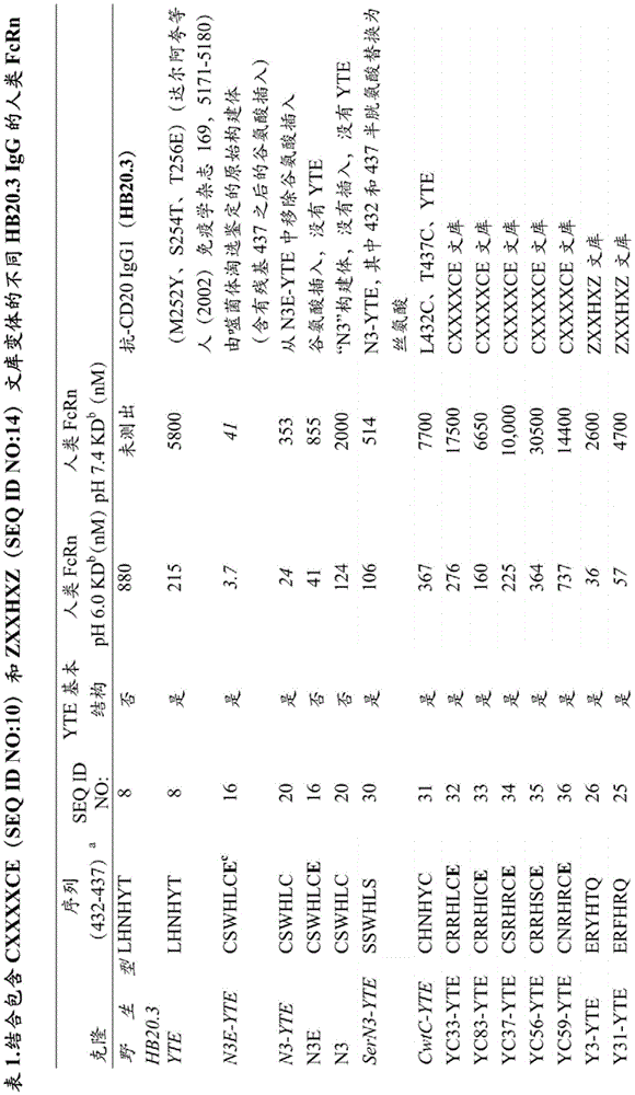


We further cleaved the N3E-YTE mutation to understand which mutation contributed to FcRn binding. Removal of glutamic acid insertion residue 437*(N3-YTE) has a moderate effect on pH 6.0 and pH 7.4 binding, indicating thatSequence variation from 432-437 was sufficient to confer most of the observed binding improvements. To evaluate the effect of cysteines at 432 and 437, they were replaced with serine (SerN 3-YTE). The resulting variants showed reduced FcRn binding at both pH 6.0 and pH 7.4, indicating that disulfide bonds bound between cysteines can be formed and positively contribute to binding. Additional variants with wild-type residues between L432C, T437C (CwtC-YTE) were generated to determine what effect cysteine alone had on FcRn binding. This construct bound FcRn slightly less than the parent YTE construct, indicating that cysteine mutation alone did not confer enhanced binding. Our observations that the pH-binding dependence of YTE is consistent with previous studies by mutational changes at the H435 loop (dalargi et al (2002) J Immunol 169, 5171-5180; bacaro et al (2005) Nature Biotechnology 23, 1283-1288) and demonstrate the role of H435 and its loop in the pH-dependent binding of Fc to FcRn. In addition to removing the glutamate insertion (N3-YTE) and YTE mutation (N3E), respectively, from N3E-YTE, we also generated a construct in which both were removed (N3). N3 had improved pH dependence compared to N3E-YTE (Table 1).
L432C, T437C stabilized C H3 Domain
The C α distance and side chain orientation of L432 and T437 in the Fc X line structure appear to be suitable for disulfide bond formation when mutated to cysteine. Using the crystal structure of YTE-Fc (Ouguey et al (2009) molecular immunology 46, 1750- This distance is in the range of the C.alpha.distances observed for cystine that have been reported based on polycrystalline structure analysis (about 4.6-
 ) Nepetsen et al (1999) protein engineering 12, 535-548). Since the stabilizing effect of intra-protein disulfide bond formation is well known, we performed different Differential Scanning Calorimetry (DSC) to examine C432C/
T437C containing variants H3 DomainWhether or not it is stabilized (fig. 3). DSC curve of HB20.3 consisting of C at 83.3 DEG CH3TmComposition of, wherein Fab and CHBoth 2 domains unfold as a peak at 73 ℃. YTE HB20.3 with wild type almost the same Fab and C H3 unfolding, but with a slightly reduced C at 65.1 ℃H2Tm. CwtC-YTE (L432C/T437C with YTE mutation) has a C at 87.1 ℃H3TmIs more wild type C H3 by almost 4 deg.c. Interestingly, though C H3 are greatly stabilized, but compared to YTE-HB20.3, C H2TmA reduction of about 3 deg.c. Comparison of N3-YTE and SerN3-YTE (with the difference that the constructs have cysteines or serines at 432 and 437) further illustrates the profound stabilizing effect caused by L432C/T437C. SerN3-YTE lowest TmIs 58.7 ℃ where CH3TmWell below 80 ℃ and the DSC curve of N3-YTE almost matches that of CwtC-YTE, with increasing CH3TmThe temperature was 87.1 ℃.
Engineering and selection of pH binding dependent YTE variants
We designed a new library at the His435 loop with the goal of generating variants with reduced pH 7.4 binding while maintaining high affinity binding at pH 6.0. We also changed our phage selection conditions to make the selection of panning pH-dependent, high affinity FcRn binders more stringent. We generated N3E-YTE based libraries by randomly inserting a sequence comprising H435(CXXXXCE (SEQ ID NO:10) library) between Cys432 and Cys 437. To hinder binding at neutral pH, a pH 7.4 exclusion step was introduced after the pH 6.0 elution step. The unbound phage is then used to infect bacteria for amplification and characterization. Phage ELISA was also performed after pH-dependent panning to characterize binding of clones. In this assay, clonal phage binding coated FcRn wells were used at pH 6.0 and 7.4. Binding at pH 6.0 and binding ratios between pH 6.0 and 7.4 were compared to the parent N3E-YTE and phage comprising YTE. Selected phage clones after 4 rounds of panning were screened for pH binding dependence in ELISA. Although almost all showed expected high binding to FcRn at pH 6.0 similar or better than YTE controls in the assay, several clones showed significantly reduced binding at pH 7.4 or higher pH 6.0 to pH 7.4 binding ratios, indicating improved pH binding dependence relative to N3E-YTE. Sequence analysis of more than 56 clones isolated after 4 rounds of panning showed that His435 was highly enriched (fig. 4A). Most of the 5 th clones contained H435, confirming the critical role of histidine at this position in FcRn binding. Sequence analysis showed that these clones also contained positively charged residues at 433, 434 or 436. The subpopulation of round 4 clones with improved pH 6/7.4 binding ratio via phage ELISA was even further enriched with positively charged residues adjacent to 435 (fig. 4B).
Additional libraries were designed to replace the cysteine residues at positions 432 and 437 with charged residues (including histidine). Since Cys432 and Cys437 are likely to form disulfide bonds in N3E-YTE, we theorize that the substitution of the rigid cysteine loop restriction in N3E-YTE with a charged amino acid at the cysteine position can introduce a pH-dependent change in the loop position that can be beneficial for pH-dependent FcRn binding. To test the hypothesis, we designed a library (ZXXHXZ (SEQ ID NO:14) library): cys432 and Cys437 were replaced with glutamic acid (E), aspartic acid (D), histidine (H), and random amino acids at positions 433, 434, and 436, while keeping H435 unchanged. In this embodiment, "Z" may be glutamic acid, aspartic acid, histidine or glutamine. Glutamic acid (E), aspartic acid (D), or histidine (H) is introduced at 432 and 437 to allow potential formation of pH-dependent salt bridges between glutamic acid or aspartic acid and histidine or to introduce negatively charged residues that can be sensitive to local electrostatic changes. Glutamine (Q) is also encoded at these positions 432 and 437 due to the use of degenerate oligonucleotides in cloning. The ZXXHXZ (SEQ ID NO:14) library was subjected to 4 rounds of panning, with an additional pH 7.4 binding exclusion step performed after each panning elution. Individual phage clones after 4 rounds of panning were sequenced and subjected to binding screening in a phage ELISA as described for CXXXXCE (SEQ ID NO:10) library panning and screening. Unlike the CXXXXCE (SEQ ID NO:10) library results, most of the clones selected from the ZXXHXZ (SEQ ID NO:14) library showed improved pH binding dependence compared to N3E-YTE, as evidenced by the high binding ratio at pH 6.0 versus pH 7.4 in the phage ELISA. In addition, sequence patterns were revealed in clones identified as pH-dependent by phage ELISA (fig. 4C). When converted to IgG, pH dependence was subsequently confirmed by SPR analysis (table 1). The results show that in clones that obtain high affinity, pH dependent binding to FcRn, positively charged amino acids (usually arginine) are preferred at positions 433 and 436; and the hydrophobic amino acid tyrosine or phenylalanine is preferred at position 434. Interestingly, the pH-dependent binding clones from this library were highly enriched for glutamic acid (E) and glutamine (Q) encoding at positions 432 and 437, respectively. The results indicate that engineering of the surface charge potential near H435 is effective for identifying high affinity and pH-dependent FcRn binding Fc variants. Although there are no histidine residues near H435 that can be sensitive to pH-dependent changes, a charge surface that accommodates the balance of these positions appears to be beneficial for FcRn binding.
SPR binding as FcRn binding variant of IgG.
A series of variants from the CXXXXCE (SEQ ID NO:10) and ZXXHXZ (SEQ ID NO:14) libraries that showed pH binding dependence in phage screens were selected and expressed as IgG1(HB20.3, anti-CD 20) for further characterization. Surface Plasmon Resonance (SPR) was used to measure binding of purified mabs to human FcRn at both pH 6.0 and pH 7.4. Table 1 summarizes the sequences and binding results of selected mabs. All tested variants derived from either the CXXXXCE (SEQ ID NO:10) or the ZXXHXZ (SEQ ID NO:14) libraries showed the expected reduced binding to FcRn at pH 7.4 compared to N3E-YTE, and many of them bound at levels similar to YTE IgG (approximately 5. mu.M). CXXXXCE (SEQ ID NO:10) variants that show pH dependence in phage ELISA maintain pH dependent binding as IgG, however, these variants do not show improved affinity at pH 6 compared to YTE. Many of the ZXXHXZ (SEQ ID NO:14) variants did show a significant pH 6.0 affinity improvement over YTE while also maintaining pH dependent binding. Several variants, such as Y3-YTE, Y12-YTE and Y31-YTE show 3-9 times higher FcRn binding affinity than YTE at pH 6, while binding at pH 7.4 remains similar or weaker than baseline. These results confirm the effectiveness of the library and panning strategy for selection and identification of clones.
The pH 6.0 and 7.4 binding affinities of the CXXXXCE (SEQ ID NO:10) and ZXXHXZ (SEQ ID NO:14) clones and the N3E-YTE variants and derivatives were grouped into quadrants (FIG. 5A, FIG. 5B). Quadrant I contains variants with high pH 6.0 binding and low pH 7.4 binding that are likely to exhibit "YTE-like" properties. Quadrant II contains clones with low pH 6.0 binding and pH 7.4 binding that are likely to exhibit "wild-type-like" characteristics. Quadrant III contains antibodies with high pH 6.0 binding and pH 7.4 binding that are likely to exhibit "Abdeg-like" properties. Quadrant IV contains binders at high pH 7.4 and low pH 6.0, none of which were isolated in these studies.
FcRn binding in an additional IgG background: movizumab
A few clones found to have the desired FcRn binding properties in an anti-CD 20 IgG1 background were moved to another IgG1 background mevizumab (wu et al (2007) journal of molecular biology 368, 652-665) in order to confirm the clonal FcRn binding profile in a different variable domain background. Since previous studies have shown that variable domains can contribute to FcRn binding variability in a wild-type Fc background (Suzuki et al (2010) journal of immunology 184, 1968-. The ZXXHXZ (SEQ ID NO:14) library clones Y12-YTE, Y31-YTE and Y3-YTE were generated as IgG with the variable domain of Movizumab. These clones and YTE and N3E-YTE pH 6.0 binding and in anti-CD 20 background pH 6.0 binding is consistent (Table 2). Interestingly, when clones translocated to different antibody variable domains, pH 7.4 binding changed, with either increased binding (Y31-YTE) or decreased binding (YTE). In addition, for Y12-YTE and Y31-YTE, clones such as Y12 and Y31 were also generated in the wild type Movizumab Fc background (without YTE). These constructs were generated to show the contribution of the H435 loop mutation alone to FcRn binding. Y12 and Y31 did maintain enhanced FcRn binding and pH dependence at pH 6.0 (low binding at pH 7.4). These data indicate that YTE and H435 mutations such as Y12 and Y31 have additive effects on pH-dependent binding. N3 with 89nM pH 6.0KD and 1630nM pH 7.4KD has similar binding in the Movizumab background as in the anti-CD 20 background.


Table 2c.dsc and hFcRn transgenic mouse pharmacokinetic data

Serum clearance in human FcRn transgenic mouse model
Next, we examined the serum clearance of different pH-sensitive FcRn binding variants using human FcRn transgenic mice (Ropelinel et al (2003) J Immunol 170, 3528-. In the PK study, hFcRn Tg mice were given a rapid intravenous dose of 2.5mg/kg at time zero and blood was taken at time points varying in the range of 30 minutes to 21 days after antibody injection. The human antibody concentration in mouse serum was quantified by ELISA.
Table 2 summarizes the PK results, lowest DSC Tm, and the sequence and binding characteristics of mevizumab and its Fc variants. The serum concentration-time curve of the selected antibodies is presented (fig. 6A). As expected, YTE mutations reduced serum clearance by approximately 2.4-fold compared to mevizumab. YTE consistently confers low antibody clearance rates in the hFcRn Tg mouse model, suggesting that this variant meets the conditions necessary and sufficient to enhance recycling through FcRn binding. Variant N3E-YTE, which has a high affinity pH 6.0 binding but no pH binding dependence, showed an extremely rapid serum clearance of 373 ml/day/kg, about 6-fold faster than mevinolizumab, thus underlining the importance of pH-dependent binding in FcRn-mediated antibody recycling. Clone Y31-YTE also showed an equivalent about 2.4-fold decrease in clearance compared to mevinolizumab as YTE. For half-life extension, N3 with 25.2 ml/day kg clearance was also equal to YTE. Interestingly, all tested pH-binding engineered variants (except N3E-YTE) significantly reduced serum clearance with or without YTE compared to wild-type mevinolizumab, thus validating our approach to identify new variants with enhanced pharmacokinetics (fig. 6B). The variants with improved PK parameters had pH 6.0 affinity compared to mevizumab by a factor in the range of approximately 4 to 40. Interestingly, a pH 7.4 affinity of 25nM resulted in a very poor clearance rate (N3E-YTE), while the pH 7.4-binding variant with an affinity of about 1 μ M still showed an enhanced half-life.
The mevizumab variants were also evaluated for Fc γ R and C1q binding. Interestingly, most variants other than N3 had reduced binding to certain Fc γ rs compared to mevizumab. These changes in binding range from small (< 2-fold decrease) to complete oblate (associated) binding to several receptors (as in the case of Y12-YTE). Interestingly, even the YTE mutations alone resulted in a decrease in the binding of Fc γ RIII, Fc γ RIIa and Fc γ RIIb, indicating that some effector function may be attenuated in the YTE background, but not in the N3 background.
Pharmacokinetics of N3 and Y31-YT in macaques
The novel Fc variant Y31-YTE, which showed low clearance in hFcRn mice, was then compared to YTE and wild-type mevinolizumab in cynomolgus monkeys. Four animals in each group received a single bolus injection of either mevizumab or mevizumab-Fc variant; serum samples were collected over the following 85 days. Consistent with previous studies (dar arg et al (2006) journal of biochemistry 281, 23514-. Y31-YTE also had improved pharmacokinetic properties relative to mevizumab (table 3). Three animals in the Y31-YTE group had a sudden decrease in Y31-YTE serum concentration between the 500 and 1000 hour period, likely indicating that the anti-drug antibody is clearing Y31-YTE mevinolizumab. Similar reductions were not seen in wild type and YTE groups during this period.
TABLE 3 pharmacokinetic data of macaques

Immunogenicity assessment of N3
To evaluate whether the N3 mutation introduced an immunogenic T cell epitope, N3-mevizumab and mevizumab were compared in ex vivo T cell proliferation experiments. PBMCs from 200 donors representing a wide range of HLA haplotypes were used in these experiments. PBMCs were incubated with mevizumab, N3-mevizumab, buffer alone or immunogenic KLH protein. T cell proliferation was measured and the results quantified as Stimulation Index (SI) which compares T cell proliferation of donor PBMC with buffer alone control (SI ═ 1). KLH protein had a significant SI increase, while both mevizumab and N3-mevizumab had no SI increase (fig. 7A, 7B). These data indicate that the N3 mutation has low immunogenicity when measured in this manner.
Opsonophagocytic killing of the N3 variant compared to the YTE variant.
Fc γ receptor binding data with N3 and YTE indicate that N3 maintained parent Fc γ receptor binding, whereas YTE had reduced Fc γ R binding (table 2). We assessed whether different Fc γ R binding affinities of these variants could lead to different effector functions in vitro, and we generated both YTE and N3 variants of the anti-pseudomonas anti-psl antibody Cam004 (digiandomenidae (DiGiandomenico) (2012) journal of experimental medicine 209(7): 1273-87). These constructs were tested in an opsonophagocytic killing assay using patient-derived polymorphonuclear cells as effectors and luciferase (PAO 1: lux) expressing pseudomonas (Pesudomonas) strain as target antibody. The results show that N3 displayed OPK similar to the parent antibody Cam004, whereas YTE had reduced OPK (fig. 8).
Stability of the N3 variant.
Accelerated stability studies were performed using N3 mevizumab in liquid formulation (50mg/ml in PBS pH 7.2 buffer). The antibody formulations were incubated at 4 ℃, 25 ℃ or 40 ℃ for one month. At the once weekly time point, aliquots were collected and analyzed by HPSEC to determine the monomer content as well as any fragments or aggregates. The data in fig. 9 plots monomer loss versus time and shows that the N3 variant is quite stable with 1.2% loss per month at 40 ℃ and only 0.3% per month at 25 ℃. These data indicate that the N3 mutation does not affect the stability of the Fc region.
Discussion of the related Art
The Fc region of IgG is the site of regeneration (motile group) of mutations that can alter the potency and efficacy of therapeutic antibodies. A variety of different studies have identified Fc variants with altered in vivo half-life or effector function (Stterol (2009) Biotechnology New 20, 685-. Possible benefits of antibodies with extended pharmacokinetic parameters include better tailoring to fit a specific therapeutic window that may allow for reduced dosing frequency while maintaining or even improving efficacy (zalinguiski et al (2010) natural biotechnology 28, 157-159). At the other end of the range is an Fc variant with reduced half-life. These antibodies are useful for imaging purposes such as Positron Emission Tomography (PET) imaging where fast clearance rates are desirable for imaging clarity and toxicity reduction (Kenanova et al (2010) Engineering of the Fc Region for Improved PK (FcRn Interaction) page 411-430; Olafsen (Olafsen) et al (2006) Natural laboratory technologies (Nature Protocols)1, 2048-2060). Abdeg is a third class of Fc variants with altered FcRn binding (baccarlo et al (2005) nature biotechnology 23, 1283-1288). The ability of Abdeg to reduce endogenous IgG levels could potentially be used to ameliorate certain autoimmune diseases characterized by destructive autoantibodies (crohn's (Killock) (2011) nature review for rheumatoid (Nat Rev rhematotol) 7, 496-496; patrel et al (2011) journal of immunology 187, 1015-1022; charla et al (2013) monoclonal antibody (mAbs)5, 655-659 electronic edition). To better understand the parameters separating the unequal in vivo pharmacokinetic results due to the engineered Fc-FcRn interaction, we used Fc phage display to select high affinity binders.
We first tried to select high affinity FcRn binding variants in YTE background, resulting in N3E-YTE that binds FcRn at pH 6 more tightly than the affinity enhanced YTE variant by >50 fold. The glutamic acid insertion mutation after position 437 contributes to the affinity increase as evidenced by a 6-fold reduction in binding after its removal (table 1). In addition to the insertions and hydrophobic residues at positions 434 and 436, N3E-YTE contains two cysteines at positions 432 and 437. The location of cysteine at the loop portion of His 435, the proximity and orientation of its side chains, and the stabilizing effect of these mutations, led us to believe that they form disulfide bonds with each other. We subsequently confirmed this result by peptide mapping. Differential scanning calorimetry confirmed the stabilizing effect of L432C and T437C on CH3 at the expense of a slight destabilizing effect of CH2 (fig. 3). These data indicate that the two cysteines are likely important for loop stability and that the intervening sequence confers most of the affinity increase and loss of pH sensitivity. The weak pH dependence seen with N3E-YTE is not unexpected. N434W alone was reported to have increased FcRn binding at both pH 6.0 and 7.4 (populus et al (2009) journal of immunology 182, 7663-. In addition, the MST-HN mutants (M252Y, S254T, T256E, H433K, N434F) that combine YTE mutations with two additional H435 loop mutants also show reduced pH dependence, resulting in abdeg (Dalgaro et al (2002) J Immunol 169, 5171-12880; Bakarlo et al (2005) Nature Biotechnology 23, 1283-1288).
This initial data prompted the creation of 2 additional libraries CXXXXCE (SEQ ID NO:10) and ZXXHXZ (SEQ ID NO: 14). These libraries were then panned with a pH 7.4 exclusion step to remove pH insensitive binders, and isolated clones were evaluated by phage ELISA (fig. 5A and 5B). The ZXXHXZ (SEQ ID NO:14) library produced more high affinity pH-dependent binders via phage ELISA, indicating that alleviating the restriction caused by 432 and 437 cysteines in CXXXXCE (SEQ ID NO:10) may be beneficial for reintroduction of pH-dependence. It is noted that the CXXXXCE (SEQ ID NO:10) library also contains a glutamic acid insertion after residue 437 (ZXXHXZ (SEQ ID NO:14) does not have this insertion) and this may also explain some unequal binding curves for the variants identified from these libraries. Interestingly, the mutations identified from the ZXXHXZ (SEQ ID NO:14) library that maintained higher affinity and pH sensitive binding than YTE alone consisted of charged residues at positions 433, 436 or both and hydrophobic residues at 434 (Table 1). The increase in positive charge may contribute to the pH dependence of these variants.
Variant N3 showed similar pharmacokinetics to YTE in human FcRn mice and cynomolgus monkeys and, in addition, unlike YTE, retained Fc γ and C1q binding similar to wild type IgG. Furthermore, also unlike YTE, N3 was found to have OPK activity equivalent to the parental IgG in the pseudomonas PA01lux system (fig. 8). Furthermore, the N3 variant did not affect the stability of the Fc region after storage. N3 is therefore expected to have utility, for example, as a modified IgG for use in antibody-mediated clearance applications as follows: these applications target infectious agents, where longer half-life and powerful effector functions are desirable.
Our data indicate that there is likely to be a threshold level of FcRn-mediated half-life extension (fig. 6B) and that exploration beyond this threshold does not yield further PK improvements. Indeed, increasing FcRn affinity too far above this threshold will yield molecules like N3E-YTE or MST-HN (meto et al (2009) journal of the national academy of sciences usa 106, 2788-Rou et al (2005) Nature Biotechnology 23, 1283-1288). Unexpectedly, we found that variants with pH 7.4 binding as low as 1uM still maintained equivalent enhanced in vivo half-life to YTE alone (table 2). With 24nM pH 7.4KDN3E-YTE showed extremely rapid clearance. We propose that a threshold of about 1uM below this appears to be a cut-off point for determining whether variants with enhanced binding at pH 6.0 have enhanced or Abdeg-like PKs that exhibit faster clearance than wild-type.
Variants N3 and Y31-YTE, which had the longest half-life in human FcRn mice, had almost 3-fold better clearance and approximately 5-fold longer half-life than wild-type mevinolizumab. These variants were included in the apical PK-improved variants described (plretta (2008) immunology nov 20, 460-. An approximately 3-5 fold enhancement in half-life or clearance rate compared to the parent antibody is the highest improvement reported to be achieved by altering FcRn binding (dalargi et al (2006) journal of biochemistry 281, 23514-. Pharmacokinetic data of Y31-YTE in cynomolgus monkeys also showed improvement, and so was N3 relative to wild-type mevinolizumab. From our data, and others, it is difficult to assume that there is room for significant improvement of PK enhancement beyond 3-4 fold purely by further altering FcRn binding. We envisage that even longer in vivo half-lives of Fc-containing molecules can be achieved by combining FcRn binding mutations with FcRn-dependent half-life extension techniques such as pI engineering (deit, Dahiyat et al (2012), us patent publication 20120028304, published 2.2.2012), pegylation (jewsevavar et al (2010) journal of biotechnology 5, 113-.
Example II.
Antibody formats for therapeutic strategies relying on antibody-mediated clearance using pH-dependent antigen binding
Antibodies have been engineered to have pH-dependent binding to their antigen. The engineered antibody binds to the antigen in the circulation (pH 7.4) to mediate clearance, but releases it in the sorting endosomes (pH 6), enabling the antibody to be recycled via FcRn, while the antigen left behind is degraded in the endosomes.
Antibody formats are described herein that have high affinity for FcRn at both pH 7.4 and 6.0. One example is N3E-YTE as described in example I, which shows a shorter half-life than a comparable wild-type antibody. A possible explanation for the shorter half-life of N3E-YTE and other variants like it may be that these antibodies remain bound to FcRn at the cell surface and are therefore not detected in the serum sample. If they circulate in the endosome in the form of a complex with FcRn, this form is expected to be more effective in antigen clearance (see figure 10).
More efficient antibody-mediated clearance is therefore envisaged. Antibody forms with high affinity for FcRn at both physiological pH (pH 7.4) and low pH (pH 6) can bind to FcRn at both the endosomal compartment and the cell surface. Thus, it will bind to the antigen at the cell surface and actively mediate its internalization into endosomes, which is more efficient for clearance of the antigen. Furthermore, antibody-driven endocytosis is a mechanism that can support sampling of immune system serum levels (Guo TT (Kuo TT) et al, J Clin Immunol (2010)30: 777-789). It should therefore be possible to increase efficiency by increasing pH affinity at physiological pH as described herein.
The efficacy of such antibodies can be tested in vivo. The model system of clinical relevance is PCSK-9. anti-PCSK-9 antibodies with pH-dependent binding are known (saparo-riegers et al, 2012, Journal of Biochemistry (JBC), 287: 11090-11097). The variable region of an anti-PCSK-9 antibody engineered to have pH-dependent binding to its antigen can be engineered into any antibody basic structure with altered FcRn binding that falls into quadrants I and III. In some cases, those basic structures that fall in quadrant III may be preferred. The generation of such antibodies can be accomplished using the procedures described herein, enabling side-by-side comparisons with wild-type anti-PCSK-9. The clearance rate of exogenous PCSK9 can be easily measured at different time points. Any other antibody with pH-dependent binding can be used in a similar study. This system provides increased potency for all applications that rely on antibody clearance, where antibodies with pH-dependent antigen binding can be engineered.
The entire disclosures of all patents, patent publications (including provisional patent applications), publications (including patent publications and non-patent publications), and electronically available materials (including, for example, nucleotide sequence submissions such as in gene banks and reference sequence banks (RefSeq) and amino acid sequence submissions such as in SwissProt, PIR, PRF, PDB, and translations of annotated coding regions from gene banks and reference sequence banks) cited herein are incorporated by reference. The foregoing detailed description and examples have been given for clarity of understanding only. No unnecessary limitations are to be understood therefrom. The invention is not limited to the specific details shown and described, and modifications apparent to a person skilled in the art are intended to be included within the invention as defined by the accompanying claims.


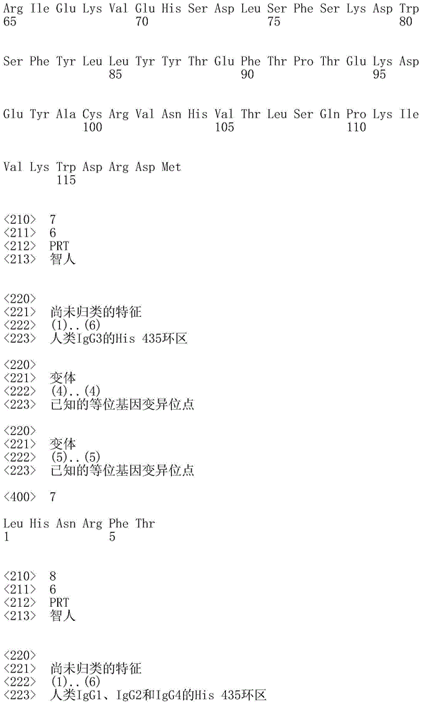


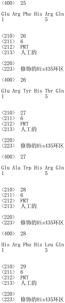


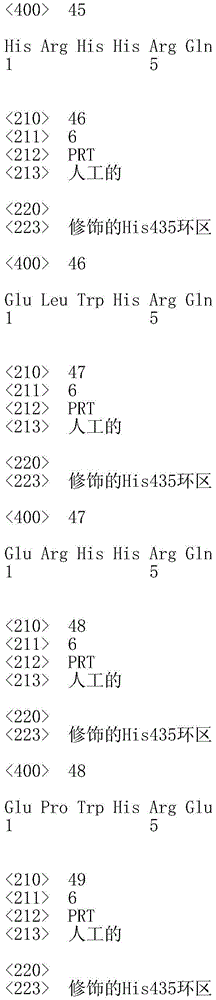


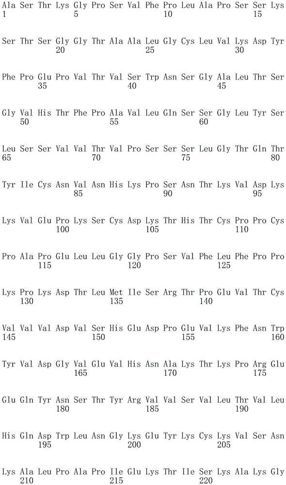


Claims (29)
1. A modified IgG1 comprising an Fc region, wherein the amino acids at positions 432 to 437 in the Fc region of the modified IgG1, numbered according to the EU numbering index of kabat, relative to a wild-type Fc region are as follows:
(i) both positions 432 and 437 are substituted with cysteine;
(ii) position 433 substituted with serine;
(iii) substitution at position 434 with tryptophan or tyrosine;
(iv) position 435 is histidine; and
(v) position 436 with leucine; and is
Wherein the modified IgG1 has an altered half-life compared to the half-life of IgG1 having the wild-type Fc region.
2. The modified IgG1 of claim 1, which modified IgG1 is human or humanized IgG 1.
3. The modified IgG1 of claim 1, wherein the modified IgG1 has a higher binding affinity for FcRn at pH 6.0 than IgG1 having a wild-type Fc region at pH 6.
4. The modified IgG1 of claim 1, wherein the modified IgG1 has a higher binding affinity for FcRn at pH 7.4 than IgG1 having a wild-type Fc region at pH 7.4.
5. The modified IgG1 of claim 1, wherein the modified IgG1 exhibits reduced pH dependence of binding affinity for FcRn compared to IgG1 having a wild-type Fc region.
6. The modified IgG1 of claim 1, wherein the amino acid sequence at positions 432 to 437 is CSWHLC.
7. The modified IgG1 of claim 1, wherein the modified IgG1 binds an antigen selected from the group consisting of: viral antigens, bacterial antigens, protist antigens, and mammalian self-antigens.
8. The modified IgG1 of claim 1, wherein the modified IgG1 binds an antigen that is a prion.
9. A polypeptide comprising at least an FcRn binding portion of an Fc region of an IgG1 molecule, wherein the amino acids at positions 432 to 437 in the FcRn binding portion of the polypeptide, numbered according to the EU numbering index of kabat, relative to a wild-type FcRn binding portion are as follows:
(i) both positions 432 and 437 are substituted with cysteine;
(ii) position 433 substituted with serine;
(iii) substitution at position 434 with tryptophan or tyrosine;
(iv) position 435 is histidine; and
(v) position 436 with leucine.
10. The polypeptide of claim 9, wherein the FcRn binding portion of the Fc region comprises amino acid residues from 231-446 of an IgG1 molecule according to the EU numbering index of kabat.
11. The polypeptide of claim 9, wherein the FcRn binding portion of the Fc region comprises amino acid residues 216-446 of an IgG1 molecule according to the EU numbering index of kabat.
12. A fusion protein comprising a non-IgG 1 polypeptide covalently linked to at least an FcRn binding portion of an Fc region of an IgG1 molecule, wherein the amino acids at positions 432 to 437 in the FcRn binding portion of the fusion protein, numbered according to the EU numbering index of kabat, relative to a wild-type Fc region are as follows:
(i) both positions 432 and 437 are substituted with cysteine;
(ii) position 433 substituted with serine;
(iii) substitution at position 434 with tryptophan or tyrosine;
(iv) position 435 is histidine; and
(v) position 436 with leucine;
wherein the fusion protein has a longer in vivo half-life than the non-IgG 1 polypeptide.
13. The fusion protein of claim 12, wherein the FcRn binding portion of the Fc region comprises amino acid residues from 231-446 of the IgG1 molecule according to the EU numbering index of kabat.
14. The fusion protein of claim 12, wherein the FcRn binding portion of the Fc region comprises amino acid residues from 216-446 of the IgG1 molecule according to the EU numbering index of kabat.
15. The fusion protein of claim 12, wherein the non-IgG 1 polypeptide is an immunomodulator, receptor or hormone.
16. The fusion protein of claim 12, wherein the non-IgG 1 polypeptide is a drug.
17. A molecule comprising a non-protein agent conjugated to at least an FcRn binding portion of an Fc region of an IgG1 molecule, wherein the amino acids at positions 432 to 437 in the FcRn binding portion of the molecule, numbered according to the EU numbering index of kabat, relative to a wild-type Fc region are as follows:
(i) both positions 432 and 437 are substituted with cysteine;
(ii) position 433 substituted with serine;
(iii) substitution at position 434 with tryptophan or tyrosine;
(iv) position 435 is histidine; and
(v) position 436 with leucine;
wherein the molecule has a longer half-life in vivo than the non-protein agent.
18. The molecule of claim 17, wherein the FcRn binding portion of the Fc region comprises amino acid residues from 231-446 of the IgG1 molecule according to the EU numbering index of kabat.
19. The molecule of claim 17, wherein the FcRn binding portion of the Fc region comprises amino acid residues 216-446 of the IgG1 molecule according to the EU numbering index of kabat.
20. A pharmaceutical composition comprising the modified IgG1, polypeptide, fusion protein or molecule of any one of claims 2 to 19, and a pharmaceutically acceptable carrier.
21. A nucleic acid comprising a nucleotide sequence encoding the modified IgG1, polypeptide, or fusion protein or FcRn-binding fragment thereof of any one of claims 1 to 16.
22. A vector comprising the nucleic acid of claim 21.
23. The vector of claim 22, comprising a cloning vector or an expression vector.
24. A host cell comprising the nucleic acid of claim 21.
25. A kit comprising the modified IgG1, polypeptide, fusion protein or molecule of any one of claims 2 to 19.
26. Use of the modified IgG1, polypeptide, fusion protein or molecule of any one of claims 2 to 19 in the manufacture of a medicament for use in a method of treating a disease, wherein the method comprises administering to a patient in need thereof a therapeutically effective amount of the modified IgG1, polypeptide, fusion protein or molecule of any one of claims 2 to 19.
27. Use of the modified IgG1, polypeptide, fusion protein or molecule of any one of claims 2 to 19 in the manufacture of a medicament for use in a method of treating a disorder, wherein the method comprises administering to a patient in need thereof a therapeutically effective amount of the modified IgG1, polypeptide, fusion protein or molecule of any one of claims 2 to 19.
28. Use of the modified IgG1, polypeptide, fusion protein or molecule of any one of claims 2 to 19 in the manufacture of a medicament for use in a method of treating an infection, wherein the method comprises administering to a patient in need thereof a therapeutically effective amount of the modified IgG1, polypeptide, fusion protein or molecule of any one of claims 2 to 19.
29. A method of altering the half-life of IgG1, the method comprising introducing the following amino acid substitutions relative to a wild-type Fc region at positions 432 to 437, numbered according to the EU numbering index of kabat; wherein
(i) Both positions 432 and 437 are substituted with cysteine;
(ii) position 433 substituted with serine;
(iii) substitution at position 434 with tryptophan or tyrosine;
(iv) position 435 is histidine; and
(v) position 436 with leucine.
Applications Claiming Priority (3)
| Application Number | Priority Date | Filing Date | Title |
|---|---|---|---|
| US201461994379P | 2014-05-16 | 2014-05-16 | |
| US61/994379 | 2014-05-16 | ||
| PCT/US2015/030964 WO2015175874A2 (en) | 2014-05-16 | 2015-05-15 | Molecules with altered neonate fc receptor binding having enhanced therapeutic and diagnostic properties |
Publications (2)
| Publication Number | Publication Date |
|---|---|
| CN106413750A CN106413750A (en) | 2017-02-15 |
| CN106413750B true CN106413750B (en) | 2022-04-29 |
Family
ID=54480933
Family Applications (1)
| Application Number | Title | Priority Date | Filing Date |
|---|---|---|---|
| CN201580026594.6A Active CN106413750B (en) | 2014-05-16 | 2015-05-15 | Molecules with altered neonatal Fc receptor binding with enhanced therapeutic and diagnostic properties |
Country Status (6)
| Country | Link |
|---|---|
| US (3) | US20170267780A1 (en) |
| EP (2) | EP3888690A3 (en) |
| JP (2) | JP6832709B2 (en) |
| CN (1) | CN106413750B (en) |
| ES (1) | ES2869459T3 (en) |
| WO (1) | WO2015175874A2 (en) |
Families Citing this family (24)
| Publication number | Priority date | Publication date | Assignee | Title |
|---|---|---|---|---|
| CN104125852B9 (en) | 2012-02-15 | 2017-05-17 | 弗·哈夫曼-拉罗切有限公司 | Fc-receptor based affinity chromatography |
| CN106103478B (en) | 2014-03-21 | 2020-04-03 | 豪夫迈·罗氏有限公司 | In vitro prediction of antibody half-life in vivo |
| US20200270363A1 (en) * | 2015-12-25 | 2020-08-27 | Chugai Seiyaku Kabushiki Kaisha | Antibody having enhanced activity, and method for modifying same |
| UA126900C2 (en) | 2016-04-28 | 2023-02-22 | Чугаі Сейяку Кабусікі Кайся | ANTIBODY CONTAINING DRUG |
| BR112019001989A2 (en) * | 2016-08-02 | 2019-08-20 | Visterra, Inc. | engineered polypeptides and uses thereof |
| CA3031735A1 (en) | 2016-08-05 | 2018-02-08 | Medimmune, Llc | Anti-o2 antibodies and uses thereof |
| US10669344B2 (en) | 2016-08-12 | 2020-06-02 | Janssen Biotech, Inc. | Engineered antibodies and other Fc-domain containing molecules with enhanced agonism and effector functions |
| US11117956B2 (en) | 2016-10-19 | 2021-09-14 | Medimmune, Llc | Anti-O1 antibodies and uses thereof |
| EP3615678B1 (en) | 2017-04-28 | 2024-07-31 | F. Hoffmann-La Roche AG | Antibody selection method |
| CN111032688A (en) * | 2017-08-11 | 2020-04-17 | 研究发展基金会 | Engineered antibody FC variants for extended serum half-life |
| IL273592B2 (en) | 2017-09-29 | 2025-11-01 | Chugai Pharmaceutical Co Ltd | A multispecific antigen-binding molecule with activity that replaces the function of a cofactor for blood coagulation factor VIII (FVIII), and a pharmaceutical formulation containing the molecule as an active ingredient |
| JPWO2019088143A1 (en) | 2017-11-01 | 2020-11-12 | 中外製薬株式会社 | Antibody variants and isoforms with reduced bioactivity |
| WO2019183228A1 (en) * | 2018-03-21 | 2019-09-26 | The University Of North Carolina At Chapel Hill | Compositions and methods for inhibiting pathogen infection |
| CA3107463A1 (en) * | 2018-07-24 | 2020-01-30 | Medimmune, Llc | Antibody directed against s. aureus clumping factor a (clfa) |
| CN113015745B (en) | 2018-10-09 | 2025-02-11 | 免疫医疗有限责任公司 | Antibodies against Staphylococcus aureus leukotoxin |
| CN113164602A (en) | 2018-10-09 | 2021-07-23 | 免疫医疗有限责任公司 | Combination of anti-staphylococcus aureus antibodies |
| CA3115321A1 (en) * | 2018-10-12 | 2020-04-16 | Eaton Intelligent Power Limited | Cable gland including internal dam |
| WO2020246891A1 (en) * | 2019-06-07 | 2020-12-10 | Sanquin Innovatie B.V. | Methods and compounds for typing rheumatoid factors |
| JP2023510781A (en) * | 2020-01-11 | 2023-03-15 | イエール ユニバーシティ | Compositions and methods for treating, ameliorating, and/or preventing diseases or disorders caused by or associated with DNAse1 and/or DNAse1L3 deficiency |
| CN115038453A (en) * | 2020-01-14 | 2022-09-09 | 辛德凯因股份有限公司 | CD122 with altered ICD STAT signaling |
| EP4126931A1 (en) | 2020-03-27 | 2023-02-08 | GlaxoSmithKline Biologicals S.A. | Hcvm pentamer binding antibodies |
| CN115321499B (en) * | 2022-08-11 | 2023-10-13 | 哈尔滨理工大学 | Preparation of black phosphorus nano-sheet composite material for biological imaging |
| CN115920006B (en) * | 2022-09-19 | 2023-09-05 | 山东大学 | Application of ABCF1 or its agonist in preparation of anti-DNA virus preparation |
| US20240199761A1 (en) | 2022-12-19 | 2024-06-20 | Astrazeneca Ab | Treatment of autoimmune disease |
Family Cites Families (174)
| Publication number | Priority date | Publication date | Assignee | Title |
|---|---|---|---|---|
| US320A (en) | 1837-07-31 | John dainty | ||
| US5985A (en) | 1848-12-26 | Pianoforte-action | ||
| US4444887A (en) | 1979-12-10 | 1984-04-24 | Sloan-Kettering Institute | Process for making human antibody producing B-lymphocytes |
| US4714681A (en) | 1981-07-01 | 1987-12-22 | The Board Of Reagents, The University Of Texas System Cancer Center | Quadroma cells and trioma cells and methods for the production of same |
| US4474893A (en) | 1981-07-01 | 1984-10-02 | The University of Texas System Cancer Center | Recombinant monoclonal antibodies |
| ATE37983T1 (en) | 1982-04-22 | 1988-11-15 | Ici Plc | DELAYED RELEASE AGENT. |
| US4716111A (en) | 1982-08-11 | 1987-12-29 | Trustees Of Boston University | Process for producing human antibodies |
| US4741900A (en) | 1982-11-16 | 1988-05-03 | Cytogen Corporation | Antibody-metal ion complexes |
| GB8308235D0 (en) | 1983-03-25 | 1983-05-05 | Celltech Ltd | Polypeptides |
| US4816567A (en) | 1983-04-08 | 1989-03-28 | Genentech, Inc. | Recombinant immunoglobin preparations |
| US4666884A (en) | 1984-04-10 | 1987-05-19 | New England Deaconess Hospital | Method of inhibiting binding of von Willebrand factor to human platelets and inducing interaction of platelets with vessel walls |
| US5807715A (en) | 1984-08-27 | 1998-09-15 | The Board Of Trustees Of The Leland Stanford Junior University | Methods and transformed mammalian lymphocyte cells for producing functional antigen-binding protein including chimeric immunoglobulin |
| US5128326A (en) | 1984-12-06 | 1992-07-07 | Biomatrix, Inc. | Drug delivery systems based on hyaluronans derivatives thereof and their salts and methods of producing same |
| EP0216846B2 (en) | 1985-04-01 | 1995-04-26 | Celltech Limited | Transformed myeloma cell-line and a process for the expression of a gene coding for a eukaryotic polypeptide employing same |
| US4980286A (en) | 1985-07-05 | 1990-12-25 | Whitehead Institute For Biomedical Research | In vivo introduction and expression of foreign genetic material in epithelial cells |
| US4676980A (en) | 1985-09-23 | 1987-06-30 | The United States Of America As Represented By The Secretary Of The Department Of Health And Human Services | Target specific cross-linked heteroantibodies |
| GB8601597D0 (en) | 1986-01-23 | 1986-02-26 | Wilson R H | Nucleotide sequences |
| US5091178A (en) | 1986-02-21 | 1992-02-25 | Oncogen | Tumor therapy with biologically active anti-tumor antibodies |
| GB8607679D0 (en) | 1986-03-27 | 1986-04-30 | Winter G P | Recombinant dna product |
| US5225539A (en) | 1986-03-27 | 1993-07-06 | Medical Research Council | Recombinant altered antibodies and methods of making altered antibodies |
| US6548640B1 (en) | 1986-03-27 | 2003-04-15 | Btg International Limited | Altered antibodies |
| US4946778A (en) | 1987-09-21 | 1990-08-07 | Genex Corporation | Single polypeptide chain binding molecules |
| US5567610A (en) | 1986-09-04 | 1996-10-22 | Bioinvent International Ab | Method of producing human monoclonal antibodies and kit therefor |
| WO1988007089A1 (en) | 1987-03-18 | 1988-09-22 | Medical Research Council | Altered antibodies |
| US5258498A (en) | 1987-05-21 | 1993-11-02 | Creative Biomolecules, Inc. | Polypeptide linkers for production of biosynthetic proteins |
| US4880078A (en) | 1987-06-29 | 1989-11-14 | Honda Giken Kogyo Kabushiki Kaisha | Exhaust muffler |
| GB8717430D0 (en) | 1987-07-23 | 1987-08-26 | Celltech Ltd | Recombinant dna product |
| US5336603A (en) | 1987-10-02 | 1994-08-09 | Genentech, Inc. | CD4 adheson variants |
| US5843708A (en) * | 1988-01-05 | 1998-12-01 | Ciba-Geigy Corporation | Chimeric antibodies |
| US4925648A (en) | 1988-07-29 | 1990-05-15 | Immunomedics, Inc. | Detection and treatment of infectious and inflammatory lesions |
| US5601819A (en) | 1988-08-11 | 1997-02-11 | The General Hospital Corporation | Bispecific antibodies for selective immune regulation and for selective immune cell binding |
| DE768377T1 (en) | 1988-09-02 | 1998-01-02 | Dyax Corp | Production and selection of recombinant proteins with different binding sites |
| US5223409A (en) | 1988-09-02 | 1993-06-29 | Protein Engineering Corp. | Directed evolution of novel binding proteins |
| GB8823869D0 (en) | 1988-10-12 | 1988-11-16 | Medical Res Council | Production of antibodies |
| KR900005995A (en) | 1988-10-31 | 1990-05-07 | 우메모또 요시마사 | Modified Interleukin-2 and Method of Making the Same |
| WO1990005144A1 (en) | 1988-11-11 | 1990-05-17 | Medical Research Council | Single domain ligands, receptors comprising said ligands, methods for their production, and use of said ligands and receptors |
| US5175384A (en) | 1988-12-05 | 1992-12-29 | Genpharm International | Transgenic mice depleted in mature t-cells and methods for making transgenic mice |
| US5530101A (en) | 1988-12-28 | 1996-06-25 | Protein Design Labs, Inc. | Humanized immunoglobulins |
| EP0394827A1 (en) | 1989-04-26 | 1990-10-31 | F. Hoffmann-La Roche Ag | Chimaeric CD4-immunoglobulin polypeptides |
| US6291159B1 (en) | 1989-05-16 | 2001-09-18 | Scripps Research Institute | Method for producing polymers having a preselected activity |
| US6291158B1 (en) | 1989-05-16 | 2001-09-18 | Scripps Research Institute | Method for tapping the immunological repertoire |
| US6291161B1 (en) | 1989-05-16 | 2001-09-18 | Scripps Research Institute | Method for tapping the immunological repertiore |
| US6291160B1 (en) | 1989-05-16 | 2001-09-18 | Scripps Research Institute | Method for producing polymers having a preselected activity |
| US6680192B1 (en) | 1989-05-16 | 2004-01-20 | Scripps Research Institute | Method for producing polymers having a preselected activity |
| ES2096590T3 (en) | 1989-06-29 | 1997-03-16 | Medarex Inc | BI-SPECIFIC REAGENTS FOR AIDS THERAPY. |
| US5112946A (en) | 1989-07-06 | 1992-05-12 | Repligen Corporation | Modified pf4 compositions and methods of use |
| US5413923A (en) | 1989-07-25 | 1995-05-09 | Cell Genesys, Inc. | Homologous recombination for universal donor cells and chimeric mammalian hosts |
| US5436146A (en) | 1989-09-07 | 1995-07-25 | The Trustees Of Princeton University | Helper-free stocks of recombinant adeno-associated virus vectors |
| WO1991005548A1 (en) | 1989-10-10 | 1991-05-02 | Pitman-Moore, Inc. | Sustained release composition for macromolecular proteins |
| WO1991006570A1 (en) | 1989-10-25 | 1991-05-16 | The University Of Melbourne | HYBRID Fc RECEPTOR MOLECULES |
| EP0550436A1 (en) | 1989-11-06 | 1993-07-14 | Alkermes Controlled Therapeutics, Inc. | Protein microspheres and methods of using them |
| GB8928874D0 (en) | 1989-12-21 | 1990-02-28 | Celltech Ltd | Humanised antibodies |
| US5780225A (en) | 1990-01-12 | 1998-07-14 | Stratagene | Method for generating libaries of antibody genes comprising amplification of diverse antibody DNAs and methods for using these libraries for the production of diverse antigen combining molecules |
| AU7247191A (en) | 1990-01-11 | 1991-08-05 | Molecular Affinities Corporation | Production of antibodies using gene libraries |
| US20040010810A1 (en) | 1990-01-12 | 2004-01-15 | Abgenix, Inc. | Generation of xenogeneic antibodies |
| US6713610B1 (en) | 1990-01-12 | 2004-03-30 | Raju Kucherlapati | Human antibodies derived from immunized xenomice |
| US6657103B1 (en) | 1990-01-12 | 2003-12-02 | Abgenix, Inc. | Human antibodies derived from immunized xenomice |
| DK0463151T3 (en) | 1990-01-12 | 1996-07-01 | Cell Genesys Inc | Generation of xenogenic antibodies |
| US6673986B1 (en) | 1990-01-12 | 2004-01-06 | Abgenix, Inc. | Generation of xenogeneic antibodies |
| US6075181A (en) | 1990-01-12 | 2000-06-13 | Abgenix, Inc. | Human antibodies derived from immunized xenomice |
| US6150584A (en) | 1990-01-12 | 2000-11-21 | Abgenix, Inc. | Human antibodies derived from immunized xenomice |
| US5314995A (en) | 1990-01-22 | 1994-05-24 | Oncogen | Therapeutic interleukin-2-antibody based fusion proteins |
| CA2078689C (en) | 1990-03-20 | 2003-02-11 | Sherie L. Morrison | Chimeric antibodies with receptor binding ligands in place of their constant region |
| US5229275A (en) | 1990-04-26 | 1993-07-20 | Akzo N.V. | In-vitro method for producing antigen-specific human monoclonal antibodies |
| US5427908A (en) | 1990-05-01 | 1995-06-27 | Affymax Technologies N.V. | Recombinant library screening methods |
| US5349053A (en) | 1990-06-01 | 1994-09-20 | Protein Design Labs, Inc. | Chimeric ligand/immunoglobulin molecules and their uses |
| GB9015198D0 (en) | 1990-07-10 | 1990-08-29 | Brien Caroline J O | Binding substance |
| US6916605B1 (en) | 1990-07-10 | 2005-07-12 | Medical Research Council | Methods for producing members of specific binding pairs |
| US6172197B1 (en) | 1991-07-10 | 2001-01-09 | Medical Research Council | Methods for producing members of specific binding pairs |
| JP2938569B2 (en) | 1990-08-29 | 1999-08-23 | ジェンファーム インターナショナル,インコーポレイティド | Method for producing xenogeneic immunoglobulin and transgenic mouse |
| US5770429A (en) | 1990-08-29 | 1998-06-23 | Genpharm International, Inc. | Transgenic non-human animals capable of producing heterologous antibodies |
| US5633425A (en) | 1990-08-29 | 1997-05-27 | Genpharm International, Inc. | Transgenic non-human animals capable of producing heterologous antibodies |
| CA2090473A1 (en) | 1990-08-29 | 1992-03-01 | Robert M. Kay | Homologous recombinatin in mammalian cells |
| US5814318A (en) | 1990-08-29 | 1998-09-29 | Genpharm International Inc. | Transgenic non-human animals for producing heterologous antibodies |
| US6255458B1 (en) | 1990-08-29 | 2001-07-03 | Genpharm International | High affinity human antibodies and human antibodies against digoxin |
| US5877397A (en) | 1990-08-29 | 1999-03-02 | Genpharm International Inc. | Transgenic non-human animals capable of producing heterologous antibodies of various isotypes |
| US5545806A (en) | 1990-08-29 | 1996-08-13 | Genpharm International, Inc. | Ransgenic non-human animals for producing heterologous antibodies |
| US5625126A (en) | 1990-08-29 | 1997-04-29 | Genpharm International, Inc. | Transgenic non-human animals for producing heterologous antibodies |
| US5789650A (en) | 1990-08-29 | 1998-08-04 | Genpharm International, Inc. | Transgenic non-human animals for producing heterologous antibodies |
| US5661016A (en) | 1990-08-29 | 1997-08-26 | Genpharm International Inc. | Transgenic non-human animals capable of producing heterologous antibodies of various isotypes |
| US5874299A (en) | 1990-08-29 | 1999-02-23 | Genpharm International, Inc. | Transgenic non-human animals capable of producing heterologous antibodies |
| US5698426A (en) | 1990-09-28 | 1997-12-16 | Ixsys, Incorporated | Surface expression libraries of heteromeric receptors |
| AU660629B2 (en) | 1990-10-01 | 1995-07-06 | University Of Connecticut, The | Targeting viruses and cells for selective internalization by cells |
| CA2093022C (en) | 1990-10-05 | 2005-02-22 | Michael W. Fanger | Targeted immunostimulation with bispecific reagents |
| EP0557300B1 (en) | 1990-10-29 | 1997-11-19 | Chiron Corporation | Bispecific antibodies, method of production, and uses thereof |
| DE69129154T2 (en) | 1990-12-03 | 1998-08-20 | Genentech, Inc., South San Francisco, Calif. | METHOD FOR ENRICHING PROTEIN VARIANTS WITH CHANGED BINDING PROPERTIES |
| KR100246529B1 (en) | 1990-12-14 | 2000-04-01 | 스티븐 에이. 서윈. 엠.디. | Chimeric Chains for Receptor-Related Signal Transduction Pathways |
| ATE269401T1 (en) | 1991-04-10 | 2004-07-15 | Scripps Research Inst | LIBRARIES OF HETERODIMERIC RECEPTORS USING PHAGEMIDS |
| WO1992019973A1 (en) | 1991-04-26 | 1992-11-12 | Surface Active Limited | Novel antibodies, and methods for their use |
| DE69232706T2 (en) | 1991-05-01 | 2002-11-28 | Henry M. Jackson Foundation For The Advancement Of Military Medicine, Rockville | METHOD FOR TREATING INFECTIOUS RESPIRATORY DISEASES |
| JPH06510524A (en) | 1991-05-14 | 1994-11-24 | ユニバーシティ オブ コネチカット | Targeted delivery of genes encoding immunogenic proteins |
| US6797492B2 (en) | 1991-05-17 | 2004-09-28 | Merck & Co., Inc. | Method for reducing the immunogenicity of antibody variable domains |
| EP0519596B1 (en) | 1991-05-17 | 2005-02-23 | Merck & Co. Inc. | A method for reducing the immunogenicity of antibody variable domains |
| DE69231385T2 (en) | 1991-06-05 | 2001-04-12 | The Board Of Regents Acting For And On Behalf Of The Unversity Of Michigan, Ann Arbor | TARGETED RELEASE OF GENES ENCODING SECRETORIC PROTEINS |
| EP0590067A1 (en) | 1991-06-14 | 1994-04-06 | Xoma Corporation | Microbially-produced antibody fragments and their conjugates |
| US5844095A (en) | 1991-06-27 | 1998-12-01 | Bristol-Myers Squibb Company | CTLA4 Ig fusion proteins |
| WO1993004169A1 (en) | 1991-08-20 | 1993-03-04 | Genpharm International, Inc. | Gene targeting in animal cells using isogenic dna constructs |
| ES2136092T3 (en) | 1991-09-23 | 1999-11-16 | Medical Res Council | PROCEDURES FOR THE PRODUCTION OF HUMANIZED ANTIBODIES. |
| DE69233782D1 (en) | 1991-12-02 | 2010-05-20 | Medical Res Council | Preparation of Autoantibodies on Phage Surfaces Starting from Antibody Segment Libraries |
| WO1993014188A1 (en) | 1992-01-17 | 1993-07-22 | The Regents Of The University Of Michigan | Targeted virus |
| US5622929A (en) | 1992-01-23 | 1997-04-22 | Bristol-Myers Squibb Company | Thioether conjugates |
| ES2193143T3 (en) | 1992-03-05 | 2003-11-01 | Univ Texas | USE OF IMMUNOCONJUGADOS FOR THE DIAGNOSIS AND / OR THERAPY OF VASCULARIZA TUMORS. |
| US5912015A (en) | 1992-03-12 | 1999-06-15 | Alkermes Controlled Therapeutics, Inc. | Modulated release from biocompatible polymers |
| US5733743A (en) | 1992-03-24 | 1998-03-31 | Cambridge Antibody Technology Limited | Methods for producing members of specific binding pairs |
| US5573905A (en) | 1992-03-30 | 1996-11-12 | The Scripps Research Institute | Encoded combinatorial chemical libraries |
| US5447851B1 (en) | 1992-04-02 | 1999-07-06 | Univ Texas System Board Of | Dna encoding a chimeric polypeptide comprising the extracellular domain of tnf receptor fused to igg vectors and host cells |
| CA2133411A1 (en) | 1992-04-03 | 1993-10-14 | Alexander T. YOUNG | Gene therapy using targeted viral vectors |
| ZA932522B (en) | 1992-04-10 | 1993-12-20 | Res Dev Foundation | Immunotoxins directed against c-erbB-2(HER/neu) related surface antigens |
| AU4116793A (en) | 1992-04-24 | 1993-11-29 | Board Of Regents, The University Of Texas System | Recombinant production of immunoglobulin-like domains in prokaryotic cells |
| ATE182651T1 (en) | 1992-06-09 | 1999-08-15 | Hoppe Ag | BOLTS AND DOOR LOCK |
| CA2141602A1 (en) | 1992-08-26 | 1994-03-03 | Philip Leder | Use of the cytokine ip-10 as an anti-tumor agent |
| US5639641A (en) | 1992-09-09 | 1997-06-17 | Immunogen Inc. | Resurfacing of rodent antibodies |
| PT671923E (en) | 1992-10-09 | 2001-10-30 | Advanced Tissue Sciences Inc | FIGURE RESERVE CELLS |
| EP1024198A3 (en) | 1992-12-03 | 2002-05-29 | Genzyme Corporation | Pseudo-adenoviral vectors for the gene therapy of haemophiliae |
| US5441050A (en) | 1992-12-18 | 1995-08-15 | Neoprobe Corporation | Radiation responsive surgical instrument |
| US5981175A (en) | 1993-01-07 | 1999-11-09 | Genpharm Internation, Inc. | Methods for producing recombinant mammalian cells harboring a yeast artificial chromosome |
| ATE267877T1 (en) | 1993-01-07 | 2004-06-15 | Sequenom Inc | DNA SEQUENCING THROUGH MASS SPECTRONOMY |
| US5934272A (en) | 1993-01-29 | 1999-08-10 | Aradigm Corporation | Device and method of creating aerosolized mist of respiratory drug |
| US5625825A (en) | 1993-10-21 | 1997-04-29 | Lsi Logic Corporation | Random number generating apparatus for an interface unit of a carrier sense with multiple access and collision detect (CSMA/CD) ethernet data network |
| AU696293B2 (en) | 1993-12-08 | 1998-09-03 | Genzyme Corporation | Process for generating specific antibodies |
| PT1231268E (en) | 1994-01-31 | 2005-11-30 | Univ Boston | BANKS OF POLYCLONE ANTIBODIES |
| US5516637A (en) | 1994-06-10 | 1996-05-14 | Dade International Inc. | Method involving display of protein binding pairs on the surface of bacterial pili and bacteriophage |
| CN1117155C (en) | 1994-07-29 | 2003-08-06 | 史密丝克莱恩比彻姆有限公司 | Novel compounds |
| GB9415379D0 (en) | 1994-07-29 | 1994-09-21 | Smithkline Beecham Plc | Novel compounds |
| US5643763A (en) | 1994-11-04 | 1997-07-01 | Genpharm International, Inc. | Method for making recombinant yeast artificial chromosomes by minimizing diploid doubling during mating |
| DE69630514D1 (en) | 1995-01-05 | 2003-12-04 | Univ Michigan | SURFACE-MODIFIED NANOPARTICLES AND METHOD FOR THEIR PRODUCTION AND USE |
| JP4026848B2 (en) | 1995-01-17 | 2007-12-26 | ブリガーム・アンド・ウーメンズ・ホスピタル・インコーポレーテッド | Receptor-specific transepithelial transport of immunogens |
| US6030613A (en) | 1995-01-17 | 2000-02-29 | The Brigham And Women's Hospital, Inc. | Receptor specific transepithelial transport of therapeutics |
| US5869046A (en) | 1995-04-14 | 1999-02-09 | Genentech, Inc. | Altered polypeptides with increased half-life |
| US6019968A (en) | 1995-04-14 | 2000-02-01 | Inhale Therapeutic Systems, Inc. | Dispersible antibody compositions and methods for their preparation and use |
| US6096871A (en) | 1995-04-14 | 2000-08-01 | Genentech, Inc. | Polypeptides altered to contain an epitope from the Fc region of an IgG molecule for increased half-life |
| US5747035A (en) | 1995-04-14 | 1998-05-05 | Genentech, Inc. | Polypeptides with increased half-life for use in treating disorders involving the LFA-1 receptor |
| DE69637481T2 (en) | 1995-04-27 | 2009-04-09 | Amgen Fremont Inc. | Human antibodies to IL-8 derived from immunized Xenomae |
| AU2466895A (en) | 1995-04-28 | 1996-11-18 | Abgenix, Inc. | Human antibodies derived from immunized xenomice |
| US6632976B1 (en) | 1995-08-29 | 2003-10-14 | Kirin Beer Kabushiki Kaisha | Chimeric mice that are produced by microcell mediated chromosome transfer and that retain a human antibody gene |
| AU710347B2 (en) | 1995-08-31 | 1999-09-16 | Alkermes Controlled Therapeutics, Inc. | Composition for sustained release of an agent |
| DE19544393A1 (en) | 1995-11-15 | 1997-05-22 | Hoechst Schering Agrevo Gmbh | Synergistic herbicidal mixtures |
| US5723125A (en) | 1995-12-28 | 1998-03-03 | Tanox Biosystems, Inc. | Hybrid with interferon-alpha and an immunoglobulin Fc linked through a non-immunogenic peptide |
| JP2978435B2 (en) | 1996-01-24 | 1999-11-15 | チッソ株式会社 | Method for producing acryloxypropyl silane |
| AU2063197A (en) | 1996-03-04 | 1997-09-22 | Massachusetts Institute Of Technology | Materials and methods for enhancing cellular internalization |
| WO1997033899A1 (en) | 1996-03-14 | 1997-09-18 | Human Genome Sciences, Inc. | Apoptosis inducing molecule i |
| CA2249195A1 (en) | 1996-03-18 | 1997-09-25 | Board Of Regents, The University Of Texas System | Immunoglobin-like domains with increased half lives |
| EP0904278A4 (en) | 1996-03-22 | 1999-09-15 | Human Genome Sciences Inc | Apoptosis inducing molecule ii |
| WO1997043316A1 (en) | 1996-05-10 | 1997-11-20 | Beth Israel Deaconess Medical Center, Inc. | Physiologically active molecules with extended half-lives and methods of using same |
| US5985309A (en) | 1996-05-24 | 1999-11-16 | Massachusetts Institute Of Technology | Preparation of particles for inhalation |
| US5874064A (en) | 1996-05-24 | 1999-02-23 | Massachusetts Institute Of Technology | Aerodynamically light particles for pulmonary drug delivery |
| US5855913A (en) | 1997-01-16 | 1999-01-05 | Massachusetts Instite Of Technology | Particles incorporating surfactants for pulmonary drug delivery |
| US5916771A (en) | 1996-10-11 | 1999-06-29 | Abgenix, Inc. | Production of a multimeric protein by cell fusion method |
| WO1998023289A1 (en) | 1996-11-27 | 1998-06-04 | The General Hospital Corporation | MODULATION OF IgG BINDING TO FcRn |
| CA2273194C (en) | 1996-12-03 | 2011-02-01 | Abgenix, Inc. | Transgenic mammals having human ig loci including plural vh and vk regions and antibodies produced therefrom |
| CA2277801C (en) | 1997-01-16 | 2002-10-15 | Massachusetts Institute Of Technology | Preparation of particles for inhalation |
| PT970126E (en) | 1997-04-14 | 2001-10-30 | Micromet Ag | A NEW METHOD FOR THE PRODUCTION OF ANTI-HUMAN ANTIGEN RECEPTORS AND THEIR USES |
| US6235883B1 (en) | 1997-05-05 | 2001-05-22 | Abgenix, Inc. | Human monoclonal antibodies to epidermal growth factor receptor |
| US5989463A (en) | 1997-09-24 | 1999-11-23 | Alkermes Controlled Therapeutics, Inc. | Methods for fabricating polymer-based controlled release devices |
| SE512663C2 (en) | 1997-10-23 | 2000-04-17 | Biogram Ab | Active substance encapsulation process in a biodegradable polymer |
| WO1999023105A1 (en) | 1997-11-03 | 1999-05-14 | Human Genome Sciences, Inc. | Vegi, an inhibitor of angiogenesis and tumor growth |
| CZ20003099A3 (en) | 1998-02-25 | 2002-04-17 | Lexigen Pharmaceuticals Corporation | Enhancing the circulating half-life of antibody-based fusion proteins |
| JP2002518432A (en) | 1998-06-24 | 2002-06-25 | アドバンスト インハレーション リサーチ,インコーポレイテッド | Large porous particles released from an inhaler |
| AU770555B2 (en) | 1998-08-17 | 2004-02-26 | Abgenix, Inc. | Generation of modified molecules with increased serum half-lives |
| US6833268B1 (en) | 1999-06-10 | 2004-12-21 | Abgenix, Inc. | Transgenic animals for producing specific isotypes of human antibodies via non-cognate switch regions |
| EP1355919B1 (en) | 2000-12-12 | 2010-11-24 | MedImmune, LLC | Molecules with extended half-lives, compositions and uses thereof |
| US7658921B2 (en) * | 2000-12-12 | 2010-02-09 | Medimmune, Llc | Molecules with extended half-lives, compositions and uses thereof |
| US7217797B2 (en) * | 2002-10-15 | 2007-05-15 | Pdl Biopharma, Inc. | Alteration of FcRn binding affinities or serum half-lives of antibodies by mutagenesis |
| AU2011265460B2 (en) * | 2003-01-09 | 2014-07-17 | Macrogenics, Inc. | Identification and engineering of antibodies with variant Fc regions and methods of using same |
| CA2545603A1 (en) * | 2003-11-12 | 2005-05-26 | Biogen Idec Ma Inc. | Neonatal fc receptor (fcrn)-binding polypeptide variants, dimeric fc binding proteins and methods related thereto |
| CN101124245A (en) * | 2003-11-12 | 2008-02-13 | 比奥根艾迪克Ma公司 | Neonatal Fc receptor (FcRn)-binding polypeptide variants, dimeric Fc-binding proteins, and methods related thereto |
| WO2005077981A2 (en) * | 2003-12-22 | 2005-08-25 | Xencor, Inc. | Fc POLYPEPTIDES WITH NOVEL Fc LIGAND BINDING SITES |
| JP5055603B2 (en) | 2004-08-04 | 2012-10-24 | メントリック・バイオテック・リミテッド・ライアビリティ・カンパニー | Mutated Fc region |
| AU2005304624B2 (en) * | 2004-11-12 | 2010-10-07 | Xencor, Inc. | Fc variants with altered binding to FcRn |
| US8802820B2 (en) * | 2004-11-12 | 2014-08-12 | Xencor, Inc. | Fc variants with altered binding to FcRn |
| AU2006204791A1 (en) * | 2005-01-12 | 2006-07-20 | Xencor, Inc | Antibodies and Fc fusion proteins with altered immunogenicity |
| TWI667257B (en) | 2010-03-30 | 2019-08-01 | 中外製藥股份有限公司 | Antibodies with modified affinity to fcrn that promote antigen clearance |
| AU2011283694B2 (en) | 2010-07-29 | 2017-04-13 | Xencor, Inc. | Antibodies with modified isoelectric points |
| CA2859755C (en) * | 2011-12-23 | 2021-04-20 | Pfizer Inc. | Engineered antibody constant regions for site-specific conjugation and methods and uses therefor |
-
2015
- 2015-05-15 CN CN201580026594.6A patent/CN106413750B/en active Active
- 2015-05-15 ES ES15793257T patent/ES2869459T3/en active Active
- 2015-05-15 WO PCT/US2015/030964 patent/WO2015175874A2/en not_active Ceased
- 2015-05-15 EP EP21159374.4A patent/EP3888690A3/en active Pending
- 2015-05-15 EP EP15793257.5A patent/EP3142700B1/en active Active
- 2015-05-15 JP JP2016567864A patent/JP6832709B2/en active Active
- 2015-05-15 US US15/310,196 patent/US20170267780A1/en not_active Abandoned
-
2019
- 2019-09-10 US US16/566,280 patent/US11767369B2/en active Active
-
2021
- 2021-02-02 JP JP2021014864A patent/JP7197616B2/en active Active
-
2023
- 2023-08-09 US US18/446,520 patent/US20240124612A1/en active Pending
Also Published As
| Publication number | Publication date |
|---|---|
| US11767369B2 (en) | 2023-09-26 |
| WO2015175874A3 (en) | 2016-03-17 |
| JP7197616B2 (en) | 2022-12-27 |
| EP3888690A3 (en) | 2021-10-20 |
| WO2015175874A2 (en) | 2015-11-19 |
| ES2869459T3 (en) | 2021-10-25 |
| CN106413750A (en) | 2017-02-15 |
| JP6832709B2 (en) | 2021-02-24 |
| EP3142700A4 (en) | 2018-05-02 |
| US20240124612A1 (en) | 2024-04-18 |
| EP3888690A2 (en) | 2021-10-06 |
| EP3142700A2 (en) | 2017-03-22 |
| JP2021074009A (en) | 2021-05-20 |
| US20200071423A1 (en) | 2020-03-05 |
| JP2017522000A (en) | 2017-08-10 |
| EP3142700B1 (en) | 2021-03-03 |
| US20170267780A1 (en) | 2017-09-21 |
Similar Documents
| Publication | Publication Date | Title |
|---|---|---|
| CN106413750B (en) | Molecules with altered neonatal Fc receptor binding with enhanced therapeutic and diagnostic properties | |
| US7704497B2 (en) | Molecules with extended half-lives, compositions and uses thereof | |
| US7658921B2 (en) | Molecules with extended half-lives, compositions and uses thereof | |
| EP2407548A1 (en) | Molecules with reduced half-lives, compositions and uses thereof | |
| AU2002248184A1 (en) | Molecules with extended half-lives, compositions and uses thereof | |
| AU2008201419C1 (en) | Molecules with extended half-lives, compositions and uses thereof | |
| AU2016202471C1 (en) | Molecules with extended half-lives, compositions and uses thereof | |
| CA2766160A1 (en) | Modified molecules with increased affinity for fcrn, compositions, and uses thereof | |
| HK1161264A (en) | Molecules with extended half-lives, compositions and uses thereof |
Legal Events
| Date | Code | Title | Description |
|---|---|---|---|
| C06 | Publication | ||
| PB01 | Publication | ||
| SE01 | Entry into force of request for substantive examination | ||
| SE01 | Entry into force of request for substantive examination | ||
| GR01 | Patent grant | ||
| GR01 | Patent grant |