CN103797023B - Plasma protein fractionation by sequential polyacid precipitation - Google Patents
Plasma protein fractionation by sequential polyacid precipitation Download PDFInfo
- Publication number
- CN103797023B CN103797023B CN201280044936.3A CN201280044936A CN103797023B CN 103797023 B CN103797023 B CN 103797023B CN 201280044936 A CN201280044936 A CN 201280044936A CN 103797023 B CN103797023 B CN 103797023B
- Authority
- CN
- China
- Prior art keywords
- protein
- plasma
- polymer
- proteins
- precipitate
- Prior art date
- Legal status (The legal status is an assumption and is not a legal conclusion. Google has not performed a legal analysis and makes no representation as to the accuracy of the status listed.)
- Active
Links
Images


Classifications
-
- C—CHEMISTRY; METALLURGY
- C07—ORGANIC CHEMISTRY
- C07K—PEPTIDES
- C07K1/00—General methods for the preparation of peptides, i.e. processes for the organic chemical preparation of peptides or proteins of any length
- C07K1/14—Extraction; Separation; Purification
- C07K1/30—Extraction; Separation; Purification by precipitation
- C07K1/303—Extraction; Separation; Purification by precipitation by salting out
-
- C—CHEMISTRY; METALLURGY
- C07—ORGANIC CHEMISTRY
- C07K—PEPTIDES
- C07K14/00—Peptides having more than 20 amino acids; Gastrins; Somatostatins; Melanotropins; Derivatives thereof
- C07K14/435—Peptides having more than 20 amino acids; Gastrins; Somatostatins; Melanotropins; Derivatives thereof from animals; from humans
- C07K14/745—Blood coagulation or fibrinolysis factors
- C07K14/75—Fibrinogen
-
- C—CHEMISTRY; METALLURGY
- C07—ORGANIC CHEMISTRY
- C07K—PEPTIDES
- C07K14/00—Peptides having more than 20 amino acids; Gastrins; Somatostatins; Melanotropins; Derivatives thereof
- C07K14/435—Peptides having more than 20 amino acids; Gastrins; Somatostatins; Melanotropins; Derivatives thereof from animals; from humans
- C07K14/76—Albumins
-
- C—CHEMISTRY; METALLURGY
- C07—ORGANIC CHEMISTRY
- C07K—PEPTIDES
- C07K16/00—Immunoglobulins [IGs], e.g. monoclonal or polyclonal antibodies
- C07K16/18—Immunoglobulins [IGs], e.g. monoclonal or polyclonal antibodies against material from animals or humans
-
- A—HUMAN NECESSITIES
- A61—MEDICAL OR VETERINARY SCIENCE; HYGIENE
- A61K—PREPARATIONS FOR MEDICAL, DENTAL OR TOILETRY PURPOSES
- A61K35/00—Medicinal preparations containing materials or reaction products thereof with undetermined constitution
- A61K35/12—Materials from mammals; Compositions comprising non-specified tissues or cells; Compositions comprising non-embryonic stem cells; Genetically modified cells
- A61K35/14—Blood; Artificial blood
- A61K35/16—Blood plasma; Blood serum
Landscapes
- Chemical & Material Sciences (AREA)
- Organic Chemistry (AREA)
- Health & Medical Sciences (AREA)
- Life Sciences & Earth Sciences (AREA)
- Proteomics, Peptides & Aminoacids (AREA)
- Biophysics (AREA)
- General Health & Medical Sciences (AREA)
- Genetics & Genomics (AREA)
- Medicinal Chemistry (AREA)
- Molecular Biology (AREA)
- Biochemistry (AREA)
- Analytical Chemistry (AREA)
- Toxicology (AREA)
- Zoology (AREA)
- Gastroenterology & Hepatology (AREA)
- Immunology (AREA)
- Hematology (AREA)
- Peptides Or Proteins (AREA)
- Medicines Containing Material From Animals Or Micro-Organisms (AREA)
- Medicines That Contain Protein Lipid Enzymes And Other Medicines (AREA)
Abstract
The present invention discloses a method for achieving the separation (clarification) of plasma proteins from blood cells using an aqueous system formed in blood or blood-containing solutions by adding a relatively low concentration of a single polymer. The present invention also discloses a simple sequential addition method using polyacids instead of the widely used coanda plasma protein fractionation method based on sequential addition of up to 40% (v/v) ethanol and other precipitants. It may also support the use of polymer film based containers in novel solvent-free plasma fractionation methods. The disclosed methods may also be applicable to smaller scale plasma protein separations in research and diagnostic applications. The general method is robust and can function over a wide range of process variables, such as temperature and pH.
Description
Technical Field
The present invention relates to the separation of blood components. In particular it relates to a method for fractionating plasma into pharmaceutically useful components.
Background
Non-chromatographic method for separation and purification of macromolecules
Producers of plasma proteins from natural sources have long relied on differential ethanol-based protein precipitation to separate protein fractions sequentially prior to purification of each protein via column chromatography. Such processes are particularly suitable for larger scale separations operating in a production intensive economy. However, they are technically complex and require highly trained technicians and expensive equipment and facilities.
Scientists involved in biotech separation processes are dealing with even larger processes and the need to reduce the cost of producing products. They re-examined the use of techniques such as precipitation and partitioning in aqueous two-phase systems (1,2, 3). The main advantage of partitioning is that it is based on the formation of two liquid (L) phases, whereby cells and cell debris can be collected at the L-L phase interface, which are maintained at the interface by interfacial tension. Thus, if the target protein can partition to a significant extent into one phase in a selective manner, it is possible to achieve a certain degree of target clarification (cell debris removal), purification and concentration all in one step (the above references). The main disadvantage of partitioning is that the formation of aqueous polymer biphasic systems usually requires the addition of two polymers such as dextran and polyethylene glycol [ PGE ] or one polymer and a high concentration of salt (e.g. PEG and 0.5M phosphate) to the solution containing the target. The two-polymer system can be expensive while the polymer-salt system has a low capacity (solubility) -often 1-2 g/L (3) for antibody proteins. Recently a method (4) for clarification of biotechnological feedstocks by addition of relatively low concentrations of a single biocompatible mesophilic polymer has been disclosed, which allows clarification via the formation of a substantially polymer-free phase, which contains most of the protein and which typically floats on top of a denser polymer-rich lower phase. The upper phase can also be modified by addition of a precipitating agent to achieve further concentration and purification of the target.
Single polymer aqueous two-phase system
It has long been known that proteins can selectively partition between phases in aqueous polymeric two-phase systems, using either two polymers (e.g., dextran and polyethylene glycol) or one polymer (e.g., PEG) and a relatively high concentration of water structuring salt (e.g., sodium phosphate) to form spontaneously. The latter systems are less expensive but they tend to have lower protein solubility and therefore lower capacity (3, 4). It has also long been understood that cells and cell debris, as well as other micron-sized colloids, tend to accumulate at the interface of the two phases, where they are maintained by interfacial tension. One variation of the two types of LL systems described above is a polymer-polymer system, where one polymer is a thermo-responsive polymer that self-associates at elevated temperatures and other conditions (3, 4). This action was previously used to separate proteins from the polymer of one phase after phase separation and two-phase isolation, and to recover the phase polymer for reuse. In addition to the two types of phase systems, a third type, a single polymer system, has also been recognized. These systems are formed under conditions where the added hydrophilic polymer self-associates and thus forms a lighter polymer-poor phase that floats on top of the much denser polymer-rich phase (4, 5). Suitable polymers include the so-called "EOPO" polymers prepared from Ethylene Oxide (EO) and Propylene Oxide (PO) components (e.g., Pluronic @, Tergitol @, Breox family of @). Other suitable polymers may include cellulose or other polysaccharide polymers that are secondary modified with ethoxy-containing groups. From a bioseparation perspective, a major disadvantage of such single polymer low salt two-phase systems is that the protein tends not to partition into the upper (polymer-poor) phase with any selectivity. It has therefore been written that "water-EOPO systems are therefore only suitable for the partitioning of hydrophobic molecules (e.g. denatured proteins or tryptophan-rich peptides or for solution concentration by selective water removal)" (5). One possible theoretical solution is to hydrophobically modify the EOPO polymer (6), which however may lead to detergency and other complications. It has recently been found that EOPO polymers can be added directly to the fermentation broth, using fermentation temperature and salts to achieve the formation of a two-phase system in which cell debris will concentrate at the phase interface and soluble materials such as proteins will partition into the polymer-poor phase where they can then be easily filtered and chromatographed (4). This work has focused on fermentation in general in connection with recombinant proteins and in particular on monoclonal antibody production.
Protein precipitation
Although flocculation and sedimentation (terms considered herein as equivalent) have been proposed for cell debris removal (2), they also have some appeal for processing bioprocess feedstocks that are pre-clarified using classical methods of centrifugation followed by filtration (1,2, 7-12). In such cases, target purification may be affected by precipitation of contaminants such as Host Cell Proteins (HCPs). Fortunately, most monoclonal antibodies, as well as many naturally occurring antibodies in the blood, carry a net positive charge at pH7, while many HCP and nucleic acid contaminants carry a net negative charge (7). The polycationic polymer can thus be used to precipitate many HCP and nucleic acid contaminants, leaving the target monoclonal antibody (Mab) protein with a net positive charge in solution (8, 10). Alternatively, it may be desirable to precipitate the net positively charged Mab target using a net negatively charged polymer and leave the net negatively charged proteins and nucleic acids in solution (7-9). In both cases some form of filtration, centrifugation or other method is required to separate the pellet from the supernatant (11).
One advantage of precipitating antibodies or other target proteins is that the target can be fairly stable in the precipitate and therefore can be stored intermittently during processing (12, 13). The disadvantage is that separation of the precipitate and redissolving of the target protein (e.g. for subsequent purification via chromatography) may require dilution of the protein, which increases the processing volume (e.g. 8). It may also require resuspension with a buffer whose conductivity, pH, or other characteristic is not compatible with processing by chromatography or other desired separation methods. Recently, methods for the purification of biotechnological proteins such as mabs and related antibody fragments (fabs) using polycarboxylic acids in the presence of salts such as sodium citrate have been disclosed (7).
In the above examples, the pH is typically controlled to achieve a situation in which the substance to be precipitated carries a net charge opposite to that of the precipitating agent with which it interacts. Similar considerations apply if proteins such as antibodies are preferentially precipitated using salts such as ammonium sulphate, again using control of pH to ensure that the antibodies carry a net charge opposite to that of the major contaminant (11). This helps to ensure that the proteins associate in complexes where they are charge neutral. When the precipitating agent is uncharged, for example in the case of solvents such as ethanol or neutral (uncharged) polymers (e.g. polyethylene glycol), it is often very effective to control the solution pH to match the pH (pi) of the isoelectric charge of the target protein (12). This is because the precipitating agent generally binds water molecules and reduces target solvation.
Both ethanol (14,15) and salt (22) have been used to separate complex protein mixtures into several fractions by sequential precipitation, wherein as the concentration of the precipitating agent increases, protein fractions one after the other are precipitated and recovered. This may be because a) different proteins precipitate at different precipitant concentrations and b) once a protein has precipitated, it remains undissolved as the precipitant concentration is further increased. However, precipitation using charged polymers is not used for fractionation by sequential precipitation as in (8,10), since the precipitate tends to re-dissolve after addition of excess precipitant polymer (8,9, 10). This behavior makes it essentially impossible to control the sequential precipitation process when two or more target proteins exhibit significantly different solubilities.
Plasma protein fractionation
Plasma is a valuable source of relatively inexpensive therapeutic proteins, especially in developing countries.billions of dollars per year of plasma proteins are isolated and sold for therapeutic use.A major concern is that proteins are highly abundant in plasma at fairly high concentrations and include albumin, fibrinogen and various immunoglobulins, such as those belonging to the IgG, IgA and IgM classes, however other proteins, such as coagulation factors V, VII, VIII, IV and von Willebrand factor (vWF), as well as transferrin, fibronectin and α -1-antitrypsin, are of increasing commercial importance (14, 15).
TABLE 1 some highly abundant human plasma proteins
| Protein | Mw (kDa) | pI | Blood plasma (g/l) |
| Serum albumin | 69 | 5.6 | 35-55 |
| Immunoglobulins | 145-190 | 6.5-9.5 | 14 |
| Fibrinogen | 340 | 5.1-6.3 | 1.5-3 |
| Transferrin | 80 | 5.6-6.0 | 2.3 |
| Factor VIII | 280 | 5-6 | 0.20 |
Generally, donated blood is centrifuged to produce a cell-rich fraction and plasma. The need for large scale centrifugation and sterility can limit the flexibility of such processes. Plasma is often stored frozen. Plasma proteins are purified based on chromatographic processing of fractions that are first separated using a general method known as Cohn Fractionation (Cohn Fractionation). It was first developed by Cohn (Cohn) et al in the united states during world war ii and subsequently improved by Kistler and Nitschmann, among other scientists (14, 15). It is typically applied to a large (e.g. 4000L) volume of thawed plasma to produce a cryoprecipitate containing a portion of fibrinogen as well as a number of factor VIII and associated vWF. The supernatant was then subjected to a complex series of low temperature precipitation steps in the presence of ethanol with varying pH, temperature and conductivity (fig. 1, redrawn from reference 16). In modern plasma fractionation processes (e.g., fig. 2A and 2B, from reference 15), these various precipitation steps result in fractions that are subsequently subjected to further purification using chromatographic processes. Advantages of the cohn fractionation in general include the fact that it can be applied to large amounts of plasma; the plasma protein fraction eventually becomes a precipitate that can be stored intermittently as needed; some fibrinogen is removed early in the process, so that it does not clog the column in subsequent separation steps; the ability of the process to inactivate certain bacteria, viruses and possibly prions; the process matches the capabilities of solvent-detergent (SD) or related antiviral methods. The disadvantages include: a. large and fairly concentrated amounts of ethanol need to be used, which presents health and explosion/fire hazards, resulting in expensive processing equipment and facilities. b. Fine-tuned (+/-3 ℃) temperature control is required, which further complicates the process and associated equipment and facilities in the case of large liquid volumes. c. Unit operations involving high concentrations of ethanol (up to 40%) and low pH and relatively long processing times may denature some target proteins. d. Proteins such as fibrinogen and immunoglobulins must be (partially) recovered as precipitates at different stages (e.g., fig. 2A and 2B). e. Protein recovery may be quite poor, for example 50% for immunoglobulins in certain processes (the above references). The alternative (22) of using salt precipitation for fractionation results in a very high salt content in the recovered protein fractions, necessitating expensive additional operations such as dilution or diafiltration prior to further processing of these fractions. It can also lead to denaturation of the protein. Fractionation using precipitation with non-ionic polyethylene glycol (PEG) and sodium chloride has also been attempted (23), but this requires very high concentrations of PEG and caustic sodium chloride. Furthermore, this process is very pH-sensitive and requires careful pH and conductivity adjustment after each precipitant addition. It also requires complex additional processing to remove PEG from the protein.
In view of the above, there is a need to identify simpler, robust and easily extensible alternatives to centrifuge-based separation of blood cells from plasma. There is also a need to identify alternatives that are simpler and easily scalable than cohn fractionation (14, 15). It should also be noted that there is a need to identify rapid and simple methods to isolate plasma proteins for diagnostic and research purposes.
Summary of The Invention
One aspect of the present invention relates to a method for selectively separating proteins from plasma separated from blood. This is done using a method according to claim 1. The method can be used in a selective manner to isolate specific high abundance proteins such as fibrinogen, immunoglobulins and albumin. Selectivity and protein recovery can be further controlled through the use of pH and salt compositions. The method is also robust with respect to: provides similar separation in the face of normally expected process variations in polymer type, polymer concentration, temperature, pH, etc. (i.e. +/-10% of the specified operating values) and does not require complex adjustments of buffer pH, conductivity, etc. between precipitation steps. It is also robust to protein concentrations, so that it can be used at different stages of plasma processing, including protein concentrations above plasma levels. It can even be used directly on undiluted plasma, which allows a significant cost reduction compared to previous plasma fractionation methods.
The second aspect of the invention also relates to a method for the spontaneous separation of plasma proteins from blood cells and blood lipid fractions. This is accomplished by a method according to claim 33. The method may be used alone or as a step in a plasma separation process together with the method of the first aspect. The advantages of the invention include: a. little or no energy is required to achieve separation in addition to mixing, b. it can be performed in a variety of vessels, including disposable single use plastic vessels, c. it uses relatively inexpensive and biocompatible reagents, d. it functions over a wide range of scales and temperatures, e. it leaves most of the protein in an aqueous phase that can be adapted for direct use in further target processing, including further purification of the protein via depth filtration (to remove any cells, cell debris, bacteria or other colloidal contaminants) and then by precipitation, chromatography, and the like.
For blood and plasma processing, the two methods described above can be used together or independently to achieve clarification and selective precipitation of proteins.
Further suitable embodiments of the invention are described in the dependent claims.
Reference to the literature
1. D J Roush, Y Lu, Biotechnol Progr 24, 488-495, 2008。
2. J Thommes, M Etzel, Biotechnol. Progr. 2007, 23, 42-45。
3. A M Azevedo et al, Trends in Biotechnology, 27, 240-.
4. WO 2010/080062。
5. H-O Johansson, et al, J. Chromatogr B, 711, 3-17, 1998.
6. H-O Johansson et al, Biotechnol Bioeng, 66, 247-.
7. WO 2010/082894。
8. WO 2008/091740。
9. P McDonald, et al, Biotechnol Bioeng, 102, 1141-.
10. P Thanmaya, et al, Biotechnol Progr, 28, 1322-1331, 2010.
11. A Venkiteshwaran et al, Biotechnol Bioeng, 101, 957-.
12. WO 2008/100578。
13. Us patent 3,974,134.
14. A Lihme et al, Anal Biochem, 399, 102-.
15. T Burnouf. Transfusion Medicine Reviews, 21, 101-117, 2007。
16. E.J. Cohn et al, J.am chem. Soc, 68, 459-.
17. L Jiang et al, J Chromatogr A, 1023, 317-.
18. J Ma et al, J. Chromatogr B, 862, 219- "226, 2008.
19. Odile Carter et al, Nature protocols, 1, 812-.
20. F. Carlsson et al, J. Phys. chem. B. 105, 9040-.
21. A, Witteman et al, progr, Colloid Polymer, Sci, 133, 58-64, 2006.
22. Us patent 7,879,332.
23. Y-Z Lee et al, J. Agr. Food chem. 35, 958-962, 1987.
Brief Description of Drawings
Figure 1 is a graph showing the complexity of cohn fractionation method 6 (1946) in terms of changes in pH and ethanol concentration, redrawn from cohn et al, reference 16.
Figures 2A and b are depictions showing the complexity of modern cohn-based precipitation and related chromatographic plasma fractionation methods, excerpted from Burnouf, reference 15.
Figure 3. brief summary of cohn-based precipitation and related chromatography plasma fractionation methods of cell-free plasma samples compared to the inventive method based on a single polymer phase system for bulk cell removal prior to polyacid-based precipitation. In both cases, fibrinogen is removed in a first precipitation step (not shown).
FIG. 4. one-dimensional (ID) Sodium Dodecyl Sulfate (SDS) reduction gel electrophoresis analysis of normal human plasma and protein MW standards showing certain common bands associated with highly abundant serum proteins and their subunits (from this work) is compared to a literature picture (18) of two-dimensional (2D) isoelectric pH followed by SDS reduction gel electrophoresis analysis of plasma showing its complexity.
FIG. 5 ImageQuantTL comparing the relative intensities (peaks) of each of the ten major bands and their relative pixel positionsTMAnalysis showed 1D reduced SDS of human plasmaGel electrophoresis analysis (left with increasing MW from bottom to top). The number 7 at the top of the SDS gel indicates the lane. The gel contains many other reproducible peaks associated with lower abundance proteins.
FIG. 6 1D reduction SDS gel electrophoresis analysis of human plasma proteins dispensed in a representative EOPO phase system (i.e., 8% EOPO, 150mM NaCl, 100mM sodium citrate, pH7, 40 ℃) which is believed to be useful for blood clarification and protein separation. A lane; 1-HSA polymer phase, 2-HSA aqueous phase, 3-IgG aqueous phase, 4-IgG polymer phase, 7-fibrinogen aqueous phase, 8-fibrinogen polymer phase. The three fibrinogen subunits in lane 7 are shown by arrows.
FIG. 7 SDS gel electrophoresis analysis of 1D reduction of human plasma proteins precipitated with 10% polyacrylic acid. Most of the serum proteins precipitated, but much albumin (HSA) and transferrin appeared to remain in the supernatant. System (5 ml): 1.5ml plasma, 10% (w/w) PAA15000, 250mM sodium citrate pH7, 150mM NaCl.
FIG. 8 1D reduction SDS gel electrophoresis analysis of pellet and supernatant samples associated with polyacid precipitation of plasma proteins in the presence of 200mM sodium citrate, pH 7. PVS (11% (w/w) polyvinylsulfonic acid 3000, CMD (20% carboxymethyldextran), PAA (10% polyacrylic acid 15000), polymer concentration was varied to produce approximately similar concentrations of charged groups in solution.
FIG. 9 1D reduction SDS gel electrophoresis analysis showing the effect of adding polyacrylic acid (PAA15000) at different concentrations (4-10% w/w) to 5ml of a plasma precipitation solution consisting of 1.5ml of plasma, 50mM sodium citrate, pH7, 50mM NaCl. The precipitate (PPT) and Supernatant (SUP) fractions were analyzed. The appearance of the newly highlighted protein band is indicated by the arrow.
Figure 10. percentage of total amount of IgG, HSA and fibrinogen present in the precipitate as a function of PAA concentration. Data are based on ImageQuantTL analysis of fig. 9 electrophoresis. The results of Biacore SPR analysis of the antibodies are also shown.
Figure 11. three-step four-fractionation method for fractionating plasma using solutions containing polyacrylic acid (PAA) and at least 50mM sodium citrate, pH7, at different% w/w concentrations. The addition of 5% PAA followed by separation of the precipitate by settling, filtration or centrifugation (shown) increased the supernatant concentration to 8% PAA and then followed by separation of the precipitate increased the supernatant concentration to 10% PAA. The pellet was then resuspended, leaving three pellet fractions and a final supernatant fraction.
Figure 12 shows 1D reducing SDS gel electrophoresis of sequential fractionation of plasma proteins using increasing concentrations of PAA according to figure 11. System (5 ml): 1.5ml plasma, 4-10% (w/w) PAA (15000), 50mM sodium citrate (pH7), 50mM NaCl.
FIG. 13 conductivity versus PAA15000 concentration (% w/w) in a solution containing 50mM NaCl and 50mM sodium citrate, pH 7.
FIG. 14 1D reduction SDS gel electrophoresis of plasma protein fractionation in a 5ml system containing 5% (w/w) PAA15000, added 1.5ml plasma plus 50 or 250mM sodium citrate, pH 7. Arrows indicate the appearance of new prominent protein bands in the pellet samples.
FIG. 15. results from a Cubic Centered (CCF) experimental design study in which the concentration% w/w of PAA15000 and the mM concentration of sodium citrate were varied in a 5ml system with 200mM NaCl to optimize the conditions in which antibody or albumin could be selectively precipitated.
FIG. 16. Effect of pH on plasma protein fractionation in a 5ml system containing 8% (w/w) PAA15000, 50mM NaCl and 50mM sodium citrate (final system pH 6-8). The arrow indicates a newly highlighted band in the pellet.
FIG. 17. Effect of temperature on plasma precipitation in a 5ml system containing 1.5ml plasma plus 8% PAA15000, 50mM NaCl and 50mM sodium citrate, pH 7.
Definition of
The term "polyacid" means herein a polymer comprising a functional group that is negatively charged at pH 6 and higher.
The term "self-associating responsive polymer" means herein a water-soluble polymer comprising hydrophobic segments and having a cloud point (i.e., the temperature above which an aqueous solution of the polymer separates into two phases).
Detailed Description
In one aspect, the present invention discloses a method for separating at least a first and a second protein from a sample of plasma, said method comprising the steps of:
a) a sample of the plasma is provided in a container,
b) adding a polyelectrolyte, such as a polyacid, and a salt to the plasma, resulting in the formation of a first protein precipitate and recovering the first protein precipitate and a first supernatant,
c) adding a polyelectrolyte/polyacid and/or a salt to the first supernatant resulting in the formation of a second protein precipitate and a second supernatant and separately recovering the second protein precipitate,
wherein the first protein precipitate comprises a first protein concentrate and the second protein precipitate comprises a second protein concentrate.
Concentrate means that the mass ratio of the protein in question (first or second protein) to the total protein amount in the protein precipitate is significantly higher than said mass ratio in plasma or in the first supernatant. For example, it may be at least twice as high, at least 10 times as high, or at least 50 times as high. This may also be expressed in such a way that the first and second proteins are enriched in the respective protein precipitate. Furthermore, the first protein precipitate may comprise at least 50% by weight, such as at least 70% by weight or at least 90% by weight of the total amount of the first protein in the plasma sample, and the second protein precipitate may comprise at least 50% by weight, such as at least 70% by weight or at least 90% by weight of the total amount of the second protein in the first supernatant or in the plasma sample. The plasma sample used may be undiluted plasma.
The method selectively concentrates and separates proteins via the addition of a polyacid from plasma that is separated from blood by standard methods such as centrifugation and filtration, or by partitioning. The method can be used with salts, including citrate anticoagulants, to non-specifically precipitate most plasma proteins. It can also be used in an alternative manner to separate specific high abundance proteins such as fibrinogen, and then expose the supernatant to higher polymer concentrations to fractionally precipitate immunoglobulins. The supernatant from the separation can then be exposed to still higher polymer and/or salt concentrations to separate other residual proteins from the serum albumin. Plasma contains a large amount of proteins-about 100 proteins in addition to the most abundant (table 1) and gel electrophoresis indicates that the precipitant fraction will contain other proteins which may also have commercial, diagnostic or research significance. Selectivity and protein recovery can be further controlled through the use of pH and salt compositions. However, the method is very robust to the effects of normal process variations (i.e. +/-10% of the specified operating values) in terms of polymer type, polymer concentration, temperature, pH, etc. In either or both of steps b) and c), it may be operated at a temperature of from 0 to 40 ℃ (e.g. from 4 to 20 ℃). The method may also be combined with a pathogen inactivation or removal step, such as a solvent-detergent step, a virus filtration step, and/or a low pH virus inactivation step. The plasma may be human, but it may also be of animal origin, such as bovine or equine plasma.
The polyacid precipitation method has the advantages that:
I. it does not require any organic solvent.
It can be performed in a variety of containers including disposable single use plastic containers.
It uses mild and inexpensive reagents.
It is expected to separate nucleic acids and some other contaminants into the supernatant.
V. it can be carried out over a wide range of temperatures.
It produces a defined, stable and easily handled precipitate that is also easily re-dissolved in various solutions for further processing.
It can be adapted for the processing of plasma protein samples from small scale (microliters) to large scale (kiloliters).
It may be suitable for processing not only human plasma proteins but also proteins in mixtures from other sources (e.g. bovine plasma).
The basic advantages of the combined method are shown in fig. 3, which provides a brief description of a standard cohn-based plasma protein fractionation method (shown in more detail in fig. 1 and 2) compared to a method in which cells can be separated via partitioning (or with classical centrifugation) followed by polyacid-based precipitation.
The polyacid precipitation method appears to satisfy all the requirements for a method replacing the cohn fractionation method, which are outlined below. The advantages of the combined partitioning and precipitation method are summarized in table 2. Other features of the above-described method will be apparent from the following detailed examples and from the claims.
Requirements for a cohn fractionation alternative:
1. comparable to the cohn fractionation method so as to be easily applied to existing facilities and methods. For example, it should separate fibrinogen, immunoglobulin and albumin proteins in separate fractions and be able to separate the proteins in (easily resolubilized) precipitant complexes that also provide stable short-term storage.
2. Little or no organic solvents are involved to reduce health, safety and cost concerns; and to processes supplied at simpler facilities.
3. May be suitable for processing at uniform temperatures from 4 c to room temperature (e.g., 20 c) thereby eliminating the need for complex control systems for varying temperatures.
4. Providing equally good or better protein (e.g. immunoglobulin) yields.
5. Most of the fibrinogen is removed early in the process to facilitate efficient subsequent purification via chromatography or other porous media-based steps.
6. In contrast to the complex pH, conductivity and solvent composition changes of the cohn fractionation process (fig. 1), it is possible to operate without significantly changing the working liquid composition.
7. Including fewer precipitation steps and no steps requiring other precipitants such as polyethylene glycol as compared to cohn fractionation (fig. 2A). For example, the addition of a single precipitant at a single temperature and sequentially at relatively low concentrations (compared to 20-40% ethanol levels of the cohn fractionation method (16)).
8. It is cost-effective to utilize relatively low concentrations of biocompatible agents that can be easily removed via subsequent chromatography steps.
9. Easy implementation using sterile plastic bags and other disposable equipment to allow flexible manufacturing and to simplify assembly of new facilities.
10. Liquid process streams and robust methods are involved to easily allow simple integration of low pH steps, solvent-detergent steps and filtration steps to help remove pathogenic contaminants such as bacteria, viruses, prions and related substances. This is in addition to any removal provided by the chromatography steps associated with the overall process.
11. Provides a significant reduction (10-100x) of nucleic acids and other contaminants, in addition to any removal provided by the chromatography steps associated with the overall process.
12. It may be appropriate to add stabilizers, anti-aggregants, anti-proteolytic agents (if desired) to facilitate recovery of the active protein target in its native functional state.
TABLE 2 summary of plasma fractionation methods based on cohn and on novel polymers

In some embodiments, the first protein may be fibrinogen. The second protein may be an immunoglobulin, such as immunoglobulin G. Fibrinogen generally precipitates at lower polyacid concentrations than immunoglobulins, and precipitation selectivity is similar to that observed in the cohn fractionation method is an advantage of the method. This reduces fouling or other process complexity associated with fibrinogen. Both fibrinogen and immunoglobulin G are valuable proteins and are commonly used as pharmaceuticals. In some embodiments, the first and second proteins are suitable for use as a medicament, meaning that they are (optionally after further isolation steps) of sufficient purity for pharmaceutical use and free of toxic contaminants introduced by the method. In certain embodiments, sufficient polyacid is added in step b) or c) such that the second protein precipitate contains substantially all residual protein from the plasma. This precipitate can then be further processed for individual recovery or analysis of numerous proteins.
In certain embodiments, the salt addition in step b) is controlled such that in step b) the plasma comprises at least 50mmol/L (e.g. 50-250 mmol/L or 50-100 mmol/L) of salt (calculated as the anion of the salt). It can also be controlled so that the total salt concentration in step b) is at least 50mmol/L, for example 50-250 mmol/L or 50-100mmol/L (again calculated as anion). The polyacid and the salt show a strong synergistic effect, meaning that a relatively low salt concentration can be used to obtain an efficient fractionation. This is advantageous because the low salt concentration in the precipitate allows the redissolved precipitate to be processed directly by methods such as cation exchange chromatography without the need for excessive dilution or diafiltration. Yet another advantage of operating at low salt concentrations is reduced corrosion of stainless steel equipment if chloride salts are used. The salt may be added as an aqueous solution or as a solid powder.
In some embodiments, the polyacid in step b) is added in an amount that results in a total polyacid concentration in the plasma of at least 3% by weight (e.g., 4-10% by weight or 4-5% by weight). The exact concentration to be used will depend to some extent on the salt concentration and will require some limited experimentation to determine the concentration used for a particular separation task. If desired also as a mixed polyacid-salt solution, the polyacid may suitably be added in the form of an aqueous solution. Optimization can be performed, for example, according to a cubic face center (CCF) experimental design, using pure protein or via experiments performed with simple electrophoretic analysis, as shown in fig. 8,9, 15, 16.
In certain embodiments, the polyacid in step c) is added in an amount that results in a total polyacid concentration in the first supernatant that is equal to or higher than the polyacid concentration in step b), which is at least 4% by weight, such as between 5-20% by weight, 5-15% by weight, or 5-8% by weight. If the salt concentration in step c) is the same as in step b), a higher polyacid concentration will be required in step c) than in step b), but if additional salt is added in step c), the polyacid concentration may be equal to or higher than the polyacid concentration in step b). In certain embodiments, particularly when the polyacid concentration is about 15-20%, the second protein precipitate in step c) may be recovered as a supernatant or upper phase.
In some embodiments, the salt addition in step c) is controlled such that in step c) the first supernatant comprises at least 50mmol/L (e.g. 50-250 mmol/L or 50-100 mmol/L) of the salt calculated as the anion of the salt. It may also be controlled such that the total salt concentration in step c) is at least 50mmol/L, such as 50-250 mmol/L or 50-100 mmol/L. The salt concentration in step c) may be equal to the concentration in step b) (no salt is added in step c)) or it may be higher than the concentration in step b) when additional salt is added. The total salt concentration can be kept low to facilitate further downstream processing of the precipitate and to minimize corrosion, which is again an advantage.
In certain embodiments, the polyacid is selected from the group consisting of polyacrylic acid (PAA), polymethacrylic acid (PMAA), polyvinylsulfonic acid (PVS), polystyrenesulfonic acid (PSS), Carboxymethyldextran (CMD), and carboxymethylcellulose (CMC). These polymers are commercially available and can be produced to a sufficient degree of purity and with a suitable molecular weight. In general, the polyacid may be a polymer comprising, for example, carboxylate, sulfonate and/or sulfate groups, which groups are negatively charged at a pH of greater than about 5 and the content of these groups may be from about 2 to 15 mmol/g of polymer (e.g. 5 to 15 mmol/g), as calculated from the acid form of the polymer (PAA 14 mmol/g, PMAA 12 mmol/g, PVS 9 mmol/g, PSS 5 mmol/g, CMD and CMC depending on the degree of substitution). The molecular weight of the polyacid may be at least 5000 Da, such as 5000-. Lower molecular weights may result in less robust precipitation, while too high molecular weights result in solutions with high viscosities.
In some embodiments, the salt added in step b) (and optionally in step c) is selected from sodium phosphate, potassium phosphate, ammonium phosphate, sodium citrate, potassium citrate, ammonium citrate, sodium sulfate, potassium sulfate, ammonium sulfate, sodium acetate, potassium acetate, ammonium acetate, or any combination thereof. Several of these salts have polyvalent anions and mono-, di-, tri-and mixed salts of these anions can be used depending on the pH of the system. It is to be understood that the addition of a mixed solution of an acid corresponding to the anion of the salt and sodium hydroxide, potassium hydroxide or ammonium hydroxide is equivalent to the addition of such a salt. In general, the salts may be selected from lyotropic salts (lyotropic salts) which produce a high salting-out effect and do not produce toxicity or other negative effects, according to the well-known Hofmeister series. Sodium phosphate and sodium citrate are advantageous because they are often used in plasma processing where they provide pH buffering and anticoagulant properties. The salts may be used alone (e.g. according to fig. 1) or in combination. This includes combination with other salts (e.g. sodium chloride) as per fig. 15 and 16. It is advantageously avoided that the salt concentration exceeds the solubility product of any salt used under the conditions of use.
In certain embodiments, the pH in step b) and/or c) is from 4 to 9, e.g., from 6 to 8. The pH can be kept constant throughout the fractionation or it can be optimized for each precipitation step. In some embodiments, no pH adjustment is performed between precipitation steps. While the method is relatively robust to normal process pH variations (e.g., +/-0.2) within these intervals, pH can be used to fine tune the selectivity of the precipitation step.
In some embodiments, step c) further comprises recovering the second supernatant and additionally comprises step d): adding a polyacid and/or a salt to the second supernatant resulting in precipitation of a third protein precipitate comprising a third protein concentrate, and recovering this third protein precipitate. The third protein may be an albumin, such as human serum albumin, but it may also be a lower abundance protein, such as factor VIII or transferrin. By concentrate is meant that the mass ratio of the third protein to the total protein amount in the protein precipitate is significantly higher than said mass ratio in plasma or in the second supernatant. For example, it may be at least twice as high, at least 10 times as high, or at least 50 times as high. This may also be expressed in such a way that the third protein is enriched in the third protein precipitate. Furthermore, the third protein precipitate may comprise at least 50% by weight, such as at least 70% by weight or at least 90% by weight of the total amount of the third protein in the plasma sample or in the second supernatant.
The precipitate may comprise more than one target protein, which requires further processing of the mixture. In certain embodiments, at least one protein precipitate (e.g., each protein precipitate) is redissolved and subjected to additional steps such as precipitation, crystallization, chromatography, and/or filtration to isolate the first, second, and/or third proteins. Ion exchange chromatography is an interesting method for subsequent steps due to the relatively low salt content in the redissolved precipitate. Both cation exchange and anion exchange chromatography and also multiple ion exchange chromatography can be used directly for the redissolved precipitate. Residual amounts of polyacid present in the precipitate can be removed by a flow-through anion exchange chromatography step. It is also possible to use bind-elute chromatography, e.g. via hydrophobic interactions, affinity, mixed mode or cation exchange chromatography.
In some embodiments, step a) is preceded by a step a') of separating blood cells from blood by aqueous two-phase separation. Step a') may further comprise the following sub-steps:
i) adding a self-association responsive polymer and optionally a salt to a blood sample,
ii) increasing the temperature or adding salts, resulting in the formation of and/or the formation of a polymer-rich aqueous phase, a polymer-poor aqueous phase and a phase interface comprising blood cells
iii) recovering the polymer-depleted aqueous phase as plasma.
This step is useful for providing cell-free plasma, but also for recovering blood cells and lipids from the blood.
In some embodiments, the total responsive polymer content in step ii) comprises about 4 to 20% by weight of the total system.
In certain embodiments, the pH in step ii) is from 6 to 8, e.g. from 7.3 to 7.5.
In some embodiments, the concentration of the salt added in step ii) is in the range of 1-500 mmol/L, for example in the range of 100-300 mmol/L. The salt in this step can be selected from sodium chloride, sodium phosphate, potassium phosphate, sodium sulfate, potassium citrate, sodium citrate, ammonium sulfate and sodium acetate; or any combination thereof.
In certain embodiments, 1-10% by weight ethanol is added to the blood in sub-step i) or ii). This may enhance phase formation, especially the formation of lipid-rich phases.
In some embodiments, the self-associating responsive polymer exhibits a cloud point in a 1% aqueous solution at 2 to 100 ℃ (e.g., 2 to 40 ℃). The polymer may comprise polyethylene glycol or oligoethylene glycol segments and may be selected from ethylene oxide-propylene oxide copolymers and ethylhydroxyethylcellulose. An example of such a polymer is ethylene oxide-propylene oxide random copolymer available under the trade names Breox and Dow, USA from Cognis, Germany. Another example is an ethylene oxide-propylene oxide block copolymer available under the trade names Pluronic from BASF, Germany and Tergitol L from Dow, USA. Yet another example is ethylhydroxyethyl cellulose available as Bermocoll from Akzo Nobel cellulose specialties, Sweden. The self-association responsive polymer may have a molecular weight in the range of 900-100000 Da.
In some embodiments, a second polymer, such as dextran, polyacrylic acid, starch, or starch derivatives, may be combined with the self-association responsive polymer to form a two-polymer aqueous system.
In certain embodiments, step a') is operated in a continuous mode to separate the phases, for example by applying a decanter centrifuge or a continuous gravity settler.
In some embodiments, one or more of the sequential precipitation steps b) -d) and/or the aqueous two-phase separation step a') is performed in one or more flexible single-use containers, such as one or more flexible plastic bags. The steps are well suited for such containers as they do not involve volatile and flammable solvents and this is a very useful feature as it reduces the need for expensive cleaning validation and for investments on heavy stainless steel equipment. The necessary agitation for settling and/or aqueous two-phase separation can be provided by mounting the flexible plastic bag on a shaking platform or via other mixing schemes used with, for example, single-use bioreactors.
In certain embodiments, the polymer-lean aqueous phase of step ii) may be freeze-dried or stored at low temperature for at least 1 day or 1 week. The storage temperature may be below 8 deg.C (e.g., 48 deg.C), or below 0 deg.C (e.g., -10 deg.C to-20 deg.C, or-50 deg.C to-80 deg.C). This means that the method steps may be separated in time, which may be practical in many manufacturing or analysis situations. The residual polymer from the dispensing step can have a cryoprotectant effect, which improves the stability of the sensitive protein during storage.
In some embodiments, the polymer-lean aqueous phase of step ii) may comprise added stabilizers, anti-aggregation agents and/or anti-proteolytic agents (if needed) to facilitate storage and recovery of the active protein target in its native functional state.
In some embodiments, step a') is performed in a plastic container, such as a flexible plastic bag, a microtiter plate, or the like. The plastic bag can be connected with a plastic tube forming a disposable device for carrying out the dispensing step. The self-association responsive polymer, and optionally salt and any other liquids or reagents, may be present in the plastic container prior to introduction of blood into the container. Such pre-filled plastic containers, along with written instructions for performing the separation, may be provided in the form of a kit.
Also of particular interest are precipitation and partitioning methods for analytical or laboratory scale preparation that can isolate highly abundant plasma proteins that tend to mask the analysis of lower abundance but physiologically important proteins. Such methods can be run in high-throughput mode in microliter volumes at automated liquid handling stations and do not involve active (actively driven) separation methods such as chromatography, electrophoresis, or centrifugation. Analysis and laboratory scale precipitation and/or partitioning will typically be applied at a scale of <500 ml, for example below 10 ml.
A second aspect of the invention discloses a method for preparing plasma, the method comprising the steps of: i) adding a self-association responsive polymer and optionally a salt to a blood sample,
ii) increasing the temperature or adding salts, resulting in the formation of and/or the formation of a polymer-rich aqueous phase, a polymer-poor aqueous phase and a phase interface comprising blood cells
iii) recovering the polymer-depleted aqueous phase as plasma.
In other words, this is a method of spontaneously separating plasma proteins from blood cells and blood lipid components by simply adding a thermo-responsive polymer to blood under the condition of temperature at which the polymer induces the formation of a two-phase system. One phase has a relatively high concentration (i.e., rich) of polymer and the other phase contains a much lower concentration of polymer (e.g., 1% by weight) and is rich in water. The protein is mainly partitioned to the aqueous phase, which is free of cell and lipid components and contains very little polymer (e.g. 1%). If a large amount of lipid is present, it may be present in the third phase. In some cases, the addition of a small wt% solvent (e.g., 5-10% ethanol) may aid in the separation of the lipid phase compared to the other phases. Cellular components are present at the phase interface, which is maintained at the phase interface by interfacial tension. The system can be mixed in the presence of sodium citrate at a concentration corresponding to anticoagulation. The methods can be used to separate blood proteins (and lipid components) from cellular components for a variety of purposes, including large volume plasma protein fractionation, or small volume diagnostic or research purposes. It is particularly useful as a first step in the processing of plasma, followed by at least one step in which a water-soluble polymer is added to the plasma, resulting in the precipitation of at least one protein. This step may be, for example, a sequential precipitation step of the polyacid according to the first aspect of the invention.
The main advantages of the allocation method are:
a. little or no energy is required to effect separation in addition to mixing,
b. can be carried out in a variety of containers, including disposable single-use plastic containers,
c. with relatively inexpensive and biocompatible reagents,
d. function over a wide range of scales and temperatures,
e. leaving the majority of the protein in an aqueous phase that may be suitable for direct use in further target processing, including further purification of the protein via depth filtration (to remove any cells, cell debris, bacteria or other colloidal contaminants) and subsequent precipitation, chromatography, or the like.
In some embodiments, the polymer-lean aqueous phase of step ii) or iii) may be mixed with a self-association-responsive polymer and optionally a salt, resulting in a second formation of a polymer-rich aqueous phase and a polymer-lean aqueous phase. The different target proteins can then be enriched in each phase and recovered for subsequent processing or analysis. The operation can also be carried out with the polymer solution being recycled and can also be carried out in cascade mode.
Examples
A. Materials and methods
Polymer and method of making same
The anionic polymers used in this study were sodium Polyacrylate (PAA), polyvinyl sulfonic acid (PVA), and carboxymethyl dextran (CMD), fig. 8. Different molecular weights and storage concentrations (M)w8000, 45% (w/w) and M w15000, 35% (w/w), assuming each Mw72 monomer having a carboxylate group), and M w3000, 25% (w/w) PVS (assuming each M)w107 monomers having a sulfonate group) was purchased from Sigma-Aldrich (St. Louis, USA). MW40000. CMD with a Degree of Substitution (DS) of 1.39 was purchased from Meito Sangyo (Japan). Polymers typically exhibit a range of pKa. PAA and CMD are expected to have a pKa range of less than 5 and PVS is expected to have a pKa range of less than 4, and thus they are expected to be at pH>Both are highly negatively charged at 6. The "EOPO" type copolymers used in this study to assist the formation of ATPS are a common industrial surfactant known as Breox @. Quite similar copolymers, including Pluronic block copolymers and Tergitol random copolymers, are available from several sources. Certain copolymers are used in the food industry or as surfactants in bioprocessing. Breox 50A 1000 is a random copolymer consisting of 50% ethylene oxide and 50% propylene oxide. Its molecular mass is 3900 and it was obtained from International Speciality Chemicals (Southampton, UK). Breox polymers are currently supplied by Cognis in a form suitable for food and other processing. All polymers can be used directly from the shipping container.
Protein and plasma
The protein Gamma (165mg/ml) used in this study was a blood-derived human polyclonal Ig purchased from Octaphama AB (Stockholm, Sweden), which is referred to throughout as IgG. Human Serum Albumin (HSA) and bovine fibrinogen were purchased from Sigma-Aldrich (St. Louis, USA) and human transferrin from Kabi (Uppsala, Sweden). The mAb (20.7 mg/ml) used in this study was obtained internally (GE Healthcare, Uppsala, Sweden) and generated with Chinese Hamster Ovary (CHO) cells (mAb2, 20mM citrate pH7, 0.22 μm filtration). Plasma was purchased from Uppsala blood center, obtained from plasmapheresis and freshly frozen. It was not degreased or otherwise treated prior to the experiment.
Buffers, salts and other solutions
When the distribution of plasma proteins in an aqueous two-phase system (ATPS) was detected based on EOPO polymers and the precipitation of plasma proteins with PAA, the following buffers were used: sodium citrate 0.8M, pH 3, 5, 7, 9; sodium phosphate 0.8M pH 7; NaCl5M
In the HiTrap. sup. Q Sepharose ™ Fast Flow assay, the following buffers were used;
buffer A50 mM sodium citrate, pH 4.5; buffer B50 mM sodium citrate pH 4.5+1M NaCl
Cleaning In Place (CIP) 1M NaOH (Merck, Darmstadt, Germany); and (3) storage: 20% (v/v) ethanol.
Sodium citrate buffers of varying concentrations and pH were prepared by mixing appropriate amounts of sodium citrate × 2H in MilliQ water (prepared using Millipore water purification equipment)2O (Merck, Darmstadt, Germany) and citric acid (Merck, Darmstadt, Germany). The sodium phosphate buffer used was prepared by mixing the appropriate amount of NaH in MilliQ water2PO4(Merck, Darmstadt, Germany) and Na2HPO4×2H2O (Merck, Darmstadt, Germany). The 5M sodium chloride solution was prepared by dissolving 14.6g NaCl (VWR, Leuven, Belgium) in 50mlMilliQ water.
The biuret solution for the biuret assay was prepared as follows:
3.0g of CuSO4×5 H2O、9g C4H4KNaO6×4 H2O, 5.0g KI was dissolved in 800ml MilliQ water. 100ml NaOH (6M) was added and the volume was adjusted to 1000ml with MilliQ water. All chemicals were purchased from Merck (Darmstadt, Germany).
In Biacore analysis, HBS-EP + (10X) (GE Healthcare, Uppsala, Sweden) was used as a buffer. It contained 10mM hydroxyethylpiperazine ethanesulfonic acid (pH 7.4), 150mM NaCl, 0.5mM EDTA, and 0.5% surfactant P20 and was diluted 10-fold with MilliQ water.
Two aqueous phase system (ATPS)
To assess the possibility of obtaining the partitioning of plasma proteins using an aqueous two-phase system (ATPS) (as seen for mabs), ATPS based on EOPO polymer and purified plasma proteins were prepared in 10ml Sarstedt tubes (Sarstedt, N ü mbrecht, Germany.) the following protein solutions were first formulated in 10ml Sarstedt tubes:
gamma (5 mg/ml): 0.15ml of Gamma anorm (165mg/ml) was dissolved in 5ml of MilliQ water.
Human Serum Albumin (HSA) (5mg/ml) 25mg HSA was dissolved in 5ml MilliQ water and subsequently filtered through a 0.45 μm sterile filter (Sarstedt, N ü mbrecht, Germany).
Fibrinogen (2 mg/ml): 10mg of fibrinogen was dissolved in 5ml of an aqueous solution containing 150mM NaCl and 100mM sodium citrate, 100mM sodium phosphate or 250mM sodium citrate (depending on the system to be investigated (3.1)), MilliQ water and subsequently filtered with a 0.45 μm sterile filter (Sarstedt).
Appropriate volumes of sodium citrate or sodium phosphate buffer at the appropriate pH, sodium chloride, protein and polymer were mixed in a 10ml Sarstedt tube to a final volume of 5ml to achieve the desired concentration. For each ATPS protein system, a control sample containing the same buffer, salt and protein concentrations but no polymer was prepared for use as a reference in mass balance. In addition, samples containing all reagents except protein were prepared for each ATPS system to be used as blanks in the spectrophotometer analysis. The systems were mixed and then allowed to stand for phase formation at room temperature or in a water bath at 40 ℃ depending on the system to be tested. ATPS formation was also detected at 4 ℃, but no phase formation was observed as expected considering the cloud point (Tc) of the polymer used, around 40-50 ℃. It may be possible to achieve phase formation using another type of EOPO polymer at 4 ℃. After phase formation, 0.1ml samples from each phase were diluted with 0.4ml MilliQ water for SDS-PAGE analysis.
Polymer precipitation of plasma proteins
PAA is applied directly to plasma to detect the possibility of selective precipitation and subsequent separation of valuable therapeutic plasma proteins from complex mixtures of plasma proteins. The use of other anionic polymers such as PVS and CMD in the fractionation of plasma proteins has also been investigated.
To a 10ml Sarstedt tube, the appropriate volume of plasma, plus the polymer to be tested, and the preferred buffer of the desired pH (described above) and sodium chloride are added and vortexed for about 1 minute. The mixture was then allowed to stand at room temperature for 15 minutes to allow precipitation. The precipitate was allowed to settle and collected by centrifugation at 4000 or less rpm (see below) at room temperature for 10 minutes or less (see results) (Eppendorf 5810R centrifuge). The supernatant was separated from the pellet and transferred to a new 10ml sarstedt tube. The pellet was resuspended with the appropriate volume of MilliQ water and sodium chloride (if necessary). Mu.l samples from supernatants and pellets were diluted with 960. mu.l MilliQ water for SDS-PAGE analysis.
Spectrophotometric method
After phase formation in ATPS containing EOPO polymer, 0.1ml samples from each phase were extracted and diluted with 0.9 ml MilliQ water in 2 ml microcentrifuge tubes (Axygen Scientific, California, US). The tube was vortexed and the solution was transferred to a 1.5ml cuvette (Plastibrand, Sigma-Aldrich, St. Louis, USA). The distribution of purified plasma protein between the two phases was monitored spectrophotometrically at 280nm (Thermo spectra uv1, Thermo Fisher Scientific, Walthman, mass., USA).
Partition coefficient (G), equation [1], and the percent concentration of each protein in the aqueous phase (C/o), equation (2) were calculated for each system. The recovery of each protein was calculated (% recovery) by means of a reference sample, equation (3).

The error for this assay is estimated to be about + -5%, which is why the G values listed below may differ in range, 12-15 for HSA, 171-209 for HSA, and 18-22 for IgG. The problems associated with dissolving pure fibrinogen also contribute to possible errors in the estimation of absolute G values. However, the values given below are considered indicative and it should be remembered that G values above 10 (e.g. 90% protein in one phase) generally become less accurate as the G value increases.
Electrophoresis
The relative protein distribution in ATPS is calculated and visualized by 1-dimensional polyacrylamide gel electrophoresis (1D PAGE) for both analysis and visualization of the protein distribution in ATPS and plasma protein precipitation using PAA. to 10. mu.l SDS-PAGE loading buffer containing 50% NuPAGE LDS sample buffer (4x) (Invitrogen, Carlsbad, CA, USA) and 20% (w/w) β -mercaptoethanol 10. mu.l of the sample to be analyzed is added and the mixture is reduced by heating at 70 ℃ for 10 min. the sample is cooled and 10. mu.l loaded into NuPage 4-12% Bis Tris gel (Invitrogen, Carlsbad, CA, USA) and analyzed alongside 5. mu.l Precission plus molecular weight markers (Gerad, Hemel Hempad, UK) in NuPage PS electrophoresis buffer (20x) (SDS, Carlsbad, Swap, Mild, USA) using the relative protein distribution in the other gel bands, e.g. the relative protein distribution in the gel is calculated based on the relative protein distribution in the relative gel bands of the relative protein distribution in the relative gel after 5 hours using the relative protein distribution in the 5. the relative protein distribution in the Massachs gel (Invitrogen, Calif, the gel, the relative protein distribution in the 5. the relative protein distribution in the 12. 5. the relative protein bands, the relative protein bands in the 12. 5. the relative protein bands, the relative protein bands in the relative gel.
Accuracy of image analysis and quantification
By the above method, the ten major protein peaks shown in fig. 5 have the following relative mw (kda) abundances (% of total): 1 = 374 kDa, 1.1%; 2 = 190 kDa, 2.6%; 3 = 139, 1.1%; 4 = 119 kDa, 2.2%; 5 = 77kDa, 6.3%; 6 = 60kDa, 55.4%; 7 = 55 kDa, 4.7%; 8 = 53 kDa, 10.5%; 9 = 52 kDa, 1.6% and 10 = 25 kDa, 14.5%. It is therefore expected that band 5 would likely represent transferrin, band 6 albumin, bands 8 and 10 Ig light chain subunit, and bands 7 and 9 fibrinogen subunit. Albumin typically comprises 60% of total protein, transferrin 4% and IgG 20% (heavy and light chains), thus the imagequantl image analysis allows for reasonably accurate protein analysis within reasonable human sample variation and different analytical methods (optical dye quantitative versus protein analysis).
However, this method is more accurate for high concentrations of proteins such as albumin than for low concentrations of proteins such as fibrinogen. It also requires accurate identification of protein bands. It is expected that different subunits of a protein (e.g., Ig) will be present in the same abundance, but not. In this plasma sample, only two bands are visible for fibrinogen, the third band may be hidden by the albumin band because of its molecular weight, which is quite similar to albumin. Certain Ig proteins may be present in bands thought to be associated with fibrinogen subunits. Another concern is to specify which proteins are related to which band in the electrophoresis gel and also to estimate the relative positions of the protein bands based on the calculated molecular weight standard curve. While this analysis gives a good estimate of the amount of each protein in the sample, it is only an estimate. It must also be borne in mind that plasma is a complex mixture of proteins and that Ig levels can vary significantly from person to person and even for an individual within hours based on the individual's health.
The information obtained from the imagequantl assay was used together with the information from the biuret assay to estimate the total protein concentration in the sample as well as the sample concentration for each plasma protein.
Biuret assay
For the separate quantification of protein concentration in the supernatant and the pellet, biuret assay was used. Biuret assay is a chemical assay for detecting the presence of peptide bonds by means of copper (II) ions. Copper (II) ions form a purple complex in the presence of peptide bonds and the absorbance measured at 540nm is directly proportional to the protein concentration according to Lambert-Beer's law.
A high or low standard curve is made depending on the expected concentration in the sample. The high standard curve ranges from 80mg/ml to 5mg/ml and the low standard curve ranges from 5mg/ml to 0.5 mg/ml. The standards were made using 80mg/ml protein standards containing human serum albumin and gamma globulin (Sigma Aldrich, St. Louis, USA). When using a high standard curve, 10 μ l of standard is added in duplicate and 10 μ l and 5 μ l aliquots of each sample are added in duplicate to a 96-well microtiter plate (Grenier Bio-One, US). When using the low standard curve, 100 μ l standard and 100 μ l aliquots of each sample were added in duplicate to a 96-well microtiter plate, also in duplicate. MilliQ water and PAA diluted with MilliQ were used as blanks to detect whether PAA interfered with the assay. Thereafter, 200. mu.l of biuret solution was added to each well and the plates were incubated on a shaker for 30min and subsequently with a spectrophotometer (SPECTRA max PLUS)384Molecular Devices, Sunnyvale, California, USA) at 540 nm. The protein concentration in the supernatant and the pellet was determined by means of a standard curve. To be able to compare the concentration in the precipitate with the concentration in the supernatant, the concentration in the precipitate was back-estimated to 5ml as follows
Precipitate concentration = ([ precipitate ]. resuspension volume)/5 ml) [4]
To quantify the concentration of each protein in the plasma samples (pellet and supernatant), the percentage of each protein in each sample obtained when the electrophoresis gel was analyzed with imagequantl (see above) was multiplied by the total protein concentration in each sample obtained from the biuret assay, see below;
the concentration of each protein in the precipitate and supernatant;
the concentration of a single type of protein in a plasma or plasma fraction or strip sample is calculated as follows:
concentration (mg/ml) =% protein in sample% (ImageQuantTL). times.Total protein concentration in sample (biuret assay) (mg/ml) [5]
To verify the accuracy of the concentrations obtained with the biuret assay, the total protein concentration in the starting sample (pellet (ppt) + supernatant (sup)) and the total concentration of each protein in the starting sample (ppt + sup)) were calculated and compared to the estimated values. Plasma samples typically have a total protein concentration of about 70 g/l and when the samples are diluted 3.3 times (1.5ml plasma/5 ml system), the total protein concentration in the starting sample (ppt + sup) is expected to be about 23 g/l. The major plasma protein concentrations in the starting experimental samples are therefore expected to approach albumin at about 14 g/l, IgG at about 4.7 g/l and fibrinogen at about 1.2 g/l, which is consistent with their normal value of 3.3x dilution (table 1). To quantify the percentage of each protein precipitated under different sample conditions, the concentration of each protein in the precipitate (calculated as described above) was divided by the total concentration of the protein in the starting sample (ppt + sup).
HiTrap. Gene. Q Sepharose Fast Flow chromatography
Precipitation of purified plasma proteins was studied at different PAA concentrations the appropriate volumes of Human Serum Albumin (HSA), gamma globulin (Gamma random) and monoclonal antibodies were dissolved in MilliQ water to reach a final concentration of 5mg/ml the HSA samples were subsequently filtered with a 0.45 μm sterile filter (Sarstedt, N ü mbrecht, Germany)280For reduction of PAA concentration, the supernatant was filtered with a 0.45 μm sterile filter (Sarstedt, N ü mbrecht, Germany) and 1ml was loaded into Ä KTA @ explorer (GE Healthcare, Uppsala, Sweden) pre-loaded with epsqsharose (quaternary amine based strong anion exchanger) in Unicorn @ -system software (GE Healthcare, Uppsala, Sweden) controlFilled HiTrapTMQ Sepharose @ Fast Flow column (GE Healthcare, Uppsala, Sweden). The eluate was monitored at 280nm, 260nm and 220 nm. PAA (diluted with water to reach the sample concentration of PAA) and a standard protein solution were loaded onto the column. Protein concentration in supernatant was calculated as A280(supernatant) -A280(blank). The protein concentration in the precipitate can then be calculated as A280(Standard substance) -A280(supernatant).
Biacore-based surface plasmon resonance analysis
To evaluate the reliability of the estimated protein concentration by means of SDS-PAGE analysis and biuret assay, the IgG concentrations in certain precipitates and supernatants were analyzed using Biacore-based analysis. The instruments used for this study were a Biacore ™ T100 (GE Healthcare, Uppsala, Sweden) and Series S Sensor Chip CM5 (carboxymethyldextran-coated) Chip from GE Healthcare (Uppsala, Sweden). Mouse-derived Antibody monoclonal Antibody Human IgG (Fc) (part of Human Antibody Capture Kit;, GE Healthcare, Uppsala, Sweden) was immobilized on the chip using standard protocols from the manufacturer using an Amine Coupling Kit; (GE Healthcare, Uppsala, Sweden). All supernatants were diluted 100-fold (10 μ l + 990 μ l) followed by two-fold dilutions and the pellet was diluted 200-fold (5 μ l + 995 μ l) followed by three-fold dilutions using HBS-EP + buffer (buffered saline) to fit within the standard curve. The standard curve ranged from 50. mu.g/ml to 0.512. mu.g/ml, and the dilution factor was 2.5 (240. mu.l + 360. mu.l buffer). Standards were formulated using human IgG from Sigma-Aldrich (St. Louis, USA). The sample was injected at a flow rate of 20. mu.l/min for 20 seconds. At a flow rate of 30. mu.l/min with 3M MgCl2(part of Human antibody Capture Kit, GE Healthcare, Uppsala, Sweden) for 30 seconds. Control samples formulated with human IgG from Sigma-Aldrich (St. Louis, USA) at 20. mu.g/ml and 1.28. mu.g/ml were used. The priming cycle (startup cycle) was performed at 8 μ g/ml and was prepared using the same antibody as the standard curve and control samples.
Conductivity measurement
The conductivity of the system was measured. The system was prepared by mixing the appropriate volumes of plasma, citrate buffer, sodium chloride and PAA to the desired concentration and changing the volume to 1ml with MilliQ water. The conductivity was monitored using a Pharmacia conductivity Monitor (Pharmacia Biotech, currently GE Healthcare, Uppsala, Sweden).
Example 1 partitioning of plasma proteins in ATPS
Representative purified major plasma protein samples were partitioned in an aqueous two-phase system (ATPS). The partition of HSA, human ig (gamma anorm) and fibrinogen was examined in three different systems;
1.8% EOPO, 150mM NaCl, 100mM sodium citrate (pH7), 40 ℃.
2.8% EOPO, 150mM NaCl, 250mM sodium citrate (pH7), RT.
3.8% EOPO, 150mM NaCl, 100mM sodium phosphate (pH7), 40 ℃.
In all three systems, the biphasic system was rapidly established. In systems 1 and 3, the polymer-rich phase (p-phase) is denser than the complementary phase and is present at the bottom of the vessel. In system 2, phase separation occurs at room temperature and the brine-rich phase is denser, making it relatively easy to separate by draining. All three systems passing through A for the polymer-rich phase and the water-rich phase280nmSpectrophotometry and SDS-PAGE analysis to investigate the partitioning of the three proteins between phases. The results for all three systems are shown in table 3 and a representative electrophoretic analysis (for system 1 results) is shown in fig. 6.
TABLE 3 plasma protein partitioning A in EOPO monopolymer phase System280Analysis of

All three major plasma proteins studied were significantly partitioned into the water-rich phase. The fibrinogen results were based on relatively low concentrations of protein and showed some variation. Some fibrinogen aggregation in system 2 may affect its estimated recovery. However, the absence of fibrinogen absorbance in the polymer-rich phase or the absence of a fibrinogen protein band in the electrophoresis gel associated with the polymer-rich phase supports the following conclusions: fibrinogen was significantly partitioned into the water-rich phase in all three systems.
Example 2 precipitation of plasma proteins Using polyacrylic acid
A system of 10% (w/w) PAA (15000), 250mM sodium citrate (pH7) and 150mM NaCl (prepared as described above) was selected for the initial fractionation of plasma samples, and analyzed according to the method also described above. The results of electrophoresis of the supernatant and resuspended pellet are shown in FIG. 7. Most of the serum proteins, including fibrinogen and antibody proteins, precipitate while much of the albumin (HSA) and transferrin appear to remain in the supernatant.
Example 3 precipitation of purified plasma proteins Using polyacrylic acid
The inclusion of sodium phosphate, sodium citrate, or similar chaotropic salts along with PAA in this work appears to provide a path to a much more robust process (example 2 above) in which it may only be necessary to alter the PAA concentration to achieve selectivity. This pathway was first tested using purified plasma proteins HSA, IgG and Mab and a precipitation system containing 5%, 7% and 9% (w/w) PAA together with 50mM sodium citrate and 50mM NaCl, pH 7. Analysis of the supernatant was performed as described above using HiTrap. Q Sepharose. Fast Flow, and the results were expressed as a percentage of total protein relative to the control. The reference without PAA15000 was a 5mg/ml protein solution. The results are reported in table 4.
The difference in polarity, conductivity or other properties of the test solutions containing PAA compared to the control solutions may yield certain values that exceed 100%. It was found that the tendency of PAA to precipitate plasma proteins out of solution followed the same general sequence (i.e. fibrinogen, then antibody, then albumin) as in ethanol-based (i.e. cohn fractionation) precipitation. The results indicate certain protein selectivity based on PAA concentration. Other factors are investigated in the examples below. It is noted that the assay does not necessarily determine all precipitated (flocculated) particles, which may be suitable for biological processing using filtration or other capture methods. Some antibody precipitation, marked as solution turbidity in appearance at 5% PAA, did not appear in the data-perhaps because the microcomplexes did not precipitate under the centrifugation conditions of these initial studies. Results that appear similar to those using real plasma (see below) indicate that different types of proteins behave somewhat independently of each other even when precipitated from complex solutions such as plasma. It also indicates that while PAA concentration variation (i.e., 1% w/w variation) may allow some control over selectivity, the general approach is robust and not adversely affected by slight variations in polymer concentration, MW of the PAA polymer sample, or other properties (i.e., 0.2% w/w).
TABLE 4 proteins of interest in supernatants exposed to precipitation with different PAA concentrations

Example 4 Effect of PAA MW on plasma protein precipitation
Polyacrylic acid polymer solutions tend to increase viscosity significantly as the molecular weight of the polymer increases. Furthermore, higher MW PAA polymers tend to exhibit a broader MW range and are therefore less reproducible in their solution properties. As the polymer size increases, it is expected that any residual polymer may be more difficult to separate from the target protein (e.g., using filtration or size exclusion chromatography) and may more easily clog the filter or chromatography bed for further purification of the target protein. It was therefore decided to examine the effect of different PAA MW 8000 and 15000 using 10% (w/w) PAA, 250mM sodium citrate and 150mM NaCl. In both experiments, the electrophoretic analysis results (not shown) were almost the same as those shown in fig. 7 for 10% PAA15000, emphasizing the robustness of the method.
Example 5 Effect of Polymer acid type, MW and substitution
The following precipitation systems based on three different polymers were investigated (experiments); polyvinyl sulfonic acid (PVS) 5ml final volume solution consisting of 1.5ml plasma, 200mM sodium citrate (pH7), 11% (w/w) PVS (3000 MW), MilliQ water added to 5 ml; carboxymethyl dextran (CMD) 1.25 ml final volume solution consisting of 0.4ml plasma, 200mM sodium citrate (pH7), 20% (w/w) CMD (40000 MW, DS 1.39), MilliQ water added to 1.25 ml; polyacrylic acid (PAA) 5ml final volume, consisting of 1.5ml plasma, 200mM sodium citrate (pH7), 10% (w/w) PAA (15000), MilliQ water added to final 5 ml. The polymer concentration was slightly different to balance the ionized groups based on pKa or Degree of Substitution (DS) (in the case of CMD). Note that the polymers exhibited different molecular weights (3000. sup. 40000) and three different acidic groups (FIG. 8), different polymer concentrations (10-20%), and different degrees of substitution based on the ratio of monomeric groups to acid moieties from 1 to 1.39 (see above). As shown by the electrophoresis results in fig. 8, all three polymers worked well and provided nearly identical precipitation selectivity under conditions comprising 200mM sodium citrate pH 7. All of the above resulted in protein-polymer complexes that could be easily separated from the supernatant via low-level (4000 rpm) bench top centrifugation. The process using a polyacid in conjunction with a lyotropic salt such as sodium citrate therefore appears to be very robust. Additional robustness was demonstrated in the next experiment relating to the effect of polyacid concentration.
EXAMPLE 6 Selective Effect of polyacrylic acid concentration
FIG. 9 shows 1D reducing SDS PAGE analysis relating to the effect of adding polyacrylic acid (PAA15000) at different concentrations (4-10% w/w) to 5ml of a plasma precipitation solution consisting of 1.5ml of plasma, 50mM sodium citrate, pH7, 50mM NaCl. The pellet (PPT) and Supernatant (SUP) fractions were analyzed as described above in the methods section. The first significant appearance of certain protein bands in the gel is shown by the arrow. These results are consistent with the results for pure plasma protein shown in example 3. It can be seen that as the concentration of polyacid increases, different proteins tend to precipitate. It appears that some selectivity in the separation of plasma proteins present in significant concentrations (and in some cases proteins present in lesser concentrations) can be achieved by merely changing the polyacid concentration without the need to add solvents or to change the pH or salt concentration. The robustness of the method allows for selective precipitation of different proteins at 4-10%, however slight operational variations in polyacid concentration (e.g. +/-0.2% w/w) should not significantly affect the results. Note that this study was done using salt concentrations of 50mM sodium citrate and 50mM NaCl compared to 200mM sodium citrate in example 5. This also provides some insight into the robustness of the general approach. Further ion intensity impact data are shown in the examples below.
Whereas plasma has a total protein concentration of about 70 g/l and was diluted about 3-fold in the experiment (i.e. 1.5ml plasma was diluted to a total of 5ml), the total protein concentration in the system was expected to be about 23 g/l. HSA typically accounts for 60%, Ig 20% and fibrinogen 5% of the total protein mass. Overall average mg/ml values of about 14, 5 and 1.2 mg/ml can thus be expected for HSA, Ig and fibrinogen, respectively, which match well with the values of 13, 5 and 1.3 in the starting samples determined by the biuret assay (data not shown).
The pellet and supernatant fractions were analyzed for antibodies by Biacore SPR assay and the major protein gel electrophoresis bands from coomassie-type staining with GelCode Blue were further analyzed by imagequantl assay. In addition, the major protein band MW estimate was used for protein (subunit) identification. These results are discussed below.
The PAGE results (figure 9) show that below a PAA concentration of 7% (w/w) fibrinogen is predominant in the precipitate along with one or more proteins of about 60kDa appearing to be in varying amounts. This may represent a small fraction of HSA or it may represent other protein chains, such as those associated with IgD, IgA or IgM. At 6% (w/w) PAA, plasma IgG begins to precipitate as indicated by its approximately 20kD light chain, and when PAA concentration reaches 10% (w/w), almost all plasma proteins precipitate; some HSAs are included. Most of the globular protein HSA sample remains in solution, as well as a slightly larger protein (assumed to be globular protein transferrin). Biacore and ImageQuant TL analysis of total and protein fractions supports the general explanation above.
Figure 10 shows the estimated percentage of independent fibrinogen, IgG and albumin (HSA) proteins in the precipitate as a function of PAA concentration. ImageQuantTL analysis showed that only 30% of the fibrinogen precipitated at 4% PAA, whereas seemingly electrophoretic gels would show that more fibrinogen precipitated. This may be due to the non-linearity of the optical method at lower protein concentrations and the reduced sensitivity of the image analysis. Regardless, the data indicate that it should be possible to identify a range of PAA concentrations in which the major protein fraction selectively precipitates. Note that in the above examples only the PAA concentration was varied, so that at each concentration many proteins compete with each other to a different extent for inclusion in the precipitated complex. This can help to reduce the slope of the linear region of the data in fig. 10, as proteins vary considerably in size, diffusivity, net charge, and other factors (inherent hydrophobicity and solubility). Thus, better selectivity is expected to occur if the polymer concentration and precipitation are varied in a stepwise manner according to the next example.
Example 7 three step plasma fractionation method based on three PAA concentrations
Figure 11 shows a possible three-step plasma fractionation based on the above results. Note that the process yielded four fractions-three pellet fractions (which could be resuspended) and one final supernatant fraction. Figure 12 shows a 1D reduction PAGE showing sequential fractionation of plasma proteins using increasing concentrations of PAA according to figure 11. System (5 ml): 1.5ml plasma, 4-10% (w/w) PAA (15000), 50mM sodium citrate (pH7), 50mM NaCl. The PAA concentration was adjusted in these experiments, assuming that insignificant relatively small amounts of PAA were removed in the previous precipitation step. The results are consistent with those in example 6, however the protein fraction appears to be more clear due to the removal of complex-forming proteins in the previous precipitation step.
TABLE 5 estimated concentration (mg/ml) of fibrinogen, IgG and albumin (HSA) in 5ml supernatant and 5ml resuspended pellet from fractionation according to FIG. 12.

TABLE 6 Total% of each protein recovered is estimated from Table 5.

Table 5 shows the estimated concentrations (mg/ml) of fibrinogen, IgG and albumin (HSA) in the 5ml supernatant and (5ml resuspended) pellet from fractionation according to fig. 12. Total protein was determined via the biuret assay and the relative amount of each PAGE band (fig. 13) was determined via imagequantl analysis of coomassie stained gels. As described above, based on normal plasma values, the total values of HSA, Ig and fibrinogen for 5%, 8% and 10% precipitates and 10% supernatant were expected to be about 14, 5 and 1.2, which matched reasonably well with the total values determined 13, 5 and 1.3, which indicate recovery values of 10.6 (82%), 2.6 (52%) and 1.0 (77%) for HAS, Ig and fibrinogen. The imagequantl estimate was lower than expected, probably due to the effect of PAA on protein-dye interactions, the large number of low abundance protein bands in each gel, and misassignment of protein bands. In particular, IgG estimates may be lower than expected due to the complexity of the antibody subtypes present and their relative sensitivity to reduction, as well as the tendency of IgG subunit gel bands to overlap with other protein bands on MV. The above calculated values are expected to have a considerable standard error (perhaps +/-0.1 of the recorded values), but even allowing for the error confirms that the method in figure 11 can yield three fractions (5% PPT, 8% PPT and 10% SUP) each containing most fibrinogen, IgG and HSA proteins. The electrophoresis results shown in figure 12 also indicate that the four fractions are enriched in other (as yet unidentified) proteins of possible biomedical importance, a large amount of which is present in the 10% PAA precipitate. The ability of the sequential method to improve the purity of each fraction in terms of fibrinogen, IgG and HAS may be related to a relative reduction in total protein or specific protein types competing for complex formation in each step.
It is expected that large amounts of HSA in the 10% PAA supernatant may precipitate in another step involving higher concentrations of PAA. In the above example, when PAA concentration was increased to 15%, more than 35% of albumin could be precipitated (data not shown). Increasing salt concentrations further increased albumin precipitation (see below). Obtaining albumin in a precipitated form may be useful for volume reduction and temporary storage of HSA prior to further processing. It may also facilitate resuspension of the precipitate in solutions with lower net concentrations of PAA. It is contemplated, however, that the 10% soluble fraction may also be readily subjected to further purification via filtration and chromatography (including separation of PAA from the protein).
Example 8 Effect of conductivity
The effect of conductivity on selectivity for fractionation of plasma proteins using a polyacid and lyotropic salt system was investigated. The conductivity of systems with different concentrations of sodium citrate buffer, NaCl, and 15000 MW polyacrylic acid (PAA) was determined in multiple pH7 systems (table 7 and fig. 13). The conductivity of the system increased with increasing PAA concentration (fig. 13). As expected, the effect of PAA concentration on system conductivity is more pronounced at lower added salt concentrations, and the effect of added salt on system conductivity is more pronounced at lower PAA concentrations. When 1.5ml of plasma was precipitated in a 5ml total volume system containing 10% PAA15000, electrophoretic analysis of the precipitate and supernatant fractions showed little change in conductivity or protein distribution between fractions when varying from 100mM sodium citrate (39.8 mS/cm) to 200mM sodium citrate (42.8 mS/cm). However, in the 5% PAA containing system, there was a significant change in both conductivity and protein distribution when varying from 50mM sodium citrate (24.5 mS/cm) to 250mM sodium citrate (39.0 mS/cm), with more protein bands appearing in the precipitate fraction electrophoresis gel (fig. 14). Thus, it appears that the general method is very robust with respect to slight variations in conductivity, and that at least at lower polyacid concentrations the salt composition in the precipitation solution can be varied to control in part the selectivity of protein precipitation (see also example 9).
The above results show that although the selectivity of the fractionation process can be fine-tuned using salt concentration based on varying polyacid concentration (as per example 7), it is also possible to develop a process in which the polyacid concentration is kept constant and the salt concentration is increased stepwise. Data supporting this is shown in example 9.
Table 7 conductivity of systems with different concentrations of sodium citrate, NaCl and PAA 15000.
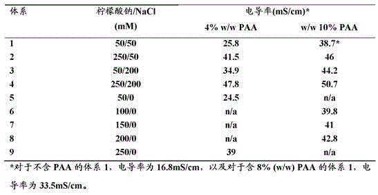
Example 9 Adaptation to optimization by high throughput screening and design of experiments
The standard cubic face center (CCF) design of experiment (DOE) was used together with small scale (5ml) experiments to further investigate the ability of varying concentrations of PAA15000 (4-12%) and sodium citrate (50-250 mM) to achieve selectivity for inclusion of albumin and antibody in the precipitate in a system containing 1.5ml plasma and 0 mM-200mM NaCl. Example results at 200mM NaCl are shown in FIG. 15. At higher PAA (12%) and increasing sodium citrate to 250mM, 56% of the albumin present was expected to be in the precipitate along with more than 90% of the antibody. With the reduction of sodium citrate to 60 mM, the antibody precipitation was still close to 90%, however only 25% of the albumin was in the precipitate. The general polyacid-lyotropic salt method can be adapted to the simplicity of the rapid screening, and its selective control that should be allowed, supporting the commercial potential of the method.
Example 10 influence of pH
To investigate the effect of system pH, stock solutions of sodium citrate (0.8M) were formulated at pH 3, 5, 7 and 9. Sodium citrate was added to an equivalent system containing 8% (w/w) PAA and 50mM NaCl to 50 mM. The pH in the final system increased to 6.3, 7, 8.2 and 8.3 due to the pH of plasma and water and the pKa of citrate, respectively. The effect of pH on protein precipitation was analyzed using 1D SDS-PAGE (and GelCodeBlue coomassie staining) (fig. 16) and those results were combined with a biuret assay (total protein) plus imagequantl (PAGE gel band analysis of relative protein amounts) to provide an estimate of the percentage of each protein (albumin, antibody and fibrinogen) in the precipitate. Almost complete incorporation of fibrinogen into the precipitate in the above system and relatively low levels of albumin into the precipitate in the above system is hardly affected by pH changes from 6 to 8. However, the incorporation of antibody into the precipitate increased with increasing pH (data not shown). Precipitation of certain other unassigned higher MW proteins was also affected by pH (figure 16). Thus it may be possible to use pH to fine tune the fractionation of a particular protein by the general method. Again, the slight change in antibody precipitation between pH 8.2 and 8.3 as visualized by the gel indicates the robustness of the method.
In this example a system with a relatively low concentration of sodium citrate was used, which limits the buffering capacity of the system and the pH range studied. Cohn fractionation may involve a change in pH from 4 to 8 (reference 16, see also fig. 1). It may be possible to vary the pH over a wider range if higher concentrations of chaotropic buffer salts (e.g. sodium citrate or sodium phosphate) are used or if another buffer system is used in combination with any desired lyotropic salt.
Considering the isoelectric pH range of the proteins studied (table 1), it is expected that under the conditions studied in this example (i.e. pH 6-8), albumin should be and remain net negatively charged, fibrinogen should be predominantly net negatively charged, and the different antibodies (pI ranging from 6-10) normally present in plasma can be neutral, net negatively charged or net positively charged. Many other plasma proteins, such as transferrin and factor VIII, are also expected to be predominantly neutral or net negative over the pH range studied. Thus, the electrophoresis results show that in the case of fibrinogen, the general method is capable of precipitating proteins with a net negative charge, as well as antibodies with a net positive charge. Furthermore, as pH increases and various antibody proteins appear to become less net positively charged, their complexing effect appears to increase. This is counter-intuitive to methods based solely on the charge-charge interaction of a net negatively charged polymer with a net positively charged protein. This may be related to protein solubility (surface hydrophobicity) and the significant effects of protein-salt and protein-protein interactions. Precipitation appears to be the result of a combination of phenomena, and without being bound by theory, it is speculated that protein size, relative surface hydrophobicity, and other factors (e.g., surface area of net positive electrical properties) may play a role. Regardless of the mechanism involved under the conditions studied, the polyacid PAA is capable of mediating complex formation with proteins, which may be net negative, net positive or neutral.
Example 11 Effect of centrifugation, temperature and salt addition
Certain variables of interest regarding practical large-scale application of the general method were also investigated. These variables include centrifugation speed and time, addition of citrate as salt rather than as a stock solution, and changes in temperature.
It is assumed that any flocs/precipitates will be recovered by centrifugation or filtration. In this work the precipitate was settled down using centrifugation to enable its separation from the supernatant. Centrifugation of 5ml or less in volume was carried out at 4000 revolutions per minute (rpm) for 10 minutes (see methods above), with successful results. The same successful results were obtained at 4000 rpm for 5 minutes or even at 2000 rpm for 5 minutes. The precipitate was well separated and recovered in these studies and 1D SDS gel electrophoresis analysis showed no change in the precipitate or supernatant samples for the 5ml system containing 1.5ml plasma, 50mM naci and 50mM sodium citrate pH7 (data not shown). These results also indicate that the polymer-protein complex is both sufficiently tight and mechanically robust to cope with filtration or other possible methods.
The addition of salt as a solid may be advantageous compared to as a stock solution. Thus in experiments involving a 5ml system containing 1.5ml plasma plus a final concentration of 8% PAA and 200mM sodium citrate, citrate was added as salt crystals rather than from the stock solution (see method above). The pellet appeared in an apparently normal manner and 1D SDS gel electrophoretic analysis of the pellet and supernatant fractions showed reasonably similar results (data not shown).
PAA precipitate-related experiments were typically performed at 22 ℃ (room temperature). However, in plasma processing based on the cohn fractionation method, the frozen plasma should be thawed to 4 ℃ (which affects the first fractionation of the plasma, see also references 15, 16). The solution temperature can thereafter be varied to increase the selectivity to the various fractionation steps. Even for isothermal PAA-precipitation based methods, it may be desirable to perform the method at 4 ℃. Thus, plasma was used to prepare a system at 4 ℃ and the sample was kept at 4 ℃ during precipitation. No significant difference was seen when compared to the pellet or supernatant samples prepared at 22 ℃ (fig. 17).
Example 12 sequential precipitation of undiluted plasma
The frozen pooled heparinized plasma was thawed in tepid water. 10 g of plasma was mixed with polyacrylic acid (35% aqueous, Aldrich 411637-500 ml, MW 15000) in a15 ml Falcon tube.
The mixture was left standing on the bench for 1 h at RT.
The mixture was centrifuged at 500 g for 10 min in an Eppendorf benchtop centrifuge at room temperature.
The clear supernatant solution was diluted and measured for a 280. The results are shown in Table 8.
Table 8 results from the first precipitation series.

10% PAA is not sufficient to precipitate all proteins (compounds with A280 absorbance) in plasma. Thus, a second round was performed to further increase the PAA concentration to 20%. The results from this round are shown in table 9.
Table 9 results from the second precipitation series.
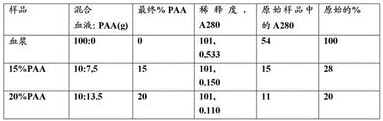
At PAA concentrations as high as 15% and 20%, most of the plasma proteins precipitated. At these concentrations, the pH increases by about 1 unit, so pH adjustment during processing can be beneficial for the stability of certain sensitive plasma proteins. The precipitates obtained with 15% and 20% PAA floated on the liquid phase, which is also an interesting observation. This may have advantages for the recovery of the precipitate.
This written description uses examples to disclose the invention, including the best mode, and also to enable any person skilled in the art to practice the invention, including making and using any devices or systems and performing any incorporated methods. It is pointed out that the specific advantageous embodiments of the aspects of the invention disclosed above can be freely combined into other embodiments within the scope of the invention. The patentable scope of the invention is defined by the claims, and may include other examples that occur to those skilled in the art. Such other embodiments are intended to be within the scope of the claims if they do not have structural elements that differ from the literal language of the claims, or if they include equivalent structural elements with insubstantial differences from the literal languages of the claims.
Claims (26)
Applications Claiming Priority (3)
| Application Number | Priority Date | Filing Date | Title |
|---|---|---|---|
| SE1150842 | 2011-09-16 | ||
| SE1150842-1 | 2011-09-16 | ||
| PCT/SE2012/050971 WO2013039449A1 (en) | 2011-09-16 | 2012-09-14 | Plasma protein fractionation by sequential polyacid precipitation |
Publications (2)
| Publication Number | Publication Date |
|---|---|
| CN103797023A CN103797023A (en) | 2014-05-14 |
| CN103797023B true CN103797023B (en) | 2020-04-10 |
Family
ID=47883540
Family Applications (1)
| Application Number | Title | Priority Date | Filing Date |
|---|---|---|---|
| CN201280044936.3A Active CN103797023B (en) | 2011-09-16 | 2012-09-14 | Plasma protein fractionation by sequential polyacid precipitation |
Country Status (5)
| Country | Link |
|---|---|
| US (1) | US9624262B2 (en) |
| EP (1) | EP2755992B1 (en) |
| CN (1) | CN103797023B (en) |
| AU (1) | AU2012309202B2 (en) |
| WO (1) | WO2013039449A1 (en) |
Families Citing this family (16)
| Publication number | Priority date | Publication date | Assignee | Title |
|---|---|---|---|---|
| CN105111306A (en) * | 2015-08-28 | 2015-12-02 | 北京工业大学 | Separation method of American alligator albumin |
| WO2017058877A1 (en) * | 2015-09-28 | 2017-04-06 | Kieu Hoang | A method for separating proteins from animal or human plasma, or plants, using a ph gradient method |
| AU2017214678B2 (en) | 2016-02-03 | 2024-02-01 | Plasma Technologies, Llc | Methods for extracting proteins from a blood-based material |
| WO2018165309A1 (en) | 2017-03-08 | 2018-09-13 | The Regents Of The University Of Michigan | Analyte detection |
| AU2018233184B2 (en) * | 2017-03-16 | 2022-06-16 | Evolve Biologics Inc. | Method for purification of albumin |
| CN114839374A (en) | 2017-03-28 | 2022-08-02 | 相达生物科技国际有限公司 | Method for accurate diagnosis of disease-targeted biomarkers in liquid biopsies |
| WO2018222972A1 (en) * | 2017-06-01 | 2018-12-06 | Yin To Chiu | Phase separation behavior modifying agents for aqueous two-phase separation within porous material |
| US20200208137A1 (en) | 2017-09-18 | 2020-07-02 | Yin To Chiu | Method for Using Aqueous Two-Phase System for the Isolation, Purification and/or Concentration of Short Nucleic Acid Fragments |
| JP7270992B2 (en) * | 2017-12-14 | 2023-05-11 | ザ リージェンツ オブ ザ ユニバーシティ オブ ミシガン | Analyte concentration |
| US12043826B2 (en) | 2018-01-19 | 2024-07-23 | Phase Scientific International, Ltd. | Spontaneous nucleic acid purification and concentration in a single step |
| US10815270B1 (en) * | 2019-09-20 | 2020-10-27 | Plasma Technologies, Llc | Compositions and methods for high efficiency protein precipitation |
| JP2023502303A (en) * | 2019-09-20 | 2023-01-24 | プラズマ テクノロジーズ エルエルシー | Therapeutic protein compositions and methods |
| TW202136283A (en) * | 2019-12-12 | 2021-10-01 | 日商武田藥品工業股份有限公司 | Method for continuous protein recovering |
| CA3175618A1 (en) | 2020-03-20 | 2021-09-23 | Phase Scientific International, Ltd. | Compositions and methods for ribonucleic acid extraction |
| CN114574457B (en) * | 2022-01-25 | 2024-07-23 | 华东理工大学 | A method for rapid separation and purification of proteins |
| AU2023374883A1 (en) | 2022-11-02 | 2025-05-22 | Phase Scientific International, Ltd. | Methods for isolating target analytes from biological samples using atps and solid phase media |
Citations (2)
| Publication number | Priority date | Publication date | Assignee | Title |
|---|---|---|---|---|
| WO2010080062A1 (en) * | 2009-01-08 | 2010-07-15 | Ge Healthcare Bio-Sciences Ab | Separation method using single polymer phase systems |
| WO2010082894A1 (en) * | 2009-01-13 | 2010-07-22 | Ge Healthcare Bio-Sciences Ab | Precipitation of biomolecules with negatively charged polymers |
Family Cites Families (4)
| Publication number | Priority date | Publication date | Assignee | Title |
|---|---|---|---|---|
| US3555001A (en) | 1969-05-29 | 1971-01-12 | Monsanto Co | Process for the fractionation of plasma and serum using water-insoluble polyelectrolytes containing diloweralkylaminoloweralkylimide groups |
| WO1984003628A1 (en) * | 1983-05-09 | 1984-09-27 | Nordisk Insulinlab | A concentrate of the antihemophilic factor viii and a process for producing it |
| GB8505882D0 (en) * | 1985-03-07 | 1985-04-11 | Central Blood Lab Authority | Purification of blood coagulation factor viii |
| US7879332B2 (en) * | 2005-09-01 | 2011-02-01 | Plasma Technologies, Llc | Ultra-high yield intravenous immune globulin preparation |
-
2012
- 2012-09-14 CN CN201280044936.3A patent/CN103797023B/en active Active
- 2012-09-14 WO PCT/SE2012/050971 patent/WO2013039449A1/en not_active Ceased
- 2012-09-14 US US14/345,302 patent/US9624262B2/en active Active
- 2012-09-14 AU AU2012309202A patent/AU2012309202B2/en active Active
- 2012-09-14 EP EP12832274.0A patent/EP2755992B1/en active Active
Patent Citations (2)
| Publication number | Priority date | Publication date | Assignee | Title |
|---|---|---|---|---|
| WO2010080062A1 (en) * | 2009-01-08 | 2010-07-15 | Ge Healthcare Bio-Sciences Ab | Separation method using single polymer phase systems |
| WO2010082894A1 (en) * | 2009-01-13 | 2010-07-22 | Ge Healthcare Bio-Sciences Ab | Precipitation of biomolecules with negatively charged polymers |
Non-Patent Citations (1)
| Title |
|---|
| Optimization for selective fractionation of bovine blood plasma proteins using poly(ethylene glycol);Young Zoon Lee;《J. Agric. Food Chem.》;19871130;第35卷(第6期);pp 958–962 * |
Also Published As
| Publication number | Publication date |
|---|---|
| EP2755992B1 (en) | 2018-11-14 |
| NZ621197A (en) | 2016-01-29 |
| EP2755992A1 (en) | 2014-07-23 |
| WO2013039449A1 (en) | 2013-03-21 |
| US20140343253A1 (en) | 2014-11-20 |
| AU2012309202A1 (en) | 2014-04-24 |
| EP2755992A4 (en) | 2015-04-29 |
| US9624262B2 (en) | 2017-04-18 |
| CN103797023A (en) | 2014-05-14 |
| AU2012309202B2 (en) | 2017-04-27 |
Similar Documents
| Publication | Publication Date | Title |
|---|---|---|
| CN103797023B (en) | Plasma protein fractionation by sequential polyacid precipitation | |
| AU2010203836B2 (en) | Separation method using single polymer phase systems | |
| JP6200949B2 (en) | Selective binding of biological targets to solid phase ureides | |
| JP5917417B2 (en) | Selective antibody enhancement | |
| JP6491277B2 (en) | Method for reducing the level of protein-impurity complexes and aggregates in protein formulations by treatment with positively charged organic additives | |
| CN102272146A (en) | Precipitation of biomolecules with negatively charged polymers | |
| CN105008383A (en) | Protein purification in the presence of nonionic organic polymers and electropositive surfaces | |
| CN105121457B (en) | Materials and methods for removing endotoxin from protein preparations | |
| WO2014133460A1 (en) | Protein purification in the presence of nonionic organic polymers at elevated conductivity | |
| CN105051055A (en) | Method for reducing aggregate levels in protein preparations by treatment with thioheterocyclic cations | |
| WO2008068455A1 (en) | Protein purification | |
| EP3016966B1 (en) | Concentration and purification of hydrophobins and antibodies with a phase separation method | |
| NZ621197B2 (en) | Plasma protein fractionation by sequential polyacid precipitation | |
| KR20160127087A (en) | Methods for reducing chromatin content in protein preparations by treatment with alkyl cations | |
| US20210087224A1 (en) | Compositions and methods for generating modified cryo poor plasma |
Legal Events
| Date | Code | Title | Description |
|---|---|---|---|
| C06 | Publication | ||
| PB01 | Publication | ||
| C10 | Entry into substantive examination | ||
| SE01 | Entry into force of request for substantive examination | ||
| C41 | Transfer of patent application or patent right or utility model | ||
| TA01 | Transfer of patent application right |
Effective date of registration: 20160816 Address after: uppsala Applicant after: GE HEALTHCARE BIO-SCIENCES AB Address before: uppsala Applicant before: GE Health Bio-science Co., Ltd. |
|
| GR01 | Patent grant | ||
| GR01 | Patent grant | ||
| CP01 | Change in the name or title of a patent holder |
Address after: uppsala Patentee after: Situofan Biotechnology R & D Co., Ltd Address before: uppsala Patentee before: GE HEALTHCARE BIOPROCESS R&D AB |
|
| CP01 | Change in the name or title of a patent holder |