CN103214584B - Fusion protein with double functions of inhibiting revascularization of tumor microenvironment and activating adaptive immune response, and gene and application thereof - Google Patents
Fusion protein with double functions of inhibiting revascularization of tumor microenvironment and activating adaptive immune response, and gene and application thereof Download PDFInfo
- Publication number
- CN103214584B CN103214584B CN201310162738.3A CN201310162738A CN103214584B CN 103214584 B CN103214584 B CN 103214584B CN 201310162738 A CN201310162738 A CN 201310162738A CN 103214584 B CN103214584 B CN 103214584B
- Authority
- CN
- China
- Prior art keywords
- seq
- cd137l
- tumstatin
- recombinant protein
- protein
- Prior art date
- Legal status (The legal status is an assumption and is not a legal conclusion. Google has not performed a legal analysis and makes no representation as to the accuracy of the status listed.)
- Expired - Fee Related
Links
- 108090000623 proteins and genes Proteins 0.000 title claims abstract description 96
- 206010028980 Neoplasm Diseases 0.000 title claims abstract description 24
- 230000006870 function Effects 0.000 title abstract description 26
- 108020001507 fusion proteins Proteins 0.000 title abstract description 16
- 230000002401 inhibitory effect Effects 0.000 title abstract description 14
- 102000037865 fusion proteins Human genes 0.000 title abstract description 6
- 230000003213 activating effect Effects 0.000 title abstract description 4
- 230000033289 adaptive immune response Effects 0.000 title abstract description 4
- 230000000250 revascularization Effects 0.000 title abstract 2
- 101000638251 Homo sapiens Tumor necrosis factor ligand superfamily member 9 Proteins 0.000 claims abstract description 80
- 102100032101 Tumor necrosis factor ligand superfamily member 9 Human genes 0.000 claims abstract description 80
- 108010008281 Recombinant Fusion Proteins Proteins 0.000 claims abstract description 75
- 102000007056 Recombinant Fusion Proteins Human genes 0.000 claims abstract description 70
- 108010012374 type IV collagen alpha3 chain Proteins 0.000 claims abstract description 66
- 102000004169 proteins and genes Human genes 0.000 claims abstract description 46
- 239000003814 drug Substances 0.000 claims abstract description 11
- 230000036039 immunity Effects 0.000 claims abstract description 5
- 239000004037 angiogenesis inhibitor Substances 0.000 claims abstract description 4
- 229940121369 angiogenesis inhibitor Drugs 0.000 claims abstract description 4
- 125000003275 alpha amino acid group Chemical group 0.000 claims abstract 5
- 102400000731 Tumstatin Human genes 0.000 claims description 61
- 108090000765 processed proteins & peptides Proteins 0.000 claims description 48
- 230000014509 gene expression Effects 0.000 claims description 43
- 230000000694 effects Effects 0.000 claims description 39
- 235000018102 proteins Nutrition 0.000 claims description 38
- 239000012634 fragment Substances 0.000 claims description 31
- 239000013604 expression vector Substances 0.000 claims description 22
- 230000015572 biosynthetic process Effects 0.000 claims description 12
- 238000002360 preparation method Methods 0.000 claims description 12
- 102000004196 processed proteins & peptides Human genes 0.000 claims description 12
- 239000002773 nucleotide Substances 0.000 claims description 10
- 125000003729 nucleotide group Chemical group 0.000 claims description 10
- 238000000746 purification Methods 0.000 claims description 9
- 238000010276 construction Methods 0.000 claims description 8
- 230000009465 prokaryotic expression Effects 0.000 claims description 7
- 206010058467 Lung neoplasm malignant Diseases 0.000 claims description 6
- 201000005202 lung cancer Diseases 0.000 claims description 6
- 208000020816 lung neoplasm Diseases 0.000 claims description 6
- 201000001441 melanoma Diseases 0.000 claims description 6
- 240000004808 Saccharomyces cerevisiae Species 0.000 claims description 5
- 238000012258 culturing Methods 0.000 claims description 4
- VEEGZPWAAPPXRB-BJMVGYQFSA-N (3e)-3-(1h-imidazol-5-ylmethylidene)-1h-indol-2-one Chemical compound O=C1NC2=CC=CC=C2\C1=C/C1=CN=CN1 VEEGZPWAAPPXRB-BJMVGYQFSA-N 0.000 claims description 2
- 206010005003 Bladder cancer Diseases 0.000 claims description 2
- 208000008839 Kidney Neoplasms Diseases 0.000 claims description 2
- 206010060862 Prostate cancer Diseases 0.000 claims description 2
- 208000000236 Prostatic Neoplasms Diseases 0.000 claims description 2
- 206010038389 Renal cancer Diseases 0.000 claims description 2
- 208000017442 Retinal disease Diseases 0.000 claims description 2
- 206010038923 Retinopathy Diseases 0.000 claims description 2
- 208000007097 Urinary Bladder Neoplasms Diseases 0.000 claims description 2
- 239000002246 antineoplastic agent Substances 0.000 claims description 2
- 229940041181 antineoplastic drug Drugs 0.000 claims description 2
- 238000013461 design Methods 0.000 claims description 2
- 201000010982 kidney cancer Diseases 0.000 claims description 2
- 238000000926 separation method Methods 0.000 claims description 2
- 201000005112 urinary bladder cancer Diseases 0.000 claims description 2
- 241000588722 Escherichia Species 0.000 claims 1
- 201000003741 Gastrointestinal carcinoma Diseases 0.000 claims 1
- 201000002313 intestinal cancer Diseases 0.000 claims 1
- 210000003734 kidney Anatomy 0.000 claims 1
- 108010075254 C-Peptide Proteins 0.000 abstract description 13
- 229940079593 drug Drugs 0.000 abstract description 9
- 208000037265 diseases, disorders, signs and symptoms Diseases 0.000 abstract description 7
- 201000010099 disease Diseases 0.000 abstract description 6
- 230000005747 tumor angiogenesis Effects 0.000 abstract description 3
- 238000011160 research Methods 0.000 abstract description 2
- 102100033780 Collagen alpha-3(IV) chain Human genes 0.000 abstract 2
- 150000001413 amino acids Chemical class 0.000 description 68
- 238000000034 method Methods 0.000 description 29
- 210000001744 T-lymphocyte Anatomy 0.000 description 25
- 238000011144 upstream manufacturing Methods 0.000 description 25
- 210000004027 cell Anatomy 0.000 description 22
- 239000000243 solution Substances 0.000 description 21
- 229940024606 amino acid Drugs 0.000 description 20
- 235000001014 amino acid Nutrition 0.000 description 20
- 239000013598 vector Substances 0.000 description 20
- 239000000047 product Substances 0.000 description 15
- XSQUKJJJFZCRTK-UHFFFAOYSA-N Urea Chemical compound NC(N)=O XSQUKJJJFZCRTK-UHFFFAOYSA-N 0.000 description 14
- 230000009977 dual effect Effects 0.000 description 14
- 239000002609 medium Substances 0.000 description 14
- 230000035755 proliferation Effects 0.000 description 12
- 241000894006 Bacteria Species 0.000 description 10
- 230000033115 angiogenesis Effects 0.000 description 9
- 210000002889 endothelial cell Anatomy 0.000 description 9
- 238000003786 synthesis reaction Methods 0.000 description 9
- 239000000427 antigen Substances 0.000 description 8
- 210000003000 inclusion body Anatomy 0.000 description 8
- 210000003606 umbilical vein Anatomy 0.000 description 8
- 244000063299 Bacillus subtilis Species 0.000 description 7
- 235000014469 Bacillus subtilis Nutrition 0.000 description 7
- 102000017420 CD3 protein, epsilon/gamma/delta subunit Human genes 0.000 description 7
- 101000914514 Homo sapiens T-cell-specific surface glycoprotein CD28 Proteins 0.000 description 7
- KDXKERNSBIXSRK-UHFFFAOYSA-N Lysine Natural products NCCCCC(N)C(O)=O KDXKERNSBIXSRK-UHFFFAOYSA-N 0.000 description 7
- 206010027476 Metastases Diseases 0.000 description 7
- 102100027213 T-cell-specific surface glycoprotein CD28 Human genes 0.000 description 7
- 102000036639 antigens Human genes 0.000 description 7
- 108091007433 antigens Proteins 0.000 description 7
- 230000001588 bifunctional effect Effects 0.000 description 7
- 239000004202 carbamide Substances 0.000 description 7
- 230000009401 metastasis Effects 0.000 description 7
- 238000002415 sodium dodecyl sulfate polyacrylamide gel electrophoresis Methods 0.000 description 7
- 108700031361 Brachyury Proteins 0.000 description 6
- 238000001962 electrophoresis Methods 0.000 description 6
- 238000001976 enzyme digestion Methods 0.000 description 6
- 230000004048 modification Effects 0.000 description 6
- 238000012986 modification Methods 0.000 description 6
- 238000004153 renaturation Methods 0.000 description 6
- 239000006228 supernatant Substances 0.000 description 6
- 230000009182 swimming Effects 0.000 description 6
- 102000004127 Cytokines Human genes 0.000 description 5
- 108090000695 Cytokines Proteins 0.000 description 5
- 241000588724 Escherichia coli Species 0.000 description 5
- 230000006052 T cell proliferation Effects 0.000 description 5
- 239000000499 gel Substances 0.000 description 5
- 239000007788 liquid Substances 0.000 description 5
- 239000008194 pharmaceutical composition Substances 0.000 description 5
- 229920001184 polypeptide Polymers 0.000 description 5
- 238000012360 testing method Methods 0.000 description 5
- 230000004614 tumor growth Effects 0.000 description 5
- 241000699666 Mus <mouse, genus> Species 0.000 description 4
- 208000015634 Rectal Neoplasms Diseases 0.000 description 4
- 238000004458 analytical method Methods 0.000 description 4
- 239000000872 buffer Substances 0.000 description 4
- 238000010586 diagram Methods 0.000 description 4
- 239000003937 drug carrier Substances 0.000 description 4
- 238000004520 electroporation Methods 0.000 description 4
- 230000028993 immune response Effects 0.000 description 4
- 108091005601 modified peptides Proteins 0.000 description 4
- 229920001223 polyethylene glycol Polymers 0.000 description 4
- 230000008569 process Effects 0.000 description 4
- 206010038038 rectal cancer Diseases 0.000 description 4
- 201000001275 rectum cancer Diseases 0.000 description 4
- 230000004936 stimulating effect Effects 0.000 description 4
- 230000000638 stimulation Effects 0.000 description 4
- 230000004083 survival effect Effects 0.000 description 4
- 210000004881 tumor cell Anatomy 0.000 description 4
- 238000005406 washing Methods 0.000 description 4
- XLYOFNOQVPJJNP-UHFFFAOYSA-N water Substances O XLYOFNOQVPJJNP-UHFFFAOYSA-N 0.000 description 4
- BFSVOASYOCHEOV-UHFFFAOYSA-N 2-diethylaminoethanol Chemical compound CCN(CC)CCO BFSVOASYOCHEOV-UHFFFAOYSA-N 0.000 description 3
- IAZDPXIOMUYVGZ-UHFFFAOYSA-N Dimethylsulphoxide Chemical compound CS(C)=O IAZDPXIOMUYVGZ-UHFFFAOYSA-N 0.000 description 3
- 241001198387 Escherichia coli BL21(DE3) Species 0.000 description 3
- ODKSFYDXXFIFQN-BYPYZUCNSA-N L-arginine Chemical compound OC(=O)[C@@H](N)CCCN=C(N)N ODKSFYDXXFIFQN-BYPYZUCNSA-N 0.000 description 3
- 241000699670 Mus sp. Species 0.000 description 3
- 229920002472 Starch Polymers 0.000 description 3
- 230000006044 T cell activation Effects 0.000 description 3
- AYFVYJQAPQTCCC-UHFFFAOYSA-N Threonine Natural products CC(O)C(N)C(O)=O AYFVYJQAPQTCCC-UHFFFAOYSA-N 0.000 description 3
- 108060008683 Tumor Necrosis Factor Receptor Proteins 0.000 description 3
- 230000004913 activation Effects 0.000 description 3
- 230000003321 amplification Effects 0.000 description 3
- 238000005349 anion exchange Methods 0.000 description 3
- 230000005809 anti-tumor immunity Effects 0.000 description 3
- 230000001580 bacterial effect Effects 0.000 description 3
- 210000002469 basement membrane Anatomy 0.000 description 3
- 201000011510 cancer Diseases 0.000 description 3
- 238000005119 centrifugation Methods 0.000 description 3
- 238000006243 chemical reaction Methods 0.000 description 3
- 230000000139 costimulatory effect Effects 0.000 description 3
- 238000005520 cutting process Methods 0.000 description 3
- 239000012636 effector Substances 0.000 description 3
- 238000005516 engineering process Methods 0.000 description 3
- 230000004927 fusion Effects 0.000 description 3
- 230000013595 glycosylation Effects 0.000 description 3
- 238000006206 glycosylation reaction Methods 0.000 description 3
- 239000003446 ligand Substances 0.000 description 3
- 238000009630 liquid culture Methods 0.000 description 3
- 108091005573 modified proteins Proteins 0.000 description 3
- 102000035118 modified proteins Human genes 0.000 description 3
- 238000003199 nucleic acid amplification method Methods 0.000 description 3
- 230000000771 oncological effect Effects 0.000 description 3
- 239000013612 plasmid Substances 0.000 description 3
- 239000013641 positive control Substances 0.000 description 3
- 239000002244 precipitate Substances 0.000 description 3
- 230000001105 regulatory effect Effects 0.000 description 3
- 230000028327 secretion Effects 0.000 description 3
- 238000012163 sequencing technique Methods 0.000 description 3
- 210000000952 spleen Anatomy 0.000 description 3
- 239000008107 starch Substances 0.000 description 3
- 235000019698 starch Nutrition 0.000 description 3
- 239000000126 substance Substances 0.000 description 3
- 210000001519 tissue Anatomy 0.000 description 3
- 102000003298 tumor necrosis factor receptor Human genes 0.000 description 3
- QKNYBSVHEMOAJP-UHFFFAOYSA-N 2-amino-2-(hydroxymethyl)propane-1,3-diol;hydron;chloride Chemical compound Cl.OCC(N)(CO)CO QKNYBSVHEMOAJP-UHFFFAOYSA-N 0.000 description 2
- GUBGYTABKSRVRQ-XLOQQCSPSA-N Alpha-Lactose Chemical compound O[C@@H]1[C@@H](O)[C@@H](O)[C@@H](CO)O[C@H]1O[C@@H]1[C@@H](CO)O[C@H](O)[C@H](O)[C@H]1O GUBGYTABKSRVRQ-XLOQQCSPSA-N 0.000 description 2
- IJGRMHOSHXDMSA-UHFFFAOYSA-N Atomic nitrogen Chemical compound N#N IJGRMHOSHXDMSA-UHFFFAOYSA-N 0.000 description 2
- 210000001266 CD8-positive T-lymphocyte Anatomy 0.000 description 2
- 108010042086 Collagen Type IV Proteins 0.000 description 2
- 102000004266 Collagen Type IV Human genes 0.000 description 2
- 229920000858 Cyclodextrin Polymers 0.000 description 2
- KCXVZYZYPLLWCC-UHFFFAOYSA-N EDTA Chemical compound OC(=O)CN(CC(O)=O)CCN(CC(O)=O)CC(O)=O KCXVZYZYPLLWCC-UHFFFAOYSA-N 0.000 description 2
- DHMQDGOQFOQNFH-UHFFFAOYSA-N Glycine Chemical compound NCC(O)=O DHMQDGOQFOQNFH-UHFFFAOYSA-N 0.000 description 2
- 241001506991 Komagataella phaffii GS115 Species 0.000 description 2
- DCXYFEDJOCDNAF-REOHCLBHSA-N L-asparagine Chemical compound OC(=O)[C@@H](N)CC(N)=O DCXYFEDJOCDNAF-REOHCLBHSA-N 0.000 description 2
- ROHFNLRQFUQHCH-YFKPBYRVSA-N L-leucine Chemical compound CC(C)C[C@H](N)C(O)=O ROHFNLRQFUQHCH-YFKPBYRVSA-N 0.000 description 2
- AYFVYJQAPQTCCC-GBXIJSLDSA-N L-threonine Chemical compound C[C@@H](O)[C@H](N)C(O)=O AYFVYJQAPQTCCC-GBXIJSLDSA-N 0.000 description 2
- ROHFNLRQFUQHCH-UHFFFAOYSA-N Leucine Natural products CC(C)CC(N)C(O)=O ROHFNLRQFUQHCH-UHFFFAOYSA-N 0.000 description 2
- 241001529936 Murinae Species 0.000 description 2
- MSHZHSPISPJWHW-UHFFFAOYSA-N O-(chloroacetylcarbamoyl)fumagillol Chemical compound O1C(CC=C(C)C)C1(C)C1C(OC)C(OC(=O)NC(=O)CCl)CCC21CO2 MSHZHSPISPJWHW-UHFFFAOYSA-N 0.000 description 2
- 238000012408 PCR amplification Methods 0.000 description 2
- FAPWRFPIFSIZLT-UHFFFAOYSA-M Sodium chloride Chemical compound [Na+].[Cl-] FAPWRFPIFSIZLT-UHFFFAOYSA-M 0.000 description 2
- 108091008874 T cell receptors Proteins 0.000 description 2
- 102000016266 T-Cell Antigen Receptors Human genes 0.000 description 2
- 102100040247 Tumor necrosis factor Human genes 0.000 description 2
- 238000002835 absorbance Methods 0.000 description 2
- 239000004480 active ingredient Substances 0.000 description 2
- -1 amino acid fragment Amino acid Chemical class 0.000 description 2
- AVKUERGKIZMTKX-NJBDSQKTSA-N ampicillin Chemical compound C1([C@@H](N)C(=O)N[C@H]2[C@H]3SC([C@@H](N3C2=O)C(O)=O)(C)C)=CC=CC=C1 AVKUERGKIZMTKX-NJBDSQKTSA-N 0.000 description 2
- 229960000723 ampicillin Drugs 0.000 description 2
- 230000003698 anagen phase Effects 0.000 description 2
- 230000000259 anti-tumor effect Effects 0.000 description 2
- 210000000612 antigen-presenting cell Anatomy 0.000 description 2
- 230000005975 antitumor immune response Effects 0.000 description 2
- 230000008901 benefit Effects 0.000 description 2
- UCMIRNVEIXFBKS-UHFFFAOYSA-N beta-alanine Chemical compound NCCC(O)=O UCMIRNVEIXFBKS-UHFFFAOYSA-N 0.000 description 2
- 230000004071 biological effect Effects 0.000 description 2
- 210000004204 blood vessel Anatomy 0.000 description 2
- 239000002775 capsule Substances 0.000 description 2
- 238000004113 cell culture Methods 0.000 description 2
- 230000024245 cell differentiation Effects 0.000 description 2
- 230000004663 cell proliferation Effects 0.000 description 2
- 230000008859 change Effects 0.000 description 2
- 239000003153 chemical reaction reagent Substances 0.000 description 2
- 238000004925 denaturation Methods 0.000 description 2
- 230000036425 denaturation Effects 0.000 description 2
- 230000029087 digestion Effects 0.000 description 2
- 238000010790 dilution Methods 0.000 description 2
- 239000012895 dilution Substances 0.000 description 2
- 239000002552 dosage form Substances 0.000 description 2
- 238000012377 drug delivery Methods 0.000 description 2
- 230000002708 enhancing effect Effects 0.000 description 2
- 238000002474 experimental method Methods 0.000 description 2
- 238000002347 injection Methods 0.000 description 2
- 239000007924 injection Substances 0.000 description 2
- 238000005342 ion exchange Methods 0.000 description 2
- 229930027917 kanamycin Natural products 0.000 description 2
- 229960000318 kanamycin Drugs 0.000 description 2
- SBUJHOSQTJFQJX-NOAMYHISSA-N kanamycin Chemical compound O[C@@H]1[C@@H](O)[C@H](O)[C@@H](CN)O[C@@H]1O[C@H]1[C@H](O)[C@@H](O[C@@H]2[C@@H]([C@@H](N)[C@H](O)[C@@H](CO)O2)O)[C@H](N)C[C@@H]1N SBUJHOSQTJFQJX-NOAMYHISSA-N 0.000 description 2
- 229930182823 kanamycin A Natural products 0.000 description 2
- 229940124280 l-arginine Drugs 0.000 description 2
- HQKMJHAJHXVSDF-UHFFFAOYSA-L magnesium stearate Chemical compound [Mg+2].CCCCCCCCCCCCCCCCCC([O-])=O.CCCCCCCCCCCCCCCCCC([O-])=O HQKMJHAJHXVSDF-UHFFFAOYSA-L 0.000 description 2
- 230000001404 mediated effect Effects 0.000 description 2
- 230000011987 methylation Effects 0.000 description 2
- 238000007069 methylation reaction Methods 0.000 description 2
- 239000013642 negative control Substances 0.000 description 2
- 239000000546 pharmaceutical excipient Substances 0.000 description 2
- 239000008363 phosphate buffer Substances 0.000 description 2
- 229920000036 polyvinylpyrrolidone Polymers 0.000 description 2
- 235000013855 polyvinylpyrrolidone Nutrition 0.000 description 2
- 108091008146 restriction endonucleases Proteins 0.000 description 2
- 229920006395 saturated elastomer Polymers 0.000 description 2
- 239000011734 sodium Substances 0.000 description 2
- 229910052708 sodium Inorganic materials 0.000 description 2
- 239000007787 solid Substances 0.000 description 2
- 239000002904 solvent Substances 0.000 description 2
- 210000000130 stem cell Anatomy 0.000 description 2
- UCSJYZPVAKXKNQ-HZYVHMACSA-N streptomycin Chemical compound CN[C@H]1[C@H](O)[C@@H](O)[C@H](CO)O[C@H]1O[C@@H]1[C@](C=O)(O)[C@H](C)O[C@H]1O[C@@H]1[C@@H](NC(N)=N)[C@H](O)[C@@H](NC(N)=N)[C@H](O)[C@H]1O UCSJYZPVAKXKNQ-HZYVHMACSA-N 0.000 description 2
- 238000012546 transfer Methods 0.000 description 2
- 230000002792 vascular Effects 0.000 description 2
- 210000003556 vascular endothelial cell Anatomy 0.000 description 2
- 238000012795 verification Methods 0.000 description 2
- RMYPEYHEPIZYDJ-SNVBAGLBSA-N (2r)-2-azaniumyl-3-(4-ethoxyphenyl)propanoate Chemical compound CCOC1=CC=C(C[C@@H](N)C(O)=O)C=C1 RMYPEYHEPIZYDJ-SNVBAGLBSA-N 0.000 description 1
- IYKLZBIWFXPUCS-VIFPVBQESA-N (2s)-2-(naphthalen-1-ylamino)propanoic acid Chemical compound C1=CC=C2C(N[C@@H](C)C(O)=O)=CC=CC2=C1 IYKLZBIWFXPUCS-VIFPVBQESA-N 0.000 description 1
- VSJCWFOIYIGHFO-HRRFRDKFSA-N (2s)-2-[[(2r,3r,4r,5s,6r)-3-acetamido-4,5-dihydroxy-6-(hydroxymethyl)oxan-2-yl]amino]-4-amino-4-oxobutanoic acid Chemical compound CC(=O)N[C@@H]1[C@@H](O)[C@H](O)[C@@H](CO)O[C@H]1N[C@@H](CC(N)=O)C(O)=O VSJCWFOIYIGHFO-HRRFRDKFSA-N 0.000 description 1
- IIZPXYDJLKNOIY-JXPKJXOSSA-N 1-palmitoyl-2-arachidonoyl-sn-glycero-3-phosphocholine Chemical compound CCCCCCCCCCCCCCCC(=O)OC[C@H](COP([O-])(=O)OCC[N+](C)(C)C)OC(=O)CCC\C=C/C\C=C/C\C=C/C\C=C/CCCCC IIZPXYDJLKNOIY-JXPKJXOSSA-N 0.000 description 1
- CHHHXKFHOYLYRE-UHFFFAOYSA-M 2,4-Hexadienoic acid, potassium salt (1:1), (2E,4E)- Chemical compound [K+].CC=CC=CC([O-])=O CHHHXKFHOYLYRE-UHFFFAOYSA-M 0.000 description 1
- 125000000979 2-amino-2-oxoethyl group Chemical group [H]C([*])([H])C(=O)N([H])[H] 0.000 description 1
- 229920000936 Agarose Polymers 0.000 description 1
- 102000004400 Aminopeptidases Human genes 0.000 description 1
- 108090000915 Aminopeptidases Proteins 0.000 description 1
- 101000942941 Arabidopsis thaliana DNA ligase 6 Proteins 0.000 description 1
- DCXYFEDJOCDNAF-UHFFFAOYSA-N Asparagine Natural products OC(=O)C(N)CC(N)=O DCXYFEDJOCDNAF-UHFFFAOYSA-N 0.000 description 1
- 102100026596 Bcl-2-like protein 1 Human genes 0.000 description 1
- 108010017384 Blood Proteins Proteins 0.000 description 1
- 102000004506 Blood Proteins Human genes 0.000 description 1
- 108091003079 Bovine Serum Albumin Proteins 0.000 description 1
- 125000001433 C-terminal amino-acid group Chemical group 0.000 description 1
- UXVMQQNJUSDDNG-UHFFFAOYSA-L Calcium chloride Chemical compound [Cl-].[Cl-].[Ca+2] UXVMQQNJUSDDNG-UHFFFAOYSA-L 0.000 description 1
- 102000005367 Carboxypeptidases Human genes 0.000 description 1
- 108010006303 Carboxypeptidases Proteins 0.000 description 1
- 208000005623 Carcinogenesis Diseases 0.000 description 1
- 206010009944 Colon cancer Diseases 0.000 description 1
- 208000001333 Colorectal Neoplasms Diseases 0.000 description 1
- OUYCCCASQSFEME-MRVPVSSYSA-N D-tyrosine Chemical compound OC(=O)[C@H](N)CC1=CC=C(O)C=C1 OUYCCCASQSFEME-MRVPVSSYSA-N 0.000 description 1
- 102000012410 DNA Ligases Human genes 0.000 description 1
- 108010061982 DNA Ligases Proteins 0.000 description 1
- 206010059866 Drug resistance Diseases 0.000 description 1
- 102000004190 Enzymes Human genes 0.000 description 1
- 108090000790 Enzymes Proteins 0.000 description 1
- WQZGKKKJIJFFOK-GASJEMHNSA-N Glucose Natural products OC[C@H]1OC(O)[C@H](O)[C@@H](O)[C@@H]1O WQZGKKKJIJFFOK-GASJEMHNSA-N 0.000 description 1
- 239000004471 Glycine Substances 0.000 description 1
- 208000009329 Graft vs Host Disease Diseases 0.000 description 1
- 108010033040 Histones Proteins 0.000 description 1
- 241000282412 Homo Species 0.000 description 1
- 101100005713 Homo sapiens CD4 gene Proteins 0.000 description 1
- 108091006905 Human Serum Albumin Proteins 0.000 description 1
- 102000008100 Human Serum Albumin Human genes 0.000 description 1
- PMMYEEVYMWASQN-DMTCNVIQSA-N Hydroxyproline Chemical compound O[C@H]1CN[C@H](C(O)=O)C1 PMMYEEVYMWASQN-DMTCNVIQSA-N 0.000 description 1
- 206010021143 Hypoxia Diseases 0.000 description 1
- DGAQECJNVWCQMB-PUAWFVPOSA-M Ilexoside XXIX Chemical compound C[C@@H]1CC[C@@]2(CC[C@@]3(C(=CC[C@H]4[C@]3(CC[C@@H]5[C@@]4(CC[C@@H](C5(C)C)OS(=O)(=O)[O-])C)C)[C@@H]2[C@]1(C)O)C)C(=O)O[C@H]6[C@@H]([C@H]([C@@H]([C@H](O6)CO)O)O)O.[Na+] DGAQECJNVWCQMB-PUAWFVPOSA-M 0.000 description 1
- 108060003951 Immunoglobulin Proteins 0.000 description 1
- 102100034343 Integrase Human genes 0.000 description 1
- 102000006992 Interferon-alpha Human genes 0.000 description 1
- 108010047761 Interferon-alpha Proteins 0.000 description 1
- SNDPXSYFESPGGJ-BYPYZUCNSA-N L-2-aminopentanoic acid Chemical compound CCC[C@H](N)C(O)=O SNDPXSYFESPGGJ-BYPYZUCNSA-N 0.000 description 1
- QNAYBMKLOCPYGJ-REOHCLBHSA-N L-alanine Chemical compound C[C@H](N)C(O)=O QNAYBMKLOCPYGJ-REOHCLBHSA-N 0.000 description 1
- AGPKZVBTJJNPAG-UHNVWZDZSA-N L-allo-Isoleucine Chemical compound CC[C@@H](C)[C@H](N)C(O)=O AGPKZVBTJJNPAG-UHNVWZDZSA-N 0.000 description 1
- UKAUYVFTDYCKQA-VKHMYHEASA-N L-homoserine Chemical compound OC(=O)[C@@H](N)CCO UKAUYVFTDYCKQA-VKHMYHEASA-N 0.000 description 1
- SNDPXSYFESPGGJ-UHFFFAOYSA-N L-norVal-OH Natural products CCCC(N)C(O)=O SNDPXSYFESPGGJ-UHFFFAOYSA-N 0.000 description 1
- LRQKBLKVPFOOQJ-YFKPBYRVSA-N L-norleucine Chemical compound CCCC[C@H]([NH3+])C([O-])=O LRQKBLKVPFOOQJ-YFKPBYRVSA-N 0.000 description 1
- COLNVLDHVKWLRT-QMMMGPOBSA-N L-phenylalanine Chemical compound OC(=O)[C@@H](N)CC1=CC=CC=C1 COLNVLDHVKWLRT-QMMMGPOBSA-N 0.000 description 1
- KZSNJWFQEVHDMF-BYPYZUCNSA-N L-valine Chemical compound CC(C)[C@H](N)C(O)=O KZSNJWFQEVHDMF-BYPYZUCNSA-N 0.000 description 1
- GUBGYTABKSRVRQ-QKKXKWKRSA-N Lactose Natural products OC[C@H]1O[C@@H](O[C@H]2[C@H](O)[C@@H](O)C(O)O[C@@H]2CO)[C@H](O)[C@@H](O)[C@H]1O GUBGYTABKSRVRQ-QKKXKWKRSA-N 0.000 description 1
- 239000004166 Lanolin Substances 0.000 description 1
- 102000003960 Ligases Human genes 0.000 description 1
- 108090000364 Ligases Proteins 0.000 description 1
- 239000004472 Lysine Substances 0.000 description 1
- 241001465754 Metazoa Species 0.000 description 1
- 229920000168 Microcrystalline cellulose Polymers 0.000 description 1
- XXEWFEBMSGLYBY-ZETCQYMHSA-N N(6),N(6)-dimethyl-L-lysine Chemical compound CN(C)CCCC[C@H](N)C(O)=O XXEWFEBMSGLYBY-ZETCQYMHSA-N 0.000 description 1
- DTERQYGMUDWYAZ-UHFFFAOYSA-N N-acetyl-N-thioacetyl-Lysine Natural products CC(=O)NCCCCC(N)C(O)=O DTERQYGMUDWYAZ-UHFFFAOYSA-N 0.000 description 1
- 230000004988 N-glycosylation Effects 0.000 description 1
- 102000003945 NF-kappa B Human genes 0.000 description 1
- 108010057466 NF-kappa B Proteins 0.000 description 1
- 230000004989 O-glycosylation Effects 0.000 description 1
- 229910019142 PO4 Inorganic materials 0.000 description 1
- 229930182555 Penicillin Natural products 0.000 description 1
- JGSARLDLIJGVTE-MBNYWOFBSA-N Penicillin G Chemical compound N([C@H]1[C@H]2SC([C@@H](N2C1=O)C(O)=O)(C)C)C(=O)CC1=CC=CC=C1 JGSARLDLIJGVTE-MBNYWOFBSA-N 0.000 description 1
- 239000004372 Polyvinyl alcohol Substances 0.000 description 1
- ONIBWKKTOPOVIA-UHFFFAOYSA-N Proline Natural products OC(=O)C1CCCN1 ONIBWKKTOPOVIA-UHFFFAOYSA-N 0.000 description 1
- 108010007568 Protamines Proteins 0.000 description 1
- 102000007327 Protamines Human genes 0.000 description 1
- 108010092799 RNA-directed DNA polymerase Proteins 0.000 description 1
- 239000012980 RPMI-1640 medium Substances 0.000 description 1
- PLXBWHJQWKZRKG-UHFFFAOYSA-N Resazurin Chemical compound C1=CC(=O)C=C2OC3=CC(O)=CC=C3[N+]([O-])=C21 PLXBWHJQWKZRKG-UHFFFAOYSA-N 0.000 description 1
- VYPSYNLAJGMNEJ-UHFFFAOYSA-N Silicium dioxide Chemical compound O=[Si]=O VYPSYNLAJGMNEJ-UHFFFAOYSA-N 0.000 description 1
- 239000004473 Threonine Substances 0.000 description 1
- 239000007983 Tris buffer Substances 0.000 description 1
- 108091005956 Type II transmembrane proteins Proteins 0.000 description 1
- KZSNJWFQEVHDMF-UHFFFAOYSA-N Valine Natural products CC(C)C(N)C(O)=O KZSNJWFQEVHDMF-UHFFFAOYSA-N 0.000 description 1
- 241000700605 Viruses Species 0.000 description 1
- 238000010521 absorption reaction Methods 0.000 description 1
- DPXJVFZANSGRMM-UHFFFAOYSA-N acetic acid;2,3,4,5,6-pentahydroxyhexanal;sodium Chemical compound [Na].CC(O)=O.OCC(O)C(O)C(O)C(O)C=O DPXJVFZANSGRMM-UHFFFAOYSA-N 0.000 description 1
- 230000009471 action Effects 0.000 description 1
- 238000009098 adjuvant therapy Methods 0.000 description 1
- 238000013019 agitation Methods 0.000 description 1
- 235000004279 alanine Nutrition 0.000 description 1
- PNEYBMLMFCGWSK-UHFFFAOYSA-N aluminium oxide Inorganic materials [O-2].[O-2].[O-2].[Al+3].[Al+3] PNEYBMLMFCGWSK-UHFFFAOYSA-N 0.000 description 1
- CEGOLXSVJUTHNZ-UHFFFAOYSA-K aluminium tristearate Chemical compound [Al+3].CCCCCCCCCCCCCCCCCC([O-])=O.CCCCCCCCCCCCCCCCCC([O-])=O.CCCCCCCCCCCCCCCCCC([O-])=O CEGOLXSVJUTHNZ-UHFFFAOYSA-K 0.000 description 1
- 229940063655 aluminum stearate Drugs 0.000 description 1
- 150000001408 amides Chemical class 0.000 description 1
- 150000003862 amino acid derivatives Chemical class 0.000 description 1
- 125000003277 amino group Chemical group 0.000 description 1
- 229960004977 anhydrous lactose Drugs 0.000 description 1
- 230000003527 anti-angiogenesis Effects 0.000 description 1
- 230000001772 anti-angiogenic effect Effects 0.000 description 1
- 230000000840 anti-viral effect Effects 0.000 description 1
- 230000006907 apoptotic process Effects 0.000 description 1
- 229960001230 asparagine Drugs 0.000 description 1
- 235000009582 asparagine Nutrition 0.000 description 1
- 238000003556 assay Methods 0.000 description 1
- QVGXLLKOCUKJST-UHFFFAOYSA-N atomic oxygen Chemical compound [O] QVGXLLKOCUKJST-UHFFFAOYSA-N 0.000 description 1
- 239000011324 bead Substances 0.000 description 1
- 230000009286 beneficial effect Effects 0.000 description 1
- 229940000635 beta-alanine Drugs 0.000 description 1
- 239000011230 binding agent Substances 0.000 description 1
- 230000033228 biological regulation Effects 0.000 description 1
- 230000000903 blocking effect Effects 0.000 description 1
- 230000037396 body weight Effects 0.000 description 1
- 238000009835 boiling Methods 0.000 description 1
- 239000007853 buffer solution Substances 0.000 description 1
- 239000001110 calcium chloride Substances 0.000 description 1
- 229910001628 calcium chloride Inorganic materials 0.000 description 1
- 238000004364 calculation method Methods 0.000 description 1
- 230000036952 cancer formation Effects 0.000 description 1
- 125000003178 carboxy group Chemical group [H]OC(*)=O 0.000 description 1
- 239000001768 carboxy methyl cellulose Substances 0.000 description 1
- 125000002057 carboxymethyl group Chemical group [H]OC(=O)C([H])([H])[*] 0.000 description 1
- 231100000504 carcinogenesis Toxicity 0.000 description 1
- 230000021164 cell adhesion Effects 0.000 description 1
- 230000003915 cell function Effects 0.000 description 1
- 230000010261 cell growth Effects 0.000 description 1
- 210000000170 cell membrane Anatomy 0.000 description 1
- 230000012292 cell migration Effects 0.000 description 1
- 238000001516 cell proliferation assay Methods 0.000 description 1
- 239000006285 cell suspension Substances 0.000 description 1
- 230000007969 cellular immunity Effects 0.000 description 1
- 230000036755 cellular response Effects 0.000 description 1
- 210000003477 cochlea Anatomy 0.000 description 1
- 239000003086 colorant Substances 0.000 description 1
- 238000004737 colorimetric analysis Methods 0.000 description 1
- 239000012141 concentrate Substances 0.000 description 1
- 238000007796 conventional method Methods 0.000 description 1
- NKLPQNGYXWVELD-UHFFFAOYSA-M coomassie brilliant blue Chemical compound [Na+].C1=CC(OCC)=CC=C1NC1=CC=C(C(=C2C=CC(C=C2)=[N+](CC)CC=2C=C(C=CC=2)S([O-])(=O)=O)C=2C=CC(=CC=2)N(CC)CC=2C=C(C=CC=2)S([O-])(=O)=O)C=C1 NKLPQNGYXWVELD-UHFFFAOYSA-M 0.000 description 1
- 229940097362 cyclodextrins Drugs 0.000 description 1
- 230000001086 cytosolic effect Effects 0.000 description 1
- 231100000433 cytotoxic Toxicity 0.000 description 1
- 210000001151 cytotoxic T lymphocyte Anatomy 0.000 description 1
- 230000001472 cytotoxic effect Effects 0.000 description 1
- 238000004042 decolorization Methods 0.000 description 1
- 230000002950 deficient Effects 0.000 description 1
- 239000008367 deionised water Substances 0.000 description 1
- 229910021641 deionized water Inorganic materials 0.000 description 1
- 230000018109 developmental process Effects 0.000 description 1
- 235000014113 dietary fatty acids Nutrition 0.000 description 1
- 230000004069 differentiation Effects 0.000 description 1
- 239000003085 diluting agent Substances 0.000 description 1
- ZPWVASYFFYYZEW-UHFFFAOYSA-L dipotassium hydrogen phosphate Chemical compound [K+].[K+].OP([O-])([O-])=O ZPWVASYFFYYZEW-UHFFFAOYSA-L 0.000 description 1
- 229910000396 dipotassium phosphate Inorganic materials 0.000 description 1
- 235000019797 dipotassium phosphate Nutrition 0.000 description 1
- 239000007884 disintegrant Substances 0.000 description 1
- BNIILDVGGAEEIG-UHFFFAOYSA-L disodium hydrogen phosphate Chemical compound [Na+].[Na+].OP([O-])([O-])=O BNIILDVGGAEEIG-UHFFFAOYSA-L 0.000 description 1
- 208000035475 disorder Diseases 0.000 description 1
- 238000004090 dissolution Methods 0.000 description 1
- PMMYEEVYMWASQN-UHFFFAOYSA-N dl-hydroxyproline Natural products OC1C[NH2+]C(C([O-])=O)C1 PMMYEEVYMWASQN-UHFFFAOYSA-N 0.000 description 1
- 231100000673 dose–response relationship Toxicity 0.000 description 1
- 238000002651 drug therapy Methods 0.000 description 1
- 239000003792 electrolyte Substances 0.000 description 1
- 230000004528 endothelial cell apoptotic process Effects 0.000 description 1
- 239000003623 enhancer Substances 0.000 description 1
- 230000007515 enzymatic degradation Effects 0.000 description 1
- BEFDCLMNVWHSGT-UHFFFAOYSA-N ethenylcyclopentane Chemical compound C=CC1CCCC1 BEFDCLMNVWHSGT-UHFFFAOYSA-N 0.000 description 1
- 239000000194 fatty acid Substances 0.000 description 1
- 229930195729 fatty acid Natural products 0.000 description 1
- 150000004665 fatty acids Chemical class 0.000 description 1
- 238000000855 fermentation Methods 0.000 description 1
- 230000004151 fermentation Effects 0.000 description 1
- 239000012091 fetal bovine serum Substances 0.000 description 1
- 239000000945 filler Substances 0.000 description 1
- 239000000796 flavoring agent Substances 0.000 description 1
- 235000013355 food flavoring agent Nutrition 0.000 description 1
- 235000003599 food sweetener Nutrition 0.000 description 1
- GDSRMADSINPKSL-HSEONFRVSA-N gamma-cyclodextrin Chemical compound OC[C@H]([C@H]([C@@H]([C@H]1O)O)O[C@H]2O[C@@H]([C@@H](O[C@H]3O[C@H](CO)[C@H]([C@@H]([C@H]3O)O)O[C@H]3O[C@H](CO)[C@H]([C@@H]([C@H]3O)O)O[C@H]3O[C@H](CO)[C@H]([C@@H]([C@H]3O)O)O[C@H]3O[C@H](CO)[C@H]([C@@H]([C@H]3O)O)O[C@H]3O[C@H](CO)[C@H]([C@@H]([C@H]3O)O)O3)[C@H](O)[C@H]2O)CO)O[C@@H]1O[C@H]1[C@H](O)[C@@H](O)[C@@H]3O[C@@H]1CO GDSRMADSINPKSL-HSEONFRVSA-N 0.000 description 1
- 229940080345 gamma-cyclodextrin Drugs 0.000 description 1
- 230000024924 glomerular filtration Effects 0.000 description 1
- 239000008103 glucose Substances 0.000 description 1
- 125000005456 glyceride group Chemical group 0.000 description 1
- 229960002449 glycine Drugs 0.000 description 1
- 208000024908 graft versus host disease Diseases 0.000 description 1
- 239000008187 granular material Substances 0.000 description 1
- 230000012010 growth Effects 0.000 description 1
- 229960002591 hydroxyproline Drugs 0.000 description 1
- 230000007954 hypoxia Effects 0.000 description 1
- 210000000987 immune system Anatomy 0.000 description 1
- 230000005847 immunogenicity Effects 0.000 description 1
- 102000018358 immunoglobulin Human genes 0.000 description 1
- 238000009169 immunotherapy Methods 0.000 description 1
- 238000001727 in vivo Methods 0.000 description 1
- 238000011534 incubation Methods 0.000 description 1
- 239000000411 inducer Substances 0.000 description 1
- 230000006698 induction Effects 0.000 description 1
- 239000000893 inhibin Substances 0.000 description 1
- ZPNFWUPYTFPOJU-LPYSRVMUSA-N iniprol Chemical compound C([C@H]1C(=O)NCC(=O)NCC(=O)N[C@H]2CSSC[C@H]3C(=O)N[C@@H](CCCCN)C(=O)N[C@@H](C)C(=O)N[C@@H](CCCNC(N)=N)C(=O)N[C@H](C(N[C@H](C(=O)N[C@@H](CCCNC(N)=N)C(=O)N[C@@H](CC=4C=CC(O)=CC=4)C(=O)N[C@@H](CC=4C=CC=CC=4)C(=O)N[C@@H](CC=4C=CC(O)=CC=4)C(=O)N[C@@H](CC(N)=O)C(=O)N[C@@H](C)C(=O)N[C@@H](CCCCN)C(=O)N[C@@H](C)C(=O)NCC(=O)N[C@@H](CC(C)C)C(=O)N[C@@H](CSSC[C@H](NC(=O)[C@H](CC(O)=O)NC(=O)[C@H](CCC(O)=O)NC(=O)[C@H](C)NC(=O)[C@H](CO)NC(=O)[C@H](CCCCN)NC(=O)[C@H](CC=4C=CC=CC=4)NC(=O)[C@H](CC(N)=O)NC(=O)[C@H](CC(N)=O)NC(=O)[C@H](CCCNC(N)=N)NC(=O)[C@H](CCCCN)NC(=O)[C@H](C)NC(=O)[C@H](CCCNC(N)=N)NC2=O)C(=O)N[C@@H](CCSC)C(=O)N[C@@H](CCCNC(N)=N)C(=O)N[C@@H]([C@@H](C)O)C(=O)N[C@@H](CSSC[C@H](NC(=O)[C@H](CC=2C=CC=CC=2)NC(=O)[C@H](CC(O)=O)NC(=O)[C@H]2N(CCC2)C(=O)[C@@H](N)CCCNC(N)=N)C(=O)N[C@@H](CC(C)C)C(=O)N[C@@H](CCC(O)=O)C(=O)N2[C@@H](CCC2)C(=O)N2[C@@H](CCC2)C(=O)N[C@@H](CC=2C=CC(O)=CC=2)C(=O)N[C@@H]([C@@H](C)O)C(=O)NCC(=O)N2[C@@H](CCC2)C(=O)N3)C(=O)NCC(=O)NCC(=O)N[C@@H](C)C(O)=O)C(=O)N[C@@H](CCC(N)=O)C(=O)N[C@H](C(=O)N[C@@H](CC=2C=CC=CC=2)C(=O)N[C@H](C(=O)N1)C(C)C)[C@@H](C)O)[C@@H](C)CC)=O)[C@@H](C)CC)C1=CC=C(O)C=C1 ZPNFWUPYTFPOJU-LPYSRVMUSA-N 0.000 description 1
- 102000006495 integrins Human genes 0.000 description 1
- 108010044426 integrins Proteins 0.000 description 1
- 230000003993 interaction Effects 0.000 description 1
- BPHPUYQFMNQIOC-NXRLNHOXSA-N isopropyl beta-D-thiogalactopyranoside Chemical compound CC(C)S[C@@H]1O[C@H](CO)[C@H](O)[C@H](O)[C@H]1O BPHPUYQFMNQIOC-NXRLNHOXSA-N 0.000 description 1
- 239000008101 lactose Substances 0.000 description 1
- 229960001375 lactose Drugs 0.000 description 1
- 229940039717 lanolin Drugs 0.000 description 1
- 235000019388 lanolin Nutrition 0.000 description 1
- 238000011031 large-scale manufacturing process Methods 0.000 description 1
- 235000010445 lecithin Nutrition 0.000 description 1
- 239000000787 lecithin Substances 0.000 description 1
- 229940067606 lecithin Drugs 0.000 description 1
- 230000021633 leukocyte mediated immunity Effects 0.000 description 1
- 239000002502 liposome Substances 0.000 description 1
- 239000012160 loading buffer Substances 0.000 description 1
- 229940031703 low substituted hydroxypropyl cellulose Drugs 0.000 description 1
- 239000000314 lubricant Substances 0.000 description 1
- 210000004698 lymphocyte Anatomy 0.000 description 1
- HCWCAKKEBCNQJP-UHFFFAOYSA-N magnesium orthosilicate Chemical compound [Mg+2].[Mg+2].[O-][Si]([O-])([O-])[O-] HCWCAKKEBCNQJP-UHFFFAOYSA-N 0.000 description 1
- 239000000391 magnesium silicate Substances 0.000 description 1
- 229910052919 magnesium silicate Inorganic materials 0.000 description 1
- 235000019792 magnesium silicate Nutrition 0.000 description 1
- 235000019359 magnesium stearate Nutrition 0.000 description 1
- 239000000463 material Substances 0.000 description 1
- 230000007246 mechanism Effects 0.000 description 1
- 230000010534 mechanism of action Effects 0.000 description 1
- 229920000609 methyl cellulose Polymers 0.000 description 1
- 239000001923 methylcellulose Substances 0.000 description 1
- 229940016286 microcrystalline cellulose Drugs 0.000 description 1
- 235000019813 microcrystalline cellulose Nutrition 0.000 description 1
- 239000008108 microcrystalline cellulose Substances 0.000 description 1
- 238000013508 migration Methods 0.000 description 1
- 238000002715 modification method Methods 0.000 description 1
- 238000010369 molecular cloning Methods 0.000 description 1
- 238000009126 molecular therapy Methods 0.000 description 1
- OHDXDNUPVVYWOV-UHFFFAOYSA-N n-methyl-1-(2-naphthalen-1-ylsulfanylphenyl)methanamine Chemical compound CNCC1=CC=CC=C1SC1=CC=CC2=CC=CC=C12 OHDXDNUPVVYWOV-UHFFFAOYSA-N 0.000 description 1
- 229910052757 nitrogen Inorganic materials 0.000 description 1
- 231100000252 nontoxic Toxicity 0.000 description 1
- 230000003000 nontoxic effect Effects 0.000 description 1
- 235000015097 nutrients Nutrition 0.000 description 1
- 210000001672 ovary Anatomy 0.000 description 1
- 239000001301 oxygen Substances 0.000 description 1
- 229910052760 oxygen Inorganic materials 0.000 description 1
- 125000001312 palmitoyl group Chemical group O=C([*])C([H])([H])C([H])([H])C([H])([H])C([H])([H])C([H])([H])C([H])([H])C([H])([H])C([H])([H])C([H])([H])C([H])([H])C([H])([H])C([H])([H])C([H])([H])C([H])([H])C([H])([H])[H] 0.000 description 1
- 238000007911 parenteral administration Methods 0.000 description 1
- 239000008188 pellet Substances 0.000 description 1
- 229940049954 penicillin Drugs 0.000 description 1
- 239000000825 pharmaceutical preparation Substances 0.000 description 1
- COLNVLDHVKWLRT-UHFFFAOYSA-N phenylalanine Natural products OC(=O)C(N)CC1=CC=CC=C1 COLNVLDHVKWLRT-UHFFFAOYSA-N 0.000 description 1
- NBIIXXVUZAFLBC-UHFFFAOYSA-K phosphate Chemical compound [O-]P([O-])([O-])=O NBIIXXVUZAFLBC-UHFFFAOYSA-K 0.000 description 1
- 239000010452 phosphate Substances 0.000 description 1
- 238000002264 polyacrylamide gel electrophoresis Methods 0.000 description 1
- 229920000058 polyacrylate Polymers 0.000 description 1
- 229940113116 polyethylene glycol 1000 Drugs 0.000 description 1
- 229920000642 polymer Polymers 0.000 description 1
- 238000006116 polymerization reaction Methods 0.000 description 1
- 229920000136 polysorbate Polymers 0.000 description 1
- 229920002451 polyvinyl alcohol Polymers 0.000 description 1
- 239000001267 polyvinylpyrrolidone Substances 0.000 description 1
- 235000010241 potassium sorbate Nutrition 0.000 description 1
- 239000004302 potassium sorbate Substances 0.000 description 1
- 229940069338 potassium sorbate Drugs 0.000 description 1
- 239000000843 powder Substances 0.000 description 1
- 238000001556 precipitation Methods 0.000 description 1
- 229950008679 protamine sulfate Drugs 0.000 description 1
- 238000003257 protein preparation method Methods 0.000 description 1
- 230000035484 reaction time Effects 0.000 description 1
- 230000008929 regeneration Effects 0.000 description 1
- 238000011069 regeneration method Methods 0.000 description 1
- 238000012827 research and development Methods 0.000 description 1
- 150000003839 salts Chemical class 0.000 description 1
- 238000012216 screening Methods 0.000 description 1
- 238000011218 seed culture Methods 0.000 description 1
- 230000019491 signal transduction Effects 0.000 description 1
- 230000011664 signaling Effects 0.000 description 1
- 239000000741 silica gel Substances 0.000 description 1
- 229910002027 silica gel Inorganic materials 0.000 description 1
- 238000009097 single-agent therapy Methods 0.000 description 1
- 235000019812 sodium carboxymethyl cellulose Nutrition 0.000 description 1
- 229920001027 sodium carboxymethylcellulose Polymers 0.000 description 1
- 239000011780 sodium chloride Substances 0.000 description 1
- 235000010199 sorbic acid Nutrition 0.000 description 1
- 229940075582 sorbic acid Drugs 0.000 description 1
- 239000004334 sorbic acid Substances 0.000 description 1
- 239000007921 spray Substances 0.000 description 1
- 238000005507 spraying Methods 0.000 description 1
- 229940032147 starch Drugs 0.000 description 1
- 229960005322 streptomycin Drugs 0.000 description 1
- KDYFGRWQOYBRFD-UHFFFAOYSA-L succinate(2-) Chemical compound [O-]C(=O)CCC([O-])=O KDYFGRWQOYBRFD-UHFFFAOYSA-L 0.000 description 1
- 239000000829 suppository Substances 0.000 description 1
- 239000004094 surface-active agent Substances 0.000 description 1
- 239000003765 sweetening agent Substances 0.000 description 1
- 208000024891 symptom Diseases 0.000 description 1
- 230000002195 synergetic effect Effects 0.000 description 1
- 239000003826 tablet Substances 0.000 description 1
- 210000001550 testis Anatomy 0.000 description 1
- 229940124597 therapeutic agent Drugs 0.000 description 1
- 230000001225 therapeutic effect Effects 0.000 description 1
- 208000008732 thymoma Diseases 0.000 description 1
- 229960001295 tocopherol Drugs 0.000 description 1
- 239000011732 tocopherol Substances 0.000 description 1
- 230000000699 topical effect Effects 0.000 description 1
- 231100000331 toxic Toxicity 0.000 description 1
- 230000002588 toxic effect Effects 0.000 description 1
- FGMPLJWBKKVCDB-UHFFFAOYSA-N trans-L-hydroxy-proline Natural products ON1CCCC1C(O)=O FGMPLJWBKKVCDB-UHFFFAOYSA-N 0.000 description 1
- 230000001131 transforming effect Effects 0.000 description 1
- 238000013519 translation Methods 0.000 description 1
- 239000013638 trimer Substances 0.000 description 1
- LENZDBCJOHFCAS-UHFFFAOYSA-N tris Chemical compound OCC(N)(CO)CO LENZDBCJOHFCAS-UHFFFAOYSA-N 0.000 description 1
- 239000004474 valine Substances 0.000 description 1
- 235000013311 vegetables Nutrition 0.000 description 1
- 239000000080 wetting agent Substances 0.000 description 1
- 150000003751 zinc Chemical class 0.000 description 1
Images


Landscapes
- Peptides Or Proteins (AREA)
- Medicines That Contain Protein Lipid Enzymes And Other Medicines (AREA)
- Preparation Of Compounds By Using Micro-Organisms (AREA)
Abstract
Description
技术领域technical field
本发明属于生物工程技术领域。本发明涉及一种具有抑制肿瘤微环境血管再生和激活适应性免疫应答双功能的融合蛋白CD137L-Tumstatin及其基因,以及含有该基因的表达载体与由该载体转化得到的菌株,还涉及该融合蛋白的制备方法,同时涉及具有CD137L和Tumstatin双功能的蛋白在制备抑制肿瘤微血管再生以及相关肿瘤学疾病(如,黑色素瘤、直肠癌、肺癌等)和激活适应性免疫应答功效药物方面的应用,另外涉及给药途径方面的应用(如,口服给药、喷雾给药途径等)。The invention belongs to the technical field of bioengineering. The invention relates to a fusion protein CD137L-Tumstatin and its gene, which has dual functions of inhibiting tumor microenvironment angiogenesis and activating adaptive immune response, as well as an expression vector containing the gene and a bacterial strain transformed by the vector, and also relates to the fusion The protein preparation method also involves the application of the protein with dual functions of CD137L and Tumstatin in the preparation of drugs for inhibiting tumor microvascular regeneration and related oncological diseases (such as melanoma, rectal cancer, lung cancer, etc.) and activating adaptive immune response, In addition, it involves the application of administration route (eg, oral administration, spray administration route, etc.).
背景技术Background technique
癌症发生是一个多基因合作、不同信号途径参与的过程。就其本质而言,癌症是一种分子病症。当前,靶点单一的分子治疗策略逐渐显示出诸多弊端,而多靶点、多机制联合用药的治疗方法显示出更好的治疗效果。Carcinogenesis is a process in which multiple genes cooperate and different signaling pathways participate. By its very nature, cancer is a molecular disorder. At present, molecular therapy strategies with a single target have gradually shown many disadvantages, while multi-target, multi-mechanism combined drug therapy methods have shown better therapeutic effects.
1971年哈佛大学Folkman教授提出“肿瘤的生长和转移依赖于微环境血管再生”学说。该理论认为,通过抑制肿瘤微环境血管再生,切断肿瘤的营养和氧气供应能够抑制肿瘤细胞的生长和转移。自发表至今,该学说在世界范围内已得到了大量实验室和临床上的数据支持,因而成为了近来肿瘤治疗领域内的新策略。人肿瘤抑制素(Tumstatin)是一个源于血管基底膜IV型胶原α3链羧基C末端的肿瘤血管再生抑制因子,由244个氨基酸组成。In 1971, Professor Folkman of Harvard University put forward the theory that "tumor growth and metastasis depend on microenvironment angiogenesis". The theory holds that by inhibiting angiogenesis in the tumor microenvironment, cutting off the nutrient and oxygen supply of the tumor can inhibit the growth and metastasis of tumor cells. Since its publication, this theory has been supported by a large number of laboratory and clinical data around the world, and thus has become a new strategy in the field of tumor treatment recently. Human tumor inhibin (Tumstatin) is a tumor angiogenesis inhibitor derived from the carboxyl C-terminus of type IV collagen α3 chain of vascular basement membrane, consisting of 244 amino acids.
在所有血管基底膜中,IV型胶原蛋白形成的三维网状骨架结构是起支架作用的主要成分,可促进细胞的黏附、迁移、分化和生长。它由6个独特的基因分别编码产生6条链α1~α6,并以不同或相同的α链形成三聚体,进一步形成网状结构。IV型胶原蛋白的每条α3链都由3部分功能区组成(7S结构域、三螺旋区、非胶原肽NC1结构域),分布于肾小球、肺泡毛细血管、耳蜗、晶状体囊、卵巢和睾丸的基底膜。In all vascular basement membranes, the three-dimensional network skeleton structure formed by type IV collagen is the main component of the scaffold, which can promote cell adhesion, migration, differentiation and growth. It is encoded by 6 unique genes to produce 6 chains α1-α6 respectively, and forms trimers with different or the same α chains, and further forms a network structure. Each α3 chain of type IV collagen consists of three functional regions (7S domain, triple helix region, non-collagenous peptide NC1 domain), distributed in glomeruli, alveolar capillaries, cochlea, lens capsule, ovary and The basement membrane of the testis.
新生血管的形成是肿瘤生长和转移的关键性步骤,包含了一系列复杂的过程。肿瘤组织内由于低氧,使血管形成刺激因子合成增加,从而刺激肿瘤组织微环境新生血管的生成。近来研究表明,Tumstatin可特异性地抑制肿瘤血管内皮细胞蛋白的合成,导致内皮细胞凋亡,使血管生成受到抑制,从而抑制肿瘤生长和转移。另外,Tumstatin还具有直接作用于肿瘤细胞并抑制其增殖的特性。其作用机制为通过与整合素受体αvβ3结合,特异地抑制肿瘤微环境血管内皮细胞蛋白的合成,进而阻断血管再生,抑制肿瘤的生长和转移。The formation of new blood vessels is a key step in tumor growth and metastasis, including a series of complex processes. Due to hypoxia in tumor tissue, the synthesis of angiogenesis stimulating factors increases, thereby stimulating the formation of new blood vessels in the microenvironment of tumor tissue. Recent studies have shown that Tumstatin can specifically inhibit the synthesis of tumor vascular endothelial cell proteins, lead to apoptosis of endothelial cells, inhibit angiogenesis, and thus inhibit tumor growth and metastasis. In addition, Tumstatin also has the property of directly acting on tumor cells and inhibiting their proliferation. Its mechanism of action is to specifically inhibit the synthesis of vascular endothelial cell proteins in the tumor microenvironment by binding to integrin receptor αvβ3, thereby blocking angiogenesis and inhibiting tumor growth and metastasis.
目前学术界就抗肿瘤免疫已达成共识,即在人和动物体内对恶性肿瘤存在着某种程度的免疫反应。肿瘤患者免疫系统的细胞能够识别肿瘤细胞表达的抗原,如组织分化抗原、癌胚抗原以及突变基因的产物等等。随着对肿瘤抗原识别及免疫应答机制的了解,研究已经表明通过辅助分子提供免疫共刺激信号可以提高抗肿瘤免疫应答。由于肿瘤抗原特异性T细胞需要协同刺激来辅助第一抗原信号激发效应细胞的功能,因而,协同刺激分子的辅助治疗可用于调节针对恶性肿瘤的免疫反应。At present, the academic community has reached a consensus on anti-tumor immunity, that is, there is a certain degree of immune response to malignant tumors in humans and animals. The cells of the immune system of tumor patients can recognize antigens expressed by tumor cells, such as tissue differentiation antigens, carcinoembryonic antigens, and products of mutated genes. With the understanding of tumor antigen recognition and immune response mechanism, studies have shown that providing immune costimulatory signals through auxiliary molecules can enhance anti-tumor immune response. Since tumor antigen-specific T cells require co-stimulation to assist the first antigen signal to stimulate the function of effector cells, adjuvant therapy with co-stimulatory molecules can be used to modulate the immune response against malignant tumors.
共刺激分子在免疫应答中起了极其重要的作用。一般而言,激活T淋巴细胞需要两个信号的参与,分别是T细胞受体(T cell receptor,TCR)接受抗原递呈细胞(AntigenPresenting Cells,APC)传导的主要组织相容复合体-抗原肽信号(第一信号),和细胞膜表面黏附分子提供的协同刺激信号(亦即第二信号)。增强T细胞介导的抗肿瘤免疫的重要方法之一即是提供T淋巴细胞活化所需要的协同刺激信号。CD137与其配体CD137L是继CD28/B7之外新发现的另一对重要的T细胞协同刺激信号分子。Costimulatory molecules play an extremely important role in the immune response. Generally speaking, the activation of T lymphocytes requires the participation of two signals, namely, the T cell receptor (T cell receptor, TCR) accepts the major histocompatibility complex-antigen peptide transmitted by antigen presenting cells (Antigen Presenting Cells, APC) Signal (the first signal), and the co-stimulatory signal provided by the adhesion molecules on the cell membrane surface (that is, the second signal). One of the important methods to enhance T cell-mediated anti-tumor immunity is to provide co-stimulatory signals required for T lymphocyte activation. CD137 and its ligand CD137L are another pair of important T cell co-stimulatory signaling molecules newly discovered besides CD28/B7.
根据结构可将共刺激分子分为两类:肿瘤坏死因子受体超家族(Tumor NecrosisFactor Receptor,TNFR)和免疫球蛋白超家族。CD137是肿瘤坏死因子受体家族的成员。它在调节细胞增殖、分化、凋亡中发挥着重要作用。其配体CD137L也是TNF家族的成员,系细胞表面II型跨膜蛋白,与其他TNF家族有相似的C-端氨基酸。编码人CD137L的基因位于19p3.3,其产物为254个氨基酸,其中胞浆区28个氨基酸,跨膜区21个氨基酸,胞外区205个氨基酸。CD137L首先由Goodwin等人利用表达筛选技术在小鼠胸腺瘤细胞中分离提出,随后又在人CD4+T细胞克隆中分离得到。Costimulatory molecules can be divided into two categories according to their structure: Tumor Necrosis Factor Receptor (TNFR) and immunoglobulin superfamily. CD137 is a member of the tumor necrosis factor receptor family. It plays an important role in regulating cell proliferation, differentiation and apoptosis. Its ligand CD137L is also a member of the TNF family, a type II transmembrane protein on the cell surface, and has similar C-terminal amino acids to other TNF families. The gene encoding human CD137L is located at 19p3.3, and its product is 254 amino acids, including 28 amino acids in the cytoplasmic region, 21 amino acids in the transmembrane region, and 205 amino acids in the extracellular region. CD137L was first isolated from mouse thymoma cells by Goodwin et al. using expression screening technology, and then isolated from human CD4+ T cell clones.
CD137与其配体CD137L之间的相互作用(CD137/CD137L)对T细胞活化的重要作用已得到充分认可。在鼠和人T细胞中均可观察到,在CD3抗体(第一信号)存在下,CD137便可诱导T细胞增殖、合成细胞因子(如IFN-α),以及延长活化细胞的生存期。协同刺激信号可通过提高抗原特异性和效应CD8+T细胞的数量来增强效应功能;但在缺乏CD3抗体信号时,CD137分子的刺激并不能改变T细胞的功能,表明CD137与CD137L相互作用所提供的只是一种协同刺激信号。The important role of the interaction between CD137 and its ligand CD137L (CD137/CD137L) on T cell activation is well recognized. As observed in both murine and human T cells, CD137 induces T cell proliferation, synthesis of cytokines (such as IFN-α), and prolongs the survival of activated cells in the presence of CD3 antibodies (the first signal). Co-stimulatory signals can enhance effector function by increasing antigen specificity and the number of effector CD8+ T cells; however, in the absence of CD3 antibody signals, stimulation of CD137 molecules does not alter T cell function, suggesting that CD137 interacts with CD137L to provide is just a co-stimulatory signal.
Kim等对T细胞及细胞因子的研究显示,CD137单克隆抗体介导的抗肿瘤免疫依赖于CD4+T、CD8+T细胞的共同参与。CD137介导NF-kB的活化,进而上调bcl-xL和bfl-1分子的表达,延长CD8+T、CD4+T细胞的生存并促进其增殖。Melero等人利用激活型鼠源CD137单克隆抗体来开展CD137靶向免疫治疗。结果表明CD137单抗能够消除小鼠体内已接种的P815实体瘤。与激活型CD137单克隆抗体的抗肿瘤效果一致,CD137L可协同激发细胞毒性T淋巴细胞效应(Cytotoxic lymphocyte,CTL)和抗肿瘤效应。CD137L-/-小鼠很好地说明了CD137/CD137L系统在T细胞介导的对病毒和肿瘤免疫应答中所起的重要作用。对CD137或CD137L缺陷型小鼠的研究表明CD137/CD137L的协同刺激作用对移植物抗宿主病、T细胞的抗病毒细胞性应答很重要。Research on T cells and cytokines by Kim et al. showed that the anti-tumor immunity mediated by CD137 monoclonal antibody depends on the joint participation of CD4+T and CD8+T cells. CD137 mediates the activation of NF-kB, and then up-regulates the expression of bcl-xL and bfl-1 molecules, prolongs the survival and promotes the proliferation of CD8+T and CD4+T cells. Melero et al. used an activated murine CD137 monoclonal antibody to carry out CD137-targeted immunotherapy. The results showed that CD137 monoclonal antibody could eliminate the inoculated P815 solid tumor in mice. Consistent with the anti-tumor effect of activated CD137 monoclonal antibody, CD137L can synergistically stimulate cytotoxic T lymphocyte effect (Cytotoxic lymphocyte, CTL) and anti-tumor effect. CD137L-/- mice well illustrate the important role of the CD137/CD137L system in T cell-mediated immune responses to viruses and tumors. Studies with CD137 or CD137L-deficient mice have shown that co-stimulation of CD137/CD137L is important for graft-versus-host disease and antiviral cellular responses of T cells.
综上所述,CD137/CD137L提供的刺激信号能协同CD28/B7分子对进一步活化T细胞,维持CD8+T细胞的增殖和生存。而Tumstatin可以有效抑制肿瘤血管的生成,对肿瘤细胞的存活、转移起阻滞作用。所以本发明中的融合蛋白Tumstatin-CD137L可以从抑制血管生成与增强抗肿瘤免疫应答两个方面共同抑制肿瘤的生长和转移,从而避免单一治疗引起的药物耐受,为治疗肿瘤患者提供新的策略。In summary, the stimulating signal provided by CD137/CD137L can cooperate with the CD28/B7 molecule pair to further activate T cells and maintain the proliferation and survival of CD8+ T cells. Tumstatin can effectively inhibit tumor angiogenesis and block the survival and metastasis of tumor cells. Therefore, the fusion protein Tumstatin-CD137L in the present invention can jointly inhibit tumor growth and metastasis from the two aspects of inhibiting angiogenesis and enhancing anti-tumor immune response, thereby avoiding drug resistance caused by monotherapy, and providing a new strategy for treating tumor patients .
发明内容Contents of the invention
本发明通过短的柔性连接肽将Tumstatin抗血管生成活性片段与CD137L胞外区蛋白合成一个同时具有增强T细胞免疫与抗血管生成的双功能、双靶点的分子;并且在本发明中选取了Tumstatin不同的相关活性位点(Tumstatin1第45位至第98位氨基酸片段,Tumstatin2第60位至第132位氨基酸片段,Tumstatin3第60位至第98位氨基酸片段,Tumstatin7第74位至第98位氨基酸片段)与CD137L胞外区蛋白氨基酸序列(CD137L1第46位至第254位氨基酸片段,CD137L5第83位至第254位氨基酸片段,CD137L6第45位至第85位氨基酸和第167位至第254位氨基酸的接合片段,示意图如图2所示)通过连接肽相连接,利用原核或真核表达系统制备,此方法具有制备简单及避免了全长Tumstatin和全长CD137L的副作用问题,本发明将在制备血管生成抑制剂、各种肿瘤相关疾病(如黑色素瘤、前列腺癌、肺癌、大肠癌、肾癌、膀胱癌等)、视网膜病变、细胞增殖、机体细胞因子合成与分泌、调节机体免疫力等药物,以及在制备以口服或注射等相关剂型药物中有很好的应用前景与市场价值。The present invention synthesizes a Tumstatin anti-angiogenic active fragment and CD137L extracellular region protein into a molecule with dual functions and dual targets of enhancing T cell immunity and anti-angiogenesis through a short flexible linker peptide; and selected in the present invention Different active sites of Tumstatin (amino acid fragment from
本发明的目的是提供一个兼有Tumstatin和CD137L功能的蛋白。The purpose of the present invention is to provide a protein with functions of Tumstatin and CD137L.
本发明的另一个目的是提供编码上述具有Tumstatin和CD137L功能的蛋白的基因。Another object of the present invention is to provide genes encoding the above-mentioned proteins with functions of Tumstatin and CD137L.
本发明的另一个目的是提供上述具有Tumstatin和CD137L功能的蛋白的制备方法。Another object of the present invention is to provide a method for preparing the above-mentioned protein with functions of Tumstatin and CD137L.
本发明的另一个目的是提供上述具有Tumstatin和CD137L功能的蛋白及其基因在治疗肿瘤学相关疾病(如,黑色素瘤、直肠癌、肺癌等)和给药途径方面(如,口服给药、喷雾给药途径等)的应用。Another object of the present invention is to provide the above-mentioned proteins with Tumstatin and CD137L functions and their genes in the treatment of tumor-related diseases (such as melanoma, rectal cancer, lung cancer, etc.) and administration routes (such as oral administration, spraying, etc.) route of administration, etc.).
本发明技术方案具体如下:Technical scheme of the present invention is specifically as follows:
一种具有Tumstatin和CD137L双功能重组蛋白,具有Tumstatin活性片段氨基酸序列和CD137L胞外区蛋白片段氨基酸序列,所述CD137L胞外区蛋白片段氨基酸序列和Tumstatin活性片段氨基酸序列通过柔性连接肽融合而成,所述Tumstatin活性片段氨基酸序列选自SEQ ID NO.52或SEQ ID NO.55,所述CD137L胞外区蛋白片段氨基酸序列选自SEQ IDNO.64至SEQ ID NO.66所示的氨基酸序列中的一种。A recombinant protein with dual functions of Tumstatin and CD137L, which has the amino acid sequence of the active fragment of Tumstatin and the amino acid sequence of the protein fragment of the extracellular region of CD137L, and the amino acid sequence of the protein fragment of the extracellular region of CD137L and the amino acid sequence of the active fragment of Tumstatin are fused through a flexible linking peptide , the amino acid sequence of the Tumstatin active fragment is selected from SEQ ID NO.52 or SEQ ID NO.55, and the amino acid sequence of the CD137L extracellular domain protein fragment is selected from the amino acid sequences shown in SEQ ID NO.64 to SEQ ID NO.66 kind of.
上述连接肽氨基酸序列可采用本领域常规技术手段进行设计,优选SEQ ID NO.56至SEQ ID NO.63中的一种。The above linking peptide amino acid sequence can be designed by conventional technical means in the art, preferably one of SEQ ID NO.56 to SEQ ID NO.63.
本发明所述双功能重组蛋白氨基酸序列优选SEQ ID NO.19-SEQ ID NO.36所述中的一种,所述重组蛋白结构示意图如图1所示。The amino acid sequence of the bifunctional recombinant protein of the present invention is preferably one of SEQ ID NO.19-SEQ ID NO.36, and the structural schematic diagram of the recombinant protein is shown in Figure 1.
本发明还提供了编码上述具有Tumstatin和CD137L双功能重组蛋白基因,包括编码CD137L胞外区蛋白片段的基因、编码连接肽的基因和编码tumstatin活性片段的基因,其中所述编码CD137L胞外区蛋白片段的基因选自SEQ ID NO.49-SEQ ID NO.51中的一种,所述编码Tumstatin活性片段的基因选自SEQ ID NO.37-SEQ ID NO.40中的一种。The present invention also provides the gene encoding the above-mentioned bifunctional recombinant protein with Tumstatin and CD137L, including the gene encoding the CD137L extracellular domain protein fragment, the gene encoding the connecting peptide and the gene encoding the tumstatin active fragment, wherein the encoding CD137L extracellular domain protein The gene of the fragment is selected from one of SEQ ID NO.49-SEQ ID NO.51, and the gene encoding the Tumstatin active fragment is selected from one of SEQ ID NO.37-SEQ ID NO.40.
上述编码连接肽的基因优选SEQ ID NO.41-SEQ ID NO.48中的一种。The above-mentioned gene encoding the connecting peptide is preferably one of SEQ ID NO.41-SEQ ID NO.48.
上述重组蛋白基因,其优选具有SEQ ID NO.1至SEQ ID NO.18之一的核苷酸序列。The above-mentioned recombinant protein gene preferably has a nucleotide sequence of one of SEQ ID NO.1 to SEQ ID NO.18.
本发明还提供了一种编码上述重组蛋白Tumstatin-CD137L的基因,与上述的核苷酸序列相比具有70%及以上的同源性,能编码本发明所述重组蛋白或其保守性变异多肽或其活性片段或其活性衍生物。The present invention also provides a gene encoding the above-mentioned recombinant protein Tumstatin-CD137L, which has a homology of 70% or more compared with the above-mentioned nucleotide sequence, and can encode the recombinant protein of the present invention or its conservative variant polypeptide or its active fragments or its active derivatives.
本发明还提供了上述具有Tumstatin和CD137L双功能重组蛋白的制备方法,包括如下步骤:The present invention also provides a method for preparing the above-mentioned bifunctional recombinant protein having Tumstatin and CD137L, comprising the following steps:
(1)设计得到本发明所述编码具有Tumstatin活性与CD137L活性的重组蛋白基因序列;(1) Design and obtain the gene sequence encoding the recombinant protein with Tumstatin activity and CD137L activity according to the present invention;
(2)构建含上述基因序列表达系统,包括构建表达载体再将表达载体转化入宿主细胞,形成可表达本发明所述具有Tumstatin活性与CD137L活性的重组蛋白的重组细胞;(2) Constructing an expression system containing the above-mentioned gene sequence, including constructing an expression vector and then transforming the expression vector into a host cell to form a recombinant cell that can express the recombinant protein with Tumstatin activity and CD137L activity described in the present invention;
(3)培养步骤(2)重组细胞;(3) culturing step (2) recombinant cells;
(4)分离纯化得到本发明所述具有Tumstatin活性与CD137L活性的重组蛋白。(4) Separation and purification to obtain the recombinant protein with Tumstatin activity and CD137L activity of the present invention.
上述表达系统可选用原核表达系统或真核表达系统,原核表达系统优选大肠杆菌表达系统或枯草芽孢杆菌表达系统,这两个表达系统普遍适应于本发明所述重组蛋白的表达,其中大肠杆菌系统中表达载体优选pET-11a、pET-22b,枯草芽孢杆菌表达系统表达载体优选pP43;真核表达系统优选酵母表达系统,表达载体优选pPIC9K、pPICZαA,酵母宿主细胞优选GS115或SMD1168。The above-mentioned expression system can be selected from a prokaryotic expression system or a eukaryotic expression system. The prokaryotic expression system is preferably an Escherichia coli expression system or a Bacillus subtilis expression system. These two expression systems are generally suitable for the expression of the recombinant protein described in the present invention. Medium expression vectors are preferably pET-11a and pET-22b, Bacillus subtilis expression system expression vectors are preferably pP43; eukaryotic expression systems are preferably yeast expression systems, expression vectors are preferably pPIC9K, pPICZαA, and yeast host cells are preferably GS115 or SMD1168.
上述制备方法的一种优选方案为:将编码上述具有Tumstatin活性与CD137L活性的重组蛋白Tumstatin-CD137L的基因通过Ndel及Nhel双酶切,然后连接至表达载体pET-11a的相应酶切位点,再转化大肠杆菌BL21(DE3),经液体培养工程菌获得包涵体形式的目的蛋白。通过将包涵体进行稀释复性的方法,即用含低浓度尿素的溶液多次洗涤包涵体蛋白,然后用含8M尿素的变性液在50℃溶解包涵体,最后用含0.4M L-Arg的复性液稀释复性包涵体(目的产物纯度达80%以上),得到本发明所述重组蛋白。A preferred solution of the above preparation method is: the gene encoding the recombinant protein Tumstatin-CD137L having Tumstatin activity and CD137L activity is double digested with Ndel and Nhel, and then connected to the corresponding restriction site of the expression vector pET-11a, Then transform Escherichia coli BL21(DE3), and obtain the target protein in the form of inclusion body through liquid culture engineering bacteria. The inclusion body is diluted and refolded, that is, the inclusion body protein is washed several times with a solution containing low concentration of urea, and then the inclusion body is dissolved with a denaturing solution containing 8M urea at 50°C, and finally the inclusion body is dissolved with a solution containing 0.4M L-Arg. The renaturation solution dilutes the renaturation inclusion body (the purity of the target product is over 80%) to obtain the recombinant protein of the present invention.
上述制备方法的一种优选方案为:将编码上述具有Tumstatin活性与CD137L活性的重组蛋白Tumstatin-CD137L的基因通过PstI及HindIII双酶切,然后连接至表达载体pP43的相应酶切位点,再电转枯草杆菌WB800,经液体培养工程菌获得分泌至胞外可溶形式的目的蛋白。利用DEAE阴离子交换的方法进行纯化,纯度达80%以上,得到本发明所述重组蛋白。A preferred solution of the above preparation method is: the gene encoding the recombinant protein Tumstatin-CD137L having Tumstatin activity and CD137L activity is double digested with PstI and HindIII, then connected to the corresponding restriction site of the expression vector pP43, and then electroporated Bacillus subtilis WB800, the target protein secreted into the extracellular soluble form was obtained by liquid culture engineering bacteria. The method of DEAE anion exchange is used for purification, and the purity reaches more than 80%, so as to obtain the recombinant protein of the present invention.
上述制备方法的一种优选方案为:将编码上述具有Tumstatin活性与CD137L活性的重组蛋白Tumstatin-CD137L的基因通过EcoRI及NotI双酶切,然后连接至表达载体pPICZαA的相应酶切位点,再电转毕赤酵母GS115,经液体培养工程菌获得分泌至胞外可溶形式的目的蛋白。通过DEAE阴离子交换的方法进行纯化,纯度达90%以上,得到本发明所述重组蛋白。A preferred solution of the above preparation method is: the gene encoding the recombinant protein Tumstatin-CD137L with Tumstatin activity and CD137L activity is double-digested with EcoRI and NotI, then connected to the corresponding restriction site of the expression vector pPICZαA, and then electroporated Pichia pastoris GS115, the target protein secreted into the extracellular soluble form was obtained by liquid culture engineering bacteria. Purify by means of DEAE anion exchange, and the purity reaches more than 90%, to obtain the recombinant protein of the present invention.
本发明还提供了上述具有Tumstatin和CD137L双功能重组蛋白在制备抑制肿瘤微环境血管再生以及相关肿瘤学疾病(如,黑色素瘤、直肠癌、肺癌等)、调节机体免疫力、T细胞增殖和机体细胞因子的合成与分泌的药物中的应用。The present invention also provides the above-mentioned Tumstatin and CD137L dual-functional recombinant protein in the preparation of inhibiting tumor microenvironment angiogenesis and related oncological diseases (such as melanoma, rectal cancer, lung cancer, etc.), regulating body immunity, T cell proliferation and body Synthesis and secretion of cytokines for drug application.
本发明还提供了上述具有Tumstatin和CD137L双功能重组蛋白基因在制备抑制肿瘤微环境血管再生以及相关肿瘤学疾病(如,黑色素瘤、直肠癌、肺癌等)、调节机体免疫力、T细胞增殖和机体细胞因子的合成与分泌的药物中的应用。The present invention also provides that the above-mentioned Tumstatin and CD137L bifunctional recombinant protein gene can be used in the preparation of inhibiting tumor microenvironment angiogenesis and related oncological diseases (such as melanoma, rectal cancer, lung cancer, etc.), regulating body immunity, T cell proliferation and The application of drugs in the synthesis and secretion of cytokines in the body.
上述应用中,本发明所述的重组蛋白可以单独使用或以药物组合物的形式使用。药物组合物包括作为活性成分的本发明所述的重组蛋白和可药用载体。较佳的,药物组合物有0.1-99.9%重量百分比的作为活性成分本发明所述的重组蛋白。“可药用载体”不会破坏本发明重组蛋白的药学活性,同时其有效用量,即能够起药物载体作用时的用量对人体无毒。In the above applications, the recombinant protein of the present invention can be used alone or in the form of a pharmaceutical composition. The pharmaceutical composition includes the recombinant protein of the present invention as an active ingredient and a pharmaceutically acceptable carrier. Preferably, the pharmaceutical composition contains 0.1-99.9% by weight of the recombinant protein of the present invention as an active ingredient. The "pharmaceutically acceptable carrier" will not destroy the pharmaceutical activity of the recombinant protein of the present invention, and its effective dosage, that is, the dosage when it can function as a drug carrier, is non-toxic to the human body.
“可药用载体”包括但不限于:离子交换材料、氧化铝、硬脂酸铝、卵磷脂、自乳化药物传递系统(SEDDS)如d-维生素E聚乙二醇1000琥珀酸酯、吐温或其他类似聚合介质等药物制剂用的表面活性剂、血清蛋白如人血清白蛋白、缓冲物质如磷酸盐、氨基乙酸、山梨酸、山梨酸钾、饱和植物脂肪酸部分甘油酯混合、水、盐、电解质如硫酸盐精蛋白、磷酸氢二钠、磷酸氢钾、氯化钠、锌盐、硅胶、硅酸镁等。聚乙烯吡咯酮、纤维素物质、聚乙烯醇、羧甲基纤维素钠、聚丙烯酸酯、乙烯-聚氧乙烯-嵌段聚合物和羊毛脂、环糊精如α-、β-、γ-环糊精或其经化学修饰的衍生物如2-和3-羟丙基-β-环糊精等羟烷基环糊精或其他可溶性衍生物等均可用于促进本发明所述重组蛋白的药物传递。"Pharmaceutically acceptable carriers" include, but are not limited to: ion exchange materials, alumina, aluminum stearate, lecithin, self-emulsifying drug delivery systems (SEDDS) such as d-tocopherol polyethylene glycol 1000 succinate, Tween Surfactants for pharmaceutical preparations such as polymerization media, serum proteins such as human serum albumin, buffer substances such as phosphate, glycine, sorbic acid, potassium sorbate, mixed glycerides of saturated vegetable fatty acids, water, salt, Electrolytes such as protamine sulfate, disodium hydrogen phosphate, potassium hydrogen phosphate, sodium chloride, zinc salt, silica gel, magnesium silicate, etc. Polyvinylpyrrolidone, cellulosic substances, polyvinyl alcohol, sodium carboxymethylcellulose, polyacrylates, ethylene-polyoxyethylene-block polymers and lanolin, cyclodextrins such as α-, β-, γ- Cyclodextrin or its chemically modified derivatives such as 2- and 3-hydroxypropyl-β-cyclodextrin and other hydroxyalkylcyclodextrins or other soluble derivatives can be used to promote the recombinant protein of the present invention. drug delivery.
其他可药用辅料如填充剂(如无水乳糖、淀粉、乳糖珠粒和葡萄糖)、粘合剂(如微晶纤维素)、崩解剂(如交联羧甲基淀粉钠、交联羧甲基纤维素钠、低取代羟丙基纤维素和交联PVP)、润滑剂(如硬脂酸镁)、吸收促进剂、香味剂、甜味剂、稀释剂、赋形剂、润湿剂、溶剂、增溶剂和着色剂等也可加入本发明的药物组合物中。Other pharmaceutically acceptable excipients such as fillers (such as anhydrous lactose, starch, lactose beads and glucose), binders (such as microcrystalline cellulose), disintegrants (such as cross-linked sodium carboxymethyl starch, cross-linked carboxymethyl starch Sodium methylcellulose, low-substituted hydroxypropylcellulose and cross-linked PVP), lubricants (such as magnesium stearate), absorption enhancers, flavoring agents, sweeteners, diluents, excipients, wetting agents , solvents, solubilizers and colorants, etc. can also be added to the pharmaceutical composition of the present invention.
在上述的药物组合物中,没有限制可以任选使用的任何剂型。例如,可举例说明的有口服给药形式如片剂、胶囊剂、颗粒剂、粉剂或液体制剂,或胃肠外给药形式如注射、局部产品或栓剂,他们可以以常规方法配制或非常规方法如脂质体等。In the above-mentioned pharmaceutical composition, there is no limitation to any dosage form that may optionally be used. For example, there may be illustrated oral administration forms such as tablets, capsules, granules, powders or liquid preparations, or parenteral administration forms such as injections, topical products or suppositories, which may be formulated in conventional ways or unconventional Methods such as liposomes.
当使用本发明所述重组蛋白作为治疗剂时,其使用量对于成人大致每天0.01mg至1g的范围内,这取决于各患者的年龄、性别、体重和症状程度,并且日剂量可分为几个剂量。When the recombinant protein of the present invention is used as a therapeutic agent, its dosage is approximately in the range of 0.01 mg to 1 g per day for adults, depending on the age, sex, body weight and degree of symptoms of each patient, and the daily dose can be divided into several dose.
本发明所述的重组蛋白还包括采用现有技术领域常规方法对本发明所述重组蛋白进行修饰的修饰蛋白。The recombinant protein of the present invention also includes modified proteins modified by conventional methods in the field of the present invention.
对于蛋白质和肽类药物,在多数情况下,机体内的氨肽酶及羧肽酶很容易从常见的直链肽的两端进行逐步的切割分解,使直链肽被降解。多肽修饰是改变肽链主链结构和侧链基团的重要手段,已有大量文献表明经过修饰后的多肽药物可以显著降低免疫原性、减少毒副作用、增加水溶性、延长体内作用时间、改变其生物分布状况等等,明显改善药物的疗效。For protein and peptide drugs, in most cases, aminopeptidase and carboxypeptidase in the body can easily cut and decompose the two ends of common linear peptides step by step, so that the linear peptides are degraded. Polypeptide modification is an important means to change the main chain structure and side chain groups of the peptide chain. A large number of literatures have shown that the modified polypeptide drugs can significantly reduce immunogenicity, reduce toxic and side effects, increase water solubility, prolong the in vivo action time, change Its biodistribution status and the like can significantly improve the curative effect of the drug.
本发明所述重组蛋白常用修饰方法包括中间残基的修饰、氨基酸替换、糖基化修饰及PEG修饰等,基本原理都是增加多肽分子的相对分子量和空间位阻,提高其对多肽水解酶的稳定性,减少肾小球的滤过作用。替换肽链中的某几个氨基酸是另一种推迟酶降解使多肽药物的半衰期延长的方式,替换对象通常为肽链中的易酶解的氨基酸。具体的说,可对重组蛋白中间残基进行糖基化、磷酸化、甲基化、乙酰化、硝基化、磺酸化或者连接PEG修饰或者偶联蛋白质,其中:The commonly used modification methods for recombinant proteins of the present invention include modification of intermediate residues, amino acid replacement, glycosylation modification, and PEG modification. Stability, reduce glomerular filtration. Replacing certain amino acids in the peptide chain is another way to delay enzymatic degradation and prolong the half-life of polypeptide drugs. The replacement objects are usually amino acids that are easily enzymatically degraded in the peptide chain. Specifically, the intermediate residues of the recombinant protein can be glycosylated, phosphorylated, methylated, acetylated, nitrosated, sulfonated or connected to PEG to modify or couple proteins, wherein:
糖基化修饰最常用的为N-糖基化和O-糖基化。糖基化修饰肽优选在本发明所述重组蛋白氨基酸序列中的一个或多个Tyr、Ser或Thr残基上的氧与糖相连或本发明所述重组蛋白的氨基酸序列中的一个或多个天冬酰胺侧链的酰胺氮与糖相连。The most commonly used glycosylation modifications are N-glycosylation and O-glycosylation. The glycosylation modified peptide is preferably one or more oxygens on one or more Tyr, Ser or Thr residues in the amino acid sequence of the recombinant protein of the present invention are connected to sugar or one or more of the amino acid sequences of the recombinant protein of the present invention The amide nitrogen of the asparagine side chain is attached to the sugar.
磷酸化修饰肽优选在本发明所述重组蛋白氨基酸序列中的一个或多个Tyr、Ser或Thr位点进行磷酸化。The phosphorylated modified peptide is preferably phosphorylated at one or more Tyr, Ser or Thr sites in the amino acid sequence of the recombinant protein of the present invention.
甲基化修饰肽包括侧链甲基化修饰肽和N端甲基化修饰肽,侧链甲基化优选在本发明所述重组蛋白氨基酸序列中的一个或多个Lys、Tyr或Arg侧链上进行甲基化,如Lys(For),Lys(Me),Lys(Me)2,Lys(Me)3,Arg(Me)2symmetrical,D-Tyr(Me),D-Tyr(Et);Methylation-modified peptides include side chain methylation-modified peptides and N-terminal methylation-modified peptides, side chain methylation is preferably one or more Lys, Tyr or Arg side chains in the recombinant protein amino acid sequence of the present invention For methylation, such as Lys(For), Lys(Me), Lys(Me)2, Lys(Me)3, Arg(Me)2symmetrical, D-Tyr(Me), D-Tyr(Et);
乙酰化修饰肽优选在本发明所述重组蛋白氨基酸序列中的一个或多个Lys或Ser侧链进行乙酰化,如Ser(Ac)或Lys(Ac)。The acetylated modified peptide is preferably acetylated on one or more Lys or Ser side chains in the amino acid sequence of the recombinant protein of the present invention, such as Ser(Ac) or Lys(Ac).
硝基化或磺酸化修饰肽优选在本发明所述重组蛋白氨基酸序列中的一个或多个Tyr侧链上进行硝基化或磺酸化,Tyr(3-NO2),Tyr(SO3H2)。The nitrated or sulfonated modified peptide is preferably nitrated or sulfonated on one or more Tyr side chains in the amino acid sequence of the recombinant protein of the present invention, Tyr(3-NO 2 ), Tyr(SO 3 H 2 ).
中间残基的PEG修饰优选在本发明所述重组蛋白氨基酸序列中的一个或多个Lys侧链的氨基进行PEG修饰,PEG分子量优选为2000-10000。The PEG modification of the middle residue is preferably carried out on the amino groups of one or more Lys side chains in the amino acid sequence of the recombinant protein of the present invention, and the molecular weight of PEG is preferably 2000-10000.
或者,将本发明所述重组蛋白或其上述修饰蛋白的氨基酸序列中的一种或多种氨基酸替换成相应的氨基酸衍生物或特殊氨基酸,如将丙氨酸替换成β-丙氨酸、高苯丙氨或萘基丙氨酸,将脯氨酸替换成羟脯氨酸,亮氨酸替换成正亮氨酸,缬氨酸替换成正缬氨酸,苏氨酸替换成别苏氨酸,异亮氨酸替换成别异亮氨酸,天冬酰胺替换成2-乙酰氨基-2-脱氧-β-D-吡喃葡萄糖基天冬酰胺(Asn(GlcNac(Ac)3-β-D)),赖氨酸替换成Lys(palmitoyl)。Alternatively, one or more amino acids in the amino acid sequence of the recombinant protein of the present invention or its above-mentioned modified protein are replaced with corresponding amino acid derivatives or special amino acids, such as replacing alanine with β-alanine, high Phenylalanine or naphthylalanine, replace proline with hydroxyproline, leucine with norleucine, valine with norvaline, threonine with allothreonine, isothreonine Replacement of leucine with alloisoleucine and asparagine with 2-acetylamino-2-deoxy-β-D-glucopyranosylasparagine (Asn(GlcNac(Ac)3-β-D)) , Lysine is replaced by Lys (palmitoyl).
或者,将本发明所述重组蛋白或其上述修饰蛋白的氨基酸序列中的一种或多种氨基酸替换成相应的D型氨基酸。Alternatively, one or more amino acids in the amino acid sequence of the recombinant protein of the present invention or its above-mentioned modified protein are replaced with corresponding D-type amino acids.
本发明中编码上述具有Tumstatin活性与CD137L活性的双功能重组蛋白的基因(NCBI中Gene ID:1285与Gene ID:8744)使用常规策略通过全基因合成、PCR方法或其两者结合的方法获得,其中CD137L全长序列载体模板可参考文献(Wang shuzhen.J IndMicrobiol Biotechnol.2012Mar;39(3):471-6.doi:10.1007/s10295-011-1045-1.)中公开的方法制备得到。In the present invention, the gene encoding the above-mentioned bifunctional recombinant protein with Tumstatin activity and CD137L activity (Gene ID: 1285 and Gene ID: 8744 in NCBI) was obtained by whole gene synthesis, PCR method or a combination of both methods using conventional strategies, The CD137L full-length sequence vector template can be prepared by referring to the method disclosed in the literature (Wang shuzhen.J IndMicrobiol Biotechnol.2012Mar;39(3):471-6.doi:10.1007/s10295-011-1045-1.).
本发明构建了含有上述编码具有Tumstatin和CD137L双功能的蛋白基因的表达载体,即通过常规PCR技术及酶切、连接将Tumstatin-连接肽-CD137L胞外区基因片段经NdeI和NheI双酶切后,连接入原核表达载体pET11a的相应酶切位点之间,经过测序验证得到正确的表达载体。The present invention constructs the expression vector containing the above-mentioned protein gene encoding the dual function of Tumstatin and CD137L, that is, the Tumstatin-connecting peptide-CD137L extracellular region gene fragment is double-digested by NdeI and NheI through conventional PCR technology, enzyme digestion and connection , connected between the corresponding restriction sites of the prokaryotic expression vector pET11a, and the correct expression vector was obtained through sequencing verification.
本发明构建了含上述表达载体的基因工程菌,即通过将目的基因转化BL21,通过液体培养基小量培养筛选表达目的蛋白的阳性工程菌。The present invention constructs the genetically engineered bacteria containing the above-mentioned expression vector, that is, transforms the target gene into BL21, and selects the positive engineered bacteria expressing the target protein through small-scale culture in a liquid medium.
本发明提供了获得具有Tumstatin和CD137L双功能的蛋白的方法,该方法是将阳性工程菌种进行培养发酵并经常温诱导使其高效表达具有Tumstatin和CD137L双功能的蛋白,再收集菌体,高压破碎细胞、离心得到的菌体沉淀经变性溶解和稀释复性获得具有Tumstatin和CD137L双功能的重组蛋白。The invention provides a method for obtaining a protein with dual functions of Tumstatin and CD137L. The method is to cultivate and ferment the positive engineered strains and induce them to efficiently express the protein with dual functions of Tumstatin and CD137L at room temperature, then collect the bacteria, and pressurize The cell pellet obtained by breaking the cells and centrifuging was denatured, dissolved, diluted and refolded to obtain a recombinant protein with dual functions of Tumstatin and CD137L.
本发明对具有Tumstatin和CD137L双功能的重组蛋白的活性测定是通过开展人脐静脉内皮细胞活性试验(HUVEC assay)和小鼠T细胞激活试验来进行的。其中,人脐静脉内皮细胞活性试验结果显示,对内皮细胞的增殖有显著的抑制作用。小鼠T细胞激活试验表明,所述重组蛋白能保持CD137L的生物活性,可协同抗CD3和抗CD28单克隆抗体刺激T细胞的增殖。The present invention measures the activity of the recombinant protein with dual functions of Tumstatin and CD137L by carrying out a human umbilical vein endothelial cell activity test (HUVEC assay) and a mouse T cell activation test. Among them, the results of human umbilical vein endothelial cell activity test show that it has a significant inhibitory effect on the proliferation of endothelial cells. The mouse T cell activation test shows that the recombinant protein can maintain the biological activity of CD137L, and can cooperate with anti-CD3 and anti-CD28 monoclonal antibodies to stimulate the proliferation of T cells.
本发明的有益效果:Beneficial effects of the present invention:
本发明表明,采用原核表达系统表达由Tumstatin和CD137L胞外区活性片段重组蛋白,可以产生兼具Tumstatin和CD137L双活性的蛋白。利用本发明生产的Tumstatin-连接肽-CD137L重组蛋白(兼具Tumstatin和CD137L功能的蛋白),具有表达效率高、表达量大、表达周期短,易于纯化等优点。同时,本发明提供的融合蛋白对人脐静脉内皮细胞的增殖有显著的抑制作用,并且具有剂量依赖性,同时也可协同抗CD3和抗CD28单克隆抗体刺激小鼠T细胞的增殖。因此,本发明为大规模生产Tumstatin和CD137L重组蛋白提供了新的、安全的途径,为进一步研究和开发成为新一代抗肿瘤药物奠定了扎实的基础,在制药行业具有广阔的应用前景。The present invention shows that the prokaryotic expression system is used to express the recombinant protein composed of active fragments of Tumstatin and CD137L extracellular region, and the protein with dual activities of Tumstatin and CD137L can be produced. The Tumstatin-connecting peptide-CD137L recombinant protein produced by the present invention (a protein with both Tumstatin and CD137L functions) has the advantages of high expression efficiency, large expression amount, short expression cycle, and easy purification. At the same time, the fusion protein provided by the invention has a significant inhibitory effect on the proliferation of human umbilical vein endothelial cells in a dose-dependent manner, and can also cooperate with anti-CD3 and anti-CD28 monoclonal antibodies to stimulate the proliferation of mouse T cells. Therefore, the present invention provides a new and safe way for large-scale production of Tumstatin and CD137L recombinant protein, lays a solid foundation for further research and development into a new generation of antitumor drugs, and has broad application prospects in the pharmaceutical industry.
附图说明Description of drawings
图1为本发明所述具有Tumstatin和CD137L双功能的重组蛋白结构示意图。柔性连接肽氨基酸序列选自SEQ ID NO.56-SEQ ID NO.63中的一种。Fig. 1 is a schematic diagram of the structure of the recombinant protein having dual functions of Tumstatin and CD137L according to the present invention. The amino acid sequence of the flexible connecting peptide is selected from one of SEQ ID NO.56-SEQ ID NO.63.
图2为CD137L1(SEQ ID NO.64)、CD137L5(SEQ ID NO.65)和CD137L6(SEQID NO.66)在CD137L氨基酸全长序列中的区域示意图。Figure 2 is a schematic diagram of the regions of CD137L1 (SEQ ID NO.64), CD137L5 (SEQ ID NO.65) and CD137L6 (SEQ ID NO.66) in the full-length CD137L amino acid sequence.
图3为氨基酸序列如SEQ ID NO.19-SEQ ID NO.36所示的重组蛋白Tumstatin-连接肽-CD137L在大肠杆菌中的表达。泳道1-18分别对应SEQ ID NO.19-36。Fig. 3 is the expression of the recombinant protein Tumstatin-connecting peptide-CD137L shown in the amino acid sequence as SEQ ID NO.19-SEQ ID NO.36 in Escherichia coli. Lanes 1-18 correspond to SEQ ID NO.19-36, respectively.
图4为代表性Tumstatin-连接肽-CD137L蛋白氨基酸序列(SEQ ID NO.23-SEQ IDNO.30)在枯草芽孢杆菌中的表达。泳道1-8分别对应SEQ ID NO.23、24、25、26、27、28、29、30。Figure 4 shows the expression of a representative Tumstatin-connecting peptide-CD137L protein amino acid sequence (SEQ ID NO.23-SEQ ID NO.30) in Bacillus subtilis. Lanes 1-8 correspond to SEQ ID NO.23, 24, 25, 26, 27, 28, 29, and 30, respectively.
图5为代表性Tumstatin-连接肽-CD137L蛋白氨基酸序列(SEQ ID NO.23-SEQ IDNO.30)在酵母菌中的表达。泳道1-8分别对应蛋白氨基酸序列SEQ ID NO.23、24、25、26、27、28、29、30。Figure 5 shows the expression of representative Tumstatin-connecting peptide-CD137L protein amino acid sequence (SEQ ID NO.23-SEQ ID NO.30) in yeast. Lanes 1-8 correspond to protein amino acid sequences of SEQ ID NO.23, 24, 25, 26, 27, 28, 29, and 30, respectively.
图6为代表性Tumstatin-连接肽-CD137L蛋白样品经稀释复性后进行的SDS-PAGE电泳结果图。其中,第1泳道样品氨基酸序列为SEQ ID NO.19;第2泳道样品氨基酸序列为SEQ ID NO.20;第3泳道样品氨基酸序列为SEQ ID NO.21;第4泳道样品氨基酸序列为SEQ ID NO.31;第5泳道样品氨基酸序列为SEQ ID NO.32;第6泳道样品氨基酸序列为SEQ ID NO.35。Figure 6 is the SDS-PAGE electrophoresis results of a representative Tumstatin-connecting peptide-CD137L protein sample after dilution and renaturation. Among them, the amino acid sequence of the sample in the first swimming lane is SEQ ID NO.19; the amino acid sequence of the sample in the second swimming lane is SEQ ID NO.20; the amino acid sequence of the sample in the third swimming lane is SEQ ID NO.21; the amino acid sequence of the sample in the fourth swimming lane is SEQ ID NO. NO.31; the amino acid sequence of the sample in the fifth swimming lane is SEQ ID NO.32; the amino acid sequence of the sample in the sixth swimming lane is SEQ ID NO.35.
图7为代表性Tumstatin-连接肽-CD137L蛋白样品对人脐静脉内皮细胞增殖的影响。Fig. 7 shows the effect of representative Tumstatin-connecting peptide-CD137L protein samples on the proliferation of human umbilical vein endothelial cells.
图8为代表性Tumstatin-连接肽-CD137L蛋白样品对小鼠T淋巴细胞增殖的影响。Fig. 8 shows the effect of representative Tumstatin-connecting peptide-CD137L protein samples on the proliferation of mouse T lymphocytes.
具体实施方式Detailed ways
下面结合附图及实施例对本发明作进一步描述:The present invention will be further described below in conjunction with accompanying drawing and embodiment:
本发明共设计18种具有Tumstatin活性与CD137L活性的双功能重组蛋白,其核苷酸序列为SEQ ID NO.1至SEQ ID NO.18所示序列,翻译编码氨基酸序列为SEQ ID NO.19至SEQ IDNO.36所示序列。该组蛋白具有Tumstatin1第45位至第98位氨基酸片段、Tumstatin2第60位至第132位氨基酸片段、Tumstatin3第60位至第98位氨基酸片段或Tumstatin7第74位至第98位氨基酸片段与CD137L胞外区蛋白氨基酸序列(CD137L1第46位至第254位氨基酸片段,CD137L5第83位至第254位氨基酸片段,CD137L6第45位至第85位氨基酸和第167位至第254位氨基酸的接合片段中的一种,两者通过柔性连接肽连接融合而成,其中连接肽氨基酸序列如SEQ ID NO.56至SEQ ID NO.63所示。本发明在连接有上述Tumstatin活性片段基因和连接肽基因的核苷酸序列的(如SEQ ID NO.76-SEQ ID NO.80所示)的5’端和3’设计EcoRI和BamHI酶切位点,交由上海捷瑞生物工程有限公司进行全合成,并连入具有相应酶切位点pBluescriptII SK(+)载体,重组载体由上海捷瑞生物工程有限公司提供。In the present invention, 18 kinds of bifunctional recombinant proteins with Tumstatin activity and CD137L activity were designed, the nucleotide sequence of which is shown in SEQ ID NO. The sequence shown in SEQ ID NO.36. The histone has the 45th to 98th amino acid fragment of Tumstatin1, the 60th to 132nd amino acid fragment of Tumstatin2, the 60th to 98th amino acid fragment of Tumstatin3 or the 74th to 98th amino acid fragment of Tumstatin7 and the CD137L cell The amino acid sequence of the outer domain protein (the 46th to 254th amino acid fragment of CD137L1, the 83rd to 254th amino acid fragment of CD137L5, the 45th to 85th amino acid and the 167th to 254th amino acid fragment of CD137L6 The two are connected and fused by a flexible connecting peptide, wherein the amino acid sequence of the connecting peptide is as shown in SEQ ID NO.56 to SEQ ID NO.63. The present invention is connected with the above-mentioned Tumstatin active fragment gene and connecting peptide gene The 5' end and 3' of the nucleotide sequence (as shown in SEQ ID NO.76-SEQ ID NO.80) are designed with EcoRI and BamHI restriction sites, which are fully synthesized by Shanghai Jierui Bioengineering Co., Ltd. And connected into the pBluescriptII SK(+) vector with the corresponding enzyme cutting site, the recombinant vector was provided by Shanghai Jierui Bioengineering Co., Ltd.
各类表达载体由Novagen公司购得,Top10和BL21(DE3)菌株由Invitrogen公司购得。pMD18-T载体、溶液I(货号:D103A)、逆转录酶、T4DNA连接酶和NdeI、NheI等限制性内切酶均购自TAKARA公司。引物的合成和核苷酸序列测序由上海英骏生物技术有限公司完成。变性溶解和稀释复性所采用的尿素(分析纯)购自南京试剂化学品有限公司。人脐静脉内皮细胞株购自南京凯基生物科技发展有限公司。小鼠T细胞增殖试验所使用的EasySepTM Negative Selection Kit购自Stem Cell公司。抗CD3和抗CD28单克隆抗体购自Santa Cruz公司。其他试剂均为国产分析纯。Various expression vectors were purchased from Novagen, and Top10 and BL21 (DE3) strains were purchased from Invitrogen. The pMD18-T vector, solution I (product number: D103A), reverse transcriptase, T4DNA ligase, and restriction endonucleases such as NdeI and NheI were all purchased from TAKARA Company. The synthesis of primers and nucleotide sequence sequencing were completed by Shanghai Yingjun Biotechnology Co., Ltd. The urea (analytical grade) used for denaturation dissolution and dilution refolding was purchased from Nanjing Reagent Chemicals Co., Ltd. Human umbilical vein endothelial cell lines were purchased from Nanjing Kaiji Biotechnology Development Co., Ltd. The EasySep TM Negative Selection Kit used in the mouse T cell proliferation assay was purchased from Stem Cell. Anti-CD3 and anti-CD28 monoclonal antibodies were purchased from Santa Cruz. All other reagents were of domestic analytical grade.
实施例1:具有Tumstatin活性与CD137L活性的重组蛋白Tumstatin-CD137L在大肠杆菌表达系统中的表达Example 1: Expression of recombinant protein Tumstatin-CD137L with Tumstatin activity and CD137L activity in E. coli expression system
1.表达系统的构建1. Construction of expression system
共设计了18种具有Tumstatin活性与CD137L活性的双功能重组蛋白,该蛋白为Tumstatin1/Tumstatin2/Tumstatin3/Tumstatin7和CD137L1/CD137L5/CD137L6与连接肽融合而成(图1)。其中,CD137L1、CD137L5、CD137L6分别来自CD137L全长氨基酸序列的46-254位氨基酸、83-254位氨基酸,以及45-85位氨基酸和167-254位氨基酸(图2)。该蛋白核苷酸序列为SEQ ID NO.1至SEQ ID NO.18所示序列,翻译编码氨基酸序列为SEQ ID NO.19至SEQ ID NO.36所示序列,连接肽选自SEQ ID NO.56至SEQ ID NO.63所示序列中的一种。A total of 18 bifunctional recombinant proteins with Tumstatin activity and CD137L activity were designed, which were fusions of Tumstatin1/Tumstatin2/Tumstatin3/Tumstatin7 and CD137L1/CD137L5/CD137L6 with connecting peptides (Figure 1). Among them, CD137L1, CD137L5, and CD137L6 are from amino acids 46-254, 83-254, 45-85, and 167-254 of the full-length amino acid sequence of CD137L, respectively (Figure 2). The nucleotide sequence of the protein is the sequence shown in SEQ ID NO.1 to SEQ ID NO.18, the amino acid sequence encoded by translation is the sequence shown in SEQ ID NO.19 to SEQ ID NO.36, and the connecting peptide is selected from SEQ ID NO. A kind of in the sequence shown in 56 to SEQ ID NO.63.
编码CD137L胞外区蛋白(CD137L1第46位至254位氨基酸片段,CD137L5第83位至第254位氨基酸片段,CD137L6第45位至第85位氨基酸和第167位至第254位)氨基酸序列的核苷酸序列如SEQ ID NO.49-SEQ ID NO.51所示,采用PCR方法扩增得到,其中,CD137L全长序列模板参考文献(Wang shuzhen.J Ind Microbiol Biotechnol.2012Mar;39(3):471-6.doi:10.1007/s10295-011-1045-1.)中公开的方法制备得到。The nucleus encoding the amino acid sequence of CD137L extracellular domain protein (the 46th to 254th amino acid fragment of CD137L1, the 83rd to 254th amino acid fragment of CD137L5, the 45th to 85th amino acid and the 167th to 254th amino acid fragment of CD137L6) The nucleotide sequence is as shown in SEQ ID NO.49-SEQ ID NO.51, obtained by PCR amplification, wherein, CD137L full-length sequence template reference (Wang shuzhen.J Ind Microbiol Biotechnol.2012Mar;39(3): 471-6.doi:10.1007/s10295-011-1045-1.) prepared by the method disclosed.
以上述文献中提及方法制备本专利所用的CD137L模板。以SEQ ID NO.49的上游引物(SEQ ID NO.81)和SEQ ID NO.49的下游引物(SEQ ID NO.82)为引物,rTaqDNA聚合酶催化扩增得到CD137L1(SEQ ID NO.49)基因片段(此时该基因两端酶切位点分别为5’BamHI和3’NotI),并将该产物连至pMD18-T载体。随后通过酶切连接等常规分子生物学手段,将CD137L4基因片段从pMD18-T载体上切下连接至由捷瑞公司全合成的已含有Tumstatin和连接肽(SEQ ID NO.73-SEQ ID NO.80)的pBluescriptII SK(+)载体上。到这一步,已将完整的融合蛋白基因(SEQ ID NO.5-SEQ ID NO.12)整合在pBluescriptII SK(+)载体上。之后选用上游引物SEQ ID NO.83,下游引物SEQ ID NO.84,通过PCR的方法扩增该融合蛋白基因,产物5’酶切位点为NheI,产物3’酶切位点为NdeI,最终连接至表达载体pET11a。The CD137L template used in this patent was prepared by the method mentioned in the above documents. Using the upstream primer of SEQ ID NO.49 (SEQ ID NO.81) and the downstream primer of SEQ ID NO.49 (SEQ ID NO.82) as primers, rTaqDNA polymerase catalyzed amplification to obtain CD137L1 (SEQ ID NO.49) Gene fragment (at this time, the restriction sites at both ends of the gene are 5'BamHI and 3'NotI), and the product is connected to the pMD18-T vector. Then, by conventional molecular biology methods such as enzyme digestion and ligation, the CD137L4 gene fragment was excised from the pMD18-T vector and connected to the Tumstatin and connecting peptide (SEQ ID NO.73-SEQ ID NO. 80) on the pBluescriptII SK(+) carrier. Up to this point, the complete fusion protein gene (SEQ ID NO.5-SEQ ID NO.12) has been integrated on the pBluescriptII SK(+) vector. Afterwards, the upstream primer SEQ ID NO.83 and the downstream primer SEQ ID NO.84 were selected to amplify the fusion protein gene by PCR. The 5' restriction site of the product was NheI, and the product 3' restriction site was NdeI. Finally, Ligated into the expression vector pET11a.
以上述文献中提及方法制备本专利所用的CD137L模板。以SEQ ID NO.50的上游引物(SEQ ID NO.85)和SEQ ID NO.50的下游引物(SEQ ID NO.86)为引物,rTaqDNA聚合酶催化扩增得到CD137L5(SEQ ID NO.50)基因片段(此时该基因两端酶切位点分别为5’BamHI和3’NotI),并将该产物连至pMD18-T载体。随后通过酶切连接等常规分子生物学手段,将CD137L5基因片段从pMD18-T载体上切下连接至由捷瑞公司全合成的已含有Tumstatin和连接肽(SEQ ID NO.67-SEQ ID NO.72)的pBluescriptII SK(+)载体上。到这一步,已将完整的融合蛋白基因(SEQ ID NO.1、SEQ ID NO.3、SEQ ID NO.13-SEQ IDNO.16)整合在pBluescriptII SK(+)载体上。之后选用SEQ ID NO.1上游引物SEQ IDNO.87、SEQ ID NO.3上游引物SEQ ID NO.88、SEQ ID NO.13-SEQ ID NO.14上游引物SEQ ID NO.89、SEQ ID NO.15-SEQ ID NO.16上游引物90,SEQ ID NO.1、SEQ ID NO.3、SEQ ID NO.13-SEQ ID NO.16下游引物SEQ ID NO.91,通过PCR的方法扩增该融合蛋白基因,产物5’酶切位点为NheI,产物3’酶切位点为NdeI,最终连接至表达载体pET11a。The CD137L template used in this patent was prepared by the method mentioned in the above documents. Using the upstream primer of SEQ ID NO.50 (SEQ ID NO.85) and the downstream primer of SEQ ID NO.50 (SEQ ID NO.86) as primers, rTaqDNA polymerase catalyzed amplification to obtain CD137L5 (SEQ ID NO.50) Gene fragment (at this time, the restriction sites at both ends of the gene are 5'BamHI and 3'NotI), and the product is connected to the pMD18-T vector. Then, by conventional molecular biology methods such as enzyme digestion and ligation, the CD137L5 gene fragment was excised from the pMD18-T vector and connected to the Tumstatin and connecting peptide (SEQ ID NO.67-SEQ ID NO. 72) on the pBluescriptII SK(+) carrier. At this point, the complete fusion protein gene (SEQ ID NO.1, SEQ ID NO.3, SEQ ID NO.13-SEQ ID NO.16) has been integrated on the pBluescriptII SK(+) vector. Then choose SEQ ID NO.1 upstream primer SEQ ID NO.87, SEQ ID NO.3 upstream primer SEQ ID NO.88, SEQ ID NO.13-SEQ ID NO.14 upstream primer SEQ ID NO.89, SEQ ID NO. 15-SEQ ID NO.16
以上述文献中提及方法制备本专利所用的CD137L模板。以SEQ ID NO.51的上游引物1(SEQ ID NO.92)和SEQ ID NO.51的下游引物1(SEQ ID NO.93)为引物,以SEQ IDNO.51的上游引物2(SEQ ID NO.94)和SEQ ID NO.51的下游引物2(SEQ ID NO.95)为引物,rTaqDNA聚合酶催化扩增得到CD137L6(SEQ ID NO.51)的两个基因片段,琼脂糖电泳切胶回收该两段基因。随即使用分子生物学OverlapPCR技术,将回收得到的两段基因用做模板,The CD137L template used in this patent was prepared by the method mentioned in the above documents. With the upstream primer 1 (SEQ ID NO.92) of SEQ ID NO.51 and the downstream primer 1 (SEQ ID NO.93) of SEQ ID NO.51 as primers, with the
以CD137L6的上游引物1(SEQ ID NO.92)和CD137L6的下游引物2(SEQ ID NO.95)为引物,扩增出最终的CD137L6(SEQ ID NO.51)片段(两端酶切位点5’BamHI和3’NotI)并连接至pMD18-T载体。随后将CD137L6(SEQ ID NO.51)基因片段从pMD18-T载体上切下连接至由上海捷瑞公司合成的已含有Tumstatin和连接肽(SEQ ID NO.67、SEQID NO.68、SEQ ID NO.70、SEQ ID NO.72)的pBluescriptII SK(+)载体上。到这一步,已把完整的融合蛋白基因(SEQ ID NO.2、SEQ ID NO.4、SEQ ID NO.17-18)整合在pBluescriptII SK(+)载体上。之后分别选用SEQ ID NO.2、SEQ ID NO.4上游引物SEQ IDNO.96、SEQ ID NO.17上游引物SEQ ID NO.97、SEQ ID NO.18上游引物SEQ ID NO.98,SEQ ID NO.2、SEQ ID NO.4、SEQ ID NO.17、SEQ ID NO.18下游引物SEQ ID NO.99,通过PCR的方法扩增该融合蛋白基因(SEQ ID NO.2、SEQ ID NO.4、SEQ ID NO.17、SEQ IDNO.18),并连接至最终表达载体pET11a。Using CD137L6 upstream primer 1 (SEQ ID NO.92) and CD137L6 downstream primer 2 (SEQ ID NO.95) as primers, the final CD137L6 (SEQ ID NO.51) fragment was amplified (restriction sites at both ends 5'BamHI and 3'NotI) and ligated to pMD18-T vector. Subsequently, the CD137L6 (SEQ ID NO.51) gene fragment was excised from the pMD18-T vector and connected to the Tumstatin and connecting peptide (SEQ ID NO.67, SEQ ID NO.68, SEQ ID NO. .70, SEQ ID NO.72) on the pBluescriptII SK(+) carrier. At this point, the complete fusion protein gene (SEQ ID NO.2, SEQ ID NO.4, SEQ ID NO.17-18) has been integrated on the pBluescriptII SK(+) vector. Then select the upstream primers SEQ ID NO.96 of SEQ ID NO.2, SEQ ID NO.4, SEQ ID NO.17 upstream primers SEQ ID NO.97, SEQ ID NO.18 upstream primers SEQ ID NO.98, SEQ ID NO. .2, SEQ ID NO.4, SEQ ID NO.17, SEQ ID NO.18 downstream primers SEQ ID NO.99, amplify the fusion protein gene by PCR (SEQ ID NO.2, SEQ ID NO.4 , SEQ ID NO.17, SEQ ID NO.18), and connected to the final expression vector pET11a.
上述实验操作过程中涉及到的引物有:The primers involved in the above experimental operation process are:
SEQ ID NO.49的上游引物The upstream primer of SEQ ID NO.49
5’-GGATCCGCCGTCTTCCTCGCCTGCC-3’(下划线部分为BamHI酶切位点)5'- GGATCC GCCGTCTTCCTCGCCTGCC-3' (the underlined part is the BamHI restriction site)
SEQ ID NO.49的下游引物The downstream primer of SEQ ID NO.49
5’-GCGGCCGCTTCCGACCTCGGTGAAGGGAGT-3’(下划线部分为NotI酶切位点)5'- GCGGCCGC TTCCGACCTCGGTGAAGGGAGT-3' (the underlined part is the NotI restriction site)
融合蛋白基因(SEQ ID NO.5-SEQ ID NO.12)的上游引物Upstream primers of fusion protein gene (SEQ ID NO.5-SEQ ID NO.12)
5’-GCTAGCACAATGCCATTCTTATTCTGCAATG-3’(下划线部分为NheI酶切位点)5'- GCTAGC ACAATGCCATTCTTATTCTGCAATG-3' (the underlined part is the NheI restriction site)
融合蛋白基因(SEQ ID NO.5-SEQ ID NO.12)的下游引物Downstream primers of fusion protein gene (SEQ ID NO.5-SEQ ID NO.12)
5’-CATATGTTCCGACCTCGGTGAAGGGAGTCCG-3’(下划线部分为NdeI酶切位点)5'- CATATG TTCCGACCTCGGTGAAGGGAGTCCG-3' (the underlined part is the NdeI restriction site)
SEQ ID NO.50的上游引物The upstream primer of SEQ ID NO.50
5’-CGGGATCCGCCTCTTGGACCTGCGGCAG-3’(下划线部分为BamHI酶切位点)5'-C GGGATCC GCCTCTTGGACCTGCGGCAG-3' (the underlined part is the BamHI restriction site)
SEQ ID NO.50的下游引物The downstream primer of SEQ ID NO.50
5’-GCGGCCGCTTCCGACCTCGGTGAAGGGAG-3’(下划线部分为NotI酶切位点)5'- GCGGCCGC TTCCGACCTCGGTGAAGGGAG-3' (the underlined part is the NotI restriction site)
SEQ ID NO.1上游引物SEQ ID NO.1 upstream primer
5’-GCTAGCGGTTTTTCTTTCTTATTTGTTCAAG-3’(下划线部分为NheI酶切位点)5'- GCTAGC GGTTTTTCTTTCTTATTTGTTCAAG-3' (the underlined part is the NheI restriction site)
SEQ ID NO.3上游引物SEQ ID NO.3 upstream primer
5’-GCTAGCGGTTTTTCTTTCTTATTTGTTCAAG-3’(下划线部分为NheI酶切位点)5'- GCTAGC GGTTTTTCTTTCTTATTTGTTCAAG-3' (the underlined part is the NheI restriction site)
SEQ ID NO.13-SEQ ID NO.14上游引物SEQ ID NO.13-SEQ ID NO.14 upstream primer
5’-GCTAGCCAAGATTTAGGTACTTTGGGCTCTT-3’(下划线部分为NheI酶切位点)5'- GCTAGC CAAGATTTAGGTACTTTGGGCTCTT-3' (the underlined part is the NheI restriction site)
SEQ ID NO.15-SEQ ID NO.16上游引物SEQ ID NO.15-SEQ ID NO.16 upstream primer
5’-GCTAGCAAGAGCCCAAAGTACCTAAATCTTG-3’(下划线部分为NheI酶切位点)5'- GCTAGC AAGAGCCCAAAGTACCTAAATCTTG-3' (the underlined part is the NheI restriction site)
SEQ ID NO.1、SEQ ID NO.3、SEQ ID NO.13-SEQ ID NO.16下游引物SEQ ID NO.1, SEQ ID NO.3, SEQ ID NO.13-SEQ ID NO.16 downstream primers
5’-CATATGTTCCGACCTCGGTGAAGGGAG-3’(下划线部分为NdeI酶切位点)5'- CATATG TTCCGACCTCGGTGAAGGGAG-3' (the underlined part is the NdeI restriction site)
SEQ ID NO.51的上游引物1The
5’-CGGGATCCGCCGTCTTCCTCGCCTGC-3’(下划线部分为BamHI酶切位点)5'-CG GGATCC GCCGTCTTCCTCGCCTGC-3' (the underlined part is the BamHI restriction site)
SEQ ID NO.51的下游引物1
5’-CGCAAACATGCCCTGCCCTG-3’5'-CGCAAACATGCCCTGCCCTG-3'
SEQ ID NO.51的上游引物2The
5’-CAGGGCAGGGCATGTTTGCGGGTTTCCAGGGCCGCTTGC-3’5'-CAGGGCAGGGCATGTTTGCGGGTTTCCAGGGCCGCTTGC-3'
SEQ ID NO.51的下游引物2
5’-GCGGCCGCTTCCGACCTCGGTGAAGGGAG-3’(下划线部分为NotI酶切位点)5'- GCGGCCGC TTCCGACCTCGGTGAAGGGAG-3' (the underlined part is the NotI restriction site)
SEQ ID NO.2、SEQ ID NO.4上游引物SEQ ID NO.2, SEQ ID NO.4 upstream primers
5’-GCTAGCGGTTTTTCTTTCTTATTTGTTCAAG-3’(下划线部分为NheI酶切位点)5'- GCTAGC GGTTTTTCTTTCTTATTTGTTCAAG-3' (the underlined part is the NheI restriction site)
SEQ ID NO.17上游引物SEQ ID NO.17 upstream primer
5’-GCTAGCCAAGATTTAGGTACTTTG-3’(下划线部分为NheI酶切位点)5'- GCTAGC CAAGATTTAGGTACTTTG-3' (the underlined part is the NheI restriction site)
SEQ ID NO.18上游引物SEQ ID NO.18 upstream primer
5’-GCTAGCCAAGATTTAGGTACTTTGGGCTCTT-3’(下划线部分为NheI酶切位点)5'- GCTAGC CAAGATTTAGGTACTTTGGGCTCTT-3' (the underlined part is the NheI restriction site)
SEQ ID NO.2、SEQ ID NO.4、SEQ ID NO.17、SEQ ID NO.18下游引物SEQ ID NO.2, SEQ ID NO.4, SEQ ID NO.17, SEQ ID NO.18 downstream primers
5’-CATATGTTCCGACCTCGGTGAAGGGAG-3’(下划线部分为NdeI酶切位点)5'- CATATG TTCCGACCTCGGTGAAGGGAG-3' (the underlined part is the NdeI restriction site)
上述PCR扩增条件均为:The above PCR amplification conditions are:
94℃ 5min94℃ 5min
94℃ 1min,60℃ 30s,72℃ 30s,30cycles94℃ 1min, 60℃ 30s, 72℃ 30s, 30cycles
72℃ 5min,72℃ 5min,
4℃∞,Hold4℃∞,Hold
将PCR产物回收后,与pMD-18T载体连接,体系为pMD-18T1μL,目的片段4μL,溶液I5μL,连接反应条件为16℃,反应时间为16h。化学氯化钙法转化大肠杆菌Top10菌株。在含有氨苄青霉素的LB固体培养基上培养12h后挑取单克隆,提取质粒后用限制性内切酶NdeI和NheI进行双酶切鉴定。经酶切验证正确的菌落送交上海英骏生物技术有限公司测序,以确定基因序列的正确性。将测序正确的目的片段与原核表达载体pET11a连接,反应体系为T4连接酶10×缓冲液1μL,pET11a1μL,目的基因7μL,T4连接酶1μL,反应条件为16℃,16h。双酶切验证后,转化入大肠杆菌表达菌株BL21(DE3)。After the PCR product was recovered, it was ligated with the pMD-18T vector. The system was 1 μL of pMD-18T, 4 μL of the target fragment, and 5 μL of the solution. The ligation reaction conditions were 16° C. and the reaction time was 16 h. The chemical calcium chloride method was used to transform Escherichia coli Top10 strains. After culturing on LB solid medium containing ampicillin for 12 hours, a single clone was picked, and the plasmid was extracted and identified by double digestion with restriction endonucleases NdeI and NheI. The correct colonies verified by enzyme digestion were sent to Shanghai Yingjun Biotechnology Co., Ltd. for sequencing to confirm the correctness of the gene sequence. The correctly sequenced target fragment was ligated with the prokaryotic expression vector pET11a. The reaction system was
重组蛋白的表达Expression of recombinant protein
在超净台中,向含100μg/mL氨苄青霉素的LB培养基中分别加入1mL上述保存的含有目的基因质粒的大肠杆菌BL21(DE3)菌种至100mL菌液中,置于摇床上,37℃,250rpm条件下过夜培养。将活化后的种子培养基转接到无菌LB培养基中,于OD6000值为0.9时加入IPTG,诱导剂浓度为100μM,37℃培养8h后室温离心收菌,转速12000rpm,30min。菌体湿重1g加入10mLPBS吹悬,加入高压破细胞仪中裂解细胞,收集流出液,4℃,12000rpm,30min离心,弃上清,留沉淀做SDS-PAGE分析。In an ultra-clean bench, add 1 mL of the Escherichia coli BL21 (DE3) strain containing the target gene plasmid preserved above to the LB medium containing 100 μg/mL ampicillin to 100 mL of the bacterial liquid, place on a shaker, 37 ° C, Cultivate overnight at 250rpm. The activated seed medium was transferred to sterile LB medium, IPTG was added when the OD 6000 value was 0.9, the concentration of the inducer was 100 μM, and the bacteria were harvested by centrifugation at room temperature after incubation at 37°C for 8 hours at 12,000 rpm for 30 minutes. Add 10mL PBS to 10mL PBS and blow-suspend the wet weight of the bacteria, add the high-pressure cell breaker to lyse the cells, collect the effluent, centrifuge at 4°C, 12000rpm, for 30min, discard the supernatant, and save the precipitate for SDS-PAGE analysis.
SDS-PAGE实验操作流程如下所示:The SDS-PAGE experimental operation flow is as follows:
1) 每管各取适量裂解菌体沉淀于EP管中,用1mLddH2O洗涤,离心12000rpm,1min,弃上清,再用100μLddH2O重悬。1) Take an appropriate amount of lysed cells from each tube and precipitate them into EP tubes, wash with 1mL ddH 2 O, centrifuge at 12000rpm for 1min, discard the supernatant, and resuspend with 100μL ddH 2 O.
2) 取20μL重悬菌液加5μL5×SPS-PAGE上样缓冲液,沸水浴5min。2) Take 20 μL of resuspended bacteria, add 5 μL of 5×SPS-PAGE loading buffer, and bathe in boiling water for 5 minutes.
3) 参照《分子克隆手册》灌制SDS-PAGE胶,5%浓缩胶,12%分离胶,1×Tri-Gly蛋白电泳缓冲液。3) Refer to the "Molecular Cloning Handbook" to fill SDS-PAGE gel, 5% stacking gel, 12% separating gel, and 1×Tri-Gly protein electrophoresis buffer.
4) 各取20μL上清液上样,进行电泳,参数为恒压90V,30min而后130V,1h。电泳结束后取下SDS-PAGE胶,考马斯亮蓝染色1-2h。取出后用去离子水冲洗三次,置脱色液中浸泡过夜。4) Take 20 μL of supernatant to load each sample, and perform electrophoresis. The parameters are constant voltage 90V, 30min, then 130V, 1h. After electrophoresis, remove the SDS-PAGE gel and stain with Coomassie brilliant blue for 1-2 hours. After taking it out, rinse it with deionized water three times, and soak it in the decolorization solution overnight.
5) 蛋白电泳结果详见附图。与阴性对照相比,经过诱导之后,阳性菌种得到了显著表达。5) The results of protein electrophoresis are shown in the attached figure. Compared with the negative control, the positive strains were significantly expressed after induction.
具体表达情况见图3所示。由图可见以上所述蛋白均已在大肠杆菌BL21(DE3)中表达。The specific expression is shown in Figure 3. It can be seen from the figure that the above-mentioned proteins have been expressed in Escherichia coli BL21 (DE3).
3.重组蛋白的纯化3. Purification of Recombinant Proteins
包涵体复性研究Inclusion body renaturation study
采用上述方法收集高压破碎后所得的菌体沉淀,经过洗涤液A和B反复洗涤2次,12000rpm,10min,离心弃上清,收集备用。洗涤液A的配方为20mMTris-HCL,pH8.5,2M尿素,2%TritonX-100;洗涤液B配方为20mMTris-HCL,pH8.5,2M尿素,5mMEDTA。洗涤完毕后,用溶解液(20mMTris,pH8.5,8M尿素)于50℃过夜变性。经过完全变性后,在4℃层析柜中将变性液通过恒流装置加入复性溶液中,整个过程在缓慢搅动中持续12h。复性溶液配方为,20mMTris-HCL pH8.5,2M尿素,0.4-0.6M L-Arg,1mM EDTA。将所得含有样品的复性溶液进行浓缩、置换。浓缩使用的装置为Millpore公司的Amicon Ultra-15浓缩管,4500rpm,30min,4℃。同时将浓缩液置换至pH7.4,100mM PBS缓冲液中,最终体积为1mL。4℃保藏备用(图6)。经测算,复性后目的产物的纯度达80%以上,由图可见以上所述变性包涵体蛋白复性情况良好。The bacterial cell precipitate obtained after high-pressure crushing was collected by the above-mentioned method, washed twice with washing solution A and B repeatedly, 12000rpm, 10min, the supernatant was discarded by centrifugation, and collected for later use. The formula of washing solution A is 20mM Tris-HCL, pH8.5, 2M urea, 2% TritonX-100; the formula of washing solution B is 20mM Tris-HCL, pH8.5, 2M urea, 5mM EDTA. After washing, denature overnight at 50°C with a solution (20mM Tris, pH 8.5, 8M urea). After complete denaturation, the denaturing solution was added to the refolding solution through a constant flow device in a chromatographic cabinet at 4°C, and the whole process lasted for 12 hours under slow agitation. The formula of refolding solution is 20mMTris-HCL pH8.5, 2M urea, 0.4-0.6M L-Arg, 1mM EDTA. Concentrate and replace the obtained refolding solution containing the sample. The device used for concentration was Amicon Ultra-15 concentration tube from Millpore Company, 4500rpm, 30min, 4°C. At the same time, the concentrated solution was replaced with pH7.4, 100mM PBS buffer solution, and the final volume was 1mL. Store at 4°C for later use (Figure 6). After calculation, the purity of the target product after renaturation is over 80%. It can be seen from the figure that the renaturation of the above-mentioned denatured inclusion body protein is good.
实施例2:具有Tumstatin活性与CD137L活性的重组蛋白Tumstatin-CD137L在枯草芽孢杆菌表达系统中的表达Example 2: Expression of recombinant protein Tumstatin-CD137L with Tumstatin activity and CD137L activity in Bacillus subtilis expression system
1.表达系统的构建1. Construction of expression system
使用实施例1中构建好的包含完整重组蛋白基因序列(SEQ ID NO.1-18)的pBluescriptIISK(+)载体为模板,通过设计不同引物(5’PstI、3’HindIII)并进行PCR,扩增连在pBluescriptII SK(+)载体上的不同融合蛋白基因并将该完整片段连接至表达载体pP43(连接实验前需PstI、HindIII双酶切该载体)。PCR以及酶切连接等操作步骤同实施例1中融合蛋白基因的构建部分。待表达载体构建完毕,即进行电转操作(具体方法详见Bio-Rad电转仪使用说明),完成枯草芽孢杆菌表达系统WB800重组菌株的构建。Using the pBluescriptIISK(+) vector constructed in Example 1 containing the complete recombinant protein gene sequence (SEQ ID NO.1-18) as a template, different primers (5'PstI, 3'HindIII) were designed and PCR was performed to amplify Amplify the different fusion protein genes on the pBluescriptII SK(+) vector and connect the complete fragment to the expression vector pP43 (PstI and HindIII double digestion of the vector is required before the ligation experiment). The operation steps such as PCR and enzyme digestion and connection are the same as the construction part of the fusion protein gene in Example 1. After the expression vector was constructed, the electroporation operation was carried out (for details, please refer to the instructions of the Bio-Rad electroporation instrument), and the construction of the recombinant strain of the Bacillus subtilis expression system WB800 was completed.
重组蛋白的表达Expression of recombinant protein
种子液的培养在超净台中,向含50μg/mL卡那霉素的LB培养基中分别加入10μL上述保存的含有目的基因质粒的枯草芽孢杆菌WB800菌种至5mL试管培养基中,置于摇床上,37℃,250rpm条件下培养12h。重组WB800发酵将活化后的种子菌液按10%的转接量转接到含有50μg/mL卡那霉素无菌2×YT培养基中,pH7.0,37℃培养96h后4℃低温离心收集上清同时取样做SDS-PAGE分析。Cultivation of the seed liquid In a super-clean bench, add 10 μL of the above-mentioned Bacillus subtilis WB800 strain containing the target gene plasmid to the LB medium containing 50 μg/mL kanamycin respectively to 5 mL of the test tube medium, and place in a shaker. Incubate on the bed at 37°C and 250rpm for 12h. Fermentation of recombinant WB800, the activated seed liquid was transferred to sterile 2×YT medium containing 50 μg/mL kanamycin at a transfer rate of 10%, pH 7.0, cultured at 37°C for 96 hours, then centrifuged at 4°C at low temperature The supernatant was collected and sampled for SDS-PAGE analysis.
重组蛋白的纯化Purification of recombinant proteins
重组蛋白的纯化采用GE公司的DEAE阴离子交换法纯化该目的蛋白,纯化缓冲液为PBS磷酸盐缓冲液,pH8.5。The recombinant protein was purified by DEAE anion exchange method of GE Company, and the purification buffer was PBS phosphate buffer, pH 8.5.
实施例3:具有Tumstatin活性与CD137L活性的重组蛋白Tumstatin-CD137L在酵母表达系统中的表达Example 3: Expression of recombinant protein Tumstatin-CD137L with Tumstatin activity and CD137L activity in yeast expression system
1.表达系统的构建1. Construction of expression system
将整合在pBluescriptII SK(+)载体上的不同融合蛋白基因(SEQ ID NO.1-18)用EcoRI和NotI双酶切并连接至pPIC9K,完成载体构建工作。随后进行电转操作(具体方法详见Bio-Rad电转仪使用说明),完成毕赤酵母GS115重组菌株的构建。The different fusion protein genes (SEQ ID NO.1-18) integrated on the pBluescriptII SK(+) vector were digested with EcoRI and NotI and ligated to pPIC9K to complete the vector construction. Subsequently, the electroporation operation was carried out (see the instruction manual of the Bio-Rad electroporation instrument for details), and the construction of the recombinant strain of Pichia pastoris GS115 was completed.
重组蛋白的表达Expression of recombinant protein
使用BMGY富集菌体,BMMY诱导表达的策略来发酵获得目的产物。第一天将按上述方法构建完成的表达菌株接菌至YPD种子培养基,37℃过夜培养。第二天转移适量种子培养液至BMGY培养基中,生长48h,待OD600为10时,将培养基更换为BMMY,诱导表达96h,12,000rpm30min离心收集上清同时取样做SDS-PAGE分析。Using BMGY enrichment bacteria, BMMY induced expression strategy to ferment to obtain the target product. On the first day, the expression strain constructed according to the above method was inoculated into the YPD seed medium, and cultivated overnight at 37°C. The next day, transfer an appropriate amount of seed culture solution to BMGY medium and grow for 48 hours. When the OD600 is 10, the medium is replaced with BMMY, and the expression is induced for 96 hours. The supernatant is collected by centrifugation at 12,000 rpm for 30 minutes and samples are taken for SDS-PAGE analysis.
重组蛋白的纯化Purification of recombinant proteins
重组蛋白的纯化采用GE公司的DEAE阴离子柱进行离子交换纯化分析。磷酸盐缓冲液体系下pH为8.5,产物纯度可达80%以上。The recombinant protein was purified by using GE's DEAE anion column for ion exchange purification analysis. The pH of the phosphate buffer system is 8.5, and the product purity can reach more than 80%.
实施例4:具有Tumstatin和CD137L双功能的重组蛋白对人脐静脉内皮细胞增殖的影响。Example 4: The effect of the recombinant protein with dual functions of Tumstatin and CD137L on the proliferation of human umbilical vein endothelial cells.
1)细胞培养:人脐静脉内皮细胞系(HUVEC)用含10%胎牛血清、100U/mL青霉素及100U/mL链霉素的RPMI-1640培养基,在37℃,5%CO2、饱和湿度的培养箱中培养,2~3天待细胞贴壁长满后传代,取对数生长期细胞用于实验。1) Cell culture: Human umbilical vein endothelial cell line (HUVEC) was cultured in RPMI-1640 medium containing 10% fetal bovine serum, 100 U/mL penicillin and 100 U/mL streptomycin, at 37°C, 5% CO 2 , saturated Cultivate in a humid incubator, passage after 2 to 3 days after the cells adhere to the wall, and take the cells in the logarithmic growth phase for the experiment.
2)MTT比色法测定:取对数生长期细胞,以每孔5000个细胞接种于96孔培养板中,每孔加入细胞悬液100μL,在细胞培养箱中培养12h,待细胞贴壁后加入100μL不同浓度蛋白样品,阳性对照为TNP-470,阴性对照组加入相同体积的PBS,每组设置3个复孔,分别处理48h。培养结束前4h于96孔培养板中每孔加入5mg/mL的MTT液20μL,置细胞培养箱中继续培养4h后轻轻吸去培养基,然后每孔加入DMSO150μL,震荡10min使蓝紫色沉淀充分溶解。用酶标仪测定吸光值(A490)。2) Determination of MTT colorimetric method: Take cells in the logarithmic growth phase, inoculate 5000 cells per well in a 96-well culture plate, add 100 μL of cell suspension to each well, and culture in a cell incubator for 12 hours. Add 100 μL of protein samples with different concentrations. The positive control is TNP-470, and the negative control group is added with the same volume of PBS. Each group is set up with 3 replicate wells and treated for 48 hours respectively. 4 hours before the end of the culture, add 20 μL of 5 mg/mL MTT solution to each well of the 96-well culture plate, place it in the cell culture incubator for 4 hours, then gently suck off the medium, then add 150 μL of DMSO to each well, shake for 10 minutes to make the blue-purple precipitation fully dissolve. The absorbance (A490) was measured with a microplate reader.
实验结果如图7所示,其中,TNP-470为阳性对照,“115”为Tumstatin1-连接肽1-CD137L5(SEQ ID NO.19),“155”为Tumstatin1-连接肽5-CD137L5(SEQ ID NO.21),“116”为Tumstatin1-连接肽1-CD137L6(SEQ ID NO.20),“215”为Tumstatin2-连接肽1-CD137L5(SEQ ID NO.31),“255”为Tumstatin2-连接肽5-CD137L5(SEQ ID NO.32),“256”为Tumstatin2-连接肽5-CD137L6(SEQ ID NO.35),“711”为Tumstatin7-连接肽1-CD137L1(SEQ ID NO.23),“721”为Tumstatin7-连接肽2-CD137L1(SEQ ID NO.24),“731”为Tumstatin7-连接肽3-CD137L1(SEQ ID NO.25),“741”为Tumstatin7-连接肽4-CD137L1(SEQ ID NO.26),“751”为Tumstatin7-连接肽5-CD137L1(SEQ ID NO.27),“761”为Tumstatin7-连接肽6-CD137L1(SEQ ID NO.28),“771”为Tumstatin7-连接肽7-CD137L1(SEQ ID NO.29),“781”为Tumstatin7-连接肽8-CD137L1(SEQ ID NO.30)。上述结果表明所选的代表性具有Tumstatin和CD137L功能的重组蛋白对人脐静脉内皮细胞显示出抑制效应,其中SEQ ID NO.20、SEQ ID NO.31、SEQ ID NO.35、SEQ ID NO.24、SEQID NO.25显示出的抑制效应高于阳性对照。The experimental results are shown in Figure 7, where TNP-470 is the positive control, "115" is Tumstatin1-connecting peptide 1-CD137L5 (SEQ ID NO.19), and "155" is Tumstatin1-connecting peptide 5-CD137L5 (SEQ ID NO.21), "116" is Tumstatin1-linker peptide 1-CD137L6 (SEQ ID NO.20), "215" is Tumstatin2-linker peptide 1-CD137L5 (SEQ ID NO.31), "255" is Tumstatin2-linker Peptide 5-CD137L5 (SEQ ID NO.32), "256" is Tumstatin2-linked peptide 5-CD137L6 (SEQ ID NO.35), "711" is Tumstatin7-linked peptide 1-CD137L1 (SEQ ID NO.23), "721" is Tumstatin7-connecting peptide 2-CD137L1 (SEQ ID NO.24), "731" is Tumstatin7-connecting peptide 3-CD137L1 (SEQ ID NO.25), "741" is Tumstatin7-connecting peptide 4-CD137L1 ( SEQ ID NO.26), "751" is Tumstatin7-connecting peptide 5-CD137L1 (SEQ ID NO.27), "761" is Tumstatin7-connecting peptide 6-CD137L1 (SEQ ID NO.28), "771" is Tumstatin7 - connecting peptide 7-CD137L1 (SEQ ID NO.29), "781" is Tumstatin7-connecting peptide 8-CD137L1 (SEQ ID NO.30). The above results show that the selected representative recombinant proteins with functions of Tumstatin and CD137L exhibit an inhibitory effect on human umbilical vein endothelial cells, wherein SEQ ID NO.20, SEQ ID NO.31, SEQ ID NO.35, SEQ ID NO. 24. The inhibitory effect shown by SEQID NO.25 is higher than that of the positive control.
实施例5:具有Tumstatin和CD137L双功能的重组蛋白对小鼠脾脏T淋巴细胞增殖的影响。Example 5: The effect of a recombinant protein with dual functions of Tumstatin and CD137L on the proliferation of mouse spleen T lymphocytes.
按照Stemcell公司的EasySep Negative Selection Mouse T cell Enrichment Kit试剂盒说明书来操作。从小鼠脾脏中分离获得纯化的T细胞群体。人CD3单克隆抗体按7.5μg/mL包被96孔板,4℃过夜,第2天加入分离的小鼠脾脏T细胞100ul/孔(105个),共分成4个实验组:1)未加任何抗体刺激的T细胞空白组(Medium+T cell);2)抗CD3(7.5μg/mL)和抗CD28单克隆抗体(2.5μg/mL)联合刺激组(CD3/CD28);3)抗CD137单抗联合抗CD3和抗CD28单克隆抗体刺激组(CD3/CD28/anti-CD137mAb);4)融合蛋白样品分别联合抗CD3和抗CD28单克隆抗体刺激组(CD3/CD28/Tumstatin1-CD137L),每组置3个复孔。培养92h后,每孔加10μL alamar blue,12h后测吸光度A570值,以A600为参照。实验结果如图8所示,其中,“115”为Tumstatin1-连接肽1-CD137L5(SEQ ID NO.19),“155”为Tumstatin1-连接肽5-CD137L5(SEQ ID NO.21),“116”为Tumstatin1-连接肽1-CD137L6(SEQ ID NO.20),“215”为Tumstatin2-连接肽1-CD137L5(SEQ ID NO.31),“255”为Tumstatin2-连接肽5-CD137L5(SEQ ID NO.32),“256”为Tumstatin2-连接肽5-CD137L6(SEQ ID NO.35),“711”为Tumstatin7-连接肽1-CD137L1(SEQ ID NO.23),“721”为Tumstatin7-连接肽2-CD137L1(SEQ ID NO.24),“731”为Tumstatin7-连接肽3-CD137L1(SEQ ID NO.25),“741”为Tumstatin7-连接肽4-CD137L1(SEQ ID NO.26),“751”为Tumstatin7-连接肽5-CD137L1(SEQ ID NO.27),“761”为Tumstatin7-连接肽6-CD137L1(SEQ ID NO.28),“771”为Tumstatin7-连接肽7-CD137L1(SEQ ID NO.29),“781”为Tumstatin7-连接肽8-CD137L1(SEQ ID NO.30)。结果表明所选的代表性具有Tumstatin和CD137L功能的重组蛋白在终浓度为2μg/mL时对T细胞的增殖作用显著高于“CD3+CD28”协同刺激组,证明CD137L和CD28具有协同效应。由此表明,本发明所获得的代表性具有Tumstatin和CD137L功能的重组蛋白具有良好的协同刺激T细胞增殖的生物学活性。Follow the instructions of the EasySep Negative Selection Mouse T cell Enrichment Kit kit from Stemcell Company. Purified T cell populations were isolated from mouse spleens. Human CD3 monoclonal antibody was coated on a 96-well plate at 7.5 μg/mL, overnight at 4°C, and 100ul/well ( 105 cells) of isolated mouse spleen T cells were added on the second day, and were divided into 4 experimental groups: 1) Untreated Add any antibody-stimulated T cell blank group (Medium+T cell); 2) anti-CD3 (7.5 μg/mL) and anti-CD28 monoclonal antibody (2.5 μg/mL) combined stimulation group (CD3/CD28); 3) anti- CD137 monoclonal antibody combined with anti-CD3 and anti-CD28 monoclonal antibody stimulation group (CD3/CD28/anti-CD137mAb); 4) Fusion protein samples combined with anti-CD3 and anti-CD28 monoclonal antibody stimulation group (CD3/CD28/Tumstatin1-CD137L) , and set 3 replicate holes in each group. After culturing for 92 hours, add 10 μL of alamar blue to each well, and measure the absorbance A570 value after 12 hours, taking A600 as a reference. The experimental results are shown in Figure 8, where "115" is Tumstatin1-connecting peptide 1-CD137L5 (SEQ ID NO.19), "155" is Tumstatin1-connecting peptide 5-CD137L5 (SEQ ID NO.21), "116 " is Tumstatin1-connecting peptide 1-CD137L6 (SEQ ID NO.20), "215" is Tumstatin2-connecting peptide 1-CD137L5 (SEQ ID NO.31), "255" is Tumstatin2-connecting peptide 5-CD137L5 (SEQ ID NO.32), "256" is Tumstatin2-linked peptide 5-CD137L6 (SEQ ID NO.35), "711" is Tumstatin7-linked peptide 1-CD137L1 (SEQ ID NO.23), "721" is Tumstatin7-linked Peptide 2-CD137L1 (SEQ ID NO.24), "731" is Tumstatin7-linked peptide 3-CD137L1 (SEQ ID NO.25), "741" is Tumstatin7-linked peptide 4-CD137L1 (SEQ ID NO.26), "751" is Tumstatin7-linked peptide 5-CD137L1 (SEQ ID NO.27), "761" is Tumstatin7-linked peptide 6-CD137L1 (SEQ ID NO.28), "771" is Tumstatin7-linked peptide 7-CD137L1 ( SEQ ID NO.29), "781" is Tumstatin7-connecting peptide 8-CD137L1 (SEQ ID NO.30). The results showed that the selected representative recombinant proteins with functions of Tumstatin and CD137L had a significantly higher effect on the proliferation of T cells than the "CD3+CD28" co-stimulatory group at a final concentration of 2 μg/mL, proving that CD137L and CD28 have a synergistic effect. This shows that the representative recombinant protein with Tumstatin and CD137L functions obtained in the present invention has good biological activity of synergistically stimulating T cell proliferation.
以上所述实施例仅表达了本发明的实施方法,是结合具体的优选实施方法对本发明所作的进一步详细说明,不能因此理解为本发明的具体实施只局限于这些说明。除此之外,实施例仅选取了不同活性位点的Tumstatin基因与CD137L胞外区相连接,不能理解为本发明中的Tumstatin基因只与CD137L胞外区相连接,其也能够与其他蛋白或多肽相连接,或者不与其他蛋白基因相连接。对于本领域的普通技术人员来说,在不脱离本发明构思的前提下,对实施例做出的若干简单推演或替换,都应视为属于本发明的保护范围。The above-described embodiment only expresses the implementation method of the present invention, and is a further detailed description of the present invention in conjunction with specific preferred implementation methods, so it cannot be understood that the specific implementation of the present invention is only limited to these descriptions. In addition, the embodiment only selects the Tumstatin gene with different active sites to be connected to the extracellular region of CD137L. It cannot be understood that the Tumstatin gene in the present invention is only connected to the extracellular region of CD137L, and it can also be connected to other proteins or The polypeptide is connected, or not connected with other protein genes. For those of ordinary skill in the art, on the premise of not departing from the concept of the present invention, some simple deductions or replacements made to the embodiments shall be deemed to belong to the protection scope of the present invention.


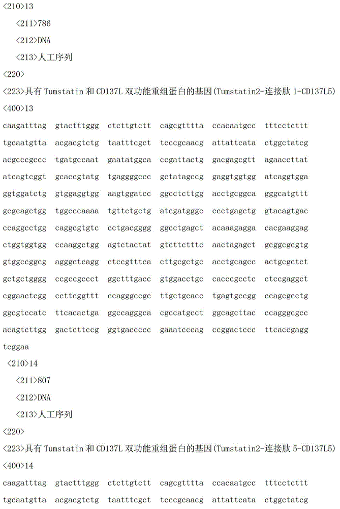


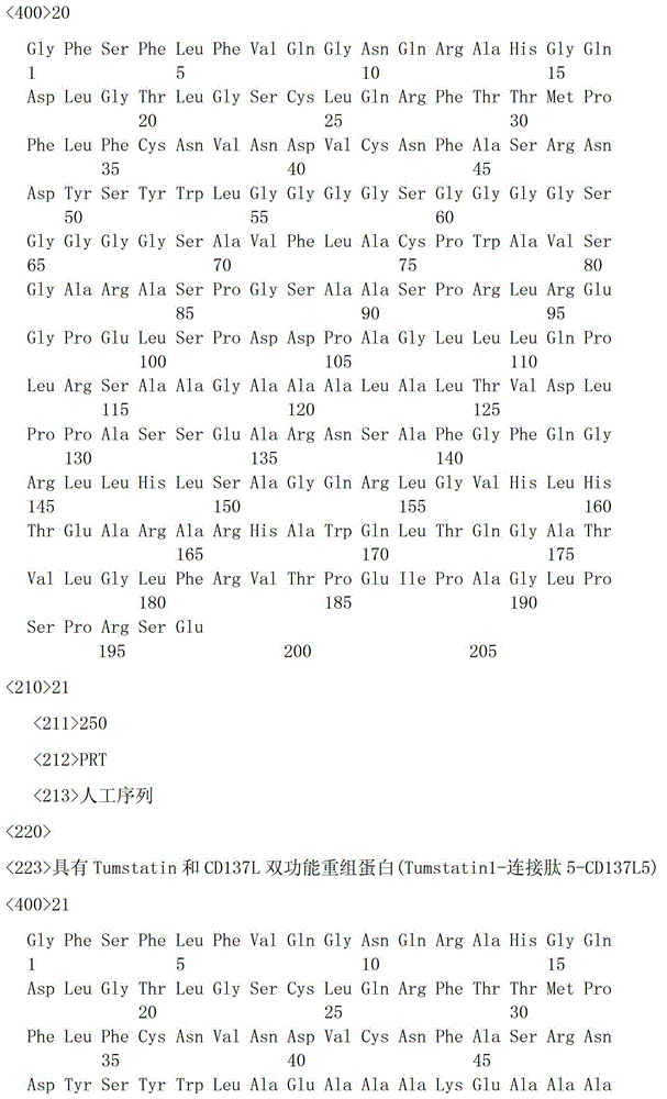
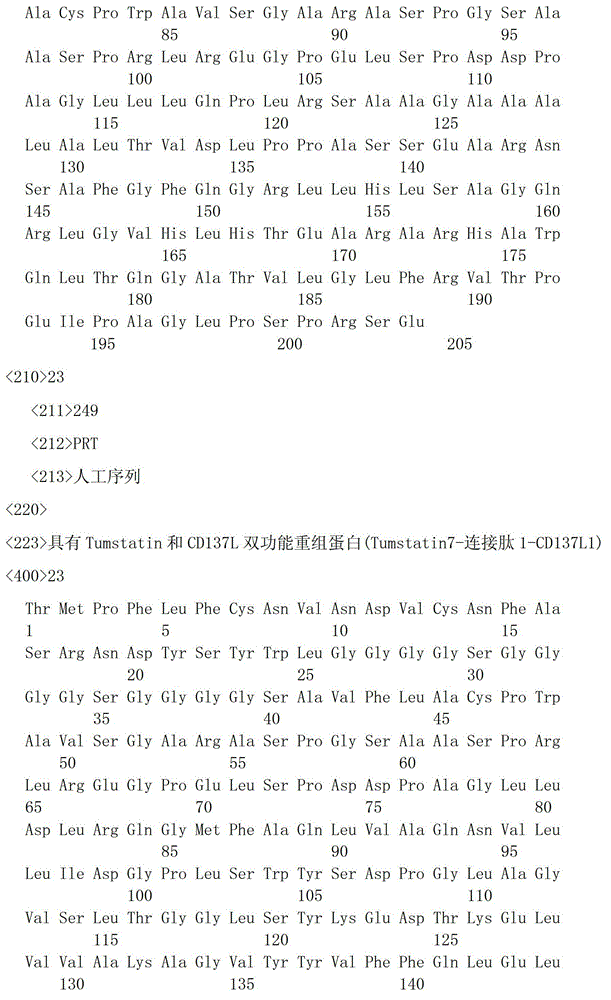
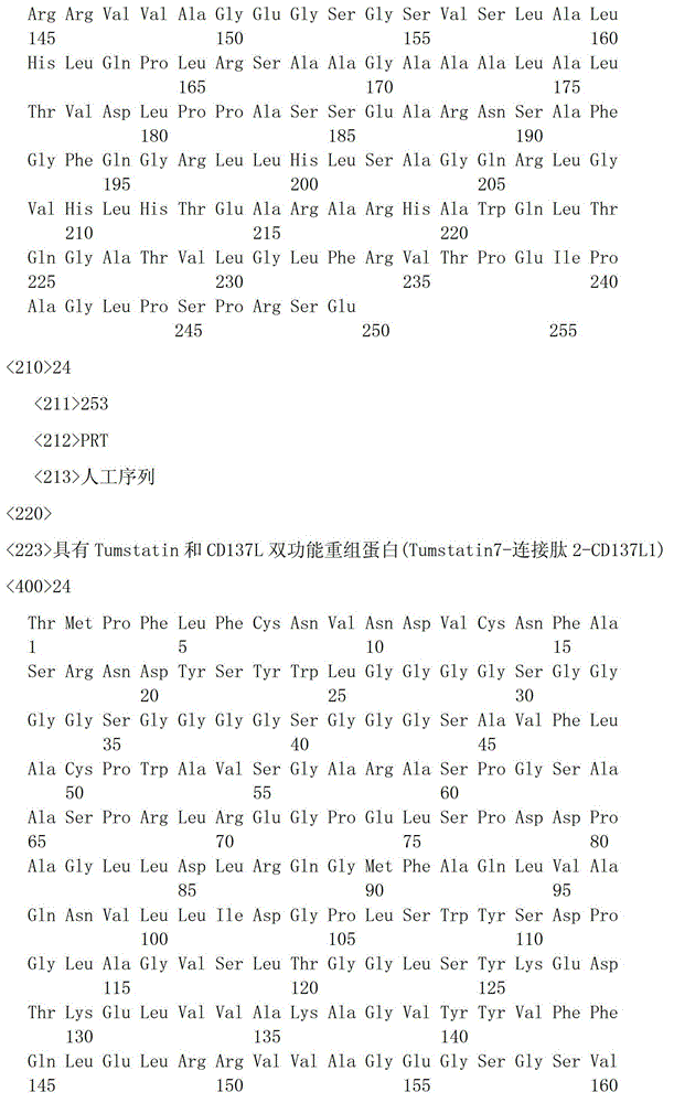

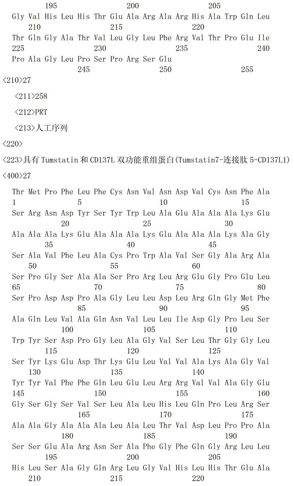

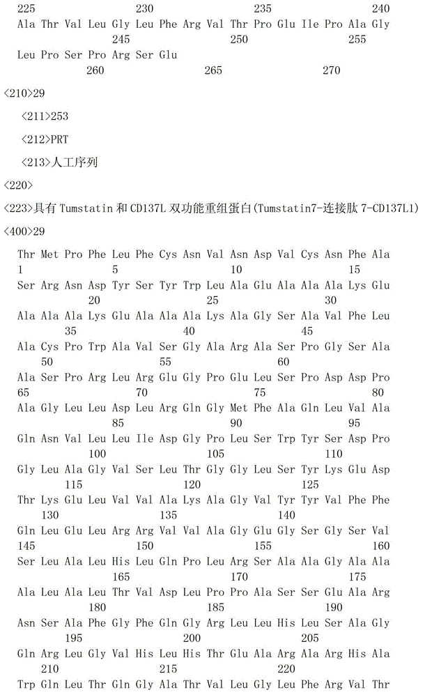
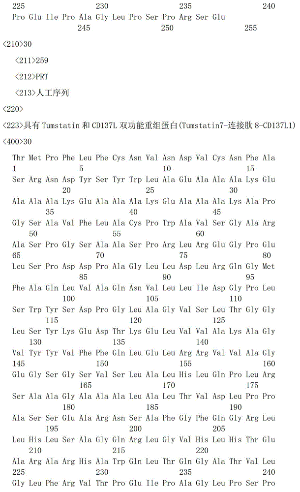

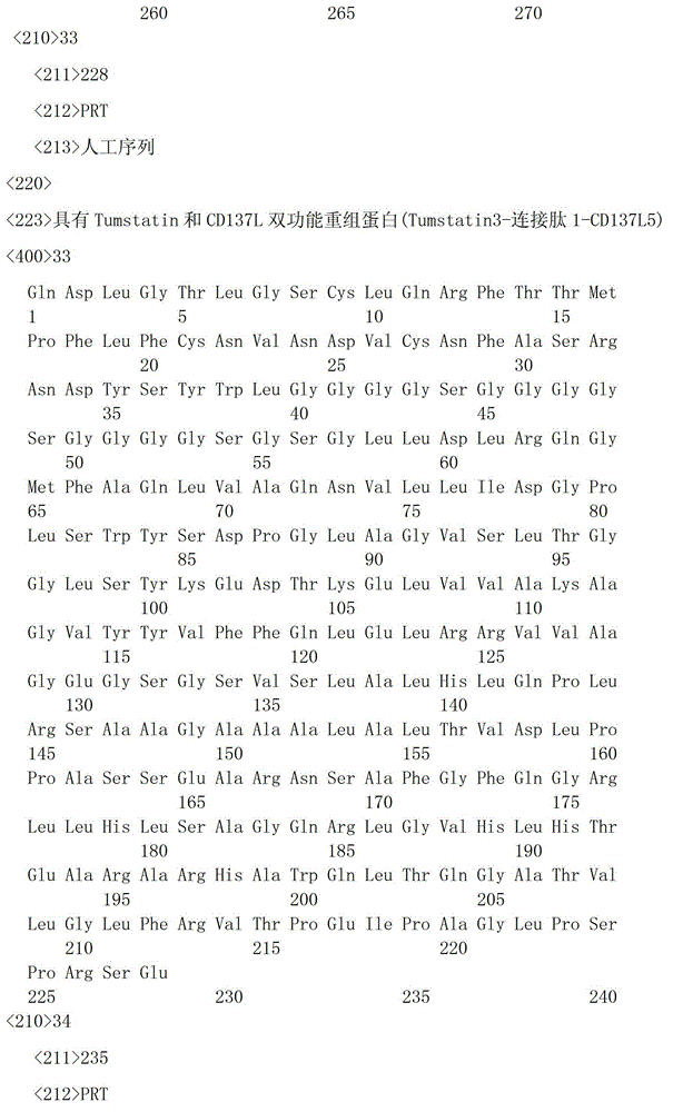
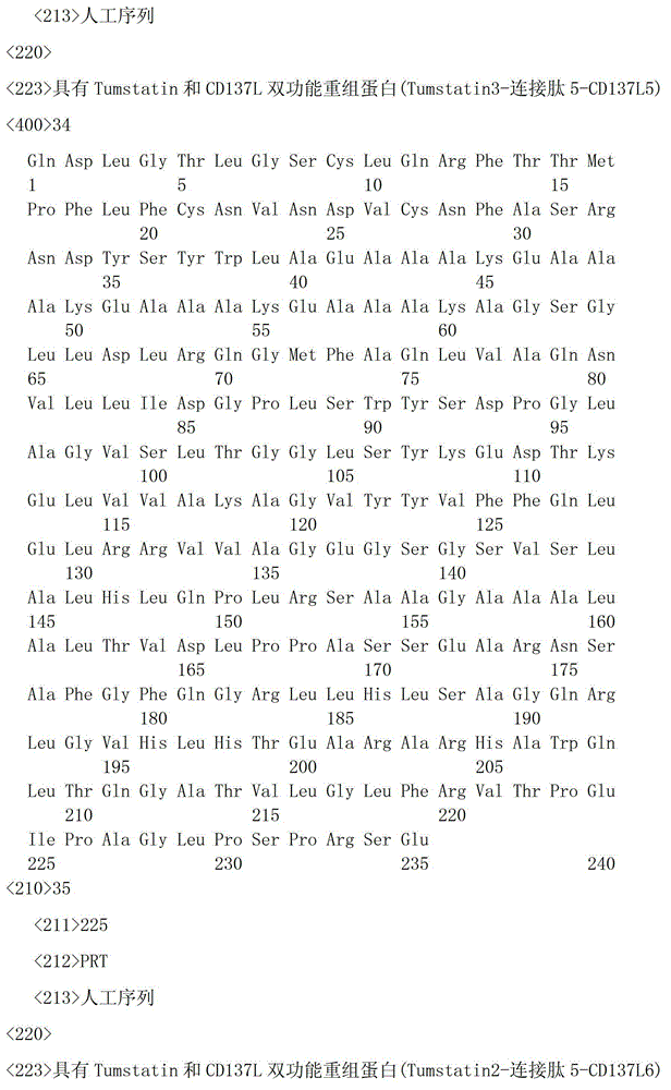
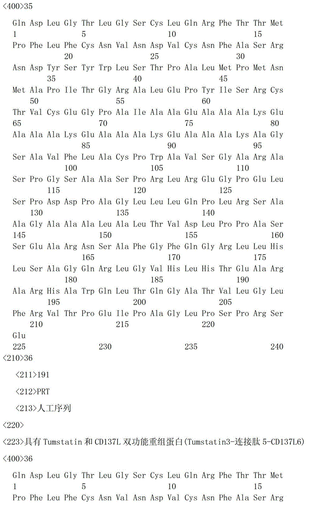

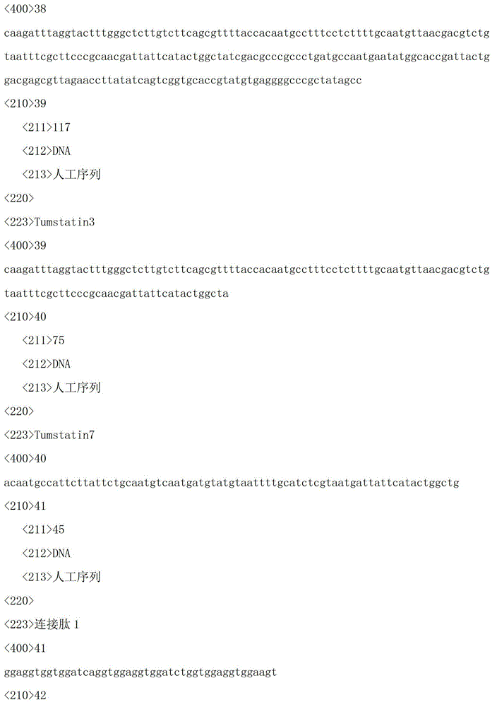
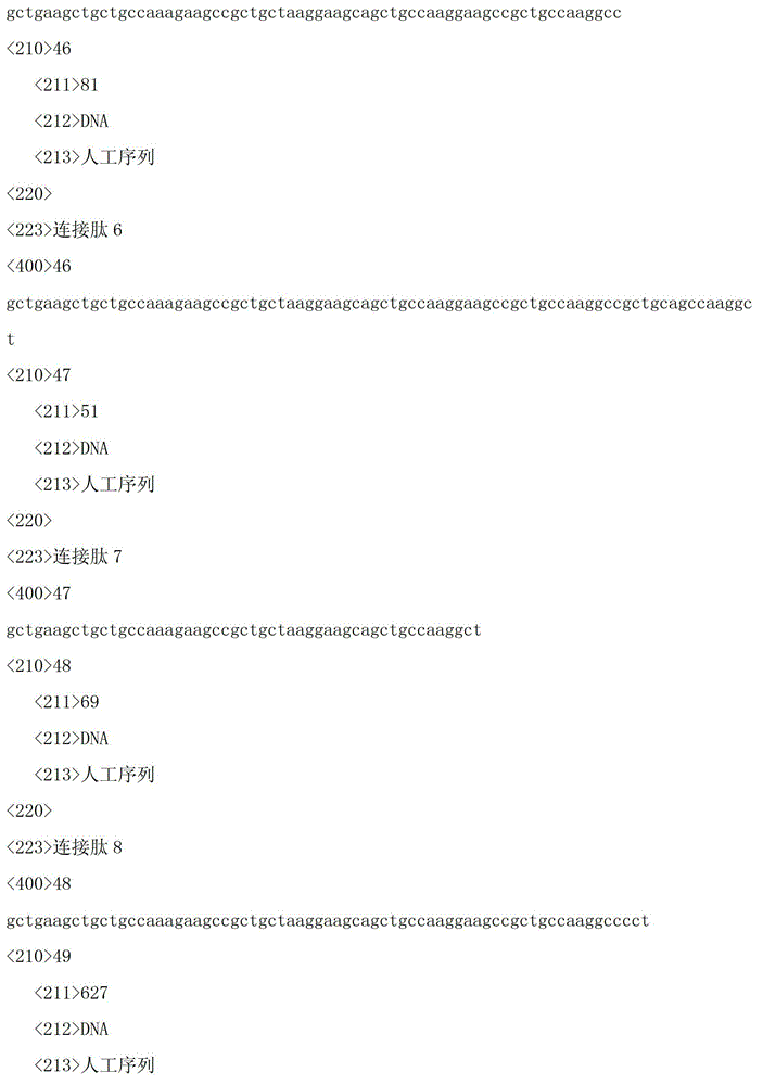
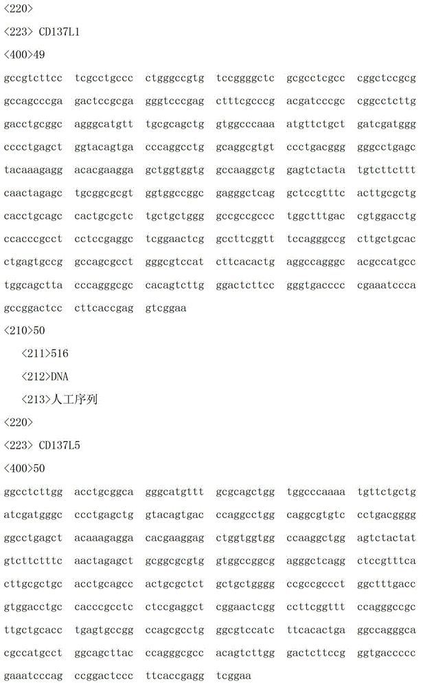
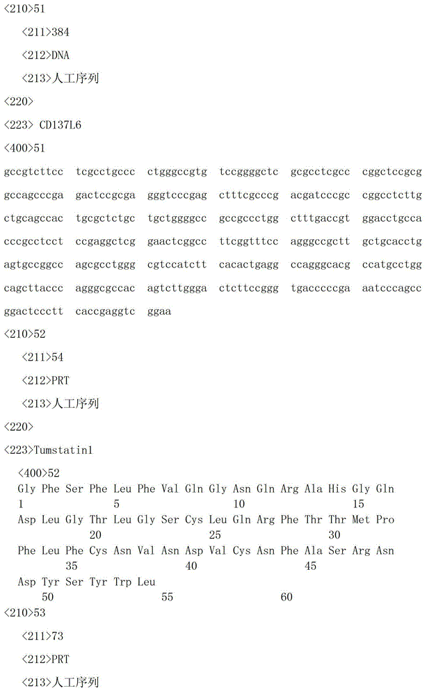

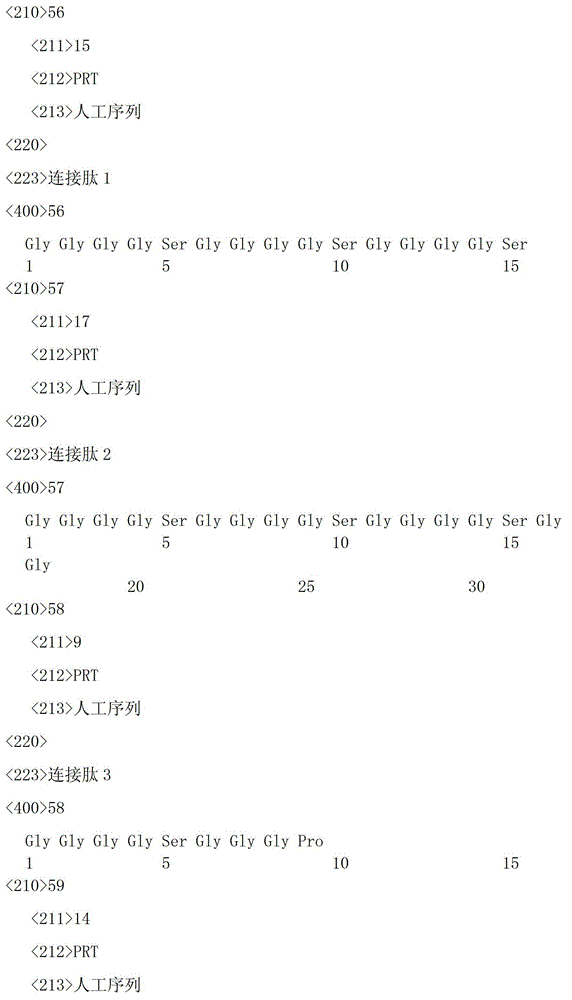
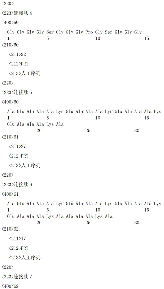


Claims (5)
Priority Applications (7)
| Application Number | Priority Date | Filing Date | Title |
|---|---|---|---|
| CN201310162738.3A CN103214584B (en) | 2013-05-06 | 2013-05-06 | Fusion protein with double functions of inhibiting revascularization of tumor microenvironment and activating adaptive immune response, and gene and application thereof |
| PCT/CN2014/076768 WO2014180288A1 (en) | 2013-05-06 | 2014-05-05 | Fusion protein having dual-functions for inhibiting angiogenesis in tumour microenvironment and activating adaptive immune response and gene and use thereof |
| DK14795356.6T DK2995626T3 (en) | 2013-05-06 | 2014-05-05 | BIFUNCTIONAL FUSION PROTEINS TO INHIBIT ANGIOGENESIS IN TUMOR ENVIRONMENTS AND ACTIVATE ADAPTIVE IMMUNE RESPONSE, AND THE GENES AND APPLICATIONS THEREOF |
| EP14795356.6A EP2995626B1 (en) | 2013-05-06 | 2014-05-05 | Bifunctional fusion proteins to inhibit angiogenesis in tumour microenvironment and to activate adaptive immune responses and the genes and uses thereof |
| JP2016512207A JP2016520053A (en) | 2013-05-06 | 2014-05-05 | Bifunctional fusion protein with inhibition of revascularization and activation of adaptive immune response in tumor microenvironment and gene and use thereof |
| US14/895,959 US10875903B2 (en) | 2013-05-06 | 2014-05-05 | Bifunctional fusion proteins to inhibit angiogenesis in tumor microenvironment and to activate adaptive immune responses and the genes and uses thereof |
| ES14795356.6T ES2686968T3 (en) | 2013-05-06 | 2014-05-05 | Bifunctional fusion proteins to inhibit angiogenesis in the tumor microenvironment and to activate adaptive immune responses and their genes and uses |
Applications Claiming Priority (1)
| Application Number | Priority Date | Filing Date | Title |
|---|---|---|---|
| CN201310162738.3A CN103214584B (en) | 2013-05-06 | 2013-05-06 | Fusion protein with double functions of inhibiting revascularization of tumor microenvironment and activating adaptive immune response, and gene and application thereof |
Publications (2)
| Publication Number | Publication Date |
|---|---|
| CN103214584A CN103214584A (en) | 2013-07-24 |
| CN103214584B true CN103214584B (en) | 2014-07-09 |
Family
ID=48812783
Family Applications (1)
| Application Number | Title | Priority Date | Filing Date |
|---|---|---|---|
| CN201310162738.3A Expired - Fee Related CN103214584B (en) | 2013-05-06 | 2013-05-06 | Fusion protein with double functions of inhibiting revascularization of tumor microenvironment and activating adaptive immune response, and gene and application thereof |
Country Status (1)
| Country | Link |
|---|---|
| CN (1) | CN103214584B (en) |
Families Citing this family (1)
| Publication number | Priority date | Publication date | Assignee | Title |
|---|---|---|---|---|
| US10875903B2 (en) | 2013-05-06 | 2020-12-29 | China Pharmaceutical University | Bifunctional fusion proteins to inhibit angiogenesis in tumor microenvironment and to activate adaptive immune responses and the genes and uses thereof |
Citations (3)
| Publication number | Priority date | Publication date | Assignee | Title |
|---|---|---|---|---|
| WO2006050172A2 (en) * | 2004-10-29 | 2006-05-11 | University Of Southern California | Combination cancer immunotherapy with co-stimulatory molecules |
| CN101524528A (en) * | 2009-03-09 | 2009-09-09 | 中国药科大学 | Recombination NuBCP-9 and Tumstatin(74-98) antitumor fusion polypetide |
| CN101698852A (en) * | 2009-10-23 | 2010-04-28 | 江苏先声药物研究有限公司 | Protein or polypeptide with function of CD137L, and gene and application thereof |
-
2013
- 2013-05-06 CN CN201310162738.3A patent/CN103214584B/en not_active Expired - Fee Related
Patent Citations (3)
| Publication number | Priority date | Publication date | Assignee | Title |
|---|---|---|---|---|
| WO2006050172A2 (en) * | 2004-10-29 | 2006-05-11 | University Of Southern California | Combination cancer immunotherapy with co-stimulatory molecules |
| CN101524528A (en) * | 2009-03-09 | 2009-09-09 | 中国药科大学 | Recombination NuBCP-9 and Tumstatin(74-98) antitumor fusion polypetide |
| CN101698852A (en) * | 2009-10-23 | 2010-04-28 | 江苏先声药物研究有限公司 | Protein or polypeptide with function of CD137L, and gene and application thereof |
Non-Patent Citations (1)
| Title |
|---|
| Sang Jun Park,et al..Reverse Signaling through the Co-Stimulatory Ligand, CD137L, as a Critical Mediator of Sterile Inflammation.《Molecules and Cells》.2012,第33卷(第6期),第533-537页. * |
Also Published As
| Publication number | Publication date |
|---|---|
| CN103214584A (en) | 2013-07-24 |
Similar Documents
| Publication | Publication Date | Title |
|---|---|---|
| WO2014180288A1 (en) | Fusion protein having dual-functions for inhibiting angiogenesis in tumour microenvironment and activating adaptive immune response and gene and use thereof | |
| CN102153653B (en) | Fusion protein of tumor blood vessel targeted polypeptide and tissue factor and preparation method thereof | |
| CN102898526A (en) | Tumor necrosis factor-related apoptosis-inducing ligand fusion protein, its preparation and application | |
| CN110023333B (en) | High affinity soluble PD-1 molecules | |
| CN101643511A (en) | Fusion protein for inhibiting telomerase activity, preparation and application thereof | |
| WO2019109954A1 (en) | Pd-1-fc fusion protein and preparation method and use thereof | |
| CN101698852B (en) | Protein or polypeptide with function of CD137L, and gene and application thereof | |
| KR20160113268A (en) | Bifunctional fusion protein, preparation method therefor, and use thereof | |
| CN110903402B (en) | A kind of bispecific fusion protein and its construction method and application | |
| JPH03201985A (en) | Human macif active protein gene, manifestation vector bonded to same gene, transformed cell and human macif active protein | |
| CN103214584B (en) | Fusion protein with double functions of inhibiting revascularization of tumor microenvironment and activating adaptive immune response, and gene and application thereof | |
| CN101456913B (en) | Anti-tumor fusion protein and use thereof | |
| CN103360497A (en) | Novel antitumor fusion protein vaccine, and preparation method and application thereof | |
| CN109400711B (en) | A PDGFRβ-targeted tumor necrosis factor-related apoptosis-inducing ligand variant and its preparation method and use | |
| CN117843800A (en) | A bispecific antibody targeting intracellular NY-ESO-1 and its application | |
| CN116554329A (en) | A kind of Fc fusion nanobody CTLA-4 Nb16-Fc and its preparation method and application | |
| KR101651330B1 (en) | Methods of TAT-A20 fusion protein with good cell penetration and use thereof | |
| CN106674353B (en) | A new fusion protein of Trichosanthes kirilowii and its application | |
| CN101830975B (en) | Recombinant galectin-1 binary protein and its preparation method | |
| WO2011147138A1 (en) | Targeting fusion protein of interleukin and preparation method and use thereof | |
| CN103232543A (en) | Recombinant protein Tumstatin-CD137L4 with Tumstatin activities as well as preparation method and application thereof | |
| JPS6034915A (en) | Polypeptide | |
| CN114716566A (en) | A fusion protein and its application in the preparation of tumor drugs | |
| CN101648995B (en) | Human recombination Reg4 protein and coding gene thereof as well as preparation method thereof | |
| CN112480262B (en) | Fusion protein and preparation and application thereof |
Legal Events
| Date | Code | Title | Description |
|---|---|---|---|
| C06 | Publication | ||
| PB01 | Publication | ||
| C10 | Entry into substantive examination | ||
| SE01 | Entry into force of request for substantive examination | ||
| C14 | Grant of patent or utility model | ||
| GR01 | Patent grant | ||
| CF01 | Termination of patent right due to non-payment of annual fee |
Granted publication date: 20140709 Termination date: 20210506 |
|
| CF01 | Termination of patent right due to non-payment of annual fee |