CN102656280B - Method for analyzing RNA - Google Patents
Method for analyzing RNA Download PDFInfo
- Publication number
- CN102656280B CN102656280B CN201080056692.1A CN201080056692A CN102656280B CN 102656280 B CN102656280 B CN 102656280B CN 201080056692 A CN201080056692 A CN 201080056692A CN 102656280 B CN102656280 B CN 102656280B
- Authority
- CN
- China
- Prior art keywords
- rna
- amplification
- analytical procedure
- analysis
- mentioned
- Prior art date
- Legal status (The legal status is an assumption and is not a legal conclusion. Google has not performed a legal analysis and makes no representation as to the accuracy of the status listed.)
- Expired - Fee Related
Links
Images


Classifications
-
- C—CHEMISTRY; METALLURGY
- C12—BIOCHEMISTRY; BEER; SPIRITS; WINE; VINEGAR; MICROBIOLOGY; ENZYMOLOGY; MUTATION OR GENETIC ENGINEERING
- C12Q—MEASURING OR TESTING PROCESSES INVOLVING ENZYMES, NUCLEIC ACIDS OR MICROORGANISMS; COMPOSITIONS OR TEST PAPERS THEREFOR; PROCESSES OF PREPARING SUCH COMPOSITIONS; CONDITION-RESPONSIVE CONTROL IN MICROBIOLOGICAL OR ENZYMOLOGICAL PROCESSES
- C12Q1/00—Measuring or testing processes involving enzymes, nucleic acids or microorganisms; Compositions therefor; Processes of preparing such compositions
- C12Q1/68—Measuring or testing processes involving enzymes, nucleic acids or microorganisms; Compositions therefor; Processes of preparing such compositions involving nucleic acids
- C12Q1/6844—Nucleic acid amplification reactions
- C12Q1/6851—Quantitative amplification
-
- G—PHYSICS
- G01—MEASURING; TESTING
- G01N—INVESTIGATING OR ANALYSING MATERIALS BY DETERMINING THEIR CHEMICAL OR PHYSICAL PROPERTIES
- G01N27/00—Investigating or analysing materials by the use of electric, electrochemical, or magnetic means
- G01N27/26—Investigating or analysing materials by the use of electric, electrochemical, or magnetic means by investigating electrochemical variables; by using electrolysis or electrophoresis
- G01N27/416—Systems
- G01N27/447—Systems using electrophoresis
-
- G—PHYSICS
- G16—INFORMATION AND COMMUNICATION TECHNOLOGY [ICT] SPECIALLY ADAPTED FOR SPECIFIC APPLICATION FIELDS
- G16B—BIOINFORMATICS, i.e. INFORMATION AND COMMUNICATION TECHNOLOGY [ICT] SPECIALLY ADAPTED FOR GENETIC OR PROTEIN-RELATED DATA PROCESSING IN COMPUTATIONAL MOLECULAR BIOLOGY
- G16B25/00—ICT specially adapted for hybridisation; ICT specially adapted for gene or protein expression
-
- C—CHEMISTRY; METALLURGY
- C12—BIOCHEMISTRY; BEER; SPIRITS; WINE; VINEGAR; MICROBIOLOGY; ENZYMOLOGY; MUTATION OR GENETIC ENGINEERING
- C12Q—MEASURING OR TESTING PROCESSES INVOLVING ENZYMES, NUCLEIC ACIDS OR MICROORGANISMS; COMPOSITIONS OR TEST PAPERS THEREFOR; PROCESSES OF PREPARING SUCH COMPOSITIONS; CONDITION-RESPONSIVE CONTROL IN MICROBIOLOGICAL OR ENZYMOLOGICAL PROCESSES
- C12Q2525/00—Reactions involving modified oligonucleotides, nucleic acids, or nucleotides
- C12Q2525/10—Modifications characterised by
- C12Q2525/207—Modifications characterised by siRNA, miRNA
Landscapes
- Life Sciences & Earth Sciences (AREA)
- Chemical & Material Sciences (AREA)
- Health & Medical Sciences (AREA)
- Physics & Mathematics (AREA)
- Organic Chemistry (AREA)
- Engineering & Computer Science (AREA)
- Molecular Biology (AREA)
- General Health & Medical Sciences (AREA)
- Proteomics, Peptides & Aminoacids (AREA)
- Zoology (AREA)
- Wood Science & Technology (AREA)
- Bioinformatics & Cheminformatics (AREA)
- Biophysics (AREA)
- Genetics & Genomics (AREA)
- Biotechnology (AREA)
- Chemical Kinetics & Catalysis (AREA)
- Immunology (AREA)
- Biochemistry (AREA)
- Analytical Chemistry (AREA)
- Microbiology (AREA)
- General Engineering & Computer Science (AREA)
- Bioinformatics & Computational Biology (AREA)
- Evolutionary Biology (AREA)
- Medical Informatics (AREA)
- Spectroscopy & Molecular Physics (AREA)
- Theoretical Computer Science (AREA)
- Electrochemistry (AREA)
- General Physics & Mathematics (AREA)
- Pathology (AREA)
- Measuring Or Testing Involving Enzymes Or Micro-Organisms (AREA)
- Investigating Or Analysing Biological Materials (AREA)
Abstract
Description
技术领域 technical field
本发明涉及从被固定液固定了的组织或细胞中提取出的RNA的分析方法。The present invention relates to a method for analyzing RNA extracted from tissues or cells fixed with a fixative.
背景技术 Background technique
近年来,对在医院、研究机构等保存有大量样本的、以福尔马林固定石蜡包埋(FFPE)组织为代表那样的、被固定液固定了的组织、细胞的基因的分析技术的期待大幅提高。特别是对于FFPE组织,由于积累了过去的大量病患数据,因而如果确立能够从FFPE组织提取基因、分析它们的表达的技术,则能够进行使用长期保存后的组织的可追溯研究,可以对将来的病患的治疗和/或预防做出巨大贡献。In recent years, there are expectations for gene analysis techniques for tissues and cells fixed with fixatives, such as formalin-fixed paraffin-embedded (FFPE) tissues, where a large number of samples are stored in hospitals and research institutions. A substantial increase. Especially for FFPE tissues, since a large amount of patient data has accumulated in the past, if a technology that can extract genes from FFPE tissues and analyze their expression is established, retroactive research using long-term preserved tissues will be possible, and future research can be carried out. contribute significantly to the treatment and/or prevention of the disease.
然而,认为从FFPE等固定组织、固定细胞中提取出的RNA在一般的固定条件、保管条件下进行分解、片段化,因此难以进行基因表达分析。另外,由于作为固定液最常用的甲醛(福尔马林),有时RNA-RNA间、RNA-蛋白质间发生交联、和/或甲醛被添加/修饰到RNA上,RNA在这样的状态下难以进行酶反应和/或化学反应,基因表达分析变得困难。因此,要求由进行了分解、片段化的RNA样品、和/或被交联、添加/修饰了的RNA样品进行基因表达分析的技术。另外,在进行这样的RNA样品的基因表达分析时,在分析之前确认RNA样品的分解、片段化、或者交联和/或添加/修饰的程度来判断品质、确认可否分析,对于进行正确的基因表达分析以及miRNA表达分析是非常有用的,要求能够进行这些确认的技术。However, RNA extracted from fixed tissues such as FFPE or fixed cells is decomposed and fragmented under normal fixation and storage conditions, making gene expression analysis difficult. In addition, due to the most commonly used formaldehyde (formalin) as a fixative, sometimes RNA-RNA, RNA-protein cross-linking occurs, and/or formaldehyde is added/modified to RNA, and RNA is difficult to fix in this state. Performing enzymatic and/or chemical reactions, gene expression analysis becomes difficult. Therefore, techniques for analyzing gene expression from decomposed and fragmented RNA samples, and/or cross-linked, added/modified RNA samples are required. In addition, when performing gene expression analysis of such an RNA sample, the degree of decomposition, fragmentation, or cross-linking and/or addition/modification of the RNA sample is confirmed before analysis to judge the quality and confirm whether the analysis is possible. Expression profiling as well as miRNA expression profiling are very useful and techniques that enable these validations are required.
专利文献1~4中公开了关于扩增分解了的RNA来进行基因表达分析的方法的技术。Patent Documents 1 to 4 disclose techniques related to methods for amplifying decomposed RNA and performing gene expression analysis.
专利文献1提供与扩增靶多核苷酸、产生其大量拷贝相关的方法、组合物和试剂盒。1次扩增反应中的mRNA的扩增倍数标准达50~100或250倍,或者为500~1000倍或500~2000倍以上,从不足纳克量的总RNA获得尽可能多的扩增RNA。然而,如果使用这样从少量的RNA尽可能多地扩增来分析的方法,则基因的每个扩增倍率产生偏差的所谓扩增偏倚变大,因而可以说不能正确地分析基因的表达量。Patent Document 1 provides methods, compositions and kits related to amplifying a target polynucleotide and producing a large number of copies thereof. The standard amplification factor of mRNA in one amplification reaction is 50-100 or 250 times, or 500-1000 times or more than 500-2000 times, and obtain as much amplified RNA as possible from less than nanograms of total RNA . However, if such a method of amplifying and analyzing as much as possible from a small amount of RNA is used, the so-called amplification bias in which the amplification factor of each gene varies becomes large, so it can be said that the expression level of the gene cannot be accurately analyzed.
专利文献2涉及为了全基因表达谱分析而使用例如保管的固定化石蜡包埋组织材料等片段化RNA等的方法,提供即使是非常小的、或极度片段化的RNA试样,也能够完全扩增的试样的调制方法。然而,该方法包含将被片段化的RNA进行多聚腺苷酸化的工序,认为该工序中的反应的偏差作为结果可能严重影响基因表达谱。另外在专利文献2中,公开了新的线性RNA扩增法。通过合成5’末端具有锚定序列、且3’末端具有RNA聚合酶启动子序列的双链cDNA,由该cDNA依赖上述RNA聚合酶启动子序列合成cRNA,由该cRNA通过引发上述锚定序列而再次合成cDNA的方法,来扩增从利用激光捕获显微切割、细胞分选仪获得的少量的细胞和/或组织中得到的微量的RNA,从而抑制作为现有的扩增法的问题的、在每次重复cDNA合成-cRNA合成循环时产生的cDNA和cRNA上的mRNA5’相当区的缺失。Patent Document 2 relates to a method of using fragmented RNA, such as preserved fixed paraffin-embedded tissue material, for the analysis of global gene expression profiling, and provides the ability to completely expand even a very small or extremely fragmented RNA sample The modulation method of the increased sample. However, this method includes a step of polyadenylation of the fragmented RNA, and it is considered that the variation of the reaction in this step may seriously affect the gene expression profile as a result. Also, Patent Document 2 discloses a novel linear RNA amplification method. By synthesizing a double-stranded cDNA with an anchor sequence at the 5' end and an RNA polymerase promoter sequence at the 3' end, cRNA is synthesized from the cDNA depending on the above-mentioned RNA polymerase promoter sequence, and the cRNA is activated by priming the above-mentioned anchor sequence. A method of re-synthesizing cDNA to amplify a small amount of RNA obtained from a small amount of cells and/or tissues obtained by laser capture microdissection or a cell sorter, thereby suppressing the problems of existing amplification methods, Deletion of the 5'-equivalent region of mRNA on cDNA and cRNA produced at each repeated cDNA synthesis-cRNA synthesis cycle.
专利文献1、专利文献2均作为无扩增偏倚的方法记载,认为每1个循环的偏倚与以往相比较小,但由于着眼于由少量的RNA尽可能多地扩增,因而作为扩增倍率相当大。因此,相应地偏倚积累,不可否认结果产生大的扩增偏倚。Both Patent Document 1 and Patent Document 2 are described as a method without amplification bias, and it is considered that the bias per cycle is smaller than conventional ones. However, since they focus on amplifying as much as possible from a small amount of RNA, the amplification factor quite big. Thus, with a corresponding bias accumulation, it is undeniable that the results generate a large amplification bias.
专利文献3涉及为了全基因表达谱分析而使用例如保管的固定化石蜡包埋组织材料等片段化RNA等的方法,提供即使是非常小的、或极度片段化的RNA试样也能够完全扩增的试样的调制方法。然而,该方法包含将被片段化的RNA进行多聚腺苷酸化的工序,认为该工序中的反应的偏差作为结果可能严重影响基因表达谱。Patent Document 3 relates to a method using, for example, fragmented RNA, such as preserved fixed paraffin-embedded tissue material, for the analysis of global gene expression profiling, and provides that even a very small or extremely fragmented RNA sample can be completely amplified The preparation method of the sample. However, this method includes a step of polyadenylation of the fragmented RNA, and it is considered that the variation of the reaction in this step may seriously affect the gene expression profile as a result.
专利文献4公开了包含例如分解度等核酸样品中的核酸的品质测定方法的、分解核酸样品中的靶标的扩增用组合物和方法,以及用于从分解RNA样品制成基因表达谱的方法。以来源于同一基因的不同尺寸的扩增子(扩增产物)的扩增效率为标准,随着样品分解,尺寸较大的扩增子的扩增效率降低,由此来评价品质。本方法对于十几种至二十几种基因分别进行多个尺寸的PCR。认为在RNA分解进行的情况下,PCR的探针尺寸越大则越不被扩增,因而可以一定程度确认分解度,但需要对每个RNA样品PCR扩增总共数十种基因,因此认为本方式难以实际进行。Patent Document 4 discloses a composition and method for amplifying a target in a decomposed nucleic acid sample, including a method for measuring the quality of nucleic acid in a nucleic acid sample such as the degree of decomposition, and a method for preparing a gene expression profile from a decomposed RNA sample . Using the amplification efficiency of amplicons (amplification products) of different sizes derived from the same gene as a standard, the quality is evaluated by reducing the amplification efficiency of amplicons with larger sizes as the sample decomposes. In this method, PCRs of multiple sizes are respectively performed on more than ten to twenty kinds of genes. It is considered that in the case of RNA decomposition, the larger the size of the PCR probe, the less it will be amplified, so the degree of decomposition can be confirmed to a certain extent, but a total of dozens of genes need to be PCR-amplified for each RNA sample, so it is considered that this way is difficult to do practically.
专利文献5公开了将以不分解的状态下长链级分所含的RNA的碱基序列为基础设计的分解指标核酸探针装载于核酸阵列,使从总RNA分级出短链而得的RNA样品对该核酸阵列杂交,通过分解指标核酸探针的信号的有无来测定RNA样品的分解度的方法,以及涉及RNA的分解度测定用核酸阵列的技术。然而,认为该核酸阵列限于微小RNA(miRNA)等短链RNA,难以适用于基因表达分析。而且,在从FFPE等的固定组织和/或固定细胞提取RNA的情况下,与从细胞和/或冻结组织提取情况不同,常常不能得到大量的RNA,在这样的状况下,仅用于RNA样品的品质确认就要使用几μg~几十μg这样大量的样品进行实验是不现实的,从成本方面也决不能说是优选的方法。Patent Document 5 discloses that a decomposition index nucleic acid probe designed based on the nucleotide sequence of RNA contained in a long-chain fraction in an undegraded state is mounted on a nucleic acid array, and RNA obtained by fractionating short chains from total RNA A method of measuring the degree of decomposition of an RNA sample by hybridizing a sample to the nucleic acid array by the presence or absence of a signal of a decomposition index nucleic acid probe, and a technique related to a nucleic acid array for measuring the degree of resolution of RNA. However, it is considered that this nucleic acid array is limited to short RNA such as microRNA (miRNA), and it is difficult to apply it to gene expression analysis. Furthermore, in the case of extracting RNA from fixed tissues such as FFPE and/or fixed cells, unlike the case of extracting from cells and/or frozen tissues, it is often impossible to obtain a large amount of RNA. In such a situation, only for RNA samples It is unrealistic to use a large number of samples such as several μg to tens of μg for the quality confirmation of the test, and it is by no means the best method in terms of cost.
专利文献6中有关于测定核酸的片段化水平的方法的记载。该专利文献的申请人销售分别测定RNA样品所含的2种核糖体RNA(18S、28S)的量,由28S/18S之比来判定能否分析的试剂盒。据说当RNA分解时,首先28S分解,接着18S分解,在该试剂盒中,如果28S/18S为0.1以下,则判定为RNA的品质差。然而,从被固定液固定了的组织、细胞中提取的RNA一般在固定时RNA分解,进而由于固定组织、细胞一般常温保管,随着经年RNA进一步分解。因此,从例如福尔马林固定石蜡包埋(FFPE)组织提取的RNA经常检测不到上述2种核糖体RNA,在以上述基准判定RNA样品的品质的情况下,大半样品被认为不能分析。因此,在使用如上所述的长期保存的固定组织、固定细胞进行可追溯研究时,使用该试剂盒是极为困难的。Patent Document 6 describes a method for measuring the fragmentation level of nucleic acid. The applicant of this patent document sells a kit for measuring the amounts of two types of ribosomal RNA (18S, 28S) contained in an RNA sample and determining whether analysis is possible from the ratio of 28S/18S. It is said that when RNA is decomposed, 28S is decomposed first, and then 18S is decomposed. In this kit, if 28S/18S is 0.1 or less, the quality of RNA is judged to be poor. However, RNA extracted from tissues and cells fixed with a fixative is generally decomposed during fixation, and since fixed tissues and cells are generally kept at room temperature, the RNA is further decomposed over time. Therefore, for example, RNA extracted from formalin-fixed paraffin-embedded (FFPE) tissue often cannot detect the above two types of ribosomal RNA, and when the quality of RNA samples is judged by the above-mentioned criteria, most of the samples are considered unanalyzable. Therefore, it is extremely difficult to use this kit for retrospective studies using long-term preserved fixed tissues, fixed cells as described above.
专利文献7是通过加热处理等使包含分解的mRNA的保管组织中的、与RISC(RNA诱导型沉默复合体)结合而未被分解的miRNA游离,并通过PCR扩增来检测的RNA的谱分析方法。由于在从RISC使miRNA游离时,在95℃这样的高温下处理,因此不能否定由此发生分解的可能性,也谈不上涉及确认RNA的品质的方法。Patent Document 7 is a profile analysis of RNA detected by PCR amplification by freeing undecomposed miRNA bound to RISC (RNA-induced silencing complex) in a storage tissue containing decomposed mRNA by heat treatment or the like method. Since the miRNA is released from the RISC, it is treated at a high temperature of 95°C, so the possibility of decomposition cannot be ruled out, and there is no way to check the quality of the RNA.
此外,在使用毛细管电泳系统(例如,Agilent Technologies公司制“Bioanalyzer(生物分析仪)”)的情况下,作为RNA分解的指标,计算出作为由Agilent Technologies公司所开发的测定基准的RIN(RNA IntegrityNumber,RNA完整性指数)。RIN是以电泳的RNA样品全体的电泳图谱为基础算出的,其值为0~10的范围(非专利文献1)。Agilent Technologies公司的Bioanalyzer成为评价核酸的品质时一般使用的装置,在使用该装置时,RIN成为显示RNA的品质的一般指标。然而,用Bioanalyzer分析从固定组织、固定细胞提取出的RNA时,即使对于其电泳图谱明显不同、分解行为各异的RNA,RIN的值也在2~3之间基本不变,因此RIN可能未必能够反应实际的RNA的状态。而且,实际情况是,缺乏判断从被固定液固定了的各种组织、细胞提取出的RNA是否是能够供给分析的样本的方法。In addition, in the case of using a capillary electrophoresis system (for example, "Bioanalyzer (bioanalyzer)" manufactured by Agilent Technologies), as an index of RNA degradation, RIN (RNA Integrity Number), which is a measurement standard developed by Agilent Technologies, is calculated. , RNA integrity index). RIN is calculated based on the electrophoretic pattern of the entire RNA sample electrophoresed, and its value ranges from 0 to 10 (Non-Patent Document 1). Agilent Technologies' Bioanalyzer is a commonly used device for evaluating the quality of nucleic acids, and when this device is used, RIN is a general index showing the quality of RNA. However, when using Bioanalyzer to analyze RNA extracted from fixed tissues and fixed cells, even for RNAs with significantly different electrophoretic patterns and different decomposition behaviors, the value of RIN is basically unchanged between 2 and 3, so RIN may not necessarily be the same. Can reflect the actual state of RNA. Furthermore, the actual situation is that there is a lack of methods for judging whether RNA extracted from various tissues and cells fixed with a fixative is a sample that can be used for analysis.
现有技术文献prior art literature
专利文献patent documents
专利文献1:特表2006-520603号公报Patent Document 1: Special Publication No. 2006-520603
专利文献2:特开2005-224172号公报Patent Document 2: JP-A-2005-224172
专利文献3:特表2007-515964号公报Patent Document 3: Special Publication No. 2007-515964
专利文献4:特表2008-541699号公报Patent Document 4: Special Publication No. 2008-541699
专利文献5:特开2008-35779号公报Patent Document 5: Japanese Patent Laid-Open No. 2008-35779
专利文献6:特开2008-43332号公报Patent Document 6: Japanese Patent Laid-Open No. 2008-43332
专利文献7:特表2009-501531号公报Patent Document 7: Special Publication No. 2009-501531
非专利文献non-patent literature
非专利文献1:Schroeder A,Mueller O,Stocker S,Salowsky R,Leiber M,Gassmann M,Lightfoot S,Menzel W,Granzow M,Ragg T:TheRIN:an RNA integrity number for assigning integrity values to RNAmeasurements;BMC Molecular Biology 7:3(2006)Non-Patent Document 1: Schroeder A, Mueller O, Stocker S, Salowsky R, Leiber M, Gassmann M, Lightfoot S, Menzel W, Granzow M, Ragg T: The RIN: an RNA integrity number for assigning integrity values to RNA measurements; BMC Molecular Biology 7:3 (2006)
发明内容 Contents of the invention
发明所要解决的课题The problem to be solved by the invention
在现有技术中,分析从被固定液固定了的组织或细胞中提取出的RNA时,没有简便且高准确度地判断所提取的RNA是否适于分析的方法,知晓分析所得的数据的有效性是困难的。In the prior art, when analyzing RNA extracted from tissues or cells fixed with a fixative, there is no simple and highly accurate method for judging whether the extracted RNA is suitable for analysis. Knowing the validity of the data obtained from the analysis Sex is difficult.
用于课题的方法method used in the subject
鉴于上述课题,本发明者们进行了深入研究,结果发现,在进行从被固定液固定了的组织或细胞中提取出的RNA的基因表达分析时,在分析前对RNA进行电泳,根据电泳图谱来判断是否适于分析,对于判断为适于分析的RNA进行分析,可获得符合本来的RNA的存在量的有效性高的数据,而且可获得再现性高的数据,从而完成了本发明。In view of the above-mentioned problems, the inventors of the present invention conducted intensive studies and found that, when performing gene expression analysis of RNA extracted from tissues or cells fixed with a fixative, the RNA was subjected to electrophoresis before the analysis. To determine whether it is suitable for analysis, analysis of RNA judged suitable for analysis can obtain data with high validity and high reproducibility in accordance with the original amount of RNA, and completed the present invention.
另外发现,如果在扩增该RNA时,使其扩增倍率为原来的2~20倍,则能够抑制扩增所产生的偏倚,获得符合本来的RNA的存在量的有效性高的数据,而且发现数据的再现性高,从而完成了本发明。In addition, it was found that when the RNA is amplified at a magnification of 2 to 20 times the original, the bias caused by the amplification can be suppressed, and data with high validity corresponding to the original amount of RNA can be obtained, and The reproducibility of data was found to be high, and the present invention was completed.
即,本发明由以下(1)~(7)构成。That is, the present invention consists of the following (1) to (7).
(1)从被固定液固定了的组织或细胞中提取出的RNA的分析方法,该RNA的分析方法具有以下步骤:当设对该RNA总重量的电泳中的1000~4000核苷酸的范围的RNA的重量的比率为A(%)、对该RNA总重量的电泳中的超过4000核苷酸的范围的RNA的重量的比率为B(%)时,判定该RNA满足下式,(1) A method for analyzing RNA extracted from tissues or cells fixed with a fixative, the method for analyzing RNA has the following steps: when the range of 1000 to 4000 nucleotides in the electrophoresis of the total weight of the RNA is set When the ratio of the weight of the RNA is A (%), and the ratio of the weight of the RNA exceeding 4000 nucleotides in the electrophoresis of the total RNA weight is B (%), it is determined that the RNA satisfies the following formula,
式:B/A≤1。Formula: B/A≤1.
(2)根据(1)所述的RNA的分析方法,进一步具有以下步骤:判定上述比率A(%)为25%以上。(2) The RNA analysis method according to (1), further comprising the step of determining that the ratio A (%) is 25% or more.
(3)根据(1)或(2)所述的RNA的分析方法,对上述判定的RNA分析以扩增倍率2~20倍扩增所得的扩增产物。(3) The method for analyzing RNA according to (1) or (2), wherein an amplified product obtained by amplifying the determined RNA at an amplification factor of 2 to 20 times is analyzed.
(4)根据(1)或(2)所述的RNA的分析方法,对上述判定的RNA通过非扩增来进行分析。(4) The RNA analysis method described in (1) or (2), wherein the determined RNA is analyzed by non-amplification.
(5)根据(4)所述的RNA的分析方法,上述RNA是miRNA。(5) The RNA analysis method according to (4), wherein the RNA is miRNA.
(6)根据(1)~(5)的任一项所述的RNA的分析方法,上述固定液包含甲醛或低聚甲醛。(6) The RNA analysis method according to any one of (1) to (5), wherein the fixative solution contains formaldehyde or paraformaldehyde.
(7)根据(1)~(6)的任一项所述的RNA的分析方法,对上述被固定液固定了的组织或细胞进行了石蜡包埋或OCT复合物包埋。(7) The RNA analysis method according to any one of (1) to (6), wherein the tissue or cells fixed with the fixative are paraffin-embedded or OCT complex-embedded.
发明的效果The effect of the invention
通过本发明的RNA的分析方法,对于从被固定液固定了的组织或细胞中提取出的RNA,可以进行正确地反映本来的基因的存在量的分析,特别是在微阵列分析中可获得良好的效果。另外,通过本发明的RNA的分析方法,可以在分析前判定从被固定液固定了的组织或细胞中提取出的RNA是否适于分析,可以将浪费使用试剂等防患于未然。According to the RNA analysis method of the present invention, for RNA extracted from tissues or cells fixed by a fixative, it is possible to perform an analysis that accurately reflects the amount of the original gene present, and in particular, good results can be obtained in microarray analysis. Effect. In addition, according to the RNA analysis method of the present invention, it is possible to determine whether RNA extracted from tissues or cells fixed with a fixative is suitable for analysis before analysis, and it is possible to prevent wasteful use of reagents and the like.
附图说明 Description of drawings
图1显示当设通过Bioanalyzer得到的各种RNA的1000~4000核苷酸的范围的RNA的重量的比率为A(%)、超过4000核苷酸的范围的RNA的重量的比率为B(%)时,以横轴为A、以纵轴为B/A对各种RNA的数值作图而得的图。Figure 1 shows that when the ratio of the weight of RNA in the range of 1000 to 4000 nucleotides of various RNAs obtained by Bioanalyzer is A (%), and the ratio of the weight of RNA in the range of more than 4000 nucleotides is B (%) ), the graph obtained by plotting the values of various RNAs with A on the horizontal axis and B/A on the vertical axis.
图2显示对于各种RNA的RIN和扩增量进行作图而得的图。Fig. 2 shows a graph obtained by plotting RIN and amplification amount of various RNAs.
图3将利用小鼠小脑、肝脏的FFPE组织的微阵列的表达分析进行2次,对于第1次、第2次各自的分析所得的各基因的信号强度,将两者的小脑/肝脏之比进行作图而得的散点图。Figure 3. The expression analysis using the microarray of mouse cerebellum and liver FFPE tissue was performed twice, and the signal intensity of each gene obtained in the first and second analysis respectively, and the cerebellum/liver ratio of the two The resulting scatter plot.
具体实施方式 Detailed ways
以下,更具体地说明本发明。Hereinafter, the present invention will be described more specifically.
本发明的“被固定液固定了的组织或细胞”,是指通过在称为固定液的溶液中浸渍组织、或者细胞,从而进行用于将生物试样维持在尽可能自然的状态下的处理、即固定处理。这里使用的固定液,优选使用甲醛溶液、低聚甲醛溶液、包含乙醇、甲醇等醇类、丙酮、氯仿等的溶液、包含苦味酸、重铬酸钾等酸的固定液(例如,鲍音固定液、Zamboni固定液、奥尔特氏固定液)、包含乙酸锌、氯化锌、硫酸锌等金属的溶液等。另外,还优选包含乙醇、氯仿、乙酸的卡诺伊固定液、包含甲醇、氯仿、乙酸的Methacarn固定液等、将上述溶液二种以上混合使用。The "tissue or cell fixed with a fixative" in the present invention refers to a treatment for maintaining a biological sample in as natural a state as possible by immersing the tissue or cells in a solution called a fixative , That is, fixed processing. The fixative used here is preferably a formaldehyde solution, a paraformaldehyde solution, a solution containing alcohols such as ethanol and methanol, acetone, chloroform, etc., a fixative containing acids such as picric acid and potassium dichromate (for example, Baoyin fixative) solution, Zamboni's fixative, Oort's fixative), solutions containing metals such as zinc acetate, zinc chloride, zinc sulfate, etc. In addition, Carnoy's fixative containing ethanol, chloroform, and acetic acid, Methacarn's fixative containing methanol, chloroform, and acetic acid, etc., are also preferably used in combination of two or more of these solutions.
在本发明中,作为上述固定液,更优选使用包含甲醛或低聚甲醛的溶液。甲醛溶液可以使用将市售的福尔马林(甲醛浓度37%)用水稀释而得的溶液,还优选使用将水稀释后的溶液的pH值用碳酸钙、碳酸镁等调节成中性而得的溶液,和/或用磷酸缓冲液稀释而将pH值调节成中性而得的溶液。另外,还可以使用除去了恶臭、刺激性气味、调节了浓度的福尔马林溶液(商品名:マスクドホルム)。此外,甲醛溶液中的甲醛含量优选为1~30%,更优选为2~20%。低聚甲醛溶液可以使用将聚甲醛的粉末用水或磷酸缓冲液等溶解而得的溶液,和/或用水和少量的氢氧化钠溶解后、用磷酸缓冲液等将pH值调节至中性而得的溶液,也可以使用市售的低聚甲醛溶液。其浓度优选为1~10%,更优选为2~8%。In the present invention, it is more preferable to use a solution containing formaldehyde or paraformaldehyde as the above-mentioned fixative. The formaldehyde solution can be obtained by diluting commercially available formalin (formaldehyde concentration 37%) with water, and it is also preferably obtained by adjusting the pH value of the solution diluted with water to be neutral with calcium carbonate, magnesium carbonate, etc. solution, and/or a solution obtained by diluting with phosphate buffer to adjust the pH to neutral. In addition, a formalin solution (trade name: Masked Holm) whose concentration has been adjusted from which bad odors and pungent odors have been removed can also be used. In addition, the formaldehyde content in the formaldehyde solution is preferably 1 to 30%, more preferably 2 to 20%. The paraformaldehyde solution can be obtained by dissolving polyoxymethylene powder in water or phosphate buffer solution, and/or dissolving it in water and a small amount of sodium hydroxide, and then adjusting the pH to neutral with phosphate buffer solution. solution, commercially available paraformaldehyde solution can also be used. Its concentration is preferably 1 to 10%, more preferably 2 to 8%.
另外,上述被固定了的组织或者细胞可以被石蜡包埋。在将被固定了的组织或者细胞进行石蜡包理时,通过本领域技术人员公知的一般方法来操作即可。即,将固定组织或细胞用醇置换而脱水,接着用二甲苯、苯等置换,然后在注入了加热而液化的石蜡的铸模中加入组织、细胞而包埋,制成石蜡块。此外,在从石蜡包埋后的组织、细胞中提取RNA时,使用利用旋转式切片机、滑行式切片机等切片机切成薄片的样品。此时,对薄切片的厚度不特别限定,优选为1~100μm,更优选为2~50μm。此外,可以使用主要用于制作冻结组织切片的OCT(Optimal CuttingTemperature,最佳切削温度)复合物代替石蜡。另外,还优选使用以下方法:将石蜡块切成薄片而得的样品贴合在载玻片等上,使用激光捕获显微切割(LCM)和/或手术刀等从采取成为分析对象的组织的巨型拼合模块等将一部分取出而得的组织中提取RNA。在进行LCM时,为了提高视认性、确实地取出作为分析对象的组织、细胞,还可以将组织、细胞染色。此时,作为色素可利用例如甲酚紫、苏木精-伊红(HE)、核固红(NFR)等。In addition, the above-mentioned fixed tissues or cells may be embedded in paraffin. When paraffin-packing fixed tissues or cells, it may be performed by a general method known to those skilled in the art. That is, fixed tissues or cells are dehydrated by replacing them with alcohol, then replaced by xylene, benzene, etc., and then the tissues and cells are embedded in a mold filled with heated and liquefied paraffin to form a paraffin block. In addition, when extracting RNA from paraffin-embedded tissues and cells, samples cut into thin slices with a microtome such as a rotary microtome or a slide microtome are used. At this time, the thickness of the thin section is not particularly limited, but is preferably 1 to 100 μm, more preferably 2 to 50 μm. In addition, OCT (Optimal Cutting Temperature) compound, which is mainly used for making frozen tissue sections, can be used instead of paraffin. In addition, it is also preferable to use a method in which a sample obtained by cutting a paraffin block into thin slices is pasted on a glass slide or the like, and the tissue to be analyzed is collected from a sample using laser capture microdissection (LCM) and/or a scalpel or the like. RNA is extracted from tissue obtained by removing a part of the giant splicing module, etc. When performing LCM, the tissues and cells may be stained in order to improve visibility and to reliably extract the tissues and cells to be analyzed. In this case, for example, cresyl violet, hematoxylin-eosin (HE), nuclear fast red (NFR) and the like can be used as the dye.
本发明的“从被固定液固定了的组织或细胞中提取出的RNA”,是从使用上述固定液固定了的组织或者细胞,通过酶消化组织、细胞的蛋白质而提取的RNA,作为该RNA,可列举mRNA、rRNA、tRNA、miRNA,优选为mRNA或miRNA,更优选为miRNA。提取液中有时混入了DNA、蛋白质等杂质,优选提取后进行纯化操作。作为这里使用的纯化方法不特别限定,可列举例如,使用硅胶膜、担载有阴离子交换树脂等的柱的方法,使用逆相色谱法等液体色谱法的方法,使用有机溶剂使RNA沉淀的方法,将浓度高的乙酸铵溶液添加到含RNA的溶液中使RNA选择性沉淀的方法,使用磁珠的方法等。在上述提取、纯化中,适合使用例如,“RecoverAll(トレ一ドマ一ク)Total Nucleic Acid Isolation Kit for FFPE”(Ambion公司)、“RNeasy FFPE Kit”(Qiagen公司)、“ISOGEN PB Kit”(ニツポンジ一ン株式会社)、“FFPE RNA Purification Kit”(Norgen公司)、“PureLink(商标)FFPE RNA Isolation Kit”(Invitrogen公司)、“High Pure FFPE RNAMicro”(Roche Applied Science社)、“Agencourt FormaPure(商标)Kit”(Beckman Coulter公司)、“QuickExtract(商标)FFPE ExtractionKit”(Epicentre社)等福尔马林固定石蜡包埋组织用RNA提取试剂盒。The "RNA extracted from tissues or cells fixed with a fixative" in the present invention refers to RNA extracted from tissues or cells fixed with the above-mentioned fixative by enzymatically digesting the proteins of the tissues or cells. , may include mRNA, rRNA, tRNA, miRNA, preferably mRNA or miRNA, more preferably miRNA. Impurities such as DNA and protein may be mixed in the extract, and it is preferable to carry out purification after extraction. The purification method used here is not particularly limited, and examples thereof include a method using a silica gel membrane, a column loaded with an anion exchange resin, etc., a method using liquid chromatography such as reverse phase chromatography, and a method of precipitating RNA using an organic solvent. , a method of selectively precipitating RNA by adding a high-concentration ammonium acetate solution to a solution containing RNA, a method of using magnetic beads, etc. In the above extraction and purification, for example, "RecoverAll (トレドマク) Total Nucleic Acid Isolation Kit for FFPE" (Ambion), "RNeasy FFPE Kit" (Qiagen), "ISOGEN PB Kit" (ニツポンジIchin Co., Ltd.), "FFPE RNA Purification Kit" (Norgen Corporation), "PureLink (trademark) FFPE RNA Isolation Kit" (Invitrogen Corporation), "High Pure FFPE RNAMicro" (Roche Applied Science), "Agencourt FormaPure (Trademark) ) Kit" (Beckman Coulter), "QuickExtract (trademark) FFPE Extraction Kit" (Epicentre) and other RNA extraction kits for formalin-fixed paraffin-embedded tissues.
从上述被固定液固定了的组织或细胞中提取出的RNA由于被固定液成分分解、和/或交联、修饰,因而难以将该RNA供给利用该RNA与能直接或间接地选择性结合的物质(以下也称为选择结合性物质)的分子间相互作用的分析的样品较多,事先判定是否适于分析成为了技术课题,但本发明者发现,如后述的实施例等所示,在从固定组织或细胞中提取出的RNA中,通过确认对该RNA的总重量的电泳中的1000~4000核苷酸的范围的RNA的重量的比率、和对该RNA的总重量的电泳中的超过4000核苷酸的范围的RNA的重量的比率的工序,预先排除成为RNA的分解和/或片段化的指标的分子量小的RNA、和成为交联和/或添加/修饰等的指标的分子量大的RNA的比率多的RNA样品,从而可以判断该RNA是否适于上述微阵列分析。这里所说的“适于分析”,是指在进行利用了RNA与选择结合性物质的分子间相互作用的RNA的分析的情况下,能够正确地测定该样品所保有的信息的状态。如上所述,在将RNA供给分析的情况下,如果分解、片段化的程度大、和/或存在大量交联或者添加/修饰物,则有时与选择结合性物质的分子间相互作用被抑制,而且扩增时得不到充分的扩增量,产生偏倚,作为结果极有可能不能正确地分析。The RNA extracted from the tissues or cells fixed by the above-mentioned fixative solution is decomposed, and/or cross-linked and modified by the components of the fixative solution, so it is difficult to supply the RNA to the RNA that can directly or indirectly selectively bind The analysis of intermolecular interactions of substances (hereinafter also referred to as selective binding substances) has many samples, and it is a technical problem to determine in advance whether it is suitable for analysis. However, the inventors of the present invention found that, In RNA extracted from fixed tissues or cells, by confirming the ratio of the weight of RNA in the range of 1000 to 4000 nucleotides in the electrophoresis to the total weight of the RNA, and the electrophoresis to the total weight of the RNA In the process of the weight ratio of RNA exceeding 4000 nucleotides, RNA with a small molecular weight that is an indicator of RNA decomposition and/or fragmentation, and those that are indicators of crosslinking and/or addition/modification are excluded in advance. RNA samples with a large ratio of RNA with a large molecular weight can judge whether the RNA is suitable for the above-mentioned microarray analysis. The term "suitable for analysis" as used herein refers to a state in which information held in the sample can be accurately measured in the case of RNA analysis utilizing the intermolecular interaction between RNA and a selective binding substance. As described above, in the case of analyzing RNA, if the degree of decomposition and fragmentation is large, and/or there are many crosslinks or additions/modifications, the intermolecular interaction with the selective binding substance may be inhibited, In addition, a sufficient amount of amplification cannot be obtained during amplification, resulting in bias, and as a result, there is a high possibility that accurate analysis cannot be performed.
作为在从被固定了的组织或者细胞中提取出的RNA中,确认对该RNA总重量的电泳中的1000~4000核苷酸的范围的RNA的重量的比率、和对该RNA的总重量的电泳中的超过4000核苷酸的范围的RNA的重量的比率的方法,可列举将该RNA以分子量大小来区分并定量各分子量范围中的RNA的存在比例的方法。作为将从被固定了的组织、细胞等中提取出的RNA样品以分子量大小区分并确认分子量分布的方法的电泳的具体例,可列举琼脂糖凝胶电泳、聚丙烯酰胺凝胶电泳、毛细管电泳、芯片电泳,但作为定量特定的核苷酸范围中的RNA的存在比例的方法,例如在凝胶电泳的情况下,可列举使用本领域技术人员公知的各种光密度计和/或“Typhoon”(GEヘルスケア社)等的测量将谱带强度数值化的方法,另外在芯片电泳的情况下,在使用“Agilent2100Bioanalyzer”(AgilentTechnologies公司)等电泳系统进行时,通过进行专用软件所具有的Smear分析,从而可以适当地求出特定分子量范围中的RNA的存在比例。此外,在将未发生分解的RNA(完整RNA)用电泳进行分析的情况下,据说为约4700核苷酸的核糖体RNA的28S有时显示以比4000核苷酸略小的分子量为峰的谱带。认为这是由于28S的分子中含有较多的双链区,结构变得紧凑,因而在电泳中比实际的分子量泳动速度快。关于该现象,在AgilentTechnologies公司主页的FAQ中也可看到同样的记载。因此,在本发明中核糖体RNA的28S实质上包含在电泳中的1000~4000核苷酸的范围内。另外,18S的分子量为1874核苷酸,在电泳中也能在1000~4000核苷酸的范围观察到谱带,因而也包含在该范围中。In RNA extracted from fixed tissues or cells, the ratio of the weight of RNA in the range of 1000 to 4000 nucleotides in the electrophoresis to the total weight of the RNA and the ratio to the total weight of the RNA were confirmed. The method for the weight ratio of RNA exceeding 4000 nucleotides in electrophoresis includes a method of classifying the RNA by molecular weight and quantifying the presence ratio of RNA in each molecular weight range. Specific examples of electrophoresis as a method of classifying RNA samples extracted from fixed tissues, cells, etc. by molecular weight and confirming molecular weight distribution include agarose gel electrophoresis, polyacrylamide gel electrophoresis, and capillary electrophoresis. , chip electrophoresis, but as a method of quantifying the presence ratio of RNA in a specific nucleotide range, for example, in the case of gel electrophoresis, various densitometers and/or "Typhoon" well-known to those skilled in the art can be enumerated. " (GE ヘルスケア company) and other methods to quantify the band intensity, and in the case of chip electrophoresis, when using an electrophoresis system such as "Agilent2100Bioanalyzer" (Agilent Technologies Co., Ltd.), by performing Smear that is included in the dedicated software Analysis can appropriately determine the presence ratio of RNA in a specific molecular weight range. In addition, when undecomposed RNA (intact RNA) is analyzed by electrophoresis, it is said that 28S of ribosomal RNA of about 4700 nucleotides may show a spectrum with a molecular weight slightly smaller than 4000 nucleotides as a peak bring. It is believed that this is because the molecule of 28S contains more double-stranded regions, and the structure becomes compact, so the electrophoresis is faster than the actual molecular weight. Regarding this phenomenon, the same description can be seen in the FAQ on the homepage of Agilent Technologies. Therefore, in the present invention, 28S of ribosomal RNA is substantially included in the range of 1000 to 4000 nucleotides in electrophoresis. In addition, the molecular weight of 18S is 1874 nucleotides, and a band can be observed in the range of 1000 to 4000 nucleotides in electrophoresis, so it is also included in this range.
在本发明中,判断从被固定了的组织或者细胞中提取出的RNA是否适于分析的具体方法是,通过确认相对于对该RNA的总重量的电泳中的1000~4000核苷酸的范围的RNA的重量的比率A(%),对该RNA的总重量的电泳中的超过4000核苷酸的RNA的重量的比率B(%)较小或相同、即B/A≤1,从而判定是否适于分析的方法。认为包含超过4000核苷酸的RNA的级分中含有产生交联的RNA,它们有时不能与探针杂交,因此在分析比率B大于比率A的RNA样品的情况下,有定量值小于实际存在的mRNA量或者miRNA量的倾向,而且还有时会发现本来表达但不能检测的mRNA或者miRNA。因此,在进行从固定组织或细胞中提取出的RNA的分析之前,通过确认该RNA的B/A是否为1以下,可以迅速判定该RNA是否适于分析。In the present invention, a specific method for judging whether RNA extracted from fixed tissues or cells is suitable for analysis is by confirming the range of 1000 to 4000 nucleotides in electrophoresis relative to the total weight of the RNA. The ratio A (%) of the weight of the RNA, the ratio B (%) of the weight of the RNA exceeding 4000 nucleotides in the electrophoresis of the total weight of the RNA is smaller or the same, that is, B/A≤1, thus judging Is the method suitable for analysis. Fractions containing RNAs exceeding 4000 nucleotides are considered to contain cross-linked RNAs that sometimes fail to hybridize to the probe, so in the case of analyzing RNA samples with ratio B greater than ratio A, there are quantitative values smaller than those actually present The tendency of the amount of mRNA or miRNA, and sometimes the mRNA or miRNA that was originally expressed but not detected can be found. Therefore, before analyzing RNA extracted from fixed tissues or cells, whether or not the RNA is suitable for analysis can be quickly determined by confirming whether the B/A of the RNA is 1 or less.
在对应于B/A≤1的RNA的情况下,与从相同组织或细胞提取出的无分解(完整)的RNA的微阵列分析结果相比,可得到较高的相关关系。这里,相关系数是定量地表示2个数据的相互关系的强度的指标,取-1至1之间,如果为正的值则表示正相关,如果使负的值则表示负相关,如果是零则表示无相关。一般地,如果绝对值为0.5以上则可判断为有相关,如果绝对值小于0.5则可判断为无相关,2个数据的相关程度越强,则其绝对值越接近1。此外,在通过“Microsoft Office Excel”(Microsoft公司)求相关系数的情况下,使用称为“correl”的函数即可。在本发明中,在对于确认了在两者中共同表达的基因求信号强度的相关系数的情况下,优选为0.7以上,更优选为0.8以上,进一步优选为0.9以上。In the case of RNAs corresponding to B/A≦1, higher correlations were obtained compared to the results of microarray analysis of undissociated (intact) RNAs extracted from the same tissues or cells. Here, the correlation coefficient is an index that quantitatively expresses the strength of the mutual relationship between two data, and it is between -1 and 1. If it is a positive value, it indicates a positive correlation, if it is a negative value, it indicates a negative correlation, and if it is zero means no correlation. Generally, if the absolute value is above 0.5, it can be judged to be correlated, and if the absolute value is less than 0.5, it can be judged to be non-correlated. The stronger the correlation between two data, the closer the absolute value is to 1. In addition, when calculating the correlation coefficient with "Microsoft Office Excel" (Microsoft Corporation), it is sufficient to use a function called "correl". In the present invention, when the correlation coefficient of the signal intensity of a gene confirmed to be co-expressed in both genes is obtained, it is preferably 0.7 or more, more preferably 0.8 or more, and even more preferably 0.9 or more.
进而,相对于从被固定了的组织或者细胞中提取出的RNA的总重量的电泳中的1000~4000核苷酸的范围的RNA的重量的比率A(%)越大,显示该RNA越接近未分解的完整状态,这时,获得接近完整RNA的基因或者miRNA的表达行为的倾向高。具体地,如果A的比例优选为15%以上,更优选为20%以上,进一步优选为25%以上,则可以更迅速地判断是否是接近完整状态的RNA。Furthermore, the larger the ratio A (%) of the weight of RNA in the range of 1000 to 4000 nucleotides in electrophoresis to the total weight of RNA extracted from fixed tissues or cells, the closer the RNA is to In the undecomposed intact state, in this case, there is a high tendency to obtain the expression behavior of genes or miRNAs close to the intact RNA. Specifically, if the proportion of A is preferably 15% or more, more preferably 20% or more, and even more preferably 25% or more, it can be more quickly determined whether the RNA is in a nearly intact state.
进而,相对于从被固定了的组织或者细胞中提取出的RNA的总重量的电泳中的1000~4000核苷酸的范围的RNA的重量的比率A(%)、与对该RNA的总重量的电泳中的超过4000核苷酸的RNA的重量的比率B(%)的和(A+B)的值(0≤(A+B)≤100)越小,显示小于1000核苷酸的RNA的重量的比率越大,即分解的RNA越多,但如果分解的RNA多,则特别是在进行短链的miRNA的微阵列分析的情况下,有时发生除本来应杂交的RNA以外的RNA与探针结合的、被称为所谓交叉杂交的现象的可能性变高。这种情况下,有时得到RNA似乎比实际存在量更强烈地表达这样的结果,和/或表达的RNA的数比实际多的结果。在本发明中,A+B的值优选为(A+B)≥15,更优选为(A+B)≥20,进一步优选为(A+B)≥25。Furthermore, the ratio A (%) of the weight of RNA in the range of 1000 to 4000 nucleotides in electrophoresis to the total weight of RNA extracted from fixed tissues or cells, and the total weight of the RNA The smaller the value (0≤(A+B)≤100) of the ratio B (%) and (A+B) of the weight ratio of RNA exceeding 4000 nucleotides in the electrophoresis, the ratio of the weight ratio of RNA less than 1000 nucleotides is displayed The larger the size, the more decomposed RNA, but if the decomposed RNA is large, especially in the case of microarray analysis of short-chain miRNA, RNA other than the RNA that should hybridize may sometimes bind to the probe. , The possibility of a phenomenon called so-called cross-hybridization increases. In such a case, RNA may appear to be more strongly expressed than actually present, and/or the number of expressed RNA may be more than actual. In the present invention, the value of A+B is preferably (A+B)≧15, more preferably (A+B)≧20, and still more preferably (A+B)≧25.
此外,还优选通过检测从被固定了的组织或者细胞中提取出的RNA所含的特定基因的存在有无,来详细判定该RNA能否分析。通过进行该操作,可能能够以更高比率判定可否分析。这种情况下,通过RNA的逆转录酶反应合成cDNA,以该cDNA为模板通过PCR扩增特定基因,当通过电泳评价该扩增产物时认定了其存在的情况下,判定为可以分析。此外,作为这里所说的特定基因,优选选择被称为持家基因或者内参基因的、理论上在样品间基本没有表达变动的基因,可列举例如,甘油醛-3-磷酸脱氢酶、β-肌动蛋白、β2-微球蛋白、次黄嘌呤核糖基转移酶、胆色素原脱氨酶、磷酸甘油酸激酶、亲环素A、β-葡糖醛酸糖苷酶等。In addition, it is also preferable to determine whether the RNA can be analyzed in detail by detecting the presence or absence of a specific gene contained in the RNA extracted from fixed tissues or cells. By performing this operation, it may be possible to determine the possibility of analysis at a higher rate. In this case, cDNA is synthesized by reverse transcriptase reaction of RNA, a specific gene is amplified by PCR using the cDNA as a template, and when the presence of the amplified product is confirmed by electrophoresis evaluation, it is judged that it can be analyzed. In addition, as the specific gene mentioned here, it is preferable to select a gene called a housekeeping gene or an internal reference gene, which theoretically has almost no expression variation between samples, and examples thereof include glyceraldehyde-3-phosphate dehydrogenase, β- Actin, β2-microglobulin, hypoxanthine ribosyltransferase, porphyrinogen deaminase, phosphoglycerate kinase, cyclophilin A, β-glucuronidase, etc.
本发明中只要是利用RNA与选择结合性物质的分子间相互作用的分析即可,不特别限定,作为优选的分析工具,可列举微阵列。In the present invention, the analysis is not particularly limited as long as it utilizes the intermolecular interaction between RNA and a selectively binding substance, and a preferred analysis tool includes a microarray.
微阵列是在包含玻璃、陶瓷、硅等无机材料、不锈钢、金(镀)等金属类、聚对苯二甲酸乙二醇酯、聚甲基丙烯酸甲酯(PMMA)、乙酸纤维素、聚碳酸酯、聚苯乙烯、聚二甲基硅氧烷、硅胶等高分子材料的基板上固定化多种选择结合性物质而成的,作为一次检测被固定化的多种选择结合性物质与被验物质的结合的有无和/或被验物质的结合量的分析工具是有用的。作为固定化于微阵列的选择结合性物质,可列举核酸或其他抗原性化合物。核酸包含脱氧核糖核酸(DNA)、核糖核酸(RNA)、肽核酸(PNA)、互补DNA(cDNA)、互补RNA(cRNA)等。作为其他抗原性化合物,包含低分子化合物。特别优选的选择结合性物质是核酸。这样的选择结合性物质可以是市售的,或者是合成的,也可以是从活体组织或细胞等天然来源调制的。Microarrays are composed of inorganic materials such as glass, ceramics, silicon, metals such as stainless steel, gold (plating), polyethylene terephthalate, polymethyl methacrylate (PMMA), cellulose acetate, polycarbonate, etc. It is made by immobilizing multiple selective binding substances on substrates of polymer materials such as ester, polystyrene, polydimethylsiloxane, and silica gel. Analytical tools for the presence or absence of a substance bound and/or the bound amount of a test substance are useful. Examples of selective binding substances immobilized on microarrays include nucleic acids and other antigenic compounds. Nucleic acid includes deoxyribonucleic acid (DNA), ribonucleic acid (RNA), peptide nucleic acid (PNA), complementary DNA (cDNA), complementary RNA (cRNA), and the like. Other antigenic compounds include low molecular weight compounds. Particularly preferred selectively binding substances are nucleic acids. Such selectively binding substances may be commercially available, synthetic, or prepared from natural sources such as living tissues or cells.
对微阵列不特别限制,作为优选的例子,可以在基板表面具有凹凸部,还可以进一步在凸部的上面设有盖子。此时,盖子优选具备与空隙连通的1个以上贯通孔。该孔用于注入核酸溶液、结合用缓冲液等液体,另外同时也用于将基板内部的压力保持在大气压。贯通孔优选对一个空隙有多个,其中通过为3~6个而使样本溶液的填充容易,因此是特别优选的。此外,对如上所述的盖子的制造方法不特别限定,例如,在树脂的情况下优选使用注射成型法、热压印法、切削的方法等,在玻璃和/或陶瓷的情况下优选使用喷砂法,在硅的情况下优选使用公知的在半导体工艺中使用的方法等。进而,通过在微阵列与盖子之间的空隙中封入微粒,从而在空隙中加入样本溶液并向样本溶液传播振动时,封入的微粒在液体中剧烈地来回运动。其结果是搅拌效率显著提高,带来了杂交的反应促进效果,因此是优选的。这里,对微粒的材质不特别限定,优选使用例如玻璃、陶瓷(例如氧化钇稳定化氧化锆)、金属类(例如不锈钢)、聚合物(例如尼龙、聚苯乙烯)、磁性体等。其中,由于物理、化学上稳定、且比重大,因而优选使用陶瓷的微粒。进而,通过合并使用使基板旋转而使微粒沿重力方向落下的方法、和/或振荡基板的方法、使用磁性微粒通过磁力而使微粒移动的方法等,进一步提高搅拌效率。The microarray is not particularly limited. As a preferable example, the surface of the substrate may have concavo-convex parts, and a cover may be further provided on the protruding parts. In this case, the cover preferably includes one or more through holes communicating with the void. The holes are used to inject liquids such as nucleic acid solutions and binding buffers, and are also used to maintain the pressure inside the substrate at atmospheric pressure. It is preferable that there are a plurality of through-holes for one space, and it is particularly preferable to have 3 to 6 through-holes to facilitate filling of the sample solution. In addition, the method of manufacturing the above-mentioned cap is not particularly limited. For example, in the case of resin, injection molding, thermal embossing, cutting methods, etc. are preferably used, and in the case of glass and/or ceramics, spray molding is preferably used. As the sand method, in the case of silicon, it is preferable to use a known method used in a semiconductor process or the like. Furthermore, by enclosing microparticles in the space between the microarray and the cover, when a sample solution is added to the space and vibration is transmitted to the sample solution, the enclosed microparticles move vigorously back and forth in the liquid. As a result, stirring efficiency is remarkably improved, and a hybridization reaction acceleration effect is brought about, so it is preferable. Here, the material of the microparticles is not particularly limited, and glass, ceramics (eg, yttria-stabilized zirconia), metals (eg, stainless steel), polymers (eg, nylon, polystyrene), magnetic materials, and the like are preferably used. Among them, fine particles of ceramics are preferably used because they are physically and chemically stable and have a large specificity. Furthermore, the stirring efficiency can be further improved by combining the method of rotating the substrate to drop the particles in the gravitational direction, the method of oscillating the substrate, and the method of moving the particles by magnetic force using magnetic particles.
在本发明中,上述从被固定液固定了的组织或细胞中提取出的RNA(以下也称为被验物质)可以不扩增(非扩增产物)供给分析,也可以将扩增产物供给分析,可根据被验物质的种类适宜选择。例如在分析miRNA的情况下优选非扩增的分析。另外,在mRNA的分析的情况下,被验物质可以是非扩增产物也可以是扩增产物,但在分析需要防止扩增偏倚的被验物质的情况下,优选非扩增产物或如后述的以扩增倍率2~20倍扩增而得的扩增产物的分析。In the present invention, the above-mentioned RNA extracted from tissues or cells fixed with a fixative solution (hereinafter also referred to as a test substance) may not be amplified (non-amplified product) for analysis, and the amplified product may also be provided for analysis. Analysis can be appropriately selected according to the type of the substance to be tested. Non-amplified analysis is preferred, for example, in the case of miRNA analysis. In addition, in the case of mRNA analysis, the test substance may be a non-amplified product or an amplified product, but in the case of analyzing a test substance that needs to prevent amplification bias, a non-amplified product or an amplified product as described later is preferred. The analysis of the amplified product obtained by amplifying at an amplification rate of 2 to 20 times.
在分析扩增产物的情况下,作为扩增RNA的方法不特别限制,可以使用由各公司上市的与FFPE对应的扩增用试剂盒。在使用市售的试剂盒的情况下,优选利用例如“ExpressArt FFPE RNA Amplification Kit”(AmpTec公司)、“WT-Ovation FFPE System V2”(NuGen公司)、“ArcturusParadise Plus Reagent System”(MDS Analytical Technologies公司)等。另外,还优选使用“SenseAMP Plus”、“RumpUp”、“RumpUp Plus”(Genisphere公司)合成正义链RNA,由该正义链RNA通过本领域技术人员公知的方法合成反义链RNA。In the case of analyzing the amplification product, the method for amplifying RNA is not particularly limited, and amplification kits compatible with FFPE marketed by various companies can be used. In the case of using a commercially available kit, it is preferable to use, for example, "ExpressArt FFPE RNA Amplification Kit" (AmpTec Company), "WT-Ovation FFPE System V2" (NuGen Company), "ArcturusParadise Plus Reagent System" (MDS Analytical Technologies Company) )wait. In addition, it is also preferable to use "SenseAMP Plus", "RumpUp", and "RumpUp Plus" (Genisphere) to synthesize sense strand RNA, and to synthesize antisense strand RNA from the sense strand RNA by a method known to those skilled in the art.
另外,如上所述优选分析以扩增倍率2~20倍扩增而得的扩增产物。认为仅得到扩增倍率不到2倍的扩增产物的RNA,是由于RNA的分解、片段化剧烈、RNA链长非常短,或者由于RNA分子间或者RNA与蛋白质间产生的交联、和/或固定时甲醛等来源于固定液的物质与RNA结合而产生的添加/修饰物的影响,扩增反应被抑制,因此不扩增,或者扩增反应不充分,作为其结果有时不能正确地分析。另外,在扩增产物为超过20倍的扩增倍率的情况下,有时出现分解等的影响较少的RNA容易扩增、另一方面分解相当进行的RNA基本不扩增这样的所谓扩增偏倚显著出现的可能性高。In addition, as described above, it is preferable to analyze an amplification product amplified at an amplification factor of 2 to 20 times. It is believed that only the amplification products of RNA with an amplification factor of less than 2 times are obtained due to the decomposition of RNA, severe fragmentation, very short RNA chain length, or cross-linking between RNA molecules or between RNA and protein, and/or Or the influence of the addition/modification produced by the binding of substances derived from the fixative solution such as formaldehyde to the RNA during fixation, the amplification reaction is inhibited, so the amplification is not performed, or the amplification reaction is insufficient, and as a result, accurate analysis may not be possible . In addition, when the amplification factor of the amplification product is more than 20 times, there may be a so-called amplification bias in which RNA that is less affected by decomposition or the like is easily amplified, while RNA that has undergone considerable decomposition is hardly amplified. The probability of significant occurrence is high.
作为扩增产物,可以是通过RT-PCR等获得的DNA,也可以是通过RNA聚合酶等合成的RNA,但在本发明中优选为RNA。另外,在扩增产物为RNA的情况下,可以是正义链RNA也可以是反义链RNA,不特别限定,但现有的微阵列中大部分所担载的探针对应于反义链RNA,因此在使用现有的微阵列的情况下,优选作为反义链RNA扩增。The amplification product may be DNA obtained by RT-PCR or the like, or RNA synthesized by RNA polymerase or the like, but is preferably RNA in the present invention. In addition, when the amplified product is RNA, it may be a sense strand RNA or an antisense strand RNA, and is not particularly limited, but most of the probes carried on conventional microarrays correspond to antisense strand RNA. , so in the case of using an existing microarray, it is preferably amplified as an antisense strand RNA.
作为使扩增倍率为2~20倍的方法,可列举通过增减用于RNA扩增的酶和/或引物、底物等的浓度来使试剂的组成最适化的方法。另外,还有通过调整反应时间而使扩增倍率为2~20倍的方法。例如,在使用扩增倍率比较大的试剂来扩增从固定组织或细胞中提取出的RNA样品的情况下,比规定反应时间的时间缩短即可。另外,扩增次数优选为1次。如果将扩增反应重复2次以上,则有时即使在通过第1次扩增而扩增产物中产生了少许偏倚的情况下,第2次以后该偏倚也变得明显。这里,为了求溶液中的RNA重量,例如,可以由将RNA溶液使用光路长10mm的比色池用分光光度计测定时的260nm的值和溶液量通过下式A计算。As a method of increasing the amplification factor from 2 to 20 times, there is a method of optimizing the composition of the reagents by increasing or decreasing the concentrations of enzymes and/or primers, substrates, etc. used for RNA amplification. In addition, there is also a method of increasing the amplification factor by 2 to 20 times by adjusting the reaction time. For example, when using a reagent with a relatively high amplification factor to amplify an RNA sample extracted from fixed tissues or cells, the reaction time may be shorter than the predetermined reaction time. In addition, the number of times of amplification is preferably one. If the amplification reaction is repeated two or more times, even if a slight bias occurs in the amplified product by the first amplification, the bias may become apparent after the second amplification. Here, in order to find the weight of RNA in the solution, for example, the value at 260 nm when the RNA solution is measured with a cuvette with an optical path length of 10 mm is measured with a spectrophotometer and the solution amount can be calculated by the following formula A.
式A:(260nm的值)×40(ng/μL)×溶液量(μL)。Formula A: (value at 260 nm) x 40 (ng/μL) x solution volume (μL).
此外,为了确认扩增RNA的结果、扩增倍率是否为2~20倍,由使用光路长10mm的比色池通过分光光度计测定时的260nm的值和溶液量通过上式A求出扩增后的溶液的RNA重量,通过下式B计算出扩增倍率即可。In addition, in order to confirm the result of amplified RNA and whether the amplification factor is 2 to 20 times, the value of 260 nm measured by a spectrophotometer using a colorimetric cell with an optical path length of 10 mm and the amount of the solution are calculated by the above formula A. The RNA weight of the final solution can be calculated by the following formula B to calculate the amplification factor.
式B:(扩增后的RNA重量)/(扩增前的RNA重量)。Formula B: (weight of RNA after amplification)/(weight of RNA before amplification).
被验物质优选被本领域技术人员公知的核酸的标记物质进行标记,更优选被荧光标记。在将被验物质进行荧光标记的情况下,荧光标记可以在被验物质与选择结合性物质结合之前进行,也可以在结合之后进行。在被验物质是非扩增产物的情况下,优选为直接标记,作为这里使用的直接标记试剂,可列举例如,“PlatinumBright Nucleic Acid Labeling Kit”(Kreatech公司;荧光色素为Dyomics系列(Dyomics公司))、“ULS(商标)microRNA Labeling Kit”(Kreatech公司;荧光色素为Cy3、Cy5)、“miRCURY LNA(商标)miRNA Power Labeling Kit”(Exiqon公司;荧光色素为Hy3、Hy5)、“FlashTag RNA Labeling Kit”(Genesphere公司;荧光色素为Oyster-550、Oyster-650)等。另一方面,在分析扩增产物的情况下,通过使用在扩增反应时所用的核苷酸-3-磷酸(NTP)(腺苷-3-磷酸(ATP)、鸟苷-3-磷酸(GTP)、胞苷-3-磷酸(CTP)、尿苷-3-磷酸(UTP))的一部分、例如UTP的一部分上添加氨基烯丙基和/或生物素而得的化合物,在扩增产物中导入氨基烯丙基和/或生物素等反应基,从而可以将扩增产物容易地进行荧光标记,因此是优选的。在导入了氨基烯丙基的情况下,容易地与末端具有N-羟基琥珀酰亚胺(NHS)基的荧光色素进行偶联反应。作为这里所使用的荧光色素,可列举Cy3、Cy5、Hyper5(GEヘルスケア社)、“AlexaFluor”(注册商标)系列(Molecular Probes社)等。另外,也可以不导入氨基烯丙基等反应基,与非扩增产物同样地,使用将所扩增的RNA进行直接荧光标记的试剂,这时可以优选使用上述的直接标记试剂。The test substance is preferably labeled with a nucleic acid labeling substance known to those skilled in the art, more preferably fluorescently labeled. When the test substance is fluorescently labeled, the fluorescent labeling may be performed before or after the test substance binds to the selective binding substance. When the test substance is a non-amplification product, it is preferably directly labeled, and as the direct labeling reagent used here, for example, "PlatinumBright Nucleic Acid Labeling Kit" (Kreatech company; Dyomics series of fluorescent dyes (Dyomics company)) , "ULS (trademark) microRNA Labeling Kit" (Kreatech; fluorescent dyes Cy3, Cy5), "miRCURY LNA (trademark) miRNA Power Labeling Kit" (Exiqon; fluorescent dyes Hy3, Hy5), "FlashTag RNA Labeling Kit "(Genesphere company; Fluorescent pigment is Oyster-550, Oyster-650) etc. On the other hand, in the case of analyzing the amplification product, by using nucleotide-3-phosphate (NTP) (adenosine-3-phosphate (ATP), guanosine-3-phosphate ( GTP), cytidine-3-phosphate (CTP), uridine-3-phosphate (UTP)), for example, a part of UTP with aminoallyl and/or biotin added, the amplification product It is preferable to introduce a reactive group such as aminoallyl and/or biotin into the reaction group, so that the amplification product can be easily fluorescently labeled. In the case where an aminoallyl group is introduced, a coupling reaction with a fluorescent dye having an N-hydroxysuccinimide (NHS) group at the terminal is easily performed. Examples of the fluorescent dye used here include Cy3, Cy5, Hyper5 (GE ヘルスケア), "AlexaFluor" (registered trademark) series (Molecular Probes), and the like. In addition, a reagent for directly fluorescently labeling the amplified RNA may be used as in the non-amplified product without introducing a reactive group such as an aminoallyl group. In this case, the above-mentioned direct labeling reagent can be preferably used.
被荧光标记后的被验物质被供给至与固定化于微阵列等载体的选择结合性物质的结合反应。作为使被验物质与被固定化于载体的选择结合性物质结合的方法,可采用本领域技术人员公知的方法,特别是在选择结合性物质是核酸的情况下,可以通过本领域技术人员公知的杂交方法使其结合。另外,对于使核酸与选择结合性物质结合时的反应液,采用本领域技术人员公知的组成,特别是在选择结合性物质是核酸的情况下,可以使用本领域技术人员公知的杂交缓冲液。另外,作为检测与固定化于载体的选择结合性物质结合的被验物质的结合的有无、和/或所结合的被验物质的量的方法,可以通过本领域技术人员公知的荧光扫描装置读取标记于核酸的荧光的强度,从而检测、测定。The fluorescently labeled test substance is subjected to a binding reaction with a selective binding substance immobilized on a carrier such as a microarray. As a method for binding the test substance to the selective binding substance immobilized on the carrier, a method known to those skilled in the art can be used, especially when the selective binding substance is a nucleic acid, it can be obtained by a method known to those skilled in the art. The hybridization method makes it combined. In addition, the reaction solution for binding the nucleic acid to the selective binding substance has a composition known to those skilled in the art. Especially when the selective binding substance is a nucleic acid, a hybridization buffer known to those skilled in the art can be used. In addition, as a method for detecting the presence or absence of binding of the test substance bound to the selective binding substance immobilized on the carrier, and/or the amount of the bound test substance, a fluorescent scanning device known to those skilled in the art can be used. Detection and measurement are performed by reading the intensity of fluorescence labeled with nucleic acid.
实施例Example
通过以下的参考例、实施例更详细地说明本发明。当然,本发明不限于下述实施例。The present invention will be described in more detail by the following reference examples and examples. Of course, the present invention is not limited to the following examples.
参考例1Reference example 1
(RNA的荧光标记操作中的收量确认)(Confirmation of Yield in Fluorescent Labeling of RNA)
在使用“3D-Gene(注册商标)Human 25k chip”(東レ株式会社)进行微阵列分析的情况下,优选可以准备被荧光标记后的反义链RNA(aRNA)1μg。于是,研究了用于获得1μg以上的荧光标记aRNA的条件。将从人胃冻结组织中提取出的RNA使用逆转录酶(“SuperScript(注册商标)III”(Invitrogen公司))合成第一链cDNA,接着添加DNA合成酶合成与第一链DNA互补的第二链cDNA。使用二氧化硅基的柱来纯化合成的cDNA,然后使用T7RNA聚合酶在42℃进行体外转录(IVT)8小时,从而进行aRNA的扩增反应。将该反应进行5次,合成aRNA共计50μg。此外,通过在IVT反应时所用的NTP混合物(ATP、GTP、CTP、UTP)中添加附带了氨基烯丙基(AA)基的UTP(AA-UTP),从而在合成aRNA中导入AA基。将对合成的AA-aRNA 1μg、2μg、3μg、4μg标记Cy5(GEヘルスケア社)的操作进行各3次。将各样品中的标记后的回收量示于表1。显示在通过微阵列进行全面基因分析时为了获得1μg的荧光标记aRNA,未标记的扩增aRNA为2μg以上即可。When performing microarray analysis using "3D-Gene (registered trademark) Human 25k chip" (Toray Co., Ltd.), it is preferable to prepare 1 µg of fluorescently labeled antisense strand RNA (aRNA). Then, conditions for obtaining 1 μg or more of fluorescently-labeled aRNA were studied. First-strand cDNA was synthesized from RNA extracted from human gastric frozen tissue using reverse transcriptase (“SuperScript (registered trademark) III” (Invitrogen Corporation)), and then DNA synthetase was added to synthesize a second strand complementary to the first-strand DNA. Strand cDNA. The synthesized cDNA was purified using a silica-based column, followed by in vitro transcription (IVT) using T7 RNA polymerase at 42° C. for 8 hours to perform an aRNA amplification reaction. This reaction was carried out 5 times, and a total of 50 µg of aRNA was synthesized. In addition, AA groups are introduced into aRNA synthesis by adding UTP (AA-UTP) with aminoallyl (AA) groups to the NTP mixture (ATP, GTP, CTP, UTP) used in the IVT reaction. 1 μg, 2 μg, 3 μg, and 4 μg of the synthesized AA-aRNA labeled with Cy5 (GE ヘルスケア) were performed three times each. Table 1 shows the recovery amount after labeling in each sample. It has been shown that 2 μg or more of unlabeled amplified aRNA is sufficient to obtain 1 μg of fluorescently-labeled aRNA in comprehensive gene analysis by microarray.
表1Table 1
实施例1Example 1
(从固定组织的RNA提取)(RNA Extraction from Fixed Tissue)
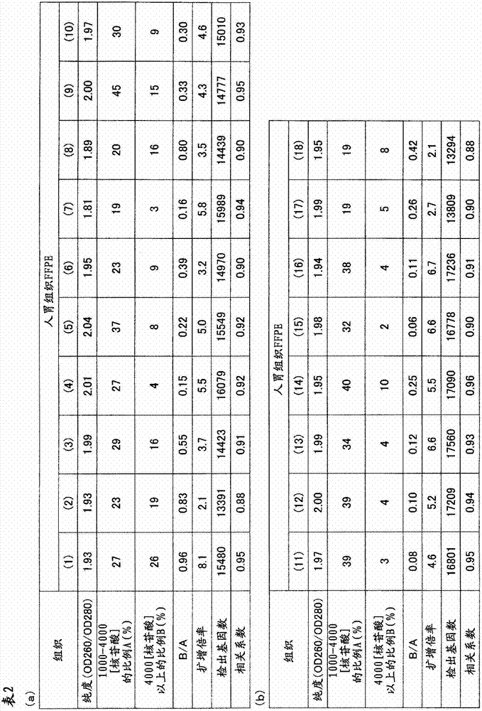
从人胃组织的FFPE标本32个样本中分别采集10μm厚的薄切片,分别装入1.5mL的管中。在其中加入二甲苯1mL进行搅拌,使石蜡溶解。以16,000×g离心5分钟,然后使用移液器避免吸入组织地除去二甲苯。接着,加入乙醇1mL进行搅拌,以16,000×g离心2分钟,然后用移液器避免吸入组织地充分除去乙醇,将该操作进行2次。在打开管盖的状态下风干约10分钟,除去组织所含的乙醇。添加蛋白酶K溶液(500μg/mL)100μL使组织悬浮,在37℃静置16小时。以16,000×g离心2分钟并除去残渣,然后使用二氧化硅柱纯化RNA。通过分光光度计(サ一モサイエンテイフイツク社、“Nano Drop”(注册商标))测定收量和纯度(260nm与280nm之比)的结果,以及通过“Agilent2100 Bioanalyzer”(Agilent Technologies公司)(以下简称为Bioanalyzer)分别计算1000~4000核苷酸的范围的RNA的重量比率A(%)和超过4000核苷酸的范围的RNA的重量比率B(%)的结果,以及由各样品的A、B的值求出B/A的结果示于表2和3;横轴取A、纵轴取B/A作图,将B/A≤1的样品作为“○”表示,将B/A>1的样品作为“×”表示,将所得的图示于图1。Thin slices with a thickness of 10 μm were collected from 32 samples of FFPE specimens of human gastric tissue, and put into 1.5 mL tubes respectively. 1 mL of xylene was added thereto and stirred to dissolve the paraffin. Centrifuge at 16,000 xg for 5 minutes, then remove xylene using a pipette to avoid aspiration of tissue. Next, 1 mL of ethanol was added and stirred, centrifuged at 16,000×g for 2 minutes, and then the ethanol was sufficiently removed with a pipette so as not to suck the tissue, and this operation was performed twice. Air-dry for about 10 minutes with the cap of the tube removed to remove ethanol contained in the tissue. 100 μL of proteinase K solution (500 μg/mL) was added to suspend the tissue, and left to stand at 37° C. for 16 hours. After centrifugation at 16,000 xg for 2 minutes to remove debris, the RNA was purified using a silica column. The results of measuring the yield and purity (ratio of 260nm to 280nm) by a spectrophotometer (Samosai Enteifitsuku Co., Ltd., "Nano Drop" (registered trademark)), and by "Agilent2100 Bioanalyzer" (Agilent Technologies) (hereinafter Abbreviated as Bioanalyzer), the results of calculating the weight ratio A (%) of RNA in the range of 1000 to 4000 nucleotides and the weight ratio B (%) of RNA in the range of more than 4000 nucleotides, and from A, The results of calculating B/A from the value of B are shown in Tables 2 and 3; A is used on the horizontal axis and B/A is taken on the vertical axis, and the samples with B/A≤1 are represented as "○", and B/A> The sample of 1 is shown as "x", and the obtained graph is shown in FIG. 1 .
(RNA的扩增)(Amplification of RNA)
对于B/A≤1的18个样品,分别由1μg使用逆转录酶(“SuperScript(注册商标)III”(Invitrogen公司))合成第一链cDNA,接着添加DNA合成酶合成与第一链DNA互补的第二链cDNA。使用二氧化硅基的柱纯化所合成的cDNA,然后使用T7RNA聚合酶在42℃进行体外转录(IVT)8小时,进行aRNA的扩增反应。此外,本反应时,与参考例1同样地,使用AA-UTP在扩增aRNA中导入氨基烯丙基。使用二氧化硅基的柱纯化所扩增的aRNA。通过扩增后的收量计算出的扩增倍率如表2所述,全部样品都为2倍以上,可知满足B/A≤1的样品是适合本发明的RNA的分析方法的样品。For the 18 samples with B/A≤1, the first-strand cDNA was synthesized from 1 μg of reverse transcriptase (“SuperScript (registered trademark) III” (Invitrogen)), and then DNA synthetase was added to synthesize the complementary first-strand DNA second-strand cDNA. The synthesized cDNA was purified using a silica-based column, followed by in vitro transcription (IVT) using T7 RNA polymerase at 42°C for 8 hours to perform aRNA amplification reaction. In addition, in this reaction, as in Reference Example 1, an aminoallyl group was introduced into amplified aRNA using AA-UTP. The amplified aRNA was purified using a silica-based column. The amplification factor calculated by the yield after amplification is as described in Table 2, and all samples are more than 2 times. It can be seen that the samples satisfying B/A≤1 are suitable for the RNA analysis method of the present invention.
(扩增RNA的荧光标记、断裂)(fluorescence labeling, fragmentation of amplified RNA)
使用离心浓缩机(株式会社トミ一精工、MV-100)浓缩各扩增aRNA溶液至约1μL。在其中添加“3D-Gene(注册商标)Hybridization Buffer”(東レ株式会社)的试剂盒所附带的Sodium Bicarbonate Buffer 5μL通过抽吸搅拌,进一步加入溶解于DMSO的Cy5-NHS(GEヘルスケア社)5μL通过抽吸搅拌,然后在40℃保温1小时,从而进行偶联反应。将各反应溶液使用凝胶过滤离心柱(バイオラツド社)除去未反应的Cy5进行纯化,然后用无核酸酶的水稀释至32μL。在其中分别加入“3D-Gene(注册商标)Hybridization Buffer”(東レ株式会社)的试剂盒所附带的“5×Fragmentation Buffer”各8μL,轻轻抽吸搅拌,在94℃处理15分钟。将各样品用碎冰骤冷3分钟,用“Microcon YM-10”(Millipore公司)纯化。Each amplified aRNA solution was concentrated to about 1 μL using a centrifugal concentrator (Tomi Seiko, MV-100). Add 5 μL of Sodium Bicarbonate Buffer attached to the kit with “3D-Gene (registered trademark) Hybridization Buffer” (Toray Co., Ltd.) and stir by suction, and then add 5 μL of Cy5-NHS (GE ヘルスケア) dissolved in DMSO The coupling reaction was carried out by stirring by suction, followed by incubation at 40° C. for 1 hour. Each reaction solution was purified by removing unreacted Cy5 using a gel filtration spin column (Biorad Corporation), and then diluted to 32 μL with nuclease-free water. Add 8 μL each of “5×Fragmentation Buffer” attached to the kit of “3D-Gene (registered trademark) Hybridization Buffer” (Toray Co., Ltd.), and gently suction and stir, and treat at 94°C for 15 minutes. Each sample was quenched with crushed ice for 3 minutes and purified with "Microcon YM-10" (Millipore).
(杂交)(hybrid)
对于B/A≤1的18个样品,将标记、纯化后的aRNA通过以下操作进行微阵列分析。将包含各1000ng左右的RNA的溶液用无核酸酶水调制成16μL,加入“3D-Gene”(注册商标)Hybridization Buffer(東レ株式会社)的“Hybridization Buffer A”2μL,在95℃热处理5分钟。用碎冰骤冷3分钟,然后加入“Hybridization Buffer B”232μL,平稳地通过抽吸搅拌,调制250μL的样本溶液。将样本溶液在减压下脱气,然后在“3D-Gene(注册商标)小鼠全基因型DNA芯片”(東レ株式会社)中加样210μL。将盖子的4个孔用封条密封,放置于固定在振荡培养箱(东京理化器械株式会社、MMS-210)的桌面上的杂交盒(タカラバイオ株式会社、TX711)中。使盒库内的温度为37℃,一边以250转/分钟旋转进行搅拌,一边反应16小时。For the 18 samples with B/A ≤ 1, the labeled, purified aRNA was subjected to microarray analysis by the following procedure. A solution containing about 1000 ng of each RNA was prepared to 16 μL with nuclease-free water, 2 μL of “Hybridization Buffer A” of “3D-Gene” (registered trademark) Hybridization Buffer (Toray Co., Ltd.) was added, and heat-treated at 95° C. for 5 minutes. Cool with crushed ice for 3 minutes, then add 232 μL of “Hybridization Buffer B”, and stir steadily by suction to prepare 250 μL of sample solution. The sample solution was degassed under reduced pressure, and 210 µL of the sample solution was added to a "3D-Gene (registered trademark) mouse whole-genotype DNA chip" (Toray Co., Ltd.). The four wells of the lid were sealed with seals, and placed in a hybridization box (Takara Bio Co., Ltd., TX711) fixed on the table of a shaking incubator (Tokyo Rikagaku Co., Ltd., MMS-210). The temperature in the cassette chamber was set at 37° C., and the reaction was carried out for 16 hours while rotating and stirring at 250 rpm.
(荧光信号值的测定)(Determination of fluorescence signal value)
将反应后的芯片洗涤后,通过用扫描仪(3D-Gene(注册商标)Scanner(東レ株式会社))测定荧光信号值,从而对有效点数进行计数。其结果如表2所示,是有效点数一样多的结果。而且,对于从同一组织的冻结样品中提取出的RNA进行同样的实验,对于与来源于各FFPE标本的RNA相同的有效点计算出相关系数。这里,相关系数是定量地表示2个数据的相互关系的强度的指标,取-1至1之间,如果是正的值则表示正相关,如果是负的值则表示负相关,如果是零则表示无相关。一般地,如果绝对值为0.5以上则可判断为有相关,如果绝对值小于0.5则可判断无相关,2个数据的相关程度越强,其绝对值越接近1。此外,在用“Microsoft Office Excel”(Microsoft)求相关系数的情况下,使用称为“correl”的函数即可,在本实施例中也使用该软件。各样品中的相关系数如表2所示,在全部样品中,确认了与来源于冻结组织的RNA的高的正相关关系。After the chip after the reaction was washed, the number of effective spots was counted by measuring the fluorescent signal value with a scanner (3D-Gene (registered trademark) Scanner (Toray Corporation)). The result, as shown in Table 2, is the result of the same number of effective points. Furthermore, the same experiment was performed on RNA extracted from the frozen sample of the same tissue, and the correlation coefficient was calculated for the same effective point as that of the RNA derived from each FFPE specimen. Here, the correlation coefficient is an index that quantitatively expresses the strength of the mutual relationship between two data, and it is between -1 and 1. If it is a positive value, it indicates a positive correlation, if it is a negative value, it indicates a negative correlation, and if it is zero, it indicates a positive correlation. Indicates no correlation. Generally, if the absolute value is above 0.5, it can be judged to be correlated, and if the absolute value is less than 0.5, it can be judged to have no correlation. The stronger the correlation between the two data, the closer the absolute value is to 1. In addition, when using "Microsoft Office Excel" (Microsoft) to obtain the correlation coefficient, a function called "correl" may be used, and this software was also used in this example. The correlation coefficients in each sample are shown in Table 2, and a high positive correlation with frozen tissue-derived RNA was confirmed in all samples.
比较例1Comparative example 1
对于B/A>1的14个样品,分别从1μg中与上述同样地作为氨基烯丙基化aRNA(AA-aRNA)进行扩增。根据扩增后的收量计算出的扩增倍率如表3所示,不足2倍的样品是大多数,给RNA的标记化带来了障碍。即可知,B/A>1的样品难以以高的准确率供给RNA的分析。For each of the 14 samples with B/A>1, 1 μg was amplified as aminoallylated aRNA (AA-aRNA) in the same manner as above. The amplification factor calculated based on the yield after amplification is shown in Table 3. Most of the samples were less than 2 times, which brought obstacles to the labeling of RNA. That is, it can be seen that samples with B/A>1 are difficult to analyze RNA with high accuracy.

实施例2Example 2
(从FFPE的RNA提取)(RNA extraction from FFPE)
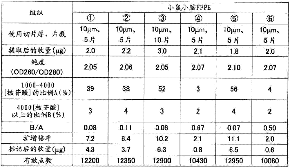
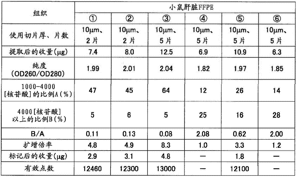
如表5准备制成条件、保存时间不同的小鼠(7周龄、雄、Slc:ICR)小脑、肝脏的福尔马林固定石蜡包埋(FFPE)块。从各FFPE块使用切片机分别采集10μm厚的薄切片10片,在1.5mL的管中各放入5片。在其中加入二甲苯1mL并用旋涡混合器搅拌10秒,使石蜡溶解。以16,000×g离心5分钟,然后使用移液器密切注意避免吸入组织地除去二甲苯。接着,加入乙醇1mL,用旋涡混合器搅拌10秒,以16,000×g离心2分钟,然后用移液器密切注意避免吸入组织地除去乙醇,重复该操作。打开管盖风干约10分钟,除去组织中所含的乙醇。添加蛋白酶K溶液(500μg/mL)100μL使组织悬浮,在37℃静置16小时。以16,000×g离心2分钟使残渣沉淀除去,然后使用二氧化硅柱纯化RNA。用分光光度计(サ一モサイエンテイフイツク社、“Nano Drop”(注册商标))测定收量和纯度(260nm与280nm之比)的结果,以及用Bioanalyzer计算1000~4000核苷酸的范围的RNA的重量比率A(%)和超过4000核苷酸的范围的RNA的重量比率B(%)的结果,以及B/A的值分别示于表4的(a)和(b)。Formalin-fixed paraffin-embedded (FFPE) blocks of the cerebellum and liver of mice (7-week-old, male, Slc: ICR) prepared under different conditions and storage times as shown in Table 5. Ten thin slices with a thickness of 10 μm were collected from each FFPE block using a microtome, and five slices were placed in each 1.5 mL tube. 1 mL of xylene was added thereto and stirred for 10 seconds with a vortex mixer to dissolve the paraffin. Centrifuge at 16,000 xg for 5 minutes, then remove the xylene using a pipette, paying close attention to avoid aspiration of tissue. Next, 1 mL of ethanol was added, stirred with a vortex mixer for 10 seconds, centrifuged at 16,000×g for 2 minutes, and then the ethanol was removed with a pipette so as not to aspirate the tissue, and the operation was repeated. Open the tube cap and air dry for about 10 minutes to remove the ethanol contained in the tissue. 100 μL of proteinase K solution (500 μg/mL) was added to suspend the tissue, and left to stand at 37° C. for 16 hours. Centrifuge at 16,000 x g for 2 minutes to remove the pellet and then purify the RNA using a silica column. The results of measuring yield and purity (ratio of 260nm to 280nm) with a spectrophotometer (Samosai Enteifitsuku Co., Ltd., "Nano Drop" (registered trademark)), and the calculation of the range of 1000 to 4000 nucleotides with Bioanalyzer The results of the weight ratio A (%) of RNA and the weight ratio B (%) of RNA exceeding 4000 nucleotides, and the value of B/A are shown in (a) and (b) of Table 4, respectively.
(RNA的扩增)(Amplification of RNA)
将各RNA样品1μg与实施例1同样地作为氨基烯丙基化aRNA(AA-aRNA)扩增。此时,对于分别从小鼠小脑、肝脏的冻结组织提取出的RNA也同样地进行扩增。将由扩增后的收量计算出的扩增倍率示于表4。与B/A≤1的样品全部扩增倍率为2倍以上相对,B/A>1的样品的扩增倍率不足2倍。1 μg of each RNA sample was amplified as aminoallylated aRNA (AA-aRNA) in the same manner as in Example 1. At this time, RNAs extracted from frozen mouse cerebellum and liver tissues were similarly amplified. Table 4 shows the amplification factor calculated from the yield after amplification. In contrast to samples with B/A ≤ 1, the amplification magnifications of all samples were more than 2 times, and samples with B/A > 1 had amplification magnifications less than 2 times.
(荧光标记、断裂)(fluorescence labeling, fragmentation)
对于上述2倍以上扩增而得的aRNA,与实施例1同样地进行荧光标记、断裂。标记、纯化后的aRNA的收量示于表4。The aRNA amplified by the above-mentioned 2-fold or more was fluorescently labeled and fragmented in the same manner as in Example 1. Table 4 shows the yield of labeled and purified aRNA.
(微阵列分析)(Microarray Analysis)
将包含各1000ng左右的RNA的溶液用无核酸酶水调制成16μL,加入“3D-Gene(注册商标)Hybridization Buffer”(東レ株式会社)的“Hybridization Buffer A”2μL,在95℃热处理5分钟。用碎冰骤冷3分钟,然后加入“Hybridization Buffer B plus”232μL,平稳地通过抽吸搅拌,调制250μL的样本溶液。将样本溶液在减压下脱气,然后在“3D-Gene(注册商标)小鼠全基因型DNA芯片”(東レ株式会社)中加样210μL。将盖子的4个孔用封条密封,放置在固定于振荡培养箱(东京理化器械株式会社、MMS-210)的桌面的杂交盒(タカラバイオ株式会社、TX711)中。盒库内的温度为37℃,一边以250转/分钟旋转进行搅拌,一边使其反应16小时。A solution containing about 1000 ng of each RNA was prepared to 16 μL with nuclease-free water, 2 μL of “Hybridization Buffer A” of “3D-Gene (registered trademark) Hybridization Buffer” (Toray Co., Ltd.) was added, and heat-treated at 95° C. for 5 minutes. Cool with crushed ice for 3 minutes, then add 232 μL of “Hybridization Buffer B plus” and stir steadily by suction to prepare 250 μL of sample solution. The sample solution was degassed under reduced pressure, and 210 µL of the sample solution was added to a "3D-Gene (registered trademark) mouse whole-genotype DNA chip" (Toray Co., Ltd.). The four wells of the lid were sealed with seals, and placed in a hybridization box (Takara Bio Co., Ltd., TX711) fixed to the tabletop of a shaking incubator (Tokyo Rikagaku Co., Ltd., MMS-210). The temperature in the chamber was 37° C., and the reaction was carried out for 16 hours while rotating and stirring at 250 rpm.
(荧光信号值的测定)(Determination of fluorescence signal value)
反应后,使分析用芯片的盖子脱离,将基板洗涤、干燥。将上述基板放置在DNA芯片用的扫描仪(Axon Instruments社、“GenePix(注册商标)4000B”)上,在激光器输出33%、光电倍增管的电压设定为500的状态下,测定进行了杂交反应的荧光标记RNA的信号值(荧光强度)、背景噪音。所有点中,1750个为背景荧光值测定用的阴性对照点,从各个信号值扣除背景信号值而计算出各点的真的信号值。这里,将真的信号值为正的情况作为“有效点”。其结果如表4的(a)和(b)所示,有效点数在各样品之间大致是同等的。After the reaction, the cover of the analysis chip was removed, and the substrate was washed and dried. The substrate was placed on a DNA chip scanner (Axon Instruments, "GenePix (registered trademark) 4000B"), and the hybridization was measured with the laser output at 33% and the voltage of the photomultiplier tube at 500. The signal value (fluorescence intensity) and background noise of the reacted fluorescently labeled RNA. Of all the points, 1750 were negative control points for the determination of the background fluorescence value, and the true signal value of each point was calculated by subtracting the background signal value from each signal value. Here, the case where the true signal value is positive is taken as an "effective point". As a result, as shown in (a) and (b) of Table 4, the number of effective points was almost equal among the samples.
表4Table 4
(a)(a)
(b)(b)
参考例2Reference example 2
对于将从小鼠小脑、肝脏、肾脏和大鼠小脑、肝脏的FFPE中与实施例1同样地分别提取出的RNA用Bioanalyzer在电泳后计算出的RIN、和由各RNA样品1μg进行扩增时的收量的关系,归纳于图2以及表5的(a)~(c)中。其结果是RIN与收量的关系中未见相关关系。而且,也有一部分样品不能计算出RIN(在表5中表示为“N/A”)。由此显示,在从以FFPE为代表的固定组织和/或固定细胞中提取出的RNA的情况下,通过RIN判定这些RNA是否是适于RNA的分析的样品是困难的。RIN calculated after electrophoresis using Bioanalyzer for RNA extracted from FFPE of mouse cerebellum, liver, kidney, and rat cerebellum and liver in the same manner as in Example 1, and when amplified from 1 μg of each RNA sample The relationship of the yield is summarized in Fig. 2 and (a) to (c) of Table 5. As a result, no correlation was found in the relationship between RIN and yield. Furthermore, RIN could not be calculated for some samples (indicated as "N/A" in Table 5). This shows that, in the case of RNA extracted from fixed tissues represented by FFPE and/or fixed cells, it is difficult to determine whether these RNAs are samples suitable for RNA analysis by RIN.

实施例3Example 3
(从固定组织的RNA提取)(RNA Extraction from Fixed Tissue)
对于制成条件、保存时间不同小鼠(7周龄、雄、Slc:ICR),用10%中性缓冲福尔马林如表6准备肝脏的福尔马林固定石蜡包埋(FFPE)块(样品(A)~(D))。从各块使用切片机分别采集10μm厚的薄切片各2片,放入1.5mL的管中。加入二甲苯1mL搅拌,使石蜡溶解。以16,000×g离心5分钟,然后使用移液器避免吸入组织地除去二甲苯。接着,加入乙醇1mL搅拌,以16,000×g离心2分钟,然后用移液器避免吸入组织地充分除去乙醇,将该操作进行2次。在打开管的盖的状态下风干约10分钟,除去组织中所含的乙醇。添加蛋白酶K溶液(500μg/mL)100μL使组织悬浮,在37℃静置16小时。以16,000×g离心2分钟除去残渣,然后使用二氧化硅柱纯化RNA。用分光光度计(サ一モサイエンテイフイツク社、“NanoDrop”(注册商标))测定RNA的收量和纯度(260nm与280nm之比)的结果,以及用Bioanalyzer计算000~4000核苷酸的范围的RNA的重量比率A(%)和超过4000核苷酸的范围的RNA的重量比率B(%)的结果,以及B/A,A+B的值示于表7。此外,作为对照,使用从小鼠(7周龄、雄、Slc:ICR)肝脏的新鲜冻结组织中提取出的RNA。For mice with different production conditions and storage time (7 weeks old, male, Slc: ICR), use 10% neutral buffered formalin as shown in Table 6 to prepare formalin-fixed paraffin-embedded (FFPE) blocks of liver (Samples (A) to (D)). Two 10-μm-thick thin sections were collected from each block using a microtome, and placed in a 1.5-mL tube. Add 1 mL of xylene and stir to dissolve the paraffin. Centrifuge at 16,000 xg for 5 minutes, then remove xylene using a pipette to avoid aspiration of tissue. Next, 1 mL of ethanol was added, stirred, centrifuged at 16,000×g for 2 minutes, and the ethanol was sufficiently removed with a pipette so as not to aspirate the tissue. This operation was performed twice. Air-dry for about 10 minutes with the cap of the tube removed to remove ethanol contained in the tissue. 100 μL of proteinase K solution (500 μg/mL) was added to suspend the tissue, and left to stand at 37° C. for 16 hours. The debris was removed by centrifugation at 16,000 xg for 2 minutes, and the RNA was purified using a silica column. The yield and purity (ratio of 260nm to 280nm) of RNA was measured with a spectrophotometer ("NanoDrop" (registered trademark) from SAMOSIENTIFITSK Co., Ltd.), and the range of 000 to 4000 nucleotides was calculated with Bioanalyzer Table 7 shows the results of the weight ratio A (%) of RNA and the weight ratio B (%) of RNA exceeding 4000 nucleotides, and the values of B/A and A+B. In addition, as a control, RNA extracted from freshly frozen liver tissue of mice (7-week-old, male, Slc:ICR) was used.
(RNA的荧光标记)(fluorescent labeling of RNA)
对于从与实施例1同样的小鼠肝脏FFPE中用同样的方法提取出的RNA,使用“miRCURY LNA microRNA Array Power Labeling kit”(EXIQON公司),按照试剂盒的方案,将各RNA 500ng进行CIP处理,然后通过酶反应标记Hy5色素。For the RNA extracted from the same mouse liver FFPE as in Example 1, 500 ng of each RNA was subjected to CIP treatment using "miRCURY LNA microRNA Array Power Labeling kit" (EXIQON company) according to the protocol of the kit. , and then labeled Hy5 pigment by enzymatic reaction.
(微阵列分析)(Microarray Analysis)
在包含各500ng左右的标记RNA的溶液中加入无核酸酶水,调制成15.4μL,分别加入“3D-Gene(注册商标)miRNA Hybridization Buffer”(東レ株式会社)的Block Reagent 0.6μL、miRNA Hybridization Buffer 105μL混合,在减压下脱气,然后在“3D-Gene(注册商标)小鼠miRNA芯片”(東レ株式会社)中加样110μL。将盖子的4个孔用封条密封,放置在固定于振荡培养箱(东京理化器械株式会社、MMS-210)的桌面上的杂交盒(タカラバイオ株式会社、TX711)中。盒库内的温度为32℃,一边以250转/分钟旋转搅拌,一边反应16小时。Nuclease-free water was added to a solution containing about 500 ng of each labeled RNA to prepare 15.4 μL, and 0.6 μL of Block Reagent and miRNA Hybridization Buffer of “3D-Gene (registered trademark) miRNA Hybridization Buffer” (Toray Co., Ltd.) were added, respectively. 105 μL was mixed, degassed under reduced pressure, and then 110 μL was added to “3D-Gene (registered trademark) mouse miRNA chip” (Toray Co., Ltd.). The four holes of the lid were sealed with seals, and placed in a hybridization box (Takara Bio Co., Ltd., TX711) fixed on the table of a shaking incubator (Tokyo Rikagaku Co., Ltd., MMS-210). The temperature in the cassette was 32° C., and the reaction was carried out for 16 hours while rotating and stirring at 250 rpm.
反应后,使芯片的盖子脱离,将基板洗涤、干燥。在DNA芯片用的扫描仪(Axon Instruments、“GenePix(注册商标)4000B”)上放置上述基板,在激光器输出33%、光电倍增管的电压设定为500的状态下测定进行了杂交反应的荧光标记RNA的信号值(荧光强度)、背景噪音。在所有点中,24个为背景(BG)信号值测定用的阴性对照点,从各个点的信号值中扣除BG信号值而计算出各点的真的信号值。这里,将真的信号值为正的情况作为“有效点”。其结果如表7所示,在B/A≤1的样本中,有效点数与对照几乎同等或略多。另一方面,在B/A>1的样本中,有效点数比对照少,启示存在不能检测的miRNA。因此显示,通过B/A的值可以判定可否分析。After the reaction, the cover of the chip was removed, and the substrate was washed and dried. The substrate was placed on a DNA chip scanner (Axon Instruments, "GenePix (registered trademark) 4000B"), and the fluorescence of the hybridization reaction was measured with the laser output at 33% and the voltage of the photomultiplier tube at 500. Signal value (fluorescence intensity) and background noise of labeled RNA. Among all the points, 24 were negative control points for the determination of the background (BG) signal value, and the true signal value of each point was calculated by subtracting the BG signal value from the signal value of each point. Here, the case where the true signal value is positive is taken as an "effective point". The results are shown in Table 7. In the samples with B/A≤1, the number of effective points is almost equal to or slightly more than that of the control. On the other hand, in the sample with B/A>1, the number of effective points is less than that of the control, suggesting that there are undetectable miRNAs. Therefore, it was shown that the analysis can be judged by the value of B/A.
(与从冻结组织中提取出的RNA的数据比较)(compared to data from RNA extracted from frozen tissue)
进而,对于从同一组织的冻结样品中提取出的RNA进行同样的实验,对于与来源于各FFPE标本的RNA共同的有效点计算相关系数,将结果示于表7。B/A≤1时,确认了与来源于冻结组织的RNA的高的正相关关系。进而,B/A≤1且A为25%以上的样品(C),与同一组织的新鲜冻结样品的相关为0.9以上,显示相关非常高。因此显示,通过B/A的值,可以事先判断可否分析。Furthermore, the same experiment was performed on RNA extracted from frozen samples of the same tissue, and correlation coefficients were calculated for common effective points with RNA derived from each FFPE specimen, and the results are shown in Table 7. When B/A≦1, a high positive correlation with frozen tissue-derived RNA was confirmed. Furthermore, the sample (C) in which B/A≦1 and A was 25% or more showed a very high correlation with the fresh-frozen sample of the same tissue at 0.9 or more. Therefore, it is shown that the possibility of analysis can be judged in advance by the value of B/A.
表6Table 6
表7Table 7
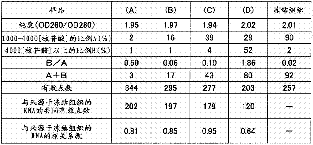
实施例4Example 4
(微阵列的制作)(fabrication of microarray)
使用作为公知的方法的LIGA(光刻电镀成型,LithographieGalvanoformung Abformung)工艺,制作注射成型用的模具,通过注射成型法来获得具有如后所述的形状的PMMA制的基板。此外,该实施例中使用的PMMA的平均分子量为5万,PMMA中以1重量%的比例含有炭黑(三菱化学株式会社、#3050B),因而基板是黑色的。测定该黑色基板的分光反射率和分光透射率,结果分光反射率在可见光区(波长为400nm~800nm)的任一波长下均为5%以下,另外,在同范围的波长下透射率为0.5%以下。分光反射率、分光透射率在可见光区均无特定的光谱图案(峰等),光谱是一样地平坦。此外,分光反射率使用担载了适合JIS Z 8722的条件C的照明-受光光学系统的装置(ミノルタカメラ、CM-2002),测定接受从基板的正反射光时的分光反射率。A mold for injection molding was fabricated using LIGA (Lithographie Galvanoformung Abformung) process, which is a known method, and a PMMA substrate having a shape as described later was obtained by injection molding. In addition, the average molecular weight of PMMA used in this example was 50,000, and carbon black (Mitsubishi Chemical Corporation, #3050B) was contained in the ratio of 1% by weight in PMMA, so the substrate was black. The spectral reflectance and spectral transmittance of the black substrate were measured. As a result, the spectral reflectance was 5% or less at any wavelength in the visible light region (wavelength 400nm to 800nm), and the transmittance was 0.5% at wavelengths in the same range. %the following. Neither the spectral reflectance nor the spectral transmittance has a specific spectral pattern (peak, etc.) in the visible region, and the spectrum is equally flat. In addition, the spectral reflectance was measured using an apparatus (Minoltacamera, CM-2002) equipped with an illumination-light receiving optical system conforming to the condition C of JIS Z 8722, and measured the spectral reflectance when receiving specularly reflected light from the substrate.
作为基板,使用外形为纵76mm、横26mm、厚1mm,基板的中央部设有纵39.4mm、横19.0mm、深0.15mm的凹部,该凹部中设有9248个直径0.1mm、高0.15mm的凸部的聚甲基丙烯酸甲酯(PMMA)树脂制基板(以下作为“基板A”)。在该基板A中,凸部上面与平坦部上面的高的差(凸部为高的平均值)为3μm以下。另外,凸部上面的高的偏差(最高的凸部上面的高与最低的凸部上面的高的差)为3μm以下。另外,凸部的间距(从凸部中央部至相邻的凸部中央部的距离)为0.5mm。As the substrate, the outer shape is 76 mm in length, 26 mm in width, and 1 mm in thickness. The central part of the substrate is provided with a concave portion of 39.4 mm in length, 19.0 mm in width, and 0.15 mm in depth. A substrate made of polymethyl methacrylate (PMMA) resin (hereinafter referred to as "substrate A") for the protrusions. In this substrate A, the difference in height between the upper surface of the convex portion and the upper surface of the flat portion (the average value of the height of the convex portion) was 3 μm or less. In addition, the variation in the height of the top surface of the convex portion (the difference between the height of the top surface of the highest convex portion and the height of the top surface of the lowest convex portion) is 3 μm or less. In addition, the pitch of the convex portions (the distance from the central portion of the convex portion to the central portion of the adjacent convex portion) was 0.5 mm.
将上述基板A在10N的氢氧化钠水溶液中在70℃的温度下浸渍12小时。将该基板A依次用纯水、0.1N的HCl水溶液、纯水洗涤,使基板表面生成羧基。The aforementioned substrate A was immersed in a 10N aqueous sodium hydroxide solution at a temperature of 70° C. for 12 hours. This substrate A was sequentially washed with pure water, 0.1N aqueous HCl solution, and pure water to generate carboxyl groups on the surface of the substrate.
对如上所述准备的基板A,在以下条件下作为选择结合性物质(探针DNA)固定化正义链寡核苷酸。作为寡核苷酸,使用オペロン社制DNA微阵列用寡核苷酸组“Homo sapiens(Human)AROS V4.0(各60碱基)”。将该寡核苷酸以0.3nmol/μL的浓度溶解在纯水中制成储备溶液。将该储备溶液点(点样)于基板时,用PBS(将8g的NaCl、2.9g的Na2HPO4·12H2O、0.2g的KCl、和0.2g的KH2PO4合并溶于纯水中,稀释至1L,加入盐酸调节至pH值5.5)稀释10倍,使探针DNA的终浓度为0.03nmol/μL,且为了使PMMA制基板表面所生成的羧基与探针DNA的末端氨基缩合,加入1-乙基-3-(3-二甲基氨基丙基)碳二亚胺(EDC),使其终浓度为50mg/mL。将该溶液使用微阵列点样仪(点样仪)(日本激光器电子、Gene Stamp-II)点样在基板A的全部凸部上面。接着,将点有溶液的基板放入密闭的塑料容器,在温度37℃、湿度100%的条件下保温20小时左右。最后,用纯水洗涤基板,用旋干机离心干燥。On the substrate A prepared as described above, a sense strand oligonucleotide was immobilized as a selective binding substance (probe DNA) under the following conditions. As the oligonucleotide, the oligonucleotide set "Homo sapiens (Human) AROS V4.0 (each 60 bases)" for DNA microarray manufactured by Opelon was used. This oligonucleotide was dissolved in pure water at a concentration of 0.3 nmol/µL to prepare a stock solution. When the stock solution is spotted (spotted) on the substrate, use PBS (the NaCl of 8g, the Na2HPO4 · 12H2O of 2.9g, the KCl of 0.2g, and the KH2PO4 of 0.2g are combined and dissolved in pure Dilute to 1L in water, add hydrochloric acid to adjust to pH 5.5) Dilute 10 times, make the final concentration of probe DNA 0.03nmol/μL, and in order to make the carboxyl group generated on the surface of the PMMA substrate and the terminal amino group of the probe DNA After condensation, 1-ethyl-3-(3-dimethylaminopropyl)carbodiimide (EDC) was added to make the
对固定化有选择结合性物质的上述基板A,如下贴合盖子。作为盖子,将纵41.4mm、横21mm、厚1mm的PMMA平板通过切削加工制作成盖子。所制作的盖子上设有贯通孔和液面停止用腔。贯通孔的直径为0.8mm,液面停止用腔的直径为2.0mm,设置在盖子的四角。将作为粘贴部件的纵41.4mm、横21mm、宽1mm、厚25μm的双面胶带裁成镶盖子的边缘的尺寸,叠层成厚(空气间隙)为50μm并贴合,然后将该盖子贴合于基板A。To the above-mentioned substrate A on which a selective binding substance is immobilized, a cover is attached as follows. As the cover, a PMMA flat plate with a length of 41.4 mm, a width of 21 mm, and a thickness of 1 mm was produced by cutting. The manufactured cover is provided with a through hole and a chamber for stopping the liquid level. The diameter of the through hole is 0.8 mm, and the diameter of the liquid level stop chamber is 2.0 mm, which are provided at the four corners of the cover. A double-sided adhesive tape with a length of 41.4 mm, a width of 21 mm, a width of 1 mm, and a thickness of 25 μm is cut to fit the size of the edge of the cover, and laminated so that the thickness (air gap) is 50 μm, and then the cover is attached. on substrate A.
对如上所述贴合了盖子的基板A,将直径180μm的氧化锆制微粒120mg封入由基板A与盖子形成的空间部(基板A表面的凹凸结构的凹部)。微粒的封入从盖子的贯通孔进行。将如上获得的分析用芯片用于以下的研究。For the substrate A with the cover bonded as described above, 120 mg of zirconia fine particles with a diameter of 180 μm were enclosed in the space formed by the substrate A and the cover (recesses of the uneven structure on the surface of the substrate A). Encapsulation of fine particles is performed through the through hole of the cap. The analytical chip obtained above was used for the following studies.
(从固定组织的RNA提取)(RNA Extraction from Fixed Tissue)
与实施例3同样地,从制成条件、保存时间不同的小鼠(7周龄、雄、Slc:ICR)肝脏的福尔马林固定石蜡包埋(FFPE)块制作薄切片,提取RNA。用分光光度计(サ一モサイエンテイフイツク社、“Nano Drop”(注册商标))测定收量和纯度(260nm与280nm之比)的结果,以及用Bioanalyzer计算1000~4000核苷酸的范围的RNA的重量比率A(%)和超过4000核苷酸的范围的RNA的重量比率B(%)的结果,以及B/A、A+B的值示于表8。此外,作为对照,使用从小鼠(7周龄、雄、Slc:ICR)肝脏的新鲜冻结组织中提取出的RNA。In the same manner as in Example 3, thin sections were prepared from formalin-fixed paraffin-embedded (FFPE) blocks of mouse (7-week-old, male, Slc:ICR) livers with different production conditions and storage times, and RNA was extracted. The results of measuring yield and purity (ratio of 260nm to 280nm) with a spectrophotometer (Samosai Enteifitsuku Co., Ltd., "Nano Drop" (registered trademark)), and the calculation of the range of 1000 to 4000 nucleotides with Bioanalyzer Table 8 shows the results of the weight ratio A (%) of RNA and the weight ratio B (%) of RNA exceeding 4000 nucleotides, and the values of B/A and A+B. In addition, as a control, RNA extracted from freshly frozen liver tissue of mice (7-week-old, male, Slc:ICR) was used.
(RNA的荧光标记)(fluorescent labeling of RNA)
将与实施例3同样地提取出的RNA使用“PlatinumBright(注册商标)Nucleic Acid Labeling Kit”(KREATECH)进行荧光标记。将各RNA 1μg用Nuclease Free Water稀释至16μL,加入ULS试剂和10×labeling solution各2μL,在85℃反应30分钟,对mRNA标记PlatinmBright647色素。使用附带的“KREApure columns”除去未反应的色素。The RNA extracted in the same manner as in Example 3 was fluorescently labeled using "PlatinumBright (registered trademark) Nucleic Acid Labeling Kit" (KREATECH). Dilute 1 μg of each RNA to 16 μL with Nuclease Free Water, add 2 μL each of ULS reagent and 10×labeling solution, and react at 85°C for 30 minutes to label the mRNA with PlatinmBright647 pigment. Use the included "KREApure columns" to remove unreacted pigments.
(微阵列分析)(Microarray Analysis)
对于各固定组织所含的mRNA通过以下步骤用微阵列进行分析。在包含各1μg左右的标记RNA的溶液中加入无核酸酶水调制成16μL,分别加入“3D-Gene(注册商标)Hybridization Buffer”(東レ株式会社)的Hybridization Buffer A 2μL、Hybridization Buffer B 232μL混合,在减压下脱气,然后在上述制作的分析用芯片中加样210μL。将盖子的4个孔用封条密封,放置于固定在振荡培养箱(东京理化器械株式会社;MMS-210)的桌面上的杂交盒(タカラバイオ株式会社;TX711)中。使盒库内的温度为37℃,一边以250转/分钟旋转搅拌,一边反应16小时。The mRNA contained in each fixed tissue was analyzed by microarray in the following procedure. Add nuclease-free water to a solution containing about 1 μg of each labeled RNA to prepare 16 μL, add 2 μL of Hybridization Buffer A and 232 μL of Hybridization Buffer B of “3D-Gene (registered trademark) Hybridization Buffer” (Toray Co., Ltd.), and mix. After degassing under reduced pressure, 210 µL of the sample was added to the analysis chip prepared above. The four holes of the lid were sealed with seals, and placed in a hybridization box (Takara Bio Co., Ltd.; TX711) fixed on the table of a shaking incubator (Tokyo Rikagaku Co., Ltd.; MMS-210). The temperature in the cassette chamber was set at 37° C., and the reaction was carried out for 16 hours while rotating and stirring at 250 rpm.
(荧光信号值的测定)(Determination of fluorescence signal value)
反应后使芯片的盖子脱离,将基板洗涤、干燥。在DNA芯片用的扫描仪(Axon Instruments社、“GenePix(注册商标)4000B”)上放置上述基板,在激光器输出33%、光电倍增管的电压设定为500的状态下测定进行了杂交反应的荧光标记RNA的信号值(荧光强度)、背景噪音。所有点中的32个为背景荧光值测定用的阴性对照点,从各个信号值中扣除背景信号值而计算出各点的真的信号值。这里,将真的信号值为正的情况作为“有效点”。其结果如表8所示,在B/A≤1的样本中,确认了与从新鲜冻结小鼠肝脏组织中提取出的参考RNA相比有效点数的差异小、而且两者的共同有效点中的相关高的倾向。而且,A的值越大,该倾向越显著。另一方面,在B/A>1的样本中,有效点数比参考RNA少,启示存在不能检测的mRNA。After the reaction, the cover of the chip was removed, and the substrate was washed and dried. The above-mentioned substrate was placed on a scanner for DNA chips (Axon Instruments, "GenePix (registered trademark) 4000B"), and the hybridization reaction was measured under the condition that the laser output was 33% and the voltage of the photomultiplier tube was set to 500. Signal value (fluorescence intensity) and background noise of fluorescently labeled RNA. 32 of all the points are negative control points for the determination of the background fluorescence value, and the true signal value of each point is calculated by subtracting the background signal value from each signal value. Here, the case where the true signal value is positive is taken as an "effective point". The results are shown in Table 8. In samples with B/A ≤ 1, it was confirmed that the difference in the number of effective points was small compared with the reference RNA extracted from freshly frozen mouse liver tissue, and that the common effective points of the two tends to be highly correlated. And, the larger the value of A, the more remarkable this tendency is. On the other hand, in the sample with B/A>1, the number of effective spots is smaller than that of the reference RNA, suggesting that there is undetectable mRNA.
(与从冻结组织中提取出的RNA的数据比较)(compared to data from RNA extracted from frozen tissue)
进而,对于从同一组织的冻结样品中提取出的RNA进行同样的实验,对于与来源于各FFPE标本的RNA共同的mRNA有效点计算出相关系数。各样品中的相关系数如表8所示,当B/A≤1时,确认了与来源于冻结组织的RNA的高的正相关关系。而且,对于B/A≤1且A为25%以上的样品(C),与同一组织的新鲜冻结样品的相关为0.9以上,显示相关非常高。因此显示,根据B/A的值可以判断可否分析。Furthermore, the same experiment was performed on RNA extracted from frozen samples of the same tissue, and correlation coefficients were calculated for mRNA effective points common to RNA derived from each FFPE specimen. The correlation coefficients in each sample are shown in Table 8, and when B/A≦1, a high positive correlation with frozen tissue-derived RNA was confirmed. Furthermore, for the sample (C) with B/A≦1 and A being 25% or more, the correlation with the fresh-frozen sample of the same tissue was 0.9 or more, indicating a very high correlation. Therefore, it is shown that the analysis can be judged according to the value of B/A.
表8Table 8
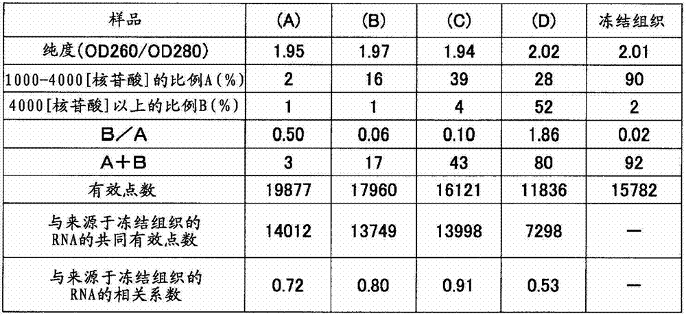
实施例5Example 5
从小鼠小脑和肝脏组织的FFPE标本分别制成10μm厚的切片,通过与实施例1同样的方法提取RNA。与实施例2同样地,与小鼠小脑和肝脏的冻结组织中提取出的RNA一起,从RNA1μg进行扩增反应,得到aRNA。RNA的收量、纯度、利用Bioanalyzer得出的1000~4000核苷酸的范围的RNA的重量比率A(%)和超过4000核苷酸的范围的RNA的重量比率B(%)、以及扩增倍率示于表9。将aRNA用Cy3(GEヘルスケア社)标记,与“Gene Expression Hybridization Kit”(Agilent Technologies公司)的杂交缓冲液混合,在“Whole Mouse Genome Oligo Microarray(4×44k)”(AgilentTechnologies公司)上杂交40小时。洗涤后,用“Agilent MicroarrayScanner”(Agilent Technologies公司)读取DNA微阵列的图像,用“FeatureExtraction Software(v.9.5.3.1)”将各点的荧光信号数值化。其结果如表9所示,显示相关系数高达小脑:0.88、肝脏:0.85。10 μm thick sections were made from FFPE specimens of mouse cerebellum and liver tissues, and RNA was extracted by the same method as in Example 1. In the same manner as in Example 2, 1 μg of RNA was amplified together with RNA extracted from frozen mouse cerebellum and liver tissues to obtain aRNA. RNA yield, purity, weight ratio A (%) of RNA in the range of 1000 to 4000 nucleotides and weight ratio B (%) of RNA in the range of more than 4000 nucleotides by Bioanalyzer, and amplification The magnification is shown in Table 9. The aRNA was labeled with Cy3 (GE ヘルスケア), mixed with the hybridization buffer of "Gene Expression Hybridization Kit" (Agilent Technologies), and hybridized on "Whole Mouse Genome Oligo Microarray (4×44k)" (Agilent Technologies) for 40 Hour. After washing, "Agilent MicroarrayScanner" (Agilent Technologies) was used to read the image of the DNA microarray, and "FeatureExtraction Software (v.9.5.3.1)" was used to digitize the fluorescence signal of each point. The results are shown in Table 9, showing that the correlation coefficients are as high as cerebellum: 0.88 and liver: 0.85.
表9Table 9
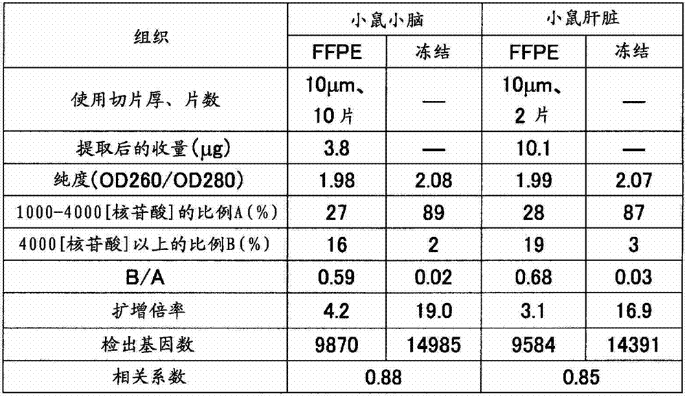
实施例6Example 6
使用实施例5所提取的来源于小鼠小脑、肝脏的FFPE的RNA、来源于冻结组织的RNA,与实施例1同样地由各RNA 5μg进行扩增反应得到aRNA。此时,导入生物素基代替氨基烯丙基。扩增倍率示于表10。将生物素化aRNA与规定量的Control Oligonucleotide、20×EukaryoticHybridization Controls、2×Hybridization Mix、DMSO、Nuclease-freeWater混合,在99℃处理5分钟,然后在45℃处理5分钟。进而,以16,000×g离心5分钟,然后上样到“Affymetrix(注册商标)Mouse Genome 430 2.0Array”(アフイメトリクス社)中杂交反应16小时。用规定的方法洗涤、染色,然后用“GeneChip(注册商标)Scanner 3000 7G System”(アフイメトリクス社)将各点的荧光信号数值化。其结果如表10所示,与冻结组织相比,FFPE的有效点数倾向于较少,但两者的相关系数为小脑:0.84、肝脏:0.82,确认了高的相关关系。Using the FFPE-derived RNAs from the mouse cerebellum and liver extracted in Example 5, and the RNAs derived from frozen tissues, amplification reactions were performed from 5 μg of each RNA in the same manner as in Example 1 to obtain aRNA. At this time, a biotin group was introduced instead of an aminoallyl group. The amplification factor is shown in Table 10. Mix biotinylated aRNA with specified amount of Control Oligonucleotide, 20×Eukaryotic Hybridization Controls, 2×Hybridization Mix, DMSO, Nuclease-freeWater, and treat at 99°C for 5 minutes, then at 45°C for 5 minutes. Furthermore, after centrifugation at 16,000×g for 5 minutes, the samples were loaded onto “Affymetrix (registered trademark) Mouse Genome 430 2.0 Array” (Affymetrix Corporation) and hybridized for 16 hours. After washing and staining according to the prescribed method, the fluorescence signal of each spot was quantified using "GeneChip (registered trademark) Scanner 3000 7G System" (Afimetricks Co., Ltd.). The results are shown in Table 10. Compared with frozen tissue, FFPE tends to have fewer effective points, but the correlation coefficients between the two were 0.84 for cerebellum and 0.82 for liver, confirming a high correlation.
表10Table 10
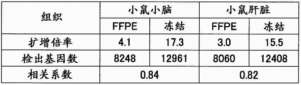
实施例7Example 7
(从固定组织的RNA提取)(RNA Extraction from Fixed Tissue)
摘取小鼠(7周龄、雄、Slc:ICR)小脑和肝脏,在10%磷酸缓冲福尔马林溶液(甲醛4%)中在室温浸渍2天进行固定后,进行石蜡包埋而制作FFPE块。从各FFPE块使用切片机分别采集10μm厚的薄切片,与实施例1同样地提取RNA。用分光光度计(サ一モサイエンテイフイツク社、“Nano Drop”(注册商标))测定收量和纯度(260nmと280nm之比)的结果、以及用Bioanalyzer计算1000~4000核苷酸的范围的RNA的重量比率A(%)和超过4000核苷酸的范围的RNA的重量比率B(%)的结果、以及B/A的值示于表11。在全部样品中B/A≤1。The cerebellum and liver of a mouse (7 weeks old, male, Slc: ICR) were excised, fixed by immersion in 10% phosphate-buffered formalin solution (4% formaldehyde) at room temperature for 2 days, and then embedded in paraffin FFPE blocks. Thin sections with a thickness of 10 μm were collected from each FFPE block using a microtome, and RNA was extracted in the same manner as in Example 1. Yield and purity (ratio of 260nm to 280nm) measured with a spectrophotometer (Samosai Enteifitsuku Co., Ltd., "Nano Drop" (registered trademark)), and the range of 1000 to 4000 nucleotides calculated with Bioanalyzer Table 11 shows the results of the weight ratio A (%) of RNA and the weight ratio B (%) of RNA exceeding 4000 nucleotides, and the value of B/A. B/A≦1 in all samples.
(RNA的扩增)(Amplification of RNA)
由提取出的小鼠小脑、肝脏的RNA 1μg与实施例1同样地进行扩增,获得了导入有氨基烯丙基的aRNA。用分光光度计(サ一モサイエンテイフイツク社、“Nano Drop”(注册商标))求出收量,计算出扩增倍率,将结果示于表11。在全部样品中扩增倍率在2~20倍的范围内。From 1 μg of the extracted mouse cerebellum and liver RNA, amplification was performed in the same manner as in Example 1 to obtain aminoallyl group-introduced aRNA. The yield was determined using a spectrophotometer ("Nano Drop" (registered trademark) from Thermo Scientific Co., Ltd.), and the amplification factor was calculated. The results are shown in Table 11. The amplification factor in all samples was in the range of 2-20 times.
(扩增RNA的荧光标记、断裂、微阵列分析)(fluorescence labeling, fragmentation, microarray analysis of amplified RNA)
对于扩增的各aRNA,与实施例1同样地进行荧光标记、断裂,然后通过以下操作进行微阵列分析。将包含各1000ng左右的RNA的溶液用无核酸酶水调制成16μL,加入“3D-Gene”(注册商标)Hybridization Buffer(東レ株式会社)的“Hybridization Buffer A”2μL,在95℃热处理5分钟。用碎冰骤冷3分钟,然后加入“Hybridization Buffer B”232μL,平稳地通过抽吸搅拌,调制250μL的样本溶液。将样本溶液在减压下脱气,然后在“3D-Gene(注册商标)小鼠全基因型DNA芯片”(東レ株式会社)中加样210μL。将盖子的4个孔用封条密封,放置于固定在振荡培养箱(东京理化器械株式会社、MMS-210)的桌面上的杂交盒(タカラバイオ株式会社、TX711)中。使盒库内的温度为37℃,一边以250转/分钟旋转搅拌,一边反应16小时。Each of the amplified aRNAs was fluorescently labeled and fragmented in the same manner as in Example 1, and microarray analysis was performed by the following procedure. A solution containing about 1000 ng of each RNA was prepared to 16 μL with nuclease-free water, 2 μL of “Hybridization Buffer A” of “3D-Gene” (registered trademark) Hybridization Buffer (Toray Co., Ltd.) was added, and heat-treated at 95° C. for 5 minutes. Cool with crushed ice for 3 minutes, then add 232 μL of “Hybridization Buffer B”, and stir steadily by suction to prepare 250 μL of sample solution. The sample solution was degassed under reduced pressure, and 210 µL of the sample solution was added to a "3D-Gene (registered trademark) mouse whole-genotype DNA chip" (Toray Co., Ltd.). The four wells of the lid were sealed with seals, and placed in a hybridization box (Takara Bio Co., Ltd., TX711) fixed on the table of a shaking incubator (Tokyo Rikagaku Co., Ltd., MMS-210). The temperature in the cassette chamber was set at 37° C., and the reaction was carried out for 16 hours while rotating and stirring at 250 rpm.
(荧光信号值的测定)(Determination of fluorescence signal value)
反应后,使分析用芯片的盖子脱离,将基板洗涤、干燥。在DNA芯片用的扫描仪(Axon Instruments社、“GenePix(注册商标)4000B”)上安装上述基板,在激光器输出33%、光电倍增管的电压设定为500的状态下测定进行了杂交反应的荧光标记RNA的信号值(荧光强度)、背景噪音。在全部点中,1750个为背景荧光值测定用的阴性对照点,由各个信号值扣除背景信号值而计算出各点的真的信号值。这里,将真的信号值为正的情况作为“有效点”。有效点数示于表11。显示同一组织中的有效点数的方差较小。进一步将由小脑、肝脏的各基因的信号的比(小脑/肝脏)制成的散点图示于图3。由该结果显示,2次实验的相关较高。After the reaction, the cover of the analysis chip was removed, and the substrate was washed and dried. The above-mentioned substrate was mounted on a scanner for DNA chips (Axon Instruments, "GenePix (registered trademark) 4000B"), and the hybridization reaction was measured under the condition that the laser output was 33% and the voltage of the photomultiplier tube was set to 500. Signal value (fluorescence intensity) and background noise of fluorescently labeled RNA. Among all the points, 1750 were negative control points for measuring the background fluorescence value, and the true signal value of each point was calculated by subtracting the background signal value from each signal value. Here, the case where the true signal value is positive is taken as an "effective point". Valid points are shown in Table 11. Shows less variance in valid points within the same organization. Furthermore, a scattergram of the signal ratio (cerebellum/liver) of each gene in the cerebellum and liver is shown in FIG. 3 . This result shows that the correlation between the two experiments is high.
表11Table 11
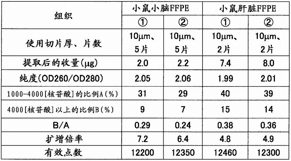
产业可利用性industry availability
通过使用本发明的RNA的分析方法,可以获得关于福尔马林固定石蜡包埋组织等、在医院和/或研究机构以极大数量保存的固定组织或细胞的基因的表达增减和/或表达有无的正确的信息,这些信息可以在医药品开发和/或基因检查、基因诊断技术等中广泛地灵活运用。而且,可以事先预见分析困难的样品,从分析对象中排除,从而还可以实现试剂等的成本削减,因此在产业上非常有用。By using the RNA analysis method of the present invention, it is possible to obtain the expression increase and decrease and/or the gene expression of fixed tissue or cells preserved in a large number of formalin-fixed paraffin-embedded tissues, etc., in hospitals and/or research institutions. The presence or absence of accurate information can be expressed, and this information can be widely used in drug development, genetic testing, and genetic diagnosis techniques. In addition, samples that are difficult to analyze can be predicted in advance and excluded from analysis targets, and cost reduction of reagents and the like can be realized, so it is very useful industrially.
Claims (11)
Applications Claiming Priority (7)
| Application Number | Priority Date | Filing Date | Title |
|---|---|---|---|
| JP2009285098 | 2009-12-16 | ||
| JP285098/2009 | 2009-12-16 | ||
| JP047557/2010 | 2010-03-04 | ||
| JP2010047557 | 2010-03-04 | ||
| JP2010239513A JP4807470B2 (en) | 2009-12-16 | 2010-10-26 | RNA analysis method |
| JP239513/2010 | 2010-10-26 | ||
| PCT/JP2010/072518 WO2011074592A1 (en) | 2009-12-16 | 2010-12-15 | Method for analyzing rna |
Publications (2)
| Publication Number | Publication Date |
|---|---|
| CN102656280A CN102656280A (en) | 2012-09-05 |
| CN102656280B true CN102656280B (en) | 2014-01-22 |
Family
ID=44167343
Family Applications (1)
| Application Number | Title | Priority Date | Filing Date |
|---|---|---|---|
| CN201080056692.1A Expired - Fee Related CN102656280B (en) | 2009-12-16 | 2010-12-15 | Method for analyzing RNA |
Country Status (9)
| Country | Link |
|---|---|
| US (1) | US9206472B2 (en) |
| EP (1) | EP2514834B1 (en) |
| JP (1) | JP4807470B2 (en) |
| KR (1) | KR101753434B1 (en) |
| CN (1) | CN102656280B (en) |
| BR (1) | BR112012011530A2 (en) |
| CA (1) | CA2784837C (en) |
| RU (1) | RU2556372C2 (en) |
| WO (1) | WO2011074592A1 (en) |
Families Citing this family (2)
| Publication number | Priority date | Publication date | Assignee | Title |
|---|---|---|---|---|
| EP3421610B1 (en) | 2016-02-22 | 2021-10-20 | Toray Industries, Inc. | METHOD FOR EVALUATING QUALITY OF miRNA DERIVED FROM BODY FLUID |
| KR20220094440A (en) | 2020-12-29 | 2022-07-06 | 주식회사 제이디케이바이오 | light cultivating device for adherent diatoms |
Citations (1)
| Publication number | Priority date | Publication date | Assignee | Title |
|---|---|---|---|---|
| WO2004090780A2 (en) * | 2003-04-05 | 2004-10-21 | Agilent Technologies, Inc. | Determining the quality of biomolecule samples |
Family Cites Families (13)
| Publication number | Priority date | Publication date | Assignee | Title |
|---|---|---|---|---|
| WO2004085681A2 (en) | 2003-03-21 | 2004-10-07 | Arcturus Bioscience, Inc. | Global linear non-biased nucleic acid amplification |
| ES2360113T3 (en) | 2003-12-23 | 2011-06-01 | Genomic Health, Inc. | UNIVERSAL AMPLIFICATION OF FRAGMENTED RNA. |
| JP2005224172A (en) | 2004-02-13 | 2005-08-25 | Sumitomo Pharmaceut Co Ltd | Novel linear RNA amplification method |
| CA2607454A1 (en) * | 2005-05-03 | 2006-11-09 | Althea Technologies, Inc. | Compositions and methods for the analysis of degraded nucleic acids |
| AU2006270063A1 (en) | 2005-07-15 | 2007-01-25 | Applied Biosystems, Llc. | Methods for RNA profiling |
| WO2007053491A2 (en) * | 2005-10-28 | 2007-05-10 | The Trustees Of Columbia University In The City Of New York | Method and kit for evaluating rna quality |
| JP2008035779A (en) | 2006-08-07 | 2008-02-21 | Mitsubishi Rayon Co Ltd | Method for measuring nucleic acid degradation and nucleic acid array |
| JP2008043332A (en) | 2006-08-17 | 2008-02-28 | Panomics Inc | Quantitative determination of nucleic acid from tissue slide |
| EP2262893B1 (en) * | 2008-04-15 | 2013-04-24 | Universität Bern | Method for optimized isolation of rna from fixed tissue |
| JP5339041B2 (en) * | 2008-08-25 | 2013-11-13 | 学校法人武庫川学院 | Novel naphthoquinone compounds and their pharmaceutical uses |
| US9150909B2 (en) | 2008-08-29 | 2015-10-06 | Bio-Rad Laboratories, Inc. | Determination of the integrity of RNA |
| JP5249111B2 (en) * | 2009-03-31 | 2013-07-31 | オリンパス株式会社 | Image processing apparatus, method, program, and imaging system |
| JP2010252754A (en) * | 2009-04-28 | 2010-11-11 | Toray Ind Inc | Method for analyzing rna |
-
2010
- 2010-10-26 JP JP2010239513A patent/JP4807470B2/en active Active
- 2010-12-15 WO PCT/JP2010/072518 patent/WO2011074592A1/en not_active Ceased
- 2010-12-15 US US13/515,377 patent/US9206472B2/en not_active Expired - Fee Related
- 2010-12-15 CN CN201080056692.1A patent/CN102656280B/en not_active Expired - Fee Related
- 2010-12-15 CA CA2784837A patent/CA2784837C/en not_active Expired - Fee Related
- 2010-12-15 EP EP10837618.7A patent/EP2514834B1/en not_active Not-in-force
- 2010-12-15 BR BR112012011530A patent/BR112012011530A2/en not_active IP Right Cessation
- 2010-12-15 KR KR1020127014100A patent/KR101753434B1/en not_active Expired - Fee Related
- 2010-12-15 RU RU2012129970/10A patent/RU2556372C2/en not_active IP Right Cessation
Patent Citations (1)
| Publication number | Priority date | Publication date | Assignee | Title |
|---|---|---|---|---|
| WO2004090780A2 (en) * | 2003-04-05 | 2004-10-21 | Agilent Technologies, Inc. | Determining the quality of biomolecule samples |
Non-Patent Citations (2)
| Title |
|---|
| "RNA extraction from ten years old formalin-fixed paraffin-embedded breast cancer samples: a comparison of column purification and magnetic bead-based technologies";Alfredo Ribeiro-silva et al.;《BMC molecular biology》;20071221;第8卷;e56第1-12页 * |
| "Towards standardization of RNA quality assessment using user-independent classifiers of microcapillary electrophoresis traces";Sandrine Imbeaud et al.;《Nucleic acids research》;20050330;第33卷(第6期);118第1-10页 * |
Also Published As
| Publication number | Publication date |
|---|---|
| EP2514834B1 (en) | 2016-03-09 |
| RU2556372C2 (en) | 2015-07-10 |
| JP4807470B2 (en) | 2011-11-02 |
| KR20120105460A (en) | 2012-09-25 |
| RU2012129970A (en) | 2014-01-27 |
| CA2784837A1 (en) | 2011-06-23 |
| CA2784837C (en) | 2017-10-10 |
| EP2514834A4 (en) | 2013-08-28 |
| CN102656280A (en) | 2012-09-05 |
| EP2514834A1 (en) | 2012-10-24 |
| US9206472B2 (en) | 2015-12-08 |
| WO2011074592A1 (en) | 2011-06-23 |
| BR112012011530A2 (en) | 2019-09-24 |
| KR101753434B1 (en) | 2017-07-03 |
| US20120253687A1 (en) | 2012-10-04 |
| JP2011200220A (en) | 2011-10-13 |
Similar Documents
| Publication | Publication Date | Title |
|---|---|---|
| US20220170083A1 (en) | Rna integrity analysis in a biological sample | |
| CN115715329A (en) | Method for determining the position of a target nucleic acid in a biological sample | |
| JP2007502116A (en) | Methods and kits for preparing nucleic acid samples | |
| US20070172841A1 (en) | Probe/target stabilization with add-in oligo | |
| US8535914B2 (en) | Probe, probe set and information acquisition method using the same | |
| CN102656280B (en) | Method for analyzing RNA | |
| US9145582B2 (en) | Microarray techniques for nucleic acid expression analyses | |
| US20050064472A1 (en) | Methods of monitoring gene expression | |
| Bibikova et al. | Quantitative expression profiling of RNA from formalin-fixed, paraffin-embedded tissues using randomly assembled bead arrays | |
| US10622093B2 (en) | Method and device for correcting level of expression of small RNA | |
| EP2484760B1 (en) | METHOD FOR PREPARING aRNA. | |
| US20070099193A1 (en) | Probe/target stabilization with add-in oligo | |
| JP2010252754A (en) | Method for analyzing rna | |
| JP2010094075A (en) | Method for detecting small-sized rna | |
| JP2010101632A (en) | Rna analyzing method | |
| JP2007097574A (en) | Method for quantifying mRNA of target gene |
Legal Events
| Date | Code | Title | Description |
|---|---|---|---|
| C06 | Publication | ||
| PB01 | Publication | ||
| C10 | Entry into substantive examination | ||
| SE01 | Entry into force of request for substantive examination | ||
| C14 | Grant of patent or utility model | ||
| GR01 | Patent grant | ||
| CF01 | Termination of patent right due to non-payment of annual fee | ||
| CF01 | Termination of patent right due to non-payment of annual fee |
Granted publication date: 20140122 Termination date: 20201215 |