CN102482497A - Accelerated Gelation Of Regenerated Fibroin - Google Patents
Accelerated Gelation Of Regenerated Fibroin Download PDFInfo
- Publication number
- CN102482497A CN102482497A CN2010800339913A CN201080033991A CN102482497A CN 102482497 A CN102482497 A CN 102482497A CN 2010800339913 A CN2010800339913 A CN 2010800339913A CN 201080033991 A CN201080033991 A CN 201080033991A CN 102482497 A CN102482497 A CN 102482497A
- Authority
- CN
- China
- Prior art keywords
- gel
- rsf
- fibroin
- gelation
- range
- Prior art date
- Legal status (The legal status is an assumption and is not a legal conclusion. Google has not performed a legal analysis and makes no representation as to the accuracy of the status listed.)
- Granted
Links
- 108010022355 Fibroins Proteins 0.000 title claims abstract description 50
- 238000001879 gelation Methods 0.000 title claims description 32
- VYPSYNLAJGMNEJ-UHFFFAOYSA-N Silicium dioxide Chemical compound O=[Si]=O VYPSYNLAJGMNEJ-UHFFFAOYSA-N 0.000 claims abstract description 63
- 238000000034 method Methods 0.000 claims abstract description 53
- 239000000377 silicon dioxide Substances 0.000 claims abstract description 30
- 239000003349 gelling agent Substances 0.000 claims abstract description 20
- 230000012010 growth Effects 0.000 claims abstract description 11
- 230000000813 microbial effect Effects 0.000 claims abstract description 9
- 108090000623 proteins and genes Proteins 0.000 claims description 26
- 102000004169 proteins and genes Human genes 0.000 claims description 26
- 238000010899 nucleation Methods 0.000 claims description 10
- 239000002245 particle Substances 0.000 claims description 7
- GWEVSGVZZGPLCZ-UHFFFAOYSA-N Titan oxide Chemical compound O=[Ti]=O GWEVSGVZZGPLCZ-UHFFFAOYSA-N 0.000 claims description 6
- 229910052588 hydroxylapatite Inorganic materials 0.000 claims description 4
- 229910010272 inorganic material Inorganic materials 0.000 claims description 4
- XYJRXVWERLGGKC-UHFFFAOYSA-D pentacalcium;hydroxide;triphosphate Chemical compound [OH-].[Ca+2].[Ca+2].[Ca+2].[Ca+2].[Ca+2].[O-]P([O-])([O-])=O.[O-]P([O-])([O-])=O.[O-]P([O-])([O-])=O XYJRXVWERLGGKC-UHFFFAOYSA-D 0.000 claims description 4
- 239000011148 porous material Substances 0.000 claims description 4
- -1 FeO2SiN3 Chemical compound 0.000 claims description 3
- 150000002484 inorganic compounds Chemical class 0.000 claims description 3
- 239000000499 gel Substances 0.000 description 97
- 239000000243 solution Substances 0.000 description 46
- 239000002105 nanoparticle Substances 0.000 description 17
- XLYOFNOQVPJJNP-UHFFFAOYSA-N water Substances O XLYOFNOQVPJJNP-UHFFFAOYSA-N 0.000 description 17
- HEMHJVSKTPXQMS-UHFFFAOYSA-M Sodium hydroxide Chemical compound [OH-].[Na+] HEMHJVSKTPXQMS-UHFFFAOYSA-M 0.000 description 12
- 239000000203 mixture Substances 0.000 description 12
- 239000008367 deionised water Substances 0.000 description 8
- 229910021641 deionized water Inorganic materials 0.000 description 8
- VEXZGXHMUGYJMC-UHFFFAOYSA-N Hydrochloric acid Chemical compound Cl VEXZGXHMUGYJMC-UHFFFAOYSA-N 0.000 description 7
- 239000002131 composite material Substances 0.000 description 7
- 238000003756 stirring Methods 0.000 description 7
- 230000001133 acceleration Effects 0.000 description 6
- 230000015572 biosynthetic process Effects 0.000 description 6
- 238000002360 preparation method Methods 0.000 description 6
- 230000000694 effects Effects 0.000 description 5
- 230000008569 process Effects 0.000 description 5
- 238000007792 addition Methods 0.000 description 4
- 238000005259 measurement Methods 0.000 description 4
- 239000003795 chemical substances by application Substances 0.000 description 3
- 230000006835 compression Effects 0.000 description 3
- 238000007906 compression Methods 0.000 description 3
- 238000001218 confocal laser scanning microscopy Methods 0.000 description 3
- 230000004927 fusion Effects 0.000 description 3
- 239000000463 material Substances 0.000 description 3
- 239000002114 nanocomposite Substances 0.000 description 3
- 238000012360 testing method Methods 0.000 description 3
- UIIMBOGNXHQVGW-UHFFFAOYSA-M Sodium bicarbonate Chemical compound [Na+].OC([O-])=O UIIMBOGNXHQVGW-UHFFFAOYSA-M 0.000 description 2
- 239000003153 chemical reaction reagent Substances 0.000 description 2
- 238000012377 drug delivery Methods 0.000 description 2
- 238000002474 experimental method Methods 0.000 description 2
- 239000000835 fiber Substances 0.000 description 2
- 239000000017 hydrogel Substances 0.000 description 2
- 238000011081 inoculation Methods 0.000 description 2
- 230000003993 interaction Effects 0.000 description 2
- AMXOYNBUYSYVKV-UHFFFAOYSA-M lithium bromide Chemical compound [Li+].[Br-] AMXOYNBUYSYVKV-UHFFFAOYSA-M 0.000 description 2
- 239000000843 powder Substances 0.000 description 2
- 239000012460 protein solution Substances 0.000 description 2
- 238000011160 research Methods 0.000 description 2
- 238000004626 scanning electron microscopy Methods 0.000 description 2
- 238000004904 shortening Methods 0.000 description 2
- RMAQACBXLXPBSY-UHFFFAOYSA-N silicic acid Chemical compound O[Si](O)(O)O RMAQACBXLXPBSY-UHFFFAOYSA-N 0.000 description 2
- 238000003786 synthesis reaction Methods 0.000 description 2
- 231100000331 toxic Toxicity 0.000 description 2
- 230000002588 toxic effect Effects 0.000 description 2
- 238000000870 ultraviolet spectroscopy Methods 0.000 description 2
- 241000239290 Araneae Species 0.000 description 1
- 238000001069 Raman spectroscopy Methods 0.000 description 1
- 238000001237 Raman spectrum Methods 0.000 description 1
- 108010013296 Sericins Proteins 0.000 description 1
- 229910010413 TiO 2 Inorganic materials 0.000 description 1
- 230000002378 acidificating effect Effects 0.000 description 1
- 239000013543 active substance Substances 0.000 description 1
- 239000000654 additive Substances 0.000 description 1
- 238000004458 analytical method Methods 0.000 description 1
- 229940121375 antifungal agent Drugs 0.000 description 1
- 239000003429 antifungal agent Substances 0.000 description 1
- 238000013459 approach Methods 0.000 description 1
- 230000008033 biological extinction Effects 0.000 description 1
- 239000012620 biological material Substances 0.000 description 1
- 230000000747 cardiac effect Effects 0.000 description 1
- 238000005266 casting Methods 0.000 description 1
- 230000010261 cell growth Effects 0.000 description 1
- 229920002301 cellulose acetate Polymers 0.000 description 1
- 238000005119 centrifugation Methods 0.000 description 1
- 238000002983 circular dichroism Methods 0.000 description 1
- 238000010367 cloning Methods 0.000 description 1
- 230000003247 decreasing effect Effects 0.000 description 1
- 238000012217 deletion Methods 0.000 description 1
- 230000037430 deletion Effects 0.000 description 1
- 229940124447 delivery agent Drugs 0.000 description 1
- 238000000502 dialysis Methods 0.000 description 1
- 230000007613 environmental effect Effects 0.000 description 1
- 239000006260 foam Substances 0.000 description 1
- 230000002538 fungal effect Effects 0.000 description 1
- 102000037865 fusion proteins Human genes 0.000 description 1
- 108020001507 fusion proteins Proteins 0.000 description 1
- 239000011147 inorganic material Substances 0.000 description 1
- 150000002500 ions Chemical class 0.000 description 1
- UQSXHKLRYXJYBZ-UHFFFAOYSA-N iron oxide Inorganic materials [Fe]=O UQSXHKLRYXJYBZ-UHFFFAOYSA-N 0.000 description 1
- 239000011159 matrix material Substances 0.000 description 1
- 230000007246 mechanism Effects 0.000 description 1
- 238000002156 mixing Methods 0.000 description 1
- 238000000465 moulding Methods 0.000 description 1
- 231100000252 nontoxic Toxicity 0.000 description 1
- 230000003000 nontoxic effect Effects 0.000 description 1
- 238000010979 pH adjustment Methods 0.000 description 1
- 229920000642 polymer Polymers 0.000 description 1
- 229920001296 polysiloxane Polymers 0.000 description 1
- 239000002244 precipitate Substances 0.000 description 1
- 238000012545 processing Methods 0.000 description 1
- 230000035755 proliferation Effects 0.000 description 1
- 230000002035 prolonged effect Effects 0.000 description 1
- 230000006920 protein precipitation Effects 0.000 description 1
- 239000002096 quantum dot Substances 0.000 description 1
- 230000009467 reduction Effects 0.000 description 1
- 238000000518 rheometry Methods 0.000 description 1
- 229920002545 silicone oil Polymers 0.000 description 1
- 229910000030 sodium bicarbonate Inorganic materials 0.000 description 1
- 235000017557 sodium bicarbonate Nutrition 0.000 description 1
- 238000004611 spectroscopical analysis Methods 0.000 description 1
- 239000006228 supernatant Substances 0.000 description 1
- 239000003643 water by type Substances 0.000 description 1
Images


Classifications
-
- C—CHEMISTRY; METALLURGY
- C08—ORGANIC MACROMOLECULAR COMPOUNDS; THEIR PREPARATION OR CHEMICAL WORKING-UP; COMPOSITIONS BASED THEREON
- C08L—COMPOSITIONS OF MACROMOLECULAR COMPOUNDS
- C08L89/00—Compositions of proteins; Compositions of derivatives thereof
-
- A—HUMAN NECESSITIES
- A61—MEDICAL OR VETERINARY SCIENCE; HYGIENE
- A61L—METHODS OR APPARATUS FOR STERILISING MATERIALS OR OBJECTS IN GENERAL; DISINFECTION, STERILISATION OR DEODORISATION OF AIR; CHEMICAL ASPECTS OF BANDAGES, DRESSINGS, ABSORBENT PADS OR SURGICAL ARTICLES; MATERIALS FOR BANDAGES, DRESSINGS, ABSORBENT PADS OR SURGICAL ARTICLES
- A61L27/00—Materials for grafts or prostheses or for coating grafts or prostheses
- A61L27/36—Materials for grafts or prostheses or for coating grafts or prostheses containing ingredients of undetermined constitution or reaction products thereof, e.g. transplant tissue, natural bone, extracellular matrix
- A61L27/3604—Materials for grafts or prostheses or for coating grafts or prostheses containing ingredients of undetermined constitution or reaction products thereof, e.g. transplant tissue, natural bone, extracellular matrix characterised by the human or animal origin of the biological material, e.g. hair, fascia, fish scales, silk, shellac, pericardium, pleura, renal tissue, amniotic membrane, parenchymal tissue, fetal tissue, muscle tissue, fat tissue, enamel
-
- C—CHEMISTRY; METALLURGY
- C07—ORGANIC CHEMISTRY
- C07K—PEPTIDES
- C07K14/00—Peptides having more than 20 amino acids; Gastrins; Somatostatins; Melanotropins; Derivatives thereof
- C07K14/435—Peptides having more than 20 amino acids; Gastrins; Somatostatins; Melanotropins; Derivatives thereof from animals; from humans
- C07K14/43504—Peptides having more than 20 amino acids; Gastrins; Somatostatins; Melanotropins; Derivatives thereof from animals; from humans from invertebrates
- C07K14/43563—Peptides having more than 20 amino acids; Gastrins; Somatostatins; Melanotropins; Derivatives thereof from animals; from humans from invertebrates from insects
- C07K14/43586—Peptides having more than 20 amino acids; Gastrins; Somatostatins; Melanotropins; Derivatives thereof from animals; from humans from invertebrates from insects from silkworms
-
- C—CHEMISTRY; METALLURGY
- C08—ORGANIC MACROMOLECULAR COMPOUNDS; THEIR PREPARATION OR CHEMICAL WORKING-UP; COMPOSITIONS BASED THEREON
- C08H—DERIVATIVES OF NATURAL MACROMOLECULAR COMPOUNDS
- C08H1/00—Macromolecular products derived from proteins
-
- C—CHEMISTRY; METALLURGY
- C08—ORGANIC MACROMOLECULAR COMPOUNDS; THEIR PREPARATION OR CHEMICAL WORKING-UP; COMPOSITIONS BASED THEREON
- C08J—WORKING-UP; GENERAL PROCESSES OF COMPOUNDING; AFTER-TREATMENT NOT COVERED BY SUBCLASSES C08B, C08C, C08F, C08G or C08H
- C08J3/00—Processes of treating or compounding macromolecular substances
- C08J3/02—Making solutions, dispersions, lattices or gels by other methods than by solution, emulsion or suspension polymerisation techniques
- C08J3/03—Making solutions, dispersions, lattices or gels by other methods than by solution, emulsion or suspension polymerisation techniques in aqueous media
- C08J3/075—Macromolecular gels
-
- C—CHEMISTRY; METALLURGY
- C08—ORGANIC MACROMOLECULAR COMPOUNDS; THEIR PREPARATION OR CHEMICAL WORKING-UP; COMPOSITIONS BASED THEREON
- C08K—Use of inorganic or non-macromolecular organic substances as compounding ingredients
- C08K3/00—Use of inorganic substances as compounding ingredients
- C08K3/34—Silicon-containing compounds
- C08K3/36—Silica
-
- C—CHEMISTRY; METALLURGY
- C08—ORGANIC MACROMOLECULAR COMPOUNDS; THEIR PREPARATION OR CHEMICAL WORKING-UP; COMPOSITIONS BASED THEREON
- C08J—WORKING-UP; GENERAL PROCESSES OF COMPOUNDING; AFTER-TREATMENT NOT COVERED BY SUBCLASSES C08B, C08C, C08F, C08G or C08H
- C08J2389/00—Characterised by the use of proteins; Derivatives thereof
Landscapes
- Chemical & Material Sciences (AREA)
- Health & Medical Sciences (AREA)
- Medicinal Chemistry (AREA)
- Organic Chemistry (AREA)
- Chemical Kinetics & Catalysis (AREA)
- Life Sciences & Earth Sciences (AREA)
- Polymers & Plastics (AREA)
- Dispersion Chemistry (AREA)
- Zoology (AREA)
- Engineering & Computer Science (AREA)
- General Health & Medical Sciences (AREA)
- Molecular Biology (AREA)
- Biomedical Technology (AREA)
- Biochemistry (AREA)
- Botany (AREA)
- Oral & Maxillofacial Surgery (AREA)
- Materials Engineering (AREA)
- Genetics & Genomics (AREA)
- Gastroenterology & Hepatology (AREA)
- Proteomics, Peptides & Aminoacids (AREA)
- Toxicology (AREA)
- Tropical Medicine & Parasitology (AREA)
- Urology & Nephrology (AREA)
- Insects & Arthropods (AREA)
- Dermatology (AREA)
- Biophysics (AREA)
- Transplantation (AREA)
- Epidemiology (AREA)
- Animal Behavior & Ethology (AREA)
- Public Health (AREA)
- Veterinary Medicine (AREA)
- Peptides Or Proteins (AREA)
- Compositions Of Macromolecular Compounds (AREA)
- Medicinal Preparation (AREA)
- Materials For Medical Uses (AREA)
Abstract
本发明公开了一种用于加速再生丝心蛋白的凝胶时间的方法,所述方法使用凝胶剂,优选二氧化硅以建立多孔结构,并且是无微生物生长的。The present invention discloses a method for accelerating the gel time of regenerated fibroin using a gelling agent, preferably silica, to create a porous structure and being free of microbial growth.
Description
发明领域 field of invention
本发明涉及用于加速再生丝心蛋白的凝胶化的方法。本发明还涉及使用试剂如二氧化硅建立再生丝心蛋白的多孔结构的方法。The present invention relates to a method for accelerating the gelation of regenerated fibroin. The present invention also relates to a method of creating a porous structure of regenerated fibroin using reagents such as silica.
发明背景Background of the invention
已知再生丝心蛋白(RSF)凝胶具有多孔微结构,这可以被转化为获得可控的机械性能。丝心蛋白凝胶的高强度,其孔隙度及其生物相容性使它成为潜在地令人感兴趣的可用于如组织工程学用3D多孔骨架的应用的生物材料。Regenerated fibroin (RSF) gels are known to have a porous microstructure, which can be translated to obtain controllable mechanical properties. The high strength of fibroin gel, its porosity and its biocompatibility make it a potentially interesting biomaterial for applications such as 3D porous scaffolds for tissue engineering.
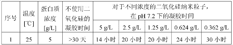
RSF的凝胶时间依赖于溶液中丝心蛋白的pH和浓度。丝心蛋白凝胶的潜在重要性是在对于细胞生长适宜的pH为7.2-7.4的生物医学研究中。已经发现浓度在1至10g/L的范围内的RSF在pH 7可能花费多至30至60天用于凝胶化,而10至100g/L的范围内的更高蛋白质浓度的RSF在pH 7花费10至20天变成凝胶(参考图1)。由更高蛋白质浓度的溶液制备的RSF凝胶不具有用于细胞进入和生长的适当的孔隙度。因此,希望的是由较低蛋白质浓度的RSF在pH 7制造微孔凝胶。长时间暴露于较高温度范围导致蛋白质的沉淀以及微孔结构的丧失,这对于为RSF所构想的应用是不适宜的。用于这种溶液的凝胶化需要相当长的时间,如果未保持无菌环境,在该过程中在丝溶液/凝胶中观察到真菌生长。考虑到其在组织工程学中的潜在应用,需要能够避免抗真菌剂的使用以使得细胞系不经历有毒的环境。因此需要缩短RSF在pH 7和室温的凝胶时间。A.Lele等在2008年8月3-8日的国际流变学大会(International Congress onRheology)的墙报上以及在Phys.Chem.Chem.Phys.,2010,12,3834-3844中研究了再生丝心蛋白溶液的凝胶化机制。他们还研究了所形成的丝水凝胶的结构和特征。The gelation time of RSF depends on the pH and concentration of fibroin in solution. The potential importance of fibroin gel is in biomedical research where pH 7.2-7.4 is optimal for cell growth. It has been found that RSF concentrations in the range of 1 to 10 g/L may take as much as 30 to 60 days for gelation at pH 7, whereas higher protein concentration RSFs in the range of 10 to 100 g/L are more stable at pH 7 It takes 10 to 20 days to become a gel (refer to Figure 1). RSF gels prepared from solutions with higher protein concentrations did not have proper porosity for cell entry and growth. Therefore, it is desirable to make microporous gels at pH 7 from RSF at lower protein concentrations. Prolonged exposure to higher temperature ranges leads to precipitation of proteins and loss of microporous structure, which is unsuitable for the applications contemplated for RSF. Gelation for such solutions takes a considerable amount of time during which fungal growth is observed in the silk solution/gel if a sterile environment is not maintained. Given its potential application in tissue engineering, there is a need to be able to avoid the use of antifungal agents so that the cell lines do not experience a toxic environment. Therefore, it is necessary to shorten the gel time of RSF at pH 7 and room temperature. A.Lele et al studied regenerated silk in the poster of the International Congress on Rheology on August 3-8, 2008 and in Phys.Chem.Chem.Phys., 2010, 12, 3834-3844 Gelation mechanism of cardiac protein solution. They also investigated the structure and characteristics of the formed silk hydrogels.
可以参考Dr.David L.Kaplan的标题为“Silk Polymer Designs forImproved Expression and Processing”的文章,其公开了丝凝胶化的控制,其中丝心蛋白的凝胶化依赖于丝心蛋白浓度、温度和凝胶形成的pH以及蛋白质结构,所述蛋白质结构可以关联到丝心蛋白蛋白质的分子构造中的一级序列特异性特征。此外,所述文章还描述了丝-无机纳米复合材料-二氧化硅体系,其中研究了用于二氧化硅纳米复合材料的形成的丝-R5融合体的克隆、表达和功能。Reference can be made to the article entitled "Silk Polymer Designs for Improved Expression and Processing" by Dr. David L. Kaplan, which discloses the control of silk gelation, wherein the gelation of silk fibroin depends on the concentration of silk fibroin, temperature and The pH of the gel formation and the protein structure, which can be linked to primary sequence-specific features in the molecular architecture of the fibroin protein. Furthermore, said article describes a silk-inorganic nanocomposite-silica system in which the cloning, expression and function of the silk-R5 fusion for the formation of the silica nanocomposite was investigated.
可以参考Wong等2006年的标题为“Novel nanocomposites from spidersilk-silica fusion(chimeric)proteins)”的文章,其公开了新仿生纳米复合材料方法以使用融合(嵌合)蛋白质合成二氧化硅复合材料。已经发现融合蛋白质具有宽范围领域内的应用,如生物医学领域[包括免疫学、癌症研究和药物递送(17-20)]以及材料科学[自组装材料(例如,凝胶),量子点生物结合物、传感器和无机材料合成(21-27)]。Reference can be made to the article titled "Novel nanocomposites from spider silk-silica fusion (chimeric) proteins)" by Wong et al. 2006, which discloses a new bioinspired nanocomposite approach to the synthesis of silica composites using fusion (chimeric) proteins. Fusion proteins have found applications in a wide range of fields, such as biomedical fields [including immunology, cancer research and drug delivery (17-20)] and materials science [self-assembling materials (eg, gels), quantum dot bioconjugation Synthesis of objects, sensors and inorganic materials (21-27)].
可以参考文章“Composite Material Made of Regenerated Silk Fibroinand Silica Sol”,Cheng Cheng等,2008/10发表,其公开了将再生丝心蛋白溶液与二氧化硅溶胶混合以制备丝心蛋白/二氧化硅复合材料。为了探测在丝蛋白质与二氧化硅之间是否有相互作用,并且更进一步地,是否该相互作用可以改善复合材料的机械性能,通过动态力学分析(DMA)与扫描电子显微镜(SEM)和拉曼光谱一起研究了丝心蛋白/二氧化硅复合材料的结构和性质。SEM结果显示复合材料中丝心蛋白与二氧化硅之间良好的相容性。纳米尺寸的二氧化硅粒子均匀地分散在丝心蛋白的连续基体中。复合材料的拉曼光谱显示丝心蛋白以β折叠构象为主。与纯丝心蛋白材料比较,该复合材料显示出更好的动态机械性能。You can refer to the article "Composite Material Made of Regenerated Silk Fibroin and Silica Sol", published by Cheng Cheng et al., 2008/10, which discloses mixing regenerated silk fibroin solution with silica sol to prepare fibroin/silica composite material . To explore whether there is an interaction between silk proteins and silica, and further, whether this interaction can improve the mechanical properties of the composite, dynamic mechanical analysis (DMA) with scanning electron microscopy (SEM) and Raman Together, spectroscopy investigates the structure and properties of the fibroin/silica composite. SEM results showed good compatibility between fibroin and silica in the composite. Nano-sized silica particles are uniformly dispersed in the continuous matrix of fibroin. The Raman spectrum of the composite material showed that fibroin was dominated by β-sheet conformation. Compared with pure fibroin material, the composite material shows better dynamic mechanical properties.
可以参考专利申请WO 200512606,其中申请要求保护水性丝心蛋白溶液和用于制备其的方法、制备纤维的方法、丝泡沫体、膜和丝水凝胶。在所述PCT申请的第30页上,讨论了关于离子、pH、温度和PEO对丝心蛋白溶液的凝胶化的影响的研究。Reference may be made to patent application WO 200512606, where the application claims aqueous fibroin solutions and methods for preparing them, methods for preparing fibers, silk foams, films and silk hydrogels. On page 30 of said PCT application, studies on the effects of ions, pH, temperature and PEO on the gelation of fibroin solutions are discussed.
现有技术调查表明没有文献教导凝胶化的加速从而缩短凝胶时间的方法。尤其是没有现有技术文献教导使用加速剂的丝心蛋白凝胶时间的加速方法。A survey of the prior art revealed that there is no literature teaching methods for acceleration of gelation and thus shortening of gelation time. In particular there is no prior art document teaching a method of accelerating the gel time of fibroin using accelerators.
发明目的purpose of invention
本发明的主要目的是缩短RSF凝胶的凝胶时间以填补本领域中的空白。The main purpose of the present invention is to shorten the gel time of RSF gel to fill the gap in this field.
本发明的另一个目的是使用不引起凝胶介质中微生物生长的方法缩短RSF凝胶时间。Another object of the present invention is to shorten the gelation time of RSF using a method that does not cause microbial growth in the gelling medium.
本发明的再另一个目的是通过对细胞系无毒的方法缩短凝胶时间。发明概述Yet another object of the present invention is to shorten the gel time by a method that is not toxic to the cell line. Summary of the invention
因此,本发明提供一种加速具有多孔结构的再生丝心蛋白(RSF)的凝胶化的方法,所述方法包括以下步骤:Therefore, the present invention provides a method for accelerating the gelation of regenerated fibroin (RSF) with a porous structure, the method comprising the following steps:
a)调节再生丝心蛋白溶液的pH至pH 5-7.5并且在0.1至40%的范围内调节其浓度;a) adjusting the pH of the regenerated fibroin solution to pH 5-7.5 and adjusting its concentration in the range of 0.1 to 40%;
b)加入所需浓度的凝胶剂并且保持温度条件,以及b) adding the desired concentration of gelling agent and maintaining temperature conditions, and
c)通过管倒置法检查凝胶化以获得RSF凝胶。c) Check gelation by tube inversion method to obtain RSF gel.
在本发明的实施方案中,所述凝胶剂选自由以下各项组成的组:二氧化硅、TiO2、FeO2SiN3、羟基磷灰石(hydroxyapetite),以及其他生物相容的无机化合物,优选二氧化硅。In an embodiment of the invention, the gelling agent is selected from the group consisting of silica, TiO2 , FeO2SiN3 , hydroxyapatite, and other biocompatible inorganic compounds , preferably silica.
在本发明的另一个实施方案中,所述凝胶化任选地在碱性条件下通过用蛋白质的β折叠,优选丝心蛋白蛋白质的β折叠自接种(self seeding)进行。In another embodiment of the invention, said gelation is performed by self seeding with a beta sheet of a protein, preferably a beta sheet of a fibroin protein, optionally under alkaline conditions.
在本发明的再另一个实施方案中,所述方法在20-70℃的范围内的温度下进行。In yet another embodiment of the present invention, the method is carried out at a temperature in the range of 20-70°C.
在本发明的再另一个实施方案中,所述凝胶剂在1g/L至25g/L,优选1至5g/L的浓度范围内使用。In yet another embodiment of the present invention, the gelling agent is used in a concentration range of 1 g/L to 25 g/L, preferably 1 to 5 g/L.
在本发明的再另一个实施方案中,所述凝胶剂的粒子大小在1nm-1μ,优选1nm至400nm的范围内。In still another embodiment of the present invention, the particle size of the gelling agent is in the range of 1 nm-1 μ, preferably 1 nm to 400 nm.
在本发明的再另一个实施方案中,再生丝心蛋白凝胶具有多孔结构,所述凝胶的孔大小在1-10微米的范围内。In yet another embodiment of the present invention, the regenerated fibroin gel has a porous structure, and the pore size of the gel is in the range of 1-10 microns.
在本发明的再另一个实施方案中,再生丝心蛋白凝胶具有多孔结构,所述凝胶包含孔隙度在1至500nm的范围内的纳米多孔壁。In yet another embodiment of the present invention, the regenerated fibroin gel has a porous structure comprising nanoporous walls with a porosity in the range of 1 to 500 nm.
在本发明的再另一个实施方案中,再生丝心蛋白凝胶具有多孔结构,所述凝胶是没有微生物生长的。In yet another embodiment of the present invention, the regenerated fibroin gel has a porous structure, said gel being free from microbial growth.
附图简述Brief description of the drawings
图1:在不同的pH下RSF的凝胶时间(D.Kaplan,JPC B,110,2006,21630-21638)Figure 1: Gel time of RSF at different pH (D. Kaplan, JPC B, 110, 2006, 21630-21638)
图2:对本发明的含有不同二氧化硅纳米粒子的RSF溶胶和RSF凝胶在pH 7和25℃的圆二色性法研究Figure 2: Circular dichroism study of RSF sols and RSF gels containing different silica nanoparticles of the present invention at pH 7 and 25°C
图3:对本发明带有和不带有纳米粒子的RSF凝胶的压缩模量测量。Figure 3: Compression modulus measurements on RSF gels of the invention with and without nanoparticles.
图4:在不同浓度的纳米粒子的存在下5g/L RSF凝胶的压缩模量和凝胶时间研究。Figure 4: Compressive modulus and gel time studies of 5 g/L RSF gels in the presence of different concentrations of nanoparticles.
图5:浓度5g/L,pH 2和50℃的RSF凝胶的CSLM图像(图像的宽度表示160μm),其中观察到蛋白质形成多孔结构。Figure 5: CSLM image of RSF gel at a concentration of 5g/L,
图6:管倒置显示了凝胶的形成。第一管显示了RSF的溶胶。第二管显示了所形成的凝胶,通过管倒置法测试证实了凝胶的形成。Figure 6: Tube inversion showing gel formation. The first tube shows a sol of RSF. The second tube shows the gel formed, which was confirmed by tube inversion testing.
发明详述Detailed description of the invention
本发明公开了一种缩短RSF的凝胶时间的方法。RSF和凝胶剂的组合物在pH 7-8下在2-24小时内凝胶,产生带有纳米多孔壁的微孔凝胶。该组合物是用于接种细胞和递送活性剂的粉末、膜、成形体、模制体等。The invention discloses a method for shortening the gel time of RSF. The combination of RSF and gelling agent gels within 2-24 hours at pH 7-8, yielding a microporous gel with nanoporous walls. The composition is a powder, film, shaped body, molded body, etc. for seeding cells and delivering active agents.
短语“再生丝心蛋白”和“再生蛋白质丝心蛋白”遍及本说明书可互换地使用并且本领域技术人员可以认识到这点。The phrases "regenerated fibroin" and "regenerated protein fibroin" are used interchangeably throughout this specification and those skilled in the art will recognize this.
短语“丝”和“蛋白质”遍及本说明书可互换地使用并且本领域技术人员可以认识到这点。The phrases "silk" and "protein" are used interchangeably throughout this specification and will be recognized by those skilled in the art.
依照本发明的目的,尝试了不同的方法以缩短RSF凝胶的凝胶时间。依赖于蛋白质(丝)浓度,在室温下在pH 7.0下RSF凝胶在约两周至三个月内形成(参考图1)。为缩短凝胶时间,一个备选方案是减小pH,但是酸性pH不允许细胞的繁殖。在一个实验中,出乎意料地发现二氧化硅纳米粒子的加入将凝胶时间由数个月缩短为数天或数小时,凝胶时间随过程的温度、蛋白质的浓度和二氧化硅纳米粒子的粒子大小而变化。所得到的包含再生丝心蛋白凝胶、水和缩短凝胶时间的试剂的组合物建立再生丝心蛋白的多孔结构。In accordance with the purpose of the present invention, different methods were tried to shorten the gel time of RSF gels. Depending on the protein (silk) concentration, RSF gels form in about two weeks to three months at room temperature at pH 7.0 (see Figure 1). To shorten the gel time, one alternative is to reduce the pH, but an acidic pH does not allow the proliferation of cells. In one experiment, it was unexpectedly found that the addition of silica nanoparticles shortened the gel time from months to days or hours, and that the gel time varied with the temperature of the process, the concentration of the protein, and the concentration of the silica nanoparticles. Variations in particle size. The resulting composition comprising the regenerated fibroin gel, water and an agent to shorten the gel time establishes the porous structure of the regenerated fibroin.
因此,本发明公开了加速具有多孔结构的再生丝心蛋白的凝胶化的方法,所述方法包括以下步骤:Therefore, the present invention discloses a method for accelerating the gelation of regenerated fibroin having a porous structure, said method comprising the steps of:
a.调节再生丝心蛋白溶液的pH至pH 7并且调节它的浓度;a. adjust the pH of the regenerated fibroin solution to pH 7 and adjust its concentration;
b.加入凝胶剂并保持温度条件;以及b. adding gelling agent and maintaining temperature conditions; and
c.通过管倒置法检查凝胶化。c. Check gelation by tube inversion.
如图6中所观察的,通过管倒置法检查凝胶形成。As observed in Figure 6, gel formation was checked by tube inversion.
凝胶剂不仅缩短用于凝胶化的时间,而且还克服了微生物生长的问题。不存在凝胶剂时,在pH 7-8对于5-70℃的范围内的温度,凝胶化花费大于三周,并且在该期间中观察到微生物生长。然而,在二氧化硅纳米粒子的存在下,在凝胶化之前观察不到微生物生长。The gelling agent not only shortens the time for gelation, but also overcomes the problem of microbial growth. In the absence of a gelling agent, gelation took more than three weeks at pH 7-8 for temperatures in the range 5-70°C, and microbial growth was observed during this period. However, in the presence of silica nanoparticles, no microbial growth was observed prior to gelation.
此外,这种试剂是生物相容的并且对将在RSF凝胶的这些微孔网络上生长的细胞系是无毒的。Furthermore, this reagent is biocompatible and nontoxic to cell lines that will grow on these microporous networks of RSF gels.
在本发明中所使用的凝胶剂选自二氧化硅、TiO2、FeO2SiN3、羟基磷灰石和其他生物相容的无机化合物的纳米粒子。The gelling agent used in the present invention is selected from nanoparticles of silica, TiO2 , FeO2SiN3 , hydroxyapatite and other biocompatible inorganic compounds.
凝胶时间随从1g/L变化至25g/L以上的凝胶剂浓度的上升而缩短。该试剂的粒子大小从1nm至400nm变化并且凝胶时间至少比没有该试剂时所花费的时间缩短一半。在一些情况下,凝胶时间缩短至少一个数量级。此外,加速凝胶化的过程在20℃至70℃的范围内的温度下进行。The gel time decreased with increasing gelling agent concentration from 1 g/L to above 25 g/L. The particle size of the agent varies from 1 nm to 400 nm and the gel time is at least half that it takes without the agent. In some cases, gel times were reduced by at least an order of magnitude. Furthermore, the process of accelerating gelation is performed at a temperature in the range of 20°C to 70°C.
采用不同的温度范围和凝胶剂浓度的多个试验的结果在下面的表1、2和3中以表格列出在这里。The results of multiple experiments using different temperature ranges and gelling agent concentrations are tabulated here in Tables 1, 2 and 3 below.
表1:显示了二氧化硅粒子对RSF样品凝胶时间的影响的凝胶时间表Table 1: Gel time chart showing the effect of silica particles on the gel time of RSF samples

表2:显示了二氧化硅粒子对RSF样品凝胶时间的影响的凝胶时间表Table 2: Gel time chart showing the effect of silica particles on the gel time of RSF samples

在15天内形成沉淀并且保持这样超过30天。A precipitate formed within 15 days and remained so for more than 30 days.
表3:显示了增加二氧化硅粒子浓度对RSF样品凝胶时间的影响的凝胶时间表Table 3: Gel time chart showing the effect of increasing silica particle concentration on the gel time of RSF samples
从以上结果发现,当蛋白质浓度为1g/L时,对于400nm的二氧化硅粒子在25℃凝胶时间低至5天,并且当该过程在70℃和pH 5-7进行时缩短至10小时。在5g/L的更高的蛋白质浓度下,对于400nm的二氧化硅粒子大小以及70℃的温度和pH 7.2,凝胶时间为3小时。因此,通过增加凝胶剂的浓度但保持温度并使用稀盐酸保持pH,获得RSF的凝胶化过程的加速。From the above results, it was found that the gel time was as low as 5 days at 25°C for 400nm silica particles when the protein concentration was 1 g/L, and shortened to 10 hours when the process was carried out at 70°C and pH 5-7 . At a higher protein concentration of 5 g/L, the gel time was 3 hours for a silica particle size of 400 nm and a temperature of 70 °C and pH 7.2. Thus, by increasing the concentration of the gelling agent but maintaining the temperature and maintaining the pH using dilute hydrochloric acid, an acceleration of the gelation process of the RSF was obtained.
在本发明中,蛋白质浓度从0.1至40%变化。组合物形成具有纳米多孔壁的、孔大小在1-10微米的范围内的微孔凝胶网络,其中壁的孔隙度在1-500nm的范围内。In the present invention, the protein concentration was varied from 0.1 to 40%. The composition forms a microporous gel network having nanoporous walls with a pore size in the range of 1-10 microns, wherein the walls have a porosity in the range of 1-500 nm.
在本发明的另外一个方面中,用蛋白质的β折叠自接种导致凝胶时间的加速。In yet another aspect of the invention, self-seeding with the [beta]-sheet of the protein results in an acceleration of the gel time.
在一个优选的方面中,RSF的凝胶时间通过将RSF溶液用碱性条件下的凝胶自接种而加速。二氧化硅不存在于该RSF中。In a preferred aspect, the gel time of RSF is accelerated by self-seeding the RSF solution with the gel under alkaline conditions. Silica is absent in this RSF.
表4Table 4
显示了RSF溶液用碱性条件下的凝胶自接种对RSF样品凝胶时间的影响的凝胶时间表Gel schedule showing the effect of RSF solution self-seeding with gel under alkaline conditions on the gel time of RSF samples
组合物是流延膜,是粉末或是成形体或模制体。该组合物包含有助于流延膜或模制为物体的添加剂。The composition is a cast film, a powder or a shaped or molded body. The composition contains additives to aid in casting a film or molding into an object.
如这里所示例的,该凝胶的特征在于它们的机械性能和3D多孔性。As exemplified here, the gels are characterized by their mechanical properties and 3D porosity.
这样的组合物可用于接种细胞、作为药物递送剂等。Such compositions are useful for seeding cells, as drug delivery agents, and the like.
实施例 Example
以下实施例仅以示例的方式给出,并且因此不应被理解为限制本发明的范围。The following examples are given by way of illustration only, and therefore should not be construed as limiting the scope of the invention.
实施例1Example 1
RSF组合物的制备Preparation of RSF composition
使用Nagarkar等(Ind.Eng.Chem.Res.2009,0-11)中描述的程序制备RSF溶液。通过在持续的搅拌下加入0.1N HCl将渗析过的RSF溶液的pH调节至7。使用pH 7的去离子水将蛋白质的浓度调节至1g/L。将已知重量的40nm的二氧化硅纳米粒子加入至RSF溶液。将这些溶液在恒温水浴中保持在25℃、50℃和70℃下并且每3小时之后监测样品的状态。使用小瓶倾倒法记录凝胶状态和凝胶时间。RSF solutions were prepared using the procedure described in Nagarkar et al. (Ind. Eng. Chem. Res. 2009, 0-11). The pH of the dialyzed RSF solution was adjusted to 7 by adding 0.1N HCl with constant stirring. The protein concentration was adjusted to 1 g/L using deionized water at pH 7. A known weight of 40 nm silica nanoparticles was added to the RSF solution. These solutions were kept at 25°C, 50°C and 70°C in a constant temperature water bath and the state of the samples was monitored after every 3 hours. Gel state and gel time were recorded using the vial pour method.
实施例2Example 2
RSF组合物的制备Preparation of RSF composition
使用Nagarkar等(Ind.Eng.Chem.Res.2009,0-11)中描述的程序制备RSF溶液。通过在持续的搅拌下加入0.1N HCl将渗析过的RSF溶液的pH调节至7。使用pH 7的去离子水将蛋白质的浓度调节至1g/L。将已知重量的150nm的二氧化硅纳米粒子加入至RSF溶液。将这些溶液在恒温水浴中保持在25℃、50℃和70℃下并且每3小时之后监测样品的状态。使用小瓶倾倒法记录凝胶状态和凝胶时间。RSF solutions were prepared using the procedure described in Nagarkar et al. (Ind. Eng. Chem. Res. 2009, 0-11). The pH of the dialyzed RSF solution was adjusted to 7 by adding 0.1N HCl with constant stirring. The protein concentration was adjusted to 1 g/L using deionized water at pH 7. A known weight of 150 nm silica nanoparticles was added to the RSF solution. These solutions were kept at 25°C, 50°C and 70°C in a constant temperature water bath and the state of the samples was monitored after every 3 hours. Gel state and gel time were recorded using the vial pour method.
实施例3Example 3
RSF组合物的制备Preparation of RSF composition
使用Nagarkar等(Ind.Eng.Chem.Res.2009,0-11)中描述的程序制备RSF溶液。通过在持续的搅拌下加入0.1N HCl将渗析过的RSF溶液的pH调节至7。使用pH 7的去离子水将蛋白质的浓度调节至1g/L。将已知重量的400nm的二氧化硅纳米粒子加入至RSF溶液。将这些溶液在恒温水浴中保持在25℃、50℃和70℃下并且每3小时之后监测样品的状态。使用小瓶倾倒法记录凝胶状态和凝胶时间。RSF solutions were prepared using the procedure described in Nagarkar et al. (Ind. Eng. Chem. Res. 2009, 0-11). The pH of the dialyzed RSF solution was adjusted to 7 by adding 0.1N HCl with constant stirring. The protein concentration was adjusted to 1 g/L using deionized water at pH 7. A known weight of 400 nm silica nanoparticles was added to the RSF solution. These solutions were kept at 25°C, 50°C and 70°C in a constant temperature water bath and the state of the samples was monitored after every 3 hours. Gel state and gel time were recorded using the vial pour method.
实施例4Example 4
RSF组合物的制备Preparation of RSF composition
使用Nagarkar等(Ind.Eng.Chem.Res.2009,0-11)中描述的程序制备RSF溶液。通过在持续的搅拌下加入0.1N HCl将渗析过的RSF溶液的pH调节至7。使用pH 7的去离子水将蛋白质的浓度调节至5g/L。将已知重量的40nm的二氧化硅纳米粒子加入至RSF溶液。将这些溶液在恒温水浴中保持在25℃、50℃和70℃下并且每3小时之后监测样品的状态。使用小瓶倾倒法记录凝胶状态和凝胶时间。RSF solutions were prepared using the procedure described in Nagarkar et al. (Ind. Eng. Chem. Res. 2009, 0-11). The pH of the dialyzed RSF solution was adjusted to 7 by adding 0.1N HCl with constant stirring. The protein concentration was adjusted to 5 g/L using deionized water at pH 7. A known weight of 40 nm silica nanoparticles was added to the RSF solution. These solutions were kept at 25°C, 50°C and 70°C in a constant temperature water bath and the state of the samples was monitored after every 3 hours. Gel state and gel time were recorded using the vial pour method.
实施例5Example 5
RSF组合物的制备Preparation of RSF composition
使用Nagarkar等(Ind.Eng.Chem.Res.2009,0-11)中描述的程序制备RSF溶液。通过在持续的搅拌下加入0.1N HCl将渗析过的RSF溶液的pH调节至7。使用pH 7的去离子水将蛋白质的浓度调节至5g/L。将已知重量的150nm的二氧化硅纳米粒子加入至RSF溶液。将这些溶液在恒温水浴中保持在25℃、50℃和70℃下并且每3小时之后监测样品的状态。使用小瓶倾倒法记录凝胶状态和凝胶时间。RSF solutions were prepared using the procedure described in Nagarkar et al. (Ind. Eng. Chem. Res. 2009, 0-11). The pH of the dialyzed RSF solution was adjusted to 7 by adding 0.1N HCl with constant stirring. The protein concentration was adjusted to 5 g/L using deionized water at pH 7. A known weight of 150 nm silica nanoparticles was added to the RSF solution. These solutions were kept at 25°C, 50°C and 70°C in a constant temperature water bath and the state of the samples was monitored after every 3 hours. Gel state and gel time were recorded using the vial pour method.
实施例6Example 6
RSF组合物的制备Preparation of RSF composition
使用Nagarkar等(Ind.Eng.Chem.Res.2009,0-11)中描述的程序制备RSF溶液。通过在持续的搅拌下加入0.1N HCl将渗析过的RSF溶液的pH调节至7。使用pH 7的去离子水将蛋白质的浓度调节至5g/L。将已知重量的400nm的二氧化硅纳米粒子加入至RSF溶液。将这些溶液在恒温水浴中保持在25℃、50℃和70℃下并且每3小时之后监测样品的状态。使用小瓶倾倒法记录凝胶状态和凝胶时间。RSF solutions were prepared using the procedure described in Nagarkar et al. (Ind. Eng. Chem. Res. 2009, 0-11). The pH of the dialyzed RSF solution was adjusted to 7 by adding 0.1N HCl with constant stirring. The protein concentration was adjusted to 5 g/L using deionized water at pH 7. A known weight of 400 nm silica nanoparticles was added to the RSF solution. These solutions were kept at 25°C, 50°C and 70°C in a constant temperature water bath and the state of the samples was monitored after every 3 hours. Gel state and gel time were recorded using the vial pour method.
对于不同浓度的二氧化硅和RSF溶液”实施例1-6所得到的RSF凝胶时间上的加速在上面的表1、2和3中以表格列出。The accelerations in RSF gelation time obtained for different concentrations of silica and RSF solutions"Examples 1-6 are tabulated in Tables 1, 2 and 3 above.
实施例7Example 7
机械性能Mechanical behavior
通过Rheometric Series RSA-III测试台(TA仪器-Waters LLC,NewCastle DE 1970)测量本发明的RSF凝胶的压缩模量(E′)。使用包括15mm直径的上盘,充当样品架的27mm内径乘以4mm高的圆筒,和下盘的自制附件测试样品。将在15ml培养瓶中形成的RSF凝胶放置在样品架中。在约半小时的等待期间之后,将硅油(SF-1000,GE Bayer Silicones,印度)涂布至样品的边缘以避免水从RSF凝胶丧失。在频率1Hz下在所有的样品上进行动态应变扫描测试,并且将应力由0.01%以0.01%的增量增加至3%。表5和6显示了所测得的带有和不带有纳米粒子的RSF凝胶的压缩模量。The compressive modulus (E') of the RSF gels of the present invention was measured by a Rheometric Series RSA-III test rig (TA Instruments - Waters LLC, New Castle DE 1970). Samples were tested using a homemade accessory consisting of a 15 mm diameter upper disc, a 27 mm inner diameter by 4 mm high cylinder serving as a sample holder, and a lower disc. The RSF gel formed in the 15 ml culture flask was placed in the sample holder. After a waiting period of about half an hour, silicone oil (SF-1000, GE Bayer Silicones, India) was applied to the edges of the samples to avoid loss of water from the RSF gel. Dynamic strain sweep tests were performed on all samples at a frequency of 1 Hz and the stress was increased from 0.01% to 3% in increments of 0.01%. Tables 5 and 6 show the measured compressive moduli of RSF gels with and without nanoparticles.
表5:带有和不带有纳米粒子的RSF凝胶的压缩模量测量。Table 5: Compression modulus measurements of RSF gels with and without nanoparticles.
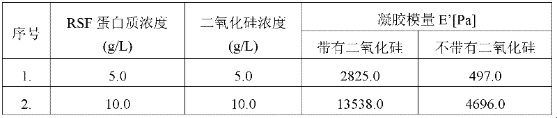
表6:在不同浓度的纳米粒子的存在下5g/L RSF凝胶的压缩模量测量。Table 6: Compression modulus measurements of 5 g/L RSF gels in the presence of different concentrations of nanoparticles.

实施例8Example 8
通过CLSM的孔隙度测量Porosity measurement by CLSM
通过共焦激光扫描显微镜(CLSM)定量计算RSF凝胶的孔隙度。使用卡尔蔡司共焦显微镜(Carl Zeiss Confocal Microscope)获得如图5中所示的RSF凝胶的显微图像。由已知的物镜放大率计算RSF凝胶的孔隙度。对于1g/L至5g/L的RSF浓度,RSF凝胶的孔隙度从100微米至10微米变化。图5显示了浓度5g/L,pH 2和50℃下的RSF凝胶的CSLM图像。The porosity of RSF gels was quantitatively calculated by confocal laser scanning microscopy (CLSM). Microscopic images of RSF gels as shown in Figure 5 were obtained using a Carl Zeiss Confocal Microscope. The porosity of the RSF gel was calculated from the known objective magnification. The porosity of RSF gels varied from 100 microns to 10 microns for RSF concentrations of 1 g/L to 5 g/L. Figure 5 shows the CSLM images of RSF gels at a concentration of 5 g/L,
实施例9Example 9
作为接种时间函数的RSF凝胶化研究RSF Gelation Study as a Function of Inoculation Time
通过使用以下方法制备RSF。将丝茧在0.5重量%的碳酸氢钠(NaHCO3)中煮沸一小时以移除丝胶蛋白。将煮沸过的丝心蛋白纤维用过量的水彻底洗涤以移除NaHCO3。之后将所获得的丝心蛋白溶解在9.3MLiBr溶液中以获得10重量%溶液。将该溶液向去离子水渗析(得自SigmaAldrich的乙酸纤维素渗析袋,MWC=10,000)48小时。将第一批的去离子水在3小时之后更换,并且之后每9小时更换。将渗析过的RSF溶液在15000RPM下离心20分钟,在这之后使用UV可见光谱测定蛋白质的浓度,用11.8作为摩尔消光系数(Izuka等,1968)。通常将渗析过的溶液储藏在5-7℃下的冰箱中。测得渗析过的溶液的pH为8.7。RSF was prepared by using the following method. Silk cocoons were boiled in 0.5% by weight sodium bicarbonate (NaHCO 3 ) for one hour to remove sericin. The boiled fibroin fibers were thoroughly washed with excess water to remove NaHCO 3 . The obtained fibroin was then dissolved in 9.3M LiBr solution to obtain a 10% by weight solution. This solution was dialyzed against deionized water (cellulose acetate dialysis bags from SigmaAldrich, MWC = 10,000) for 48 hours. The first batch of deionized water was changed after 3 hours and every 9 hours thereafter. The dialyzed RSF solution was centrifuged at 15000 RPM for 20 minutes, after which the protein concentration was determined using UV-vis spectroscopy, using 11.8 as the molar extinction coefficient (Izuka et al., 1968). Typically the dialyzed solution is stored in a refrigerator at 5-7°C. The pH of the dialyzed solution was measured to be 8.7.
通过在持续搅拌下逐滴加入0.1M HCL溶液调节渗析过的溶液的一部分的pH。在pH调节过程中,一部分的蛋白质沉淀出来,并且将这些通过在15000RPM下离心20min移除。从而获得的上清液蛋白质溶液,在下文中称作“溶胶”,是澄清的并且在将其用于进一步研究之前通过UV可见光谱测定其浓度。准备各自含有5ml的溶胶的9个样品小瓶。将27μl的3M NaOH加至样品小瓶1以将其pH升高至8.7,这等于渗析过的RSF的pH。我们将该样品称为具有零“接种”时间的样品。将在其他八个小瓶中的溶胶样品在低pH下保持如表4中所提到的0至24h之间的不同的“接种”时间,在这之后将27μl的3M NaOH加入至它们的每一个。加入NaOH溶液时样品的状态和加入NaOH之后的凝胶时间显示在表4中。The pH of a portion of the dialyzed solution was adjusted by dropwise addition of 0.1M HCL solution with constant stirring. During the pH adjustment, a portion of the proteins precipitated out and these were removed by centrifugation at 15000 RPM for 20 min. The supernatant protein solution thus obtained, hereinafter referred to as "sol", was clear and its concentration was determined by UV-vis spectroscopy before it was used for further studies. Nine sample vials each containing 5 ml of sol were prepared. 27 μl of 3M NaOH was added to sample vial 1 to raise its pH to 8.7, which is equal to the pH of the dialyzed RSF. We refer to this sample as the sample with zero "seeding" time. The sol samples in the other eight vials were kept at low pH for different "inoculation" times between 0 and 24h as mentioned in Table 4, after which 27 μl of 3M NaOH was added to each of them . The states of the samples when the NaOH solution was added and the gel time after the addition of NaOH are shown in Table 4.
本发明的益处Benefits of the invention
1.温度以及pH的环境条件加速凝胶化过程。1. Environmental conditions of temperature and pH accelerate the gelation process.
2.加速使得凝胶时间与数天比较缩短至数小时。2. Acceleration shortens the gel time to hours compared to days.
3.凝胶时间上的缩短使得凝胶可用于宽范围的多种应用。3. The reduction in gel time allows the gel to be used in a wide variety of applications.
4.凝胶对微生物生长不敏感。4. The gel is not sensitive to microbial growth.
5.获得具有宽范围的孔隙度的多孔网络。5. A porous network with a wide range of porosity is obtained.
6.自接种的备选方法也导致凝胶时间的加速。6. The alternative method of self-seeding also leads to an acceleration of the gel time.
权利要求书(按照条约第19条的修改)Claims (as amended under Article 19 of the Treaty)
1.一种用于再生丝心蛋白(RSF)的凝胶化以获得具有多孔结构的凝胶的方法,所述方法包括以下步骤:1. a method for the gelation of regenerated silk fibroin (RSF) to obtain a gel with a porous structure, said method comprising the following steps:
a)调节再生丝心蛋白溶液的pH至pH 5-7.5并且在0.1至40%的范围内调节其浓度;a) adjusting the pH of the regenerated fibroin solution to pH 5-7.5 and adjusting its concentration in the range of 0.1 to 40%;
b)加入选自由二氧化硅、TiO2、FeO2SiN3和羟基磷灰石组成的组,优选二氧化硅的凝胶剂并且将温度保持在20-70℃的范围内以获得RSF凝胶。b) adding a gelling agent selected from the group consisting of silica, TiO 2 , FeO 2 SiN 3 and hydroxyapatite, preferably silica and keeping the temperature in the range of 20-70°C to obtain the RSF gel .
2.如权利要求1所述的方法,其中所述凝胶化任选地在碱性条件下通过用蛋白质的β折叠,优选丝心蛋白蛋白质的β折叠自接种而进行。2. The method according to claim 1, wherein the gelation is carried out by self-seeding with a beta sheet of a protein, preferably a beta sheet of a fibroin protein, optionally under alkaline conditions.
3.如权利要求1所述的方法,其中用于再生丝心蛋白的凝胶化的时间期间在3小时至6天的范围内。3. The method of claim 1, wherein the time period for gelation of the regenerated fibroin is in the range of 3 hours to 6 days.
4.如权利要求1所述的方法,其中所述凝胶剂在1g/L至25g/L,优选1至5g/L的浓度范围内使用。4. The method of claim 1, wherein the gelling agent is used in a concentration range of 1 g/L to 25 g/L, preferably 1 to 5 g/L.
5.如权利要求1所述的方法,其中所述凝胶剂的粒子大小在1nm-1μ,优选1nm至400nm的范围内。5. The method according to claim 1, wherein the particle size of the gelling agent is in the range of 1 nm-1 μ, preferably 1 nm to 400 nm.
6.根据权利要求1获得的具有多孔结构的再生丝心蛋白凝胶,其中所述凝胶的孔大小在1-10微米的范围内。6. The regenerated silk fibroin gel having a porous structure obtained according to claim 1, wherein the pore size of the gel is in the range of 1-10 microns.
7.根据权利要求1获得的具有多孔结构的再生丝心蛋白凝胶,其中所述凝胶包含孔隙度在1至500nm的范围内的纳米多孔壁。7. The regenerated fibroin gel having a porous structure obtained according to claim 1, wherein the gel comprises nanoporous walls having a porosity in the range of 1 to 500 nm.
8.根据权利要求1获得的具有多孔结构的再生丝心蛋白凝胶,其中所述凝胶是没有微生物生长的。8. The regenerated silk fibroin gel having a porous structure obtained according to claim 1, wherein said gel is free from microbial growth.
说明或声明(按照条约第19条的修改)Statement or declaration (as amended under Article 19 of the Treaty)
根据第19(1)条的声明 Declaration under section 19(1)
根据国际检索机构(ISA/EP)的检索报告和书面意见,我们适当地修改了权利要求书以在本发明中确立新颖性和创造性。 According to the search report and written opinion of the International Search Agency (ISA/EP), we amended the claims appropriately to establish novelty and inventive step in the present invention.
·修改了权利要求1。 • Claim 1 was amended.
·删除了权利要求2、4和10。
•
·添加了新的权利要求3。
• A
·权利要求中的任何增加内容已经用下划线指出,并且权利要求中的任何删除已经通过删除线指出。 • Any additions in the claims have been indicated by underlining and any deletions in the claims have been indicated by strikethrough.
所修改的权利要求未超出原始提交的国际申请的公开内容并且不影响原始提交的说明书和附图。 The amended claims do not go beyond the disclosure content of the originally filed international application and do not affect the originally filed description and drawings.
Claims (10)
Applications Claiming Priority (3)
| Application Number | Priority Date | Filing Date | Title |
|---|---|---|---|
| IN1578/DEL/2009 | 2009-07-31 | ||
| IN1578DE2009 | 2009-07-31 | ||
| PCT/IN2010/000506 WO2011013145A1 (en) | 2009-07-31 | 2010-07-30 | Accelerated gelation of regenerated fibroin |
Publications (2)
| Publication Number | Publication Date |
|---|---|
| CN102482497A true CN102482497A (en) | 2012-05-30 |
| CN102482497B CN102482497B (en) | 2014-07-30 |
Family
ID=43301795
Family Applications (1)
| Application Number | Title | Priority Date | Filing Date |
|---|---|---|---|
| CN201080033991.3A Expired - Fee Related CN102482497B (en) | 2009-07-31 | 2010-07-30 | Accelerated Gelation Of Regenerated Fibroin |
Country Status (7)
| Country | Link |
|---|---|
| US (1) | US8779097B2 (en) |
| EP (1) | EP2459650B1 (en) |
| JP (1) | JP5692611B2 (en) |
| KR (1) | KR20120052314A (en) |
| CN (1) | CN102482497B (en) |
| ES (1) | ES2426478T3 (en) |
| WO (1) | WO2011013145A1 (en) |
Families Citing this family (4)
| Publication number | Priority date | Publication date | Assignee | Title |
|---|---|---|---|---|
| KR102007797B1 (en) * | 2012-11-30 | 2019-08-07 | (주)아모레퍼시픽 | Silk hydrogel composition and silk hydrogel mask |
| IL317123A (en) | 2018-08-27 | 2025-01-01 | Regeneron Pharma | Use of Raman Spectroscopy in Downstream Purification |
| CN117427223B (en) * | 2023-10-30 | 2025-12-30 | 苏州大学 | A nano-silica reinforced silk fibroin composite material and its preparation method |
| CN117462762B (en) * | 2023-11-24 | 2025-12-12 | 苏州大学 | A nano-silica/silane coupling agent/silk fibroin composite material and its preparation method |
Citations (5)
| Publication number | Priority date | Publication date | Assignee | Title |
|---|---|---|---|---|
| US4233212A (en) * | 1979-04-17 | 1980-11-11 | Kanebo, Ltd. | Process for producing a fine powder of silk fibroin |
| WO2003022909A1 (en) * | 2001-09-11 | 2003-03-20 | Consorzio Per Gli Studi Universitari | Method for the preparation of silk fibroin hydrogels |
| CN1861666A (en) * | 2006-04-19 | 2006-11-15 | 东华大学 | Preparation process of SF/SiO2 Nano composite membrane |
| CN101235204A (en) * | 2008-02-26 | 2008-08-06 | 天津工业大学 | A kind of preparation method of nano-phase hydroxyapatite/silk fibroin composite material |
| CN101474429A (en) * | 2009-01-22 | 2009-07-08 | 浙江理工大学 | Method for preparing hydroxylapatite-silk fibroin compound stent material using two-step method |
Family Cites Families (5)
| Publication number | Priority date | Publication date | Assignee | Title |
|---|---|---|---|---|
| JP2912430B2 (en) * | 1990-07-18 | 1999-06-28 | 鐘紡株式会社 | Oily occluded regenerated protein fine powder and method for producing the same |
| JP3738255B2 (en) * | 2003-02-21 | 2006-01-25 | 株式会社オードレマン | Cosmetics containing amorphous silk fibroin |
| JP4698596B2 (en) | 2003-04-10 | 2011-06-08 | タフツ ユニバーシティー | Concentrated aqueous silk fibroin solutions and their use |
| ES2527125T3 (en) * | 2007-05-29 | 2015-01-20 | Trustees Of Tufts College | Silk fibroin gelation method using sonication |
| JP5637424B2 (en) * | 2008-05-26 | 2014-12-10 | 国立大学法人群馬大学 | Composite, method for producing the same, and cosmetic material |
-
2010
- 2010-07-30 KR KR1020127003751A patent/KR20120052314A/en not_active Ceased
- 2010-07-30 WO PCT/IN2010/000506 patent/WO2011013145A1/en not_active Ceased
- 2010-07-30 CN CN201080033991.3A patent/CN102482497B/en not_active Expired - Fee Related
- 2010-07-30 EP EP10757845.2A patent/EP2459650B1/en not_active Not-in-force
- 2010-07-30 ES ES10757845T patent/ES2426478T3/en active Active
- 2010-07-30 US US13/387,870 patent/US8779097B2/en not_active Expired - Fee Related
- 2010-07-30 JP JP2012522340A patent/JP5692611B2/en not_active Expired - Fee Related
Patent Citations (5)
| Publication number | Priority date | Publication date | Assignee | Title |
|---|---|---|---|---|
| US4233212A (en) * | 1979-04-17 | 1980-11-11 | Kanebo, Ltd. | Process for producing a fine powder of silk fibroin |
| WO2003022909A1 (en) * | 2001-09-11 | 2003-03-20 | Consorzio Per Gli Studi Universitari | Method for the preparation of silk fibroin hydrogels |
| CN1861666A (en) * | 2006-04-19 | 2006-11-15 | 东华大学 | Preparation process of SF/SiO2 Nano composite membrane |
| CN101235204A (en) * | 2008-02-26 | 2008-08-06 | 天津工业大学 | A kind of preparation method of nano-phase hydroxyapatite/silk fibroin composite material |
| CN101474429A (en) * | 2009-01-22 | 2009-07-08 | 浙江理工大学 | Method for preparing hydroxylapatite-silk fibroin compound stent material using two-step method |
Non-Patent Citations (2)
| Title |
|---|
| UNG-JIN KIM, ET AL.: "Structure and properties of Silk Hydrogels", 《BIOMACROMOLECULES》 * |
| 郑桂芬等: "纳米TiO2对丝素蛋白膜构象转变的影响", 《功能高分子学报》 * |
Also Published As
| Publication number | Publication date |
|---|---|
| JP2013501087A (en) | 2013-01-10 |
| KR20120052314A (en) | 2012-05-23 |
| US20120130047A1 (en) | 2012-05-24 |
| ES2426478T3 (en) | 2013-10-23 |
| EP2459650B1 (en) | 2013-06-05 |
| CN102482497B (en) | 2014-07-30 |
| WO2011013145A4 (en) | 2011-04-28 |
| US8779097B2 (en) | 2014-07-15 |
| EP2459650A1 (en) | 2012-06-06 |
| WO2011013145A1 (en) | 2011-02-03 |
| JP5692611B2 (en) | 2015-04-01 |
Similar Documents
| Publication | Publication Date | Title |
|---|---|---|
| Marrella et al. | Enhanced mechanical performances and bioactivity of cell laden-graphene oxide/alginate hydrogels open new scenario for articular tissue engineering applications | |
| Naumenko et al. | Clay nanotube–biopolymer composite scaffolds for tissue engineering | |
| Toskas et al. | Chitosan (PEO)/silica hybrid nanofibers as a potential biomaterial for bone regeneration | |
| Peng et al. | Graphene oxide as an interface phase between polyetheretherketone and hydroxyapatite for tissue engineering scaffolds | |
| Wang et al. | Dual-enzymatically crosslinked and injectable hyaluronic acid hydrogels for potential application in tissue engineering | |
| Liu et al. | Thixotropic silk nanofibril-based hydrogel with extracellular matrix-like structure | |
| CN105566872B (en) | Poly-dopamine modified lithium halloysite nanotubes/lactic acid composite material and its preparation and application | |
| CN108066819B (en) | High-strength natural polymer hydrogel film and preparation method thereof | |
| Rajamanickam et al. | Soft colloidal scaffolds capable of elastic recovery after large compressive strains | |
| CN107118360A (en) | A kind of soybean protein isolate base natural polymer hydrogel and preparation method thereof | |
| CN102482497A (en) | Accelerated Gelation Of Regenerated Fibroin | |
| WO2011018995A1 (en) | Organic/inorganic composite hydrogel and production method for same | |
| Dieudonné et al. | Bioactive glass hybrids: A simple route towards the gelatin–SiO 2–CaO system | |
| Zhao et al. | Amphiphilic nanofibrillated cellulose/polyurethane composites with antibacterial, antifouling and self-healing properties for potential catheter applications | |
| Mallakpour et al. | Physicochemical inspection and in vitro bioactivity behavior of bio-nanocomposite alginate hydrogels filled by magnesium fluoro-hydroxyapatite | |
| US11541148B2 (en) | POSS nanocomposite hydrogel for 3D bioprinting | |
| Ma et al. | A highly efficient dual-diazonium reagent for protein crosslinking and construction of a virus-based gel | |
| KR101684900B1 (en) | silk fibroin gel produced by adding nitrate salt and production method of the same | |
| CN110452397B (en) | Three-dimensional graphene foam/natural polysaccharide-based hydrogel composite scaffold and preparation method thereof | |
| WO2025190321A1 (en) | Preparation method for and ocular use of cellulose nanocrystal-based polymer hydrogel | |
| Bayattork et al. | Enhanced formation of bioactive and strong silk–bioglass hybrid materials through organic–inorganic mutual molecular nucleation induction and templating | |
| Miyazaki et al. | Apatite formation abilities and mechanical properties of hydroxyethylmethacrylate-based organic–inorganic hybrids incorporated with sulfonic groups and calcium ions | |
| CN108164713A (en) | Degradable and water soluble POSS-PEG hybridized hydrogels and its preparation method and application | |
| Mallakpour et al. | Hydrogel bio-nanocomposite beads based on alginate and silica: physicochemical and in vitro bioactivity evaluations | |
| Baruah et al. | Reinforced non-mulberry silk fibroin–fibroin nanocomposites and their optimization for tissue engineering applications |
Legal Events
| Date | Code | Title | Description |
|---|---|---|---|
| C06 | Publication | ||
| PB01 | Publication | ||
| C10 | Entry into substantive examination | ||
| SE01 | Entry into force of request for substantive examination | ||
| C14 | Grant of patent or utility model | ||
| GR01 | Patent grant | ||
| CF01 | Termination of patent right due to non-payment of annual fee | ||
| CF01 | Termination of patent right due to non-payment of annual fee |
Granted publication date: 20140730 Termination date: 20170730 |