CN102030754B - Non-viral vector, and preparation method and application thereof - Google Patents
Non-viral vector, and preparation method and application thereof Download PDFInfo
- Publication number
- CN102030754B CN102030754B CN2009100930813A CN200910093081A CN102030754B CN 102030754 B CN102030754 B CN 102030754B CN 2009100930813 A CN2009100930813 A CN 2009100930813A CN 200910093081 A CN200910093081 A CN 200910093081A CN 102030754 B CN102030754 B CN 102030754B
- Authority
- CN
- China
- Prior art keywords
- viral vector
- formula
- tumor
- compound
- dna
- Prior art date
- Legal status (The legal status is an assumption and is not a legal conclusion. Google has not performed a legal analysis and makes no representation as to the accuracy of the status listed.)
- Expired - Fee Related
Links
Images


Landscapes
- Medicinal Preparation (AREA)
Abstract
本发明公开一种非病毒载体及其制备方法与应用。该非病毒载体,是聚乙烯亚胺与式V所示化合物共混而得的纳米颗粒。本发明提供的药物,其中,该药物的载体是上述本发明提供的非病毒载体。该药物包括上述本发明提供的非病毒载体和包裹在所述非病毒载体中的治疗性的DNA;所述药物的靶向肿瘤为叶酸受体FRα高表达的肿瘤;所述药物能被动靶向的肿瘤为叶酸受体FRα阴性表达的肿瘤和心脏。通过静脉注射接种有叶酸受体阳性细胞的肿瘤的荷瘤小鼠,本发明提供的非病毒载体能够携带基因特异到达肿瘤细胞,并在肿瘤细胞内持续高效表达目的基因。
Description
技术领域 technical field
本发明属于高分子化学和生物医学工程领域,涉及一种非病毒载体及其制备方法与应用。The invention belongs to the field of macromolecular chemistry and biomedical engineering, and relates to a non-viral vector and its preparation method and application.
背景技术 Background technique
肿瘤是基因病,肿瘤发生的遗传背景是癌基因或抑癌基因的异常,基因治疗是对肿瘤细胞或正常细胞转移一个或几个正常基因,它的表达产物有利于直接或间接的杀伤肿瘤细胞;或保护正常细胞免受化疗与放疗的严重伤害,但是传统的基因导入系统存在的问题在于:(1)缺乏靶向性,而且具有较高的细胞毒性和免疫原性,如以携带p53基因的腺病毒治疗恶性肿瘤的方案中,只能直接将腺病毒注射到肿瘤局部。若静脉注射,病毒颗粒将很快被清除,真正能够到达肿瘤组织的目的基因很少,难以达到治疗效果,且增加了副作用;(2)存在安全性问题,目前针对遗传性疾病的基因治疗方案大多采用逆转录病毒载体,其插入或整合到染色体的位置是随机的,有引起插入突变及细胞恶性转化的潜在危险。(3)转染和表达效率低,质粒载体因为含有CpG序列,进入细胞后能够被机体沉默而不能持续高效的表达。Tumor is a genetic disease. The genetic background of tumor occurrence is the abnormality of oncogenes or tumor suppressor genes. Gene therapy is to transfer one or several normal genes to tumor cells or normal cells, and its expression products are conducive to directly or indirectly killing tumor cells. ; or protect normal cells from the severe damage of chemotherapy and radiotherapy, but the problems in the traditional gene transfer system are: (1) lack of targeting, and have high cytotoxicity and immunogenicity, such as carrying the p53 gene In the current adenovirus treatment of malignant tumors, the adenovirus can only be injected directly into the tumor. If injected intravenously, the virus particles will be cleared quickly, and there are very few target genes that can really reach the tumor tissue, it is difficult to achieve the therapeutic effect, and the side effects are increased; (2) There are safety problems, and the current gene therapy program for genetic diseases Retroviral vectors are mostly used, and the position of insertion or integration into the chromosome is random, which has the potential danger of causing insertion mutation and malignant transformation of cells. (3) The efficiency of transfection and expression is low. Because the plasmid vector contains a CpG sequence, it can be silenced by the body after entering the cell and cannot be continuously and efficiently expressed.
非病毒载体作为肿瘤基因治疗的载体优势在于具有较高的安全性和较低的免疫原性,并且由于肿瘤血管表皮的特殊构造,能够被动靶向肿瘤组织,目前主要应用的非病毒载体有脂质体,多聚阳离子,树枝状高分子等,非病毒载体与DNA一般通过电荷的相互作用形成聚阳离子复合物,由于聚阳离子复合物对细胞的毒性较大,容易形成肺部栓塞,并且与蛋白非特异性结合,如静脉注射,不容易到达靶器官等。为减少聚阳离子的细胞毒性和增加聚阳离子复合物的生物相溶性,可以对高分化合物进行相应的修饰,如PEG的修饰等。The advantage of non-viral vectors as carriers for tumor gene therapy is that they have high safety and low immunogenicity, and due to the special structure of tumor vascular epidermis, they can passively target tumor tissues. Currently, the main non-viral vectors used are lipid Plastids, polycations, dendrimers, etc., non-viral vectors and DNA generally form polycation complexes through the interaction of charges. Due to the high toxicity of polycation complexes to cells, it is easy to form pulmonary embolism, and with Protein non-specific binding, such as intravenous injection, is not easy to reach the target organ, etc. In order to reduce the cytotoxicity of polycations and increase the biocompatibility of polycation complexes, high-scoring compounds can be modified accordingly, such as PEG modification.
由于遗传物质的变异,在一些肿瘤细胞表面高表达叶酸受体分子,叶酸受体是一个特异的抗肿瘤药物传送靶位,在大部分来源于上皮组织的恶性肿瘤如卵巢癌,子宫内膜癌,肾癌,乳腺癌,肺癌,结肠癌和鼻炎癌中均有高度表达,特别是妇科肿瘤,90%的卵巢癌细胞系都有叶酸受体的高度表达,而叶酸受体在正常组织中缺失或只在极化上皮细胞的顶点表达,所以叶酸在正常组织中通常不会积累。Due to the variation of genetic material, folic acid receptor molecules are highly expressed on the surface of some tumor cells. Folate receptor is a specific anti-tumor drug delivery target, which is found in most malignant tumors derived from epithelial tissue such as ovarian cancer and endometrial cancer. , kidney cancer, breast cancer, lung cancer, colon cancer and rhinitis cancer are all highly expressed, especially gynecological tumors, 90% of ovarian cancer cell lines have high expression of folate receptors, while folate receptors are absent in normal tissues Or expressed only at the apex of polarized epithelial cells, so folate does not normally accumulate in normal tissues.
因为叶酸与叶酸受体具有高亲和力,并且叶酸分子自身稳定性高,免疫原性低,与纳米有机材料相容性好,是肿瘤靶标的理想配体。Because folic acid has high affinity with folic acid receptors, and the folic acid molecule itself has high stability, low immunogenicity, and good compatibility with nano-organic materials, it is an ideal ligand for tumor targets.
FA-PEG-PEI纳米材料是一种理想的肿瘤靶向纳米材料。之前人们应用的FA-PEG-PEI材料在细胞水平取得良好的效果,但是在动物体内通过系统注射时发现在质粒浓度高的情况下形成的纳米颗粒容易发生聚沉,注射后容易导致肺部血管栓塞而致动物死亡,由于颗粒的聚沉导致注射后不能很好的透过肿瘤血管内皮,进而不能有效靶向肿瘤器官。叶酸配体在材料表面的丰度对材料在肿瘤部位的聚集具有一定的影响,所以选取合适的叶酸接枝率有利于提高纳米颗粒在肿瘤部位的富集。FA-PEG-PEI nanomaterial is an ideal tumor-targeting nanomaterial. The FA-PEG-PEI material used by people before has achieved good results at the cellular level, but it was found that the nanoparticles formed under the condition of high plasmid concentration are prone to coagulation when injected systematically in animals, and it is easy to cause pulmonary blood vessels after injection. Animals died due to embolism. Due to the coagulation of particles, they could not penetrate the tumor vascular endothelium well after injection, and thus could not effectively target tumor organs. The abundance of folic acid ligands on the surface of the material has a certain influence on the accumulation of the material at the tumor site, so choosing an appropriate folic acid grafting rate is conducive to improving the enrichment of nanoparticles at the tumor site.
研究表明,外源基因表达盒与细菌质粒骨架之间的共价连接会导致外源基因的沉默,这是造成质粒载体所携带的外源基因在体内只能瞬时表达的主要原因,也是质粒载体临床应用的主要限制因素。小环DNA(minicircleDNA)载体是一种自1997年发展起来的新型的基因载体,与常用的非病毒载体质粒载体相比,小环DNA载体仅由外源基因的表达盒构成,不含有来自细菌质粒的骨架结构。因为它仅由外源基因的表达盒构成,不含来自细菌质粒的骨架结构,从而减少了CpG模序甲基化所致的免疫反应,提高了安全性;而且更有效的提高了转基因的表达和生物利用度。Studies have shown that the covalent linkage between the exogenous gene expression cassette and the bacterial plasmid backbone can lead to the silencing of the exogenous gene, which is the main reason why the exogenous gene carried by the plasmid vector can only be expressed transiently in vivo, and it is also the The main limiting factor of clinical application. Minicircle DNA (minicircleDNA) vector is a new type of gene carrier developed since 1997. Compared with commonly used non-viral vector plasmid vectors, minicircle DNA vectors are only composed of expression cassettes of foreign genes and do not contain bacterial Plasmid backbone structure. Because it is only composed of expression cassettes of foreign genes and does not contain the backbone structure from bacterial plasmids, it reduces the immune response caused by methylation of CpG motifs and improves safety; moreover, it improves the expression of transgenes more effectively and bioavailability.
发明内容 Contents of the invention
本发明的目的是提供一种非病毒载体和非病毒载体及其制备方法与应用。The object of the present invention is to provide a non-viral vector, a non-viral vector and its preparation method and application.
本发明提供的非病毒载体,是聚乙烯亚胺与式V所示化合物共混而得的纳米颗粒。The non-viral carrier provided by the present invention is a nanoparticle obtained by blending polyethyleneimine and a compound represented by formula V.

其中,m=145,n为小于m的正整数;式V所述化合物的相对数均分子量为20600,分子量分布为1.73。Wherein, m=145, n is a positive integer smaller than m; the relative number average molecular weight of the compound described in formula V is 20600, and the molecular weight distribution is 1.73.
该非病毒载体中,所述纳米颗粒的粒径为70-90纳米;所述聚乙烯亚胺与式V所示化合物的摩尔比为10-15∶85-90;所述聚乙烯亚胺的分子量为25kDa;共混的温度为20-30℃,共混的时间为15-20分钟。In the non-viral vector, the particle diameter of the nanoparticles is 70-90 nanometers; the molar ratio of the polyethyleneimine to the compound shown in formula V is 10-15:85-90; the polyethyleneimine The molecular weight is 25kDa; the blending temperature is 20-30°C, and the blending time is 15-20 minutes.
本发明提供的制备上述非病毒载体的方法,包括如下步骤:将聚乙烯亚胺与式V所示化合物进行共混,得到上述非病毒载体。The method for preparing the above-mentioned non-viral vector provided by the present invention comprises the following steps: blending polyethyleneimine and the compound represented by formula V to obtain the above-mentioned non-viral vector.
该方法中,所述聚乙烯亚胺与式V所示化合物的摩尔比为10-15∶85-90;所述聚乙烯亚胺的分子量为25kDa;共混的温度为20-30℃,共混的时间为15-20分钟。In this method, the molar ratio of the polyethyleneimine to the compound represented by formula V is 10-15:85-90; the molecular weight of the polyethyleneimine is 25kDa; the blending temperature is 20-30°C, and Mixing time is 15-20 minutes.
本发明还提供了一种药物,其中,该药物的载体是上述本发明提供的非病毒载体。The present invention also provides a drug, wherein the carrier of the drug is the above-mentioned non-viral carrier provided by the present invention.
该药物包括上述本发明提供的非病毒载体和包裹在所述非病毒载体中的治疗性的DNA;所述药物的靶向肿瘤为叶酸受体FRα高表达的肿瘤;所述药物能被动靶向的肿瘤为叶酸受体FRα阴性表达的肿瘤和心脏。The drug includes the above-mentioned non-viral vector provided by the present invention and the therapeutic DNA wrapped in the non-viral vector; the target tumor of the drug is a tumor with high expression of folic acid receptor FRα; the drug can passively target The tumors were folate receptor FRα-negative tumors and hearts.
本发明提供的制备上述药物的方法,包括如下步骤:The method for preparing the above-mentioned medicine provided by the invention comprises the following steps:
1)往治疗性的DNA中加入浓度为10-50克/100毫升的葡萄糖溶液,加水稀释至所述葡萄糖溶液的终浓度为5-10克/100毫升,所述治疗性的DNA的终浓度为0-50μg/mL,不包括0,得到DNA稀释液;1) Add a glucose solution with a concentration of 10-50 g/100 ml to the therapeutic DNA, dilute with water until the final concentration of the glucose solution is 5-10 g/100 ml, the final concentration of the therapeutic DNA 0-50μg/mL, excluding 0, get DNA dilution;
往所述非病毒载体中加入浓度为10-60克/100毫升的葡萄糖溶液,加水稀释至所述葡萄糖溶液的终浓度为5-10克/100毫升,所述非病毒载体的终浓度为0-5μg/ml,不包括0,得到非病毒载体稀释液;Add a glucose solution with a concentration of 10-60 g/100 ml to the non-viral carrier, dilute with water until the final concentration of the glucose solution is 5-10 g/100 ml, and the final concentration of the non-viral carrier is 0 -5 μg/ml, excluding 0, to obtain a non-viral vector dilution;
2)将步骤1)得到的非病毒载体稀释液与DNA稀释液等体积混匀,室温静置孵育后浓缩,得到本发明提供的药物。2) Mix equal volumes of the non-viral vector diluent obtained in step 1) and the DNA diluent, incubate at room temperature and then concentrate to obtain the drug provided by the present invention.
该方法的步骤1)中,任一所述葡萄糖溶液的终浓度优选为5克/100毫升,所述治疗性的DNA的终浓度优选为20μg/mL;所述非病毒载体的终浓度优选为0.39μg/ml。In step 1) of the method, the final concentration of any glucose solution is preferably 5 g/100 milliliters, the final concentration of the therapeutic DNA is preferably 20 μg/mL; the final concentration of the non-viral vector is preferably 0.39 μg/ml.
所述步骤2)中,孵育的时间为15-30分钟,优选15分钟;浓缩步骤是用超滤杯进行浓缩的;所述超滤膜的截留分子量为30-90kDa。In the step 2), the incubation time is 15-30 minutes, preferably 15 minutes; the concentration step is concentrated with an ultrafiltration cup; the molecular weight cut-off of the ultrafiltration membrane is 30-90kDa.
此外,本发明提供的式V所示化合物,是按照如下流程制备得到的:In addition, the compound represented by formula V provided by the present invention is prepared according to the following process:
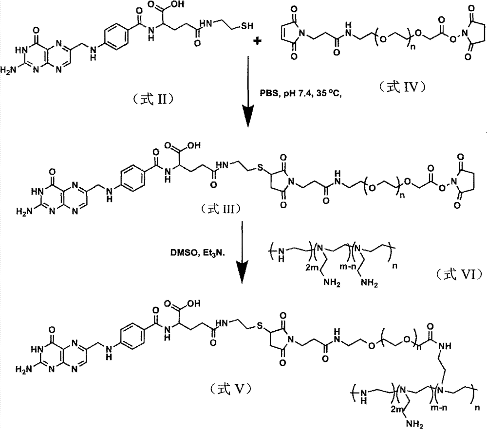
其中,FA为叶酸,式I为叶酸琥珀酸酯,式II为叶酸巯基乙胺,式I、式II、式III及式V化合物的制备方法如下:Wherein, FA is folic acid, formula I is folic acid succinate, formula II is folic acid mercaptoethylamine, and the preparation methods of formula I, formula II, formula III and formula V compounds are as follows:
1、制备式I所示化合物的方法,包括如下步骤:在惰性气氛中,将叶酸与三乙胺混匀至完全溶解后,在避光及惰性气氛的条件下,再加入N羟基琥珀酸胺酯和N,N′-二环己基碳二亚胺进行反应,得到式I所示化合物。1. The method for preparing the compound shown in formula I, comprising the following steps: in an inert atmosphere, after mixing folic acid and triethylamine until completely dissolved, then adding N-hydroxysuccinamide The ester is reacted with N,N'-dicyclohexylcarbodiimide to obtain the compound shown in formula I.
该方法中,叶酸、三乙胺的摩尔比为2-2.5∶30-40,优选2.26∶36,反应温度为25-30℃,优选28℃,反应时间为12-16小时,优选14小时;所述N羟基琥珀酸胺酯与叶酸的摩尔比为2-2.5∶2-2.5,优选2.26∶2.26,N,N′-二环己基碳二亚胺与叶酸的摩尔比为的2-2.5∶2-2.5,优选2.26∶2.4,反应时间为45-50小时,优选48小时;反应温度为25-30℃,优选28℃;反应介质选自无水二甲基亚砜和无水N,N-二甲基甲酰胺中的至少一种。In the method, the molar ratio of folic acid and triethylamine is 2-2.5:30-40, preferably 2.26:36, the reaction temperature is 25-30°C, preferably 28°C, and the reaction time is 12-16 hours, preferably 14 hours; The molar ratio of N-hydroxysuccinate to folic acid is 2-2.5: 2-2.5, preferably 2.26: 2.26, and the molar ratio of N, N'-dicyclohexylcarbodiimide to folic acid is 2-2.5: 2-2.5, preferably 2.26:2.4, the reaction time is 45-50 hours, preferably 48 hours; the reaction temperature is 25-30°C, preferably 28°C; the reaction medium is selected from anhydrous dimethyl sulfoxide and anhydrous N,N - at least one of dimethylformamides.
2、制备式II所示化合物的方法,包括如下步骤:在避光及惰性气氛的条件下,将式I所示化合物与三乙胺、2-巯基乙胺盐酸盐进行反应,得到式II所示化合物。2. The method for preparing the compound shown in formula II, comprising the steps of: reacting the compound shown in formula I with triethylamine and 2-mercaptoethylamine hydrochloride under the conditions of avoiding light and an inert atmosphere to obtain formula II Compounds shown.
该方法中,式I所示化合物与2-巯基乙胺盐酸盐的摩尔比为0.55-0.60∶0.60-0.65,优选0.56∶0.62;所述三乙胺与2-巯基乙胺盐酸盐的用量比为2mL-4mL∶60mg-80mg;反应温度为25-30℃,优选28℃,反应时间为15-20小时,优选18小时;反应介质选自无水二甲基亚砜和无水N,N-二甲基甲酰胺中的至少一种。In the method, the molar ratio of the compound shown in formula I to 2-mercaptoethylamine hydrochloride is 0.55-0.60:0.60-0.65, preferably 0.56:0.62; The dosage ratio is 2mL-4mL: 60mg-80mg; the reaction temperature is 25-30°C, preferably 28°C, and the reaction time is 15-20 hours, preferably 18 hours; the reaction medium is selected from anhydrous dimethyl sulfoxide and anhydrous N , at least one of N-dimethylformamide.
3、制备式III所示化合物的方法,包括如下步骤:将式II所示化合物、式IV所示化合物与三乙胺进行反应,得到式III所示化合物;所述式IV化合物的分子量为3500±350Da。3. The method for preparing the compound shown in formula III, comprising the steps of: reacting the compound shown in formula II, the compound shown in formula IV and triethylamine to obtain the compound shown in formula III; the molecular weight of the compound shown in formula IV is 3500 ±350 Da.
该方法中,式II所示化合物与式IV所示化合物的摩尔比为0.112-0.118∶0.055-0.060,优选0.115∶0.057;所述三乙胺与所述式IV化合物的用量比为0.01mL-0.03mL∶0.055mol-0.060mol,优选0.02mL∶0.057mol;反应温度为30-40℃,优选35℃;反应时间为45-50小时,优选48小时;反应介质为pH值为7.2~7.4的磷酸盐缓冲溶液;其中,所述磷酸盐缓冲溶液中,NaCl的浓度为137mmol/L,KCl的浓度为2.7mmol/L,Na2HPO4的浓度为4.3mmol/L,KH2PO4的浓度为1.4mmol/。In this method, the molar ratio of the compound shown in formula II to the compound shown in formula IV is 0.112-0.118:0.055-0.060, preferably 0.115:0.057; the usage ratio of the triethylamine to the compound of formula IV is 0.01mL- 0.03mL: 0.055mol-0.060mol, preferably 0.02mL: 0.057mol; the reaction temperature is 30-40°C, preferably 35°C; the reaction time is 45-50 hours, preferably 48 hours; the reaction medium is pH 7.2-7.4 Phosphate buffer solution; wherein, in the phosphate buffer solution, the concentration of NaCl is 137mmol/L, the concentration of KCl is 2.7mmol /L, the concentration of Na2HPO4 is 4.3mmol /L, and the concentration of KH2PO4 1.4mmol/.
4、制备式V所示化合物的方法,包括如下步骤:将式III所示化合物与式VI所示化合物进行反应,得到式V所述化合物;所述式VI中,m=145,n为小于m的正整数,式VI所示化合物的相对数均分子量为25000Da。4. The method for preparing the compound shown in formula V, comprising the following steps: reacting the compound shown in formula III with the compound shown in formula VI to obtain the compound described in formula V; in the formula VI, m=145, n is less than m is a positive integer, and the relative number average molecular weight of the compound shown in formula VI is 25000Da.
该方法中,式III所示化合物与式VI所述化合物的质量比为56-60∶315-325,优选59.6∶320;反应介质选自二甲基亚砜和N,N-二甲基甲酰胺中的至少一种;反应的温度为30-40℃,优选35℃;反应的时间为6-48小时,优选48小时。In this method, the mass ratio of the compound shown in formula III to the compound described in formula VI is 56-60:315-325, preferably 59.6:320; the reaction medium is selected from dimethyl sulfoxide and N,N-dimethylformaldehyde At least one of amides; the reaction temperature is 30-40°C, preferably 35°C; the reaction time is 6-48 hours, preferably 48 hours.
本发明提供的非病毒载体纳米颗粒能够克服病毒载体的高免疫原性,PEG对纳米材料表面的修饰能够增加纳米颗粒在体内的相容性,降低聚阳离子的细胞毒性,减少蛋白的非特异性吸附,靶向基团的引入使得纳米颗粒能够在被动靶向肿瘤组织的基础上,主动靶向肿瘤细胞,质粒DNA或小环DNA不但能够克服病毒载体安全性的问题,而且能增加基因在体内的表达效率,延长基因表达持续时间。靶向材料包裹质粒或小环DNA形成的纳米颗粒经静脉注射能通过血液循环靶向肿瘤组织,在肿瘤细胞中持续高效表达目的基因,从而减小对正常组织细胞伤害。The non-viral carrier nanoparticles provided by the present invention can overcome the high immunogenicity of viral vectors, and the modification of the surface of nanomaterials with PEG can increase the compatibility of nanoparticles in vivo, reduce the cytotoxicity of polycations, and reduce the non-specific adsorption of proteins , the introduction of targeting groups enables nanoparticles to actively target tumor cells on the basis of passively targeting tumor tissues. Expression efficiency, prolonging the duration of gene expression. Nanoparticles formed by targeting materials wrapped with plasmids or small circular DNA can target tumor tissues through blood circulation through intravenous injection, and continuously and efficiently express target genes in tumor cells, thereby reducing damage to normal tissue cells.
本发明具有以下优点:The present invention has the following advantages:
1、本发明制备的纳米颗粒在高浓度质粒条件(100-400μg/mL)下不形成聚沉,并且有较好的分散性和颗粒均一性,颗粒大小在70-90纳米之间,能够很好的用于体内试验。1. The nanoparticles prepared by the present invention do not form coagulation under high-concentration plasmid conditions (100-400 μg/mL), and have good dispersion and particle uniformity, and the particle size is between 70-90 nanometers, which can be easily Good for in vivo testing.
2、本发明制备的纳米颗粒由于调整了叶酸在材料中的比例,具有更好的肿瘤富集效果,基本上80%的颗粒都聚集到肿瘤部位。2. The nanoparticle prepared by the present invention has a better tumor enrichment effect because the ratio of folic acid in the material is adjusted, and basically 80% of the particles are gathered in the tumor site.
3、本发明制备的纳米颗粒由于合适的尺寸大小和PEG的保护作用,显示出较低的毒性,注射含100μgDNA的纳米颗粒后,所有动物均为出现由于颗粒毒性造成的死亡。3. Due to the suitable size and the protective effect of PEG, the nanoparticles prepared by the present invention show low toxicity. After injecting the nanoparticles containing 100 μg DNA, all animals died due to the toxicity of the particles.
4、本发明制备的包裹小环的纳米颗粒具有在体内持续高效表达的特征,克服了当前非病毒载体在体内表达效率低下的缺陷。4. The minicircle-encapsulated nanoparticles prepared by the present invention have the characteristics of continuous and high-efficiency expression in vivo, which overcomes the defect of low expression efficiency of current non-viral vectors in vivo.
5、本发明制备的包裹质粒和小环的纳米颗粒能够在荷瘤动物肿瘤部位有较高的表达,并且与未经修饰的材料相比,减少了在肺部和其他非肿瘤组中的聚集,降低对正常组织的毒性,增加对肿瘤的杀伤力。5. The nanoparticles wrapped with plasmids and minicircles prepared by the present invention can have higher expression in the tumor site of tumor-bearing animals, and compared with unmodified materials, it reduces the aggregation in the lung and other non-tumor groups , reduce the toxicity to normal tissues, and increase the lethality to tumors.
6、本发明制备的包裹质粒和小环的纳米颗粒能够被动靶向到叶酸受体阴性的肿瘤部位,所以可以用于叶酸受体阴性瘤的治疗。6. The nanoparticles coated with plasmids and minicircles prepared by the present invention can passively target folate receptor-negative tumor sites, so they can be used for the treatment of folate receptor-negative tumors.
7、在没有肿瘤的情况下,纳米颗粒能够集中心脏部位,所以可以用于心血管疾病的治疗。7. In the absence of tumors, nanoparticles can concentrate in the heart, so they can be used for the treatment of cardiovascular diseases.
本发明提供的非病毒载体,靶向纳米颗粒能够携带目的基因特异到达肿瘤部位,进入肿瘤细胞的目的基因在细胞内能够持续高效的表达,从而增加了治疗基因对肿瘤的杀伤效果,减少了对正常细胞的毒副作用,具有广泛的应用前景。In the non-viral vector provided by the present invention, targeting nanoparticles can carry the target gene to the tumor site specifically, and the target gene entering the tumor cell can be continuously and efficiently expressed in the cell, thereby increasing the killing effect of the therapeutic gene on the tumor and reducing the effect on tumor cells. Toxic and side effects of normal cells, has a wide range of application prospects.
附图说明 Description of drawings
图1为市售叶酸产品的核磁表征图。Figure 1 is a NMR characterization diagram of commercially available folic acid products.
图2为叶酸分子的重结晶后叶酸分子的核磁表征图。Fig. 2 is a NMR characterization diagram of folic acid molecules after recrystallization.
图3为式I化合物的核磁表征图。Figure 3 is a NMR characterization diagram of the compound of formula I.
图4为式II化合物的核磁表征图。Figure 4 is a NMR characterization diagram of the compound of formula II.
图5为式III化合物的核磁表征图。Fig. 5 is a NMR characterization diagram of the compound of formula III.
图6为纳米颗粒透射电镜图和粒径分布图,其中,图6A-图6D为纳米颗粒的透射电镜图,其中,图6A、6C和6D中标尺长度为1μm,图6B中标尺长度为200nm;图6E为纳米颗粒的粒径分布图。Figure 6 is a nanoparticle transmission electron microscope image and a particle size distribution diagram, wherein Figure 6A-Figure 6D is a nanoparticle transmission electron microscope image, wherein the scale length in Figures 6A, 6C and 6D is 1 μm, and the scale length in Figure 6B is 200nm ; FIG. 6E is a particle size distribution diagram of nanoparticles.
图7为非病毒载体复合DNA的能力。Figure 7 shows the ability of non-viral vectors to complex DNA.
图8为核酸酶降解实验,检测非病毒载体(非病毒载体)对质粒DNA(pUC-EGFP)或小环DNA(mc-GFP)的保护作用。Figure 8 is a nuclease degradation experiment to detect the protective effect of non-viral vectors (non-viral vectors) on plasmid DNA (pUC-EGFP) or small circular DNA (mc-GFP).
图9为纳米颗粒对细胞毒性的影响,A为HeLa细胞,B为SKOV3,C为HepG2,D为A431。Figure 9 shows the effect of nanoparticles on cytotoxicity, A is HeLa cells, B is SKOV3, C is HepG2, D is A431.
图10为非病毒载体体外转染荧光素酶实验结果。A为HeLa细胞,B为SKOV3,C为HepG2,D为A431转染小环DNA;E为HeLa细胞,F为SKOV3细胞,G为HepG2细胞转染质粒DNA。Fig. 10 is the result of luciferase transfection experiment in vitro with non-viral vector. A is HeLa cell, B is SKOV3, C is HepG2, D is A431 transfection minicircle DNA; E is HeLa cell, F is SKOV3 cell, G is HepG2 cell transfection plasmid DNA.
图11为非病毒载体体外转染绿色荧光蛋白实验。Figure 11 is an experiment of non-viral vector transfection of green fluorescent protein in vitro.
图12为叶酸阻断实验验证纳米材料的靶向性。Figure 12 is a folic acid blocking experiment to verify the targeting of nanomaterials.
图13为靶向纳米颗粒在叶酸受体阳性肿瘤的荷瘤小鼠体内的分布。Figure 13 shows the distribution of targeted nanoparticles in tumor-bearing mice with folate receptor positive tumors.
图14为小环DNA与质粒DNA在体内表达量和表达时间比较。Figure 14 is a comparison of the expression amount and expression time of minicircular DNA and plasmid DNA in vivo.
图15为mc-GFP和mc-luciferase的构建流程图。Figure 15 is a flow chart of the construction of mc-GFP and mc-luciferase.
图16为p2ΦC31载体质粒图谱。Figure 16 is a plasmid map of the p2ΦC31 vector.
图17为活体动物成像检测目的基因在小鼠体内的分布图。Fig. 17 is a distribution diagram of the target gene detected by live animal imaging in mice.
具体实施方式 Detailed ways
下面结合具体实施例对本发明作进一步说明,但本发明并不限于以下实施例。The present invention will be further described below in conjunction with specific examples, but the present invention is not limited to the following examples.
实施例1、制备式I所示化合物
由于市售叶酸产品含有相当的杂质,其核磁表征如图1所示,故在制备之前,先将叶酸按照下述步骤进行重结晶:将5.0g叶酸原料溶于400mL 0.2N NaOH的水溶液中,加热至沸,使叶酸全部溶解,溶液为橙黄色,保持溶液温度,慢慢滴加1N的盐酸水溶液至pH值为5,有部分沉淀析出,冷却,离心(离心转速为3800rpm,离心时间为5min),除去粘稠状棕色不溶物,继续加热余液,再用上述稀盐酸调至pH值为3,过夜冷却后,过滤得到产物,用纯水清洗2次,乙醇清洗2次,真空干燥,得到亮黄色粉末状固体(FA),收率为71%。Because the commercially available folic acid product contains considerable impurities, its NMR characterization is shown in Figure 1, so before the preparation, folic acid is carried out recrystallization according to the following steps: 5.0g folic acid raw material is dissolved in the aqueous solution of 400mL 0.2N NaOH, Heat to boiling to dissolve all the folic acid, the solution is orange yellow, keep the temperature of the solution, slowly add 1N hydrochloric acid aqueous solution dropwise to the pH value of 5, some precipitates, cool, and centrifuge (centrifugation speed is 3800rpm, centrifugation time is 5min ), remove the viscous brown insoluble matter, continue to heat the remaining liquid, then adjust the pH value to 3 with the above-mentioned dilute hydrochloric acid, after cooling overnight, filter to obtain the product, wash 2 times with pure water, wash 2 times with ethanol, and dry in vacuo. A bright yellow powdery solid (FA) was obtained in a yield of 71%.
核磁表征如图2所示,由图可知,该化合物结构正确。NMR characterization is shown in Figure 2, from which it can be seen that the structure of the compound is correct.
取1.0g(2.26mmol)重结晶后的叶酸,溶于40mL无水处理的DMSO中加入5mL无水三乙胺,鼓入N210分钟,28℃暗处搅拌12小时至全部溶解。次日加入0.26g(2.26mmol)NHS(N羟基琥珀酸胺酯),0.5g(2.4mmol)DCC(N,N′-二环己基碳二亚胺),鼓入N2十分钟,28℃避光反应48h,过滤除去DCU(1,3-二环己基脲),将溶液沉于300mL乙酸乙酯中,过滤,用乙醇冲洗两次,再将产物溶于DMSO中,沉于乙醚两次。真空干燥,得到亮黄色固体粉末1.8g(FA-NHS),收率为77%。Take 1.0 g (2.26 mmol) of recrystallized folic acid, dissolve it in 40 mL of anhydrous DMSO, add 5 mL of anhydrous triethylamine, blow in N 2 for 10 minutes, and stir in the dark at 28°C for 12 hours until completely dissolved. The next day, add 0.26g (2.26mmol) NHS (N-hydroxysuccinate), 0.5g (2.4mmol) DCC (N,N'-dicyclohexylcarbodiimide), blow N2 for ten minutes, 28°C Protect from light for 48 hours, remove DCU (1,3-dicyclohexylurea) by filtration, sink the solution in 300 mL of ethyl acetate, filter, rinse twice with ethanol, then dissolve the product in DMSO, sink in ether twice . After vacuum drying, 1.8 g of bright yellow solid powder (FA-NHS) was obtained with a yield of 77%.
该产物的核磁表征见图3。由图可知,该化合物结构正确。The NMR characterization of the product is shown in Figure 3. It can be seen from the figure that the structure of the compound is correct.
实施例2、制备式II所示化合物
取0.3g(0.56mmol)实施例1制备所得式I所示化合物(FA-NHS),溶于15mL无水DMSO中,再加入3mL无水三乙胺,加入70mg(0.62mmol)2-巯基乙胺盐酸盐,鼓入N2,28℃避光反应18h。将溶液沉于无水乙腈中,过滤。再溶于DMSO中,沉于乙醚中两次,真空干燥,得到亮黄色固体粉末0.21g,收率为75%。Take 0.3g (0.56mmol) of the compound (FA-NHS) shown in formula I prepared in Example 1, dissolve it in 15mL of anhydrous DMSO, then add 3mL of anhydrous triethylamine, add 70mg (0.62mmol) of 2-mercaptoethane Amine hydrochloride, bubbled with N 2 , reacted at 28°C in the dark for 18h. The solution was sunk in anhydrous acetonitrile and filtered. Redissolve in DMSO, sink in diethyl ether twice, and dry in vacuo to obtain 0.21 g of bright yellow solid powder with a yield of 75%.
该产物的核磁表征见图4。由图可知,该化合物结构正确。The NMR characterization of the product is shown in Figure 4. It can be seen from the figure that the structure of the compound is correct.
实施例3、制备式III所示化合物
将200mg(0.057mmol)分子量为3500Da的式IV所示化合物(Mal-PEG-NHS)溶解于8mL PBS中,再将56mg(0.115mmol)实施例2制备得到的式II所示化合物溶于8mL PBS溶液中,将上述两种溶液在N2气氛中混合,加入一滴三乙胺Et3N,在磷酸缓冲溶液作为反应介质的体系中密封进行反应,在35℃下反应48h。该磷酸缓冲溶液的pH值为7.2-7.4,其中,NaCl的浓度为137mmol/L,KCl的浓度为2.7mmol/L,Na2HPO4的浓度为4.3mmol/L,KH2PO4的浓度为1.4mmol/L。反应完成后,将体系冻干,加入氯仿,过滤,将有机相浓缩,沉于冷的乙醚中,得到乳白色固体粉末0.16g,收率为60%。该产物的核磁表征如图5。由图可知,该化合物结构正确。200 mg (0.057 mmol) of the compound shown in Formula IV (Mal-PEG-NHS) with a molecular weight of 3500 Da was dissolved in 8 mL of PBS, and then 56 mg (0.115 mmol) of the compound shown in Formula II prepared in Example 2 was dissolved in 8 mL of PBS In the solution, the above two solutions were mixed in a N 2 atmosphere, a drop of triethylamine Et 3 N was added, and the reaction was carried out in a system in which a phosphate buffer solution was used as a reaction medium, and the reaction was carried out at 35° C. for 48 hours. The pH value of this phosphate buffer solution is 7.2-7.4, wherein, the concentration of NaCl is 137mmol/L, the concentration of KCl is 2.7mmol /L, the concentration of Na2HPO4 is 4.3mmol /L, and the concentration of KH2PO4 is 1.4mmol/L. After the reaction was completed, the system was freeze-dried, added with chloroform, filtered, and the organic phase was concentrated and sunk in cold ether to obtain 0.16 g of milky white solid powder with a yield of 60%. The NMR characterization of the product is shown in Figure 5. It can be seen from the figure that the structure of the compound is correct.
实施例4、制备式V所示化合物
将式VI所示化合物(中文名称为树枝状聚乙烯亚胺,英文名称为Branched-Polyethylenimine,简称BPEI,购自sigma公司,产品编号为21195-U,式VI中m=145,n<m,该化合物的相对数均分子量为25000Da)溶于无水DMSO中,制成320mg/mL溶液。将实施例3制备得到的式III所示化合物溶于无水DMSO中,制成100mg/mL溶液。取式VI所示化合物的溶液1mL,式III所示化合物的溶液0.596mL,在N2保护下分别加入到1mL DMSO中反应48小时。反应完成后,用去离子水进行稀释,采用透析袋的截留分子量为12000-14000Da,在N2保护下,于超纯水中透析2天,将反应体系冻干,得到黄色粉末状产品。加入氯仿,过滤,将溶液于冷乙醚中沉淀两次,得到式V所示化合物。The compound shown in formula VI (Chinese name is dendritic polyethyleneimine, English name is Branched-Polyethyleneimine, referred to as BPEI, purchased from sigma company, product number is 21195-U, m=145, n<m in formula VI, The relative number average molecular weight of this compound is 25000Da) was dissolved in anhydrous DMSO to make a 320 mg/mL solution. The compound represented by formula III prepared in Example 3 was dissolved in anhydrous DMSO to prepare a 100 mg/mL solution. Take 1 mL of the solution of the compound shown in formula VI and 0.596 mL of the solution of the compound shown in formula III, and add them to 1 mL of DMSO under the protection of N 2 to react for 48 hours. After the reaction is completed, dilute with deionized water, use a dialysis bag with a molecular weight cut-off of 12000-14000Da, dialyze in ultrapure water for 2 days under the protection of N2 , and freeze-dry the reaction system to obtain a yellow powder product. Chloroform was added, filtered, and the solution was precipitated twice in cold ether to obtain the compound represented by formula V.
对所得式V所示产物进行核磁表征,可知2.5到2.8ppm处的式VI所示化合物(BPEI)的特征峰与3.5到3.8ppm处PEG的特征峰比例,计算得到FA-PEG(FA为叶酸)结构单元为BPEI结构单元摩尔数的2.3倍。式V所示化合物的相对重均分子量为20600,分子量分布为1.73。Carry out NMR characterization to the product shown in gained formula V, can know the characteristic peak ratio of the compound (BPEI) shown in the formula VI shown in 2.5 to 2.8ppm place and the characteristic peak ratio of 3.5 to 3.8ppm place PEG, calculate and obtain FA-PEG (FA is folic acid ) structural unit is 2.3 times the molar number of BPEI structural unit. The relative weight average molecular weight of the compound represented by formula V is 20600, and the molecular weight distribution is 1.73.
实施例5、制备本发明提供的非病毒载体FA-PEG-PEI
将聚乙烯亚胺(PEI)与实施例4制备得到的式V所示化合物以摩尔比为15∶85,在20℃共混15分钟,得到本发明提供的非病毒载体,该载体为纳米颗粒。其中,所用聚乙烯亚胺(PEI)的分子量为25kDa。图6为纳米颗粒透射电镜图和粒径分布图,其中,图6A-图6D为纳米颗粒的透射电镜图,其中,图6A、6C和6D中标尺长度为1μm,图6B中标尺长度为200nm;图6E为纳米颗粒的粒径分布图。Polyethyleneimine (PEI) and the compound represented by formula V prepared in Example 4 were mixed at a molar ratio of 15:85 at 20°C for 15 minutes to obtain the non-viral carrier provided by the present invention, which is a nanoparticle . Wherein, the molecular weight of polyethyleneimine (PEI) used is 25kDa. Figure 6 is a nanoparticle transmission electron microscope image and a particle size distribution diagram, wherein Figure 6A-Figure 6D is a nanoparticle transmission electron microscope image, wherein the scale length in Figures 6A, 6C and 6D is 1 μm, and the scale length in Figure 6B is 200nm ; FIG. 6E is a particle size distribution diagram of nanoparticles.
实施例6、制备本发明提供的治疗性药物Embodiment 6, prepare the therapeutic drug provided by the present invention
该治疗性药物由非病毒载体与DNA复合而得。The therapeutic drug is compounded by non-viral carrier and DNA.
一、DNA的制备1. Preparation of DNA
1、pUC-EGFP的制备1. Preparation of pUC-EGFP
(1)从pEGFP-N1质粒(GenBank Accession#:U55762)上通过PCR的方法克隆CMV-EGFP-polyA的片段,上游引物引入SpeI酶切位点,下游引物引入SpeI酶切位点,上下游引物如下:(1) Clone the fragment of CMV-EGFP-polyA from the pEGFP-N1 plasmid (GenBank Accession#: U55762) by PCR method, introduce the upstream primer into the SpeI restriction site, the downstream primer into the SpeI restriction site, and the upstream and downstream primers as follows:
上游引物:GGACTAGTGTAATCAATTACGGGGTCATTAG(SpeI)Upstream primer: GGACTAGTGTAATCAATTACGGGGTCATTAG (SpeI)
下有引物:GGACTAGTCGCCTTAAGA TACATTGATG AGT(SpeI)There are primers below: GGACTAGTCGCCTTAAGA TACATTGATG AGT(SpeI)
(2)将上述PCR产物链接到pBS-T载体(购自天根公司,目录号:VT201-01)上,命名为pBS-EGFPN1。(2) Link the above PCR product to pBS-T vector (purchased from Tiangen Company, catalog number: VT201-01), and name it pBS-EGFPN1.
(3)SpeI酶切步骤(2)得到的pBS-EGFPN1,将得到的携带EGFP的酶切产物与上述步骤(1)中的PCR产物连接,得到的连接产物命名为pBS-EGFPN1-d1。(3) The pBS-EGFPN1 obtained in step (2) was digested with SpeI, and the resulting digested product carrying EGFP was connected to the PCR product in the above step (1), and the obtained connection product was named pBS-EGFPN1-d1.
(4)用Bgl II与BamHI酶切步骤(3)得到的pBS-EGFPN1-d1,将得到的载体酶切产物回收、纯化、自连,得到的自连产物命名为pBS-EGFPN1-d2。(4) The pBS-EGFPN1-d1 obtained in step (3) was digested with Bgl II and BamHI, and the obtained vector digestion product was recovered, purified, and self-ligated, and the obtained self-ligated product was named pBS-EGFPN1-d2.
(5)用XbaI酶切pUC19C(GenBank Accession#:L09137),得到pUC19C的酶切产物。(5) Digest pUC19C (GenBank Accession#: L09137) with XbaI to obtain the digested product of pUC19C.
(6)用SpeI酶切步骤(4)得到的pBS-EGFPN1-d2载体,得到的携带EGFP的酶切产物。(6) Digest the pBS-EGFPN1-d2 vector obtained in step (4) with SpeI to obtain a digested product carrying EGFP.
(7)将步骤(6)与步骤(7)中的酶切产物连接,得到的连接产物命名为pUC-EGFP。(7) Ligate step (6) with the restriction product in step (7), and the obtained ligation product is named pUC-EGFP.
2、p2ΦC31-EGFPN12. p2ΦC31-EGFPN1
(1)用XhoI和SpeI酶切步骤1中步骤(4)得到的pBS-EGFPN1-d2载体,回收纯化得到携带EGFP的酶切产物。(1) Digest the pBS-EGFPN1-d2 vector obtained in step (4) in
(2)用XhoI和SpeI酶切p2ΦC31(由斯坦福大学陈志英教授惠赠),得到载体的酶切产物,其中p2ΦC31载体质粒图谱如图16所示,构建请参见以下文献:“ChenZY,He CY,Ehrhardt A,Kay MA.Minicircle DNA vectors devoid of bacterial DNAresult in persistent and high-level transgene expression in vivo.Mol Ther 2003Sep;8(3):495-500.”(2) Use XhoI and SpeI to digest p2ΦC31 (gifted by Professor Chen Zhiying, Stanford University) to obtain the digested product of the vector. The p2ΦC31 vector plasmid map is shown in Figure 16. For construction, please refer to the following literature: "ChenZY, He CY, Ehrhardt A, Kay MA.Minicircle DNA vectors devoid of bacterial DNA result in persistent and high-level transgene expression in vivo.Mol Ther 2003Sep;8(3):495-500."
(3)将步骤(1)得到的酶切产物与步骤(2)得到的酶切产物连接,得到的连接产物命名为p2ΦC31-EGFPN1-d2。(3) Ligate the digested product obtained in step (1) with the digested product obtained in step (2), and the obtained ligated product is named p2ΦC31-EGFPN1-d2.
3、mc-GFP的制备3. Preparation of mc-GFP
构建流程如图15所示(注:图15中minicircle的缩写即mc)The construction process is shown in Figure 15 (Note: the abbreviation of minicircle in Figure 15 is mc)
(1)将步骤2得到的p2ΦC31-EGFPN1-d2转化TOP菌株(购自Invitrogen公司),经验证正确后,接种到3mL含氨苄青霉素(100微克/毫升)的抗性LB培养基(胰蛋白胨(Tryptone)10g/L酵母提取物(Yeast extract)5g/L氯化钠(NaCl)5g/L)中,37℃,250rpm,振荡10小时,(1) Transform the p2ΦC31-EGFPN1-d2 obtained in
(2)将上述菌液接种到200mLTB培养基(TB,培养基配置:配制每升培养基,在900ml去离子水中加入:胰化蛋白胨12g、酵母提取物24g、甘油4ml),摇动容器使溶质完全溶解。然后在15psi(1.05kg/cm2)高压下蒸汽灭菌20min。当溶液冷至60℃或60℃以下时加入100ml无菌的0.17mol/L KH2PO4,0.72mol/LK2HPO4溶液[该溶液的配制方法是:用90ml去离子水溶解2.31g KH2PO4和12.54g K2HPO4,完全溶解后,用去离子水定容至100ml并在15psi(1.05kg/cm2)高压下蒸汽灭菌20min)中,37℃,250rpm,振荡培养至OD260=2.5(2) Inoculate the above bacterial solution into 200mL TB medium (TB, medium configuration: prepare per liter of medium, add in 900ml deionized water: tryptone 12g, yeast extract 24g, glycerol 4ml), shake the container to make the solute completely dissolved. Then steam sterilize at 15psi (1.05kg/cm2) under high pressure for 20min. When the solution is cooled to 60°C or below, add 100ml of sterile 0.17mol/L KH2PO4, 0.72mol/LK2HPO4 solution [the preparation method of this solution is: dissolve 2.31g KH2PO4 and 12.54g K2HPO4 with 90ml deionized water, completely After dissolving, dilute to 100ml with deionized water and steam sterilize under high pressure at 15psi (1.05kg/cm2) for 20min), 37°C, 250rpm, shake culture to OD 260 =2.5
(3)20℃,3000rpm离心收集菌液(3) Centrifuge at 20°C and 3000rpm to collect the bacterial liquid
(4)用50mL(原培养液的1/4体积)含1.5%阿拉伯糖的具有氨苄青霉素抗性的LB(pH=7.0)培养基重悬细菌,32℃,250rpm振荡2-4小时,(4) resuspend the bacteria with 50 mL (1/4 volume of the original culture solution) containing 1.5% arabinose in ampicillin-resistant LB (pH=7.0) medium, shake at 250 rpm for 2-4 hours at 32°C,
(5)加入25mL体积的含1.5%阿拉伯糖的pH=8.0的具有氨苄青霉素抗性LB,将培养温度升至37℃,250rpm,振荡2小时,(5) Add 25 mL of ampicillin-resistant LB containing 1.5% arabinose at pH=8.0, raise the culture temperature to 37° C., shake at 250 rpm for 2 hours,
(6)4℃,6000rpm离心30分钟收菌,(6) Centrifuge at 6000rpm for 30 minutes at 4°C to collect the bacteria.
(7)用质粒抽提试剂盒(购自Qiagen公司)提取小环mc-GFP,至此,得到mc-GFP。(7) Using a plasmid extraction kit (purchased from Qiagen) to extract the minicircle mc-GFP, so far, mc-GFP was obtained.
4、pShuttle-luciferase(简称pShuttle-luc)的制备4. Preparation of pShuttle-luciferase (pShuttle-luc for short)
(1)从载体pSh pGL3-Control(GenBank Accession#:U47296)上扩增萤火虫的luciferase基因(1683bp),引物为:(1) Amplify the firefly luciferase gene (1683bp) from the vector pSh pGL3-Control (GenBank Accession#: U47296 ), the primers are:
luc-F GGACTAGTGCATTCCGGTACTGTTGGT (Spe I)luc-F GG ACTAGT GCATTCCGGTACTGTTGGT (Spe I)
luc-R TTGCGGCCGCCCGCCCCGACTCTAGAATTA (Not I)luc-R TT GCGGCCGC CCGCCCCGACTCTAGAATTA (Not I)
(2)用Nhe I和Not I双酶切pshuttle(GenBank Accession#:AF334399)(4135bp),Spe I和Not I双酶切上述PCR得到的luciferase基因,并纯化、连接转化后得到重组质粒pshuttle-luciferase。(2) Digest pshuttle (GenBank Accession#: AF334399 ) (4135bp) with Nhe I and Not I, double-digest the luciferase gene obtained by the above PCR with Spe I and Not I, purify, ligate and transform to obtain the recombinant plasmid pshuttle- luciferase.
5、p2ΦC31-luciferase的制备5. Preparation of p2ΦC31-luciferase
(1)pBS-T-[Spe I-cmv-luciferase-BGHpolyA-Xho I]以及pBS-T-luciferase的制备(1) Preparation of pBS-T-[Spe I-cmv-luciferase-BGHpolyA-Xho I] and pBS-T-luciferase
用Spe I和EcoR I双酶切步骤4中步骤(2)得到的pshuttle-luciferase得到2.7kb的[Spe I-cmv-luciferase-BGHpolyA-EcoR I]片断;用Spe I和EcoR I双酶切pBS-T载体;切胶回收/纯化后两者连接转化获得pBS-T-luciferase,Use Spe I and EcoR I to double digest the pshuttle-luciferase obtained in step 4 (2) to obtain a 2.7kb [Spe I-cmv-luciferase-BGHpolyA-EcoR I] fragment; use Spe I and EcoR I to double digest pBS -T vector; pBS-T-luciferase obtained by ligation and transformation after gel cutting recovery/purification,
(2)用Spe I和Xho I双酶切p2Φc31以及pBS-T-[SpeI-cmv-luciferase-BGHpolyA-Xho I],连接转化得到p2Φc31-luciferase。(2) Digest p2Φc31 and pBS-T-[SpeI-cmv-luciferase-BGHpolyA-Xho I] with Spe I and Xho I, and connect and transform to obtain p2Φc31-luciferase.
6、mc-luciferase(mc-luc)6. mc-luciferase (mc-luc)
该mc-luciferase的构建流程图如图15所示。The flow chart of the mc-luciferase construction is shown in Figure 15.
(1)步骤5得到的p2Φc31-luciferase转化TOP菌株(购自Invitrogen公司),接种到3mL含氨苄青霉素(100微克/毫升)的抗性LB培养基(胰蛋白胨(Tryptone)10g/L酵母提取物(Yeast extract)5g/L氯化钠(NaCl)5g/L)中,37℃,250rpm,振荡10小时。(1) The p2Φc31-luciferase transformation TOP strain (purchased from Invitrogen) obtained in
(2)将上述菌液接种到200mLTB培养基(TB,培养基配置:配制每升培养基,在900ml去离子水中加入:胰化蛋白胨12g、酵母提取物24g、甘油4ml),摇动容器使溶质完全溶解。然后在15psi(1.05kg/cm2)高压下蒸汽灭菌20min。当溶液冷至60℃或60℃以下时加入100ml无菌的0.17mol/L KH2PO4,0.72mol/L K2HPO4溶液[该溶液的配制方法是:用90ml去离子水溶解2.31g KH2PO4和12.54g K2HPO4,完全溶解后,用去离子水定容至100ml并在15psi(1.05kg/cm2)高压下蒸汽灭菌20min)中,37℃,250rpm,振荡培养至OD260=2.5。(2) Inoculate the above bacterial solution into 200mL TB medium (TB, medium configuration: prepare per liter of medium, add in 900ml deionized water: tryptone 12g, yeast extract 24g, glycerol 4ml), shake the container to make the solute completely dissolved. Then steam sterilize at 15psi (1.05kg/cm2) under high pressure for 20min. When the solution is cooled to 60°C or below, add 100ml of sterile 0.17mol/L KH2PO4, 0.72mol/L K2HPO4 solution [the preparation method of this solution is: dissolve 2.31g KH2PO4 and 12.54g K2HPO4 with 90ml deionized water, After complete dissolution, dilute to 100ml with deionized water and steam sterilize under high pressure at 15psi (1.05kg/cm2) for 20min), 37°C, 250rpm, shake culture until OD 260 =2.5.
(3)20℃,3000rpm离心收集菌液(3) Centrifuge at 20°C and 3000rpm to collect the bacterial liquid
(4)用50mL(原培养液的1/4体积)含1.5%阿拉伯糖的具有氨苄青霉素抗性的LB(pH=7.0)培养基重悬细菌,32℃,250rpm振荡2-4小时,(4) resuspend the bacteria with 50 mL (1/4 volume of the original culture solution) containing 1.5% arabinose in ampicillin-resistant LB (pH=7.0) medium, shake at 250 rpm for 2-4 hours at 32°C,
(5)加入25mL体积的含1.5%阿拉伯糖的pH=8.0的具有氨苄青霉素抗性LB,将培养温度升至37℃,250rpm,振荡2小时,(5) Add 25 mL of ampicillin-resistant LB containing 1.5% arabinose at pH=8.0, raise the culture temperature to 37° C., shake at 250 rpm for 2 hours,
(6)4℃,6000rpm离心30分钟收菌,(6) Centrifuge at 6000rpm for 30 minutes at 4°C to harvest the bacteria.
(7)用质粒抽提试剂盒(购自Qiagen公司)提取小环mc-luc,至此得到mc-luc。(7) Using a plasmid extraction kit (purchased from Qiagen) to extract the minicircle mc-luc, thus obtaining mc-luc.
二、非病毒载体与DNA共混成的复合物(FA-PEG-PEI/pUC-EGFP、FA-PEG-PEI/mc-GFP、FA-PEG-PEI/pShuttle-luc和FA-PEG-PEI/mc-luc)的制备2. Complexes blended with non-viral vectors and DNA (FA-PEG-PEI/pUC-EGFP, FA-PEG-PEI/mc-GFP, FA-PEG-PEI/pShuttle-luc and FA-PEG-PEI/mc -luc) preparation
1、FA-PEG-PEI/pUC-EGFP的制备1. Preparation of FA-PEG-PEI/pUC-EGFP
1)制备N/P=15的FA-PEG-PEI/pUC-EGFP1) Preparation of FA-PEG-PEI/pUC-EGFP with N/P=15
a、往本实施例步骤一得到的pUC-EGFP中加入浓度为10克/100毫升的葡萄糖溶液,加水稀释至所述葡萄糖溶液的终浓度为5克/100毫升,得到pUC-EGFP的终浓度为20μg/mL的pUC-EGFP稀释液;a. Add a glucose solution with a concentration of 10 g/100 ml to the pUC-EGFP obtained in
b、往所述实施例5制备所得非病毒载体中加入浓度为10克/100毫升的葡萄糖溶液,加水稀释至所述葡萄糖溶液的终浓度为5克/100毫升,得到非病毒载体的终浓度为0.39μg/mL的非病毒载体稀释液;b. Add a glucose solution with a concentration of 10 g/100 ml to the non-viral vector prepared in Example 5, and dilute with water until the final concentration of the glucose solution is 5 g/100 ml to obtain the final concentration of the non-viral vector 0.39 μg/mL non-viral carrier dilution;
c、将步骤b所得所述非病毒载体稀释液等体积加入到步骤a所得pUC-EGFP稀释液中,边滴加边振荡,滴加完后继续振荡30s,室温静置孵育15-30分钟后,10-50KDa的超滤杯(购自millipore公司)进行浓缩,得到本发明提供的治疗性药物(N/P=15),其中,因上述浓缩程度不同,制备浓度为20-200μg/mL pUC-EGFP。c. Add an equal volume of the non-viral vector dilution obtained in step b to the pUC-EGFP dilution obtained in step a, shake while adding, continue to shake for 30 seconds after the addition, and incubate at room temperature for 15-30 minutes , 10-50KDa ultrafiltration cup (purchased from millipore company) is concentrated to obtain the therapeutic drug (N/P=15) provided by the present invention, wherein, because of the above-mentioned concentration difference, the preparation concentration is 20-200 μ g/mL pUC -EGFP.
图6为该药物(复合物)中纳米颗粒(非病毒载体)的透射电镜图和粒径分布图,其中A、C是现有技术制备的纳米颗粒;B图是A图箭头所示颗粒的放大图,D是本步骤得到的纳米颗粒的透射电镜图,图E是本步骤得到的纳米颗粒的粒径分布图。如图所示该纳米颗粒未出现聚沉,分布均匀,粒径均一。A,C图为根据现有文献(Boussif O,Lezoualc′h F,Zanta MA,Mergny MD,Scherman D,Demeneix B,et al.A versatile vector for gene and oligonucleotide transfer into cells in cultureand in vivo:polyethylenimine.Proceedings of the National Academy of Sciencesof the United States of America 1995 Aug 1;92(16):7297-7301.)的制备方法所得质粒浓度分别为20μg/mL,和200μg/mL的纳米颗粒电镜图,由图可知在在低质粒浓度(20μg/mL)条件下,颗粒大小均以,分布均匀,但在高浓度200μg/mL下,现有制备方法得到的纳米颗粒将出现明显的聚沉。Fig. 6 is the transmission electron microscope picture and the particle size distribution figure of nanoparticle (non-viral vector) in this medicine (composite), wherein A, C are the nanoparticle prepared by prior art; B picture is the particle shown in A figure arrow Enlarged view, D is the transmission electron microscope image of the nanoparticles obtained in this step, and Figure E is the particle size distribution diagram of the nanoparticles obtained in this step. As shown in the figure, there is no coagulation of the nanoparticles, the distribution is uniform, and the particle size is uniform. Figures A and C are based on existing literature (Boussif O, Lezoualc′h F, Zanta MA, Mergny MD, Scherman D, Demeneix B, et al. A versatile vector for gene and oligonucleotide transfer into cells in culture and in vivo: polyethylenimine. Proceedings of the National Academy of Sciences of the United States of America 1995
综上所述,在进行动物实验时,要求质粒浓度限制在100-200μg/mL,但现有制备方法不能满足制备高质粒浓度纳米颗粒的要求,本方法克服了现有制备技术的缺陷,在高质粒浓度条件下能够制备分布均匀,粒径均一的70-90纳米的纳米颗粒。In summary, when conducting animal experiments, the plasmid concentration is required to be limited to 100-200 μg/mL, but the existing preparation methods cannot meet the requirements for preparing nanoparticles with high plasmid concentration. This method overcomes the defects of the existing preparation technology. Under the condition of high plasmid concentration, nanoparticles of 70-90 nanometers with uniform distribution and uniform particle size can be prepared.
2)制备不同N/P的FA-PEG-PEI/pUC-EGFP2) Preparation of FA-PEG-PEI/pUC-EGFP with different N/P
按照步骤1)中步骤b的方法,制备浓度为0.013,0.026,0.039,0.052,0.065,0.077,0.090,0.10,0.13,0.26,0.39,0.52,0.65,0.77μg/mL的非病毒载体稀释液,然后与步骤1)中步骤a得到的pUC-EGFP的稀释液等体积混合,混合条件与步骤1)中步骤c相同,制备出N/P比为0.5、1、1.5、2、2.5、3、3.5、4、5、10、15、20、25、30的治疗性药物,命名为FA-PEF-PEI/pUC-EGFP。According to the method of step b in step 1), prepare a non-viral carrier dilution with a concentration of 0.013, 0.026, 0.039, 0.052, 0.065, 0.077, 0.090, 0.10, 0.13, 0.26, 0.39, 0.52, 0.65, 0.77 μg/mL, Then mix it with the diluent of pUC-EGFP obtained in step a in step 1) in equal volumes, the mixing conditions are the same as in step c in step 1), and prepare N/P ratios of 0.5, 1, 1.5, 2, 2.5, 3, Therapeutic drugs for 3.5, 4, 5, 10, 15, 20, 25, and 30 are named FA-PEF-PEI/pUC-EGFP.
2、FA-PEG-PEI/mc-GFP的制备2. Preparation of FA-PEG-PEI/mc-GFP
按照上述步骤1的方法,将pUC-EGFP换成mc-GFP,制备出N/P为0.5、1、1.5、2、2.5、3、3.5、4、5、10、15、20、25、30的非病毒载体与DNA共混成的复合物,命名为FA-PEG-PEI/mc-GFP。According to the method of
3、FA-PEG-PEI/pShuttle-luc的制备3. Preparation of FA-PEG-PEI/pShuttle-luc
按照上述步骤1的方法,将pUC-EGFP换成pShuttle-luc,制备出N/P为0.5、1、1.5、2、2.5、3、3.5、4、5、10、15、20、25、30的非病毒载体与DNA共混成的复合物,命名为FA-PEG-PEI/pShuttle-luc。According to the method of
4、FA-PEG-PEI/mc-luc的制备4. Preparation of FA-PEG-PEI/mc-luc
按照上述步骤1的方法,将pUC-EGFP换成mc-luc,制备出N/P为0.5、1、1.5、2、2.5、3、3.5、4、5、10、15、20、25、30的非病毒载体与DNA共混成的复合物,命名为FA-PEG-PEI/mc-luc。According to the method of
四组对照的制备:Preparation of four groups of controls:
按照上述步骤1的方法,制备出N/P为0.5、1、1.5、2、2.5、3、3.5、4、5、10、15、20、25、30的四组对照:PEI/pUC-EGFP、PEI/mc-GFP、PEI/pShuttle-luc和PEI/mc-luc。According to the method of
实施例7、非病毒载体复合DNA的能力
按照下述步骤进行本发明提供的非病毒载体复合DNA的凝胶阻滞实验:Carry out the gel retardation experiment of the non-viral vector composite DNA provided by the invention according to the following steps:
1)将实施例6得到的N/P为0.5、1、1.5、2、2.5、3、3.5、4的FA-PEG-PEI/pUC-EGFP、FA-PEG-PEI/mc-GFP在一克/100mL的琼脂糖凝胶Biowest,Spain中进行电泳,电泳缓冲液为Tris-acetate(TAE)(缓冲液浓度:0.04摩尔/升TrisHAc、0.002摩尔/升EDTA),电压为140伏,电泳时间为30分钟;1) FA-PEG-PEI/pUC-EGFP and FA-PEG-PEI/mc-GFP obtained in Example 6 with N/P of 0.5, 1, 1.5, 2, 2.5, 3, 3.5, 4 in one gram /100mL of agarose gel Biowest, Spain for electrophoresis, the electrophoresis buffer is Tris-acetate (TAE) (buffer concentration: 0.04 mole/liter TrisHAc, 0.002 mole/liter EDTA), the voltage is 140 volts, and the electrophoresis time is 30 minutes;
2)电泳完成后在0.5μg/mL的溴化乙锭水溶液(EB)中染色五分钟;2) After electrophoresis, stain in 0.5 μg/mL ethidium bromide aqueous solution (EB) for five minutes;
3)在312纳米的紫外光下进行观察并拍照,所得结果如图7所示。其中,图7A表示非病毒载体FA-PEG-PE在所给定的N/P下复合pUC-EGFP的能力,图7B表示非病毒载体FA-PEG-PE在所给定的N/P下复合mc-GFP的能力。由于该非病毒载体完全复合DNA需要该材料与DNA具有适当N/P比(N/P比为非病毒载体中所含N原子数与核酸分子中所含P原子数的比例)。由图7可知,非病毒载体在N/P比为4时能将pUC-EGFP或mc-GFP完全复合,随着N/P比的增加,复合能力也随之增加。3) Observing and taking pictures under ultraviolet light of 312 nanometers, the obtained results are shown in FIG. 7 . Among them, Figure 7A shows the ability of the non-viral vector FA-PEG-PE to complex pUC-EGFP under the given N/P, and Figure 7B shows the ability of the non-viral vector FA-PEG-PE to complex under the given N/P The capacity of mc-GFP. Since the non-viral vector is fully complexed with DNA, the material and DNA need to have an appropriate N/P ratio (the N/P ratio is the ratio of the number of N atoms contained in the non-viral vector to the number of P atoms contained in the nucleic acid molecule). It can be seen from Figure 7 that the non-viral vector can completely complex pUC-EGFP or mc-GFP when the N/P ratio is 4, and the complex ability also increases with the increase of the N/P ratio.
实施例8、非病毒载体对DNA的保护作用Example 8, the protective effect of non-viral vectors on DNA
按照下述步骤进行降解实验。The degradation experiments were carried out according to the following steps.
1)取90微升实施例6得到的N/P比15的FA-PEF-PEI/pUC-EGFP,FA-PEG-PEI/mc-GFP,PEI/pUC-EGFP,PEI/mc-GFP复合物溶液(其中,pUC-EGFP或mc-GFP的量为1.2微克)以及裸露的1.2微克pUC-EGFP和1.2微克mc-GFP。1) Take 90 microliters of FA-PEF-PEI/pUC-EGFP, FA-PEG-PEI/mc-GFP, PEI/pUC-EGFP, PEI/mc-GFP complex with N/
2)分别向上述复合物溶液以及裸露的DNA中加入10微升含有0.05酶活单位的DNA酶液,该DNA酶液是将DNase I(无RNase酶)(购于TAKAR公司,产品编号为D2215)溶于DNase I缓冲溶液(详情请参见产品说明说);以小牛胸腺DNA为底物,在25℃、pH5.0的条件为下,1分钟内使反应液的260nm吸光度增加0.001所需要的酶量定义为1个活性单位(Kunitz Unit)2) Add 10 microliters of DNase solution containing 0.05 enzyme activity units to the above-mentioned complex solution and exposed DNA respectively. ) dissolved in DNase I buffer solution (for details, please refer to the product description); using calf thymus DNA as the substrate, at 25°C and pH 5.0, it is necessary to increase the 260nm absorbance of the reaction solution by 0.001 within 1 minute The amount of enzyme is defined as 1 activity unit (Kunitz Unit)
3)反应开始后用紫外分光光度计在260纳米光源下记录吸光值的变化,每隔10秒记录一次,记录20分钟,其中,吸光值的变化率按照下述公式进行计算:3) After the reaction starts, use a UV spectrophotometer to record the change of the absorbance value under a light source of 260 nanometers, record once every 10 seconds, and record for 20 minutes, wherein the rate of change of the absorbance value is calculated according to the following formula:
变化率=[A260-A260(初始)]/[A260(20min)-A260(初始)]Rate of change = [A260-A260(initial)]/[A260(20min)-A260(initial)]
因为组织液、血液中含有核酸酶,裸DNA进入机体后容易被核酸酶消化,纳米材料包裹DNA后形成纳米颗粒能够对DNA有保护作用,大幅度降低DNA的降解。如图8所示,裸DNA在DNA酶存在的情况下很快被降解,经纳米材料包裹的DNA能够阻止DNA被DNA酶消化。Because interstitial fluid and blood contain nucleases, naked DNA is easily digested by nucleases after entering the body. Nanomaterials wrapping DNA to form nanoparticles can protect DNA and greatly reduce DNA degradation. As shown in Figure 8, naked DNA is quickly degraded in the presence of DNase, and DNA encapsulated by nanomaterials can prevent DNA from being digested by DNase.
实施例9、非病毒载体对细胞毒性的影响
按照下述步骤对经过PEG修饰的纳米材料(FA-PEG-PEI/mc-GFP)和未经修饰的纳米材料(PEI/mc-GFP)进行细胞毒性实验。Cytotoxicity experiments were performed on PEG-modified nanomaterials (FA-PEG-PEI/mc-GFP) and unmodified nanomaterials (PEI/mc-GFP) as follows.
1)收集对数期细胞,调整细胞悬液浓度,每孔加入100ul,铺板使待测四株细胞密度为10000个每孔(边缘孔用无菌PBS填充)。所用四株细胞均购自美国菌种保藏中心,其中,HeLa(人宫颈癌细胞,ATCC号:CCL-2TM),SKOV3(人卵巢癌细胞,ATCC号:HTB-7TM),HepG2(人肝癌细胞,ATCC号:HB-8065TM),A431(人表皮癌细胞,CRL-2592TM);1) Collect the logarithmic phase cells, adjust the concentration of the cell suspension, add 100ul to each well, and plate so that the density of the four cell lines to be tested is 10,000 per well (the edge wells are filled with sterile PBS). The four cell lines used were all purchased from the American Type Culture Collection, among them, HeLa (human cervical cancer cells, ATCC number: CCL-2 TM ), SKOV3 (human ovarian cancer cells, ATCC number: HTB-7 TM ), HepG2 (human Liver cancer cell, ATCC number: HB-8065 TM ), A431 (human epidermal cancer cell, CRL-2592 TM );
2)在5%CO2、37℃的条件下进行孵育,至细胞单层铺满80%-90%的96孔平底板的孔底,加入具有实施例6制备得到的6个浓度梯度的不同N/P比(N/P比分别为5、10、15、20、25和30)的PEI/mc-GFP和FA-PEG-PEI/mc-GFP复合物,每孔加50μl复合物,50μl复合物中所含的DNA的量为0.2微克,50μl无血清1640培养基(购自Gibco公司),,复孔设为5个;2) Incubate under the conditions of 5% CO 2 and 37°C until the cell monolayer covers 80%-90% of the bottom of the 96-well flat-bottomed plate, and add the 6 different concentration gradients prepared in Example 6. PEI/mc-GFP and FA-PEG-PEI/mc-GFP complexes at N/P ratios (5, 10, 15, 20, 25 and 30, respectively), add 50 μl of complexes per well, 50 μl The amount of DNA contained in the complex is 0.2 micrograms, 50 μl of serum-free 1640 medium (purchased from Gibco Company), and the duplicate wells are set to 5;
3)在5%CO2、37℃的条件下孵育6小时,换上100ul有血清(胎牛血清,购自Gibco公司)的培养基(培养基中血清浓度为10%),继续培养24小时,倒置显微镜下观察。3) Incubate for 6 hours under the condition of 5% CO 2 and 37° C., replace with 100 ul of medium with serum (fetal bovine serum, purchased from Gibco Company) (serum concentration in the medium is 10%), and continue to cultivate for 24 hours , observed under an inverted microscope.
4)每孔加入20ulMTT溶液(5mg/ml,即0.5%MTT),继续培养4h。离心后弃去培养液,小心用PBS冲2-3遍后,再加入含MTT的培养液。4) Add 20ul of MTT solution (5mg/ml, ie 0.5% MTT) to each well, and continue culturing for 4h. Discard the culture medium after centrifugation, carefully rinse with PBS 2-3 times, and then add the culture medium containing MTT.
5)终止培养,小心吸去孔内培养液。5) Terminate the culture, and carefully suck off the culture medium in the well.
6)每孔加入100ul二甲基亚砜,置摇床上低速振荡10min,使结晶物充分溶解。在酶联免疫检测仪OD570处测量各孔的吸光值。6) Add 100ul of dimethyl sulfoxide to each well, place on a shaker and shake at low speed for 10min to fully dissolve the crystals. The absorbance of each well was measured at OD 570 in an enzyme-linked immunosorbent assay instrument.
7)同时设置调零孔(培养基、MTT、二甲基亚砜),对照孔(细胞、相同浓度的药物溶解介质、培养液、MTT、二甲基亚砜),按照下述公式对细胞存活率进行计算:7) At the same time, set the zero adjustment well (medium, MTT, dimethyl sulfoxide) and the control well (cells, the same concentration of drug dissolution medium, culture medium, MTT, dimethyl sulfoxide), and adjust the cells according to the following formula The survival rate is calculated by:
存活率=(给药-空白)]/(对照-空白)X100(这里的给药组就是步骤2)加复合物的实验组吧,空白组是凋零孔组,对照就是对照孔组)Survival rate=(administration-blank)]/(control-blank)X100 (here, the administration group is the experimental group in step 2) plus the compound, the blank group is the withered well group, and the control is the control well group)
所得结果如图9所示,由图9可知,经过PEG修饰的纳米材料(FA-PEG-PEI/mc-GFP)比未经修饰的纳米材料(PEI/mc-GFP)毒性显著降低。The results obtained are shown in FIG. 9 . It can be seen from FIG. 9 that the toxicity of the PEG-modified nanomaterial (FA-PEG-PEI/mc-GFP) is significantly lower than that of the unmodified nanomaterial (PEI/mc-GFP).
实施例10、非病毒载体对细胞的转染效率Example 10, the transfection efficiency of non-viral vectors to cells
1、体外转染荧光素酶实验1. Luciferase transfection experiment in vitro
1)收集对数期细胞,调整细胞悬液浓度,每孔加入100ul,铺板使待测细胞密度为10000每孔(边缘孔用无菌PBS填充)。其中,所述待测细胞HeLa(人宫颈癌细胞,ATCC号:CCL-2TM)和SKOV3(人卵巢癌细胞,ATCC号:HTB-7TM)是叶酸受体阳性细胞,A431(人表皮癌细胞,CRL-2592TM)和HepG2(人肝癌细胞,ATCC号:HB-8065TM)是叶酸受体阴性细胞。1) Collect the logarithmic phase cells, adjust the concentration of the cell suspension, add 100ul to each well, and spread the plate so that the density of the cells to be tested is 10000 per well (the edge wells are filled with sterile PBS). Wherein, the cells to be tested HeLa (human cervical cancer cells, ATCC number: CCL-2 TM ) and SKOV3 (human ovarian cancer cells, ATCC number: HTB-7 TM ) are folate receptor positive cells, A431 (human epidermal carcinoma The cells, CRL-2592 ™ ) and HepG2 (human hepatoma cells, ATCC number: HB-8065 ™ ) are folate receptor negative cells.
2)5%CO2,37℃孵育,至细胞单层铺满80%-90%孔底(96孔平底板),吸除旧培养基,加入50ul无叶酸无血清1640培养基,加入N/P比为5、10、15、20的FA-PEF-PEI/pShuttle-luc,FA-PEG-PEI/mc-luc,PEI/pshuttle-luc,PEI/mc-luc复合物,复合物中DNA量为0.2微克每孔,每孔加入50ul上述复合物,设3个复孔,3.5%CO2,37℃孵育6小时,换上100ul有血清(胎牛血清,购自Gibco公司)的培养基(血清浓度为10%),继续培养30小时2) Incubate in 5% CO 2 at 37°C until the cell monolayer covers 80%-90% of the bottom of the well (96-well flat bottom plate), suck off the old medium, add 50ul of folic acid-free serum-free 1640 medium, add N/ FA-PEF-PEI/pShuttle-luc, FA-PEG-PEI/mc-luc, PEI/pshuttle-luc, PEI/mc-luc complexes with P ratios of 5, 10, 15, 20, amount of DNA in the complexes 0.2 μg per well, 50ul of the above complex was added to each well, and 3 duplicate wells were set up, incubated at 37°C for 6 hours in 3.5% CO 2 , and replaced with 100ul of medium containing serum (fetal bovine serum, purchased from Gibco) ( Serum concentration is 10%), continue to culture for 30 hours
3)检测荧光素酶的活性,参照Promega公司产品E1500的说明书步骤3) To detect the activity of luciferase, refer to the instruction manual of Promega company product E1500
图10为本发明提供的非病毒载体的体外转染荧光素酶实验结果。其中,图A,B,C,D为非病毒载体(FA-PEG-PEI,PEI)包裹小环DNA(mc-luc)形成的纳米颗粒转染细胞(A:HeLa,B:SKOV3,C:HepG2,D:A431)的结果;图10E、F和G分别为非病毒载体(FA-PEG-PEI,PEI)包裹质粒DNA(pShuttle-luc)形成的纳米颗粒转染HeLa,SKOV3,HepG2的结果,由图可知,在不同的N/P比下纳米材料的转染效率有所不同,其中本发明提供的非病毒载体的转染效率均低于PEI/DNA的转染效率,但是在叶酸受体阳性的细胞中所降低的幅度明显小于在叶酸受体阴性细胞中降低的幅度,说明在叶酸受体阳性的细胞中叶酸受体介导了一部分纳米材料的内吞。Fig. 10 is the result of the in vitro transfection luciferase experiment of the non-viral vector provided by the present invention. Among them, Figures A, B, C, and D are transfected cells (A: HeLa, B: SKOV3, C: HepG2, D: A431) results; Figure 10E, F and G are the results of transfection of HeLa, SKOV3, HepG2 with nanoparticles formed by non-viral vector (FA-PEG-PEI, PEI) encapsulating plasmid DNA (pShuttle-luc) , as can be seen from the figure, the transfection efficiency of nanomaterials is different under different N/P ratios. The decrease in the body-positive cells was significantly smaller than that in the folate receptor-negative cells, indicating that the folate receptor-mediated endocytosis of a part of nanomaterials in the folate receptor-positive cells.
表2、荧光素酶的表达量列表Table 2. The expression level list of luciferase


从图10和表2中可以看出,靶向材料FA-PEG-PEI/mc-GFP在叶酸受体阳性的细胞HeLa,SKOV3的转染效率比较PEI/mc-GFP的转染效率稍低,但是靶向材料FA-PEG-PEI/mc-GFP在叶酸受体阴性的细胞HepG2和A431中转染效率显著低于PEI/mc-GFP的转染效率,说明叶酸受体参与了FA-PEG-PEI/mc-GFP进入细胞的过程,证明FA-PEG-PEI/mc-GFP具有叶酸靶向性。It can be seen from Figure 10 and Table 2 that the transfection efficiency of the targeting material FA-PEG-PEI/mc-GFP in folate receptor-positive cells HeLa and SKOV3 is slightly lower than that of PEI/mc-GFP, However, the transfection efficiency of the targeting material FA-PEG-PEI/mc-GFP in the folate receptor-negative cells HepG2 and A431 was significantly lower than that of PEI/mc-GFP, indicating that the folate receptor was involved in the FA-PEG- The process of PEI/mc-GFP entering cells proved that FA-PEG-PEI/mc-GFP has folic acid targeting.
2、体外转染绿色荧光蛋白实验2. GFP transfection experiment in vitro
1)重复本实施例步骤1的步骤1);1) Repeat step 1) of
2)5%CO2,37℃孵育,至细胞单层铺满80%-90%孔底(96孔平底板),吸除旧培养基,加入50ul无叶酸无血清1640培养基,加入N/P比为5、10、15、20,的,FA-PEG-PEI/mc-GFP,,PEI/mc-GFP复合物,复合物中DNA量为0.2微克每孔,每孔加入50ul上述复合物,设3个复孔,3.5%CO2,37℃孵育6小时,换上100ul有血清(胎牛血清)的培养基(血清浓度为10%),继续培养30小时,倒置显微镜下观察。2) Incubate in 5% CO 2 at 37°C until the cell monolayer covers 80%-90% of the bottom of the well (96-well flat bottom plate), suck off the old medium, add 50ul of folic acid-free serum-free 1640 medium, add N/ The P ratio is 5, 10, 15, 20, FA-PEG-PEI/mc-GFP, PEI/mc-GFP complex, the amount of DNA in the complex is 0.2 micrograms per well, and 50ul of the above complex is added to each well , set 3 duplicate wells, 3.5% CO 2 , incubate at 37°C for 6 hours, replace with 100ul medium containing serum (fetal bovine serum) (serum concentration is 10%), continue to cultivate for 30 hours, observe under an inverted microscope.
3)检测GFP(绿色荧光蛋白)的表达,在荧光倒置显微镜下检测,激发光为488nm,发生光为507nm。3) Detect the expression of GFP (Green Fluorescent Protein), and detect it under a fluorescent inverted microscope, the excitation light is 488nm, and the generated light is 507nm.
PEG的修饰能够降低多聚阳离子表面的电荷,从而降低了转染效率,但是叶酸靶向基团的引入能够介导一部分纳米颗粒通过叶酸受体介导进入细胞,能够增强PEG修饰的纳米颗粒的转染效率,并且具备靶向性。The modification of PEG can reduce the charge on the surface of polycations, thereby reducing the transfection efficiency, but the introduction of folic acid targeting groups can mediate a part of nanoparticles into cells through folate receptors, which can enhance the transfection efficiency of PEG-modified nanoparticles. High transfection efficiency and targeting.
图11(F是在荧光显微镜下观察的图片,B是在明场下观测的图片)为本发明提供的非病毒载体进行体外转染绿色荧光蛋白实验的结果。从图11中可以看出,靶向材料FA-PEG-PEI/mc-GFP在叶酸受体阳性的细胞HeLa,SKOV3的转染效率比较PEI/mc-GFP的转染效率稍低,但是靶向材料FA-PEG-PEI/mc-GFP在叶酸受体阴性的细胞HepG2和A431中转染效率显著低于PEI/mc-GFP的转染效率,说明叶酸受体参与了FA-PEG-PEI/mc-GFP进入细胞的过程,证明FA-PEG-PEI/mc-GFP具有叶酸靶向性,该结果应证图10的结果。Figure 11 (F is a picture observed under a fluorescent microscope, B is a picture observed under a bright field) is the result of the in vitro transfection of green fluorescent protein with the non-viral vector provided by the present invention. It can be seen from Figure 11 that the transfection efficiency of the targeting material FA-PEG-PEI/mc-GFP in folate receptor-positive cells HeLa and SKOV3 was slightly lower than that of PEI/mc-GFP, but the targeting The transfection efficiency of material FA-PEG-PEI/mc-GFP in folate receptor-negative cells HepG2 and A431 was significantly lower than that of PEI/mc-GFP, indicating that folate receptors were involved in FA-PEG-PEI/mc - The process of GFP entering the cells, which proves that FA-PEG-PEI/mc-GFP has folic acid targeting, which should be confirmed by the results in Figure 10.
实施例11、非病毒载体叶酸受体靶向性的验证Example 11. Verification of Folate Receptor Targeting of Non-viral Vectors
将本发明提供的非病毒载体分别置于叶酸浓度分别为0、0.01、0.1和1毫克/升的培养基中进行转染,所用培养基为1640+folate free(无叶酸)(购自Gibco公司),所用转染细胞为中转染细胞叶酸受体阳性的HeLa细胞(人宫颈癌细胞,ATCC号:CCL-2TM)和SKOV3细胞(人卵巢癌细胞,ATCC号:HTB-7TM)。具体步骤如下:The non-viral vector provided by the present invention is respectively placed in the culture medium with folic acid concentration of 0, 0.01, 0.1 and 1 mg/liter for transfection, and the culture medium used is 1640+folate free (no folic acid) (purchased from Gibco company ), the transfected cells used were HeLa cells (human cervical cancer cells, ATCC number: CCL-2 TM ) and SKOV3 cells (human ovarian cancer cells, ATCC number: HTB-7 TM ) which were positive for folate receptors in the transfected cells. Specific steps are as follows:
1)收集对数期细胞,调整细胞悬液浓度,每孔加入100ul,铺板使待测细胞密度为10000个每孔,(边缘孔用无菌PBS填充)。1) Collect the logarithmic phase cells, adjust the concentration of the cell suspension, add 100ul to each well, and plate so that the density of the cells to be tested is 10,000 per well (the edge wells are filled with sterile PBS).
2)5%CO2,37℃孵育,至细胞单层铺满80%-90%孔底(96孔平底板),加入浓度梯度的药物(N/P比为5,10,15,20的PEI/mc-luc和FA-PEG-PEI/mc-luc复合物,mc-luc量为0.2微克每孔),每孔50ul复合物,50ul 1640-folate free(购自Gibco公司)无血清培养基,设3个复孔.3.5%CO2,37℃孵育6小时,换上100ul有血清(10%胎牛血清,购自Gibco公司)的培养基,继续培养30小时,倒置显微镜下观察。2) 5% CO 2 , incubate at 37°C until the cell monolayer covers 80%-90% of the bottom of the well (96-well flat-bottom plate), and add the drug with a concentration gradient (N/P ratio of 5, 10, 15, 20 PEI/mc-luc and FA-PEG-PEI/mc-luc complexes, the amount of mc-luc is 0.2 micrograms per well), 50ul complexes per well, 50ul 1640-folate free (purchased from Gibco company) serum-free medium , set 3 duplicate wells. 3.5% CO 2 , incubate at 37° C. for 6 hours, replace with 100 ul medium containing serum (10% fetal bovine serum, purchased from Gibco Company), continue culturing for 30 hours, and observe under an inverted microscope.
3)检测荧光素酶的活性,参照Promega公司产品E1500的说明书步骤。3) To detect the activity of luciferase, refer to the steps in the instructions of Promega's product E1500.
随着叶酸剂量浓度的增加,非病毒载体的转染效率随之减小,呈现叶酸阻断的剂量依赖关系,而在非靶向材料中叶酸的存在对转染效率无显著影响,如图12所示。As the dose concentration of folic acid increases, the transfection efficiency of non-viral vectors decreases, showing a dose-dependent relationship of folic acid blocking, while the presence of folic acid in non-targeting materials has no significant effect on transfection efficiency, as shown in Figure 12 shown.
实施例12、动物实验
1、制备荷瘤小鼠1. Preparation of tumor-bearing mice
在4-6周的裸鼠(购自北京大学医学部实验动物中心)左掖接种200微升1*106HeLa细胞,右掖接种200微升1*104H22细胞,当肿瘤体积长至0.5-1cm时,用于下述实验。Inoculate 4-6 week old nude mice (purchased from the Experimental Animal Center of Peking University Health Science Center) with 200 microliters of 1*10 6 HeLa cells on the left side and 200 microliters of 1*10 4 H22 cells on the right side. When the tumor volume grows to 0.5 -1cm, used for the following experiments.
2、将实施例6制备的N/P等于6的PEI/mc-luc和FA-PEG-PEI/mc-luc复合物的溶液,其中非病毒载体包裹的mc-luc 50μg,通过尾静脉注射到接种有叶酸受体阳性和叶酸受体阴性的荷瘤小鼠中,24h小时后检测基因表达,不带靶向的纳米材料做为阴性对照。2. The solution of PEI/mc-luc and FA-PEG-PEI/mc-luc complex equal to 6 N/P prepared in Example 6, wherein 50 μg of mc-luc wrapped by non-viral vector was injected into In the tumor-bearing mice inoculated with folate receptor-positive and folate-receptor-negative mice, the gene expression was detected 24 hours later, and the nanomaterial without targeting was used as a negative control.
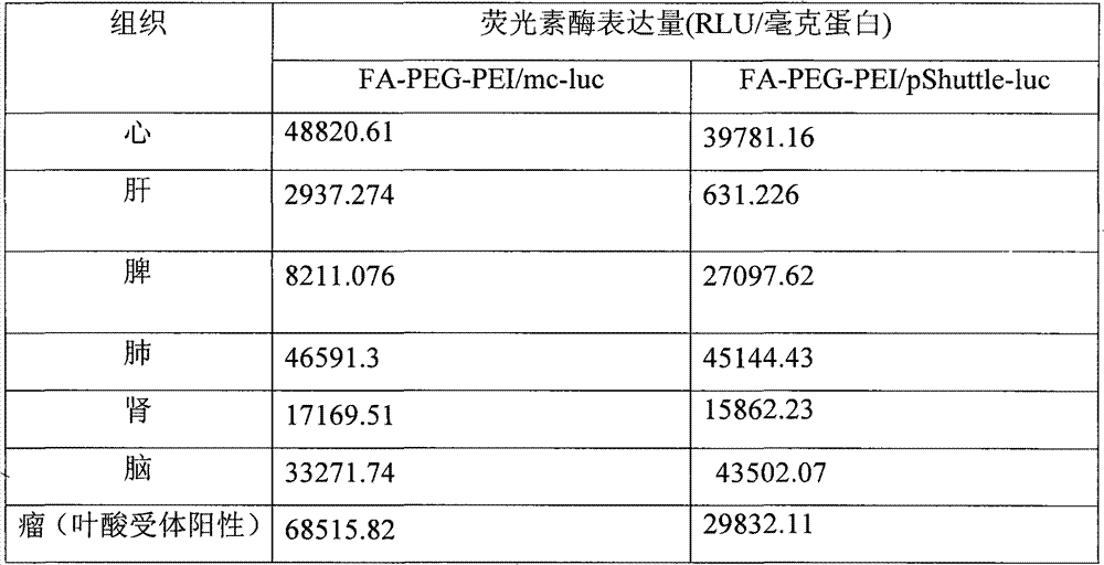
结果如图13所示,可知本发明提供的治疗肿瘤的药物能够在叶酸受体阳性的肿瘤中富集,而在其他组织器官积累很少,证明本发明提供的治疗肿瘤的药物能将目的基因高效特异的运送到肿瘤部位!图13的数据所对应的具体数值如表3所示。The results are shown in Figure 13. It can be seen that the drug for treating tumors provided by the present invention can be enriched in folic acid receptor-positive tumors, while it accumulates very little in other tissues and organs, which proves that the drugs for treating tumors provided by the present invention can target gene Efficient and specific delivery to the tumor site! The specific values corresponding to the data in Figure 13 are shown in Table 3.
表3、荧光素酶的表达量Table 3, the expression level of luciferase

实施例13、动物实验
证明纳米材料携带小环的比携带质粒DNA在肿瘤内的表达量更高,持续时间更长。具体步骤如下:It is proved that nanomaterials carrying small circles have a higher expression level and a longer duration in tumors than those carrying plasmid DNA. Specific steps are as follows:
将实施例6制备的N/P等于6的FA-PEG-PEI/mc-luc和FA-PEG-PEI/pShuttle-luc复合物的溶液,其中非病毒载体包裹的mc-luc或pShuttle-luc 50μg,通过尾静脉注射到接种有叶酸受体阳性荷瘤裸鼠中,7天以后检测目的基因的表达,荧光素酶的表达量如表4所示,图14为mc-luc与pShuttle-luc在体内表达量和表达时间比较。由图可知,目的基因在肿瘤部位富集,而且小环DNA(mc-luc)的表达量和持续时间明显优于质粒DNA(pShuttle-luc)。在受体阳性的肿瘤部位富集比较多,而在受体阴性的肿瘤部位富集很少。The solution of the FA-PEG-PEI/mc-luc and FA-PEG-PEI/pShuttle-luc complex with N/P equal to 6 prepared in Example 6, wherein the non-viral vector-wrapped mc-luc or pShuttle-luc 50 μg , injected into nude mice inoculated with folic acid receptor-positive tumors through the tail vein, and detected the expression of the
表4、荧光素酶的表达量Table 4, the expression level of luciferase
实施例14、动物实验
1)用EMA(ethidium monoazide,叠氮乙锭,购自博麦德)标记pShuttle-luc或mc-luc,得到能够产生红色荧光的pShuttle-luc或mc-luc,将pShuttle-luc或mc-luc用Tris Buffer(pH 8.5)稀释到到1ug/ul,将EMA(10ug/ul)储备液稀释到0.1ug/ul,1∶10(v/v)避光反应30min,UV照射1h(365nm,铺展均匀,较大面积激活),无水乙醇稀释到60%进行DNA沉淀,室温放置15min,21000g离心20min,弃上清,适当去离子水复溶。得到的带红色荧光的小环命名为EMA-mc-luc,得到的带红色荧光的质粒命名为EMA-pShuttle-luc。红色荧光的激发光为488纳米,发生光为630纳米。1) Label pShuttle-luc or mc-luc with EMA (ethidium monoazide, ethidium azide, purchased from Biomed) to obtain pShuttle-luc or mc-luc capable of producing red fluorescence, and pShuttle-luc or mc-luc Dilute to 1ug/ul with Tris Buffer (pH 8.5), dilute EMA (10ug/ul) stock solution to 0.1ug/ul, 1:10 (v/v) dark reaction for 30min, UV irradiation for 1h (365nm, spread Uniformity, larger area activation), dilute to 60% with absolute ethanol for DNA precipitation, place at room temperature for 15 minutes, centrifuge at 21000g for 20 minutes, discard the supernatant, and reconstitute with deionized water. The obtained small circle with red fluorescence was named EMA-mc-luc, and the obtained plasmid with red fluorescence was named EMA-pShuttle-luc. The excitation light of red fluorescence is 488 nanometers, and the generation light is 630 nanometers.
2)按照实施例6的方法制备FA-PEG-PEI/EMA-pShuttle-luc,FA-PEG-PEI/EMA-mc-luc复合物,复合物中质粒浓度为200μg/mL,N/P比为6;2) Prepare FA-PEG-PEI/EMA-pShuttle-luc and FA-PEG-PEI/EMA-mc-luc complex according to the method of Example 6, the plasmid concentration in the complex is 200 μg/mL, and the N/P ratio is 6;
3)制备荷瘤小鼠3) Preparation of tumor-bearing mice
在4-6周的裸鼠(购自北京大学医学部实验动物中心)左掖接种200微升1*106HeLa细胞,右掖接种200微升1*104H22细胞,当肿瘤体积长至0.5-1cm时,用于下述实验。Inoculate 4-6 week old nude mice (purchased from the Experimental Animal Center of Peking University Health Science Center) with 200 microliters of 1*10 6 HeLa cells on the left side and 200 microliters of 1*10 4 H22 cells on the right side. When the tumor volume grows to 0.5 -1cm, used for the following experiments.
4)将步骤1)和2)制备的N/P等于6的FA-PEG-PEI/EMA-mc-luc和FA-PEG-PEI/EMA-pShuttle-luc复合物的溶液,其中非病毒载体包裹的EMA-mc-luc或EMA-pShuttle-luc为50μg,通过尾静脉注射到接种有叶酸受体阳性和叶酸受体阴性的荷瘤小鼠中,3-5小时后,通过活体动物成像检测目的基因在小鼠体内的分布。结果如图17所示,左侧小鼠为FA-PEG-PEI/EMA-pShuttle-luc复合物注射的小鼠,从图中可以看出EMA-pShuttle-luc在左掖肿瘤(叶酸受体阳性肿瘤)部位大量富集,而在右掖(叶酸受体阴性肿瘤部位)分布相对较弱,说明靶向纳米材料主要通过叶酸受体介导的途径进入肿瘤细胞。4) The solution of FA-PEG-PEI/EMA-mc-luc and FA-PEG-PEI/EMA-pShuttle-luc complex with N/P equal to 6 prepared in steps 1) and 2), wherein the non-viral vector is wrapped 50 μg of EMA-mc-luc or EMA-pShuttle-luc was injected via tail vein into tumor-bearing mice inoculated with folate receptor-positive and folate-receptor-negative mice, and after 3-5 hours, the purpose was detected by live animal imaging Gene distribution in mice. The results are shown in Figure 17, the mouse on the left is a mouse injected with the FA-PEG-PEI/EMA-pShuttle-luc complex, and it can be seen from the figure that EMA-pShuttle-luc is in the left tumor (folate receptor positive Tumors) were enriched in large quantities, while the distribution in the right side (folate receptor-negative tumors) was relatively weak, indicating that targeted nanomaterials mainly entered tumor cells through folate receptor-mediated pathways.
右侧小鼠为FA-PEG-PEI/EMA-mc-luc复合物注射的小鼠,从图中可以看出EMA-mc-luc在左掖肿瘤(叶酸受体阳性肿瘤)部位大量富集,而在右掖(叶酸受体阴性肿瘤部位)分布相对较弱,说明靶向纳米材料主要通过叶酸受体介导的途径进入肿瘤细胞。The mouse on the right side is a mouse injected with FA-PEG-PEI/EMA-mc-luc complex. It can be seen from the figure that EMA-mc-luc is enriched in a large amount in the left tumor (folate receptor positive tumor). However, the distribution in the right side (folate receptor-negative tumor site) was relatively weak, indicating that targeted nanomaterials mainly entered tumor cells through folate receptor-mediated pathways.
Claims (14)


Priority Applications (1)
| Application Number | Priority Date | Filing Date | Title |
|---|---|---|---|
| CN2009100930813A CN102030754B (en) | 2009-09-28 | 2009-09-28 | Non-viral vector, and preparation method and application thereof |
Applications Claiming Priority (1)
| Application Number | Priority Date | Filing Date | Title |
|---|---|---|---|
| CN2009100930813A CN102030754B (en) | 2009-09-28 | 2009-09-28 | Non-viral vector, and preparation method and application thereof |
Publications (2)
| Publication Number | Publication Date |
|---|---|
| CN102030754A CN102030754A (en) | 2011-04-27 |
| CN102030754B true CN102030754B (en) | 2013-07-10 |
Family
ID=43884263
Family Applications (1)
| Application Number | Title | Priority Date | Filing Date |
|---|---|---|---|
| CN2009100930813A Expired - Fee Related CN102030754B (en) | 2009-09-28 | 2009-09-28 | Non-viral vector, and preparation method and application thereof |
Country Status (1)
| Country | Link |
|---|---|
| CN (1) | CN102030754B (en) |
Families Citing this family (3)
| Publication number | Priority date | Publication date | Assignee | Title |
|---|---|---|---|---|
| CN103333169B (en) * | 2013-01-10 | 2014-07-02 | 青岛大学 | Sulfhydrylated folic acid and preparation method thereof |
| LT3027646T (en) * | 2013-07-31 | 2018-09-25 | Novartis Ag | Novel selection vectors and methods of selecting eukaryotic host cells |
| TW202421200A (en) * | 2022-11-09 | 2024-06-01 | 大陸商同宜醫藥(蘇州)有限公司 | Treatment of cancer by means of administration of ligand-medicament conjugate |
-
2009
- 2009-09-28 CN CN2009100930813A patent/CN102030754B/en not_active Expired - Fee Related
Non-Patent Citations (1)
| Title |
|---|
| Atkinson Sarah F.等."Conjugation of folate via gelonin carbohydrate residues retains ribosomal-inactivating properties of the toxin and permits targeting to folate receptor positive cells".《Journal of Biological Chemistry》.2001,第276卷(第30期),第27931页第1栏第3段、图1和第2栏第3段. |
Also Published As
| Publication number | Publication date |
|---|---|
| CN102030754A (en) | 2011-04-27 |
Similar Documents
| Publication | Publication Date | Title |
|---|---|---|
| CN101035835B (en) | Biodegradable cationic polymers | |
| CN1330765C (en) | Polycationic water-soluble copolymers and methods for delivering polyanionic macromolecules across biological barriers | |
| CN102274521B (en) | Graphene oxide-based target gene vector material and preparation and use thereof | |
| ES2744827T3 (en) | Polymer derived from linear polyethyleneimine for gene transfer | |
| CN102380103B (en) | Mannose-modified thiolated chitosan quaternary ammonium salt nanoparticle, preparing method and application thereof | |
| CN105727307B (en) | A nano-polypeptide carrier modified with lipoic acid and its preparation method and application | |
| O’Mahony et al. | In vitro investigations of the efficacy of cyclodextrin-siRNA complexes modified with lipid-PEG-Octaarginine: towards a formulation strategy for non-viral neuronal siRNA delivery | |
| JPH10502918A (en) | Compositions containing nucleic acids, their production and use | |
| CN102397554B (en) | Tumor-targeting double-drug carrying and delivery system and preparation method thereof | |
| CN104910252B (en) | A kind of pH response type lipids based on dendrimer and preparation method and application | |
| CN102260356B (en) | Chitosan derivative used as gene vector, and preparation method and application thereof | |
| Hsieh et al. | Oral gene delivery with cyclo-(D-Trp-Tyr) peptide nanotubes | |
| CN104983716A (en) | Tumor cell membrane/nuclear membrane double-targeting tumor nano-drug slow-release system and preparation and application thereof | |
| Li et al. | A novel cationic liposome formulation for efficient gene delivery via a pulmonary route | |
| CN107129522B (en) | Lipoic acid modified inherent disordered protein nano-carrier and preparation method and application thereof | |
| CN103665169B (en) | Genophore that three functional peptides are modified and preparation method thereof and application | |
| CN106798923A (en) | Function targeting vector material PEG-DSPE-PEI compounds and its liposome of modification | |
| CN102030754B (en) | Non-viral vector, and preparation method and application thereof | |
| CN103143033A (en) | A carrier targeting liver cells and its preparation method | |
| Liu et al. | Non‐viral nucleic acid delivery system for RNA therapeutics | |
| CN115151278A (en) | Tumor-targeted polypeptide nanoparticle delivery system for nucleic acid therapy | |
| CN115927480A (en) | Gene delivery system based on nucleic acid nanostructure and preparation method and application thereof | |
| CN102517332B (en) | EGF (epidermal growth factor)-modified PAMAM (polyamidoamine) self-assembled composite for transgenosis as well as preparation method and applications thereof | |
| JP5510808B2 (en) | Nucleic acid introduction agent comprising organic nanotubes | |
| CN118307765A (en) | Preparation method and application of amino acid modified poly (β-amino ester)-polyethylene glycol carrier for delivering nucleic acid drugs |
Legal Events
| Date | Code | Title | Description |
|---|---|---|---|
| C06 | Publication | ||
| PB01 | Publication | ||
| C10 | Entry into substantive examination | ||
| SE01 | Entry into force of request for substantive examination | ||
| C14 | Grant of patent or utility model | ||
| GR01 | Patent grant | ||
| CF01 | Termination of patent right due to non-payment of annual fee |
Granted publication date: 20130710 Termination date: 20200928 |
|
| CF01 | Termination of patent right due to non-payment of annual fee |