CN101918855B - MRI surgical systems for real-time visualizations using MRI image data and predefined data of surgical tools - Google Patents
MRI surgical systems for real-time visualizations using MRI image data and predefined data of surgical tools Download PDFInfo
- Publication number
- CN101918855B CN101918855B CN2008801173424A CN200880117342A CN101918855B CN 101918855 B CN101918855 B CN 101918855B CN 2008801173424 A CN2008801173424 A CN 2008801173424A CN 200880117342 A CN200880117342 A CN 200880117342A CN 101918855 B CN101918855 B CN 101918855B
- Authority
- CN
- China
- Prior art keywords
- trajectory
- patient
- circuitry
- mri
- data
- Prior art date
- Legal status (The legal status is an assumption and is not a legal conclusion. Google has not performed a legal analysis and makes no representation as to the accuracy of the status listed.)
- Expired - Fee Related
Links
Images


Classifications
-
- G—PHYSICS
- G01—MEASURING; TESTING
- G01R—MEASURING ELECTRIC VARIABLES; MEASURING MAGNETIC VARIABLES
- G01R33/00—Arrangements or instruments for measuring magnetic variables
- G01R33/20—Arrangements or instruments for measuring magnetic variables involving magnetic resonance
- G01R33/28—Details of apparatus provided for in groups G01R33/44 - G01R33/64
- G01R33/283—Intercom or optical viewing arrangements, structurally associated with NMR apparatus
-
- A—HUMAN NECESSITIES
- A61—MEDICAL OR VETERINARY SCIENCE; HYGIENE
- A61B—DIAGNOSIS; SURGERY; IDENTIFICATION
- A61B34/00—Computer-aided surgery; Manipulators or robots specially adapted for use in surgery
- A61B34/70—Manipulators specially adapted for use in surgery
- A61B34/71—Manipulators operated by drive cable mechanisms
-
- A—HUMAN NECESSITIES
- A61—MEDICAL OR VETERINARY SCIENCE; HYGIENE
- A61B—DIAGNOSIS; SURGERY; IDENTIFICATION
- A61B34/00—Computer-aided surgery; Manipulators or robots specially adapted for use in surgery
- A61B34/70—Manipulators specially adapted for use in surgery
- A61B34/74—Manipulators with manual electric input means
-
- A—HUMAN NECESSITIES
- A61—MEDICAL OR VETERINARY SCIENCE; HYGIENE
- A61B—DIAGNOSIS; SURGERY; IDENTIFICATION
- A61B5/00—Measuring for diagnostic purposes; Identification of persons
- A61B5/05—Detecting, measuring or recording for diagnosis by means of electric currents or magnetic fields; Measuring using microwaves or radio waves
- A61B5/055—Detecting, measuring or recording for diagnosis by means of electric currents or magnetic fields; Measuring using microwaves or radio waves involving electronic [EMR] or nuclear [NMR] magnetic resonance, e.g. magnetic resonance imaging
-
- A—HUMAN NECESSITIES
- A61—MEDICAL OR VETERINARY SCIENCE; HYGIENE
- A61B—DIAGNOSIS; SURGERY; IDENTIFICATION
- A61B5/00—Measuring for diagnostic purposes; Identification of persons
- A61B5/70—Means for positioning the patient in relation to the detecting, measuring or recording means
- A61B5/702—Posture restraints
-
- A—HUMAN NECESSITIES
- A61—MEDICAL OR VETERINARY SCIENCE; HYGIENE
- A61B—DIAGNOSIS; SURGERY; IDENTIFICATION
- A61B90/00—Instruments, implements or accessories specially adapted for surgery or diagnosis and not covered by any of the groups A61B1/00 - A61B50/00, e.g. for luxation treatment or for protecting wound edges
- A61B90/10—Instruments, implements or accessories specially adapted for surgery or diagnosis and not covered by any of the groups A61B1/00 - A61B50/00, e.g. for luxation treatment or for protecting wound edges for stereotaxic surgery, e.g. frame-based stereotaxis
- A61B90/11—Instruments, implements or accessories specially adapted for surgery or diagnosis and not covered by any of the groups A61B1/00 - A61B50/00, e.g. for luxation treatment or for protecting wound edges for stereotaxic surgery, e.g. frame-based stereotaxis with guides for needles or instruments, e.g. arcuate slides or ball joints
-
- G—PHYSICS
- G01—MEASURING; TESTING
- G01R—MEASURING ELECTRIC VARIABLES; MEASURING MAGNETIC VARIABLES
- G01R33/00—Arrangements or instruments for measuring magnetic variables
- G01R33/20—Arrangements or instruments for measuring magnetic variables involving magnetic resonance
- G01R33/28—Details of apparatus provided for in groups G01R33/44 - G01R33/64
- G01R33/285—Invasive instruments, e.g. catheters or biopsy needles, specially adapted for tracking, guiding or visualization by NMR
- G01R33/286—Invasive instruments, e.g. catheters or biopsy needles, specially adapted for tracking, guiding or visualization by NMR involving passive visualization of interventional instruments, i.e. making the instrument visible as part of the normal MR process
-
- G—PHYSICS
- G01—MEASURING; TESTING
- G01R—MEASURING ELECTRIC VARIABLES; MEASURING MAGNETIC VARIABLES
- G01R33/00—Arrangements or instruments for measuring magnetic variables
- G01R33/20—Arrangements or instruments for measuring magnetic variables involving magnetic resonance
- G01R33/28—Details of apparatus provided for in groups G01R33/44 - G01R33/64
- G01R33/285—Invasive instruments, e.g. catheters or biopsy needles, specially adapted for tracking, guiding or visualization by NMR
- G01R33/287—Invasive instruments, e.g. catheters or biopsy needles, specially adapted for tracking, guiding or visualization by NMR involving active visualization of interventional instruments, e.g. using active tracking RF coils or coils for intentionally creating magnetic field inhomogeneities
-
- G—PHYSICS
- G01—MEASURING; TESTING
- G01R—MEASURING ELECTRIC VARIABLES; MEASURING MAGNETIC VARIABLES
- G01R33/00—Arrangements or instruments for measuring magnetic variables
- G01R33/20—Arrangements or instruments for measuring magnetic variables involving magnetic resonance
- G01R33/28—Details of apparatus provided for in groups G01R33/44 - G01R33/64
- G01R33/32—Excitation or detection systems, e.g. using radio frequency signals
- G01R33/34—Constructional details, e.g. resonators, specially adapted to MR
- G01R33/34046—Volume type coils, e.g. bird-cage coils; Quadrature bird-cage coils; Circularly polarised coils
-
- G—PHYSICS
- G01—MEASURING; TESTING
- G01R—MEASURING ELECTRIC VARIABLES; MEASURING MAGNETIC VARIABLES
- G01R33/00—Arrangements or instruments for measuring magnetic variables
- G01R33/20—Arrangements or instruments for measuring magnetic variables involving magnetic resonance
- G01R33/28—Details of apparatus provided for in groups G01R33/44 - G01R33/64
- G01R33/32—Excitation or detection systems, e.g. using radio frequency signals
- G01R33/34—Constructional details, e.g. resonators, specially adapted to MR
- G01R33/341—Constructional details, e.g. resonators, specially adapted to MR comprising surface coils
- G01R33/3415—Constructional details, e.g. resonators, specially adapted to MR comprising surface coils comprising arrays of sub-coils, i.e. phased-array coils with flexible receiver channels
-
- A—HUMAN NECESSITIES
- A61—MEDICAL OR VETERINARY SCIENCE; HYGIENE
- A61B—DIAGNOSIS; SURGERY; IDENTIFICATION
- A61B17/00—Surgical instruments, devices or methods
- A61B2017/00017—Electrical control of surgical instruments
- A61B2017/00212—Electrical control of surgical instruments using remote controls
-
- A—HUMAN NECESSITIES
- A61—MEDICAL OR VETERINARY SCIENCE; HYGIENE
- A61B—DIAGNOSIS; SURGERY; IDENTIFICATION
- A61B17/00—Surgical instruments, devices or methods
- A61B2017/00831—Material properties
- A61B2017/00902—Material properties transparent or translucent
- A61B2017/00911—Material properties transparent or translucent for fields applied by a magnetic resonance imaging system
-
- A—HUMAN NECESSITIES
- A61—MEDICAL OR VETERINARY SCIENCE; HYGIENE
- A61B—DIAGNOSIS; SURGERY; IDENTIFICATION
- A61B17/00—Surgical instruments, devices or methods
- A61B17/34—Trocars; Puncturing needles
- A61B17/3403—Needle locating or guiding means
- A61B2017/3405—Needle locating or guiding means using mechanical guide means
- A61B2017/3407—Needle locating or guiding means using mechanical guide means including a base for support on the body
-
- A—HUMAN NECESSITIES
- A61—MEDICAL OR VETERINARY SCIENCE; HYGIENE
- A61B—DIAGNOSIS; SURGERY; IDENTIFICATION
- A61B17/00—Surgical instruments, devices or methods
- A61B17/34—Trocars; Puncturing needles
- A61B17/3403—Needle locating or guiding means
- A61B2017/3405—Needle locating or guiding means using mechanical guide means
- A61B2017/3409—Needle locating or guiding means using mechanical guide means including needle or instrument drives
-
- A—HUMAN NECESSITIES
- A61—MEDICAL OR VETERINARY SCIENCE; HYGIENE
- A61B—DIAGNOSIS; SURGERY; IDENTIFICATION
- A61B34/00—Computer-aided surgery; Manipulators or robots specially adapted for use in surgery
- A61B34/10—Computer-aided planning, simulation or modelling of surgical operations
- A61B2034/101—Computer-aided simulation of surgical operations
-
- A—HUMAN NECESSITIES
- A61—MEDICAL OR VETERINARY SCIENCE; HYGIENE
- A61B—DIAGNOSIS; SURGERY; IDENTIFICATION
- A61B34/00—Computer-aided surgery; Manipulators or robots specially adapted for use in surgery
- A61B34/10—Computer-aided planning, simulation or modelling of surgical operations
- A61B2034/107—Visualisation of planned trajectories or target regions
-
- A—HUMAN NECESSITIES
- A61—MEDICAL OR VETERINARY SCIENCE; HYGIENE
- A61B—DIAGNOSIS; SURGERY; IDENTIFICATION
- A61B34/00—Computer-aided surgery; Manipulators or robots specially adapted for use in surgery
- A61B34/25—User interfaces for surgical systems
- A61B2034/252—User interfaces for surgical systems indicating steps of a surgical procedure
-
- A—HUMAN NECESSITIES
- A61—MEDICAL OR VETERINARY SCIENCE; HYGIENE
- A61B—DIAGNOSIS; SURGERY; IDENTIFICATION
- A61B34/00—Computer-aided surgery; Manipulators or robots specially adapted for use in surgery
- A61B34/25—User interfaces for surgical systems
- A61B2034/254—User interfaces for surgical systems being adapted depending on the stage of the surgical procedure
-
- A—HUMAN NECESSITIES
- A61—MEDICAL OR VETERINARY SCIENCE; HYGIENE
- A61B—DIAGNOSIS; SURGERY; IDENTIFICATION
- A61B34/00—Computer-aided surgery; Manipulators or robots specially adapted for use in surgery
- A61B34/25—User interfaces for surgical systems
- A61B2034/256—User interfaces for surgical systems having a database of accessory information, e.g. including context sensitive help or scientific articles
-
- A—HUMAN NECESSITIES
- A61—MEDICAL OR VETERINARY SCIENCE; HYGIENE
- A61B—DIAGNOSIS; SURGERY; IDENTIFICATION
- A61B90/00—Instruments, implements or accessories specially adapted for surgery or diagnosis and not covered by any of the groups A61B1/00 - A61B50/00, e.g. for luxation treatment or for protecting wound edges
- A61B90/10—Instruments, implements or accessories specially adapted for surgery or diagnosis and not covered by any of the groups A61B1/00 - A61B50/00, e.g. for luxation treatment or for protecting wound edges for stereotaxic surgery, e.g. frame-based stereotaxis
- A61B2090/103—Cranial plugs for access to brain
-
- A—HUMAN NECESSITIES
- A61—MEDICAL OR VETERINARY SCIENCE; HYGIENE
- A61B—DIAGNOSIS; SURGERY; IDENTIFICATION
- A61B90/00—Instruments, implements or accessories specially adapted for surgery or diagnosis and not covered by any of the groups A61B1/00 - A61B50/00, e.g. for luxation treatment or for protecting wound edges
- A61B90/30—Devices for illuminating a surgical field, the devices having an interrelation with other surgical devices or with a surgical procedure
- A61B2090/306—Devices for illuminating a surgical field, the devices having an interrelation with other surgical devices or with a surgical procedure using optical fibres
-
- A—HUMAN NECESSITIES
- A61—MEDICAL OR VETERINARY SCIENCE; HYGIENE
- A61B—DIAGNOSIS; SURGERY; IDENTIFICATION
- A61B90/00—Instruments, implements or accessories specially adapted for surgery or diagnosis and not covered by any of the groups A61B1/00 - A61B50/00, e.g. for luxation treatment or for protecting wound edges
- A61B90/36—Image-producing devices or illumination devices not otherwise provided for
- A61B90/361—Image-producing devices, e.g. surgical cameras
- A61B2090/3614—Image-producing devices, e.g. surgical cameras using optical fibre
-
- A—HUMAN NECESSITIES
- A61—MEDICAL OR VETERINARY SCIENCE; HYGIENE
- A61B—DIAGNOSIS; SURGERY; IDENTIFICATION
- A61B90/00—Instruments, implements or accessories specially adapted for surgery or diagnosis and not covered by any of the groups A61B1/00 - A61B50/00, e.g. for luxation treatment or for protecting wound edges
- A61B90/36—Image-producing devices or illumination devices not otherwise provided for
- A61B90/37—Surgical systems with images on a monitor during operation
- A61B2090/373—Surgical systems with images on a monitor during operation using light, e.g. by using optical scanners
-
- A—HUMAN NECESSITIES
- A61—MEDICAL OR VETERINARY SCIENCE; HYGIENE
- A61B—DIAGNOSIS; SURGERY; IDENTIFICATION
- A61B90/00—Instruments, implements or accessories specially adapted for surgery or diagnosis and not covered by any of the groups A61B1/00 - A61B50/00, e.g. for luxation treatment or for protecting wound edges
- A61B90/36—Image-producing devices or illumination devices not otherwise provided for
- A61B90/37—Surgical systems with images on a monitor during operation
- A61B2090/374—NMR or MRI
-
- A—HUMAN NECESSITIES
- A61—MEDICAL OR VETERINARY SCIENCE; HYGIENE
- A61B—DIAGNOSIS; SURGERY; IDENTIFICATION
- A61B90/00—Instruments, implements or accessories specially adapted for surgery or diagnosis and not covered by any of the groups A61B1/00 - A61B50/00, e.g. for luxation treatment or for protecting wound edges
- A61B90/39—Markers, e.g. radio-opaque or breast lesions markers
- A61B2090/3933—Liquid markers
-
- A—HUMAN NECESSITIES
- A61—MEDICAL OR VETERINARY SCIENCE; HYGIENE
- A61B—DIAGNOSIS; SURGERY; IDENTIFICATION
- A61B90/00—Instruments, implements or accessories specially adapted for surgery or diagnosis and not covered by any of the groups A61B1/00 - A61B50/00, e.g. for luxation treatment or for protecting wound edges
- A61B90/39—Markers, e.g. radio-opaque or breast lesions markers
- A61B2090/3937—Visible markers
- A61B2090/395—Visible markers with marking agent for marking skin or other tissue
-
- A—HUMAN NECESSITIES
- A61—MEDICAL OR VETERINARY SCIENCE; HYGIENE
- A61B—DIAGNOSIS; SURGERY; IDENTIFICATION
- A61B90/00—Instruments, implements or accessories specially adapted for surgery or diagnosis and not covered by any of the groups A61B1/00 - A61B50/00, e.g. for luxation treatment or for protecting wound edges
- A61B90/39—Markers, e.g. radio-opaque or breast lesions markers
- A61B2090/3954—Markers, e.g. radio-opaque or breast lesions markers magnetic, e.g. NMR or MRI
-
- A—HUMAN NECESSITIES
- A61—MEDICAL OR VETERINARY SCIENCE; HYGIENE
- A61B—DIAGNOSIS; SURGERY; IDENTIFICATION
- A61B90/00—Instruments, implements or accessories specially adapted for surgery or diagnosis and not covered by any of the groups A61B1/00 - A61B50/00, e.g. for luxation treatment or for protecting wound edges
- A61B90/39—Markers, e.g. radio-opaque or breast lesions markers
- A61B2090/3983—Reference marker arrangements for use with image guided surgery
-
- A—HUMAN NECESSITIES
- A61—MEDICAL OR VETERINARY SCIENCE; HYGIENE
- A61B—DIAGNOSIS; SURGERY; IDENTIFICATION
- A61B34/00—Computer-aided surgery; Manipulators or robots specially adapted for use in surgery
- A61B34/10—Computer-aided planning, simulation or modelling of surgical operations
-
- A—HUMAN NECESSITIES
- A61—MEDICAL OR VETERINARY SCIENCE; HYGIENE
- A61B—DIAGNOSIS; SURGERY; IDENTIFICATION
- A61B34/00—Computer-aided surgery; Manipulators or robots specially adapted for use in surgery
- A61B34/25—User interfaces for surgical systems
-
- A—HUMAN NECESSITIES
- A61—MEDICAL OR VETERINARY SCIENCE; HYGIENE
- A61B—DIAGNOSIS; SURGERY; IDENTIFICATION
- A61B5/00—Measuring for diagnostic purposes; Identification of persons
- A61B5/40—Detecting, measuring or recording for evaluating the nervous system
- A61B5/4058—Detecting, measuring or recording for evaluating the nervous system for evaluating the central nervous system
- A61B5/4064—Evaluating the brain
-
- A—HUMAN NECESSITIES
- A61—MEDICAL OR VETERINARY SCIENCE; HYGIENE
- A61B—DIAGNOSIS; SURGERY; IDENTIFICATION
- A61B90/00—Instruments, implements or accessories specially adapted for surgery or diagnosis and not covered by any of the groups A61B1/00 - A61B50/00, e.g. for luxation treatment or for protecting wound edges
- A61B90/10—Instruments, implements or accessories specially adapted for surgery or diagnosis and not covered by any of the groups A61B1/00 - A61B50/00, e.g. for luxation treatment or for protecting wound edges for stereotaxic surgery, e.g. frame-based stereotaxis
- A61B90/14—Fixators for body parts, e.g. skull clamps; Constructional details of fixators, e.g. pins
-
- A—HUMAN NECESSITIES
- A61—MEDICAL OR VETERINARY SCIENCE; HYGIENE
- A61B—DIAGNOSIS; SURGERY; IDENTIFICATION
- A61B90/00—Instruments, implements or accessories specially adapted for surgery or diagnosis and not covered by any of the groups A61B1/00 - A61B50/00, e.g. for luxation treatment or for protecting wound edges
- A61B90/36—Image-producing devices or illumination devices not otherwise provided for
- A61B90/361—Image-producing devices, e.g. surgical cameras
-
- A—HUMAN NECESSITIES
- A61—MEDICAL OR VETERINARY SCIENCE; HYGIENE
- A61B—DIAGNOSIS; SURGERY; IDENTIFICATION
- A61B90/00—Instruments, implements or accessories specially adapted for surgery or diagnosis and not covered by any of the groups A61B1/00 - A61B50/00, e.g. for luxation treatment or for protecting wound edges
- A61B90/36—Image-producing devices or illumination devices not otherwise provided for
- A61B90/37—Surgical systems with images on a monitor during operation
-
- A—HUMAN NECESSITIES
- A61—MEDICAL OR VETERINARY SCIENCE; HYGIENE
- A61N—ELECTROTHERAPY; MAGNETOTHERAPY; RADIATION THERAPY; ULTRASOUND THERAPY
- A61N1/00—Electrotherapy; Circuits therefor
- A61N1/18—Applying electric currents by contact electrodes
- A61N1/32—Applying electric currents by contact electrodes alternating or intermittent currents
- A61N1/36—Applying electric currents by contact electrodes alternating or intermittent currents for stimulation
- A61N1/372—Arrangements in connection with the implantation of stimulators
Landscapes
- Health & Medical Sciences (AREA)
- Physics & Mathematics (AREA)
- Life Sciences & Earth Sciences (AREA)
- Surgery (AREA)
- General Health & Medical Sciences (AREA)
- Engineering & Computer Science (AREA)
- Condensed Matter Physics & Semiconductors (AREA)
- General Physics & Mathematics (AREA)
- Nuclear Medicine, Radiotherapy & Molecular Imaging (AREA)
- Pathology (AREA)
- Heart & Thoracic Surgery (AREA)
- Veterinary Medicine (AREA)
- Biomedical Technology (AREA)
- Medical Informatics (AREA)
- Molecular Biology (AREA)
- Animal Behavior & Ethology (AREA)
- Public Health (AREA)
- Robotics (AREA)
- Biophysics (AREA)
- High Energy & Nuclear Physics (AREA)
- Radiology & Medical Imaging (AREA)
- Oral & Maxillofacial Surgery (AREA)
- Physical Education & Sports Medicine (AREA)
- Magnetic Resonance Imaging Apparatus (AREA)
Abstract
Description
相关申请related application
本申请要求2008年6月6日提交的美国申请序列号No.12/134,412的优先权,并且还要求2007年9月24日提交的美国临时申请序列号No.60/974,821的优先权,通过参考将它们的内容结合于此,就如在本文中完全陈述一样。This application claims priority to U.S. Application Serial No. 12/134,412, filed June 6, 2008, and also claims priority to U.S. Provisional Application Serial No. 60/974,821, filed September 24, 2007, by References are hereby incorporated in their content as if fully set forth herein.
技术领域 technical field
本发明涉及可以特别适合于治疗和/或介入医疗器械在身体中的放置/定位的MRI引导诊断或介入系统。本发明的实施例可以特别适合于放置诸如深部脑刺激(“DBS”)引线之类的神经调节引线、放置可植入式副交感或交感神经链引线和/或CNS刺激引线以及/或者适合于将包括心房颤动(AFIB)治疗的治疗递送到身体中的目标内部位置。The present invention relates to an MRI-guided diagnostic or interventional system that may be particularly suitable for the placement/positioning of therapeutic and/or interventional medical devices in the body. Embodiments of the present invention may be particularly suitable for placement of neuromodulation leads such as deep brain stimulation ("DBS") leads, placement of implantable parasympathetic or sympathetic chain leads and/or CNS stimulation leads, and/or for placement of Therapy, including atrial fibrillation (AFIB) therapy, is delivered to a targeted internal location in the body.
背景技术 Background technique
深部脑刺激(DBS)正变成遭受慢性疼痛、帕金森病或发作以及其它医疗状况的患者的神经外科手术治疗的可接受的治疗形态。也已实行或提议了使用交感神经链和/或脊髓的内部刺激等等的其它电刺激治疗。Deep brain stimulation (DBS) is becoming an accepted treatment modality for neurosurgical treatment of patients suffering from chronic pain, Parkinson's disease or episodes, and other medical conditions. Other electrical stimulation treatments using internal stimulation of the sympathetic chain and/or spinal cord, etc. have also been practiced or proposed.
现有技术的DBS系统的一个实例是来自Medtronic有限公司的

认为特定医疗过程(特别是使用DBS的那些医疗过程)的临床结果可以取决于与感兴趣的组织接触的电极的精确位置。例如,为了治疗帕金森颤抖,通常基于预先操作的MRI和CT图像,在立体定向外科手术期间将DBS刺激引线植入。这些过程的持续时间会很长,并且具有减小的功效,正如所报道的那样,在大约30%的植入这些器械的患者中,器械/过程的临床功效要比最佳功效要低。It is believed that the clinical outcome of certain medical procedures, particularly those using DBS, may depend on the precise location of the electrodes in contact with the tissue of interest. For example, to treat Parkinsonian tremors, DBS stimulation leads are implanted during stereotaxic surgery, usually based on pre-manipulated MRI and CT images. These procedures can be of prolonged duration and have reduced efficacy, as reported in approximately 30% of patients implanted with these devices have less than optimal clinical efficacy of the device/procedure.
发明内容 Contents of the invention
本发明的一些实施例涉及MRI引导系统,该MRI引导系统可以基本上实时地生成患者以及逻辑空间中的一个或多个外科手术工具的患者特定可视化(visualization),并且向临床医生提供反馈以改善体内过程的速度和/或可靠性。Some embodiments of the invention relate to MRI guidance systems that can generate patient-specific visualizations of the patient and one or more surgical tools in logical space substantially in real time, and provide feedback to the clinician to improve Speed and/or reliability of in vivo processes.
该可视化可以(部分)基于(多个)工具的预定义数据,所述预定义数据基于(多个)工具的预先定义的特性来限定系统(例如软件)的接口点,所述(多个)工具的预先定义的特性例如是一个或多个外科手术工具的尺寸、形状或配置以及/或者已知的旋转、平移以及/或者其它功能以及/或者动态行为。该可视化可以包括患者的功能数据(例如在限定的刺激期间脑的活动区域、fMRI数据、电活动、纤维轨迹等等)。This visualization may be based (in part) on predefined data of the tool(s) defining interface points of the system (e.g. software) based on predefined properties of the tool(s), the A predefined characteristic of a tool is, for example, the size, shape or configuration and/or known rotational, translational and/or other functional and/or dynamic behavior of one or more surgical tools. The visualization may include functional data of the patient (eg active regions of the brain during defined stimulation periods, fMRI data, electrical activity, fiber trajectories, etc.).
该系统可以被配置成询问且分割图像数据以定位基准标记,并且使用MRI图像数据和(多个)工具的先验数据来生成(多个)工具和患者的解剖结构的连续可视化,从而提供(基本上实时的)患者的可视化。The system can be configured to interrogate and segment the image data to locate fiducial markers, and use the MRI image data and the tool(s) prior data to generate a continuous visualization of the tool(s) and the patient's anatomy to provide ( substantially real-time) visualization of the patient.
一些实施例涉及MRI引导外科手术系统。该系统包括:(a)至少一个MRI兼容的外科手术工具;(b)适于与MRI扫描仪通信的电路;以及(c)与所述电路通信的至少一个显示器。该电路电子地识别所述至少一个工具的预先定义的物理特性以自动分割由所述MRI扫描仪提供的MR图像数据,由此所述至少一个工具构成与该系统的接口点。该电路被配置成提供用户界面,所述用户界面定义MRI引导外科手术过程的工作流程进展并且允许用户选择该工作流程中的步骤,并且其中所述电路被配置成在外科手术过程期间使用来自患者的MRI图像的数据以及所述至少一个工具的预定义数据来基本上实时地生成多维可视化。Some embodiments relate to MRI guided surgery systems. The system includes: (a) at least one MRI-compatible surgical tool; (b) circuitry adapted to communicate with the MRI scanner; and (c) at least one display in communication with the circuitry. The circuitry electronically recognizes predefined physical characteristics of the at least one tool to automatically segment MR image data provided by the MRI scanner, whereby the at least one tool constitutes an interface point with the system. The circuitry is configured to provide a user interface that defines the workflow progression of the MRI-guided surgical procedure and allows the user to select steps in the workflow, and wherein the circuitry is configured to use data from the patient during the surgical procedure. The data of the MRI image and the predefined data of the at least one tool are used to generate the multidimensional visualization substantially in real time.
其他实施例涉及用于执行MRI引导外科手术过程的方法。该方法包括:(a)定义至少一个MRI兼容的外科手术工具的尺寸和/或功能数据:(b)获得患者的MRI图像数据;(c)基于所述定义步骤电子地分割所述MRI图像数据以在所述至少一个工具上标识已知的基准标记;(d)生成配准到患者解剖结构的所述至少一个工具的可视化;(e)电子地生成对俯仰(pitch)、滚动(roll)或X-Y致动器的调整方向,以调整轨迹导引的轨迹;以及(f)使用患者MRI图像数据、所述调整方向以及所述可视化来将所述工具引导到患者中的位置,从而促进MRI引导外科手术过程。Other embodiments relate to methods for performing MRI-guided surgical procedures. The method comprises: (a) defining dimensional and/or functional data of at least one MRI compatible surgical tool; (b) obtaining MRI image data of a patient; (c) electronically segmenting said MRI image data based on said defining step to identify known fiducial markers on the at least one tool; (d) generate a visualization of the at least one tool registered to the patient's anatomy; (e) electronically generate references to pitch, roll, or the adjustment direction of the X-Y actuator to adjust the trajectory of the trajectory guide; and (f) using the patient MRI image data, the adjustment direction and the visualization to guide the tool to a position in the patient, thereby facilitating the MRI Guide the surgical procedure.
还有其他实施例涉及用于促进MRI引导外科手术过程的计算机程序产品。该计算机程序产品包括计算机可读存储介质,其具有包括在所述介质中的计算机可读程序代码。该计算机可读程序代码包括:(a)包括多个不同的外科手术工具的预先定义的物理数据的计算机可读程序代码;(b)与MRI扫描仪通信以获得患者的MRI图像数据的计算机可读程序代码;以及(c)使用患者的图像数据以及来自所述工具的数据来基本上实时地生成患者的可视化的计算机可读程序代码。Still other embodiments relate to computer program products for facilitating MRI-guided surgical procedures. The computer program product includes a computer readable storage medium having computer readable program code embodied in said medium. The computer readable program code includes: (a) computer readable program code including predefined physical data for a plurality of different surgical tools; (b) computer readable program code for communicating with an MRI scanner to obtain MRI image data of a patient reading the program code; and (c) using the image data of the patient and the data from the tool to generate computer readable program code for visualization of the patient in substantially real time.
又有其他实施例涉及MRI引导介入深部脑系统。该系统包括:(a)MRI扫描仪;(b)具有电路和显示器的临床工作站,所述工作站与所述MRI扫描仪通信;(c)在其上具有网格的至少一个柔性补片,其被配置成可释放地贴附到患者的颅骨;以及(d)可贴附到患者的颅骨的至少一个轨迹导引。该导引具有带孔的基座,所述孔被配置成位于形成在患者的颅骨中的钻洞上。基座孔为与轨迹导引相关联的枢轴提供旋转的机械中心,基座具有围绕该基座孔间隔开的多个基准标记。所述电路包括关于所述补片的物理数据,并且被配置成询问由所述MRI扫描仪提供的患者成像数据且分割所述图像数据以限定使所述补片与期望的脑内轨迹相交的钻洞位置。所述电路包括轨迹导引的工具特定数据,并且被配置成询问由所述MRI扫描仪提供的患者成像数据且交互地生成到所述显示器的轨迹导引和患者的脑的可视化。Yet other embodiments relate to MRI-guided intervention in deep brain systems. The system includes: (a) an MRI scanner; (b) a clinical workstation having circuitry and a display in communication with the MRI scanner; (c) at least one flexible patch having a mesh thereon, which configured to releasably attach to the patient's skull; and (d) at least one trajectory guide attachable to the patient's skull. The guide has a base with holes configured to sit over a bore formed in the patient's skull. A base hole provides a mechanical center of rotation for a pivot associated with the track guide, and the base has a plurality of fiducial marks spaced around the base hole. The circuitry includes physical data about the patch, and is configured to interrogate patient imaging data provided by the MRI scanner and segment the image data to define a path that intersects the patch with a desired intracerebral trajectory. Drill location. The circuitry includes trajectory-guided tool-specific data and is configured to interrogate patient imaging data provided by the MRI scanner and interactively generate trajectory-guided and visualization of the patient's brain to the display.
在一些实施例中,该电路被配置成将延伸通过网格补片的中心位置的轨迹导引的默认轨迹提供到所述显示器上。In some embodiments, the circuitry is configured to provide on the display a default trajectory guided by a trajectory extending through a central location of the grid patch.
本发明的实施例可以为用户提供输出,诸如下述中的一个或多个:(a)用于警告轨迹导引的不适当计划的轨迹的电子生成警报;(b)关于与和MRI开孔(bore)大小和(等中心)位置(以及可选地,患者的头部大小和外科手术工具的(多个)角度或者配置)相关联的计划的投影轨迹的物理干扰的警报;(c)关于使用什么网格入口位置来获得进入患者脑的期望的轨迹或入口点的电子指令;(d)计算提议的物理调整并将其提供给致动器以获得期望的轨迹定向,并且生成关于对与所述轨迹导引相关联的X、Y、俯仰和滚动调整机制做出什么样的调整(例如可能以多个旋转或增量旋转X按钮或者向左拨或向右拨,等等)以获得期望的轨迹的指令;以及(e)生成用于脑中的刺激引线的电极偏移值的电子数据,以限定所述电极定位在哪个解剖位置,由此脉冲发生器编程可以相对于常规技术加速。Embodiments of the invention may provide outputs to the user, such as one or more of: (a) electronically generated alerts to warn of improperly planned trajectories for trajectory guidance; Alerts of physical disturbance of the planned projected trajectory associated with (bore) size and (isocentre) position (and optionally, patient's head size and angle(s) or configuration of the surgical tool); (c) Electronic instructions on what grid entry locations to use to obtain desired trajectories or entry points into the patient's brain; (d) calculate and provide proposed physical adjustments to actuators to obtain desired trajectory orientations, and generate information on What adjustments are made by the X, Y, pitch, and roll adjustment mechanisms associated with the track guide (e.g., perhaps rotating the X button in multiple rotations or increments or dialing left or right, etc.) to obtaining instructions for a desired trajectory; and (e) generating electronic data of electrode offset values for stimulation leads in the brain to define at which anatomical location said electrodes are positioned, whereby pulse generator programming can be performed relative to conventional techniques accelerate.
本发明的一些实施例可以提供可视化以允许治疗的更精确控制、递送和/或反馈,使得治疗或与其相关联的工具可以被更精确地放置、递送、确认以及可视化。Some embodiments of the invention may provide visualization to allow for more precise control, delivery and/or feedback of treatments so that treatments or tools associated therewith can be more precisely placed, delivered, confirmed and visualized.
下面将进一步描述这些和其他实施例。These and other embodiments are described further below.
附图说明 Description of drawings
图1是根据本发明的一些实施例的MRI引导外科手术系统的示意图。FIG. 1 is a schematic diagram of an MRI-guided surgical system according to some embodiments of the invention.
图2是根据本发明的实施例的具有MRI兼容照相机的MRI引导外科手术系统的示意图。2 is a schematic diagram of an MRI-guided surgical system with an MRI-compatible camera, according to an embodiment of the invention.
图3是根据本发明的一些实施例的MRI引导外科手术系统的示意图。3 is a schematic diagram of an MRI-guided surgical system according to some embodiments of the invention.
图4是根据本发明的一些实施例的用户界面的示例性屏幕截图的示意图。4 is a schematic diagram of an exemplary screenshot of a user interface according to some embodiments of the invention.
图5是可以用来实施本发明的实施例的示例性一次性使用硬件的示意图。Figure 5 is a schematic diagram of exemplary single-use hardware that may be used to implement embodiments of the present invention.
图6A是根据本发明的一些实施例在患者的适当位置上的示例性轨迹导引的示意图。Figure 6A is a schematic illustration of an exemplary trajectory guidance in place on a patient according to some embodiments of the present invention.
图6B是根据本发明的一些实施例具有协同操作的细长构件的限深档块(depth stop)的侧视图。6B is a side view of a depth stop with cooperating elongate members according to some embodiments of the invention.
图6C是根据本发明的一些实施例与细长构件和撕脱型护套协同操作的限深档块的侧视图。6C is a side view of a depth stop cooperating with an elongate member and a tear-away sheath according to some embodiments of the present invention.
图6D是根据本发明的一些实施例与轨迹导引协同操作的限深档块和护套的侧面透视图。6D is a side perspective view of a depth stop and sheath cooperating with a trajectory guide according to some embodiments of the invention.
图7是根据本发明的一些实施例的轨迹导引和可选的照相机装置的侧面透视图。Figure 7 is a side perspective view of a trajectory guide and optional camera arrangement according to some embodiments of the invention.
图8是根据本发明的一些实施例的具有目标套管的轨迹导引的截面图。8 is a cross-sectional view of a trajectory guide with a targeting cannula, according to some embodiments of the invention.
图9是根据本发明的一些实施例的带有基准的轨迹导引的基座的顶视图。9 is a top view of a trajectory-guided base with fiducials, according to some embodiments of the invention.
图10是在图9中示出的基座的侧面透视图。FIG. 10 is a side perspective view of the base shown in FIG. 9 .
图11是根据本发明的实施例的标记网格补片以及相关联的外科手术入口位点坐标的屏幕显示的示意图。11 is a schematic illustration of a screen display marking grid patches and associated surgical entry site coordinates, according to an embodiment of the present invention.
图12A-12D是根据本发明的实施例的可以用来限定入口位点位置的网格分割和网格变形的示意图。12A-12D are schematic illustrations of mesh division and mesh deformation that may be used to define entry site locations, according to embodiments of the present invention.
图13A-13C是根据本发明的实施例的可以用来限定轨迹导引的基座或框架的位置和定向的基座或框架标记分割的示意图。13A-13C are schematic illustrations of base or frame marker segmentation that may be used to define the position and orientation of a trajectory-guided base or frame in accordance with an embodiment of the present invention.
图14A-14B是根据本发明的实施例的具有示例性工作流程组的用户界面(UI)工具栏的说明。图14B说明了根据本发明的一些实施例的所选择的工作流程组的UI可选择的步骤。14A-14B are illustrations of user interface (UI) toolbars with exemplary workflow groups, according to an embodiment of the invention. Figure 14B illustrates UI selectable steps for a selected workflow group according to some embodiments of the invention.
图15是根据本发明的实施例的用于工作站显示器的示例性(例如DBS)工作站开始窗口的屏幕截图。15 is a screenshot of an exemplary (eg, DBS) workstation start window for a workstation display, according to an embodiment of the present invention.
图16-19以及图22-38是与提供给用户以促进MRI引导过程的用户界面相关联的不同工作流程组和/或步骤的示例性显示的屏幕截图。16-19 and 22-38 are screenshots of exemplary displays of different workflow groups and/or steps associated with a user interface provided to a user to facilitate the MRI guidance process.
图20和图21是根据本发明的一些实施例提供给工作站/显示器的该系统可以自动生成的操作警报的实例。Figures 20 and 21 are examples of operational alerts provided to workstations/displays that the system may automatically generate in accordance with some embodiments of the present invention.
图39是根据本发明的一些实施例的数据处理系统。Figure 39 is a data processing system according to some embodiments of the invention.
具体实施方式 Detailed ways
现在将参考在其中示出本发明的实施例的附图在下文中更完全地描述本发明。然而,本发明可以以不同的形式体现,并且不应该被解释为限于本文所陈述的实施例;相反,提供这些实施例以便使本公开成为彻底的且完整的,并且将本发明的范围完全地传达给本领域技术人员。相同的编号在全篇中指相同元件。将要认识到,尽管关于某个实施例进行了讨论,但是一个实施例的特征(feature)或操作可以应用于其他实施例。The invention will now be described more fully hereinafter with reference to the accompanying drawings, in which embodiments of the invention are shown. This invention may, however, be embodied in different forms and should not be construed as limited to the embodiments set forth herein; rather, these embodiments are provided so that this disclosure will be thorough and complete, and will fully convey the scope of the invention. communicated to those skilled in the art. Like numbers refer to like elements throughout. It will be appreciated that although discussed in relation to a certain embodiment, features or operations of one embodiment may apply to other embodiments.
在图中,为了清楚起见,线、层、特征、部件和/或区域的厚度可以放大,并且虚线(例如在流程图的电路中示出的那些虚线)说明可选的特征或操作,除非另外声明。此外,操作(或步骤)的序列不限于在权利要求中给出的顺序,除非另有具体声明。In the figures, the thickness of lines, layers, features, components, and/or regions may be exaggerated for clarity, and dashed lines, such as those shown in the circuits of the flowcharts, illustrate optional features or operations unless otherwise indicated. statement. Furthermore, the sequence of operations (or steps) is not limited to the order presented in the claims, unless specifically stated otherwise.
本文所使用的术语是为了达到仅描述特定实施例的目的,并且不打算限制本发明。如本文所使用的那样,单数形式“一”、以及“该”也打算包括复数形式,除非上下文另有明确声明。还将要理解,当在本说明书中使用术语“包括”和/或“包含”时,其指定所陈述的特征、步骤、操作、元件和/或部件的存在,但是不排除一个或多个其他特征、步骤、操作、元件、部件和/或其群体的存在或添加。如本文所述使用的那样,术语“和/或”包括所列出的相关联项的一个或多个的任何以及所有组合。The terminology used herein is for the purpose of describing particular embodiments only and is not intended to be limiting of the invention. As used herein, the singular forms "a", "an" and "the" are intended to include the plural forms as well, unless the context clearly dictates otherwise. It will also be understood that when the terms "comprising" and/or "comprises" are used in this specification, they specify the presence of stated features, steps, operations, elements and/or parts, but do not exclude one or more other features. , steps, operations, elements, components and/or the presence or addition of groups thereof. As used herein, the term "and/or" includes any and all combinations of one or more of the associated listed items.
除非另有限定,本文所述使用的所有术语(包括技术和科学术语)具有本发明所属于的技术领域的普通技术人员通常所理解的相同含义。还将进一步理解,术语(诸如在通常所使用的词典中限定的那些术语)应当被解释成具有与在说明书和相关领域的背景中的它们的含义相一致的含义,并且不应当以理想或者过度正式的意义来解释,除非在本文中明确地这样限定。为了简洁和/或清楚,不详细描述公知的功能或构造。Unless defined otherwise, all terms (including technical and scientific terms) used herein have the same meaning as commonly understood by one of ordinary skill in the art to which this invention belongs. It will also be further understood that terms, such as those defined in commonly used dictionaries, should be construed to have a meaning consistent with their meaning in the context of the specification and related art, and should not be interpreted ideally or unduly. are to be construed in the formal sense unless expressly so qualified herein. Well-known functions or constructions are not described in detail for brevity and/or clarity.
将要理解,当特征(例如层、区域或基片)被称为处于另一个特征或元件的“上面”时,它可能直接在其他元件的上面或者还可能存在介入元件。相反,当元件被称为直接处于另一个特征或元件的“上面”时,不存在介入元件。还将要理解,当特征或元件被称为“连接”或“耦合”到另一个特征或元件时,它可以直接连接到其他软件或者可能存在介入元件。相反,当特征或元件被称为“直接连接”或“直接耦合”到另一个元件时,不存在介入元件。尽管参考一个实施例进行了描述或示出,但是这样描述或示出的特征可以应用于其他实施例。It will be understood that when a feature (eg, a layer, region or substrate) is referred to as being "on" another feature or element, it can be directly on the other element or intervening elements may also be present. In contrast, when an element is referred to as being "directly on" another feature or element, there are no intervening elements present. It will also be understood that when a feature or element is referred to as being "connected" or "coupled" to another feature or element, it can be directly connected to the other software or intervening elements may be present. In contrast, when a feature or element is referred to as being "directly connected" or "directly coupled" to another element, there are no intervening elements present. Although described or illustrated with reference to one embodiment, features so described or illustrated may apply to other embodiments.
术语“电解剖可视化”或指解剖结构(例如脑或心脏)的可视化或图(通常是体积的、三维图或四维图),其说明或示出与解剖和/或坐标空间位置有关的组织的电活动。可视化可以是带有颜色的并且颜色编码以提供活动的不同度量或梯度处于不同颜色和/或强度的容易理解的图或图像。术语“颜色编码”意味着以不同颜色和/或强度的限定颜色示出某些特征的电活动或其他输出以在视觉上突出不同的组织、组织中不同和相似的电活动或电位,以及/或者示出与正常或非病变组织相对的组织中的异常或病变。在一些实施例中,系统可以被配置成允许临床医生增加或降低强度或者改变某些组织类型或电输出的颜色,例如以高对比度的颜色和/或强度、较暗的不透明等等。The term "electroanatomical visualization" or refers to a visualization or map (usually volumetric, three-dimensional, or four-dimensional) of an anatomical structure (such as the brain or heart) that illustrates or shows the location of tissue in relation to anatomy and/or coordinate space electrical activity. The visualization can be colored and color-coded to provide an easy-to-understand graph or image of different measures or gradients of activity in different colors and/or intensities. The term "color coding" means showing certain features of electrical activity or other output in different colors and/or intensities of defined colors to visually highlight different tissues, different and similar electrical activities or potentials in tissues, and/or Alternatively, abnormalities or lesions in tissue are shown as opposed to normal or non-diseased tissue. In some embodiments, the system may be configured to allow a clinician to increase or decrease intensity or change the color of certain tissue types or electrical output, eg, in a high contrast color and/or intensity, darker opacity, etc.
可以在屏幕或显示器上示出实际的可视化,以便图和/或解剖或工具结构处于平的二维视图以及/或者处于看起来是三维体积图像的二维中,其中表示特征或电输出的数据具有不同视觉特性(例如具有不同强度、不透明度、颜色、纹理(texture)等等)。四维图说明了与时间有关的活动,例如电活动或血流运动。The actual visualization can be shown on a screen or display so that the diagram and/or anatomy or tool structures are in a flat two-dimensional view and/or in two dimensions that appear to be a three-dimensional volumetric image with data representing features or electrical output Have different visual properties (eg, have different intensities, opacities, colors, textures, etc.). A four-dimensional diagram illustrates time-dependent activity, such as electrical activity or blood flow movement.
该系统被配置成基于一个或多个外科手术工具的公知的物理特性来操作,以便该硬件成为电路或软件的接口点。该系统可以与限定(多个)工具上的部件的尺寸、配置或形状和间距的数据库进行通信。可以从工具的CAD模型获得所限定的物理数据。物理特性可以包括尺寸或其他物理特征或属性,并且还可以包括在工具或其部分的位置的变化后某些部件或特征的位置的相对变化。所限定的物理特性可以由系统以电子(编程)的方式访问或者已知先验和以电子的方式被本地或远程地存储,并且被用来自动计算某些信息和/或分割图像数据。也就是说,来自该模型的工具数据可以被用来分割图像数据,以及/或者关联工具的定向和位置,以及/或者提供轨迹调整指引(guideline)或误差估计、不适当轨迹的警报等等。例如,用于标记钻洞位置的网格和/或调整用于放置诊断或治疗装置等等的脑内路径的轨迹导引可以被输入、调换和/或叠加在患者结构和工具的可视化中,或者以其他方式使用,以例如将该信息投影到患者的解剖结构上或者确定某些操作参数,所述操作参数包括包含目标套管的选择部分的哪个图像体积能获得高分辨率MRI图像数据。所得到的可视化中的至少一些不仅仅是手术过程中患者的MRI图像。The system is configured to operate based on known physical characteristics of one or more surgical tools such that the hardware becomes the interface point for circuitry or software. The system may communicate with a database defining the size, configuration or shape and spacing of components on the tool(s). The defined physical data can be obtained from a CAD model of the tool. Physical characteristics may include dimensions or other physical characteristics or attributes, and may also include relative changes in the position of certain components or features following a change in the position of the tool or part thereof. The defined physical properties may be electronically (programmed) accessed by the system or known a priori and stored electronically locally or remotely and used to automatically calculate certain information and/or segment image data. That is, tool data from the model can be used to segment image data, and/or correlate tool orientation and position, and/or provide trajectory adjustment guidelines or error estimates, alerts of inappropriate trajectories, and the like. For example, grids for marking drill hole locations and/or trajectory guidance for adjusting intracerebral pathways for placement of diagnostic or therapeutic devices, etc. can be imported, transposed and/or superimposed in visualizations of patient structures and tools, Or used in other ways, for example to project this information onto the patient's anatomy or to determine certain operating parameters including which image volume containing the selected portion of the target cannula to obtain high resolution MRI image data. At least some of the resulting visualizations are not just MRI images of the patient during surgery.
可视化是绘制的(rendered)可视化,其可以组合多个数据源以提供具有解剖结构的空间编码的工具位置和定向的可视化,并且可以被用来提供位置调整数据输出以便临时医生可以将控制器移动一定量来获得期望的轨迹路径,从而在不需要关于做什么样的调整来获得期望轨迹做出不合理“猜测”的情况下提供智能调整系统。The visualization is a rendered visualization that can combine multiple data sources to provide a visualization of the tool position and orientation with a spatial encoding of the anatomy, and can be used to provide a position adjustment data output so that the casual physician can move the controller amount to obtain the desired trajectory path, thereby providing an intelligent adjustment system without making unreasonable "guesses" about what adjustments to make to obtain the desired trajectory.
术语“动画(animation)”指例如每秒大概1-50个帧连续(通常相对快速地连续)示出的一系列或一连串图像。术语“帧”指单个可视化或静态图像。术语“动画帧”指一系列图像中的不同图像的一个图像帧。术语“ACPC坐标空间”指由前连合和后连合(AC、PC)以及正中矢状平面点限定的右手坐标系,其中正方向对应于患者的解剖右方向、前方向和头部方向,原点在中间连合点处。The term "animation" refers to a series or series of images shown in succession, usually in relatively rapid succession, for example at approximately 1-50 frames per second. The term "frame" refers to a single visual or static image. The term "animation frame" refers to an image frame of a different image in a series of images. The term "ACPC coordinate space" refers to the right-handed coordinate system defined by the anterior and posterior commissures (AC, PC) and the points of the midsagittal plane, where the positive direction corresponds to the patient's anatomical right, anterior and head directions, The origin is at the middle commissure.
术语“网格”指用作定位点或小空间的参考的交叉线或形状的图案,例如,诸如水平行和垂直列的一系列行和相交的列(但是也可以使用不同于垂直和水平的定向)。网格可以包括相关联的视觉标志,例如用于行的字母标记(例如A-Z等等)以及用于列的数字(例如1-10)或者反过来。还可以使用其它标记标志。网格可以被提供为可以被可释放地贴附到患者的颅骨的柔性补片。对于合适的网格装置的附加描述,参见共同未决、共同受让的美国专利申请序列No.12/236,621。The term "grid" refers to a pattern of intersecting lines or shapes used as a reference for anchor points or small spaces, for example, a series of rows and intersecting columns such as horizontal rows and vertical columns (although different vertical and horizontal orientation). The grid may include associated visual designations, such as letter designations for rows (eg, A-Z, etc.) and numbers for columns (eg, 1-10) or vice versa. Other marking flags may also be used. The mesh may be provided as a flexible patch that may be releasably affixed to the patient's skull. For additional descriptions of suitable grid arrangements, see co-pending, commonly assigned US Patent Application Serial No. 12/236,621.
术语“基准标记”指可以使用MRI图像数据的电子询问和/或图像识别来电子标识的标记。可以以任何合适的方式来提供基准标记,例如但不限于:工具的一部分的几何形状、工具上或工具中的部件、涂层或流体填充的部件或特征(或不同类型的基准标记的组合),其使得(多个)基准标记以足够的信号强度(亮度)MRI可见以标识工具和/或其部件在空间中的位置和/或定向信息。The term "fiducial marker" refers to a marker that can be electronically identified using electronic interrogation of MRI image data and/or image recognition. Fiducial markers may be provided in any suitable manner, such as, but not limited to: the geometry of a portion of a tool, a component on or in a tool, a coating or a fluid-filled component or feature (or a combination of different types of fiducial markers) , which makes the fiducial marker(s) MRI visible with sufficient signal strength (brightness) to identify position and/or orientation information of the tool and/or its components in space.
术语“RF安全”意味着引线或探针被配置成当暴露于RF信号(特别是与MRI系统相关联的RF信号)时安全地工作而不会引起非故意地过度加热局部组织或干扰计划治疗的未计划的电流。术语“MRI可见的”意味着在MRI图像中该装置是直接或间接可见的。可见性可以由接近该装置的MRI信号的增加的SNR指示。The term "RF safe" means that the leads or probes are configured to function safely when exposed to RF signals (particularly those associated with MRI systems) without causing unintentional excessive heating of local tissue or interfering with planned treatment unplanned currents. The term "MRI visible" means that the device is directly or indirectly visible in the MRI image. Visibility may be indicated by increased SNR of MRI signals close to the device.
该系统可以包括用于从局部组织收集信号的体内MRI接收成像探针天线。术语“MRI兼容的”意味着所谓的(多个)部件在MRI环境中使用是安全的,并且因此通常由适合于驻留在强磁场环境中和/或在其中工作的(多种)非铁磁MRI兼容的材料制成。术语“强磁场”指场强高于大约0.5T,通常高于1.0T,并且更典型地在约1.5T和10T之间。MRI扫描仪是公知的并且包括强场闭合的开孔和开放式开孔(bore)系统。The system may include an in vivo MRI receiving imaging probe antenna for collecting signals from local tissue. The term "MRI compatible" means that the so-called component(s) are safe for use in an MRI environment, and are therefore generally made of non-ferrous Made of magnetic MRI compatible material. The term "strong magnetic field" refers to field strengths above about 0.5T, usually above 1.0T, and more typically between about 1.5T and 10T. MRI scanners are well known and include strong field closed bore and open bore systems.
本发明的实施例可以被配置成实行诊断和介入过程,以便将介入装置引导和/或放置到身体或对象的任何期望的内部区域,并且可能特别适合于神经外科手术。对象可以是任何对象,并且可能特别适合于动物和/或人类对象。尽管主要关于脑中的刺激引线的放置进行了描述,但是本发明不限于此。例如,该系统可以用于基于基因和/或干细胞的治疗递送或其他神经治疗递送,并且允许脑或其它位置中的用户限定的定制目标。此外,该系统的实施例可以用于消融脑或其它位置中的组织。在一些实施例中,预期该系统可以被配置成治疗心脏组织中的AFIB,以及/或者在心脏跳动的时候(即不需要患者在心肺机上心脏停止跳动)经由微创性MRI引导过程将干细胞或其它心脏重建细胞或产品递送到心脏组织(例如心脏壁)中。Embodiments of the present invention may be configured to perform diagnostic and interventional procedures to guide and/or place an interventional device to any desired internal region of the body or subject, and may be particularly suitable for neurosurgery. The subject may be any subject, and may be particularly suitable for animal and/or human subjects. Although primarily described with respect to placement of stimulation leads in the brain, the invention is not limited thereto. For example, the system can be used for gene and/or stem cell-based therapy delivery or other neurotherapy delivery, and allows for user-defined custom targeting in the brain or other locations. Additionally, embodiments of the system may be used to ablate tissue in the brain or other locations. In some embodiments, it is contemplated that the system can be configured to treat AFIB in cardiac tissue and/or to deliver stem cells or Other cardiac remodeling cells or products are delivered into cardiac tissue (eg, heart wall).
在美国专利No.6,708,064、No.6,438,423、No.6,356,786、No.6,526,318、No.6,405,079、No.6,167,311、No.6,539,263、No.6,609,030以及No.6,050,992中描述了已知的治疗和/或目标身体区域的实例,通过参考将它们的内容结合于此,就如在本文中完全陈述一样。Known treatments and/or targets for the body are described in U.S. Pat. Examples of regions, the contents of which are hereby incorporated by reference as if fully set forth herein.
本发明的实施例可以采用完全的软件实施例或组合软件和硬件方面的实施例的形式,在本文中所有这些都被统称为“电路”或“模块”。在一些实施例中,电路包括软件和硬件二者,并且软件被配置成与具有已知物理属性和/或配置的特定硬件一起工作。此外,本发明可以采用计算机可用存储介质上的计算机程序产品的形式,所述计算机可用存储介质具有包括在该介质中的计算机可用程序代码。可以利用任何合适的计算机可读介质,其包括硬盘、CD-ROM、光存储装置、诸如支持因特网或内联网的那些介质的传输介质、或其它存储装置。Embodiments of the invention may take the form of an entirely software embodiment or an embodiment combining software and hardware aspects, all of which are referred to collectively as "circuits" or "modules" herein. In some embodiments, circuitry includes both software and hardware, and the software is configured to work with specific hardware of known physical properties and/or configuration. Furthermore, the present invention may take the form of a computer program product on a computer-usable storage medium having computer-usable program code embodied in the medium. Any suitable computer readable medium may be utilized including hard disks, CD-ROMs, optical storage devices, transmission media such as those supporting the Internet or an intranet, or other storage devices.
用于实行本发明操作的计算机程序代码可以以面向对象的编程语言(例如
下面部分参考根据本发明的实施例的方法、装置(系统)和计算机程序产品的流程图和/或框图来描述本发明。将要理解,该流程图和/或框图的每个框、以及流程图和/或框图中的框的组合可以由计算机程序指令来实施。可以将这些计算机程序指令提供给通用计算机、专用计算机或其它可编程数据处理装置的处理器以产生一种机器(machine),以便经由所述计算机或其它可编程数据处理装置的处理器而执行的指令创建用于实施在流程图和/或框图的一个框或者多个框中所指定的功能/动作的装置。The following sections describe the present invention with reference to flowchart illustrations and/or block diagrams of methods, apparatus (systems) and computer program products according to embodiments of the invention. It will be understood that each block of the flowchart illustrations and/or block diagrams, and combinations of blocks in the flowchart illustrations and/or block diagrams, can be implemented by computer program instructions. These computer program instructions may be provided to a processor of a general purpose computer, special purpose computer, or other programmable data processing apparatus to produce a machine for execution via the processor of said computer or other programmable data processing apparatus The instructions create means for implementing the function/action specified in a block or blocks of the flowchart and/or block diagrams.
还可以将指导计算机或其它可编程数据处理装置以特定方式运行的这些计算机程序指令存储在计算机可读存储器中,以便存储在该计算机可读存储器中的指令产生包括实施在流程图和/或框图的一个框或者多个框中所指定的功能/动作的指令装置的一件制品(article ofmanufacure)。These computer program instructions that direct a computer or other programmable data processing apparatus to operate in a specific manner may also be stored in a computer-readable memory, so that the instructions stored in the computer-readable memory produce An article of manufacture (article of manufacture) of the instruction device for the function/action specified in a box or boxes.
计算机程序指令还可以被加载在计算机或其它可编程数据处理装置上,以使得在计算机或其它可编程装置上执行一系列操作步骤以产生计算机执行过程,以便在计算机或其它可编程装置上执行的指令提供用于实施在流程图和/或框图的一个框或者多个框中所指定的功能/动作的步骤。Computer program instructions can also be loaded on a computer or other programmable data processing device, so that a series of operation steps are executed on the computer or other programmable device to generate a computer-executed process, so that the The instructions provide steps for implementing the function/action specified in a block or blocks of the flowchart and/or block diagrams.
本文的某些附图的流程图和框图说明本发明的实施例的可能实施方式的示例性体系结构、功能和操作。在这一点上,流程图或框图中的每个框表示代码的部分、模块或段,其包括用于实施(多个)指定的逻辑功能的一个或多个可执行指令。还应当注意,在一些可替换实施方式中,框中所指出的功能可能不以附图中所指出的顺序出现。例如,连续示出的两个框可能实际上基本上同时执行,或者有时可能以相反的顺序来执行框,或者可以根据所涉及的功能来组合两个或更多个框。The flowchart and block diagrams of certain figures herein illustrate exemplary architecture, functionality, and operation of possible implementations of embodiments of the invention. In this regard, each block in the flowchart or block diagrams represents a portion, module, or segment of code, which includes one or more executable instructions for implementing the specified logical function(s). It should also be noted that, in some alternative implementations, the functions noted in the blocks may occur out of the order noted in the figures. For example, two blocks shown in succession may, in fact, be executed substantially concurrently, or the blocks may sometimes be executed in the reverse order, or two or more blocks may be combined depending upon the functionality involved.
一般来说,系统的实施例被配置成提供具有限定的工作流程步骤和交互式可视化的基本上自动或半自动的且相对容易使用的MRI引导系统。在特定实施例中,系统限定并且给出工作流程,所述工作流程具有分立的步骤用于找到目标和(多个)入口点、用于将所述(多个)入口点局部化到物理标识的网格位置、用于将目标套管的对准引导到计划迹线、用于监控探针的插入、以及用于在放置需要校正的情况下调整X-Y位置。在使用特定MR扫描的步骤期间,电路或计算机模块可以在控制台显示扫描平面中心以及进入的角度的数据。工作站/电路可以被动地或主动地与MR扫描仪进行通信。该系统还可以被配置成使用功能患者数据(例如纤维跟踪、fMRI等等)来帮助计划或改进目标外科手术位点。In general, embodiments of the system are configured to provide a substantially automatic or semi-automatic and relatively easy-to-use MRI guidance system with defined workflow steps and interactive visualization. In a particular embodiment, the system defines and presents a workflow with discrete steps for finding a target and entry point(s) for localizing the entry point(s) to a physical identifier grid position for guiding the alignment of the target cannula to the planned trajectory, for monitoring the insertion of the probe, and for adjusting the X-Y position if the placement requires correction. During the procedure using a particular MR scan, a circuit or computer module can display data on the center of the scan plane and the angle of entry at the console. The workstation/circuitry can communicate passively or actively with the MR scanner. The system can also be configured to use functional patient data (eg, fiber tracking, fMRI, etc.) to help plan or refine targeted surgical sites.
现在将参考附图进一步详细描述本发明的实施例。图1说明MRI引导介入系统10,其具有MRI扫描仪20、具有至少一个电路30c的临床医生工作站30、至少一个显示器32以及至少一个MRI兼容的介入和/或外科手术工具50。MRI扫描仪接口40可以用来允许工作站30和扫描仪20之间的通信。接口40和/或电路30c可以是硬件、软件或它们的组合。接口40和/或电路30c可以部分地或完全地驻留于扫描仪20中、部分地或完全地驻留于工作站30中、或者部分地或完全地驻留于它们之间的分立设备中。系统10可以被配置成使用MRI图像数据和至少一个外科手术工具的预定义数据来绘制或生成目标解剖空间的实时可视化,以分割图像数据并且在解剖上配准患者以3D空间中的正确定向和位置将工具50放置在所绘制的可视化中。工具50可以包括跟踪、监控和/或介入部件或者与该跟踪、监控和/或介入部件协同操作。系统10可以可选地包括阅读器30r,其可以电子地读取(例如,光学地(比如经由条形码)或者以其它方式电子地(比如经由RFID标签)读取)签条或标签或其它标志,以确认该硬件是可信的或者兼容的,从而禁止伪造的硬件和系统的潜在误用,因为该系统被配置成使得某硬件限定与软件或电路30c的接口点。作为替代或者附加地,系统10可以允许用户手动地输入工具/硬件标志。系统的适当操作需要将该系统使用的具有特定预先定义特性的适当硬件用于外科手术过程。Embodiments of the present invention will now be described in further detail with reference to the accompanying drawings. 1 illustrates an MRI-guided
图2和图3是系统10的实施例的示意图,其说明系统10可以包括经由光纤纤维束电缆115与照相机/成像装置110进行通信的光源100。图2说明系统10可以用于双侧(bilateral)过程。(具有光纤电缆的)照相机装置110可以具有远端透镜,并且可以被配置有相对小的局部视场(邻近钻洞或外科手术入口位置)以允许临床医生监控外科手术入口点。该照相机装置110可以被安装到轨迹导引50。返回的信号被馈送给MRI兼容的视频照相机120,并且该信号被作为患者的视频传输且可以在工作站30处的显示器或分区屏幕32中示出。工作站30可以在控制室200中,并且从光纤电缆115c来自照相机的馈送可以经由RF滤波器123以禁止在显示器32上示出的视频流的信号失真。单独的显示器或监控器也可以位于外科手术室210中。消过毒的外科手术盖布118可以用于保持外科手术室210内部的在面向照相机120的磁体的开孔端部上的一侧消过毒。合适的MR兼容的视频照相机的一个实例是从德国的海德堡的MRC Syetems GmbH可得到的。2 and 3 are schematic diagrams of an embodiment of the
系统10可以被配置成提供用于单侧或双侧(或甚至三侧或更多)过程的工作流程。过程类型的选择可以启动所呈现的相关联的工作流程。图4说明显示屏32上的工作站控制面板30p的实例。面板30p可以说明当前的工作流程步骤并且允许用户(例如经由下拉式列表或工具栏中的工作流程步骤的选择等等)直接进行一个步骤,并且可以与体内轨迹和患者解剖结构的不同视图相邻地呈现。具有关于每一侧的步骤的状态的视觉反馈的标签或其他用户可选的特征可以用于具有偏侧化(laterality)的步骤(例如双侧过程的左侧或右侧),以允许用户控制偏侧化的选择(例如左侧30a和右侧30b),从而独立地完成每一侧的轨迹计划(或者允许用户在保持对每一侧的控制的同时来回切换)。显示器32可以包括查看工具,例如缩放、摇摄(pan)、调宽(width)/调高(level)、放大器等等。
MRI扫描仪20可以包括控制台,其具有用于允许到工作站30的电路30c的通信的“发起”应用或入口(portal)。扫描仪控制台可以获取患者的头部或其他解剖结构的体积的T1加权(增强扫描(post-contrast scan))数据或其他图像数据(例如特定体积的高分辨率图像数据)。在一些实施例中,控制台可以将DICOM图像或其他适当的图像数据推入到工作站30和/或电路30c。工作站30和/或电路30c可以被配置成被动地等待从MR扫描仪20发送数据,并且电路30c/工作站30不询问扫描仪或者启动到该扫描仪的通信。在其他实施例中,电路30c/工作站30和扫描仪20之间的动态或主动通信协议可以用于获取图像数据并且启动或请求特定扫描和/或扫描体积。而且,在一些实施例中,DICOM之前的(pre-DICOM)但是重建的图像数据可以被发送到电路30c/工作站30以用于处理或显示。在其他实施例中,重建之前的图像数据(例如基本上“原始的”图像数据)可以被发送到电路30c/工作站30以用于傅立叶变换和重建。The
一般来说,对于一些单侧场景,用户将通过一组分立的工作流程步骤进行,以加载MR图像数据、标识目标点、标识入口点、检验计划轨迹、以及对准目标套管。目标点或区域还可以基于患者的实时功能图像数据而被计划或精化。功能图像数据可以包括但不限于:纤维路线(track)的图像、在发声(例如阅读、唱歌、讲话)期间脑区域中的活动的图像、或者以物理或嗅觉或基于感觉的刺激为基础,例如暴露于电(不舒适/冲击输入)、热和/或冷、亮或暗、视觉图像、图片或电影、化学品、气味、味觉和声音等等)以及/或者使用fMRI或其他成像技术。增强的可视化为神经外科手术给出了患者的脑结构的空间关系的清楚得多的图片。该可视化可以用作外科手术过程(例如脑瘤移除和癫痫外科手术)的轨迹导引。在一些实施例中,可以使用根据不同类型的脑成像方法而整理的数据来生成可视化,所述不同类型的脑成像方法包括常规的磁共振成像(MRI)、功能MRI(fMRI)、扩散张量成像(DTI)、以及还有超极化惰性气体MRI成像。MRI给出关于解剖结构的细节,fMRI或者其他主动的基于刺激的成像协议可以提供关于脑的激活区的信息,并且DTI提供连接不同脑区的神经纤维的网络的图像。一个或所有这些不同图像和工具信息的融合可以用于产生具有外科手术可以操控的轨迹信息的三维显示。Generally, for some one-sided scenarios, the user will proceed through a set of discrete workflow steps to load MR image data, identify target points, identify entry points, verify planned trajectory, and align target cannula. Target points or regions can also be planned or refined based on the patient's real-time functional image data. Functional image data may include, but is not limited to, images of fiber tracks, images of activity in brain regions during vocal production (e.g., reading, singing, speaking), or based on physical or olfactory or sensory-based stimuli, such as Exposure to electricity (discomfort/shock input), heat and/or cold, light or dark, visual images, pictures or movies, chemicals, smells, tastes and sounds, etc.) and/or use of fMRI or other imaging techniques. The enhanced visualization gives neurosurgery a much clearer picture of the spatial relationships of the patient's brain structures. This visualization can be used as a trajectory guide for surgical procedures such as brain tumor removal and epilepsy surgery. In some embodiments, visualizations can be generated using data organized according to different types of brain imaging methods, including conventional magnetic resonance imaging (MRI), functional MRI (fMRI), diffusion tensor imaging (DTI), and also hyperpolarized noble gas MRI imaging. MRI gives details about anatomy, fMRI or other active stimulus-based imaging protocols can provide information about active regions of the brain, and DTI provides images of the network of nerve fibers connecting different brain regions. Fusion of one or all of these different images and tool information can be used to generate a three-dimensional display with surgically maneuverable trajectory information.
因此,可以部分基于患者的功能信息来计划、确认或精化目标位置和轨迹。可以在具有用于轨迹路径和目标计划的轨迹导引工具的患者的实时可视化中提供该功能信息,例如显现患者的纤维路线(track)结构和/或患者脑的功能信息,以便外科医生容易参考。还可以经由UI选择(例如在图4中示出的“显示纤维路线(Show Fiber Tracks)”32F1和/或“显示功能输出(Show Functional Output)”32F2(例如工具栏选项))从视图选择或抑制(suppress)该信息。要注意,患者的功能信息可以自动示出,而不需要用户选择或者响应于过程的阶段或者当选择某些步骤时示出该功能信息。此外,可以在本文所描述的任何适当的显示或步骤中示出或选择这样的信息,尽管没有关于该特定步骤或屏幕显示来具体描述。知道易感的或敏感的脑区域在哪个位置或者关键的纤维路线在患者脑中的哪个位置可以允许外科医生计划较好的、风险较低的或者创伤较小的轨迹以及/或者允许外科医生更精确地到达期望目标位点和/或更精确地放置装置和/或递送计划的治疗,例如植入刺激引线、消融组织和/或治疗肿瘤位点(site)和/或切除(excise)肿瘤、递送基因和/或干细胞治疗等等。Accordingly, target locations and trajectories can be planned, confirmed or refined based in part on functional information of the patient. This functional information can be provided in real-time visualization of the patient with trajectory guidance tools for trajectory path and target planning, such as visualizing the patient's fiber track structure and/or functional information of the patient's brain, for easy reference by the surgeon . It can also be selected from the view via a UI selection (such as "Show Fiber Tracks (Show Fiber Tracks)" 32F 1 and/or "Show Functional Output (Show Functional Output)" 32F 2 (such as a toolbar option) shown in Figure 4). Select or suppress this information. It is to be noted that the patient's functional information may be shown automatically without user selection or in response to a stage of the procedure or when certain steps are selected. Furthermore, such information may be shown or selected in any suitable display or step described herein, although not specifically described with respect to that particular step or screen display. Knowing where susceptible or sensitive brain regions are or where critical fiber routes are in a patient's brain may allow the surgeon to plan better, less risky, or less traumatic trajectories and/or allow the surgeon to be more precise. Precisely reaching the desired target site and/or more precisely placing the device and/or delivering a planned treatment, such as implanting a stimulating lead, ablating tissue and/or treating a tumor site and/or excising a tumor, Delivery of gene and/or stem cell therapy and more.
为了对准目标套管,系统可以基于该套管的已知尺寸(例如套管长度)、该套管上的近端或远端标记的位置、以及角度和横向(X-Y)枢轴限制来限定扫描体积。在特定实施例中,然后用户可以逐步地使探针和撕脱型护套(其被配置成沿着限定的轨迹将介入装置引导到期望位置)前进并且获取图像,以检查出血并且检验轨迹和/或避免功能上敏感的结构。当探针已前进到目标点时,可以获得高分辨率确认图像来检验相对于计划位置的尖端位置。如果实际放置是不可接受的,则可以撤回探针。在这个时候,可以适当地调整X-Y放置(例如通过移动平台或台(stage)一定量来产生期望的调整)、或者可以再次计划轨迹角度,并且可以进行再次尝试。To align the target cannula, the system can be defined based on the known dimensions of the cannula (e.g., cannula length), the location of proximal or distal markers on the cannula, and angular and lateral (X-Y) pivot limits scan volume. In certain embodiments, the user can then step-by-step advance the probe and tear-off sheath (which is configured to guide the interventional device to the desired location along a defined trajectory) and acquire images to check for bleeding and verify trajectory and /or avoid functionally sensitive constructs. When the probe has advanced to the target point, a high resolution confirmation image can be obtained to verify the tip position relative to the planned position. If physical placement is unacceptable, the probe may be withdrawn. At this point, the X-Y placement can be adjusted appropriately (eg, by moving the platform or stage an amount to produce the desired adjustment), or the trajectory angle can be replanned, and another attempt can be made.
对于一些双侧场景,可以对于左侧和右侧来重复上述步骤,其中额外的目标是不应该将患者移进或移出扫描仪。为了实现该目标,应该在将患者从扫描仪移走之前完成对这两侧的轨迹计划。同样,应该在将患者移回到扫描仪中之前完成除毛刺(burring)和框架贴附(将轨迹导引固定到患者头部的构件)以提高该过程的速度。For some bilateral scenarios, the above steps can be repeated for the left and right sides, with the additional goal that the patient should not be moved in or out of the scanner. To achieve this goal, trajectory planning for both sides should be done before the patient is removed from the scanner. Also, burring and frame attachment (the member that secures the trajectory guide to the patient's head) should be done before moving the patient back into the scanner to increase the speed of the process.
系统10可以被配置成具有提供网络连接(例如通过以太网连接的标准TCP/IP)的硬件接口(例如DICOM服务器),以提供对MR扫描仪20的访问。工作站30可以提供DICOM C-STORE存储类别供应商。扫描仪控制台可以被配置成能够将图像推入到工作站30,并且该工作站30可以被配置成直接或间接接收从MR扫描仪控制台推入的DICOM MR图像数据。可替换地,如上面所指出的那样,系统可以被配置成具有允许与扫描仪20的动态和交互式通信的接口,并且可以获得其他格式和阶段的图像数据(例如DICOM之前重建的或原始图像数据)。The
如上面所指出的那样,系统10被配置成使得硬件(例如一个或多个特定外科手术工具)构成与系统(软件或计算机程序)的接口点,因为电路30c被配置成具有识别特定工具硬件的物理特性的预先定义的工具数据。As noted above, the
如图5所示,为了保证适当的操作,系统10可以被配置成需要录入有效标识符和/或修正控制的/基于修正的部件编号以验证计划使用的硬件适合于系统10(或者至少系统的该版本)使用。因此,与工作站30相关联的阅读器30r可以被配置成读取单个“组”标识符66,可以将标识符66放在工具箱包装上或者连同工具箱10k一起提供,以及/或者该阅读器可以被配置成读取具有预先定义特性的每个工具以确认适当的部件和版本已在工具箱中。替换地或附加地,系统10可以允许用户手动地将工具/硬件标识符数据(例如硬件版本和/或部件编号)输入到与电路30c相关联的UI中。工作站30可以包括相关表的查找图31,其确认正确的硬件在工具箱10k中或者以其他方式提供以便使用。因此,工作站30可以被配置有用户界面32I(被示出为工具版本标识符面板),其需要用户电子地或手动地录入标识符66和/或确认符合系统10的工具特定操作。设想电路30c可以被配置成具有对于特定工具的将来受控变化的更新和向后兼容,以及/或者被配置成具有根据那时在外科手术位点处使用的工具或多个工具的版本来使用该系统的不同模块版本的能力。As shown in FIG. 5, in order to ensure proper operation, the
为了禁止系统10使用伪造的硬件,标识符可以包括由特定授权使用地和/或授权用户键控的标志。该系统可以被配置成需要用户证明硬件是OEM硬件或者能够接收能够激活该系统的电子密钥的授权硬件。可能需要用户与0EM或其他授权方联系以获得允许系统10使用该硬件的电子密钥或标识符。To inhibit the use of counterfeit hardware by the
如图5所示,在一些实施例中,以编程的方式将系统配置成识别不同工具的限定的物理特性。这些工具可以被提供为工具箱10k(典型为一次使用的硬件),或者以其他组或子组或甚至单独地提供,典型地以适合的消过毒的包装提供。这些工具可以包括:至少一个标记网格50g(也被称为网格补片)、具有远端标记60m和该套管的相对近部60p的目标套管60。目标套管60可以包括空心的腔管或通道61(图8)。远端标记60m通常包括基本上球形的流体填充部件65(图8)。该套管的近部60p可以包括标记,但是通常基于目标套管60的物理特性(至少部分)基于远端标记60m以及它相对于远端标记60m的已知距离和定向来在图像数据中标识该近端部分60p。仍参考图5,系统10还可以包括轨迹导引50t,其具有围绕其基座50b的多个MRI可见框架基准标记50fm。该系统10还可以包括管心针(stylet),其可以与撕脱型护套50s和成像探针50a(其提供可以经由目标套管60的通道滑动引入的体内接收天线)通信。工具箱的某些部件可以根据期望的过程而被代替或省略。对双侧过程来说,某些部件可以加倍提供。如图5所示,可以可选地提供也具有诸如签条或标签66之类的标识符的治疗递送装置52。装置52可以被配置成可流动地引入和/或注入期望的治疗(例如基因治疗或干细胞治疗或其他治疗类型)或者获得活组织检查(biopsy)等等。As shown in FIG. 5, in some embodiments, the system is programmatically configured to recognize defined physical characteristics of different tools. These tools may be provided as a kit 10k (typically single-use hardware), or in other groups or sub-groups or even individually, typically in suitable sterile packaging. These tools may include at least one
图6A-6C以及图7-10说明具有目标套管60和上述各种特征的轨迹导引50t。图6A说明在患者的适当位置上的轨迹导引50t和目标套管60,其中轨迹导引致动器151和相应的致动器电缆150a-150d(提供X-Y调整以及俯仰和滚动调整)与轨迹调整控制器400进行通信。框架150f可以包括控制弧152(图7、10),其与平台153(图7)协同操作以提供俯仰和滚动调整。平台153可以允许轨迹的X-Y调整。对于合适的轨迹导引的附加讨论,见美国专利申请序列No.12/134,412,以及共同未决、共同受让的由代理人编号No.9450-34IP标识的美国专利申请序列No.____,通过参考将它们的内容结合于此,就如在本文中完全陈述一样。6A-6C and 7-10 illustrate a
图6B和图6C说明了与细长构件212协同操作的限深档块210的实例,所述细长构件212例如刺激或消融引线、诊断和/或成像探针、可移除护套和/或如所说明的插入并固定到其中的其他治疗或诊断装置。所说明的限深档块210具有大致圆柱形的配置,其具有相对的近端和远端210a和210b,并且适于可移除地固定在管状轨迹导引构件50t的近端内(图6A)。限深档块210被配置成当将该限深档块插入到管状构件50t或60中时限制构件212延伸到患者体内的距离。该构件212可以包括插入深度215的视觉标志以允许用户在提供期望插入深度的适当位置处在可视地贴附该档块210。6B and 6C illustrate examples of depth stops 210 cooperating with
如图6C和6D所示,在一些实施例中,限深档块210被贴附到撕脱型护套212s(还参见图5的探针50s),并且可以被配置成容纳且引导通过其的细长介入装置,例如管心针或成像探针。如图6C和6D所示,护套212s可以包括相对的突出部,当该突出部被拉开时使得护套撕脱以从目标套管60移去。在其他实施例中,限深档块210可以贴附到刺激引线以允许限定的插入深度(参见例如下面的图38)或贴附到期望控制插入深度的其他装置。As shown in FIGS. 6C and 6D , in some embodiments, a
如图7所示,目标套管60可以被贴附到轨迹导引50t。该目标套管60可以包括穿过通道61。该目标套管60的远端具有基准标记60m,其被示出为基本上球形的或圆形的(横截面)标记形状。近端60p可以被配置成具有与可以限定圆柱形基准标记的通道61同心的流体填充管道68。沿着套管60的轴线,存在可以前进到脑中的腔管或通道61,通过所述腔管或通道61可以滑动地引入和/或撤回(withdrawn)另一个装置,所述另一个装置例如管心针50s、成像探针50a、治疗递送或诊断装置52(图5)以及用于植入的DBS刺激引线。As shown in FIG. 7, the targeting
图11A说明具有预先定义的并且电路30c(例如软件应用)可用或在该电路30c中可用的物理特性的网格补片50g的实例。图11B说明网格可以允许MR体积中的逻辑点与物理患者精确相关。系统10可以被配置成在显示器32上生成三维体积视图,其中叠加示出网格50g和网格坐标以及入口点(以及可选地,网格边缘)以允许在颅骨的期望计划入口点处制成钻洞标记。FIG. 11A illustrates an example of a
图12A-12E说明可以用于实行网格50g分割的一系列操作。可以在后台(例如实际上没有被显示)实行一些或所有这些操作。图12A说明直方图的实例。图12B是初始距离图像的实例。图12C是在初始距离图像中搜索网格的结果的图像。图12D说明以小步长在最优距离图像中搜索网络的结果。图12E说明在空间上变形网格以适应头部表面并且内插网格单元以找到顶点(vertices)的结果。12A-12E illustrate a series of operations that may be used to effectuate
参考图12A,可以估计输入图像堆栈中的背景噪声的幅度。为此,可以构造堆栈的直方图。可以定位该直方图的斜率的第一负最大值。可以定位该最大值左侧的第一峰值(这可以被称为“噪声峰值”)。噪声峰值和斜率的第一负最大值之间的差值可以近似于该噪声的标准偏差。可以使用下面的等式(1)来获得噪声阈值。Referring to Figure 12A, the magnitude of background noise in the input image stack can be estimated. To do this, a histogram of the stack can be constructed. The first negative maximum of the slope of the histogram can be located. The first peak to the left of this maximum can be located (this can be referred to as the "noise peak"). The difference between the noise peak and the first negative maximum value of the slope may approximate the standard deviation of the noise. The noise threshold can be obtained using equation (1) below.
(噪声阈值)=(噪声峰值)+4*(噪声标准偏差) 等式(1)(Noise Threshold)=(Noise Peak)+4*(Noise Standard Deviation) Equation (1)
上面的内容可以看作图像数据的网格分割的第一步骤。步骤2-5可以如下面那样实行以将网格置于适当的位置并且将其变形到颅骨的曲率以用于网格分割。The above content can be regarded as the first step of grid segmentation of image data. Steps 2-5 can be performed as follows to place the mesh in place and deform it to the curvature of the skull for mesh segmentation.
2.估计网格的“最佳视向”。这被限定为从网格中心到AC-PC线的中点的向量。2. Estimate the "best viewing direction" for the grid. This is defined as the vector from the grid center to the midpoint of the AC-PC line.
a.从最佳视向的粗略估计开始构造如图12B所示的初始距离图像:a. Starting from a rough estimate of the best viewing direction, construct an initial range image as shown in Figure 12B:
i.创建垂直于所估计的视向并且在头部体积的外部的图像平面。i. Create an image plane perpendicular to the estimated viewing direction and outside the head volume.
ii.对于该图像平面中的每个像素,记录其到该图像体积中的超过噪声阈值的第一体素的距离。ii. For each pixel in the image plane, record its distance to the first voxel in the image volume that exceeds the noise threshold.
b.找出距离图像中的网格单元:b. Find the grid cells in the distance image:
i.网格单元是头部表面上的“凸起”,所以它们看起来是该距离图像中的局部最小点。通过对该距离图像应用拉普拉斯-高斯算子来增强这一特性。i. Grid cells are "bumps" on the surface of the head, so they appear to be local minima in the image for that distance. This property is enhanced by applying a Laplacian-Gaussian to this range image.
ii.对于图像中的每个位置,以一些定向放置虚拟网格(其尺寸与物理网格的尺寸匹配)。通过为落在图像最小点之一上的每个虚拟网格单元计数1个点来计算得分。最高的得分对应于该距离图像中的网格的位置和定向。ii. For each location in the image, place a virtual grid (its dimensions match those of the physical grid) at some orientation. The score is calculated by counting 1 point for each virtual grid cell that falls on one of the smallest points of the image. The highest score corresponds to the location and orientation of the grid in the distance image.
c.这样获得的网格的中心给出了对最佳视向的新的估计。这一估计被用作步骤2a和2b的新迭代的起点,以便精化该估计。c. The center of the grid thus obtained gives a new estimate of the optimum viewing direction. This estimate is used as a starting point for new iterations of steps 2a and 2b in order to refine the estimate.
3.找出使用最佳视向的网格:3. Find the mesh that uses the best viewing direction:
a.如步骤2a所述的那样构造距离图像,但是使用在步骤2在中获得的最佳视向。a. Construct the range image as described in step 2a, but using the best view direction obtained in step 2a.
b.如2b所述的那样找出新的距离图像中的网格。b. Find the grid in the new range image as described in 2b.
c.通过重复2b的搜索过程来精化网格位置和定向,但是仅在围绕已知近似位置的小区域中以非常细小的步长进行搜索(参见图12D)。c. Refine the grid position and orientation by repeating the search process of 2b, but only in very small steps in a small area around known approximate positions (see Figure 12D).
4.将网格变形成头部的形状:4. Morph the mesh to the shape of the head:
a.到此为止,认为网格是平面状的。a. So far, the grid is considered to be planar.
b.使用鲁棒的回归算法使距离图像中的网格单元位置适合于立方体表面,并且将来自步骤3的每个网格单元抓取到该表面。(这样会避免产生因距离图像中的噪声和低分辨率而造成的非常不规则的网格表面)。b. Use a robust regression algorithm to fit the grid cell locations in the range image to the cube surface, and grab each grid cell from
注意,一般来说,当将网格单元抓取到任意弯曲的表面时,在这些网格单元之间不可能保持相等的距离。Note that, in general, when snapping mesh cells to arbitrarily curved surfaces, it is not possible to maintain equal distances between these mesh cells.
因此,模拟物理网格补片的行为的算法用于在变形期间最小化伸展和弯曲的量。Therefore, algorithms that simulate the behavior of physical mesh patches are used to minimize the amount of stretching and bending during deformation.
5.在网格单元之间内插以计算网格顶点。图12E说明变形结果。5. Interpolate between mesh cells to compute mesh vertices. Figure 12E illustrates deformation results.
图13A-13C说明与可以用于实行框架标记分割的一系列操作相关联的图像。图9和图10说明具有围绕中心“cp”并且围绕钻洞的空间在圆周上间隔开的基准标记50fm的框架50f的实例,其中两个标记50fm1和50fm2比第三个标记50fm3更靠近在一起(图10)。可以使用不同布置、配置或数目的基准标记以及在框架50f和/或导引50t上的不同位置,并且可以相应地改变框架分割。13A-13C illustrate images associated with a series of operations that may be used to perform frame label segmentation. 9 and 10 illustrate an example of a
在图13A-C中说明的或者参考图13A-C描述的一些或所有这些操作可以在后台实行(例如实际上没有显示)。图13A说明具有噪声区域的示例性直方图。图13B说明成像的框架标记的横截面。图13C说明使圆圈适合于图13B所示的边缘掩模的结果。框架标记分割可以限定轨迹导引在患者上的定向,其可以与引起轨迹导引50t平移以提供期望的轨迹的机械输出相关联,例如旋转和/或向左、向右、逆时针或顺时针,或者用于移动致动器以改变俯仰和/或滚动(或X-Y位置)的其他平移。Some or all of the operations illustrated in or described with reference to Figures 13A-C may be performed in the background (eg, not actually shown). FIG. 13A illustrates an exemplary histogram with noisy regions. Figure 13B illustrates a cross-section of an imaged frame marker. Figure 13C illustrates the result of fitting circles to the edge mask shown in Figure 13B. The frame marker segmentation may define the orientation of the trajectory guide on the patient, which may be associated with a mechanical output that causes the
参考图13A,可以估计输入图像堆栈中的背景噪声的幅度。为此,可以构造堆栈的直方图,可以定位该直方图的斜率的第一负最大值,可以定位该最大值的左侧的第一峰值(这是噪声峰值)。噪声峰值和斜率的第一负最大值之间的差值可以近似于该噪声的标准偏差。可以使用上面的等式(1)来获得噪声阈值。此外,这可以看作分割的第一步骤。然后,可以基于预期的基准几何形状与所观察的基准位置的拟合来关于框架标记分割实行步骤2-6,这是因为以该体积内的固定几何关系布置基准标记。Referring to Figure 13A, the magnitude of background noise in the input image stack can be estimated. To this end, a histogram of the stack can be constructed, the first negative maximum of the slope of the histogram can be located, the first peak to the left of this maximum (this is the noise peak) can be located. The difference between the noise peak and the first negative maximum value of the slope may approximate the standard deviation of the noise. The noise threshold can be obtained using equation (1) above. Furthermore, this can be seen as the first step of segmentation. Steps 2-6 can then be carried out with respect to the frame marker segmentation based on the fit of the expected fiducial geometry to the observed fiducial positions, since the fiducial markers are arranged in a fixed geometric relationship within the volume.
2.使用区域增长算法来找出超过噪声阈值的像素的所有“块”。2. Use a region growing algorithm to find all "blocks" of pixels that exceed the noise threshold.
3.丢弃其体积远不是已知的框架标记体积的所有块。3. Discard all blocks whose volume is far from the known frame-marked volume.
4.丢弃其边界框尺寸远不是框架标记的已知边界框尺寸的所有块。4. Discard all blocks whose bounding box size is far from the known bounding box size of the frame marker.
5.在剩余的块中,寻找其空间布置与轨迹基座50b的框架标记的已知空间布置匹配的三元组(triplet)(即这些块的质心应该形成已知尺寸的三角形(例如等腰三角形))。5. Among the remaining blocks, look for triplets whose spatial arrangement matches the known spatial arrangement of the frame markers of the
这时,如果对于每个框架来说(在使用双侧过程的情况下存在两个框架)找出的块的数目不是正好为3,则认为分割已失败。At this time, if the number of blocks found is not exactly 3 for each frame (there are two frames in the case of using the two-sided process), the segmentation is considered to have failed.
6.对于标记的每个三元组,精化标记位置:6. For each triplet of markers, refine the marker position:
a.通过将堆栈重新安排到由3个标记质心限定的平面上来提取每个标记的二维图像。a. A 2D image of each marker was extracted by rearranging the stack into a plane defined by the 3 marker centroids.
b.计算每个二维标记图像的坎尼(Canny)边缘掩模。b. Compute the Canny edge mask for each 2D labeled image.
c.将圆圈拟合到坎尼边缘掩模。该圆圈直径被设置成等于物理框架标记的直径。通过移动该圆圈直到其与边缘掩模的重叠达到最大来执行该拟合。将拟合的圆圈的中心作为框架标记的中心的位置。c. Fitting the circle to the Canny edge mask. The circle diameter is set equal to the diameter of the physical frame marker. The fitting is performed by moving the circle until its overlap with the edge mask is maximized. Take the center of the fitted circle as the location of the center of the frame marker.
在一些实施例中,电路30c可以被配置成使得程序应用可以具有不同排序的工作流程步骤,所述步骤被组织成基于临床工作流程中的主要划分的逻辑组,如表1所示。如果需要,用户可以返回到之前的工作流程步骤。如果必需的步骤还没有完成,则随后的工作流程步骤可以是非交互的。主要的工作流程组和步骤可以包括最终导致递送治疗(这里是放置刺激引线)的普通的工作流程步骤“开始”、“计划入口”、“计划目标”、“导航”以及“精化”中的下述特征或步骤。In some embodiments,
表1:示例性临床工作流程组/步骤Table 1: Exemplary clinical workflow groups/steps
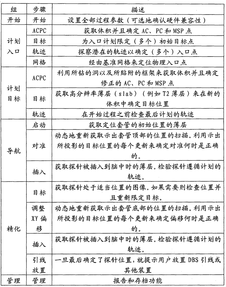
表2A-2P提供了可能与根据本发明的一些实施例的示例性工作流程步骤相关联的一些示例性操作的附加实例。Tables 2A-2P provide additional examples of some exemplary operations that may be associated with exemplary workflow steps according to some embodiments of the invention.
表2A:工作流程组开始步骤Table 2A: Workflow Group Start Steps
表2B:计划入口-AC-PC步骤Table 2B: Program Entry-AC-PC Steps
可以以任何适合的方式来识别AC、PC和MSP位置。在一些实施例中,AC-PC步骤可以具有自动的、电子的AC、PC、MSP标识库。AC、PC和MSP解剖界标限定AC-PC坐标系统,例如可用于外科手术计划的Talairach-Tournoux坐标系。该库可以用来自动标识界标的位置。它可以被提供为主机应用可以通过一组Microsoft


在一些实施例中,通过使用一些从输入体积的下部提取的轴向切片(例如大约15个等间隔的切片)来近似正中矢状平面(MSP)。可以向每个切片应用亮度均衡,并且可以使用坎尼算法来创建来自每个切片的边缘掩模。可以为每个边缘掩模找出对称轴,并且基于迭代检查和试验性对称轴的排序或计分来标识实际对称轴。排序/计分可以基于在坎尼掩模上反射通过对称轴的点是否落在坎尼掩模上(如果是那样的话,该轴为该切片计分)。主动外观模型(AAM)可以以重新确定格式的输入堆栈的形式应用于脑干,其中限定的MSP标识AC和PC点。In some embodiments, the midsagittal plane (MSP) is approximated by using a number of axial slices (eg, about 15 equally spaced slices) taken from the lower portion of the input volume. Luminance equalization can be applied to each slice, and the Canny algorithm can be used to create edge masks from each slice. An axis of symmetry may be found for each edge mask, and the actual axis of symmetry identified based on iterative inspection and ranking or scoring of the tentative symmetry axes. The sorting/scoring can be based on whether a point reflected on the Canny mask through the axis of symmetry falls on the Canny mask (if so, the axis scores the slice). The Active Appearance Model (AAM) can be applied to the brainstem in the form of a reformatted input stack with defined MSPs identifying AC and PC points.
MSP平面估计以及AC和PC点可以被精化。可以使用具有环绕一部分脑室的小区域的裁切的图像以及使用坎尼算法的边缘掩模来精化MSP平面估计。如果在该边缘掩模上发现对称轴,则遵循上述过程。如上面指出的那样使用精化的MSP来估计AC和PC点,并且在该估计值周围的小区域(例如6x6mm)中搜索亮度峰值。最大的峰值是AC点。可以使用上面的PC估计和精化的MSP来精化PC点。可以计算MSP图像的坎尼边缘图。再次,可以在小区域(例如大约6mmx6mm)中搜索位于坎尼边缘上的点,并且对于该点来说图像梯度几乎平行于AC-PC方向。将该点沿着AC-PC方向朝向PC移动大约1mm。取垂直于AC-PC方向上的最大强度峰值为PC点。MSP plane estimation and AC and PC points can be refined. The MSP plane estimation can be refined using a cropped image with a small region surrounding a portion of the ventricle and an edge mask using the Canny algorithm. If an axis of symmetry is found on this edge mask, follow the above procedure. The AC and PC points are estimated using the refined MSP as indicated above, and a brightness peak is searched in a small area (eg 6x6mm) around this estimate. The largest peak is the AC point. The PC points can be refined using the PC estimation above and the refined MSP. Canny edge maps of MSP images can be computed. Again, a point lying on the Canny edge can be searched in a small area (eg about 6mmx6mm) and for which the image gradient is almost parallel to the AC-PC direction. Move the point about 1 mm towards PC along the AC-PC direction. Take the maximum intensity peak perpendicular to the AC-PC direction as the PC point.
表2C:计划入口-目标步骤Table 2C: Program entry-goal steps
图2D:计划入口-轨迹步骤Figure 2D: Plan entry-trajectory steps
表2E:计划入口-网格步骤Table 2E: Plan Entry - Grid Steps
这时,已在入口点处钻洞并且已经附接轨迹导引50t。注意:因为已经移动了患者,所以在先前图像坐标系中限定的点已不再有效。而且,这时可能发生脑移位。At this point, a hole has been drilled at the entry point and the
表2F:计划目标-AC-PC步骤Table 2F: Program Objective-AC-PC Steps
表2G:计划目标-目标步骤Table 2G: Program Goals - Goal Steps
表2H:计划目标-轨迹检查步骤Table 2H: Program Goals - Trajectory Check Steps
表2I-导航-启动Form 2I - Navigation - Launch
表2J:导航-对准Table 2J: Navigation - Alignment
表2K:导航-插入Table 2K: Navigation - Insert
表2L:精化-目标Table 2L: Refinement - Target
在放置不可接受的情况下,用户可以选择进行X-Y调整工作流程步骤。In cases where placement is unacceptable, the user has the option of performing an X-Y adjustment workflow step.
表2M:精化-调整X-Y偏移Table 2M: Refinement - Adjust X-Y Offset
表2N:精化-插入Table 2N: Refinement - Insertion
表20:精化-引线放置Table 20: Refinement - Lead Placement
表2P:管理步骤Table 2P: Administrative steps
另外,要注意,可以实时获得功能患者数据,并且可以将其在显示器32上提供给电路30c/工作站30,其中患者解剖结构可视化帮助精化或计划外科手术过程的轨迹和/或目标位置。Also, note that functional patient data can be obtained in real-time and can be provided to
当显示利用成像探针50a(在图5中使用)创建的图像或可视化时,电路30c可以电子地应用(西格马)校正以校正“火山(volcano)”或“光晕”形状的强度失真。也就是说,在一些特定实施方式中,成像探针天线或线圈可以在利用其而创建的图像中引入失真,该失真可以在它在图像中出现的位置处被描述为探针本身周围的亮光晕。因此,当来自成像探针的图像具有这样的特征时,电路30c可以被配置成电子地自动应用校正以消除光晕(或至少减少它)。该特征将仅影响经由成像探针自身而创建的小场的高分辨率图像。使用主头部线圈获得的图像通常不具有这样的失真。When displaying an image or visualization created with imaging probe 50a (used in FIG. 5 ),
参考图14A,电路30c可以被配置成具有在显示器32上显示的单个控制工具栏30t,所述单个控制工具栏30t允许用户选择要进行哪个组和步骤,并且还示出已完成了哪些组和步骤。图14B示出具有已选择的“计划入口”组以及被放大为当前步骤的“限定目标”的工作流程控制工具栏30t的实例。工具栏30t可以包括彩色边框130,其可以被用来部分或全部围绕按钮130b以说明步骤的完成。对于双侧过程,当一个侧的任务已完成时,可以增强该侧边框的颜色,例如当在限定目标步骤中完成左侧目标时可以增强左侧边框的颜色。Referring to FIG. 14A, the
当用户进行该过程时,存储某些临床信息以将其合并到可以在该过程结束时检查的以及/或者被存档为患者记录的过程报告中。电路30c可以被配置成提供用户界面(UI)30I,其提供诸如一个或多个下述特征的查看工具:As the user goes through the procedure, certain clinical information is stored to incorporate it into a procedure report that can be reviewed at the end of the procedure and/or archived as a patient record.
-绘制测量线- draw measurement lines
-全景视图(pan view)-pan view
-缩放、缩放全部、缩放到区域、缩放到点- Zoom, Zoom All, Zoom to Region, Zoom to Point
-放大镜-magnifier
-示出/隐藏绘注-show/hide annotation
-示出/隐藏十字线- show/hide crosshairs
-在面板之间拖动视图- Drag views between panels
-重新调整视图面板的大小- Resize the view panel
-将视图最大化成1×1显示-Maximize view to 1×1 display
-保存屏幕捕获(可以被添加到报告中)- Save screen captures (can be added to reports)
-将视图设置重新设置成默认- reset view settings to default
图15是开始组的示例性UI 30I的屏幕截图,所述UI 30I方便地被配置成设置诸如偏侧化、目标类型和MR扫描仪开孔大小的全部过程参数的单屏输入(认识到也可以使用开放式开孔MRI扫描仪系统)。作为对开孔大小的代替,可以提供下拉列表,其允许用户选择所使用MR扫描仪的类型和制造商,其提供相关联的开孔大小。当然,系统10可以被配置成MR扫描仪特定系统,或者未来的MR扫描仪可以具有标准的开孔大小或者被配置成使得开孔大小不是约束且可能不需要该信息。如所示出的那样,UI 30I还可以包括需要硬件标识符数据66的录入的输入32I,其被示出为一次性使用的工具箱版本输入,如上面参考图5所述的那样。如果标识符数据66不匹配,则为了保证硬件和软件的兼容和/或适当的操作,系统10可以被配置成不允许用户继续进行到下一步骤或者可以向用户提示录入其他关键代码。Figure 15 is a screen shot of an
在步骤计划入口/轨迹中使用开孔大小。如果用户选择的轨迹因为探针在物理上不适合在扫描仪开孔内而使得该探针不能被插入到套管60中,则生成警报(在显示器32上可见和/或可听见的)。见上面的计划入口/轨迹步骤。Use opening size in step plan entry/trajectories. If the user selected trajectory prevents the probe from being inserted into
图16是与具有限定目标步骤(被示出为左侧STN)的计划入口工作流程组的示例性UI 30I相关联的屏幕截图,其说明工具栏30t以及具有有患者图像数据的默认界标的十字线位置数据。该步骤可以用来建立AC PC坐标系统。当接收到整个头部体积时,该步骤可以自动找出这些点的候选。如果需要,用户可以检查并且校正这些点。用户可以将十字线定位在一个点处,并且“点击”“Set(设置)”按钮来设置期望的绘注,或者他们可以在屏幕上到处拖动现有绘注。一旦限定了这些点,视平面就可以自动重新确定格式以使它们与ACPC坐标系对准,从而示出解剖平面:冠状、矢状和轴向平面。对界标的任何后续编辑可以使得视平面立即重新对准以匹配。16 is a screenshot associated with an exemplary UI 30I of a plan entry workflow group with a defined goal step (shown as the left STN), illustrating a
图17说明限定目标步骤的另一个示例性屏幕截图,所述限定目标步骤被用来设置目标点以便可以在下一步骤研究通过潜在入口点的轨迹。用户可以选择将来自标准脑图谱的轮廓叠加到患者解剖结构上以用于比较目的,所述患者解剖结构可以以彩色提供,其中不同的颜色用于不同的结构。图17示出了不具有图谱的限定目标步骤的进行(play)入口。图18说明具有图谱的UI,该图谱在三个正交视图中示出了目标轮廓,且图19说明在1×1布局中示出结构的图谱。视平面示出在ACPC步骤中限定的解剖平面。用与编辑ACPC点类似的方式来编辑目标点。对于双侧情况,一旦已为一个侧限定了目标,则当用户为另一侧选择目标时,十字线将自动跳到镜像位置。如果患者具有对称的解剖结构,这将为找出等同位置节省了时间。当使用脑图谱时,用户可以选择示出仅目标结构(STN或GPi)或所有结构。在任何一种情况下,(例如弹出式)工具提示(tooltip)可以帮助用户标识不熟悉的结构。用户还可以选择相对于患者图像缩放和/或移位脑图谱以实现更好的匹配。为此,用户可以拖动围绕脑图谱模板的白色轮廓。可以以视觉上突出的方式(例如颜色编码的或其他视觉表现)来提供患者的脑的功能信息和/或纤维路线结构以便外科医生容易参考。还可以经由图4中示出的UI选择32F(例如工具栏选项)来从视图中选择或抑制该信息。FIG. 17 illustrates another exemplary screen shot of the Define Goals step, which is used to set goal points so that trajectories through potential entry points can be studied in the next step. Users may choose to overlay contours from standard brain atlases onto patient anatomy for comparison purposes, which may be provided in color, with different colors used for different structures. Figure 17 shows a play entry for a defined goal step without a graph. Figure 18 illustrates a UI with a map showing object outlines in three orthogonal views, and Figure 19 illustrates a map showing structures in a 1x1 layout. The view plane shows the anatomical plane defined in the ACPC step. Edit target points in a similar manner to editing ACPC points. For the dual sided case, once a target has been defined for one side, when the user selects a target for the other side, the reticle will automatically jump to the mirrored position. This saves time in finding equivalent positions if the patient has symmetrical anatomy. When using a brain atlas, the user can choose to show only target structures (STN or GPi) or all structures. In either case, (eg pop-up) tooltips (tooltips) can help users identify unfamiliar constructs. Users can also choose to scale and/or shift the brain atlas relative to the patient image to achieve a better match. To do this, users can drag a white outline around the brain map template. Functional information and/or fiber routing structure of the patient's brain may be provided in a visually prominent manner (eg, color-coded or other visual representation) for easy reference by the surgeon. This information can also be selected or suppressed from view via UI selection 32F (eg, toolbar option) shown in FIG. 4 .
图20是计划入口工作流程组的轨迹步骤的示例性屏幕显示的屏幕截图。该步骤用于找出临床上可行的轨迹,其通常经由(例如由网格补片50g(图11)提供的)网格在颅骨上确定入口点。可以显示视觉警报,例如在两个顶部视图的顶部上的红色警报消息30W,并且红色轨迹线30R可以被用来指示所选择的轨迹不与网格相交。在操作中,一旦进入该步骤,工作站30可以自动在图像体积中搜索(多个)标记网格50g。它可以被配置成定位初始轨迹以便它可以穿过网格50g的中间。如果用户移动该轨迹以使得入口点不在该网格上,则显示警报30W。20 is a screenshot of an exemplary screen display of a track step of a plan entry workflow group. This step is used to find clinically feasible trajectories, which typically determine entry points on the skull via a mesh (eg, provided by
图20上面的两个视图示出沿着当前轨迹线对准的冠状和矢状视图。用户拖动轨迹线并且它围绕目标点自由旋转。左下视图示出在冠状和矢状视图中的绿线水平处垂直于轨迹的平面。这是“探针的眼睛”的视图。为了使探针的眼睛的视图沿着轨迹前进,除了沿着轨迹线拖动绿线或者使用鼠标滚轮之外,屏幕上的VCR样式控制可以提供动画的飞越。The top two views of Figure 20 show coronal and sagittal views aligned along the current trajectory line. The user drags the trajectory line and it rotates freely around the target point. The lower left view shows the plane perpendicular to the trajectory at the level of the green line in the coronal and sagittal views. This is the view from the "eye of the probe". To advance the probe's eye's view along the trajectory, in addition to dragging the green line along the trajectory or using the mouse wheel, an on-screen VCR style control can provide an animated fly-through.
图21说明当用户选择可能被扫描仪开孔壁阻挡的轨迹时自动生成的(弹出式)警报30W’。也就是说,如果用户设置的轨迹使得扫描仪开孔将干扰探针的插入,则显示警报。这一计算基于开孔大小、当前轨迹角度以及为探针50a/50s/52、套管60和框架/轨迹导引50t(图5)的大小而预先配置的值。在双侧的情况下,与目标步骤类似,一旦已在一个侧上设置了轨迹,则另一侧的初始默认就可以是镜像轨迹,以使用户开始更接近更可能的轨迹。在保存该轨迹时,该步骤自动找出沿着所计划/选择的轨迹的颅骨表面、在网格上标识坐标并且将该位置存储为入口点。Figure 21 illustrates an automatically generated (popup) alert 30W' when the user selects a track that may be blocked by the scanner aperture walls. That is, if the trajectory set by the user is such that the scanner opening will interfere with the insertion of the probe, an alert is displayed. This calculation is based on the aperture size, current trajectory angle, and pre-configured values for the size of probe 50a/50s/52,
图22说明示出为在显示器32上叠加患者颅骨的示例性网格50g(没有绘注线),其示出了用于利用左侧STN入口位置来选择入口位置(被示出为列1-6和行A-F)的坐标。图23示出与图22相同的屏幕视图,但是带有可选的绘注网格线。典型地,用户可以足够清楚地看到网格坐标,而不需要可选的叠加网格线来标识网格元素。然而,在一些实施例中,如所示出的那样UI将允许用户显示这些线。22 illustrates an
图24是选择了ACPC步骤的计划目标组工作流程的示例性屏幕截图。该工作流程组用于在已经为单侧过程形成了一个钻洞或者已经为双侧过程形成了两个钻洞(已钻开外科手术入口并且已附接框架)之后,精确地确定目标点。先前的计划通常因为随着脑髓液压力的减小发生的脑移位而被无效。该步骤基本上类似于上面讨论的计划入口ACPC步骤。差异是除了整个头部体积之外,用户还可以可选地发送包含ACPC点的一个或多个高分辨率薄层。用户可以使用缩略图栏来选择使用哪些数据来显示并且编辑ACPC界标。图25说明示出薄层数据的计划目标/ACPC步骤的屏幕截图。Figure 24 is an exemplary screen shot of a planning target group workflow with the ACPC step selected. This workflow group is used to precisely determine the target point after one burr hole has been formed for a unilateral procedure or two burr holes have been made for a bilateral procedure (surgical access has been drilled and frame has been attached). Previous plans are often invalidated due to brain displacement that occurs with a decrease in CSF pressure. This step is basically similar to the program entry ACPC step discussed above. The difference is that in addition to the entire head volume, the user can optionally send one or more high-resolution thin slices containing ACPC points. The user can use the thumbnail bar to select which data to display and edit the ACPC landmarks. Fig. 25 illustrates a screen shot showing the plan target/ACPC step for thin layer data.
图26是选择了限定目标步骤的计划目标组工作流程的示例性屏幕截图。脑图像可以与混和的体积和薄层图像数据一起示出。该步骤类似于计划入口组中的限定目标步骤,但是与先前的步骤相似它也支持高分辨率薄层以增加可以限定目标的精度。因此,该步骤具有使用来自体积和高分辨率薄层二者的数据来示出混合图像的能力。UI中的滑动块30S(如词“blend(混合)”下面的右侧所示)允许用户控制来自任何一个源的图像的比率是多少。Figure 26 is an exemplary screen shot of a planning target group workflow with the define target step selected. Brain images can be shown with blended volumetric and thin-slice image data. This step is similar to the Define Targets step in the Plan Entry group, but like the previous steps it also supports high-resolution slabs to increase the precision with which targets can be defined. Thus, this step has the ability to show a blended image using data from both the volume and high resolution slices. A slider 30S in the UI (shown to the right below the word "blend") allows the user to control what ratio of images from any one source is.
图27是计划目标工作流程组中的轨迹步骤的屏幕截图。该步骤类似于计划入口/轨迹步骤,但是在这种情况下该步骤通常仅被用来检查轨迹。因为已经钻了洞并且附接了框架所以入口点不能改变。同样,与先前的步骤相似,这可以支持使用高分辨率薄层并且可以在体积和薄层之间混合。在进入到该步骤时,软件可以自动搜索整个头部体积并且找出框架标记基准。系统10和/或电路30c可以根据这些来确定框架位置和定向,并且使用其来计算头部上的实际入口点。这可以连同来自先前步骤的目标一起用来确定轨迹。另外,可以如上面在计划入口/轨迹步骤中所描述的那样来检查轨迹。Figure 27 is a screenshot of a track step in a plan target workflow group. This step is similar to the plan entry/trajectories step, but in this case it is usually only used to check trajectories. The entry point cannot be changed since the hole has already been drilled and the frame attached. Again, similar to the previous steps, this enables the use of high-resolution thin layers and the ability to mix between volumetric and thin layers. Upon entering this step, the software can automatically search the entire head volume and find frame marker fiducials. The
图28是选择了启动导航步骤的导航工作流程组的屏幕截图。该可视化示出了近端套管薄层。在进入该组时,用户已经钻开了入口洞、附接了框架并且最终定下来它们的计划目标。该组将引导用户通过对准套管60(图8)以与所述计划轨迹匹配以及穿过套管将探针插入到正确深度。该步骤用来确定套管的初始物理位置。用户获取通过套管顶部的扫描以及通过套管底部的另一个扫描。电路30c(例如软件)自动在每个薄层中找出套管并且确定其在空间中的位置和定向。图29说明在导航/启动导航步骤中的远端套管薄层。Figure 28 is a screenshot of the Navigation Workflow Group with the Start Navigation step selected. This visualization shows the proximal cannula laminae. By the time the group is entered, the user has drilled the entry hole, attached the frame and finalized their planning goals. The group will guide the user through aligning the cannula 60 (Fig. 8) to match the planned trajectory and inserting the stylet through the cannula to the correct depth. This step is used to determine the initial physical location of the casing. The user acquires a scan through the top of the cannula and another scan through the bottom of the cannula.
图30是选择了对准套管步骤的导航工作流程组的屏幕截图。该步骤用于物理地使套管60(图8)对准计划轨迹。用户经由俯仰和滚动控制旋钮来反复调整套管角度,因为它们迅速地重新获取通过套管顶部60p的图像。电路30c(例如软件)利用每个更新来计算套管60的位置并且显示绘注,该绘注示出其在目标平面上当前所指向的位置。图30说明用户反馈(例如叠加上的提示或工具提示样式的弹出)可以告诉用户旋转哪个控制以及哪个方向。在一些实施例中,如所示出的那样,绘注可以被绘制为对应于探针直径的圆圈。具有十字线的圆圈是计划目标,其他圆圈是基于套管轨迹的当前投影的点。在中间的线示出要应用的俯仰和滚动的相对量,并且文本可以指定旋转哪个旋钮以及以哪个方向(示出为“向左旋转俯仰轮”)。Figure 30 is a screenshot of the Navigate workflow group with the Align Cannula step selected. This step is used to physically align the cannula 60 (Fig. 8) with the planned trajectory. The user repeatedly adjusts the cannula angle via the pitch and roll control knobs as they rapidly reacquire the image through the top 60p of the cannula. With each update,
图31说明来自示例性导航/插入探针步骤的屏幕截图。该屏幕截图说明目标(例如STN)的冠状和矢状视图,并且可以提供设置的限深档块尺寸(在UI的右上侧示出)。该步骤允许用户看到在插入探针时探针遵循轨迹的程度。用户可以选择沿着探针扫描冠状和矢状薄层,以在视觉上确定在这些平面中的探针对准。用户还可以垂直于探针进行扫描。在这种情况下,电路30c(例如软件)可以自动标识探针在薄层的哪个位置,并且其然后示出当前路径在目标平面上的投影以指示在继续当前路径的情况下误差的程度和方向。图32说明在继续当前轨迹的情况下具有投影误差的投影点和轴向薄层(右图像示出的探针偏移了4.6mm并且还在UI上将该偏移注释为“投影误差”)。用户可以在插入期间多次执行这些扫描。在目标平面上探针的自动分割以及投影目标的显示将为检验当前路径提供全自动支持。冠状/矢状视图可以为医生提供不依赖于软件分割的探针路径的视觉确认。Figure 31 illustrates a screenshot from an exemplary Navigate/Insert Probe step. This screenshot illustrates the coronal and sagittal views of the target (eg STN) and may provide the set depth stop size (shown on the upper right side of the UI). This step allows the user to see how well the probe follows the trajectory as it is inserted. The user may choose to scan coronal and sagittal slices along the probe to visually confirm probe alignment in these planes. Users can also scan perpendicular to the probe. In this case,
图33说明选择了目标修正步骤的精化放置工作流程组的示例性屏幕截图。该步骤可以说明目标薄层。在完成初始插入之后,用户(例如医生)可能发现放置没有足够接近或完美地对应于计划或者该计划是不正确的。在使用成像探针(50a)的情况下这是特别有可能的,因为用户将能够更清楚地可视化通常用外部线圈不清楚的结构,比如STN。该工作流程组可以支持功能,由此医生可以撤回探针并且使用X和Y偏移调整以获得到修正目标的平行轨迹。该步骤可以提示用户或者以其他方式获取通过探针远尖端的图像薄层。(可选地,该步骤可以使用成像探针)。该步骤显示该薄层并且用户可以在其上选择将目标点修改到新的位置或者将当前位置作为最终结果来接受。FIG. 33 illustrates an exemplary screen shot of a refinement placement workflow group with the target modification step selected. This step can account for the thin layer of interest. After the initial insertion is complete, the user (eg, a doctor) may find that the placement does not correspond closely enough or perfectly to the plan or that the plan is incorrect. This is particularly possible with the use of the imaging probe (50a), as the user will be able to more clearly visualize structures, such as the STN, that are normally unclear with external coils. This workflow group can support functionality whereby the physician can withdraw the probe and use the X and Y offset adjustments to obtain a parallel trajectory to the corrected target. This step may prompt the user or otherwise acquire an image slice through the distal tip of the probe. (Optionally, an imaging probe can be used for this step). This step displays the thin layer and on it the user can choose to either modify the target point to a new location or accept the current location as the final result.
图34说明选择了调整X-Y偏移步骤的精化放置工作流程组的示例性屏幕截图。该步骤与上述导航/对准导管步骤非常相似。主要的差异是不调整导管60的角度,而是用户调整小的X-Y偏移以将该导管60设置到平行于原始轨迹的轨迹。图34示出该显示,其具有探针尖端相对于目标的位置的可视化并且具有关于作什么样的物理调整来获得期望的平行轨迹(被示为“向右旋转Y轮”)以及投影误差(被示为2.7mm)的指令。图35说明调整绘注和弹出(被示为“向左旋转X轮”)的细节。34 illustrates an exemplary screenshot of a refinement placement workflow group with the Adjust X-Y Offset step selected. This step is very similar to the Navigate/Align Catheter step above. The main difference is that instead of adjusting the angle of the
在完成X-Y调整之后,选择精化放置工作流程组的插入探针,并且以与上述导航/插入探针步骤相同的方式来实行。After completing the X-Y adjustment, select the Insert Probe of the Refinement Placement workflow group and proceed in the same manner as the Navigate/Insert Probe step above.
图36说明选择了“结束过程”步骤的精化放置工作流程组。该步骤发生在所有的探针都已被插入并且医生已经检验了它们的位置之后。这时,UI可以提示他们(使用限定轨迹)在要放置可植入刺激引线的位置插入两个引线,并且可以警告他们如果使用MRI不兼容的或潜在不兼容的引线则不能执行任何附加的扫描。如所示出的那样,系统10可以被配置成限定(和向用户输出)限深档块以为每个STN或目标位点设置引线或其他治疗或诊断装置。对于(双侧过程)的左侧和右侧目标上的每个植入位置来说限深挡块可以是不同的,以便引线的或用于其他装置的其他部件的电极被定位在期望的位置中。Figure 36 illustrates the refinement placement workflow group with the "End Process" step selected. This step occurs after all probes have been inserted and the doctor has verified their positions. At this point, the UI can prompt them (using a defined trajectory) to insert both leads where the implantable stimulation leads are to be placed, and can warn them not to perform any additional scans if using an MRI-incompatible or potentially incompatible lead . As shown, the
图37说明可以用于ADMIN工作流程组的显示的实例。该组具有提供报告和存档功能的一个步骤。该报告自动为整个过程提供文件,包括绘注、测量和屏幕捕获。电路30c可以生成该报告的完全版本和匿名版本,并且可以包括关于将所有事情都存档到CD的时候的日期。Figure 37 illustrates an example of a display that may be used for ADMIN workflow groups. This group has a step that provides reporting and archiving capabilities. The report automatically documents the entire process, including annotations, measurements and screen captures.
电路30c还可以被配置成确定DBS引线上的各个电极位于ACPC坐标中的哪个位置处。在给定MR坐标中的尖端位置的情况下(电路30c可能在计划位置处不能工作(ill),但是用户可以改变它),用户将提供一组偏移值,这些偏移值表示每个电极到引线尖端的距离。在其他实施例中,引线类型可以从例如下拉式列表中选择,并且可以基于制造商和引线类型(例如其设计)来自动输入那些值。电路30c可以被配置成使得UI显示ACPC坐标中的对应电极位置。
图38说明具有可以用于ADMIN工作流程组(被示出为选择了管理页/电极偏移对话步骤)的UI的显示的实例。该步骤还可以包括在结束过程/步骤中或被提供为单独的工作流程组。电极偏移值可以显著加速编程脉冲发生器的过程,因为医生知道电极的解剖位置。Figure 38 illustrates an example of a display with a UI that may be used for the ADMIN workflow group (shown with the Manage Pages/Electrode Offset dialog step selected). This step can also be included in the closing process/step or provided as a separate workflow group. Electrode offset values can significantly speed up the process of programming the pulse generator, since the physician knows the anatomical location of the electrodes.
系统10还可以包括允许系统与MRI扫描仪20和滤波器等等协同操作的去耦/调协电路。参见例如美国专利No.6,701,176、No.6,904,307以及美国专利申请公开No.2003/0050557,通过参考将它们的内容结合于此,就如在本文中完全陈述一样。如上面所指出的那样,一个或多个工具可以包括体内MRI天线50a(图5),其被配置成在MRI过程期间拾取局部组织中的MRI信号。该MRI天线可以被配置成驻留于探针的远端部分上。在一些实施例中,该天线具有大约1-5cm的焦距或信号接收长度,并且通常被配置成具有大约1-2.5cm的从局部组织接收MRI信号的观看长度。MRI天线可以被形成为包括同轴和/或三轴天线。然而,可以使用其他天线配置,例如鞭状天线、环形天线、无回路(loopless)天线和/或回路天线。参见例如美国专利No.5,699,801、No.5,928,145、No.6,263,229、No.6,606,513、No.6,628,980、No.6,284,971、No.6,675,033以及No.6,701,176,通过参考将它们的内容结合于此,就如在本文中完全陈述一样。还参见美国专利申请公开No.2003/0050557、No.2004/0046557和No.2003/0028095,通过参考将它们的内容结合于此,就如在本文中完全陈述一样。The
在一些实施例中,植入式引线和/或体内工具可以被配置成允许安全MRI操作,以便降低组织中不期望的电流或电压沉积的可能性。引线或工具可以包括RF扼流圈,例如一系列轴向间隔开的Balun电路或其他合适的电路配置。对于可以禁止RF感应电流的RF禁止同轴电缆的附加描述,参见例如美国专利No.6,284,971,通过参考将其内容结合于此,就如在本文中完全陈述一样。连接电极或工具上或中的其他部件的导体还可以包括一系列往返段(例如引线可以沿着其长度以纵向方向翻转到其自身上(turn on itself)多次)和/或包括高阻抗电路。参见美国专利申请序列No.11/417,594、No.12/047,602和No.12/909,583,通过参考将它们的内容结合于此,就如在本文中完全陈述一样。In some embodiments, implantable leads and/or intracorporeal tools may be configured to allow safe MRI procedures in order to reduce the likelihood of undesired current or voltage deposition in tissue. The leads or tool may include RF chokes, such as a series of axially spaced Balun circuits or other suitable circuit configurations. For an additional description of RF disabling coaxial cables that may inhibit RF induced currents, see, eg, US Patent No. 6,284,971, the contents of which are hereby incorporated by reference as if fully set forth herein. Conductors connecting electrodes or other components on or in the tool may also include a series of round-trip segments (e.g. a lead may turn on itself multiple times along its length in a longitudinal direction) and/or include high-impedance circuitry . See US Patent Application Serial Nos. 11/417,594, 12/047,602, and 12/909,583, the contents of which are hereby incorporated by reference as if fully set forth herein.
尽管没有示出,但是在一些实施例中,一个或多个外科手术工具可以被配置成具有一个或多个管腔和出口,其将期望的细胞、生物和/或药物治疗递送到目标区域,例如脑。工具还可以结合经间隔穿刺针、活组织检查和/或注入针以及消融装置。在使用的情况下,管腔可以容纳可伸长的针,其可以从远端或者从侧面、近端、远端或者甚至通过电极离开探针以便将细胞/生物治疗精确地递送到期望的解剖目标。这样的递送配置可以是治疗患者的潜在方式,其中细胞/生物治疗可以被递送到期望的解剖结构中以修改它们的细胞功能。细胞(例如干细胞)可以改进功能。MRI通常可以有效地用来监控治疗的功效和/或递送。Although not shown, in some embodiments, one or more surgical tools can be configured with one or more lumens and outlets that deliver desired cellular, biological, and/or drug treatments to the target area, For example the brain. The tools may also incorporate septal puncture needles, biopsy and/or injection needles, and ablation devices. In the case of use, the lumen can house an extendable needle that can exit the probe distally or laterally, proximally, distally, or even through electrodes for precise delivery of cell/biotherapy to the desired anatomy Target. Such a delivery configuration could be a potential way to treat patients, where cell/biotherapy can be delivered into desired anatomical structures to modify their cellular functions. Cells, such as stem cells, can improve function. MRI can often be used effectively to monitor the efficacy and/or delivery of therapy.
系统10可以包括电路和/模块,其可以包括计算机程序代码,所述计算机程序代码被用来自动或半自动地实行操作以生成可视化并且将输出提供给用户以促进MRI引导诊断和治疗过程。图39是可以与系统10一起使用的电路或数据处理系统的示意图。所述电路和/或数据处理系统可以被结合到任何合适的装置或多个装置中的一个或多个数字信号处理器中。如图39所示,处理器410经由地址/数据总线448与MRI扫描仪20并且与存储器414进行通信。处理器410可以是商业上可获得的或定制的微处理器。存储器414表示包含被用来实施数据处理系统的功能的数据和软件的存储器装置的总体系。存储器414可以包括但不限于下述类型的装置:高速缓冲存储器、ROM、PROM、EPROM、EEPROM、闪存、SRAM和DRAM。
如图39所示,其说明了存储器414可以包括在数据处理系统中使用的一些种类的数据和软件:操作系统452、应用程序454、输入/输出(I/O)装置驱动器458、以及数据456。数据456还可以包括不同外科手术工具的预先定义的特性和患者图像数据455。图39还说明应用程序454可以包括可视化模块450、介入工具数据模块451、工具分割模块452(例如用于网格补片、目标套管和轨迹导引框架和/或基座的分割模块)以及工作流程组用户界面模块453(其促进用户动作并且提供指引以获得期望的轨迹,例如达到期望轨迹的物理调整)。As shown in Figure 39, which illustrates that
如本领域技术人员将认识到的那样,操作系统452可以是适合于与数据处理系统一起使用的任何操作系统,所述操作系统例如来自ArmonkNY的国际商业机器公司的OS/2、AIX、DOS、OS/390或System390;来自Redmond、WA的微软公司的Windows CE、Windows NT、Windows95、Windows98、Windows2000或其他Windows版本;来自Palm有限公司的、Unix或Linux或FreeBSD、Palm OS;来自苹果电脑公司的Mac OS;LabView或专用的操作系统。I/O装置驱动器458通常包括应用程序454为与诸如(多个)I/O数据端口、数据存储装置456和某些存储器414部件的装置通信而通过操作系统452访问的软件例程。应用程序454是实施数据处理系统的各种特征的程序的说明,并且可以包括至少一个支持根据本发明的实施例的操作的应用。最后,数据456表示应用程序454、操作系统452、I/O装置驱动器458和可能驻留于存储器414中的其他软件程序所使用的静态和动态数据。As will be appreciated by those skilled in the art,
尽管例如已参考作为图39中的应用程序的模块450-453说明了本发明,但是如本领域技术人员将认识的那样,在仍然得益于本发明的教导的同时还可以利用其他配置。例如,模块450-453和/或还可以结合到操作系统452、I/O装置驱动器458或数据处理系统的其他这样的逻辑划分中。因此,本发明不应该被解释成限于图39的配置,其意图包括能够实行本文所述的操作的任何配置。此外,模块(即模块450-453)中的一个或多个可以与其他部件通信或者全部或部分结合到该其他部件中,所述其他部件例如工作站、MRI扫描仪、接口装置。典型地,工作站30将包括模块450-453和MR扫描仪,MR扫描仪包括与工作站30通信并且可以将图像数据推入到其中的模块。Although the invention has been described, for example, with reference to modules 450-453 as applications in FIG. 39, other configurations may be utilized while still benefiting from the teachings of the invention, as will be recognized by those skilled in the art. For example, modules 450-453 and/or may also be incorporated into
I/O数据端口可以被用来在数据处理系统、电路30c或工作站30、MRI扫描仪20以及另一个计算机系统或网络(例如因特网)之间传递信息,或者将该信息传递到由处理器控制或者与该处理器通信的其他装置。这些部件可以是传统部件,例如在许多传统数据处理系统中使用的那些部件,所述部件可以根据本发明而配置以如本文所述的那样操作。The I/O data ports may be used to pass information between the data processing system,
在附图和说明书中,已公开了本发明的实施例,尽管使用了特定术语,但是仅以普通和描述性的意义使用它们并且不用于限制目标,本发明的范围在下面的权利要求中陈述。因此,前面的描述是本发明的说明并且不被解释为对其进行限制。更特别地,可以以不同方式、不同顺序以及/或者与其他工作流程步骤一起来实行工作流程步骤,或者可以省略某些工作流程步骤或者用其他步骤来代替某些工作流程步骤。尽管已描述了本发明的一些示例性实施例,但是本领域技术人员将会容易认识到在不实质偏离本发明的创新教导和优点的情况下有可能在示例性实施例中存在许多修改。因此,所有这样的修改意图被包括在如权利要求所限定的本发明的范围内。在权利要求中,所使用的装置加功能的条款意图覆盖本文所描述的执行所列举的功能的结构,并且不仅仅是结构等同物而且还有等同的结构。因此,应该理解,前面的描述是本发明的说明并且不被解释为限于所公开的特定实施例,并且对所公开的实施例的修改以及其他实施例都意图被包括在所附权利要求的范围内。本发明由下面的权利要求限定,其中这些权利要求的等同物被包括在其中。In the drawings and specification, embodiments of the present invention have been disclosed, and while specific terms have been used, they have been used in a generic and descriptive sense only and not for purposes of limitation, the scope of the invention being set forth in the following claims . Accordingly, the foregoing description is illustrative of the invention and is not to be construed as limiting thereof. More particularly, workflow steps may be performed in a different manner, in a different order, and/or with other workflow steps, or certain workflow steps may be omitted or replaced by other steps. Although a few exemplary embodiments of this invention have been described, those skilled in the art will readily appreciate that many modifications are possible in the exemplary embodiments without materially departing from the innovative teachings and advantages of this invention. Accordingly, all such modifications are intended to be included within the scope of this invention as defined in the claims. In the claims, use of means-plus-function clauses is intended to cover the structures described herein as performing the recited function and not only structural equivalents but also equivalent structures. It is therefore to be understood that the foregoing description is illustrative of the invention and is not to be construed as limited to the particular embodiments disclosed and that modifications to the disclosed embodiments, as well as other embodiments, are intended to be included within the scope of the appended claims Inside. The invention is defined by the following claims, with equivalents of those claims to be included therein.
Claims (45)
Applications Claiming Priority (7)
| Application Number | Priority Date | Filing Date | Title |
|---|---|---|---|
| US97482107P | 2007-09-24 | 2007-09-24 | |
| US60/974,821 | 2007-09-24 | ||
| US60/974821 | 2007-09-24 | ||
| US12/134,412 | 2008-06-06 | ||
| US12/134412 | 2008-06-06 | ||
| US12/134,412 US8175677B2 (en) | 2007-06-07 | 2008-06-06 | MRI-guided medical interventional systems and methods |
| PCT/US2008/011050 WO2009042135A2 (en) | 2007-09-24 | 2008-09-24 | Mri surgical systems for real-time visualizations using mri image data and predefined data of surgical tools |
Publications (2)
| Publication Number | Publication Date |
|---|---|
| CN101918855A CN101918855A (en) | 2010-12-15 |
| CN101918855B true CN101918855B (en) | 2013-07-17 |
Family
ID=40515033
Family Applications (1)
| Application Number | Title | Priority Date | Filing Date |
|---|---|---|---|
| CN2008801173424A Expired - Fee Related CN101918855B (en) | 2007-09-24 | 2008-09-24 | MRI surgical systems for real-time visualizations using MRI image data and predefined data of surgical tools |
Country Status (6)
| Country | Link |
|---|---|
| US (4) | US8175677B2 (en) |
| EP (4) | EP2192870A1 (en) |
| JP (1) | JP5632286B2 (en) |
| CN (1) | CN101918855B (en) |
| CA (4) | CA2700529A1 (en) |
| WO (4) | WO2009042135A2 (en) |
Cited By (2)
| Publication number | Priority date | Publication date | Assignee | Title |
|---|---|---|---|---|
| TWI737404B (en) | 2020-07-15 | 2021-08-21 | 臺北醫學大學 | Medical image processing system and method thereof |
| US11238591B1 (en) | 2020-07-15 | 2022-02-01 | Taipei Medical University (Tmu) | Medical image processing system and method thereof |
Families Citing this family (197)
| Publication number | Priority date | Publication date | Assignee | Title |
|---|---|---|---|---|
| US8256430B2 (en) | 2001-06-15 | 2012-09-04 | Monteris Medical, Inc. | Hyperthermia treatment and probe therefor |
| US20040162637A1 (en) | 2002-07-25 | 2004-08-19 | Yulun Wang | Medical tele-robotic system with a master remote station with an arbitrator |
| US7813836B2 (en) | 2003-12-09 | 2010-10-12 | Intouch Technologies, Inc. | Protocol for a remotely controlled videoconferencing robot |
| US8077963B2 (en) | 2004-07-13 | 2011-12-13 | Yulun Wang | Mobile robot with a head-based movement mapping scheme |
| US9198728B2 (en) | 2005-09-30 | 2015-12-01 | Intouch Technologies, Inc. | Multi-camera mobile teleconferencing platform |
| US8849679B2 (en) | 2006-06-15 | 2014-09-30 | Intouch Technologies, Inc. | Remote controlled robot system that provides medical images |
| US9160783B2 (en) | 2007-05-09 | 2015-10-13 | Intouch Technologies, Inc. | Robot system that operates through a network firewall |
| US8175677B2 (en) * | 2007-06-07 | 2012-05-08 | MRI Interventions, Inc. | MRI-guided medical interventional systems and methods |
| US20100172777A1 (en) * | 2007-07-02 | 2010-07-08 | Borgwarner Inc. | Inlet design for a pump assembly |
| EP2192871B8 (en) | 2007-09-24 | 2015-01-28 | MRI Interventions, Inc. | Mri-compatible patch and method for identifying a position |
| US8315689B2 (en) | 2007-09-24 | 2012-11-20 | MRI Interventions, Inc. | MRI surgical systems for real-time visualizations using MRI image data and predefined data of surgical tools |
| CA2700523A1 (en) | 2007-09-24 | 2009-04-02 | Surgivision, Inc. | Mri-guided medical interventional systems and methods |
| US8340743B2 (en) * | 2007-11-21 | 2012-12-25 | MRI Interventions, Inc. | Methods, systems and computer program products for positioning a guidance apparatus relative to a patient |
| US10875182B2 (en) | 2008-03-20 | 2020-12-29 | Teladoc Health, Inc. | Remote presence system mounted to operating room hardware |
| US8179418B2 (en) | 2008-04-14 | 2012-05-15 | Intouch Technologies, Inc. | Robotic based health care system |
| US8170241B2 (en) | 2008-04-17 | 2012-05-01 | Intouch Technologies, Inc. | Mobile tele-presence system with a microphone system |
| US9193065B2 (en) | 2008-07-10 | 2015-11-24 | Intouch Technologies, Inc. | Docking system for a tele-presence robot |
| US9842192B2 (en) | 2008-07-11 | 2017-12-12 | Intouch Technologies, Inc. | Tele-presence robot system with multi-cast features |
| US8728092B2 (en) | 2008-08-14 | 2014-05-20 | Monteris Medical Corporation | Stereotactic drive system |
| US8747418B2 (en) | 2008-08-15 | 2014-06-10 | Monteris Medical Corporation | Trajectory guide |
| US20100063422A1 (en) * | 2008-09-08 | 2010-03-11 | Sunnybrook Health Sciences Center | Ultrasound therapy transducer head and ultrasound therapy system incorporating the same |
| WO2010030373A2 (en) * | 2008-09-12 | 2010-03-18 | Surgivision, Inc. | Intrabody mri stacked flat loop antennas and related systems |
| US8340819B2 (en) | 2008-09-18 | 2012-12-25 | Intouch Technologies, Inc. | Mobile videoconferencing robot system with network adaptive driving |
| US8270698B2 (en) * | 2008-09-24 | 2012-09-18 | Merge Healthcare Incorporated | Anterior commissure and posterior commissure segmentation system and method |
| US8996165B2 (en) | 2008-10-21 | 2015-03-31 | Intouch Technologies, Inc. | Telepresence robot with a camera boom |
| US9138891B2 (en) | 2008-11-25 | 2015-09-22 | Intouch Technologies, Inc. | Server connectivity control for tele-presence robot |
| US8463435B2 (en) | 2008-11-25 | 2013-06-11 | Intouch Technologies, Inc. | Server connectivity control for tele-presence robot |
| US20100198052A1 (en) * | 2009-01-28 | 2010-08-05 | Kimble Jenkins | Mri-compatible articulating arms and related systems and methods |
| US8849680B2 (en) | 2009-01-29 | 2014-09-30 | Intouch Technologies, Inc. | Documentation through a remote presence robot |
| US20100217115A1 (en) * | 2009-02-25 | 2010-08-26 | Hushek Stephen G | Temperature sensing within a patient during mr imaging |
| CN201384493Y (en) * | 2009-03-12 | 2010-01-20 | 西门子迈迪特(深圳)磁共振有限公司 | Positioning device for magnetic resonance systems |
| WO2010110881A1 (en) * | 2009-03-27 | 2010-09-30 | Hetherington Hoby P | Improved transceiver apparatus, system, and methodology for superior in-vivo imaging of human anatomy |
| US9232977B1 (en) * | 2009-03-27 | 2016-01-12 | Tausif-Ur Rehman | Instrument guiding device |
| US8897920B2 (en) | 2009-04-17 | 2014-11-25 | Intouch Technologies, Inc. | Tele-presence robot system with software modularity, projector and laser pointer |
| GB0908784D0 (en) * | 2009-05-21 | 2009-07-01 | Renishaw Plc | Apparatus for imaging a body part |
| GB0908787D0 (en) | 2009-05-21 | 2009-07-01 | Renishaw Plc | Head clamp for imaging and neurosurgery |
| US9259290B2 (en) | 2009-06-08 | 2016-02-16 | MRI Interventions, Inc. | MRI-guided surgical systems with proximity alerts |
| WO2010148095A2 (en) * | 2009-06-16 | 2010-12-23 | Neocoil, Llc | Modular apparatus for magnetic resonance imaging |
| US8396532B2 (en) | 2009-06-16 | 2013-03-12 | MRI Interventions, Inc. | MRI-guided devices and MRI-guided interventional systems that can track and generate dynamic visualizations of the devices in near real time |
| DE102009034312A1 (en) * | 2009-07-23 | 2011-01-27 | Martin Hempel | Protective housing for flexible MR coils |
| US9107575B2 (en) * | 2009-07-29 | 2015-08-18 | Mauna Kea Technologies | Apparatus and method for brain fiber bundle microscopy |
| US11399153B2 (en) | 2009-08-26 | 2022-07-26 | Teladoc Health, Inc. | Portable telepresence apparatus |
| US8384755B2 (en) | 2009-08-26 | 2013-02-26 | Intouch Technologies, Inc. | Portable remote presence robot |
| US9844414B2 (en) * | 2009-08-31 | 2017-12-19 | Gregory S. Fischer | System and method for robotic surgical intervention in a magnetic resonance imager |
| JP2013508103A (en) * | 2009-10-28 | 2013-03-07 | イムリス インク. | Automatic registration of images for image guided surgery |
| US9864032B2 (en) * | 2010-01-05 | 2018-01-09 | National Health Research Institutes | Magnetic resonance imaging system |
| US20110187875A1 (en) * | 2010-02-04 | 2011-08-04 | Intouch Technologies, Inc. | Robot face used in a sterile environment |
| US11154981B2 (en) | 2010-02-04 | 2021-10-26 | Teladoc Health, Inc. | Robot user interface for telepresence robot system |
| US8670017B2 (en) | 2010-03-04 | 2014-03-11 | Intouch Technologies, Inc. | Remote presence system including a cart that supports a robot face and an overhead camera |
| WO2011128800A1 (en) * | 2010-04-14 | 2011-10-20 | Koninklijke Philips Electronics N.V. | Instrument guiding during magnetic resonance imaging |
| WO2011130107A2 (en) * | 2010-04-16 | 2011-10-20 | Surgivision, Inc. | Mri surgical systems including mri-compatible surgical cannulae for transferring a substance to and/or from a patient |
| US9814885B2 (en) | 2010-04-27 | 2017-11-14 | Medtronic, Inc. | Stimulation electrode selection |
| DE102010020152B4 (en) * | 2010-05-11 | 2013-10-24 | Siemens Aktiengesellschaft | Device with local coil arrangement and implantable device |
| US10343283B2 (en) | 2010-05-24 | 2019-07-09 | Intouch Technologies, Inc. | Telepresence robot system that can be accessed by a cellular phone |
| US10808882B2 (en) | 2010-05-26 | 2020-10-20 | Intouch Technologies, Inc. | Tele-robotic system with a robot face placed on a chair |
| DE102010023844A1 (en) * | 2010-06-15 | 2011-12-15 | Siemens Aktiengesellschaft | MR RF coils with modulable flexibility |
| JP6040151B2 (en) * | 2010-06-16 | 2016-12-07 | エーツー・サージカル | Method for determining access region from 3D patient image |
| US9264664B2 (en) | 2010-12-03 | 2016-02-16 | Intouch Technologies, Inc. | Systems and methods for dynamic bandwidth allocation |
| US12093036B2 (en) | 2011-01-21 | 2024-09-17 | Teladoc Health, Inc. | Telerobotic system with a dual application screen presentation |
| US9323250B2 (en) | 2011-01-28 | 2016-04-26 | Intouch Technologies, Inc. | Time-dependent navigation of telepresence robots |
| CN103459099B (en) | 2011-01-28 | 2015-08-26 | 英塔茨科技公司 | Interact with a mobile telebot |
| US11482326B2 (en) | 2011-02-16 | 2022-10-25 | Teladog Health, Inc. | Systems and methods for network-based counseling |
| US20140034377A1 (en) * | 2011-02-17 | 2014-02-06 | Kamal Vij | Thin-sleeve apparatus for reducing rf coupling of devices in mri environments |
| KR101875122B1 (en) * | 2011-02-25 | 2018-07-11 | 주식회사 칼라세븐 | A Light therapy system using sunlight |
| EP2494936A3 (en) * | 2011-03-03 | 2012-09-19 | Imris Inc. | MR compatible optical viewing device for use in the bore of an MR magnet |
| CN103443825B (en) * | 2011-03-18 | 2016-07-06 | 皇家飞利浦有限公司 | Brain deformation is followed the tracks of during neurosurgery |
| CN104254296A (en) * | 2011-04-08 | 2014-12-31 | 曼特瑞斯医药有限责任公司 | Head fixation system and method |
| US8406890B2 (en) * | 2011-04-14 | 2013-03-26 | Medtronic, Inc. | Implantable medical devices storing graphics processing data |
| US10058267B2 (en) | 2011-04-15 | 2018-08-28 | Neocoil, Llc | Antenna support structure for magnetic resonance imaging |
| US10769739B2 (en) | 2011-04-25 | 2020-09-08 | Intouch Technologies, Inc. | Systems and methods for management of information among medical providers and facilities |
| US20140139616A1 (en) | 2012-01-27 | 2014-05-22 | Intouch Technologies, Inc. | Enhanced Diagnostics for a Telepresence Robot |
| US9098611B2 (en) | 2012-11-26 | 2015-08-04 | Intouch Technologies, Inc. | Enhanced video interaction for a user interface of a telepresence network |
| DE102011079565B4 (en) * | 2011-07-21 | 2022-09-15 | Siemens Healthcare Gmbh | Direct connection head coil with height adjustment for e.g. Bechterew patients |
| TWI442905B (en) * | 2011-09-30 | 2014-07-01 | Univ Nat Chiao Tung | Apparatus for locating the target of the stimulation |
| US8836751B2 (en) | 2011-11-08 | 2014-09-16 | Intouch Technologies, Inc. | Tele-presence system with a user interface that displays different communication links |
| CN103974672B (en) * | 2011-12-05 | 2017-03-22 | 皇家飞利浦有限公司 | Positioning and orientation of surgical tools during patient-specific incision placement |
| US10842409B2 (en) | 2012-01-03 | 2020-11-24 | Koninklijke Philips N.V. | Position determining apparatus and associated method |
| TWI463964B (en) * | 2012-03-03 | 2014-12-11 | Univ China Medical | System and apparatus for an image guided navigation system in surgery |
| US8902278B2 (en) | 2012-04-11 | 2014-12-02 | Intouch Technologies, Inc. | Systems and methods for visualizing and managing telepresence devices in healthcare networks |
| US9251313B2 (en) | 2012-04-11 | 2016-02-02 | Intouch Technologies, Inc. | Systems and methods for visualizing and managing telepresence devices in healthcare networks |
| EP2666428B1 (en) * | 2012-05-21 | 2015-10-28 | Universität Bern | System and method for estimating the spatial position of a tool within an object |
| WO2013176758A1 (en) | 2012-05-22 | 2013-11-28 | Intouch Technologies, Inc. | Clinical workflows utilizing autonomous and semi-autonomous telemedicine devices |
| US9361021B2 (en) | 2012-05-22 | 2016-06-07 | Irobot Corporation | Graphical user interfaces including touchpad driving interfaces for telemedicine devices |
| US9192393B2 (en) | 2012-05-31 | 2015-11-24 | MRI Interventions, Inc. | MRI compatible surgical drills and related methods |
| EP2866723A4 (en) | 2012-06-27 | 2016-12-14 | Monteris Medical Corp | GUIDED THERAPY BY IMAGE OF A FABRIC |
| US9498290B2 (en) | 2012-07-19 | 2016-11-22 | MRI Interventions, Inc. | Surgical navigation devices and methods |
| US9610048B2 (en) | 2012-08-09 | 2017-04-04 | MRI Interventions, Inc. | Fiber optic systems for MRI suites and related devices and methods |
| BR112015003964A2 (en) | 2012-08-24 | 2017-07-04 | Univ Houston | robotic device and systems for robot-guided and image-guided surgery |
| US10136955B2 (en) * | 2012-08-24 | 2018-11-27 | University Of Houston System | Robotic device for image-guided surgery and interventions |
| US9192446B2 (en) | 2012-09-05 | 2015-11-24 | MRI Interventions, Inc. | Trajectory guide frame for MRI-guided surgeries |
| CN203101610U (en) * | 2012-09-25 | 2013-07-31 | 西门子(深圳)磁共振有限公司 | Receiving coil of magnetic resonance system |
| US20140188440A1 (en) | 2012-12-31 | 2014-07-03 | Intuitive Surgical Operations, Inc. | Systems And Methods For Interventional Procedure Planning |
| US9078588B2 (en) | 2013-03-08 | 2015-07-14 | MRI Interventions, Inc. | MRI compatible intrabody stylets and related methods and systems |
| US9600138B2 (en) * | 2013-03-15 | 2017-03-21 | Synaptive Medical (Barbados) Inc. | Planning, navigation and simulation systems and methods for minimally invasive therapy |
| US9730762B2 (en) | 2013-03-15 | 2017-08-15 | Neocoil, Llc | Automatic needle insertion location identification |
| US10274553B2 (en) * | 2013-03-15 | 2019-04-30 | Canon U.S.A., Inc. | Needle placement manipulator with attachment for RF-coil |
| US9222996B2 (en) * | 2013-03-15 | 2015-12-29 | The Brigham And Women's Hospital, Inc. | Needle placement manipulator with two rotary guides |
| KR102234145B1 (en) * | 2013-03-15 | 2021-03-31 | 인튜어티브 서지컬 오퍼레이션즈 인코포레이티드 | Shape sensor systems for tracking interventional instruments and methods of use |
| WO2014155257A1 (en) * | 2013-03-28 | 2014-10-02 | Koninklijke Philips N.V. | Localization of robotic remote center of motion point using custom trocar |
| CN103340685A (en) * | 2013-06-27 | 2013-10-09 | 苏州边枫电子科技有限公司 | Auxiliary needle feeding device of pneumatic type puncture needle |
| WO2015002846A2 (en) | 2013-07-02 | 2015-01-08 | Surgical Information Sciences, Inc. | Method and system for a brain image pipeline and brain image region location and shape prediction |
| WO2015010189A1 (en) | 2013-07-24 | 2015-01-29 | Centre For Surgical Invention & Innovation | Multi-function mounting interface for an image-guided robotic system and quick release interventional toolset |
| US9891296B2 (en) | 2013-09-13 | 2018-02-13 | MRI Interventions, Inc. | Intrabody fluid transfer devices, systems and methods |
| US9877795B2 (en) * | 2013-09-18 | 2018-01-30 | Imirge Medical Inc | Optical targeting and visualization of trajectories |
| EP3062724B1 (en) | 2013-10-25 | 2018-04-18 | Brainlab AG | Magnetic resonance coil unit and method for its manufacture |
| CN103646182B (en) * | 2013-12-13 | 2018-11-02 | 陕西理工学院 | A kind of application process of flexibility dynamic and visual technology on the medical image |
| US10595744B2 (en) | 2014-02-14 | 2020-03-24 | MRI Interventions, Inc. | Surgical tool-positioning devices and related methods |
| US9867667B2 (en) | 2014-02-27 | 2018-01-16 | Canon Usa Inc. | Placement apparatus |
| US10675113B2 (en) | 2014-03-18 | 2020-06-09 | Monteris Medical Corporation | Automated therapy of a three-dimensional tissue region |
| US20150265353A1 (en) | 2014-03-18 | 2015-09-24 | Monteris Medical Corporation | Image-guided therapy of a tissue |
| WO2015143025A1 (en) * | 2014-03-18 | 2015-09-24 | Monteris Medical Corporation | Image-guided therapy of a tissue |
| US10251670B2 (en) | 2014-05-09 | 2019-04-09 | Canon U.S.A., Inc. | Positioning apparatus |
| MX388115B (en) | 2014-09-05 | 2025-03-19 | Hyperfine Res Inc | AUTOMATIC CONFIGURATION OF A LOW-FIELD MAGNETIC RESONANCE IMAGING SYSTEM. |
| US10866291B2 (en) * | 2014-09-12 | 2020-12-15 | Emory University | Devices and systems for MRI-guided procedures |
| EP3190996A4 (en) | 2014-09-12 | 2018-04-25 | Canon U.S.A., Inc. | Needle positioning apparatus |
| WO2016077417A1 (en) | 2014-11-11 | 2016-05-19 | Hyperfine Research, Inc. | Low field magnetic resonance methods and apparatus |
| EP3632923A1 (en) | 2015-01-16 | 2020-04-08 | Voyager Therapeutics, Inc. | Central nervous system targeting polynucleotides |
| US10327830B2 (en) | 2015-04-01 | 2019-06-25 | Monteris Medical Corporation | Cryotherapy, thermal therapy, temperature modulation therapy, and probe apparatus therefor |
| US10542961B2 (en) | 2015-06-15 | 2020-01-28 | The Research Foundation For The State University Of New York | System and method for infrasonic cardiac monitoring |
| CN107708568B (en) * | 2015-06-30 | 2020-11-20 | 佳能美国公司 | Registration of fiducial markers, systems and methods |
| US10420626B2 (en) | 2015-06-30 | 2019-09-24 | Canon U.S.A., Inc. | Fiducial markers, systems, and methods of registration |
| US9867673B2 (en) | 2015-07-14 | 2018-01-16 | Canon U.S.A, Inc. | Medical support device |
| US10639065B2 (en) | 2015-07-21 | 2020-05-05 | Canon U.S.A., Inc. | Medical assist device |
| US10324594B2 (en) * | 2015-10-30 | 2019-06-18 | Siemens Healthcare Gmbh | Enterprise protocol management |
| EP3376990B1 (en) * | 2015-11-16 | 2025-11-05 | Think Surgical, Inc. | Method for confirming registration of tracked bones |
| WO2017093846A1 (en) * | 2015-11-30 | 2017-06-08 | Koninklijke Philips N.V. | Clinical discovery wheel - a system to explore clinical concepts |
| JP6906239B2 (en) * | 2015-12-28 | 2021-07-21 | ザクト ロボティクス リミテッド | Adjustable registration frame |
| USD829904S1 (en) | 2016-01-13 | 2018-10-02 | MRI Interventions, Inc. | Curved bracket for surgical navigation systems |
| USD824027S1 (en) | 2016-01-13 | 2018-07-24 | MRI Interventions, Inc. | Fins for a support column for a surgical trajectory frame |
| US10376333B2 (en) | 2016-01-14 | 2019-08-13 | MRI Interventions, Inc. | Devices for surgical navigation systems |
| WO2017132505A1 (en) | 2016-01-29 | 2017-08-03 | Canon U.S.A., Inc. | Tool placement manipulator |
| CN105726124B (en) * | 2016-02-06 | 2019-01-15 | 齐欣 | Near end of thighbone location of operation orientation system and preparation method thereof |
| WO2017142698A1 (en) | 2016-02-17 | 2017-08-24 | MRI Interventions, Inc. | Intrabody surgical fluid transfer assemblies with adjustable exposed cannula to needle tip length, related systems and methods |
| CN105943171B (en) * | 2016-05-31 | 2018-11-02 | 万伟东 | RC intracranial minimally invasive treatment positioning device and positioning marking line and positioning system |
| CN106175893B (en) * | 2016-08-03 | 2018-10-30 | 福建医科大学附属第一医院 | A kind of device of arching trajectory implantation intracranial electrode |
| CN106137283B (en) * | 2016-08-27 | 2018-08-17 | 天津大学 | A kind of natural cavity apparatus work rotary switch with regulatory function |
| DE102016216203A1 (en) * | 2016-08-29 | 2017-09-14 | Siemens Healthcare Gmbh | Medical imaging system |
| WO2018044933A1 (en) | 2016-08-30 | 2018-03-08 | The Regents Of The University Of California | Methods for biomedical targeting and delivery and devices and systems for practicing the same |
| US10695087B2 (en) | 2016-10-19 | 2020-06-30 | Canon U.S.A., Inc. | Placement manipulator and attachment for positioning a puncture instrument |
| US10539637B2 (en) | 2016-11-22 | 2020-01-21 | Hyperfine Research, Inc. | Portable magnetic resonance imaging methods and apparatus |
| KR20190087455A (en) | 2016-11-22 | 2019-07-24 | 하이퍼파인 리서치, 인크. | Systems and methods for automated detection in magnetic resonance images |
| US10627464B2 (en) | 2016-11-22 | 2020-04-21 | Hyperfine Research, Inc. | Low-field magnetic resonance imaging methods and apparatus |
| US11842030B2 (en) * | 2017-01-31 | 2023-12-12 | Medtronic Navigation, Inc. | Method and apparatus for image-based navigation |
| CN106618507A (en) * | 2017-03-13 | 2017-05-10 | 北京理工大学 | Touch cerebration stimulation device and fine adjustment assembly |
| WO2018167246A1 (en) | 2017-03-15 | 2018-09-20 | Orthotaxy | System for guiding a surgical tool relative to a target axis in spine surgery |
| CN110663084B (en) | 2017-04-18 | 2024-08-06 | 直观外科手术操作公司 | Graphical user interface for planning a program |
| US11862302B2 (en) | 2017-04-24 | 2024-01-02 | Teladoc Health, Inc. | Automated transcription and documentation of tele-health encounters |
| EP3618738A1 (en) | 2017-05-03 | 2020-03-11 | Medtronic Vascular Inc. | Tissue-removing catheter with guidewire isolation liner |
| US11690645B2 (en) | 2017-05-03 | 2023-07-04 | Medtronic Vascular, Inc. | Tissue-removing catheter |
| US10548674B2 (en) * | 2017-07-06 | 2020-02-04 | YellowDot Innovations, LLC | Robotic guide for medical device |
| CA3070087A1 (en) * | 2017-07-17 | 2019-01-24 | Voyager Therapeutics, Inc. | Trajectory array guide system |
| US10483007B2 (en) | 2017-07-25 | 2019-11-19 | Intouch Technologies, Inc. | Modular telehealth cart with thermal imaging and touch screen user interface |
| KR102850887B1 (en) | 2017-08-03 | 2025-08-28 | 보이저 테라퓨틱스, 인크. | Compositions and methods for delivering AAV |
| US11202652B2 (en) | 2017-08-11 | 2021-12-21 | Canon U.S.A., Inc. | Registration and motion compensation for patient-mounted needle guide |
| US10987016B2 (en) * | 2017-08-23 | 2021-04-27 | The Boeing Company | Visualization system for deep brain stimulation |
| US11636944B2 (en) | 2017-08-25 | 2023-04-25 | Teladoc Health, Inc. | Connectivity infrastructure for a telehealth platform |
| CN109620407B (en) * | 2017-10-06 | 2024-02-06 | 皇家飞利浦有限公司 | Treatment trajectory guidance system |
| US10893911B2 (en) | 2017-11-26 | 2021-01-19 | Canon U.S.A., Inc. | Automated image cropping for enhanced automatic device-to-image registration |
| WO2019168973A1 (en) * | 2018-02-27 | 2019-09-06 | The Regents Of The University Of Colorado, A Body Corporate | Robotic stereotaxic platform with computer vision |
| FR3078879B1 (en) * | 2018-03-14 | 2020-03-06 | Assistance Publique Hopitaux De Paris | SURGICAL KIT TO BE USED DURING A CRANIECTOMY PROCEDURE |
| US10617299B2 (en) | 2018-04-27 | 2020-04-14 | Intouch Technologies, Inc. | Telehealth cart that supports a removable tablet with seamless audio/video switching |
| US11253237B2 (en) | 2018-05-09 | 2022-02-22 | Clearpoint Neuro, Inc. | MRI compatible intrabody fluid transfer systems and related devices and methods |
| WO2019216953A1 (en) | 2018-05-09 | 2019-11-14 | MRI Interventions, Inc. | Mri compatible intrabody fluid transfer systems and related devices and methods |
| EP3794126A1 (en) | 2018-05-15 | 2021-03-24 | Voyager Therapeutics, Inc. | Compositions and methods for the treatment of parkinson's disease |
| US20210230632A1 (en) | 2018-05-15 | 2021-07-29 | Voyager Therapeutics, Inc. | Compositions and methods for delivery of aav |
| US11209509B2 (en) * | 2018-05-16 | 2021-12-28 | Viewray Technologies, Inc. | Resistive electromagnet systems and methods |
| EP3793615A2 (en) | 2018-05-16 | 2021-03-24 | Voyager Therapeutics, Inc. | Directed evolution of aav to improve tropism for cns |
| CN108814658B (en) * | 2018-06-27 | 2021-10-01 | 中国人民解放军陆军军医大学第三附属医院(野战外科研究所) | A craniotomy device and craniotomy method |
| MX2021000810A (en) | 2018-07-24 | 2021-04-28 | Voyager Therapeutics Inc | Systems and methods for producing gene therapy formulations. |
| US20210371470A1 (en) | 2018-10-12 | 2021-12-02 | Voyager Therapeutics, Inc. | Compositions and methods for delivery of aav |
| EP3880096B1 (en) | 2018-11-16 | 2024-09-18 | Medtronic Vascular Inc. | Tissue-removing catheter |
| US11744655B2 (en) | 2018-12-04 | 2023-09-05 | Globus Medical, Inc. | Drill guide fixtures, cranial insertion fixtures, and related methods and robotic systems |
| JP2022514531A (en) * | 2018-12-19 | 2022-02-14 | リヴァノヴァ・ドイチュラント・ゲーエムベーハー | Heart-lung machine with control device |
| US11364086B2 (en) | 2019-02-01 | 2022-06-21 | Advanced Neuromodulation Systems, Inc. | Trajectory guide with dual arc arrangement |
| US11298204B2 (en) * | 2019-02-01 | 2022-04-12 | Advanced Neuromodulation Systems, Inc. | Trajectory guide with dual gimbal drive arrangement |
| US11406470B2 (en) | 2019-02-01 | 2022-08-09 | Advanced Neuromodulation Systems, Inc. | Trajectory guide with double X-Y sliding tables |
| EP3948323A4 (en) * | 2019-03-25 | 2022-12-21 | Promaxo, Inc. | FAST SINGLE-SIDED MRI FIELD-GRADIENT COILS AND THEIR APPLICATIONS |
| US11819236B2 (en) | 2019-05-17 | 2023-11-21 | Medtronic Vascular, Inc. | Tissue-removing catheter |
| US20220287776A1 (en) * | 2019-08-16 | 2022-09-15 | Intuitive Surgical Operations, Inc. | Systems and methods for performance of external body wall data and internal depth data-based performance of operations associated with a computer-assisted surgical system |
| WO2021046155A1 (en) | 2019-09-03 | 2021-03-11 | Voyager Therapeutics, Inc. | Vectorized editing of nucleic acids to correct overt mutations |
| US11684750B2 (en) | 2019-10-08 | 2023-06-27 | Clearpoint Neuro, Inc. | Extension tube assembly and related medical fluid transfer systems and methods |
| WO2021113370A1 (en) | 2019-12-02 | 2021-06-10 | The General Hospital Corporation | Systems and methods for multi-modal bioimaging data integration and visualization |
| US20230056943A1 (en) * | 2019-12-13 | 2023-02-23 | Dinesh Vyas | Stapler apparatus and methods for use |
| WO2021126336A1 (en) | 2019-12-19 | 2021-06-24 | Clearpoint Neuro, Inc. | Front-loadable fluid transfer assemblies and related medical fluid transfer systems |
| US11602400B2 (en) * | 2020-01-10 | 2023-03-14 | Stryker European Operations Limited | Surgical arm and method of providing visual guidance for operating same |
| WO2021154574A1 (en) | 2020-01-31 | 2021-08-05 | Clearpoint Neuro, Inc. | Surgical tool support systems including elongate support legs with adjustable lengths and related methods |
| WO2021183295A2 (en) * | 2020-03-12 | 2021-09-16 | Clearpoint Neuro, Inc. | Image-guided surgical systems with automated trajectory guide systems and related devices and methods |
| WO2021221929A1 (en) | 2020-04-30 | 2021-11-04 | Clearpoint Neuro, Inc. | Surgical planning systems that automatically assess different potential trajectory paths and identify candidate trajectories for surgical systems |
| CN115988987A (en) * | 2020-06-26 | 2023-04-18 | 凯利博成像和诊断公司 | System for providing remote and rapid access to scanned image data |
| CN113940754B (en) * | 2020-07-15 | 2024-03-22 | 台北医学大学 | Medical image processing system and method |
| WO2022154975A1 (en) | 2021-01-14 | 2022-07-21 | Corindus, Inc. | Systems and methods for a control station for robotic interventional procedures using a plurality of elongated medical devices |
| US20220296308A1 (en) * | 2021-03-16 | 2022-09-22 | Clearpoint Neuro, Inc. | Directional-device intrabody placement systems and related methods |
| US20240358399A1 (en) * | 2021-03-31 | 2024-10-31 | Clear Guide Medical, Inc. | System and method for image guided interventions |
| CN113764074B (en) * | 2021-09-13 | 2024-01-02 | 杭州太美星程医药科技有限公司 | Image processing method and device, computer equipment and storage medium |
| DE102022125703A1 (en) * | 2022-10-05 | 2024-04-11 | Otto-von-Guericke-Universität Magdeburg, Körperschaft des öffentlichen Rechts | COUPLING STATION AND REMOTE MANIPULATION SYSTEM |
| CN116549069B (en) * | 2023-04-25 | 2024-07-02 | 苏州新云医疗设备有限公司 | Medical stabilizer |
| CN117860380B (en) * | 2024-03-11 | 2024-07-30 | 北京壹点灵动科技有限公司 | Data processing method and device for knee joint replacement, storage medium and electronic equipment |
Citations (10)
| Publication number | Priority date | Publication date | Assignee | Title |
|---|---|---|---|---|
| US4319136A (en) * | 1979-11-09 | 1982-03-09 | Jinkins J Randolph | Computerized tomography radiograph data transfer cap |
| US4838265A (en) * | 1985-05-24 | 1989-06-13 | Cosman Eric R | Localization device for probe placement under CT scanner imaging |
| US5655084A (en) * | 1993-11-26 | 1997-08-05 | Access Radiology Corporation | Radiological image interpretation apparatus and method |
| DE19625834A1 (en) * | 1996-06-27 | 1998-01-02 | Siemens Ag | Medical system architecture |
| US5961455A (en) * | 1996-12-31 | 1999-10-05 | Daum Gmbh | Device for positioning a medical instrument and method |
| US6282437B1 (en) * | 1998-08-12 | 2001-08-28 | Neutar, Llc | Body-mounted sensing system for stereotactic surgery |
| WO2004029782A3 (en) * | 2002-09-30 | 2004-06-24 | Stereotaxis Inc | A method and apparatus for improved surgical navigation employing electronic indentification with automatically actuated flexible medical devices |
| WO2006099475A2 (en) * | 2005-03-15 | 2006-09-21 | Tdk Electronics Corporation | Medical archive library and method |
| WO2007106558A2 (en) * | 2006-03-14 | 2007-09-20 | The Johns Hopkins University | Apparatus for insertion of a medical device within a body during a medical imaging process and devices and methods related thereto |
| WO2007064739A3 (en) * | 2005-11-29 | 2007-12-21 | Surgivision Inc | Mri-guided localization and/or lead placement systems, related methods, devices and computer program products |
Family Cites Families (134)
| Publication number | Priority date | Publication date | Assignee | Title |
|---|---|---|---|---|
| US2697433A (en) | 1951-12-04 | 1954-12-21 | Max A Zehnder | Device for accurately positioning and guiding guide wires used in the nailing of thefemoral neck |
| US3878830A (en) * | 1973-05-31 | 1975-04-22 | Mediscience Technology Corp | Catheter system for blood gas monitoring |
| US4051845A (en) * | 1976-03-05 | 1977-10-04 | The Kendall Company | Drape assembly with pouch and method |
| US4386602A (en) * | 1977-05-17 | 1983-06-07 | Sheldon Charles H | Intracranial surgical operative apparatus |
| US4209258A (en) * | 1978-02-14 | 1980-06-24 | Oakes W Peter | Automatic continuous mixer apparatus |
| US4276697A (en) * | 1980-04-21 | 1981-07-07 | Astek Engineering, Inc. | Compliance element for remote center compliance unit |
| US5085219A (en) * | 1987-10-30 | 1992-02-04 | The Regents Of The University Of California | Adjustable holders for magnetic reasonance imaging rf surface coil |
| DE3804491A1 (en) * | 1987-12-02 | 1989-06-15 | Olympus Optical Co | Device for brain surgery |
| JPH0252642A (en) | 1988-08-17 | 1990-02-22 | Olympus Optical Co Ltd | Brain operation equipment |
| JPH0298335A (en) * | 1988-10-04 | 1990-04-10 | Toshiba Corp | Treatment portion positioning mechanism for magnetic resonance imaging |
| US5125888A (en) * | 1990-01-10 | 1992-06-30 | University Of Virginia Alumni Patents Foundation | Magnetic stereotactic system for treatment delivery |
| US6006126A (en) * | 1991-01-28 | 1999-12-21 | Cosman; Eric R. | System and method for stereotactic registration of image scan data |
| JPH0584309A (en) | 1991-09-27 | 1993-04-06 | Olympus Optical Co Ltd | Water supply connector device of medical instrument |
| US5469847A (en) * | 1992-09-09 | 1995-11-28 | Izi Corporation | Radiographic multi-modality skin markers |
| US5342356A (en) * | 1992-12-02 | 1994-08-30 | Ellman Alan G | Electrical coupling unit for electrosurgery |
| US5799099A (en) * | 1993-02-12 | 1998-08-25 | George S. Allen | Automatic technique for localizing externally attached fiducial markers in volume images of the head |
| US6419680B1 (en) * | 1993-06-10 | 2002-07-16 | Sherwood Services Ag | CT and MRI visible index markers for stereotactic localization |
| US5450293A (en) * | 1993-12-30 | 1995-09-12 | Hoffman; Elliott S. | Finger mounted fiber optic illumination system |
| US5537704A (en) * | 1994-07-19 | 1996-07-23 | Ohio Medical Instrument Company, Inc. | Radiolucent head clamp |
| DE69531994T2 (en) * | 1994-09-15 | 2004-07-22 | OEC Medical Systems, Inc., Boston | SYSTEM FOR POSITION DETECTION BY MEANS OF A REFERENCE UNIT ATTACHED TO A PATIENT'S HEAD FOR USE IN THE MEDICAL AREA |
| US5728079A (en) * | 1994-09-19 | 1998-03-17 | Cordis Corporation | Catheter which is visible under MRI |
| US5695501A (en) * | 1994-09-30 | 1997-12-09 | Ohio Medical Instrument Company, Inc. | Apparatus for neurosurgical stereotactic procedures |
| US5971997A (en) * | 1995-02-03 | 1999-10-26 | Radionics, Inc. | Intraoperative recalibration apparatus for stereotactic navigators |
| US5971984A (en) | 1995-03-01 | 1999-10-26 | Smith & Nephew, Inc. | Method of using an orthopaedic fixation device |
| US5699801A (en) * | 1995-06-01 | 1997-12-23 | The Johns Hopkins University | Method of internal magnetic resonance imaging and spectroscopic analysis and associated apparatus |
| US5697958A (en) * | 1995-06-07 | 1997-12-16 | Intermedics, Inc. | Electromagnetic noise detector for implantable medical devices |
| US5638819A (en) * | 1995-08-29 | 1997-06-17 | Manwaring; Kim H. | Method and apparatus for guiding an instrument to a target |
| US5855582A (en) * | 1995-12-19 | 1999-01-05 | Gildenberg; Philip L. | Noninvasive stereotactic apparatus and method for relating data between medical devices |
| US5800353A (en) * | 1996-02-12 | 1998-09-01 | Mclaurin, Jr.; Robert L. | Automatic image registration of magnetic resonance imaging scans for localization, 3-dimensional treatment planning, and radiation treatment of abnormal lesions |
| US5735278A (en) * | 1996-03-15 | 1998-04-07 | National Research Council Of Canada | Surgical procedure with magnetic resonance imaging |
| US6675033B1 (en) * | 1999-04-15 | 2004-01-06 | Johns Hopkins University School Of Medicine | Magnetic resonance imaging guidewire probe |
| US6263229B1 (en) * | 1998-11-13 | 2001-07-17 | Johns Hopkins University School Of Medicine | Miniature magnetic resonance catheter coils and related methods |
| US5928145A (en) * | 1996-04-25 | 1999-07-27 | The Johns Hopkins University | Method of magnetic resonance imaging and spectroscopic analysis and associated apparatus employing a loopless antenna |
| DE19618945C2 (en) * | 1996-05-10 | 2003-02-27 | Phonak Ag Staefa | Fixable positioning system for a firm, play-free connection to the human skull |
| US6177797B1 (en) * | 1996-12-19 | 2001-01-23 | Advanced Imaging Research, Inc. | Radio-frequency coil and method for resonance/imaging analysis |
| US5993463A (en) * | 1997-05-15 | 1999-11-30 | Regents Of The University Of Minnesota | Remote actuation of trajectory guide |
| US6752812B1 (en) * | 1997-05-15 | 2004-06-22 | Regent Of The University Of Minnesota | Remote actuation of trajectory guide |
| EP0981761A1 (en) | 1997-05-15 | 2000-03-01 | Regents Of The University Of Minnesota | Method and apparatus for targeted drug delivery into a living patient using magnetic resonance imaging |
| US6267769B1 (en) * | 1997-05-15 | 2001-07-31 | Regents Of The Universitiy Of Minnesota | Trajectory guide method and apparatus for use in magnetic resonance and computerized tomographic scanners |
| US6050992A (en) * | 1997-05-19 | 2000-04-18 | Radiotherapeutics Corporation | Apparatus and method for treating tissue with multiple electrodes |
| SE9800126D0 (en) * | 1998-01-20 | 1998-01-20 | Pacesetter Ab | Implantable medical device |
| US6360116B1 (en) | 1998-02-27 | 2002-03-19 | Varian Medical Systems, Inc. | Brachytherapy system for prostate cancer treatment with computer implemented systems and processes to facilitate pre-operative planning and post-operative evaluations |
| US6273896B1 (en) * | 1998-04-21 | 2001-08-14 | Neutar, Llc | Removable frames for stereotactic localization |
| US6529765B1 (en) * | 1998-04-21 | 2003-03-04 | Neutar L.L.C. | Instrumented and actuated guidance fixture for sterotactic surgery |
| US6298262B1 (en) * | 1998-04-21 | 2001-10-02 | Neutar, Llc | Instrument guidance for stereotactic surgery |
| US6546277B1 (en) * | 1998-04-21 | 2003-04-08 | Neutar L.L.C. | Instrument guidance system for spinal and other surgery |
| FR2779339B1 (en) * | 1998-06-09 | 2000-10-13 | Integrated Surgical Systems Sa | MATCHING METHOD AND APPARATUS FOR ROBOTIC SURGERY, AND MATCHING DEVICE COMPRISING APPLICATION |
| US6459927B1 (en) * | 1999-07-06 | 2002-10-01 | Neutar, Llc | Customizable fixture for patient positioning |
| US6351662B1 (en) * | 1998-08-12 | 2002-02-26 | Neutar L.L.C. | Movable arm locator for stereotactic surgery |
| US6117143A (en) | 1998-09-11 | 2000-09-12 | Hybex Surgical Specialties, Inc. | Apparatus for frameless stereotactic surgery |
| US6954802B2 (en) * | 1998-09-29 | 2005-10-11 | Tdk Electronics Corporation | Removable media recording station for the medical industry |
| US6195577B1 (en) * | 1998-10-08 | 2001-02-27 | Regents Of The University Of Minnesota | Method and apparatus for positioning a device in a body |
| US7844319B2 (en) * | 1998-11-04 | 2010-11-30 | Susil Robert C | Systems and methods for magnetic-resonance-guided interventional procedures |
| US6701176B1 (en) * | 1998-11-04 | 2004-03-02 | Johns Hopkins University School Of Medicine | Magnetic-resonance-guided imaging, electrophysiology, and ablation |
| US6198961B1 (en) * | 1998-11-12 | 2001-03-06 | Picker International, Inc. | Interventional radio frequency coil assembly for magnetic resonance (MR) guided neurosurgery |
| WO2000028900A1 (en) * | 1998-11-17 | 2000-05-25 | Metra Biosystems, Inc. | Test object geometry for ultrasound transmission calibration |
| US6198962B1 (en) * | 1998-11-25 | 2001-03-06 | Toshiba America Mri, Inc. | Quadrature detection coil for interventional MRI |
| US6798206B2 (en) | 1998-11-25 | 2004-09-28 | Medrad, Inc. | Neurovascular coil system and interface and system therefor and method of operating same in a multitude of modes |
| US6284971B1 (en) * | 1998-11-25 | 2001-09-04 | Johns Hopkins University School Of Medicine | Enhanced safety coaxial cables |
| US6470207B1 (en) | 1999-03-23 | 2002-10-22 | Surgical Navigation Technologies, Inc. | Navigational guidance via computer-assisted fluoroscopic imaging |
| US7848788B2 (en) * | 1999-04-15 | 2010-12-07 | The Johns Hopkins University | Magnetic resonance imaging probe |
| US6491699B1 (en) * | 1999-04-20 | 2002-12-10 | Surgical Navigation Technologies, Inc. | Instrument guidance method and system for image guided surgery |
| US6539263B1 (en) * | 1999-06-11 | 2003-03-25 | Cornell Research Foundation, Inc. | Feedback mechanism for deep brain stimulation |
| US6167311A (en) * | 1999-06-14 | 2000-12-26 | Electro Core Techniques, Llc | Method of treating psychological disorders by brain stimulation within the thalamus |
| US20030205233A1 (en) * | 1999-12-02 | 2003-11-06 | A-Med Systems, Inc. | Surgical drape and panel assembly |
| JP2001161660A (en) * | 1999-12-10 | 2001-06-19 | Hitachi Medical Corp | Magnetic resonance imaging system |
| DK200001852A (en) * | 1999-12-14 | 2001-06-15 | Asahi Optical Co Ltd | Manipulation section for an endoscopic treatment instrument |
| US6356786B1 (en) * | 2000-01-20 | 2002-03-12 | Electrocore Techniques, Llc | Method of treating palmar hyperhydrosis by electrical stimulation of the sympathetic nervous chain |
| US6438423B1 (en) * | 2000-01-20 | 2002-08-20 | Electrocore Technique, Llc | Method of treating complex regional pain syndromes by electrical stimulation of the sympathetic nerve chain |
| ATE484757T1 (en) * | 2000-02-01 | 2010-10-15 | Surgivision Inc | TRANSSEPTAL NEEDLE ANTENNA FOR AN MR IMAGING DEVICE |
| US6708064B2 (en) * | 2000-02-24 | 2004-03-16 | Ali R. Rezai | Modulation of the brain to affect psychiatric disorders |
| US6609030B1 (en) * | 2000-02-24 | 2003-08-19 | Electrocore Techniques, Llc | Method of treating psychiatric diseases by neuromodulation within the dorsomedial thalamus |
| US6725080B2 (en) * | 2000-03-01 | 2004-04-20 | Surgical Navigation Technologies, Inc. | Multiple cannula image guided tool for image guided procedures |
| EP1269206A2 (en) * | 2000-03-24 | 2003-01-02 | Surgi-Vision | Apparatus, systems and methods for in vivo magnetic resonance imaging |
| US6477399B2 (en) * | 2000-03-29 | 2002-11-05 | Mcw Research Foundation, Inc. | Method for determining the reliability of fMRI parameters |
| DE10015670A1 (en) * | 2000-03-29 | 2001-10-11 | Forschungszentrum Juelich Gmbh | Head holder has arrangement for holding head in one position and at least three markers. whose positions can be freely selected in non-symmetrical arrangement with respect to each other |
| US6315783B1 (en) * | 2000-04-07 | 2001-11-13 | Odin Technologies, Ltd. | Surgical head support |
| US7366561B2 (en) * | 2000-04-07 | 2008-04-29 | Medtronic, Inc. | Robotic trajectory guide |
| US7660621B2 (en) * | 2000-04-07 | 2010-02-09 | Medtronic, Inc. | Medical device introducer |
| US6575904B2 (en) * | 2000-05-09 | 2003-06-10 | Matsushita Electric Industrial Co., Ltd. | Biodata interfacing system |
| US6526318B1 (en) * | 2000-06-16 | 2003-02-25 | Mehdi M. Ansarinia | Stimulation method for the sphenopalatine ganglia, sphenopalatine nerve, or vidian nerve for treatment of medical conditions |
| DE10029736B4 (en) | 2000-06-23 | 2004-05-06 | Mri Devices Daum Gmbh | Minimally invasive neurosurgery access |
| DE10029737B4 (en) | 2000-06-23 | 2006-01-19 | Mri Devices Daum Gmbh | Navigation of a medical instrument |
| WO2002013714A1 (en) * | 2000-08-17 | 2002-02-21 | Image Guided Neurologies, Inc. | Trajectory guide with instrument immobilizer |
| US6712773B1 (en) * | 2000-09-11 | 2004-03-30 | Tyco Healthcare Group Lp | Biopsy system |
| US6405079B1 (en) * | 2000-09-22 | 2002-06-11 | Mehdi M. Ansarinia | Stimulation method for the dural venous sinuses and adjacent dura for treatment of medical conditions |
| US6577888B1 (en) * | 2000-09-29 | 2003-06-10 | Usa Instruments, Inc. | Sliding-dome and split-top MRI radio frequency quadrature array coil system |
| US6591128B1 (en) * | 2000-11-09 | 2003-07-08 | Koninklijke Philips Electronics, N.V. | MRI RF coil systems having detachable, relocatable, and or interchangeable sections and MRI imaging systems and methods employing the same |
| DE60041893D1 (en) | 2000-11-24 | 2009-05-07 | Kent Ridge Digital Labs | METHOD AND DEVICES FOR PROCESSING MEDICAL IMAGES |
| EP1395306B1 (en) * | 2001-05-21 | 2006-05-31 | Medtronic, Inc. | Malleable elongated medical device |
| FI110478B (en) * | 2001-05-29 | 2003-02-14 | Planmeca Oy | Method and apparatus for delimiting a radiation beam |
| US7532920B1 (en) * | 2001-05-31 | 2009-05-12 | Advanced Cardiovascular Systems, Inc. | Guidewire with optical fiber |
| US20030055436A1 (en) | 2001-09-14 | 2003-03-20 | Wolfgang Daum | Navigation of a medical instrument |
| US6772000B2 (en) * | 2001-10-19 | 2004-08-03 | Scimed Life Systems, Inc. | Magnetic resonance imaging devices with a contrast medium for improved imaging |
| US7020844B2 (en) | 2001-11-21 | 2006-03-28 | General Electric Company | Method and apparatus for managing workflow in prescribing and processing medical images |
| US7340108B2 (en) * | 2001-12-07 | 2008-03-04 | Koninklijke Philips Electronics, N.V. | Medical viewing system and method for spatially enhancing structures in noisy images |
| US20040092810A1 (en) * | 2002-02-14 | 2004-05-13 | Wolfgang Daum | Method and apparatus for MR-guided biopsy |
| WO2003088833A1 (en) * | 2002-04-22 | 2003-10-30 | The Johns Hopkins University | Apparatus for insertion of a medical device during a medical imaging process |
| US6725092B2 (en) * | 2002-04-25 | 2004-04-20 | Biophan Technologies, Inc. | Electromagnetic radiation immune medical assist device adapter |
| US7022082B2 (en) * | 2002-05-13 | 2006-04-04 | Sonek Jiri D | Needle guide systems and methods |
| US7894877B2 (en) * | 2002-05-17 | 2011-02-22 | Case Western Reserve University | System and method for adjusting image parameters based on device tracking |
| AU2003249665B2 (en) * | 2002-05-29 | 2008-04-03 | Surgi-Vision, Inc. | Magnetic resonance probes |
| EP1550024A2 (en) * | 2002-06-21 | 2005-07-06 | Cedara Software Corp. | Computer assisted system and method for minimal invasive hip, uni knee and total knee replacement |
| US6978167B2 (en) * | 2002-07-01 | 2005-12-20 | Claron Technology Inc. | Video pose tracking system and method |
| US7720522B2 (en) * | 2003-02-25 | 2010-05-18 | Medtronic, Inc. | Fiducial marker devices, tools, and methods |
| EP1531749A2 (en) * | 2002-08-13 | 2005-05-25 | Microbotics Corporation | Microsurgical robot system |
| US20060173283A1 (en) * | 2002-11-27 | 2006-08-03 | Oskar Axelsson | Method of magnetic resonance imaging |
| US7636596B2 (en) | 2002-12-20 | 2009-12-22 | Medtronic, Inc. | Organ access device and method |
| CN1741768A (en) * | 2003-01-24 | 2006-03-01 | 通用医疗有限公司 | System and method for identifying tissue using low-coherence interferometry |
| US7559935B2 (en) * | 2003-02-20 | 2009-07-14 | Medtronic, Inc. | Target depth locators for trajectory guide for introducing an instrument |
| US7896889B2 (en) * | 2003-02-20 | 2011-03-01 | Medtronic, Inc. | Trajectory guide with angled or patterned lumens or height adjustment |
| US7203551B2 (en) * | 2003-04-25 | 2007-04-10 | Medtronic, Inc. | Implantable lead-based sensor powered by piezoelectric transformer |
| WO2004096018A2 (en) * | 2003-04-28 | 2004-11-11 | Vanderbilt University | Apparatus and methods of optimal placement of deep brain stimulator |
| CA2473963A1 (en) | 2003-07-14 | 2005-01-14 | Sunnybrook And Women's College Health Sciences Centre | Optical image-based position tracking for magnetic resonance imaging |
| US7313430B2 (en) * | 2003-08-28 | 2007-12-25 | Medtronic Navigation, Inc. | Method and apparatus for performing stereotactic surgery |
| US10342558B2 (en) * | 2003-09-30 | 2019-07-09 | Koninklijke Philips N.V. | Target tracking method and apparatus for radiation treatment planning and delivery |
| JP2005137498A (en) | 2003-11-05 | 2005-06-02 | Sanko Sangyo Co Ltd | Storage |
| US20050131522A1 (en) * | 2003-12-10 | 2005-06-16 | Stinson Jonathan S. | Medical devices and methods of making the same |
| US8620406B2 (en) * | 2004-01-23 | 2013-12-31 | Boston Scientific Scimed, Inc. | Medical devices visible by magnetic resonance imaging |
| US7174219B2 (en) * | 2004-03-30 | 2007-02-06 | Medtronic, Inc. | Lead electrode for use in an MRI-safe implantable medical device |
| CA2569101C (en) * | 2004-05-21 | 2013-07-16 | Timothy G. Dietz | Mri biopsy apparatus incorporating an imageable penetrating portion |
| US6939165B1 (en) * | 2004-07-22 | 2005-09-06 | Hon Hai Precision Ind. Co., Ltd. | Cable connector assembly with cable holder |
| EP1786320B1 (en) * | 2004-07-27 | 2016-09-14 | MRI Interventions, Inc. | Mri systems having mri compatible universal delivery cannulas with cooperating mri antenna probes and related systems and methods |
| CN101829400B (en) * | 2004-08-09 | 2011-12-14 | 约翰斯·霍普金斯大学 | Implantable MRI compatible stimulation leads and antennas and related systems and methods |
| JP2008528197A (en) * | 2005-01-28 | 2008-07-31 | マサチユセツツ・ジエネラル・ホスピタル | Guide and insertion system |
| CN101553165B (en) * | 2005-05-04 | 2011-05-18 | 波士顿科学神经调制公司 | Electrical lead for an electronic device such as an implantable device |
| US7869858B2 (en) * | 2005-05-12 | 2011-01-11 | General Electric Company | Patient table system and apparatus |
| EP1932556A4 (en) | 2005-09-12 | 2014-05-28 | Nemoto Kyorindo Co Ltd | Medicinal liquid injection system |
| EP3492008B1 (en) | 2005-09-13 | 2021-06-02 | Veran Medical Technologies, Inc. | Apparatus and method for image guided accuracy verification |
| WO2007056458A2 (en) * | 2005-11-07 | 2007-05-18 | Vanderbilt University | Adjustable universal surgical platform |
| DE102005062716A1 (en) * | 2005-12-28 | 2007-07-05 | Precisis Ag | Stereotaxis system for neurosurgical procedures with minimal image disturbances and optimal handling |
| US7359226B2 (en) * | 2006-08-28 | 2008-04-15 | Qimonda Ag | Transistor, memory cell array and method for forming and operating a memory device |
| US8175677B2 (en) * | 2007-06-07 | 2012-05-08 | MRI Interventions, Inc. | MRI-guided medical interventional systems and methods |
| CA2700523A1 (en) | 2007-09-24 | 2009-04-02 | Surgivision, Inc. | Mri-guided medical interventional systems and methods |
-
2008
- 2008-06-06 US US12/134,412 patent/US8175677B2/en active Active
- 2008-09-24 CA CA2700529A patent/CA2700529A1/en not_active Abandoned
- 2008-09-24 CA CA2700577A patent/CA2700577A1/en not_active Abandoned
- 2008-09-24 US US12/237,033 patent/US8208993B2/en active Active
- 2008-09-24 CN CN2008801173424A patent/CN101918855B/en not_active Expired - Fee Related
- 2008-09-24 US US12/237,075 patent/US9097756B2/en active Active
- 2008-09-24 CA CA2704739A patent/CA2704739A1/en not_active Abandoned
- 2008-09-24 EP EP08833037A patent/EP2192870A1/en not_active Withdrawn
- 2008-09-24 EP EP08832936A patent/EP2193384A1/en not_active Withdrawn
- 2008-09-24 EP EP08832787A patent/EP2194894A1/en not_active Withdrawn
- 2008-09-24 WO PCT/US2008/011050 patent/WO2009042135A2/en not_active Ceased
- 2008-09-24 US US12/237,102 patent/US7602190B2/en not_active Expired - Fee Related
- 2008-09-24 JP JP2010525854A patent/JP5632286B2/en active Active
- 2008-09-24 EP EP08833057.6A patent/EP2195676B1/en active Active
- 2008-09-24 WO PCT/US2008/011043 patent/WO2009042131A1/en not_active Ceased
- 2008-09-24 CA CA2700607A patent/CA2700607A1/en not_active Abandoned
- 2008-09-24 WO PCT/US2008/011051 patent/WO2009042136A1/en not_active Ceased
- 2008-09-24 WO PCT/US2008/011073 patent/WO2009042152A1/en not_active Ceased
Patent Citations (10)
| Publication number | Priority date | Publication date | Assignee | Title |
|---|---|---|---|---|
| US4319136A (en) * | 1979-11-09 | 1982-03-09 | Jinkins J Randolph | Computerized tomography radiograph data transfer cap |
| US4838265A (en) * | 1985-05-24 | 1989-06-13 | Cosman Eric R | Localization device for probe placement under CT scanner imaging |
| US5655084A (en) * | 1993-11-26 | 1997-08-05 | Access Radiology Corporation | Radiological image interpretation apparatus and method |
| DE19625834A1 (en) * | 1996-06-27 | 1998-01-02 | Siemens Ag | Medical system architecture |
| US5961455A (en) * | 1996-12-31 | 1999-10-05 | Daum Gmbh | Device for positioning a medical instrument and method |
| US6282437B1 (en) * | 1998-08-12 | 2001-08-28 | Neutar, Llc | Body-mounted sensing system for stereotactic surgery |
| WO2004029782A3 (en) * | 2002-09-30 | 2004-06-24 | Stereotaxis Inc | A method and apparatus for improved surgical navigation employing electronic indentification with automatically actuated flexible medical devices |
| WO2006099475A2 (en) * | 2005-03-15 | 2006-09-21 | Tdk Electronics Corporation | Medical archive library and method |
| WO2007064739A3 (en) * | 2005-11-29 | 2007-12-21 | Surgivision Inc | Mri-guided localization and/or lead placement systems, related methods, devices and computer program products |
| WO2007106558A2 (en) * | 2006-03-14 | 2007-09-20 | The Johns Hopkins University | Apparatus for insertion of a medical device within a body during a medical imaging process and devices and methods related thereto |
Non-Patent Citations (2)
| Title |
|---|
| A WAVELET-BASED APPROXIMATION OF SURFACE COIL SENSITIVITY PROFILES FOR CORRECTION OF IMAGE INTENSITY INHOMOGENEITY AND PARALLEL IMAGING RECONSTRUCTION;LIN F-H ET AL;《HUMAN BRAIN MAPPING, WILEY-LISS, NEW YORK, NY, US》;20030601;第19卷(第2期);96-111 * |
| Accurate Intensity Correction for Endorectal Surface Coil MR Imaging of the Prostate;SINGH M, NESSAIVER M;《IEEE TRANSACTIONS ON NUCLEAR SCIENCE》;19930831;第40卷(第4期);1169-1173 * |
Cited By (2)
| Publication number | Priority date | Publication date | Assignee | Title |
|---|---|---|---|---|
| TWI737404B (en) | 2020-07-15 | 2021-08-21 | 臺北醫學大學 | Medical image processing system and method thereof |
| US11238591B1 (en) | 2020-07-15 | 2022-02-01 | Taipei Medical University (Tmu) | Medical image processing system and method thereof |
Also Published As
| Publication number | Publication date |
|---|---|
| JP2010540021A (en) | 2010-12-24 |
| US20090079431A1 (en) | 2009-03-26 |
| JP5632286B2 (en) | 2014-11-26 |
| EP2195676A2 (en) | 2010-06-16 |
| US7602190B2 (en) | 2009-10-13 |
| US8175677B2 (en) | 2012-05-08 |
| US20090082783A1 (en) | 2009-03-26 |
| CA2700577A1 (en) | 2009-04-02 |
| WO2009042152A1 (en) | 2009-04-02 |
| EP2194894A1 (en) | 2010-06-16 |
| WO2009042136A1 (en) | 2009-04-02 |
| EP2193384A1 (en) | 2010-06-09 |
| US9097756B2 (en) | 2015-08-04 |
| WO2009042135A2 (en) | 2009-04-02 |
| WO2009042135A3 (en) | 2009-08-06 |
| US20090112082A1 (en) | 2009-04-30 |
| US8208993B2 (en) | 2012-06-26 |
| WO2009042131A1 (en) | 2009-04-02 |
| CA2704739A1 (en) | 2009-04-02 |
| CN101918855A (en) | 2010-12-15 |
| EP2195676B1 (en) | 2016-12-14 |
| US20080306375A1 (en) | 2008-12-11 |
| CA2700529A1 (en) | 2009-04-02 |
| EP2192870A1 (en) | 2010-06-09 |
| CA2700607A1 (en) | 2009-04-02 |
Similar Documents
| Publication | Publication Date | Title |
|---|---|---|
| CN101918855B (en) | MRI surgical systems for real-time visualizations using MRI image data and predefined data of surgical tools | |
| US11317982B2 (en) | Image processing circuits for real-time visualizations using MRI image data and predefined data of surgical tools | |
| US20240017005A1 (en) | Mri-compatible surgical cannulae for transferring a substance to and/or from a patient | |
| CN110461265B (en) | Method and apparatus for image-based navigation | |
| CA2931877C (en) | Trajectory guidance alignment system and methods | |
| US20210315532A1 (en) | Target site selection, entry and update with automatic remote image annotation | |
| St-Jean et al. | Automated atlas integration and interactive three-dimensional visualization tools for planning and guidance in functional neurosurgery | |
| EP3046500B1 (en) | Surgical navigation system and related device | |
| US9082215B2 (en) | Method of and system for overlaying NBS functional data on a live image of a brain | |
| US20170265947A1 (en) | Trajectory guidance alignment system and methods | |
| US12087429B2 (en) | Surgical planning systems that automatically assess different potential trajectory paths and identify candidate trajectories for surgical systems | |
| CA2927381C (en) | Trajectory alignment system and methods |
Legal Events
| Date | Code | Title | Description |
|---|---|---|---|
| C06 | Publication | ||
| PB01 | Publication | ||
| C10 | Entry into substantive examination | ||
| SE01 | Entry into force of request for substantive examination | ||
| C53 | Correction of patent of invention or patent application | ||
| CB03 | Change of inventor or designer information |
Inventor after: Jenkins Kimble L. Inventor after: Piferi Peter Inventor after: Hotte Philip Bradley Inventor after: Farberov Pavel Inventor after: Orr Timothy Neil Inventor after: Geiger Paul Arthur Inventor after: Zenkich Ramona Inventor before: Jenkins Kimble L. Inventor before: Piferi Peter Inventor before: Hotte Philip Bradley Inventor before: Farberov Pavel Inventor before: Orr Timothy Neil Inventor before: Geiger Paul Arthur |
|
| COR | Change of bibliographic data |
Free format text: CORRECT: INVENTOR; FROM: JENKINS KIMBLE L. PIFERI PETER HOTTE PHILIP BRADLEY FARBEROV PAVEL ORR TIMOTHY NEIL GEIGER PAUL ARTHUR TO: JENKINS KIMBLE L. PIFERI PETER HOTTE PHILIP BRADLEY FARBEROV PAVEL ORR TIMOTHY NEIL GEIGER PAUL ARTHUR SAYLER DAVID JOHN |
|
| C53 | Correction of patent of invention or patent application | ||
| CB02 | Change of applicant information |
Address after: Tennessee Applicant after: Magnatic Resonance Imaging Intervention Technology Co., Ltd. Address before: American Georgia Applicant before: Surgivision Inc. |
|
| COR | Change of bibliographic data |
Free format text: CORRECT: APPLICANT; FROM: SURGI VISION INC. TO: MAGNATIC RESONANCE IMAGING INTERVENTION TECHNOLOGY CO., LTD. |
|
| C14 | Grant of patent or utility model | ||
| GR01 | Patent grant | ||
| CF01 | Termination of patent right due to non-payment of annual fee |
Granted publication date: 20130717 Termination date: 20150924 |
|
| EXPY | Termination of patent right or utility model |