CN101878757A - Methods and compositions for enhancing vascular access - Google Patents
Methods and compositions for enhancing vascular access Download PDFInfo
- Publication number
- CN101878757A CN101878757A CN2010102362140A CN201010236214A CN101878757A CN 101878757 A CN101878757 A CN 101878757A CN 2010102362140 A CN2010102362140 A CN 2010102362140A CN 201010236214 A CN201010236214 A CN 201010236214A CN 101878757 A CN101878757 A CN 101878757A
- Authority
- CN
- China
- Prior art keywords
- cell
- implantable material
- graft
- cryopreservation
- fistula
- Prior art date
- Legal status (The legal status is an assumption and is not a legal conclusion. Google has not performed a legal analysis and makes no representation as to the accuracy of the status listed.)
- Pending
Links
Images


Classifications
-
- C—CHEMISTRY; METALLURGY
- C12—BIOCHEMISTRY; BEER; SPIRITS; WINE; VINEGAR; MICROBIOLOGY; ENZYMOLOGY; MUTATION OR GENETIC ENGINEERING
- C12N—MICROORGANISMS OR ENZYMES; COMPOSITIONS THEREOF; PROPAGATING, PRESERVING, OR MAINTAINING MICROORGANISMS; MUTATION OR GENETIC ENGINEERING; CULTURE MEDIA
- C12N5/00—Undifferentiated human, animal or plant cells, e.g. cell lines; Tissues; Cultivation or maintenance thereof; Culture media therefor
- C12N5/06—Animal cells or tissues; Human cells or tissues
- C12N5/0602—Vertebrate cells
- C12N5/069—Vascular Endothelial cells
-
- A—HUMAN NECESSITIES
- A61—MEDICAL OR VETERINARY SCIENCE; HYGIENE
- A61L—METHODS OR APPARATUS FOR STERILISING MATERIALS OR OBJECTS IN GENERAL; DISINFECTION, STERILISATION OR DEODORISATION OF AIR; CHEMICAL ASPECTS OF BANDAGES, DRESSINGS, ABSORBENT PADS OR SURGICAL ARTICLES; MATERIALS FOR BANDAGES, DRESSINGS, ABSORBENT PADS OR SURGICAL ARTICLES
- A61L27/00—Materials for grafts or prostheses or for coating grafts or prostheses
- A61L27/36—Materials for grafts or prostheses or for coating grafts or prostheses containing ingredients of undetermined constitution or reaction products thereof, e.g. transplant tissue, natural bone, extracellular matrix
- A61L27/38—Materials for grafts or prostheses or for coating grafts or prostheses containing ingredients of undetermined constitution or reaction products thereof, e.g. transplant tissue, natural bone, extracellular matrix containing added animal cells
- A61L27/3804—Materials for grafts or prostheses or for coating grafts or prostheses containing ingredients of undetermined constitution or reaction products thereof, e.g. transplant tissue, natural bone, extracellular matrix containing added animal cells characterised by specific cells or progenitors thereof, e.g. fibroblasts, connective tissue cells, kidney cells
- A61L27/3808—Endothelial cells
Landscapes
- Health & Medical Sciences (AREA)
- Engineering & Computer Science (AREA)
- Life Sciences & Earth Sciences (AREA)
- Biomedical Technology (AREA)
- Chemical & Material Sciences (AREA)
- Zoology (AREA)
- Cell Biology (AREA)
- Organic Chemistry (AREA)
- Genetics & Genomics (AREA)
- Wood Science & Technology (AREA)
- Bioinformatics & Cheminformatics (AREA)
- Biotechnology (AREA)
- General Health & Medical Sciences (AREA)
- Microbiology (AREA)
- Biochemistry (AREA)
- General Engineering & Computer Science (AREA)
- Vascular Medicine (AREA)
- Dermatology (AREA)
- Epidemiology (AREA)
- Chemical Kinetics & Catalysis (AREA)
- Urology & Nephrology (AREA)
- Medicinal Chemistry (AREA)
- Oral & Maxillofacial Surgery (AREA)
- Transplantation (AREA)
- Botany (AREA)
- Animal Behavior & Ethology (AREA)
- Public Health (AREA)
- Veterinary Medicine (AREA)
- Materials For Medical Uses (AREA)
- Prostheses (AREA)
- Micro-Organisms Or Cultivation Processes Thereof (AREA)
- External Artificial Organs (AREA)
Abstract
Disclosed is a kind of implantable material that comprises biocompatible matrix and cell, and when when the vascular access structure provides, it can usually improve function.For example, implantable material of the present invention can strengthen the slaking of arteriovenous natural fistula and prolong described fistula be in the maturation that is suitable for dialysis, functional status.In addition, the present invention can promote to be suitable for the formation of the functional arteriovenous graft of dialysis, and the formation that promotes the functional peripheral bypass graft.But implantable material can be configured to the flexible flat form or have the flow composition of shape keeping property, be suitable for abutment or arteriovenous graft place, vicinity or near implantation.According to method disclosed herein, vasotropic outer surface provides described implantable material.Some embodiment of flexible flat form defines the groove crack.Material of the present invention and method comprise cell, preferred endothelial cell or the cell with endothelium sample phenotype.
Description
Relevant request for data
This non-temporary patent application was submitted on June 5th, 2006, required the temporary patent application U.S.S.N.60/692 that submitted on June 21st, 2005,708 rights and interests according to 35U.S.C.Section 119 (e); And require the priority of common International Application PCT/US05/43967 co-pending of submitting on December 6th, 2005 according to 35U.S.C.Sections 120,363 and/or 365; It has required the temporary patent application U.S.S.N.60/634 of submission on December 8th, 2004,155; The temporary patent application U.S.S.N.60/663 that on March 21st, 2005 submitted to, 859; With the temporary patent application U.S.S.N.60/682 that submitted on May 19th, 2005,054 priority; The complete content of each above-mentioned application is incorporated this paper by reference into.
Background of invention
It is to take care of the major complications for the treatment of in the ESRD (ESRD) for the patient provides when haemodialysis that vascular access lost efficacy.Since nineteen eighty the ratio in the existing ESRD case of the U.S. improves year by year.In calendar year 2001, generally rate almost reaches per 1,400 patient of 1,000,000 colonies, and comparing with the previous year increases by percent 2.4.According to divide age, ethnic group, race and diabetic disease states aspect population change, expect the year two thousand thirty in the U.S. general ESRD colony and reach 1,300,000.Current, about 65% general ESRD colony uses hemodialysis (about 264,710 patients).1997 between calendar year 2001, general haemodialysis colony is annual to increase by 4.5%.Use the medical insurance data, determined that to calendar year 2001 total ESRD expense reaches 15,500,000,000 dollars, 6.4% (from all sources, totle drilling cost reaches 22,800,000,000 dollars) of whole 2,420 hundred million medical insurance budgets.In fact, the annual charge of the relevant morbidity of vascular access is current above annual 1000000000 dollars in the U.S..
It is the single most important reason of falling ill in the haemodialysis colony that vascular access lost efficacy.Analyze the report recently of U.S.'s kidney data system (USRDS) data and find, overall elementary path of not assisting takes up rate and 1 is only 53%.Elementary path of not assisting took up rate in 1 year, for the vascular access structure example as relating to
 The arteriovenous graft of reparation property bridge (prosthetic bridge) is 49%, is 62% for arteriovenous (AV) fistula.Is 54%, 46% and 36% for underarm fistula respectively in 1,3 and 5 year for the taking up rate of accumulation of path first, is 54%, 28% and 0% for the AV graft.Current, the use that relates to the graft of ePTFE reparation property bridge in the U.S. has occupied 70% of whole blood dialyzing access process, and the current suggestion of national kidney foundation AV fistula is the preferable methods of vascular access.Being contemplated that will have raising in the U.S. aspect the new AV fistula ratio in the future.
Spontaneous arteriovenous fistula is considered to the preferably selection of hemodialysis patients medium vessels path in history.When AV fistula after operation is built up successfully in the slaking, it can work the several years with low complication risk and the low probability of proofreading and correct.Yet the non-slaking ratio of the AV fistula of report changes very widely, but is maintained at about 20-50%.Non-slaking is generally defined as allowing the repeated intubate of the fistula that is used to dialyse, or can not obtain the dialysis blood flow of abundance after operation is built up in 12 weeks.The quality and the size of the blood vessel that is used to form the AV fistula may be partly depended in the generation of the non-slaking of AV fistula.Evaluation has shown that being used for the blood vessel that is fit to that the AV fistula builds up in evaluation has useful effect before the operation of blood vessel feature.
The inefficacy of vascular access structure is attributable to the cumulative effect of various acute and chronic phenomenon, particularly locates in the what is called " toe " of abutment and its downstream ambient.For example, AV transplants and may go out relevant narrow relevant with the transplanting obturation of transplanting the junction point development of vein engage side.In the disclosed report of portion, disclosed the endometrial hyperplasia that constitutes by smooth muscle cell and extracellular matrix from suffering from the histological examination of transplanting the fragment that the relevant narrow patient of zygosity takes off.Transplant thrombosis and also may in the saturating dialysis of ePTFE is transplanted, cause the vascular access functional disorder.In addition, make vein segment be exposed to artery blood flow and pressure may cause inevitable ischemic and heavy perfusion injury at the general after separating of vein and artery.Surgical procedures is for example sewed up, and also can cause the endothelium in vein and artery medium vessels middle level and the direct injury of smooth muscle cell.Natural or transplant to engage build up during may influence open and occlusion rate to the damage of artery and venous endothelial.Except physical injury relevant with artery with the stitching vein with cutting during the formation of vascular access structure, the wall pressure power of raising and shearing force also may cause the physics of endothelium and/or biochemical damage.Hint that arterial pressure may change the normal generation that endothelial growth is regulated compound, and produce morphologic and biochemical change in the media of vein.
The current treatment that vascular access lost efficacy is the angioplasty that surgical correction or the exhibition of being with or without are stretched.It may be dangerous being generally many sick patient's Chinese and foreign departments treatments at these, and the long-term results of stretching with exhibition of angioplasty is because they self mortality generally is disappointed.For haemodialysis purpose and the target of improving vascular access for peripheral circulation are to keep the anatomy integrality of original implant site, support dialysis treatment or periphery bypass site to have sufficient blood flow to allow velocity of blood flow.
Promote the successful slaking of the vascular access structure newly build up or existed other factors of slaking of the prolongation of vascular access structure to remain unclear.Yet few relatively randomized clinical trials has been implemented in vascular access inefficacy prevention area.The research of assessment vascular access failure reasons has obtained inconsistent conclusion.In fact, at present, although face this difficult problem, the clinician is not to the prolongation of functional dialysis path fistula survive effective surgery, treatment or pharmacology measure.Significantly, exist demand eventually at this key areas of patient care.
Summary of the invention
The present invention has utilized following discovery, comprises the implantable material of cell and biocompatible matrix, when to the vascular access structure partial when providing, can promote the formation of structure and/or strengthen the slaking of structure, and the slaking of extending structure, functional status.According to the present invention, described implantable material is positioned at and is in or is adjacent to or approaches on the outer surface of blood vessel of vascular access structure.The present invention can promote the integration of newly-built vascular access structure effectively and/or strengthen its slaking; Improve and keep the functional lifetime of prior function structure; And, can help to rescue inefficacy or lost efficacy in structure.
In one aspect, the present invention is a method of handling patient's medium vessels access structure, comprise step: described vascular access structure place, vicinity or near placement comprise the implantable material of cell and biocompatible matrix, wherein said implantable material effectively improves the function of described structure.According to some embodiment as described below, described vascular access structure is used for dialysis.
According to various embodiments, described vascular access structure is arteriovenous natural fistula, arteriovenous graft, periphery graft, ductus venosus or interior port of staying.In one embodiment, described arteriovenous graft comprises reparation property bridge.In other embodiments, described conduit is interior double lumen of staying, and handles the double lumen of staying in described and has improved clinical stability and be enough to allow haemodialysis.
In one embodiment, handle described vascular access structure and improved the normal or near normal blood flow that flows through described structure and flow to described structure downstream.For example, normal or near normal blood flow is the blood flow that is in the speed of the recycle that is enough to prevent hemodialysis session.According to other embodiment, handle described vascular access structure and improved normal or near normal blood vessel diameter, and reduced the mobile recycle of haemodialysis.
Concerning arteriovenous natural fistula, handle described arteriovenous natural fistula and strengthened clinical slaking and be enough to allow haemodialysis, reduced the delay in the slaking of arteriovenous natural fistula, improve and repeat intubate.For arteriovenous graft, handle described arteriovenous graft and improved clinical stability and be enough to recover normal or near normal circulation.In various embodiments, described implantable material has reduced the generation of proofreading and correct among the patient with access structure.
In one embodiment, strengthen slaking and be characterised in that the ability that described fistula is used for dialysis of repeatedly conduit being inserted.According to another embodiment, strengthen slaking and be characterised in that the ability that during saturating dialysis, obtains sufficient blood flow.Preferably, Chong Zu blood flow comprises about 350mL/ minute speed.According to various embodiments, biocompatible materials being applied on arteriovenous fistula use treatment reagent, physics expansion or exhibition stretch after or carry out simultaneously.Arteriovenous fistula be oar head, an arm or expensive (brachiobasilic) that wants of arm.
One preferred embodiment in, the present invention is the method that prevents arteriovenous fistula slaking failure in human body, comprise step: the implantable material that will comprise biocompatible matrix and vascular endothelial cell is held in place, the position of contiguous or approaching described fistula, thereby prevents fistula slaking failure.In one embodiment, the slaking failure is characterised in that can not repeatedly conduit be inserted described fistula is used for dialysis, or can not obtain sufficient blood flow during saturating dialysis, and the blood flow of wherein said abundance comprises about 350mL/ minute speed.In other embodiments, ripe failure is characterised in that after building up at least 2 months, at least 3 months or at least 4 months and can not inserts conduit in arteriovenous fistula.
In another embodiment, the present invention keeps the method that the VPV of arteriovenous graft is enough to allow dialysis, comprise the step that the implantable material that comprises cell and biocompatible matrix is provided, wherein be enough to allow the quantity of dialysis with the VPV of effectively keeping graft, the vein of described arteriovenous graft flow out the zone reparation Qiao Chu, it is contiguous or near, described implantable material is placed on the outer surface of described arteriovenous graft.In one embodiment, the vein of described arteriovenous graft flow out location VPV basically similar in appearance to the VPV of the regional upstream of described outflow.
In another embodiment, the present invention keeps the method that the normal blood flow of periphery bypass graft is enough to keep peripheral circulation, comprise the step that the implantable material that comprises cell and biocompatible matrix is provided, wherein be enough to allow to keep the quantity of peripheral circulation with the VPV of effectively keeping described bypass graft, reparation property Qiao Chu, it is contiguous or near, described implantable material is placed on the outer surface of described bypass graft.In one embodiment, flowing into blood flow velocity is similar basically with flowing out blood flow velocity.
In another embodiment, the present invention keeps the method that the blood pressure of arteriovenous graft is enough to allow dialysis, comprise the step that the implantable material that comprises cell and biocompatible matrix is provided, wherein be enough to allow the quantity of dialysis effectively to keep blood pressure, the vein of described arteriovenous graft flow out the zone reparation Qiao Chu, it is contiguous or near, described implantable material is placed on the outer surface of described arteriovenous graft.In one embodiment, the vein of described arteriovenous graft flow out location blood pressure basically similar in appearance to the blood pressure of the regional upstream of described outflow.Described reparation bridge is selected from: saphena; The ox xenograft; Umbilical vein; Terylene; PTFE; EPTFE, polyurethanes; Roll mesentery vein; Femoral vein allograft with the low temperature preservation.According to preferred embodiment, described reparation bridge is ePTFE.
In another embodiment, the present invention is the method that promotes the organizational integration of the reparation bridge of arteriovenous graft or periphery bypass graft, comprise the step that the implantable material that comprises cell and biocompatible matrix is provided, wherein with the quantity of the organizational integration of the described bridge of effective promotion, reparation property Qiao Chu, it is contiguous or near, described implantable material is placed on the outer surface of described arteriovenous graft or described periphery bypass graft.In some embodiments, described implantable material promotes within the interior luminal surface of described reparation bridge or near smooth muscle cell proliferation or migration, or promotes within the interior luminal surface of described reparation bridge or near endothelial cell proliferation or migration.In some other embodiment, described implantable material promotes described graft place, it is contiguous or near smooth muscle cell and/or endothelial cell proliferation.
In another embodiment, the present invention is prevention or reduces arteriovenous fistula or the method for arteriovenous graft generation cracking, comprise the step that the implantable material that comprises cell and biocompatible matrix is provided, wherein with effective prevention or reduce the quantity of cracking incidence, the vein of described arteriovenous graft flow out the zone reparation Qiao Chu, it is contiguous or near, described implantable material is placed on the outer surface of described fistula or arteriovenous graft.
According to other embodiments, the described step that provides was carried out as interventional therapy after natural arteriovenous fistula loses efficacy or after natural or the bypass of saphena blood vessel periphery were lost efficacy.
In yet another aspect, the present invention is the implantable material that comprises cell and biocompatible matrix that is suitable for handling the vascular access structure.Described cell is endothelial cell or cell with endothelium sample phenotype.But described biocompatible matrix is flexible flat form or flow composition.In particularly preferred embodiments, described cell is a vascular endothelial cell.Described flexible flat form be arranged to vascular access structure place, it is contiguous or near implantation.In some embodiments, this form defines the groove crack.But according to an embodiment of flow composition, but described flow composition is the composition that keeps shape.
In other embodiments, the present invention is the implantable material that comprises cell and biocompatible matrix, is applicable to by preventing that the arteriovenous fistula slaking is failed to strengthen the method for arteriovenous fistula slaking.Described cell is endothelial cell or cell with endothelium sample phenotype, but described biocompatible matrix is flexible flat form or flow composition.In one embodiment, described flexible flat form be arranged to natural fistula place, it is contiguous or near implantation.In some embodiments, this form is configured, thereby it defines groove crack or series of grooves crack.But for described flow composition, it is the composition that keeps shape.
In another embodiment, the present invention is the implantable material that comprises cell and biocompatible matrix, and wherein said implantable material is placed on the outer surface of reparation property Qiao Chu, near its contiguous or blood vessel.Described reparation bridge location in or flow out the zone near the vein of arteriovenous graft, or be positioned at or near flow export.
In yet another aspect, the present invention is used to preserve the transfer medium composition that comprises biocompatible matrix and move the implantable material of the cell that connects.Described transfer medium composition comprises a certain amount of VEGF, when being kept in the described transfer medium composition under cell is being lower than the temperature of standard cell lines cultivation temperature of described cell, the amount of VEGF is enough to keep cell viability or inhibition phenotype and makes cell keep the time that survival prolongs.
In yet another aspect, the present invention is the method that long preservation comprises biocompatible matrix and moves the implantable material of the cell that connects under the temperature of the standard cell lines cultivation temperature that is lower than cell.Described method comprises described implantable material is bathed in step in the transfer medium composition, and described transfer medium composition comprises the VEGF of the amount that during preservation is enough to keep cell viability or inhibition phenotype.When preserving in described transfer medium composition under the temperature of the standard cell lines cultivation temperature that is being lower than described cell, described cell keeps survival for a long time.
According to an embodiment, described transfer medium composition contains a certain amount of VEGF, the amount of VEGF is enough to keep cell viability or inhibition phenotype under the temperature of the standard cell lines cultivation temperature that is lower than described cell, and is higher than VEGF quantity required under the standard cell lines cultivation temperature of described cell.According to an embodiment, described VEGF quantity is about 4ng/mL.
According to other embodiments, described implantable material is being lower than under 37 ℃ the temperature or is preserving the long-time of about 1 week, about 2 weeks or about 3 weeks at ambient temperature in described transfer medium composition.Described cell is nearly (confluent) or (post-confluent) after the fusion and at least 80% endothelial cell or the endothelioid cells of surviving that merges (near-confluent), fusion when being preservation.
In yet another aspect, the present invention is used for low temperature to preserve the low temperature preservation media compositions that comprises biocompatible matrix and move the implantable material of the cell that connects.Described low temperature is preserved media compositions and is comprised low temperature preservative agent, polysaccharide and serum.When being kept at least about-4 ℃, cell viability or inhibition phenotype and matrix integrality are held long-time.
In one embodiment, the serum quantity that the quantity that described low temperature is preserved serum in the media compositions is cultivated above the routine that is used for cell, for example, 20% serum or 50% serum.In one embodiment, described serum is fetal bovine serum.In one embodiment, described low temperature is preserved the polysaccharide quantity that the polysaccharide in the media compositions is cultivated above the routine that is used for cell, for example, and at least about 4% polysaccharide or at least about 4.5% polysaccharide.In one embodiment, described polysaccharide is a glucan.In another embodiment, described low temperature preservation media compositions further comprises about 10%DMSO.
According to an embodiment, described low temperature preserve media compositions be used at least about-80 ℃, at least about-140 ℃ or preserve down at least about-160 ℃.According to various embodiments, described is about 1 month, about 6 months or about 1 year for a long time.In one embodiment, cell viability is at least about 80%.
In yet another aspect, the present invention is the implantable material that low temperature is preserved, comprise the low temperature that moves the biocompatible matrix that connects and be enough to keep the volume of cell viability or inhibition phenotype and matrix integrality when low temperature is preserved with cell and preserve media compositions, wherein said low temperature preservation media compositions comprises low temperature preservative agent, polysaccharide and serum.
In yet another aspect, the present invention is the method that preparation comprises biocompatible matrix and moves the implantable material of the cell that connects.Described method comprises step: the working cardial cell storehouse, the biocompatible matrix material that aquation is provided that comprise cell is provided, use from the cell in described working cardial cell storehouse sow described aquation the biocompatible matrix material, the biocompatible matrix material of described cell sowing is placed in the couveuse after described cell closely merges, merges or merges and cell counting, cell viability and the cell function of evaluating the biocompatible matrix material that described cell sows.
In one embodiment, described method further comprises step: the biocompatible matrix material of described cell sowing is placed in the bottle that is suitable for the low temperature preservation, to nearly fusion, merge, or the low temperature of introducing certain volume in the biocompatible matrix material of the sowing of the cell after merging is preserved media compositions, described low temperature is preserved media compositions and is comprised the low temperature preservative agent, polysaccharide and serum, when described material is preserved by low temperature, the composition of described volume is enough to keep cell viability or inhibition phenotype and matrix integrality, contain the biocompatible matrix material of cell sowing and bottle that low temperature is preserved media compositions is placed in the refrigerated container with described, in described refrigerated container, introduce the reagent of control chilling rate, the described refrigerated container that will contain described reagent is placed at least in-4 ℃ the freezer unit,, at least about shifting out described refrigerated container-4 ℃ of freezer units described refrigerated container is placed at least about in-80 ℃ the freezer unit from described.
In another embodiment, described method further comprises step: from shifting out described refrigerated container ℃ freezer unit at least-80, and described refrigerated container is placed at least-160 ℃ freezer unit.
In another embodiment, described method further comprises step: shift out described bottle from described freezer unit, described bottle was placed in the ambient temperature air about 15 minutes, subsequently described bottle was placed in the environmental temperature water-bath about 15 minutes, from described bottle, shift out described implantable material, in the cleansing medium composition, cleaned described implantable material about 5 minutes, and described implantable material was placed in the cell culture medium about 48 hours.
In another embodiment, described method further comprises step: shift out described bottle from described freezer unit, described bottle was placed in the ambient temperature air about 15 minutes, subsequently described bottle was placed in the environmental temperature water-bath about 15 minutes, from described bottle, shift out described implantable material, and in the cleaning solution composition, cleaned described implantable material about 30 minutes.
In another embodiment, described method further comprises step: the biocompatible matrix material of described cell sowing is placed in the bottle that is suitable for preserving, introduce the transfer medium composition of certain volume in the biocompatible matrix material of the cell sowing after nearly fusion, fusion or fusion, described transfer medium composition comprises a certain amount of VEGF, is enough to keep when described material is kept in the described composition cell viability or inhibition phenotype.
In another embodiment, described method further comprises step: the biocompatible matrix material of preparation cell sowing, be used for preserving or being used for preserving according to disclosed method according to disclosed method low temperature, the preparation bottle, be used for shifting and transporting described outer box being used for using to the patient to clinical position.
In another embodiment, described method further comprises step: the bottle that will contain the biocompatible matrix material of cell sowing is placed in the inner box, described inner box is placed in the outer box of isolation, and product documentation is provided.
In an embodiment of described method, the biocompatible matrix material of described cell sowing is the clinical testing material, and described patient is the participant in the clinical testing.In another embodiment of described method, described implantable material prepares with commercial scale.In yet another aspect, the present invention is a robot system of implementing any method disclosed herein.
In yet another aspect, the present invention is a method of making the implantable material that comprises cell and biocompatible matrix, described method comprises step: use the reagent that is fit to condition cell to be contacted with biocompatible matrix, the quantity of wherein said cell be enough to propagation (populate) described matrix and grow into fusions, closely merge or fusion after colony, and further, wherein said matrix use with cell type irrelevant, without compatibility test, the not cell proliferation of coupling.In yet another aspect, the present invention provides the method for the implantable material of making according to this method.Further, the present invention is the implantable material that comprises cell and biocompatible matrix according to this method manufacturing.
Description of drawings
In the accompanying drawings, identical Reference numeral generally refers to identical part in different views.And described accompanying drawing does not have in proportion or proportional drafting, and emphasis generally is an explanation principle of the present invention.
Fig. 1 is according to an illustrative embodiment of the invention, is used for the schematic perspective view of the flexible flat form of the implantable material used to the outer surface of tubular anatomical structure.
Fig. 2 A is according to an illustrative embodiment of the invention, is used for the schematic perspective view of the contoured flexible flat form of the implantable material used to the outer surface of tubular anatomical structure.
Fig. 2 B, Fig. 2 C, Fig. 2 D, Fig. 2 E, Fig. 2 F and Fig. 2 G are the schematic isometric of the contoured flexible flat form of the various illustrative embodiments according to the present invention implantable material that comprises the groove crack.
Fig. 3 A and Fig. 3 B are representative cell growth curves according to an illustrative embodiment of the invention.
Fig. 4 A, Fig. 4 B and Fig. 4 C illustrated according to an illustrative embodiment of the invention from top perspective, use implantable material with a plurality of flexible flat forms series of steps to the outer surface at blood vessel abutment.
Fig. 5 is according to an illustrative embodiment of the invention, the vertical view of the implantable material of the contoured form of using to the outer surface at blood vessel abutment.
Fig. 6 is according to an illustrative embodiment of the invention, the vertical view of the implantable material of using to tubular anatomical structure of flexible flat form.
Fig. 7 is according to an illustrative embodiment of the invention, preparation, preserves and shift the flow chart of the method that is used for the implantable material used to the recipient.
Embodiment
As in this explanation, the present invention is based on such discovery, can be used to handle the vascular access structure based on the treatment of cell.The instruction that below presents provides enough guidances of making and using material of the present invention and method, further for identifying that suitable index and experimenter provide enough guidances, are used to test, measure and monitor the performance of material of the present invention and method.
Thereby, developed treatment based on cell, be used for managing clinically vascular access complication and/or inefficacy.Exemplary embodiments of the present invention comprises biocompatible matrix and the cell that is applicable to processing example described here.Especially, one preferred embodiment in, described implantable material comprises biocompatible matrix and endothelial cell or endothelioid cells.In one embodiment, described implantable material has the flexible flat form, and comprises endothelial cell or endothelioid cells, preferred people's aortic endothelial cells, and biocompatible matrix
 Gelfoam (NY is to call " Gelfoam matrix " in the following text for Pfizer, New York).According to another preferred embodiment, but described implantable material has liquid form, and comprises endothelial cell or endothelioid cells, preferred people's aortic endothelial cells, and biocompatible matrix
 Gelatin particle or powder (NY is to call " Gelfoam particle " in the following text for Pfizer, New York).
Implantable material of the present invention is included on the biocompatible matrix, among and/or within move the cell that connects.Move to connect and be meant via cell and be connected reliably with the matrix phase mutual effect, thereby cell is stood the harsh conditions of preparatory operation disclosed herein with cell and/or cell.As illustrating in these other place, the operability embodiment of implantable material comprises the cell colony after nearly fusion, fusion or the fusion with preferred phenotype.The embodiment that it being understood that implantable material may be at preparatory operating period cast-off cells, and/or some cell is not the same with other cells connects reliably.All need be that implantable material comprises the cell that satisfies function set forth herein or phenotype index.
Implantable material of the present invention is based on the exploitation of organizational project principle, has represented the new method that solves above-mentioned clinical demand.Implantable material of the present invention is unique, reason be on the described biocompatible matrix, among and/or within move the living cells that connects can be under the physiology FEEDBACK CONTROL with the physiology ratio supply with the vascular access structure with relevant based on the cellulous product of vascular system.As describing in these other place, the cell that is fit to use with implantable material is endothelial cell or endothelioid cells.Dynamic administration provides the more effective adjusting to process on polyvoltine compound local transmit by these cells and the physiology, and described process is responsible for keeping functional vascular access structure and is reduced vascular access complication and/or inefficacy.Importantly, owing to be placed on the non-housing surface of blood vessel, for example, in the outer surface of adventitia or contact blood vessel, the protected influence that avoids being subjected to the aggressivity blood flow in the vessel lumen of for example endothelial cell of implantable material of the present invention.Implantable material of the present invention is when being wrapped in, being deposited on or otherwise when contacting around this external object site is abutment and/or its, being used for rebuilding stable state.That is to say that implantable material of the present invention can provide a kind of environment, it has simulated supportive physiology, and helps the formation of vascular access structure, slaking, integration and/or stable.
For the present invention, contact is meant directly or indirectly with outside the chamber of this other local definition or the surface interaction of non-chamber.For some preferred embodiment, the actual physical contact is not that validity is required.In other embodiments, the actual physical contact is preferred.Put into practice and required for the present invention all be, or the quantity of ill position impaired, implantable material with effective treatment impaired or ill position, vicinity or near outside the generation cavity or the deposition of non-chamber.For some i or I, ill or damage position can be apparent on the interior luminal surface clinically.For other diseases or damage, ill or damage position can be apparent in outside the chamber or on the surface, non-chamber clinically.In some i or I, position ill or damage can be apparent in outside interior luminal surface and the chamber or on the surface, non-chamber clinically.The present invention is effective for any above-mentioned clinical manifestation of treatment.
For example, endothelial cell can discharge various reagent, and it combines and can suppress or alleviation and the vascular access structure relevant harmful physiological event of acute complications after building up.As illustrative, summarized the composition of normal physiology and administration and using method for strengthening the formation of vascular access structure, slaking, integration and/or site, and promoted that the long-term opening of this vascular access structure is useful at this.Usually, handle and to be included in vascular access locations of structures place, it is contiguous or near, for example, place implantable material of the present invention in the perivascular space in the artery that relates in operation and the vein tube chamber outside.When twine, during around, deposition or otherwise contact damage, wound or diseased vessel, the cell of described implantable material can provide the growth regulating compound for vascular system, for example, endovascular bottom smooth muscle cell is provided.Be contemplated that; when being positioned at the chamber external position; the cell of described implantable material provides the without interruption of multiple adjusting compound, and described compound can permeate vascular tissue and arrive tube chamber, and the protected harmful mechanism that avoids being subjected to blood flow in the blood vessel of described cell.As described here, a preferred chamber external position is the outer surface of blood vessel.
Use preferred implementation of the present invention to handle and to promote normal or near normal recovery from illness and normal physiology.On the contrary, when the processing that lacks preferred implementation of the present invention, normal physiology recovery from illness is compromised, for example, natural endothelial cell and smooth muscle cell may be after the vascular access structure be built up with exceedingly or the growth of uncontrolled velocity anomaly, cause harmful clinical effectiveness, comprise the vascular access structural failure.Thereby, as in this expection, use implantable material processed of the present invention will improve the recovery from illness of joint place natural tissues to keep the vascular access structure opening.
For the present invention, the vascular access structure can form various configurations.The vascular access structure can comprise arteriovenous fistula natural generation or that operation is created, arteriovenous graft, periphery bypass graft, interiorly stay ductus venosus, interior port in the blood vessel or other are created to improve the blood vessel connected structure of the vascular access among the patient.In addition, the various embodiments of vascular access structure form various configurations, comprise face-to-face, end opposite, end-to-end and in the face of the termination chalaza.The vascular access structure also can be placed in the various anatomical locations.
Implantable material of the present invention can be placed on vascular access structure to be processed place with various configurations.According to some embodiment, implantable material of the present invention can place and engage contact and place proximal venal section away from the abutment.In other embodiments, implantable material of the present invention can place artery segment, proximal venal section, far-end vein segment, and/or bridge joint vascular access structure.In another embodiment, described implantable material also can place on the graft materials that engages the contact place or on the part of graft materials.Blood vessel can completely or partially contact, and for example, implantable material of the present invention can circumferentially or with arc configuration be applied to blood vessel.Blood vessel and/or vascular access structure only need to contact with the implantable material of the quantity of enough improvement, formation, slaking, integration and/or stable blood vessel access structure.
Arteriovenous fistula.According to some embodiment, the arteriovenous fistula of creating for vascular access (" AV fistula ") can be with implantable material processed of the present invention.The AV fistula can place all places in patient's body, comprises, for example, is placed on neck, wrist, upper arm or forearm.Clinical AV fistula structure comprises (between radial artery and cephalic vein), Brescia-Cimino (the face-to-face joint of radial artery and cephalic vein in wrist), (between the arteria brachialis and the cephalic vein) of an arm, expensive (basilic vein of displacement) wanted of (between arteria brachialis and the antecubital vein), arm before arm-elbow of oar head, (ulnarcephalic) (between ulnar artery and the cephalic vein) and saphena loop (right side of saphena and the femoral artery) fistula of chi head.
The complication of AV fistula operation generally occurs at three phases.These stages are usually to be that the people of feature is that acute phase, clinical marker are interstages that fistula can not slaking and last with the thrombosis, for example may be because the chronicer inefficacy of the functional fistula that has built up that gradual phlebostenosis causes.
The feature of AV fistula slaking comprises, for example repeatedly conduit is inserted the ability that described fistula is used to carry out dialysis.Another feature of AV fistula slaking is the ability that obtains the useful sufficient saturating dialysis blood flow of haemodialysis.Sufficient blood flow is to use the dialysis machine enough to support the flow velocity of saturating dialysis at least, thereby recycle do not occur.For purposes of the invention, sufficient saturating dialysis blood flow is to be no more than for 24 weeks after the building up of fistula, preferably above 20 weeks, more preferably 16 weeks, most preferably the time point in 12 weeks is at least about 350mL/ minute blood flow.The AV fistula can not slaking current being interpreted as of mechanism comprise, for example, the early stage thrombosis of fistula blood vessel, the binding site place or near the existence of narrow, attached vein, comprise pleurapophysis or vein side shoot, inadequate vein size, comprise inadequate vein internal diameter and because the gradual narrow fistula inefficacy in late period that causes.By shifting blood flow and becoming enough sizes by the vein that does not allow to link to each other with fistula to allow intubate, attached vein can stop the growth of fistula.Currently think that attached vein can be in response to narrow existence in the fistula for example and grown.The mechanism of AV fistula slaking is multimodal, generally need be to the evaluation of various clinical sign.It is independent that to lack narrow generally be insufficient index of ripe AV fistula.
In addition, think the blood flow that the AV fistula that enough is used for the dialysis purpose needs the slaking of fistula and is enough to support dialysis, the slaking of described fistula be exactly that vein segment at fistula takes place, allow that fistula is repeatedly inserted those variations of conduit.Even when having seldom blood flow, it is open that the AV fistula can keep, but the AV fistula of exploitation may be enough to carry out dialysis clinically.For the present invention, the sufficient clinically blood flow that is used for dialysis is about 150-500mL/ minute, preferably about 300-500mL/ minute, most preferably from about 350-400mL/ minute; The blood pressure that is fit to is about 50-180mmHg, preferably about 50-120mmHg.
For the present invention, what believe is, use implantable material processed of the present invention that useful homeostasis environment is provided, thereby no matter when fistula builds up or the more late stage be placed on fistula contiguous or near the time, common complication in the slaking of AV fistula, for example thrombosis, narrow, condense and/or the growth of attached vein is reduced.Such useful environment allows the AV fistula to continue slaking and/or remains on the stage of ripeness.For example, AV fistula vein thickens and can guide and high flows, during high pressure blood, realizes slaking on the function.This implantable material processed AV fistula that provides is provided have been strengthened the slaking of fistula and/or has prevented that fistula can not slaking.Be understood that for the present invention the enhancing of AV fistula slaking is included in any improvement in the fistula function, comprise it formation, reach the required time of functional status and keep fistula and be in the mature form form.
The instant thrombosis in operation back may stop the formation of open AV fistula and cause the initial failure of fistula.As in this explanation, when operation, use implantable material processed of the present invention can prevent the AV fistula because operation back thrombosis imminent failure.For example, described implantable material discharges and reduces thrombotic antithrombotic amboceptor, can form or maturation stage is kept the opening of fistula at whole fistula.
When operation AV fistula position or near the implantable material of placement also can strengthen slaking by following measure: junction point or near reduction narrow, allow fistula become enough sizes with provide sufficient blood flow support dialysis, promote vein and artery expansion, reduce the attached vein of parasitism formation, keep the size that natural attached vein strengthens the slaking of fistula and improves vein.
In addition, the AV fistula needs the longer time period to reach slaking than AV fistula graft.Between the maturation period, generally use transdermal conduit or the interior conduit of staying to carry out haemodialysis at fistula, cause infection risk to improve and damage the opening of central vein.When fistula builds up, prevent that in AV fistula site implantable material from can reduce the required time of fistula slaking, thereby reduce the opening of relevant infection risk and infringement central vein.Place in the interior position of staying conduit that implantable material can reduce and interior risk of staying relevant thrombosis, endometrial hyperplasia and ISR of conduit, thereby reduce relevant infection risk and damage the opening of central vein.
At last, the use of implantable material described here can reduce the inefficacy in late period of ripe AV fistula.Because late period is gradual narrow, ripe fistula may experience the blood flow of minimizing and the phlebostenosis of increase.Fistula is narrow in or inaccessible the degree that is enough to that blood flow is reduced to and is lower than the dialysis desired level, may need the exhibition of intervention property angioplasty or fistula to stretch the blood flow level of recovering enough.This interventional therapy has increased phlebostenosis and inaccessible risk, has further stoped the formation of ripe fistula.In addition, exhibition is stretched and may be caused that vascularization occluding thrombus processed in the part of blood vessel far-end that exhibition is stretched and near-end forms or ISR, is commonly referred to edge effect.
The application of implantable material can produce change initiatively (blood vessel dilatation and the combination that suppresses the vein neointimal hyperplasia simultaneously), thereby prevent the gradual narrow of late period, increase the blood flow of ripe fistula, the demand that reduction is stretched the exhibition of nerve angioplasty or obstruction fistula, prevent to open up and stretch relevant edge effect, and the operating period and the availability that prolong ripe fistula.
Whenever implantable material of the present invention can providing to fistula in many different phases.For example, can prevent that in the processing in when operation the AV fistula can not be ripe and/or can strengthen the slaking of fistula.Implantable material also can provide after beginning operation usually and promote to heal, and provides after having formed ripe AV fistula and keep it and be in stable status clinically.In addition, described implantable material also can be rescued the AV fistula of the maturation that will lose efficacy subsequently, and/or can prolong the operating period of ripe fistula.These situations are limiting examples that the slaking of AV fistula strengthens.Thereby, be contemplated that implantable material not only can use when the operation of creating the AV fistula begins, can also use (for example, be used to keep ripe AV fistula or rescue ripe fistula) at later time point in order to avoid lost efficacy.Subsequently use can perform the operation ground or non-invasively realize.
Arteriovenous graft.According to other embodiment, the arteriovenous graft of creating for vascular access (" AV graft ") can be with implantable material processed of the present invention.The AV graft can be the form of forearm translation plant, forearm loop graft or upper arm graft.Artery flows into the arteria brachialis, arteria brachialis, axillary artery and the femoral artery of oxter of low portion of arteria brachialis, the arm at radial artery that the position includes, but not limited to arteria carotis communis, wrist place, fossa cubitalis place.Antecubital vein in the middle part of vein outflow position includes, but not limited to, proximal head vein, distal head vein, the basilic vein of elbow level, basilic vein, axillary vein, jugular vein and the femoral vein of upper arm level.Other be suitable for artery that the AV graft forms and venous locations comprise the wall of the chest (axillary artery is to subclavian vein), low level end (femoral artery/vein, saphena or shin bone (preceding) artery, sustainer to vena cava, axillary artery to femoral vein or femoral artery to axillary vein.
For the present invention, the functional AV graft that relates to the reparation bridge that is suitable for dialysis can guide highly flow, the blood of high pressure is by this reparation bridge.In this AV graft, the VPV of the vein of described graft outflow location flows out the VPV of regional upstream basically similar in appearance to described graft.The VPV that is suitable for dialysis is about 150-500mL/ minute, preferably about 300-500mL/ minute, most preferably from about 350-400mL/ minute; The blood pressure that is fit to is about 50-180mmHg, preferably about 50-120mmHg.
Because the endometrial hyperplasia that graft is relevant is at vein-graft junction point or at the relevant thrombosis of proximal venal section place graft subsequently, causes the AV graft to lose efficacy usually.Owing to the organizational integration bad between natural blood vessel and the reparation bridge material and the last cracking of bridge material and blood vessel, the AV graft also is to lose efficacy easily.
For the present invention, use implantable material processed of the present invention that useful homeostasis environment is provided, thereby no matter when graft is created or the more late stage, integrate the complication relevant with slaking with the AV graft, for example thrombosis, narrow, condensing and/or ftractureing is reduced.Such useful environment allow the blood vessel relevant with the AV graft fully with reparation property bridge material sorting.For example, the AV graft is integrated and can guide and highly flows, during high pressure blood, realizes slaking on the function.As representing, use implantable material processed AV graft to strengthen integration and the slaking of graft at this.For the present invention, be understood that the AV graft is integrated and/or the enhancing of slaking is included in any improvement of the function aspects of graft, comprise it formation, reach the required time of functional status and graft keeps its functional form.
The instant thrombosis relevant with graft in operation back can stop the last formation of organizational integration and open AV graft, and can cause the initial failure of graft.As in this explanation, the processing when operation can prevent the AV graft because operation back thrombosis and imminent failure.For example, described implantable material discharges and reduces thrombotic antithrombotic amboceptor, and keeps open graft in whole graft integration and maturation stage.
In when operation, AV graft junction point, vicinity or near; The vein of graft flow out location, vicinity or near; And/or graft place, contiguous or near the implantable material of placement also can by the graft junction point or near the instant thrombosis of minimizing and gradual narrow the enhancing integrate and slaking.This result of treatment is allowed that graft has sufficient time to become and vascular thrombosis formation and obturation are fully integrated, minimized to reparation property bridge material and is kept enough vessel diameter and support enough blood flows of saturating dialysis.
Lost efficacy the late period that also can minimize ripe AV graft of using of implantable material.Because late period gradual narrow, ripe graft can experience the blood flow that reduces and the phlebostenosis of increase.The application of implantable material can produce change initiatively (blood vessel dilatation and the combination that suppresses the vein neointimal hyperplasia simultaneously), thereby prevents the gradual narrow of late period, increases the blood flow of ripe graft, and the operating period and the availability that prolong ripe graft.
As representing, use implantable material processed AV graft of the present invention abutment to promote to be suitable for the formation of the functional AV graft of dialysis at this.Further be understood that, for the present invention, the formation of functional AV graft comprises any improvement or the forming process at graft abutment or the integration process and/or the graft abutment maintenance mature form of reparation property bridge of the clinical function of graft, comprises the reduction of the incidence that ftractures.
Generally acknowledge that as the clinician enough blood flows are supported and kept to enough sexual needs grafts of AV graft.For the AV graft that is used for haemodialysis, sufficient blood flow is the flow velocity of supporting that enough the saturating dialysis machine of use carries out dialysis at least, thereby recycle do not occur.The AV graft of Shi Xiaoing is the graft that can not support enough to support the blood flow of saturating dialysis clinically.Be contemplated that the formation of the functional engrafted thing of the sufficient blood flow by promoting to support dialysis, of the present inventionly preferred embodiment will postpone the outbreak that the AV graft lost efficacy or reduce the inefficacy of AV graft.
The periphery bypass graft.According to other embodiment, be created the peripheral vascular periphery graft of walking around inefficacy and can use implantable material processed of the present invention.The periphery bypass graft can place various anatomical locations, comprises end, for example is below or above the zone of the leg of knee.The periphery bypass graft can be used to walk around the peripheral vascular of obstruction, is included in the obstruction in peripheral arterial or the vein.The periphery bypass graft can be used to repair and/or keep lead to terminal normal blood flow, for example, is enough to keep the VPV of normal or near normal circumference circulation.According to some embodiment, the periphery bypass graft is being higher than the obstruction zone or is being lower than the formation of obstruction location.In some embodiments, the present invention can be used to improve function, integration, the slaking and/or stable of the periphery bypass with the bridge that comprises natural material; In some other embodiment, described graft has the property of reparation bridge.
For the periphery bypass graft, implantable material can place on the outer surface of blood vessel of graft one or both ends and/or on the outer surface of graft material.In some embodiments, described implantable material can be at one end or two end in contact periphery bypass graft contacts.In some other embodiment, described implant can place the blood vessel outside of periphery bypass graft upstream.
When operation the inflow of periphery bypass graft flow out location or near the preferred implementation of the implantable material of placement also can strengthen the functional engrafted thing formation, promote to integrate and/or prevent cracking.For the present invention, the functional peripheral bypass graft can guide normal blood flow under normal pressure.The normal flow of the bypass graft that knee is following is about 50-150mL/ minute, preferably about 80-100mL/ minute; The normal flow of bypass graft above knee is about 50-150mL/ minute, preferably about 80-100mL minute; The normal flow of pin graft is about 25-30mL/ minute.The blood pressure that is fit to is about 50-180mmHg, preferably about 50-120mmHg.
For as treated periphery bypass graft described here, discharge rate is basically similar in appearance to rate of influx.The present invention has recovered to lead to the sufficient blood flow of low level acra, and has reduced and the not enough relevant symptom of the blood flow that leads to the low level acra.Preferred implementation of the present invention is by promoting that forming the functional peripheral bypass graft with blood flow of enough keeping peripheral circulation has postponed the outbreak of periphery bypass graft inefficacy or reduced the periphery bypass graft to lose efficacy.
For the present invention, any reparation bridge material is suitable for creating the vascular access structure, as long as it is supported in required VPV and the pressure of haemodialysis under the situation of AV graft, and be supported in required VPV and the pressure of peripheral circulation under the situation of periphery bypass graft.Usually, reparation property bridge is preferably flexible, and is compatible with cellular integration, and has suitable size and support required VPV.One has preferred embodiment been utilized PTFE or ePTFE, polytetrafluoroethylene (PTFE) bridge; Another embodiment utilizes
 (E.I.duPont de Nemours and Co.).Reparation property bridge also can be formed by the PTFE and the composite graft of the PTFE material of modifying, polyurethanes, carbon bag quilt.The PTFE graft can be made into various physical configurations, comprises taper, extension, the rib shape, level and smooth, can contain the PTFE of multiple level.Reparation property graft can comprise that also tip modifies, comprise vein paster, the axle collar and be inserted in artery and fistula between sleeve pipe.Other embodiments comprise natural material, and for example saphenous vein graft, umbilical vein graft, femoral vein allograft and biology heterograft comprises ox arteria carotis and Roll mesentery vein transplantation thing.Comprise above-mentioned any composite graft also expects herein.Skilled practitioners will be approved the equivalent that is fit to.
Additionally and importantly, for AV graft or periphery bypass graft, normal or near normal recovery from illness speed impels endothelial cell in the surface, chamber of reparation property bridge propagation, thereby promotes the integration of the graft and the vascular system that links to each other.In order to promote to integrate, the treatment factor that the cell of implantable material provides is diffused into tube wall.For synthetic graft material, the hole of synthetic material also can influence the ability that the treatment factor arrives proliferating cells on the surface, chamber of synthetic graft.
The PTFE graft.Some preferred embodiment in, the 6-mm internal diameter PTFE tube of 15-25cm length be used to form graft (Atrium Advanta VS Standard Wall PTFE graft, 0.6mm, Atrium Medical Corp, Hudson, NH).Particularly preferred graft material PTFE is flexible polymer, and it has shown that right and wrong are thrombotic when being used for surgical procedures.Be contemplated that alternative polymeric material, for example
Have similar character, also can be used as graft material with PTFE.
The length that graft can be cut into expectation is convenient to accurately place in particular patient.Graft can be forearm loop graft or translation plant.The end of graft can be cut into the angle, have flange, or has other configuration, and the surface area that is enough to improve the graft end is used for sewing up or is enough to improve graft and gets final product in the adaptation of particular patient.The end of graft also can roughening or is otherwise modified and promote cell adhesion.In addition, graft material can be coated with gelatin, albumin or another kind of treatment reagent.
At last, in losing efficacy or the recovery that provides implantable material can cause original implant of the AV graft of losing efficacy or periphery bypass graft, thereby recover the function of graft.In relevant environment, the natural A V fistula of inefficacy can be used with the AV graft of implantable combination of materials of the present invention and replace, as interventional therapy.
The vascular access conduit.According to some embodiment, for the interior ductus venosus of staying that vascular access is created can be with implantable material processed of the present invention.Conduit can place all places in patient's body, comprises, for example, is placed on neck, chest and groin.For haemodialysis, when fistula during in slaking or graft when integrating after operation, double lumen can the interim vascular access of implanted conduct.
Because the endometrial hyperplasia relevant with conduit is at the path junction point of vein-conduit or at the proximal venal section place thrombosis relevant with conduit subsequently, cause the vascular access conduit to lose efficacy prematurely usually.
For the present invention, use implantable material processed of the present invention that useful homeostasis environment is provided, thereby no matter when conduit is placed or the complication relevant of more late stage with the vascular access catheter functions, thrombosis, narrow and/or condense and be reduced for example.For the present invention, be understood that the enhancing of vascular access catheter functions comprises any improvement of catheter functions aspect, or conduit is kept any improvement of functional form aspect.
The vascular access port.According to some embodiment, for the interior port of staying that vascular access is created can be with implantable material processed of the present invention.Port can place all places in patient's body, comprises, for example, is placed on vein or artery position in arm, chest and the groin.
Because the endometrial hyperplasia relevant with port is at vein-port junction point or at the proximal venal joint place thrombosis relevant with port subsequently, cause the vascular access port generally to lose efficacy prematurely.
For the present invention, use implantable material processed of the present invention that useful homeostasis environment is provided, thereby no matter when port is placed or the complication relevant of more late stage with the vascular access port function, thrombosis, narrow and/or condense in the port and be reduced for example.For the present invention, be understood that the enhancing of vascular access port function comprises any improvement of port function aspect, or port is kept any improvement of functional form aspect.
General considerations.In some embodiments of the present invention, before the using of described implantable material, simultaneously or use other treatment reagent afterwards.For example, can use the reagent that prevents or reduce blood clot formation, platelet aggregation or other similar obstructions.Exemplary reagent comprises, for example, and Heparan sulfate and TGF-β.The clinical indices that depends on the implant needs, other cell factors or growth factor also can be incorporated in the implantable material, comprise VEGF, promote endothelial cell to live again, and comprise that b-FGF promotes graft to integrate.The other treatment reagent type includes, but not limited to antiproliferative reagent and anti-tumor agent comprising salmosin.Example comprises that rapamycin, taxol (paclitaxel) and E2F lure thing (Decoy) reagent.Any mentioned reagent can be used partly or capapie; If use partly, some reagent can be coated in the implantable material or by described cell and provide.
In addition, the reagent of the positive tissue remodeling of mediation also can with implantable material embodiment combined administration described here.For example, some reagent can promote the position at injury of blood vessel, comprises that surgical site tube chamber normal or normal sample is regenerated or cavity tissue is reinvented.Again, these reagent can be included in the described implantable material or by described cell provides.
Confessed as the clinician, the vascular access of enough haemodialysis needs the blood flow that the vascular access structure is ripe and have abundance.As in this other local explanation, slaking relates to the dissection that takes place and changes in vein, and it allows the intubate that repeats during the dialysis.Allow some change of repeated intubate to relate to vessel size and/or vessel wall thickening and/or lumen diameter.And as in this explanation, certain some above-mentioned variation allows enough VPVs of the saturating dialysis of support.In addition, as illustrating in these other place, the vascular access structure of Shi Xiaoing is repeatedly conduit to be inserted the structure that be used for the structure of dialysis and can not support to be enough to support the blood flow of dialysis clinically.These clinical inefficacies can be directly directly related with the functional disorder in the aforesaid anatomic parameter.
Thereby, the present invention also provides the method that realizes the clinical endpoint that vascular access is relevant, comprise and improve the intubate frequency, improve vascular access structure blood flow, improve vascular wall thickness, keep lumen diameter and/or above-mentioned combination, wherein said method comprise with the quantity of effective one or more above-mentioned terminal points of realization described vascular access structure place, vicinity or near the step of the described implantable material of placement.
In addition, the present invention also provides the method for the vascular access structure of identifying successfully slaking, and wherein said method comprises each the step that monitors following clinical parameter: the intubate of repetition; Enough during saturating dialysis, prevent the blood flow of recycle; Vessel wall thickening; Be enough to allow during saturating dialysis the lumen diameter of blood flow, the vascular access structure of wherein said successfully slaking represents at least a above-mentioned parameter.
Implantable material of the present invention need can be applied to interventional therapy to keep any tubular anatomical structure of stable state.Tubular anatomical structure comprises the structure of the chamber system of vascular system, genital system, urogenital system, gastronintestinal system, pulmonary system, respiratory system and brain and spinal cord.As in this expection, tubular anatomical structure is those structures with inner chamber surface and chamber outer surface.For the present invention, the chamber outer surface can be but be not limited to the outer surface of tubular-shaped structures.In some structure, described inner chamber surface is an endothelial layer; In some other structure, the inner chamber surface is non-endothelial layer.
The cell source.As described here, implantable material of the present invention comprises cell.Cell can be allochthonous, xenogenesis or from body.In some embodiments, the source of living cells can be from the donor that is fit to.In some other embodiment, the source of cell can be from corpse or from cell bank.
In presently preferred embodiments, described cell is an endothelial cell.In particularly preferred embodiments, this endothelial cell obtains from vascular tissue, and is preferred but be not limited to arterial tissue.As following illustrative, a kind of vascular endothelial cell that is fit to use is an aortic endothelial cell.The another kind of vascular endothelial cell that is fit to use is the umbilical vein endothelial cell.And the another kind of vascular endothelial cell that is fit to use is a coronary artery endothelial cell.The vascular endothelial cell that is fit to other kinds of the present invention's use comprises pulmonary artery endothelial cell and iliac endothelial cells.
In the current preferred implementation of another kind, the endothelial cell that is fit to can obtain from non-vascular tissue.Non-vascular tissue can be from other local any tubular anatomical structures of describing herein, or can be from any non-vascular tissue or organ.
In another embodiment, endothelial cell can be from endothelial progenitor cells or stem cell; In another embodiment, endothelial cell can be usually from CFU-GM or stem cell.Other preferred embodiment in, cell can be from the allochthonous, xenogenesis of blood vessel, non-vascular tissue or organ or from the non-endothelial cell of body.The present invention also expects any above-mentioned cell of hereditary change, that modify or through engineering approaches.
In further embodiment, two or more cells are cultivated altogether prepares current composition.For example, first cell can import in the biocompatible implantable material and cultivate up to fusion.First cell type can comprise, for example, the cell type of smooth muscle cell, fibroblast, stem cell, endothelial progenitor cells, smooth muscle cell and fibroblastic combination, any other expectation or be suitable for creating the combination of the expectation cell type of the environment that helps endothelial cell growth.In case first cell type reaches fusion, second cell type is seeded among the biocompatible matrix, on or within the top of the first fusion cell type, and cultivate and reach fusion up to first cell type and second cell type.Second cell type can comprise, for example, and the cell type of endothelial cell or any other expectation or the combination of cell type.Be contemplated that first and second cell types can introduce step by step or as single mixture.Also be contemplated that and change the ratio that cell density changes smooth muscle cell and endothelial cell.
In order to prevent that smooth muscle cell or other from tending to the hyper-proliferative of the cell type of hyper-proliferative, can revise incubation.For example, after first cell type merges, can be with the attachment element bag cultured object that is suitable for second cell type before introducing second cell type.Exemplary attachment element comprises with gelatin bag cultured object and improves adhering to of endothelial cell.According to another embodiment, heparin can add propagation and first cell type of optimizing expectation and the ratio of second cell type that reduces by first cell type in the medium to during cultivating second cell type.For example, after the first one-step growth of smooth muscle cell, can use heparin and control smooth muscle cell growth to realize the bigger endothelial cell and the ratio of smooth muscle cell.
In preferred embodiment,, create blood vessel structure by at first creating coculture with the implantable material of smooth muscle cell plantation biocompatibility.In case smooth muscle cell reaches fusion, endothelial cell is seeded into the blood vessel that simulation is created at the top, smooth muscle cell surface of cultivating on the implantable material.This embodiment can be applied to according to method described here, and for example, AV graft or periphery bypass graft promote the integration of reparation property graft material.
What all needed is that the cell of present composition represents one or more preferred phenotype or functional characters.As early describing, the present invention is based on following discovery: when promoting, repair and/or otherwise regulate with preferred matrix (this other local description) has a phenotype that can be easy to identify when linking to each other cell and the relevant vascular endothelial cell physiological function and/or the chamber stable state of processing of vascular access structure example such as arteriovenous fistula or arteriovenous graft at this.
For the present invention, the typical phenotype a kind of preferred like this, that can be easy to identify of cell of the present invention is to suppress or otherwise disturb the ability of vascular smooth muscle cell proliferation, and analyzed in vitro is measured as described below.This is called the inhibition phenotype at this.
The phenotype that the another kind that is represented by the cell of present composition can be identified easily is, they be antithrombotic maybe can suppress platelet adhesion reaction and gathering.Antithrombotic acitivity can use external Heparan sulfate analysis as described below and/or external platelet aggregation analysis to measure.
In typical operation embodiment of the present invention, cell needn't represent and surpass a kind of above-mentioned phenotype.In some embodiments, cell can represent and surpass a kind of above-mentioned phenotype.
Though above-mentioned phenotype has been represented functional endothelial cell separately, such as but not limited to vascular endothelial cell, the non-endothelial cell that has represented this phenotype is considered endothelioid for the present invention, and thereby is applicable to the present invention.Endothelioid cell is also referred to as the functional analogue of endothelial cell at this; Or the functional simulation thing of endothelial cell.Thereby, only for instance, be applicable to that the cell of material disclosed herein and method also comprises stem cell or the CFU-GM that produces endothelioid cells; Non-endothelial cell source but on the function performance picture use cell at the endothelial cell of this parameter of listing; Any source, by through engineering approaches or otherwise modified to have the cell of use in the endothelium sample function of this parameter of listing.
Usually, when back colony exists and with for example when these other local preferred biocompatible matrix of describing links to each other, cell of the present invention represents one or more above-mentioned phenotypes to merge, closely to merge or merge.Those of ordinary skills are understood that, the fusion of cell, closely merge or merge back colony and can easily identify that the most common and technology that accept extensively is a direct examination by various technology.Other technology comprises the cell quantity that uses standard cell lines counting technology assessment per surface area, and described standard cell lines counting technology is such as but not limited to haemocytometer or Coulter-counter.
In addition, for the present invention, endothelioid cells includes but not limited to imitate or the cell simulation fusion, nearly fusion or fusion back endothelial cell on the function with on the phenotype, and is measured in this listed parameter as passing through.
Thereby, using following detailed description and guidance of setting forth, the doctor of ordinary skill will understand operability embodiment how to produce, use, test and identify implantable material disclosed herein.That is to say, disclose preparation and used the necessary all the elements of implantable material of the present invention in this instruction that provides.Further, in this instruction that provides composition necessary all the elements that evaluation, preparation and use contain the cell of equivalence in the operation are disclosed.In fact, all need is that the composition that contains equivalent cell is effective for handling the vascular access structure according to method disclosed herein.Skilled practitioners will be appreciated that the equivalent embodiment of present composition can only use normal experiment and identifies in the instruction that this provides.
Some preferred embodiment in, the endothelial cell that uses in the implantable material of the present invention separates from the sustainer of human corpse's donor.Each batch cell is tested endothelial cell purity widely from single or multiple donors, biological function, and bacterium, fungi, known human pathogen and the existence of other external reagent.Use technique known low temperature to preserve cell and warehouse-in, be used for increasing in cultivation after a while, thereby be formulated in subsequently in the implantable material of biocompatibility.
Cell preparation.As mentioned above, the cell of Shi Heing can obtain from various types of organizations and cell type.Some preferred embodiment in, the human aortic endothelial cell that uses in the implantable material separates the sustainer from cadaveric donors.In other embodiments, (CellApplications, San Diego is CA) by being used to separate the similar operations separation of human aortic endothelial cell from normal porcine aorta for the aortic endothelial cell of pig.Each batch cell is tested the endothelial cell vigor widely from single or multiple donors, purity, biological function, and mycoplasma, bacterium, fungi, yeast, known human pathogen and the existence of other external reagent.Use that technique known further increases, sign and low temperature preserves cell, forms to be in the working cardial cell storehouse of going down to posterity for the 3rd to the 6th time, be used for increasing in cultivation after a while, thereby be formulated into subsequently in the implantable material of biocompatibility.
Preparing people or porcine aorta endothelial cell in the pretreated T-75 flask of about 15ml endothelial cell growth medium by in each flask, adding.Human aortic endothelial cell is in endothelial growth medium (EGM-2, Cambrex Biosciences, East Rutherford, NJ) middle preparation.EGM-2 is made of the endothelial basal medium (EBM-2, Cambrex Biosciences) that is supplemented with the single aliquot of EGM-2 (singlequot) that contains 2%FBS.Pig cell prepares in the EBM-2 that is supplemented with 5%FBS and 50 μ g/ml gentamicins.Flask placed maintain about 37 ℃ and 5%CO
2In the incubator of/95% air, 90% humidity at least 30 minutes.Shift out the bottle of one or two cell from-160 ℃ to-140 ℃ the freezer unit, thaw at about 37 ℃.The bottle of each cell that thaws is with about 3 * 10
3The every cm of individual cell
3Density, preferably be not less than 1.0 * 10
3Be no more than 7.0 * 10
3Density be seeded in two T-75 flasks; The flask that will contain above-mentioned cell is put back in the incubator.After about 8-24 hour, remove the medium that has consumed, replace with fresh culture.Changed medium in per two to three days, after this, reach the fusion of about 85-100% up to cell, but preferably be not less than 60% be no more than 100%.When described implantable material is estimated to be used for clinical practice, in the preparation of the back cultivation neutralization of thawing of human aortic endothelial cell implantable material of the present invention, only use the medium of antibiotic-free.
Remove the endothelial cell growth medium then, with 10ml HEPES buffer saline (HEPES) flushing monolayer.Remove HEPES, add 2ml trypsase and come from T-75 flask sur-face peeling cell.In case separate, add 3ml trypsase neutralization solution (TNS) and stop enzyme reaction.Add other 5ml HEPES, use hemocytometer pair cell counting.Centrifuge cell suspension for the human cell, uses the EGM-2 of antibiotic-free to be adjusted to density about 1.75 * 10
6Individual cell/ml, or, use the EBM-2 that is supplemented with 5%FBS and 50 μ g/ml gentamicins to be adjusted to density about 1.50 * 10 for pig cell
6Individual cell/ml.
Biocompatible matrix.According to the present invention, described implantable material comprises biocompatible matrix.Described matrix for cell growth and be attached on the matrix or within allow.Described matrix is flexible and (conformable) that attach.Described matrix can be solid, semisolid or flowable porous composition.For the present invention,, injects or the injection-type delivery apparatus flow composition but being meant to utilize, such as but not limited to, the composition that pin, syringe or conduit are used.Other delivery apparatus that adopt extruding, ejection or discharge are also expected at this.Porous matrix is preferred.But preferred flow composition is to keep shape.Described matrix also can be to have the flexible flat form.Described matrix also can be the form of gel, foam, suspension, particle, microcarrier, micro-capsule or fibre structure.Current preferred matrix has particle form.
Described matrix for example when on the outer surface that is implanted to blood vessel, can remain in implantation position at least about 56-84 days before its bioerosion, and preferred at least 7 days approximately, more preferably from about at least 14 days, most preferably from about at least 28 days.
A kind of preferred matrix is
 (Pfizer, New York, NY), a kind of can absorbed gelfoam (to call " Gelfoam matrix " in the following text).Gelfoam matrix is from porous and surgical sponge flexibility of the pigskin gelatin formulations prepared from solutions of special processing, purifying.
According to another embodiment, described biocompatible matrix material can be modified host material.When cell links to each other with matrix, can select modification to host material, optimize and/or control the function of cell, comprise the phenotype (for example, inhibition phenotype) of aforesaid cell.According to an embodiment, modification to host material comprises that the adhesin polypeptide with attachment element or enhancing cell inhibition smooth muscle cell proliferation ability wraps by described matrix, with reduction inflammation, raising Heparan sulfate output, raising prostacyclin output, and/or improve TGF-β
1Output.Exemplary attachment element comprises that for example, fibronectin, fibrin gel and the water that utilizes standard become the covalently bound cell adhesion part of carbodiimide chemical action (aqueouscarbodiimide) (comprising for example RGD).Other cell adhesion parts comprise the peptide with cell adhesion recognition sequence, and described cell adhesion recognition sequence includes but not limited to: RGDY, REDVY, GRGDF, GPDSGR, GRGDY and REDV.
According to another embodiment, described matrix is the matrix beyond the Gelfoam.Other exemplary host materials comprise, for example, and the dextran microcarrier of fibrin gel, alginates, kayexalate microcarrier, glue primordial covering, PLA/PGA and pHEMA/MMA copolymer (polymerization ratio of every kind of copolymer is from 1-100%).According to preferred embodiment, these other matrix is modified to comprise attachment element or adhesin polypeptide, as above narration and description.Exemplary attachment element comprises that for example, gelatin, collagen, fibronectin, fibrin gel and the water that utilizes standard become the covalently bound cell adhesion part (comprising RGD) of carbodiimide chemical action.Other cell adhesion parts comprise the peptide with cell adhesion recognition sequence, and described cell adhesion recognition sequence includes but not limited to: RGDY, REDVY, GRGDF, GPDSGR, GRGDY and REDV.
According to another embodiment, described biocompatible matrix material is physically modified the cell attachment of improving matrix.According to an embodiment, described matrix is crosslinked the mechanical property that strengthens it and improves its cell attachment and growth properties.According to preferred embodiment, at first use calcium sulphate crosslinked alginate matrix, be to use calcium chloride and conventional scheme to implement second cross-linking step subsequently.
According to another embodiment, change the pore size of described biocompatible matrix.Preferred matrix pore size is that about 25 μ m are to about 100 μ m; Preferred about 25 μ m are to 50 μ m; More preferably from about 50 μ m are to 75 μ m; Further preferred about 75 μ m are to 100 μ m.Other preferred pore sizes comprise the pore size that is lower than about 25 μ m and is higher than about 100 μ m.According to an embodiment, described pore size uses the salt leaching-out technique to change.Sodium chloride is mixed in the solution of host material and solvent, and solution is injected mould, allows the solvent evaporation.Then matrix/salt block is immersed in the water, salt leaches and stays loose structure.Selective solvent makes matrix be dissolved in the solution and salt is insoluble.A kind of exemplary solution comprises PLA and carrene.
According to selectable embodiment, the carbonic acid gas bubble is incorporated in the matrix of non-solid-state form, stable with suitable surfactant then.Utilize vacuum removal of air bubbles subsequently, stay loose structure.
According to another embodiment, adopt freeze drying technology to control the pore size of matrix, utilize the chilling rate of ice particulate to form different big or small apertures.For example, the gelatin solution of about 0.1-2% pig or Bos taurus domesticus Gmelin can be injected in mould or the plate, and freezing in advance in various different temperatures, then freeze-drying a period of time.Can utilize then, preferred, ultraviolet light (254nm) or come cross-linked material by interpolation glutaraldehyde (formaldehyde).Precooling temperature (for example ,-20 ℃ ,-80 ℃ or-180 ℃), freeze temperature (in-50 ℃ of freeze-drying approximately) and gelatin concentration (0.1% to 2.0%; The concentration of gelatin is inversely proportional in the general and solution of pore size) variation all may influence the pore size of the host material of generation, and can be modified and produce preferable material.Those of skill in the art will understand, and the pore size that is fit to is the pore size that promotes and keep the cell colony of the best with phenotype of describing in these other place.
The flexible flat form.As in this instruction, the biocompatible matrix of plane form can be configured to different shape and size, preferably be suitable for being implanted in part around fistula, graft, periphery graft or other vascular access structures and its, vicinity or near shape and size, described shape and big or smallly can attach link to each other with it contoured surfaces of blood vessel of access structure.According to preferred embodiment, adjust the size of monolithic matrix and dispose monolithic matrix, to be applied to particular blood vessel access structure to be processed.
According to an embodiment, described biocompatible matrix is configured to the flexible flat form.For using such as but not limited to blood vessel, or in Fig. 1, illustrate for using the exemplary embodiments that disposes such as but not limited to the blood vessel abutment to the vascular access structure to tubular structure.The feature of length, width, thickness and surface area is not described in Fig. 1 in proportion or in proportional mode; Fig. 1 is nonrestrictive illustrative embodiments.
With reference to figure 1, flexible flat form 20 is formed by the suitable biocompatible matrix of a slice.What all needed is that flexible flat form 20 is flexible, attaching and/or is suitable for for example outer surface of the contoured of blood vessel of tubular structure.Flexible flat form 20 can contact the outer surface of blood vessel, can wrap up outer surface, the outer surface of maybe can reeling.
According to an exemplary embodiment that illustrates among Fig. 2 A, the flexible flat form 20 of contoured ' can be configured to and contain the zone that can demarcate, for example body 30, are connected to bridge 50, are connected to sheet 40.Sheet 40 can separate from body 30 by bridge 50, and these several zones form continuous integral body.According to an exemplary embodiment, the internal edge in these several zones be provided to limit the flexible flat form 20 of contoured ' in interior groove crack 60.According to preferred embodiment, these several zones that limit interior groove crack 60 further define first end points 62 of the flexible flat form 20 ' inside that is positioned at contoured, be positioned at the flexible flat form 20 of contoured ' external margin on second end points 64, and width 66.In this specific exemplary embodiment, first end points 62 is on the border between sheet 40 and the bridge 50; Second end points 64 is on the border between sheet 40 and the body 30.
In some embodiments, be contemplated that the width 66 that is limited groove crack 60 by described above 40, body 30 and bridge 50, preferably about 0.01 to about 0.04, and more preferably from about 0.05 to about 0.08, most preferably from about 0.06 inch.Preferably, flexible flat form 20 ' the width 66 in groove crack 60 have enough sizes, move continual fused layer or the cytodesma that the cell that connects forms the width 66 of crossing over groove crack 60 with prevention.Yet be contemplated that the embodiment that limits groove crack 60 and groove gap length degree 66 can be as use described here, even only make cell cross over width 66 by cutting or otherwise block this cell tier or cytodesma.
The present invention further contemplates that the flexible flat form 20 of Fig. 1 ' can be suitable for limited groove crack 60 before being about to use, and only by instructing skilled practitioners to use scalpel or other cutters partly to cut described plane form, thereby limits the groove crack.
Partly, invention disclosed herein is based on following discovery, and flexible flat form contoured or that attach allows that implantable material is applied to tubular structure best and does not damage implant or move the cell integrity that connects on it.A kind of preferred embodiment optimization and the contacting and conform to of the anatomical structure of the blood vessel of surgical procedure, and control the degree of implantable material imbrication.The excessive imbrication in the intermembrane space outside of implantable material can cause pressure spot on the blood vessel of handling, restriction of blood flow is by blood vessel or produce the destruction that other may postpone and/or suppress stable state and normally fully recover potentially.Excessive imbrication when skilled practitioners will be recognized and implant, and will recognize reappose or Change Example as repairing the needs of implantable material.In addition, in other embodiments, the imbrication of implantable material can cause the excessive administration that is dispersed in the treatment reagent in the implantable material.As describing in these other place, the treatment reagent that chemical substance or other external sources provide can randomly add in the implant.In some other embodiment, this reagent can add biocompatible matrix to and use under the situation that does not have cell; The optional qualification groove crack of the biocompatible matrix of Shi Yonging in this manner.
By contrast, the implantable material of insufficient contact target tubular structure can cause the insufficient exposure that this is moved the clinical benefit that cell provided that connects, or adds the not enough administration of the treatment reagent in the described implantable material to.Skilled doctor will recognize that when implanting suboptimum contact is for reapposing and/or the needs of other implantable materials.
But flow composition.Herein in Yu Qi some embodiment, but implantable material of the present invention is the flow composition that comprises the graininess biocompatible matrix.Be used for coming in the tube chamber (in the blood vessel) by the intravasation inner length use or the injectable type delivery apparatus by the transdermal local application, but any non-solid flow composition expects herein.But flow composition preferably keeps the composition of shape.Thereby, but herein expection, among the flow model graininess matrix, on or within comprise the implantable material of cell, can be used for any injectable delivery apparatus by preparation, described delivery apparatus inside diameter ranges from about 22 specifications (gauge) to about 26 specifications, but and can send the flow composition that about 50mg comprises granular materials, contains 100 ten thousand cells of having an appointment in about 1 to about 3ml.
According to presently preferred embodiments, but flow composition comprises biocompatible graininess matrix, for example
 Particle,
 Powder or pulverizing
 (Pfizer Inc., New York, NY) (to call " Gelfoam particle " in the following text), it is the product from pigskin gelatin.According to another kind of embodiment, described graininess matrix is that (NJ) microcarrier comprises the denatured collagen that connects with the matrix of cross-link dextran to Cytodex-3 for Amersham Biosciences, Piscataway.
According to selectable embodiment, described biocompatibility implantable particles shape matrix is modified biocompatible matrix.Modification comprises above those that describe for implantable host material.
But the example of the suitable flow composition that uses in such a way is open in the application PCT/US_______ common co-pending (agent numbers No.ELV-008PC) that submits on the same day, and its complete content is incorporated this paper by reference into; And open in the application PCT/US________ common co-pending (agent numbers No.ELV-009PC) that submits on the same day, its complete content is incorporated this paper by reference into.
The cell sowing of biocompatible matrix.Can the flow aliquot of matrix of the pre-cut fragment of the biocompatible matrix that is fit to or suitable biocompatibility does not have antibiotic EGM-2 at about 37 ℃ and 5%CO by interpolation
2Again aquation 12 to 24 hours in/95% air.From their container of aquation again, shift out implantable material then, be placed in each tissue culture plate.Biocompatible matrix is with about 1.5-2.0 * 10
5Individual cell (1.25-1.66 * 10
5Individual cell/cm
3Matrix) preferred density sowing is placed into and maintains about 37 ℃ and 5%CO
2In the incubator of/95% air, 90% humidity 3-4 hour to promote cell attachment.The matrix of sowing is placed into each container then, and (AmericanMaster Tech, Lodi CA) in the pipe, are equipped with cap and EGM-2 with 0.2 μ m filter separately, at about 37 ℃ and 5%CO
2Hatch under/95% air.Changed medium, and after this, reached fusion in per two to three days up to cell.Preferably go down to posterity for the 6th time at a cell in preferred embodiment, but can use still less or cell that more times goes down to posterity.Its implantable material preparation scheme according to other embodiments of the present invention is open in the application PCT/US_____ common co-pending (agent numbers No.ELV-009PC) that submits to, and its complete content is incorporated this paper by reference into.
Cell growth curve and fusion.At or about the sample that shifted out implantable material on the the 3rd or 4,6 or 7,9 or 10 and 12 or 13 day, counting cells and evaluation vigor, make up growth curve and assessment with the assessment growth characteristic and determine whether to reach fusions, closely merge or fusion after.The representative growth curve that in Fig. 3 A and Fig. 3 B, has presented two goods of the implantable material that comes self-contained porcine aorta endothelial cell implant.In these examples, described implantable material has the flexible flat form.Usually, those of ordinary skill will be understood in early days, mid-term and late period time point the feature of the cell accepted growth, for example time point is observed the cell quantity increase (when with reference to figure 3A in early days, between about 2-6 days), be that nearly fusing stage is (during with reference to figure 3A subsequently, between about 6-8 days), in case be that the cell plateau that reaches cell quantity when merging is (during with reference to figure 3A subsequently, between about 8-10 days), with when cell be the keeping of cell quantity when merging the back (with reference to figure 3A time, between about 10-14 days).For the present invention, it is preferred being in the cell colony of at least 72 hours plateaus.
By use the aliquot of the implantable material of solution catapepsis that the 0.8mg/ml clostridiopetidase A forms in trypsase-EDTA solution, realize cell counting.After the volume of the implantable material of measuring digestion, with the cell suspension (cell: trypan blue is 4: 1) of 0.4% trypan blue dilution known volume, by trypan blue exclusion evaluation vigor.Use haemocytometer living cell counting, non-survivaling cell and total cell.By being mapped with respect to the cultivation fate, the living cells number makes up growth curve.After reaching fusion, cell is loaded and transported and transplants.
For the present invention, merge be defined as when implantable material be that (existence is at least about 4 * 10 1.0 * 4.0 * 0.3cm) time for the flexible flat form
5Individual cell/cm
3, preferred every aliquot (50-70mg) about 7 * 10 when being flexible composition
5To 1 * 10
6Individual total cell.To the two, cell viability is at least about 90%, but preferably is not less than 80%.If do not merge to the 12nd or 13 day cell, change medium, continued to hatch one day.Continuing this process merges or up to after planting 14 days up to reaching.At the 14th day,, all abandon if cell does not merge.If determine that cell merges after in the process of carrying out, checking, carry out last medium and change.This last medium replacing use does not have phenol red and does not have antibiotic EGM-2 to carry out.After medium is changed outright to cuvette cartridge on aseptic embolism sealing cap be used for loading and transporting.
Functional assessment.For invention described here, before implanting, further test the function of implantable material.For example, the collection condition medium is determined Heparan sulfate, the transforming growth factor-beta that endothelial cells cultured produces in the training period
1(TGF-β
1), the level of basic fibroblast growth factor (b-FGF) and nitrogen oxide.Some preferred embodiment in, when total cell is at least about 2, preferably at least about 4 * 10
5Individual cell/cm
3The flexible flat form; The percentage of living cells is at least about 80-90%, preferred 〉=90%, most preferably at least about 90%; Heparan sulfate is at least about 0.5-1.0, preferably at least about 1.0 micrograms/10 in the conditioned medium
6During individual cell/sky, described implantable material can be used for purpose described here.TGF-β in conditioned medium
1Be at least about 200-300, preferably at least about 300 piks/ml/ days; B-FGF in the conditioned medium is less than about 200 piks/ml, preferably is no more than about 400 piks/ml.
The Heparan sulfate level can use conventional dimethylated methylene base blueness-chondroitinase abc digestion spectrophotometric analysis to measure.Total sulphation glycosaminoglycan (GAG) level use dimethylated methylene base indigo plant (DMB) dyestuff binding analysis is measured, and wherein unknown sample compares with the calibration curve that the purification of sulphuric acids chondroitin that uses the dose known amounts of diluting in concentrating medium produces.Before adding the DMB colour reagent, other conditioned medium sample mixed with chondroitinase abc and digest chondroitin and dermatan sulfate.Under the maximum wavelength absorbance of the DMB dyestuff that mixes with the GAG reference material, under generally about 515-525nm, measure all absorbances.By deducting the concentration of chondroitin and dermatan sulfate in the total sulphation glycosaminoglycan concentration from the conditioned medium sample, calculate per 10
6The Heparan sulfate concentration of individual cell every day.The chondroitinase abc activity is confirmed by the chondroitin sulfate sample of digestion purifying.Digested if be lower than the chondroitin sulfate of 100% purifying, suitably the correcting condition media samples.The Heparan sulfate level also can be used and adopt the elisa assay of monoclone antibody to measure.
TGF-β
1Can use with the b-FGF level and to adopt the elisa assay of monoclonal or polyclonal antibody, preferred polyclonal antibody to measure.Contrast concentrates medium also can use elisa assay to measure, according to the TGF-β that exists in the control medium
1With b-FGF level correcting sample suitably.
Nitrogen oxide (NO) level can use the Griess reaction analysis of standard to measure.Of short duration and the volatile nature of nitrogen oxide makes him be not suitable for most of detection methods.Yet, two kinds of stable catabolites of nitrogen oxide, nitrate (NO
3) and nitrite (NO
2) can use conventional photometric method to detect.Exist under the situation of nitrate reductase, zymetology ground conversion nitrate is analyzed in Griess reaction becomes nitrite.Detect nitrite as coloured azo dyes product in the chromatogram mode, in the scope of about 540nm, absorb visible light.The level of the nitrogen oxide that exists in the system is measured by the following method: the calibration curve that the concentration of the nitrite that transform all nitrate and be nitrite, measure the total concentration of nitrite in the unknown sample, relatively produces then and the nitrate that uses the dose known amounts that is transformed into nitrite produce.
Use aforesaid quantitative Heparan sulfate, TGF-β
1, NO and/or b-FGF analyze, and be and following to smooth muscle cell growth and the inhibiting quantitative analyzed in vitro of thrombosis, evaluates the preferred inhibition phenotype of early describing.For the present invention, when one or more these selectable analyzed in vitro had determined that implantable material represents preferred inhibition phenotype, implantable material preparation was used for implanting.
In order to assess inhibitory action, measure the inhibitory action size relevant with endothelial cells cultured to external smooth muscle cell growth.Pig or human aortic smooth muscle cell sparsely are seeded in the smooth muscle cell growth medium (SmGM-2, Cambrex BioScience) in the 24 hole tissue culture plate.Permissive cell adhered to 24 hours.Using the smooth muscle cell basal medium (SmBM) contain 0.2%FBS to replace this medium then reaches 48-72 hour and comes the growth of retardation cell.From merging back endothelial cell culture preparation condition medium,, and add in the culture with the dilution in 1: 1 of 2 * SMC growth medium.Inhibiting positive control to smooth muscle cell growth is included in each analysis.After three to four days, the use Coulter-counter is counted the cell quantity in each sample.Smooth muscle cell number by relatively being about to every reacting hole before the adding conditional medium and be exposed to after the conditioned medium three to four days number, and the number of control medium (having and do not add the standard growth medium of growth factor), the condition determination medium is to the influence of smooth muscle cell proliferation.The inhibiting size relevant with the conditioned medium sample compared with the inhibiting size relevant with positive control.According to preferred embodiment, if conditioned medium has suppressed amount that the heparin tester can suppress about 20%, described implantable material is considered to inhibition.
In order to assess external thrombosis inhibitory action, measure the Heparan sulfate level relevant with endothelial cells cultured.Heparan sulfate has antiproliferative and antithrombotic character.Use the dimethylated methylene base indigo plant-chondroitinase abc spectrophotometric analysis or the elisa assay of the above routine of describing in detail, calculate per 10
6The concentration of the Heparan sulfate of individual cell.Heparan sulfate in conditioned medium is at least about 0.5-1.0, preferably at least about 1.0 micrograms/10
6During individual cell/sky, described implantable material can be used for purpose described here.
The inhibiting another kind of method of assessment thrombosis relates to the size of measuring the external platelet aggregation inhibitor effect relevant with platelet rich plasma.Obtain pig blood plasma by at room temperature adding sodium citrate to the pig blood sample.The blood plasma of Citrated is centrifugal with soft speed, and erythrocyte and leucocyte are extracted in granule (pellet), stays platelet suspension in blood plasma.From merging back endothelial cell culture preparation condition medium, and add in the aliquot of platelet rich plasma.Add platelet aggregating agent (activator) in contrast to blood plasma.Platelet agonist generally includes arachidonic acid (arachidonate), ADP, collagen, adrenaline and ristocetin (ristocetin) (can be from Sigma-Aldrich Co., St.Louis, MO acquisition).Other blood plasma aliquot is not added platelet agonist or conditioned medium, evaluates the spontaneous platelet aggregation of baseline.The positive control that is used for suppressing platelet aggregation also is included in each and analyzes.Exemplary positive control comprise aspirin, heparin, Abciximab (abciximab) (
 Eli Lilly, Indianapolis, IN), tirofiban (tirofiban) (
 Merck ﹠amp; Co., Inc., Whitehouse Station, NJ) or eptifibatide (eptifibatide) (
 Millennium Pharmaceuticals, Inc., Cambridge, MA).Use the platelet aggregation of the generation of assembling all test conditions of instrumentation amount then.Assemble meter and measure platelet aggregation by monitoring optical density.During platelet aggregation, more rays can be passed through sample.Assemble meter and report the result with " platelet aggregation unit ", this is the platelet aggregation function of speed.Assemble as 6 minutes maximum after adding activator and to evaluate gathering.Be exposed to conditioned medium platelet aggregation afterwards by comparing adding conditional medium baseline platelet aggregation and platelet rich plasma before, and compare positive control, come of the influence of condition determination medium platelet aggregation.The result is expressed as the percentage that accounts for baseline.The inhibiting size relevant with the conditioned medium sample compared with the inhibiting size relevant with positive control.According to preferred embodiment, if conditioned medium has suppressed that positive control can suppress about 20%, described implantable material is considered to inhibition.
When preparing to implant, the implantable material with flexible flat form is supplied in the end product container, preferably contains 1 * 4 * 0.3cm (1.2cm separately
3) aseptic, described aseptic has 5-8 * 10 preferably approximately
5, preferably at least about 4 * 10
5Individual cell/cm
3With at least about 90% living cells, for example, human aortic endothelial cell from single cadaveric donors source, every cubic centimetre, at about 45-60ml, preferably (for example, do not comprise phenol red and antibiotic endothelial growth medium (EGM-2) in about 50ml endothelial growth medium.When using the porcine aorta endothelial cell, growth medium also is not comprise phenol red EBM-2, but is supplemented with 5%FBS and 50 μ g/ml gentamicins.
Other preferred embodiment in, but the implantable material that comprises the flow particles form in the end product container, provide, comprise, for example, the syringe that is equipped with the tissue culture vessel of the sealing of filtering cap or loads in advance preferably contains the 50-60mg granular materials of having an appointment separately, uses about 7 * 10
5To about 1 * 10
6Individual total endothelial cell moves and connects, and these cells are present in the about 45-60ml of every aliquot, preferably in about 50ml endothelial growth medium.
The shelf life of implantable material.The implantable material that comprise fusion, closely merges or merge the back cell colony can at room temperature keep at least two weeks with stable and survival condition.Preferably, this implantable material is maintained at about 45-60ml, more preferably 50ml is with or without in the transport medium of extra FBS.Transport medium comprises does not have phenol red EGM-2 medium.FBS can add in the volume of transport medium, until about 10%FBS, or the total concentration of about 12%FBS.Yet because FBS must remove from implantable material before implanting, preferably the quantity of the FBS that uses in the restriction of transfer medium reduces the preceding required cleaning duration of implantation.
The low temperature of implantable material is preserved.The implantable material of fusion that comprises the cell colony of fusion can be preserved by low temperature, is used for preserving and/or is transported to clinically, and does not reduce its clinical efficacy or integrality when in the end thawing.Preferably, described implantable material at low temperature be kept at the 15ml cryovial (
 Nalge Nunc Int ' l, Rochester, NY) in, be present in contain have an appointment 5% to about 5ml CryoStor CS-10 solution of 20%DMSO, about 2-8% glucan and about 50-75%FBS (BioLifeSolutions, Oswego, NY) in.Cryovial is placed cold isopropanol (or any such reagent of control chilling rate) water-bath, transfer to about 4 hours of-80 ℃ of freezer units of pact, transfer to subsequently (-150 to-165 ℃ approximately) in the liquid nitrogen.
The aliquot that the low temperature of implantable material is preserved was at room temperature slowly thawed about 15 minutes then, continued subsequently to thaw in room-temperature water bath about 15 minutes.About 3 times of detergent in about 15ml washing medium then.The washing medium comprises does not have the EBM phenol red, that 50 μ g/ml gentamicins are arranged.Preceding twice cleaning operation at room temperature carried out about 5 minutes.Final cleaning operation at 37 ℃ at 5%CO
2In carried out about 30 minutes.
Thaw and cleaning operation after, allow material that low temperature preserves about 48 hours of tranquillization in about 10ml actified solution.For the pig endothelial cell, actified solution be supplemented with 5%FBS and 50 μ g/ml gentamicins, at 37 ℃ at 5%CO
2In EBM-2.For human endothelial cell, actified solution is not have antibiotic EGM-2.Further thaw the back adjusting can use and packing is used for preserving or transhipment before carried out at least 24 hours in addition.
Before being about to implantation, topple over medium, in about 250-500ml Sterile Saline (USP), clean implantable material.If necessary, the medium in the final products contains a spot of FBS and keeps cell viability during being transported to clinical position.FBS has tested the existence of bacterium, fungi and other viral agents widely according to Title 9CFR:Animal andAnimal Products.Only taked cleaning operation before implanting, it reduces the quantity of the FBS of transhipment, preferably between the unit's of being reduced to implant 0-60ng.
The preferably about 1.6-2.6 of total cell load of everyone patient * 104 every kg body weight of cell, but be not less than about 2 * 10
3And be no more than about 2 * 10
6The every kg body weight of individual cell.
As in this expection, implantable material of the present invention comprises cell, preferred vascular endothelial cell, its preferably about 90% survival, preferably about 4 * 105 the cell/cm of density
3The flexible flat form when merging, produces and contains at least about 0.5-1.0, preferably at least about 1.0 micrograms/10
6The Heparan sulfate in individual cell/sky is at least about 200-300, preferably at least about 300 piks/ml/ days TGF-β
1And be lower than the conditioned medium that at least about 210 piks/ml, preferably is no more than the b-FGF of about 400 piks/ml.
Sending of the implantable material of flexible flat form
General considerations.Implantable material can be administered to the vascular access structure with various forms.According to one preferred embodiment, described implantable material is the flexible flat form by shape and size cutting, make near it is suitable for implantation in around fistula, graft, periphery graft or other vascular access structures and its, and it can be attached to the contoured surfaces of the blood vessel that access structure links to each other with it.
According to preferred embodiment, for adjust the size of the implantable material of monolithic to vascular access structure applications to be processed.According to another embodiment, surpass the implantable material of the flexible flat form of a slice, for example two, three, four, five, six, seven, eight or multi-disc host material more, can be used for single vascular access position.In addition, can be with one or more pieces implantable material processed along the position that surpasses of vascular access structure length.For example, for arteriovenous graft, each proximal venal abutment, far-end vein abutment and far-end vein joint can be handled with one or more pieces described implantable host materials.
According to a non-limiting embodiment, described implantable material is configured to the outer surface that attaches blood vessel.Exemplary non-limiting plane form has been described in Fig. 1.With reference to figure 1, exemplary flexible flat form 20 has length 12, width 14 and height 16.Preferred embodiment the length 12 of flexible flat form 20 is about 2cm to about 6cm according to one, and the width 14 of flexible flat form 20 is that about 0.5cm arrives about 2cm, and the height 16 of flexible flat form 20 is that about 0.1cm arrives about 0.5cm.
According to another embodiment, flexible flat form 20 can be configured to the form of contoured on the anatomy, and it attaches the outer surface of blood vessel or vascular access structure.In Fig. 2 A, described to be arranged to the flexible flat form 20 of the exemplary anatomical contoursization of using to the vascular access structure ', and discuss in more detail following.
As other the local explanations at this, the flexible flat form 20 of the contoured of Fig. 2 A ' can be configured to various geometric formats.For example, according to an embodiment, the flexible flat form 20 of contoured ' contain limits several zones in interior groove crack 60.According to other embodiments, the flexible flat form 20 of contoured ' the edge and/or the edge in interior groove crack 60 be angled or curve.According to another embodiment, the flexible flat form 20 of contoured ' height 16 ' spanning length 12 ' and/or width 14 ' ground change.In addition, depend on the flexible flat form 20 of contoured ' configuration and intended purposes, can have one or surpass one sheet 40, bridge 50 and/or groove crack 60.For the feature in groove crack, can the flexible flat form 20 of contoured ' among, on or within the groove of qualification Anywhere crack.The groove crack can be restricted to the width homogeneous or width changes.The groove crack can be restricted to linearity, non-linear or curve.
With reference to figure 2B, Fig. 2 C, Fig. 2 D and Fig. 2 E, its described to contain the contoured flexible flat form 20 of the present invention at least one groove crack 60 ' numerous embodiments, one of the flexible flat form 20 of described contoured ' can limit or surpass one groove crack in some embodiments, and can use according to method disclosed herein.The flexible flat form 20 of contoured ' on or within the groove crack 60 that limits can along the flexible flat form 20 of described contoured ' any edge arrange, maybe can be penetrated into the form 20 of contoured ' inside.With reference now to Fig. 2 E,, the flexible flat form 20 of described contoured ' among or within groove crack 60 width 66 or overall shape can be restricted to the width homogeneous or width changes, can be defined as linearity, non-linear or curve.
For Fig. 2 F and Fig. 2 G, the flexible flat form 20 of contoured ' can limit has different in width 66 and 66 respectively ' groove crack 60 or 60 '.
As depicted, a kind of embodiment in the groove crack 60 of Fig. 2 G ' and width 66 ' represent, its traditional Chinese physician in implanting, cutting this other local flexible flat form 20 that proposes ', thereby with it be transformed into the flexible flat form 20 of the contoured of describing among Fig. 2 G '.
According to an embodiment, end blood vessel abutment, opposite connects, and for example arteriovenous fistula can be used implantable material processed of the present invention.The step that the implantable material delivery of flexible flat form is arrived the exemplary method at end blood vessel abutment, opposite has been described in Fig. 4 A, Fig. 4 B and Fig. 4 C.
With reference to figure 4A, move to joining section 110 bottoms by the end 34 or second end 36 with first implantable material 22, be in contact 112 places up to the stage casing 32 of first implantable material 22, join, provide first implantable material 22 to the vascular access structure at this blood vessel 100,110.Terminal 34,36 then at contact 112 coiling sutures 114, keep implantable material center on suture 114.According to an embodiment, the end 34,36 of first implantable host material 22 imbrication mutually only is enough to fix first implantable host material 22 in place getting final product.According to another embodiment, the end 34,36 of first implantable host material 22 is imbrication mutually not.The end 34,36 of first implantable host material 22 or the implantable host material of any other sheet needn't be joined, the mutual whole circumference of imbrication or arbitrary blood vessel 100,110 of reeling mutually.According to one preferred embodiment, the end of first implantable material 22 34,36 is reeled and is engaged contacts 112 and far and not stretch as far as possible or tear.What all needed is to realize the abundant covering of blood vessel.Those of skill in the art will understand and realize using of implantable material when rightly.
With reference to figure 4B, according to another embodiment, 42 centers, stage casing of 24, the second implantable materials 24 of second implantable material of optional application engage contact 112 places, vicinity or near.End 44, the 45 coiling blood vessels 100 of second implantable material 24.As described for first implantable material 22 in Fig. 4 A, the end 44,46 of second implantable host material 24 can but do not need to contact, the whole circumference of imbrication or arbitrary blood vessel 100,110 of reeling.
With reference to figure 4C,, near-end vessel segment 116 places of the blood vessel that places processing 100 that the 3rd implantable material 26 is optional, engage the far-end of contact 112 according to another embodiment.According to an embodiment, the 3rd implantable material 26 longitudinally placed along the length of blood vessel 100, first end 54 of the 3rd implantable material 26 engage contact 112 places, it is contiguous or near, second end 56 of the 3rd implantable material 26 is away from engaging contact 112.As described for first implantable material 22 in Fig. 4 A, the end 54,56 of the 3rd implantable host material 26 can but do not need to contact, the whole circumference of imbrication or coiling blood vessel 100.
According to selectable embodiment, the monolithic contoured flexible flat form 20 in qualification groove crack 60 ', for example the form of the exemplary profileization that illustrates among Fig. 2 A is provided for the vascular access structure, for example holds the abutment, opposite.In Fig. 5, illustrated end opposite junction point, vicinity or near implantation limit the flexible flat form 20 of the contoured in groove crack 60 ' implantable material.When implantable material uses with canoe, be contemplated that the implantable host material of monolithic is enough handled abutment and contiguous vascular system.Adjust the size and the shape of the flexible flat form 20 ' of each contoured, be used to be applied to the particular blood vessel access structure, and thereby, be pre-formed the endothelial cell factor and/or treatment reagent, come to create homeostatic environment for this specific vascular access structure and near vascular system so that enough covering and adequate level to be provided.
With reference to figure 5, according to an embodiment, by from sheet 40 choristas 30, to the abutment provide the flexible flat form 20 of the single contoured that limits the groove crack '.Body 30 is placed along the surface of main blood vessel 100.Bridge 50 places on the surface of main blood vessel 100, and the below of the branch of secondary blood vessel 110.With the blood vessel 110 of sheet 40, sheet 40 is placed along the upper surface of the blood vessel 110 of branch then around branch.
According to Fig. 5, the flexible flat form 20 of the contoured of monolithic ' contain two reference points 70,80 (also referring to Fig. 2 A).When being administered to the position at end abutment, opposite,, aim at two reference points 70,80 as illustrated in fig. 5.First reference point 70 is positioned on the sheet 40, and second reference point 80 is positioned at (also referring to Fig. 2 A) on the bridge 50.The flexible flat form 20 of contoured ' an embodiment in, before implanting, reference point opened in 70,80 minutes about 1/2nd inches, preferably less than about 1 inch, more preferably from about 1 inch, be most preferably not exceeding 1.5 inches distance.When the flexible flat form 20 of contoured ' when being applied to the position at abutment, the flexible flat form 20 of the contoured that groove crack feature is allowed ' make reference point 70,80 aim at around the rotation of the blood vessel 110 of branch.
According to an embodiment (referring again to Fig. 4 A, Fig. 4 B and Fig. 4 C), for example, when handling arteriovenous graft, first implantable material 22 and second implantable material 24 are applied to proximal venal abutment and far-end vein abutment respectively.In addition, the 3rd implantable material 26 can be positioned at the downstream at far-end vein, far-end vein abutment.
According to illustrative another selectable exemplary embodiment among Fig. 6, the implantable material 20 of the monolithic in the flexible flat form 20 is applied to tubular structure for example on the blood vessel 100.Be contemplated that implantable material can be applied to tubular structure, for example do not contain the blood vessel of vascular access structure.For example, the vein segment in vascular access structure downstream may experience inflammation, thrombosis, ISR or the obturation that increases the weight of, its by the vascular access structure form or the sticking pin (needle stick) at the vascular access structure place of processed vein segment upstream causes.In this case, these situations can be handled, manage and/or be improved to implantable material of the present invention, and these situations occur in distance vascular access structure a distance.
But implantable material sends in the flow composition
General considerations.But implantable material of the present invention comprises graininess biocompatible matrix and cell when being in the flow composition form, preferred endothelial cell, more preferably vascular endothelial cell, described cell is about 90% survival, about 0.8 * 104 the cell/mg of preferred density, more preferably from about 1.5 * 104 cell/mg, most preferably from about 2 * 104 cell/ml, described cell can produce conditioned medium, and described medium contains at least 0.5-1.0, preferably at least about 1.0 micrograms/10
6The Heparan sulfate in individual cell/sky is at least about 200-300, preferably at least about 300 piks/ml/ days TGF-β
1, and be lower than about 200 piks/ml and preferably be no more than the b-FGF of about 400 piks/ml; And described cell shows the inhibition phenotype of describing in the early time.
For the present invention, usually, but using of flow particles material is positioned to vascular access structure place, it is contiguous or near position.The deposition position of implantable material is outside the chamber.As in this expection, the deposition outside local, the chamber can followingly realize.
In particularly preferred embodiments, but described flow composition use with at first using suitable pin, conduit or other transdermal injection-type delivery apparatus transdermals that are fit to, intravasation peripheral clearance is deposited on the chamber external position then.Alternatively, but described flow composition uses pin, conduit or other delivery apparatus transdermals that are fit to ground to send, and is convenient to sending to the chamber external position of expecting together with authentication step.Authentication step can take place before transdermal is sent or simultaneously.Described authentication step can use intravascular ultrasound, other conventional ultrasonic, fluoroscopy and/or endoscope inspection method to realize, slightly lifts numerical example.Randomly carry out described authentication step, rather than put into practice method necessity of the present invention.
But described flow composition also can the chamber in ground, that is, blood vessel is used interiorly.For example, described composition can be sent by being inserted into endovascular any equipment.In this case, delivery apparatus possesses and traverses or penetrate equipment in such tube chamber, and its chamber wall that penetrates blood vessel arrives the surface, non-chamber of blood vessel.But described then flow composition vascular access locations of structures place, vicinity or near be deposited on the surface, non-chamber of blood vessel.
This be contemplated that non-chamber, be also referred to as outer surface or blood vessel perimeter surface that surface outside the chamber can comprise blood vessel, maybe can be within adventitia, media or the inner membrance of blood vessel.For the present invention, non-chamber or the chamber outside the surface be any surface except the tube chamber inner surface.
The equipment of penetrating in this expection can allow, and for example, but single delivery sites or a plurality of delivery sites of arranging with the geometric configuration of expectation realize sending of surface, the vasotropic non-chamber of flow composition, and do not destroy the vascular access structure.Can arrange a plurality of delivery sites, for example, annular, buphthalmos shape or alignment are arranged, and slightly lift numerical example.The described equipment that penetrates also can be the form that perforator is stretched in exhibition, such as but not limited to, comprise the balloon support (balloon stent) of a plurality of delivery sites.
According to preferred implementation of the present invention, the described equipment that penetrates inserts at the near-end of described vascular access locations of structures or the far-end surface of internal cavity via blood vessel.In some clinical experimenter, penetrate the insertion of equipment and may destroy the vascular access structure and/or cause arteriovenous graft or the cracking of periphery graft at vascular access locations of structures place.Thereby, in these experimenters, should be carefully insert the described equipment that penetrates in position with a certain distance from the vascular access structure, preferably depend on that by the clinician specific environment at hand determines this distance.
Preferably, but flow composition be deposited on the blood vessel perimeter surface of blood vessel, in the position of vascular access structure to be processed, or the vicinity of vascular access locations of structures or near.Described composition can be deposited on all places with respect to the vascular access structure, and for example, at the near-end junction point, at distal engagement point place, be adjacent to arbitrary abutment, for example, the upstream at abutment is facing to the outside blood vessel surface at abutment.According to preferred embodiment, about 2mm that contiguous position is positioned at the vascular access locations of structures is within the 20mm.Another preferred embodiment in, the position at about 21mm within the 40mm; Another preferred embodiment in, the position at about 41mm within about 60mm.Another preferred embodiment in, the position at about 61mm within the 100mm.Alternatively, contiguous position is the close position that any other, clinician determine, can represent desired effects near the blood vessel the vascular access structure at the composition of this deposition.
In another embodiment, but described flow composition directly is delivered to the chamber external position of vascular access structure place, vicinity or near surgical exposure.In this case, guide by direct observation and guidance is sent this position.Still in this case, by using aforesaid authentication step simultaneously, can help to send.Again, authentication step is chosen wantonly.
Use outside the chamber.For the present invention, but flow composition use be positioned to be adjacent to, neighbouring in, maybe need the position of the position part handled.As in this expection, the deposition outside local, the chamber can followingly realize.
But send flow composition with using pin, conduit or other delivery apparatus transdermals that are fit to.Alternatively, but with the use of the guidance method described flow composition of transdermal delivery side by side, be convenient to sending to position that needs are handled.When intravasation peripheral clearance, but the clinician is deposited on described flow composition position part, vicinity or near the chamber external position that needs processing.Transdermal delivery best can be ultrasonic by routine, fluoroscopy, endoscope inspection method is guided and instruct, and slightly lifts numerical example.
In another embodiment, but described flow composition is delivered to the chamber external position of position part that need to handle, vicinity or near surgical exposure partly.In this case, guide and guidance is sent the position that need handle by direct observation; Equally in this case, by using aforesaid other guidance methods simultaneously, can help to send.
But the example of the suitable flow composition that uses in such a way is open in the application PCT/US_________ common co-pending (agent numbers No ELV-008PC) that submits on the same day, and its complete content is incorporated this paper by reference into; And open in the application PCT/US________ common co-pending (agent numbers No ELV-009PC) that submits on the same day, its complete content is incorporated this paper by reference into.
Engage sealant.In some other embodiment, but flow composition of the present invention can serve as the joint sealant in addition especially, or usually serves as surgical sealants.In this dual purpose embodiment, when contacting or be applied in the arc on the outer surface or when using circumferentially with the outer surface of structure, described composition also is useful for the contact or the space in the sealed tubular structure of the two or more tubular structures of sealing.This sealant can be eliminated the demand of stitching, and described stitching is the injured blood vessel tissue further, for example, causes the injury of chamber endothelium.This sealant can provide extra stability near the abutment, thereby strengthens any stitching reparation.What all needed is, the sealing formulation character of this dual purpose composition does not influence or damage the uniformity expression of expectation phenotype of cell and the function based on cell of described composition.
For some sealant embodiment, but described flow composition comprises biocompatible matrix, described biocompatible matrix itself comprises the composition with sealant properties, such as but not limited to the fibrin network, also has the essential character of supporting endothelial cell or endothelioid cells colony simultaneously.And biocompatible matrix itself can have sealant properties, and those required character of supportint cell colony.For other embodiments, the sealant function can be contributed at least in part by cell.For example, be contemplated that the cell that links to each other with said composition can produce the material that can modify matrix, thereby described matrix obtains sealant properties, also represent/keep their essential cell function simultaneously.Some cell can produce this material natively, and other cells can be produced by through engineering approaches.
Preparation, the method for preserving and transporting implantable material
Obtain and prepare the method for cell.Described implantable material comprises allochthonous, xenogenesis or from the endothelial cell or the endothelioid cells of body.Described endothelial cell obtains from patient, corpse or cell bank.Described endothelial cell derives from vascular tissue, more preferably derives from aortic tissue, most preferably coronary tissue, pulmonary artery tissue or common iliac artery tissue.Alternatively, endothelial cell or endothelioid cells derive from non-vascular tissue or organ, endothelial progenitor cells or other CFU-GM, or from stem cell.According to other embodiments, described cell is changed hereditarily, modification or through engineering approaches.
Test endothelial cell purity widely, biological function, and mycoplasma, bacterium, fungi, yeast, known human pathogen and the existence of other external reagent from each batch cell of donor.According to preferred embodiment, cell obtains from the donor of O type blood.Described cell uses technique known further to be expanded to for the 2nd generation or the 4th generation, sign and low temperature are kept at-140 ℃, forms master cell bank, is used for after a while the preparation work cell bank, at medium amplification and preparation in implantable material subsequently.
The master cell bank of selecting is amplification formation working cardial cell storehouse in the T-75 flask that contains the 15mL endothelial cell growth medium of having an appointment then.Flask placed maintain about 37 ℃ and 5%CO
2In the incubator of/95% air, 90% humidity at least 30 minutes.From-160 ℃ of-140 ℃ of freezer units, shift out one or two bottle, thaw at about 37 ℃.Every bottle of cell that thaws is with about 3 * 10
3The every cm of individual cell
3, but preferably be not less than 1.0 * 10
3And be no more than 7.0 * 10
3Density be seeded in two T-75 flasks.The flask that contains cell is put back in the incubator.After about 8-24 hour, remove the medium that has consumed, replace with fresh culture.Changed medium in per two to three days, after this, reach the fusion of about 85-100% up to cell, but be not less than 60% be no more than 100%.When described implantable material is estimated to be used for clinical practice, cultivate the medium that only uses antibiotic-free in the neutralization implantable material preparation of the present invention in the back that thaws of cell.
Remove the endothelial cell growth medium then, with 10mL HEPES buffer saline (HEPES) flushing monolayer.Remove HEPES, add 2mL trypsase (about 0.25mg/mL) and come from T-75 flask sur-face peeling cell.In case separate, add 3mL trypsase neutralization solution (TNS) and stop enzyme reaction.Add other 5mL HEPES, use hemocytometer pair cell counting.The pair cell suspension is centrifugal and use and not have antibiotic EGM-2 to be adjusted to about 1.75 * 10
6The density of individual cell/mL.
Form the working cardial cell storehouse if cell will be frozen, use extra 10%FBS (final 12%FBS) and 10% methyl-sulfoxide (DMSO) supplementing culture medium.The cell suspending liquid of the generation of one ml volumes is distributed in the cryovial, and places-80 ℃ freezer unit 4-24 hour.Freezing cell, that is, the working cardial cell storehouse is transferred to the freezer unit that is arranged on-140 ℃ then and is used for preserving up to use.Freezing work cell bank when the 5th generation.
The method for preparing implantable material.Maybe can the flow aliquot of matrix of the pre-section of biocompatible matrix does not have antibiotic EGM-2 at about 37 ℃ and 5%CO by interpolation
2Came rehydration in/95% air in 12 to 48 hours.From its rehydration container, shift out host material then, be placed in the independent tissue culture plate.Use cell from the working cardial cell storehouse with about 1.5-2.0 * 105 cell (1.25-1.66 * 105 cell/cm
3Matrix) preferred density sowing media material is put into and is maintained about 37 ℃ and 5%CO
2In the incubator of/95% air, 90% humidity 3-4 hour to promote cell attachment.According to an embodiment, the matrix of sowing is put into then to be had with the cap of 0.2 μ m filter and resealable container EGM-2, independent or test tube is housed, at about 37 ℃ and 5%CO
2Hatch in/95% air.Changed medium, and after this, reached fusion in per two to three days up to cell.According to selectable embodiment, the matrix of sowing is put into independent resealable container or the test tube that possesses the embolism seal cover, and uses 10%CO
2Purge.According to this method, use 10%CO when changing medium at every turn
2Purge vessel.
Measure the method for cytomixis and function.Shift out the sample of implantable material during at or about the 3rd or 4,6 or 7,9 or 10 and 12 or 13 day.Pair cell counting and evaluation vigor make up growth curve and also assess, with the evaluation growth characteristics and determine whether to reach fusions, closely merge or fusion after.The aliquot of the implantable material of solution catapepsis by using the 0.5mg/mL clostridiopetidase A in the HEPES/CaCl2 solution realizes cell counting.After the volume of the implantable material of measuring digestion, with the cell suspension (cell: trypan blue is 4:1) of 0.4% trypan blue dilution known volume, by trypan blue exclusion evaluation vigor.Use hemocytometer living cell counting, non-survivaling cell and total cell.By being mapped with respect to the cultivation fate, the living cells number makes up growth curve.Preferably, the implantable material that comprises cell is implanted after cell reaches fusion, but can use the cell that merges back or nearly fusion.If do not merge to the 12nd or 13 day cell, change medium, continue to hatch additional day.Continuing this process merges or up to after planting 14 days up to reaching.At the 14th day, if cell does not also merge, this batch usually but not necessarily be dropped.If determine that cell merges after in the process of carrying out, checking, carry out last medium and change.This last medium replacing use does not have phenol red and does not have antibiotic EGM-2 to carry out.Closely load onto aseptic embolism sealing cap after changing medium, for outright test tube and be used for shipment.
Before implanting, further test the function sign of implantable material.For example, the collection condition medium is determined Heparan sulfate, the transforming growth factor-beta that endothelial cells cultured produces in the training period
1(TGF-β
1), the level of basic fibroblast growth factor (b-FGF) and nitrogen oxide (NO), and cells in vitro suppresses smooth muscle cell growth and thrombotic ability.Analysis and parameter evaluation conditioned medium according to previous description.Current preferred analysis is the Heparan sulfate analysis, and it can make separately and be used for confirming function.Alternatively, it can be used to move the TGF-β that the cell that connects produces
1, b-FGF and NO one or more analyses be used in combination, and/or be combined in this other local vitro inhibition smooth muscle cell analysis of describing.
Low temperature is preserved the composition and the method for implantable material.Comprise and move nearly fusion, fusion of receiving in the biocompatible matrix or the implantable material that merges the back cell colony, can be preserved to be used for prolonging by low temperature and preserve the several months, or preserve indefinite duration to the several years.Except reducing production time and cost, low temperature preserve provides available, fully test, live with the implantable material of confirming function, be used at any time clinical use, and without any producing the delay relevant with transportation.
In the time of after cell is nearly fusion, fusion or fusion, implantable material can be preserved by low temperature.According to various embodiments, described implantable material low temperature after cell is seeded in the biocompatible matrix preserve 10 to 14 days, more preferably after sowing, preserve 10 to 12 days, most preferably preserved 12 days after sowing.Usually, before after planting 10 days or about 10 days endothelial cells are to merge or merge, be to merge afterwards 2-3 days at after planting 12 days or about 12 days endothelial cells.
Before low temperature is preserved and after the preservation of optional low temperature, assess cell quantity, vigor and the functional parameter of implantable material.Exemplary cell function analysis comprises the level of assessment Heparan sulfate (HS), TGF (TGF)-β 1, basic fibroblast growth factor (b-FGF) and nitrogen oxide (NO), and the ability that suppresses smooth muscle cell (SMC) growth of cultivation.In addition, before using implantable material to the patient, producer and/or doctor can use the trypan blue analyzing evaluation cell viability of as above describing in detail.According to preferred embodiment, if total cell count is equal to or greater than 400,000 cell/cm
3, 80% to 90% or more cell be survival, there is TGF-β in Heparan sulfate with 0.23 μ g/mL/ days or higher amount
1Exist with 300pg/mL/ days or higher amount, then described implantable material is acceptable.According to other embodiment, if the level of b-FGF is 300pg/mL/ days or lower, then described implantable material is acceptable.
According to an embodiment, described implantable material is kept at low temperature by low temperature and preserves in the media compositions, and described low temperature is preserved media compositions and comprised the low temperature preservative agent that is supplemented with polysaccharide and serum.According to preferred embodiment, described implantable material is kept at low temperature by low temperature and preserves in the media compositions, and described low temperature is preserved the CryoStor that media compositions comprises about 5mL
TM(NY), described solution contains has an appointment 10%DMSO and replenishes have an appointment 4.5% glucan and about 50%FBS CS-10 solution for BioLife Solutions, Oswego.According to other embodiment, the concentration of FBS is greater than the amount of the FBS that uses in the cell culture, and described concentration is about 20% to 80%, and more preferably from about 40% to 60%, most preferably from about 50%.According to other embodiment, the concentration of DMSO is about 5% to 20%DMSO, more preferably from about 7% to 15%, and 10%DMSO most preferably from about.According to other embodiment, the concentration of glucan is about 2% to 8%, more preferably from about 4% to 6%, most preferably from about 4.5%.According to an embodiment, glucan has about molecular weight of 10,000 to 500,000, more preferably from about 20,000 to 200,000, most preferably from about 70,000.According to preferred embodiment, described low temperature is preserved media compositions and is had about pH value of 6.8 to 8.0, more preferably from about 7.2 to 7.6, most preferably from about 7.4.
According to a kind of method that low temperature is preserved, described implantable material from its cell culture bottle, transfer to the freezing bottle of 15mL (
 Nalge Nunc Int ' l, Rochester, NY) in, added the low temperature of about 5mL in the described freezing bottle and preserved media compositions.According to other embodiment, described freezing bottle has about 6 to 10mL volume, more preferably has about 10 to 15mL volume and most preferably has the volume of about 15ml.According to an embodiment, the volume ratio of low temperature preservation medium and air is about 1: 1 to 1: 2, more preferably from about 1: 1 to 2: 3, most preferably from about 1: 1 in the described freezing bottle.
According to a kind of method that low temperature is preserved, the described freezing bottle that implantable material and low temperature preservation media compositions are housed is placed in refrigerated container (Mr.Frosty
TM, Nalge Nunc Int ' l, Rochester, NY) in.The interpolation isopropyl alcohol is filled about 1/2nd refrigerated container volume in described refrigerated container.According to an embodiment, described refrigerated container is transferred in-20 ℃ of freezer units then.According to another embodiment, described refrigerated container is transferred in-80 ℃ of freezer units then.According to further embodiment, after keeping about 16 hours in-80 ℃ of freezer units, refrigerated container is transferred to (approximately-140 ℃ to-160 ℃) in the liquid nitrogen gas phase.According to various embodiments, described refrigerated container be maintained at-4 ℃ to-160 ℃ approximately, more preferably from about-20 ℃ ,-80 ℃ or-160 ℃ approximately, most preferably from about-140 ℃ arrive-160 ℃.According to various embodiments, described implantable material can remain on the low temperature preservation state 2 months, 4 months, 6 months, 8 months, 10 months, 12 months and more of a specified duration.
If described implantable material is not thawed at leisure, described biocompatible materials tends to split into several, reduces matrix integrality, cytomixis and vigor.By the described implantable material that thaws at leisure, improved the integrality of material.Preferable methods according to the implantable material that slowly thaws, containing the described freezing implantable material and the freezing bottle of freezing preservation medium takes out from freezer unit (-4 ℃ to-80 ℃ approximately) or liquid nitrogen gas phase (approximately-40 ℃ to-160 ℃), at room temperature thawed about 15 minutes, and in room-temperature water bath, thawed in addition 15 minutes subsequently.Shift out implantable material then from described freezing bottle, washing is removed remaining low temperature and is preserved medium.
According to an embodiment, the described implantable material that thaws in the laboratory is used for extracorporeal evaluate.According to this embodiment, described implantable material washs in the medium (EBM-PRF and 50 μ g/ml gentamicins) at room temperature at 15mL that washed twice reaches 5 minutes, washs in the medium at 37 ℃ and 5%CO at about 15mL subsequently
2In last washing 30 minutes.After washing process, implantable material places 37 ℃ and 5%CO
210mL EGM-2 in reach about 48 hours recovery time.Before clinical use, or be used for the packing of implantable subsequently material, randomly can adjust implantable material other 24 hours in order to transport.
According to another embodiment, the described implantable material that thaws in clinical is used for the patient and implants.According to this embodiment, described implantable material shifts out from freezing bottle, and washs washed twice in the medium at about 500mL.According to various embodiments, at room temperature, described washing medium comprises the Ringer's mixture and the EBM-PRF of USP rank salt solution, lactic acidization.Described implantable material remain on first the washing culture medium solution in about 1 to 40 minute, more preferably from about 2 to 25 minutes, remain on second the washing culture medium solution in about 1 to 20 minute, more preferably from about 1 to 10 minute.Shift out implantable material the medium and implant among the patient from described second washing.
Prolong the composition and the method for the shelf life of implantable material.Comprise the nearly fusion that is embedded into biocompatible matrix, fusion or merge after the embodiment of implantable material of endothelial cell colony can at room temperature keep and be used for preserving and/or transporting about 21 to 28 days with the condition of survival, shelf-stable.According to other embodiment, described implantable material can be kept at least about 1 week, at least about 2 weeks, at least about 3 weeks or at least about 4 weeks in room temperature.
According to various embodiments, after cell being seeded in the biocompatible materials 10 to 14 days, can prepare implantable material and be used at room temperature preserving.According to presently preferred embodiments, be used at room temperature preserving at the after planting 12 days described implantable materials of preparation.According to another presently preferred embodiments, be used at room temperature preserving at the after planting 10 days described implantable materials of preparation.
Before in being kept at transport medium or after optional, can assess cell quantity and the vigor and the functional parameter of described implantable material.Exemplary cell function analysis comprises assessment Heparan sulfate (HS), TGF (TGF)-β
1, basic fibroblast growth factor (b-FGF) and nitrogen oxide (NO) level, and the ability that suppresses smooth muscle cell (SMC) growth of cultivating.In addition, before using implantable material to the patient, producer and/or doctor can use the trypan blue analyzing evaluation cell viability of as above describing in detail.According to preferred embodiment, if total cell count is equal to or greater than 400,000 cell/cm
3, 80% to 90% or more cell be survival, there is TGF-β in Heparan sulfate with 0.23 μ g/mL/ days or higher amount
1Exist with 300pg/mL/ days or higher amount, then described implantable material is acceptable.According to other embodiment, if the level of b-FGF is 300pg/mL/ days or lower, then described implantable material is acceptable.
Described implantable material at room temperature remains in the transfer medium composition that comprises additional EGM-2.When being used for the cell culture purpose, EGM-2 standard, unsupplemented contains the 2%FBS that has an appointment, about 0.2mg/mL hydrocortisone, about 2ng/mL VEGF, about 4ng/mL hFGF, about 5ng/mLR3-IGF-1, about 75mg/mL ascorbic acid, about 10ng/mL hEGF and about 1ng/mL heparin.According to other embodiment, described unsupplemented EGM-2 further contains antibiotic, includes but not limited to about 30 μ g/ml gentamicins or about 15ng/ml anphotericin-B.
According to preferred embodiment, described implantable material at room temperature is kept in the transfer medium composition, described transfer medium composition comprises does not have phenol red about 50mL EGM-2 (CambrexBioScience, East Rutherford, NJ), be supplemented with extra about 2ng/mL VEGF, the total concentration that makes VEGF in the transport medium is to about 4ng/mL.According to other embodiment, normal EGM-2 replenishes with about 0.1 to 4ng/mL VEGF, more preferably from about 1 to 3ng/mL VEGF, and 2ng/mL VEGF most preferably from about.According to preferred embodiment, transport medium pH value is about 7.4 to 8.0 before cellular exposure.Along with the raising of the exposure of pair cell, the pH value of transport medium reduces, and produces about 6.8 to 7.4 transport medium pH value after cellular exposure.
According to another embodiment, described implantable material at room temperature is kept in the transfer medium composition, described transfer medium composition comprises does not have phenol red about 50mL EGM-2 (CambrexBioScience, East Rutherford, NJ), be supplemented with extra about 8%FBS, the total concentration that makes FBS in the transport medium is to about 10%.According to other embodiment, normal EGM-2 replenishes with about 1 to 50%FBS, more preferably from about 2 to 20%FBS and 8%FBS most preferably from about.
The volume of transport medium is to be lower than under about 37 ℃ temperature, for example at room temperature keeps about 21 to the 28 days essential condition of vigor as many as of implantable material.The volume of transport medium should be enough to provide optium concentration or dilution cellular waste product, and the beneficial products of the emiocytosis of optium concentration is provided simultaneously.Usually, the optimal volume of keeping the transport medium of implantable material is lower than 37 ℃ and improve along with temperature drops to, and 37 ℃ is the standard cell lines cultivation temperature of cell.According to preferred embodiment, described implantable material at room temperature is kept in the transport medium of about 50mL.According to other embodiment, described implantable material is kept at about 28 to 150mL transport mediums, 50 to 100mL transport mediums and most preferably from about in the 50mL transport medium more preferably from about.According to an embodiment, the transhipment bottle contains the 4.2-17 that has an appointment * 10
5Individual cell/cm
3Host material.According to another embodiment, described transhipment bottle contains the 0.1-0.4 that has an appointment * 10
5Individual cell/mL transport medium.
According to a kind of method that shelf life is preserved, implantable material remains in its cell culture bottle and is used for preserving and/or transhipment.According to preferred embodiment, described cell culture bottle is 50mL cell culture bottle (Evergreen Scientific, Los Angeles, CA; Becton, Dickenson andCompany, Franklin Lakes, NJ).According to the another kind of method of preserving, described implantable material is cultivated in 30mL cell culture bottle, transfers to from its cell culture bottle before preserving and/or transporting then in the 50mL transhipment bottle to hold the transport medium of more volume.According to the further method of preserving, described implantable material from its cell culture bottle, transfer to 150mL transhipment bottle (
 Nalge Nunc Int ' l, Rochester, NY) in.According to various embodiments, described transhipment bottle has about 53 to 58mL volume, more preferably from about 54 to 56mL volume and the most preferably from about volume of 57ml.According to preferred embodiment, described transhipment bottle contains the 5%CO at least about 5 to 6mL
2/ 95% air, or the ratio of the volume of volume of air and medium and implantable material is about 1: 8 to 1: 12, more preferably from about 1: 10.Before at room temperature preserving, remove filter cap from bottle, and usefulness embolism sealing cap (Evergreen Scientific, Los Angeles, CA) sealed vial, and cover tight cap.
Nalge Nunc Int ' l, Rochester, NY) in.According to various embodiments, described transhipment bottle has about 53 to 58mL volume, more preferably from about 54 to 56mL volume and the most preferably from about volume of 57ml.According to preferred embodiment, described transhipment bottle contains the 5%CO at least about 5 to 6mL
2/ 95% air, or the ratio of the volume of volume of air and medium and implantable material is about 1: 8 to 1: 12, more preferably from about 1: 10.Before at room temperature preserving, remove filter cap from bottle, and usefulness embolism sealing cap (Evergreen Scientific, Los Angeles, CA) sealed vial, and cover tight cap.
Be used for the ground of implantable material and the packing of air transportation.According to the transhipment bottle of the implantable material of one of following method packing, it is packaged to be used for preserving under the environmental temperature and to be designed for via ground carrier or the transportation of aviation carrier.According to a kind of method, in each of the plastic sack of two resealables, pack into three and transport bottle and sealed bag.Two sacks (six bottles) are packaged in the inner box then.According to another kind of method, each bottle is placed the independent plastic sack that can encapsulate again, sack is sealed.Then four bottles are packaged in the plastic cylinder.Each of described packaging structure is to be designed to provide a plurality of boundary layers to avoid heat effects, transport the aseptic environments of damaging and keeping cleaning with the protection product.Inner box or plastic cylinder are packaged in the outside shipment box of insulation then.Described outside shipment box utilizes foam-filled thing and gel-filled thing to keep the thermal environment of expectation (preferably about 15-25 ℃) and protection antagonism transhipment damages.Be included in each product is suitable file.The bottle of implantable material and described inner box or plastic cylinder and described outside shipment box are housed also by mark suitably.
According to the freezing bottle of the implantable material of one of following method packing, be used for preserving and being designed for via ground carrier or aviation carrier under to-160 ℃ transporting-20 ℃ ,-80 ℃ or-140 ℃.According to a kind of method, each freezing bottle is placed the independent plastic sack that can encapsulate again, sack is sealed.Described freezing bottle is packaged into the inner box of the insulation that contains dry ice, for example Styrofoam then
TM(DowChemical Co., Midland is MI) in the inner box.Freezing bottle is embedded in or immerses in the dry ice.Described inner box utilizes dry ice (preferred-80 ℃ approximately) to keep the thermal environment (preferred-80 ℃ to-160 ℃ approximately) of expectation and protects described freezing bottle antagonism transhipment to damage.Styrofoam
TMInner box is packaged in the outside shipment box of insulation then.According to an embodiment, arrive when clinical, freezing bottle is placed in the holding time that continues to prolong in-20 ℃ or-80 ℃ of freezer units.According to another embodiment, when arrival was clinical, described freezing bottle experience was cleaned and the operation of thawing, and as mentioned above, is used for instant patient and implants.
According to various embodiments, before using, described implantable material can be kept in the low temperature preservation medium by low temperature and keep about one month to 1 year, be under about room temperature, in transport medium, to preserve at least three weeks approximately before and/or afterwards, kept the vigor and the function of described implantable material simultaneously.
Before be about to implanting, described implantable material can from transhipment, low temperature preserve or conditioned medium shift out, washed twice or remove remaining medium component three times in about 250-500mL Sterile Saline (USP) comprises FBS.Before implanting, the sample aliquot amount of implantable material can detect vigor by producer and/or by the doctor, for example uses the above trypan blue analysis of describing in detail to detect.
When the agent by applicant or applicant receives demand to implantable material, to start following illustrative a series of incidents: the implant that low temperature is preserved will be prepared transhipment as mentioned above in dry ice, implant will at room temperature be prepared transhipment as mentioned above, maybe will be by preparing implant and allow growth in vitro to represent aforesaid one or more function phenotypes up to it with cell sowing biocompatible matrix.The cell that is used to sow can obtain from the cell bank of above explanation, or can be directly from intended recipient's acquisition of implant.Cell source or the type not considering to exist in the implantable material do not need the at first compatibility of test cell and intended recipient.That is to say, use implantable material processed of the present invention before producing or implanting, need not come cell typing, cell coupling or cytocompatibility property testing with respect to predetermined acceptance person.When according to when this instruction of listing prepares, use that the treatment of implantable material and cell type are irrelevant, cell compatibility freely, mate therapeutic modality freely.This feature of the present invention makes a sharp contrast with the treatment based on cell, tissue or organ of routine, and it needs usually to test in advance to determine to exist between intended recipient and the cell that will implant, tissue or organ and mates; Under the situation that lacks coupling, will not treat.The present invention has avoided testing in advance the needs to determine that coupling exists, thereby for the clinician provides implantable material supply that can obtain easily, continual, is used for any damage described here or treatment of diseases.
Embodiment
Embodiment 1: the research of people AV fistula
This embodiment provides test and has used the experimental program preferred embodiment of implantable material, and described implantable material comprises vascular endothelial cell to be strengthened the slaking of fistula and/or prevent fistula slaking failure.The operation of use standard is created arteriovenous fistula at the anatomical position of expectation.Then the implantable material of flexible flat form is placed in the contiguous perivascular space of fistula that operation creates; Below listed the details of an example operation.As early describing, can change the placement of implantable material and configuration to be fit to clinical setting.In this research, describing preferred exemplary flexible flat form among Fig. 1 or the 2A at least.
Experiment of below listing and scheme provide enough guidances:
1. the arteriovenous fistula of slaking failure when being evaluated at 3 months
For this research, slaking failure is defined in fistula and builds up the back can not allow fistula in about 12 weeks repeated intubate and be used for dialysis and can not obtain enough saturating dialysis blood flows in 35-500mL/ minute the scope, preferably 350mL/ minute blood flow at least.The clinical practice of employing standard.
The 5th day, 2 weeks, 1,3 and 6 months and later time point, assess path flow velocity and anatomical structure (regional narrow %) by the colour doppler ultrasound that flows.
Measured as the mobile doppler ultrasound of colour, in the base line measurement (operation back the 5th day) and the absolute path flow reduction between back 6 months of performing the operation.When comparing, doppler ultrasound is determined narrow amount in the time of 6 months with baseline (the 5th day the value in operation back).The clinical practice of employing standard.
3. the assessment hla antibody reaction relevant with using allochthonous cell products.
The quantitative immunologic evaluation result of the existence of donor hla antibody compared with level before the operation during with 6 months the 5th day, 2 weeks, 1,3.The clinical practice of employing standard.
Especially, this research comprises the uremic patient of 10 experience arteriovenous fistula operations.These patients of experience AV fistula operation will accept (after the operation immediately) two (2) 1 * 4 * 0.3cm (1.2cm
3) application of flexible flat form embodiment; One (i) places the joint contact, and another longitudinally places on the proximal venal section away from the abutment.Other 5 patients will be incorporated into, but not accept implant.These 5 patients will be used for comparing with standard care.
The 5th day, 2 weeks with 1,3 with will carry out clinical trail 6 months the time.Use the path flow measurement of the colored doppler ultrasound that flows will be after operation to carry out in the 5th day, determine baseline values, 2 weeks, 1 month, 3 months and 6 months carry out after operation subsequently.Carry out angiogram to having represented the absolute flow rate that is lower than about 350mL/ minute or in previous the measurement, having represented above 25% flow reduction or the patient who represents greater than 50% zone narrow (measured) as doppler ultrasound.For angiogram measure greater than 50% stenotic lesion, allow the clinical intervention of remedial, for example angioplasty.Fail the patient of fistula of slaking in 12 weeks and will carry out diagnosing image having.The clinical intervention of the remedial of permission standard comprises tie a knot side shoot or revise fistula of angioplasty and operation, realizes functional slaking to help to fail the fistula of slaking in 12 weeks.It will be 6 months that every patient's research participates in the duration.
Thereby 15 patients will add this test altogether.Ten patients will accept 2 implants separately, and 5 patients that accept standard care are used for comparison.The patient that experience is used for the AV fistula placement of haemodialysis path also will be incorporated into.
According to following research and design, will accept the placement of AV fistula, pharmacotherapy, processing and the implant of standard separately with the patient of ten processing of implantable material processed of the present invention.These patients' preceding 5 stars accept two grafts of flexible flat form, and one is in the binding site position, and another longitudinally places on the proximal venal section away from the abutment.After the whipper-in patient in having handled this first group, before the processing of next group, there is one month observation period.After 5 patients obtain 1 month satisfied data in the past, will handle back 5 patients.
Five patients will be incorporated into clinical testing, will accept the placement of AV fistula, pharmacotherapy, the processing of standard, but not have implantable material.These patients will be used for comparing with standard care, and similar imaging of patient and the immunology accepting to handle with implant are followed the tracks of.
Conventional AV fistula operation will carry out according to the standard operation technology.In fistula finishes, but before implanting, carry out flowing out the measurement of vein diameter.
Anodontia tweezers are used to take out lightly the implantable material of plane form from clean bowl.Path operation finish and the flow of the fistula that carried out determining under the situation of all base line measurements flowing through after, use implantable material.Before placing implantable material, control all hemorrhagely, make zone to be processed dry as far as possible.After placing, implant will no longer wash this zone.One or two implant will be used to handle bonding station.Another implant is used to handle the proximal venal section away from the abutment.In some embodiments, move to the joining section bottom, on the point of center at blood vessel of implant, come end for process opposite blood vessel to connect by a end with implant.The suture of being reeled in two ends then keeps the implant center on suture.By begin implantable material longitudinally to be placed processing proximal venal section (away from vein-artery abutment) along the length of vein from bonding station.Implantable material do not need fully the to reel circumference of vein.
During hospital's recovery process after the operation of AV fistula, use the NP of standard to follow the tracks of the patient.Closely monitor vital sign.The pharmacotherapy that record is followed.Inform that the patient is the 5th day, 2 weeks and 1,3 and tracking requirements for access 6 months the time.
Will be in operation back 5 days (baseline), 2 weeks and after this 1,3 and record path flow 6 months the time.Narrow degree also will be measured at the 5th day by doppler ultrasound and set up baseline values, once more in 2 weeks, 1,3 and measured in 6 months and be used for the comparison purpose.In back 5 days of operation, 2 weeks, 1,3 and obtained the 5cc whole blood in 6 months serum was provided originally, be used to measure anti-hla antibody level.
Use the colored doppler ultrasound the 5th day (± 24 hours) after operation that flows to measure the path flow and set up baseline measures, and after operation 2 weeks (± 2 days), 1 month (± 4 days), 3 and 6 months (± 7 days) measure.Carry out angiogram to having represented the absolute flow rate that is lower than about 350mL/ minute or in their previous measurement, having represented above 25% flow reduction or the patient who represents greater than 50% zone narrow (measured) as doppler ultrasound.For angiogram measure greater than 50% narrow stenotic lesion, allow the clinical intervention of remedial, for example angioplasty.Fail the patient of fistula of slaking in 12 weeks and will carry out diagnosing image having.The clinical intervention of the remedial of permission standard comprises tie a knot side shoot or revise fistula of angioplasty and operation, realizes functional slaking to help to fail the fistula of slaking in 12 weeks.This intervention can be succeeded by the implantation of implantable material, strengthens the slaking of fistula of correction and/or the function and rescuing of keeping the fistula of correction and loses efficacy or failed fistula.
The expected results of AV fistula research.Be contemplated that as mentioned above the patient with implantable material processed of the present invention will show that the fistula slaking strengthens and/or prevent one or more signs of fistula slaking failure.Especially, the patient who handles will show respectively, for example, and the CBF of improvement, until enough the flow of dialysis is (for example thoroughly, blood flow in 35-500mL/ minute scope, preferably at least 350mL/ minute) and/or repeatedly sleeve pipe insert the ability that described fistula is used for the improvement of dialysis.The another kind sign of fistula slaking is an astillen thickness; Successfully slaking or slaking in fistula show astillen and thicken.This will use intravascular ultrasound (IVUS) to measure according to the clinical practice of standard.Briefly, IVUS will be used to measure astillen thickness and describe inner membrance and the middle level between thickness.Fistula of handling or contrast fistula will be inserted into conduit, and ultrasonic probe places target vein and artery inside.Another sign of functional fistula is sufficient lumen diameter.Be contemplated that implantable material of the present invention will allow to keep sufficient lumen diameter, thereby allow blood to flow in the clear, that is, be slightly larger than the CBF of the pump speed of dialysis machine with the speed that is suitable for effectively saturating dialysis; Or the blood flow velocity that is enough to prevent the recycle during the dialysis at least.Built up after back beginning in the 5th day and after this operation at least 3 months from fistula, use the angiogram of fistula to come the continuous monitoring lumen diameter.Utilize the doppler ultrasound scheme of standard, the narrow and VPV of tube chamber is associated with performing the operation afterwards.Be contemplated that described implantable material will prevent from or postpone to narrow down, described narrowing down stops that blood flow makes it be lower than the speed that is fit to saturating dialysis described here.This tube chamber that has characterized the inefficacy fistula narrows down, and can cause shrinkage that maybe can be by blood vessel and/or contraction and cause without any relevant thickening owing to narrow with relevant the thickening of inner membrance.For thickening of reality, it is the clinical means of current standard that angioplasty gets involved; For shrinkage and/or contraction, for example owing to the shrinkage and/or the contraction of passive tissue remodeling, dilatation is the clinical intervention of current standard.Be contemplated that the fistula that implant is handled will not need angioplasty or dilatation.
As a whole, the patient of processing estimates showing the difference that increases aspect at least a these aforesaid slaking signs compared with the control at least.
Embodiment 2:AV graft zooscopy
This embodiment provides experimental program, is used for testing and uses the formation that preferred embodiment promotes the functional AV graft of animal test subject of the present invention.The operation of use standard is created the AV graft between arteria carotis and jugular vein.Then implantable material is placed in the contiguous perivascular space, AV graft abutment that each operation creates; Below listed the details of an example operation.As early describing, the placement of implantable material and configuration can change.In this research, described implantable material has the flexible flat form of describing as Fig. 4 A, Fig. 4 B and Fig. 4 C.
Especially, this research comprises 26 pig test subject that experienced the operation of AV graft.Conventional AV transfer operation operation will be carried out according to the standard operation technology.Transfer operation finish and graft unobstructed after, implantable material is applied to AV graft abutment and on every side, as described below.
For each test subject that has experienced the AV transfer operation, the PTFE graft of six millimeters internal diameters is placed between the left carotid and right side external jugular vein of test subject.Use motion 6-0 polypropylene suture, at each terminal abutment, end opposite of inclination of creating of graft.After, all test subject are accepted the inner heparin that works and are used aspirin every day.
Ten bit test experimenters accepted to comprise the implantable material of aortic endothelial cell the same day in operation.Five such implants are employed to each test subject.Each of two bonding stations all uses two implants to reel.Under this environment, an end of implantable material is moved to the joining section bottom, up to the center of implant on the point of blood vessel and graft.With implant two ends coiling suture, keep the implant center on suture then.It is in place that terminal minimally imbrication fixes material.For each test subject, that junction point of another one implant place begins longitudinally to place along the length of proximal venal section.Reel the by halves circumference of vein of implant.
Twine joint position with implantable material, for example, as illustrating among Fig. 4 A and Fig. 4 B.In addition, implantable material is longitudinally placed along the length of vein by beginning, for example,, handled proximal venal section (far-end at vein-artery abutment) as illustrating among Fig. 4 C from bonding station.
Ten test subject accept not have the contrast implant of cell, and will contrast implant coiling joint position the same day and place on the proximal venal section of graft in operation, for example, as describing among Fig. 4 A, Fig. 4 B and Fig. 4 C.Other 6 test subject are not accepted the implant of any type.These 6 test subject will be used for taking care of relatively with standard.Total cell load according to body weight is about 2.0 * 105 every kg of cell.Be contemplated that this cell load be the estimation that will be used for human clinical research as described below cell load 6-10 approximately at least doubly.
Operation.Producing the vertical cervical incision of center line of 15cm, separate left carotid, is the right side external jugular vein afterwards.Peel off the 8cm vein segment from surrounding tissue, all branches that stretch out from vein 3-0 silk suture of drawing connects.Clamp left carotid artery, carry out the hoop arteriotomy of 7mm diameter.Use continuous 6-0 polypropylene suture, between artery and 6mm internal diameter PTFE graft, produce the abutment, end opposite of inclination.In case form, remove the artery anchor clamps, with heparin-saline solution flushing graft.The record artery enters the flow of graft.Graft passes under the nutator meat then and enters near the external jugular vein of right side.
Directly on external jugular vein, carry out 7mm diameter hoop venesecton.Use continuous 6-0 polypropylene suture between PTFE graft and right side external jugular vein, to finish arteriovenous graft then, (length of graft writes down in placing between 15-25cm) with the end opposite joint that tilts.Remove all anchor clamps, confirm that graft is unobstructed.Left carotid artery 3-0 silk suture ligation doubly away from the PTFE abutment.
After finishing joint, location PTFE arteriovenous graft is to prevent kink.The PTFE arteriovenous graft uses 23-specification (gauge) butterfly pin in arteria carotis-far-end transdermal ground, graft abutment intubate.In order to confirm to place, blood is sucked in the system with 10cc syringe.Use 10cc normal saline washing system then.On the neck of zoologizeing, place C-arm fluorescope then, thereby can manifest vein-graft abutment and vein efferent tract.Under continuous fluoroscopy, injection 10-15cc contains diodone (iodinated contrast) (Renograffin, full strength).Cineangiogram also preserved in record, and the angiogram that is used for obtaining with putting to death in advance compares.
After angiogram is finished, joint position is wrapped in during moistening 4 " * 4 " gauze cotton wipes away.The pressure of keeping joint position continues about 5 minutes time, removes the gauze cotton afterwards and wipes away, and checks bonding station.Prove as oozing of blood,, twined this position once more other 5 minutes if also do not realize hemostasis.If from the hemorrhage of this position is serious, under surgical judgement, place other sutures.In case realize hemostasis, fill the neck wound with Sterile Saline, use 6mm transonic speed flow detector to carry out the traffic probe analysis at far-end vein efferent tract place.If necessary, remove salt solution, make the abutment dry as far as possible, handle with implantable material that comprises aortic endothelial cell or contrast implant.Handle without any implant some positions, all hemorrhage up to having controlled, and confirms that graft is unobstructed, makes this zone dry as far as possible.When finishing,, allow that animal recovers from anesthesia by layer closure of wound.
At the operation previous crops is that the bolus injection of 100U/kg adds that the continuous infusion of 35U/kg/hr uses heparin, keeps up to operation and finishes.Use other bullet dosage (100U/kg) as required, to keep ACT 〉=200 second.
The graft property opened.After operation immediately, operation afterwards 3-7 days and after this once in a week, use colored doppler ultrasound and transonic speed flow detector (the Transonic Systems of flowing, Inc., Ithaca NY) confirms the AV graft property opened by path flow measurement.The look over one's shoulder CBF of graft.
Pathologic process.The animal experiment experimenter uses yellow Jackets (65mg/kg, IV) anesthesia.Expose the PTFE graft, carry out the digital photography at PTFE graft and vein abutment.The PTFE arteriovenous graft uses 23-specification butterfly pin in arteria carotis-far-end transdermal ground, graft abutment intubate then.In order to confirm to place, blood is sucked in the system of 10cc syringe.Use 10cc normal saline washing system then.On the neck of zoologizeing, place C-arm fluorescope then, thereby can manifest vein-graft abutment and vein efferent tract.Under continuous fluoroscopy, injection 10-15cc contains diodone (Renograffin, full strength).0 ° and place, 90 ° of angles record cineangiogram the PTFE graft.By with the blind reading number of postmortem angiogram with place after angiogram compare in pairs, measure that graft takes up rate and the stenosis of vein efferent tract.Depend on the stenosis of observing in the angiogram, classification angiogram on the yardstick of 0-5.Hierarchy plan adopts following: 0=0% is narrow, and 1=20% is narrow, and 2=40% is narrow, and 3=60% is narrow, and 4=80% is narrow and 5=100% is narrow.What expect is, when checking angiogram, compares with tester, will represent the narrow percentage of reduction with the graft of implantable material processed of the present invention.
Histology.(5 routine cells move the implant experimenter who connects to the animal test subject of half; 5 example contrast implant experimenters; 3 examples do not have the experimenter of implant) be performed euthanasia in back 3 days in operation.(5 routine cells move the implant experimenter who connects to remaining animal test subject; 5 example contrast implant experimenters; 3 examples do not have the experimenter of implant) be performed euthanasia in back one month in operation.
On all test subject, carry out limited postmortem, be defined as the macro-graph of application position, comprise all abutments and proximal venal position and surrounding tissue, comprise the drain lymph node.For in back all test subject that were performed euthanasia in month of operation, collect to comprise the tissue of brain, lung, kidney, liver, heart and spleen, and preserve from main organ.Only when from the macro-graph of health outer surface or when the microexamination of application position and surrounding tissue produces anomaly, organ is just analyzed.Unusual discovery do not occur, this has guaranteed further to check being incorporated in any animal in this research major organs.
All AV graft bonding station and surrounding tissue comprise the 5cm section of the vein and the artery of each joint, are trimmed, in stuck-at-0% formalin (or equivalent), and are embedded in the glycolmethacrylate (or equivalent).The thick section of about 3 μ m of using the stainless steel knife of C-shape (or equivalent) to downcut is cut into slices from least three zone preparations: vein transplantation thing abutment, graft-artery abutment and vein efferent tract.Ground, cross-section vein transplantation thing abutment makes three sections.Pass the vein efferent tract and make five sections (thereby outflow vein of covering 1.5cm).Pass graft-artery abutment with the interval of 1mm and make three sections.These sections are fixed on (or equivalent) glass slide of gelatin bag quilt, dye with haematoxylin and eosin or Verhoeffs elastin laminin colouring agent.
(3 days experimenters) and (1 month experimenter) mensuration periangiitis disease and tube chamber inflammation acutely chronically.The acute inflammation sign is granulocyte, mainly is neutrophil cell, and the chronic inflammation sign is macrophage and lymphocyte.In addition, section also can be used for following specific marker thing dyeing: anti-CD45 identifies leucocyte, and anti-CD3 identifies the T cell, and CD79a identifies the B cell, and MAC387 identifies monocyte/macrophage.
The existence of smooth muscle cell and endothelial cell on the slide of inspection and record dyeing, and the integration index between artery or vein abutment and the artificial graft's thing material.All sections of the tissue that separates, the exterior section and the adventitia that comprise the media of the interior section of the media of the graft material, inner membrance/pseudo-inner membrance of each vein transplantation thing abutment, graft-artery abutment, vein efferent tract, nearly tube chamber, nearly adventitia will be evaluated and score.Each organizes compartment, and for example the size of inner membrance, media and adventitia will be measured by micron.The existence and/or the degree that indicate below every kind with each section of assessment.Sign with the assessment inflammation includes but not limited to neutrophil(e) cell, lymphocyte, macrophage, eosinocyte, giant cell and plasmacytic existence and degree.To assess graft section and whether have fibroblast, neovascularization, calcification, hemorrhage, congested, fibrin, graft fibrillatable and graft infiltration (graft infiltration).To assess the degeneration sign of histologic section in addition, and include but not limited to degenerate, elastin laminin loss and/or the myofibrillar vacuolation of disappearance, smooth muscle of tissue part and/or the calcification of tissue.Also will assess cell proliferation under the endothelial cell proliferation, inner membrance of histologic section, include but not limited to neovascularization and smooth muscle muscle fibre, fibroblast and Fibrotic existence.Also will assess the necrosis of histologic section of each measurement and the existence of foreign substance.(0=is not significant to be changed for each variable distributes score value by 0 to 4 scale; The 1=minimum; 2=is slight; The 3=moderate; 4=is serious).
Only will be fixed on glass slide and dyeing (Verhoeff ' s elastin laminin) and be used for morphological analysis from other sections of 1 month animal experiment experimenter's arteriovenous graft bonding station.Use software to carry out the measurement of chamber, middle level, inner membrance and total blood vessel volume to each section with the microscopical computerized digit area method of image and customization.To measure the degree of endometrial hyperplasia to each section.Quantitatively a kind of method of endometrial hyperplasia is to come standardization inner membrance area [(inner membrance, mm by total vascular wall area
2)/(inner membrance+media, mm
2)], or come standardization inner membrance area [(tube chamber, mm by measuring remaining tube chamber
2)/tube chamber+inner membrance, mm
2)].
The result of AV graft animal subjects.Experimenter with implantable material processed of the present invention has shown one or more signs that functional clinically AV graft forms as mentioned above.Supported to be enough to allow the VPV of dialysis according to the AV graft of material disclosed herein and method processing.Effectively saturating dialysis need be slightly larger than the CBF of the pump speed of dialysis machine, or at least enough prevents the blood flow velocity of the recycle during the dialysis.And, the experimenter who handles has shown the cracking incidence that reduces respectively, vein that described cracking is defined as engaging or artery separate from the PTFE graft, and having shown that the reparation bridge that improves integrates, described integration is defined as tube chamber or propagation within it and/or the migration that smooth muscle cell or endothelial cell enter reparation property bridge.The blood flow that vein flows out position AV graft is comparable with the blood flow that enters the graft position.As used herein, the comparable meaning is similar for clinical purpose basically.For example, the VPV of expectation is about 150-500mL/ minute, preferably about 300-500mL/ minute, and more preferably from about 350-400mL/ minute.
In addition, smooth muscle cell and/or endothelial cell enter reparation property bridge or within migration will measure as the sign of integrating.Be contemplated that implantable material of the present invention will promote smooth muscle cell proliferation and endothelial cell proliferation, and both migrations in bridge.Cut into slices and, assess and identify SMC and Factor IX (the von Willebrands factor) and/or PECAM-1 for three five microns that can obtain on the whole PTFE graft, to identify endothelial cell the dyeing of SMC actin.Endothelial cell will use microexamination/morphometry and customized software to measure.
Another sign of functional A/V graft is sufficient lumen diameter.By reducing hemadostewnosis, implant of the present invention will allow to keep sufficient lumen diameter, thereby allow blood to flow in the clear with the speed that is suitable for effectively saturating dialysis, promptly, effectively saturating dialysis need be slightly larger than the CBF of the pump speed of dialysis machine, or is enough to prevent the VPV of the recycle during the dialysis at least.Before arteriovenous graft is built up the same day and is about to execution in 30 days, use angiogram to monitor lumen diameter and narrow percentage to the arteriovenous graft abutment.Utilize the doppler ultrasound scheme of standard, narrowing down of tube chamber of operation back is associated with VPV.
Compare with the contrast implant, implantable material of the present invention has reduced the narrow existence and the degree at the abutment of handling.The narrow percentage of determining by angiogram for each test subject in the research shows in following table 1.On an average, it is narrow 95 percent that implantable material has reduced, and is reduced to 2.5% ([46-2.5]/46 * 100) of the animal of the graft of accepting to comprise cell from 46% of control-animal.The result confirms histology ground.These researchs have illustrated that the present invention stops or postponed to narrow down, and from having improved the function at A/V graft abutment, described narrowing down stops that blood flow makes it be lower than the speed that is suitable for dialysis.
The summary of table 1AV graft research
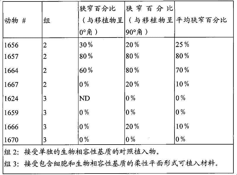
Embodiment 3: the clinical research of people AV graft
This embodiment provides experimental program, is used for test and uses the present invention to promote people's clinical trial experimenter to form functional AV graft.The operation of use standard is created AV graft abutment at the anatomical position of expectation, places ePTFE reparation property bridge between artery and vein abutment.Then implantable material is placed in the contiguous perivascular space, AV graft abutment that each operation creates; Below listed the details of an example operation.As early describing, the placement of implantable material and configuration can be changed in a conventional manner by skilled practitioners.
Particularly, this research comprises the people's test subject that has experienced the AV transfer operation.Conventional AV transfer operation operation will be carried out according to the standard operation technology.After transfer operation is finished and confirmed that graft is unobstructed, implantable material of the present invention will be applied to AV graft abutment and on every side, as described below.
The clinical experimenter of people will accept one or more parts of implantable material in operation the same day.Two to three this parts will be applied to each test subject.A part of implantable material each joint position of reeling.Then an end is moved to the joining section bottom, up to the center of twister on the point of blood vessel and graft.With implant two ends coiling suture, keep the implant center on suture then.It is in place that end can imbrication fixes material.For each test subject, the single part of other of implantable material will place the proximal venal section of arteriovenous graft, begin longitudinally to place along the length of vein from junction point.Implantable material do not need fully the to reel circumference of vein.
Joint position will be handled with preferred implant, for example, and as in Fig. 4 A, Fig. 4 B and Fig. 4 C, or illustrated in fig. 5.In addition, in some patient, by begin preferred implant longitudinally to be placed processing proximal venal section (away from vein-artery abutment) along the length of vein from bonding station.Be contemplated that the total cell load based on body weight will be that about 2.0 * 104 every kg of cell are to about 6.0 * 104 every kg of cell.
The 5th day, 2 weeks with 1,3 with will carry out clinical trail 6 months the time.To use the colored doppler ultrasound that flows to carry out path flow measurement by the 5th day needs after operation, and determine baseline values, 2 weeks, 1 month, 3 months and 6 months be measured flow after operation subsequently.Carry out angiogram to having represented the absolute flow rate that is lower than about 350mL/ minute or in previous the measurement, having represented to surpass 25% flow reduction or represent greater than the test subject in 50% zone narrow (measured) as doppler ultrasound.For angiogram measure greater than 50% stenotic lesion, allow the clinical intervention of remedial, for example angioplasty.
At the baseline place, in the time of 3 months, carry out the angiogram contrast of graft and artery and vein bonding station.Lumen diameter is calculated in each zone, measure peak systolic velocity.
The expected results of people AV graft clinical research.Be contemplated that one or more signs that will show functional clinically AV graft formation as mentioned above with the experimenter of implantable material processed of the present invention.Especially, the experimenter of treatment will show respectively, for example, the blood flow that improves, up to the flow of at least enough saturating dialysis (for example, the 35-500mL/ minute interior blood flow of scope, preferably at least 350mL/ minute), the cracking incidence that reduces, described cracking is defined as engaging the separation from the PTFE graft of vein or artery, serum is concentrated the incidence of (serous perigraft collection) and false aneurysm around the graft that reduces, and/or the reparation bridge that improves integrates, and described integration is defined as tube chamber or propagation within it and/or the migration that smooth muscle cell or endothelial cell enter reparation property bridge.The blood flow that vein flows out position AV graft is comparable with the blood flow that enters the graft position.The comparable meaning is similar for clinical purpose basically.For example, the VPV of expectation is about 150-500mL/ minute, preferably about 300-500mL/ minute, and more preferably from about 350-400mL/ minute.
In addition, entering reparation property bridge or smooth muscle cell within it and/or endothelial cell migration will measure by intravascular ultrasound as the sign of integrating.Be contemplated that implantable material of the present invention will promote smooth muscle cell proliferation and/or endothelial cell proliferation, and both migrations in bridge when as use described here.
Another sign of functional AV graft is sufficient lumen diameter.Be contemplated that, implant of the present invention will allow to keep sufficient lumen diameter, thereby allow blood to flow in the clear with the speed that is suitable for effectively saturating dialysis, promptly, effectively saturating dialysis need be slightly larger than the CBF of the pump speed of dialysis machine, or is enough to prevent the VPV of the recycle during the dialysis at least.After baseline place (arteriovenous graft build up back about 5 days) and operation after this at least 3 months, use angiogram supervision lumen diameter to the arteriovenous graft abutment.Utilize the doppler ultrasound scheme of standard, the narrow and VPV of tube chamber is associated with performing the operation afterwards.Be contemplated that the present invention will prevent from or postpone to narrow down when as use described here, described narrowing down stops blood flow, makes it be lower than the speed that is fit to saturating dialysis described here.
For the AV graft, be contemplated that implantable material of the present invention will prevent or reduce the incidence of cracking.
As a whole, the experimenter of processing estimates showing the difference that increases aspect at least a these aforesaid function signs compared with the control at least.
Embodiment 4: the research of periphery graft
This embodiment provides experimental program, is used for test and uses the test subject that preferred embodiment promotes of the present invention to form the functional peripheral graft.The operation of use standard is created periphery graft abutment at the anatomical position of expectation, places ePTFE reparation property bridge between the abutment.Then implantable material is placed in the contiguous perivascular space, periphery graft abutment that each operation creates; Below listed the details of an example operation.As early describing, the placement of implantable material and structure can change.
Particularly, this research comprises the test subject that has experienced the periphery transfer operation.Conventional periphery graft operation will carry out according to the standard operation technology.After transfer operation is finished and confirmed that graft is unobstructed, implantable material will be applied to periphery graft abutment and on every side, as described below.
Test subject will be accepted one or more preferred implantable materials the same day in operation.Two to three this implants will be applied to each test subject.A this implant each joint position of reeling.End with implantable material moves to the joining section bottom then, up to the center of twister on the point of blood vessel and graft.With implant two ends coiling suture, keep the implant center on suture then.End mutually imbrication to fix material in place.For each test subject, other single implant can place the proximal venal section of periphery graft, begins longitudinally to place along the length of vein from junction point.Implant do not need fully the to reel circumference of vein.
Joint position will be twined with implantable material, for example, and as in Fig. 4 A, Fig. 4 B and Fig. 4 C, or illustrated in fig. 5.In addition, by begin implantable material longitudinally to be placed processing near-end vessel segment (away from the abutment) along the length of blood vessel from bonding station.Total cell load based on body weight will be that about 2.0 * 104 every kg of cell are to about 6.0 * 10
4The every kg of individual cell.
The 5th day, 2 weeks with 1,3 with will carry out clinical trail 6 months the time.To after operation, use the colored doppler ultrasound that flows to carry out the CBF measurement by the 5th day needs, determine baseline values, and after operation, carry out CBF subsequently in 2 weeks, 1 month, 3 months and 6 months and measure.Carry out angiogram to having represented the absolute flow rate that is lower than about 350mL/ minute or in previous the measurement, having represented to surpass 25% flow reduction or represent greater than the test subject in 50% zone narrow (measured) as doppler ultrasound.For angiogram measure greater than 50% stenotic lesion, allow the clinical intervention of remedial, for example angioplasty.
To carry out the angiogram contrast of graft and bonding station.Lumen diameter is calculated in each zone, measure peak systolic velocity.
Periphery graft experimenter's expected results.Be contemplated that as mentioned above experimenter with implantable material processed of the present invention will show one or more signs that functional peripheral graft clinically forms.Periphery graft according to material disclosed herein and method processing is enough to recover or keep acceptable clinically sanguimotor CBF with support.And, the experimenter who handles will show respectively, for example, the cracking incidence that reduces, the vein that described cracking is defined as engaging separates from the PTFE graft, and/or the reparation bridge that show to improve integrates, and described integration is defined as tube chamber or propagation within it and/or the migration that smooth muscle cell or endothelial cell enter reparation property bridge.The blood flow that flows out position periphery graft is comparable with the blood flow that enters the graft position.As used herein, the comparable meaning is similar for clinical purpose basically.For example, the VPV of expectation is about 150-500mL/ minute, preferably about 300-500mL/ minute, and more preferably from about 350-400mL/ minute.
In addition, entering reparation property bridge or smooth muscle cell within it and/or endothelial cell migration will measure as the sign of integrating.Be contemplated that implantable material of the present invention will promote smooth muscle cell proliferation and/or endothelial cell proliferation, and both migrations in bridge.
Another sign of functional peripheral graft is sufficient lumen diameter.Be contemplated that implant of the present invention will allow to keep enough lumen diameters, thereby allow blood to flow in the clear with the speed of enough keeping peripheral circulation.At the baseline place, after graft builds up at least 3 months, use the angiogram of periphery graft to monitor lumen diameter.Utilize the doppler ultrasound scheme of standard, the narrow and VPV of tube chamber is associated with performing the operation afterwards.Be contemplated that implantable material of the present invention will prevent from or postpone to narrow down, described narrowing down stops blood flow, makes it be lower than the speed that is suitable for peripheral circulation described here.
For the periphery bypass graft, be contemplated that and use implantable material processed of the present invention will produce the permission VPV or the approximate normal speed of acceptable circulation clinically.The inflow of graft and to reserve to be comparable.The comparable meaning is similar for clinical purpose basically.For example, the VPV of expectation is about 150-500mL/ minute, preferably about 300-500mL/ minute, and more preferably from about 350-400mL/ minute.In addition, be contemplated that described processing will promote the propagation of smooth muscle cell and/or endothelial cell and migration to enter reparation property graft or natural graft.
For the periphery bypass graft, be contemplated that implantable material of the present invention will prevent or reduce the incidence of cracking.
As a whole, the experimenter of processing estimates showing the difference that increases aspect at least a these aforesaid function signs compared with the control at least.
The present invention can not deviate from its spirit and essential characteristic with other specific forms embodiments.Current embodiment thereby be considered to illustrative and nonrestrictive, scope of the present invention show by claims rather than by above-mentioned specification, thereby the present invention includes the implication that is in claims and the institute in the equivalent scope changes.
Claims (32)
Applications Claiming Priority (4)
| Application Number | Priority Date | Filing Date | Title |
|---|---|---|---|
| US69270805P | 2005-06-21 | 2005-06-21 | |
| US60/692,708 | 2005-06-21 | ||
| USPCT/US2005/043967 | 2005-12-06 | ||
| PCT/US2005/043967 WO2006062909A2 (en) | 2004-12-08 | 2005-12-06 | Methods and compositions for enhancing vascular access |
Related Parent Applications (1)
| Application Number | Title | Priority Date | Filing Date |
|---|---|---|---|
| CN2006800279776A Division CN101257861B (en) | 2005-06-21 | 2006-06-05 | Methods and compositions for enhancing vascular access |
Publications (1)
| Publication Number | Publication Date |
|---|---|
| CN101878757A true CN101878757A (en) | 2010-11-10 |
Family
ID=39892157
Family Applications (3)
| Application Number | Title | Priority Date | Filing Date |
|---|---|---|---|
| CN2010102362140A Pending CN101878757A (en) | 2005-06-21 | 2006-06-05 | Methods and compositions for enhancing vascular access |
| CN201210246619.1A Expired - Fee Related CN102743792B (en) | 2005-06-21 | 2006-06-05 | Methods and compositions for enhancing vascular access |
| CN2006800279776A Expired - Fee Related CN101257861B (en) | 2005-06-21 | 2006-06-05 | Methods and compositions for enhancing vascular access |
Family Applications After (2)
| Application Number | Title | Priority Date | Filing Date |
|---|---|---|---|
| CN201210246619.1A Expired - Fee Related CN102743792B (en) | 2005-06-21 | 2006-06-05 | Methods and compositions for enhancing vascular access |
| CN2006800279776A Expired - Fee Related CN101257861B (en) | 2005-06-21 | 2006-06-05 | Methods and compositions for enhancing vascular access |
Country Status (10)
| Country | Link |
|---|---|
| US (2) | US20090035346A1 (en) |
| EP (2) | EP1901680B1 (en) |
| JP (2) | JP5632580B2 (en) |
| CN (3) | CN101878757A (en) |
| AU (1) | AU2006262682B2 (en) |
| CA (2) | CA2818182C (en) |
| ES (1) | ES2541684T3 (en) |
| IL (2) | IL188263A (en) |
| SG (2) | SG195537A1 (en) |
| WO (1) | WO2007001744A2 (en) |
Cited By (1)
| Publication number | Priority date | Publication date | Assignee | Title |
|---|---|---|---|---|
| CN105407934A (en) * | 2014-05-27 | 2016-03-16 | 诺瓦赫普有限公司 | Bioengineered allogeneic valve |
Families Citing this family (17)
| Publication number | Priority date | Publication date | Assignee | Title |
|---|---|---|---|---|
| JP2926510B2 (en) | 1990-08-09 | 1999-07-28 | 京都電子工業株式会社 | Ammonia concentration measurement device |
| CN101268183B (en) | 2005-04-21 | 2011-11-30 | 麻省理工学院 | Materials and methods for altering an immune response to exogenous and endogenous immunogens, including syngeneic and non-syngeneic cells, tissues or organs |
| CN101878757A (en) | 2005-06-21 | 2010-11-10 | 渗透治疗有限公司 | Methods and compositions for enhancing vascular access |
| EP2079478A2 (en) * | 2006-11-07 | 2009-07-22 | Pervasis Therapeutics, Inc. | Materials and methods for treating and managing angiogenesis-mediated diseases |
| WO2008057590A2 (en) * | 2006-11-07 | 2008-05-15 | Pervasis Therapeutics, Inc. | Methods and compositions for modulating tissue modeling |
| ES2545781T3 (en) * | 2008-01-28 | 2015-09-15 | Biomet 3I, Llc | Superficial implant with greater hydrophilicity |
| US9061014B2 (en) * | 2011-04-28 | 2015-06-23 | Abraxis Bioscience, Llc | Intravascular delivery of nanoparticle compositions and uses thereof |
| CN102763642B (en) * | 2012-08-14 | 2014-04-02 | 郑州赛英科干细胞技术有限公司 | Cryoprotectant and method for cryopreserving placenta amnion and chorion |
| CN107949349B (en) * | 2015-03-10 | 2021-03-23 | 斯滕特泰克有限公司 | Surgical system, device and method thereof for percutaneous arteriovenous fistula (AVF) creation |
| US12017032B1 (en) | 2017-11-13 | 2024-06-25 | Sbr Innovations, Llc | Arteriovenous graft and method of using thereof |
| US11389622B1 (en) * | 2017-11-13 | 2022-07-19 | Simon B. Rayhanabad | Patch for providing dialysis |
| WO2019152799A1 (en) | 2018-02-02 | 2019-08-08 | University Of Louisville Research Foundation, Inc. | Sutureless graft anastomotic quick connect system |
| IL311092B2 (en) * | 2018-08-31 | 2025-05-01 | Wisconsin Alumni Res Found | Generating arterial endothelial cell-seeded vascular grafts |
| US12310836B2 (en) | 2020-03-03 | 2025-05-27 | Akeo Hagiwara | Vein cover |
| CN115376392B (en) * | 2022-08-18 | 2024-04-26 | 昆明理工大学 | Device and method for simulating influence of aortic aging on vascular biomechanics |
| EP4439070B1 (en) * | 2023-03-27 | 2025-12-03 | Siemens Healthcare Diagnostics Products GmbH | Automatic analysis system |
| CN120227529B (en) * | 2025-05-29 | 2025-08-12 | 鼎科医疗技术(苏州)有限公司 | Extravascular stent |
Family Cites Families (88)
| Publication number | Priority date | Publication date | Assignee | Title |
|---|---|---|---|---|
| US4418691A (en) * | 1981-10-26 | 1983-12-06 | Massachusetts Institute Of Technology | Method of promoting the regeneration of tissue at a wound |
| US4787900A (en) * | 1982-04-19 | 1988-11-29 | Massachusetts Institute Of Technology | Process for forming multilayer bioreplaceable blood vessel prosthesis |
| IL78950A (en) | 1985-06-06 | 1991-12-15 | Univ Jefferson | Coating for prosthetic devices |
| US4820626A (en) * | 1985-06-06 | 1989-04-11 | Thomas Jefferson University | Method of treating a synthetic or naturally occuring surface with microvascular endothelial cells, and the treated surface itself |
| US4732155A (en) * | 1985-08-27 | 1988-03-22 | The Children's Medical Center Corporation | Implantable chemoattractant system |
| US5863531A (en) * | 1986-04-18 | 1999-01-26 | Advanced Tissue Sciences, Inc. | In vitro preparation of tubular tissue structures by stromal cell culture on a three-dimensional framework |
| CA1340581C (en) * | 1986-11-20 | 1999-06-08 | Joseph P. Vacanti | Chimeric neomorphogenesis of organs by controlled cellular implantation using artificial matrices |
| US5804178A (en) * | 1986-11-20 | 1998-09-08 | Massachusetts Institute Of Technology | Implantation of cell-matrix structure adjacent mesentery, omentum or peritoneum tissue |
| US5567612A (en) * | 1986-11-20 | 1996-10-22 | Massachusetts Institute Of Technology | Genitourinary cell-matrix structure for implantation into a human and a method of making |
| US6309635B1 (en) * | 1986-11-20 | 2001-10-30 | Children's Medical Center Corp. | Seeding parenchymal cells into compression resistant porous scaffold after vascularizing in vivo |
| US5736372A (en) * | 1986-11-20 | 1998-04-07 | Massachusetts Institute Of Technology | Biodegradable synthetic polymeric fibrous matrix containing chondrocyte for in vivo production of a cartilaginous structure |
| US5073492A (en) * | 1987-01-09 | 1991-12-17 | The Johns Hopkins University | Synergistic composition for endothelial cell growth |
| US5145769A (en) * | 1987-08-21 | 1992-09-08 | Cryolife Inc. | Method for cryopreserving blood vessels |
| US5202120A (en) * | 1987-09-11 | 1993-04-13 | Case Western Reserve University | Methods of reducing glial scar formation and promoting axon and blood vessel growth and/or regeneration through the use of activated immature astrocytes |
| US4945086A (en) | 1988-05-03 | 1990-07-31 | The Board Of Trustees Of The Leland Stanford Junior University | Smooth muscle cell growth inhibitor |
| US5843156A (en) * | 1988-08-24 | 1998-12-01 | Endoluminal Therapeutics, Inc. | Local polymeric gel cellular therapy |
| US5575815A (en) * | 1988-08-24 | 1996-11-19 | Endoluminal Therapeutics, Inc. | Local polymeric gel therapy |
| CH676195A5 (en) * | 1988-10-07 | 1990-12-28 | Sulzer Ag | |
| JPH03133901A (en) * | 1989-10-19 | 1991-06-07 | Osaka Gas Co Ltd | Method for freezing and storing living body cell |
| AU6747790A (en) * | 1989-11-13 | 1991-06-13 | President And Fellows Of Harvard College | Extraluminal regulation of the growth and repair of tubular structures in vivo |
| US5527532A (en) * | 1989-11-13 | 1996-06-18 | President And Fellows Of Harvard College | Extraluminal regulation of the growth and repair of tubular structures in vivo |
| US5540928A (en) * | 1991-02-27 | 1996-07-30 | President And Fellows Of Harvard College | Extraluminal regulation of the growth and repair of tubular structures in vivo |
| CA2071137A1 (en) * | 1991-07-10 | 1993-01-11 | Clarence C. Lee | Composition and method for revitalizing scar tissue |
| EP0591462B1 (en) | 1991-07-15 | 2003-04-16 | Oklahoma Medical Research Foundation | Universal donor cells |
| US5336615A (en) | 1992-01-06 | 1994-08-09 | Yale University | Genetically engineered endothelial cells exhibiting enhanced migration and plasminogen activator activity |
| JPH0646840A (en) * | 1992-08-04 | 1994-02-22 | Nippon Zenyaku Kogyo Kk | Solution for freezing and storing cell |
| US5399665A (en) * | 1992-11-05 | 1995-03-21 | Massachusetts Institute Of Technology | Biodegradable polymers for cell transplantation |
| US5981568A (en) * | 1993-01-28 | 1999-11-09 | Neorx Corporation | Therapeutic inhibitor of vascular smooth muscle cells |
| US6491938B2 (en) * | 1993-05-13 | 2002-12-10 | Neorx Corporation | Therapeutic inhibitor of vascular smooth muscle cells |
| GB9306449D0 (en) * | 1993-03-29 | 1993-05-19 | Nat Heart Research Fund | Tissue equivalents |
| US5709854A (en) * | 1993-04-30 | 1998-01-20 | Massachusetts Institute Of Technology | Tissue formation by injecting a cell-polymeric solution that gels in vivo |
| US5645829A (en) * | 1993-06-18 | 1997-07-08 | Beth Israel Hospital Association | Mesothelial cell gene therapy |
| US6176874B1 (en) * | 1993-10-18 | 2001-01-23 | Masschusetts Institute Of Technology | Vascularized tissue regeneration matrices formed by solid free form fabrication techniques |
| US6608182B1 (en) * | 1994-03-08 | 2003-08-19 | Human Genome Sciences, Inc. | Human vascular endothelial growth factor 2 |
| EP0757562A1 (en) | 1994-04-29 | 1997-02-12 | W.L. Gore & Associates, Inc. | Improved blood contact surfaces using endothelium on a subendothelial extracellular matrix |
| US5834029A (en) * | 1994-07-20 | 1998-11-10 | Cytotherapeutics, Inc. | Nerve guidance channel containing bioartificial three-dimensional hydrogel extracellular matrix derivatized with cell adhesive peptide fragment |
| US6911216B1 (en) * | 1994-10-12 | 2005-06-28 | Genzyme Corporation | Targeted delivery via biodegradable polymers |
| ES2155534T3 (en) * | 1994-10-12 | 2001-05-16 | Focal Inc | ADMINISTRATION DIRECTED THROUGH BIODEGRADABLE POLYMERS. |
| US5914268A (en) * | 1994-11-21 | 1999-06-22 | National Jewish Center For Immunology & Respiratory Medicine | Embryonic cell populations and methods to isolate such populations |
| US6281015B1 (en) * | 1994-12-16 | 2001-08-28 | Children's Medical Center Corp. | Localized delivery of factors enhancing survival of transplanted cells |
| US5716404A (en) * | 1994-12-16 | 1998-02-10 | Massachusetts Institute Of Technology | Breast tissue engineering |
| US5855610A (en) * | 1995-05-19 | 1999-01-05 | Children's Medical Center Corporation | Engineering of strong, pliable tissues |
| US5766584A (en) * | 1995-06-02 | 1998-06-16 | Massachusetts Institute Of Technology | Inhibition of vascular smooth muscle cell proliferation with implanted matrix containing vascular endothelial cells |
| US6615071B1 (en) * | 1995-09-20 | 2003-09-02 | Board Of Regents, The University Of Texas System | Method and apparatus for detecting vulnerable atherosclerotic plaque |
| US5897987A (en) * | 1996-03-25 | 1999-04-27 | Advanced Reproduction Technologies, Inc. | Use of arabinogalactan in cell cryopreservation media |
| US6886568B2 (en) * | 1998-04-08 | 2005-05-03 | The Johns Hopkins University | Method for fabricating cell-containing implants |
| WO2000032750A1 (en) * | 1998-11-24 | 2000-06-08 | Regents Of The University Of Minnesota | Transgenic circulating endothelial cells |
| US6328762B1 (en) * | 1999-04-27 | 2001-12-11 | Sulzer Biologics, Inc. | Prosthetic grafts |
| US6387116B1 (en) * | 1999-06-30 | 2002-05-14 | Pharmasonics, Inc. | Methods and kits for the inhibition of hyperplasia in vascular fistulas and grafts |
| US6461631B1 (en) * | 1999-11-16 | 2002-10-08 | Atrix Laboratories, Inc. | Biodegradable polymer composition |
| WO2001062241A1 (en) * | 2000-02-23 | 2001-08-30 | Smithkline Beecham Corporation | Method for the prevention or reduction of vascular access dysfunction |
| EP1263803B1 (en) * | 2000-03-13 | 2007-09-19 | BioCure, Inc. | Embolic compositions |
| EP1263484B1 (en) * | 2000-03-15 | 2007-05-16 | OrbusNeich Medical, Inc. | Coating which promotes endothelial cell adherence |
| CN1184884C (en) * | 2000-03-24 | 2005-01-19 | 株式会社美你康 | Method for cryopreservation of tissue equivalent and cryopreserved tissue equivalent |
| US6506398B1 (en) * | 2000-04-28 | 2003-01-14 | Hosheng Tu | Device for treating diabetes and methods thereof |
| AU2001275392A1 (en) * | 2000-06-09 | 2001-12-24 | Organ Recovery Systems | Cryopreservation method using cryoprotective composition of propanediol and a vehicle solution |
| AU2001277112A1 (en) | 2000-07-21 | 2002-02-05 | Board Of Regents, The University Of Texas System | Device providing regulated growth factor delivery for the regeneration of peripheral nerves |
| US6696483B2 (en) * | 2000-10-03 | 2004-02-24 | Oncopharmaceutical, Inc. | Inhibitors of angiogenesis and tumor growth for local and systemic administration |
| JP2002200161A (en) * | 2000-10-24 | 2002-07-16 | Menicon Co Ltd | Method for producing cultured skin substitute and cultured skin substitute obtained thereby |
| US6565601B2 (en) * | 2000-11-15 | 2003-05-20 | Micro Therapeutics, Inc. | Methods for vascular reconstruction of diseased arteries |
| US6638709B2 (en) * | 2000-12-27 | 2003-10-28 | Ortec International, Inc. | Processes for making cryopreserved composite living constructs and products resulting therefrom |
| US6726923B2 (en) * | 2001-01-16 | 2004-04-27 | Vascular Therapies, Llc | Apparatus and methods for preventing or treating failure of hemodialysis vascular access and other vascular grafts |
| US6723131B2 (en) * | 2001-02-28 | 2004-04-20 | The Cleveland Clinic Foundation | Composite bone marrow graft material with method and kit |
| WO2003026489A2 (en) | 2001-09-28 | 2003-04-03 | University Of Florida | Biopolymer and biopolymer-cell compositions for nerve tissue repair |
| US7297540B2 (en) * | 2002-01-15 | 2007-11-20 | Yissum Research Development Company Of The Hebrew University Of Jerusalem | Methods of generating tissue using devitalized, acellular scaffold matrices derived from micro-organs |
| US20040047843A1 (en) * | 2002-02-12 | 2004-03-11 | Uab Research Foundation | Method for spinal cord reconnection |
| AU2003223416A1 (en) * | 2002-03-25 | 2003-10-13 | Condros, Inc. | Tissue analogs for in vitro testing and method of use therefor |
| US20050008629A1 (en) * | 2002-05-08 | 2005-01-13 | Interpore Orthopaedics, A Delaware Corporation | Encapsulated AGF cells |
| US20060013852A1 (en) * | 2002-06-28 | 2006-01-19 | Prescott Margaret F | Use of organic compounds |
| FR2845691B1 (en) * | 2002-10-10 | 2004-12-10 | Centre Nat Rech Scient | NOVEL ENDOTHELIAL CELLS, ANTIBODIES DIRECTED AGAINST THESE CELLS AND THEIR USE, IN PARTICULAR FOR SCREENING ANGIOGENESIS-INHIBITING SUBSTANCES |
| US20040156878A1 (en) * | 2003-02-11 | 2004-08-12 | Alireza Rezania | Implantable medical device seeded with mammalian cells and methods of treatment |
| WO2004111207A1 (en) * | 2003-06-09 | 2004-12-23 | Centelion | Multicellular in vitro model of angiogenesis and methods for using the same |
| US8192348B2 (en) * | 2003-07-01 | 2012-06-05 | Regents Of The University Of Minnesota | Engineered blood vessels |
| GB0322789D0 (en) * | 2003-09-30 | 2003-10-29 | Lifeforce Group Plc | Blood pack for leukocyte banking |
| WO2005052138A1 (en) * | 2003-11-19 | 2005-06-09 | Wisconsin Alumni Research Foundation | Cryopreservation of pluripotent stem cells |
| US8067368B2 (en) * | 2004-01-27 | 2011-11-29 | The Ohio State University Research Foundation | Vascular endothelial growth factors and methods of their use |
| DE602005027557D1 (en) * | 2004-04-23 | 2011-06-01 | Bioe Llc | MULTIPLE LINE Progenitor Cells |
| FR2869533B1 (en) | 2004-05-03 | 2006-07-28 | Sedat Sa | SYRINGE FOR MEDICAL INTERVENTIONS AND NECESSARY FOR RECONSTITUTION OF EXTEMPORANEOUS SUBSTANCES COMPRISING SUCH A SYRINGE |
| US7842304B2 (en) | 2004-10-29 | 2010-11-30 | Nexeon Medsystems, Inc. | Methods and apparatus for treating an injured nerve pathway |
| CN102772824A (en) | 2004-12-08 | 2012-11-14 | 渗透治疗有限公司 | Methods and compositions for enhancing vascular access |
| WO2006062871A2 (en) | 2004-12-08 | 2006-06-15 | Pervasis Therapeutics, Inc. | Materials and methods for minimally-invasive administration of a cell-containing flowable composition |
| WO2006062909A2 (en) * | 2004-12-08 | 2006-06-15 | Pervasis Therapeutics, Inc. | Methods and compositions for enhancing vascular access |
| CN101268183B (en) | 2005-04-21 | 2011-11-30 | 麻省理工学院 | Materials and methods for altering an immune response to exogenous and endogenous immunogens, including syngeneic and non-syngeneic cells, tissues or organs |
| GB0509845D0 (en) | 2005-05-13 | 2005-06-22 | Btg Int Ltd | Preparation of therapeutic foam |
| CN101878757A (en) | 2005-06-21 | 2010-11-10 | 渗透治疗有限公司 | Methods and compositions for enhancing vascular access |
| KR20080070820A (en) | 2005-10-12 | 2008-07-31 | 셀룰라 바이오엔지니어링 인코포레이티드 | Resorbable Corneal Flakes |
| US7882863B2 (en) * | 2005-10-19 | 2011-02-08 | Cd Solutions, Llc | Apparatus and method for mixing and transferring medications |
| WO2008036101A2 (en) * | 2005-11-07 | 2008-03-27 | Industrie Borla S.P.A. | Vented safe handling vial adapter |
-
2006
- 2006-06-05 CN CN2010102362140A patent/CN101878757A/en active Pending
- 2006-06-05 CN CN201210246619.1A patent/CN102743792B/en not_active Expired - Fee Related
- 2006-06-05 JP JP2008518189A patent/JP5632580B2/en not_active Expired - Fee Related
- 2006-06-05 SG SG2013075387A patent/SG195537A1/en unknown
- 2006-06-05 SG SG201002482-6A patent/SG161258A1/en unknown
- 2006-06-05 CA CA 2818182 patent/CA2818182C/en not_active Expired - Fee Related
- 2006-06-05 US US11/922,729 patent/US20090035346A1/en not_active Abandoned
- 2006-06-05 WO PCT/US2006/021755 patent/WO2007001744A2/en not_active Ceased
- 2006-06-05 ES ES06772165.4T patent/ES2541684T3/en active Active
- 2006-06-05 AU AU2006262682A patent/AU2006262682B2/en not_active Ceased
- 2006-06-05 EP EP20060772165 patent/EP1901680B1/en not_active Not-in-force
- 2006-06-05 CA CA 2612751 patent/CA2612751A1/en not_active Abandoned
- 2006-06-05 EP EP20110001333 patent/EP2361591A3/en not_active Withdrawn
- 2006-06-05 CN CN2006800279776A patent/CN101257861B/en not_active Expired - Fee Related
-
2007
- 2007-12-19 IL IL188263A patent/IL188263A/en not_active IP Right Cessation
-
2012
- 2012-06-12 JP JP2012133043A patent/JP2012166084A/en not_active Withdrawn
- 2012-07-05 IL IL220806A patent/IL220806A/en not_active IP Right Cessation
- 2012-11-02 US US13/667,227 patent/US9023377B2/en not_active Expired - Fee Related
Cited By (2)
| Publication number | Priority date | Publication date | Assignee | Title |
|---|---|---|---|---|
| CN105407934A (en) * | 2014-05-27 | 2016-03-16 | 诺瓦赫普有限公司 | Bioengineered allogeneic valve |
| CN107670109A (en) * | 2014-05-27 | 2018-02-09 | 诺瓦赫普有限公司 | The allogeneic valve transformed through bioengineering |
Also Published As
| Publication number | Publication date |
|---|---|
| EP1901680B1 (en) | 2015-04-15 |
| IL188263A0 (en) | 2009-02-11 |
| AU2006262682A1 (en) | 2007-01-04 |
| SG161258A1 (en) | 2010-05-27 |
| IL220806A (en) | 2014-05-28 |
| CN101257861B (en) | 2012-12-05 |
| CN102743792A (en) | 2012-10-24 |
| IL220806A0 (en) | 2012-08-30 |
| CN102743792B (en) | 2014-09-24 |
| US20090035346A1 (en) | 2009-02-05 |
| JP2012166084A (en) | 2012-09-06 |
| WO2007001744B1 (en) | 2007-12-21 |
| CA2818182C (en) | 2015-04-14 |
| JP5632580B2 (en) | 2014-11-26 |
| AU2006262682B2 (en) | 2012-07-05 |
| SG195537A1 (en) | 2013-12-30 |
| ES2541684T3 (en) | 2015-07-23 |
| CN101257861A (en) | 2008-09-03 |
| EP1901680A2 (en) | 2008-03-26 |
| JP2008546406A (en) | 2008-12-25 |
| CA2612751A1 (en) | 2007-01-04 |
| US20130210142A1 (en) | 2013-08-15 |
| EP2361591A2 (en) | 2011-08-31 |
| WO2007001744A2 (en) | 2007-01-04 |
| WO2007001744A3 (en) | 2007-10-04 |
| US9023377B2 (en) | 2015-05-05 |
| EP2361591A3 (en) | 2011-12-21 |
| IL188263A (en) | 2014-05-28 |
| CA2818182A1 (en) | 2007-01-04 |
Similar Documents
| Publication | Publication Date | Title |
|---|---|---|
| CN101940801B (en) | Methods and compositions for enhancing vascular access | |
| US9023377B2 (en) | Methods and compositions for enhancing vascular access | |
| US20100204783A1 (en) | Methods and compositions for enhancing vascular access | |
| CN101141988B (en) | Methods and compositions for enhancing vascular access | |
| WO2006062909A2 (en) | Methods and compositions for enhancing vascular access | |
| AU2012238220B2 (en) | Methods and compositions for enhancing vascular access | |
| AU2011211370B2 (en) | Methods and compositions for enhancing vascular access | |
| HK1174859A (en) | Methods and compositions for enhancing vascular access | |
| HK1143517A (en) | Compositions and their use for enhancing vascular access | |
| HK1112429B (en) | Compositions and their use for enhancing vascular access |
Legal Events
| Date | Code | Title | Description |
|---|---|---|---|
| C06 | Publication | ||
| PB01 | Publication | ||
| C10 | Entry into substantive examination | ||
| SE01 | Entry into force of request for substantive examination | ||
| C02 | Deemed withdrawal of patent application after publication (patent law 2001) | ||
| WD01 | Invention patent application deemed withdrawn after publication |
Open date: 20101110 |