CN101675169A - Rare cell analysis using sample resolution and DNA tagging - Google Patents
Rare cell analysis using sample resolution and DNA tagging Download PDFInfo
- Publication number
- CN101675169A CN101675169A CN200780030309A CN200780030309A CN101675169A CN 101675169 A CN101675169 A CN 101675169A CN 200780030309 A CN200780030309 A CN 200780030309A CN 200780030309 A CN200780030309 A CN 200780030309A CN 101675169 A CN101675169 A CN 101675169A
- Authority
- CN
- China
- Prior art keywords
- cells
- fetal
- cell
- pcr
- sample
- Prior art date
- Legal status (The legal status is an assumption and is not a legal conclusion. Google has not performed a legal analysis and makes no representation as to the accuracy of the status listed.)
- Granted
Links
Images


Classifications
-
- C—CHEMISTRY; METALLURGY
- C12—BIOCHEMISTRY; BEER; SPIRITS; WINE; VINEGAR; MICROBIOLOGY; ENZYMOLOGY; MUTATION OR GENETIC ENGINEERING
- C12Q—MEASURING OR TESTING PROCESSES INVOLVING ENZYMES, NUCLEIC ACIDS OR MICROORGANISMS; COMPOSITIONS OR TEST PAPERS THEREFOR; PROCESSES OF PREPARING SUCH COMPOSITIONS; CONDITION-RESPONSIVE CONTROL IN MICROBIOLOGICAL OR ENZYMOLOGICAL PROCESSES
- C12Q1/00—Measuring or testing processes involving enzymes, nucleic acids or microorganisms; Compositions therefor; Processes of preparing such compositions
- C12Q1/68—Measuring or testing processes involving enzymes, nucleic acids or microorganisms; Compositions therefor; Processes of preparing such compositions involving nucleic acids
- C12Q1/6876—Nucleic acid products used in the analysis of nucleic acids, e.g. primers or probes
- C12Q1/6883—Nucleic acid products used in the analysis of nucleic acids, e.g. primers or probes for diseases caused by alterations of genetic material
-
- C—CHEMISTRY; METALLURGY
- C12—BIOCHEMISTRY; BEER; SPIRITS; WINE; VINEGAR; MICROBIOLOGY; ENZYMOLOGY; MUTATION OR GENETIC ENGINEERING
- C12Q—MEASURING OR TESTING PROCESSES INVOLVING ENZYMES, NUCLEIC ACIDS OR MICROORGANISMS; COMPOSITIONS OR TEST PAPERS THEREFOR; PROCESSES OF PREPARING SUCH COMPOSITIONS; CONDITION-RESPONSIVE CONTROL IN MICROBIOLOGICAL OR ENZYMOLOGICAL PROCESSES
- C12Q1/00—Measuring or testing processes involving enzymes, nucleic acids or microorganisms; Compositions therefor; Processes of preparing such compositions
- C12Q1/68—Measuring or testing processes involving enzymes, nucleic acids or microorganisms; Compositions therefor; Processes of preparing such compositions involving nucleic acids
- C12Q1/6809—Methods for determination or identification of nucleic acids involving differential detection
-
- C—CHEMISTRY; METALLURGY
- C12—BIOCHEMISTRY; BEER; SPIRITS; WINE; VINEGAR; MICROBIOLOGY; ENZYMOLOGY; MUTATION OR GENETIC ENGINEERING
- C12Q—MEASURING OR TESTING PROCESSES INVOLVING ENZYMES, NUCLEIC ACIDS OR MICROORGANISMS; COMPOSITIONS OR TEST PAPERS THEREFOR; PROCESSES OF PREPARING SUCH COMPOSITIONS; CONDITION-RESPONSIVE CONTROL IN MICROBIOLOGICAL OR ENZYMOLOGICAL PROCESSES
- C12Q1/00—Measuring or testing processes involving enzymes, nucleic acids or microorganisms; Compositions therefor; Processes of preparing such compositions
- C12Q1/68—Measuring or testing processes involving enzymes, nucleic acids or microorganisms; Compositions therefor; Processes of preparing such compositions involving nucleic acids
- C12Q1/6869—Methods for sequencing
-
- C—CHEMISTRY; METALLURGY
- C12—BIOCHEMISTRY; BEER; SPIRITS; WINE; VINEGAR; MICROBIOLOGY; ENZYMOLOGY; MUTATION OR GENETIC ENGINEERING
- C12Q—MEASURING OR TESTING PROCESSES INVOLVING ENZYMES, NUCLEIC ACIDS OR MICROORGANISMS; COMPOSITIONS OR TEST PAPERS THEREFOR; PROCESSES OF PREPARING SUCH COMPOSITIONS; CONDITION-RESPONSIVE CONTROL IN MICROBIOLOGICAL OR ENZYMOLOGICAL PROCESSES
- C12Q1/00—Measuring or testing processes involving enzymes, nucleic acids or microorganisms; Compositions therefor; Processes of preparing such compositions
- C12Q1/68—Measuring or testing processes involving enzymes, nucleic acids or microorganisms; Compositions therefor; Processes of preparing such compositions involving nucleic acids
- C12Q1/6876—Nucleic acid products used in the analysis of nucleic acids, e.g. primers or probes
- C12Q1/6881—Nucleic acid products used in the analysis of nucleic acids, e.g. primers or probes for tissue or cell typing, e.g. human leukocyte antigen [HLA] probes
-
- B—PERFORMING OPERATIONS; TRANSPORTING
- B01—PHYSICAL OR CHEMICAL PROCESSES OR APPARATUS IN GENERAL
- B01L—CHEMICAL OR PHYSICAL LABORATORY APPARATUS FOR GENERAL USE
- B01L3/00—Containers or dishes for laboratory use, e.g. laboratory glassware; Droppers
- B01L3/50—Containers for the purpose of retaining a material to be analysed, e.g. test tubes
- B01L3/502—Containers for the purpose of retaining a material to be analysed, e.g. test tubes with fluid transport, e.g. in multi-compartment structures
- B01L3/5027—Containers for the purpose of retaining a material to be analysed, e.g. test tubes with fluid transport, e.g. in multi-compartment structures by integrated microfluidic structures, i.e. dimensions of channels and chambers are such that surface tension forces are important, e.g. lab-on-a-chip
- B01L3/502761—Containers for the purpose of retaining a material to be analysed, e.g. test tubes with fluid transport, e.g. in multi-compartment structures by integrated microfluidic structures, i.e. dimensions of channels and chambers are such that surface tension forces are important, e.g. lab-on-a-chip specially adapted for handling suspended solids or molecules independently from the bulk fluid flow, e.g. for trapping or sorting beads, for physically stretching molecules
-
- C—CHEMISTRY; METALLURGY
- C12—BIOCHEMISTRY; BEER; SPIRITS; WINE; VINEGAR; MICROBIOLOGY; ENZYMOLOGY; MUTATION OR GENETIC ENGINEERING
- C12Q—MEASURING OR TESTING PROCESSES INVOLVING ENZYMES, NUCLEIC ACIDS OR MICROORGANISMS; COMPOSITIONS OR TEST PAPERS THEREFOR; PROCESSES OF PREPARING SUCH COMPOSITIONS; CONDITION-RESPONSIVE CONTROL IN MICROBIOLOGICAL OR ENZYMOLOGICAL PROCESSES
- C12Q2600/00—Oligonucleotides characterized by their use
- C12Q2600/156—Polymorphic or mutational markers
-
- C—CHEMISTRY; METALLURGY
- C12—BIOCHEMISTRY; BEER; SPIRITS; WINE; VINEGAR; MICROBIOLOGY; ENZYMOLOGY; MUTATION OR GENETIC ENGINEERING
- C12Q—MEASURING OR TESTING PROCESSES INVOLVING ENZYMES, NUCLEIC ACIDS OR MICROORGANISMS; COMPOSITIONS OR TEST PAPERS THEREFOR; PROCESSES OF PREPARING SUCH COMPOSITIONS; CONDITION-RESPONSIVE CONTROL IN MICROBIOLOGICAL OR ENZYMOLOGICAL PROCESSES
- C12Q2600/00—Oligonucleotides characterized by their use
- C12Q2600/158—Expression markers
-
- C—CHEMISTRY; METALLURGY
- C12—BIOCHEMISTRY; BEER; SPIRITS; WINE; VINEGAR; MICROBIOLOGY; ENZYMOLOGY; MUTATION OR GENETIC ENGINEERING
- C12Q—MEASURING OR TESTING PROCESSES INVOLVING ENZYMES, NUCLEIC ACIDS OR MICROORGANISMS; COMPOSITIONS OR TEST PAPERS THEREFOR; PROCESSES OF PREPARING SUCH COMPOSITIONS; CONDITION-RESPONSIVE CONTROL IN MICROBIOLOGICAL OR ENZYMOLOGICAL PROCESSES
- C12Q2600/00—Oligonucleotides characterized by their use
- C12Q2600/16—Primer sets for multiplex assays
-
- G—PHYSICS
- G01—MEASURING; TESTING
- G01N—INVESTIGATING OR ANALYSING MATERIALS BY DETERMINING THEIR CHEMICAL OR PHYSICAL PROPERTIES
- G01N15/00—Investigating characteristics of particles; Investigating permeability, pore-volume or surface-area of porous materials
- G01N15/10—Investigating individual particles
- G01N2015/1006—Investigating individual particles for cytology
-
- G—PHYSICS
- G01—MEASURING; TESTING
- G01N—INVESTIGATING OR ANALYSING MATERIALS BY DETERMINING THEIR CHEMICAL OR PHYSICAL PROPERTIES
- G01N15/00—Investigating characteristics of particles; Investigating permeability, pore-volume or surface-area of porous materials
- G01N15/10—Investigating individual particles
- G01N2015/1029—Particle size
-
- Y—GENERAL TAGGING OF NEW TECHNOLOGICAL DEVELOPMENTS; GENERAL TAGGING OF CROSS-SECTIONAL TECHNOLOGIES SPANNING OVER SEVERAL SECTIONS OF THE IPC; TECHNICAL SUBJECTS COVERED BY FORMER USPC CROSS-REFERENCE ART COLLECTIONS [XRACs] AND DIGESTS
- Y10—TECHNICAL SUBJECTS COVERED BY FORMER USPC
- Y10T—TECHNICAL SUBJECTS COVERED BY FORMER US CLASSIFICATION
- Y10T436/00—Chemistry: analytical and immunological testing
- Y10T436/14—Heterocyclic carbon compound [i.e., O, S, N, Se, Te, as only ring hetero atom]
- Y10T436/142222—Hetero-O [e.g., ascorbic acid, etc.]
- Y10T436/143333—Saccharide [e.g., DNA, etc.]
Landscapes
- Chemical & Material Sciences (AREA)
- Life Sciences & Earth Sciences (AREA)
- Organic Chemistry (AREA)
- Proteomics, Peptides & Aminoacids (AREA)
- Health & Medical Sciences (AREA)
- Zoology (AREA)
- Wood Science & Technology (AREA)
- Engineering & Computer Science (AREA)
- Analytical Chemistry (AREA)
- Genetics & Genomics (AREA)
- Immunology (AREA)
- Microbiology (AREA)
- Bioinformatics & Cheminformatics (AREA)
- Molecular Biology (AREA)
- Biophysics (AREA)
- Physics & Mathematics (AREA)
- General Health & Medical Sciences (AREA)
- Biochemistry (AREA)
- Biotechnology (AREA)
- General Engineering & Computer Science (AREA)
- Pathology (AREA)
- Cell Biology (AREA)
- Measuring Or Testing Involving Enzymes Or Micro-Organisms (AREA)
- Apparatus Associated With Microorganisms And Enzymes (AREA)
- Investigating Or Analysing Biological Materials (AREA)
Abstract
本发明提供在胎儿细胞与母体细胞群混合时检测样品中的胎儿细胞存在情况并测试胎儿异常如异倍性的系统、装置和方法。本发明包括用不同标记来标记所述混合样品中每个细胞中的基因组DNA的区域,其中每个标记对每个细胞都是特异性的,并定量混合样品中每个细胞的基因组DNA的标记区。更具体地说,本发明涉及定量混合样品中每个细胞的标记DNA多态性。The present invention provides systems, devices, and methods for detecting the presence of fetal cells in a sample when the cells are mixed with a population of maternal cells and testing for fetal abnormalities, such as aneuploidy. The invention involves labeling regions of genomic DNA in each cell in the mixed sample with different markers, wherein each marker is specific for each cell, and quantifying the labeled regions of genomic DNA in each cell in the mixed sample. More specifically, the invention relates to quantifying polymorphisms in the marker DNA of each cell in the mixed sample.
Description
交叉参考cross reference
[0001]本申请要求保护2006年6月14日申请的美国临时申请号60/804,819的权益,该申请在此引入作为参考。[0001] The application claims the benefit of U.S. Provisional Application No. 60/804,819, filed June 14, 2006, which is incorporated herein by reference.
发明背景Background of the invention
[0002]分析特定细胞可以洞察多种疾病。这些分析可为检测、诊断和预后例如癌症或胎儿异常的疾病提供非侵入性检查,从而消除侵入性诊断的风险。关于胎儿异常,现有的产前诊断,例如羊膜穿刺术和绒毛膜绒毛取样(CVS),可能对母亲和胎儿造成伤害。经历羊膜穿刺术的孕妇的流产率增加0.5-1%,而对于CVS,该数值还要稍高一些。由于羊膜穿刺术和CVS引起的特有风险,所以这些方法主要用于孕育先天性缺陷儿的统计学概率较高的高龄妇女,比如35岁以上的妇女。因此,35岁的孕妇就不得不在羊膜穿刺术诱发流产的0.5-1%平均风险与小于0.3%的年龄相关的21三体性概率之间权衡。[0002] Analysis of specific cells can provide insight into a variety of diseases. These assays provide non-invasive tests for the detection, diagnosis and prognosis of diseases such as cancer or fetal abnormalities, thereby eliminating the risk of invasive diagnostics. With regard to fetal abnormalities, available prenatal diagnostics, such as amniocentesis and chorionic villus sampling (CVS), can be harmful to both mother and fetus. Pregnant women undergoing amniocentesis have an increased miscarriage rate of 0.5-1%, and with CVS this figure is slightly higher. Because of the unique risks associated with amniocentesis and CVS, these procedures are primarily reserved for older women, such as women over the age of 35, who have a higher statistical chance of conceiving a child with a congenital defect. Thus, a 35-year-old pregnant woman would have to weigh the average risk of amniocentesis-induced miscarriage of 0.5-1% against an age-related probability of
[0003]关于产前诊断,已经开发出一些非侵入性方法来筛选特异性先天缺陷风险较高的胎儿。例如,产妇血清甲胎蛋白,以及未缀合的雌三醇和人绒毛膜促性腺激素的水平,可用于鉴定患唐氏综合征的胎儿的比例。然而,这些检测出现许多假阳性。类似地,超声波检查法用于确定先天性缺陷,包括神经管缺陷和四肢异常,但这些方法只限于15周妊娠期之后的时间段,且提供的结果不可靠。[0003] With regard to prenatal diagnosis, several non-invasive methods have been developed to screen fetuses at higher risk for specific birth defects. For example, maternal serum levels of alpha-fetoprotein, as well as unconjugated estriol and human chorionic gonadotropin, can be used to identify the proportion of fetuses with Down syndrome. However, many false positives occur with these tests. Similarly, ultrasonography is used to identify birth defects, including neural tube defects and abnormalities of the extremities, but these methods are limited to the time period beyond 15 weeks of gestation and provide unreliable results.
[0004]孕妇血液中存在的胎儿细胞为开发替代羊膜穿刺术并由此消除现今的侵入性诊断风险的产前诊断提供了机会。然而,相对于血液中大量母体细胞的背景,胎儿细胞仅占少数,这使得分析既耗时又易出错。[0004] The presence of fetal cells in the blood of pregnant women provides an opportunity to develop prenatal diagnostics that can replace amniocentesis and thereby eliminate the risks of today's invasive diagnostics. However, fetal cells are a minority against the background of large numbers of maternal cells in blood, making analysis time-consuming and error-prone.
[0005]对于癌症诊断,早期检测极为重要。癌症是以不受控的异常细胞增殖为标志的疾病。在正常组织中,细胞发生分裂在周围细胞信号作用下在组织中组织化。癌细胞不以相同的方式响应这些信号,使得它们发生增殖,在许多器官中形成肿瘤。随着肿瘤持续生长,遗传改变可以累积,表现为更具侵袭性的癌细胞生长表型。如果放任不治疗,则可能接着发生转移-癌细胞通过淋巴系统或血流扩散至机体的远方区域。转移导致在多个部位形成继发性肿瘤,伤害健康组织。大部分癌症死亡都是由这些继发性肿瘤引起的。尽管癌症诊断和治疗已发展了几十年,但许多癌症仍然是直到晚期才能检测到。作为一个实例,最早期的肺癌是无症状的,不能及时检测到而进行药物治疗,导致肺癌患者大体上5年的生存率低于15%。然而,在早期检测到肺癌并治疗的情况下,预后有利得多。[0005] For cancer diagnosis, early detection is extremely important. Cancer is a disease marked by uncontrolled abnormal cell proliferation. In normal tissues, cells divide and organize in the tissue in response to signals from surrounding cells. Cancer cells do not respond to these signals in the same way, allowing them to proliferate and form tumors in many organs. As tumors continue to grow, genetic alterations can accumulate, manifesting in a more aggressive cancer cell growth phenotype. If left untreated, metastasis—the spread of cancer cells to distant areas of the body through the lymph system or bloodstream—may ensue. Metastasis leads to the formation of secondary tumors at multiple sites, harming healthy tissue. Most cancer deaths are caused by these secondary tumors. Despite decades of advances in cancer diagnosis and treatment, many cancers remain undetected until advanced stages. As an example, the earliest stage of lung cancer is asymptomatic and cannot be detected in time to be treated with drugs, resulting in a general 5-year survival rate of less than 15% for lung cancer patients. However, when lung cancer is detected early and treated, the prognosis is much more favorable.
[0006]本发明的方法使得在胎儿细胞与母体细胞群混合时,即便在细胞混合体中母体细胞占主要时,也可以检测胎儿细胞和胎儿异常。另外,本发明的方法还可以用于检测或诊断癌症。[0006] The method of the present invention allows detection of fetal cells and fetal abnormalities when fetal cells are mixed with a population of maternal cells, even when maternal cells predominate in the mixture of cells. In addition, the methods of the present invention can also be used to detect or diagnose cancer.
发明概述Summary of the invention
[0007]本发明涉及检测混合样品中的胎儿细胞或癌细胞的方法。在一个实施方案中,本发明提供在含有与母体细胞群混合的胎儿细胞的样品中测定胎儿异常的方法。在某些实施方案中,测定胎儿细胞和胎儿异常的存在情况包括用不同标记来标记含有至少一个胎儿细胞的混合样品中的每个细胞的基因组DNA的一个或多个区,其中每个标记对每个细胞都是特异性的。在某些实施方案中,待标记的基因组DNA包含一种或多种多态性,尤其是STR或SNP。[0007] The present invention relates to methods of detecting fetal or cancer cells in a mixed sample. In one embodiment, the present invention provides methods for determining fetal abnormalities in a sample containing fetal cells mixed with a population of maternal cells. In certain embodiments, determining the presence of fetal cells and fetal abnormalities comprises labeling one or more regions of the genomic DNA of each cell in a mixed sample containing at least one fetal cell with a different marker, wherein each marker corresponds to Every cell is specific. In certain embodiments, the genomic DNA to be marked comprises one or more polymorphisms, especially STRs or SNPs.
[0008]在某些实施方案中,本发明的方法使得在胎儿细胞与母体细胞群混合时,即便母体细胞在混合物中占优势时,也可以同时检测胎儿细胞和胎儿异常的存在情况。在某些实施方案中,富集样品,以包含至少一个胎儿细胞和一个非胎儿细胞,在其它实施方案中,富集群的细胞可分配在两个或更多个可用作可寻址部位的离散位置。可寻址部位的实施例包括孔(wells)、区(bins)、筛、小孔(pore)、几何位点、载玻片、基质、膜、电阱(electric traps)、间隙、障碍物或细胞膜或核膜原位。[0008] In certain embodiments, the methods of the present invention allow simultaneous detection of fetal cells and the presence of fetal abnormalities when fetal cells are mixed with a population of maternal cells, even when maternal cells predominate in the mixture. In certain embodiments, the sample is enriched to contain at least one fetal cell and one non-fetal cell, and in other embodiments, the cells of the enriched population can be distributed among two or more cells that can be used as addressable sites. discrete locations. Examples of addressable sites include wells, bins, screens, pores, geometric sites, slides, substrates, membranes, electric traps, gaps, obstacles, or Cell membrane or nuclear membrane in situ.
[0009]在某些实施方案中,所述方法包括用不同标记来标记富集样品中的每个细胞的基因组DNA的一个或多个区,其中每个标记对每个细胞都是特异性的,并定量标记的DNA区。标记方法可包括在混合样品中加入每个细胞的独有标签序列。在某些实施方案中,独有的标签序列鉴定混合样品的每个细胞中是否存在DNA多态性。使用可通过PCR方法进行的扩增反应将标记加至细胞/DNA。例如,扩增可通过多重PCR实现。在某些实施方案中,使用基因组DNA区的嵌套引物进行进一步的PCR扩增。[0009] In certain embodiments, the method comprises labeling one or more regions of genomic DNA of each cell in the enriched sample with different markers, wherein each marker is specific to each cell , and quantify labeled DNA regions. Labeling methods can include adding unique tag sequences to each cell in the mixed sample. In certain embodiments, unique tag sequences identify the presence or absence of DNA polymorphisms in each cell of a pooled sample. Labels are added to the cells/DNA using amplification reactions that can be performed by PCR methods. For example, amplification can be achieved by multiplex PCR. In certain embodiments, further PCR amplification is performed using nested primers for the genomic DNA region.
[0010]在某些实施方案中,DNA区可在定量前扩增。标记的DNA可以使用测序方法定量,测序方法在某些实施方案中可在扩增DNA区之前。扩增的DNA区可通过测序方法分析。例如,超深测序可用于为每个STR或SNP提供精确定量的等位基因丰度检测。在其它实施方案中,定量基因分型可用于显示胎儿细胞的存在情况及确定胎儿染色体的拷贝数。优选地,定量基因分型使用分子倒置探针进行。[0010] In certain embodiments, regions of DNA may be amplified prior to quantification. Labeled DNA can be quantified using sequencing methods, which in some embodiments may precede amplification of the DNA region. The amplified DNA region can be analyzed by sequencing methods. For example, ultra-deep sequencing can be used to provide precise quantitative allelic abundance detection for each STR or SNP. In other embodiments, quantitative genotyping can be used to reveal the presence of fetal cells and determine the copy number of fetal chromosomes. Preferably, quantitative genotyping is performed using molecular inversion probes.
[0011]本发明还涉及对含非母体基因组DNA的混合样品进行细胞鉴定并鉴定具有非母体基因组DNA的细胞为胎儿细胞的方法。在某些实施方案中,对由混合样品鉴定出的胎儿细胞比较母体对父体等位基因的比率。[0011] The present invention also relates to a method for performing cell identification on a mixed sample containing non-maternal genomic DNA and identifying cells with non-maternal genomic DNA as fetal cells. In certain embodiments, maternal to paternal allele ratios are compared for fetal cells identified from a pooled sample.
[0012]在一个实施方案中,本发明提供测定母体样品中的胎儿异常的方法,所述母体样品含有至少一个胎儿细胞和一个非胎儿细胞。可富集样品,以包含至少一个胎儿细胞,并可将富集的母体样品阵列入大量离散位置中。在某些实施方案中,每个离散位置都含有不超过一个细胞。[0012] In one embodiment, the invention provides a method of determining a fetal abnormality in a maternal sample comprising at least one fetal cell and one non-fetal cell. The sample can be enriched to contain at least one fetal cell, and the enriched maternal sample can be arrayed into a number of discrete locations. In certain embodiments, each discrete location contains no more than one cell.
[0013]在某些实施方案中,本发明包括使用对每个DNA区或位置特异性的引物标记阵列样品的一个或多个基因组DNA区,扩增DNA区,并定量标记的DNA区。DNA区的标记可以包括用独特的标签序列标记每个区,所述标签序列可用于鉴定阵列细胞是否存在DNA多态性和细胞的不同位置。[0013] In certain embodiments, the invention includes labeling one or more genomic DNA regions of an array sample with primers specific for each DNA region or position, amplifying the DNA regions, and quantifying the labeled DNA regions. Labeling of DNA regions can include labeling each region with a unique tag sequence that can be used to identify cells of the array for the presence of DNA polymorphisms and different locations of the cells.
[0014]测定步骤可包括鉴定不同位置的非母体等位基因,这可由比较该位置的母体对父体等位基因的比率产生。在某些实施方案中,鉴定阵列样品中的胎儿异常的方法可进一步包括扩增基因组DNA区。基因组DNA区可以包含一种或多种多态性,例如STR和SNP,其可使用包括多重PCR的PCR法扩增。附加扩增步骤可以使用嵌套引物进行。[0014] The determining step may include identifying non-maternal alleles at different positions, which may result from comparing the ratio of maternal to paternal alleles at that position. In certain embodiments, the method of identifying a fetal abnormality in an array sample can further comprise amplifying a region of genomic DNA. A region of genomic DNA may contain one or more polymorphisms, such as STRs and SNPs, which can be amplified using PCR methods including multiplex PCR. Additional amplification steps can be performed using nested primers.
[0015]扩增的DNA区可通过测序方法分析。例如,超深测序可用于为每个STR或SNP提供精确定量的等位基因丰度检测。在其它实施方案中,定量基因分型可用于表明胎儿细胞的存在情况,并确定胎儿染色体的拷贝数。优选地,定量基因分型使用分子倒置探针进行。[0015] The amplified DNA region can be analyzed by sequencing methods. For example, ultra-deep sequencing can be used to provide precise quantitative allelic abundance detection for each STR or SNP. In other embodiments, quantitative genotyping can be used to indicate the presence of fetal cells and determine the copy number of fetal chromosomes. Preferably, quantitative genotyping is performed using molecular inversion probes.
[0016]在一个实施方案中,本发明提供如下诊断癌症并给出预后的方法:由患者获得血样并富集血样的上皮细胞,将富集的样品拆分(splitting)至离散位置,并对富集并拆分的样品进行一种或多种分子和/或形态学分析。分子分析可以包括检测在图10中公开的基因的表达水平或突变。优选地,所述方法包括对每个阵列细胞中的EGFR、EpCAM、GA733-2、MUC-1、HER-2或紧密连接蛋白(Claudin)-7进行分子分析。形态学分析可以包括鉴定、定量和/或表征线粒体DNA、端粒酶或核基质蛋白。[0016] In one embodiment, the present invention provides a method of diagnosing cancer and giving a prognosis by obtaining a blood sample from a patient and enriching the blood sample for epithelial cells, splitting the enriched sample into discrete locations, and analyzing The enriched and resolved samples are subjected to one or more molecular and/or morphological analyses. Molecular analysis may include detection of expression levels or mutations of the genes disclosed in FIG. 10 . Preferably, the method comprises molecular analysis of EGFR, EpCAM, GA733-2, MUC-1, HER-2 or Claudin-7 in each arrayed cell. Morphological analysis can include identifying, quantifying, and/or characterizing mitochondrial DNA, telomerase, or nuclear matrix proteins.
[0017]在某些实施方案中,可富集样品的上皮细胞达至少10,000倍,并可以在治疗癌症患者之前提供诊断和预后。优选地,血样以定期间隔得自患者,例如每日1次,或每2、3或4天1次、每周1次、每半个月1次、每月1次、每半年1次或每年1次。[0017] In certain embodiments, a sample can be enriched for epithelial cells by at least 10,000-fold and can provide a diagnosis and prognosis prior to treatment of a cancer patient. Preferably, blood samples are obtained from the patient at regular intervals, for example daily, or every 2, 3 or 4 days, weekly, semi-monthly, monthly, semi-annually or 1 time per year.
[0018]在某些实施方案中,富集患者血样的上皮细胞的步骤包括使样品流经第一障碍物阵列,该阵列将大于预定尺寸的细胞选择性地导向第一出口,将小于预定尺寸的细胞选择性地导向第二出口。可选地,可通过使样品流经第二障碍物阵列对样品进行进一步的富集,该阵列可用选择性结合白细胞或上皮细胞的抗体包被。例如,第二阵列的障碍物可以用抗EpCAM抗体包被。[0018] In certain embodiments, the step of enriching the patient's blood sample for epithelial cells includes flowing the sample through a first array of obstacles that selectively direct cells larger than a predetermined size to a first outlet and cells smaller than a predetermined size The cells are selectively directed to the second outlet. Optionally, the sample can be further enriched by passing the sample through a second barrier array, which can be coated with antibodies that selectively bind leukocytes or epithelial cells. For example, the barriers of the second array can be coated with anti-EpCAM antibodies.
[0019]拆分富集群的细胞样品可以包括拆分富集样品,以将单个细胞定位在可为可寻址部位的离散位置。可寻址部位的实例包括孔、区、筛、小孔、几何位点、载玻片、基质、膜、电阱、间隙、障碍物或细胞膜或核膜原位。[0019] Resolving the cell sample of the enriched population may include resolving the enriched sample to localize individual cells at discrete locations that may be addressable sites. Examples of addressable sites include wells, regions, sieves, apertures, geometric sites, slides, substrates, membranes, electrical traps, gaps, barriers, or cell or nuclear membrane in situ.
[0020]在某些实施方案中,提供含有富集样品的装置以及进行遗传分析所需要的装置和试剂的试剂盒。该试剂盒可含有基于尺寸分离的阵列、用于独有地标记细胞的试剂、将细胞拆分至单独的可寻址部位中的装置和用于遗传分析的试剂。[0020] In certain embodiments, kits are provided that contain a device for enriching a sample, as well as the devices and reagents needed to perform a genetic analysis. The kit may contain an array for size-based separation, reagents for uniquely labeling cells, means for resolving cells into individually addressable sites, and reagents for genetic analysis.
附图概述Figure overview
[0021]图1A-1D图解了基于尺寸的分离模块的多个实施方案。[0021] Figures 1A-1D illustrate various embodiments of size-based separation modules.
[0022]图2A-2C图解阐述了亲和分离模块的一个实施方案。[0022] Figures 2A-2C schematically illustrate one embodiment of an affinity separation module.
[0023]图3图解了磁分离模块的一个实施方案。[0023] Figure 3 illustrates one embodiment of a magnetic separation module.
[0024]图4图解了诊断、预后或监测胎儿的出生前状况的纵览。[0024] FIG. 4 illustrates an overview of diagnosing, prognosing, or monitoring the prenatal condition of a fetus.
[0025]图5图解了诊断、预后或监测胎儿的出生前状况的纵览。[0025] FIG. 5 illustrates an overview of diagnosing, prognosing, or monitoring the prenatal condition of a fetus.
[0026]图6图解了诊断、预后或监测患者癌症的纵览。[0026] FIG. 6 illustrates an overview of diagnosing, prognosing, or monitoring cancer in a patient.
[0027]图7A-7B图解了使用分子倒置探针的测定。图7C图解了使用核酸标签的纵览。[0027] Figures 7A-7B illustrate assays using molecular inversion probes. Figure 7C illustrates an overview of using nucleic acid tags.
[0028]图8A-8C图解了样品拆分装置(a sample splittingapparatus)的一个实例。[0028] Figures 8A-8C illustrate an example of a sample splitting apparatus.
[0029]图9图解了将2个以上的CTC加载至单个样品孔中的的概率。[0029] FIG. 9 illustrates the probability of loading more than 2 CTCs into a single sample well.
[0030]图10图解了其表达或突变可能与本文诊断的癌症或另一种病症相关的基因。[0030] FIG. 10 illustrates genes whose expression or mutation may be associated with cancer or another condition diagnosed herein.
[0031]图11图解了可用于本文方法的引物。[0031] Figure 11 illustrates primers that can be used in the methods herein.
[0032]图12A-B图解了产物和废物级分的细胞涂片。[0032] Figures 12A-B illustrate cell smears of product and waste fractions.
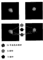
[0033]图13A-F图解了分离的胎儿细胞,其通过可信的雄细胞存在而证实。[0033] Figures 13A-F illustrate isolated fetal cells as evidenced by the presence of authentic male cells.
[0034]图14图解了具有异常21三体病理的细胞。[0034] FIG. 14 illustrates cells with
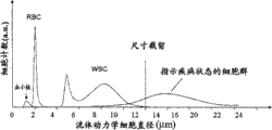
[0035]图15图解了基于尺寸的分离模块的性能。[0035] FIG. 15 illustrates performance of separation modules based on size.
[0036]图16图解了由基于尺寸的分离模块产生的这些细胞级分的柱形图。[0036] FIG. 16 illustrates a histogram of these cell fractions produced by the size-based separation module.
[0037]图17图解了基于尺寸的分离模块的第一出口和第二出口。[0037] FIG. 17 illustrates a first outlet and a second outlet of a size-based separation module.
[0038]图18图解了结合包被抗EpCAM的障碍物阵列的捕获模块的上皮细胞。[0038] FIG. 18 illustrates an epithelial cell bound to a capture module coated with an anti-EpCAM barrier array.
[0039]图19A-C图解了一个基于流通尺寸的分离模块的实施方案,及可用于该装置的备选参数,所述分离模块适于由血液中分离上皮细胞。[0039] FIGS. 19A-C illustrate an embodiment of a flow-through size-based separation module suitable for isolating epithelial cells from blood, and alternative parameters that can be used with the device.
[0040]图20A-D图解了多个被靶向的细胞亚群和多个截留尺寸,所述亚群可使用基于尺寸的分离来分离,所述截留尺寸可用于分离这些被靶向的亚群。[0040] Figures 20A-D illustrate various targeted cell subpopulations that can be separated using size-based separation and multiple cut-off sizes that can be used to separate these targeted subpopulations group.
[0041]图21图解了本发明的装置和确定富集样品中的细胞数的计数方法。[0041] FIG. 21 illustrates a device of the present invention and a counting method for determining the number of cells in an enriched sample.
[0042]图22图解了用于诊断、预后或监测患者癌症的本发明的一个方面的纵览。[0042] FIG. 22 illustrates an overview of one aspect of the invention for diagnosing, prognosing, or monitoring cancer in a patient.
[0043]图23图解了EGFR mRNA产生测序模板的应用。[0043] Figure 23 illustrates the use of EGFR mRNA to generate sequencing templates.
[0044]图24图解了进行实时定量等位基因特异性PCR反应,以证实mEGFR mRNA中的突变序列。[0044] FIG. 24 illustrates performing real-time quantitative allele-specific PCR reactions to confirm mutant sequences in mEGFR mRNA.
[0045]图25表明,当突变等位基因探针的信号超过荧光背景水平时,证实存在突变。[0045] Figure 25 shows that the presence of a mutation is confirmed when the signal from the mutant allele probe exceeds fluorescent background levels.
[0046]图26A-B图解了在上皮细胞中存在EGFR mRNA,但在白细胞中不存在EGFR mRNA。[0046] Figures 26A-B illustrate the presence of EGFR mRNA in epithelial cells but not in leukocytes.
[0047]图27图解了第一个和第二个EGFR PCR反应的结果。[0047] Figure 27 illustrates the results of the first and second EGFR PCR reactions.
[0048]图28A-B图解了第一个和第二个EGFR PCR反应的结果。[0048] Figures 28A-B illustrate the results of the first and second EGFR PCR reactions.
[0049]图29表明,易于检测EGFR野生型和突变体扩增片段,尽管白细胞背景高。[0049] Figure 29 demonstrates that EGFR wild-type and mutant amplified fragments are readily detected despite a high white blood cell background.
[0050]图30图解了通过qPCR检测单个拷贝的胎儿细胞基因组。[0050] FIG. 30 illustrates detection of a single copy of the fetal cell genome by qPCR.
[0051]图31图解了通过SNP分析检测分区样品(binned samples)中的单个胎儿细胞。[0051] FIG. 31 illustrates detection of single fetal cells in binned samples by SNP analysis.
[0052]图32图解了三体性测试方法。21三体筛选基于得自母体血液的靶细胞计数。血液使用用于血红蛋白富集的细胞分离模块(CSM-HE)处理。将富集细胞转移至载玻片,首先对载玻片染色,随后通过FISH探测。例如由亮视野或荧光显微镜获取图像,并计数。某些类型的三体性细胞的比例用作胎儿21三体风险的分类器。胎儿基因组鉴定可以使用诸如以下的测定进行:(1)STR标记;(2)使用针对基因座(例如Y-染色体上的多次重复的DYZ基因座)的引物和探针的qPCR;(3)SNP检测;和(4)CGH(比较基因组杂交)阵列检测。[0052] Figure 32 illustrates the trisomy testing method.
[0053]图33图解了可产生有关靶细胞中异倍性和其它遗传疾病的存在情况的信息的测定。有关靶细胞中异倍性和其它遗传疾病的信息可以使用诸如以下的技术获得:(1)建立用于染色体计数的CGH阵列,其可用于异倍性测定和/或染色体内缺失检测;(2)SNP/Taqman测定,其可用于检测单核苷酸多态性;和(3)超深测序,其可用于产生部分或完整的基因组序列用于分析。[0053] Figure 33 illustrates an assay that can yield information about the presence of aneuploidy and other genetic disorders in target cells. Information about aneuploidy and other genetic disorders in target cells can be obtained using techniques such as: (1) building a CGH array for chromosome enumeration, which can be used for aneuploidy determination and/or intrachromosomal deletion detection; (2) ) SNP/Taqman assay, which can be used to detect single nucleotide polymorphisms; and (3) ultra-deep sequencing, which can be used to generate partial or complete genome sequences for analysis.
[0054]图34图解了胎儿诊断测定的方法。胎儿细胞通过由血液CSM-HE富集靶细胞而分离。胎儿细胞的认定可使用包括FISH染色(使用载玻片或膜和可选的自动化检测器)、FACS和/或分区(binning)的技术证实。分区可以包括将富集细胞分配在板(例如96孔或384孔板)中的各孔间、细胞在于乳液中分离的液滴中微囊化或通过将细胞导入纳流化区的微阵列中。然后使用下列方法鉴定胎儿细胞:可包括使用生物标记(例如胎儿(γ)血红蛋白)、可检测胎儿基因组DNA的等位基因特异性SNP组、检测差异表达的母体和胎儿转录物(例如Affymetrix芯片)或针对胎儿特定基因座(例如在Y-染色体上的多次重复的DYZ基因座)的引物和探针。然后可使用诸如CGH阵列检测、超深测序(例如Solexa、454或质谱)、STR分析或SNP检测的技术分析含有胎儿细胞的分区部位(binning sites)的异倍性和/或其它遗传缺陷。[0054] Figure 34 illustrates a method for a fetal diagnostic assay. Fetal cells were isolated by enriching target cells from blood CSM-HE. Identification of fetal cells can be confirmed using techniques including FISH staining (using slides or membranes and optional automated detectors), FACS and/or binning. Partitioning can include distributing enriched cells between wells in a plate (e.g., a 96- or 384-well plate), microencapsulating cells in separate droplets in an emulsion, or by introducing cells into a microarray in a nanofluidized zone . Fetal cells are then identified using methods that may include the use of biomarkers such as fetal (gamma) hemoglobin, allele-specific SNP panels that detect fetal genomic DNA, detection of differentially expressed maternal and fetal transcripts (such as Affymetrix arrays) Or primers and probes for fetal specific loci such as the multi-repeated DYZ locus on the Y-chromosome. Binning sites containing fetal cells can then be analyzed for aneuploidy and/or other genetic defects using techniques such as CGH array detection, ultra-deep sequencing (eg, Solexa, 454, or mass spectrometry), STR analysis, or SNP detection.
图35图解了胎儿诊断测定方法,该方法进一步包括在异倍性和/或其它遗传缺陷分析之前进行完整基因组扩增的步骤。Figure 35 illustrates a fetal diagnostic assay method further comprising the step of performing whole genome amplification prior to analysis for aneuploidy and/or other genetic defects.
通过引用结合bind by reference
[0055]在本说明书中提及的所有出版物和专利申请都在此引入作为参考,其程度如同每个单独的出版物或专利申请被明确地且单独地指出引入作为参考一样。[0055] All publications and patent applications mentioned in this specification are herein incorporated by reference to the same extent as if each individual publication or patent application was specifically and individually indicated to be incorporated by reference.
发明详述Detailed description of the invention
[0056]本发明提供在混合分析物或细胞群样品(例如母体外周血样)中检测稀有分析物或细胞的存在情况或异常的系统、装置和方法,所述稀有分析物或细胞例如为造血骨髓祖细胞、内皮细胞、胎儿细胞、上皮细胞或循环肿瘤细胞。[0056] The present invention provides systems, devices and methods for detecting the presence or abnormality of rare analytes or cells, such as hematopoietic bone marrow, in samples of mixed analytes or cell populations, such as maternal peripheral blood samples Progenitor cells, endothelial cells, fetal cells, epithelial cells or circulating tumor cells.
I.样品收集/制备 I. Sample Collection/Preparation
[0057]含有稀有细胞的样品可得自需要诊断或预后的任何动物,或者得自需要诊断或预后的怀有胎儿的怀孕动物。在一个实施例中,样品可以得自怀疑怀孕、怀孕或已怀孕的动物,以检测胎儿或胎儿异常的存在情况。在另一个实施例中,样品得自怀疑患有、患有或曾经患有疾病或病症(例如癌症)的动物。所述病症可被诊断、预后、监测,并可基于本文的方法和系统确定疗法。本发明的动物可以为人或驯养动物,例如母牛、鸡、猪、马、兔、狗、猫或山羊。来源于动物或人的样品可以包括例如全血、汗、眼泪、耳流出物、痰、淋巴、骨髓悬浮液、淋巴、尿、唾液、精液、阴道流出物、脑脊髓液、脑液、腹水、奶、呼吸道分泌物、肠道液或泌尿生殖道液。[0057] A sample containing rare cells can be obtained from any animal in need of diagnosis or prognosis, or from a pregnant animal with a fetus in need of diagnosis or prognosis. In one embodiment, a sample may be obtained from an animal suspected of being pregnant, pregnant or pregnant, to detect the presence of a fetus or fetal abnormality. In another embodiment, the sample is obtained from an animal suspected of having, having or having had a disease or condition (eg, cancer). Such conditions can be diagnosed, predicted, monitored, and therapy can be determined based on the methods and systems herein. The animal of the present invention may be a human or a domesticated animal such as a cow, chicken, pig, horse, rabbit, dog, cat or goat. Samples of animal or human origin may include, for example, whole blood, sweat, tears, ear discharge, sputum, lymph, bone marrow suspension, lymph, urine, saliva, semen, vaginal discharge, cerebrospinal fluid, cerebral fluid, ascites, milk, respiratory secretions, intestinal or genitourinary fluids.
[0058]为获得血样,可以使用本领域已知的任何技术,例如注射器或其它真空抽吸装置。血样可选地在富集前预处理或加工。预处理步骤的实例包括加入诸如以下的试剂:稳定剂、防腐剂、固定剂(fixant)、裂解剂、稀释剂、抗凋亡剂、抗凝剂、抗血栓形成剂、磁性调节剂、缓冲剂、重量克分子渗透浓度调剂、pH调节剂和/或交联剂。[0058] To obtain a blood sample, any technique known in the art may be used, such as a syringe or other vacuum aspiration device. Blood samples are optionally pretreated or processed prior to enrichment. Examples of pretreatment steps include the addition of reagents such as: stabilizers, preservatives, fixants, lysing agents, diluents, antiapoptotic agents, anticoagulants, antithrombotic agents, magnetic modulators, buffers , an osmolality regulator, a pH regulator and/or a crosslinking agent.
[0059]当获得血样时,经常在富集前将防腐剂如抗凝剂和/或稳定剂加入样品中。这使得可以延长分析/检测的时间。因此,可以按照本文的任何方法和系统,在距离获得样品的时间1周、6天、5天、4天、3天、2天、1天、12小时、6小时、3小时、2小时或1小时内富集和/或分析样品,如血样。[0059] When a blood sample is obtained, preservatives such as anticoagulants and/or stabilizers are often added to the sample prior to enrichment. This makes it possible to extend the analysis/detection time. Thus, according to any of the methods and systems herein, within 1 week, 6 days, 5 days, 4 days, 3 days, 2 days, 1 day, 12 hours, 6 hours, 3 hours, 2 hours or Enrich and/or analyze samples such as blood samples within 1 hour.
[0060]在某些实施方案中,血样可以与选择性裂解血样中的一种或多种细胞或组分的物质混合。例如,当将包含胎儿细胞的血样与去离子水混合时,可以选择性裂解胎儿细胞,释放其细胞核。这样的选择性裂解允许随后使用例如基于尺寸或亲和性的分离来富集胎儿细胞核。在另一个实施例中,选择性裂解血小板和/或无核红细胞,产生富含有核细胞的样品,所述有核细胞例如为胎儿有核红细胞(fnRBC)、母体有核红细胞(mnBC)、上皮细胞和循环肿瘤细胞。随后可利用例如抗原-i亲和性或血红蛋白差异将fnRBC与mnBC分离。[0060] In certain embodiments, a blood sample may be mixed with a material that selectively lyses one or more cells or components of the blood sample. For example, when a blood sample containing fetal cells is mixed with deionized water, the fetal cells can be selectively lysed, releasing their nuclei. Such selective lysis allows subsequent enrichment of fetal nuclei using eg size or affinity based separations. In another embodiment, platelets and/or enucleated red blood cells are selectively lysed to produce a sample enriched in nucleated cells, such as fetal nucleated red blood cells (fnRBC), maternal nucleated red blood cells (mnBC), Epithelial cells and circulating tumor cells. fnRBCs can then be separated from mnBCs using eg antigen-i affinity or hemoglobin differences.
[0061]在由动物获得样品(例如血样)时,样品量可以根据动物大小、其妊娠期和待筛选的病症改变。在某些实施方案中,获得至多50、40、30、20、10、9、8、7、6、5、4、3、2或1mL样品。在某些实施方案中,获得1-50、2-40、3-30或4-20mL样品。在某些实施方案中,获得超过5、10、15、20、25、30、35、40、45、50、55、60、65、70、75、80、85、90、95或100mL的样品。[0061] When obtaining a sample (eg, a blood sample) from an animal, the amount of sample can vary depending on the size of the animal, its gestation period, and the condition to be screened for. In certain embodiments, up to 50, 40, 30, 20, 10, 9, 8, 7, 6, 5, 4, 3, 2, or 1 mL of sample are obtained. In certain embodiments, 1-50, 2-40, 3-30, or 4-20 mL samples are obtained. In certain embodiments, more than 5, 10, 15, 20, 25, 30, 35, 40, 45, 50, 55, 60, 65, 70, 75, 80, 85, 90, 95, or 100 mL of sample are obtained .
[0062]为检测胎儿异常,血样可以得自妊娠期36、24、22、20、18、16、14、12、10、8、6或4周的怀孕的动物或人。[0062] For detection of fetal abnormalities, a blood sample can be obtained from a pregnant animal or human at 36, 24, 22, 20, 18, 16, 14, 12, 10, 8, 6, or 4 weeks of gestation.
II.富集 II. Enrichment
[0063]可以使用本领域已知的(例如Guetta,EM等,Stem CellDev,13(1):93-9(2004))或本文描述的一种或多种任何方法富集样品(例如血样)的稀有分析物或稀有细胞(例如胎儿细胞、上皮细胞或循环肿瘤细胞)。富集增加了样品中稀有细胞的浓度或稀有细胞对非稀有细胞的比率。例如,富集可以将目标分析物(例如胎儿细胞或上皮细胞或CTC)的浓度增加达其在原始样品中的浓度的至少2、4、6、8、10、20、50、100、200、500、1,000、2,000、5,000、10,000、20,000、50,000、100,000、200,000、500,000、1,000,000、2,000,000、5,000,000、10,000,000、20,000,000、50,000,000、100,000,000、200,000,000、500,000,000、1,000,000,000、2,000,000,000或5,000,000,000倍。具体地说,当由母体外周静脉血样富集胎儿细胞时,胎儿细胞的初始浓度可为约1∶50,000,000,其可增加至至少1∶5,000或1∶500。富集还可以增加以稀有细胞量/总样品量(除去液体)计的稀有细胞浓度。可以浓缩含有目标稀有组分的大于10、15、20、50或100mL总体积的流体样品(例如血样),使得目标稀有组分成为低于0.5、1、2、3、5或10mL总体积的浓缩溶液。Can use known in the art (for example Guetta, EM etc., Stem CellDev, 13 (1): 93-9 (2004)) or described herein one or more any method enrichment sample (for example blood sample) rare analytes or rare cells (such as fetal cells, epithelial cells, or circulating tumor cells). Enrichment increases the concentration of rare cells or the ratio of rare cells to non-rare cells in a sample. For example, enrichment can increase the concentration of an analyte of interest (e.g., fetal cells or epithelial cells or CTCs) by at least 2, 4, 6, 8, 10, 20, 50, 100, 200, 500、1,000、2,000、5,000、10,000、20,000、50,000、100,000、200,000、500,000、1,000,000、2,000,000、5,000,000、10,000,000、20,000,000、50,000,000、100,000,000、200,000,000、500,000,000、1,000,000,000、2,000,000,000或5,000,000,000倍。 Specifically, when fetal cells are enriched from a maternal peripheral venous blood sample, the initial concentration of fetal cells may be about 1:50,000,000, which may be increased to at least 1:5,000 or 1:500. Enrichment can also increase the concentration of rare cells in terms of rare cell volume/total sample volume (fluid removed). Fluid samples (e.g., blood samples) containing rare components of interest greater than 10, 15, 20, 50, or 100 mL total volume can be concentrated such that the target rare component becomes less than 0.5, 1, 2, 3, 5, or 10 mL total volume Concentrate the solution.
[0064]富集可以使用一类或多类分离模块发生。本文描述了几种不同模块,它们全部都可以在流体学上彼此串联耦合,用以增强性能。[0064] Enrichment can occur using one or more types of separation modules. This article describes several different modules, all of which can be fluidically coupled in series with each other for enhanced performance.
[0065]在某些实施方案中,富集通过如上所述的选择性裂解发生。[0065] In certain embodiments, enrichment occurs by selective cleavage as described above.
[0066]在一个实施方案中,使用一个或多个基于尺寸的分离模块进行稀有细胞富集。基于尺寸的分离模块的实例包括过滤模块、筛、基质等。本发明设想的基于尺寸的分离模块的实例包括在国际公布号WO 2004/113877中公开的那些。其它基于尺寸的分离模块公开于国际公布号WO 2004/0144651。[0066] In one embodiment, rare cell enrichment is performed using one or more size-based separation modules. Examples of size-based separation modules include filtration modules, screens, matrices, and the like. Examples of size-based separation modules contemplated by the present invention include those disclosed in International Publication No. WO 2004/113877. Other size-based separation modules are disclosed in International Publication No. WO 2004/0144651.
[0067]在某些实施方案中,基于尺寸的分离模块包含一个或多个障碍物阵列,形成间隙网络。配置障碍物,在颗粒流经阵列/间隙网络时基于颗粒的流体动力学尺寸将颗粒导向不同方向或出口。例如,在血样流经障碍物阵列时,有核细胞或流体动力学尺寸大于预定的某些大小(例如截留尺寸或预定尺寸,例如8μm)的细胞被由流体流动进口导向位于障碍物阵列对侧的第一出口,而无核细胞或流体动力学尺寸小于预定尺寸(例如8μm)的细胞被由流体流动进口导向也位于障碍物阵列对侧的第二出口。[0067] In certain embodiments, the size-based separation module comprises one or more arrays of obstacles forming a network of gaps. Barriers are configured to direct particles in different directions or outlets based on their hydrodynamic size as they flow through the array/gap network. For example, when a blood sample flows through an array of obstacles, nucleated cells or cells with a hydrodynamic size larger than some predetermined size (e.g., cut-off size or predetermined size, such as 8 μm) are directed by the fluid flow inlet to the opposite side of the array of obstacles A first outlet for anucleated cells or cells with a hydrodynamic size smaller than a predetermined size (eg, 8 μm) is directed from the fluid flow inlet to a second outlet also on the opposite side of the obstacle array.
[0068]可通过调节间隙、障碍物和每个连续行障碍物之间的周期中的偏移的尺寸配置阵列,以便分离小于或大于预定尺寸的细胞。例如,在某些实施方案中,障碍物或障碍物之间的间隙可至多长10、20、50、70、100、120、150、170或200μm,或者长约2、4、6、8或10μm。在某些实施方案中,用于基于尺寸分离的阵列包括100、500、1,000、5,000、10,000、50,000或100,000个以上的障碍物,它们排列成10、20、50、100、200、500或1000多行。优选地,第一行障碍物中的障碍物偏离前(上游)行障碍物达前行障碍物周期的至多50%。在某些实施方案中,第一行障碍物中的障碍物偏离前行障碍物达前行障碍物周期的至多45%、40%、35%、30%、25%、20%、15%或10%。而且,第一行障碍物和第二行障碍物之间的距离可至多为10、20、50、70、100、120、150、170或200μm。具体的偏移可以为连续的(重复多行)或非连续的。在某些实施方案中,分离模块包括多个独立的障碍物阵列,它们在流体学上耦合,使得它们彼此串联。每个障碍物阵列都具有连续偏移。但每个随后(下游)的障碍物阵列所具有的偏移都不同于之前(上游)的偏移。优选地,每个随后的障碍物阵列所具有的偏离都小于之前的障碍物阵列。这允许在细胞移经障碍物阵列时在分离过程中进行细分。因此,大量阵列可在流体学上串联或并联耦合,(例如2、4、6、8、10、20、30、40、50个以上)。在流体学上并联耦合的分离模块(例如阵列)允许高通量分析样品,使得每小时至少1、2、5、10、20、50、100、200或500mL流经富集模块,或者每小时100、500、1000或5000万个细胞被分拣或流经该装置。[0068] The array can be configured by adjusting the size of the gaps, obstacles, and offsets in the period between each successive row of obstacles so as to separate cells smaller or larger than a predetermined size. For example, in certain embodiments, obstacles or gaps between obstacles may be at most 10, 20, 50, 70, 100, 120, 150, 170, or 200 μm in length, or about 2, 4, 6, 8, or 10 μm. In certain embodiments, the array for size-based separation includes more than 100, 500, 1,000, 5,000, 10,000, 50,000, or 100,000 obstacles arranged in 10, 20, 50, 100, 200, 500, or 1000 Multi-line. Preferably, the obstacles in the first row of obstacles deviate from the preceding (upstream) row of obstacles by at most 50% of the period of the preceding obstacle. In certain embodiments, the obstacles in the first row of obstacles deviate from the preceding obstacle by at most 45%, 40%, 35%, 30%, 25%, 20%, 15%, or 10%. Furthermore, the distance between the first row of obstacles and the second row of obstacles may be at most 10, 20, 50, 70, 100, 120, 150, 170 or 200 μm. Specific offsets can be continuous (repeat multiple lines) or non-sequential. In certain embodiments, the separation module includes multiple independent arrays of obstacles that are fluidly coupled such that they are in series with each other. Each obstacle array has a continuous offset. But each subsequent (downstream) obstacle array has a different offset than the previous (upstream) offset. Preferably, each subsequent array of obstacles has a smaller offset than the previous array of obstacles. This allows for subdivision during separation as the cells move through the array of obstacles. Thus, a large number of arrays can be fluidically coupled in series or in parallel, (eg, 2, 4, 6, 8, 10, 20, 30, 40, 50 or more). Fluidically coupled separation modules (e.g., arrays) in parallel allow high-throughput analysis of samples such that at least 1, 2, 5, 10, 20, 50, 100, 200, or 500 mL per hour flow through the enrichment module, or 100, 500, 1000 or 50 million cells are sorted or flowed through the device.
[0069]图1A图解了基于尺寸的分离模块的实例。障碍物(其可以为任何形状)与平面基质耦合,形成间隙阵列。可使用透明盖或罩覆盖该阵列。障碍物构成二维阵列,每个连续行相对于前一行障碍物平移,其中障碍物阵列将流体动力学尺寸小于预定尺寸的组分导向第一方向,将流体动力学尺寸大于预定尺寸的组分导向第二方向。为由无核细胞富集上皮细胞或循环肿瘤细胞,可以得到6-12μm或6-8μm的预定尺寸的障碍物阵列。为了由混合样品(例如母体血样)富集胎儿细胞,预定尺寸的障碍物阵列可介于4-10μm或6-8μm之间。就阵列视线而论,可以小角度(流通角)定位样品向障碍物阵列的流动。可选地,阵列与灌输泵偶联,以经由障碍物灌注样品。本文所述的基于尺寸的分离模块的流动条件使得细胞以最小损伤被阵列分拣。这使得可更有效和可靠地进行完整细胞和完整细胞核的下游分析。[0069] FIG. 1A illustrates an example of a size-based separation module. Obstacles (which can be of any shape) are coupled to the planar substrate, forming an array of gaps. The array can be covered with a transparent cover or cover. Obstacles form a two-dimensional array, and each continuous row is translated relative to the previous row of obstacles, wherein the array of obstacles directs components whose hydrodynamic size is smaller than a predetermined size to a first direction, and directs components whose hydrodynamic size is larger than a predetermined size Orientate in the second direction. For the enrichment of epithelial cells or circulating tumor cells from anucleated cells, arrays of barriers of predetermined dimensions of 6-12 [mu]m or 6-8 [mu]m can be obtained. For enrichment of fetal cells from a mixed sample such as a maternal blood sample, the array of obstacles of predetermined size may be between 4-10 μm or 6-8 μm. In terms of line of sight to the array, the flow of the sample to the array of obstacles can be positioned at a small angle (flow angle). Optionally, the array is coupled to an infusion pump to perfuse the sample through the obstruction. The flow conditions of the size-based separation module described herein allow cells to be sorted by the array with minimal damage. This enables more efficient and reliable downstream analysis of intact cells and intact nuclei.
[0070]在某些实施方案中,基于尺寸的分离模块包含障碍物阵列,该阵列经配置引导大于预定尺寸的细胞在阵列中沿着视线迁移(例如向第一出口或通向第一出口的旁路通道),同时引导小于预定尺寸的细胞和分析物以和较大细胞不同的方向经障碍物阵列迁移(例如向第二出口)。这样的实施方案部分图解于图1B-1D。[0070] In certain embodiments, the size-based separation module comprises an array of obstacles configured to direct cells larger than a predetermined size to migrate along a line of sight in the array (e.g., toward or towards the first outlet). bypass channel) while directing cells smaller than a predetermined size and analyte to migrate through the array of obstacles (eg, toward the second outlet) in a different direction than larger cells. Such an embodiment is illustrated in part in Figures 1B-1D.
[0071]可利用多种富集方案,但需要温和操作细胞,以降低对细胞或其DNA的任何机械损伤。该温和操作还保存样品中的少量胎儿细胞或稀有细胞。待评价核酸的完整性是重要特征,允许区分样品中胎儿细胞或稀有细胞和其它细胞的基因组材料。具体地说,使用障碍物阵列富集和分离胎儿细胞或稀有细胞产生温和处理,其使细胞损伤最小,使核酸完整性最大,可提供非同一般的分离水平,以及随后利用多种形式非常精确地分析以极低量存在于样品中的细胞基因组的能力。[0071] Various enrichment protocols are available, but require gentle manipulation of the cells to reduce any mechanical damage to the cells or their DNA. This gentle procedure also preserves small amounts of fetal or rare cells in the sample. The integrity of the nucleic acid to be evaluated is an important feature that allows the differentiation of fetal or rare cells from the genomic material of other cells in the sample. Specifically, the use of obstacle arrays to enrich and isolate fetal or rare cells yields a gentle process that minimizes cell damage, maximizes nucleic acid integrity, provides exceptional levels of separation, and is subsequently very precise using multiple formats The ability to accurately analyze cellular genomes present in samples in very low amounts.
[0072]在某些实施方案中,使用一个或多个捕获模块进行稀有细胞(例如胎儿细胞、上皮细胞或循环肿瘤细胞(CTC))富集,所述捕获模块选择性抑制一种或多种目标细胞的活动性。优选的捕获模块在流体学上耦合在基于尺寸的分离模块的下游。捕获模块可以包括具有多个障碍物的基质,所述障碍物限制大于预定尺寸的细胞或分析物移动。基于尺寸抑制细胞迁移的捕获模块的实例公开于美国专利号5,837,115和6,692,952。[0072] In certain embodiments, enrichment of rare cells (e.g., fetal cells, epithelial cells, or circulating tumor cells (CTCs)) is performed using one or more capture modules that selectively inhibit one or more Mobility of target cells. A preferred capture module is fluidly coupled downstream of the size-based separation module. The capture module may include a matrix having a plurality of obstacles that restrict movement of cells or analytes larger than a predetermined size. Examples of capture modules that inhibit cell migration based on size are disclosed in US Patent Nos. 5,837,115 and 6,692,952.
[0073]在某些实施方案中,捕获模块包括二维障碍物阵列,其选择性过滤或捕获流体动力学尺寸大于特定间隙尺寸(预定尺寸)的细胞或分析物,国际公布号WO 2004/113877。[0073] In certain embodiments, the capture module includes a two-dimensional array of obstacles that selectively filters or captures cells or analytes whose hydrodynamic size is greater than a specific gap size (predetermined size), International Publication No. WO 2004/113877 .
[0074]在某些情况下,捕获模块基于其亲和性捕获分析物(例如目标细胞或非目标细胞)。例如,可捕获细胞或分析物的基于亲和性的分离模块可以包括要不是以下事实会适于使样品流通的障碍物阵列:障碍物被选择性结合一种或多种目标(例如红细胞、胎儿细胞、上皮细胞或有核细胞)分析物(例如细胞群)或非目标(例如白细胞)分析物的结合部分覆盖。适于通过捕获分离的障碍物阵列可包括具有一种或多种形状的障碍物,并可以均一或非均一的顺序排列。在某些实施方案中,二维障碍物阵列交错,使得随后的每行障碍物偏离前行障碍物,以增加待分拣(分离)的分析物和障碍物之间的相互作用数。[0074] In some cases, a capture module captures an analyte (eg, a target cell or a non-target cell) based on its affinity. For example, an affinity-based separation module that can capture cells or analytes can include an array of obstacles that would be suitable to flow through the sample but for the fact that the obstacles are selectively bound to one or more targets (e.g., red blood cells, fetal Cells, epithelial cells, or nucleated cells) analytes (eg, cell populations) or non-target (eg, leukocytes) analytes are partially covered. An array of obstacles suitable for separation by capture may include obstacles having one or more shapes, and may be arranged in a uniform or non-uniform order. In certain embodiments, the two-dimensional array of obstacles is staggered such that each subsequent row of obstacles is offset from preceding obstacles to increase the number of interactions between the analytes to be sorted (separated) and the obstacles.
[0075]与障碍物偶联的结合部分可以包括例如蛋白(例如配体/受体)、在保留的分析物中具有互补对应物的核酸、抗体等。在某些实施方案中,基于亲和性的分离模块包含二维障碍物阵列,其被选自以下的一种或多种抗体覆盖:抗-CD71、抗-CD235a、抗-CD36、抗-碳水化合物、抗-选择素、抗-CD45、抗-GPA、抗-抗原-i、抗-EpCAM、抗-E-钙粘素和抗-Muc-1。[0075] Binding moieties coupled to barriers can include, for example, proteins (eg, ligands/receptors), nucleic acids with complementary counterparts in retained analytes, antibodies, and the like. In certain embodiments, the affinity-based separation module comprises a two-dimensional barrier array covered with one or more antibodies selected from the group consisting of: anti-CD71, anti-CD235a, anti-CD36, anti-carbohydrate Compound, anti-selectin, anti-CD45, anti-GPA, anti-antigen-i, anti-EpCAM, anti-E-cadherin and anti-Muc-1.
[0076]图2A图解了第一个分析物通过柱阵列的路径,其中不特异性结合某个柱的分析物通过阵列一直迁移,而确实结合某个柱的分析物被阵列捕获。图2B是包被抗体的柱的图片。图2C图解了抗体与本发明设想的基质(例如障碍物、侧壁等)的偶联。这些基于亲和性的分离模块的实例描述于国际公布号WO 2004/029221。[0076] FIG. 2A illustrates the path of a first analyte through an array of columns, wherein analytes that do not specifically bind to a column migrate through the array, while analytes that do bind to a column are captured by the array. Figure 2B is a picture of a column coated with antibody. Figure 2C illustrates the conjugation of antibodies to substrates (eg, barriers, side walls, etc.) contemplated by the present invention. Examples of these affinity-based separation modules are described in International Publication No. WO 2004/029221.
[0077]在某些实施方案中,捕获模块利用磁场,基于在此目标分析物或非目标分析物中的磁性或磁位,分离和/或富集一种或多种分析物(细胞)。例如,可通过将血红蛋白脱氧为高铁血红蛋白,将在生理状态下稍微反磁性(被磁场排斥)的红细胞变成顺磁的(被磁场吸引)。该磁性可通过物理或化学处理红细胞实现。因此,含有一个或多个红细胞和一个或多个白细胞的样品可如下被富集红细胞:首先诱导红细胞中的磁性,然后使样品通过磁场(均一的或非均一的)分离红细胞和白细胞。[0077] In certain embodiments, the capture module utilizes a magnetic field to separate and/or enrich one or more analytes (cells) based on magnetic properties or magnetic potentials in the target analyte or non-target analyte. For example, red blood cells, which are slightly diamagnetic (repelled by a magnetic field) under physiological conditions, can be made paramagnetic (attracted by a magnetic field) by deoxygenating hemoglobin to methemoglobin. This magnetism can be achieved by physical or chemical manipulation of red blood cells. Thus, a sample containing one or more red blood cells and one or more white blood cells can be enriched for red blood cells by first inducing magnetism in the red blood cells and then passing the sample through a magnetic field (homogeneous or non-homogeneous) to separate the red and white blood cells.
[0078]例如,母体血样可首先流经基于尺寸的分离模块,以基于尺寸除去无核细胞和细胞组分(例如流体动力学尺寸小于6μm的分析物)。随后,用试剂如CO2、N2或NaNO2处理富集的有核细胞(例如流体动力学尺寸大于6μm的分析物)、白细胞和有核红细胞,所述试剂改变红细胞的血红蛋白的磁性。然后使处理的样品流经磁场(例如耦合外部磁体的柱),使得顺磁分析物(例如红细胞)被磁场捕获,而白细胞和任何其它的非红细胞流经该装置,导致样品富集有核红细胞(包括胎儿有核红细胞或fnRBC)。磁性分离模块的其它实例描述于2005年12月29日申请的题为“Devices and Methods for Magnetic Enrichmentof Cells and Other Particles”的美国申请序号11/323,971和2005年9月15日申请的题为“Devices and Methods for Enrichment and Alterationof Cells and Other Particles”的美国申请序号11/227,904。[0078] For example, a maternal blood sample may first be passed through a size-based separation module to remove anucleated cells and cellular components (eg, analytes with a hydrodynamic size of less than 6 μm) based on size. Subsequently, the enriched nucleated cells (eg, analytes with a hydrodynamic size greater than 6 μm), leukocytes, and nucleated erythrocytes are treated with a reagent that alters the magnetic properties of the hemoglobin of the erythrocytes, such as CO2 , N2, or NaNO2 . The processed sample is then passed through a magnetic field (e.g., a column coupled to an external magnet), such that paramagnetic analytes (e.g., red blood cells) are captured by the magnetic field, while white blood cells and any other non-red blood cells flow through the device, resulting in a sample enriched in nucleated red blood cells (including fetal nucleated red blood cells or fnRBC). Other examples of magnetic separation modules are described in U.S. Application Serial No. 11/323,971, filed December 29, 2005, entitled "Devices and Methods for Magnetic Enrichment of Cells and Other Particles," and in U.S. Application Serial No. 11/323,971, filed September 15, 2005, entitled "Devices and Methods for Enrichment and Alteration of Cells and Other Particles", US Application Serial No. 11/227,904.
[0079]随后的富集步骤可用于分离稀有细胞(例如fnRBC)和非稀有细胞母体有核红细胞。在某些实施方案中,使用荧光活化的细胞分拣(FACS)或选择性裂解部分细胞,对通过基于尺寸的分离继之以亲和/磁性分离富集的样品进一步富集稀有细胞。[0079] A subsequent enrichment step can be used to separate rare cells (eg, fnRBCs) from non-rare cell parent nucleated erythrocytes. In certain embodiments, samples enriched by size-based separation followed by affinity/magnetic separation are further enriched for rare cells using fluorescence-activated cell sorting (FACS) or selectively lysing a fraction of the cells.
[0080]在某些实施方案中,富集包括通过选择性启动稀有细胞凋亡检测和/或分离稀有细胞或稀有DNA(例如胎儿细胞或胎儿DNA)。这可例如通过对包含稀有细胞的样品(例如混合样品)施加高压(增加CO2水平;例如4%CO2)实现。这将选择性启动样品中稀有细胞或易碎细胞(例如胎儿细胞)的凋亡。一旦稀有细胞(例如胎儿细胞)开始凋亡,则它们的细胞核将浓缩,并可选地从稀有细胞排出。在那时,可使用本领域已知的任何检测浓缩细胞核的技术,包括DNA凝胶电泳、使用末端脱氧核苷酸转移酶(TdT)介导的dUTP原位缺口标记(TUNEL)原位标记DNA缺口(Gavrieli,Y.等,J.Cell Biol.119:493-501(1992))和连接具有一或二个碱基的3′突出端的DNA链断裂体(基于Taq聚合酶的原位连接),检测稀有细胞或细胞核。(Didenko V.等,J.Cell Biol.135:1369-76(1996))。[0080] In certain embodiments, enrichment includes detecting and/or isolating rare cells or rare DNA (eg, fetal cells or fetal DNA) by selectively initiating apoptosis in rare cells. This can be achieved, for example, by applying high pressure (increased CO2 levels; eg 4% CO2 ) to samples containing rare cells (eg mixed samples). This will selectively initiate apoptosis of rare or fragile cells (such as fetal cells) in the sample. Once rare cells (eg, fetal cells) undergo apoptosis, their nuclei will condense and optionally be expelled from the rare cells. At that time, any technique known in the art to detect condensed nuclei can be used, including DNA gel electrophoresis, in situ labeling of DNA using terminal deoxynucleotidyl transferase (TdT)-mediated dUTP in situ nick labeling (TUNEL) Gap (Gavrieli, Y. et al., J. Cell Biol. 119:493-501 (1992)) and ligation of DNA strand breaks with 3' overhangs of one or two bases (in situ ligation based on Taq polymerase) , to detect rare cells or nuclei. (Didenko V. et al., J. Cell Biol. 135:1369-76 (1996)).
[0081]在某些实施方案中,可使用基于尺寸的分离模块进一步检测排出的细胞核,所述分离模块适于选择性富集小于预定尺寸(例如6μm)的细胞核和其它分析物,并将它们与流体动力学直径大于6μm的细胞和分析物分离。因此,在一个实施方案中,本发明设想检测胎儿细胞/胎儿DNA,并可选地使用这些胎儿DNA诊断或预后胎儿中的病症。这样的检测和诊断可如下进行:由怀有胎儿的孕妇获得血样,并使用例如适于基于尺寸分离的障碍物阵列富集样品中大于8μm的细胞和分析物,其中预定尺寸的分离为8μm(例如障碍物之间的间隙至多8μm)。然后,通过氧化样品,以使血红蛋白顺磁,并使样品流经一个或多个磁性区,使已富集的产物进一步富集红细胞(RBC)。这选择性捕获RBC,并由样品中除去其它细胞(例如白细胞)。随后,可对第二次富集产物施加高压或选择性引起胎儿细胞开始凋亡和浓缩/排出其细胞核的其它刺激,由第二次富集产物中的mnRBC富集fnRBC。然后,使用例如激光捕获显微解剖或由样品中分离小于3、4、5或6μm的组分的基于尺寸的分离模块,鉴定/分离这些浓缩细胞核。然后可使用本领域已知的或本文描述的任何方法分析该胎儿细胞核。[0081] In certain embodiments, expelled nuclei may be further detected using a size-based separation module adapted to selectively enrich nuclei and other analytes smaller than a predetermined size (e.g., 6 μm) and separate them Separation from cells and analytes with a hydrodynamic diameter greater than 6 µm. Thus, in one embodiment, the present invention contemplates the detection of fetal cells/fetal DNA, and optionally the use of such fetal DNA to diagnose or prognose a disorder in the fetus. Such detection and diagnosis may be performed by obtaining a blood sample from a pregnant woman carrying a fetus, and enriching the sample for cells and analytes larger than 8 μm using, for example, an obstacle array suitable for size-based separation, wherein the predetermined size separation is 8 μm ( For example, the gap between obstacles is at most 8 μm). The enriched product is then further enriched in red blood cells (RBCs) by oxidizing the sample to render the hemoglobin paramagnetic and passing the sample through one or more magnetic zones. This selectively captures RBCs and removes other cells (eg white blood cells) from the sample. Subsequently, fnRBCs may be enriched from mnRBCs in the second enrichment product by applying high pressure or other stimuli that selectively cause fetal cells to initiate apoptosis and concentrate/extrude their nuclei. These condensed nuclei are then identified/isolated using, for example, laser capture microdissection or a size-based separation module that separates components smaller than 3, 4, 5 or 6 μm from the sample. The fetal nuclei can then be analyzed using any method known in the art or described herein.
[0082]在某些实施方案中,当要分离的分析物(例如红细胞或白细胞)不是铁磁体或者没有潜在磁性时,可使磁性粒子(例如珠)或化合物(例如Fe3+)与分析物偶联,给予其磁性。在某些实施方案中,可以用选自抗CD71或CD75的抗体修饰偶联选择性结合目标分析物的抗体的珠。在某些实施方案中,磁性化合物,例如Fe3+,可以偶联诸如上述的那些抗体。本文的磁性粒子或磁性抗体可以偶联本文的任一个或多个装置,之后接触样品,或者可以与样品混合,之后将样品传递至所述装置。磁性粒子还可以用于修饰一种或多种分析物(目标细胞或非目标细胞),以增加尺寸,然后进行基于尺寸的分离。[0082] In certain embodiments, when the analyte to be separated (e.g., red blood cells or white blood cells) is not ferromagnetic or has no potential magnetic properties, magnetic particles (e.g., beads) or compounds (e.g., Fe 3+ ) can be combined with the analyte Coupled, giving it magnetic properties. In certain embodiments, the beads coupled to the antibody that selectively binds the analyte of interest can be modified with an antibody selected from anti-CD71 or CD75. In certain embodiments, magnetic compounds, such as Fe3 + , can be conjugated to antibodies such as those described above. The magnetic particles or magnetic antibodies herein can be coupled to any one or more of the devices herein and then contacted with a sample, or can be mixed with a sample before delivering the sample to the device. Magnetic particles can also be used to modify one or more analytes (target cells or non-target cells) to increase size, followed by size-based separations.
[0083]用于在本文的任何实施方案中分离分析物/细胞的磁场可为均一的或非均一的,以及对本文的装置可为外部的或内部的。外部磁场是其来源处于本文装置(例如容器、通道、障碍物)之外的磁场。内部磁场是其来源在本文设想装置之内的磁场。内部磁场的实例是:其中磁性粒子可附着至装置中存在的障碍物(或被操作,以产生障碍物),以增加与分析物相互作用的表面积,从而增加结合的可能性。可通过去磁保留磁性粒子的磁性区释放磁场捕获的分析物。为了由区域选择性释放分析物,可将去磁限于选定的障碍物或区域。例如,可将磁场设计成电磁的,使得能够对每个单独区域或障碍物随意开启和关闭磁场。[0083] The magnetic field used to separate analytes/cells in any of the embodiments herein may be uniform or non-uniform, and may be external or internal to the devices herein. An external magnetic field is a magnetic field whose source is outside the device herein (eg, container, channel, obstacle). An internal magnetic field is a magnetic field whose origin is within the device contemplated herein. An example of an internal magnetic field is where magnetic particles can attach to (or be manipulated to create) obstacles present in the device to increase the surface area for interaction with the analyte, thereby increasing the likelihood of binding. Analytes trapped by the magnetic field can be released by demagnetizing the magnetic regions of the retained magnetic particles. For selective release of analytes from regions, demagnetization can be limited to selected obstacles or regions. For example, the magnetic field can be designed to be electromagnetic such that it can be switched on and off at will for each individual area or obstacle.
[0084]图3图解了装置的实施方案,其经配置用于由复杂混合物捕获和分离表达转铁蛋白受体的细胞。针对CD71受体的单克隆抗体易于现成获得,并可以共价偶联至含有任何常规铁粒的磁性物质,例如但不限于掺杂亚铁的聚苯乙烯和铁粒或铁-胶体(例如得自Miltenyi和Dynal)。结合磁粒的抗CD71流入所述装置中。将包被抗体的粒子引入障碍物(例如柱)、层和壁,并被粒子和磁场之间的磁场相互作用的强度保留。通过冲洗除去障碍物之间的粒子和在远离障碍物的局部磁场影响范围内松散保留的那些粒子。[0084] FIG. 3 illustrates an embodiment of a device configured for capture and isolation of transferrin receptor expressing cells from a complex mixture. Monoclonal antibodies against the CD71 receptor are readily available and can be covalently coupled to magnetic substances containing any conventional iron particles, such as, but not limited to, ferrous-doped polystyrene and iron particles or iron-colloids (e.g., from from Miltenyi and Dynal). Magnetic particle-bound anti-CD71 flowed into the device. The antibody-coated particles are introduced into obstacles (eg pillars), layers and walls and are retained by the strength of the magnetic field interaction between the particles and the magnetic field. Particles between the obstacles and those loosely retained within the influence of the local magnetic field away from the obstacles are removed by washing.
[0085]本文的一个或多个富集模块(例如基于尺寸的分离模块和捕获模块)可以在流体学上彼此串联或并联耦合。例如,分离模块的第一出口可与捕获模块在流体学上耦合。在某些实施方案中,分离模块和捕获模块整合,使得大量障碍物既用于根据尺寸偏转某些分析物,并将它们导向不同于目标分析物方向的路径,还用作捕获模块,以基于尺寸、亲和性、磁性或其它物理特性捕获、保留或结合某些分析物。[0085] One or more enrichment modules herein (eg, size-based separation modules and capture modules) can be fluidically coupled to each other in series or in parallel. For example, the first outlet of the separation module can be fluidically coupled with the capture module. In certain embodiments, the separation module and the capture module are integrated such that the large number of obstacles both serve to deflect certain analytes based on size and direct them in a different direction than the target analyte, and also serve as the capture module to Size, affinity, magnetic properties, or other physical properties capture, retain, or bind certain analytes.
[0086]在本文的任一实施方案中,实施的富集步骤具有的特异性和/或灵敏度大于50%、60%、70%、80%、90%、95%、96%、97%、98%、99%、99.1%、99.2%、99.3%、99.4%、99.5%、99.6%、99.7%、99.8%、99.9%或99.95%。本文的富集模块的保留率使≥50%、60%、70%、80%、90%、91%、92%、93%、94%、95%、96%、97%、98%、99%或99.9%的目标分析物或细胞(例如有核细胞或有核红细胞或得自红细胞的有核细胞)得以保留。同时,配置富集模块,以由样品中除去≥50%、60%、70%、80%、85%、90%、91%、92%、93%、94%、95%,96%、97%、98%、99%或99.9%的全部不需要的分析物(例如红细胞-血小板富集细胞)。In any of the embodiments herein, the enrichment step is performed with a specificity and/or sensitivity greater than 50%, 60%, 70%, 80%, 90%, 95%, 96%, 97%, 98%, 99%, 99.1%, 99.2%, 99.3%, 99.4%, 99.5%, 99.6%, 99.7%, 99.8%, 99.9%, or 99.95%. The retention rate of the enrichment module herein makes ≥50%, 60%, 70%, 80%, 90%, 91%, 92%, 93%, 94%, 95%, 96%, 97%, 98%, 99% % or 99.9% of the target analyte or cells (eg nucleated cells or nucleated erythrocytes or nucleated cells derived from erythrocytes) are retained. At the same time, configure the enrichment module to remove ≥50%, 60%, 70%, 80%, 85%, 90%, 91%, 92%, 93%, 94%, 95%, 96%, 97% from the sample %, 98%, 99% or 99.9% of all unwanted analytes (eg erythrocyte-platelet enriched cells).
[0087]本文的任一富集方法都可以进一步将富集样品拆分为等份试样或子样品。在某些实施方案中,将富集样品拆分成至少2、5、10、20、50、100、200、500或1000个子样品。因此,当富集样品包含约500个细胞并被拆分成500或1000个不同子样品时,每个子样品都将具有1或0个细胞。[0087] Any of the enrichment methods herein may further split the enriched sample into aliquots or subsamples. In certain embodiments, the enriched sample is split into at least 2, 5, 10, 20, 50, 100, 200, 500, or 1000 subsamples. So when an enriched sample contains about 500 cells and is split into 500 or 1000 different subsamples, each subsample will have 1 or 0 cells.
[0088]在某些情况下,拆分或排列样品,使得每个子样品处于独有的或不同的位置(例如孔)。该位置可为可寻址的。每个位置都可以进一步包含将细胞捕获至目标位置的捕获机制和/或由目标位置选择性释放细胞的释放机制。在某些情况下,设置所述孔使其保有单个细胞。[0088] In some cases, the sample is split or arranged such that each sub-sample is in a unique or distinct location (eg, a well). This location may be addressable. Each location may further comprise a capture mechanism to capture cells to a target location and/or a release mechanism to selectively release cells from a target location. In some cases, the wells are configured to hold single cells.
III.样品分析 III. Sample Analysis
[0089]在某些实施方案中,本文的方法用于检测稀有细胞的存在或状况,所述稀有细胞在混合样品中(可选地即便在富集后)的浓度至多为混合样品中所有细胞的90%、80%、70%、60%、50%、40%、30%、20%、10%、5%或1%,或者低于样品中所有细胞的1∶2、1∶4、1∶10、1∶50、1∶100、1∶200、1∶500、1∶1000、1∶2000、1∶5000、1∶10,000、1∶20,000、1∶50,000、1∶100,000、1∶200,000、1∶1,000,000、1∶2,000,000、1∶5,000,000、1∶10,000,000、1∶20,000,000、1∶50,000,000或1∶100,000,000,或者低于1×10⁻³、1×10⁻⁴、1×10⁻⁵、1×10⁻⁶或1×10⁻⁷个细胞/μL流体样品。在某些实施方案中,混合样品具有总共至多2、3、4、5、6、7、8、9、10、15、20、30、40、50或100个稀有细胞(例如胎儿细胞或上皮细胞)。[0089] In certain embodiments, the methods herein are used to detect the presence or status of rare cells in a pooled sample (optionally even after enrichment) at a concentration of at most all cells in the pooled sample 90%, 80%, 70%, 60%, 50%, 40%, 30%, 20%, 10%, 5% or 1% of all cells in the sample, or less than 1:2, 1:4, 1:10, 1:50, 1:100, 1:200, 1:500, 1:1000, 1:2000, 1:5000, 1:10,000, 1:20,000, 1:50,000, 1:100,000, 1: 200,000, 1:1,000,000, 1:2,000,000, 1:5,000,000, 1:10,000,000, 1:20,000,000, 1:50,000,000 or 1:100,000,000, or less than 1×10 -3 , 1×10 -4 , 1×10 -5 , 1×10 -6 or 1×10 -7 cells/μL fluid sample. In certain embodiments, the pooled sample has a total of at most 2, 3, 4, 5, 6, 7, 8, 9, 10, 15, 20, 30, 40, 50, or 100 rare cells (e.g., fetal cells or epithelial cell).
[0090]富集的靶细胞(例如fnRBC)可以在进一步分析富集细胞之前“分区”(图34和35)。分区是导致富集细胞输出的复杂性和/或总细胞数减少的任何方法。分区可以通过本领域已知的或本文描述的任何方法进行。一种分区方法是通过连续稀释。该稀释可使用任何适宜的平台(例如PCR孔、微量滴定板)和适宜的缓冲液进行。其它方法包括可将样品分离成液滴的纳流系统(例如BioTrove、Raindance、Fluidigm)。这类纳流系统可使纳液滴中存在单个细胞。[0090] Enriched target cells (eg, fnRBCs) can be "partitioned" prior to further analysis of the enriched cells (Figures 34 and 35). Partitioning is any method that results in a reduction in the complexity of the enriched cell output and/or the total cell number. Partitioning can be performed by any method known in the art or described herein. One method of partitioning is by serial dilution. This dilution can be performed using any suitable platform (eg PCR wells, microtiter plates) and suitable buffer. Other methods include nanofluidic systems (eg, BioTrove, Raindance, Fluidigm) that can separate samples into droplets. This type of nanofluidic system enables the presence of single cells in nanodroplets.
[0091]可在正选择靶细胞之前分区,所述正选择包括但不限于亲和结合(例如使用抗-CD71抗体)。或者,可在分区之前负选择非靶细胞。例如,基于尺寸的分离模块的输出可通过磁性血红蛋白富集模块(MHEM),该模块通过吸引含磁性血红蛋白的细胞由富集样品中选择性除去WBC。[0091] Target cells can be partitioned prior to positive selection including, but not limited to, affinity binding (eg, using an anti-CD71 antibody). Alternatively, non-target cells can be negatively selected prior to partitioning. For example, the output of the size-based separation module may pass through a Magnetic Hemoglobin Enrichment Module (MHEM), which selectively removes WBCs from enriched samples by attracting magnetic hemoglobin-containing cells.
[0092]例如,已通过基于尺寸的分离模块(有或没有通过使富集样品经过MHEM而进一步富集)的富集母体血液的输出的可能细胞内容物可由以下组成:1)约20个fnRBC;2)1,500个mnRBC;3)4,000-40,000个WBC;4)15×106个RBC。如果将该样品分成100个区(bins)(PCR孔或其它可接受的分区平台),则预期每个区应包含:1)80个阴性区和20个对1个fnRBC阳性的区;2)150个mnRBC;3)400-4,000个WBC;4)15×104个RBC。如果分成10,000个区,则预期每个区应包含:1)9,980个阴性区和20个对1个fnRBC阳性的区;2)8,500个阴性区和1,500个对1个mnRBC阳性的区;3)<1-4个WBC;4)15×102个RBC。本领域技术人员将认识到,可根据用于分区的实验设计和/或平台增加或降低区数量。降低分区细胞群的复杂性可有利于通过减少单个区中的非靶细胞数目对靶细胞进行进一步的遗传的和/或细胞的分析。[0092] For example, the possible cellular content of an output of enriched maternal blood that has passed through a size-based separation module (with or without further enrichment by subjecting the enriched sample to MHEM) may consist of: 1) about 20 fnRBC ; 2) 1,500 mnRBCs; 3) 4,000-40,000 WBCs; 4) 15 x 106 RBCs. If this sample is divided into 100 bins (PCR wells or other acceptable binning platform), it is expected that each bin should contain: 1) 80 negative bins and 20 positive to 1 fnRBC; 2) 150 mnRBCs; 3) 400-4,000 WBCs; 4) 15 x 104 RBCs. If divided into 10,000 blocks, each block is expected to contain: 1) 9,980 negative blocks and 20 positive blocks for 1 fnRBC; 2) 8,500 negative blocks and 1,500 positive blocks for 1 mnRBC; 3) <1-4 WBCs; 4) 15 x 102 RBCs. Those skilled in the art will recognize that the number of regions can be increased or decreased depending on the experimental design and/or platform used for partitioning. Reducing the complexity of a compartmentalized cell population can facilitate further genetic and/or cellular analysis of target cells by reducing the number of non-target cells in a single compartment.
[0093]可对单个区进行分析,以证实在单个区中存在靶细胞(例如fnRBC)。该分析可由本领域已知的任何方法组成,包括但不限于FISH、PCR、STR检测、SNP分析、生物标记检测和序列分析(图34和35)。[0093] Individual regions can be analyzed to confirm the presence of target cells (eg, fnRBCs) in the individual regions. The analysis can consist of any method known in the art, including but not limited to FISH, PCR, STR detection, SNP analysis, biomarker detection, and sequence analysis (Figures 34 and 35).
[0094]例如,可分析通过本文方法富集的外周母体静脉血样,以确定妊娠或胎儿状态(例如胎儿性别或异倍性)。胎儿细胞的分析步骤可进一步包括针对所鉴定的胎儿细胞比较母体对父体基因组DNA的比率。[0094] For example, peripheral maternal venous blood samples enriched by the methods herein can be analyzed to determine pregnancy or fetal status (eg, fetal sex or aneuploidy). The step of analyzing fetal cells may further comprise comparing the ratio of maternal to paternal genomic DNA for the identified fetal cells.
IV.胎儿生物标记 IV. Fetal Biomarkers
[0095]在某些实施方案中,胎儿生物标记可用于在富集后或在检测胎儿异常或其缺失后检测和/或分离胎儿细胞。例如,这可通过基于在胎儿发育过程中差异表达的基因(例如DYS1、DYZ、CD-71、ε-和ζ-球蛋白)的相对表达来区分胎儿和母体nRBC而进行。在优选的实施方案中,生物标记基因在妊娠头三个月和/或妊娠中三个月差异表达。“差异表达的”在应用于细胞或细胞核中的核苷酸序列或多肽序列时,是指在与另一个样品、对照或参比样品中的相同序列的表达水平相比时,该序列过表达/表达不足的差异。在某些实施方案中,表达差异可为暂时的和/或细胞特异性的。例如,对于生物标记的细胞特异性表达,目标细胞中的一个或多个生物标记的差异表达可高于或低于背景细胞群。检测此生物标记表达的差异可以指示混合样品(例如背景细胞群)中稀有细胞(例如fnRBC)相对于其它细胞的存在情况。在其它实施方案中,可以检测两个或多个这类差异表达的生物标记的比率,并可用于检测稀有细胞。[0095] In certain embodiments, fetal biomarkers can be used to detect and/or isolate fetal cells following enrichment or following detection of fetal abnormalities or their absence. For example, this can be done by differentiating between fetal and maternal nRBCs based on the relative expression of genes that are differentially expressed during fetal development (eg, DYS1, DYZ, CD-71, ε- and ζ-globulins). In preferred embodiments, the biomarker genes are differentially expressed in the first trimester and/or the second trimester. "Differentially expressed" when applied to a nucleotide sequence or polypeptide sequence in a cell or nucleus means that the sequence is overexpressed when compared to the expression level of the same sequence in another sample, control or reference sample / Underexpressed differences. In certain embodiments, differences in expression can be transient and/or cell-specific. For example, for cell-specific expression of biomarkers, one or more biomarkers may be differentially expressed in cells of interest above or below a population of background cells. Detecting differences in the expression of this biomarker can indicate the presence of rare cells (eg, fnRBCs) relative to other cells in a mixed sample (eg, a background cell population). In other embodiments, the ratio of two or more such differentially expressed biomarkers can be detected and can be used to detect rare cells.
[0096]在一个实施方案中,胎儿生物标记包含差异表达的血红蛋白。成红血细胞(nRBC)在早期胎儿循环中非常丰富,实际上在正常成人血液中不存在,由于有限寿命短,所以没有获得可由先前妊娠存留的fnRBC的风险。此外,与滋养层细胞不同,胎儿成红细胞不倾向于嵌合体特征。[0096] In one embodiment, the fetal biomarker comprises differentially expressed hemoglobin. Erythroblasts (nRBCs) are very abundant in the early fetal circulation and practically absent in normal adult blood, and due to their limited lifespan, there is no risk of acquiring fnRBCs that may persist from previous gestations. Furthermore, unlike trophoblasts, fetal erythroblasts are not prone to mosaic traits.
[0097]卵黄囊成红细胞合成ε-、ζ-、γ-和α-球蛋白,这些组合形成胚胎血红蛋白。在6-8周之间,红细胞生成的主要部位由卵黄囊迁移至肝脏,3种胚胎血红蛋白被作为主要氧运输系统的胎儿血红蛋白(HbF)替代,在定向红细胞中ε-和ζ-球蛋白生产被γ-、α-和β-球蛋白生产所替代(Peschle等,1985)。在发生第二种球蛋白转换和β-球蛋白生产加速时,HbF保留主要血红蛋白直至出生。[0097] Yolk sac erythroblasts synthesize ε-, ζ-, γ-, and α-globulins, which combine to form embryonic hemoglobin. Between 6 and 8 weeks, the major site of erythropoiesis migrates from the yolk sac to the liver, the three embryonic hemoglobins are replaced by fetal hemoglobin (HbF) as the major oxygen transport system, and ε- and ζ-globulin production occurs in committed erythrocytes Replaced by gamma-, alpha- and beta-globulin production (Peschle et al., 1985). HbF retains major hemoglobin until birth when the second globulin switch occurs and β-globulin production accelerates.
[0098]血红蛋白(Hb)是由两条相同的α球蛋白链和两个拷贝的第二种球蛋白组成的杂二聚体。由于在胎儿发育过程中的差异基因表达,第二条链的组成由早期胚胎发育过程(妊娠1-4周)中的ε球蛋白改变为胎儿发育过程(妊娠6-8周)中的γ球蛋白,直至新生儿和成人中的β球蛋白,如在(表1)中所示。[0098] Hemoglobin (Hb) is a heterodimer composed of two identical alpha globin chains and two copies of a second globin. Due to differential gene expression during fetal development, the composition of the second chain changes from an epsilon globulin during early embryonic development (1–4 weeks gestation) to a gamma globule during fetal development (6–8 weeks gestation) proteins, up to β-globulin in neonates and adults, as shown in (Table 1).
[0099]表1.在母体和胎儿RBC中的ε、γ和β的相对表达Table 1. Relative expression of ε, γ and β in maternal and fetal RBCs

[00100]在头三个月后期(可通过CVS取样胎儿细胞的最早期),fnRBC除了含有α球蛋白之外,主要含有ε和γ球蛋白。在头三个月至中三个月,当通常进行羊膜穿刺术时,fnRBC主要含有γ球蛋白和一些成人β球蛋白。母体细胞含有几乎独有的α和β球蛋白,在某些样品中含有可检测的痕量γ。因此,通过检测由母体血样纯化的RBC中的ε、γ和β基因的相对表达,可以确定样品中胎儿细胞的存在情况。而且,可利用阳性对照评价FISH分析自身的失败。[00100] During the late first trimester (the earliest period at which fetal cells can be sampled by CVS), fnRBCs contain predominantly epsilon and gamma globulins in addition to alpha globulins. During the first to second trimester, when amniocentesis is usually performed, fnRBCs contain predominantly gamma globulin with some adult beta globulin. Maternal cells contain almost exclusively alpha and beta globulins, with detectable traces of gamma in some samples. Thus, by measuring the relative expression of the ε, γ, and β genes in RBCs purified from a maternal blood sample, the presence of fetal cells in the sample can be determined. Furthermore, a positive control can be used to assess the failure of the FISH assay itself.
[00101]在多个实施方案中,基于血红蛋白β、γ或ε的差异表达区分胎儿细胞和母体细胞。可测定胞质或细胞核中的表达水平或RNA水平。因此,在某些实施方案中,本文的方法包括测定信使RNA(mRNA)、核糖体RNA(rRNA)或核RNA(nRNA)的水平。[00101] In various embodiments, fetal cells are distinguished from maternal cells based on differential expression of hemoglobin [beta], [gamma], or [epsilon]. Expression levels or RNA levels in the cytoplasm or nucleus can be determined. Accordingly, in certain embodiments, the methods herein comprise determining the level of messenger RNA (mRNA), ribosomal RNA (rRNA) or nuclear RNA (nRNA).
[00102]在某些实施方案中,可通过检测胞质或细胞核中至少两种血红蛋白的水平实现fnRBC的鉴定。在多个实施方案中,鉴定和测定来自1、2、3、4、5、6、7、8、9、10、15或20个胎儿细胞核。此外,在一个或多个载玻片上排列的总细胞核可计为约100、200、300、400、500、700、800、5000、10,000、100,000、1,000,000、2,000,000至约3,000,000个。在某些实施方案中,γ/β或ε/β的比率用于确定胎儿细胞的存在情况,其中低于1个的数量指示不存在fnRBC。在某些实施方案中,γ/β或ε/β的相对表达提供了以γ或ε相对于β检测的fnRBC指数(“FNI”)。在某些实施方案中,γ/β的FNI大于5、10、15、20、25、30、35、40、45、90、180、360、720、975、1020、1024、1250至约1250指示存在fnRBC。在又一个实施方案中,低于约1的γ/β的FNI指示不存在fnRBC。优选地,由在头三个月当中获得的样品测定以上FNI。然而,在中三个月和后三个月当中可以使用类似的比率。[00102] In certain embodiments, identification of fnRBCs can be achieved by detecting the levels of at least two hemoglobins in the cytoplasm or nucleus. In various embodiments, nuclei from 1, 2, 3, 4, 5, 6, 7, 8, 9, 10, 15, or 20 fetal cells are identified and assayed. In addition, the total nuclei arrayed on one or more slides can be counted from about 100, 200, 300, 400, 500, 700, 800, 5000, 10,000, 100,000, 1,000,000, 2,000,000 to about 3,000,000. In certain embodiments, a gamma/beta or epsilon/beta ratio is used to determine the presence of fetal cells, wherein a number below 1 indicates the absence of fnRBCs. In certain embodiments, the relative expression of gamma/beta or epsilon/beta provides a fnRBC index ("FNI") measured as gamma or epsilon relative to beta. In certain embodiments, an FNI of γ/β greater than 5, 10, 15, 20, 25, 30, 35, 40, 45, 90, 180, 360, 720, 975, 1020, 1024, 1250 to about 1250 indicates fnRBCs exist. In yet another embodiment, a FNI of γ/β below about 1 indicates the absence of fnRBCs. Preferably, the above FNI is determined from samples obtained during the first trimester. However, similar ratios can be used in the second and third trimesters.
[00103]在某些实施方案中,通过检测细胞核RNA转录物(包括初生或未加工的转录物)测定表达水平。在另一个实施方案中,通过检测mRNA(包括核糖体RNA)测定表达水平。有许多本领域已知的方法成像(例如检测)核酸或RNA,包括但不限于使用Affymetrix,Inc.或Illumina,Inc的表达阵列。[00103] In certain embodiments, expression levels are determined by detection of nuclear RNA transcripts, including nascent or unprocessed transcripts. In another embodiment, expression levels are determined by detection of mRNA, including ribosomal RNA. There are a number of methods known in the art to image (eg, detect) nucleic acid or RNA, including but not limited to using expression arrays from Affymetrix, Inc. or Illumina, Inc.
[00104]可如下设计RT-PCR引物:靶向球蛋白可变区,选择扩增子大小,并调节引物退火温度,以实现相等的PCR扩增效率。因此,可针对每种扩增子设计TaqMan探针,对于ε用充分分离的荧光染料Alexa

[00105]可首先使用靶cDNA模板以双链体形式检验选定的引物,以证实它们的特异性、检测限度和扩增效率。可以三链体形式进一步检验最好的引物组合的扩增效率、检测动态范围和检测限度。[00105] Selected primers can first be tested in duplex form using the target cDNA template to demonstrate their specificity, limit of detection, and amplification efficiency. The best primer combination can be further tested for amplification efficiency, detection dynamic range and limit of detection in triplex format.
[00106]多种市售可得的试剂可用于RT-PCR,例如一步RT-PCR试剂,包括Qiagen一步RT-PCR试剂盒和Applied Biosytems TaqMan一步RT-PCR Master Mix Reagents试剂盒。这些试剂可用于使用由富集样品纯化的RNA建立ε、γ和β的表达比率。可以针对每个靶标记正向引物,对ε使用Alexa fluor-355,对γ使用Alexa fluor-488,对β使用Alexa fluor-555。富集细胞可通过细胞离心涂片在玻璃载玻片上沉积。另外,细胞离心涂片富集细胞可在原位RT-PCR后进行。此后,可通过荧光显微镜目测荧光标记的扩增子的存在情况。逆转录时间和PCR循环可被优化,以最大化扩增子信号:背景比率,从而获得最大的胎儿相对于母体特征的分离。优选地,信号:背景比大于5、10、50或100,该过程中的总体细胞损失低于50%、10%或5%。[00106] A variety of commercially available reagents are available for RT-PCR, such as one-step RT-PCR reagents, including Qiagen One-Step RT-PCR Kit and Applied Biosytems TaqMan One-Step RT-PCR Master Mix Reagents Kit. These reagents can be used to establish expression ratios of ε, γ, and β using RNA purified from enriched samples. Forward primers can be labeled for each target, using Alexa fluor-355 for ε, Alexa fluor-488 for γ, and Alexa fluor-555 for β. Enriched cells can be deposited on glass slides by cytospin. Alternatively, cytospin enrichment of cells can be performed after in situ RT-PCR. Thereafter, the presence of fluorescently labeled amplicons can be visualized by fluorescence microscopy. Reverse transcription time and PCR cycles can be optimized to maximize the amplicon signal:background ratio for maximum separation of fetal versus maternal characteristics. Preferably, the signal:background ratio is greater than 5, 10, 50 or 100 and the overall cell loss during the process is less than 50%, 10% or 5%.
V.胎儿细胞分析 V. Fetal Cell Analysis
[00107]图4图示了本发明的某些实施方案的纵览。[00107] Figure 4 illustrates an overview of certain embodiments of the invention.
[00108]异倍性是指低于或大于染色体的正常二倍数的状态。换句话说,其为整倍性的任何偏离。异倍性包括诸如以下的状态:单体性(在细胞核中仅存在1对中的1个染色体)、三体性(在细胞核中具有特定类型的3个染色体)、四体性(在细胞核中具有特定类型的4个染色体)、五体性(在细胞核中具有特定类型的5个染色体)、三倍性(在细胞核中每种染色体具有3个)和四倍性(在细胞核中每种染色体具有4个)。活体三倍体出生极其稀有,这类个体相当异常,然而,三倍性占所有人类妊娠大约2-3%,似乎是约15%的流产的影响因素。四倍性占所有流产的大约8%。(http://www.emedicine.com/med/topic3241.htm)。[00108] Aneuploidy refers to the state of being less than or greater than the normal diploid number of chromosomes. In other words, it is any deviation from euploidy. Aneuploidy includes states such as monosomy (only 1 chromosome of 1 pair present in the nucleus), trisomy (3 chromosomes of a specific type in the nucleus), tetrasomy (in the nucleus having 4 chromosomes of a specific type), pentasomy (having 5 chromosomes of a specific type in the nucleus), triploidy (3 of each chromosome in the nucleus) and tetraploidy (having 3 of each chromosome in the nucleus) 4). Living triploid births are extremely rare and such individuals are rather abnormal, however, triploidy accounts for about 2-3% of all human pregnancies and appears to be a contributing factor in about 15% of miscarriages. Tetraploidy accounts for approximately 8% of all miscarriages. (http://www.emedicine.com/med/topic3241.htm).
[00109]在步骤400中,样品得自动物,例如人。在某些实施方案中,动物或人为怀孕的、怀疑怀孕的,或者可为已怀孕的,本文的系统和方法用于诊断妊娠和/或胎儿状态(例如三体性)。在某些实施方案中,动物或人怀疑患病、患病或已患病(例如癌症),本文的系统和方法用于诊断病症、确定适宜的治疗和/或监测复发。[00109] In
[00110]在两种情况中,得自动物的样品可为血样,例如至多50、40、30、20或15mL。在某些情况中,多个样品于不同时间点及时得自相同动物(例如治疗前、治疗中和治疗后,或者在妊娠的头三个月、中三个月和后三个月当中)。[00110] In both cases, the sample obtained from the animal may be a blood sample, for example up to 50, 40, 30, 20 or 15 mL. In some cases, multiple samples are obtained from the same animal at different time points in time (eg, before treatment, during treatment, and after treatment, or during the first, second, and last trimesters of pregnancy).
[00111]在可选步骤402中,使用本领域已知的或本文描述的一种或多种方法富集稀有细胞(例如胎儿细胞或上皮细胞)或这些稀有细胞的DNA。例如,为了由母体血样富集胎儿细胞,可将样品施加至基于尺寸的分离模块(例如二维障碍物阵列),该模块被配置成将样品中大于8μm的细胞或粒子导向第一出口,将样品中小于8μm的细胞或粒子导向第二出口。随后,可基于胎儿细胞潜在的磁性,由母体白细胞(其也大于8μm)进一步富集胎儿细胞。例如,将包被N2或抗-CD71的磁珠加入第一个富集产物,以使红细胞(母体和胎儿)中的血红蛋白变为顺磁的。然后使富集样品流经耦合外磁铁的柱子。这既捕获fnRBC,又捕获mnRBC,产生第二个富集产物。然后可对样品施加高压或其它刺激以启动胎儿细胞凋亡。然后可使用例如显微解剖富集胎儿细胞/细胞核。应当指出的是,即便是富集产物也可以非目标细胞(例如母体红细胞)为主(>50%)。在某些情况下,富集样品具有的稀有细胞(或稀有基因组)由富集样品中所有细胞(或基因组)的至多0.01%、0.02%、0.05%、0.1%、0.2%、0.5%、1%、2%、5%、10%、20%或50%组成。例如,使用本文的系统,可富集孕妇的20mL母体血样的胎儿细胞,使得富集样品具有总共约500个细胞,其中2%为胎儿的,余下的为母体的。[00111] In optional step 402, rare cells (eg, fetal cells or epithelial cells) or DNA from these rare cells are enriched using one or more methods known in the art or described herein. For example, to enrich fetal cells from a maternal blood sample, the sample can be applied to a size-based separation module (e.g., a two-dimensional obstacle array) configured to direct cells or particles in the sample larger than 8 μm to a first outlet, directing Cells or particles smaller than 8 μm in the sample are directed to the second outlet. Fetal cells can then be further enriched from maternal leukocytes (which are also larger than 8 μm) based on their potential magnetic properties. For example, magnetic beads coated with N2 or anti-CD71 are added to the first enrichment product to render hemoglobin in red blood cells (maternal and fetal) paramagnetic. The enriched sample is then passed through the column coupled to an external magnet. This traps both fnRBCs and mnRBCs, yielding a second enrichment product. High voltage or other stimuli can then be applied to the sample to initiate fetal cell apoptosis. Fetal cells/nuclei can then be enriched using, for example, microdissection. It should be noted that even enriched products can be dominated (>50%) by non-target cells (eg, maternal erythrocytes). In some cases, the enriched sample has rare cells (or rare genomes) by at most 0.01%, 0.02%, 0.05%, 0.1%, 0.2%, 0.5%, 1% of all cells (or genomes) in the enriched sample %, 2%, 5%, 10%, 20% or 50% composition. For example, using the system herein, a 20 mL sample of maternal blood from a pregnant woman can be enriched for fetal cells such that the enriched sample has a total of approximately 500 cells, 2% of which are fetal and the remainder maternal.
[00112]在步骤404中,将富集产物拆分在两个或更多个离散位置之间。在某些实施方案中,将样品拆分到总共至少2、10、20、50、100、200、300、400、500、600、700、800、900、1000、2000、3,000、4,000、5000或10,000个不同离散位置中或约100、200、500、1000、1200、1500个位置中。在某些实施方案中,将富集模块的输出连续分入1536个微孔的板的各孔中(图8)。这可导致每个位置1个细胞或基因组或者每个位置0个或1个细胞或基因组。在某些实施方案中,细胞拆分产生每个位置1、2、3、4、5、6、7、8、9、10、20、50、100、200、500、1000、2000、5000、10,000、20,000、50,000、100,000、200,000或500,000个以上的细胞或基因组。在拆分富集上皮细胞、内皮细胞或CTC的样品时,在每个离散位置(例如孔)的载荷可以包括几个白细胞,而仅一个数个载荷包含一个或多个CTC。在拆分富集胎儿细胞的样品时,优选每个位置包含0或1个胎儿细胞。[00112] In step 404, the enriched product is resolved between two or more discrete locations. In certain embodiments, the sample is split into a total of at least 2, 10, 20, 50, 100, 200, 300, 400, 500, 600, 700, 800, 900, 1000, 2000, 3,000, 4,000, 5000 or In 10,000 different discrete positions or in about 100, 200, 500, 1000, 1200, 1500 positions. In certain embodiments, the output of the enrichment module is serially dispensed into wells of a 1536 microwell plate (Figure 8). This can result in 1 cell or genome per location or 0 or 1 cell or genome per location. In certain embodiments, cell splits yield each
[00113]可用作可寻址部位的离散位置的实例包括但不限于孔、区、筛、小孔、几何位点、载玻片、基质、膜、电阱、间隙、障碍物或细胞膜或核膜中的原位。在某些实施方案中,离散细胞是可寻址的,使得人们可将细胞或细胞样品与特定位置关联起来。[00113] Examples of discrete locations that may be used as addressable sites include, but are not limited to, wells, regions, sieves, apertures, geometric sites, slides, substrates, membranes, electrical wells, gaps, barriers, or cell membranes or In situ in the nuclear membrane. In certain embodiments, discrete cells are addressable such that one can associate a cell or cell sample with a specific location.
[00114]将样品拆分到离散的可寻址部位的方法的实例包括但不限于荧光活化的细胞分拣(FACS)(Sherlock,JV等,Ann.Hum.Genet.62(第1部分):9-23(1998))、显微操作(Samura,O.,Ct al Hum.Genet.107(1):28-32(2000))和稀释策略(Findlay,I.等,Mol.Cell.Endocrinol.183,增刊1:S5-12(2001))。还可以使用本领域已知用于样品拆分细胞分拣的其它方法和拆分方法。例如,可使用结合任何固定化或移动的基质的亲和物质(例如抗体)通过亲和分拣技术拆分样品(Samura O.等,Hum.Genet.107(1):28-32(2000))。这些亲和物质对某个细胞类型可为特异性的,所述细胞类型例如为RBC、胎儿细胞、上皮细胞,包括特异性结合EpCAM、抗原-i或CD-71的那些。[00114] Examples of methods for splitting a sample into discrete addressable sites include, but are not limited to, fluorescence-activated cell sorting (FACS) (Sherlock, JV et al., Ann. Hum. Genet. 62 (Part 1): 9-23 (1998)), micromanipulation (Samura, O., Ct al Hum. Genet. 107(1): 28-32 (2000)) and dilution strategies (Findlay, I. et al., Mol. Cell. Endocrinol .183, Suppl. 1: S5-12 (2001)). Other methods and splitting methods known in the art for sample splitting cell sorting can also be used. For example, samples can be resolved by affinity sorting techniques using affinity substances (such as antibodies) that bind to any immobilized or mobile substrate (Samura O. et al., Hum. Genet. 107(1): 28-32 (2000) ). These affinity substances may be specific for a cell type such as RBC, fetal cells, epithelial cells, including those that specifically bind EpCAM, Antigen-1 or CD-71.
[00115]在某些实施方案中,将样品或富集样品转移至细胞分拣装置,该装置包含离散位置阵列,用于捕获顺着液流移动的细胞。离散位置可在整个表面上以限定模式排列,使得离散地址也是可寻址的。在某些实施方案中,分拣装置耦合本领域已知的或本文公开的任一种富集装置。所包括的细胞分拣装置的实例描述于国际公布号WO01/35071。可用于在离散的可寻址部位建立细胞阵列的表面的实例包括但不限于纤维素、乙酸纤维素、硝酸纤维素、玻璃、石英或其它晶体基质如砷化镓、二氧化硅、金属、半导体、多种塑料和塑料共聚物、环-烯烃聚合物、多种膜和凝胶、微球、珠以及顺磁性或超磁性微粒。[00115] In certain embodiments, the sample or enriched sample is transferred to a cell sorting device comprising an array of discrete locations for capturing cells moving along the fluid flow. Discrete locations can be arranged in a defined pattern across the surface so that discrete addresses are also addressable. In certain embodiments, the sorting device is coupled to any enrichment device known in the art or disclosed herein. Examples of cell sorting devices included are described in International Publication No. WO 01/35071. Examples of surfaces that can be used to create arrays of cells at discrete addressable sites include, but are not limited to, cellulose, cellulose acetate, nitrocellulose, glass, quartz, or other crystalline substrates such as gallium arsenide, silicon dioxide, metal, semiconductor , various plastics and plastic copolymers, cyclo-olefin polymers, various films and gels, microspheres, beads, and paramagnetic or supermagnetic particles.
[00116]在某些实施方案中,分拣装置含有孔或离散位置的阵列,其中每个孔或离散位置都被配置得以保留至多1个细胞。每个孔或离散的可寻址部位均可具有适于保留该细胞的捕获机制(例如重力、吸力等),并可选地具有由特定孔或位置选择性释放目标细胞的释放机制(例如鼓泡驱动)。图B图解了该实施方案。[00116] In certain embodiments, the sorting device comprises an array of wells or discrete locations, wherein each well or discrete location is configured to retain at most 1 cell. Each well or discrete addressable site can have a capture mechanism (e.g., gravity, suction, etc.) bubble drive). Panel B illustrates this embodiment.
[00117]在步骤406中,通过扩增标记阵列的每个细胞或细胞核的目标核酸。优选地,扩增的/标记的核酸含有至少1、2、3、4、5、6、7、8、9、10、20、30、40、50、60、70、90、90或100个多态基因组DNA区,例如短串联重复序列(STR)或可变数目的串联重复序列(“VNTR”)。当扩增的DNA区包含一种或多种STR时,针对高杂合性(等位基因多样性)选择STR,使得任何胎儿细胞的父体等位基因在长度上更有可能与母体等位基因不同。这导致检测混合样品中的胎儿细胞存在情况和这些细胞中的任何潜在胎儿异常性的能力改善。在某些实施方案中,根据其与特定病症的关联选择扩增的STR。例如,为测定胎儿异常,扩增含有与胎儿异常或病症相关的突变的STR序列。可通过本文方法扩增/分析的STR的实例包括但不限于D21S1414、D21S1411、D21S1412、D21S11 MBP、D13S634、D13S631、D18S535、AmgXY和XHPRT。可通过本文方法扩增/分析的其它STR包括但不限于在基因座F13B(1:q31-q32);TPOX(2:p23-2pter);FIBRA(FGA)(4:q28);CSFIPO(5:q33.3-q34);FI3A(6:p24-p25);THOI(11:p15-15.5);VWA(12:p12-pter);CDU(12p12-pter);D14S1434(14:q32.13);CYAR04(P450)(15:q21.1)、D21S11(21:q11-q21)和D22S1045(22:q12.3)的那些。在某些情况下,选择在怀疑三体性的染色体上和对照染色体上的STR基因座。经常为三体性的染色体的实例包括染色体21、18、13和X。在某些情况下,每个测试的染色体扩增1个或1个以上、2、3、4、5、6、7、8、9、10、15或20个STR(Samura,O.等,Clin.Chem.47(9):1622-6(2001))。例如,扩增可用于产生至多20个、至多30个、至多40个、至多50个、至多60个、至多70个、至多80个、至多90个、至多100个、至多150个、至多200个、至多300个、至多400个、至多500个或至多1000个核苷酸长度的扩增子。二-、三-、四-或五-核苷酸重复STR基因座可用于本文所述的方法。[00117] In step 406, each cell or nucleus of the array is labeled with the target nucleic acid by amplification. Preferably, the amplified/labeled nucleic acid contains at least 1, 2, 3, 4, 5, 6, 7, 8, 9, 10, 20, 30, 40, 50, 60, 70, 90, 90 or 100 Polymorphic genomic DNA regions, such as short tandem repeats (STRs) or variable number tandem repeats ("VNTRs"). When the amplified DNA region contains one or more STRs, the STRs are selected for high heterozygosity (allelic diversity) such that the paternal allele in any fetal cell is more likely to match the maternal allele in length The genes are different. This results in an improved ability to detect the presence of fetal cells in the mixed sample and any potential fetal abnormalities in these cells. In certain embodiments, amplified STRs are selected for their association with a particular disorder. For example, to determine fetal abnormalities, STR sequences containing mutations associated with fetal abnormalities or disorders are amplified. Examples of STRs that can be amplified/analyzed by the methods herein include, but are not limited to, D21S1414, D21S1411, D21S1412, D21S11 MBP, D13S634, D13S631, D18S535, AmgXY, and XHPRT. Other STRs that can be amplified/analyzed by the methods herein include, but are not limited to, at loci F13B (1: q31-q32); TPOX (2: p23-2pter); FIBRA (FGA) (4: q28); CSFIPO (5: q33.3-q34); FI3A (6: p24-p25); THOI (11: p15-15.5); VWA (12: p12-pter); CDU (12p12-pter); D14S1434 (14: q32.13); Those of CYAR04(P450)(15:q21.1), D21S11(21:q11-q21) and D22S1045(22:q12.3). In some cases, STR loci on the chromosome in which the trisomy is suspected and on the control chromosome are selected. Examples of chromosomes that are frequently trisomy include
[00118]为扩增和标记目标基因组DNA区,PCR引物可以包括:(i)引物元件,(ii)测序元件,和(iii)定位元件。[00118] To amplify and label a region of genomic DNA of interest, a PCR primer can include: (i) a primer element, (ii) a sequencing element, and (iii) a positioning element.
[00119]配置引物元件,以扩增目标基因组DNA区(例如STR)。引物元件必要时包含用于扩增反应的上游和下游引物。可以选择在同一扩增反应中可与其它标记的其它引物对多重化的引物元件(例如相当均一的解链温度、没有针对人基因组的交叉引发以及基于序列分析没有引物-引物相互作用)。引物元件可具有至少4、5、6、7、8、9、10、15、20、25、30、40或50个核苷酸碱基,这些碱基设计用于特异性杂交并扩增目标基因组DNA区。[00119] Primer elements are configured to amplify the genomic DNA region (eg, STR) of interest. The primer element contains upstream and downstream primers for the amplification reaction as necessary. Primer elements can be selected that can multiplex with other primer pairs for other markers in the same amplification reaction (eg, fairly uniform melting temperatures, no cross-priming to the human genome, and no primer-primer interactions based on sequence analysis). Primer elements can have at least 4, 5, 6, 7, 8, 9, 10, 15, 20, 25, 30, 40, or 50 nucleotide bases designed to specifically hybridize and amplify the target Genomic DNA region.
[00120]测序元件可位于每个引物元件或核酸标签的5′末端。测序元件适于克隆和/或测序扩增子。(Marguiles,M,Nature 437(7057):376-80)。测序元件可为约4、6、8、10、18、20、28、36、46或50个核苷酸碱基长。[00120] A sequencing element can be located at the 5' end of each primer element or nucleic acid tag. The sequencing elements are suitable for cloning and/or sequencing amplicons. (Marguiles, M, Nature 437(7057):376-80). A sequencing element can be about 4, 6, 8, 10, 18, 20, 28, 36, 46, or 50 nucleotide bases in length.
[00121]定位元件(也称为单一标签序列)经常掺入上游引物的中间部分,可以包括长度介于4-20bp之间的短DNA或核酸序列(例如约4、6、8、10或20个核苷酸碱基)。定位元件使得有可能在扩增步骤之后合并所有离散的可寻址部位的扩增子,并平行分析扩增子。在某些实施方案中,每个定位元件对单个可寻址部位都是特异性的。[00121] Positioning elements (also referred to as single tag sequences) are often incorporated into the middle of an upstream primer and may include short DNA or nucleic acid sequences (e.g. about 4, 6, 8, 10 or 20 bp) in length between 4-20 bp. nucleotide bases). The positioning element makes it possible to combine the amplicons of all discrete addressable sites after the amplification step and to analyze the amplicons in parallel. In certain embodiments, each localization element is specific for a single addressable site.
[00122]使用扩增反应将标签加至每个离散位置的细胞/DNA。扩增可以使用PCR或通过多种方法进行,所述方法包括但不限于单重PCR、定量PCR、定量荧光PCR(QF-PCR)、多重荧光PCR(MF-PCR)、实时PCR(RT-PCR)、单细胞PCR、限制片段长度多态性PCR(PCR-RFLP)、PCR-RFLP/RT-PCR-RFLP、热启动PCR、嵌套PCR、原位polonony PCR(in situ polonony PCR)、原位滚环扩增(RCA)、桥接PCR、微微量滴定板PCR、多链置换扩增(MDA)和乳液PCR。其它适宜的扩增方法包括连接酶链式反应(LCR)、转录扩增、自主序列复制、靶多核苷酸序列的选择扩增、共有序列引物聚合酶链式反应(CP-PCR)、随机引物聚合酶链式反应(AP-PCR)、简并寡核苷酸引物PCR(DOP-PCR)和基于核酸的序列扩增(NABSA)。使用PCR引物的扩增技术的其它实例描述于美国专利号5,242,794、5,494,810、4,988,617和6,582,938。[00122] Amplification reactions are used to add tags to the cells/DNA at each discrete location. Amplification can be performed using PCR or by a variety of methods including, but not limited to, singleplex PCR, quantitative PCR, quantitative fluorescent PCR (QF-PCR), multiplex fluorescent PCR (MF-PCR), real-time PCR (RT-PCR ), single-cell PCR, restriction fragment length polymorphism PCR (PCR-RFLP), PCR-RFLP/RT-PCR-RFLP, hot-start PCR, nested PCR, in situ polonony PCR (in situ polonony PCR), in situ Rolling circle amplification (RCA), bridging PCR, microtiter plate PCR, multiple strand displacement amplification (MDA), and emulsion PCR. Other suitable amplification methods include ligase chain reaction (LCR), transcriptional amplification, autonomous sequence replication, selective amplification of target polynucleotide sequences, consensus primer polymerase chain reaction (CP-PCR), random primer Polymerase chain reaction (AP-PCR), degenerate oligonucleotide-primed PCR (DOP-PCR), and nucleic acid-based sequence amplification (NABSA). Other examples of amplification techniques using PCR primers are described in US Patent Nos. 5,242,794, 5,494,810, 4,988,617, and 6,582,938.
[00123]在某些实施方案中,进一步的PCR扩增使用针对一个或多个目标基因组DNA区的嵌套引物进行,以确保多重扩增的最佳效力。嵌套PCR扩增产生足够用于进一步分析的基因组DNA原料,例如用于以下的平行测序方法。[00123] In certain embodiments, further PCR amplification is performed using nested primers for one or more genomic DNA regions of interest to ensure optimal efficiency of multiplex amplification. Nested PCR amplification yields enough genomic DNA starting material for further analysis, such as for the following parallel sequencing methods.
[00124]在步骤408中,在进一步处理前合并和纯化标记/扩增的基因组DNA区。用于合并和纯化基因组DNA的方法是本领域已知的。[00124] In step 408, the tagged/amplified genomic DNA regions are pooled and purified prior to further processing. Methods for pooling and purifying genomic DNA are known in the art.
[00125]在步骤410中,分析合并的基因组DNA/扩增子,以检测例如基因组DNA区(例如扩增的STR)的等位基因丰度。在某些实施方案中,该分析包括使用毛细管凝胶电泳(CGE)。在其它实施方案中,该分析包括测序或超深测序。[00125] In step 410, the pooled genomic DNA/amplicon is analyzed to detect, for example, the allelic abundance of a region of genomic DNA (eg, an amplified STR). In certain embodiments, the analysis involves the use of capillary gel electrophoresis (CGE). In other embodiments, the analysis comprises sequencing or ultra-deep sequencing.
[00126]测序可以使用经典的Sanger测序法或本领域已知的任何其它方法进行。[00126] Sequencing can be performed using classical Sanger sequencing or any other method known in the art.
[00127]例如,测序可通过合成测序进行,合成测序包括通过合成与靶核酸序列互补的链推测模板序列。合成测序可使用与核酸标签上的测序元件互补的测序引物启动。该方法包括在聚合酶反应中将标记的核苷酸或核苷酸类似物掺入互补核酸序列的增长链中之后(基本上实时)或当时(实时)立即检测每个核苷酸的身份。在成功掺入标记核苷酸后,检测信号,然后通过本领域已知的方法消除信号。合成测序方法的实例描述于美国申请公布号2003/0044781、2006/0024711、2006/0024678和2005/0100932。可用于标记合成测序用核苷酸或核苷酸类似物的标记的实例包括但不限于发色团、荧光部分、酶、抗原、重金属、磁探针、染料、磷光团、放射性物质、化学发光部分、散射或荧光纳颗粒、Raman信号产生部分和电化学检测部分。合成测序每小时可以产生至少1,000个、至少5,000个、至少10,000个、至少20,000个、30,000个、至少40,000个、至少50,000个、至少100,000个或至少500,000个读取序列。这些读取序列可具有每个读取序列至少50个、至少60个、至少70个、至少80个、至少90个、至少100个、至少120个或至少150个碱基。[00127] For example, sequencing can be performed by sequencing-by-synthesis, which involves deducing a template sequence by synthesizing a strand that is complementary to a target nucleic acid sequence. Sequencing by synthesis can be initiated using sequencing primers that are complementary to sequencing elements on nucleic acid tags. The method involves detecting the identity of each nucleotide either immediately after (in substantially real time) or while (in real time) the labeled nucleotide or nucleotide analog is incorporated into the growing strand of complementary nucleic acid sequence in a polymerase reaction. After successful incorporation of the labeled nucleotide, the signal is detected and then eliminated by methods known in the art. Examples of sequencing-by-synthesis methods are described in US Application Publication Nos. 2003/0044781, 2006/0024711, 2006/0024678, and 2005/0100932. Examples of labels that can be used to label nucleotides or nucleotide analogs for sequencing by synthesis include, but are not limited to, chromophores, fluorescent moieties, enzymes, antigens, heavy metals, magnetic probes, dyes, phosphorophores, radioactive substances, chemiluminescence part, scattering or fluorescent nanoparticles, Raman signal generation part and electrochemical detection part. Sequencing by synthesis can generate at least 1,000, at least 5,000, at least 10,000, at least 20,000, 30,000, at least 40,000, at least 50,000, at least 100,000, or at least 500,000 sequence reads per hour. The reads can have at least 50, at least 60, at least 70, at least 80, at least 90, at least 100, at least 120, or at least 150 bases per read.
[00128]另一种测序方法包括使扩增的目标基因组区与同其互补的引物杂交。该杂交复合物与聚合酶、ATP硫酸化酶、萤光素酶、三磷酸腺苷双磷酸酶以及底物萤光素和腺苷酰硫酸温育。接着,顺序加入对应于碱基A、C、G和T(U)的脱氧核苷三磷酸。每个碱基掺入伴随着焦磷酸释放,通过硫酸化酶转变为ATP,ATP驱动氧化萤光素的合成和可见光的释放。因为焦磷酸释放与掺入碱基的数量等摩尔,所以发出的光与在任一个步骤加入的核苷酸数成比例。重复该过程,直至完整序列被测定。[00128] Another sequencing method involves hybridizing an amplified genomic region of interest to a primer complementary thereto. The hybridization complex is incubated with polymerase, ATP sulfurylase, luciferase, apyrase and the substrates luciferin and adenylsulfate. Next, deoxynucleoside triphosphates corresponding to the bases A, C, G and T (U) are sequentially added. Each base incorporation is accompanied by the release of pyrophosphate, converted to ATP by sulfurylase, which drives the synthesis of oxyluciferin and the release of visible light. Since pyrophosphate release is equimolar to the number of bases incorporated, the light emitted is proportional to the number of nucleotides added at any one step. This process is repeated until the complete sequence is determined.
[00129]又一个测序方法包括通过连接方案(简并连接)进行的4色测序,其包括使锚定引物与4个位置中的1个杂交。然后,进行锚定引物与用荧光染料标记的简并九聚体群的酶连接反应。在任意给定的循环,所用的九聚体群的结构使其位置之一的身份与连接至该九聚体的荧光团的身份相关联。就连接酶区分该查询位置的互补性而言,荧光信号允许推断碱基身份。在进行连接和4色成像后,剥下锚定引物:九聚体复合物,开始新循环。在进行连接后成像序列信息的方法是本领域已知的。[00129] Yet another sequencing method includes 4-color sequencing by ligation scheme (degenerate ligation), which involves hybridizing an anchor primer to 1 of 4 positions. Then, an enzymatic ligation reaction of the anchor primer to the population of degenerate nonamers labeled with a fluorescent dye is performed. At any given cycle, the structure of the nonamer population used is such that the identity of one of its positions is related to the identity of the fluorophore attached to that nonamer. The fluorescent signal allows the base identity to be inferred for the ligase to distinguish complementarity at the query position. After ligation and 4-color imaging, the anchor primer:nonamer complex was stripped off and a new cycle started. Methods for imaging sequence information after ligation is performed are known in the art.
[00130]优选地,分析包括使用超深测序,例如描述于Marguiles等,Nature 437(7057):376-80(2005)。简而言之,稀释扩增子,并与珠混合,使得每个珠均捕获单个分子的扩增材料。然后扩增每个珠上的DNA分子,以产生数百万拷贝的序列,它们全部保持与珠结合。该扩增可通过PCR进行。每个珠都可以置于单独的孔中,所述孔可为(可选地为可寻址的)皮升大小的孔。在某些实施方案中,每个珠都被捕获在油乳液包PCR反应混合物的液滴中,PCR扩增在每个液滴中发生。在珠上的扩增导致每个珠携带至少100万、至少500万或至少1000万拷贝的与其偶联的原始扩增子。最后,将珠置于高度平行的合成测序机器中,该机器在单次4小时运行中产生400,000多个读取序列(每个读取序列约100bp)。[00130] Preferably, analysis involves the use of ultra-deep sequencing, such as described in Marguiles et al., Nature 437(7057):376-80 (2005). Briefly, amplicons are diluted and mixed with beads such that each bead captures a single molecule of amplified material. The DNA molecules on each bead are then amplified to produce millions of copies of the sequence, all of which remain bound to the bead. This amplification can be performed by PCR. Each bead can be placed in a separate well, which can be an (optionally addressable) picoliter sized well. In certain embodiments, each bead is captured in a droplet of a PCR reaction mixture in an oil emulsion, and PCR amplification occurs within each droplet. Amplification on beads results in each bead carrying at least 1 million, at least 5 million, or at least 10 million copies of the original amplicon coupled thereto. Finally, the beads were placed in a highly parallel sequencing-by-synthesis machine that generated more than 400,000 reads (approximately 100 bp each) in a single 4-hour run.
[00131]可以使用的其它超深测序方法描述于Hong,S.等,Nat.Biotechnol.22(4):435-9(2004);Bennett,B.等,Pharmacogenomics6(4):373-82(2005);Shendure,P.等,Science 309(5741):1728-32(2005)。Other ultra-deep sequencing methods that can be used are described in Hong, S. et al., Nat.Biotechnol.22 (4): 435-9 (2004); Bennett, B. et al., Pharmacogenomics 6 (4): 373-82 ( 2005); Shendure, P. et al., Science 309(5741): 1728-32(2005).
[00132]超深测序的作用是提供精确定量的方式来检测每个STR的等位基因丰度。每个等份试样孔所需要的读取序列总数由STR数、多重PCR的出错率和与测序方法相关的Poisson抽样统计确定。[00132] The role of ultra-deep sequencing is to provide an accurate and quantitative way to detect the allelic abundance of each STR. The total number of reads required per aliquot well is determined by the number of STRs, the error rate of the multiplex PCR, and the Poisson sampling statistics associated with the sequencing method.
[00133]在一个实施例中,步骤402的富集输出产生约500个细胞,其98%为母体细胞,2%为胎儿细胞。随后将这些富集细胞拆分入微量滴定板中的500个离散位置(例如孔),使得每个孔含有1个细胞。使用PCR扩增每个目标染色体上的STR(约3-10个STR基因座)。基于以上实施例,随着胎儿/母体比率下降,异倍性信号被稀释,需要更多的基因座来达到与基因座之间的可变DNA扩增效率相关的检测误差的平均数。在本文所述方法中提出的含约1个细胞的样品分入孔中实现了在某些孔中纯且高度富集的胎儿/母体比率,缓解了对许多基因座求PCR错误平均值的需求。[00133] In one embodiment, the enriched output of step 402 yields about 500 cells, 98% of which are maternal cells and 2% are fetal cells. These enriched cells are then split into 500 discrete locations (eg, wells) in a microtiter plate such that each well contains 1 cell. PCR is used to amplify STRs on each chromosome of interest (approximately 3-10 STR loci). Based on the above example, as the fetal/maternal ratio decreases, the aneuploidy signal is diluted, requiring more loci to average out the detection error associated with variable DNA amplification efficiencies between loci. The splitting of samples containing about 1 cell into wells proposed in the methods described herein achieves pure and highly enriched fetal/maternal ratios in some wells, alleviating the need to average PCR errors over many loci.
[00134]在一个实施例中,令‘f’为具体PCR反应中的胎儿/母体DNA拷贝比率。三体性将母体对父体等位基因的比率增加1+f/2倍。PCR效率在基因座内的等位基因之间以由σ等位基因 2给出的对数均方差变化,并在基因座之间以σ基因座 2变化,其中该第二个方差由于引物效率的差异而往往更大。Na是每个怀疑的异倍体染色体的基因座,Nc是对照基因座。如果在任何基因座的两个母体等位基因强度的平均值为‘m’,父体等位基因强度为‘p’,那么预期的方差为ln(比率(m/p))的平均值,其中对N个基因座所取的该平均值由2(σ等位基因 2)/N给出。在考虑怀疑的异倍性区和对照区之间的该ln(比率(m/p))的平均值差异时,该差异的误差如下给出:[00134] In one embodiment, let 'f' be the fetal/maternal DNA copy ratio in a particular PCR reaction. Trisomy increases the ratio of maternal to paternal alleles by a factor of 1+f/2. PCR efficiency varies between alleles within a locus with a log mean square variance given by σ allele2 , and between loci with σ locus2 , where this second variance is due to the primer efficiency The difference is often even greater. Na is the locus for each suspected aneuploid chromosome and Nc is the control locus. If the mean of the two maternal allele strengths at any locus is 'm' and the paternal allele strength is 'p', then the expected variance is the mean of ln(ratio(m/p)), where this mean taken over N loci is given by 2 ( σallele2 )/N. When considering the mean difference in this ln (ratio (m/p)) between the suspected aneuploid area and the control area, the error for this difference is given as follows:
σ差异 2=2(σ等位基因 2)/Na+2(σ等位基因 2)/Nc (1)σ difference 2 = 2(σ allele 2 )/N a + 2 (σ allele 2 )/N c (1)
[00135]为粗略检测异倍性,我们要求[00135] For roughly detecting aneuploidy, we require
3σ差异<f/23σ difference < f/2
[00136]为简化起见,假定等式1中Na=Nc=N,这得到必要条件[00136] For simplicity, it is assumed that Na= Nc =N in
6σ等位基因/N1/2<f/2 (3)6σ allele /N 1/2 < f/2 (3)
或最小值Nor minimum N
N=144(σ等位基因/f)2 (4)N = 144 (σ alleles /f) 2 (4)
[00137]在三体性检测背景下,怀疑的异倍性区通常为整个染色体,N代表每个染色体的基因座数。作为参考,在下表2中针对σ等位基 因和f的多个值求等式3的N。[00137] In the context of trisomy detection, the region of suspected aneuploidy is usually an entire chromosome, and N represents the number of loci per chromosome. For reference, N in
表2.随σ等位基因和f而变的每个染色体需要的基因座数目。Table 2. Number of loci required per chromosome as a function of σ allele and f.
因为样品拆分降低了起始基因组拷贝数(其增加σ等位基因,同时其增加某些孔中的f值),所以本文的方法基于以下假设:拆分的总体作用是有利的;即PCR错误没有随着起始基因组拷贝数降低而增加得太快以致抵消某些孔具有大f的利益。需要的基因座数可略微较大,因为对于许多基因座而言父体等位基因与母体等位基因无差别,该发生率取决于基因座的杂合性。对于高度多态性的STR,这相当于约双倍的N。Because sample splitting reduces the starting genome copy number (which increases σ alleles , which at the same time increases f-values in some wells), the method here is based on the assumption that the overall effect of splitting is favorable; i.e., PCR Errors did not increase too rapidly with decreasing starting genome copy number to offset the benefit of some wells having large f. The number of loci required can be somewhat larger, since for many loci the paternal allele is indistinguishable from the maternal allele, the incidence depends on the heterozygosity of the locus. For highly polymorphic STRs, this corresponds to approximately double N.
[00138]测序的作用是检测扩增步骤的等位基因丰度输出。合乎需要的是这样做而没有由于选择仅有限量的测序用扩增子的Poisson统计增加显著更多的错误。归因于Poisson统计的ln(丰度)的rms误差约为(N读取序列)-1/2。理想的是保持该值低于或等于PCR误差σ等位基因。因此,典型的父体等位基因需要分配至少(σ等位基因)-2个读取序列。母体等位基因由于更丰富而没有在形成针对m/p的比估计时可观地增加该误差。输入测序的混合物包含得自N基因座的扩增子,其基因座约f/2丰度分数为父体等位基因。因此,每个等份孔所需要的读取序列总数以约2N基因座/(fσ等位基因 2)给出。合并等式4的该结果发现,所有孔中读取序列的总数由以下近似给出:[00138] The function of the sequencing is to detect the allelic abundance output of the amplification step. It is desirable to do this without adding significantly more errors to Poisson statistics due to selecting only a limited number of amplicons for sequencing. The rms error of ln (abundance) attributed to the Poisson statistic is approximately (N reads ) -1/2 . It is ideal to keep this value below or equal to the PCR error σ allele . Thus, a typical paternal allele needs to be assigned at least (σ allele ) -2 reads. The maternal allele, being more abundant, does not add appreciably to this error in forming the ratio estimate for m/p. The input sequencing mixture contained amplicons from the N locus for which the ~f/2 abundance fraction of the locus was the paternal allele. Thus, the total number of reads required per aliquot well is given by approximately 2N loci /( fσallele2 ). Combining this result of
N读取序列=288个N孔f3 (5)N reads = 288 N wells f 3 (5)
[00139]在进行样品拆分时,粗略近似将保证样品拆分使至少几个孔中的f接近一致。如果样品拆分要有优势,那么这些孔必须在最终结果中占信息量优势。因此,采用f=1的等式(5),其表示每孔至少约300个读取序列。对于500个孔,这产生的最低需求为约150,000个读取序列。考虑到有限的基因座杂合性往往增加需求(就STR而言达约2倍),而多个孔的数据增强作用往往放松对该结果的要求(在以上检查的基线情况下,假定约10个孔具有纯胎儿细胞)。因此,每名患者需要的读取序列总数预计在100,000-300,000个的范围内。[00139] When performing sample splits, a rough approximation will ensure that the sample splits make f in at least a few wells nearly identical. If sample resolution is to be advantageous, these holes must be informative in the final result. Therefore, using equation (5) for f = 1, which represents at least about 300 reads per well. For 500 wells, this yields a minimum requirement of approximately 150,000 reads. Considering that limited locus heterozygosity tends to increase the requirement (up to about 2-fold for STRs), the data augmentation effect of multiple wells tends to relax the requirement for this result (in the baseline case examined above, assuming about 10 wells with pure fetal cells). Therefore, the total number of reads required per patient is expected to be in the range of 100,000-300,000 reads.
[00140]在步骤412中,鉴定具有稀有细胞/等位基因(例如胎儿等位基因)的孔。每个标记的定位元件可用于将读取序列(约200,000个读取序列)拣选入对应于微量滴定板的单独孔的‘区’(约500个区)中。然后使用标准序列比对算法将每个区的读取序列(每个区约400个读取序列)分入不同的基因组DNA区类别(例如STR基因座)。每个区的比对序列用于鉴定稀有(例如非母体)等位基因。估计平均15ml孕妇血样将产生约10个区,每个区具有一个胎儿细胞。[00140] In step 412, wells with rare cells/alleles (eg, fetal alleles) are identified. Each labeled localization element can be used to sort reads (approximately 200,000 reads) into 'zones' (approximately 500 zones) corresponding to individual wells of a microtiter plate. Reads from each region (approximately 400 reads per region) were then sorted into different genomic DNA region categories (eg, STR loci) using standard sequence alignment algorithms. Aligned sequences for each region are used to identify rare (eg, non-maternal) alleles. It is estimated that an average 15 ml sample of maternal blood will yield about 10 fields, each with one fetal cell.
[00141]以下是鉴定稀有等位基因的两个实例。在第一个方案中,可如上所述分析已知仅含母体细胞的独立血样级分,以便获得母体等位基因。该样品可为白细胞级分,或者仅为富集前的原始样品稀释液。在第二个方案中,所有孔的序列或基因型均可进行相似度聚类,以鉴定与母体细胞相关的显性模式。在任一个方案中,检出非母体等位基因确定哪个离散位置(例如孔)含有胎儿细胞。确定相对于总区数的具有非母体等位基因的区数提供了对原始细胞群或富集样品中存在的胎儿细胞数的估计。因为通过多个独立的多态DNA区如STR基因座检测出非母体等位基因,所以以高置信水平鉴定出含有胎儿细胞的区。[00141] The following are two examples of identifying rare alleles. In a first approach, an independent blood sample fraction known to contain only maternal cells can be analyzed as described above in order to obtain maternal alleles. This sample may be the leukocyte fraction, or simply a dilution of the original sample prior to enrichment. In the second approach, the sequences or genotypes of all wells can be similarly clustered to identify dominant patterns associated with maternal cells. In either approach, calling the non-maternal allele determines which discrete locations (eg, wells) contain fetal cells. Determining the number of blocks with non-maternal alleles relative to the total number of blocks provides an estimate of the number of fetal cells present in the original cell population or enriched sample. Because non-maternal alleles are detected by multiple independent polymorphic DNA regions, such as STR loci, regions containing fetal cells are identified with a high level of confidence.
[00142]在步骤414中,确定稀有细胞或DNA的状况。这可通过确定含稀有细胞/DNA的区中的选定等位基因(多态基因组DNA区)的丰度来实现。在某些实施方案中,等位基因丰度用于确定异倍性,例如染色体13、18和21。等位基因的丰度可通过比较每个扩增的基因组区(例如约12个STR)的母体对父体等位基因的比率来确定。例如,如果分析12个STR,则对于每个区而言,对每个STR有33个读取序列。在正常胎儿中,给定STR的母体对父体等位基因的比率为1∶1,对应于每个等位基因(正常二等位基因)有约16个读取序列。在三体性胎儿中,3剂STR标记将被检测为具有1∶1∶1比率的3个等位基因(三体性三等位基因)或具有2∶1比率的2个等位基因(三体性二等位基因)。(Adinolfi,P.等,Prenat.Diagn,17(13):1299-311(1997))。在罕有的情况下,全部3个等位基因都可以一致,基因座将不会为该个体患者提供信息。在某些实施方案中,组合每个染色体上的不同DNA区的信息,以增加给出的异倍性检出信号的置信度。在某些实施方案中,也可以组合含有胎儿细胞的独立分区的信息,以进一步增加检出信号的置信度。[00142] In step 414, the status of the rare cells or DNA is determined. This can be achieved by determining the abundance of selected alleles in regions containing rare cells/DNA (polymorphic genomic DNA regions). In certain embodiments, allelic abundance is used to determine aneuploidy, eg,
[00143]在某些实施方案中,等位基因丰度用于确定节段异倍性。正常二倍体细胞每个染色体具有两个拷贝,因此每个基因或基因座具有两个等位基因。特定染色体区的等位基因丰度的改变可指示染色体重排,例如缺失、复制或转座事件。在某些实施方案中,组合每个染色体上不同DNA区的信息,以增加给出的节段异倍性检出信号的置信度。在某些实施方案中,也可以组合含有胎儿细胞的独立分区的信息,以进一步增加检出信号的置信度。[00143] In certain embodiments, allelic abundance is used to determine segmental aneuploidy. A normal diploid cell has two copies of each chromosome and therefore two alleles for each gene or locus. Changes in the allelic abundance of specific chromosomal regions can indicate chromosomal rearrangements, such as deletions, duplications, or transposition events. In certain embodiments, information from different DNA regions on each chromosome is combined to increase the confidence of a given segmental aneuploidy call signal. In certain embodiments, information from separate partitions containing fetal cells may also be combined to further increase confidence in the call.
[00144]胎儿三体性测定可用于诊断例如异常胎儿基因型的病症,包括13三体、18三体、21三体(唐氏综合征)和克兰费尔特综合征(XXY)。其它异常胎儿基因型的实例包括但不限于异倍性,例如一个或多个染色体的单体性(X染色体单体性,也称为Turner综合征)、一个或多个染色体的三体性(13、18、21和X)、一个或多个染色体的四体性和五体性(其在人中最常于性染色体中观察到,例如XXXX、XXYY、XXXY、XYYY、XXXXX、XXXXY、XXXYY、XYYYY和XXYYY)、三倍性(每个染色体有3个,例如在人中的69个染色体)、四倍性(每个染色体有4个,例如在人中的92个染色体)和多倍性。在某些实施方案中,异常胎儿基因型为节段异倍性。节段异倍性的实例包括但不限于1p36复制、dup(17)(p11.2p11.2)综合征、唐氏综合征、Pelizaeus-Merzbacher病、dup(22)(q11.2q11.2)综合征和猫眼综合征。在某些情况下,异常胎儿基因型归因于性或常染色体的一个或多个缺失,该缺失可导致诸如以下的疾病:猫叫综合征、Wolf-Hirschhorn综合征、Wilhams-Beuren综合征、腓骨肌萎缩症、有压力麻痹倾向的遗传性神经病、Smith-Magenis综合征、神经纤维瘤病、Alagille综合征、颚心脸综合征(Velocardiofacial syndrome)、DiGeorge综合征、类固醇硫酸酯酶缺乏症、Kallmann综合征、具有线性皮肤缺陷的小眼球、肾上腺发育不全、甘油激酶缺陷、Pelizaeus-Merzbacher病、Y上的睾丸决定因子、无精子症(因子a)、无精子症(因子b)、无精子症(因子c)或1p36缺失。在某些实施方案中,染色体数目下降导致XO综合征。[00144] Fetal trisomy assays can be used to diagnose conditions such as abnormal fetal genotypes, including
[00145]在一个实施方案中,本发明的方法允许确定母体或父体三体性。在某些实施方案中,本发明的方法允许测定由1个以上的胎儿产生的混合母体样品中的胎儿细胞的三体性或其它病症。[00145] In one embodiment, the methods of the invention allow determination of maternal or paternal trisomies. In certain embodiments, the methods of the invention allow determination of trisomies or other disorders in fetal cells in pooled maternal samples arising from more than 1 fetus.
[00146]在本发明的另一方面,使用标准定量基因分型技术推断胎儿细胞的存在情况和确定胎儿染色体的拷贝数(倍数性)。几个小组已证实,定量基因分型方法可用于检测拷贝数变化(Wang,Moorhead等,2005)。然而,这些方法对细胞混合物实施得不好,通常需要相对大量的输入细胞(约10,000个)。本发明通过对单个细胞进行定量基因分型反应解决了复杂问题。另外,使用多重PCR和DNA标签以高度平行的方式对单个细胞进行成千上万的基因分型反应。[00146] In another aspect of the invention, standard quantitative genotyping techniques are used to infer the presence of fetal cells and to determine the copy number (ploidy) of fetal chromosomes. Several groups have demonstrated that quantitative genotyping methods can be used to detect copy number changes (Wang, Moorhead et al., 2005). However, these methods do not perform well with cell mixtures and typically require relatively large numbers of input cells (approximately 10,000). The present invention solves complex problems by performing quantitative genotyping responses on single cells. Alternatively, perform thousands of genotyping reactions on single cells in a highly parallel fashion using multiplex PCR and DNA tagging.
[00147]该实施方案的纵览示于图5。[00147] An overview of this embodiment is shown in FIG. 5 .
[00148]在步骤500中,样品(例如稀有和非稀有细胞的混合样品)得自动物或人。参见例如图4的步骤400。优选地,样品为外周母体血样。[00148] In
[00149]在步骤502中,通过本领域已知的或本文描述的任何方法富集样品的稀有细胞(例如胎儿细胞)。参见例如图4的步骤402。[00149] In
[00150]在步骤504中,将富集的产物拆分入多个不同位置(例如孔)。参见例如图4的步骤404。[00150] In
[00151]在步骤506中,将用于扩增多个(例如2-100个)高度多态性的基因组DNA区(例如SNP)的PCR引物对加至阵列或微量滴定板中的每个离散位置或孔。例如,可设计用于沿着染色体13、18、21和/或X扩增SNP的PCR引物对,以检测最常见的异倍性。可设计其它PCR引物对,以沿着其中预期无异倍性的基因组对照区扩增SNP。根据高多态性选择异倍性区或怀疑为异倍性区中的基因组基因座(例如SNP),使得胎儿细胞的父体等位基因更有可能与母体等位基因不同。这改善了检测混合样品的胎儿细胞存在情况以及胎儿病症或异常性的能力。还可以根据其与胎儿中待检测的具体病症的关联选择SNP。在某些情况下,每个目标染色体(例如13、18、21和/或X)分析1个或1个以上的、2、3、4、5、6、7、8、9、10、20、30、40、50、60、70、80、90或100个SNP。每个染色体所询问的SNP数的增加确保了精确的结果。选择PCR引物,以便可与其它引物对多重化(相当一致的解链温度、对人基因组没有交叉引发,以及基于序列分析没有引物-引物相互作用)。所述引物设计用于产生大小为10-200、20-180、40-160、60-140或70-100bp的扩增子,以增加多重PCR的性能。[00151] In
[00152]可使用嵌套引物进行第二轮PCR,以确保多重扩增的最佳性能。单个细胞的多重扩增有助于产生足够用于平行基因分型方法的原料。可以最低水平的等位基因脱落和优先扩增对单个细胞进行多重PCR。参见Sherlock,J.等,Ann.Hum.Genet.61(第1部):9-23(1998);和Findlay,I.等,Mol.Cell.Endocrinol.183,增刊1:S5-12(2001)。[00152] A second round of PCR can be performed using nested primers to ensure optimal performance of multiplex amplification. Multiplex expansion of single cells helps to generate sufficient starting material for parallel genotyping methods. Multiplex PCR can be performed on single cells with minimal allelic shedding and preferential amplification. See Sherlock, J. et al., Ann. Hum. Genet. 61 (Part 1): 9-23 (1998); and Findlay, I. et al., Mol. Cell. Endocrinol. 183, Suppl. 1: S5-12 (2001 ).
[00153]在步骤508中,例如采用核酸标签标记扩增的目标多态DNA区(例如SNP)。优选地,核酸标签起两个作用:确定不同SNP的身份,以及确定得到基因型的分区的身份。核酸标签可包含允许等位基因特异性扩增和/或检测的引物。核酸标签可为多种大小,包括至多长10个碱基对、10-40个、15-30个、18-25个或约22个碱基对。[00153] In
[00154]在某些实施方案中,核酸标签包含分子倒置探针(MIP)。MIP的实例及其应用描述于Hardenbol,P.等,Nat.Biotechnol.21(6):673-8(2003);Hardenbol,P.等,Genome Res.15(2):269-75(2005);和Wang,Y.等,Nucleid acids Res.33(21):e183(2005)。图7A图解了本文使用的MIP测定的1个实例。MIP标签可包含定位元件,以确定得到基因型的分区的身份。例如,当富集方法的输出产生约500个细胞时,可将富集产物/细胞拆分入含有500个孔的微量滴定板中,使得每个细胞都处于不同的孔中。图7B图解了具有500个孔的微量滴定板,其中每个孔都含有单个细胞。每个细胞都以每个染色体10个不同SNP对4个染色体(例如染色体13、18、21和X)询问。对于每500个孔总共20,000个标签(即4个染色体×10个SNP×500个孔),该分析需要40个MIP/细胞/孔。标记步骤还可以包括在MIP重排或酶促“补平缺口”后扩增MIP。[00154] In certain embodiments, the nucleic acid tag comprises a Molecular Inversion Probe (MIP). Examples of MIPs and their applications are described in Hardenbol, P. et al., Nat. Biotechnol. 21(6):673-8 (2003); Hardenbol, P. et al., Genome Res. 15(2):269-75 (2005) and Wang, Y. et al., Nucleid acids Res. 33(21): e183 (2005). Figure 7A illustrates one example of the MIP assay used herein. MIP tags can contain positioning elements to determine the identity of the partition from which the genotype is derived. For example, when the output of the enrichment method yields approximately 500 cells, the enriched product/cell can be split into a microtiter plate containing 500 wells such that each cell is in a different well. Figure 7B illustrates a microtiter plate with 500 wells, each well containing a single cell. Each cell interrogates 4 chromosomes (eg
[00155]在一个实施方案中,核酸标签包含独有的特性,例如与其它标签的质量或化学特性差异。在又一个实施方案中,核酸标签包含可光敏化的标记,使得其在其结合的位置交联。在又一个实施方案中,核酸标签可以用作超深测序的接头。在又一个实施方案中,核酸标签可以用作阵列的接头。在又一个实施方案中,核酸标签包含独特的荧光标记,(例如FAM、JOE、ROX、NED、HEX、SYBR、PET、TAMRA、VIC、CY-3、CY-5、dR6G、DS-33、LIZ、DS-02、dR110和Texas Red),该标记可用于区分单独的DNA片段。在又一个实施方案中,核酸标签可用作引物或探针的杂交位点,以利于使用易处理的标记由单个细胞进行信号扩增或检测。在某些实施方案中,可以使用耦合光源的系统分析标记的核酸标签,所述系统例如为ABI 377、310、3700或任何其它可检测荧光标记的DNA的系统。[00155] In one embodiment, the nucleic acid tags comprise unique properties, such as differences in mass or chemical properties from other tags. In yet another embodiment, the nucleic acid tag comprises a photoactivatable label such that it is cross-linked where it binds. In yet another embodiment, nucleic acid tags can be used as adapters for ultra-deep sequencing. In yet another embodiment, nucleic acid tags can be used as linkers to the array. In yet another embodiment, the nucleic acid tag comprises a unique fluorescent label, (e.g., FAM, JOE, ROX, NED, HEX, SYBR, PET, TAMRA, VIC, CY-3, CY-5, dR6G, DS-33, LIZ , DS-02, dR110, and Texas Red), this marker can be used to distinguish individual DNA fragments. In yet another embodiment, nucleic acid tags can be used as hybridization sites for primers or probes to facilitate signal amplification or detection from single cells using tractable labels. In certain embodiments, labeled nucleic acid tags can be analyzed using a system coupled to a light source, such as the ABI 377, 310, 3700, or any other system that can detect fluorescently labeled DNA.
[00156]在步骤510中,将被标记的扩增子合并在一起,用于进一步的分析。[00156] In
[00157]在步骤512中,使用本领域已知的任何技术测定和/或定量在每个多态位点的基因型。在一个实施方案中,通过使MIP标签与含有与每个MIP标签的序列互补的探针的微阵列杂交进行基因分型。参见美国专利号6,858,412。[00157] In
[00158]使用采用MIP探针的上述实施例,使20,000个标签与含有每个标记的MIP探针的互补序列的单个标签阵列杂交。微阵列(例如标签阵列)可包含大量固定至基质表面上的离散点(例如限定的位置或分配的位置)的核酸探针。例如,微阵列可以具有至少5、10、20、30、40、50、60、70、80、90、100、500、1,000、5,000、10,000、15,000、20,000、30,000、40,000、50,000、60,000、70,000、80,000、90,000或100,000个与MIP标记探针互补的不同探针。制备能够按照本发明方法监测几个基因的微阵列的方法在本领域众所周知。可以使用可用于核酸分析的微阵列的实例描述于美国专利6,300,063、美国专利5,837,832、美国专利6,969,589、美国专利6,040,138、美国专利6,858,412、美国公布号2005/0100893、美国公布号2004/0018491、美国公布号2003/0215821和美国公布号2003/0207295。[00158] Using the above example with MIP probes, 20,000 tags were hybridized to a single tag array containing the complementary sequence of each labeled MIP probe. A microarray (eg, tag array) may comprise a plurality of nucleic acid probes immobilized at discrete points (eg, defined or assigned positions) on a substrate surface. For example, a microarray can have at least 5, 10, 20, 30, 40, 50, 60, 70, 80, 90, 100, 500, 1,000, 5,000, 10,000, 15,000, 20,000, 30,000, 40,000, 50,000, 60,000, 70,000 , 80,000, 90,000, or 100,000 different probes complementary to MIP-labeled probes. Methods for preparing microarrays capable of monitoring several genes according to the methods of the present invention are well known in the art. Examples of microarrays that can be used for nucleic acid analysis are described in US Patent 6,300,063, US Patent 5,837,832, US Patent 6,969,589, US Patent 6,040,138, US Patent 6,858,412, US Publication No. 2005/0100893, US Publication No. 2004/0018491, US Publication No. 2003/0215821 and US Publication No. 2003/0207295.
[00159]在步骤516中,鉴定具有稀有等位基因(例如胎儿等位基因)的分区。使用上述实施例,首先可通过使用22bp标签将20,000个基因型分拣入对应于原始微量滴定板上的单独孔的500个分区中完成稀有等位基因鉴定。然后,人们可鉴定含有非母体等位基因的分区,该分区对应于含胎儿细胞的孔。确定含非母体等位基因的区数相对于其总数提供了对存在于原始富集细胞群中的fnRBC数的精确估计。当在给定分区中鉴定胎儿细胞时,非母体等位基因可通过40个独立的SNP检测,这些SNP提供了极高水平的结果置信度。[00159] In
[00160]在步骤518中,基于稀有细胞多态性确定诸如三体性的病症。例如,在鉴定约10个含胎儿细胞的分区之后,人们可以通过比较每个染色体(X、13、18、21)上约10个SNP中每一个的母体对父体等位基因的比率确定这些细胞的染色体13、18、21和X的倍数性。可以组合每个染色体上多个SNP的比率(平均),以增加该染色体的异倍性检出信号的置信度。另外,还可以组合约10个含有胎儿细胞的独立分区的信息,以进一步增加检出信号的置信度。[00160] In
[00161]如上所述,可将含500个细胞的富集母体样品拆分入500个离散位置,使得每个位置含1个细胞。如果对4个不同染色体的每一个分析10个SNP,则每个离散位置加入40个标记的MIP探针,从而每个细胞分析40个不同的SNP。然后使用如上所述的MIP探针中的引物元件在该位置扩增40个SNP。然后将所有离散位置的所有扩增子合并在一起,并如上所述使用定量基因分型分析。在该实施例中,在微阵列中对500个离散位置的每一个中的40个相同SNP进行基因分型需要总共20,000个探针(4个染色体×10个SNP×500个离散位置)。[00161] As described above, an enriched maternal sample containing 500 cells can be split into 500 discrete locations such that each location contains 1 cell. If 10 SNPs are analyzed for each of 4 different chromosomes, 40 labeled MIP probes are added per discrete position, thereby analyzing 40 different SNPs per cell. 40 SNPs were then amplified at this location using the primer elements in the MIP probe as described above. All amplicons from all discrete positions were then pooled together and analyzed using quantitative genotyping as described above. In this example, genotyping the 40 identical SNPs in each of 500 discrete locations in the microarray required a total of 20,000 probes (4 chromosomes x 10 SNPs x 500 discrete locations).
[00162]还可以通过使核酸标签与珠阵列杂交修改以上实施方案,以供基因分型使用,所述珠阵列可由Illumina,Inc.商购,并描述于美国专利号7,040,959;7,035,740;7033,754;7,025,935;6,998,274;6,942,968;6,913,884;6,890,764;6,890,741;6,858,394;6,846,460;6,812,005;6,770,441;6,663,832;6,620,584;6,544,732;6,429,027;6,396,995;6,355,431和美国专利申请号20060019258;20050266432;20050244870;20050216207;20050181394;20050164246;20040224353;20040185482;20030198573;20030175773;20030003490;20020187515;和20020177141;以及Shen,R.等,MutationResearch 573 70-82(2005)。[00162] The above embodiments can also be modified for genotyping use by hybridizing nucleic acid tags to bead arrays commercially available from Illumina, Inc. and described in U.S. Pat. Nos. 7,040,959; 7,035,740; 7033,754 ;7,025,935;6,998,274;6,942,968;6,913,884;6,890,764;6,890,741;6,858,394;6,846,460;6,812,005;6,770,441;6,663,832;6,620,584;6,544,732;6,429,027;6,396,995;6,355,431和美国专利申请号20060019258;20050266432;20050244870;20050216207;20050181394;20050164246;20040224353 ; 20040185482; 20030198573; 20030175773; 20030003490; 20020187515; and 20020177141;
[00163]应用核酸标签的纵览描述于图7C。在如上所述的富集和扩增之后,在步骤702中活化靶基因组DNA区,使得它们可结合顺磁粒子。在步骤703中,将测定寡核苷酸、杂交缓冲液和顺磁粒子与活化的DNA组合在一起,并使之杂交(杂交步骤)。在某些情况下,对每个待检测的SNP加入3个寡核苷酸。3个寡核苷酸中的2个对在SNP位的2个等位基因中的每一个都是特异性的,被称为等位基因特异性寡核苷酸(ASO)。第三个寡核苷酸与SNP位点下游的几个碱基杂交,被称为基因座特异性寡核苷酸(LSO)。全部3个寡核苷酸均含有基因组互补区(C1、C2和C3)和通用PCR引物位点(P1、P2和P3)。LSO还含有靶向特定珠类型的独有地址序列(地址)。在某些情况下,以该方式至多可查询1,536个SNP。在引物杂交过程中,测定寡核苷酸与结合顺磁粒子的基因组DNA样品杂交。因为杂交在任何扩增步骤之前进行,所以没有将扩增偏差引入到该测定中。以上引物可被进一步修饰,以起两种作用:测定不同SNP的身份和测定得到基因型的分区的身份。在步骤704中,在杂交步骤后,进行几个洗涤步骤,通过除去过量和错杂交的寡核苷酸降低噪音。延伸适宜的ASO和延伸产物连接至LSO将关于SNP位点存在的基因型的信息与LSO上的地址序列接合。在步骤705中,已接合的全长产物提供了模板,用于使用通用PCR引物P1、P2和P3进行PCR反应。用两个不同标记(例如Cy3和Cy5)标记通用引物P1和P2。其它可使用的标记包括发色团、荧光部分、酶、抗原、重金属、磁探针、染料、磷光团、放射性物质、化学发光部分、散射或荧光纳颗粒、Raman信号产生部分和电化学检测部分。在步骤706中,洗脱单链的标记DNA,并准备杂交。在步骤707中,单链的标记DNA通过其独有地址序列与其互补珠类型杂交。在Beadchip阵列基质上的GoldenGate Assay产物的杂交允许分离溶液中的、固体表面上的测定产物,用于单独的SNP基因型读出。在步骤708中,洗涤阵列并干燥。在步骤709中,使用读取器如BeadArray读取器分析标记的信号。例如,当标记为染料标记如Cy3和Cy5时,读取器可分析Sentrix Array Matrix或BeadChip上的荧光信号。在步骤710中,其上刻录计算机可执行逻辑的计算机可读介质可在计算机中用于由一个或多个定量DNA基因组区进行数据接收,以自动化基因分型聚类和信号检出。使用微阵列进行表达检测和分析部分地描述于Valk,P.J.等,New England Journal of Medicine 350(16),1617-28,2004;Modlich,O.等,Clinical Cancer Research 10(10),3410-21,2004;Onken,Michael D.等,Cancer Res.64(20),7205-7209,2004;Gardian等,J.Biol.Chem.280(1),556-563,2005;Becker,M.等,Mol.Cancer Ther.4(1),151-170,2005;和Flechner,SM等,Am J Transplant 4(9),1475-89,2004,以及美国专利号5,445,934;5,700,637;5,744,305;5,945,334;6,054,270;6,140,044;6,261,776;6,291,183;6,346,413;6,399,365;6,420,169;6,551,817;6,610,482;6,733,977;和EP 619 321;323 203。[00163] An overview of the application of nucleic acid tags is depicted in Figure 7C. Following enrichment and amplification as described above, target genomic DNA regions are activated in
[00164]在本文的任一个实施方案中,优选地,平行询问1000、5,000、10,000、50,000、100,000、500,000或1,000,000以上的SNP。[00164] In any of the embodiments herein, preferably, 1000, 5,000, 10,000, 50,000, 100,000, 500,000, or more than 1,000,000 SNPs are interrogated in parallel.
[00165]图6部分地图解了本发明的另一方面,本文的系统和方法可用于诊断、预后和监测肿瘤病症,例如患者的癌症。本文考虑的肿瘤病症的实例包括急性淋巴性白血病、急性或慢性淋巴性或粒细胞肿瘤、急性骨髓性白血病、急性早幼粒细胞性白血病、腺癌、腺瘤、肾上腺癌、基底细胞癌、骨癌、脑癌、乳癌、支气管癌、子宫颈非典型增生、慢性骨髓性白血病、结肠癌、表皮样癌、Ewing肉瘤、胆囊癌、胆石肿瘤、巨细胞瘤、多形性胶质母细胞瘤、毛细胞瘤、头癌、增生、增生性角膜神经肿瘤、原位癌、肠神经节瘤、胰岛细胞肿瘤、卡波西肉瘤、肾癌、喉癌、平滑肌瘤、肝癌、肺癌、淋巴瘤、恶性类癌瘤、恶性高钙血症、恶性黑素瘤、马方综合征样肿瘤、髓样癌、转移性皮肤癌、粘膜神经瘤、蕈样肉芽肿、骨髓发育异常综合征、骨髓瘤、颈癌、神经组织癌、成神经细胞瘤、骨原性肉瘤、骨肉瘤、卵巢肿瘤、胰腺癌、甲状旁腺癌、嗜铬细胞瘤、真性红细胞增多症、原发性脑肿瘤、前列腺癌、直肠癌、肾细胞肿瘤、成视网膜细胞瘤、横纹肌肉瘤、精原细胞瘤、皮肤癌、小细胞肺癌、软组织肉瘤、鳞状细胞癌、胃癌、甲状腺癌、局部皮肤病变、网状细胞(veticulum cell)肉瘤和Wilm肿瘤。[00165] FIG. 6 illustrates in part another aspect of the present invention, the systems and methods herein can be used to diagnose, prognose, and monitor neoplastic conditions, such as cancer in a patient. Examples of neoplastic disorders contemplated herein include acute lymphoid leukemia, acute or chronic lymphoid or granulocytic neoplasms, acute myelogenous leukemia, acute promyelocytic leukemia, adenocarcinoma, adenoma, adrenal gland carcinoma, basal cell carcinoma, bone Cancer, brain cancer, breast cancer, bronchial cancer, cervical dysplasia, chronic myelogenous leukemia, colon cancer, epidermoid carcinoma, Ewing sarcoma, gallbladder cancer, gallstone tumor, giant cell tumor, glioblastoma multiforme, Pilocytoma, head carcinoma, hyperplasia, proliferative corneal neurotumor, carcinoma in situ, enteric ganglionoma, islet cell tumor, Kaposi's sarcoma, renal carcinoma, laryngeal carcinoma, leiomyoma, liver carcinoma, lung carcinoma, lymphoma, Malignant carcinoid tumor, malignant hypercalcemia, malignant melanoma, Marfan syndrome-like tumor, medullary carcinoma, metastatic skin cancer, mucosal neuroma, mycosis fungoides, myelodysplastic syndrome, myeloma, Neck cancer, nerve tissue cancer, neuroblastoma, osteogenic sarcoma, osteosarcoma, ovarian tumor, pancreatic cancer, parathyroid carcinoma, pheochromocytoma, polycythemia vera, primary brain tumor, prostate cancer, Rectal cancer, renal cell tumor, retinoblastoma, rhabdomyosarcoma, seminoma, skin cancer, small cell lung cancer, soft tissue sarcoma, squamous cell carcinoma, gastric cancer, thyroid cancer, focal skin lesions, veticulum cell ) sarcomas and Wilm tumors.
[00166]诸如乳癌、结肠癌、肝癌、卵巢癌、前列腺癌和肺癌以及其它肿瘤的癌症将上皮细胞剥落入血流中。存在增加数量的上皮细胞与活动肿瘤或其它肿瘤病症、肿瘤发展和扩散、对治疗的响应差、疾病复发和/或在几年的时间段内存活率下降相关。因此,计数和/或分析血流中的上皮细胞和CTC可用于诊断、预后和/或监测肿瘤病症。[00166] Cancers such as breast, colon, liver, ovary, prostate, and lung cancers, among others, shed epithelial cells into the bloodstream. The presence of increased numbers of epithelial cells is associated with active tumors or other neoplastic conditions, tumor development and spread, poor response to therapy, disease recurrence and/or decreased survival over a period of several years. Thus, counting and/or analyzing epithelial cells and CTCs in the bloodstream can be used to diagnose, prognose and/or monitor neoplastic conditions.
[00167]在步骤600中,样品得自诸如人的动物。可怀疑人患有癌症或癌症复发或者可能患有癌症,并需要治疗选择。获得的样品是含有正常细胞以及一个或多个CTC、上皮细胞、内皮细胞、干细胞或指示癌症的其它细胞的混合样品。在某些情况下,样品为血样。在某些情况下,多个样品于不同时间点及时得自动物(例如定期间隔,例如每日1次,或2、3或4天1次、每周1次、每半个月1次、每月1次、每半年1次或每年1次)。[00167] In
[00168]在步骤602中,然后富集混合样品的上皮细胞或CTC或指示癌症的其它细胞。已发现由实体瘤脱落的上皮细胞以非常低的浓度存在于晚期乳癌、结肠癌、肝癌、卵巢癌、前列腺癌和肺癌患者循环中,这些细胞在血液中的存在情况或相对数量与对治疗的总体预后和响应有关。这些实际上为CTC的上皮细胞可以在出现临床症状之前用作肿瘤扩增或转移的早期指示剂。[00168] In step 602, the pooled sample is then enriched for epithelial cells or CTCs or other cells indicative of cancer. Epithelial cells shed from solid tumors have been found in very low concentrations in the circulation of patients with advanced breast, colon, liver, ovarian, prostate, and lung cancers, and the presence or relative amount of these cells in the blood correlates with response to treatment Overall prognosis is related to response. These epithelial cells, which are actually CTCs, can be used as early indicators of tumor expansion or metastasis before clinical symptoms appear.
[00169]CTC一般大于大部分血液细胞。因此,一种可用于获得血液中的CTC的方法是基于尺寸富集它们,产生富集CTC的细胞群。另一种富集CTC的方法是通过亲和分离,可以使用对特定细胞表面标记特异性的抗体。有用的内皮细胞表面标记包括CD105、CD106、CD144和CD146;有用的肿瘤内皮细胞表面标记包括TEM1、TEM5和TEM8(参见例如Carson-Walter等,Cancer Res.61:6649-6655(2001));有用的间充质细胞表面标记包括CD133。针对这些或其它标记的抗体可得自例如Chemicon、Abcam和R&D Systems。[00169] CTCs are generally larger than most blood cells. Therefore, one method that can be used to obtain CTCs in blood is to enrich them based on size, resulting in a CTC-enriched cell population. Another way to enrich CTCs is through affinity isolation, which can use antibodies specific for specific cell surface markers. Useful endothelial cell surface markers include CD105, CD106, CD144, and CD146; useful tumor endothelial cell surface markers include TEM1, TEM5, and TEM8 (see, e.g., Carson-Walter et al., Cancer Res. 61:6649-6655 (2001)); useful Mesenchymal cell surface markers include CD133. Antibodies against these or other markers are available from, for example, Chemicon, Abcam and R&D Systems.
[00170]在一个实施例中,由流体样品(例如血液)富集CTC的基于尺寸的分离模块包含障碍物阵列,其将流体动力学尺寸大于10μm的粒子选择性偏转入第一出口,将流体动力学尺寸小于10μm的粒子选择性偏转入第二出口,使用该阵列由样品富集上皮细胞和CTC。[00170] In one embodiment, the size-based separation module for enriching CTCs from a fluid sample (e.g., blood) comprises an array of obstacles that selectively deflect particles with a hydrodynamic size greater than 10 μm into a first outlet that Particles with a hydrodynamic size of less than 10 μm are selectively deflected into a second outlet, and the array is used to enrich epithelial cells and CTCs from the sample.
[00171]在步骤603中,将富集产物拆分入多个离散地址,例如微孔。可用于本发明的示例性微孔包括具有1536个孔的微板以及更低密度的微板(例如96个和384个孔)。本文设想的微孔板设计包括具有14个出口的那些,其可在同一时间自动分配,以及具有16、24或32个出口的那些,使得例如32个出口可同时分配。图9图示了本文设想的微孔板的一个实施方案。[00171] In step 603, the enriched product is split into a plurality of discrete locations, such as microwells. Exemplary microwells useful in the present invention include microplates having 1536 wells as well as lower density microplates (eg, 96 and 384 wells). Microplate designs contemplated herein include those with 14 outlets, which can be automatically dispensed at the same time, and those with 16, 24, or 32 outlets, such that, for example, 32 outlets can be dispensed simultaneously. Figure 9 illustrates one embodiment of a microwell plate contemplated herein.
[00172]将细胞分配入多个离散地址优选是自动化的。在某些情况下,将约1、5、10或15μL富集样品分配入每个孔中。优选地,孔的大小和分配入每个孔中的体积是使得每个孔仅分配1个细胞,每个孔可适合仅1-5个或少于3个细胞。[00172] The distribution of the cells into the plurality of discrete addresses is preferably automated. In some cases, about 1, 5, 10, or 15 μL of the enriched sample was dispensed into each well. Preferably, the size of the wells and the volume dispensed into each well is such that only 1 cell is dispensed per well, each well may accommodate only 1-5 or less than 3 cells.
[00173]用于样品拆分的示例性阵列示于图8A。图8B图示了等距视图,图8B图示了该阵列的顶视图和横断面视图。将孔排列为方形阵列,使得随后的每行或每列孔分别与前一行或列的孔相同。在某些实施方案中,孔阵列被布置在约2.0cm2、2.5cm2、3cm2或更大的基质或板上。孔可以为任何形状,例如圆形、方形或椭圆形。每个孔的高度或宽度可为5-50μm、10-40μm或约25μm。每个孔的深度可为至多100、80、60或40μm;一列中两个孔中心之间的半径为10-60μm、20-50μm或约35μm。使用这些配置,2.5cm2面积的孔阵列可具有至少0.1×10⁶个孔、0.2×10⁶个孔、0.3×10⁶个孔、0.4×10⁶个孔或0.5×10⁶个孔。[00173] An exemplary array for sample resolution is shown in Figure 8A. Figure 8B illustrates an isometric view, Figure 8B illustrates a top view and a cross-sectional view of the array. The holes are arranged in a square array such that each subsequent row or column of holes is identical to the holes in the previous row or column, respectively. In certain embodiments, the array of wells is arranged on a substrate or plate of about 2.0 cm 2 , 2.5 cm 2 , 3 cm 2 , or larger. The holes can be of any shape, such as circular, square or oval. The height or width of each well may be 5-50 μm, 10-40 μm, or about 25 μm. The depth of each well may be up to 100, 80, 60 or 40 μm; the radius between the centers of two wells in a column is 10-60 μm, 20-50 μm or about 35 μm. Using these configurations, a well array of 2.5 cm2 area can have at least 0.1 x 10⁶ wells, 0.2 x 10⁶ wells, 0.3 x 10⁶ wells, 0.4 x 10⁶ wells, or 0.5 x 10⁶ wells.
[00174]在某些实施方案中,例如示于图8C中的那些,每个孔可在底部具有开口。底部开口优选小于目标细胞的大小。在此情况下,如果CTC的平均半径为约10μm,则每个孔的底部开口可具有至多8、7、6、5、4、3、2或1μm的半径。底部开口使得可使用流动压力将非目标细胞和小于目标细胞的其它组分由孔中除去,将目标细胞留在孔中,用于进一步的加工。执行由离散的预定位置除去细胞的方法和系统公开于美国专利号6,692,952和美国申请序号11/146,581。[00174] In certain embodiments, such as those shown in Figure 8C, each well can have an opening at the bottom. The bottom opening is preferably smaller than the size of the target cell. In this case, if the average radius of the CTCs is about 10 μm, the bottom opening of each well may have a radius of at most 8, 7, 6, 5, 4, 3, 2 or 1 μm. The bottom opening allows flow pressure to remove non-target cells and other components smaller than the target cells from the well, leaving the target cells in the well for further processing. Methods and systems for performing cell removal from discrete predetermined locations are disclosed in US Patent No. 6,692,952 and US Application Serial No. 11/146,581.
[00175]在某些情况下,孔的阵列可为微电子机械系统(MEMS),使得其通过微细加工技术将机械元件、传感器、传动装置和电子元件整合在普通的硅基质上。系统中的任何电子元件均可以使用集成电路(IC)工艺步骤(例如CMOS、Bipolar、或BICMOS工艺)加工,而微型机械元件使用相适的显微机械加工工艺加工,该工艺选择性地将部分硅片蚀刻掉,或者加入新的结构层,以形成机械和电子机械装置。孔的MEMS阵列的一个实例包括每个孔中的MEMS分离元件。MEMS分离元件可使用压力和/或真空建立流动,以增加对非目标细胞和粒子的压力,从而通过孔开口脱离孔。在本文的任一个实施方案中,孔的阵列可与显微镜载玻片或其它基质偶联,以允许在显微镜下便利且快速地光学扫描所有室(即离散地址)。在某些实施方案中,使用1536个孔的微量滴定板增强试剂添加和其它操作的便利性。[00175] In some cases, the array of holes may be a microelectromechanical system (MEMS), which enables the integration of mechanical elements, sensors, actuators, and electronics on a common silicon substrate through microfabrication techniques. Any electronic component in the system can be fabricated using an integrated circuit (IC) process step (such as a CMOS, Bipolar, or BICMOS process), while the micromechanical components can be fabricated using a suitable micromachining process that selectively places some Silicon wafers are etched away, or new structural layers are added, to form mechanical and electromechanical devices. One example of a MEMS array of wells includes separate MEMS elements in each well. MEMS separation elements can use pressure and/or vacuum to create flow to increase the pressure on non-target cells and particles to escape the pores through the pore openings. In any of the embodiments herein, the array of wells can be coupled to a microscope slide or other substrate to allow convenient and rapid optical scanning of all chambers (ie discrete addresses) under the microscope. In certain embodiments, the convenience of reagent addition and other manipulations is enhanced using a 1536-well microtiter plate.
[00176]在某些情况下,可将富集产物拆分入孔中,使得每个孔加载大量白细胞(例如多于100、200、500、1000、2000或5000个)。在某些情况下,每个孔分散约2500个白细胞,而随机的孔将具有单个上皮CTC或至多2、3、4或5个上皮细胞或CTC。优选地,计算使单个上皮细胞或CTC进入孔中的概率,使得每个孔加载不超过1个CTC。可以使用Poisson统计计算将CTC由样品分配入孔中的概率。当将15mL样品以10μL/孔分配入1536个孔的板中时,直到样品中的CTC数>100,样品孔中加载两个以上CTC的概率才不可忽略。图9图示了将2个CTC加载入同一平板中的概率密度函数。[00176] In some cases, the enriched product can be split into wells such that each well is loaded with a large number of leukocytes (eg, greater than 100, 200, 500, 1000, 2000, or 5000). In some cases, approximately 2500 leukocytes were dispersed per well, while random wells would have a single epithelial CTC or as many as 2, 3, 4 or 5 epithelial cells or CTCs. Preferably, the probability of getting a single epithelial cell or CTC into a well is calculated such that no more than 1 CTC is loaded per well. The probability of partitioning a CTC from a sample into a well can be calculated using Poisson statistics. When 15 mL of sample was dispensed into a 1536-well plate at 10 μL/well, until the number of CTCs in the sample was >100, the probability of loading more than two CTCs in the sample well was not negligible. Figure 9 illustrates the probability density function for loading 2 CTCs into the same plate.
[00177]在步骤604中,检测和/或分析每个孔中的稀有细胞(例如上皮细胞或CTC)或稀有DNA。[00177] In step 604, rare cells (eg, epithelial cells or CTCs) or rare DNA in each well are detected and/or analyzed.
[00178]在某些实施方案中,检测和分析包括计数上皮细胞和/或CTC。CTC通常具有约1天的短半衰期,它们的存在一般指示增殖肿瘤的近期流入。因此,CTC代表了可反映患者疾病和治疗响应的当前临床状态的动态过程。因此,在某些实施方案中,步骤604包括计数样品中的CTC和/或上皮细胞(孔阵列),并基于它们的数目确定患者是否患有癌症、疾病的严重性、要使用的治疗或所给予治疗的有效性。[00178] In certain embodiments, detection and analysis includes counting epithelial cells and/or CTCs. CTCs typically have a short half-life of about 1 day, and their presence generally indicates a recent influx of proliferating tumors. Thus, CTCs represent a dynamic process that may reflect the current clinical state of a patient's disease and treatment response. Thus, in certain embodiments, step 604 includes counting the CTCs and/or epithelial cells in the sample (well array) and based on their number determining whether the patient has cancer, the severity of the disease, the treatment to be used or the The effectiveness of the given treatment.
[00179]在某些情况下,本文的方法包括进行一系列检测,可选地以定期间隔进行,例如1天、2天、3天、1周、2周、1个月、2个月、3个月、6个月或1年,人们可追踪随时间变化的患者血流中存在的上皮细胞水平。对于存在癌症的患者,这提供了有用的疾病发展指示,帮助医务工作者基于患者血流中的上皮细胞(例如CTC)的增加、降低或没有变化做出适宜的治疗选择。对于有癌症风险的那些人,所检测细胞数的突然增加可提供患者已出现肿瘤的早期警告。这种早期诊断连同随后的治疗干预,有可能使患者结果与没有诊断信息相比有所改善。[00179] In some cases, the methods herein comprise performing a series of assays, optionally at regular intervals, such as 1 day, 2 days, 3 days, 1 week, 2 weeks, 1 month, 2 months, At 3 months, 6 months or 1 year, one can follow the level of epithelial cells present in the patient's bloodstream over time. For patients with cancer, this provides a useful indication of disease progression, helping healthcare providers make appropriate treatment choices based on increases, decreases, or no changes in epithelial cells (eg, CTCs) in the patient's bloodstream. For those at risk of cancer, a sudden increase in the number of cells detected can provide an early warning that a tumor has developed in the patient. This early diagnosis, together with subsequent therapeutic intervention, has the potential to lead to improved patient outcomes compared with no diagnostic information.
[00180]在某些情况下,可计数1种以上的细胞(例如上皮细胞、内皮细胞等),并可获得细胞数比率或多种细胞分布的测定结果,以产生诊断或预后。[00180] In certain instances, more than one type of cell can be counted (eg, epithelial cells, endothelial cells, etc.), and measurements of cell number ratios or distribution of the various cells can be obtained to yield a diagnosis or prognosis.
[00181]或者,稀有细胞或稀有DNA(例如上皮细胞或CTC)的检测可通过检测阵列中一个或多个细胞中的一个或多个癌症生物标记进行,例如在图10中列出的任一个癌症生物标记。癌症生物标记的检测可以使用例如对标记特异性的抗体或通过检测编码癌症生物标记的核酸(例如在图9中所列出的)来完成。[00181] Alternatively, detection of rare cells or rare DNA (such as epithelial cells or CTCs) can be performed by detecting one or more cancer biomarkers in one or more cells in the array, such as any of those listed in Figure 10 Cancer biomarkers. Detection of cancer biomarkers can be accomplished using, for example, antibodies specific for the markers or by detection of nucleic acids encoding cancer biomarkers (such as listed in Figure 9).
[00182]在某些情况下,使用单个细胞分析技术分析每个孔中的单个细胞。例如,可对离散位置中的单个细胞进行单细胞PCR,以检测细胞中的一个或多个突变等位基因(Thornhill AR,J.Mol.Diag;(4)11-29(2002))或在图9中所列基因的突变。即便在每个孔的细胞数非常低时(例如每个孔1个细胞),也可以使用本领域已知的技术进行胞内PCR基因表达分析。(Giordano等,Am.J.Pathol.159:1231-1238(2001),和Buckhaults等,Cancer Res.63:4144-4149(2003)。在某些情况下,可进行单个细胞表达分析,以检测一个或多个目标基因(包括在图9中列出的那些)的表达(Liss B.,Nucleic Acids Res.,30(2002))。而且,可使用诸如在Marguiles M.等,Nature,“Genome sequencing inmicrofabricated high-density picolitre reactors.”DOI 10.1038中所述的方法对单个细胞进行超深测序,其中片段化整个基因组,使用在它们自身珠上和乳液液滴中的通用适体捕获片段,克隆性扩增。该超深测序还可用于检测与癌症相关的基因的突变,例如在图9中列出的那些。另外,可以使用例如荧光原位杂交,以测定待分析的细胞的一个或多个来源组织。[00182] In some cases, single cells in each well are analyzed using single cell analysis techniques. For example, single-cell PCR can be performed on individual cells in discrete locations to detect one or more mutant alleles in the cell (Thornhill AR, J. Mol. Diag; (4) 11-29 (2002)) or in Mutations in the genes listed in Figure 9. Even at very low cell numbers per well (eg, 1 cell per well), intracellular PCR gene expression analysis can be performed using techniques known in the art. (Giordano et al., Am.J.Pathol.159: 1231-1238 (2001), and Buckhaults et al., Cancer Res. 63: 4144-4149 (2003). In some cases, single cell expression analysis can be performed to detect Expression (Liss B., Nucleic Acids Res., 30 (2002)) of one or more target genes (including those listed in Fig. 9).And, can use such as in Marguiles M. etc., Nature, " Genome "sequencing inmicrofabricated high-density picolitre reactors." The method described in DOI 10.1038 performs ultra-deep sequencing of single cells in which entire genomes are fragmented using universal aptamer capture fragments on their own beads and in emulsion droplets, clonality Amplification. This ultra-deep sequencing can also be used to detect mutations in genes associated with cancer, such as those listed in Figure 9. In addition, fluorescence in situ hybridization, for example, can be used to determine one or more of the cells to be analyzed. source organization.
[00183]在某些情况下,对每个孔中的细胞进行形态学分析。形态学分析包括鉴定、定量和表征线粒体DNA、端粒酶或核基质蛋白。Parrella等,Cancer Res.61:7623-7626(2001),Jones等,Cancer Res.611299-1304(2001);Fliss等,Science 287:2017-2019(2000),和Soria等,Clin.Cancer Res.5:971-975(1999)。具体地说,在某些情况下,分子分析包括确定是否存在任何线粒体异常或是否存在核周区室。Carew等,Mol.Cancer 1:9(2002);和Wallace,Science 283:1482-1488(1999)。[00183] In some cases, cells in each well were subjected to morphological analysis. Morphological analysis includes identification, quantification and characterization of mitochondrial DNA, telomerase or nuclear matrix proteins. Parrella et al., Cancer Res. 61: 7623-7626 (2001), Jones et al., Cancer Res. 611299-1304 (2001); Fliss et al., Science 287: 2017-2019 (2000), and Soria et al., Clin. Cancer Res. 5:971-975 (1999). Specifically, in some cases, molecular analysis included determining whether there were any mitochondrial abnormalities or whether a perinuclear compartment was present. Carew et al., Mol. Cancer 1:9 (2002); and Wallace, Science 283:1482-1488 (1999).
[00184]可以使用本领域已知的任何技术检测多种细胞特征,包括:蛋白磷酸化、蛋白糖基化、DNA甲基化(Das等,J.Clin.Oncol.22:4632-4642(2004))、微RNA水平(He等,Nature 435:828-833(2005),Lu等,Nature 435:834-838(2005),O′Donnell等,Nature 435:839-843(2005),和Calm等,N.Engl.J.Med.353:1793-1801(2005))、细胞形态或其它结构特征,例如多形性、粘附、迁移、结合、分裂、基因表达水平和存在体细胞突变。该分析可对任何数量的细胞进行,包括单个目标细胞,例如癌细胞。[00184] Various cellular characteristics can be detected using any technique known in the art, including: protein phosphorylation, protein glycosylation, DNA methylation (Das et al., J.Clin.Oncol.22:4632-4642 (2004 )), microRNA levels (He et al., Nature 435:828-833 (2005), Lu et al., Nature 435:834-838 (2005), O'Donnell et al., Nature 435:839-843 (2005), and Calm et al., N. Engl. J. Med. 353: 1793-1801 (2005)), cell morphology or other structural characteristics such as pleomorphism, adhesion, migration, association, division, gene expression levels and presence of somatic mutations. The analysis can be performed on any number of cells, including single cells of interest, such as cancer cells.
[00185]在一个实施方案中,裂解每个孔中的细胞(例如胎儿、母体、上皮或CTC),使用本领域已知的任何方法提取RNA。例如,Quiagen RNeasyTM 96 bioRobotTM 8000系统可用于由每个离散地址自动化高通量分离总RNA。一旦提取出RNA,就可以进行逆转录反应,以产生cDNA序列,该序列然后可用于对靶基因进行多重PCR反应。例如,可以同一反应扩增1个或1个以上的、2、3、4、5、6、7、8、9、10、15或20个靶基因。当1个以上的靶基因用于同一扩增反应时,选择可与其它引物对多重化的引物(相当均一的解链温度、没有对人基因组的交叉引发,以及基于序列分析没有引物-引物相互作用)。多种染料和多色荧光读数可用于增加多重化能力。可用于标记扩增用引物的染料的实例包括但不限于发色团、荧光部分、酶、抗原、重金属、磁探针、染料、磷光团、放射性物质、化学发光部分、散射或荧光纳颗粒、Raman信号产生部分和电化学检测部分。[00185] In one embodiment, cells (eg, fetal, maternal, epithelial, or CTC) in each well are lysed and RNA is extracted using any method known in the art. For example, the Quiagen RNeasy ™ 96 bioRobot ™ 8000 system can be used for automated high-throughput isolation of total RNA from each discrete site. Once the RNA is extracted, a reverse transcription reaction can be performed to generate the cDNA sequence, which can then be used in multiplex PCR reactions for the target gene. For example, one or more, 2, 3, 4, 5, 6, 7, 8, 9, 10, 15 or 20 target genes can be amplified in the same reaction. When more than 1 target gene is used in the same amplification reaction, choose primers that can multiplex with other primer pairs (fairly uniform melting temperature, no cross-priming to the human genome, and no primer-primer interaction based on sequence analysis). effect). Multiple dyes and multicolor fluorescence readouts are available for increased multiplexing capabilities. Examples of dyes that can be used to label primers for amplification include, but are not limited to, chromophores, fluorescent moieties, enzymes, antigens, heavy metals, magnetic probes, dyes, phosphorophores, radioactive substances, chemiluminescent moieties, scattering or fluorescent nanoparticles, Raman signal generation part and electrochemical detection part.
[00186]在又一个实施方案中,使用本文公开的一种或多种方法富集胎儿或母体细胞或细胞核。优选地,通过使样品流经障碍物阵列富集胎儿细胞,所述阵列将不同流体动力学尺寸的颗粒或细胞选择性地导向不同出口,使得胎儿细胞和大于胎儿细胞的细胞被导向第一出口,小于稀有细胞的一种或多种细胞或颗粒被导向第二出口。[00186] In yet another embodiment, fetal or maternal cells or nuclei are enriched using one or more of the methods disclosed herein. Preferably, fetal cells are enriched by passing the sample through an array of obstacles that selectively direct particles or cells of different hydrodynamic sizes to different outlets such that fetal cells and cells larger than fetal cells are directed to a first outlet , one or more cells or particles smaller than the rare cells are directed to the second outlet.
[00187]然后使用本领域已知的纯化技术由富集细胞(胎儿或母体细胞)获得总RNA或聚腺苷酸mRNA。一般而言,约1μg至2μg的总RNA就足够了。接着,使用逆转录酶和单个T7-寡聚(dT)引物合成首链互补DNA(cDNA)。接着,使用DNA连接酶、DNA聚合酶和RNA酶合成第二链cDNA。接着,纯化双链cDNA(ds-cDNA)。[00187] Total RNA or polyA mRNA is then obtained from the enriched cells (fetal or maternal cells) using purification techniques known in the art. Generally, about 1 μg to 2 μg of total RNA is sufficient. Next, first-strand complementary DNA (cDNA) was synthesized using reverse transcriptase and a single T7-oligo(dT) primer. Next, second-strand cDNA is synthesized using DNA ligase, DNA polymerase, and RNase. Next, double-stranded cDNA (ds-cDNA) was purified.
[00188]在又一个实施方案中,由富集细胞(胎儿细胞或母体细胞)提取总RNA。接着,使用200ng总RNA对每个RNA提取物进行两个四分之一规模的Message Amp II反应(Ambion,Austin,Texas)。MessageAmp是基于反义RNA(aRNA)扩增的方法,包括一系列酶促反应,导致线性扩增极少量的RNA用于阵列分析。与指数RNA扩增方法如NASBA和RT-PCR不同,aRNA扩增保持代表起始mRNA群。该方法以使用含有寡聚(dT)和T7RNA聚合酶启动子序列这二者的引物逆转录的总RNA和多聚(A)RNA开始。在首链合成后,用RNA酶H处理反应物,以将mRNA切成小片段。这些小RNA片段在产生双链cDNA模板的第二链合成反应中用作引物。[00188] In yet another embodiment, total RNA is extracted from enriched cells (fetal or maternal). Next, two quarter-scale Message Amp II reactions (Ambion, Austin, Texas) were performed on each RNA extract using 200 ng of total RNA. MessageAmp is a method based on the amplification of antisense RNA (aRNA), involving a series of enzymatic reactions that result in the linear amplification of very small amounts of RNA for array analysis. Unlike exponential RNA amplification methods such as NASBA and RT-PCR, aRNA amplification remains representative of the starting mRNA population. The method begins with total RNA and poly(A) RNA reverse transcribed using primers containing both oligo(dT) and T7 RNA polymerase promoter sequences. Following first-strand synthesis, the reaction is treated with RNase H to cleave the mRNA into small fragments. These small RNA fragments are used as primers in the second-strand synthesis reaction that produces the double-stranded cDNA template.
[00189]在某些实施方案中,标记并测序由得自胎儿或母体细胞的mRNA逆转录的cDNA。cDNA的类型和丰度可用于确定细胞是否为胎儿细胞(例如根据Y染色体特异性转录物的存在情况)或胎儿细胞是否具有遗传异常(例如异倍性、可变转录物的丰度或类型或具有DNA甲基化或印记的问题)。[00189] In certain embodiments, cDNA reverse transcribed from mRNA obtained from fetal or maternal cells is labeled and sequenced. The type and abundance of cDNA can be used to determine whether a cell is fetal (e.g., based on the presence of Y-chromosome-specific transcripts) or whether a fetal cell has a genetic abnormality (e.g., aneuploidy, variable abundance or type of transcript, or problems with DNA methylation or imprinting).
[00190]在一个实施方案中,可对在上皮细胞中表达但不在正常细胞中表达的基因进行PCR扩增,例如保留在富集产物中的白细胞或其它细胞。可按照本文方法分析的示例性基因包括EGFR、EpCAM、GA733-2、MUC-1、HER-2、紧密连接蛋白-7和在图10中鉴定的任何其它基因。[00190] In one embodiment, PCR amplification can be performed on genes that are expressed in epithelial cells but not in normal cells, such as leukocytes or other cells that remain in the enriched product. Exemplary genes that can be analyzed according to the methods herein include EGFR, EpCAM, GA733-2, MUC-1, HER-2, Claudin-7, and any other gene identified in FIG. 10 .
[00191]例如,分析这些多肽或核酸(例如细胞表面标记、基因组DNA、mRNA或微RNA)的表达水平或模式,可产生对癌症的诊断或预后。[00191] For example, analysis of the expression levels or patterns of these polypeptides or nucleic acids (eg, cell surface markers, genomic DNA, mRNA, or microRNA) can yield a diagnosis or prognosis of cancer.
[00192]在某些实施方案中,标记并测序由得自胎儿或母体细胞的mRNA逆转录的cDNA。cDNA的类型和丰度可用于确定细胞是否为胎儿细胞(例如根据Y染色体特异性转录物的存在情况)或胎儿细胞是否具有遗传异常(例如异倍性,或具有DNA甲基化或印记的问题)。[00192] In certain embodiments, cDNA reverse transcribed from mRNA obtained from fetal or maternal cells is labeled and sequenced. The type and abundance of cDNA can be used to determine whether a cell is fetal (eg, based on the presence of Y-chromosome-specific transcripts) or whether the fetal cell has a genetic abnormality (eg, aneuploidy, or problems with DNA methylation or imprinting) ).
[00193]在某些实施方案中,分析步骤604包括鉴定混合样品中表达在非稀有细胞中不表达的基因(例如EGFR或EpCAM)的细胞。例如,循环肿瘤细胞的重要指示剂是存在/以高水平表达EGFR或EGF,其中非癌性上皮细胞即便有的话也以较小量表达EGFR或EGF。[00193] In certain embodiments, analyzing step 604 includes identifying cells in the pooled sample that express a gene that is not expressed in non-rare cells (eg, EGFR or EpCAM). For example, an important indicator of circulating tumor cells is the presence/expression of EGFR or EGF at high levels, where non-cancerous epithelial cells express EGFR or EGF in lesser amounts, if any.
[00194]另外,对于肺癌和其它癌症,在EGFR中存在或不存在某些突变可能也与癌症的诊断和/或预后相关,并也可用于选择更有效的治疗(参见例如国际公布号WO 2005/094357)。例如,许多具有EGFR突变的非小细胞肺肿瘤响应于小分子EGFR抑制剂,例如吉非替尼(Iressa;AstraZeneca),但经常最终获得使它们成为药物抗性的次级突变。在某些实施方案中,人们可以如下确定对患者的治疗性治疗:通过使用本文的方法富集上皮细胞和/或CTC,拆分细胞样品(优选这样:离散位置中不超过1个CTC),并检测这些细胞的EGFR基因的一个或多个突变。可以分析的示例性突变包括聚集在EGFR TK结构域的ATP结合袋周围的那些,已知它们使细胞对吉非替尼抑制敏感。因此,存在这些突变支持有可能响应于使用吉非替尼的治疗的癌症诊断。[00194] Additionally, for lung cancer and other cancers, the presence or absence of certain mutations in EGFR may also correlate with cancer diagnosis and/or prognosis, and may also be used to select more effective treatments (see, e.g., International Publication No. WO 2005 /094357). For example, many non-small cell lung tumors with EGFR mutations respond to small molecule EGFR inhibitors, such as gefitinib (Iressa; AstraZeneca), but often end up acquiring secondary mutations that make them drug-resistant. In certain embodiments, one can determine therapeutic treatment for a patient by enriching for epithelial cells and/or CTCs using the methods herein, splitting the cell sample (preferably such that no more than 1 CTC is in a discrete location), These cells are also tested for one or more mutations in the EGFR gene. Exemplary mutations that can be analyzed include those clustered around the ATP-binding pocket of the EGFR TK domain, which are known to sensitize cells to gefitinib inhibition. Thus, the presence of these mutations supports a cancer diagnosis that is likely to respond to treatment with gefitinib.
[00195]许多响应于吉非替尼的患者最终发展出次级突变,经常在TK结构域的外显子20中的790位出现甲硫氨酸-苏氨酸置换。该类突变使这些患者抗吉非替尼。因此,本发明同样设想了针对该突变的测试,以提供进一步的诊断信息。[00195] Many patients who responded to gefitinib eventually developed secondary mutations, often a methionine-threonine substitution at position 790 in
[00196]因为迄今为止报告的许多EGFR突变,包括在NSC肺癌中的所有EGFR突变,已知赋予对吉非替尼的敏感性或抗性,位于外显子18-21的编码区内,所以可在用于突变存在情况的测定开发中强调EGFR基因的该区域。可用于检测EGFR中的突变的引物的实例包括列于图11的那些。[00196] Because many EGFR mutations reported so far, including all EGFR mutations in NSC lung cancer, are known to confer sensitivity or resistance to gefitinib, are located within the coding region of exons 18-21, This region of the EGFR gene can be highlighted in assay development for the presence of mutations. Examples of primers that can be used to detect mutations in EGFR include those listed in FIG. 11 .
[00197]在步骤605中,基于以上进行的分析针对患者病症做出决定。在某些情况下,可以诊断患者是否患有癌症。在某些情况下,可以预测患有特定类型癌症的患者的预后。对于患有癌症的患者,可基于检测到的突变类型确定疗法。[00197] In step 605, a decision is made regarding the patient's condition based on the analysis performed above. In some cases, it is possible to diagnose whether a patient has cancer. In some cases, the prognosis of patients with a particular type of cancer can be predicted. For patients with cancer, therapy can be determined based on the type of mutation detected.
[00198]在又一个实施方案中,可以使用本文所述的一种或多种测序方法检测混合样品(例如循环肿瘤细胞和循环正常细胞)中的癌细胞。简而言之,由每个位置的细胞提取RNA,并如上所述转变为cDNA。然后扩增靶基因,并进行高通量超深测序,以检测与癌症相关的突变表达水平。[00198] In yet another embodiment, cancer cells in a mixed sample (eg, circulating tumor cells and circulating normal cells) can be detected using one or more of the sequencing methods described herein. Briefly, RNA was extracted from cells at each location and converted to cDNA as described above. Target genes are then amplified and subjected to high-throughput ultra-deep sequencing to detect expression levels of cancer-associated mutations.
VI.计算机可执行逻辑 VI. Computer Executable Logic
[00199]本文的任何步骤均可使用计算机程序产品执行,所述产品包括刻录在计算机可读介质上的计算机可执行逻辑。例如,计算机程序可以使用靶基因组DNA区的数据,以确定样品中是否存在胎儿细胞,并确定所检测细胞中的胎儿异常。在某些实施方案中,计算机可执行逻辑使用关于STR或SNP密度的数据输入,以确定测试样品中胎儿细胞的存在情况,并确定所述细胞中的胎儿异常性和/或病症。[00199] Any steps herein can be performed using a computer program product comprising computer-executable logic recorded on a computer-readable medium. For example, a computer program can use data on the target genomic DNA region to determine the presence or absence of fetal cells in a sample and to determine fetal abnormalities in the cells tested. In certain embodiments, computer-executable logic uses data input regarding STR or SNP density to determine the presence of fetal cells in a test sample and to determine fetal abnormalities and/or disorders in said cells.
[00200]可特别地设计并配置计算机程序,以支持和执行部分或全部功能,用于确定混合样品中稀有细胞(例如胎儿细胞或上皮/CTC)的存在情况,以及异常性和/或与这些稀有细胞或其DNA相关的病症,所述功能包括以下作用:(i)控制细胞或DNA拆分或分拣入离散位置,(ii)在来自混合样品和可选的对照样品的细胞中扩增基因组DNA的一个或多个区,例如三体区和非三体区(尤其是DNA多态性,例如STR和SNP),(iii)接收分析的一个或多个基因组DNA区的数据(例如测序或基因分型数据);(iv)鉴定具有稀有(例如非-母体)等位基因的分区,(v)将具有稀有(例如非-母体)等位基因的分区鉴定为含胎儿细胞或上皮细胞的分区,(vi)测定混合样品中的稀有细胞(例如胎儿细胞或上皮细胞)数,(vii)检测鉴定的胎儿细胞中母体和非-母体等位基因的水平,(viii)检测所述胎儿细胞中的胎儿异常或病症,和/或(ix)检测肿瘤病症和与该病症相关的信息,例如其流行性、起源、对药物治疗的易感性等。具体地说,所述程序可将每种多态性的等位基因丰度的量的数据拟合为一个或多个数据模型。使用在高度富集胎儿细胞的样品的DNA中的基因座存在的扩增多态性的数据提供一个数据模型实例,用于确定是否存在异倍性。可由计算机程序或用户确定混合样品中胎儿细胞的存在情况和所述细胞中的胎儿异常性和/或病症。[00200] The computer program may be specially designed and configured to support and perform some or all of the functions for determining the presence of rare cells (e.g., fetal cells or epithelial/CTCs) in mixed samples, as well as abnormalities and/or associations with these Conditions associated with rare cells or their DNA, the functions of which include the role of (i) controlling cell or DNA dissociation or sorting into discrete locations, (ii) amplification in cells from pooled samples and optionally control samples One or more regions of genomic DNA, such as trisomies and non-trisomies (especially DNA polymorphisms, such as STRs and SNPs), (iii) receive data from one or more regions of genomic DNA analyzed (e.g., by sequencing or genotyping data); (iv) identifying partitions with rare (e.g., non-maternal) alleles, (v) identifying partitions with rare (e.g., non-maternal) alleles as containing fetal cells or epithelial cells (vi) determining the number of rare cells (such as fetal cells or epithelial cells) in a mixed sample, (vii) detecting the levels of maternal and non-maternal alleles in identified fetal cells, (viii) detecting the fetal Fetal abnormalities or conditions in cells, and/or (ix) detection of neoplastic conditions and information related to the conditions, such as their prevalence, origin, susceptibility to drug treatment, etc. Specifically, the program can fit the data on the amount of allelic abundance for each polymorphism to one or more data models. An example of a data model for determining the presence or absence of aneuploidy is provided using data on amplified polymorphisms present at loci in the DNA of a sample highly enriched in fetal cells. The presence of fetal cells in the mixed sample and fetal abnormalities and/or disorders in said cells can be determined by a computer program or by a user.
[00201]在一个实施例中,令‘f’为具体PCR反应中的胎儿/母体DNA拷贝比率。三体性将母体对父体等位基因的比率增加1+f/2倍。PCR效率在基因座内的等位基因之间以由σ等位基因 2给出的对数均方差变化,并在基因座之间以σ基因座 2变化,其中该第二个方差由于引物效率的差异而往往更大。Na是每个怀疑的异倍体染色体的基因座,Nc是对照基因座。如果在任何基因座的两个母体等位基因强度的平均值为‘m’,父体等位基因强度为‘p’,那么预期的方差为ln(比率(m/p))的平均值,其中对N个基因座所取的该平均值由2(σ等位基因 2)/N给出。在考虑怀疑的异倍性区和对照区之间的该ln(比率(m/p))的平均值差异时,该差异的误差如下给出:[00201] In one embodiment, let 'f' be the fetal/maternal DNA copy ratio in a particular PCR reaction. Trisomy increases the ratio of maternal to paternal alleles by a factor of 1+f/2. PCR efficiency varies between alleles within a locus with a log mean square variance given by σ allele2 , and between loci with σ locus2 , where this second variance is due to the primer efficiency The difference is often even greater. Na is the locus for each suspected aneuploid chromosome and Nc is the control locus. If the mean of the two maternal allele strengths at any locus is 'm' and the paternal allele strength is 'p', then the expected variance is the mean of ln(ratio(m/p)), where this mean taken over N loci is given by 2 ( σallele2 )/N. When considering the mean difference in this ln (ratio (m/p)) between the suspected aneuploid area and the control area, the error for this difference is given as follows:
σ差异 2=2(σ等位基因 2)/Na+2(σ等位基因 2)/Nc (1)σ difference 2 = 2(σ allele 2 )/N a +2(σ allele 2 )/N c (1)
[00202]为粗略检测异倍性,我们要求[00202] For roughly detecting aneuploidy, we require
3σ差异<f/23σ difference < f/2
[00203]为简化起见,假定等式1中Na=Nc=N,这得到必要条件[00203] For simplicity, it is assumed that Na= Nc =N in
6σ等位基因/N1/2<f/2 (3)6σ allele /N 1/2 < f/2 (3)
或最小值Nor minimum N
N=144(σ等位基因/f)2 (4)N = 144 (σ alleles /f) 2 (4)
[00204]在三体性检测背景下,怀疑的异倍性区通常为整个染色体,N代表每个染色体的基因座数。作为参考,在表2中针对σ等位基因和f的多个值求等式3的N。[00204] In the context of trisomy detection, the region of suspected aneuploidy is usually an entire chromosome, and N represents the number of loci per chromosome. For reference, N in
[00205]测序的作用是检测扩增步骤的等位基因丰度输出。合乎需要的是这样做而没有由于选择仅有限量的测序用扩增子的Poisson统计增加显著更多的错误。归因于Poisson统计的ln(丰度)的rms误差约为(N读取序列)-1/2。理想的是保持该值低于或等于PCR误差σ等位基因。因此,典型的父体等位基因需要分配至少(σ等位基因)-2个读取序列。母体等位基因由于更丰富而没有在形成针对m/p的比估计时可观地增加该误差。输入测序的混合物包含得自N基因座的扩增子,其基因座约f/2丰度分数为父体等位基因。因此,每个等份孔所需要的读取序列总数以约2N基因座/(fσ等位基因 2)给出。合并等式4的该结果发现,所有孔中读取序列的总数由N读取序列=288个N孔f3近似给出。因此,所述程序可以确定为测定患者样品中是否存在异倍性而需要获得的读取序列的总数。[00205] The function of the sequencing is to detect the allelic abundance output of the amplification step. It is desirable to do this without adding significantly more errors to Poisson statistics due to selecting only a limited number of amplicons for sequencing. The rms error of ln (abundance) attributed to the Poisson statistic is approximately (N reads ) -1/2 . It is ideal to keep this value below or equal to the PCR error σ allele . Thus, a typical paternal allele needs to be assigned at least (σ allele ) -2 reads. The maternal allele, being more abundant, does not add appreciably to this error in forming the ratio estimate for m/p. The input sequencing mixture contained amplicons from the N locus for which the ~f/2 abundance fraction of the locus was the paternal allele. Thus, the total number of reads required per aliquot well is given by approximately 2N loci /( fσallele2 ). Combining this result of
[00206]计算机程序可在任何计算机中工作,所述计算机可为众多类型的一般用途计算机中的任一种,例如个人计算机、网络服务器、工作站或现在或未来开发的其它计算机平台。在某些实施方案中,描述了含有计算机可用介质的计算机程序产品,所述介质中存储有计算机可执行逻辑(计算机软件程序,包括程序码)。计算机可执行逻辑可通过处理器执行,使处理器执行本文描述的功能。在其它实施方案中,某些功能主要在硬件中使用例如硬件状态机实现。例如,实现硬件状态机以便进行本文所述功能对相关领域的技术人员显而易见。[00206] The computer program can run on any computer, which can be any of numerous types of general purpose computers, such as personal computers, network servers, workstations, or other computer platforms developed now or in the future. In certain embodiments, a computer program product comprising a computer-usable medium having computer-executable logic (computer software program, including program code) stored therein is described. Computer-executable logic can be executed by a processor, causing the processor to perform the functions described herein. In other embodiments, certain functions are implemented primarily in hardware using, for example, hardware state machines. For example, implementing a hardware state machine to perform the functions described herein will be apparent to those skilled in the relevant arts.
[00207]在一个实施方案中,执行本发明的计算机逻辑的计算机还可以包括数字输入装置,例如扫描仪。数字输入装置可以依据本发明的方法提供靶基因组DNA区(例如DNA多态性,优选STR或SNP)的图像。例如,扫描仪可以通过检测荧光、放射性或其它发射提供图像;通过检测传播的、反射的或散射的辐射提供图像;通过检测电磁特性或特征提供图像;或通过其它技术提供图像。根据发射类型和其它因素使用多种检测方案。数据通常以数据文件的形式记录在存储装置中,例如上述的系统存储器。[00207] In one embodiment, the computer implementing the computer logic of the present invention may also include a digital input device, such as a scanner. A digital input device can provide an image of a target genomic DNA region (eg DNA polymorphism, preferably STR or SNP) according to the method of the present invention. For example, a scanner may provide images by detecting fluorescent, radioactive, or other emissions; by detecting transmitted, reflected, or scattered radiation; by detecting electromagnetic properties or characteristics; or by other techniques. Various detection schemes are used depending on emission type and other factors. Data is typically recorded in a storage device, such as the above-mentioned system memory, in the form of a data file.
[00208]在一个实施方案中,扫描仪可以鉴定一个或多个标记靶。例如,在本文描述的基因分型分析中,可以用响应于特定频率激发源以特定特征频率或窄带频率发荧光的第一种染料标记第一个DNA多态性。可以用以不同特征频率发荧光的第二种染料标记第二种DNA多态性。第二种染料的激发源可以但不是必须具有与第一种染料的激发源不同的激发频率,例如,激发源可以为相同或不同的激光器。[00208] In one embodiment, the scanner can identify one or more labeled targets. For example, in the genotyping assays described herein, a first DNA polymorphism can be labeled with a first dye that fluoresces at a specific characteristic frequency or narrowband frequency in response to a specific frequency excitation source. The second DNA polymorphism can be labeled with a second dye that fluoresces at a different characteristic frequency. The excitation source for the second dye may, but need not, have a different excitation frequency than the excitation source for the first dye, for example, the excitation source may be the same or a different laser.
[00209]在一个实施方案中,人们可以检查由图象档案中的数据建立的打印或显示图像,并可以鉴定适于按照本发明方法分析的数据(例如得自微阵列的荧光)。在又一个实施方案中,信息以自动化的、可定量的和可重复的方式提供,所述方式与多种图像处理和/或分析技术相适应。[00209] In one embodiment, one can examine printed or displayed images created from data in an image archive and can identify data suitable for analysis according to the methods of the invention (eg, fluorescence from microarrays). In yet another embodiment, the information is provided in an automated, quantifiable and repeatable manner that is compatible with various image processing and/or analysis techniques.
[00210]本发明的又一方面是允许富集和分析在样品中少量存在的稀有细胞的试剂盒。这些试剂盒可以包括描述用于单独步骤或涵盖富集至遗传分析基因组材料的步骤组合的任何材料或材料组合。因此,所述试剂盒可以包括用于基于尺寸分离或富集的阵列、用于独特标记每个细胞的标记、用于将细胞拆分入单独的可寻址部位中的装置和用于遗传分析的试剂。例如,试剂盒可以包含用于基于尺寸分离的阵列、用于细胞的独有标记和用于检测多态性(包括STR或SNP)的试剂,例如用于进行PCR的试剂。[00210] Yet another aspect of the invention is a kit that allows the enrichment and analysis of rare cells present in small amounts in a sample. These kits may include any material or combination of materials described for individual steps or combinations of steps covering genomic material enriched for genetic analysis. Thus, the kit may include arrays for size-based separation or enrichment, markers for uniquely labeling each cell, means for dissociation of cells into individually addressable sites, and for genetic analysis reagents. For example, a kit may contain an array for size-based separation, unique labels for cells, and reagents for detecting polymorphisms, including STRs or SNPs, such as for performing PCR.
[00211]尽管本文已显示并描述了本发明的优选实施方案,但对本领域技术人员显而易见的是,这些实施方案仅作为实例提供。在不偏离本发明的情况下,本领域技术人员现在会想到众多变化、改变和替代。应当理解的是,对本文描述的本发明的实施方案的多种替代可用于实施本发明。拟由以下的权利要求限定本发明的范围,并因此包括在这些权利要求范围及其等同实施方案范围内的方法和结构。[00211] While preferred embodiments of the invention have been shown and described herein, it would be obvious to those skilled in the art that these embodiments are provided by way of example only. Numerous variations, changes, and substitutions will now occur to those skilled in the art without departing from the invention. It should be understood that various alternatives to the embodiments of the invention described herein may be employed in practicing the invention. It is intended that the scope of the invention be defined by the following claims and that methods and structures within the scope of these claims and their equivalents be included thereby.
实施例 Example
实施例1.分离胎儿脐血Example 1. Isolation of fetal umbilical cord blood
[00212]图1E显示了用于由胎儿脐血分离有核细胞的装置的示意图。[00212] Figure IE shows a schematic diagram of an apparatus for isolating nucleated cells from fetal cord blood.
[00213]尺寸:100mm×28mm×1mm[00213] Size: 100mm x 28mm x 1mm
[00214]阵列设计:3段,第一、第二和第三段的间隙大小分别=18、12和8μm。[00214] Array design: 3 segments, gap size = 18, 12, and 8 μm for the first, second, and third segments, respectively.
[00215]装置加工:使用标准光刻法和深度硅反应蚀刻技术在硅中加工阵列和通道。蚀刻深度为140μm。使用KOH湿蚀刻制作用于流体进入的通孔。使用与血液相适的压力敏感性粘附剂(9795,3M,StPaul,MN)在蚀刻表面上密封硅基质,以形成密闭的流体通道。[00215] Device Fabrication: Arrays and channels were fabricated in silicon using standard photolithography and deep silicon reactive etch techniques. The etching depth is 140 μm. Vias for fluid entry were made using KOH wet etching. The silicon substrate was sealed on the etched surface using a blood-compatible pressure-sensitive adhesive (9795, 3M, StPaul, MN) to form an airtight fluid channel.
[00216]装置装配:该装置机械性紧密连接具有外部流体贮存器的塑料歧管,以将血液和缓冲液传递至所述装置,并提取产生的级分。[00216] Device Assembly: The device is mechanically coupled to a plastic manifold with external fluid reservoirs to deliver blood and buffer to the device and extract the resulting fractions.
(00217]装置操作:使用外部压力源向缓冲液和血液贮存器施加2.0PSI的压力,以调节经由装配的装置的流体传递和提取。(00217] Device Operation: An external pressure source was used to apply a pressure of 2.0 PSI to the buffer and blood reservoirs to regulate fluid delivery and withdrawal through the assembled device.
[00218]实验条件:将人胎儿脐血抽取入含有柠檬酸葡萄糖抗凝剂的磷酸缓冲盐水中。于室温并在抽取的48小时内,使用上述装置以3mL/小时加工1mL血液。将血液的有核细胞与无核细胞(红细胞和血小板)分离,将血浆传递入含有1%牛血清白蛋白(BSA)(A8412-100ML,Sigma-Aldrich,St Louis,MO)和2mM EDTA(15575-020,Invitrogen,Carlsbad,CA)的无钙和镁的Dulbecco磷酸缓冲盐水(14190-144,Invitrogen,Carlsbad,CA)缓冲液流中。[00218] Experimental conditions: human fetal cord blood was drawn into phosphate buffered saline containing citrate dextrose anticoagulant. 1 mL of blood was processed at 3 mL/hour at room temperature and within 48 hours of the draw using the device described above. The nucleated cells of the blood were separated from the anucleated cells (erythrocytes and platelets), and the plasma was passed into a solution containing 1% bovine serum albumin (BSA) (A8412-100ML, Sigma-Aldrich, St Louis, MO) and 2mM EDTA (15575 -020, Invitrogen, Carlsbad, CA) in Dulbecco's phosphate buffered saline (14190-144, Invitrogen, Carlsbad, CA) without calcium and magnesium (14190-144, Invitrogen, Carlsbad, CA) buffer flow.
[00219]检测技术:制备产物和废物级分的细胞涂片(图12A-12B),并用改良的Wright-Giemsa(WG16,Sigma Aldrich,St.Louis,MO)染色。[00219] Detection technique: Cell smears of the product and waste fractions (FIGS. 12A-12B) were prepared and stained with a modified Wright-Giemsa (WG16, Sigma Aldrich, St. Louis, MO).
[00220]性能:在产物级分中观察到胎儿有核红细胞(图12A),废物级分中不存在(图12B)。[00220] Performance: Fetal nucleated erythrocytes were observed in the product fraction (Figure 12A), but not in the waste fraction (Figure 12B).
实施例2.由母体血液分离胎儿细胞Example 2. Isolation of fetal cells from maternal blood
[00221]在实施例1中详述的装置和方法与免疫磁性亲和富集技术组合使用,以证实由母体血液分离胎儿细胞的可行性。[00221] The apparatus and methods detailed in Example 1 were used in combination with immunomagnetic affinity enrichment techniques to demonstrate the feasibility of isolating fetal cells from maternal blood.
[00222]实验条件:将怀有男性胎儿的同意母体供体的血液收集到K2EDTA真空采血管(366643,Becton Dickinson,Franklin Lakes,NJ)中,之后立即选择终止妊娠。于室温并在抽取的9小时内,使用在实施例1中描述的装置加工未稀释的血液。将血液的有核细胞与无核细胞(红细胞和血小板)分离,将血浆传递入含有1%牛血清白蛋白(BSA)(A8412-100ML,Sigma-Aldrich,St Louis,MO)的不含钙和镁的Dulbecco磷酸缓冲盐水(14190-144,Invitrogen,Carlsbad,CA)的缓冲液流中。随后,用抗-CD71微珠标记有核细胞级分(130-046-201,MiltenyiBiotech Inc.,Auburn,CA),并使用MiniMACSTM MS柱(130-042-201,Miltenyi Biotech Inc.,Auburn,CA)按照生产商的说明富集。最后,将CD71-阳性级分点样在玻璃载玻片上。[00222] Experimental conditions: Blood was collected from consenting maternal donors pregnant with male fetuses into K2EDTA vacutainer tubes (366643, Becton Dickinson, Franklin Lakes, NJ) immediately following elective termination of pregnancy. Undiluted blood was processed using the device described in Example 1 at room temperature and within 9 hours of drawing. The nucleated and non-nucleated cells (erythrocytes and platelets) of the blood were separated, and the plasma was passed into a calcium-free and Magnesium Dulbecco's phosphate-buffered saline (14190-144, Invitrogen, Carlsbad, CA) in the buffer stream. Subsequently, the nucleated cell fraction was labeled with anti-CD71 microbeads (130-046-201, MiltenyiBiotech Inc., Auburn, CA), and was analyzed using a MiniMACS TM MS column (130-042-201, Miltenyi Biotech Inc., Auburn, CA). CA) was enriched according to the manufacturer's instructions. Finally, CD71-positive fractions were spotted on glass slides.
[00223]检测技术:按照生产商的说明,使用Vysis探针(AbbottLaboratories,Downer′s Grove,IL),使用荧光原位杂交(FISH)技术染色点样的载玻片。对样品中存在的X和Y染色体染色。在一种情况下,又对由已知的21三体妊娠制备的样品的染色体21染色。[00223] Detection technique: Spotted slides were stained using fluorescence in situ hybridization (FISH) using Vysis probes (Abbott Laboratories, Downer's Grove, IL) according to the manufacturer's instructions. Stains the X and Y chromosomes present in the sample. In one instance, samples prepared from known
[00224]性能:通过在由有核细胞级分制备的CD71阳性群中可信存在的雄性细胞证实胎儿细胞分离(图13A-13F)。在测试的单个异常情况中,还鉴定到21三体病理(图14)。[00224] Performance: Fetal cell isolation was confirmed by the credible presence of male cells in the CD71 positive population prepared from nucleated cell fractions (FIGS. 13A-13F). Among the single abnormalities tested,
实施例3.证实在富集样品中存在雄性胎儿细胞Example 3. Confirmation of the presence of male fetal cells in enriched samples
[00225]证实在富集样品中存在雄性胎儿细胞使用qPCR进行,qPCR采用DYZ特异性引物,DYZ是在Y染色体上以高拷贝数重复的标记。在通过本文所述的任何方法富集fnRBC之后,通过将样品分入100个PCR孔中对产生的富集fnRBC分区。在分区前,可通过FISH筛选富集样品,以确定含有目标异倍性的任何fnRBC的存在情况。因为在母体血液中的fnRBC数量低,所以仅一部分孔包含单个fnRBC(预期其它孔对fnRBC为阴性的)。在2%多聚甲醛中固定细胞,并储存于4℃。沉淀每个分区中的细胞,并重悬浮在5μl PBS加1μl 20mg/ml蛋白酶K(Sigma #P-2308)中。细胞于65℃温育60分钟裂解,之后于95℃温育15分钟失活蛋白酶K。对于每个反应,加入引物组(DYZ正向引物TCGAGTGCATTCCATTCCG;DYZ反向引物ATGGAATGGCATCAAACGGAA;和DYZ Taqman探针6FAM-TGGCTGTCCATTCCA-MGBNFQ)、TaqMan通用PCR mastermix、No AmpErase和水。操作样品,并在ABI 7300上进行分析:2分钟,50℃;10分钟,95℃;接着40个循环,95℃(15秒)和60℃(1分钟)。在证实存在雄性胎儿细胞后,对含有fnRBC的分区进行进一步的分析。可在进一步分析前合并阳性分区。[00225] Confirmation of the presence of male fetal cells in the enriched samples was performed using qPCR with primers specific for DYZ, a marker that is repeated in high copy number on the Y chromosome. Following enrichment of fnRBCs by any of the methods described herein, the resulting enriched fnRBCs were partitioned by splitting the sample into 100 PCR wells. Prior to partitioning, enriched samples can be screened by FISH for the presence of any fnRBCs containing aneuploidy of interest. Because of the low fnRBC numbers in maternal blood, only a fraction of the wells contained a single fnRBC (other wells were expected to be negative for fnRBC). Cells were fixed in 2% paraformaldehyde and stored at 4°C. Cells in each partition were pelleted and resuspended in 5
[00226]图30显示了该实验的预期结果。图30中的数据通过以下方案收集。通过两个蔗糖梯度的血红素提取(HE)由男性胎儿的脐带细胞血富集有核红细胞。在2%多聚甲醛中固定细胞,并储存于4℃。沉淀约10×1000细胞,并每个均重悬浮在5μl PBS加1μl 20mg/ml蛋白酶K(Sigma #P-2308)中。于65℃温育60分钟裂解细胞,接着于95℃温育15分钟失活蛋白酶K。合并细胞,并用PBS连续稀释10倍,用以获得每6μl 100个、10个和1个细胞的终浓度。以96孔形式一式四份测定6μl的每个稀释度。对于每个反应,将引物组(0.9μMDYZ正向引物TCGAGTGCATTCCATTCCG,0.9μM DYZ反向引物ATGGAATGGCATCAAACGGAA;和0.5μM DYZ TaqMan探针6FAM-TGGCTGTCCATTCCA-MGBNFQ)、TaqMan通用PCR mastermix、No AmpErase和水加入至每个反应25μl终体积。操作平板并在ABI 7300上分析:2分钟,50℃;10分钟,95℃;接着40个循环的95℃(15秒)和60℃(1分钟)。这些结果表明,有可能使用该方法检测分区中的单个fnRBC。[00226] Figure 30 shows the expected results of this experiment. The data in Figure 30 were collected by the following protocol. Nucleated erythrocytes were enriched from umbilical cord blood of male fetuses by heme extraction (HE) through two sucrose gradients. Cells were fixed in 2% paraformaldehyde and stored at 4°C. Approximately 10 x 1000 cells were pelleted and each resuspended in 5
实施例4.通过STR分析证实在富集样品中存在胎儿细胞Example 4. Confirmation of the presence of fetal cells in enriched samples by STR analysis
[00227]通过基于尺寸的分离模块处理母体血液,随后有或没有MHEM富集fnRBC。然后使用对目标异倍性特异性的探针对增强的样品进行FISH分析(例如13三体、18三体和XYY)。使用标准的显微操作技术通过由增强的样品“拔出”单独的阳性细胞分离单独的阳性细胞。使用嵌套PCR方案,扩增STR标记组并分析,以证实FISH-阳性异倍体细胞为胎儿来源。对于该分析,典型的是与母体基因型比较。潜在的所获数据集的实例示于表3。非母体等位基因可通过父体基因分型或已知胎儿组织样品的基因分型证实为父体等位基因。可以看出,在产生的细胞中存在父体等位基因,证实细胞为胎儿来源(细胞#1、2、9和10)。可合并阳性细胞,用于进一步的分析,以诊断胎儿异倍性,或者可以单独地进一步分析。[00227] Maternal blood was processed through a size-based separation module followed by fnRBC enrichment with or without MHEM. The enhanced samples are then subjected to FISH analysis using probes specific to the aneuploidy of interest (eg,
表3.在母体和胎儿细胞中的STR基因座等位基因Table 3. STR locus alleles in maternal and fetal cells
实施例5.通过SNP分析证实富集样品中存在胎儿细胞Example 5. Confirmation of the presence of fetal cells in enriched samples by SNP analysis
[00228]通过基于尺寸的分离模块处理母体血液,随后有或没有MHEM富集fnRBC。然后使用对目标异倍性特异性的探针对增强的样品进行FISH分析(例如13三体、18三体和XYY)。然后将FISH分析测试为阳性的样品分区入96孔微量滴定板的孔中,每个孔均含有15μl增强的样品。在96孔中,预期5-10个含单个fnRBC,每个孔应当含有约1000个有核母体细胞(WBC和mnRBC这二者)。沉淀细胞,并重悬浮在5μl PBS加1μl 20mg/ml蛋白酶K(Sigma #P-2308)中。于65℃温育60分钟裂解细胞,之后于95℃温育15分钟失活蛋白酶K。[00228] Maternal blood was processed through a size-based separation module followed by fnRBC enrichment with or without MHEM. The enhanced samples are then subjected to FISH analysis using probes specific to the aneuploidy of interest (eg,
[00229]在本实施例中,针对特定SNP组别的母体基因型(BB)和胎儿基因型(AB)是已知的。基因型A和B包含全部3个SNP,并在全部3个SNP方面彼此不同。以下的染色体7的序列含有这3个SNP(分别在括弧中指示的rs7795605、rs7795611和rs7795233):(ATGCAGCAAGGCACAGACTAA[G/A]CAAGGAGA[G/C]GCAAAATTTTC[A/G]TAGGGGAGAGAAATGGGTCATT)。[00229] In this example, the maternal genotype (BB) and fetal genotype (AB) for a particular group of SNPs are known. Genotypes A and B contain all 3 SNPs and differ from each other in all 3 SNPs. The following sequence for
[00230]在第一轮PCR中,使用对胎儿特异性的等位基因A的外部部分特异性的引物扩增分区的富集细胞的基因组DNA,该引物侧接内部SNP(正向引物ATGCAGCAAGGCACAGACTACG;反向引物AGAGGGGAGAGAAATGGGTCATT)。在第二轮PCR中,用对等位基因A的内部部分特异性的引物使用实时SYBR Green PCR进行扩增,该引物包含内部SNP(正向引物CAAGGCACAGACTAAGCAAGGAGAG;反向引物GGCAAAATTTTCATAGGGGAGAGAAATGGGTCATT)。[00230] In the first round of PCR, the genomic DNA of the partitioned enriched cells was amplified using primers specific for the outer portion of the fetal-specific allele A flanking the inner SNP (forward primer ATGCAGCAAGGCACAGACTACG; reverse primer AGAGGGGAGAGAAATGGGTCATT). In a second round of PCR, real-time SYBR Green PCR was used to amplify with primers specific for the internal portion of allele A, containing the internal SNP (forward primer CAAGGCACAGACTAAGCAAGGAGAG; reverse primer GGCAAAATTTTCATAGGGGAGAGAAATGGGTCATT).
[00231]预期结果示于图31。在此,96个孔中的6个对等位基因A测试阳性,证实存在胎儿来源的细胞,因为母体基因型(BB)是已知的,不可能对等位基因A为阳性。可合并阳性孔的DNA进行进一步的分析,或者可单独分析阳性孔的DNA。[00231] The expected results are shown in Figure 31. Here, 6 of 96 wells tested positive for allele A, confirming the presence of cells of fetal origin, since the maternal genotype (BB) was known and it was impossible to be positive for allele A. DNA from positive wells can be pooled for further analysis, or DNA from positive wells can be analyzed separately.
实施例6.使用分子倒置探针进行定量基因分型,用于胎儿细胞的三Example 6. Quantitative Genotyping Using Molecular Inversion Probes for Triad of Fetal Cells 体性诊断Physical diagnosis
[00232]胎儿细胞或细胞核可以如在富集部分中所述或如在实施例1中所述分离。然后,可使用定量基因分型检测染色体拷贝数改变。图5图示了流程图,其指示的主要步骤涉及使用本文描述的方法检测染色体拷贝数改变。例如,在实施例1中描述的富集方法可以由取自头三个月后期的最初20ml血样开始,产生含约500个母体白细胞(WBC)、约100个母体有核红细胞(mnBC)和最低约10个胎儿有核红细胞(fnRBC)的最终混合物。富集方法的输出应被分入具有选定的孔数目的微量滴定板的独立孔中,所以每个孔定位不超过1个细胞或基因组拷贝,其中某些孔可能根本没有细胞或基因组拷贝。[00232] Fetal cells or nuclei can be isolated as described in the enrichment section or as described in Example 1. Chromosomal copy number alterations can then be detected using quantitative genotyping. Figure 5 illustrates a flowchart indicating the main steps involved in the detection of chromosomal copy number alterations using the methods described herein. For example, the enrichment method described in Example 1 could start with an initial 20 ml blood sample taken late in the first trimester, yielding a blood sample containing about 500 maternal white blood cells (WBC), about 100 maternal nucleated red blood cells (mnBC) and a minimum Final mixture of approximately 10 fetal nucleated red blood cells (fnRBC). The output of the enrichment method should be split into individual wells of a microtiter plate with a selected number of wells, so no more than 1 cell or genome copy is located per well, where some wells may have no cells or genome copies at all.
[00233]进行多重PCR和嵌套PCR:然后可将用于多个(40-100个)高多态性SNP的PCR引物对加入微量滴定板中的每个孔。例如,可根据染色体13、18、21和X设计SNP引物,以检测最常见的异倍体,并根据其中预期无异倍性的基因组的对照区设计SNP引物。应针对每个目标染色体设计多个(约10个)SNP,以允许无信息的基因型,并确保精确的结果。在下表中列出的SNP可用于进行分析,并可如下所述设计相关的PCR引物。[00233] Perform multiplex PCR and nested PCR: PCR primer pairs for multiple (40-100) highly polymorphic SNPs can then be added to each well in the microtiter plate. For example, SNP primers can be designed for
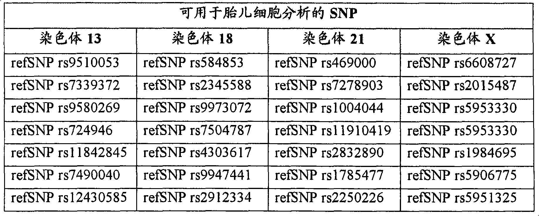

[00234]应选择与其它引物对多重化的PCR引物(相当均一的解链温度、没有对人基因组的交叉引发,以及基于序列分析没有引物-引物相互作用)。应设计引物,以产生70-100bp大小的扩增子,以增加多重PCR的效力。引物应在5′上包含22bp的标记,其用于基因分型分析。多重PCR方案可如在Findlay等,Molecular Cell Endocrinology183(2001)S5-S12中所述进行。引物浓度可以由每个反应0.7pmol-60pmol变化。简而言之,PCR以每孔25μl总体积、Taq聚合酶缓冲液(Perkin-Elmer)、200μM dNTP、引物、1.5mM MgCl2和0.6单位AmpliTaq(Perkin-Elmer)进行。在于95℃变性5分钟后,在MJ DNAengine热循环仪中进行41个循环的94℃、60℃和72℃,45秒。可根据要扩增的引物对,用不同于60℃的退火温度进行扩增。最终的延伸可达10分钟。[00234] PCR primers should be chosen that multiplex with other primer pairs (fairly uniform melting temperature, no cross-priming to the human genome, and no primer-primer interactions based on sequence analysis). Primers should be designed to generate amplicons of 70-100 bp in size to increase the efficiency of multiplex PCR. Primers should contain a 22bp marker on 5' for genotyping analysis. Multiplex PCR protocols can be performed as described in Findlay et al., Molecular Cell Endocrinology 183 (2001) S5-S12. Primer concentrations can vary from 0.7 pmol-60 pmol per reaction. Briefly, PCR was performed in a total volume of 25 μl per well, Taq polymerase buffer (Perkin-Elmer), 200 μM dNTPs, primers, 1.5 mM MgCl 2 and 0.6 units of AmpliTaq (Perkin-Elmer). After denaturation at 95°C for 5 minutes, 41 cycles of 94°C, 60°C and 72°C for 45 seconds were performed in a MJ DNAengine thermal cycler. Amplification may be performed with an annealing temperature other than 60°C, depending on the primer pair to be amplified. The final stretch can be up to 10 minutes.
[00235]可使用嵌套引物进行第二轮PCR,以确保多重扩增的最佳效力。用得自PCR试剂盒的无核酸酶的水将2μl等份的每个PCR反应物稀释40倍(至80μl总量)。产生无模板或阴性的对照,以测试污染。根据要扩增的引物对,用60℃-68℃的退火温度进行采用嵌套PCR引物的扩增。[00235] A second round of PCR can be performed using nested primers to ensure optimal efficiency of multiplex amplification. A 2 μl aliquot of each PCR reaction was diluted 40-fold (to a total of 80 μl) with nuclease-free water from the PCR kit. Generate no template or negative controls to test for contamination. Amplification with nested PCR primers was performed with an annealing temperature of 60°C-68°C, depending on the primer pair to be amplified.
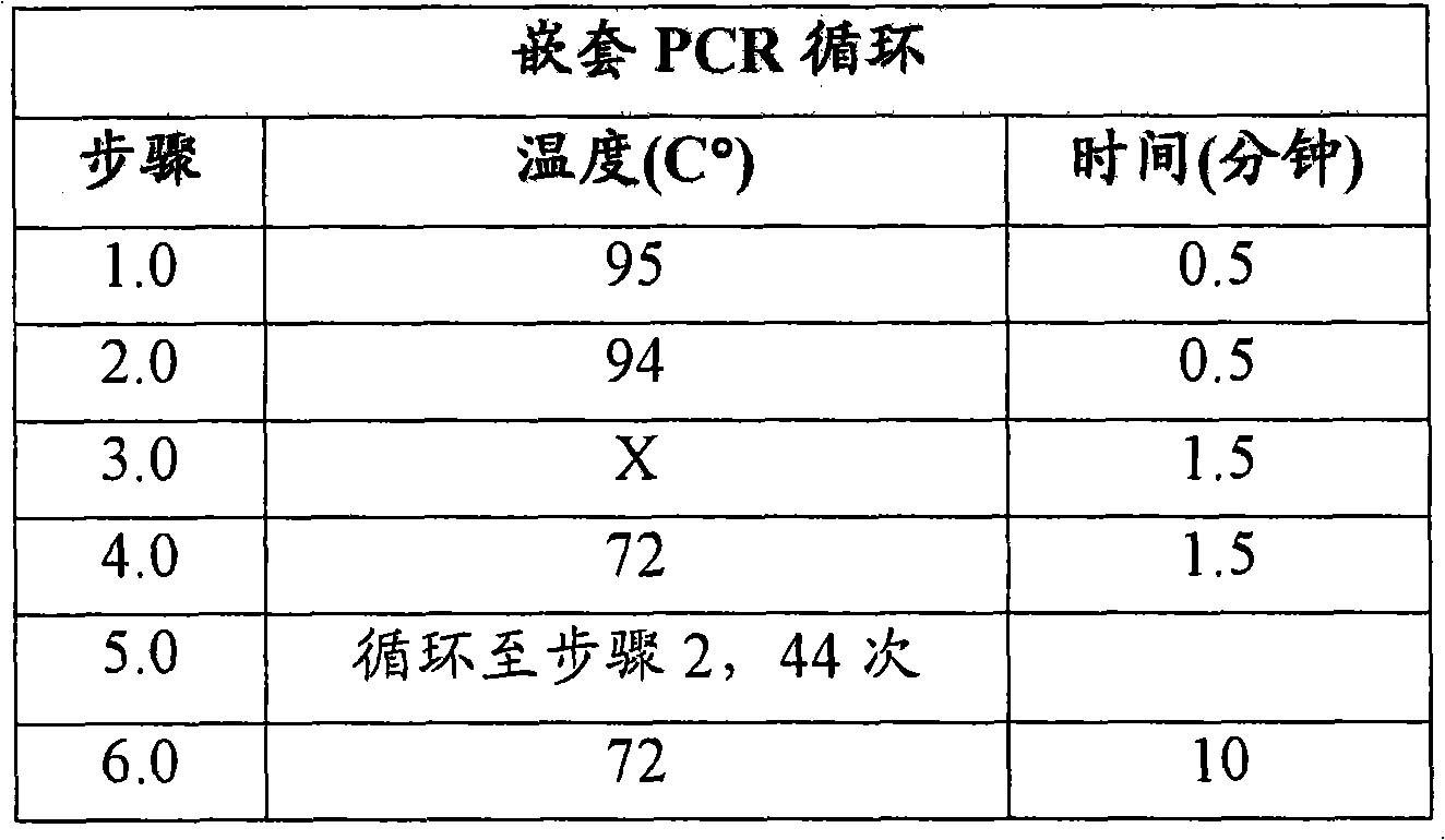
[00236]由Affymetrix(Santa Clara,CA)开发的分子倒置探针(MIP)技术可以在单一反应中基因分型20,000以上的SNP。在典型的MIP测定中,每个SNP应被分配22bp的DNA标签,其允许在高度平行的基因分型测定中独有地鉴定SNP。在该实施例中,DNA标签起两个作用:(1)确定不同SNP的身份,和(2)确定得到基因型的孔的身份。例如,基因分型500个不同孔中的40个相同SNP应需要总共20,000个标签(4个染色体×10个SNP×500个孔)。[00236] Molecular Inversion Probe (MIP) technology developed by Affymetrix (Santa Clara, CA) can genotype more than 20,000 SNPs in a single reaction. In a typical MIP assay, each SNP should be assigned a 22bp DNA tag, which allows unique identification of SNPs in highly parallel genotyping assays. In this example, the DNA tag serves two purposes: (1) to determine the identity of the different SNPs, and (2) to determine the identity of the well from which the genotype was derived. For example, genotyping the 40 identical SNPs in 500 different wells should require a total of 20,000 tags (4 chromosomes x 10 SNPs x 500 wells).
[00237]标记的MIP探针应与初始多重单细胞PCR(或嵌套PCR)的扩增子组合,并应进行基因分型反应。探针/模板混合物应被分入4个管中,每个管含有不同的核苷酸(例如G、A、T或C)。在延伸和连接步骤后,应用外切核酸酶处理混合物,以除去所有线性分子,继续存在的环形分子的标签应使用PCR扩增。然后合并构成所有分区的扩增标签,并与含有20,000个标签中的每一个的互补序列的单个DNA微阵列杂交。[00237] Labeled MIP probes should be combined with amplicons from the initial multiplexed single cell PCR (or nested PCR) and a genotyping reaction should be performed. The probe/template mix should be divided into 4 tubes, each tube containing a different nucleotide (e.g. G, A, T or C). After the extension and ligation steps, the mixture should be treated with exonuclease to remove all linear molecules and the tags of remaining circular molecules should be amplified using PCR. The amplified tags making up all partitions were then pooled and hybridized to a single DNA microarray containing the complementary sequence for each of the 20,000 tags.
[00238]鉴定含非母体等位基因(例如胎儿细胞)的分区:数据分析程序中的第一步应是使用22bp的标签,以将20,000个基因型分拣入对应于原始微量滴定板的单独孔的分区中。第二步应鉴定含非母体等位基因的分区,这些分区对应于含胎儿细胞的各孔。确定含非母体等位基因的区数相对于总区数应提供在原始富集细胞群中存在的fnRBC数的精确估计。在鉴定给定分区中的胎儿细胞时,应通过40个独立SNP检测非母体等位基因,这提供了极高水平的结果置信度。[00238] Identification of partitions containing non-maternal alleles (e.g. fetal cells): The first step in the data analysis procedure should be to use the 22bp tag to sort the 20,000 genotypes into individual bins corresponding to the original microtiter plate In the partition of the hole. The second step should identify partitions containing non-maternal alleles that correspond to wells containing fetal cells. Determining the number of blocks containing the non-maternal allele relative to the total number of blocks should provide an accurate estimate of the number of fnRBCs present in the original enriched cell population. When identifying fetal cells in a given partition, non-maternal alleles should be detected by 40 independent SNPs, which provides an extremely high level of confidence in the results.
[00239]检测染色体13、18和21的倍数性:在鉴定含胎儿细胞的约10个分区后,下一步应通过比较每个染色体上的10个SNP的每一个的母体对父体等位基因的比率,确定染色体13、18、21和X的倍数性。每个染色体上的多个SNP的比率可以组合(平均),以增加该染色体的异倍性检出信号的置信度。另外,含胎儿细胞的约10个独立分区的信息也可以组合,以进一步增加检出信号的置信度。[00239] Detection of ploidy for
实施例7.用于胎儿细胞的三体性诊断的超深测序Example 7. Ultra-deep sequencing for trisomy diagnosis of fetal cells
[00240]胎儿细胞或细胞核可以如在富集部分中所述或如在实施例1中所述分离。在实施例1中描述的富集方法可以由取自头三个月后期的最初20ml血样开始,产生含约500个母体白细胞(WBC)、约100个母体有核红细胞(mnBC)和最低约10个胎儿有核红细胞(fnRBC)的最终混合物。富集方法的输出应被分入具有选定的孔数目的微量滴定板的独立孔中,所以每个孔定位不超过1个细胞或基因组拷贝,其中某些孔可能根本没有细胞或基因组拷贝。[00240] Fetal cells or nuclei can be isolated as described in the enrichment section or as described in Example 1. The enrichment method described in Example 1 can be started with an initial 20 ml blood sample taken late in the first trimester, yielding about 500 maternal white blood cells (WBC), about 100 maternal nucleated red blood cells (mnBC) and a minimum of about 10 Final mixture of fetal nucleated red blood cells (fnRBC). The output of the enrichment method should be split into individual wells of a microtiter plate with a selected number of wells, so no more than 1 cell or genome copy is located per well, where some wells may have no cells or genome copies at all.
[00241]进行多重PCR和用分区特异性标签进行超深测序:然后将高度多态性的STR基因座(每个目标染色体多个基因座)的PCR引物对加入微量滴定板中的每个孔。在下表中列出的多态STR可用于进行分析,并可设计相关的PCR引物。[00241] Perform multiplex PCR and ultra-deep sequencing with partition-specific tags: PCR primer pairs for highly polymorphic STR loci (multiple loci per chromosome of interest) are then added to each well in a microtiter plate . The polymorphic STRs listed in the table below can be used for analysis and related PCR primers can be designed.
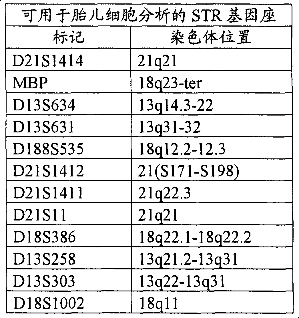
[00242]每个STR的引物将具有两个重要特征。首先,每个引物将在5′末端包含共有的约18bp序列,该序列用于随后的DNA克隆和测序程序。其次,微量滴定板中的每个孔被分配独有的约6bp的DNA标签序列,其掺入到每个不同STR的上游引物的中间部分。DNA标签使得有可能在多重PCR之后合并所有的STR扩增子,这使得有可能在超深测序程序中更成本有效地平行分析扩增子。长约6bp的DNA标签兼顾了信息量(4096个潜在分区)和合成引物的成本。[00242] The primers for each STR will have two important features. First, each primer will contain a consensus approximately 18 bp sequence at the 5' end, which is used in subsequent DNA cloning and sequencing procedures. Second, each well in the microtiter plate was assigned a unique ~6 bp DNA tag sequence that was incorporated into the middle of the upstream primer for each different STR. DNA tagging makes it possible to combine all STR amplicons after multiplex PCR, which makes possible more cost-effective parallel analysis of amplicons in ultra-deep sequencing procedures. The DNA tag with a length of about 6bp takes into account the amount of information (4096 potential partitions) and the cost of synthetic primers.
[00243]多重PCR方案可如在Findlay等,Molecular CellEndocrinology 183(2001)S5-S12中所述进行。引物浓度可以由每个反应0.7pmol-60pmol变化。简而言之,PCR以每孔25μl总体积、Taq聚合酶缓冲液(Perkin-Elmer)、200μM dNTP、引物、1.5mM MgCl2和0.6单位AmpliTaq(Perkin-Elmer)进行。在于95℃变性5分钟后,在MJ DNA engine热循环仪中进行41个循环的94℃、60℃和72℃,45秒。可根据要扩增的引物对,用不同于60℃的退火温度进行扩增。最终的延伸可达10分钟。[00243] Multiplex PCR protocols can be performed as described in Findlay et al., Molecular Cell Endocrinology 183 (2001) S5-S12. Primer concentrations can vary from 0.7 pmol-60 pmol per reaction. Briefly, PCR was performed in a total volume of 25 μl per well, Taq polymerase buffer (Perkin-Elmer), 200 μM dNTPs, primers, 1.5 mM MgCl 2 and 0.6 units of AmpliTaq (Perkin-Elmer). After denaturation at 95°C for 5 minutes, 41 cycles of 94°C, 60°C and 72°C for 45 seconds were performed in a MJ DNA engine thermal cycler. Amplification may be performed with an annealing temperature other than 60°C, depending on the primer pair to be amplified. The final stretch can be up to 10 minutes.
[00244]在PCR之后,合并微量滴定板中的每孔的扩增子,纯化,并使用如在Margulies等,Nature 437(2005)376-380中所述的单分子测序策略分析。简而言之,稀释扩增子,并与珠混合,使得每个珠均捕获单个分子的扩增材料。通过产生油包PCR反应混合物乳液制备的单独的100μm水性液滴分离携带DNA的珠。然后扩增每个珠上的DNA分子,产生数百万拷贝的序列,它们全部保持与珠结合。最后,将珠置于高度平行的合成测序机器中,该机器在单次4小时运行中产生400,000多个读取序列(每个读取序列约100bp)。[00244] Following PCR, amplicons from each well in the microtiter plate were pooled, purified, and analyzed using a single molecule sequencing strategy as described in Margulies et al., Nature 437 (2005) 376-380. Briefly, amplicons are diluted and mixed with beads such that each bead captures a single molecule of amplified material. DNA-bearing beads were isolated by generating individual 100 μm aqueous droplets prepared as emulsions of PCR reaction mixtures in oil. The DNA molecules on each bead are then amplified, producing millions of copies of the sequence, all of which remain bound to the bead. Finally, the beads were placed in a highly parallel sequencing-by-synthesis machine that generated more than 400,000 reads (approximately 100 bp each) in a single 4-hour run.
[00245]超深测序提供了精确定量的方式来检测每个STR的等位基因丰度。每个等份试样孔所需要的读取序列总数由STR数和多重PCR的误差率以及与测序方法相关的Poisson抽样统计来确定。可说明扩增差异的统计学模型可用于以高置信水平检测倍数性变化。使用该统计学模型,可以预测分析每名患者需要约100,000-300,000个读取序列,每个染色体约3-10个STR基因座。具体地说,在微量滴定板的每个单独孔中的12个STR中的每一个都将读取约33个读取序列(33个读取序列×12个STR/孔×500个孔=200,000个读取序列)。[00245] Ultra-deep sequencing provides an accurate and quantitative way to detect the allelic abundance of each STR. The total number of reads required per aliquot well was determined from the number of STRs and the error rate of the multiplex PCR and Poisson sampling statistics associated with the sequencing method. Statistical models that account for differences in amplification can be used to detect fold changes with a high level of confidence. Using this statistical model, it can be predicted that ~100,000-300,000 reads per patient and ~3-10 STR loci per chromosome will be required for analysis. Specifically, approximately 33 reads will be read from each of the 12 STRs in each individual well of the microtiter plate (33 reads x 12 STRs/well x 500 wells = 200,000 read sequences).
[00246]鉴定含非母体等位基因(例如胎儿细胞)的分区:数据分析步骤中的第一步应使用6bp DNA标签将200,000个读取序列分拣入对应于微量滴定板的单独孔的分区中。然后,使用标准序列比对算法将每个分区的约400个读取序列分入不同的STR组别中。然后,应分析每个分区的比对序列,以鉴定非母体等位基因。这些可以以两种方式之一鉴定。首先,可以如上所述分析已知仅含母体细胞的独立血样级分。该样品可以为白细胞级分(其将仅包含可忽略数的胎儿细胞),或者仅仅在富集前稀释原初样品。或者,所有孔的基因型分布均可进行相似度聚类,以鉴定与母体细胞相关的显性模式。在任一种方案中,非母体等位基因的检测然后确定在初始微量滴定板中的哪些孔含有胎儿细胞。确定含非母体等位基因的区数相对于总区数提供了对初始富集细胞群中存在的胎儿细胞数的估计。含有胎儿细胞的分区应以高置信水平鉴定,因为非母体等位基因通过多个独立的STR检测。[00246] Identify partitions containing non-maternal alleles (e.g. fetal cells): The first step in the data analysis step should be to sort 200,000 reads into partitions corresponding to individual wells of the microtiter plate using 6bp DNA tags middle. The ~400 reads from each partition were then sorted into distinct STR groups using standard sequence alignment algorithms. Then, the aligned sequences for each partition should be analyzed to identify non-maternal alleles. These can be identified in one of two ways. First, an independent blood sample fraction known to contain only maternal cells can be analyzed as described above. This sample can be the leukocyte fraction (which will only contain negligible numbers of fetal cells), or simply dilute the naive sample before enrichment. Alternatively, the genotype distributions of all wells can be similarly clustered to identify dominant patterns associated with maternal cells. In either approach, detection of the non-maternal allele then determines which wells in the original microtiter plate contained fetal cells. Determining the number of bins containing the non-maternal allele relative to the total number of bins provides an estimate of the number of fetal cells present in the initial enriched cell population. Partitions containing fetal cells should be identified with a high level of confidence because non-maternal alleles are detected by multiple independent STRs.
[00247]检测染色体13、18和21的倍数性:在鉴定含有胎儿细胞的分区后,下一步应通过比较每个STR的母体对父体等位基因的比率确定染色体13、18和21的倍数性。再者,对于每个分区,12个STR中的每一个都将有约33个读取序列。在正常胎儿中,给定的STR具有1∶1的母体对父体等位基因比率,每个等位基因(正常二等位基因)对应约16个读取序列。在三体性胎儿中,3剂STR标记可被检测为具有1∶1∶1比率(三体性三等位基因)的3个等位基因或具有21比率(三体性二等位基因)的2个等位基因。在罕有情况下,全部3个等位基因都可以相同,基因座将不提供该单独患者的信息。每个染色体上的不同STR的信息可以组合,以增加给出的异倍性检出信号的置信度。另外,含有胎儿细胞的独立分区的信息也可以组合,以进一步增加检出信号的置信度。[00247] Detection of ploidy for
实施例8.用于胎儿细胞的三体性诊断的测序Example 8. Sequencing for Trisomy Diagnosis of Fetal Cells
[00248]胎儿细胞或细胞核可以如在富集部分中所述或如在实施例1和2中所述分离。然后,测序方法可用于检测染色体拷贝数改变。图4图示了流程图,其指示的主要步骤涉及使用本文描述的方法检测染色体拷贝数改变。例如,在实施例1中描述的富集方法可以由取自头三个月后期的最初20ml血样开始,产生含约500个母体白细胞(WBC)、约100个母体有核红细胞(mnBC)和最低约10个胎儿有核红细胞(fnRBC)的最终混合物。富集方法的输出应被分入具有选定的孔数目的微量滴定板的独立孔中,所以每个孔定位不超过1个细胞或基因组拷贝,其中某些孔可能根本没有细胞或基因组拷贝。[00248] Fetal cells or nuclei can be isolated as described in the enrichment section or as described in Examples 1 and 2. Sequencing methods can then be used to detect chromosomal copy number alterations. Figure 4 illustrates a flowchart indicating the main steps involved in the detection of chromosomal copy number alterations using the methods described herein. For example, the enrichment method described in Example 1 could start with an initial 20 ml blood sample taken late in the first trimester, yielding a blood sample containing about 500 maternal white blood cells (WBC), about 100 maternal nucleated red blood cells (mnBC) and a minimum Final mixture of approximately 10 fetal nucleated red blood cells (fnRBC). The output of the enrichment method should be split into individual wells of a microtiter plate with a selected number of wells, so no more than 1 cell or genome copy is located per well, where some wells may have no cells or genome copies at all.
[00249]进行多重PCR和采用分区特异性标签进行测序:[00249] Performing multiplex PCR and sequencing with partition-specific tags:
[00250]可将用于高多态性STR基因座(每个目标染色体多个基因座)的PCR引物对加入微量滴定板中的每个孔。例如,可根据染色体13、18、21和X设计STR,以检测最常见的异倍体,并根据其中预期无异倍性的基因组的对照区设计STR。通常,每个目标染色体应分析4个以上的STR,以确保精确的异倍性检测。[00250] PCR primer pairs for highly polymorphic STR loci (multiple loci per chromosome of interest) can be added to each well in a microtiter plate. For example, STRs can be designed for
[00251]每个STR的引物都可以设计得具有两个重要特征。首先,每个引物都可以在5′末端上包含共有的约18bp序列,其可用于随后的DNA克隆和测序程序。其次,微量滴定板中的每个孔都可以分配独有的约6bp DNA标签序列,其可掺入到不同STR的每一个的上游引物的中间部分。该DNA标签使得有可能在多重PCR之后合并所有的STR扩增子,这使得有可能在超深测序程序中平行分析扩增子。而且,用于STR扩增的嵌套PCR策略可以由单个细胞实现更高的扩增可靠性。[00251] Primers for each STR can be designed to have two important features. First, each primer can contain a consensus sequence of about 18 bp on the 5' end, which can be used in subsequent DNA cloning and sequencing procedures. Second, each well in the microtiter plate can be assigned a unique ~6bp DNA tag sequence that can be incorporated into the middle of the upstream primer for each of the different STRs. This DNA tag makes it possible to combine all STR amplicons after multiplex PCR, which makes it possible to analyze amplicons in parallel in ultra-deep sequencing procedures. Moreover, the nested PCR strategy for STR amplification can achieve higher amplification reliability from single cells.
[00252]测序可以使用经典的Sanger测序法或本领域已知的任何其它方法进行。[00252] Sequencing can be performed using classical Sanger sequencing or any other method known in the art.
[00253]例如,测序可通过合成测序进行,合成测序包括通过合成与靶核酸序列互补的链推测模板序列。合成测序可使用与核酸标签上的测序元件互补的测序引物启动。该方法包括在聚合酶反应中将标记的核苷酸或核苷酸类似物掺入互补核酸序列的增长链中之后(基本上实时)或当时(实时)立即检测每个核苷酸的身份。在成功掺入标记核苷酸后,检测信号,然后通过本领域已知的方法消除信号。合成测序方法的实例描述于美国申请公布号2003/0044781、2006/0024711、2006/0024678和2005/0100932。可用于标记合成测序用核苷酸或核苷酸类似物的标记的实例包括但不限于发色团、荧光部分、酶、抗原、重金属、磁探针、染料、磷光团、放射性物质、化学发光部分、散射或荧光纳颗粒、Raman信号产生部分和电化学检测部分。合成测序每小时可以产生至少1,000个、至少5,000个、至少10,000个、至少20,000个、30,000个、至少40,000个、至少50,000个、至少100,000个或至少500,000个读取序列。每个这样读取序列可具有至少50个、至少60个、至少70个、至少80个、至少90个、至少100个、至少120个或至少150个碱基。[00253] For example, sequencing can be performed by sequencing-by-synthesis, which involves deducing a template sequence by synthesizing a strand that is complementary to a target nucleic acid sequence. Sequencing by synthesis can be initiated using sequencing primers that are complementary to sequencing elements on nucleic acid tags. The method involves detecting the identity of each nucleotide either immediately after (in substantially real time) or while (in real time) the labeled nucleotide or nucleotide analog is incorporated into the growing strand of complementary nucleic acid sequence in a polymerase reaction. After successful incorporation of the labeled nucleotide, the signal is detected and then eliminated by methods known in the art. Examples of sequencing-by-synthesis methods are described in US Application Publication Nos. 2003/0044781, 2006/0024711, 2006/0024678, and 2005/0100932. Examples of labels that can be used to label nucleotides or nucleotide analogs for sequencing by synthesis include, but are not limited to, chromophores, fluorescent moieties, enzymes, antigens, heavy metals, magnetic probes, dyes, phosphorophores, radioactive substances, chemiluminescence part, scattering or fluorescent nanoparticles, Raman signal generation part and electrochemical detection part. Sequencing by synthesis can generate at least 1,000, at least 5,000, at least 10,000, at least 20,000, 30,000, at least 40,000, at least 50,000, at least 100,000, or at least 500,000 sequence reads per hour. Each such read sequence may have at least 50, at least 60, at least 70, at least 80, at least 90, at least 100, at least 120, or at least 150 bases.
[00254]另一种测序方法包括使扩增的目标基因组区与同其互补的引物杂交。该杂交复合物与聚合酶、ATP硫酸化酶、萤光素酶、三磷酸腺苷双磷酸酶以及底物萤光素和腺苷酰硫酸温育。接着,顺序加入对应于碱基A、C、G和T(U)的脱氧核苷三磷酸。每个碱基掺入伴随着焦磷酸释放,通过硫酸化酶转变为ATP,ATP驱动氧化萤光素的合成和可见光的释放。因为焦磷酸释放与掺入碱基的数量等摩尔,所以发出的光与在任一个步骤加入的核苷酸数成比例。重复该过程,直至完整序列被测定。[00254] Another sequencing method involves hybridizing the amplified genomic region of interest to a primer complementary thereto. The hybridization complex is incubated with polymerase, ATP sulfurylase, luciferase, apyrase and the substrates luciferin and adenylsulfate. Next, deoxynucleoside triphosphates corresponding to the bases A, C, G and T (U) are sequentially added. Each base incorporation is accompanied by the release of pyrophosphate, converted to ATP by sulfurylase, which drives the synthesis of oxyluciferin and the release of visible light. Since pyrophosphate release is equimolar to the number of bases incorporated, the light emitted is proportional to the number of nucleotides added at any one step. This process is repeated until the complete sequence is determined.
[00255]又一个测序方法包括通过连接方案(简并连接)进行的4色测序,其包括使锚定引物与4个位置中的1个杂交。然后,进行锚定引物与用荧光染料标记的简并九聚体群的酶连接反应。在任意给定的循环,所用的九聚体群的结构使其位置之一的身份与连接至该九聚体的荧光团的身份相关联。就连接酶区分该查询位置的互补性而言,荧光信号允许推断碱基身份。在进行连接和4色成像后,剥下锚定引物:九聚体复合物,开始新循环。[00255] Yet another sequencing method includes 4-color sequencing by ligation scheme (degenerate ligation), which involves hybridizing an anchor primer to 1 of 4 positions. Then, an enzymatic ligation reaction of the anchor primer to the population of degenerate nonamers labeled with a fluorescent dye is performed. At any given cycle, the structure of the nonamer population used is such that the identity of one of its positions is related to the identity of the fluorophore attached to that nonamer. The fluorescent signal allows the base identity to be inferred for the ligase to distinguish complementarity at the query position. After ligation and 4-color imaging, the anchor primer:nonamer complex was stripped off and a new cycle started.
[00256]鉴定含非母体等位基因(例如胎儿细胞)的分区:数据分析步骤中的第一步应使用6bp DNA标签将200,000个读取序列分拣入对应于微量滴定板的单独孔的分区中。然后,使用标准序列比对算法将每个分区的约400个读取序列分入不同的STR组别中。然后,应分析每个分区的比对序列,以鉴定非母体等位基因。这些可以以两种方式之一鉴定。首先,可以如上所述分析已知仅含母体细胞的独立血样级分。该样品可以为白细胞级分(其将仅包含可忽略数的胎儿细胞),或者仅仅在富集前稀释原初样品。或者,所有孔的基因型分布均可进行相似度聚类,以鉴定与母体细胞相关的显性模式。在任一个方案中,检出非母体等位基因随后确定在初始微量滴定板中的哪些孔含有胎儿细胞。确定相对于总区数的含非母体等位基因的区数提供了对初始富集细胞群中存在的胎儿细胞数的估计。因为通过多个独立STR检测出非母体等位基因,所以以高置信水平鉴定出含有胎儿细胞的分区。[00256] Identify partitions containing non-maternal alleles (e.g. fetal cells): The first step in the data analysis step should be to sort 200,000 reads into partitions corresponding to individual wells of the microtiter plate using 6bp DNA tags middle. The ~400 reads from each partition were then sorted into distinct STR groups using standard sequence alignment algorithms. Then, the aligned sequences for each partition should be analyzed to identify non-maternal alleles. These can be identified in one of two ways. First, an independent blood sample fraction known to contain only maternal cells can be analyzed as described above. This sample can be the leukocyte fraction (which will only contain negligible numbers of fetal cells), or simply dilute the naive sample before enrichment. Alternatively, the genotype distributions of all wells can be similarly clustered to identify dominant patterns associated with maternal cells. In either approach, detection of the non-maternal allele is followed by determination of which wells in the initial microtiter plate contained fetal cells. Determining the number of non-maternal allele-containing regions relative to the total number of regions provides an estimate of the number of fetal cells present in the initial enriched cell population. Because non-maternal alleles were detected by multiple independent STRs, partitions containing fetal cells were identified with a high level of confidence.
[00257]检测染色体13、18和21的倍数性:在鉴定含有胎儿细胞的分区后,下一步应通过比较每个STR的母体对父体等位基因的比率确定染色体13、18和21的倍数性。再者,对于每个分区,12个STR中的每一个都将有约33个读取序列。在正常胎儿中,给定的STR具有1∶1的母体对父体等位基因比率,每个等位基因(正常二等位基因)对应约16个读取序列。在三体性胎儿中,3剂STR标记可被检测为具有1∶1∶1比率(三体性三等位基因)的3个等位基因或具有2∶1比率(三体性二等位基因)的2个等位基因。在罕有情况下,全部3个等位基因都可以相同,基因座将不提供该单独患者的信息。每个染色体上不同STR的信息可以组合,以增加给出的异倍性检出信号的置信度。另外,含有胎儿细胞的独立分区的信息也可以组合,以进一步增加该检出信号的置信度。[00257] Detection of ploidy for
实施例9.装置实施方案Example 9. Device implementation
[00258]本发明的微流化装置通过计算机辅助设计(CAD)来设计,并通过光刻法显微加工。开发两步法,其中首先压实血样,以除去一大群小细胞,然后通过免疫亲和捕获回收稀有目标-上皮细胞靶细胞。所述装置通过光刻法勾画轮廓,并基于CAD产生的设计蚀刻入硅基质中。细胞富集模块约为标准显微镜载玻片的大小,含有14个平行样品处理部分和相关的样品操作通道,这些通道连接共用的样品和缓冲液进口以及产物和废物出口。每个部分都包含显微加工的障碍物阵列,其进行了优化,以经由将较大细胞置换入产物流中而根据流体动力学尺寸富集靶细胞类型。在该实施例中,设计微芯片,以将红细胞(RBC)和血小板与较大白细胞和CTC分离。通过该装置由全血回收富集的靶细胞群。细胞富集微芯片的效力通过将正常全血中的RBC和血小板与白细胞(WBC)分离来评价(图15)。在癌症患者中,在较大的WBC级分中发现CTC。血液被最低稀释(30%),以至多6ml/小时的流速加工6ml样品。以Coulter“AC-T diff”型临床血液分析仪评价产物和废物流,该分析仪自动辨别、大小分离和计数不同的血细胞群。富集芯片实现了RBC与WBC的分离,其中WBC级分具有>99%的有核细胞保留、>99%的RBC缺失和>97%的血小板缺失。这些细胞级分的代表性直方图示于图16。常规细胞学证实WBC和RBC级分的高度富集(图17)。[00258] The microfluidized device of the present invention was designed by computer aided design (CAD) and microfabricated by photolithography. A two-step method was developed in which the blood sample was first compacted to remove a large population of small cells, followed by recovery of rare targets - epithelial target cells - by immunoaffinity capture. The device was outlined by photolithography and etched into a silicon substrate based on the CAD-generated design. The Cell Enrichment Module is about the size of a standard microscope slide and contains 14 parallel sample handling sections and associated sample manipulation channels that connect to common sample and buffer inlets and product and waste outlets. Each section contains an array of microfabricated obstacles optimized to enrich target cell types according to hydrodynamic size via the displacement of larger cells into the product stream. In this example, a microchip was designed to separate red blood cells (RBCs) and platelets from larger white blood cells and CTCs. An enriched population of target cells is recovered from whole blood by the device. The efficacy of the cell enrichment microchip was assessed by separating RBCs and platelets from white blood cells (WBCs) in normal whole blood (Figure 15). In cancer patients, CTCs are found in the larger WBC fraction. Blood was minimally diluted (30%) and 6ml samples were processed at a flow rate of up to 6ml/hour. The product and waste streams were evaluated with a Coulter " AC -T diff" clinical hematology analyzer, which automatically discriminates, size separates and counts distinct blood cell populations. The enrichment chip achieved the separation of RBC from WBC, wherein the WBC fraction had >99% nucleated cell retention, >99% RBC loss and >97% platelet loss. Representative histograms of these cell fractions are shown in FIG. 16 . Conventional cytology confirmed high enrichment of WBC and RBC fractions (Figure 17).
[00259]接着,用固定化抗体官能化的微流化模块通过亲和捕获回收上皮细胞。设计具有单室的捕获模块,该单室含有包被抗体的显微操作障碍物的规则阵列。通过增加捕获面积约4倍,并减慢邻近障碍物的层流下的细胞流动,以增加细胞和固定化抗体之间的接触时间,来处理这些障碍物,以最大化细胞捕获。捕获模块可在相对高流速但低剪切力的条件下操作,以保护细胞对抗损伤。捕获模块的表面通过用10%硅烷、0.5%戊二醛和抗生物素蛋白继之以生物素化抗-EpCAM序贯处理而官能化。活性位点用3%牛血清白蛋白的PBS溶液封闭,并用稀释的Tris HCl猝灭,并用稀释的L-组氨酸稳定。在每个阶段后用PBS洗涤模块,最后干燥,并储存于室温。捕获效力用人晚期肺癌细胞系NCI-H1650(ATCC编号CRL-5883)检测。该细胞系在EGFR的外显子19中具有15bp杂合的符合读框的缺失,使该细胞系对吉非替尼敏感。用胰蛋白酶收获汇合培养物的细胞,用活体染料CellTracker Orange(CMRA reagent,Molecular Probes,Eugene,OR)染色,重悬浮在新鲜全血中,并以多种流速在微流化芯片中分级分离。在这些初始可行性实验中,直接在捕获模块中处理细胞悬浮液,而没有预先在细胞富集模块中分级分离而压实红细胞;因此,样品流含有正常红细胞和白细胞以及肿瘤细胞。在捕获模块中加工细胞后,用缓冲液以较高流速(3ml/小时)洗涤该装置,以除去非特异性结合的细胞。移开粘性上层,用多聚甲醛将粘附细胞固定在芯片上,并通过荧光显微镜观察。由血细胞计数器计数计算细胞回收率;代表性的捕获结果示于表4。采用未分级分离血液的重建研究中的初始得率大于60%,非特异性结合低于5%。[00259] Next, epithelial cells were recovered by affinity capture with a microfluidic module functionalized with immobilized antibodies. A capture module was designed with a single chamber containing a regular array of antibody-coated micromanipulated obstacles. These obstacles were addressed to maximize cell capture by increasing the capture area approximately 4-fold and slowing down the cell flow under laminar flow adjacent to the obstacle to increase the contact time between the cells and the immobilized antibody. The capture module can be operated at relatively high flow rates but low shear to protect cells against damage. The surface of the capture module was functionalized by sequential treatment with 10% silane, 0.5% glutaraldehyde and avidin followed by biotinylated anti-EpCAM. Active sites were blocked with 3% bovine serum albumin in PBS, quenched with diluted Tris HCl, and stabilized with diluted L-histidine. Blocks were washed with PBS after each stage, finally dried, and stored at room temperature. The capture efficiency was tested with human advanced lung cancer cell line NCI-H1650 (ATCC number CRL-5883). This cell line has a 15 bp heterozygous in-frame deletion in
表4Table 4
[00260]接着,将NCI-H1650细胞搀入全血中,并通过如上所述的大小分级分离和亲和捕获回收,成功地对该细胞进行原位分析。在进行上皮细胞和白细胞区分的实验中,将荧光素-标记的CD45泛-白细胞单克隆抗体的0.5ml母液通入捕获模块中,并于室温温育30分钟。用缓冲液洗涤模块,以除去未结合的抗体,用1%多聚甲醛将细胞固定在芯片上,并通过荧光显微镜观察。如在图18中所示,上皮细胞结合捕获模块的障碍物和基底。CD45泛-白细胞抗体流道的背景染色是可见的,几种染色的白细胞也是可见的,这明显是因低水平的非特异性捕获所致。[00260] Next, NCI-H1650 cells were successfully analyzed in situ by spiking whole blood and recovering by size fractionation and affinity capture as described above. In experiments for epithelial and leukocyte differentiation, 0.5 ml stock solution of fluorescein-labeled CD45 pan-leukocyte monoclonal antibody was passed into the capture module and incubated for 30 minutes at room temperature. Blocks were washed with buffer to remove unbound antibody, cells were fixed on the chip with 1% paraformaldehyde, and visualized by fluorescence microscopy. As shown in Figure 18, epithelial cells bind the barrier and substrate of the capture module. Background staining of the CD45 pan-leukocyte antibody channel was visible, as were several stained leukocytes, apparently due to low levels of non-specific capture.
实施例10:装置实施方案Embodiment 10: device embodiment
[00261]本发明的优选装置实施方案的设计示于图19A,对应于与该设计相关的3个优选装置实施方案的参数示于图19B和19C。这些实施方案尤其可用于由血液富集上皮细胞。[00261] The design of a preferred device embodiment of the present invention is shown in Figure 19A, and the parameters corresponding to the three preferred device embodiments associated with this design are shown in Figures 19B and 19C. These embodiments are particularly useful for enriching epithelial cells from blood.
实施例11:测定大细胞类型的计数Example 11: Determining Enumeration of Large Cell Types
[00262]使用本发明的方法,没有癌症、有癌症或癌症发展的诊断可基于细胞样品中大于特定截留尺寸的细胞数。例如,可以选择14μm或更大的流体动力学尺寸的细胞。该截留尺寸应消除大部分白细胞。这些细胞的性质然后可以通过下游分子或细胞学分析确定。[00262] Using the methods of the invention, a diagnosis of the absence of cancer, the presence of cancer, or the development of cancer can be based on the number of cells in a sample of cells that are greater than a particular cut-off size. For example, cells with a hydrodynamic size of 14 μm or larger can be selected. This cut-off size should eliminate most leukocytes. The properties of these cells can then be determined by downstream molecular or cytological analysis.
[00263]应可用于分析的非上皮细胞的细胞类型包括内皮细胞、内皮祖细胞、子宫内膜细胞或指示病症的滋养层。而且,确定单独的上皮细胞(例如癌细胞)和其它细胞类型(例如内皮细胞)的计数,接着确定上皮细胞数和其它细胞类型数之间的比率,可以提供有用的诊断信息。[00263] Cell types other than epithelial cells that should be assayed include endothelial cells, endothelial progenitor cells, endometrial cells, or trophoblasts indicative of a disorder. Furthermore, determining individual counts of epithelial cells (eg, cancer cells) and other cell types (eg, endothelial cells), followed by determining the ratio between the number of epithelial cells and the number of other cell types, can provide useful diagnostic information.
[00264]可配置本发明的装置,以分离作为目标的细胞亚群,例如上述的那些,如在图20A-D中所示。可以选择截留尺寸,使得大部分天然血细胞,包括红细胞、白细胞和血小板,流向废物端,而非天然细胞,可以包括内皮细胞、内皮祖细胞、子宫内膜细胞或滋养层,收集在富集样品中。该富集样品可进一步分析。[00264] Devices of the invention can be configured to isolate targeted cell subpopulations, such as those described above, as shown in Figures 20A-D. The cut-off size can be selected such that the majority of native blood cells, including red blood cells, white blood cells, and platelets, flow to the waste end, while non-native cells, which can include endothelial cells, endothelial progenitor cells, endometrial cells, or trophoblasts, are collected in enriched samples . This enriched sample can be further analyzed.
[00265]因此,使用本发明的装置,有可能基于尺寸由血液或其它体液分离细胞亚群,该装置在以大细胞类型为目标时允许便利地除去大比例的天然血细胞。如在图21中所示,本发明的装置可包括计数工具,以确定富集样品中的细胞数或富集样品中特定类型的细胞(例如癌细胞)的数目,进一步分析富集样品中的细胞可提供用于诊断或其它用途的其它信息。[00265] Thus, it is possible to separate cell subpopulations from blood or other bodily fluids on the basis of size using the device of the present invention, which allows for the convenient removal of a large proportion of native blood cells when targeting large cell types. As shown in FIG. 21, the device of the present invention may include counting means to determine the number of cells in the enriched sample or the number of specific types of cells (such as cancer cells) in the enriched sample for further analysis of the number of cells in the enriched sample. Cells can provide additional information for diagnostic or other uses.
实施例12:检测EGFR突变的方法Embodiment 12: the method for detecting EGFR mutation
[00266]使用本发明的装置和方法处理和分析癌症患者的血样,产生含CTC的上皮细胞的富集样品。然后,分析该样品,以鉴定潜在的EGFR突变。该方法允许鉴定已知的临床相关的EGFR突变以及发现新突变。该方法的纵览示于图22。[00266] Blood samples from cancer patients were processed and analyzed using the devices and methods of the present invention, resulting in an enriched sample of CTC-containing epithelial cells. This sample is then analyzed to identify potential EGFR mutations. This method allows the identification of known clinically relevant EGFR mutations as well as the discovery of novel mutations. An overview of the method is shown in Figure 22.
[00267]以下是检测和证实EGFR突变的策略的概览:[00267] The following is an overview of strategies for detecting and confirming EGFR mutations:
1)序列CTC EGFR mRNA1) Sequence CTC EGFR mRNA
a)由血样纯化CTC;a) purifying CTCs from blood samples;
b)由CTC纯化总RNA;b) purify total RNA by CTC;
c)使用逆转录酶将RNA转变为cDNA;c) converting RNA to cDNA using reverse transcriptase;
d)使用产生的cDNA进行第一轮和第二轮PCR反应,以产生测序模板;和d) performing first and second rounds of PCR reactions using the generated cDNA to generate sequencing templates; and
e)纯化嵌套PCR扩增子,并用作测序模板,以测序EGFR外显子18-21。e) Nested PCR amplicons were purified and used as sequencing templates to sequence EGFR exons 18-21.
2)使用CTC基因组DNA证实RNA序列2) Confirm RNA sequence using CTC genomic DNA
a)由血样纯化CTC;a) purifying CTCs from blood samples;
b)由CTC纯化基因组DNA(gDNA);b) purification of genomic DNA (gDNA) from CTCs;
c)经PCR反应扩增外显子18、19、20和/或21;和c) PCR reactions to amplify
d)在实时定量等位基因特异性PCR反应中使用产生的PCR扩增子,以便证实经RNA测序发现的突变序列。d) Use of the generated PCR amplicons in real-time quantitative allele-specific PCR reactions in order to confirm the mutated sequences discovered by RNA sequencing.
[00268]以上概述的每个步骤的进一步的细节如下。[00268] Further details of each of the steps outlined above are as follows.
1)序列CTC EGFR mRNA1) Sequence CTC EGFR mRNA
a)由血样纯化CTC。使用本发明的任何基于尺寸的富集和/或亲和纯化装置分离CTC。a) Purification of CTCs from blood samples. CTCs are isolated using any of the size-based enrichment and/or affinity purification devices of the invention.
b)由CTC纯化总RNA。然后使用例如Qiagen Micro RNeasy试剂盒,或另一个生产商的相似总RNA纯化方案,由分离的CTC群纯化总RNA;或者,可以使用标准RNA纯化方案,例如异硫氰酸胍匀化,接着苯酚/氯仿提取和乙醇沉淀。一种这样的方法描述于J.Sambrook,E.F.Fritch和T.Mamatis的“Molecular Cloning-A Laboratory Manual,第二版”(1989)的7.24页。b) Purification of total RNA from CTCs. Total RNA is then purified from the isolated CTC population using, for example, the Qiagen Micro RNeasy kit, or a similar total RNA purification protocol from another manufacturer; alternatively, standard RNA purification protocols can be used, such as homogenization with guanidine isothiocyanate followed by phenol /chloroform extraction and ethanol precipitation. One such method is described in "Molecular Cloning-A Laboratory Manual, Second Edition" (1989) by J. Sambrook, E.F. Fritch and T. Mamatis, page 7.24.
c)使用逆转录酶将RNA转变为cDNA。基于逆转录酶供应商的方案进行cDNA反应。通常,将RNA输入cDNA反应中的量在10pg至2μg总RNA的范围内。通过使随机7聚体DNA引物或寡聚dT引物或基因特异性引物与RNA模板于65℃杂交,接着在冰上急冷,进行首链DNA合成。通过加入iScript逆转录酶(BioRad)或Superscript逆转录酶(Invitrogen)或另一个商业供应商的逆转录酶连同适宜的酶反应缓冲液启动cDNA合成。对于iScript,逆转录酶反应于42℃进行30-45分钟,接着于85℃酶失活5分钟。cDNA储存于-20℃,直至使用,或者立刻用于PCR反应。通常,cDNA反应以终体积20μl进行,并将10%(2μl)的所获cDNA用于随后的PCR反应。c) Conversion of RNA to cDNA using reverse transcriptase. cDNA reactions were performed based on the reverse transcriptase supplier's protocol. Typically, the amount of RNA input into a cDNA reaction ranges from 10 pg to 2 μg of total RNA. First-strand DNA synthesis was performed by hybridizing random 7mer DNA primers or oligo-dT primers or gene-specific primers to RNA templates at 65°C, followed by quenching on ice. cDNA synthesis was initiated by adding iScript Reverse Transcriptase (BioRad) or Superscript Reverse Transcriptase (Invitrogen) or another commercial supplier's Reverse Transcriptase along with appropriate enzyme reaction buffer. For iScript, the reverse transcriptase reaction was performed at 42°C for 30-45 minutes, followed by enzyme inactivation at 85°C for 5 minutes. cDNA was stored at -20°C until use, or immediately used in PCR reactions. Typically, cDNA reactions were performed in a final volume of 20 μl, and 10% (2 μl) of the obtained cDNA was used in subsequent PCR reactions.
d)使用产生的cDNA进行第一轮和第二轮PCR反应,产生测序模板。将逆转录酶反应的cDNA与目标区域特异性的DNA引物混合(图23)。参见表5关于可用于扩增外显子18-21的引物组。在表5中,引物组M13(+)/M12(-)在引物组M11(+)/M14(-)内部。因此,如果引物M11(+)和M14(-)用于第一轮扩增,则引物M13(+)和M12(-)可用于嵌套轮次的扩增。同样,引物组M11(+)/M14(-)在引物组M15(+)/M16(-)内部,引物组M23(+)/M24(-)在引物组M21(+)/M22(-)内部。热启动PCR反应使用Qiagen热启动Taq聚合酶试剂盒或Applied Biosystems热启动TaqMan聚合酶或其它热启动热稳定聚合酶进行,或者没有热启动,使用Promega GoTaq Green Taq聚合酶master mix、TaqMan DNA聚合酶或其它热稳定DNA聚合酶。通常,反应体积为50μl,核苷酸三磷酸以每个核苷酸200μM的终浓度存在,MgCl2以1-4mM的终浓度存在,寡聚引物以0.5μM的终浓度存在。热启动方案于95℃温育10-15分钟开始,接着40个循环的94℃、1分钟(变性);52℃、1分钟(退火)以及72℃、1分钟(延伸)。于72℃进行10分钟末端延伸,之后将样品储存于4℃,直至它们或者在第二轮(嵌套)PCR中用作模板,或者在测序前使用QiaQuick离心柱(Qiagen)纯化。如果不使用热启动方案,则省略于95℃的初始温育。如果PCR产物用于第二轮PCR,则2μl(4%)的初始PCR产物在第二轮反应中用作模板,使用相等的试剂浓度和循环参数。d) Using the generated cDNA to perform first and second rounds of PCR reactions to generate sequencing templates. The cDNA from the reverse transcriptase reaction was mixed with DNA primers specific for the region of interest (Figure 23). See Table 5 for primer sets that can be used to amplify exons 18-21. In Table 5, the primer set M13(+)/M12(-) is inside the primer set M11(+)/M14(-). Thus, if primers M11(+) and M14(-) are used for the first round of amplification, primers M13(+) and M12(-) can be used for nested rounds of amplification. Similarly, primer set M11(+)/M14(-) is inside primer set M15(+)/M16(-), primer set M23(+)/M24(-) is inside primer set M21(+)/M22(-) internal. Hot-start PCR reactions were performed using Qiagen Hot-Start Taq Polymerase Kit or Applied Biosystems Hot-Start TaqMan Polymerase or other Hot-Start thermostable polymerases, or without Hot-Start, Promega GoTaq Green Taq Polymerase master mix, TaqMan DNA Polymerase or other thermostable DNA polymerases. Typically, the reaction volume is 50 µl, nucleotide triphosphates are present at a final concentration of 200 µM per nucleotide, MgCl is present at a final concentration of 1-4 mM, and oligo primers are present at a final concentration of 0.5 µM. The hot start protocol begins with an incubation at 95°C for 10-15 minutes, followed by 40 cycles of 94°C, 1 minute (denaturation); 52°C, 1 minute (annealing) and 72°C, 1 minute (extension). End extension was performed at 72°C for 10 min, after which samples were stored at 4°C until they were either used as templates in a second round of (nested) PCR or purified using QiaQuick spin columns (Qiagen) prior to sequencing. If the hot start protocol is not used, the initial incubation at 95°C is omitted. If the PCR product was used for a second round of PCR, 2 μl (4%) of the initial PCR product was used as template in the second round reaction using equal reagent concentrations and cycling parameters.
表5.用于扩增外显子18-21周围的EGFR mRNA的引物组Table 5. Primer sets used to amplify EGFR mRNA around exons 18-21
e)纯化嵌套PCR扩增子,并用作测序模板,以测序EGFR外显子18-21。通过ABI自动化荧光测序机器以及荧光标记的DNA测序梯进行测序,所述DNA测序梯经由Sanger型测序反应使用荧光双脱氧核苷酸混合物产生。使用Qiagen QuickSpin柱、Agencourt AMPure PCR纯化系统或得自其它供应商的PCR产物纯化试剂盒纯化PCR产物。在纯化PCR产物后,用Nanodrop 7000分光光度计确定核苷酸浓度和纯度,使PCR产物浓度达到25ng/μl的浓度。作为质量控制检测,只有UV线吸收比率(A260/A280)大于1.8的PCR产物才用于测序。使测序引物浓度达到3.2pmol/μl。e) Nested PCR amplicons were purified and used as sequencing templates to sequence EGFR exons 18-21. Sequencing was performed by an ABI automated fluorescent sequencing machine with fluorescently labeled DNA sequencing ladders generated via Sanger-type sequencing reactions using fluorescent dideoxynucleotide mixtures. PCR products were purified using Qiagen QuickSpin columns, Agencourt AMPure PCR purification system, or PCR product purification kits from other suppliers. After purifying the PCR product, the nucleotide concentration and purity were determined with a Nanodrop 7000 spectrophotometer to bring the PCR product concentration to a concentration of 25 ng/μl. As a quality control check, only PCR products with a UV light absorbance ratio (A 260 /A 280 ) greater than 1.8 were used for sequencing. The sequencing primer concentration was brought to 3.2 pmol/μl.
2)使用CTC基因组DNA证实RNA序列:2) Confirm the RNA sequence using CTC genomic DNA:
a)由血样纯化CTC。如上使用本发明的任一种基于尺寸的富集和/或亲和纯化装置分离CTC。a) Purification of CTCs from blood samples. CTCs are isolated as above using any of the size-based enrichment and/or affinity purification devices of the invention.
b)由CTC纯化基因组DNA(gDNA)。基因组DNA使用QiagenDNeasy Mini试剂盒、Invitrogen ChargeSwitch gDNA试剂盒或另一个市售试剂盒或经由以下方案纯化:b) Purification of genomic DNA (gDNA) from CTCs. Genomic DNA was purified using the Qiagen DNeasy Mini Kit, Invitrogen ChargeSwitch gDNA Kit, or another commercially available kit, or via the following protocol:
1.细胞沉淀或者新鲜裂解,或者储存于-80℃,并在裂解前立即融化。1. Cell pellets are either freshly lysed, or stored at -80°C and thawed immediately before lysing.
2.加入500μl的50mM Tris pH 7.9/100mM EDTA/0.5%SDS(TES缓冲液)。2. Add 500 μl of 50mM Tris pH 7.9/100mM EDTA/0.5% SDS (TES buffer).
3.加入12.5μl蛋白酶K(IBI5406,20mg/ml),产生的最终[ProtK]=0.5mg/ml。3. Add 12.5 [mu]l proteinase K (IBI5406, 20 mg/ml), resulting in a final [ProtK] = 0.5 mg/ml.
4.在旋转温育器中于55℃过夜温育。4. Incubate overnight at 55°C in a rotary incubator.
5.加入20μl RNA酶混合物(500U/ml RNA酶A+20,000U/mlRNA酶T1,Ambion#2288),并于37℃温育4小时。5. Add 20 μl RNase mixture (500 U/ml RNase A + 20,000 U/ml RNase T1, Ambion #2288), and incubate at 37°C for 4 hours.
6.用苯酚(Kodak,Tris pH 8平衡)提取,振摇混合,在台式离心机中离心5分钟。6. Extract with phenol (Kodak,
7.将水相转移至新鲜管。7. Transfer the aqueous phase to a fresh tube.
8.用苯酚/氯仿/异戊醇(EMD,25∶24∶1比率,Tris pH 8平衡的)提取,振摇混合,在台式离心机中离心5分钟。8. Extract with phenol/chloroform/isoamyl alcohol (EMD, 25:24:1 ratio,
9.加入50μl 3M NaOAc pH=6。9. Add 50 μl 3M NaOAc pH=6.
10.加入500μl EtOH。10. Add 500 μl EtOH.
11.振摇混合。可见沉淀DNA的条带。如果预期的DNA浓度非常低,则加入载体核苷酸(通常为酵母tRNA)。11. Shake to mix. Bands of precipitated DNA are visible. If the expected DNA concentration is very low, add carrier nucleotides (usually yeast tRNA).
12.在台式离心机中以最大速度离心1分钟。12. Centrifuge for 1 minute at maximum speed in a tabletop centrifuge.
13.除去上清液。13. Remove the supernatant.
14.于室温(RT)加入500μl 70%EtOH。14. Add 500 μl 70% EtOH at room temperature (RT).
15.振摇混合。15. Shake to mix.
16.在台式离心机中以最大速度离心1分钟。16. Centrifuge for 1 minute at maximum speed in a tabletop centrifuge.
17.在加入TE之前风干10-20分钟。17. Air dry for 10-20 minutes before adding TE.
18.重悬浮在400μl TE中。于65℃温育10分钟,然后于室温过夜放置,之后在Nanodrop上定量。18. Resuspend in 400 μl TE. Incubate at 65°C for 10 minutes, then leave overnight at room temperature before quantification on the Nanodrop.
c)经PCR反应扩增外显子18、19、20和/或21。如在步骤1d中所述进行热启动嵌套PCR扩增,只是没有嵌套轮次的扩增。初始PCR步骤可在对数期当中终止,以便使扩增过程中的等位基因特异性信息丢失的可能性最小。用于扩增EGFR外显子18-21的引物组列于表6(另参见Paez等,Science 304:1497-1500(补充材料)(2004))。c)
表6.用于扩增EGFR基因组DNA的引物组Table 6. Primer sets used to amplify EGFR genomic DNA
d)在实时定量等位基因特异性PCR反应中使用产生的PCR扩增子,以便证实经由RNA测序发现的突变序列。等份的PCR扩增子在多重等位基因特异性定量PCR反应中用作模板,该PCR反应使用TaqMan PCR 5′核酸酶测定和Applied Biosystems 7500型实时PCR机器(图24)。该轮PCR扩增对每个目标突变特异性的初始PCR产物的子区域。已知实时PCR的敏感性非常高,有可能获得关于EGFR基因突变状态的完整信息,即使分离出少至10个CTC。实时PCR在8个对数的输入DNA浓度内提供了等位基因序列的定量;因此,即便在不纯的群体中的杂合突变也容易使用该方法检测。d) Use of the generated PCR amplicons in real-time quantitative allele-specific PCR reactions in order to confirm the mutated sequences discovered via RNA-sequencing. Aliquots of the PCR amplicons were used as templates in a multiplex allele-specific quantitative PCR reaction using a TaqMan PCR 5' nuclease assay and an Applied Biosystems model 7500 real-time PCR machine (Figure 24). This round of PCR amplifies a subregion of the initial PCR product specific for each mutation of interest. The sensitivity of real-time PCR is known to be very high, and it is possible to obtain complete information on the mutation status of the EGFR gene, even with as few as 10 CTCs isolated. Real-time PCR provides quantification of allelic sequence within 8 logs of input DNA concentration; thus, heterozygous mutations even in impure populations are readily detected using this method.
[00269]针对影响吉非替尼在NSCLC患者中的响应性的所有已知突变设计探针和引物组,所述突变包括40多个这样的体细胞突变,包括点突变、缺失和插入,它们已在医学文献中报告。为了阐述用途,其中5个点突变的引物和探针组的实例列于表7。一般而言,可以使用引物优化软件程序Primer Express(Applied Biosystems)设计寡核苷酸,杂交条件进行了优化,以区分野生型EGFR DNA序列和突变等位基因。由已知携带EGFR突变(例如H358(野生型)、H1650(15-bp缺失,Δ2235-2249)和H1975(两个点突变,2369C→T,2573T→G))的肺癌细胞系扩增的EGFR基因组DNA,用于优化等位基因特异性实时PCR反应。使用TaqMan 5′核酸酶测定,开发对野生型序列或已知的EGFR突变特异性的等位基因特异性标记探针。设计所具有的解链温度易于区分匹配和错配的寡核苷酸,并优化实时PCR条件,以区分野生型和突变等位基因。所有的实时PCR反应都以一式三份进行。[00269] Design probes and primer sets for all known mutations affecting the responsiveness of gefitinib in NSCLC patients, said mutations comprising more than 40 such somatic mutations, including point mutations, deletions and insertions, which Has been reported in the medical literature. Examples of primer and probe sets in which 5 point mutations are used are listed in Table 7 for illustrative purposes. In general, oligonucleotides can be designed using the primer optimization software program Primer Express (Applied Biosystems), with hybridization conditions optimized to discriminate between wild-type EGFR DNA sequences and mutant alleles. EGFR amplified from lung cancer cell lines known to carry EGFR mutations such as H358 (wild-type), H1650 (15-bp deletion, Δ2235-2249), and H1975 (two point mutations, 2369C→T, 2573T→G) Genomic DNA for optimization of allele-specific real-time PCR reactions. Using the TaqMan 5' Nuclease Assay, develop allele-specific labeling probes specific for wild-type sequences or known EGFR mutations. Design oligonucleotides with melting temperatures that readily discriminate between matched and mismatched, and optimize real-time PCR conditions to discriminate between wild-type and mutant alleles. All real-time PCR reactions were performed in triplicate.
[00270]最初,含有野生型序列的标记探针在同一反应中与单个突变探针多重化。将结果表示为1个突变等位基因序列对野生型序列的比率可以鉴定含有或没有给定突变的样品。在针对给定探针组优化条件后,然后有可能在同一实时PCR测定中多重化针对给定外显子中的所有突变等位基因的探针,增加该分析工具在临床环境中的易用性。[00270] Initially, a labeled probe containing the wild-type sequence is multiplexed with a single mutant probe in the same reaction. Expressing the results as the ratio of 1 mutant allele sequence to the wild-type sequence allows the identification of samples with or without a given mutation. After optimizing conditions for a given probe set, it is then possible to multiplex probes for all mutant alleles in a given exon in the same real-time PCR assay, increasing the ease of use of this analytical tool in the clinical setting sex.
[00271]针对每个野生型等位基因和突变等位基因序列设计独特的探针。用在5′末端的荧光染料VIC标记野生型序列,用荧光FAM标记突变序列。荧光猝灭剂和小沟结合部分连接至探针的3′末端。ROX用作用于标准化用途的被动参比染料。产生针对野生型序列的标准曲线,并用于相对定量。不需要精确定量突变信号,因为输入细胞群是未知的,且纯度可变。如ABI产品文献所述设置测定,当产生的突变等位基因探针的信号高于荧光的背景水平时,证实存在突变(图25),该阈值循环产生输入样品中的突变等位基因的相对频率。[00271] Unique probes were designed for each wild-type allele and mutant allele sequence. The wild-type sequence was labeled with the fluorescent dye VIC at the 5' end and the mutant sequence was labeled with fluorescent FAM. A fluorescent quencher and a minor groove binding moiety are attached to the 3' end of the probe. ROX was used as a passive reference dye for standardization purposes. A standard curve was generated against the wild-type sequence and used for relative quantification. Precise quantification of the mutational signal is not required because the input cell population is unknown and of variable purity. The assay was set up as described in the ABI product literature, and the presence of a mutation was confirmed when the signal generated by the mutant allele probe was above the background level of fluorescence (Figure 25). frequency.
表7.用于等位基因特异性qPCR的探针和引物Table 7. Probes and primers for allele-specific qPCR


实施例13:在白细胞中没有EGFR表达Example 13: No EGFR expression in leukocytes
[00272]为测试白细胞中是否存在EGFR mRNA,进行几个PCR实验。设计示于表8的4个引物组,以扩增4个对应基因:[00272] To test for the presence of EGFR mRNA in leukocytes, several PCR experiments were performed. 4 primer sets shown in Table 8 were designed to amplify 4 corresponding genes:
1)BCKDK(支链酮酸脱氢酶复合激酶)-在所有细胞类型中表达的“看家”基因,是白细胞和肿瘤细胞这二者的阳性对照;1) BCKDK (branched-chain ketoacid dehydrogenase complex kinase) - a "housekeeping" gene expressed in all cell types, a positive control for both leukocytes and tumor cells;
2)CD45-在白细胞中特异性表达,是白细胞的阳性对照和肿瘤细胞的阴性对照,2) CD45- is specifically expressed in leukocytes and is a positive control for leukocytes and a negative control for tumor cells,
3)EpCaM-在上皮细胞中特异性表达,是白细胞的阴性对照和肿瘤细胞的阳性对照,和3) EpCaM - specifically expressed in epithelial cells, is a negative control for leukocytes and a positive control for tumor cells, and
4)EGFR-要检验的靶mRNA4) EGFR-target mRNA to be tested
表8Table 8
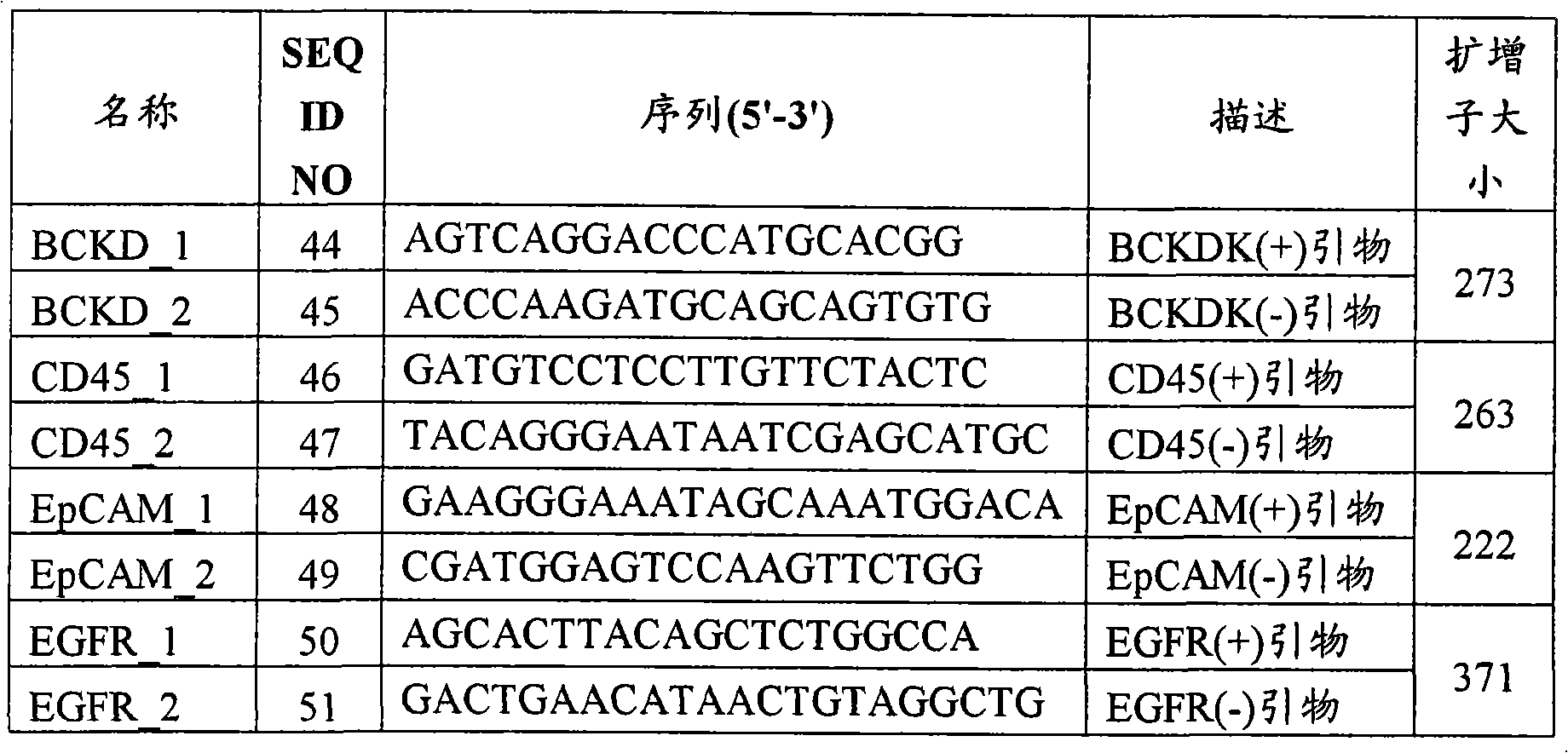
[00273]使用本发明的细胞富集装置(截留尺寸4μm)分离约9×10⁶个白细胞的总RNA,使用RNeasy小型试剂盒(Qiagen)分离5×10⁶个H1650细胞。使用100pmol随机六聚体(Roche)和200U SuperscriptII(Invitrogen)在20μl反应中逆转录白细胞和H1650细胞的2μg总RNA,以获得首链cDNA。使用0.5μl首链cDNA反应物和10pmol正向和反向引物在总共25μl混合物中进行随后的PCR。进行40个循环的PCR:95℃,20秒;56℃,20秒,和70℃,30秒。在1%琼脂糖凝胶上分离扩增的产物。如在图26A中所示,发现BCKDK在白细胞和H1650细胞中均表达;CD45仅在白细胞中表达;EpCAM和EGFR均仅在H1650细胞中表达。这些结果与示于图26B的EGFR表达模式完全一致,证实EGFR对于测定含白细胞和癌细胞的细胞混合物是特别有用的靶,因为预期只有癌细胞才产生信号。[00273] The cell enrichment device of the present invention (cut-
实施例14:采用少量靶RNA或大量背景RNA的EGFR测定Example 14: EGFR Assay Using Low Target RNA or Large Background RNA
[00274]为了测定在实施例12中描述的测定的灵敏度,测试多种量的输入NSCLC细胞系总RNA,其在100pg-50ng的范围内。第一轮和第二轮EGFR PCR反应的结果(步骤1d,实施例12)示于图27。已表明第一轮PCR反应对检测1ng输入RNA足够灵敏,而第二轮增加灵敏度至100pg以下的输入RNA。这相当于7-10个细胞,表明使用该测定甚至极度稀释的样品也可以产生可检测的信号。[00274] To determine the sensitivity of the assay described in Example 12, various amounts of input NSCLC cell line total RNA ranging from 100 pg to 50 ng were tested. The results of the first and second rounds of EGFR PCR reactions (step 1d, Example 12) are shown in Figure 27. The first round of PCR reactions has been shown to be sensitive enough to detect 1 ng of input RNA, while the second round increases sensitivity to less than 100 pg of input RNA. This corresponds to 7-10 cells, showing that even extremely dilute samples can generate a detectable signal using this assay.
[00275]接着,将含1ng NCI-H1975 RNA的样品与1ng-1μg范围的可变量的外周血单核细胞(PBMC)RNA混合,并用于如前的PCR反应。如在图28A中所示,第一组PCR反应证实,尽管扩增在所有情况下都发生,但在最高污染水平出现乱真谱带。然而,如在图28B中所示,在第二组嵌套PCR反应后,产生所需的特异性扩增子,即便在最高污染水平也没有乱真谱带。因此,本实施例表明,本文描述的EGFR PCR测定即便在靶RNA占测试样品中的总RNA的微小部分时也是有效的。[00275] Next, the sample containing 1 ng NCI-H1975 RNA was mixed with a variable amount of peripheral blood mononuclear cell (PBMC) RNA in the range of 1 ng-1 μg, and used for the previous PCR reaction. As shown in Figure 28A, the first set of PCR reactions demonstrated that, although amplification occurred in all cases, spurious bands appeared at the highest contamination levels. However, as shown in Figure 28B, after the second set of nested PCR reactions, the desired specific amplicon was generated without spurious bands even at the highest contamination level. Thus, this example demonstrates that the EGFR PCR assay described herein is effective even when the target RNA constitutes a small fraction of the total RNA in the test sample.
[00276]表8列出了多种细胞中的RNA得率,表明每个细胞的得率广泛变化,这取决于细胞类型。该信息是有用的,以便基于细胞计数估计样品中靶和背景RNA的量。例如,1ng NCL-H1975 RNA对应于约100个细胞,而1μg PBMC RNA对应于约106个细胞。因此,在上述实验中的最高污染水平1,000∶1的PBMC RNA比NCL-H1975RNA实际上对应于10,000∶1比率的PBMC对NCL-H1975细胞。因此,这些数据表明,EGFR可由被多达106个白细胞污染的少至100个CTC测序。[00276] Table 8 lists RNA yields in a variety of cells, showing that yields vary widely per cell, depending on the cell type. This information is useful in order to estimate the amount of target and background RNA in a sample based on cell counts. For example, 1 ng of NCL-H1975 RNA corresponds to about 100 cells, while 1 μg of PBMC RNA corresponds to about 106 cells. Thus, the highest contamination level of 1,000:1 PBMC RNA to NCL-H1975 RNA in the above experiment actually corresponds to a 10,000:1 ratio of PBMC to NCL-H1975 cells. Thus, these data suggest that EGFR can be sequenced from as few as 100 CTCs contaminated with as many as 106 leukocytes.
表8.RNA得率对细胞类型Table 8. RNA Yield vs. Cell Type
[00277]接着,搀入1,000个细胞/ml的Cell Tracker(Invitrogen)-标记的H1650细胞的全血跑过图19C的捕获模块芯片。为避免由固定样品提取的RNA无效,在跑过后立即计数捕获的H1650细胞,随后裂解,在没有甲醛固定的情况下用于RNA提取。用0.5ml的4M异硫氰酸胍溶液将约800个捕获的H1650细胞和>10,000个污染白细胞在芯片上裂解。裂解物用0.5ml苯酚/氯仿提取,并在作为载体的10μg酵母tRNA存在下用1ml乙醇沉淀。DNA酶I处理沉淀的RNA 30分钟,然后用苯酚/氯仿提取,并用乙醇沉淀,然后进行首链cDNA合成,随后进行PCR扩增。这些步骤用第二个血样和第二个芯片重复。使用2组引物CD45_1和CD45_2(表7)以及EGFR_5(正向引物5′-GTTCGGCACGGTGTATAAGG-3′)(SEQ ID NO:_)和EGFR_6(反向引物,5′-CTGGCCATCACGTAGGCTTC-3′)(SEQ ID NO:_),PCR扩增由芯片1和芯片2RNA连同H1650和白细胞cDNA合成的cDNA。EGFR_5和EGFR_6在H1650细胞中产生138bp的野生型扩增片段和123bp的突变扩增片段。PCR产物在2.5%琼脂糖凝胶上分离。如在图29中所示,EGFR野生型和突变扩增的片段易于检测,不管高白细胞背景,表明EGFR测定是较粗的,不需要高度纯化的样品。[00277] Next, whole blood spiked with 1,000 cells/ml of Cell Tracker (Invitrogen)-labeled H1650 cells was run across the capture module chip of Figure 19C. To avoid invalidation of RNA extraction from fixed samples, captured H1650 cells were counted immediately after the run and subsequently lysed for RNA extraction without formaldehyde fixation. Approximately 800 captured H1650 cells and >10,000 contaminating leukocytes were lysed on the chip with 0.5 ml of 4M guanidine isothiocyanate solution. Lysates were extracted with 0.5 ml phenol/chloroform and precipitated with 1 ml ethanol in the presence of 10 μg yeast tRNA as carrier. Precipitated RNA was treated with DNase I for 30 minutes, extracted with phenol/chloroform, and precipitated with ethanol, followed by first-strand cDNA synthesis followed by PCR amplification. These steps are repeated with a second blood sample and a second chip.
Claims (54)
Priority Applications (1)
| Application Number | Priority Date | Filing Date | Title |
|---|---|---|---|
| CN201711191019.9A CN108048549B (en) | 2006-06-14 | 2007-06-14 | Rare cell analysis using sample resolution and DNA tagging |
Applications Claiming Priority (5)
| Application Number | Priority Date | Filing Date | Title |
|---|---|---|---|
| US80481906P | 2006-06-14 | 2006-06-14 | |
| US60/804,819 | 2006-06-14 | ||
| US82077806P | 2006-07-28 | 2006-07-28 | |
| US60/820,778 | 2006-07-28 | ||
| PCT/US2007/071256 WO2007147079A2 (en) | 2006-06-14 | 2007-06-14 | Rare cell analysis using sample splitting and dna tags |
Related Child Applications (1)
| Application Number | Title | Priority Date | Filing Date |
|---|---|---|---|
| CN201711191019.9A Division CN108048549B (en) | 2006-06-14 | 2007-06-14 | Rare cell analysis using sample resolution and DNA tagging |
Publications (2)
| Publication Number | Publication Date |
|---|---|
| CN101675169A true CN101675169A (en) | 2010-03-17 |
| CN101675169B CN101675169B (en) | 2018-01-02 |
Family
ID=38832878
Family Applications (2)
| Application Number | Title | Priority Date | Filing Date |
|---|---|---|---|
| CN201711191019.9A Active CN108048549B (en) | 2006-06-14 | 2007-06-14 | Rare cell analysis using sample resolution and DNA tagging |
| CN200780030309.3A Active CN101675169B (en) | 2006-06-14 | 2007-06-14 | Rare Cell Analysis Using Sample Splitting and DNA Labeling |
Family Applications Before (1)
| Application Number | Title | Priority Date | Filing Date |
|---|---|---|---|
| CN201711191019.9A Active CN108048549B (en) | 2006-06-14 | 2007-06-14 | Rare cell analysis using sample resolution and DNA tagging |
Country Status (6)
| Country | Link |
|---|---|
| US (1) | US20240279738A1 (en) |
| EP (3) | EP2024513B1 (en) |
| CN (2) | CN108048549B (en) |
| AU (1) | AU2007260676A1 (en) |
| CA (1) | CA2655272C (en) |
| WO (1) | WO2007147079A2 (en) |
Cited By (55)
| Publication number | Priority date | Publication date | Assignee | Title |
|---|---|---|---|---|
| CN102108406A (en) * | 2010-12-20 | 2011-06-29 | 北京贝瑞和康生物技术有限公司 | Kit, device and method for detecting copy number of embryo chromosomes |
| CN102776276A (en) * | 2011-05-06 | 2012-11-14 | 爱科来株式会社 | Oligonucleotide for detection test of polymorphism of EGFR exon 19 and use thereof |
| CN103069004A (en) * | 2010-08-13 | 2013-04-24 | 深圳华大基因科技有限公司 | Method for analyzing cell chromosome |
| WO2013086744A1 (en) * | 2011-12-17 | 2013-06-20 | 深圳华大基因研究院 | Method and system for determining whether genome is abnormal |
| CN103397089A (en) * | 2013-07-24 | 2013-11-20 | 安诺优达基因科技(北京)有限公司 | Semi-specific amplification primer group, method and kit for quickly detecting chromosome number abnormality |
| CN103620055A (en) * | 2010-12-07 | 2014-03-05 | 利兰·斯坦福青年大学托管委员会 | Non-invasive determination of fetal inheritance of parental haplotypes at a genome-wide scale |
| CN104120181A (en) * | 2011-06-29 | 2014-10-29 | 深圳华大基因医学有限公司 | Method and device for carrying out GC correction on chromosome sequencing results |
| US9547748B2 (en) | 2011-06-29 | 2017-01-17 | Bgi Health Service Co., Ltd. | Method for determining fetal chromosomal abnormality |
| CN107206380A (en) * | 2015-01-23 | 2017-09-26 | 和卓生物科技(上海)有限公司 | Microfluidics-based detection and isolation of fetal cells for non-invasive prenatal testing |
| CN107603849A (en) * | 2017-09-14 | 2018-01-19 | 中国科学院半导体研究所 | Unicellular RT pcr chips and preparation method thereof |
| CN107988343A (en) * | 2011-02-09 | 2018-05-04 | 纳特拉公司 | The antenatal ploidy recognition methods of Noninvasive |
| CN108138228A (en) * | 2015-09-29 | 2018-06-08 | 卡帕生物系统公司 | High Molecular Weight DNA Sample Tracking Tags for Next Generation Sequencing |
| CN108841960A (en) * | 2018-07-12 | 2018-11-20 | 吉林大学 | A kind of adenocarcinoma of colon neurological susceptibility prediction kit and system |
| US10174369B2 (en) | 2010-05-18 | 2019-01-08 | Natera, Inc. | Methods for non-invasive prenatal ploidy calling |
| US10179937B2 (en) | 2014-04-21 | 2019-01-15 | Natera, Inc. | Detecting mutations and ploidy in chromosomal segments |
| US10216896B2 (en) | 2009-09-30 | 2019-02-26 | Natera, Inc. | Methods for non-invasive prenatal ploidy calling |
| US10227652B2 (en) | 2005-07-29 | 2019-03-12 | Natera, Inc. | System and method for cleaning noisy genetic data from target individuals using genetic data from genetically related individuals |
| US10260096B2 (en) | 2005-07-29 | 2019-04-16 | Natera, Inc. | System and method for cleaning noisy genetic data and determining chromosome copy number |
| US10262755B2 (en) | 2014-04-21 | 2019-04-16 | Natera, Inc. | Detecting cancer mutations and aneuploidy in chromosomal segments |
| US10316362B2 (en) | 2010-05-18 | 2019-06-11 | Natera, Inc. | Methods for simultaneous amplification of target loci |
| US10351906B2 (en) | 2014-04-21 | 2019-07-16 | Natera, Inc. | Methods for simultaneous amplification of target loci |
| CN110366680A (en) * | 2017-02-14 | 2019-10-22 | 阿克斯比尔公司 | Apparatus and method for continuous diagnosis of macromolecules |
| US10526658B2 (en) | 2010-05-18 | 2020-01-07 | Natera, Inc. | Methods for simultaneous amplification of target loci |
| US10533219B2 (en) | 2016-12-07 | 2020-01-14 | Natera, Inc. | Compositions and methods for identifying nucleic acid molecules |
| US10577655B2 (en) | 2013-09-27 | 2020-03-03 | Natera, Inc. | Cell free DNA diagnostic testing standards |
| US10590482B2 (en) | 2010-05-18 | 2020-03-17 | Natera, Inc. | Amplification of cell-free DNA using nested PCR |
| WO2020056889A1 (en) * | 2018-09-20 | 2020-03-26 | 清华大学 | Method and apparatus for detecting content of components in mixed system |
| CN110951580A (en) * | 2019-09-29 | 2020-04-03 | 中国科学院苏州生物医学工程技术研究所 | High-throughput single-cell transcriptome and gene mutation integration analysis integrated device |
| CN111315487A (en) * | 2017-09-05 | 2020-06-19 | 迪森德克斯公司 | Marker analysis for quality control and disease detection |
| US10894976B2 (en) | 2017-02-21 | 2021-01-19 | Natera, Inc. | Compositions, methods, and kits for isolating nucleic acids |
| US20210198733A1 (en) | 2018-07-03 | 2021-07-01 | Natera, Inc. | Methods for detection of donor-derived cell-free dna |
| US11111543B2 (en) | 2005-07-29 | 2021-09-07 | Natera, Inc. | System and method for cleaning noisy genetic data and determining chromosome copy number |
| US11111544B2 (en) | 2005-07-29 | 2021-09-07 | Natera, Inc. | System and method for cleaning noisy genetic data and determining chromosome copy number |
| US11306357B2 (en) | 2010-05-18 | 2022-04-19 | Natera, Inc. | Methods for non-invasive prenatal ploidy calling |
| US11322224B2 (en) | 2010-05-18 | 2022-05-03 | Natera, Inc. | Methods for non-invasive prenatal ploidy calling |
| US11326208B2 (en) | 2010-05-18 | 2022-05-10 | Natera, Inc. | Methods for nested PCR amplification of cell-free DNA |
| US11332793B2 (en) | 2010-05-18 | 2022-05-17 | Natera, Inc. | Methods for simultaneous amplification of target loci |
| US11332785B2 (en) | 2010-05-18 | 2022-05-17 | Natera, Inc. | Methods for non-invasive prenatal ploidy calling |
| US11339429B2 (en) | 2010-05-18 | 2022-05-24 | Natera, Inc. | Methods for non-invasive prenatal ploidy calling |
| CN114728256A (en) * | 2019-11-14 | 2022-07-08 | 生物辐射实验室股份有限公司 | Compartmentalized determination of target copy number of single cells by non-end-point amplification |
| US11408031B2 (en) | 2010-05-18 | 2022-08-09 | Natera, Inc. | Methods for non-invasive prenatal paternity testing |
| US11479812B2 (en) | 2015-05-11 | 2022-10-25 | Natera, Inc. | Methods and compositions for determining ploidy |
| US11485996B2 (en) | 2016-10-04 | 2022-11-01 | Natera, Inc. | Methods for characterizing copy number variation using proximity-litigation sequencing |
| US11939634B2 (en) | 2010-05-18 | 2024-03-26 | Natera, Inc. | Methods for simultaneous amplification of target loci |
| US12024738B2 (en) | 2018-04-14 | 2024-07-02 | Natera, Inc. | Methods for cancer detection and monitoring |
| CN118571324A (en) * | 2024-07-31 | 2024-08-30 | 杭州华大生命科学研究院 | Data processing method and device, storage medium and electronic device |
| US12084720B2 (en) | 2017-12-14 | 2024-09-10 | Natera, Inc. | Assessing graft suitability for transplantation |
| US12100478B2 (en) | 2012-08-17 | 2024-09-24 | Natera, Inc. | Method for non-invasive prenatal testing using parental mosaicism data |
| US12146195B2 (en) | 2016-04-15 | 2024-11-19 | Natera, Inc. | Methods for lung cancer detection |
| US12152275B2 (en) | 2010-05-18 | 2024-11-26 | Natera, Inc. | Methods for non-invasive prenatal ploidy calling |
| US12221653B2 (en) | 2010-05-18 | 2025-02-11 | Natera, Inc. | Methods for simultaneous amplification of target loci |
| US12260934B2 (en) | 2014-06-05 | 2025-03-25 | Natera, Inc. | Systems and methods for detection of aneuploidy |
| US12305235B2 (en) | 2019-06-06 | 2025-05-20 | Natera, Inc. | Methods for detecting immune cell DNA and monitoring immune system |
| US12398389B2 (en) | 2018-02-15 | 2025-08-26 | Natera, Inc. | Methods for isolating nucleic acids with size selection |
| US12460264B2 (en) | 2016-11-02 | 2025-11-04 | Natera, Inc. | Method of detecting tumour recurrence |
Families Citing this family (155)
| Publication number | Priority date | Publication date | Assignee | Title |
|---|---|---|---|---|
| WO2002037938A2 (en) | 2000-10-24 | 2002-05-16 | Oncosis Llc | Method and device for selectively targeting cells within a three -dimensional specimen |
| WO2007147074A2 (en) | 2006-06-14 | 2007-12-21 | Living Microsystems, Inc. | Use of highly parallel snp genotyping for fetal diagnosis |
| US20080050739A1 (en) | 2006-06-14 | 2008-02-28 | Roland Stoughton | Diagnosis of fetal abnormalities using polymorphisms including short tandem repeats |
| US8309317B2 (en) * | 2008-04-05 | 2012-11-13 | Single Cell Technology, Inc. | Method of screening single cells for the production of biologically active agents |
| EP2350652A2 (en) | 2008-10-10 | 2011-08-03 | Cnrs-Dae | Cell sorting device |
| EP2356249B1 (en) * | 2008-10-10 | 2016-02-17 | Picovitro Ab | Genetic analysis in microwells |
| CN102325901A (en) * | 2008-12-22 | 2012-01-18 | 赛卢拉有限公司 | Methods and Genotyping Profiles for Detecting Alleles, Genomes, and Transcriptomes |
| CN102341505A (en) * | 2009-01-09 | 2012-02-01 | 英特拉克森公司 | Genetic analysis of cells |
| ES2706227T3 (en) | 2009-03-30 | 2019-03-27 | Illumina Inc | Analysis of gene expression in individual cells |
| ES2562159T3 (en) | 2009-08-20 | 2016-03-02 | Population Genetics Technologies Ltd. | Compositions and methods for the rearrangement of molecular nucleic acid |
| CA2767028A1 (en) * | 2009-11-25 | 2011-06-03 | Quantalife, Inc. | Methods and compositions for detecting genetic material |
| US8835358B2 (en) | 2009-12-15 | 2014-09-16 | Cellular Research, Inc. | Digital counting of individual molecules by stochastic attachment of diverse labels |
| EP2848703A1 (en) | 2010-01-19 | 2015-03-18 | Verinata Health, Inc | Simultaneous determination of aneuploidy and fetal fraction |
| US10388403B2 (en) | 2010-01-19 | 2019-08-20 | Verinata Health, Inc. | Analyzing copy number variation in the detection of cancer |
| EP2513341B1 (en) | 2010-01-19 | 2017-04-12 | Verinata Health, Inc | Identification of polymorphic sequences in mixtures of genomic dna by whole genome sequencing |
| US9260745B2 (en) | 2010-01-19 | 2016-02-16 | Verinata Health, Inc. | Detecting and classifying copy number variation |
| AU2011207561B2 (en) | 2010-01-19 | 2014-02-20 | Verinata Health, Inc. | Partition defined detection methods |
| WO2011090556A1 (en) | 2010-01-19 | 2011-07-28 | Verinata Health, Inc. | Methods for determining fraction of fetal nucleic acid in maternal samples |
| US20120100548A1 (en) | 2010-10-26 | 2012-04-26 | Verinata Health, Inc. | Method for determining copy number variations |
| US9323888B2 (en) | 2010-01-19 | 2016-04-26 | Verinata Health, Inc. | Detecting and classifying copy number variation |
| US20110312503A1 (en) | 2010-01-23 | 2011-12-22 | Artemis Health, Inc. | Methods of fetal abnormality detection |
| US9267174B2 (en) | 2010-10-26 | 2016-02-23 | Stanford University | Method of simultaneously screening for multiple genotypes and/or mutations |
| AU2012214312A1 (en) | 2011-02-09 | 2013-08-22 | Bio-Rad Laboratories, Inc. | Analysis of nucleic acids |
| SI3078752T1 (en) | 2011-04-12 | 2018-12-31 | Verinata Health, Inc | Resolving genome fractions using polymorphism counts |
| US9411937B2 (en) | 2011-04-15 | 2016-08-09 | Verinata Health, Inc. | Detecting and classifying copy number variation |
| US20130059738A1 (en) | 2011-04-28 | 2013-03-07 | Life Technologies Corporation | Methods and compositions for multiplex pcr |
| EP3495497B1 (en) | 2011-04-28 | 2021-03-24 | Life Technologies Corporation | Methods and compositions for multiplex pcr |
| US20130059762A1 (en) | 2011-04-28 | 2013-03-07 | Life Technologies Corporation | Methods and compositions for multiplex pcr |
| BR112014009269A8 (en) * | 2011-10-18 | 2017-06-20 | Multiplicom N V | diagnosis of fetal chromosomal aneuploidy |
| JP6375230B2 (en) | 2012-02-27 | 2018-08-15 | セルラー リサーチ, インコーポレイテッド | Compositions and kits for molecular counting |
| US20130261984A1 (en) * | 2012-03-30 | 2013-10-03 | Illumina, Inc. | Methods and systems for determining fetal chromosomal abnormalities |
| US10273541B2 (en) | 2012-08-14 | 2019-04-30 | 10X Genomics, Inc. | Methods and systems for processing polynucleotides |
| US10400280B2 (en) | 2012-08-14 | 2019-09-03 | 10X Genomics, Inc. | Methods and systems for processing polynucleotides |
| US9701998B2 (en) | 2012-12-14 | 2017-07-11 | 10X Genomics, Inc. | Methods and systems for processing polynucleotides |
| US9951386B2 (en) | 2014-06-26 | 2018-04-24 | 10X Genomics, Inc. | Methods and systems for processing polynucleotides |
| US10221442B2 (en) | 2012-08-14 | 2019-03-05 | 10X Genomics, Inc. | Compositions and methods for sample processing |
| US10752949B2 (en) | 2012-08-14 | 2020-08-25 | 10X Genomics, Inc. | Methods and systems for processing polynucleotides |
| US11591637B2 (en) | 2012-08-14 | 2023-02-28 | 10X Genomics, Inc. | Compositions and methods for sample processing |
| US10323279B2 (en) | 2012-08-14 | 2019-06-18 | 10X Genomics, Inc. | Methods and systems for processing polynucleotides |
| CA2881685C (en) | 2012-08-14 | 2023-12-05 | 10X Genomics, Inc. | Microcapsule compositions and methods |
| EP2931919B1 (en) | 2012-12-14 | 2019-02-20 | 10X Genomics, Inc. | Methods and systems for processing polynucleotides |
| US10533221B2 (en) | 2012-12-14 | 2020-01-14 | 10X Genomics, Inc. | Methods and systems for processing polynucleotides |
| EP2954104B1 (en) | 2013-02-08 | 2020-09-16 | 10X Genomics, Inc. | Polynucleotide barcode generation |
| WO2014134790A1 (en) * | 2013-03-06 | 2014-09-12 | 广州百赫医疗信息科技有限公司 | Pcr reagent kit for diagnosing neurofibromatosis |
| CN105247042B (en) | 2013-03-15 | 2021-06-11 | 普林斯顿大学理事会 | Method and apparatus for high throughput purification |
| EP2971287B1 (en) | 2013-03-15 | 2019-08-14 | GPB Scientific, LLC | On-chip microfluidic processing of particles |
| US20150064153A1 (en) | 2013-03-15 | 2015-03-05 | The Trustees Of Princeton University | High efficiency microfluidic purification of stem cells to improve transplants |
| EP3039158B1 (en) | 2013-08-28 | 2018-11-14 | Cellular Research, Inc. | Massively parallel single cell analysis |
| US10395758B2 (en) | 2013-08-30 | 2019-08-27 | 10X Genomics, Inc. | Sequencing methods |
| US9824068B2 (en) | 2013-12-16 | 2017-11-21 | 10X Genomics, Inc. | Methods and apparatus for sorting data |
| DE202015009609U1 (en) | 2014-04-10 | 2018-08-06 | 10X Genomics, Inc. | Microfluidic system for the production of emulsions |
| US12492429B2 (en) | 2014-04-21 | 2025-12-09 | Natera, Inc. | Detecting mutations and ploidy in chromosomal segments |
| EP4574974A3 (en) | 2014-06-26 | 2025-10-08 | 10x Genomics, Inc. | Methods of analyzing nucleic acids from individual cells or cell populations |
| US12312640B2 (en) | 2014-06-26 | 2025-05-27 | 10X Genomics, Inc. | Analysis of nucleic acid sequences |
| JP2017522866A (en) | 2014-06-26 | 2017-08-17 | 10エックス ジェノミクス, インコーポレイテッド | Nucleic acid sequence analysis |
| CN114807307A (en) | 2014-10-29 | 2022-07-29 | 10X 基因组学有限公司 | Methods and compositions for sequencing target nucleic acids |
| US9975122B2 (en) | 2014-11-05 | 2018-05-22 | 10X Genomics, Inc. | Instrument systems for integrated sample processing |
| BR112017014902A2 (en) | 2015-01-12 | 2018-03-13 | 10X Genomics Inc | processes and systems for the preparation of nucleic acid sequencing libraries and libraries prepared using them |
| US10697010B2 (en) | 2015-02-19 | 2020-06-30 | Becton, Dickinson And Company | High-throughput single-cell analysis combining proteomic and genomic information |
| JP2018505688A (en) | 2015-02-24 | 2018-03-01 | 10エックス ゲノミクス,インコーポレイテッド | Method for targeted nucleic acid sequence coverage (COVERAGE) |
| EP4286516A3 (en) | 2015-02-24 | 2024-03-06 | 10X Genomics, Inc. | Partition processing methods and systems |
| ES2836802T3 (en) | 2015-02-27 | 2021-06-28 | Becton Dickinson Co | Spatially addressable molecular barcodes |
| CN107406888A (en) | 2015-03-30 | 2017-11-28 | 赛卢拉研究公司 | Methods and compositions for combining barcoding |
| EP4582556A3 (en) | 2015-04-23 | 2025-10-08 | Becton, Dickinson and Company | Methods and compositions for whole transcriptome amplification |
| US11124823B2 (en) | 2015-06-01 | 2021-09-21 | Becton, Dickinson And Company | Methods for RNA quantification |
| EP3329010B1 (en) * | 2015-07-29 | 2019-07-10 | Progenity, Inc. | Nucleic acids and methods for detecting chromosomal abnormalities |
| WO2017044574A1 (en) | 2015-09-11 | 2017-03-16 | Cellular Research, Inc. | Methods and compositions for nucleic acid library normalization |
| EP3373822A1 (en) * | 2015-11-11 | 2018-09-19 | Medizinische Universität Graz | In vivo collection and localized quantification and profiling of circulating cells, proteins and nucleic acids |
| US11371094B2 (en) | 2015-11-19 | 2022-06-28 | 10X Genomics, Inc. | Systems and methods for nucleic acid processing using degenerate nucleotides |
| KR20240161696A (en) | 2015-12-04 | 2024-11-12 | 10엑스 제노믹스, 인크. | Methods and compositions for nucleic acid analysis |
| EP3414341A4 (en) | 2016-02-11 | 2019-10-09 | 10X Genomics, Inc. | SYSTEMS, METHODS, AND MEDIA FOR ASSEMBLING NOVO OF GENOME SEQUENCE DATA OVERALL |
| US10822643B2 (en) | 2016-05-02 | 2020-11-03 | Cellular Research, Inc. | Accurate molecular barcoding |
| WO2017197343A2 (en) | 2016-05-12 | 2017-11-16 | 10X Genomics, Inc. | Microfluidic on-chip filters |
| WO2017197338A1 (en) | 2016-05-13 | 2017-11-16 | 10X Genomics, Inc. | Microfluidic systems and methods of use |
| US10301677B2 (en) | 2016-05-25 | 2019-05-28 | Cellular Research, Inc. | Normalization of nucleic acid libraries |
| ES2979395T3 (en) | 2016-05-26 | 2024-09-25 | Becton Dickinson Co | Molecular tag count adjustment methods |
| US10202641B2 (en) | 2016-05-31 | 2019-02-12 | Cellular Research, Inc. | Error correction in amplification of samples |
| US10640763B2 (en) | 2016-05-31 | 2020-05-05 | Cellular Research, Inc. | Molecular indexing of internal sequences |
| EP4300099A3 (en) | 2016-09-26 | 2024-03-27 | Becton, Dickinson and Company | Measurement of protein expression using reagents with barcoded oligonucleotide sequences |
| US10011872B1 (en) | 2016-12-22 | 2018-07-03 | 10X Genomics, Inc. | Methods and systems for processing polynucleotides |
| US10815525B2 (en) | 2016-12-22 | 2020-10-27 | 10X Genomics, Inc. | Methods and systems for processing polynucleotides |
| US10550429B2 (en) | 2016-12-22 | 2020-02-04 | 10X Genomics, Inc. | Methods and systems for processing polynucleotides |
| ES2961580T3 (en) | 2017-01-13 | 2024-03-12 | Cellular Res Inc | Hydrophilic coating of fluid channels |
| CN110214186B (en) | 2017-01-30 | 2023-11-24 | 10X基因组学有限公司 | Methods and systems for droplet-based single cell barcoding |
| US12264411B2 (en) | 2017-01-30 | 2025-04-01 | 10X Genomics, Inc. | Methods and systems for analysis |
| EP3577232A1 (en) | 2017-02-01 | 2019-12-11 | Cellular Research, Inc. | Selective amplification using blocking oligonucleotides |
| US10995333B2 (en) | 2017-02-06 | 2021-05-04 | 10X Genomics, Inc. | Systems and methods for nucleic acid preparation |
| US20180340169A1 (en) | 2017-05-26 | 2018-11-29 | 10X Genomics, Inc. | Single cell analysis of transposase accessible chromatin |
| EP4230746A3 (en) | 2017-05-26 | 2023-11-01 | 10X Genomics, Inc. | Single cell analysis of transposase accessible chromatin |
| WO2018223057A1 (en) * | 2017-06-02 | 2018-12-06 | Affymetrix, Inc. | Array-based methods for analysing mixed samples using different allele-specific labels, in particular for detection of fetal aneuploidies |
| US11441174B2 (en) | 2017-06-02 | 2022-09-13 | Affymetrix, Inc. | Array-based methods for analysing mixed samples using differently labelled allele-specific probes |
| US11603557B2 (en) | 2017-06-02 | 2023-03-14 | Affymetrix, Inc. | Array-based methods for analysing mixed samples using differently labelled allele-specific probes |
| EP4345172A3 (en) | 2017-06-05 | 2024-07-03 | Becton, Dickinson and Company | Sample indexing for single cells |
| US10837047B2 (en) | 2017-10-04 | 2020-11-17 | 10X Genomics, Inc. | Compositions, methods, and systems for bead formation using improved polymers |
| WO2019084043A1 (en) | 2017-10-26 | 2019-05-02 | 10X Genomics, Inc. | Methods and systems for nuclecic acid preparation and chromatin analysis |
| EP3700672B1 (en) | 2017-10-27 | 2022-12-28 | 10X Genomics, Inc. | Methods for sample preparation and analysis |
| WO2019099751A1 (en) | 2017-11-15 | 2019-05-23 | 10X Genomics, Inc. | Functionalized gel beads |
| US10829815B2 (en) | 2017-11-17 | 2020-11-10 | 10X Genomics, Inc. | Methods and systems for associating physical and genetic properties of biological particles |
| WO2019108851A1 (en) | 2017-11-30 | 2019-06-06 | 10X Genomics, Inc. | Systems and methods for nucleic acid preparation and analysis |
| CN118818037A (en) | 2017-12-12 | 2024-10-22 | 10X基因组学有限公司 | Systems and methods for single cell processing |
| US11946095B2 (en) | 2017-12-19 | 2024-04-02 | Becton, Dickinson And Company | Particles associated with oligonucleotides |
| WO2019126789A1 (en) | 2017-12-22 | 2019-06-27 | 10X Genomics, Inc. | Systems and methods for processing nucleic acid molecules from one or more cells |
| EP3752832A1 (en) | 2018-02-12 | 2020-12-23 | 10X Genomics, Inc. | Methods characterizing multiple analytes from individual cells or cell populations |
| US11639928B2 (en) | 2018-02-22 | 2023-05-02 | 10X Genomics, Inc. | Methods and systems for characterizing analytes from individual cells or cell populations |
| WO2019169028A1 (en) | 2018-02-28 | 2019-09-06 | 10X Genomics, Inc. | Transcriptome sequencing through random ligation |
| CN112262218B (en) | 2018-04-06 | 2024-11-08 | 10X基因组学有限公司 | Systems and methods for quality control in single cell processing |
| US11365409B2 (en) | 2018-05-03 | 2022-06-21 | Becton, Dickinson And Company | Molecular barcoding on opposite transcript ends |
| US11773441B2 (en) | 2018-05-03 | 2023-10-03 | Becton, Dickinson And Company | High throughput multiomics sample analysis |
| WO2019217758A1 (en) | 2018-05-10 | 2019-11-14 | 10X Genomics, Inc. | Methods and systems for molecular library generation |
| US11932899B2 (en) | 2018-06-07 | 2024-03-19 | 10X Genomics, Inc. | Methods and systems for characterizing nucleic acid molecules |
| US11703427B2 (en) | 2018-06-25 | 2023-07-18 | 10X Genomics, Inc. | Methods and systems for cell and bead processing |
| US12188014B1 (en) | 2018-07-25 | 2025-01-07 | 10X Genomics, Inc. | Compositions and methods for nucleic acid processing using blocking agents |
| US20200032335A1 (en) | 2018-07-27 | 2020-01-30 | 10X Genomics, Inc. | Systems and methods for metabolome analysis |
| CN112703252B (en) | 2018-08-03 | 2024-09-10 | 10X基因组学有限公司 | Method and system for minimizing barcode exchange |
| US12065688B2 (en) | 2018-08-20 | 2024-08-20 | 10X Genomics, Inc. | Compositions and methods for cellular processing |
| WO2020041148A1 (en) | 2018-08-20 | 2020-02-27 | 10X Genomics, Inc. | Methods and systems for detection of protein-dna interactions using proximity ligation |
| JP7670607B2 (en) | 2018-10-01 | 2025-04-30 | ベクトン・ディキンソン・アンド・カンパニー | Determination of 5' transcript sequence |
| EP3877520B1 (en) | 2018-11-08 | 2025-03-19 | Becton, Dickson And Company | Whole transcriptome analysis of single cells using random priming |
| US11459607B1 (en) | 2018-12-10 | 2022-10-04 | 10X Genomics, Inc. | Systems and methods for processing-nucleic acid molecules from a single cell using sequential co-partitioning and composite barcodes |
| WO2020120442A2 (en) | 2018-12-13 | 2020-06-18 | Dna Script | Direct oligonucleotide synthesis on cells and biomolecules |
| US11492660B2 (en) | 2018-12-13 | 2022-11-08 | Becton, Dickinson And Company | Selective extension in single cell whole transcriptome analysis |
| US12169198B2 (en) | 2019-01-08 | 2024-12-17 | 10X Genomics, Inc. | Systems and methods for sample analysis |
| US11845983B1 (en) | 2019-01-09 | 2023-12-19 | 10X Genomics, Inc. | Methods and systems for multiplexing of droplet based assays |
| WO2020150356A1 (en) | 2019-01-16 | 2020-07-23 | Becton, Dickinson And Company | Polymerase chain reaction normalization through primer titration |
| WO2020154247A1 (en) | 2019-01-23 | 2020-07-30 | Cellular Research, Inc. | Oligonucleotides associated with antibodies |
| US12305239B2 (en) | 2019-02-12 | 2025-05-20 | 10X Genomics, Inc. | Analysis of nucleic acid sequences |
| US11851683B1 (en) | 2019-02-12 | 2023-12-26 | 10X Genomics, Inc. | Methods and systems for selective analysis of cellular samples |
| WO2020167862A1 (en) | 2019-02-12 | 2020-08-20 | 10X Genomics, Inc. | Systems and methods for transfer of reagents between droplets |
| US12275993B2 (en) | 2019-02-12 | 2025-04-15 | 10X Genomics, Inc. | Analysis of nucleic acid sequences |
| US11584953B2 (en) | 2019-02-12 | 2023-02-21 | 10X Genomics, Inc. | Methods for processing nucleic acid molecules |
| WO2020167866A1 (en) | 2019-02-12 | 2020-08-20 | 10X Genomics, Inc. | Systems and methods for transposon loading |
| US11467153B2 (en) | 2019-02-12 | 2022-10-11 | 10X Genomics, Inc. | Methods for processing nucleic acid molecules |
| EP3924506A1 (en) | 2019-02-14 | 2021-12-22 | Becton Dickinson and Company | Hybrid targeted and whole transcriptome amplification |
| US11655499B1 (en) | 2019-02-25 | 2023-05-23 | 10X Genomics, Inc. | Detection of sequence elements in nucleic acid molecules |
| SG11202111242PA (en) | 2019-03-11 | 2021-11-29 | 10X Genomics Inc | Systems and methods for processing optically tagged beads |
| WO2020214642A1 (en) | 2019-04-19 | 2020-10-22 | Becton, Dickinson And Company | Methods of associating phenotypical data and single cell sequencing data |
| CN114051534B (en) | 2019-07-22 | 2025-02-21 | 贝克顿迪金森公司 | Single-cell chromatin immunoprecipitation sequencing assay |
| US12235262B1 (en) | 2019-09-09 | 2025-02-25 | 10X Genomics, Inc. | Methods and systems for single cell protein analysis |
| US11773436B2 (en) | 2019-11-08 | 2023-10-03 | Becton, Dickinson And Company | Using random priming to obtain full-length V(D)J information for immune repertoire sequencing |
| WO2021146207A1 (en) | 2020-01-13 | 2021-07-22 | Becton, Dickinson And Company | Methods and compositions for quantitation of proteins and rna |
| WO2021155057A1 (en) | 2020-01-29 | 2021-08-05 | Becton, Dickinson And Company | Barcoded wells for spatial mapping of single cells through sequencing |
| US12449419B1 (en) | 2020-02-12 | 2025-10-21 | 10X Genomics, Inc. | Methods for detecting binding of peptide-MHC monomers to T cells |
| WO2021173719A1 (en) | 2020-02-25 | 2021-09-02 | Becton, Dickinson And Company | Bi-specific probes to enable the use of single-cell samples as single color compensation control |
| US11851700B1 (en) | 2020-05-13 | 2023-12-26 | 10X Genomics, Inc. | Methods, kits, and compositions for processing extracellular molecules |
| WO2021231779A1 (en) | 2020-05-14 | 2021-11-18 | Becton, Dickinson And Company | Primers for immune repertoire profiling |
| WO2021247593A1 (en) | 2020-06-02 | 2021-12-09 | Becton, Dickinson And Company | Oligonucleotides and beads for 5 prime gene expression assay |
| US11932901B2 (en) | 2020-07-13 | 2024-03-19 | Becton, Dickinson And Company | Target enrichment using nucleic acid probes for scRNAseq |
| US12391940B2 (en) | 2020-07-31 | 2025-08-19 | Becton, Dickinson And Company | Single cell assay for transposase-accessible chromatin |
| US12480158B1 (en) | 2020-11-05 | 2025-11-25 | 10X Genomics, Inc. | Methods and systems for processing polynucleotides |
| US12084715B1 (en) | 2020-11-05 | 2024-09-10 | 10X Genomics, Inc. | Methods and systems for reducing artifactual antisense products |
| EP4247967A1 (en) | 2020-11-20 | 2023-09-27 | Becton, Dickinson and Company | Profiling of highly expressed and lowly expressed proteins |
| WO2022132206A1 (en) | 2020-12-15 | 2022-06-23 | Becton, Dickinson And Company | Single cell secretome analysis |
| US12398262B1 (en) | 2021-01-22 | 2025-08-26 | 10X Genomics, Inc. | Triblock copolymer-based cell stabilization and fixation system and methods of use thereof |
| CN117015617B (en) | 2021-02-23 | 2025-04-04 | 10X基因组学有限公司 | Probe-based nucleic acid and protein analysis |
| CN118581198B (en) * | 2024-08-08 | 2025-01-10 | 上海美吉生物医药科技有限公司 | Method, kit and application of emQPCR multi-target joint inspection of trace DNA |
Citations (3)
| Publication number | Priority date | Publication date | Assignee | Title |
|---|---|---|---|---|
| US20030165852A1 (en) * | 2000-11-15 | 2003-09-04 | Schueler Paula A. | Methods and reagents for identifying rare fetal cells in the maternal circulation |
| US6618679B2 (en) * | 2000-01-28 | 2003-09-09 | Althea Technologies, Inc. | Methods for analysis of gene expression |
| US20060051775A1 (en) * | 1989-11-13 | 2006-03-09 | The Children's Hospital | Non-invasive method for isolation and detection of fetal DNA |
Family Cites Families (71)
| Publication number | Priority date | Publication date | Assignee | Title |
|---|---|---|---|---|
| US6969A (en) | 1849-12-25 | Heney blbeckee | ||
| US6858A (en) | 1849-11-06 | File-supporter | ||
| US589A (en) | 1838-02-03 | Joseph smart | ||
| US412A (en) | 1837-09-28 | Water-wheel aktd mode of letting the water ow the same | ||
| US5242794A (en) | 1984-12-13 | 1993-09-07 | Applied Biosystems, Inc. | Detection of specific sequences in nucleic acids |
| JPH0610416B2 (en) | 1987-12-28 | 1994-02-09 | いすゞ自動車株式会社 | Controller for turbocharger with rotating electric machine |
| US4988617A (en) | 1988-03-25 | 1991-01-29 | California Institute Of Technology | Method of detecting a nucleotide change in nucleic acids |
| US6054270A (en) | 1988-05-03 | 2000-04-25 | Oxford Gene Technology Limited | Analying polynucleotide sequences |
| US5700637A (en) | 1988-05-03 | 1997-12-23 | Isis Innovation Limited | Apparatus and method for analyzing polynucleotide sequences and method of generating oligonucleotide arrays |
| US5744101A (en) | 1989-06-07 | 1998-04-28 | Affymax Technologies N.V. | Photolabile nucleoside protecting groups |
| US6040138A (en) | 1995-09-15 | 2000-03-21 | Affymetrix, Inc. | Expression monitoring by hybridization to high density oligonucleotide arrays |
| US5143854A (en) | 1989-06-07 | 1992-09-01 | Affymax Technologies N.V. | Large scale photolithographic solid phase synthesis of polypeptides and receptor binding screening thereof |
| US5424186A (en) | 1989-06-07 | 1995-06-13 | Affymax Technologies N.V. | Very large scale immobilized polymer synthesis |
| US5800992A (en) | 1989-06-07 | 1998-09-01 | Fodor; Stephen P.A. | Method of detecting nucleic acids |
| US6346413B1 (en) | 1989-06-07 | 2002-02-12 | Affymetrix, Inc. | Polymer arrays |
| US5494810A (en) | 1990-05-03 | 1996-02-27 | Cornell Research Foundation, Inc. | Thermostable ligase-mediated DNA amplifications system for the detection of genetic disease |
| US5427663A (en) | 1993-06-08 | 1995-06-27 | British Technology Group Usa Inc. | Microlithographic array for macromolecule and cell fractionation |
| US5837832A (en) | 1993-06-25 | 1998-11-17 | Affymetrix, Inc. | Arrays of nucleic acid probes on biological chips |
| US5556773A (en) * | 1993-08-06 | 1996-09-17 | Yourno; Joseph | Method and apparatus for nested polymerase chain reaction (PCR) with single closed reaction tubes |
| EP0695941B1 (en) | 1994-06-08 | 2002-07-31 | Affymetrix, Inc. | Method and apparatus for packaging a chip |
| US6287850B1 (en) | 1995-06-07 | 2001-09-11 | Affymetrix, Inc. | Bioarray chip reaction apparatus and its manufacture |
| US5780231A (en) * | 1995-11-17 | 1998-07-14 | Lynx Therapeutics, Inc. | DNA extension and analysis with rolling primers |
| US6300063B1 (en) | 1995-11-29 | 2001-10-09 | Affymetrix, Inc. | Polymorphism detection |
| AU758463B2 (en) * | 1998-02-04 | 2003-03-20 | Applied Biosystems, Llc | Determination of a genotype of an amplification product at multiple allelic sites |
| EP1078257A4 (en) * | 1998-05-09 | 2007-07-18 | Iconisys Inc | Method and apparatus for computer controlled rare cell, including fetal cell, based diagnosis |
| EP2045334A1 (en) | 1998-06-24 | 2009-04-08 | Illumina, Inc. | Decoding of array sensors with microspheres |
| US6429027B1 (en) | 1998-12-28 | 2002-08-06 | Illumina, Inc. | Composite arrays utilizing microspheres |
| US7612020B2 (en) | 1998-12-28 | 2009-11-03 | Illumina, Inc. | Composite arrays utilizing microspheres with a hybridization chamber |
| US6846460B1 (en) | 1999-01-29 | 2005-01-25 | Illumina, Inc. | Apparatus and method for separation of liquid phases of different density and for fluorous phase organic syntheses |
| US6355431B1 (en) | 1999-04-20 | 2002-03-12 | Illumina, Inc. | Detection of nucleic acid amplification reactions using bead arrays |
| US20050244870A1 (en) | 1999-04-20 | 2005-11-03 | Illumina, Inc. | Nucleic acid sequencing using microsphere arrays |
| US20030207295A1 (en) | 1999-04-20 | 2003-11-06 | Kevin Gunderson | Detection of nucleic acid reactions on bead arrays |
| US7056661B2 (en) | 1999-05-19 | 2006-06-06 | Cornell Research Foundation, Inc. | Method for sequencing nucleic acid molecules |
| US6544732B1 (en) | 1999-05-20 | 2003-04-08 | Illumina, Inc. | Encoding and decoding of array sensors utilizing nanocrystals |
| EP1190100B1 (en) | 1999-05-20 | 2012-07-25 | Illumina, Inc. | Combinatorial decoding of random nucleic acid arrays |
| DE60030436T2 (en) | 1999-05-20 | 2007-03-29 | Illumina, Inc., San Diego | DEVICE FOR MOUNTING AND PRESENTING AT LEAST ONE MICROBALL MIXTURE FOR SOLUTIONS AND / OR OPTICAL PICTURE SYSTEMS |
| CA2382436C (en) | 1999-08-30 | 2011-05-17 | Illumina, Inc. | Methods for improving signal detection from an array |
| CA2390375A1 (en) | 1999-11-10 | 2001-05-17 | Massachusetts Institute Of Technology | Cell analysis and sorting apparatus for manipulation of cells |
| US6692952B1 (en) | 1999-11-10 | 2004-02-17 | Massachusetts Institute Of Technology | Cell analysis and sorting apparatus for manipulation of cells |
| DK1239952T3 (en) | 1999-12-13 | 2012-01-02 | Illumina Inc | Synthesizer for oligonucleotides using a centrifugal force |
| US6582938B1 (en) | 2001-05-11 | 2003-06-24 | Affymetrix, Inc. | Amplification of nucleic acids |
| AU3806701A (en) | 2000-02-07 | 2001-08-14 | Illumina Inc | Nucleic acid detection methods using universal priming |
| US6913884B2 (en) | 2001-08-16 | 2005-07-05 | Illumina, Inc. | Compositions and methods for repetitive use of genomic DNA |
| CA2399733C (en) | 2000-02-07 | 2011-09-20 | Illumina, Inc. | Nucleic acid detection methods using universal priming |
| US6770441B2 (en) | 2000-02-10 | 2004-08-03 | Illumina, Inc. | Array compositions and methods of making same |
| DE60131903T2 (en) | 2000-10-24 | 2008-11-27 | The Board of Trustees of the Leland S. Stanford Junior University, Palo Alto | DIRECT MULTIPLEX CHARACTERIZATION OF GENOMIC DNA |
| US20040018491A1 (en) | 2000-10-26 | 2004-01-29 | Kevin Gunderson | Detection of nucleic acid reactions on bead arrays |
| US7078168B2 (en) * | 2001-02-27 | 2006-07-18 | Biotage Ab | Method for determining allele frequencies |
| AUPR749901A0 (en) * | 2001-09-06 | 2001-09-27 | Monash University | Method of identifying chromosomal abnormalities and prenatal diagnosis |
| WO2003065019A1 (en) | 2002-01-25 | 2003-08-07 | Illumina, Inc. | Sensor arrays for detecting analytes in fluids |
| BR0308161A (en) * | 2002-03-01 | 2006-06-06 | Ravgen Inc | methods for detection of genetic disorders |
| US6977162B2 (en) * | 2002-03-01 | 2005-12-20 | Ravgen, Inc. | Rapid analysis of variations in a genome |
| US7727720B2 (en) * | 2002-05-08 | 2010-06-01 | Ravgen, Inc. | Methods for detection of genetic disorders |
| ATE391942T1 (en) | 2002-08-07 | 2008-04-15 | Vim Technologies Ltd | LITHOGRAPHIC PRINTING ELEMENTS AND METHOD AND SYSTEM FOR PRODUCING LITHOGRAPHIC PRINTING ELEMENTS |
| ES2375724T3 (en) | 2002-09-27 | 2012-03-05 | The General Hospital Corporation | MICROFLUDE DEVICE FOR SEPERATION OF CELLS AND ITS USES. |
| US7025935B2 (en) | 2003-04-11 | 2006-04-11 | Illumina, Inc. | Apparatus and methods for reformatting liquid samples |
| CN1539992A (en) * | 2003-04-23 | 2004-10-27 | 林大钦 | Method for prenatal gene examination |
| US8150626B2 (en) | 2003-05-15 | 2012-04-03 | Illumina, Inc. | Methods and compositions for diagnosing lung cancer with specific DNA methylation patterns |
| US20070160503A1 (en) | 2003-06-13 | 2007-07-12 | Palaniappan Sethu | Microfluidic systems for size based removal of red blood cells and platelets from blood |
| US20050181394A1 (en) | 2003-06-20 | 2005-08-18 | Illumina, Inc. | Methods and compositions for whole genome amplification and genotyping |
| US7169560B2 (en) | 2003-11-12 | 2007-01-30 | Helicos Biosciences Corporation | Short cycle methods for sequencing polynucleotides |
| WO2005047532A1 (en) * | 2003-11-17 | 2005-05-26 | Gribbles Molecular Science Pty Ltd | Improved method of performing genetic analyses on reproductive tract cell samples |
| WO2005056838A1 (en) * | 2003-12-08 | 2005-06-23 | The Clinic For Special Children | Association of tspyl polymorphisms with siddt syndrome |
| CN1590563A (en) * | 2004-01-07 | 2005-03-09 | 四川大学 | Detecting method of biological noneuploid |
| US7040959B1 (en) | 2004-01-20 | 2006-05-09 | Illumina, Inc. | Variable rate dispensing system for abrasive material and method thereof |
| US20050266432A1 (en) | 2004-02-26 | 2005-12-01 | Illumina, Inc. | Haplotype markers for diagnosing susceptibility to immunological conditions |
| US7035740B2 (en) | 2004-03-24 | 2006-04-25 | Illumina, Inc. | Artificial intelligence and global normalization methods for genotyping |
| EP2439285B1 (en) | 2004-03-31 | 2019-05-08 | The General Hospital Corporation | Method to determine responsiveness of cancer to epidermal growth factor receptor targeting treatments |
| US20060024711A1 (en) | 2004-07-02 | 2006-02-02 | Helicos Biosciences Corporation | Methods for nucleic acid amplification and sequence determination |
| US20060019258A1 (en) | 2004-07-20 | 2006-01-26 | Illumina, Inc. | Methods and compositions for detection of small interfering RNA and micro-RNA |
| US20060024678A1 (en) | 2004-07-28 | 2006-02-02 | Helicos Biosciences Corporation | Use of single-stranded nucleic acid binding proteins in sequencing |
-
2007
- 2007-06-14 CA CA2655272A patent/CA2655272C/en active Active
- 2007-06-14 EP EP07784444.7A patent/EP2024513B1/en active Active
- 2007-06-14 AU AU2007260676A patent/AU2007260676A1/en not_active Abandoned
- 2007-06-14 EP EP18171950.1A patent/EP3424598B1/en active Active
- 2007-06-14 CN CN201711191019.9A patent/CN108048549B/en active Active
- 2007-06-14 CN CN200780030309.3A patent/CN101675169B/en active Active
- 2007-06-14 EP EP22174784.3A patent/EP4108780A1/en active Pending
- 2007-06-14 WO PCT/US2007/071256 patent/WO2007147079A2/en not_active Ceased
-
2023
- 2023-09-08 US US18/463,607 patent/US20240279738A1/en active Pending
Patent Citations (3)
| Publication number | Priority date | Publication date | Assignee | Title |
|---|---|---|---|---|
| US20060051775A1 (en) * | 1989-11-13 | 2006-03-09 | The Children's Hospital | Non-invasive method for isolation and detection of fetal DNA |
| US6618679B2 (en) * | 2000-01-28 | 2003-09-09 | Althea Technologies, Inc. | Methods for analysis of gene expression |
| US20030165852A1 (en) * | 2000-11-15 | 2003-09-04 | Schueler Paula A. | Methods and reagents for identifying rare fetal cells in the maternal circulation |
Cited By (107)
| Publication number | Priority date | Publication date | Assignee | Title |
|---|---|---|---|---|
| US10266893B2 (en) | 2005-07-29 | 2019-04-23 | Natera, Inc. | System and method for cleaning noisy genetic data and determining chromosome copy number |
| US10227652B2 (en) | 2005-07-29 | 2019-03-12 | Natera, Inc. | System and method for cleaning noisy genetic data from target individuals using genetic data from genetically related individuals |
| US11111544B2 (en) | 2005-07-29 | 2021-09-07 | Natera, Inc. | System and method for cleaning noisy genetic data and determining chromosome copy number |
| US11111543B2 (en) | 2005-07-29 | 2021-09-07 | Natera, Inc. | System and method for cleaning noisy genetic data and determining chromosome copy number |
| US12065703B2 (en) | 2005-07-29 | 2024-08-20 | Natera, Inc. | System and method for cleaning noisy genetic data and determining chromosome copy number |
| US10260096B2 (en) | 2005-07-29 | 2019-04-16 | Natera, Inc. | System and method for cleaning noisy genetic data and determining chromosome copy number |
| US10392664B2 (en) | 2005-07-29 | 2019-08-27 | Natera, Inc. | System and method for cleaning noisy genetic data and determining chromosome copy number |
| US12509728B2 (en) | 2005-07-29 | 2025-12-30 | Natera, Inc. | System and method for cleaning noisy genetic data and determining chromosome copy number |
| US11306359B2 (en) | 2005-11-26 | 2022-04-19 | Natera, Inc. | System and method for cleaning noisy genetic data from target individuals using genetic data from genetically related individuals |
| US10597724B2 (en) | 2005-11-26 | 2020-03-24 | Natera, Inc. | System and method for cleaning noisy genetic data from target individuals using genetic data from genetically related individuals |
| US10711309B2 (en) | 2005-11-26 | 2020-07-14 | Natera, Inc. | System and method for cleaning noisy genetic data from target individuals using genetic data from genetically related individuals |
| US10240202B2 (en) | 2005-11-26 | 2019-03-26 | Natera, Inc. | System and method for cleaning noisy genetic data from target individuals using genetic data from genetically related individuals |
| US10522242B2 (en) | 2009-09-30 | 2019-12-31 | Natera, Inc. | Methods for non-invasive prenatal ploidy calling |
| US10216896B2 (en) | 2009-09-30 | 2019-02-26 | Natera, Inc. | Methods for non-invasive prenatal ploidy calling |
| US10597723B2 (en) | 2010-05-18 | 2020-03-24 | Natera, Inc. | Methods for simultaneous amplification of target loci |
| US10793912B2 (en) | 2010-05-18 | 2020-10-06 | Natera, Inc. | Methods for simultaneous amplification of target loci |
| US11746376B2 (en) | 2010-05-18 | 2023-09-05 | Natera, Inc. | Methods for amplification of cell-free DNA using ligated adaptors and universal and inner target-specific primers for multiplexed nested PCR |
| US12410476B2 (en) | 2010-05-18 | 2025-09-09 | Natera, Inc. | Methods for simultaneous amplification of target loci |
| US10174369B2 (en) | 2010-05-18 | 2019-01-08 | Natera, Inc. | Methods for non-invasive prenatal ploidy calling |
| US11525162B2 (en) | 2010-05-18 | 2022-12-13 | Natera, Inc. | Methods for simultaneous amplification of target loci |
| US11519035B2 (en) | 2010-05-18 | 2022-12-06 | Natera, Inc. | Methods for simultaneous amplification of target loci |
| US11939634B2 (en) | 2010-05-18 | 2024-03-26 | Natera, Inc. | Methods for simultaneous amplification of target loci |
| US11482300B2 (en) | 2010-05-18 | 2022-10-25 | Natera, Inc. | Methods for preparing a DNA fraction from a biological sample for analyzing genotypes of cell-free DNA |
| US11408031B2 (en) | 2010-05-18 | 2022-08-09 | Natera, Inc. | Methods for non-invasive prenatal paternity testing |
| US11339429B2 (en) | 2010-05-18 | 2022-05-24 | Natera, Inc. | Methods for non-invasive prenatal ploidy calling |
| US11332785B2 (en) | 2010-05-18 | 2022-05-17 | Natera, Inc. | Methods for non-invasive prenatal ploidy calling |
| US10316362B2 (en) | 2010-05-18 | 2019-06-11 | Natera, Inc. | Methods for simultaneous amplification of target loci |
| US11332793B2 (en) | 2010-05-18 | 2022-05-17 | Natera, Inc. | Methods for simultaneous amplification of target loci |
| US11326208B2 (en) | 2010-05-18 | 2022-05-10 | Natera, Inc. | Methods for nested PCR amplification of cell-free DNA |
| US11322224B2 (en) | 2010-05-18 | 2022-05-03 | Natera, Inc. | Methods for non-invasive prenatal ploidy calling |
| US11312996B2 (en) | 2010-05-18 | 2022-04-26 | Natera, Inc. | Methods for simultaneous amplification of target loci |
| US10526658B2 (en) | 2010-05-18 | 2020-01-07 | Natera, Inc. | Methods for simultaneous amplification of target loci |
| US12270073B2 (en) | 2010-05-18 | 2025-04-08 | Natera, Inc. | Methods for preparing a biological sample obtained from an individual for use in a genetic testing assay |
| US10538814B2 (en) | 2010-05-18 | 2020-01-21 | Natera, Inc. | Methods for simultaneous amplification of target loci |
| US10557172B2 (en) | 2010-05-18 | 2020-02-11 | Natera, Inc. | Methods for simultaneous amplification of target loci |
| US11306357B2 (en) | 2010-05-18 | 2022-04-19 | Natera, Inc. | Methods for non-invasive prenatal ploidy calling |
| US11286530B2 (en) | 2010-05-18 | 2022-03-29 | Natera, Inc. | Methods for simultaneous amplification of target loci |
| US10590482B2 (en) | 2010-05-18 | 2020-03-17 | Natera, Inc. | Amplification of cell-free DNA using nested PCR |
| US12020778B2 (en) | 2010-05-18 | 2024-06-25 | Natera, Inc. | Methods for non-invasive prenatal ploidy calling |
| US11111545B2 (en) | 2010-05-18 | 2021-09-07 | Natera, Inc. | Methods for simultaneous amplification of target loci |
| US12110552B2 (en) | 2010-05-18 | 2024-10-08 | Natera, Inc. | Methods for simultaneous amplification of target loci |
| US10774380B2 (en) | 2010-05-18 | 2020-09-15 | Natera, Inc. | Methods for multiplex PCR amplification of target loci in a nucleic acid sample |
| US12221653B2 (en) | 2010-05-18 | 2025-02-11 | Natera, Inc. | Methods for simultaneous amplification of target loci |
| US12152275B2 (en) | 2010-05-18 | 2024-11-26 | Natera, Inc. | Methods for non-invasive prenatal ploidy calling |
| US10655180B2 (en) | 2010-05-18 | 2020-05-19 | Natera, Inc. | Methods for simultaneous amplification of target loci |
| US10731220B2 (en) | 2010-05-18 | 2020-08-04 | Natera, Inc. | Methods for simultaneous amplification of target loci |
| CN103069004A (en) * | 2010-08-13 | 2013-04-24 | 深圳华大基因科技有限公司 | Method for analyzing cell chromosome |
| CN103620055A (en) * | 2010-12-07 | 2014-03-05 | 利兰·斯坦福青年大学托管委员会 | Non-invasive determination of fetal inheritance of parental haplotypes at a genome-wide scale |
| CN102108406A (en) * | 2010-12-20 | 2011-06-29 | 北京贝瑞和康生物技术有限公司 | Kit, device and method for detecting copy number of embryo chromosomes |
| CN102108406B (en) * | 2010-12-20 | 2014-05-21 | 北京贝瑞和康生物技术有限公司 | Kit, device and method for detecting embryo chromosome copy number |
| CN107988343A (en) * | 2011-02-09 | 2018-05-04 | 纳特拉公司 | The antenatal ploidy recognition methods of Noninvasive |
| CN102776276B (en) * | 2011-05-06 | 2016-09-28 | 爱科来株式会社 | EGFR exons 19 polymorphism detection assays oligonucleotide and application thereof |
| CN102776276A (en) * | 2011-05-06 | 2012-11-14 | 爱科来株式会社 | Oligonucleotide for detection test of polymorphism of EGFR exon 19 and use thereof |
| CN104120181A (en) * | 2011-06-29 | 2014-10-29 | 深圳华大基因医学有限公司 | Method and device for carrying out GC correction on chromosome sequencing results |
| US9547748B2 (en) | 2011-06-29 | 2017-01-17 | Bgi Health Service Co., Ltd. | Method for determining fetal chromosomal abnormality |
| CN103987856B (en) * | 2011-12-17 | 2016-08-24 | 深圳华大基因股份有限公司 | Determine whether genome exists abnormal method and system |
| WO2013086744A1 (en) * | 2011-12-17 | 2013-06-20 | 深圳华大基因研究院 | Method and system for determining whether genome is abnormal |
| CN103987856A (en) * | 2011-12-17 | 2014-08-13 | 深圳华大基因医学有限公司 | Method and system for determining whether genome is abnormal |
| US12100478B2 (en) | 2012-08-17 | 2024-09-24 | Natera, Inc. | Method for non-invasive prenatal testing using parental mosaicism data |
| CN103397089B (en) * | 2013-07-24 | 2014-10-22 | 安诺优达基因科技(北京)有限公司 | Semi-specific amplification primer group, method and kit for quickly detecting chromosome number abnormality |
| CN103397089A (en) * | 2013-07-24 | 2013-11-20 | 安诺优达基因科技(北京)有限公司 | Semi-specific amplification primer group, method and kit for quickly detecting chromosome number abnormality |
| US10577655B2 (en) | 2013-09-27 | 2020-03-03 | Natera, Inc. | Cell free DNA diagnostic testing standards |
| US11414709B2 (en) | 2014-04-21 | 2022-08-16 | Natera, Inc. | Detecting mutations and ploidy in chromosomal segments |
| US11371100B2 (en) | 2014-04-21 | 2022-06-28 | Natera, Inc. | Detecting mutations and ploidy in chromosomal segments |
| US11530454B2 (en) | 2014-04-21 | 2022-12-20 | Natera, Inc. | Detecting mutations and ploidy in chromosomal segments |
| US10351906B2 (en) | 2014-04-21 | 2019-07-16 | Natera, Inc. | Methods for simultaneous amplification of target loci |
| US10597709B2 (en) | 2014-04-21 | 2020-03-24 | Natera, Inc. | Methods for simultaneous amplification of target loci |
| US10262755B2 (en) | 2014-04-21 | 2019-04-16 | Natera, Inc. | Detecting cancer mutations and aneuploidy in chromosomal segments |
| US11319596B2 (en) | 2014-04-21 | 2022-05-03 | Natera, Inc. | Detecting mutations and ploidy in chromosomal segments |
| US10179937B2 (en) | 2014-04-21 | 2019-01-15 | Natera, Inc. | Detecting mutations and ploidy in chromosomal segments |
| US11486008B2 (en) | 2014-04-21 | 2022-11-01 | Natera, Inc. | Detecting mutations and ploidy in chromosomal segments |
| US12305229B2 (en) | 2014-04-21 | 2025-05-20 | Natera, Inc. | Methods for simultaneous amplification of target loci |
| US11319595B2 (en) | 2014-04-21 | 2022-05-03 | Natera, Inc. | Detecting mutations and ploidy in chromosomal segments |
| US11408037B2 (en) | 2014-04-21 | 2022-08-09 | Natera, Inc. | Detecting mutations and ploidy in chromosomal segments |
| US11390916B2 (en) | 2014-04-21 | 2022-07-19 | Natera, Inc. | Methods for simultaneous amplification of target loci |
| US10597708B2 (en) | 2014-04-21 | 2020-03-24 | Natera, Inc. | Methods for simultaneous amplifications of target loci |
| US12260934B2 (en) | 2014-06-05 | 2025-03-25 | Natera, Inc. | Systems and methods for detection of aneuploidy |
| CN107206380A (en) * | 2015-01-23 | 2017-09-26 | 和卓生物科技(上海)有限公司 | Microfluidics-based detection and isolation of fetal cells for non-invasive prenatal testing |
| US11946101B2 (en) | 2015-05-11 | 2024-04-02 | Natera, Inc. | Methods and compositions for determining ploidy |
| US11479812B2 (en) | 2015-05-11 | 2022-10-25 | Natera, Inc. | Methods and compositions for determining ploidy |
| CN108138228B (en) * | 2015-09-29 | 2022-05-27 | 卡帕生物系统公司 | High molecular weight DNA sample tracking tag for next generation sequencing |
| CN108138228A (en) * | 2015-09-29 | 2018-06-08 | 卡帕生物系统公司 | High Molecular Weight DNA Sample Tracking Tags for Next Generation Sequencing |
| US12146195B2 (en) | 2016-04-15 | 2024-11-19 | Natera, Inc. | Methods for lung cancer detection |
| US11485996B2 (en) | 2016-10-04 | 2022-11-01 | Natera, Inc. | Methods for characterizing copy number variation using proximity-litigation sequencing |
| US12460264B2 (en) | 2016-11-02 | 2025-11-04 | Natera, Inc. | Method of detecting tumour recurrence |
| US11519028B2 (en) | 2016-12-07 | 2022-12-06 | Natera, Inc. | Compositions and methods for identifying nucleic acid molecules |
| US10533219B2 (en) | 2016-12-07 | 2020-01-14 | Natera, Inc. | Compositions and methods for identifying nucleic acid molecules |
| US10577650B2 (en) | 2016-12-07 | 2020-03-03 | Natera, Inc. | Compositions and methods for identifying nucleic acid molecules |
| US11530442B2 (en) | 2016-12-07 | 2022-12-20 | Natera, Inc. | Compositions and methods for identifying nucleic acid molecules |
| US10913978B2 (en) | 2017-02-14 | 2021-02-09 | Axbio Inc. | Apparatus and methods for continuous diagnostics of macromolecules |
| CN110366680A (en) * | 2017-02-14 | 2019-10-22 | 阿克斯比尔公司 | Apparatus and method for continuous diagnosis of macromolecules |
| US10894976B2 (en) | 2017-02-21 | 2021-01-19 | Natera, Inc. | Compositions, methods, and kits for isolating nucleic acids |
| CN111315487A (en) * | 2017-09-05 | 2020-06-19 | 迪森德克斯公司 | Marker analysis for quality control and disease detection |
| CN107603849A (en) * | 2017-09-14 | 2018-01-19 | 中国科学院半导体研究所 | Unicellular RT pcr chips and preparation method thereof |
| US12084720B2 (en) | 2017-12-14 | 2024-09-10 | Natera, Inc. | Assessing graft suitability for transplantation |
| US12398389B2 (en) | 2018-02-15 | 2025-08-26 | Natera, Inc. | Methods for isolating nucleic acids with size selection |
| US12385096B2 (en) | 2018-04-14 | 2025-08-12 | Natera, Inc. | Methods for cancer detection and monitoring |
| US12024738B2 (en) | 2018-04-14 | 2024-07-02 | Natera, Inc. | Methods for cancer detection and monitoring |
| US12234509B2 (en) | 2018-07-03 | 2025-02-25 | Natera, Inc. | Methods for detection of donor-derived cell-free DNA |
| US20210198733A1 (en) | 2018-07-03 | 2021-07-01 | Natera, Inc. | Methods for detection of donor-derived cell-free dna |
| CN108841960B (en) * | 2018-07-12 | 2022-02-01 | 吉林大学 | Reagent box and system for colon adenocarcinoma susceptibility prediction |
| CN108841960A (en) * | 2018-07-12 | 2018-11-20 | 吉林大学 | A kind of adenocarcinoma of colon neurological susceptibility prediction kit and system |
| WO2020056889A1 (en) * | 2018-09-20 | 2020-03-26 | 清华大学 | Method and apparatus for detecting content of components in mixed system |
| US12305235B2 (en) | 2019-06-06 | 2025-05-20 | Natera, Inc. | Methods for detecting immune cell DNA and monitoring immune system |
| CN110951580A (en) * | 2019-09-29 | 2020-04-03 | 中国科学院苏州生物医学工程技术研究所 | High-throughput single-cell transcriptome and gene mutation integration analysis integrated device |
| CN114728256A (en) * | 2019-11-14 | 2022-07-08 | 生物辐射实验室股份有限公司 | Compartmentalized determination of target copy number of single cells by non-end-point amplification |
| CN118571324A (en) * | 2024-07-31 | 2024-08-30 | 杭州华大生命科学研究院 | Data processing method and device, storage medium and electronic device |
Also Published As
| Publication number | Publication date |
|---|---|
| EP3424598A1 (en) | 2019-01-09 |
| CA2655272C (en) | 2017-04-18 |
| CN108048549A (en) | 2018-05-18 |
| EP2024513B1 (en) | 2016-10-19 |
| EP3424598B1 (en) | 2022-06-08 |
| AU2007260676A1 (en) | 2007-12-21 |
| EP2024513A2 (en) | 2009-02-18 |
| CA2655272A1 (en) | 2007-12-21 |
| EP4108780A1 (en) | 2022-12-28 |
| CN101675169B (en) | 2018-01-02 |
| US20240279738A1 (en) | 2024-08-22 |
| CN108048549B (en) | 2021-10-26 |
| WO2007147079A3 (en) | 2008-03-20 |
| EP2024513A4 (en) | 2010-01-20 |
| WO2007147079A2 (en) | 2007-12-21 |
Similar Documents
| Publication | Publication Date | Title |
|---|---|---|
| US20240279738A1 (en) | Rare Cell Analysis Using Sample Splitting And DNA Tags | |
| US11781187B2 (en) | Rare cell analysis using sample splitting and DNA tags | |
| US11261492B2 (en) | Methods for the diagnosis of fetal abnormalities | |
| US20080090239A1 (en) | Rare cell analysis using sample splitting and dna tags | |
| EP2024512A2 (en) | Methods for the diagnosis of fetal abnormalities | |
| AU2013204127B2 (en) | Rare cell analysis using sample splitting and DNA tags | |
| HK40078675A (en) | Rare cell analysis using sample splitting and dna tags | |
| HK40002766B (en) | Rare cell analysis using sample splitting and dna tags | |
| HK40002766A (en) | Rare cell analysis using sample splitting and dna tags | |
| HK1127928A (en) | Rare cell analysis using sample splitting and dna tags | |
| HK1127928B (en) | Rare cell analysis using sample splitting and dna tags | |
| HK1142373B (en) | Rare cell analysis using sample splitting and dna tags | |
| HK1142373A (en) | Rare cell analysis using sample splitting and dna tags | |
| HK1255281A1 (en) | Rare cell analysis using sample splitting and dna tags | |
| HK1255281B (en) | Rare cell analysis using sample splitting and dna tags |
Legal Events
| Date | Code | Title | Description |
|---|---|---|---|
| C06 | Publication | ||
| PB01 | Publication | ||
| C10 | Entry into substantive examination | ||
| SE01 | Entry into force of request for substantive examination | ||
| REG | Reference to a national code |
Ref country code: HK Ref legal event code: DE Ref document number: 1142373 Country of ref document: HK |
|
| C53 | Correction of patent of invention or patent application | ||
| CB02 | Change of applicant information |
Address after: American California Applicant after: Verinata Health Inc. Co-applicant after: Cellpoint Diagnostics Inc Co-applicant after: The General Hospital Corp. Address before: Massachusetts Applicant before: Living Microsystems Inc Co-applicant before: Cellpoint Diagnostics Inc Co-applicant before: The General Hospital Corp. |
|
| COR | Change of bibliographic data |
Free format text: CORRECT: APPLICANT; FROM: LIVING MICROSYSTEMS INC;CELLPOINT DIAGNOSTICS INC;GEN HOSPITAL CORP. TO: VERINATA HEALTH, INC. |
|
| C41 | Transfer of patent application or patent right or utility model | ||
| TA01 | Transfer of patent application right |
Effective date of registration: 20161207 Address after: American California Applicant after: Verinata Health Inc. Applicant after: GPB Scientific, LLC Applicant after: The General Hospital Corp. Address before: American California Applicant before: Verinata Health Inc. Applicant before: Cellpoint Diagnostics Inc Applicant before: The General Hospital Corp. |
|
| GR01 | Patent grant | ||
| GR01 | Patent grant | ||
| REG | Reference to a national code |
Ref country code: HK Ref legal event code: GR Ref document number: 1142373 Country of ref document: HK |