CN101600471A - Methods and devices for renal neuromodulation - Google Patents
Methods and devices for renal neuromodulation Download PDFInfo
- Publication number
- CN101600471A CN101600471A CNA2005800416660A CN200580041666A CN101600471A CN 101600471 A CN101600471 A CN 101600471A CN A2005800416660 A CNA2005800416660 A CN A2005800416660A CN 200580041666 A CN200580041666 A CN 200580041666A CN 101600471 A CN101600471 A CN 101600471A
- Authority
- CN
- China
- Prior art keywords
- renal
- electrodes
- electrode
- electric field
- pulsed electric
- Prior art date
- Legal status (The legal status is an assumption and is not a legal conclusion. Google has not performed a legal analysis and makes no representation as to the accuracy of the status listed.)
- Granted
Links
- HGCIXCUEYOPUTN-UHFFFAOYSA-N C1CC=CCC1 Chemical compound C1CC=CCC1 HGCIXCUEYOPUTN-UHFFFAOYSA-N 0.000 description 1
Images


Classifications
-
- A—HUMAN NECESSITIES
- A61—MEDICAL OR VETERINARY SCIENCE; HYGIENE
- A61N—ELECTROTHERAPY; MAGNETOTHERAPY; RADIATION THERAPY; ULTRASOUND THERAPY
- A61N1/00—Electrotherapy; Circuits therefor
- A61N1/18—Applying electric currents by contact electrodes
- A61N1/32—Applying electric currents by contact electrodes alternating or intermittent currents
- A61N1/36—Applying electric currents by contact electrodes alternating or intermittent currents for stimulation
- A61N1/36007—Applying electric currents by contact electrodes alternating or intermittent currents for stimulation of urogenital or gastrointestinal organs, e.g. for incontinence control
-
- A—HUMAN NECESSITIES
- A61—MEDICAL OR VETERINARY SCIENCE; HYGIENE
- A61B—DIAGNOSIS; SURGERY; IDENTIFICATION
- A61B18/00—Surgical instruments, devices or methods for transferring non-mechanical forms of energy to or from the body
- A61B18/04—Surgical instruments, devices or methods for transferring non-mechanical forms of energy to or from the body by heating
- A61B18/12—Surgical instruments, devices or methods for transferring non-mechanical forms of energy to or from the body by heating by passing a current through the tissue to be heated, e.g. high-frequency current
- A61B18/14—Probes or electrodes therefor
- A61B18/1492—Probes or electrodes therefor having a flexible, catheter-like structure, e.g. for heart ablation
-
- A—HUMAN NECESSITIES
- A61—MEDICAL OR VETERINARY SCIENCE; HYGIENE
- A61N—ELECTROTHERAPY; MAGNETOTHERAPY; RADIATION THERAPY; ULTRASOUND THERAPY
- A61N1/00—Electrotherapy; Circuits therefor
- A61N1/02—Details
- A61N1/04—Electrodes
- A61N1/05—Electrodes for implantation or insertion into the body, e.g. heart electrode
- A61N1/0551—Spinal or peripheral nerve electrodes
-
- A—HUMAN NECESSITIES
- A61—MEDICAL OR VETERINARY SCIENCE; HYGIENE
- A61N—ELECTROTHERAPY; MAGNETOTHERAPY; RADIATION THERAPY; ULTRASOUND THERAPY
- A61N1/00—Electrotherapy; Circuits therefor
- A61N1/18—Applying electric currents by contact electrodes
- A61N1/32—Applying electric currents by contact electrodes alternating or intermittent currents
- A61N1/327—Applying electric currents by contact electrodes alternating or intermittent currents for enhancing the absorption properties of tissue, e.g. by electroporation
-
- A—HUMAN NECESSITIES
- A61—MEDICAL OR VETERINARY SCIENCE; HYGIENE
- A61N—ELECTROTHERAPY; MAGNETOTHERAPY; RADIATION THERAPY; ULTRASOUND THERAPY
- A61N1/00—Electrotherapy; Circuits therefor
- A61N1/18—Applying electric currents by contact electrodes
- A61N1/32—Applying electric currents by contact electrodes alternating or intermittent currents
- A61N1/36—Applying electric currents by contact electrodes alternating or intermittent currents for stimulation
- A61N1/3605—Implantable neurostimulators for stimulating central or peripheral nerve system
- A61N1/3606—Implantable neurostimulators for stimulating central or peripheral nerve system adapted for a particular treatment
- A61N1/36114—Cardiac control, e.g. by vagal stimulation
-
- A—HUMAN NECESSITIES
- A61—MEDICAL OR VETERINARY SCIENCE; HYGIENE
- A61B—DIAGNOSIS; SURGERY; IDENTIFICATION
- A61B18/00—Surgical instruments, devices or methods for transferring non-mechanical forms of energy to or from the body
- A61B2018/00053—Mechanical features of the instrument of device
- A61B2018/00214—Expandable means emitting energy, e.g. by elements carried thereon
-
- A—HUMAN NECESSITIES
- A61—MEDICAL OR VETERINARY SCIENCE; HYGIENE
- A61B—DIAGNOSIS; SURGERY; IDENTIFICATION
- A61B18/00—Surgical instruments, devices or methods for transferring non-mechanical forms of energy to or from the body
- A61B2018/00315—Surgical instruments, devices or methods for transferring non-mechanical forms of energy to or from the body for treatment of particular body parts
- A61B2018/00345—Vascular system
- A61B2018/00404—Blood vessels other than those in or around the heart
-
- A—HUMAN NECESSITIES
- A61—MEDICAL OR VETERINARY SCIENCE; HYGIENE
- A61B—DIAGNOSIS; SURGERY; IDENTIFICATION
- A61B18/00—Surgical instruments, devices or methods for transferring non-mechanical forms of energy to or from the body
- A61B2018/00315—Surgical instruments, devices or methods for transferring non-mechanical forms of energy to or from the body for treatment of particular body parts
- A61B2018/00434—Neural system
-
- A—HUMAN NECESSITIES
- A61—MEDICAL OR VETERINARY SCIENCE; HYGIENE
- A61B—DIAGNOSIS; SURGERY; IDENTIFICATION
- A61B18/00—Surgical instruments, devices or methods for transferring non-mechanical forms of energy to or from the body
- A61B2018/00315—Surgical instruments, devices or methods for transferring non-mechanical forms of energy to or from the body for treatment of particular body parts
- A61B2018/00505—Urinary tract
- A61B2018/00511—Kidney
-
- A—HUMAN NECESSITIES
- A61—MEDICAL OR VETERINARY SCIENCE; HYGIENE
- A61N—ELECTROTHERAPY; MAGNETOTHERAPY; RADIATION THERAPY; ULTRASOUND THERAPY
- A61N1/00—Electrotherapy; Circuits therefor
- A61N1/02—Details
- A61N1/04—Electrodes
- A61N1/05—Electrodes for implantation or insertion into the body, e.g. heart electrode
- A61N1/0551—Spinal or peripheral nerve electrodes
- A61N1/0558—Anchoring or fixation means therefor
-
- A—HUMAN NECESSITIES
- A61—MEDICAL OR VETERINARY SCIENCE; HYGIENE
- A61N—ELECTROTHERAPY; MAGNETOTHERAPY; RADIATION THERAPY; ULTRASOUND THERAPY
- A61N1/00—Electrotherapy; Circuits therefor
- A61N1/18—Applying electric currents by contact electrodes
- A61N1/32—Applying electric currents by contact electrodes alternating or intermittent currents
- A61N1/36—Applying electric currents by contact electrodes alternating or intermittent currents for stimulation
- A61N1/3605—Implantable neurostimulators for stimulating central or peripheral nerve system
- A61N1/3606—Implantable neurostimulators for stimulating central or peripheral nerve system adapted for a particular treatment
- A61N1/36114—Cardiac control, e.g. by vagal stimulation
- A61N1/36117—Cardiac control, e.g. by vagal stimulation for treating hypertension
Landscapes
- Health & Medical Sciences (AREA)
- Life Sciences & Earth Sciences (AREA)
- Engineering & Computer Science (AREA)
- Public Health (AREA)
- Biomedical Technology (AREA)
- Nuclear Medicine, Radiotherapy & Molecular Imaging (AREA)
- Animal Behavior & Ethology (AREA)
- General Health & Medical Sciences (AREA)
- Veterinary Medicine (AREA)
- Radiology & Medical Imaging (AREA)
- Heart & Thoracic Surgery (AREA)
- Cardiology (AREA)
- Surgery (AREA)
- Neurology (AREA)
- Neurosurgery (AREA)
- Orthopedic Medicine & Surgery (AREA)
- Gastroenterology & Hepatology (AREA)
- Physics & Mathematics (AREA)
- Plasma & Fusion (AREA)
- Otolaryngology (AREA)
- Biophysics (AREA)
- Medical Informatics (AREA)
- Molecular Biology (AREA)
- Surgical Instruments (AREA)
- Electrotherapy Devices (AREA)
- External Artificial Organs (AREA)
Abstract
Description
相关申请的交叉参考Cross References to Related Applications
本申请是于2005年5月13日提交的同时待审的美国专利申请序列号11/129,765的部分继续申请,其要求于2004年10月5日提交的美国临时专利申请序列号60/616,254,和于2004年11月2日提交的序列号60/624,793的提出日的利益;二者通过参考完全结合于此。此外,本申请是于2004年7月28日提交的同时待审的美国专利申请序列号10/900,199和于2003年4月8号提交的序列号10/408,665的部分继续申请,所述申请在2003年11月20日公布为美国专利公开2003/0216792;二者要求于2002年4月8日提交的美国临时专利申请序列号60/370,190,于2002年10月3日提交的序列号60/415,575,和于2003年1月29日提交的序列号60/442,970的提出日的利益;所有这些临时申请通过参考完全结合于此。This application is a continuation-in-part of co-pending U.S. Patent Application Serial No. 11/129,765, filed May 13, 2005, which claims U.S. Provisional Patent Application Serial No. 60/616,254, filed October 5, 2004, and Serial No. 60/624,793 filed November 2, 2004 for the benefit of the filing date; both are hereby incorporated by reference in their entirety. Additionally, this application is a continuation-in-part of co-pending U.S. Patent Application Serial No. 10/900,199, filed July 28, 2004, and Serial No. 10/408,665, filed April 8, 2003, which were filed at Published November 20, 2003 as U.S. Patent Publication 2003/0216792; both claim U.S. Provisional Patent Application Serial No. 60/370,190, filed April 8, 2002, and Serial No. 60/ 415,575, and for the benefit of the filing date of Serial No. 60/442,970, filed January 29, 2003; all of these provisional applications are hereby incorporated by reference in their entirety.
参考文献的结合combination of references
本说明书中提及的所有出版物和专利申请通过参考以相同的程度并入本申请,就象每一单独的出版物或专利申请具体地并且独立地结合而作为参考。All publications and patent applications mentioned in this specification are herein incorporated by reference to the same extent as if each individual publication or patent application was specifically and individually indicated to be incorporated by reference.
技术领域 technical field
本发明涉及肾神经调节的方法和装置。更具体地,本发明涉及通过脉冲电场和/或电穿孔或电融合获得肾神经调节的方法和装置。The present invention relates to methods and devices for renal neuromodulation. More specifically, the present invention relates to methods and devices for obtaining renal neuromodulation by pulsed electric fields and/or electroporation or electrofusion.
背景background
充血性心力衰竭(“CHF”)是当心脏受到损伤并且减少供应机体器官的血流时发生的一种病症。如果血流充分减少,那么肾功能将受到损害,并且导致流体滞留、异常的激素分泌和增加的血管收缩。这些结果增加了心脏的负担,并且进一步减少心脏通过肾和循环系统泵血的能力。Congestive heart failure ("CHF") is a condition that occurs when the heart is damaged and blood flow to the body's organs is reduced. If blood flow is sufficiently reduced, kidney function is compromised and results in fluid retention, abnormal hormone secretion, and increased vasoconstriction. These results increase the workload on the heart and further reduce the ability of the heart to pump blood through the kidneys and circulatory system.
这种减少的能力进一步减少流向肾脏的血液。据信,肾脏的逐渐减少的灌注是使得CHF下行盘旋永续的主要非心脏因素。此外,流体负担和由这些生理变化导致的相关临床症状是由于CHF引起的过多的医院许可、很坏的生活质量和卫生保健系统的过重的成本的主要因素。This reduced capacity further reduces blood flow to the kidneys. Declining perfusion of the kidney is believed to be the major non-cardiac factor perpetuating the downward spiral of CHF. Furthermore, the fluid burden and the associated clinical symptoms resulting from these physiological changes are major factors in the excessive hospital admissions, poor quality of life and overburdened costs to the healthcare system due to CHF.
尽管起初有许多不同的疾病可以损伤心脏,但是一旦存在,CHF分成两种类型:慢性CHF和急性(或代偿失调-慢性)CHF。慢性充血性心力衰竭是一种长期的、缓慢发展的变性疾病。几年后,慢性充血性心力衰竭导致心机能不全。慢性CHF临床上由患者锻炼或者进行正常的日常生活活动的能力(诸如由New York Heart Association Functional Class定义的那样)而分类。慢性CHF患者通常基于门诊典型地用药进行应付。Although many different diseases can damage the heart in the first place, once present, CHF is divided into two types: chronic CHF and acute (or decompensated-chronic) CHF. Chronic congestive heart failure is a long-term, slowly progressive degenerative disease. Years later, chronic congestive heart failure leads to cardiac insufficiency. Chronic CHF is clinically classified by the patient's ability to exercise or perform normal activities of daily living, such as defined by the New York Heart Association Functional Class. Patients with chronic CHF are usually managed on an outpatient basis with typical medications.
慢性CHF患者可能经历心脏功能的突发的严重退化,叫作急性充血性心力衰竭,这导致心脏没有能力维持充足的血流和压力来保持机体重要器官的存活。在稳定的慢性CHF患者中,当额外的压力(诸如感染或过多的流体负担)显著地增加心脏负担时,可能发生这些急性CHF退化。与慢性CHF的逐步的下行发展相反,患有急性CHF的患者可能从甚至是CHF的最早阶段退化到严重的血液动力崩溃。另外,急性CHF可以在急性心肌梗塞(“AMI”)后的几个小时或几天内发生,所述急性心肌梗塞是对于心肌的突发的、不可复原的损伤,通常叫作心脏病发作。Patients with chronic CHF may experience a sudden and severe deterioration in heart function, called acute congestive heart failure, which results in the heart's inability to maintain sufficient blood flow and pressure to keep the body's vital organs alive. In stable chronic CHF patients, these acute CHF regressions may occur when additional stress, such as infection or excess fluid burden, significantly increases the burden on the heart. In contrast to the gradual downward progression of chronic CHF, patients with acute CHF may regress from even the earliest stages of CHF to severe hemodynamic collapse. Additionally, acute CHF can occur within hours or days following an acute myocardial infarction ("AMI"), which is a sudden, irreversible injury to the heart muscle, commonly known as a heart attack.
如提及的那样,肾脏在CHF的发展中,以及在慢性肾衰竭(“CRF”)、晚期肾病(“ESRD”)、高血压(疾病引起的高血压)以及其它心-肾疾病中起着重要作用。肾脏功能可以在3个主要方面进行总结:滤过血液并且排泄机体新陈代谢产生的废物;调节盐、水、电解质和酸碱平衡;并且分泌激素维持重要器官的血流。没有正确行使功能的肾脏,那么患者将患有水滞留,减少的尿流,并且在血液和机体内累积废物毒素。据信,由减少的肾功能或肾衰竭(肾衰)导致的这些病症增加心脏负担。在CHF患者中,由于弱功能性肾脏引起的水的积聚和血液毒素累积,肾衰竭将引起心脏进一步退化,并且又将引起心脏进一步损伤。As mentioned, the kidney plays a role in the development of CHF, as well as in chronic renal failure ("CRF"), end-stage renal disease ("ESRD"), hypertension (high blood pressure caused by disease), and other cardio-renal diseases important role. Kidney function can be summarized in 3 main areas: filtering the blood and excreting waste from the body's metabolism; regulating salt, water, electrolyte, and acid-base balance; and secreting hormones to maintain blood flow to vital organs. Without properly functioning kidneys, patients suffer from water retention, decreased urine flow, and accumulation of waste toxins in the blood and body. These conditions, resulting from reduced kidney function or kidney failure (renal failure), are believed to increase the burden on the heart. In CHF patients, kidney failure will cause further deterioration of the heart due to water accumulation and accumulation of blood toxins caused by weakly functioning kidneys, which in turn will cause further damage to the heart.
肾脏参与尿形成的主要功能单位叫作“肾单位”。每个肾由大约一百万个肾单位组成。肾单位由肾小球及其肾小管组成,肾小管可以分成许多部分:近端小管,髓襻(亨利袢)和远端小管。每个肾单位由不同类型的细胞围绕,所述细胞具有分泌一些物质和激素(诸如肾素和促红细胞生成素)的能力。尿的形成是从将血浆水分从血液滤过到肾小球中起始的复杂的过程的结果。肾小球的壁对于水和小分子是自由通透的,而对于蛋白和大分子几乎不可通透。因此,在健康的肾脏中,滤过物几乎没有蛋白,并且没有细胞成分。最后变成尿的滤过的流体流过导管。尿的最后的化学组成由尿保持动态平衡所需要的物质的分泌和重吸收而确定。The main functional unit of the kidney involved in the formation of urine is called the "nephron". Each kidney is composed of approximately one million nephrons. The nephron consists of the glomerulus and its renal tubules, which can be divided into many parts: the proximal tubule, the loop of Henle (loop of Henle), and the distal tubule. Each nephron is surrounded by different types of cells that have the ability to secrete substances and hormones such as renin and erythropoietin. Urine formation is the result of a complex process that begins with the filtration of plasma water from the blood into the glomeruli. The walls of the glomerulus are freely permeable to water and small molecules, but nearly impermeable to proteins and large molecules. Thus, in a healthy kidney, the filtrate has little protein and no cellular components. The filtered fluid, which eventually becomes urine, flows through the catheter. The final chemical composition of urine is determined by the secretion and reabsorption of substances required for the urine to maintain homeostasis.
接收大约20%的心脏输出,两个肾脏每分钟过滤大约125ml的血浆水。滤过由于跨越肾小球膜的压力梯度而发生。肾脏动脉中的压力推动血浆水穿过肾小球,而引起滤过。为了保持肾小球滤过速率(“GFR”)相对稳定,肾小球的压力通过传入和传出微动脉的收缩或扩张而保持恒定,所述传入和传出微动脉的肌肉壁血管进出每个肾小球。Receiving approximately 20% of cardiac output, both kidneys filter approximately 125ml of plasma water per minute. Filtration occurs due to a pressure gradient across the glomerular membrane. Pressure in the renal arteries pushes plasma water across the glomeruli, causing filtration. To keep the glomerular filtration rate ("GFR") relatively constant, glomerular pressure is kept constant by constriction or dilation of the afferent and efferent arterioles, the muscular walled blood vessels of the afferent and efferent arterioles In and out of each glomerulus.
在CHF患者中,心脏日益衰竭,并且在患者的循环系统中血流和压力将下降。在急性心力衰竭中,短期补偿提供保持主要器官灌注的作用,特别是保持长期缺少血流不能存活的大脑和心脏的灌注。然而,这些最初在急性心力衰竭中辅助存活的相同的反应在慢性心力衰竭过程中是有害的。In CHF patients, the heart is increasingly failing and blood flow and pressure will drop in the patient's circulatory system. In acute heart failure, short-term compensation serves to maintain perfusion of major organs, especially the brain and heart, which cannot survive long-term lack of blood flow. However, these same responses that initially aid survival in acute heart failure are detrimental in the course of chronic heart failure.
复杂机制的组合构成CHF中有害的流体超负荷。由于心脏衰竭和血压下降,肾脏不能行使功能,并且由于不足的灌注血压而受到损害。肾功能的这种损害最终导致尿输出量的减少。没有充分的尿输出,机体保留流体,并且在患者中其它的不希望病症中,所引起的流体超负荷引起末梢水肿(腿的水肿)、呼吸短缺(由于肺部的流体)、和腹部流体滞留。A complex combination of mechanisms constitutes deleterious fluid overload in CHF. As a result of heart failure and a drop in blood pressure, the kidneys are unable to function and are compromised due to insufficient perfusion blood pressure. This impairment of kidney function eventually leads to a decrease in urine output. Without adequate urine output, the body retains fluid, and the resulting fluid overload causes peripheral edema (edema of the legs), shortness of breath (due to fluid in the lungs), and abdominal fluid retention, among other undesirable conditions in patients .
另外,心脏输出的减少导致减少的肾脏血流,增加的神经激素刺激,并且从肾脏的肾小球旁器释放激素肾素。这导致钠的急剧滞留,并且因此导致体积膨胀。增加的肾素导致形成血管紧张素,一种有效的血管收缩剂。心力衰竭和引起的血压减少还减少通过除肾脏外的机体器官的血流和灌注压。由于它们遭受减少的血压,这些器官可能缺氧,引起代谢性酸中毒,这减少药物治疗的效力并且增加突发死亡的危险。In addition, the reduction in cardiac output results in decreased renal blood flow, increased neurohormonal stimulation, and release of the hormone renin from the juxtaglomerular apparatus of the kidney. This leads to a dramatic retention of sodium and thus to a volume expansion. Increased renin leads to the formation of angiotensin, a potent vasoconstrictor. Heart failure and the resulting reduction in blood pressure also reduces blood flow and perfusion pressure through body organs other than the kidneys. As they suffer from reduced blood pressure, these organs may be starved of oxygen, causing metabolic acidosis, which reduces the efficacy of drug treatments and increases the risk of sudden death.
据信,医生在心力衰竭患者中观察到的退化的这种盘旋至少部分受到心脏功能和肾脏功能之间的微细相互作用的激活的调节,所述相互作用叫作肾素-血管紧张素系统。心脏泵血功能的干扰导致减少的心脏输出和减少的血流。肾脏响应减少的血流就好像总的血液体积减少了一样,实际上此时检测的体积是正常的或者甚至是增加的。这引起肾脏的流体滞留,并且形成水肿,因而引起流体超负荷和关于心脏的增加的压力。This spiral of degeneration that doctors observe in heart failure patients is believed to be mediated, at least in part, by the activation of a subtle interplay between cardiac and renal function called the renin-angiotensin system. Interference in the heart's pumping function results in reduced cardiac output and decreased blood flow. The kidneys respond to reduced blood flow as if the total blood volume had decreased, when in reality the measured volume was normal or even increased. This causes fluid retention in the kidneys and edema develops, thus causing fluid overload and increased pressure on the heart.
系统地,CHF与异常升高的外周血管阻力相关,并且受到由于交感神经系统功能的强烈干扰引起的循环改变的控制。增加的交感神经系统活性促进这样的下行恶性循环,即,增加的动脉血管收缩(增加的血管对血流的阻力),然后进一步减少心脏输出,这引起更多减少的流向重要器官的血液。Systemically, CHF is associated with abnormally elevated peripheral vascular resistance and is governed by circulatory alterations due to strong disturbances in sympathetic nervous system function. Increased sympathetic nervous system activity promotes a downward vicious cycle of increased arterial vasoconstriction (increased resistance of blood vessels to blood flow), which then further reduces cardiac output, which causes even more reduced blood flow to vital organs.
在先前通过血管收缩机制解释的CHF中,心脏和循环系统显著地减少流向肾脏的血液。在CHF过程中,肾脏通过神经途径和激素信使从高级神经中枢得到命令,保留机体内的流体和钠。应答对于心脏的压力,神经中枢命令肾脏减少它们的滤过功能。尽管在短期内,这些命令可能是有利的,如果这些命令持续几个小时和几天,它们可能通过使得肾脏停止起作用而危害个体的生命,或者使得个体依赖人工肾脏维持生命。In CHF, previously explained by vasoconstriction mechanisms, the heart and circulatory system dramatically reduces blood flow to the kidneys. During CHF, the kidneys retain fluid and sodium in the body, receiving commands from higher nerve centers through neural pathways and hormonal messengers. In response to stress on the heart, the nerve center orders the kidneys to reduce their filtering function. While these orders may be beneficial in the short term, if these orders last for hours and days, they may endanger the life of the individual by causing the kidneys to stop functioning, or make the individual dependent on artificial kidneys for life.
当肾脏不能充分过滤血液时,大量的流体滞留在机体内,这导致肿胀(组织中流体滞留),并且增加心脏负担。流体可以透到肺部,并且患者变得呼吸短缺。这种奇怪的和自我-破坏性现象最可能由机体的正常补偿机制而解释,所述机体错误地将CHF的慢性的缓慢血压感知为临时干扰诸如流血的征兆。When the kidneys cannot filter the blood adequately, large amounts of fluid remain in the body, which causes swelling (fluid retention in the tissues) and increases the workload on the heart. Fluid can penetrate the lungs and the patient becomes short of breath. This strange and self-destructive phenomenon is most likely explained by the body's normal compensatory mechanisms, which mistakenly perceive the chronically slow blood pressure of CHF as a sign of temporary disturbances such as bleeding.
在急性情形中,机体尝试保护其最重要的器官,脑和心脏,免受氧气匮乏的危险。命令通过神经和激素途径和信使传出。这些命令针对维持脑和心脏的血压的目的,脑和心脏由机体视为最重要的器官。脑和心脏不能持续低灌注任何基本的时间阶段。如果这些器官的血压减少到不可接受的水平,那么将引起中风或心搏停止。其它的器官,诸如肾脏,可以经受稍微长期的缺血而不会遭受长期的损害。因此,为了支持脑和心脏,机体牺牲对这些其它器官的血液供应。In an acute situation, the body tries to protect its most vital organs, the brain and heart, from the danger of oxygen deprivation. Commands are sent through neural and hormonal pathways and messengers. These commands are aimed at the purpose of maintaining blood pressure in the brain and heart, which are considered by the body to be the most important organs. The brain and heart cannot sustain hypoperfusion for any substantial period of time. If blood pressure in these organs decreases to unacceptable levels, a stroke or cardiac arrest will result. Other organs, such as the kidney, can withstand somewhat prolonged ischemia without long-term damage. Thus, in order to support the brain and heart, the body sacrifices blood supply to these other organs.
CHF引起的血液动力学损害激活一些神经激素系统,诸如肾素-血管紧张素和醛固酮系统、交感-肾上腺系统和血管升压素的释放。由于肾脏遭受增加的肾血管收缩,所以GFR下降,并且循环系统中钠的负荷增加。同时,更多的肾素从肾脏的近肾小球释放。减少的肾功能的组合作用包括减少的肾小球钠负荷,醛固酮-介导的钠管式重吸收的增加,以及钠和水分在机体内的滞留。这些作用导致一些CHF病症的迹象和症状,包括扩大的心脏、增加的收缩壁压、增加的心肌氧需求、以及基于流体和钠在肾脏中的滞留形成水肿。因此,持续减少的肾血流和血管收缩是引起与CHF相关的流体滞留的直接原因。The hemodynamic impairment caused by CHF activates several neurohormonal systems, such as the renin-angiotensin and aldosterone systems, the sympathetic-adrenal system, and the release of vasopressin. As the kidneys experience increased renal vasoconstriction, the GFR decreases and the sodium load on the circulatory system increases. At the same time, more renin is released from the juxta-glomerulus of the kidney. The combined effects of reduced renal function include reduced glomerular sodium load, increased aldosterone-mediated sodium tubular reabsorption, and sodium and water retention in the body. These effects lead to some of the signs and symptoms of CHF disorders, including an enlarged heart, increased systolic wall pressure, increased myocardial oxygen demand, and edema based on fluid and sodium retention in the kidneys. Therefore, persistently reduced renal blood flow and vasoconstriction are the direct causes of fluid retention associated with CHF.
CHF是日益加重的,并且到目前还是不可治愈的。药物治疗的局限及其没有能力逆转或者甚至是阻止CHF患者的恶化是显而易见的。在一些情形中,手术治疗是有效的,但是由于相关的危险和费用,仅限于末期患者群体。此外,肾脏在CHF患者恶化中的显著作用不能由目前的手术治疗充分地阐明。CHF is progressive and as yet incurable. The limitations of medical therapy and their inability to reverse or even arrest progression in CHF patients are evident. Surgical treatment is effective in some settings, but is limited to the terminally ill population due to associated risks and costs. Furthermore, the significant role of the kidney in the progression of CHF patients cannot be fully elucidated by current surgical treatments.
自主神经系统被视作重要的信号控制途径,其负责调节对于维持血管流体平衡和血压重要的机体功能。自主神经系统以信号的形式将来自机体生物传感器诸如压力感受器(应答压力和血液体积)和化学感受器(应答血液的化学组合物)的信息通过其感觉纤维传导到中枢神经系统。它还通过其运动纤维传导来自中枢神经系统的命令信号,所述命令信号控制血管系统的受神经支配的各种成分。The autonomic nervous system is considered an important signal control pathway responsible for regulating bodily functions important for maintaining vascular fluid balance and blood pressure. The autonomic nervous system conducts information in the form of signals from the body's biosensors such as baroreceptors (responsive to pressure and blood volume) and chemoreceptors (responsive to the chemical composition of blood) to the central nervous system through its sensory fibers. It also conducts, through its motor fibers, command signals from the central nervous system that control the various innervated components of the vascular system.
关于人肾脏植入的经验提供了关于神经系统在肾脏功能中的作用的早期证据。注意到,植入后,当所有的肾脏神经完全起作用时,肾脏增加水和钠的排泄。当肾神经被切断或者化学破坏时,这一现象也在动物中观察到。由于去神经对肾脏的作用类似于利尿药物,所以,所述现象叫作“去神经利尿”。后来,发现“去神经利尿”与导致通过肾脏的增加的血流的肾动脉系统的血管舒张相关。这一观察得到在动物中下述观察的证实:减少的供应肾脏的血压逆转了“去神经利尿”。Experience with human kidney implantation provided early evidence on the role of the nervous system in kidney function. Note that after implantation, when all renal nerves are fully functional, the kidney increases excretion of water and sodium. This phenomenon has also been observed in animals when renal nerves are severed or chemically destroyed. Since the effect of denervation on the kidneys is similar to that of diuretic drugs, the phenomenon is called "denervation diuresis". Later, "denervated diuresis" was found to be associated with vasodilation of the renal arterial system leading to increased blood flow through the kidney. This observation is corroborated by the observation in animals that reduced blood pressure supplying the kidneys reverses the "denervation diuresis".
还观察到,在成功病例植入手术过后的几个月后,植入受体的“去神经利尿”停止,并且肾功能恢复正常。最初,据信,“肾利尿”是暂时现象,并且将来自中枢神经系统的信号传导至肾脏的神经对于肾脏功能是不重要的。后来的发现表明,肾神经具有深刻的再生能力,并且“去神经利尿”的逆转可能归因于为肾脏提供必需的刺激的新的神经纤维的生长。It was also observed that the "denervation diuresis" of the recipients of the implant ceased and the kidney function returned to normal after several months after the implant surgery in the successful cases. Originally, it was believed that "renal diuresis" was a temporary phenomenon and that the nerves that conduct signals from the central nervous system to the kidneys were not critical to kidney function. Later discoveries suggested that renal nerves have profound regenerative capacity, and that the reversal of "denervation diuresis" may be due to the growth of new nerve fibers that provide the necessary stimulus to the kidneys.
另一个研究团体集中在肾脏对激素肾素分泌的神经控制上。如先前所讨论的那样,肾素是负责心力衰竭患者中血管收缩和水与钠滞留的“恶性循环”的激素。证明肾交感神经活性的增加或减少分别在肾脏分泌肾素的速率中产生相应的增加和减少。Another research group has focused on the kidney's neural control of secretion of the hormone renin. As previously discussed, renin is the hormone responsible for the "vicious cycle" of vasoconstriction and water and sodium retention in heart failure patients. It was demonstrated that an increase or decrease in renal sympathetic nerve activity produces a corresponding increase and decrease, respectively, in the rate of renal secretion of renin.
概括来说,从临床经验和大的动物研究团体可知,肾交感神经活性的增加导致供应肾脏的血管的血管收缩、减少的肾血流、减少的水和钠从机体的去除、以及增加的肾素分泌。还可知肾交感神经活性的减少,例如,通过去神经作用,可以逆转这些作用。In summary, it is known from clinical experience and a large animal research community that increased renal sympathetic nerve activity results in vasoconstriction of blood vessels supplying the kidney, decreased renal blood flow, decreased removal of water and sodium from the body, and increased renal hormone secretion. It is also known that reduction of renal sympathetic nerve activity, eg, by denervation, can reverse these effects.
已经在动物模型中确定,心力衰竭病症导致肾脏异常高的交感刺激。这一现象追溯到将来自压力感受器的信号传导到中枢神经系统的感受神经。压力感受器存在于血管系统的不同位置。在颈动脉(为脑提供动脉血)的压力感受器和针对肾脏的交感神经刺激之间存在强有力的关系。当在患有心力衰竭的实验动物中突然减小动脉血压力时,交感紧张增加。然而,在慢性CHF患者中,正常的压力反射可能不完全负责升高的肾神经活性。如果暴露于减少的动脉压力水平一段延长的时间,那么压力感受器通常“重新调定”,即,恢复到基线活性水平,直到引入新的干扰。因此,据信,在慢性CHF患者中,负责控制血压和肾脏功能神经控制的自主神经系统成分变得异常。虽然引起这种异常的准确机制没有得到充分理解,但是它对CHF患者综合病症的作用是深刻地阴性的。It has been established in animal models that the condition of heart failure results in abnormally high sympathetic stimulation of the kidneys. This phenomenon is traced to sensory nerves that carry signals from baroreceptors to the central nervous system. Baroreceptors are present in various locations in the vascular system. There is a strong relationship between baroreceptors in the carotid arteries (which supply arterial blood to the brain) and sympathetic stimulation to the kidneys. Sympathetic tone increases when arterial blood pressure is suddenly reduced in experimental animals with heart failure. However, normal baroreflex may not be entirely responsible for elevated renal nerve activity in chronic CHF patients. If exposed to reduced arterial pressure levels for an extended period of time, baroreceptors typically "reset," ie, return to baseline activity levels, until a new disturbance is introduced. Therefore, it is believed that in chronic CHF patients, components of the autonomic nervous system responsible for the control of blood pressure and the neural control of kidney function become abnormal. Although the precise mechanism causing this abnormality is not well understood, its effect on the syndrome in CHF patients is profoundly negative.
晚期肾脏疾病(End-Stage Renal Disease)是至少部分由肾神经活性控制的另一种病症。由于糖尿病性肾病、慢性肾小球肾炎和不可控制的高血压,在患有ESRD的患者中存在显著的增加。慢性肾衰竭缓慢地发展成ESRD。CRF代表ESRD发展的关键时期。CRF的迹象和症状最初是很小的,但是经过2-5年的过程,变得日益严重并且不可恢复。尽管在与ESRD进展和并发症的抗争中已经取得了一些进展,但是现有发明的临床益处是有限的。End-Stage Renal Disease is another condition controlled at least in part by renal nerve activity. There is a significant increase in patients with ESRD due to diabetic nephropathy, chronic glomerulonephritis and uncontrolled hypertension. Chronic renal failure slowly progresses to ESRD. The CRF represents a critical period in the development of ESRD. The signs and symptoms of CRF are initially minor, but over the course of 2-5 years, become increasingly severe and irreversible. Although some progress has been made in combating the progression and complications of ESRD, the clinical benefits of existing inventions are limited.
几十年来已经知道,不同病因(低血压、感染、外伤、自体免疫疾病等)的肾病可以导致CRF综合征,其特征在于系统性的高血压、蛋白尿(过量的蛋白从血液过滤到尿中)和日益下降的GFR,最终导致ESRD。这些观察表明,CRF通过常规机制途径发展,并且不管起始的原因,抑制这一常规途径的治疗干预可以成功地减缓CRF的发展。It has been known for decades that renal disease of different etiologies (hypotension, infection, trauma, autoimmune disease, etc.) can lead to CRF syndrome, characterized by systemic hypertension, proteinuria (excess protein filtered from blood into urine ) and declining GFR, eventually leading to ESRD. These observations suggest that CRF develops through a conventional mechanistic pathway and that therapeutic interventions that inhibit this conventional pathway can successfully slow the progression of CRF, regardless of the initiating cause.
为了起始CRF恶性循环,肾脏的初始损伤引起一些肾单位的丢失。为了保持正常的GFR,存在在剩余的肾单位中导致超滤状态的补偿性肾脏和系统机制的激活。然而,最后,越来越多数目的“过度工作的”并且受到超滤损坏的肾单位丢失。在某种程度上,失去足够数目的肾单位,以致不再能够维持正常的GFR。CRF的这些病理变化产生恶化的系统性高血压,因而产生高肾小球压力和增加的超滤。CRF中增加的肾小球超滤和渗透性促使增加量的来自血液的蛋白穿过肾小球并且进入到肾小管中。这种蛋白对于小管有直接的毒性,并且导致肾单位的进一步损失,增加CRF的发展速度。由于GFR随着多余的肾单位的损失而下降,所以这种CRF的恶性循环持续,导致进一步的超滤,并且最终导致需要透析的ESRD。临床上,已经表明高血压和过量的蛋白过滤是CRF向ESRD发展速率中的两种主要决定因素。To initiate the vicious cycle of CRF, the initial injury of the kidney results in the loss of some nephrons. To maintain normal GFR, there is activation of compensatory renal and systemic mechanisms that lead to an ultrafiltration state in the remaining nephrons. Eventually, however, an increasing number of nephrons that are "overworked" and damaged by ultrafiltration are lost. At some point, a sufficient number of nephrons are lost such that normal GFR can no longer be maintained. These pathological changes in CRF produce exacerbated systemic hypertension and thus high glomerular pressure and increased ultrafiltration. Increased glomerular ultrafiltration and permeability in CRF drives increased amounts of blood-derived proteins to pass through the glomerulus and into the renal tubules. This protein is directly toxic to tubules and leads to further loss of nephrons, increasing the rate of CRF development. This vicious cycle of CRF continues as GFR declines with the loss of excess nephrons, leading to further ultrafiltration and ultimately ESRD requiring dialysis. Clinically, hypertension and excess protein filtration have been shown to be two major determinants in the rate of progression of CRF to ESRD.
尽管先前临床上就已经知道,但是直到20世纪80年代才确定了高血压、蛋白尿、肾单位丢失和CRF之间的生理学联系。在20世纪90年代,阐明了交感神经系统活性的作用。由于机械感受器和化学感受器的激活而产生于损伤肾脏的传入信号刺激大脑负责血压控制的区域。响应中,大脑增加关于系统水平的交感刺激,主要通过血管的血管收缩引起增加的血压。当升高的交感神经刺激通过传入交感神经纤维到达肾脏时,它产生两种形式的主要恶化作用。肾脏受到来自肾脏中交感神经递质(诸如去甲肾上腺素)不依赖高血压的释放的直接肾脏毒性的损害。此外,激活血管紧张素II的肾素的分泌增加,其增加系统性血管收缩并且加剧高血压。Although previously known clinically, the physiological link between hypertension, proteinuria, nephron loss, and CRF was not established until the 1980s. In the 1990s, the role of sympathetic nervous system activity was elucidated. Afferent signals from the injured kidney due to activation of mechanoreceptors and chemoreceptors stimulate areas of the brain responsible for blood pressure control. In response, the brain increases sympathetic stimulation on the systemic level, mainly through vasoconstriction of blood vessels causing increased blood pressure. When elevated sympathetic stimulation reaches the kidneys via afferent sympathetic fibers, it produces two forms of major exacerbation. The kidneys are damaged by direct nephrotoxicity from the hypertension-independent release of sympathetic neurotransmitters such as norepinephrine in the kidneys. In addition, there is increased secretion of renin, which activates angiotensin II, which increases systemic vasoconstriction and exacerbates hypertension.
随着时间过去,肾脏的损伤导致从肾脏到大脑的传入交感信号的进一步增加。升高的血管紧张素II进一步促进内在的神经递质的肾释放。因此,反馈回路关闭,这加快了肾脏的恶化。Over time, damage to the kidneys leads to a further increase in incoming sympathetic signals from the kidneys to the brain. Elevated angiotensin II further promotes renal release of the intrinsic neurotransmitter. Thus, the feedback loop closes, which accelerates the deterioration of the kidneys.
考虑到前文,有必要提供通过肾神经调节和/或去神经作用治疗充血性心力衰竭、肾病、高血压和/或其它心-肾疾病的方法和装置。In view of the foregoing, there is a need for methods and devices for the treatment of congestive heart failure, renal disease, hypertension and/or other cardio-renal diseases by renal neuromodulation and/or denervation.
概述overview
本发明提供使用脉冲电场(PEF)进行肾神经调节(例如,去神经作用)的方法和装置。本发明的一些方面应用脉冲电场在肾神经、引起肾神经功能或其它肾特征的其它神经纤维中完成电穿孔和/或电融合。本发明的一些实施方案是导致肾神经调节作用的血管外装置。本文所述的装置和方法可以应用实现神经调节包括去神经作用,和/或另外产生电穿孔和/或电融合作用的任何适当的电信号或场参数。例如,电信号可以结合纳秒脉冲电场(nsPEF)和/或PEF用于完成电穿孔。一个具体的实施方案包括应用第一过程的PEF电穿孔,然后第二过程的nsPEF电穿孔,以在PEF处理后保持完好的任何细胞中诱导细胞凋亡(apoptosis),或者反之亦然。一个备选的实施方案包括以某种方式应用PEF而融合神经细胞,希望这可以减少或者消除神经传导电刺激的能力。当将所述方法和装置用于肾神经和/或引起肾神经功能的其它神经纤维时,本发明人相信,尿输出将增加,肾素水平将下降,尿钠排泄将增加和/或血压将以某种方式得到控制,这将预防或治疗CHF、高血压、肾系统疾病以及其它肾异常。The present invention provides methods and devices for renal neuromodulation (eg, denervation) using pulsed electric fields (PEF). Aspects of the invention use pulsed electric fields to achieve electroporation and/or electrofusion in renal nerves, other nerve fibers responsible for renal nerve function or other renal characteristics. Some embodiments of the invention are extravascular devices that result in renal neuromodulation. The devices and methods described herein may employ any suitable electrical signal or field parameters to achieve neuromodulation, including denervation, and/or otherwise produce electroporation and/or electrofusion. For example, electrical signals can be used in conjunction with nanosecond pulsed electric fields (nsPEF) and/or PEF to accomplish electroporation. A specific embodiment involves applying a first process of PEF electroporation followed by a second process of nsPEF electroporation to induce apoptosis in any cells that remain intact after PEF treatment, or vice versa. An alternative embodiment involves fusing nerve cells using PEF in a manner that it is hoped will reduce or eliminate the nerve's ability to conduct electrical stimulation. When the method and device are applied to the renal nerves and/or other nerve fibers responsible for renal nerve function, the inventors believe that urine output will increase, renin levels will decrease, urinary sodium excretion will increase and/or blood pressure will decrease. Controlled in some way, this will prevent or treat CHF, hypertension, renal disease, and other renal abnormalities.
通过选择适当的PEFs和/或nsPEFs的参数,具体实施方案的一些方面可以实现这样的结果。脉冲电场参数可以包括,但不限于,场强、脉冲宽度、脉冲形状、脉冲数目和/或脉冲之间的时间间隔(例如,占空比)。例如,适宜的场强包括高达约10,000V/cm的强度。例如,适宜的脉冲宽度包括高达约1秒的宽度。例如,适宜的脉冲波形形状包括AC波形、正弦曲线波、余弦波、正弦和余弦波的组合、DC波形、DC-偏移AC波形、RF波形、方波、梯形波、指数衰减波、它们的组合等。例如,适宜的脉冲数目包括至少一个脉冲。例如,适宜的脉冲时间间隔包括小于大约10秒的间隔。当需要时,可以应用这些参数的任何组合。这些参数为了举例说明而提供,决不应该认为是限制。其它的和备选的波形参数应该是显而易见的。Aspects of specific embodiments can achieve such results by selecting appropriate parameters of PEFs and/or nsPEFs. Pulsed electric field parameters may include, but are not limited to, field strength, pulse width, pulse shape, number of pulses, and/or time interval between pulses (eg, duty cycle). For example, suitable field strengths include strengths up to about 10,000 V/cm. For example, suitable pulse widths include widths up to about 1 second. For example, suitable pulse waveform shapes include AC waveforms, sinusoidal waves, cosine waves, combinations of sine and cosine waves, DC waveforms, DC-offset AC waveforms, RF waveforms, square waves, trapezoidal waves, exponentially decaying waves, their combination etc. For example, a suitable number of pulses includes at least one pulse. For example, suitable pulse time intervals include intervals of less than about 10 seconds. Any combination of these parameters can be applied when desired. These parameters are provided for purposes of illustration and should in no way be considered limiting. Other and alternative waveform parameters should be apparent.
一些实施方案针对血管外系统,用于提供长期持续的去神经作用以将急性心肌梗塞(“AMI”)扩展减到最少,并且用于帮助预防与充血性心力衰竭相关的形态变化的发作。例如,本发明的一个实施方案包括治疗患有梗塞的患者,例如,通过冠状血管成形术和/或支架(stenting)进行治疗,以及例如在计算机断层摄影术(“CT”)引导下实行血管外脉冲电场肾去神经作用流程。例如,在AMI稳定后不久,可以在独立的时间输送PEF治疗。肾神经调节作用还可以用作肾手术方法的辅助治疗。在这些实施方案中,预计由肾PEF治疗提供的尿输出的预计增加,肾素水平的下降,尿钠排泄的增加和/或血压的控制将减轻心脏负荷,从而抑制梗塞的扩展并且预防CHF。Some embodiments are directed to the extravascular system for providing long-lasting denervation to minimize expansion of acute myocardial infarction ("AMI") and to help prevent the onset of morphological changes associated with congestive heart failure. For example, one embodiment of the present invention includes treating a patient with an infarct, for example, by coronary angioplasty and/or stenting, as well as performing extravascular Pulsed electric field renal denervation procedure. For example, PEF therapy may be delivered at a separate time shortly after AMI stabilization. Renal neuromodulation can also be used as an adjunct to renal surgical procedures. In these embodiments, the predicted increase in urinary output, reduction in renin levels, increase in urinary sodium excretion and/or control of blood pressure provided by renal PEF therapy is expected to reduce cardiac workload, thereby inhibiting infarct extension and preventing CHF.
本文所述的血管内脉冲电场系统的一些实施方案可以在梗塞后立即,或者在其后的任何时间,去除神经或者减少肾神经供应的活性,而不会在患者中留下永久的植入物。预计这些实施方案将在几个月的时期内增加尿输出,减少肾素水平,增加尿钠排泄和/或控制血压,在这段时期内患者的心脏可以治愈。如果确定在这一治愈期间后重复的和/或慢性神经调节将是有利的,那么可以根据需要重复肾PEF治疗,和/或可以提供永久的植入物。Some embodiments of the intravascular pulsed electric field systems described herein can denervate or reduce activity of the renal nerve supply immediately after infarction, or at any time thereafter, without leaving a permanent implant in the patient . These embodiments are expected to increase urine output, reduce renin levels, increase natriuresis and/or control blood pressure over a period of several months during which time the patient's heart can heal. If it is determined that repeated and/or chronic neuromodulation would be beneficial after this healing period, renal PEF therapy can be repeated as needed, and/or a permanent implant can be provided.
除了有效地治疗AMI,本文所述的系统的一些实施方案还希望治疗CHF、高血压、肾衰竭、以及受增加的肾交感神经活性影响的其它肾脏或心-肾疾病。例如,所述系统可以随时用于治疗CHF,例如,其在CT-引导下,将PEF系统血管外推进到治疗位点。一旦正确定位,可以将PEF治疗输送到所述治疗位点。例如,这可以改进流体清除的水平。In addition to being effective in treating AMI, some embodiments of the systems described herein are also expected to treat CHF, hypertension, renal failure, and other renal or cardio-renal diseases affected by increased renal sympathetic nerve activity. For example, the system can be readily used in the treatment of CHF, eg, with CT-guided extravascular advancement of the PEF system to the treatment site. Once properly positioned, PEF therapy can be delivered to the treatment site. For example, this can improve the level of fluid removal.
应用PEF治疗用于治疗CHF、高血压、晚期肾病以及其它心-肾疾病在下文一些不同血管外系统实施方案中详细描述。所述系统可以在如CT、超声、血管造影术或腹腔镜的引导下而引入到肾神经组织区域,或者所述系统可以应用这些或其它技术的组合进行手术植入。所述系统的各个元件可以在单次操作时间放置,或者在两次或多次阶段性时间放置。例如,可以在CT或CT/血管造影术引导下进行经皮治疗。对于部分或完全可植入的系统,导线(leads)和神经接触元件的CT、血管造影术或腹腔镜植入的组合可以与皮下接触元件或控制单元的手术植入配对使用。所述系统可以按照目的临床作用的需要单方面或双方面的应用。所述系统可以用来调节传出或传入神经信号,以及传出和传入信号的组合。The use of PEF therapy for the treatment of CHF, hypertension, end-stage renal disease, and other cardio-renal diseases is described in detail below in some of the various extravascular system embodiments. The system can be introduced into the area of renal nerve tissue under guidance such as CT, ultrasound, angiography, or laparoscopy, or the system can be surgically implanted using a combination of these or other techniques. The individual elements of the system can be placed in a single session, or in two or more phased sessions. For example, percutaneous treatment can be performed under CT or CT/angiography guidance. For partially or fully implantable systems, a combination of CT, angiographic or laparoscopic implantation of leads and nerve contact elements can be paired with surgical implantation of subcutaneous contact elements or control units. The system can be applied unilaterally or dually as required for the intended clinical effect. The system can be used to modulate efferent or afferent neural signals, as well as combinations of efferent and afferent signals.
在一种变体中,PEF治疗被输送到治疗位点,以产生非热神经阻滞(non-thermal nerve block),减少神经信号传导,或者另外调节神经活性。备选地或另外地,冷却的、低温的、脉冲RF、热RF、热或非热微波、聚焦的或非聚焦的超声、热或非热DC以及它们的任何组合可以用来减少或者另外控制神经信号传导。In one variant, PEF therapy is delivered to the treatment site to produce a non-thermal nerve block, reduce nerve signaling, or otherwise modulate neural activity. Alternatively or additionally, cooled, cryogenic, pulsed RF, thermal RF, thermal or athermal microwaves, focused or unfocused ultrasound, thermal or athermal DC, and any combination thereof can be used to reduce or otherwise control Nerve signaling.
PEF系统的一些实施方案可以完全阻滞或者去除靶点神经结构的神经,或者PEF系统可以另外调节肾神经活性。由于与完全的神经阻滞如去除神经作用相反,其它神经调节作用在肾脏和其余机体之间的肾神经活性水平上产生小于完全的变化。因此,改变脉冲电场参数将对神经活性产生不同的作用。Some embodiments of the PEF system can completely block or denervate the target neural structure, or the PEF system can additionally modulate renal neural activity. As opposed to a complete nerve block such as denervation, other neuromodulation produces a less than complete change in the level of renal nerve activity between the kidney and the rest of the body. Therefore, changing the parameters of the pulsed electric field will have different effects on neural activity.
本文所述的本发明的任何实施方案任选地可以设置成在能量应用之前、之中或之后将试剂灌注到治疗区域。灌注的试剂可以产生引入PEF系统元件诸如电极的工作空间。另外或备选地,可以选择灌注试剂,以增强或改变能量应用的神经调节作用。所述试剂还可以保护或者临时转移非靶点细胞,和/或促进显现。Any of the embodiments of the invention described herein can optionally be configured to infuse an agent into the treatment area before, during or after application of energy. The perfused reagents can create a workspace that introduces PEF system components such as electrodes. Additionally or alternatively, perfusion agents may be selected to enhance or alter the neuromodulatory effects of energy application. The agent can also protect or temporarily transfer non-target cells, and/or facilitate visualization.
本发明的一些实施方案可以包括促进确定治疗位置和/或检测或证实治疗成功性的检测器或其它元件。例如,临时神经-阻滞剂,诸如利多卡因、布比卡因等,可以通过经皮针头注射或者通过灌注端口进行灌注,所述灌注端口建立在部分或完全植入的系统中,以确保在输送PEF治疗之前神经接触元件的正确位置。备选地或另外地,所述系统可以设置成还输送刺激波形,并且监测已知应答肾神经刺激的生理参数。基于所监测的参数的结果,所述系统可以确定肾神经的位置和/或去除神经作用是否发生。监测这样的生理反应的检测器包括,例如,多普勒元件、热电偶、压力传感器、和成像手段(例如,荧光镜透视、血管内部超声等)。备选地,例如,可以直接使用电阻抗断层摄影术(“EIT”)或者其它电阻抗检测法或传感器来监测电穿孔作用。其它的监测技术和元件是显而易见的。这样的检测器可以与PEF系统结合,或者它们可以是独立的元件。Some embodiments of the invention may include detectors or other elements that facilitate determining the location of a treatment and/or detecting or confirming the success of a treatment. For example, temporary nerve-blocking agents, such as lidocaine, bupivacaine, etc., can be injected via a percutaneous needle or perfused through an infusion port built into a partially or fully implanted system to ensure Proper placement of nerve contact elements prior to delivery of PEF therapy. Alternatively or additionally, the system may be configured to also deliver stimulation waveforms and monitor physiological parameters known to respond to renal nerve stimulation. Based on the results of the monitored parameters, the system can determine the location of the renal nerves and/or whether denervation has occurred. Detectors to monitor such physiological responses include, for example, Doppler elements, thermocouples, pressure transducers, and imaging means (eg, fluoroscopy, intravascular ultrasound, etc.). Alternatively, electroporation can be monitored directly using, for example, electrical impedance tomography ("EIT") or other electrical impedance detection methods or sensors. Other monitoring techniques and components will be apparent. Such detectors can be integrated with the PEF system, or they can be separate elements.
在一些实施方案中,神经丛的刺激可以用来确定是否需要重复的治疗。例如,刺激可以用来激发来自肾神经的疼痛反应。如果患者感受到这种刺激,那么明显地神经传导已经恢复,并且重复治疗是允许的。这种方法任选地可以建立到下文所述的任何系统中-经皮的、部分植入的或完全植入的系统。In some embodiments, stimulation of the nerve plexus can be used to determine whether repeated treatments are needed. For example, stimulation can be used to elicit pain responses from renal nerves. If the patient feels this stimulation, nerve conduction has obviously been restored and repeat treatment is permitted. This method optionally can be established into any of the systems described below - transdermal, partially implanted or fully implanted systems.
其它具体的实施方案包括设置成将电场沿着靶点细胞的较长维度(dimension)排列的电极。例如,神经细胞倾向于延长结构,长度极大地超过它们的横向尺寸(例如,直径)。通过排列电场,以致场传播的方向性优先地影响细胞的纵向方面,而不是细胞的横向方面,预计可以应用更低的场强来杀死或者使得靶点细胞失去能力。这希望保持植入装置的电池寿命,减少对邻近结构的附属作用,并且另外增强调节靶点细胞神经活性的能力。Other specific embodiments include electrodes arranged to align the electric field along the longer dimension of the target cell. For example, nerve cells tend to be elongated structures, whose length greatly exceeds their lateral dimension (eg, diameter). By arranging the electric field so that the directionality of field propagation preferentially affects the longitudinal aspects of the cells rather than the lateral aspects of the cells, it is expected that lower field strengths can be applied to kill or disable target cells. This is expected to preserve the battery life of the implanted device, reduce collateral effects on adjacent structures, and otherwise enhance the ability to modulate neural activity of target cells.
本发明的其它实施方案针对这样的应用,即,其中在神经之上或之下的组织中的细胞的纵向维度关于神经细胞的纵向方向是横向的(例如,垂直或者另外以某种角度)。这些实施方案的另一个方面是排列PEF的方向,以致所述电场沿着靶点细胞的较长的维度和非靶点细胞的较短的维度排列。更具体地,动脉平滑肌细胞是典型的延长细胞,其以通常是以盘旋的方向围绕动脉圆周,以致它们的较长维度是圆周性的而不是沿着动脉纵向运行的。另一方面,肾丛的神经通常以动脉的纵向方向沿着动脉外部排列的。因此,预计应用一般沿着动脉的纵向方向排列的PEF优先在靶点神经细胞中引起电穿孔,而没有在相同程度上影响至少一些非靶点动脉平滑肌细胞。这可以使得在血管外膜或动脉周围区域中的神经细胞(靶点细胞)优先去除神经,而没有在不希望的程度上影响血管的平滑肌细胞。Other embodiments of the invention are directed to applications where the longitudinal dimension of the cells in the tissue above or below the nerve is transverse (eg, perpendicular or otherwise at some angle) with respect to the longitudinal direction of the nerve cells. Another aspect of these embodiments is to orient the PEF such that the electric field is aligned along the longer dimension of the target cells and the shorter dimension of the non-target cells. More specifically, arterial smooth muscle cells are typically elongated cells that surround the circumference of the artery in a generally spiral orientation such that their longer dimension is circumferential rather than running longitudinally along the artery. On the other hand, the nerves of the renal plexus are usually arranged along the outside of the artery in the longitudinal direction of the artery. Thus, application of PEF, which is generally aligned along the longitudinal direction of the artery, is expected to induce electroporation preferentially in target neural cells without affecting at least some non-target arterial smooth muscle cells to the same extent. This allows preferential denervation of nerve cells (target cells) in the adventitia of the vessel or in the periarterial region without affecting the smooth muscle cells of the vessel to an undesired extent.
应该理解,本申请所述的PEF系统不必需要与要治疗的组织或神经纤维进行物理接触。电能,诸如热RF能量和非热脉冲RF,可以从距离组织本身的短距离传导到要治疗的组织。因此,可以理解,“神经接触”包括系统元件与神经的物理接触,以及没有物理接触的单独的电接触,或者两者的组合。It should be understood that the PEF systems described herein do not necessarily require physical contact with the tissue or nerve fibers to be treated. Electrical energy, such as thermal RF energy and non-thermal pulsed RF, can be conducted to the tissue to be treated from a short distance from the tissue itself. Accordingly, it will be understood that "neural contact" includes physical contact of a system component with a nerve, as well as electrical contact alone without physical contact, or a combination of both.
在血管外脉冲电场系统的一个实施方案中,应用腹腔镜或经皮系统。例如,可以在如CT或射线照相术的引导下,将经皮探针插入到沿着肾动脉或静脉和/或Gerota筋膜内的肾神经供应的轨道的邻近。一旦正确定位,脉冲电场治疗可以通过所述探针应用到靶点神经纤维,之后探针可以从患者移除以终止所述流程。In one embodiment of the extravascular pulsed electric field system, a laparoscopic or percutaneous system is used. For example, a percutaneous probe can be inserted adjacent to the track along the renal artery or vein and/or the renal nerve supply within Gerota's fascia, under guidance such as CT or radiography. Once properly positioned, pulsed electric field therapy can be applied through the probe to the targeted nerve fibers, after which the probe can be removed from the patient to terminate the procedure.
预计这样的治疗将持续几个月(例如,可能高达6个月或更多)减少或减轻CHF、高血压、肾病和/或其它心-肾疾病的临床症状。这一时间阶段可以足以允许机体治愈,例如,这一阶段可以减少急性心肌梗塞之后CHF发作的危险,因而减少对于后续重新治疗的需求。备选地,当症状复发时,或者在预定的时间间隔,患者可以回访医生或者自己实行重复的治疗。作为另一种备选方案,重复的治疗可以是完全自动的。Such treatment is expected to reduce or alleviate clinical symptoms of CHF, hypertension, renal disease and/or other cardio-renal diseases for several months (eg, possibly up to 6 months or more). This period of time may be sufficient to allow the body to heal, eg, this period may reduce the risk of CHF episodes following an acute myocardial infarction, thereby reducing the need for subsequent retreatment. Alternatively, when symptoms recur, or at predetermined intervals, the patient may return to the doctor or perform repeated treatments themselves. As another alternative, repeated treatments could be fully automated.
对于重复治疗的需求任选地可以通过监测生理学参数而预测,例如,通过监测作为增加的交感神经活性指征的特异的神经激素(血浆肾素水平,等)而进行预测。备选地,可以进行已知增加交感神经活性的刺激性手段,诸如头在水外沉浸测试(head-out water immersion testing),以确定对重复治疗的需求。The need for repeat therapy can optionally be predicted by monitoring physiological parameters, for example, specific neurohormones (plasma renin levels, etc.) that are indicative of increased sympathetic activity. Alternatively, stimuli known to increase sympathetic activity, such as head-out water immersion testing, can be performed to determine the need for repeat therapy.
除了或者作为腹腔镜或经皮PEF系统的备选方案,可以应用部分植入的PEF系统。例如,外部控制盒可以通过或穿过患者皮肤与皮下元件连接。导线可以从皮下元件穿过,到达邻近Gerota筋膜、肾动脉、静脉和/或肾门的神经套或神经接触元件。PEF治疗可以从穿过或通过皮肤的外部控制盒到皮下元件并且到神经套或神经接触元件进行,以调整起肾功能的神经纤维。In addition to or as an alternative to laparoscopic or percutaneous PEF systems, partially implanted PEF systems may be employed. For example, an external control box may be connected to the subcutaneous element through or through the patient's skin. Leads can be passed from the subcutaneous element to the nerve cuff or nerve contact element adjacent to Gerota's fascia, renal artery, vein, and/or renal hilum. PEF therapy can be performed from an external control box through or through the skin to a subcutaneous element and to a nerve mantle or nerve contact element to tune the nerve fibers that function as a kidney.
PEF可以通过直接方法诸如针或套管针,或者通过间接方法诸如透皮能量转移(“TET”)系统而穿过或通过皮肤传送。TET系统临床上用来为可再充电的植入刺激或起搏装置(pacing device)、左心室辅助装置等中的电池充电。在本发明的一个TET实施方案中,皮下系统可以具有收集传送的能量的接收线圈,收集电荷的电容器或临时存储装置,产生波形的控制电子设备,以及将能量波形输送到肾神经的导线和神经电极。PEF can be delivered across or through the skin by direct methods such as needles or trocars, or by indirect methods such as transdermal energy transfer ("TET") systems. TET systems are used clinically to charge batteries in rechargeable implanted stimulation or pacing devices, left ventricular assist devices, and the like. In one TET embodiment of the invention, the subcutaneous system may have a receiving coil to collect the transmitted energy, a capacitor or temporary storage device to collect the charge, control electronics to generate the waveform, and leads and nerves to deliver the energy waveform to the renal nerves. electrode.
在另一个TET实施方案中,PEF信号本身可以通过皮肤遥感地传送到皮下接收元件。连接皮下接收元件和神经电极的被动导线可以将信号传导到治疗的神经,因而消除在PEF系统植入部分对接收电池或电容器、以及信号处理电路的需求。In another TET embodiment, the PEF signal itself can be transmitted telemetrically through the skin to a subcutaneous receiving element. Passive leads connecting the subcutaneous receiving element to the nerve electrodes can conduct signals to the treated nerve, thereby eliminating the need for receiving batteries or capacitors and signal processing circuitry in the implanted portion of the PEF system.
在另一个部分植入的实施方案中,植入的皮下元件可以是完全被动的。所述皮下元件可以包括通过简单的针容易接触的可植入的电连接器,通向神经电极的导线,以及神经电极本身。植入的系统还可以结合灌注内腔,以允许药物从皮下端口引入到治疗区域。控制盒、导线和透皮针头或套管针电连接器可以位于患者外部。In another partially implanted embodiment, the implanted subcutaneous element may be completely passive. The subcutaneous element may include implantable electrical connectors easily accessible by simple needles, leads to the nerve electrodes, and the nerve electrodes themselves. The implanted system may also incorporate a perfusion lumen to allow the introduction of drugs from a subcutaneous port to the treatment area. The control box, leads, and transdermal needle or trocar electrical connectors may be located external to the patient.
除了或者作为非植入PEF系统、或部分植入PEF系统的备选方案,可以应用完全植入的PEF系统。包含信号发生电路和能量供应电路的可植入控制室可以附着到连接肾神经套或肾神经接触电极的导线上。能量可以由可植入室包括的电池提供。所述电池,例如,在几个月或几年的时间阶段后可能需要手术更换,或者可以通过TET系统重新充电。当需要治疗时,使用接触电极,将PEF信号应用到神经,控制室作为返回电极。In addition to or as an alternative to non-implanted PEF systems, or partially implanted PEF systems, fully implanted PEF systems may be employed. An implantable control chamber containing signal generating circuitry and energy supply circuitry can be attached to leads connected to the renal nerve sheath or renal nerve contact electrodes. Power may be provided by a battery contained in the implantable chamber. The battery, for example, may require surgical replacement after a period of months or years, or may be recharged by the TET system. When therapy is required, a contact electrode is used to apply the PEF signal to the nerve, with the control chamber acting as the return electrode.
对于重复治疗的需求可以通过可植入系统检测。例如,可以通过所述系统将低频刺激信号周期性地应用到神经上。当神经恢复到基线功能时,检测信号将由患者感受到,并且然后将指示所述系统应用另一PEF治疗过程。这种重复的治疗任选地可以是患者或医生起始的。如果患者感觉到检测信号,患者或医生可以通过电遥感器、磁性开关或其它方式操纵可植入系统,以应用需要的治疗性PEF。The need for repeat treatments can be detected by the implantable system. For example, low frequency stimulation signals may be periodically applied to nerves by the system. When the nerve returns to baseline function, a detection signal will be felt by the patient and will then instruct the system to apply another course of PEF therapy. Such repeated treatments optionally can be patient or physician initiated. If the patient senses a detection signal, the patient or physician can manipulate the implantable system via electrical remote sensors, magnetic switches, or other means to apply the desired therapeutic PEF.
备选地,所述系统可以是以开环方式程序化的,以周期性地应用另一种PEF治疗,例如,每6个月一次。在另一个实施方案中,评估患者临床状态的参数或症状的监测方法可以用来确定对重复治疗的需要。Alternatively, the system may be programmed in an open loop fashion to apply another PEF treatment periodically, for example, every 6 months. In another embodiment, monitoring methods that assess parameters or symptoms of a patient's clinical status can be used to determine the need for repeat therapy.
任何经皮、部分可植入的或完全可植入的系统的神经接触元件可以包括各种实施方案。例如,植入元件可以是套、篮、杯形接触、扇形接触、空间填充接触、盘旋接触等形式。可植入的神经接触元件可以结合促进锚定和/或组织向内生长的元件。例如,织物或可植入的材料诸如达可纶或ePTEF可以结合到接触元件的设计中,以促进向内生长到所述装置的区域,所述装置的该区域将帮助把所述系统锚定在原地,同时抵制不希望区域诸如电接触区域的组织的向内生长。类似地,当需要时,涂层、材料处理、药物包被或药物洗脱可以单独地应用或组合应用,以辅助或阻止组织向植入系统的各部分内的生长。The nerve contact element of any percutaneous, partially implantable, or fully implantable system may comprise various embodiments. For example, implant elements may be in the form of sleeves, baskets, cup contacts, sector contacts, space filling contacts, spiral contacts, and the like. Implantable nerve contact elements may incorporate elements that promote anchoring and/or tissue ingrowth. For example, fabric or implantable materials such as Dacron or ePTEF can be incorporated into the design of contact elements to promote ingrowth into areas of the device that will help anchor the system In situ, while resisting tissue ingrowth in undesired areas such as electrical contact areas. Similarly, coatings, material treatments, drug encapsulation, or drug eluting may be applied individually or in combination, as desired, to assist or prevent tissue growth into portions of the implant system.
附图简述Brief description of the drawings
当结合附图考虑下述详细描述时,本发明的一些实施方案将是显而易见的,在附图中,相似的参考符号始终是指相似的部件,并且附图中:Some embodiments of the present invention will become apparent when the following detailed description is considered when taken in conjunction with the accompanying drawings, in which like reference characters refer to like parts throughout, and in which:
图1是图示人肾脏解剖的示意图。Figure 1 is a schematic diagram illustrating the anatomy of a human kidney.
图2是显示肾神经相对于肾动脉的位置的示意性详图。Figure 2 is a schematic detail showing the location of the renal nerves relative to the renal arteries.
图3A和3B分别是图示选择性影响肾神经的电流流动方向的示意性侧面图和端面图。3A and 3B are schematic side and end views, respectively, illustrating the direction of current flow selectively affecting renal nerves.
图4是图示用于肾神经调节的经皮或腹腔镜方法和装置的示意图。Figure 4 is a schematic diagram illustrating a percutaneous or laparoscopic method and device for renal neuromodulation.
图5是图示用于肾神经调节的另一种经皮或腹腔镜方法和装置的示意图,所述装置包括至少部分环绕肾血管系统的伸展电极(spreadingelectrode)。Figure 5 is a schematic diagram illustrating another percutaneous or laparoscopic method and device for renal neuromodulation comprising a spreading electrode at least partially surrounding the renal vasculature.
图6是图示用于肾神经调节的经皮或腹腔镜方法和装置的示意图,所述装置包括设置成环绕肾血管系统的盘旋电极。Figure 6 is a schematic diagram illustrating a percutaneous or laparoscopic method and device for renal neuromodulation comprising a coiled electrode disposed around the renal vasculature.
图7是图示用于肾神经调节的经皮或腹腔镜方法和装置的示意图,所述装置包括设置成至少部分环绕肾血管系统的环形电极。Figure 7 is a schematic diagram illustrating a percutaneous or laparoscopic method and device for renal neuromodulation comprising a ring electrode disposed at least partially around the renal vasculature.
图8是图示用于肾神经调节的另一种经皮或腹腔镜方法和装置的示意图,所述装置包括设置成位于肾门附近的伸展电极。Figure 8 is a schematic diagram illustrating another percutaneous or laparoscopic method and device for renal neuromodulation, the device comprising stretch electrodes positioned near the hilum of the kidney.
图9是图示用于肾神经调节的经皮或腹腔镜方法和装置的示意图,所述装置包括设置成位于肾门附近的空间占据电极(space-occupyingelectrode)。Figure 9 is a schematic diagram illustrating a percutaneous or laparoscopic method and device for renal neuromodulation including a space-occupying electrode positioned near the hilum of the kidney.
图10是图示用于评估Gerota筋膜的经皮或腹腔镜方法和装置的示意图。Figure 10 is a schematic diagram illustrating a percutaneous or laparoscopic method and device for evaluating Gerota's fascia.
图11A和11B是图示将输送系统或电极机械锚定在Gerota筋膜内的方法和装置的示意图。11A and 11B are schematic diagrams illustrating methods and devices for mechanically anchoring a delivery system or electrode within Gerota's fascia.
图12是图示将电极沿着动脉和Gerota筋膜之间的环形空间内的患者肾动脉放置以获得肾神经调节的方法和装置。12 is a diagram illustrating a method and apparatus for placing electrodes along a patient's renal artery within the annulus between the artery and Gerota's fascia to achieve renal neuromodulation.
图13A-13C是图12的电极的各种实施方案的示意详图。13A-13C are schematic details of various embodiments of the electrodes of FIG. 12 .
图14A-14C是图示沿着患者肾动脉放置电极的另一种方法和装置的示意图和详图。14A-14C are schematic and detailed views illustrating another method and apparatus for placing electrodes along a patient's renal artery.
图15A和15B是图示放置电极的另一种方法和装置的示意图和详图。15A and 15B are schematic and detailed views illustrating another method and apparatus for placing electrodes.
图16是图示沿着患者肾动脉放置电极的另一种方法和装置的示意图。16 is a schematic diagram illustrating another method and apparatus for placing electrodes along a patient's renal artery.
图17A和17B是图示沿着患者肾动脉放置电极的另一种方法和专职的示意图。17A and 17B are schematic diagrams illustrating another method and purpose of placing electrodes along a patient's renal artery.
图18是图示沿着患者肾动脉放置可植入的电极的方法和装置的示意图。18 is a schematic diagram illustrating a method and apparatus for placing an implantable electrode along a patient's renal artery.
图19A和19B是图示通过部分可植入系统进行肾神经调节的方法和装置的示意图。19A and 19B are schematic diagrams illustrating methods and devices for renal neuromodulation by a partially implantable system.
图20是图示通过完全可植入系统进行肾神经调节的方法和装置的示意图。20 is a schematic diagram illustrating methods and devices for renal neuromodulation by a fully implantable system.
图21A和21B是图示按照本发明的另一个实施方案相对于肾神经结构放置电极的方法和装置的示意图。21A and 21B are schematic diagrams illustrating a method and apparatus for placing electrodes relative to renal nerve structures according to another embodiment of the present invention.
图22A和22B是图示按照本发明的另一个实施方案相对于患者的肾神经结构放置电极的方法和装置的示意图。22A and 22B are schematic diagrams illustrating a method and apparatus for placing electrodes relative to renal neural structures of a patient according to another embodiment of the present invention.
图23是图示按照本发明的另一个实施方案相对于患者的肾神经结构放置电极的方法和装置的示意图。23 is a schematic diagram illustrating a method and apparatus for placing electrodes relative to a patient's renal neural structures according to another embodiment of the present invention.
详述detail
A.概述A. Overview
本发明涉及肾神经调节和/或其它神经调节的方法和装置。更具体地,本发明涉及使用脉冲电场完成电穿孔或电融合而进行肾神经调节的方法和装置。当用于本文时,电穿孔和电渗透作用是操作细胞膜或细胞内装置的方法。例如,短的高能脉冲导致细胞膜上的开孔。细胞膜上有孔的程度(例如,孔的大小和数目)和孔的持续时间(例如,暂时的或永久的)是场强、脉冲宽度、占空比、场方向、细胞类型和其它参数的函数。一般地,当更低强度的场或更短的脉冲宽度终止时,孔通常自动关闭(这里定义为“可逆电穿孔”)。每一细胞类型具有临界阈值,高于所述阈值,孔不会关闭,以致孔的形成不再是可逆的;这一结果定义为“不可逆电穿孔”、“不可逆击穿”或“不可逆损伤”。在这一点上,发生由高有孔性引起的细胞膜破裂和/或不可逆的化学不平衡。这样的高有孔性可能是单个大孔和/或多个更小的孔的结果。同样适于用于肾神经调节的某些类型的电穿孔能量参数是具有亚微妙范围持续时间的高电压脉冲(纳秒脉冲电场,或nsPEF),其可以使得细胞膜完好无损,但是以引起细胞死亡或破裂的方式改变细胞内装置或细胞的功能。已经表明nsPEF的某些应用通过诱导细胞凋亡(apoptosis)或程序性细胞死亡(programmed cell death)而不是急性细胞死亡而引起细胞死亡。并且,术语“包括”贯穿全文用来意指包括至少引用的特征,以便任何更大量的相同特征和/或其它类型特征不被排除在外。The present invention relates to methods and devices for renal neuromodulation and/or other neuromodulation. More specifically, the present invention relates to methods and devices for renal neuromodulation by performing electroporation or electrofusion using pulsed electric fields. As used herein, electroporation and electroosmosis are methods of manipulating cell membranes or intracellular devices. For example, short high-energy pulses cause openings in cell membranes. The degree of pores in the cell membrane (e.g., size and number of pores) and the duration of the pores (e.g., temporary or permanent) are functions of field strength, pulse width, duty cycle, field direction, cell type, and other parameters . In general, the pores usually close automatically when the field of lower intensity or shorter pulse width is terminated (herein defined as "reversible electroporation"). Each cell type has a critical threshold above which the pores do not close such that pore formation is no longer reversible; this outcome is defined as "irreversible electroporation", "irreversible breakdown" or "irreversible damage" . At this point, rupture of the cell membrane and/or an irreversible chemical imbalance due to high porosity occurs. Such high porosity may be the result of a single large pore and/or a plurality of smaller pores. Certain types of electroporation energy parameters that are also suitable for renal neuromodulation are high voltage pulses (nanosecond pulsed electric fields, or nsPEF) with durations in the sub-microsecond range, which can leave cell membranes intact, but to induce cell death or rupture in a manner that alters the intracellular machinery or the function of the cell. Certain applications of nsPEF have been shown to cause cell death by inducing apoptosis or programmed cell death rather than acute cell death. Also, the term "comprising" is used throughout to mean including at least the recited features such that any greater number of the same and/or other types of features are not excluded.
本发明的一些实施方案提供诱导肾神经调节的血管外装置,所述肾神经调节诸如靶点神经中随着时间消散的临时性变化,对神经功能的持续控制,和/或去除神经作用。本文所述的装置和方法可以使用获得理想的的神经调节(例如,电穿孔作用)的任何适宜的电信号或场参数,例如,任何电场。为了更好地理解血管外装置的结构和使用这些装置进行神经调节的方法,理解人体肾脏解剖结构是有用的。Some embodiments of the invention provide extravascular devices that induce renal neuromodulation, such as temporary changes in target nerves that dissipate over time, sustained control of nerve function, and/or denervation. The devices and methods described herein may use any suitable electrical signal or field parameters, eg, any electric field, to achieve the desired neuromodulation (eg, electroporation). To better understand the structure of extravascular devices and methods of using these devices for neuromodulation, it is useful to understand human renal anatomy.
B.神经调节方法的选择实施方案B. Selected Embodiments of Neuromodulation Methods
现在参考图1,人肾脏解剖结构包括通过肾动脉RA供应有氧血的肾脏K,肾动脉RA通过腹部大动脉AA与头部相连。去氧血液通过肾静脉RV和下腔静脉IVC从肾脏流到心脏。图2更详细地图示肾脏解剖结构的部分。更具体地,肾脏解剖结构还包括沿着通常在动脉外膜内的肾动脉RA的纵向维度而纵向延伸的肾神经RN。肾动脉RA具有平滑肌细胞SMC,其环绕动脉圆周并且围绕动脉的角轴θ而盘旋。因此,肾动脉的平滑肌细胞具有横断肾动脉的纵向维度(即不平行)延伸的纵向或较长维度。肾神经和平滑肌细胞的纵向维度的不重合(misalignment)定义为“细胞不重合”。Referring now to FIG. 1 , the human kidney anatomy includes the kidney K supplied with oxygenated blood through the renal artery RA, which is connected to the head through the abdominal aorta AA. Deoxygenated blood flows from the kidneys to the heart through the renal veins RV and inferior vena cava IVC. Figure 2 illustrates a portion of the kidney anatomy in more detail. More specifically, the renal anatomy also includes the renal nerves RN extending longitudinally along the longitudinal dimension of the renal artery RA, generally within the adventitia of the artery. The renal artery RA has smooth muscle cells SMC that surround the circumference of the artery and spiral around the angular axis θ of the artery. Thus, the smooth muscle cells of the renal artery have a longitudinal or longer dimension extending transverse to (ie, not parallel to) the longitudinal dimension of the renal artery. Misalignment of the longitudinal dimensions of renal nerve and smooth muscle cells was defined as "cell misalignment".
参考图3,肾神经和平滑肌细胞的细胞不重合可以用来选择性地影响肾神经细胞,而对平滑肌细胞具有减小的作用。更具体地,由于更大的细胞需要更少的能量超过电穿孔的不可逆阈值,所以,本发明的一些电极的实施方案设置成将由所述电极产生的至少部分电场沿着或者靠近要影响的细胞的较长维度排列。在具体的实施方案中,血管外装置具有这样的电极,即,所述电极被设置成产生沿着或者靠近肾动脉RA的纵向维度L排列的电场,以影响肾神经RN。通过排列电场,以便所述场优先影响细胞的纵向方面,而不是细胞的直径或径向方面,可以使用更低的场强来使细胞坏死。如上文所提及的那样,预计这可以减少能量消耗,并且减轻对电场中非靶点细胞的作用。Referring to Figure 3, cellular misalignment of renal nerves and smooth muscle cells can be used to selectively affect renal nerve cells with reduced effect on smooth muscle cells. More specifically, since larger cells require less energy to exceed the irreversible threshold for electroporation, some electrode embodiments of the present invention are configured to direct at least part of the electric field generated by said electrodes along or near the cells to be affected. of the longer dimension. In a specific embodiment, the extravascular device has electrodes arranged to generate an electric field aligned along or near the longitudinal dimension L of the renal artery RA to affect the renal nerve RN. By arranging the electric field so that the field preferentially affects the longitudinal aspect of the cell rather than the diameter or radial aspect of the cell, lower field strengths can be used to necrosis the cell. As mentioned above, this is expected to reduce energy consumption and mitigate effects on non-target cells in the electric field.
类似地,关于神经细胞的较长维度,在靶点神经之上或之下的组织的纵向或较长的维度是垂直的或另外偏离轴线的(off-axis)(例如,横截的)。因此,除了沿着靶点细胞的纵向或较长维度排列PEF之外,PEF可以沿着非靶点细胞的横向或较短维度传播(即,以致PEF至少部分沿着非靶点平滑肌细胞SMC排列之外传播)。因此,如在图3中所示,使用具有通常沿着肾动脉RA的纵向维度L排列的传播线Li的PEF,预计优先在靶点肾神经RN细胞中引起电穿孔、电融合、去除神经作用或其它神经调节,而不会不适当地影响非靶点动脉平滑肌细胞SMC。脉冲电场可以以沿着肾动脉纵轴的单个平面传播,或者可以以沿着从0°-360°的任何角面(angular segment)θ的纵向方向传播。Similarly, the longitudinal or longer dimension of tissue above or below the target nerve is vertical or otherwise off-axis (eg, transverse) with respect to the longer dimension of the nerve cell. Thus, in addition to aligning PEF along the longitudinal or longer dimension of the target cell, the PEF can propagate along the transverse or shorter dimension of the non-target cell (i.e., such that the PEF is at least partially aligned along the non-target smooth muscle cell SMC spread outside). Thus, as shown in Figure 3, using a PEF with a propagation line Li generally aligned along the longitudinal dimension L of the renal artery RA is expected to induce electroporation, electrofusion, denervation preferentially in the target renal neural RN cells or other neuromodulation without unduly affecting non-target arterial smooth muscle cells SMC. The pulsed electric field may propagate in a single plane along the longitudinal axis of the renal artery, or may propagate in a longitudinal direction along any angular segment θ from 0°-360°.
图3所示方法的实施方案可以具有关于本发明血管外方法和装置的具体应用。例如,置于肾动脉外的PEF系统可以传播具有纵向部分的电场,所述电场在肾神经RN和管壁的平滑肌细胞SMC区域沿着动脉的纵向维度排列运行,以致动脉壁保持至少基本上是完好的,而外部的神经细胞被破坏。The embodiment of the method shown in Figure 3 may have particular application with respect to the extravascular methods and devices of the present invention. For example, a PEF system placed outside the renal artery can propagate an electric field with a longitudinal portion that runs along the longitudinal dimension of the artery in the region of the renal nerve RN and smooth muscle cells (SMC) of the vessel wall such that the arterial wall remains at least substantially intact, while the outer nerve cells are destroyed.
C.神经调节的系统和其它方法的实施方案C. Embodiments of Systems and Other Methods of Neuromodulation
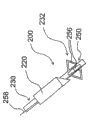
图4显示按照本发明的血管外脉冲电场装置200的一个实施方案,所述装置包括一个或多个设置成将脉冲电场输送到肾神经纤维以获得肾神经调节作用的电极。装置200包括具有探针210的腹腔镜或经皮PEF系统,例如在CT或射线成像的引导下,设置所述探针210插入到沿着肾动脉或静脉或肾门和/或Gerota筋膜内的肾神经供应的轨道的邻近。探针210的近端部件通常具有将所述探针偶联到脉冲发生器100上的电连接器,并且远端部件具有至少一个电极212。FIG. 4 shows an embodiment of an extravascular pulsed
脉冲电场发生器100位于患者外部,并且电极212通过探针210和电线211与发生器电偶联。发生器100,以及本文所述的电极实施方案中的任何一个可以与下文所述的本发明的实施方案一起用来输送具有理想的场参数的PEF。应该理解,即使发生器没有由每一实施方案明确显示或者描述,下文所述的实施方案的电极也可以与发生器电连接。The pulsed
电极212可以是单个电极,共同但是分节的电极,或者共同而连续的电极。例如,共同但是分节的电极可以通过提供适合电极的开槽导管而形成,或者通过电连接一系列的单个电极而形成。单个电极或电极212组可以设置成提供两极信号。电极212可以动态指定成促进任何电极之间和/或任何电极与外部接地垫板(ground pad)214之间的单极和/或两极能量输送。接地垫板214可以,例如,在外部附着到患者皮肤上,例如,患者的腿或胁腹上。Electrodes 212 may be a single electrode, common but segmented electrodes, or common and continuous electrodes. For example, common but segmented electrodes may be formed by providing slotted conduits that fit the electrodes, or by electrically connecting a series of individual electrodes. A single electrode or group of electrodes 212 may be configured to provide a bipolar signal. Electrodes 212 may be dynamically assigned to facilitate unipolar and/or bipolar energy delivery between any electrodes and/or between any electrodes and an external ground pad 214 . The ground pad 214 may, for example, be externally attached to the patient's skin, eg, the patient's leg or flank.
如图4所示,电极212可以包括与分离的患者接地垫板214联合使用的单个电极,所述接地垫板214位于患者外部,并且偶联到发生器100上,用于单极应用。探针210任选地可以包括导电材料,所述导电材料在除了其远端末端之外的区域是绝缘的,因而形成远端末端电极212。备选地,电极212可以,例如,通过探针210的内腔输送。探针210和电极212可以是临床上用于脉冲RF神经阻滞的标准针头或套管针型,诸如由Colorado,Boulder的Valleylab(Tyco Healthcare Group LP的部门)出售的那些。备选地,装置200可以包括柔软灵活的和/或定制的探针,以用于本文所述的肾应用。As shown in FIG. 4, electrodes 212 may comprise a single electrode used in conjunction with a separate patient grounding pad 214 external to the patient and coupled to
在图4中,经皮探针210已经通过经皮通路位点P推进到肾动脉RA内的邻近。一旦准确地定位,脉冲电场治疗可以穿过电极212和接地垫板214而应用到靶点神经纤维。治疗后,装置200可以从患者移除以终止程序。In FIG. 4 , percutaneous probe 210 has been advanced through percutaneous access site P into proximity within renal artery RA. Once properly positioned, pulsed electric field therapy may be applied through the electrodes 212 and ground pad 214 to the targeted nerve fibers. After treatment,
预计这样的治疗将在持续几个月的时间内减轻CHF、高血压、肾病和/或其它心-肾疾病的临床症状,可能高达6个月或更久。这一时间阶段可以足以允许机体治愈,例如,这一阶段可以减少急性心肌梗塞之后CHF发作的危险,因而减少对于后续重新治疗的需求。备选地,当症状复发时,或者在规律预定的时间间隔,患者可以回访医生实行重复的治疗。Such treatment is expected to reduce the clinical symptoms of CHF, hypertension, renal disease and/or other cardio-renal disease over a period of several months, possibly up to 6 months or more. This period of time may be sufficient to allow the body to heal, eg, this period may reduce the risk of CHF episodes following an acute myocardial infarction, thereby reducing the need for subsequent retreatment. Alternatively, when symptoms recur, or at regular predetermined intervals, the patient may return to the physician for repeated treatments.
对于重复治疗的需求任选地可以通过监测生理学参数而预测,例如,通过监测作为增加的交感神经活性指征的特异的神经激素(血浆肾素水平,等)而进行预测。备选地,可以进行已知增加交感神经活性的刺激性手段,诸如头在水外沉浸测试,以确定对重复治疗的需求。The need for repeat therapy can optionally be predicted by monitoring physiological parameters, for example, specific neurohormones (plasma renin levels, etc.) that are indicative of increased sympathetic activity. Alternatively, stimuli known to increase sympathetic activity, such as head submersion tests, may be performed to determine the need for repeat therapy.
在一些实施方案中,装置200可以包括具有导入器的探针,其可扩张的远端部件具有一个或多个电极。在插入到靶点神经纤维的邻近后,远端部件可以开放或者扩张成膨胀的构型。在一个实施方案中,这种膨胀的构型使用PEF治疗的单次应用沿着肾动脉和/或静脉的轮廓治疗许多神经纤维。例如,在膨胀的构型中,远端部件可以部分或完全环绕肾动脉和/或静脉。在另一个实施方案中,膨胀的构型可以促进机械解剖,例如,扩张Gerota筋膜并且产生放置电极和/或用于输送PEF治疗的工作空间。远端部件任选地可以不依赖于探针或导入器而移动(translated)。In some embodiments,
当用作电极时,远端部件可以,例如,从置于治疗区域附近的导入器延伸出来。传导的远端部件可以从鞘推出,直到接触需要数量的肾神经组织,然后可以通过远端部件电极输送PEF治疗。备选地,传导的远端部件可以允许重新形成或者扩张成为一个或多个环的盘旋、随机的空间占据形状、或者另外适宜的构型。网、带或者导电凝胶或液体可以以相似的方式应用。When used as an electrode, the distal member may, for example, extend from an introducer placed adjacent the treatment area. The conductive distal part can be pushed out of the sheath until it contacts the desired amount of renal nerve tissue, and then the PEF therapy can be delivered through the distal part electrodes. Alternatively, the conductive distal member may be allowed to reform or expand into a coil of one or more loops, a random space-occupying shape, or another suitable configuration. Mesh, tape or conductive gel or liquid can be applied in a similar manner.
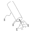
图5图示装置200的另一个实施方案,所述装置包括可扩张的远端部件。在图5中,装置200包括导入探针220和具有可扩张的远端部件232的电极元件230。探针220可以,例如,包括标准针头或套管针。电极元件230与发生器100邻近偶联,并且设置成通过探针220推进。电极元件的远端部件232可以在探针220内部以封闭的或收缩的构型输送到治疗位点,然后在治疗位点或其附近打开或扩张成治疗构型。例如,远端部件232可以通过将部件232推出探针220而扩张,和/或相对于远端部件缩回探针而扩张。图5所示的远端部件232的实施方案包括篮或杯形元件,其以散开的构型234用于输送治疗。远端部件232优选地自主扩张成治疗构型。装置200还可以包括与远端部件232偶联的一个或多个电极233。FIG. 5 illustrates another embodiment of a
如图5所示,远端部件232以散开的构型234部分或完全地环绕或围绕肾动脉RA。通过电极元件230到电极233以两极或单极方式输送的PEF治疗可以获得比从只沿着动脉的一侧的电极或者在沿着动脉的单点的电极输送的PEF治疗更彻底或更完全的肾神经调节作用。电极元件230任选地可以从探针220电分离,以致所述探针和电极233形成两极系统的两个部件,其中电极220是返回电极。As shown in FIG. 5 ,
参考图6,远端部件232备选地可以在治疗构型中包括盘旋元件236。远端部件可以,例如,预先形成盘旋构型。盘旋可以通过许多不同机制伸直(例如,放置在探针220内,激活部件232使之在直的和盘旋之间的牵引线,开关记忆材料,等),以插入到邻近,例如,肾血管系统。一旦接近靶点血管,盘旋可以被操纵成或者被允许重新形成,以更充分地环绕血管,因而促进使用PEF治疗的单一应用治疗更多的神经纤维。Referring to FIG. 6 , the
远端部件232的盘旋(spiral)或螺旋(helical)元件236设置成与血管壁并列,并且使得电极233很接近肾神经结构。螺旋的节距可以变化,以提供更长的治疗区域,或者将相邻治疗区域的圆周重叠减到最小,例如,以减少形成狭窄的危险。例如,这种节距变化可以通过下列各项而实现:(a)热调整,(b)将多个不同节距的部件组合形成部件232,(c)通过应用内部牵引线调整部件232的节距,(d)调节插入到部件内的轴,(e)定型置于导管上的鞘,或者(f)通过任何其它适当的方式在原位或者在引入到机体之前而改变所述节距。The spiral or
如同先前的实施方案,沿着远端部件232的长边的电极233可以是单个电极、共同但是分节的电极、或者共同而连续的电极。例如,共同连续电极可以包括在远端部件232的螺旋部分内部形成或者置于其上的导电线圈。单个电极或电极233的组可以设置成提供两极信号,或者电极的任何构型可以一起在公共电势处使用,与独立的外部患者接地组合,用于单极应用。电极233可以动态地指定成促进任何电极之间和/或任何电极和外部接地之间的单极和/或两极能量输送。远端部件232任选地可以在远离肾动脉朝向的侧面是绝缘的,以致设置成朝向肾动脉的所述部件侧面的至少部分暴露形成电极233。As with the previous embodiments, the
电极元件230的远端部件232可以在探针220内部以薄断面输送构型输送到肾动脉RA的邻近。一旦放置到所述动脉的邻近,远端部件232可以自主扩张或者可以主动扩张,例如,通过牵引线或者气球,扩张成围绕动脉壁的盘旋构型236。远端部件可以,例如,环绕血管导向,例如,通过操作和钝器解剖进行,并且激活以呈现盘旋构型236的紧密-节距线圈。备选地或另外地,远端部件可以相对于探针220推进,并且通过其柔软性(predisposition)迂回环绕动脉,以呈现盘旋构型。将远端部件置于Gerota筋膜内可以促使将远端部件232环绕动脉放置。The
一旦正确地定位,那么脉冲电场可以由PEF发生器100产生,通过电极元件230传递到电极233,并且通过电极输送到位于动脉周围的肾神经。在许多应用中,电极这样排列,以致脉冲电场沿着动脉的纵向维度排列,以调节沿着肾神经的神经活性(例如,去除神经作用)。例如,这可以通过神经细胞中不可逆的电穿孔作用、电融合和/或诱导细胞凋亡而实现。Once properly positioned, pulsed electric fields can be generated by the
参考图7,描述了用于肾神经调节的另一种经皮或腹腔镜方法和装置。在图7中,装置200的电极元件230的远端部件232包括具有环形或套形(cuff)构型238的电极233。环形电极可以部分地环绕肾动脉RA,如图所示。电极233任选地可以包括可伸缩的插头239,用于关闭所述环,以便当所述电极围绕动脉放置时,更充分地或完全地环绕所述动脉。PEF治疗可以通过电极输送以实现肾神经调节。作为腹腔镜放置的备选方案,环形电极238任选地可以手术放入。Referring to Figure 7, another percutaneous or laparoscopic method and device for renal neuromodulation is described. In FIG. 7 , the
图8图示用于肾神经调节的另一种经皮或腹腔镜的方法和装置,所述装置包括设置成放置在肾门附近的分散电极。如图8所示,电极元件230的远端部件232可以包括具有多个指状物的扇形部分240,在经皮引入到治疗位点和/或从治疗位点缩回过程中,扇形部分240可以折叠或约束在探针220内。一个或多个电极233可以沿着远端部件的指状物放置。一旦处于肾血管系统区域和/或肾门H区域,扇形可以展开,或者探针220可以缩回,以展开远端部件232。例如,指状物伸展出来以覆盖沿着血管系统和肾门的更大的治疗区域,这促进更多的靶点神经纤维的治疗,和/或产生后续引入电极233的工作空间。Fig. 8 illustrates another percutaneous or laparoscopic method and device for renal neuromodulation, the device comprising dispersive electrodes configured to be placed near the hilum of the kidney. As shown in FIG. 8 , the
在图8中,探针220的远端区域置于肾门H的邻近,并且扇形远端部件232已经扩张成展开的构型。然后,PEF治疗可以通过电极233输送到所述区域的神经纤维以进行肾神经调节。In FIG. 8, the distal region of
参考图9,远端部件232备选地可以包括具有一股或多股电极233的簇状元件242。远端部件232可以在探针220内部置于肾门H的邻近,然后簇状元件242可以张开形成空间占据构型。然后,电极233可以将PEF治疗输送到肾神经。Referring to FIG. 9 , the
参考图10,探针220任选地可以穿透环绕肾脏K和/或肾动脉RA的筋膜F(例如,Gerota筋膜)。远端部件232可以通过探针220推进到筋膜和肾结构之间,诸如肾门H和动脉RA之间。这可以将电极233置于具有靶点肾神经结构的邻近。例如,当远端部件232包括图8的扇形部分240或图9的簇状元件242时,远端部件在筋膜F内的扩张可以将电极233置于具有多个靶点肾神经结构的邻近,和/或可以产生用于输送一个或多个电极233或输送传导凝胶或液体等的工作空间。Referring to FIG. 10 ,
参考图11A和11B,描述了在筋膜F内部机械锚定探针220、电极元件230的远端部件232、和/或电极233的方法和装置。图11A和11B图示与电极元件230的远端部件232组合的机械锚定元件250,但是由于装置200不必包括所述锚定元件250,所以这决不应该解释成限制。在具有锚定元件的实施方案中,远端部件232可以是可扩张的或不可扩张的。Referring to FIGS. 11A and 11B , methods and apparatus for mechanically anchoring
在图11A的实施方案中,远端部件232包括具有远端部件周围的轴环(collar)252的锚定元件250。锚定元件的自主扩张的电线环254从轴环扩展。当远端部件位于探针220内时,自主扩张的电线环254可以针对远端部件232的轴折叠(如图11A中点线图像所示)。探针220可以穿透治疗位点附件的筋膜,因而将探针的末端置于筋膜内。然后,探针可以相对于电极元件230缩回(和/或电极元件可以相对于探针推进),以将远端部件232置于探针的远端。环254以某种放置自主扩张,其将远端部件232机械性地锚定在筋膜内。备选地,锚定元件250可以主动扩张,例如,以机械方式。In the embodiment of FIG. 11A , the
环254任选地可以由一种弹性聚合物诸如硅氧烷、CFlex、氨基甲酸乙酯等覆盖,以致锚定元件250至少部分地将进入筋膜F的入口密封。这可以促进流体通过探针220或电极元件230的直接灌注,没有渗漏或者具有减少的渗漏。另外地或备选地,当电极元件230设置成更长期的植入时,锚定元件250可以以多孔材料诸如聚酯织物或网覆盖,其允许或促进组织向内生长。组织向内生长可以增强由元件250提供的锚定,以保持远端部件232和/或电极233的位置。组织向内生长还可以增强在筋膜进口的密封。
在图11B的实施方案中,远端部件232沿纵向方向切断,以产生环绕导管的一系列瓣片256,其形成备选的锚定元件250。牵引线258可以,例如,沿着电极元件230的外部延伸,或者可以置于电极元件的内腔内部,并且偶联到瓣片256远端的远端部件232。一旦远端部件232置于筋膜F内,牵引线258就移动到邻近,以延伸瓣片256,并且将远端部件锚定在筋膜内。备选地,结合电线、篮、网、织物等的其它可扩张的部分可以机械性地扩张以提供锚定。In the embodiment of FIG. 11B , the
当锚定元件250扩张时,灌输剂任选地可以通过裂缝256灌输。此外,如图11A的实施方案,图11B的锚定元件250任选地可以由弹性聚合物遮盖物覆盖,以产生密封进入筋膜的进口的衬垫。在这样的构型中,灌输孔可以在锚定元件的远端提供。备选地,锚定元件250的裂缝部分的近端部分可以由弹性聚合物覆盖,而远端部分保持不被覆盖,例如,以促进通过裂缝256进行灌注。在另一个实施方案中,图11B的锚定元件可以把扩多孔材料,以促进组织向内生长,如先前所述那样。关于弹性聚合物,所述多孔材料任选地可以只覆盖锚定元件的一部分,例如以促进组织向内生长和灌注。An infusion agent can optionally be infused through
图12显示用于肾神经调节的方法和装置,其中电极在血管系统和环绕的筋膜之间的环形空间内沿着患者肾血管系统放置。通过使用如计算机断层摄影术(“CT”)引导将针导入筋膜F内,电极可以位于肾动脉和/或肾静脉的邻近。针可以包括导入探针220,或者在将针置于筋膜内部后,探针可以推出并且更换针头。Figure 12 shows a method and device for renal neuromodulation in which electrodes are placed along a patient's renal vasculature within the annular space between the vasculature and surrounding fascia. By introducing the needle into the fascia F using, for example, computed tomography ("CT") guidance, the electrodes can be positioned adjacent to the renal artery and/or renal vein. The needle can include an
当探针220位于Gerota筋膜内部时,电极元件230通过所述探针输送到肾血管系统(例如,肾动脉RA)的邻近。电极元件任选地可以沿着通向患者大动脉的动脉长度推进,以在当推进电极元件时,钝端解剖出用于电极元件的空间。电极元件230可以包括导管,并且偶联到电极元件230上的电极233可以将PEF治疗或其它类型的治疗输送到沿着肾动脉定位的肾神经结构。当需要时,可以提供两极或单极电极。When the
参考图13A-C,描述了电极元件230的电极233和远端部件232的许多其它的实施方案。在图13A中,电极233包括围绕远端部件232放置的一对两极电极线圈。在图13B中,电极233包括具有与轮廓相合的金属盘的一对两极电极,其位于朝向肾动脉RA的远端部件232的侧面,以朝向靶点肾神经结构。预计这优先引导在电极233之间输送的PEF治疗通向肾动脉。如图13C所示,远端部件232和/或电极233可以包括内弧形面,以致更多的电极表面积与肾动脉壁并列。当通过电极233输送的脉冲电场足够强,适当地引导和/或在靶点神经结构的足够进的近端时,预计没有完全环绕肾动脉,电极就可以实现理想水平的肾神经调节。Referring to Figures 13A-C, a number of other embodiments of
图14A-C显示将电极沿着患者肾动脉放置的另一种方法和装置。除了探针220和电极元件230外,图14A-C的装置200包括导管300。电极元件230置于导管300内部,并且任选地可以包括导管的防止创伤的末端。如在图14A中所示,导管300可以通过探针220在筋膜F和如RA所示的肾血管系统之间的环形空间内推进。导管和/或探针任选地可以通过导线推进。各种试剂可以导管灌注,以产生推进导管的工作空间,和/或促进电极233的放置。14A-C show another method and apparatus for placing electrodes along a patient's renal artery. In addition to
一旦按照需要置于治疗位点,导管可以相对于电极元件缩回,以暴露沿着电极元件的远端部分232的电极233,如图14B。图14A-C中电极233可以包括一对两极可扩张的电极,其可以折叠在导管300内部输送。电极可以,例如,由自主扩张的材料诸如弹簧钢或镍钛诺制备。尽管电极233示例性地在共同电极元件230上,但是应该理解,每个具有一个或多个电极233的多电极元件230可以通过导管300输送,并且按照需要沿着肾血管系统放置。Once positioned at the treatment site as desired, the catheter can be retracted relative to the electrode element to expose the
在图14B的扩张构型中,电极233至少部分地围绕或环绕肾动脉RA。预计在PEF治疗过程中至少部分环绕肾动脉将增强肾神经调节作用或去除神经作用的效果。电极可以用来输送PEF治疗和/或刺激生理反应,以监测或者刺激神经调节作用的程度,以及用来应用能量破坏、调节或阻滞神经功能。各种试剂可以在能量输送之前、之中或之后灌注到电极233的近端,例如,以辅助传导(例如,盐水和高渗盐水),提高电穿孔作用(例如,加热的溶液),或者提供对非靶点细胞的保护(例如,冷却的溶液或Poloxamer-188)。In the expanded configuration of Figure 14B,
电极233可以,例如,包括线圈、电线、带、聚合物、织物或合成物。聚合物可以与传导性材料组合应用,以引导脉冲电场进入和/或沿着靶点组织,同时绝缘周围组织。参考图14C,电极元件230的远端部件232可以包括绝缘物I,其沿着电极233的内表面局部移除或省略,其中所示电极朝向或者接触肾血管系统。The
现在参考图15A-B,描述了图14A-C的装置和方法的另一种实施方案。如在图15A中所示,电极233可以包括沿着肾血管系统延伸的波浪形或正弦曲线形构型。电极233的正弦曲线构型可以比图14A-C的电极233提供更大的沿着血管壁的接触面积,同时仍然促使将电极元件230覆盖在导管300之内,用于输送和/或收回。电极233可以包括设置成单极能量输送的一元电极,或者电极元件230的远端部件232可以包括绝缘物,其局部移除或省略以暴露电极233。备选地,如图15B所示,电极可以包括附着到波浪形远端部件232上的不连续的线圈或者其它传导性部件。电极233可以以任何组合给予电压,以形成两极电极对。Referring now to Figures 15A-B, another embodiment of the apparatus and method of Figures 14A-C is described. As shown in FIG. 15A,
图16是图示图14A-C的方法和装置的另一种实施方案。在图16中,导管300包括多个内腔304,通过所述内腔电极元件230可以推进,并且电极233可以折叠用于输送。当位于治疗位点时,电极元件230可以相对于导管300推进,和/或导管300可以相对于电极元件收回,以致电极233扩张成图16的构型,至少部分环绕肾血管系统。在图16中,装置200示例性地包括两个电极元件230,每个具有可扩张的电极233。在PEF治疗过程中,两个电极233可以用作两极电极对。当需要时,电极元件230可以独立地平移,以致电极233之间的分离距离可以动态改变。Figure 16 is a diagram illustrating another embodiment of the method and apparatus of Figures 14A-C. In Figure 16,
图17A-B显示图14A-C的方法和装置的另一种实施方案。在图17A中,电极233包括可以卷成卷轴用于在导管300内部薄断面输送的面板。如图17B所示,导管可以相对于电极收回,和/或电极可以相对于导管推进,以致所示面板展开或打开,优选地以自主扩张的形式,以部分地或完全地环绕肾动脉RA。PEF治疗可以通过电极233以单极形式输送,或者所述电极可以分节,以促进两极应用。备选地,可以将第二电极输送到电极233的近端,用于两极PEF治疗。Figures 17A-B show another embodiment of the method and apparatus of Figures 14A-C. In FIG. 17A ,
图5-17B的电极实施方案212或233中的任一种可以设置成用于单一PEF治疗时间,或者可以设置成用于应用PEF治疗时间之后的植入。在植入实施方案中,导线可以,例如,从电极212或233延伸通过皮肤可控的皮下元件或者延伸到植入控制器。Either of the
参考图18,当电极233设置成用于植入时,电极元件230的远端部件232可以,例如,在治疗位点分离,以致电极233植入到肾动脉RA和筋膜F之间的环形空间中,同时将电极元件230的近端部分从患者移除。如图18所示,电极元件230可以包括导线260,用于通到皮下元件或者通到植入控制器。电极元件还包括恰好置于远端部件232近端的分离机械装置270,用于在治疗位点分离远端部件。远端部件232任选地可以包括设置成在电极233的附近促进组织向内生长的元件。Referring to FIG. 18, when the
参考图19A-20,描述了部分和完全植入的PEF系统。图19A-B图示部分植入系统,其具有通过或者穿过患者皮肤直接或间接地与皮下元件400连接的脉冲电场发生器100。例如,皮下元件400可以置于背部,例如,在患者的下背部。在图19A-B中,皮下元件400附着到导线260上,并且导线260电偶联到位于肾动脉、肾静脉、肾门、Gerota筋膜或其它适宜结构的近端的植入电极233上。电极233可以位于两侧,即,在右侧和左侧肾血管系统的近端,但是备选地可以位于单侧。此外,多个电极可以位于两肾之一或两者的近端,用于两极PEF治疗,或者可以提供单极电极,并与返回电极组合应用,如外部接地垫板214或与皮下元件400结合的返回电极。Referring to Figures 19A-20, partially and fully implanted PEF systems are depicted. 19A-B illustrate a portion of an implant system having a pulsed
如图19A所示,皮下元件400可以包括具有电触点403的皮下端口402,电触点403包括一个或多个连接或对接电,用于将电触点偶联到发生器100上。在将PEF传递通过患者皮肤的直接方法中,将透皮针、套管针、探针或与发生器100电偶联的其它元件410穿过患者皮肤,并且易松开地偶联到接头403上。透皮元件410将PEF治疗从发生器100穿过或通过患者皮肤传导到皮下接头403和电极233,以调节对肾功能起作用的神经纤维。植入系统还可以结合灌输内腔,以允许药物从皮下端口402引入到治疗区域。As shown in FIG. 19A , the
除了将PEF信号穿过患者皮肤输送的直接方法以外,诸如通过透皮元件410进行输送的直接方法,可以备选地应用间接方法,诸如透皮能量转移(“TET”)系统。In addition to direct methods of delivering the PEF signal across the patient's skin, such as through
[]TET系统临床上用来为可再充电的植入刺激或起搏装置、左心室辅助装置等中的电池充电。在图19B的TET实施方案中,皮下元件400包括皮下接收元件404,并且外部TET发射元件420与发生器100偶联。PEF信号可以通过皮肤从外部发射元件420遥感性地传递到皮下接收元件404。被动导线260连接皮下接收元件和神经电极233,并且可以将信号传导到神经进行治疗。[ ] TET systems are used clinically to charge batteries in rechargeable implanted stimulation or pacing devices, left ventricular assist devices, etc. In the TET embodiment of FIG. 19B , the
参考图20,描述了完全植入的PEF系统。在图20中,皮下接收元件404偶联到电容器或其它能量存储元件430诸如电池上,其又偶联到植入控制器440上,植入控制器440通过导线260与电极233连接。外部发射元件420与外部充电器和编程器450偶联,用于将能量传递到植入系统,和/或对植入系统编程。充电器和编程器450不必以PEF形式提供能量,用于穿过患者皮肤从外部元件420传递到接收元件404。相反,控制器440可以从存储在存储元件430内部的能量产生PEF波形。控制器任选地可以作为返回电极,用于单极类型的PEF治疗。Referring to Figure 20, a fully implanted PEF system is depicted. In FIG. 20 , the
图20的实施方案示例性地是可充电的和/或可重新编程的。然而,应该理解,完全植入的PEF系统备选地可以既不是可充电的也不是可编程的。相反,所示系统可以通过存储元件430供电,如果有必要,其可以设置成在几个月或几年的时间后手术更换,在所述时间后存储在存储元件中的能量已经耗尽。The embodiment of Figure 20 is illustratively rechargeable and/or reprogrammable. However, it should be understood that a fully implanted PEF system may alternatively be neither rechargeable nor programmable. Instead, the system shown can be powered by a
当使用经皮或植入PEF系统时,对于重复治疗的需要,起始治疗的位置和/或治疗效果,任选地可以由所示系统确定。例如,植入系统周期性地可以对肾神经应用更低频率的刺激信号;当所示神经恢复基线功能时,检测信号将由患者感觉到,并且所示系统镜应用另一PEF治疗过程。这种重复的治疗任选地可以是由患者起始的:当患者感觉到检测信号时,患者将通过电遥感器、磁性开关和其它方法操作植入系统,以应用需要的治疗性PEF。When using a percutaneous or implanted PEF system, the need for repeat treatments, the location to initiate the treatment and/or the effect of the treatment, optionally can be determined by the system shown. For example, the implanted system may periodically apply lower frequency stimulation signals to the renal nerves; when the nerves return to baseline function, a detection signal will be felt by the patient and the system mirrors the application of another course of PEF therapy. This repeated treatment can optionally be patient-initiated: when the patient senses a detection signal, the patient will operate the implant system via electrical telemetry, magnetic switches, and other methods to apply the desired therapeutic PEF.
作为备选方案或者除了刺激疼痛反应外,可以监测已知受肾神经刺激影响的生理参数的反应。这样的参数包括,例如,肾素水平、钠水平、肾血流和血压。当使用刺激来刺激去除神经作用和监测治疗效果时,针对刺激的已知生理反应应该不再应答这样的刺激而发生。Alternatively or in addition to stimulation of pain responses, responses to physiological parameters known to be affected by renal nerve stimulation can be monitored. Such parameters include, for example, renin levels, sodium levels, renal blood flow, and blood pressure. When stimuli are used to stimulate denervation and to monitor therapeutic effects, known physiological responses to stimuli should no longer occur in response to such stimuli.
传出神经刺激波形可以,例如,包括大约1-10Hz的频率,而传入神经刺激波形可以,例如,包括高达大约50Hz的频率。波形振幅可以,例如,在大约50V范围,而脉冲持续时间可以,例如,在高达大约20毫秒的范围。尽管已经描述了刺激波形的示例性参数,但是应该理解,当需要时可以应用任何备选的参数。Efferent neural stimulation waveforms can, for example, include frequencies of about 1-10 Hz, while afferent neural stimulation waveforms can, for example, include frequencies up to about 50 Hz. The waveform amplitude can, for example, be in the range of about 50V, and the pulse duration can be, for example, in the range of up to about 20 milliseconds. Although exemplary parameters of the stimulation waveform have been described, it should be understood that any alternative parameters may be applied as desired.
在先前所示的本发明的任何变体中用于输送PEFs的电极还可以用来将刺激波形输送到肾血管系统。备选地,变体可以包括设置用于刺激的独立的电极。作为另一种备选方案,可以提供分离的刺激装置。Electrodes used to deliver PEFs in any of the previously shown variations of the invention may also be used to deliver stimulation waveforms to the renal vasculature. Alternatively, variations may include separate electrodes provided for stimulation. As another alternative, a separate stimulation device may be provided.
如提及的那样,应用刺激确定肾神经的一种方法是刺激所述神经,以便如果肾神经没有被去除神经或调节,那么肾血流受到影响-或者将受到影响。由于刺激作用减少肾血流,这一反应可以由去除神经作用而被减弱或消除。因此,在神经调节之前的刺激预计将减少血流,而当使用如神经调节之前相似的刺激参数和位置时,神经调节之后的刺激预计将不会将血液流动减少到相同的程度。这一现象可以用来定量肾神经调节的程度。As mentioned, one way to apply stimulation to identify renal nerves is to stimulate the nerves so that if the renal nerves are not denervated or modulated, renal blood flow is affected - or will be. Since stimulation reduces renal blood flow, this response can be attenuated or eliminated by denervation. Thus, stimulation prior to neuromodulation would be expected to reduce blood flow, while stimulation following neuromodulation would not be expected to reduce blood flow to the same extent when using similar stimulation parameters and locations as prior to neuromodulation. This phenomenon can be used to quantify the degree of renal neuromodulation.
本发明的实施方案可以包括监测肾血液流动的元件,或者监测已知受肾刺激影响的的任何其它肾生理参数的元件。肾血流任选地可以通过皮肤显现(例如,使用超声传感器)。电穿孔作用的程度另外或者备选地可以应用电阻抗断层摄影术(“EIT”)或其它电阻抗检测法诸如电阻抗指数直接监测。Embodiments of the invention may include elements to monitor renal blood flow, or any other renal physiological parameter known to be affected by renal stimulation. Renal blood flow optionally can be visualized through the skin (eg, using an ultrasound transducer). The extent of electroporation may additionally or alternatively be monitored directly using electrical impedance tomography ("EIT") or other electrical impedance detection methods such as electrical impedance index.
除了或者作为刺激的备选方案,检验患者临床状态的检测的其它监测方法可以用来确定对于重复治疗的需要。这些监测方法可以是完全或部分植入的,或者它们可以是用植入元件遥感通信的外部检测。例如,本领域植入的这种类型的植入压力传感器(由Atlanta,GA的CardioMEMS开发的传感器)可以检测右侧动脉压力。增加的右侧动脉压力是流体超负荷和不当CHF处理的指征。如果右侧动脉压力的增加由传感器检测到,那么信号可以发送到控制器440,并且将输送另一次PEF治疗。类似地,在其它疾病病症中,诸如高血压的治疗中,动脉压力可以被监测和/或用作控制信号。备选地,可以应用心脏输出量的侵入性的或非侵入性的检测。非侵入性的检测包括,例如,胸部电生物阻抗。In addition to or as an alternative to stimulation, other monitoring methods to verify detection of the patient's clinical status may be used to determine the need for repeat therapy. These monitoring methods may be fully or partially implanted, or they may be external detection with telemetric communication of implanted components. For example, an implanted pressure sensor of the type implanted in the art (a sensor developed by CardioMEMS of Atlanta, GA) can detect right side arterial pressure. Increased right arterial pressure is an indication of fluid overload and inappropriate CHF management. If an increase in right arterial pressure is detected by the sensor, a signal can be sent to the
在另一个实施方案中,重量波动与体脂肪百分数相关,用来确定对重复治疗的需要。已知增加的患者重量,特别是不存在体脂肪百分数增加时,是增加体积的超负荷的指征。因此,可以监测患者重量和体脂肪百分数,例如,通过特别设计的比较重量增加和体脂肪百分数的衡量法进行。如果确定体重增加是由于流体超负荷,那么衡量法或其它监测元件可以将信号传导到控制器440,例如,通过遥感传导,以应用另一次PEF治疗。In another embodiment, weight fluctuations are correlated with percent body fat to determine the need for repeat therapy. Increased patient weight, especially in the absence of an increase in body fat percentage, is known to be an indication of volume overload. Thus, patient weight and percent body fat can be monitored, for example, by specially designed measures comparing weight gain and percent body fat. If it is determined that the weight gain is due to fluid overload, the gauge or other monitoring element may transmit a signal to the
当使用部分或完全植入的PEF系统时,热电偶、其它温度或阻抗监测元件、或其它传感器,可以结合到皮下元件400上。PEF系统的外部元件可以设计连接和/或接收来自传感器元件的信息。例如,在一个实施方案中,透皮元件410连接端口402的皮下电触点403,并且可以将刺激信号输送到询问(interrogate)靶点神经信号,以确定对治疗的需要或参数,以及确定神经和神经电极的阻抗。另外,与传感器导线紧密配合的分离的连接器可以通过或者沿着透皮元件410延伸,以接触相应的皮下传感器导线。Thermocouples, other temperature or impedance monitoring elements, or other sensors, may be incorporated into the
备选地,皮下电触点403可以具有彼此相邻放置但是彼此电分离的多个靶点区域。从外部控制器如外部发生器100伸出的导线将分裂成一些独立的透皮针头,或者偶联在更大的探针内的独立针头点,其插入皮肤,独立地接触它们各自的皮下靶点区域。例如,能量输送、阻抗检测、询问(interrogative)刺激和温度每一种可以具有在皮下系统中排列的它们各自的靶点区域。在外部控制器内的诊断电极任选地可以设计成保证正确的针头与每个相应的皮下靶点区域接触。Alternatively, the subcutaneous
可以将元件结合在PEF系统的植入元件内,以促进锚定和/或组织向内生长。例如,织物或植入材料,诸如达可纶或ePTFE,可以结合到皮下元件400的设计中,以促进向内生长到所述装置的区域,所述织物或植入材料将帮助所述元件锚定在适当位置,同时任选地抵制组织向不希望区域诸如沿着电极233的向内生长。类似地,当需要时,涂层、材料处理、药物包被或药物洗脱可以单独地应用或组合应用,以辅助或阻止组织向植入PEF系统的各元件内的生长。Elements may be incorporated within implant elements of the PEF system to promote anchoring and/or tissue ingrowth. For example, fabric or implant material, such as dacron or ePTFE, can be incorporated into the design of
本文所示的本发明任何实施方案任选地可以设置成在能量应用之前、之中或之后将试剂灌注到治疗区域,例如,以产生促使电极放置的工作空间,增强或改进应用能量的神经破坏或神经调节作用,保护或临时转移非靶点细胞,和/或促进显现。灌输试剂的其它应用是显而易见的。如果需要,细胞对灌输细胞的吸收可以通过在灌输试剂的存在下起始细胞中的可逆电穿孔作用而增强。灌输剂(infusate)可以包括,例如,流体(例如,加热的或冷却的流体)、空气、CO2、盐水、肝素化的盐水、高渗盐水、造影剂、凝胶、导电材料、空间占据物质(气体、固体或液体)、保护剂诸如Poloxamer-188、抗增殖剂、或其它药物和/或药物输送成分。本发明的变体另外或者备选地可以设置成抽吸式的。Any of the embodiments of the invention shown herein can optionally be configured to infuse agents into the treatment area before, during, or after application of energy, for example, to create a working space that facilitates electrode placement, enhances or modifies nerve destruction of applied energy or neuromodulation, protection or temporary transfer of non-target cells, and/or promotion of visualization. Other applications for infusion reagents are apparent. If desired, cellular uptake into infused cells can be enhanced by initiating reversible electroporation in the cells in the presence of an infusion reagent. Infusates can include, for example, fluids (eg, heated or cooled fluids), air, CO2 , saline, heparinized saline, hypertonic saline, contrast media, gels, conductive materials, space occupying substances (gas, solid or liquid), protective agents such as Poloxamer-188, antiproliferative agents, or other drugs and/or drug delivery components. A variant of the invention can additionally or alternatively be provided as suction.
图21A和21B图示按照本发明的装置200的另一种实施方案。参考图21A,装置200包括探针220和接收在探针220内部的导管300。导管300包括锚定机制251,其具有第一环管280,相对于第一环管280位于远端的第二环管282,以及与第一和第二环管280和282连接的可扩张部分284。可扩张部分284可以是织物、网、纺织品或其它装置,当第一和第二环管280和282之间的距离减小时,其扩张。可扩张部分284可以包括聚酯、镍钛诺、埃尔吉洛伊非磁性合金、不锈钢、合成物和/或其它适宜的材料。环管280和282、和/或可扩张部分284,可以至少部分地覆盖在可扩张的聚合物中,以与患者形成密封。装置200还可以包括多个电极230,其位于可扩张部分284和/或第一或第二环管280或282。21A and 21B illustrate another embodiment of a
锚定机制250通过将环管280和282的至少一个环管向另一个移动减少环管之间的距离而进行操纵。例如,第一环管280可以沿着导管300移动,并且第二环管282可以固定在导管300上。参考图21B,可扩张部分284可以通过在导管300上回拉而使得近端环管280与探针220的远端接合而扩张。由于导管300相对于探针220邻近收回到近端,所以探针220的远端推动第一环管280向第二环管282移动,以将图21A所示的折叠放置的锚定机制251移动到图21B所示的扩张构型。备选地,装置200可以包括致动器(actuator),其可以远端推进以推动第一环管280向第二环管282移动。例如,所示致动器可以是环绕导管300的同轴套管,其可以从探针220的近端操纵。The
图22A和22B图示按照本发明的装置200的另一个实施方案。在这一实施方案中,装置200包括探针220和导管300,导管300如上文参考图11A-B所述那样通过探针220移动。这一实施方案的装置200还包括锚定机制253,其具有第一环管281,沿着导管300相对于第一环管281位于远端的第二环管283,以及与第一和第二环管281和283附着的可扩张部分285。在一个实施方案中,第一环管281沿着导管300滑行移动,并且第二环管283固定在导管300上。备选地,第一环管281可以固定在导管300上,并且第二环管283可以沿着导管300移动。可扩张部分285是自主收缩性部分,其主动伸缩成折叠构型以包含在探针220内部,用于输送到患者的理想的治疗位点。图22A图示可扩张部分285伸展成拉长状态,以约束在探针220内部。参考图22B,通过将探针220相对于导管300向近端移动和/或将导管300相对于探针220向远端移动直到可扩张部分285在探针220之外,而使装置200展开。一旦可扩张部分285在探针220之外,可扩张部分285将可移动环管拉向固定环管,以允许可扩张部分285相对于导管轴300的半径向外扩张。22A and 22B illustrate another embodiment of a
可扩张部分285可以是由聚酯、不锈钢、合成物或具有足够的弹性以本能地移动成图22B所示的扩张构型的其它适宜材料形成的弹簧。备选地,可扩张部分可以由定型记忆金属诸如镍钛诺或埃尔吉洛伊非磁性合金制成,其在给定的温度从图22A所示的折叠构型移动成图22B所示的扩张构型。在另一个实施方案中,装置200还可以包括沿着可扩张部分放置的电极(未显示),用于将脉冲电场输送到肾神经或与肾脏/心脏活性相关的其它结构。The
图23图示将电极相对于肾结构放置以输送PEF进行神经调节的方法和装置的另一个实施方案。在这一实施方案中,所示装置包括第一经皮部分510,第二经皮部分520,电极组件530,和回收器(retriever)540。第一经皮部分510可以是第一套管针,通过其电极组件530被输送到肾动脉RA或其它肾结构,并且第二经皮部分520可以是第二套管针,通过其回收器540被输送到电极组件530的一般区域。在图23所示的实施方案中,电极组件530包括电极532,并且回收器540是一个圈套器(snare),其设置成捕获电极组件。在操作时,将第一经皮部分510插入到患者中,并且将电极组件530通过第一经皮部分510,直到电极532处于或者在相对于肾结构的理想位置的附近。第二经皮部分520也被插入到患者中,以致回收器540可以接合电极组件530。在向患者输送PEF和/或在输送PEF后移除电极组件530的过程中,回收器540可以用来将电极532固定在理想的位置。Figure 23 illustrates another embodiment of a method and device for placing electrodes relative to renal structures to deliver PEF for neuromodulation. In this embodiment, the device shown includes a first transcutaneous portion 510 , a second transcutaneous portion 520 , an electrode assembly 530 , and a retriever 540 . The first percutaneous portion 510 may be a first trocar, through which the electrode assembly 530 is delivered to the renal artery RA or other renal structure, and the second percutaneous portion 520 may be a second trocar, through which the retriever 540 Delivered to the general area of the electrode assembly 530. In the embodiment shown in FIG. 23, electrode assembly 530 includes electrode 532, and retriever 540 is a snare configured to capture the electrode assembly. In operation, the first percutaneous portion 510 is inserted into a patient, and the electrode assembly 530 is passed through the first percutaneous portion 510 until the electrodes 532 are at or near a desired location relative to the renal structures. The second percutaneous portion 520 is also inserted into the patient so that the retractor 540 can engage the electrode assembly 530 . Retractor 540 may be used to hold electrode 532 in a desired location during delivery of the PEF to the patient and/or removal of electrode assembly 530 after delivery of the PEF.
尽管上文描述了本发明的优选的示例性变体,但是对于本领域的技术人员显而易见地可以进行各种改变和改进,而不背离本发明。例如,尽管主要描述了变体与脉冲电场组合使用,但是应该理解,当需要时可以输送任何其它电场。在附上的权利要求中意欲覆盖落入本发明真正的精神和范围内的所有这样的改变和改进。While preferred exemplary variations of the invention have been described above, it will be apparent to those skilled in the art that various changes and modifications can be made without departing from the invention. For example, although the variants are primarily described for use in combination with pulsed electric fields, it should be understood that any other electric field could be delivered as desired. All such changes and modifications as fall within the true spirit and scope of this invention are intended to be covered in the appended claims.
Claims (65)
Applications Claiming Priority (9)
| Application Number | Priority Date | Filing Date | Title |
|---|---|---|---|
| US61625404P | 2004-10-05 | 2004-10-05 | |
| US60/616,254 | 2004-10-05 | ||
| US62479304P | 2004-11-02 | 2004-11-02 | |
| US60/624,793 | 2004-11-02 | ||
| US11/129,765 US7653438B2 (en) | 2002-04-08 | 2005-05-13 | Methods and apparatus for renal neuromodulation |
| US11/129,765 | 2005-05-13 | ||
| US11/189,563 US8145316B2 (en) | 2002-04-08 | 2005-07-25 | Methods and apparatus for renal neuromodulation |
| US11/189,563 | 2005-07-25 | ||
| PCT/US2005/035693 WO2006041847A1 (en) | 2002-04-08 | 2005-10-04 | Methods and apparatus for renal neuromodulation |
Publications (2)
| Publication Number | Publication Date |
|---|---|
| CN101600471A true CN101600471A (en) | 2009-12-09 |
| CN101600471B CN101600471B (en) | 2012-11-07 |
Family
ID=38121338
Family Applications (1)
| Application Number | Title | Priority Date | Filing Date |
|---|---|---|---|
| CN2005800416660A Expired - Fee Related CN101600471B (en) | 2004-10-05 | 2005-10-04 | Methods and apparatus for renal neuromodulation |
Country Status (2)
| Country | Link |
|---|---|
| EP (2) | EP1804905B1 (en) |
| CN (1) | CN101600471B (en) |
Cited By (13)
| Publication number | Priority date | Publication date | Assignee | Title |
|---|---|---|---|---|
| CN103027673A (en) * | 2011-09-30 | 2013-04-10 | 财团法人交大思源基金会 | Stimulation target range calibration method |
| CN103695311A (en) * | 2013-12-12 | 2014-04-02 | 新乡医学院 | Electrotransfection device and chick embryo optic tectum electrotransfection method using same |
| CN104254291A (en) * | 2011-12-23 | 2014-12-31 | 维西克斯血管公司 | Methods and apparatuses for remodeling tissue of or adjacent to a body passage |
| CN105530883A (en) * | 2013-07-11 | 2016-04-27 | 波士顿科学国际有限公司 | Multiple electrode conductive balloon |
| US9731132B2 (en) | 2002-04-08 | 2017-08-15 | Medtronic Ardian Luxembourg S.A.R.L. | Methods for renal neuromodulation |
| CN108066889A (en) * | 2016-11-10 | 2018-05-25 | 精能医学股份有限公司 | Electrical stimulation device, electrical stimulation signal generating method and computer readable storage medium |
| CN109843160A (en) * | 2016-07-29 | 2019-06-04 | 阿克松疗法公司 | Devices, systems, and methods for treating heart failure through cardiac nerve ablation |
| CN110251229A (en) * | 2013-06-06 | 2019-09-20 | 波士顿科学国际有限公司 | Device for transmitting energy and related method of use |
| US11376066B2 (en) | 2015-02-05 | 2022-07-05 | Axon Therapies, Inc. | Devices and methods for treatment of heart failure by splanchnic nerve ablation |
| US11413090B2 (en) | 2020-01-17 | 2022-08-16 | Axon Therapies, Inc. | Methods and devices for endovascular ablation of a splanchnic nerve |
| US11712296B2 (en) | 2017-12-17 | 2023-08-01 | Axon Therapies, Inc. | Methods and devices for endovascular ablation of a splanchnic nerve |
| US11751939B2 (en) | 2018-01-26 | 2023-09-12 | Axon Therapies, Inc. | Methods and devices for endovascular ablation of a splanchnic nerve |
| US11806073B2 (en) | 2019-06-20 | 2023-11-07 | Axon Therapies, Inc. | Methods and devices for endovascular ablation of a splanchnic nerve |
Families Citing this family (7)
| Publication number | Priority date | Publication date | Assignee | Title |
|---|---|---|---|---|
| WO2012027734A1 (en) * | 2010-08-27 | 2012-03-01 | G&L Consulting, Llc | System and method for electric diuresis |
| SG11201402610QA (en) | 2011-12-09 | 2014-10-30 | Metavention Inc | Therapeutic neuromodulation of the hepatic system |
| US12403305B2 (en) | 2016-06-27 | 2025-09-02 | Galvanize Therapeutics, Inc. | Immunostimulation in the treatment of viral infection |
| EP3474760B1 (en) | 2016-06-27 | 2023-01-11 | Galvanize Therapeutics, Inc. | Generator and a catheter with an electrode for treating a lung passageway |
| AU2017387791B2 (en) | 2016-12-27 | 2022-04-28 | Avent, Inc. | Article for treating diabetic peripheral neuropathy |
| WO2025176671A1 (en) * | 2024-02-20 | 2025-08-28 | Medtronic Ireland Manufacturing Unlimited Company | Assessing flow response using combined stimulation and ablation for guidance in renal denervation |
| WO2025176672A1 (en) * | 2024-02-20 | 2025-08-28 | Medtronic Ireland Manufacturing Unlimited Company | Renal vasoconstriction and renal blood flow response to stimulation as a guide to renal denervation |
Family Cites Families (6)
| Publication number | Priority date | Publication date | Assignee | Title |
|---|---|---|---|---|
| WO1992020291A1 (en) * | 1991-05-24 | 1992-11-26 | Applied Medical Resources, Inc. | Articulating tissue cutter assembly |
| US6219577B1 (en) * | 1998-04-14 | 2001-04-17 | Global Vascular Concepts, Inc. | Iontophoresis, electroporation and combination catheters for local drug delivery to arteries and other body tissues |
| US6845267B2 (en) * | 2000-09-28 | 2005-01-18 | Advanced Bionics Corporation | Systems and methods for modulation of circulatory perfusion by electrical and/or drug stimulation |
| JP4295627B2 (en) * | 2002-03-27 | 2009-07-15 | シーブイアールエックス, インコーポレイテッド | Electrode structure and its use in controlling circulatory system reflection |
| US7162303B2 (en) * | 2002-04-08 | 2007-01-09 | Ardian, Inc. | Renal nerve stimulation method and apparatus for treatment of patients |
| DE10257146A1 (en) * | 2002-12-06 | 2004-06-24 | Admedes Schuessler Gmbh | Thermal ablation probe for minimal invasive body tissue high frequency thermal treatment has temperature sensor on star shaped flexible arms with adjustable protrusion from insulating sheath |
-
2005
- 2005-10-04 EP EP05811851.4A patent/EP1804905B1/en not_active Expired - Lifetime
- 2005-10-04 CN CN2005800416660A patent/CN101600471B/en not_active Expired - Fee Related
- 2005-10-04 EP EP12168134.0A patent/EP2517755B1/en not_active Expired - Lifetime
Cited By (24)
| Publication number | Priority date | Publication date | Assignee | Title |
|---|---|---|---|---|
| US9731132B2 (en) | 2002-04-08 | 2017-08-15 | Medtronic Ardian Luxembourg S.A.R.L. | Methods for renal neuromodulation |
| US10272246B2 (en) | 2002-04-08 | 2019-04-30 | Medtronic Adrian Luxembourg S.a.r.l | Methods for extravascular renal neuromodulation |
| US9956410B2 (en) | 2002-04-08 | 2018-05-01 | Medtronic Ardian Luxembourg S.A.R.L. | Methods and apparatus for renal neuromodulation |
| CN103027673A (en) * | 2011-09-30 | 2013-04-10 | 财团法人交大思源基金会 | Stimulation target range calibration method |
| US9987085B2 (en) | 2011-12-23 | 2018-06-05 | Vessix Vascular, Inc. | Methods and apparatuses for remodeling tissue of or adjacent to a body passage |
| CN104254291B (en) * | 2011-12-23 | 2018-02-13 | 维西克斯血管公司 | Method and apparatus for reconstructing tissue of or adjacent to a body passage |
| US9901738B2 (en) | 2011-12-23 | 2018-02-27 | Vessix Vascular, Inc. | Methods and apparatuses for remodeling tissue of or adjacent to a body passage |
| US10039599B2 (en) | 2011-12-23 | 2018-08-07 | Vessix Vascular, Inc. | Methods and apparatuses for remodeling tissue of or adjacent to a body passage |
| CN104254291A (en) * | 2011-12-23 | 2014-12-31 | 维西克斯血管公司 | Methods and apparatuses for remodeling tissue of or adjacent to a body passage |
| CN110251229A (en) * | 2013-06-06 | 2019-09-20 | 波士顿科学国际有限公司 | Device for transmitting energy and related method of use |
| CN105530883A (en) * | 2013-07-11 | 2016-04-27 | 波士顿科学国际有限公司 | Multiple electrode conductive balloon |
| CN103695311A (en) * | 2013-12-12 | 2014-04-02 | 新乡医学院 | Electrotransfection device and chick embryo optic tectum electrotransfection method using same |
| US11864826B2 (en) | 2015-02-05 | 2024-01-09 | Axon Therapies, Inc. | Devices and methods for treatment of heart failure by splanchnic nerve ablation |
| US11376066B2 (en) | 2015-02-05 | 2022-07-05 | Axon Therapies, Inc. | Devices and methods for treatment of heart failure by splanchnic nerve ablation |
| CN109843160A (en) * | 2016-07-29 | 2019-06-04 | 阿克松疗法公司 | Devices, systems, and methods for treating heart failure through cardiac nerve ablation |
| US11801092B2 (en) | 2016-07-29 | 2023-10-31 | Axon Therapies, Inc. | Devices, systems, and methods for treatment of heart failure by splanchnic nerve ablation |
| CN108066889A (en) * | 2016-11-10 | 2018-05-25 | 精能医学股份有限公司 | Electrical stimulation device, electrical stimulation signal generating method and computer readable storage medium |
| US11712296B2 (en) | 2017-12-17 | 2023-08-01 | Axon Therapies, Inc. | Methods and devices for endovascular ablation of a splanchnic nerve |
| US11751939B2 (en) | 2018-01-26 | 2023-09-12 | Axon Therapies, Inc. | Methods and devices for endovascular ablation of a splanchnic nerve |
| US11844569B1 (en) | 2018-01-26 | 2023-12-19 | Axon Therapies, Inc. | Methods and devices for endovascular ablation of a splanchnic nerve |
| US11806073B2 (en) | 2019-06-20 | 2023-11-07 | Axon Therapies, Inc. | Methods and devices for endovascular ablation of a splanchnic nerve |
| US12349966B2 (en) | 2019-06-20 | 2025-07-08 | Axon Therapies, Inc. | Methods and devices for endovascular ablation of a splanchnic nerve |
| US11413090B2 (en) | 2020-01-17 | 2022-08-16 | Axon Therapies, Inc. | Methods and devices for endovascular ablation of a splanchnic nerve |
| US11504185B2 (en) | 2020-01-17 | 2022-11-22 | Axon Therapies, Inc. | Methods and devices for endovascular ablation of a splanchnic nerve |
Also Published As
| Publication number | Publication date |
|---|---|
| EP1804905A4 (en) | 2009-11-04 |
| EP2517755B1 (en) | 2022-08-31 |
| EP1804905A1 (en) | 2007-07-11 |
| EP1804905B1 (en) | 2016-02-17 |
| CN101600471B (en) | 2012-11-07 |
| EP2517755A1 (en) | 2012-10-31 |
Similar Documents
| Publication | Publication Date | Title |
|---|---|---|
| US10272246B2 (en) | Methods for extravascular renal neuromodulation | |
| US10850091B2 (en) | Methods and apparatus for bilateral renal neuromodulation | |
| US10179027B2 (en) | Catheter apparatuses having expandable baskets for renal neuromodulation and associated systems and methods | |
| US8774922B2 (en) | Catheter apparatuses having expandable balloons for renal neuromodulation and associated systems and methods | |
| US8175711B2 (en) | Methods for treating a condition or disease associated with cardio-renal function | |
| CN101600471B (en) | Methods and apparatus for renal neuromodulation | |
| US20120130289A1 (en) | Methods for renal neuromodulation | |
| US20210330378A1 (en) | Bilateral renal neuromodulation |
Legal Events
| Date | Code | Title | Description |
|---|---|---|---|
| C06 | Publication | ||
| PB01 | Publication | ||
| C10 | Entry into substantive examination | ||
| SE01 | Entry into force of request for substantive examination | ||
| C14 | Grant of patent or utility model | ||
| GR01 | Patent grant | ||
| ASS | Succession or assignment of patent right |
Owner name: MEDTRONIC AF LUXEMBOURG S.A.R.L. Free format text: FORMER OWNER: MEDTRONIC ARDIAN LUXEMBOURG S.A.R.L. Effective date: 20121029 Owner name: MEDTRONIC ARDIAN S.A.R.L. Free format text: FORMER OWNER: ARDIAN INC. Effective date: 20121029 Owner name: MEDTRONIC ARDIAN LUXEMBOURG S.A.R.L. Free format text: FORMER OWNER: MEDTRONIC ARDIAN S.A.R.L. Effective date: 20121029 |
|
| C41 | Transfer of patent application or patent right or utility model | ||
| TR01 | Transfer of patent right |
Effective date of registration: 20121029 Address after: Luxemburg Luxemburg Patentee after: Ardian, Inc. Address before: Luxemburg Luxemburg Patentee before: Medtronic Adian Luxemburg Co. Effective date of registration: 20121029 Address after: Luxemburg Luxemburg Patentee after: Medtronic Adian Luxemburg Co. Address before: California, USA Patentee before: Medtronic Adian Co. Effective date of registration: 20121029 Address after: California, USA Patentee after: Medtronic Adian Co. Address before: California, USA Patentee before: ARDIAN, Inc. |
|
| CF01 | Termination of patent right due to non-payment of annual fee | ||
| CF01 | Termination of patent right due to non-payment of annual fee |
Granted publication date: 20121107 |