CN101589139B - Artificial cartilage comprising chondrocytes obtained from costal cartilage and method for producing the same - Google Patents
Artificial cartilage comprising chondrocytes obtained from costal cartilage and method for producing the same Download PDFInfo
- Publication number
- CN101589139B CN101589139B CN200680049510.1A CN200680049510A CN101589139B CN 101589139 B CN101589139 B CN 101589139B CN 200680049510 A CN200680049510 A CN 200680049510A CN 101589139 B CN101589139 B CN 101589139B
- Authority
- CN
- China
- Prior art keywords
- chondrocytes
- cartilage
- cells
- costal
- mesenchymal stem
- Prior art date
- Legal status (The legal status is an assumption and is not a legal conclusion. Google has not performed a legal analysis and makes no representation as to the accuracy of the status listed.)
- Active
Links
Images


Classifications
-
- C—CHEMISTRY; METALLURGY
- C12—BIOCHEMISTRY; BEER; SPIRITS; WINE; VINEGAR; MICROBIOLOGY; ENZYMOLOGY; MUTATION OR GENETIC ENGINEERING
- C12N—MICROORGANISMS OR ENZYMES; COMPOSITIONS THEREOF; PROPAGATING, PRESERVING, OR MAINTAINING MICROORGANISMS; MUTATION OR GENETIC ENGINEERING; CULTURE MEDIA
- C12N5/00—Undifferentiated human, animal or plant cells, e.g. cell lines; Tissues; Cultivation or maintenance thereof; Culture media therefor
- C12N5/06—Animal cells or tissues; Human cells or tissues
- C12N5/0602—Vertebrate cells
- C12N5/0652—Cells of skeletal and connective tissues; Mesenchyme
-
- A—HUMAN NECESSITIES
- A61—MEDICAL OR VETERINARY SCIENCE; HYGIENE
- A61K—PREPARATIONS FOR MEDICAL, DENTAL OR TOILETRY PURPOSES
- A61K35/00—Medicinal preparations containing materials or reaction products thereof with undetermined constitution
- A61K35/12—Materials from mammals; Compositions comprising non-specified tissues or cells; Compositions comprising non-embryonic stem cells; Genetically modified cells
- A61K35/28—Bone marrow; Haematopoietic stem cells; Mesenchymal stem cells of any origin, e.g. adipose-derived stem cells
-
- A—HUMAN NECESSITIES
- A61—MEDICAL OR VETERINARY SCIENCE; HYGIENE
- A61F—FILTERS IMPLANTABLE INTO BLOOD VESSELS; PROSTHESES; DEVICES PROVIDING PATENCY TO, OR PREVENTING COLLAPSING OF, TUBULAR STRUCTURES OF THE BODY, e.g. STENTS; ORTHOPAEDIC, NURSING OR CONTRACEPTIVE DEVICES; FOMENTATION; TREATMENT OR PROTECTION OF EYES OR EARS; BANDAGES, DRESSINGS OR ABSORBENT PADS; FIRST-AID KITS
- A61F2/00—Filters implantable into blood vessels; Prostheses, i.e. artificial substitutes or replacements for parts of the body; Appliances for connecting them with the body; Devices providing patency to, or preventing collapsing of, tubular structures of the body, e.g. stents
- A61F2/02—Prostheses implantable into the body
- A61F2/28—Bones
-
- A—HUMAN NECESSITIES
- A61—MEDICAL OR VETERINARY SCIENCE; HYGIENE
- A61K—PREPARATIONS FOR MEDICAL, DENTAL OR TOILETRY PURPOSES
- A61K35/00—Medicinal preparations containing materials or reaction products thereof with undetermined constitution
- A61K35/12—Materials from mammals; Compositions comprising non-specified tissues or cells; Compositions comprising non-embryonic stem cells; Genetically modified cells
- A61K35/32—Bones; Osteocytes; Osteoblasts; Tendons; Tenocytes; Teeth; Odontoblasts; Cartilage; Chondrocytes; Synovial membrane
-
- A—HUMAN NECESSITIES
- A61—MEDICAL OR VETERINARY SCIENCE; HYGIENE
- A61L—METHODS OR APPARATUS FOR STERILISING MATERIALS OR OBJECTS IN GENERAL; DISINFECTION, STERILISATION OR DEODORISATION OF AIR; CHEMICAL ASPECTS OF BANDAGES, DRESSINGS, ABSORBENT PADS OR SURGICAL ARTICLES; MATERIALS FOR BANDAGES, DRESSINGS, ABSORBENT PADS OR SURGICAL ARTICLES
- A61L27/00—Materials for grafts or prostheses or for coating grafts or prostheses
- A61L27/14—Macromolecular materials
- A61L27/20—Polysaccharides
-
- A—HUMAN NECESSITIES
- A61—MEDICAL OR VETERINARY SCIENCE; HYGIENE
- A61L—METHODS OR APPARATUS FOR STERILISING MATERIALS OR OBJECTS IN GENERAL; DISINFECTION, STERILISATION OR DEODORISATION OF AIR; CHEMICAL ASPECTS OF BANDAGES, DRESSINGS, ABSORBENT PADS OR SURGICAL ARTICLES; MATERIALS FOR BANDAGES, DRESSINGS, ABSORBENT PADS OR SURGICAL ARTICLES
- A61L27/00—Materials for grafts or prostheses or for coating grafts or prostheses
- A61L27/36—Materials for grafts or prostheses or for coating grafts or prostheses containing ingredients of undetermined constitution or reaction products thereof, e.g. transplant tissue, natural bone, extracellular matrix
- A61L27/38—Materials for grafts or prostheses or for coating grafts or prostheses containing ingredients of undetermined constitution or reaction products thereof, e.g. transplant tissue, natural bone, extracellular matrix containing added animal cells
- A61L27/3804—Materials for grafts or prostheses or for coating grafts or prostheses containing ingredients of undetermined constitution or reaction products thereof, e.g. transplant tissue, natural bone, extracellular matrix containing added animal cells characterised by specific cells or progenitors thereof, e.g. fibroblasts, connective tissue cells, kidney cells
-
- A—HUMAN NECESSITIES
- A61—MEDICAL OR VETERINARY SCIENCE; HYGIENE
- A61L—METHODS OR APPARATUS FOR STERILISING MATERIALS OR OBJECTS IN GENERAL; DISINFECTION, STERILISATION OR DEODORISATION OF AIR; CHEMICAL ASPECTS OF BANDAGES, DRESSINGS, ABSORBENT PADS OR SURGICAL ARTICLES; MATERIALS FOR BANDAGES, DRESSINGS, ABSORBENT PADS OR SURGICAL ARTICLES
- A61L27/00—Materials for grafts or prostheses or for coating grafts or prostheses
- A61L27/36—Materials for grafts or prostheses or for coating grafts or prostheses containing ingredients of undetermined constitution or reaction products thereof, e.g. transplant tissue, natural bone, extracellular matrix
- A61L27/38—Materials for grafts or prostheses or for coating grafts or prostheses containing ingredients of undetermined constitution or reaction products thereof, e.g. transplant tissue, natural bone, extracellular matrix containing added animal cells
- A61L27/3839—Materials for grafts or prostheses or for coating grafts or prostheses containing ingredients of undetermined constitution or reaction products thereof, e.g. transplant tissue, natural bone, extracellular matrix containing added animal cells characterised by the site of application in the body
- A61L27/3843—Connective tissue
- A61L27/3852—Cartilage, e.g. meniscus
-
- C—CHEMISTRY; METALLURGY
- C12—BIOCHEMISTRY; BEER; SPIRITS; WINE; VINEGAR; MICROBIOLOGY; ENZYMOLOGY; MUTATION OR GENETIC ENGINEERING
- C12N—MICROORGANISMS OR ENZYMES; COMPOSITIONS THEREOF; PROPAGATING, PRESERVING, OR MAINTAINING MICROORGANISMS; MUTATION OR GENETIC ENGINEERING; CULTURE MEDIA
- C12N5/00—Undifferentiated human, animal or plant cells, e.g. cell lines; Tissues; Cultivation or maintenance thereof; Culture media therefor
- C12N5/06—Animal cells or tissues; Human cells or tissues
- C12N5/0602—Vertebrate cells
- C12N5/0652—Cells of skeletal and connective tissues; Mesenchyme
- C12N5/0655—Chondrocytes; Cartilage
-
- C—CHEMISTRY; METALLURGY
- C12—BIOCHEMISTRY; BEER; SPIRITS; WINE; VINEGAR; MICROBIOLOGY; ENZYMOLOGY; MUTATION OR GENETIC ENGINEERING
- C12N—MICROORGANISMS OR ENZYMES; COMPOSITIONS THEREOF; PROPAGATING, PRESERVING, OR MAINTAINING MICROORGANISMS; MUTATION OR GENETIC ENGINEERING; CULTURE MEDIA
- C12N5/00—Undifferentiated human, animal or plant cells, e.g. cell lines; Tissues; Cultivation or maintenance thereof; Culture media therefor
- C12N5/06—Animal cells or tissues; Human cells or tissues
- C12N5/0602—Vertebrate cells
- C12N5/0652—Cells of skeletal and connective tissues; Mesenchyme
- C12N5/0662—Stem cells
- C12N5/0668—Mesenchymal stem cells from other natural sources
-
- A—HUMAN NECESSITIES
- A61—MEDICAL OR VETERINARY SCIENCE; HYGIENE
- A61K—PREPARATIONS FOR MEDICAL, DENTAL OR TOILETRY PURPOSES
- A61K35/00—Medicinal preparations containing materials or reaction products thereof with undetermined constitution
- A61K35/12—Materials from mammals; Compositions comprising non-specified tissues or cells; Compositions comprising non-embryonic stem cells; Genetically modified cells
-
- A—HUMAN NECESSITIES
- A61—MEDICAL OR VETERINARY SCIENCE; HYGIENE
- A61L—METHODS OR APPARATUS FOR STERILISING MATERIALS OR OBJECTS IN GENERAL; DISINFECTION, STERILISATION OR DEODORISATION OF AIR; CHEMICAL ASPECTS OF BANDAGES, DRESSINGS, ABSORBENT PADS OR SURGICAL ARTICLES; MATERIALS FOR BANDAGES, DRESSINGS, ABSORBENT PADS OR SURGICAL ARTICLES
- A61L2430/00—Materials or treatment for tissue regeneration
- A61L2430/06—Materials or treatment for tissue regeneration for cartilage reconstruction, e.g. meniscus
-
- C—CHEMISTRY; METALLURGY
- C12—BIOCHEMISTRY; BEER; SPIRITS; WINE; VINEGAR; MICROBIOLOGY; ENZYMOLOGY; MUTATION OR GENETIC ENGINEERING
- C12N—MICROORGANISMS OR ENZYMES; COMPOSITIONS THEREOF; PROPAGATING, PRESERVING, OR MAINTAINING MICROORGANISMS; MUTATION OR GENETIC ENGINEERING; CULTURE MEDIA
- C12N2501/00—Active agents used in cell culture processes, e.g. differentation
- C12N2501/10—Growth factors
- C12N2501/115—Basic fibroblast growth factor (bFGF, FGF-2)
Landscapes
- Health & Medical Sciences (AREA)
- Life Sciences & Earth Sciences (AREA)
- Engineering & Computer Science (AREA)
- Biomedical Technology (AREA)
- Chemical & Material Sciences (AREA)
- Zoology (AREA)
- General Health & Medical Sciences (AREA)
- Cell Biology (AREA)
- Biotechnology (AREA)
- Veterinary Medicine (AREA)
- Animal Behavior & Ethology (AREA)
- Public Health (AREA)
- Medicinal Chemistry (AREA)
- Developmental Biology & Embryology (AREA)
- Organic Chemistry (AREA)
- Epidemiology (AREA)
- Wood Science & Technology (AREA)
- Genetics & Genomics (AREA)
- Bioinformatics & Cheminformatics (AREA)
- Rheumatology (AREA)
- Transplantation (AREA)
- Oral & Maxillofacial Surgery (AREA)
- Immunology (AREA)
- Dermatology (AREA)
- Microbiology (AREA)
- Biochemistry (AREA)
- General Engineering & Computer Science (AREA)
- Chemical Kinetics & Catalysis (AREA)
- Virology (AREA)
- Pharmacology & Pharmacy (AREA)
- Botany (AREA)
- Orthopedic Medicine & Surgery (AREA)
- Vascular Medicine (AREA)
- Urology & Nephrology (AREA)
- Hematology (AREA)
- Cardiology (AREA)
- Heart & Thoracic Surgery (AREA)
- Micro-Organisms Or Cultivation Processes Thereof (AREA)
- Materials For Medical Uses (AREA)
- Polymers & Plastics (AREA)
Abstract
Description
技术领域 technical field
本发明涉及一种包含获得自肋软骨的软骨细胞的人工软骨及其制备方法。 The present invention relates to an artificial cartilage comprising chondrocytes obtained from costal cartilage and a preparation method thereof. the
背景技术 Background technique
关节软骨损伤是一种非常常见的影响数百万人关节的问题。由于在这些组织中没有血管和缺少干细胞,成熟软骨的再生能力受到了限制。虽然扩大到软骨下骨的缺损会激发纤维或纤维软骨组织的形成,其还是将经受早期退化,因为该修复组织在生物化学和生物力学方面不同于透明软骨。 Articular cartilage damage is a very common joint problem that affects millions of people. Due to the absence of blood vessels and lack of stem cells in these tissues, the regeneration capacity of mature cartilage is limited. Although a defect extending into the subchondral bone will stimulate the formation of fibrous or fibrocartilaginous tissue, it will undergo early degeneration because this repair tissue is biochemically and biomechanically different from hyaline cartilage. the
已经开发了各种用于修复关节软骨缺损的临床处理,但成就有限。骨髓刺激方法(精细关节整形外科和钻孔关节整形外科)穿透软骨下骨,并刺激骨髓内的多能干细胞把缺损修复成为纤维组织或纤维软骨。然而,这些方法具有如下缺点:修复的纤维软骨缺乏如同正常透明软骨具有的生物化学和生物力学性质。更进一步,对大面积缺损、骨关节炎和年老的病人,它们的作用也没有被证明。对于小尺寸的缺损,骨软骨/软骨的移植是简单和有效的,但是如果组织是从低重量部位向高重量部位移动,由于移植部位的非生理承重,将引起有害的压缩。骨膜和软骨膜移植具有引起新细胞集结的潜在益处,所述新细胞集结能够构造软骨,但也具有缺点:透明样修复组织有通过软骨骨发生而钙化的趋势。 Various clinical treatments for the repair of articular cartilage defects have been developed with limited success. Bone marrow stimulation methods (fine arthroplasty and drill arthroplasty) penetrate the subchondral bone and stimulate pluripotent stem cells in the bone marrow to repair the defect into fibrous tissue or fibrocartilage. However, these methods have the disadvantage that the repaired fibrocartilage lacks the biochemical and biomechanical properties of normal hyaline cartilage. Furthermore, their role has not been demonstrated in large defects, osteoarthritis, and elderly patients. Osteochondral/cartilage grafts are simple and effective for small-sized defects, but if the tissue is moved from a low-weight site to a high-weight site, it will cause detrimental compression due to nonphysiological load-bearing at the graft site. Periosteal and perichondrial transplantation has the potential benefit of causing an accumulation of new cells capable of building cartilage, but also has the disadvantage of a tendency for hyaline-like repair tissue to calcify through chondrogenesis. the
此外,已经发展了几个基于细胞的利用软骨细胞、间充质干细胞(MSC)、骨膜细胞和软骨膜细胞的工艺。成软骨祖细胞例如间充质干细胞、骨膜细胞和软骨膜细胞作为修复骨软骨缺损的潜在细胞源变得越来越流行。然而,所述祖细胞构造关节软骨细胞的能力是受限的,而且病人的年龄与临床结果直接相关。更进一步,据报道该修复透明样组织有钙化的趋势。 Furthermore, several cell-based processes utilizing chondrocytes, mesenchymal stem cells (MSCs), periosteal cells, and perichondrial cells have been developed. Chondrogenic progenitor cells such as mesenchymal stem cells, periosteal cells, and perichondrial cells are becoming increasingly popular as a potential cell source for repairing osteochondral defects. However, the ability of these progenitor cells to construct articular chondrocytes is limited, and patient age is directly related to clinical outcome. Furthermore, it has been reported that the repaired hyaline-like tissue has a tendency to calcify. the
自体关节软骨细胞移植(ACT)已经被临床应用于关节软骨的小缺损,但仅有限的成就被报道。虽然关节软骨细胞可以被轻易地通过酶消化从成熟关节软骨中分离,但是采集和移植这两步需要侵入关节,并且是相当昂贵的。因此自 体关节软骨细胞移植不能被应用到超过两个的小损伤、尺寸大于10cm2的损伤、类风湿性或免疫相关的关节炎病人和由于随年龄增长关节软骨退化的老年病人。 Autologous articular chondrocyte transplantation (ACT) has been clinically applied to small articular cartilage defects, but only limited successes have been reported. Although articular chondrocytes can be easily isolated from mature articular cartilage by enzymatic digestion, the two steps of harvesting and transplantation require joint invasion and are quite expensive. Therefore autologous articular chondrocyte transplantation cannot be applied to small lesions with more than two, lesions larger than 10 cm 2 in size, patients with rheumatoid or immune-related arthritis, and elderly patients with articular cartilage degeneration due to aging.
此外,仅关节软骨体积的百分之一到二是软骨细胞,其中对于培养物,大约2000细胞/mg人关节软骨可以被分离用于培养。对于自体关节软骨细胞移植步骤,如果以类似于正常人膝关节发现的细胞密度植入,平均3cm2的缺损需要9×106细胞。事实上,由于26%的年龄小于40的有IV级软骨软化损伤的病人具有多处损伤,自体关节软骨细胞移植需要更多的细胞。因此,为以类似于正常人关节软骨所示细胞密度来充分填充缺损的体积,需要大量的软骨细胞。 Furthermore, only one to two percent of the volume of articular cartilage is chondrocytes, of which approximately 2000 cells/mg human articular cartilage can be isolated for culture in culture. For the autologous articular chondrocyte transplantation procedure, an average 3 cm2 defect requires 9 x 10⁶ cells if implanted at a cell density similar to that found in normal human knee joints. In fact, since 26% of patients younger than 40 years old with grade IV chondromalacia lesions had multiple lesions, autologous articular chondrocyte transplantation required more cells. Therefore, a large number of chondrocytes is required to adequately fill the volume of the defect at a cell density similar to that shown by normal human articular cartilage.
在用于细胞扩充的连续单层培养期间,软骨细胞趋于停止表达软骨特异蛋白多糖和II型胶原,并转到表达产生少量蛋白多糖的I型胶原。这种去分化是限制体外细胞扩充和自体关节软骨细胞移植应用的主要问题。 During continuous monolayer culture for cell expansion, chondrocytes tend to stop expressing cartilage-specific proteoglycans and type II collagen and switch to expressing type I collagen, which produces small amounts of proteoglycan. This dedifferentiation is a major problem limiting the application of in vitro cell expansion and autologous articular chondrocyte transplantation. the
更进一步,肋软骨是哺乳动物体内最大的永久透明软骨,其已经被建议作为关节软骨、外耳和气管的重构中用于自体移植的可能的替代供给源。肋软骨已经被用于修复小关节,例如拇指指节间关节和颞下颌关节中骨软骨的缺损。肋软骨作为供给组织看起来与关节软骨相比有几个优点。甚至在80岁或更老的病人中也检测到了活动的增殖软骨细胞,而且小于60岁的病人有大量的肋软骨可用。另外,肋软骨是充足的,而且其容易的外科可及性允许对供体部位较小的损害。因此,如果肋软骨具有与关节软骨的透明软骨相同的表现型,它可以被认为是用于治疗各种关节软骨紊乱病最有益的来源,例如类风湿性关节炎、骨关节炎和关节软骨缺损。 Furthermore, costal cartilage, the largest permanent hyaline cartilage in mammals, has been suggested as a possible alternative supply source for autografts in the reconstruction of articular cartilage, external ear and trachea. Costal cartilage has been used to repair osteochondral defects in small joints such as the thumb interphalangeal joint and temporomandibular joint. Costal cartilage appears to have several advantages over articular cartilage as a donor tissue. Active proliferating chondrocytes were detected even in patients aged 80 years or older, and patients younger than 60 years had abundant costal cartilage available. Additionally, the costal cartilage is adequate and its easy surgical accessibility allows for less damage to the donor site. Therefore, if costal cartilage has the same phenotype as hyaline cartilage of articular cartilage, it can be considered as the most beneficial source for the treatment of various articular cartilage disorders such as rheumatoid arthritis, osteoarthritis, and articular cartilage defects . the
然而,迄今为止,仅进行了移植肋软骨本身到关节软骨部分的自体组织移植,还没有尝试从肋软骨分离软骨细胞通过组织工程方法来再生关节软骨。 However, so far, only autologous tissue transplantation of costal cartilage itself to the articular cartilage part has been performed, and no attempt has been made to regenerate articular cartilage by tissue engineering methods by isolating chondrocytes from costal cartilage. the
此外,单独软骨细胞或软骨形成细胞的移植已经被证明在兔模型中是成功的,但由于移植细胞存活力的损失和在缺损部位固定软骨细胞的困难,治愈率是有限的。为克服与外科步骤相关的困难和找到保持软骨细胞于缺损部位不在关节腔内外流的方法,已经研究了新的作为支架的不同生物材料载体方法,细胞被接种到所述支架上。理想的支架将是生物相容的、生物可吸收的或可塑的,并且提供促进新组织生长的骨架。它们还应该显示与关节软骨功能相容的材料和机械性能。各种生物材料,自然产生的例如基于胶原的生物材料;I和II 型胶原或胶原/葡萄糖胺聚糖(GAG)复合物和合成的例如聚乙醇酸(PGA)和聚乳酸(PLLA),或它们的复合混合物,PLGA(聚D,L-乳酸-乙醇酸共聚物),已经被引入作为软骨修复的潜在的细胞载体物质。它们在体外和体内实验中都显示了,软骨特异的细胞外基质(ECM)组分例如II型胶原和GAG在调节软骨细胞表现型的表达和支持软骨形成中起到了关键的作用。 Furthermore, transplantation of chondrocytes alone or chondrogenic cells has been shown to be successful in rabbit models, but the rate of cure is limited due to loss of transplanted cell viability and difficulty in anchoring chondrocytes at the defect site. To overcome the difficulties associated with the surgical procedure and to find ways to keep chondrocytes at the defect site and not flow out of the joint cavity, new methods of carriers of different biomaterials have been investigated as scaffolds onto which the cells are seeded. An ideal scaffold would be biocompatible, bioabsorbable or plastic, and provide a scaffolding that facilitates the growth of new tissue. They should also display material and mechanical properties compatible with articular cartilage function. Various biomaterials, naturally occurring such as collagen-based biomaterials; type I and II collagen or collagen/glycosaminoglycan (GAG) complexes and synthetic such as polyglycolic acid (PGA) and polylactic acid (PLLA), or Their complex mixture, PLGA (poly-D,L-lactic-co-glycolic acid), has been introduced as a potential cellular carrier substance for cartilage repair. They show, both in vitro and in vivo, that cartilage-specific extracellular matrix (ECM) components such as type II collagen and GAG play key roles in regulating the expression of chondrocyte phenotypes and supporting chondrogenesis. the
壳聚糖是几丁质的碱性去乙酰化产品,是去乙酰程度和分子量不同的聚-D-葡糖胺单元的族。许多研究者建议壳聚糖可能被考虑作为用于结缔组织修复的结构生物材料,因为其结构类似于在细胞外基质中发现的GAG。壳聚糖和某些它的降解产品可以用于关节液体组分的合成,例如软骨素、硫酸软骨素、硫酸肤质素、硫酸角质素和透明质酸(HA)。这些物质是软骨营养所必需的。事实上,壳聚糖是聚阳离子的,其结构类似于透明质酸,所述透明质酸是关节软骨细胞外基质的重要分子,对软骨组织工程具有特别的重要性。最近已经评论了基于壳聚糖的间质在透明软骨的组织工程中的用途。Lahiji et al.(2000)报道了,在壳聚糖薄膜上培养的软骨细胞保持了分化表现型并表达软骨特异的细胞外基质蛋白质例如II型胶原和硫酸蛋白多糖。壳聚糖用于强化新软骨形成的研究已经揭示了,当生长在壳聚糖薄膜上时,壳聚糖具有促进软骨细胞表现型维持和软骨特异细胞外基质组分生物合成的能力(Lahiji et al.2000)。壳聚糖还表现为强化骨祖细胞分化和加强骨形成。 Chitosan, the alkaline deacetylation product of chitin, is a family of poly-D-glucosamine units with varying degrees of deacetylation and molecular weights. Many researchers suggested that chitosan might be considered as a structural biomaterial for connective tissue repair because of its structure similar to GAGs found in extracellular matrix. Chitosan and some of its degradation products are used in the synthesis of joint fluid components such as chondroitin, chondroitin sulfate, keratin sulfate, keratan sulfate, and hyaluronic acid (HA). These substances are necessary for cartilage nutrition. In fact, chitosan is polycationic and its structure is similar to hyaluronic acid, an important molecule of the extracellular matrix of articular cartilage, of particular importance for cartilage tissue engineering. The use of chitosan-based stroma for tissue engineering of hyaline cartilage has been recently reviewed. Lahiji et al. (2000) reported that chondrocytes cultured on chitosan films maintained a differentiated phenotype and expressed cartilage-specific extracellular matrix proteins such as type II collagen and sulfated proteoglycans. Studies of the use of chitosan to enhance new cartilage formation have revealed the ability of chitosan to promote chondrocyte phenotype maintenance and biosynthesis of cartilage-specific extracellular matrix components when grown on chitosan films (Lahiji et al. al. 2000). Chitosan has also been shown to enhance osteoprogenitor differentiation and enhance bone formation. the
透明质酸在许多生物进程中起重要作用,例如组织水合作用、在细胞外基质中的蛋白多糖(PG)机化和细胞分化。它还是健康关节软骨的组成部分。Patti et al.(2001)报道了透明质酸在体外改善基质粘附能力和人软骨细胞的增生活性。透明质酸还在关节炎早期改善临床功能(Patti et al.,2001)。 Hyaluronic acid plays an important role in many biological processes, such as tissue hydration, proteoglycan (PG) organization in the extracellular matrix, and cell differentiation. It is also a building block of healthy joint cartilage. Patti et al. (2001) reported that hyaluronic acid improved matrix adhesion and proliferative activity of human chondrocytes in vitro. Hyaluronic acid also improves clinical function in the early stages of arthritis (Patti et al., 2001). the
更进一步,对于结缔组织细胞和间充质干细胞,成纤维细胞生长因子(FGF)是强促细胞分裂剂(J.Cell Biol.100,477-485,1985)。在细胞扩充期间,成纤维细胞生长因子抑制F肌动蛋白结构的形成,以维持关节软骨细胞的软骨形成潜力。(Exp.Cell Res.253,681-688,1999)。并且,成纤维细胞生长因子已知在许多有丝分裂中维持间充质干细胞的多族分化(multifamilydifferentiation)。 Further, for connective tissue cells and mesenchymal stem cells, fibroblast growth factor (FGF) is a strong mitogen (J. Cell Biol. 100, 477-485, 1985). During cell expansion, fibroblast growth factor inhibits the formation of F-actin structures to maintain the chondrogenic potential of articular chondrocytes. (Exp. Cell Res. 253, 681-688, 1999). Also, fibroblast growth factor is known to maintain multifamily differentiation of mesenchymal stem cells in many mitosis. the
发明概要 Summary of the invention
首先,本发明的发明人已经研究了获得自肋软骨的软骨细胞是否可以用作组织工程人工软骨的细胞源。并且,他们已经研究了解决问题的方法,所述问题为由于在传代期间的去分化,软骨细胞趋于失去软骨细胞表现型。更进一步,他们还继续评价了接种肋软骨细胞的基于壳聚糖的支架是否可以被用于关节软骨再生。 First, the inventors of the present invention have investigated whether chondrocytes obtained from costal cartilage can be used as a cell source for tissue-engineered artificial cartilage. Also, they have investigated ways to address the problem that chondrocytes tend to lose the chondrocyte phenotype due to dedifferentiation during passaging. Further, they also went on to evaluate whether chitosan-based scaffolds seeded with costal chondrocytes could be used for articular cartilage regeneration. the
结果,他们发现获得自肋软骨的软骨细胞与获得自关节软骨的软骨细胞相比,更适合作为软骨修复的供体细胞源。并且,通过在分化可诱导培养基中将它们再分化成为期望的细胞他们发现在传代期间去分化的软骨细胞显示间充质干细胞性质,以证实它们用作细胞治疗剂以及人工软骨的能力。另外,本发明人证实加载软骨细胞的基于壳聚糖的支架,当被移植到关节缺损时,显示了有效的关节再生,以实现本发明。 As a result, they found that chondrocytes obtained from costal cartilage were more suitable as a source of donor cells for cartilage repair than chondrocytes obtained from articular cartilage. And, they found that dedifferentiated chondrocytes showed mesenchymal stem cell properties during passaging by redifferentiating them into desired cells in a differentiation-inducible medium to confirm their ability to be used as cell therapy agents as well as artificial cartilage. In addition, the present inventors demonstrated that chondrocyte-loaded chitosan-based scaffolds, when transplanted into joint defects, showed effective joint regeneration to achieve the present invention. the
因此,本发明的目地是提供一种包含间充质干细胞样去分化细胞的人工软骨及其制备方法,所述去分化细胞通过传代肋软骨细胞获得。 Therefore, the object of the present invention is to provide an artificial cartilage comprising mesenchymal stem cell-like dedifferentiated cells obtained by passage of costal chondrocytes and a preparation method thereof. the
附图说明Description of drawings
图1显示关节软骨细胞(AC)和肋软骨细胞(CC)体外细胞扩充能力的比较。 Figure 1 shows the comparison of in vitro cell expansion capabilities of articular chondrocytes (AC) and costal chondrocytes (CC). the
图2和3显示关节软骨细胞和肋软骨细胞在各自的传代中的形态变异和II型胶原的表达。 Figures 2 and 3 show the morphological variation and expression of type II collagen in articular and costal chondrocytes at their respective passages. the
图4显示在P7的肋软骨细胞形态学,图5和6显示I、II型胶原和平滑肌肌动蛋白(SMA)抗体的表达。 Figure 4 shows the morphology of costal chondrocytes at P7, and Figures 5 and 6 show the expression of type I, II collagen and smooth muscle actin (SMA) antibodies. the
图7显示在达尔伯克改良伊格尔培养基-胎牛血清(DMEM-FBS)、间充质干细胞生长培养基(MSCGM)和间充质干细胞生长培养基-成纤维细胞生长因子(MSCGM-FGF)中的细胞扩充率。 Figure 7 shows that in Dulbecco's modified Eagle's medium-fetal bovine serum (DMEM-FBS), mesenchymal stem cell growth medium (MSCGM) and mesenchymal stem cell growth medium-fibroblast growth factor (MSCGM- Cell expansion rate in FGF). the
图8和9分别显示在SMEM-FBS、MSCGM和MSCGM-FGF中培养的细胞的I型胶原、I I型胶原和平滑肌肌动蛋白抗体表达。 Figures 8 and 9 show type I collagen, type II collagen and smooth muscle actin antibody expression of cells cultured in SMEM-FBS, MSCGM and MSCGM-FGF, respectively. the
图10显示番红精-O对葡萄糖胺聚糖染色的结果,以确认在DMEM-FBS、MSCGM和MSCGM-FGF中细胞培养物分化为软骨细胞的情况。 Figure 10 shows the results of Safranin-O staining of glycosaminoglycans to confirm the differentiation of cell cultures into chondrocytes in DMEM-FBS, MSCGM and MSCGM-FGF. the
图11显示P7肋软骨细胞在软骨分化培养基中以团块形式,再分化为软骨的结果。 Figure 11 shows the results of redifferentiation of P7 costal chondrocytes into cartilage in the form of clumps in chondrogenic differentiation medium. the
图12显示番红精-O对葡萄糖胺聚糖染色的结果,以证实分化为软骨细胞。 Figure 12 shows the results of Safranin-O staining of glycosaminoglycans to confirm differentiation into chondrocytes. the
图13显示作为成骨分化初始标记物的细胞中的碱性磷酸酶的表达,所述细胞被诱导以分化为成骨细胞,图14显示作为成骨细胞分化标记物的茜红染色的结果。 Fig. 13 shows the expression of alkaline phosphatase as an initial marker of osteogenic differentiation in cells induced to differentiate into osteoblasts, and Fig. 14 shows the results of Alizarin red staining as a marker of osteoblast differentiation. the
图15显示油红O对细胞中脂类小滴染色的结果,所述细胞被诱导分化为脂肪细胞。 Figure 15 shows the results of Oil Red O staining of lipid droplets in cells induced to differentiate into adipocytes. the
图16显示通过肋软骨细胞加载的基于壳聚糖的支架来修复关节软骨缺损的方法。 Figure 16 shows a method for repairing articular cartilage defects by costal chondrocyte-loaded chitosan-based scaffolds. the
图17为通过MTT分析显示在胶原-葡萄糖胺聚糖海绵(COL-GAG)、壳聚糖海绵(CS)和透明质酸包被的壳聚糖海绵(CS-HA)中软骨细胞生长率的比较。 Figure 17 shows the growth rate of chondrocytes in collagen-glycosaminoglycan sponge (COL-GAG), chitosan sponge (CS) and hyaluronic acid-coated chitosan sponge (CS-HA) by MTT analysis Compare. the
图18显示前胶原I型C-肽(PIP)和GAG的测量结果。 Figure 18 shows the measurement results of procollagen type I C-peptide (PIP) and GAG. the
图19显示苏木精和伊红(H&E)染色后的细胞形态。 Figure 19 shows cell morphology after hematoxylin and eosin (H&E) staining. the
图20显示通过番红精-O染色和固绿染色显示葡萄糖胺聚糖的累积量。 Figure 20 shows accumulation of glycosaminoglycans by Safranin-O staining and Fast Green staining. the
图21和22分别显示对I型胶原和II型胶原免疫染色的结果。 Figures 21 and 22 show the results of immunostaining for type I collagen and type II collagen, respectively. the
图23显示关节软骨缺损的修复组织的总体外观。 Figure 23 shows the gross appearance of the repaired tissue of the articular cartilage defect. the
图24到26显示光学显微镜下关节软骨缺损的修复组织。 Figures 24 to 26 show repaired tissues of articular cartilage defects under light microscopy. the
图27显示对在关节软骨缺损的修复组织中葡萄糖胺聚糖分布,用番红精-O染色的结果。 Fig. 27 shows the results of staining with safranin-O for the distribution of glycosaminoglycan in repaired tissues of articular cartilage defects. the
图28显示对关节软骨缺损的修复组织中的I型和II型胶原,用免疫染色的结果。 Figure 28 shows the results of immunostaining for type I and type II collagen in the repaired tissue of articular cartilage defect. the
图29是人工软骨的目视图片,所述人工软骨来自于在软骨形成培养基中加载于基于壳聚糖的支架上的间充质干细胞样去分化的细胞。 Figure 29 is a visual picture of artificial cartilage from mesenchymal stem cell-like dedifferentiated cells loaded on chitosan-based scaffolds in chondrogenic medium. the
图30显示对在软骨形成培养基中再分化的软骨细胞内的葡萄糖胺聚糖,用番红精-O染色的结果。 Fig. 30 shows the results of safranin-O staining of glycosaminoglycan in chondrocytes redifferentiated in chondrogenic medium. the
图31是兔关节软骨缺损在移植间充质干细胞样去分化的加载于基于壳聚糖的支架上的细胞6周后的目视图片。 Figure 31 is a visual image of a rabbit articular cartilage defect 6 weeks after transplantation of mesenchymal stem cell-like dedifferentiated cells loaded on a chitosan-based scaffold. the
发明内容 Contents of the invention
第一,本发明涉及一种包含间充质干细胞样去分化细胞的人工软骨,所述去分化细胞通过传代肋软骨细胞获得。优选,肋软骨细胞在MSC生长培养基(MSCGM)中传代,特别是,包含成纤维细胞生长因子(FGF)的MSCGM更有利于细胞扩充及分化为软骨。 First, the present invention relates to an artificial cartilage comprising mesenchymal stem cell-like dedifferentiated cells obtained by passage of costal chondrocytes. Preferably, costal chondrocytes are passaged in MSC growth medium (MSCGM), especially, MSCGM containing fibroblast growth factor (FGF) is more conducive to cell expansion and differentiation into cartilage.
第二、本发明涉及一种包含再分化软骨细胞的人工软骨,所述再分化软骨细胞通过在软骨形成培养基中培养间充质干细胞样去分化细胞获得。在一个具体方案中,间充质干细胞样去分化细胞是在软骨形成培养基中聚集培养的。 Second, the present invention relates to an artificial cartilage comprising redifferentiated chondrocytes obtained by culturing mesenchymal stem cell-like dedifferentiated cells in a chondrogenic medium. In a specific embodiment, the mesenchymal stem cell-like dedifferentiated cells are aggregated and cultured in a chondrogenic medium. the
第三,本发明涉及一种人工软骨,其中软骨细胞被加载到基于壳聚糖的支架上。所述基于壳聚糖的支架优选选自:壳聚糖海绵;包含转化生长因子-β(TGF-β)的壳聚糖海绵;透明质酸(HA)包衣的壳聚糖海绵;硫酸软骨素包衣的壳聚糖海绵和壳聚糖-胶原复合海绵。更优选,基于壳聚糖的支架是透明质酸包衣的壳聚糖海绵或透明质酸包衣的壳聚糖-胶原复合海绵。 Third, the present invention relates to an artificial cartilage in which chondrocytes are loaded onto a chitosan-based scaffold. The chitosan-based scaffold is preferably selected from the group consisting of: chitosan sponge; chitosan sponge comprising transforming growth factor-beta (TGF-beta); chitosan sponge coated with hyaluronic acid (HA); cartilage sulfate Vegetarian-coated chitosan sponge and chitosan-collagen composite sponge. More preferably, the chitosan-based scaffold is a hyaluronic acid-coated chitosan sponge or a hyaluronic acid-coated chitosan-collagen composite sponge. the
第四,本发明涉及一种制备人工软骨的方法,包括传代肋软骨细胞以获得间充质干细胞样去分化细胞的步骤。在一个具体方案中,将肋软骨细胞在MSCGM或包含成纤维细胞生长因子的MSCGM中传代。在另一个具体方案中,通过传代肋软骨细胞获得的间充质干细胞样去分化细胞通过在软骨形成培养基中培养来再分化,优选通过在软骨形成培养基中聚集培养。在另一个具体方案中,该方法包括加载间充质干细胞样去分化细胞到基于壳聚糖的支架的步骤。 Fourthly, the present invention relates to a method for preparing artificial cartilage, including the step of passage costal chondrocytes to obtain mesenchymal stem cell-like dedifferentiated cells. In a specific protocol, costal chondrocytes are passaged in MSCGM or MSCGM containing fibroblast growth factor. In another embodiment, the mesenchymal stem cell-like dedifferentiated cells obtained by passaging costal chondrocytes are redifferentiated by culturing in a chondrogenic medium, preferably by aggregated culturing in a chondrogenic medium. In another embodiment, the method comprises the step of loading mesenchymal stem cell-like dedifferentiated cells onto a chitosan-based scaffold. the
第五,本发明涉及一种包含间充质干细胞样去分化细胞的细胞治疗剂,所述去分化细胞通过传代肋软骨细胞获得。在一个具体方案中,该细胞治疗剂包含再分化软骨细胞,所述再分化软骨细胞通过在软骨形成培养基中培养间充质干细胞样去分化细胞获得。在另一个具体方案中,该细胞治疗剂包含成骨细胞,所述成骨细胞通过在成骨培养基中培养间充质干细胞样去分化细胞获得。在另一个具体方案中,该细胞治疗剂包含脂肪细胞,所述脂肪细胞通过在脂肪形成培养基中培养间充质干细胞样去分化细胞获得。 Fifth, the present invention relates to a cell therapy agent comprising mesenchymal stem cell-like dedifferentiated cells obtained by passage of costal chondrocytes. In a specific embodiment, the cell therapy agent comprises redifferentiated chondrocytes obtained by culturing mesenchymal stem cell-like dedifferentiated cells in a chondrogenic medium. In another embodiment, the cell therapy agent comprises osteoblasts obtained by culturing mesenchymal stem cell-like dedifferentiated cells in osteogenic medium. In another embodiment, the cell therapy agent comprises adipocytes obtained by culturing mesenchymal stem cell-like dedifferentiated cells in adipogenic medium. the
下面,本发明被更详细的解释。 In the following, the present invention is explained in more detail. the
在自体关节软骨细胞移植中对于关节软骨修复的主要技术限制是不在膝关节承受重量的供体软骨具有低的细胞生长量,和按照在体外细胞扩充期间的软骨细胞去分化,难以获得合适数量的软骨细胞用于覆盖软骨缺损。另外,自体关节软骨细胞移植应用的成功受到患者年龄和供体部位有限的限制。 The main technical limitations for articular cartilage repair in autologous articular chondrocyte transplantation are the low cell growth of the donor cartilage that does not bear weight in the knee joint, and the difficulty in obtaining suitable numbers of chondrocytes according to the dedifferentiation of chondrocytes during in vitro cell expansion. Chondrocytes are used to cover cartilage defects. Additionally, the success of autologous articular chondrocyte transplantation applications is limited by patient age and limited donor sites. the
因此,在本发明中,首次评估肋软骨是否可以用作自体关节软骨细胞移植 的供体细胞源,所述肋软骨在其存在期间保持较长的生长量而且在体内有较大的供体组织。首先,基于原始细胞产量、细胞扩充率和去分化,评价分离自肋软骨的软骨细胞是否可以用作组织工程关节软骨的潜在细胞源。特别的,在本发明中,将获得自肋软骨的软骨细胞的原始细胞产量和在单层培养中的细胞扩充率与从关节软骨获得的软骨细胞进行比较。另外,通过细胞形态和I I型胶原的表达来评估体外细胞培养期间的去分化率。 Therefore, in the present invention, it was evaluated for the first time whether costal cartilage, which maintains a longer growth volume during its existence and has a larger donor tissue in vivo, can be used as a source of donor cells for autologous articular chondrocyte transplantation . First, it was evaluated whether chondrocytes isolated from costal cartilage could be used as a potential cell source for tissue-engineered articular cartilage based on primary cell yield, cell expansion rate, and dedifferentiation. Specifically, in the present invention, the yield of primary cells and the cell expansion rate in monolayer culture of chondrocytes obtained from costal cartilage were compared with those of chondrocytes obtained from articular cartilage. Additionally, the rate of dedifferentiation during in vitro cell culture was assessed by cell morphology and expression of type II collagen. the
结果,肋软骨的原始细胞产量是关节软骨的2.6倍。在体外细胞培养期间,肋软骨细胞(CC)与关节软骨细胞(AC)相比生长更快,而且细胞扩充至P4的速度大约是关节软骨的3倍。在连续培养期间,关节软骨细胞和肋软骨细胞逐渐地失去它们的软骨细胞表现型,相反转变为成纤维细胞样细胞,而且II型胶原的表达减少。肋软骨细胞与关节软骨细胞相比它们原始表现型丢失得更快。比较相同的传代数,肋软骨细胞与关节软骨细胞相比去分化更快。 As a result, costal cartilage produced 2.6 times more blasts than articular cartilage. During in vitro cell culture, costal chondrocytes (CC) grew faster than articular chondrocytes (AC), and cells expanded to P4 approximately three times faster than articular cartilage. During continuous culture, articular and costal chondrocytes gradually lost their chondrocyte phenotype and instead transformed into fibroblast-like cells with reduced expression of type II collagen. Costal chondrocytes lost their original phenotype more rapidly than articular chondrocytes. Comparing the same passage number, costal chondrocytes dedifferentiated faster than articular chondrocytes. the
由来自相同兔的关节软骨细胞和肋软骨细胞的比较代表的首次培养的结果显示肋软骨能用作骨关节炎修复的潜在细胞源,所述第一培养物的结果具有按照体外细胞扩充传代的去分化和生长分布图。 The results of the first culture represented by the comparison of articular chondrocytes and costal chondrocytes from the same rabbit showed that costal cartilage can be used as a potential cell source for osteoarthritis repair, the results of the first culture have the following results of in vitro cell expansion passaging Dedifferentiation and outgrowth profiles. the
在进一步研究中,本发明人证实通过传代肋软骨获得的去分化细胞具有间充质干细胞的性质。间充质干细胞是软骨形成祖细胞,意味着可以作为用于修复骨软骨缺损的潜在细胞源。因此,在本发明中,首先公开了获得自肋软骨细胞的间充质干细胞样去分化细胞是软骨形成祖细胞,所述软骨形成祖细胞能被用于骨软骨缺损的修复。 In further studies, the present inventors confirmed that dedifferentiated cells obtained by passage of costal cartilage had properties of mesenchymal stem cells. Mesenchymal stem cells are chondrogenic progenitors, implying a potential cell source for repairing osteochondral defects. Therefore, in the present invention, it is first disclosed that the mesenchymal stem cell-like dedifferentiated cells obtained from costal chondrocytes are chondrogenic progenitor cells that can be used for the repair of osteochondral defects. the
根据本发明的间充质干细胞样去分化细胞能分化为成骨细胞和脂肪细胞以及软骨细胞。因此,在本发明中,术语“间充质干细胞样去分化细胞”指通过传代去分化的和具有分化为软骨细胞、成骨细胞、脂肪细胞等等的潜能的细胞,如间充质干细胞。也就是说,这个术语指多能细胞。 The mesenchymal stem cell-like dedifferentiated cells according to the present invention can differentiate into osteoblasts and adipocytes as well as chondrocytes. Therefore, in the present invention, the term "mesenchymal stem cell-like dedifferentiated cells" refers to cells dedifferentiated by passage and having the potential to differentiate into chondrocytes, osteoblasts, adipocytes, etc., such as mesenchymal stem cells. That is, the term refers to pluripotent cells. the
在进一步的研究中,本发明人公开当肋软骨细胞特别是在包含成纤维细胞生长因子的MSCGM中传代时,细胞扩充率是显著的,向软骨细胞的分化是优秀的。可以预料,如果向其中添加成纤维细胞生长因子,包含DMEM的正常细胞培养基可以具有相同的结果。 In a further study, the present inventors disclosed that when costal chondrocytes were passaged especially in MSCGM containing fibroblast growth factor, the rate of cell expansion was remarkable and the differentiation into chondrocytes was excellent. It would be expected that normal cell culture media containing DMEM would have the same results if fibroblast growth factors were added thereto. the
此外,在本发明中,作为用于加载软骨细胞的支架,基于壳聚糖的支架,特别是海绵形态的透明质酸包衣的壳聚糖支架被用于评价被加载到透明质酸包 衣的壳聚糖复合支架上的培养自体肋软骨细胞,在修复动物模型中承受重量位置的全层关节软骨缺损的作用。兔膝模型已经被广泛应用于软骨修复的评估,4至6个月龄的兔髌骨凹槽中全层3mm直径的软骨缺损在6至8周之内自然痊愈。因此,需要这个时期揭示表面上愈合软骨的大多数退化故障。在如下的设计动物模型试验中(4mm直径骨软骨的缺损),可以表明,根据本发明在基于壳聚糖的支架内的自体肋软骨细胞非常有效的修复了在承受重量位点的全层关节软骨缺损。 Furthermore, in the present invention, as a scaffold for loading chondrocytes, chitosan-based scaffolds, especially hyaluronic acid-coated chitosan scaffolds in the form of sponges, were used to evaluate Effect of cultured autologous costal chondrocytes on chitosan composite scaffolds in repairing full-thickness articular cartilage defects in weight-bearing locations in an animal model. The rabbit knee model has been widely used in the evaluation of cartilage repair. The full-thickness 3mm diameter cartilage defect in the patella groove of rabbits aged 4 to 6 months healed naturally within 6 to 8 weeks. Therefore, this period is needed to reveal most of the degenerative failures of the superficially healing cartilage. In the following designed animal model experiments (4 mm diameter osteochondral defect), it can be shown that autologous costal chondrocytes in a chitosan-based scaffold according to the invention very effectively repair full-thickness joints at weight-bearing sites Cartilage defect. the
以下,本发明将参考下述实施例被更详细的描述,但本发明的范围将不会被解释为借此以任何方式限制。 Hereinafter, the present invention will be described in more detail with reference to the following examples, but the scope of the present invention will not be construed as being limited thereby in any way. the
具体实施方式 Detailed ways
实施例 Example
实施例1:评价肋软骨细胞是否可以作为修复关节软骨的供体细胞源材料和方法 Example 1: Evaluation of whether costal chondrocytes can be used as donor cell source materials and methods for repairing articular cartilage
软骨细胞的分离 Isolation of Chondrocytes
从3至4个月大的新西兰白兔中获得关节软骨和肋软骨,然后称重。为了采集肋软骨,动物被通过静脉注射甲苯噻嗪盐酸盐(2mg/kg体重,Rompun,Bayer,Korea)和氯胺酮盐酸盐(6-10mg/kg体重,ketalar,Yuhan Co.,Korea)的混合物麻醉。将胸部附近的皮肤刮净,用酒精清洗,并用聚维酮碘溶液备皮。打开右胸的背侧,暴露第9和第10肋软骨,在移除软的粘附组织之后,称重肋软骨。这些软骨被切碎为1-2mm3的块,并在D-PBS(Dulbecco′s磷酸盐缓冲盐水;Jeil Biotech services Inc.,Taegu,Korea)中漂洗3次。漂洗后,将切碎的软骨在多酶鸡尾酒溶液37℃和5%CO2条件下消化整夜,所述多酶鸡尾酒溶液包括胶原酶D(2mg/ml,Roche Diagnostic GmbH,Germany),透明质酸酶(1mg/ml,Roche),和脱氧核糖核酸酶(0.75mg/ml,Roche)。将所述溶液通过53μm的尼龙网孔,分离的细胞用补充有10%胎牛血清(FBS;Hyclonetechnologies,U.S.A.)和1%青霉素/链霉素/两性霉素B鸡尾酒(Gibco)的DMEM(Dulbecco′s Modified Eagle Medium;Gibco Life Technologies,GrandIsland,NY,U.S.A.)洗涤2次,活细胞在血球计上基于锥虫蓝排除法计数。细胞以5×10⁵细胞/100mm直径平皿的细胞密度接种,每隔一天更换培养基,每 次更换时添加新配制的50μg/ml的L-抗坏血酸(Sigma,St.Louis,MO,U.S.A.)。汇合的原生细胞传代培养直到P4。 Articular cartilage and costal cartilage were obtained from 3- to 4-month-old New Zealand White rabbits and weighed. To harvest costal cartilage, animals were injected intravenously with xylazine hydrochloride (2 mg/kg body weight, Rompun, Bayer, Korea) and ketamine hydrochloride (6-10 mg/kg body weight, ketalar, Yuhan Co., Korea). The mixture is anesthetized. The skin around the chest was scraped, washed with alcohol, and prepared with povidone-iodine solution. The dorsal side of the right breast was opened to expose the 9th and 10th costal cartilages, and after removal of soft adherent tissue, the costal cartilages were weighed. These cartilages were minced into 1-2 mm3 pieces and rinsed 3 times in D-PBS (Dulbecco's phosphate-buffered saline; Jeil Biotech services Inc., Taegu, Korea). After rinsing, the minced cartilage was digested in a multi-enzyme cocktail solution including collagenase D (2mg/ml, Roche Diagnostic GmbH, Germany) at 37°C and 5% CO2 overnight, hyaluronic acid Acidase (1 mg/ml, Roche), and Deoxyribonuclease (0.75 mg/ml, Roche). The solution was passed through a 53 μm nylon mesh, and the isolated cells were treated with DMEM (Dulbecco) supplemented with 10% fetal bovine serum (FBS; Hyclonetechnologies, USA) and 1% penicillin/streptomycin/amphotericin B cocktail (Gibco). 's Modified Eagle Medium; Gibco Life Technologies, Grand Island, NY, USA) were washed twice, and viable cells were counted on a hemocytometer based on trypan blue exclusion. Cells were inoculated at a cell density of 5×10 5 cells/100 mm diameter plate, and the medium was changed every other day, and freshly prepared 50 μg/ml L-ascorbic acid (Sigma, St. Louis, MO, USA) was added every time it was changed. Confluent primary cells were subcultured until P4.
MTT分析 MTT Analysis
将各自传代的关节软骨细胞和肋软骨细胞以1×10⁵细胞每孔的细胞密度种植到6孔培养平板上培养5天。细胞的生长根据MTT分析测定(Mosman T.Rapidcolormetric assay for cellular growth and survival,application toproliferation and cytotoxicity assays,J.Immunol.Methods 1983;65:55-63)。为了以光密度(O.D.)校准细胞数目,在1到8×10⁵细胞范围内绘制标准曲线,将光密度被转化为细胞数目。 The respectively subcultured articular chondrocytes and costal chondrocytes were planted on 6-well culture plates at a cell density of 1×10 5 cells per well and cultured for 5 days. Cell growth was determined according to the MTT assay (Mosman T. Rapid colormetric assay for cellular growth and survival, application toproliferation and cytotoxicity assays, J. Immunol. Methods 1983; 65: 55-63). To calibrate cell numbers in terms of optical density (OD), a standard curve was drawn from 1 to 8 x 10⁵ cells, and optical densities were converted to cell numbers.
II型胶原的免疫荧光染色 Immunofluorescence staining for type II collagen
对于免疫荧光染色,将各自传代的软骨细胞铺板在盖玻片上,培养2天,用3.7%福尔马林磷酸盐缓冲盐水固定10分钟,并用0.2%Triton X-100磷酸盐缓冲盐水透化。用20%正常山羊血清培养透化细胞以阻断非特异性反应并以单克隆抗II型胶原抗体(monoclonal anti-mouse antibody;Chemiconinternational Inc.,Temecula,CA,U.S.A.)作为初级抗体。以标记了异硫氰酸荧光素(FITC)的山羊抗鼠IgG结合物(Vector Lab.,Burlingame,CA,U.S.A.)作为第二抗体,盖玻片用4′,6二脒基-2-苯吲哚(DAPI;1g/ml;SigmaChemical Co.,St.Louis,MO,U.S.A.)培养5分钟用于核染色。细胞在荧光显微镜下观察(Olympus Optical Co.,Japan)。 For immunofluorescent staining, chondrocytes of the respective passages were plated on coverslips, cultured for 2 days, fixed with 3.7% formalin phosphate-buffered saline for 10 min, and permeabilized with 0.2% Triton X-100 phosphate-buffered saline. Permeabilized cells were incubated with 20% normal goat serum to block non-specific reactions and monoclonal anti-mouse antibody (monoclonal anti-mouse antibody; Chemiconinternational Inc., Temecula, CA, U.S.A.) was used as primary antibody. Fluorescein isothiocyanate (FITC)-labeled goat anti-mouse IgG conjugate (Vector Lab., Burlingame, CA, U.S.A.) was used as the secondary antibody, and the coverslip was stained with 4′,6-diamidino-2-benzene Indole (DAPI; 1 g/ml; Sigma Chemical Co., St. Louis, MO, U.S.A.) was incubated for 5 minutes for nuclear staining. Cells were observed under a fluorescence microscope (Olympus Optical Co., Japan). the
统计 Statistics
用不配对t检验进行统计分析,以确定各试验中的变量是否具有显著性差异(p<0.05)。 Statistical analysis was performed using an unpaired t-test to determine whether variables in each experiment were significantly different (p<0.05). the
结果 result
分离自关节软骨和肪软骨的软骨细胞的原始细胞产量和细胞扩充率的比较 Comparison of blast yield and cell expansion rate of chondrocytes isolated from articular and adipose cartilage
为了寻找成人体内适合自体软骨细胞移植的供体部位,对分离自关节软骨和肋软骨的软骨细胞产量进行比较(表1)。 To find suitable donor sites for autologous chondrocyte transplantation in adults, the yield of chondrocytes isolated from articular and costal cartilage was compared (Table 1). the
表1:分离自关节软骨和肋软骨的软骨细胞原始细胞产量的比较 Table 1: Comparison of yields of chondrocyte blasts isolated from articular and costal cartilage
a:三个独立的细胞分离的平均值±标准差。按t检验(p<0.05)进行统计分析。 a : mean±s.d. of three independent cell isolations. Statistical analysis was performed by t test (p<0.05).
如表1所示,关节软骨和肋软骨的原始细胞产量分别为8800±3278细胞/mg和22900±2753细胞/mg,在肋软骨中的细胞产量相当于关节软骨中的约2.6倍。因此,依据原始细胞产量,肋软骨与关节软骨相比似乎是更好的自体软骨细胞的供体部位。 As shown in Table 1, the primary cell yields of articular cartilage and costal cartilage were 8800 ± 3278 cells/mg and 22900 ± 2753 cells/mg, respectively, and the cell yield in costal cartilage was equivalent to about 2.6 times that in articular cartilage. Thus, costal cartilage appears to be a better donor site for autologous chondrocytes than articular cartilage in terms of blast cell yield. the
然后,比较关节软骨或肋软骨的软骨细胞的体外扩充性。将各自传代的细胞分别以1×10⁵细胞每孔的密度铺板在6孔盘上,在接种后的第5天进行MTT分析(图1)。在整个细胞传代中,肋软骨细胞的总体增长率高于关节软骨细胞的总体增长率。在P0,肋软骨细胞的生长率大约两倍于关节软骨细胞的那些。在P2,关节软骨细胞的生长率类似于肋软骨细胞的那些,但P3生长率比P2减少。 Then, the in vitro expandability of chondrocytes of articular cartilage or costal cartilage was compared. The cells of the respective passages were plated on 6-well plates at a density of 1×10 5 cells per well, and MTT analysis was performed on the 5th day after inoculation ( FIG. 1 ). The overall growth rate of costal chondrocytes was higher than that of articular chondrocytes throughout cell passage. At P0, the growth rate of costal chondrocytes was approximately twice that of those of articular chondrocytes. At P2, the growth rates of articular chondrocytes were similar to those of costal chondrocytes, but P3 growth rates were reduced compared to P2.
为了评价分离自关节软骨和肋软骨的软骨细胞的扩充性,比较在接种相同的细胞数目后达到汇合的时间和每个细胞传代的细胞总数量(表2)。 To evaluate the expandability of chondrocytes isolated from articular and costal cartilage, the time to reach confluency and the total number of cells per cell passage were compared after seeding the same number of cells (Table 2). the
表2:比较关节软骨细胞和肋软骨细胞达到汇合的时间和在汇合时的总细胞扩充率。 Table 2: Comparison of time to confluence and total cell expansion rate at confluence for articular and costal chondrocytes. the
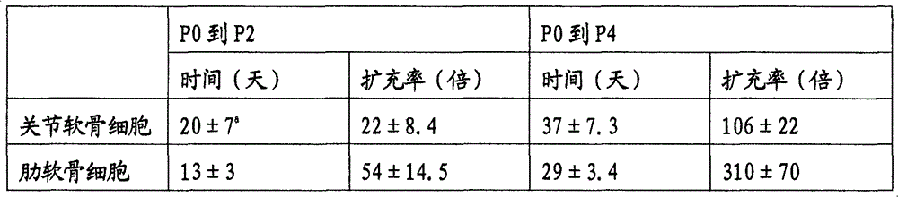
a:来自三个独立试验的平均值±标准差。 a : mean±s.d. from three independent experiments.
如上面表2所示,在体外细胞扩充方面,肋软骨细胞与关节软骨细胞相比似乎是更好的供体软骨细胞。 As shown in Table 2 above, costal chondrocytes appear to be better donor chondrocytes than articular chondrocytes for in vitro cell expansion. the
在体外扩充期间关节软骨细胞与肋软骨细胞的软骨细胞表现型的丢失的比较 Comparison of the loss of the chondrocyte phenotype of articular chondrocytes versus costal chondrocytes during in vitro expansion
在体外细胞扩充期间,软骨细胞常常丢失它们在体内的性质例如圆的形状,II型胶原和葡萄糖胺聚糖的表达。在关节软骨细胞和肋软骨细胞的每个传代都调查形态变化和II型胶原的表达(图2和3)。从P2,细胞的某些组开始丢失它们原始的圆形,而转变为成纤维细胞的梭形(图2)。在P4,多数肋软骨细胞实际上改变它们的形态成为成纤维细胞表现型。然而,关节软骨细胞的这个形态变化比肋软骨细胞缓慢得多。因此,关节软骨细胞与肋软骨细胞相比在体外更缓慢的扩充,并更缓慢的改变它们的形态。 During in vitro cell expansion, chondrocytes often lose their in vivo properties such as round shape, type II collagen and expression of glycosaminoglycans. Morphological changes and expression of type II collagen were investigated at each passage in articular and costal chondrocytes (Figures 2 and 3). From P2, certain groups of cells begin to lose their original round shape and transform into the spindle shape of fibroblasts (Fig. 2). At P4, most costal chondrocytes actually changed their morphology to a fibroblastic phenotype. However, this morphological change is much slower in articular chondrocytes than in costal chondrocytes. Therefore, articular chondrocytes expand more slowly in vitro and change their morphology more slowly than costal chondrocytes. the
为进一步证实成纤维细胞形态变化是否伴随着I I型胶原表达的丢失,用抗II型胶原抗体进行免疫荧光染色(图3)。在P0,99%获得自肋软骨或关节软骨的原生软骨细胞表达II型胶原。在P2,表达II型胶原的软骨细胞的数目快速的减少,在肋软骨细胞中尤为明显。在P4,大多数肋软骨细胞不表达II型胶原,而某些关节软骨细胞维持表达II型胶原。 To further confirm whether the morphological changes in fibroblasts were accompanied by the loss of type II collagen expression, immunofluorescent staining was performed with an anti-type II collagen antibody (Figure 3). At P0, 99% of native chondrocytes obtained from costal or articular cartilage expressed type II collagen. At P2, the number of type II collagen-expressing chondrocytes decreased rapidly, especially in costal chondrocytes. At P4, most costal chondrocytes do not express type II collagen, while some articular chondrocytes maintain expression of type II collagen. the
DAPI阳性软骨细胞中表达II型胶原的细胞数量(表3)。 Number of cells expressing type II collagen in DAPI-positive chondrocytes (Table 3). the
表3:在不同细胞传代,表达II型胶原的细胞数目(%) Table 3: Number of cells expressing type II collagen in different cell passages (%)
a:平均值±标准差 a: mean ± standard deviation
如上面表3所示,软骨细胞在体外扩充期间逐渐丢失它们的原始表现型例如圆的细胞形态和I I型胶原表达能力,肋软骨细胞此过程比关节软骨细胞快得多。 As shown in Table 3 above, chondrocytes gradually lost their original phenotypes such as round cell morphology and type II collagen expression ability during in vitro expansion, and this process was much faster for costal chondrocytes than articular chondrocytes. the
实施例2:证实完全去分化的肋软骨细胞是否显示间充质干细胞性质 Example 2: To confirm whether fully dedifferentiated costal chondrocytes show the properties of mesenchymal stem cells
材料和方法 Materials and methods
软骨细胞的分离 Isolation of Chondrocytes
从4到5个月大的新西兰白兔处获得肋软骨。将胸部附近的皮肤刮净,用酒精清洗,并用聚维配碘溶液备皮。打开右胸的背侧,暴露第9和第10肋软骨。移除软的粘附组织之后,称重肋软骨。这些软骨被切碎成1-2mm3的块并在D-PBS(Dulbecco′s磷酸盐缓冲盐水;Jeil Biotech services Inc.,Taegu,Korea)中漂洗3次。漂洗之后,将切碎的软骨在多酶鸡尾酒溶液37℃和5%CO2下整夜消化,所述多酶鸡尾酒溶液包括胶原酶D(2mg/ml,Roche DiagnosticGmbH,Germany)、透明质酸酶(1mg/ml,Roche)和脱氧核糖核酸酶(0.75mg/ml,Roche)。然后将溶液通过53μm尼龙网眼,分离的细胞用补充有10%胎牛血清(FBS;Hyclone technologies,U.S.A.)和1%青霉素/链霉素/两性霉素B鸡尾酒(Gibco)的DMEM(Dulbecco′s Modified Eagle Medium;Gibco LifeTechnologies,Grand Island,NY,U.S.A.)清洗2次,并且活细胞在血球计上基于锥虫蓝排除法计数。细胞以5×10⁵细胞/100mm直径平皿的细胞密度被涂敷,每隔一天更换细胞培养基,每次培养基更换时添加新配制的50μg/ml的L-抗坏血酸(Sigma,St.Louis,MO,U.S.A.)。汇合的原生细胞传代培养到P7。 Costal cartilages were obtained from 4- to 5-month-old New Zealand White rabbits. The skin around the chest was scraped off, washed with alcohol, and prepared with povidone iodine solution. Open the dorsal side of the right chest to expose the 9th and 10th costal cartilages. After removal of soft adherent tissue, the costal cartilage is weighed. These cartilages were minced into 1-2 mm3 pieces and rinsed 3 times in D-PBS (Dulbecco's phosphate-buffered saline; Jeil Biotech services Inc., Taegu, Korea). After rinsing, the minced cartilage was digested overnight at 37° C. and 5% CO in a multi-enzyme cocktail solution including collagenase D (2 mg/ml, Roche Diagnostic GmbH, Germany), hyaluronidase (1 mg/ml, Roche) and deoxyribonuclease (0.75 mg/ml, Roche). The solution was then passed through a 53 μm nylon mesh, and the detached cells were treated with DMEM (Dulbecco's) supplemented with 10% fetal bovine serum (FBS; Hyclone technologies, USA) and 1% penicillin/streptomycin/amphotericin B cocktail (Gibco). Modified Eagle Medium; Gibco Life Technologies, Grand Island, NY, USA) was washed twice and viable cells were counted on a hemocytometer based on trypan blue exclusion. The cells were plated at a cell density of 5×10 5 cells/100 mm diameter dish, and the cell culture medium was changed every other day, and freshly prepared 50 μg/ml of L-ascorbic acid (Sigma, St.Louis, MO, USA). Confluent primary cells were subcultured to P7.
I、II型胶原和平滑肌肌动蛋白(SMA)抗体的表达 Expression of Type I, II Collagen and Smooth Muscle Actin (SMA) Antibodies
将P7的肋软骨细胞铺板在盖玻片上,培养2天,用3.7%福尔马林磷酸盐缓冲盐水固定10分钟,并用0.2%Triton X-100磷酸盐缓冲盐水透化。抗I型胶原抗体(多克隆抗山羊抗体:southern),抗II型胶原抗体(单克隆抗小鼠抗体:Chemicon international Inc.,Temecular,U.S.A.)或抗平滑肌肌动蛋白抗体(单克隆抗小鼠抗体:DAKO)应用到标本,4℃过夜,用第二抗体培养,然后用抗生蛋白链菌素-过氧化物酶培养1小时,并用0.1%3,3′-二氨基联苯胺四盐酸盐(DAB;Chromogen,DAKO Corp.,CA,U.S.A.)磷酸盐缓冲盐水显影5分钟。用坚牢红染料(Vector Lab.,Burlingame,CA,U.S.A)复染色。 P7 costal chondrocytes were plated on coverslips, cultured for 2 days, fixed with 3.7% formalin phosphate-buffered saline for 10 minutes, and permeabilized with 0.2% Triton X-100 phosphate-buffered saline. Anti-type I collagen antibody (polyclonal anti-goat antibody: southern), anti-type II collagen antibody (monoclonal anti-mouse antibody: Chemicon international Inc., Temecular, U.S.A.) or anti-smooth muscle actin antibody (monoclonal anti-mouse antibody Antibody: DAKO) was applied to the specimen, overnight at 4°C, incubated with secondary antibody, then incubated with streptavidin-peroxidase for 1 hour, and treated with 0.1% 3,3′-diaminobenzidine tetrahydrochloride (DAB; Chromogen, DAKO Corp., CA, U.S.A.) Phosphate-buffered saline for 5 min. Counterstained with fast red dye (Vector Lab., Burlingame, CA, U.S.A). the
结果 result
证实肋软骨细胞的去分化和间充质干细胞性质的表达 Confirmation of dedifferentiation of costal chondrocytes and expression of mesenchymal stem cell properties
如图4所示,在P7的肋软骨细胞丢失它们的原始圆的形状,转变为成纤维细胞的纺锤形。由此可见肋软骨细胞完全的去分化了。图5和6显示了在去分化的软骨细胞中I、II型胶原和平滑肌肌动蛋白抗体的表达。在每个细胞中,I型胶原仍被表达,而II型胶原完全不被表达。间充质干细胞的一个特征,平滑肌肌动蛋白的表达在许多细胞中被观察到(图6)。如上所述,I型胶原和平滑肌肌动蛋白的表达证实了传代肋软骨细胞显示了间充质干细胞的性质。 As shown in Figure 4, costal chondrocytes at P7 lost their original round shape, transforming into the spindle shape of fibroblasts. It can be seen that the costal chondrocytes are completely dedifferentiated. Figures 5 and 6 show the expression of type I, II collagen and smooth muscle actin antibodies in dedifferentiated chondrocytes. In each cell, type I collagen is still expressed, while type II collagen is not expressed at all. A characteristic of mesenchymal stem cells, expression of smooth muscle actin was observed in many cells (Fig. 6). As mentioned above, the expression of type I collagen and smooth muscle actin confirmed that the passaged costal chondrocytes displayed the properties of mesenchymal stem cells. the
实施例3:不同培养条件(DMEM+10%FBS,MSCGM或包含成纤维细胞生长因子的MSCGM)下对肋软骨细胞的扩充和去分化为软骨情况进行评价 Example 3: Evaluation of rib chondrocyte expansion and dedifferentiation into cartilage under different culture conditions (DMEM+10%FBS, MSCGM or MSCGM containing fibroblast growth factor)
材料和方法 Materials and methods
软骨细胞的分离和培养 Isolation and culture of chondrocytes
软骨细胞按如上所述的方法分离,活细胞在血球计上基于锥虫蓝排除法计数。软骨细胞以5×10⁵细胞/100mm直径平皿的细胞密度被铺板,并分别在DMEM(DMEM-FBS)、MSCGM(Cambrex Bio Science Walkersville,Inc.,MD,U.S.A.)、或添加了1ng/ml FGF(R&D System Inc.,MN,U.S.A.)的MSCGM中培养。 Chondrocytes were isolated as described above and viable cells were counted on a hemocytometer based on trypan blue exclusion. Chondrocytes were plated at a cell density of 5×10 5 cells/100 mm diameter dish, and were added in DMEM (DMEM-FBS), MSCGM (Cambrex Bio Science Walkersville, Inc., MD, USA), or 1 ng/ml FGF (R&D System Inc., MN, USA) in MSCGM.
不同培养基的细胞扩充率 Cell expansion rate in different media
当细胞充满培养皿时,通过胰蛋白酶-EDTA(Gibco)移除细胞,活细胞在血球计上基于锥虫蓝排除法计数,以评价细胞扩充率。然后,细胞以5×10⁵细胞/100mm直径平皿的细胞密度铺板,并在相同培养基中培养。 When cells filled the dish, cells were removed by trypsin-EDTA (Gibco) and viable cells were counted on a hemocytometer based on trypan blue exclusion to assess cell expansion rate. Cells were then plated at a cell density of 5 x 10⁵ cells/100 mm diameter dish and cultured in the same medium.
I和II型胶原和平滑肌肌动蛋白抗体的表达 Expression of antibodies to type I and II collagen and smooth muscle actin
将在DMEM-FBS、MSCGM或MSCGM-FGF中培养的各代肋软骨细胞分别涂覆在盖玻片上,培养2天,用3.7%福尔马林磷酸盐缓冲盐水固定10分钟,并用0.2%Triton X-100磷酸盐缓冲盐水透化。透化细胞用20%正常山羊血清培养以阻断非特异性反应,抗平滑肌肌动蛋白抗体(单克隆抗小鼠抗体:DAKO)用做初始抗体并应用到标本4℃整夜。标记了异硫氰酸荧光素(FITC)的山羊抗鼠IgG结合物作为第二抗体,将盖玻片用4′,6二脒基-2-苯吲哚(DAPI;1g/ml;Sigma Chemical Co.,St.Louis,MO,U.S.A.)培养5分钟用于核染色。细 胞在荧光显微镜下观察(Olympus Optical Co.,Japan)。 Costal chondrocytes of each generation cultured in DMEM-FBS, MSCGM or MSCGM-FGF were coated on coverslips, cultured for 2 days, fixed with 3.7% formalin phosphate-buffered saline for 10 minutes, and treated with 0.2% Triton Permeabilize with X-100 Phosphate Buffered Saline. Permeabilized cells were incubated with 20% normal goat serum to block non-specific reactions, and anti-smooth muscle actin antibody (monoclonal anti-mouse antibody: DAKO) was used as the initial antibody and applied to the specimen overnight at 4°C. Fluorescein isothiocyanate (FITC)-labeled goat anti-mouse IgG conjugate was used as a secondary antibody, and the coverslip was treated with 4′,6-diamidino-2-phenylindole (DAPI; 1 g/ml; Sigma Chemical Co., St. Louis, MO, U.S.A.) were incubated for 5 minutes for nuclear staining. Cells were observed under a fluorescence microscope (Olympus Optical Co., Japan). the
用磷酸盐缓冲盐水冲洗平皿底部2次后,在DMEM-FBS、MSCGM或MSCGM-FGF中培养的各自传代的肋软骨细胞用细胞溶解缓冲液(Biolabs;20mM Tris-HCl pH 7.4,150mM NaCl,1mM Na2EDTA,1mM EGTA,1%Triton,2.5mM焦磷酸钠,1mM Na3VO4,1mM β-甘油磷酸酯,1μg/ml亮肽素)溶解,添加2mM的苯甲基磺酰氟化物(PMSF)用于抑制蛋白质降解酶的活性,并离心取上清液用于胶原分离。蛋白质加载到10%SDS-PAGE,转膜,并用抗I型胶原抗体和抗II型胶原抗体(多克隆抗山羊抗体:southern)进行western免疫印迹法检测。 After rinsing the bottom of the plate twice with phosphate-buffered saline, the costal chondrocytes of the respective passages cultured in DMEM-FBS, MSCGM or MSCGM-FGF were treated with cell lysis buffer (Biolabs; 20 mM Tris-HCl pH 7.4, 150 mM NaCl, 1 mM Na2EDTA, 1mM EGTA, 1% Triton, 2.5mM sodium pyrophosphate, 1mM Na 3 VO 4 , 1mM β-glycerophosphate, 1μg/ml leupeptin) dissolved, add 2mM phenylmethylsulfonyl fluoride (PMSF) Used to inhibit the activity of protein degrading enzymes, and centrifuged to obtain the supernatant for collagen separation. Proteins were loaded on 10% SDS-PAGE, transferred, and detected by western blot with anti-type I collagen antibody and anti-type II collagen antibody (polyclonal anti-goat antibody: southern).
分化诱导为软骨细胞 Induced differentiation into chondrocytes
将在DMEM-FBS、MSCGM或MSCGM-FGF中扩充至P7或P8的肋软骨细胞制成团块,并在软骨形成培养基中诱导3周以分化为软骨。番红精-O染色评估分化为软骨的程度。 Costal chondrocytes expanded to P7 or P8 in DMEM-FBS, MSCGM or MSCGM-FGF were pelleted and induced in chondrogenic medium for 3 weeks to differentiate into cartilage. Safranin-O staining assessed the degree of differentiation into cartilage. the
结果 result
不同培养基的细胞扩充率 Cell expansion rate in different media
当细胞充满培养皿时,用胰蛋白酶-EDTA(Gibco)移除细胞,活细胞在血球计上基于锥虫蓝排除法计数,评价细胞扩充率。 When the dish was full of cells, the cells were removed with trypsin-EDTA (Gibco), viable cells were counted on a hemocytometer based on trypan blue exclusion, and the rate of cell expansion was assessed. the
如图7所示,为达到P7,需在MSCGM-FGF中培养约30天,其中细胞扩充约107倍,在MSCGM中需要约38天,其中细胞扩充约105倍。然而,在DMEM-FBS中,在大约1个月中,细胞几乎不发生抛物线状的扩充图,在P7,细胞扩充仅约103倍。 As shown in Figure 7, in order to reach P7, it needs to be cultured in MSCGM-FGF for about 30 days, in which the cells expand by about 10 7 times, and in MSCGM for about 38 days, in which the cells expand by about 10 5 times. However, in DMEM-FBS, the cells hardly exhibited a parabolic expansion pattern in about 1 month, and the cells expanded only about 10 3 times at P7.
因此,在肋软骨细胞的培养中,在MSCGM中的肋软骨细胞扩充率好于在DMEM+10%FBS中的。另外,当在添加了FGF的MSCGM中培养时,肋软骨细胞的扩充率显著增高。可以预期,如果肋软骨细胞在添加了FGF的DMEM中培养时,将获得同样的结果。 Therefore, in the culture of costal chondrocytes, the expansion rate of costal chondrocytes in MSCGM was better than that in DMEM+10%FBS. In addition, the expansion rate of costal chondrocytes was significantly increased when cultured in MSCGM supplemented with FGF. It can be expected that the same results would be obtained if costal chondrocytes were cultured in DMEM supplemented with FGF. the
I和II型胶原和抗平滑肌肌动蛋白抗体的表达 Expression of type I and II collagen and anti-smooth muscle actin antibodies
测定分别在DMEM-FBS、MSCGM或MSCGM-FGF中培养的肋软骨细胞其I和II型胶原和抗平滑肌肌动蛋白抗体的表达程度。 The expression levels of type I and II collagen and anti-smooth muscle actin antibody in costal chondrocytes cultured in DMEM-FBS, MSCGM or MSCGM-FGF were determined. the
如图8所示,随着传代的增加II型胶原的表达减少,肋软骨细胞的II型 胶原表达减少率按MSCGM-FGF、MSCGM和DMEM-FBS的次序增高。 As shown in Figure 8, with the increase of subculture, the expression of type II collagen decreased, and the reduction rate of type II collagen expression of costal chondrocytes increased in the order of MSCGM-FGF, MSCGM and DMEM-FBS. the
在扩充于MSCGM和DMEM-FBS中的肋软骨细胞内,平滑肌肌动蛋白表达细胞约为90%,就是说,许多细胞表达平滑肌肌动蛋白。然而,在MSCGM-FGF中,随着传代仅有极少数的肋软骨细胞表达平滑肌肌动蛋白。 In costal chondrocytes expanded in MSCGM and DMEM-FBS, smooth muscle actin expressing cells were about 90%, that is, many cells expressed smooth muscle actin. However, in MSCGM-FGF, only a very small number of costal chondrocytes expressed smooth muscle actin with passage. the
如图9所示,在扩充于DMEM-FBS和MSCGM的肋软骨细胞内,随着传代的增加I型胶原的表达增加,在P7,几乎所有的细胞表达I型胶原。相反,仅60至70%的扩充于MSCGM-FGF中的细胞表达I型胶原。 As shown in Figure 9, in costal chondrocytes expanded in DMEM-FBS and MSCGM, the expression of type I collagen increased with the increase of passage, and at P7, almost all cells expressed type I collagen. In contrast, only 60 to 70% of cells expanded in MSCGM-FGF expressed type I collagen. the
葡萄糖胺聚糖的表达 Expression of glycosaminoglycans
如图10所示,扩充于DMEM-FBS的肋软骨细胞主要在团块的外壁显示葡萄糖胺聚糖的表达,在高倍显微图象中大多数的外壁细胞显示软骨细胞形态。扩充于MSCGM中的肋软骨细胞在3周的软骨分化后在某些团块表面显示了低的葡萄糖胺聚糖表达,和非常小数目的细胞显示软骨细胞的形态。相反,扩充于MSCGM-FGF中的细胞在3周的软骨分化后在大多数团块中显示了强的葡萄糖胺聚糖表达,并显示软骨细胞形态。 As shown in Figure 10, the costal chondrocytes expanded in DMEM-FBS showed the expression of glycosaminoglycan mainly on the outer wall of the mass, and most of the outer wall cells showed chondrocyte morphology in high-magnification microscopic images. Costal chondrocytes expanded in MSCGM showed low glycosaminoglycan expression on the surface of certain clumps after 3 weeks of chondrogenic differentiation, and a very small number of cells showed chondrocyte morphology. In contrast, cells expanded in MSCGM-FGF showed strong glycosaminoglycan expression in most clumps after 3 weeks of chondrogenic differentiation and displayed chondrocyte morphology. the
从而,以添加1ng/mlFGF的MSCGM为扩充肋软骨细胞的培养基时,当细胞扩充约2×107倍后软骨细胞分化能力被保持。 Therefore, when MSCGM supplemented with 1ng/ml FGF was used as the medium for expanding costal chondrocytes, the differentiation ability of chondrocytes was maintained when the cells expanded about 2×10 7 times.
实施例4:完全去分化的肋软骨细胞的再分化 Example 4: Redifferentiation of fully dedifferentiated costal chondrocytes
材料和方法 Materials and methods
软骨细胞的分离 Isolation of Chondrocytes
用实施例2中使用的方法,去分化细胞被培养直至P7。 Using the method used in Example 2, dedifferentiated cells were cultured up to P7. the
完全去分化的肋软骨细胞分化为软骨细胞 Fully dedifferentiated costal chondrocytes differentiate into chondrocytes
为了完全去分化的肋软骨细胞再分化为软骨细胞,接种细胞至1×10⁶细胞/15ml试管,并聚集培养。培养基被更换为软骨形成培养基(高葡萄糖DMEM,1%ITS+3(Sigma),100nM地塞米松(Sigma),50μg/ml维生素C(Sigma),40μg/ml果仁糖(Sigma),10ng/ml TGF β3(R&D system,Inc.,MN,U.S.A.)),对照组添加基础培养基(DMEM-HG,10%FBS)。一周更换2次培养基,并在2和3周观察。 In order to redifferentiate the fully dedifferentiated costal chondrocytes into chondrocytes, inoculate the cells to 1×10 6 cells/15ml test tube, and aggregate them for culture. The medium was replaced with chondrogenic medium (high glucose DMEM, 1% ITS+3 (Sigma), 100 nM dexamethasone (Sigma), 50 μg/ml vitamin C (Sigma), 40 μg/ml praline (Sigma), 10 ng/ml TGF β3 (R&D system, Inc., MN, USA)), the control group was supplemented with basal medium (DMEM-HG, 10% FBS). The medium was changed twice a week and observed at 2 and 3 weeks.
为确证分化为软骨细胞,在2和3周,用3.7%福尔马林磷酸盐缓冲液固定 三维结构,制备成石蜡标本,并切为5μm厚度。薄片被去石蜡和用番红精-O和固绿(Sigma)染色,用于观察葡萄糖胺聚糖分布。 To confirm differentiation into chondrocytes, at 2 and 3 weeks, the three-dimensional structures were fixed with 3.7% formalin phosphate buffer, prepared as paraffin specimens, and cut into 5 μm thickness. Sections were deparaffinized and stained with Safranin-O and Fast Green (Sigma) for visualization of glycosaminoglycan distribution. the
完全去分化的肋软骨细胞分化为成骨细胞 Fully dedifferentiated costal chondrocytes differentiate into osteoblasts
为了再分化为成骨细胞,将P7的肋软骨细胞以2×10⁴细胞/cm2的细胞密度接种6孔和24孔,在成骨培养基(DMEM,10%FBS,100nM地塞米松(Sigma),10mM β-磷酸甘油(Sigma),50μg/ml维生素C(Sigma))中培养。一周更换2次培养基,并在2和3周观察。基础培养基(DMEM,10%FBS)被用于对照组。 In order to redifferentiate into osteoblasts, P7 costal chondrocytes were seeded at a cell density of 2 × 10⁴ cells/ cm2 in 6 wells and 24 wells in osteogenic medium (DMEM, 10% FBS, 100 nM dexamethasone ( Sigma), 10mM β-glycerol phosphate (Sigma), 50μg/ml vitamin C (Sigma)). The medium was changed twice a week and observed at 2 and 3 weeks. Basal medium (DMEM, 10% FBS) was used for the control group.
为确证分化为成骨细胞,细胞分化初始标记物用碱性磷酸酶染色。用碱性磷酸酶测量试剂盒(SIGMA-ALDRICH,St.Louis,MO,U.S.A.)观察组织化学的表达程度。并且,用茜素磺酸钠(SIGMA-ALDRICH)染色细胞,所述茜素磺酸钠能够染色成骨细胞成熟期间分泌的矿物成分,以观察分化为成骨细胞的程度。 To confirm differentiation into osteoblasts, cells were stained with alkaline phosphatase for initial markers of differentiation. The degree of histochemical expression was observed with an alkaline phosphatase measurement kit (SIGMA-ALDRICH, St. Louis, MO, U.S.A.). Also, the cells were stained with sodium alizarin sulfonate (SIGMA-ALDRICH), which can stain mineral components secreted during osteoblast maturation, to observe the degree of differentiation into osteoblasts. the
完全去分化的肋软骨细胞分化为脂肪细胞 Fully dedifferentiated costal chondrocytes differentiate into adipocytes
为了再分化为脂肪细胞,P7的肋软骨细胞以2×10⁴细胞/cm2的细胞密度接种6孔和24孔,并在脂肪形成培养基(高葡萄糖DMEM(Gibco),10%FBS,10mg/ml胰岛素(Sigma),100nM地塞米松(Sigma),0.2mM消炎痛(Wako Pure ChemicalIndustries,Japan),500μm 3-异丁基-1-甲基黄嘌呤(Wako))中培养3天,并在脂肪形成培养基中(高葡萄糖DMEM,10%FBS)重复1天的培养。基础培养基(DMEM-HG,10%FBS)被用于对照组,并在2和3周观察。 To redifferentiate into adipocytes, costal chondrocytes from P7 were seeded in 6-well and 24-well at a cell density of 2× 10⁴ cells/ cm2 , and cultured in adipogenic medium (high glucose DMEM (Gibco), 10% FBS, 10mg /ml insulin (Sigma), 100nM dexamethasone (Sigma), 0.2mM indomethacin (Wako Pure Chemical Industries, Japan), 500 μ m 3-isobutyl-1-methylxanthine (Wako)) for 3 days, and The culture was repeated for 1 day in adipogenic medium (high glucose DMEM, 10% FBS). Basal medium (DMEM-HG, 10% FBS) was used in the control group and observed at 2 and 3 weeks.
为确证分化为脂肪细胞,细胞用油红O染色,所述油红O特别适合染色脂类小滴。 To confirm differentiation into adipocytes, cells were stained with Oil Red O, which is particularly suitable for staining lipid droplets. the
结果 result
分化为软骨细胞 Differentiate into chondrocytes
图11显示了P7肋软骨细胞分化诱导3周成为软骨细胞的目视图。细胞互相形成团块,软骨诱导组中的团块尺寸与对照组(基础培养基:DMEM,10%FBS)相比更大。 Fig. 11 shows the visualization of P7 rib chondrocytes induced to differentiate into chondrocytes for 3 weeks. The cells formed aggregates with each other, and the aggregate size was larger in the chondrogenic induction group than in the control group (basal medium: DMEM, 10% FBS). the
图12显示对葡萄糖胺聚糖番红精-O染色的结果。在分化诱导的3周,在团块的外壁观察葡萄糖胺聚糖的表达。细胞显示软骨细胞形态。对照组不表达葡萄糖胺聚糖,也不显示软骨细胞形态。 Figure 12 shows the results of staining for glycosaminoglycan Safranin-O. At 3 weeks after differentiation induction, the expression of glycosaminoglycan was observed on the outer wall of the clump. Cells show chondrocyte morphology. The control group did not express glycosaminoglycans and did not display chondrocyte morphology. the
分化为成骨细胞 differentiate into osteoblasts
为确证分化为成骨细胞,观察碱性磷酸酶的活性。图13显示通过碱性磷酸酶测量试剂盒(SIGMA-ALDRICH,St.Louis,MO,U.S.A.)测量碱性磷酸酶。碱性磷酸酶的活性,作为成骨分化的初始标记物,在分化的2周强烈的表达,在3周趋于稍微降低。培养于基础培养基的对照组不表达碱性磷酸酶的活性。 To confirm differentiation into osteoblasts, alkaline phosphatase activity was observed. Fig. 13 shows measurement of alkaline phosphatase by alkaline phosphatase measurement kit (SIGMA-ALDRICH, St. Louis, MO, U.S.A.). Alkaline phosphatase activity, an initial marker of osteogenic differentiation, was strongly expressed at 2 weeks of differentiation and tended to decrease slightly at 3 weeks. The control group cultured in basal medium did not express the activity of alkaline phosphatase. the
更进一步,图14显示用茜素红染色剂进行矿物染色的结果。在分化2周时没有观察到表达,但在3周的部分染色确证了成熟分化为成骨细胞。 Further, Figure 14 shows the results of mineral staining with Alizarin Red stain. No expression was observed at 2 weeks of differentiation, but partial staining at 3 weeks confirmed mature differentiation into osteoblasts. the
分化为脂肪细胞 Differentiate into fat cells
为确证分化为脂肪细胞,细胞用油红O染色,所述油红O适合染色脂类小滴。如图15所示,在细胞内显现的许多油红O染色的脂类小滴确证了分化为脂肪细胞。 To confirm differentiation into adipocytes, cells were stained with Oil Red O, which is suitable for staining lipid droplets. As shown in Figure 15, differentiation into adipocytes was confirmed by the visualization of numerous Oil Red O-stained lipid droplets within the cells. the
实施例5:基于壳聚糖支架内的肋软骨细胞对关节软骨缺损的修复作用的评价 Embodiment 5: Evaluation of the repairing effect of articular cartilage defects based on costal chondrocytes in the chitosan scaffold
材料和方法 Materials and methods
实施例5的步骤与图16图解视图的相同。图16显示肋软骨细胞被分离,培养直到P2,去分化的细胞被接种到基于壳聚糖的支架上,培养,然后将支架移植到缺损的关节软骨中以修复软骨缺损。 The steps of Embodiment 5 are the same as those of the diagrammatic view in FIG. 16 . Figure 16 shows that costal chondrocytes were isolated, cultured until P2, dedifferentiated cells were seeded onto chitosan-based scaffolds, cultured, and then the scaffolds were grafted into defective articular cartilage to repair cartilage defects. the
软骨细胞的分离 Isolation of Chondrocytes
在采取如上述实施例1相同的步骤后,汇合的原代细胞传代培养直到P2。 After taking the same steps as in Example 1 above, the confluent primary cells were subcultured until P2. the
海绵形式支架的制备 Preparation of sponge-form scaffolds
COL-GAG海绵: COL-GAG sponge:
多孔胶原基质( 
CS海绵: CS sponge:
壳聚糖(Korea chitosan Co.,Korea,1.5%w/v)溶于0.1%乙酸水液中。充分搅拌完全溶解后,溶液通过0.4μm滤膜(Millipore Co.,U.S.A.)并分配到模型中。成型凝胶在-80℃放置24小时,然后在冷冻干燥器中冻干24小时。支架用酒精和蒸馏水洗去残酸,并添加乙酸酐以乙酰化。支架用酒精和蒸馏水洗去副产物酸,并冻干。壳聚糖支架(CS)的厚度约为2mm。壳聚糖支架的精细结构和孔径大小通过扫描电子显微镜法(扫描电镜;图8)获得。通过4mm活检冲麻制备4mm直径的支架。在细胞培养实验之前,支架(CS)暴露在紫外光下30分钟灭菌,并用培养基浸泡10分钟。制备的支架应立即使用。 Chitosan (Korea chitosan Co., Korea, 1.5% w/v) was dissolved in 0.1% acetic acid aqueous solution. After thorough stirring and complete dissolution, the solution was passed through a 0.4 μm filter membrane (Millipore Co., U.S.A.) and distributed into the mold. The formed gels were kept at -80°C for 24 hours and then lyophilized in a lyophilizer for 24 hours. Scaffolds were washed with alcohol and distilled water to remove residual acid, and acetic anhydride was added for acetylation. Scaffolds were washed with alcohol and distilled water to remove by-product acids, and lyophilized. The thickness of the chitosan scaffold (CS) was about 2 mm. The fine structure and pore size of chitosan scaffolds were obtained by scanning electron microscopy (SEM; Figure 8). A 4 mm diameter scaffold was prepared by flushing a 4 mm biopsy. Prior to cell culture experiments, scaffolds (CS) were sterilized by exposure to UV light for 30 min and soaked with culture medium for 10 min. Prepared scaffolds should be used immediately. the
CS-HA海绵: CS-HA sponge:
在制备4mm直径的壳聚糖支架后,被用50%乙醇浸泡,再用0.1%透明质酸(HA;Sigma,St Louis,MO,U.S.A.)磷酸盐缓冲盐水浸泡。支架在-80℃放置24小时,冷冻干燥器中冻干24小时。在细胞培养实验之前,支架(CS-HA)暴露在紫外光下30分钟灭菌,并用培养基浸泡10分钟。制备的支架应立即使用。 After preparing chitosan scaffolds with a diameter of 4 mm, they were soaked in 50% ethanol followed by 0.1% hyaluronic acid (HA; Sigma, St Louis, MO, U.S.A.) in phosphate-buffered saline. The scaffolds were stored at -80°C for 24 hours and freeze-dried in a freeze dryer for 24 hours. Prior to cell culture experiments, scaffolds (CS-HA) were sterilized by exposure to UV light for 30 min and soaked with medium for 10 min. Prepared scaffolds should be used immediately. the
在支架上接种软骨细胞并培养 Chondrocytes were seeded and cultured on scaffolds
将含有2×10⁶二代肋软骨细胞的培养基20μl接种到COL-GAG、CS和CS-HA(4mm直径×2mm厚)上培养基中的,预先放入在37℃的5%CO2的6-孔盘中2小时。随后,向每个孔中添加6mL培养基,细胞培养4周。每周更换培养基和在整个培养期间每72小时向培养基添加新鲜的L-抗坏血酸。在第2、7、14和28天获得培养物汤和细胞支架结构,和结构仅在第2天获得。
MTT分析、Blyscan分析、细胞支架结构的组织学的和免疫组织化学的染色以及前胶原I型C-肽(PIP)的ELISA分析 MTT analysis, Blyscan analysis, histological and immunohistochemical staining of cytoskeletal structures, and ELISA analysis of procollagen type I C-peptide (PIP)
MTT分析 MTT analysis
每个支架内的细胞增殖率根据MTT分析(Mosman T.Rapid colormetricassay for cellular growth and survival,application to proliferation andcytotoxicity assays,J.Immunol.Methods 1983;65:55-63)测定。 Cell proliferation rate within each scaffold was determined according to MTT assay (Mosman T. Rapid colormetric assay for cellular growth and survival, application to proliferation and cytotoxicity assays, J. Immunol. Methods 1983; 65: 55-63). the
前胶原I型C-肽(PIP)的ELISA分析 ELISA analysis of procollagen type I C-peptide (PIP)
为了定量测定从新合成胶原中释放的前C-肽,使用前胶原I型C-肽(PIP)EIA试剂盒(Takara Bio.Inc.,Japan)。培养基用蒸馏水稀释到1∶100,其余的步骤按照制造商的说明书操作。在450nm用酶标仪(microplate reader)(Bio Rad Lab.,Richimond,CA,U.S.A.)测量吸光度。在0-640ng PIP/mL范围内的以标准曲线来校准肽的释放量。 For quantitative determination of proC-peptide released from newly synthesized collagen, procollagen type I C-peptide (PIP) EIA kit (Takara Bio. Inc., Japan) was used. The medium was diluted to 1:100 with distilled water, and the remaining steps were performed according to the manufacturer's instructions. Absorbance was measured at 450 nm with a microplate reader (Bio Rad Lab., Richimond, CA, U.S.A.). A standard curve was used to calibrate the release of peptides in the range of 0-640 ng PIP/mL. the
Blyscan分析 Blyscan Analysis
使用1,9-次甲基蓝分析( 
统计 Statistics
用不配对t检验来进行统计分析,确定试验中各变量是否有显著性差异(p<0.05)。 Unpaired t-test was used for statistical analysis to determine whether there were significant differences among the variables in the test (p<0.05). the
动物的饲养 animal husbandry
使用24只新西兰白兔(初始重量2.2到2.5kg,约4到5月大)。动物被认为是年轻成年的,因为在4个月大之后通常不再发生直立骨骼的生长(Masoudet al.,1986)。在试验前1至2周获取动物,并饲养在固定温度22±2℃和湿度50±7%和固定亮暗周期(上午7:00-下午7:00,光照期)下。动物可以自由进食和饮水(标准实验室饮食(团块类型,Purina Co.Korea)),并允许在地上自由活动。根据批准的动物饲养国家指南喂养、管理和处理动物。 Twenty-four New Zealand white rabbits (initial weight 2.2 to 2.5 kg, approximately 4 to 5 months old) were used. Animals were considered young adults because erect skeletal growth typically ceased to occur after 4 months of age (Masoud et al., 1986). Animals were harvested 1 to 2 weeks before the experiment and housed under a fixed temperature of 22±2°C and humidity of 50±7% and a fixed light-dark cycle (7:00am-7:00pm, light period). Animals were given free access to food and water (standard laboratory diet (bolus type, Purina Co. Korea)) and were allowed to move freely on the ground. Animals were fed, managed and handled according to approved national guidelines for animal care. the
手术步骤 Surgical steps
兔按上述实施例1描述的方法麻醉。辅助麻醉是静脉注射赛拉嗪和氯胺酮的混合物。在手术前,动物肌内注射应用抗生素(头孢唑啉(cefazoline),Jonggun-dang,汉城,韩国)一次。 Rabbits were anesthetized as described in Example 1 above. Assisted anesthesia is a mixture of xylazine and ketamine administered intravenously. Antibiotics (cefazoline, Jonggun-dang, Seoul, Korea) were administered once to the animals by intramuscular injection before the operation. the
刮净膝部附近的皮肤,用酒精清洗,并用聚维酮碘溶液备皮。手术方法类似于Shapiro et al.所描述的。在作出一个中央的膝盖旁切口后,髌骨被横向脱位以暴露股骨的髌骨凹槽。使用低速钻在髌-股骨接合点的承重区域制造直径为4mm的缺损。在兔中的所述区域是间歇的承受重量,并与髌骨接触。圆锥形的全层缺损从关节软骨的表面通过密质软骨下骨进入在股骨远端的骨髓腔中的松质骨内。缺损的深度大约为2.0到2.5mm。在钻孔之后,用冷盐水(NaCl0.9%w/v)大面积的冲洗伤口以移除脱落的碎片。在缺损处滴入一滴纤维蛋白粘合系统( 
修复组织的组织学评价 Histological evaluation of repaired tissue
在动物处死之后,检查膝盖一般形态学(颜色、完整性、轮廓和光滑度)、缺损的修复和周围软骨的外观。植入区域的标本被解剖、摄影和用10%缓冲福尔马林在室温下固定一周。然后冲洗标本以清除过量的固定剂,并用Calci-Clear Rapid(National Diagnotics,U.S.A.)脱钙一周。用梯度乙醇和二甲苯脱水,并用石蜡包埋。通过切片机切成6μm厚的片用于组织学研究。切片用苏木精和伊红(H&E)染色来研究形态细节,用番红精-O和固绿染色来评价葡萄糖胺聚糖分布。 After animal sacrifice, knees were examined for general morphology (color, integrity, contour and smoothness), repair of defects and appearance of surrounding cartilage. Specimens from the implanted area were dissected, photographed and fixed in 10% buffered formalin for one week at room temperature. Specimens were then rinsed to remove excess fixative and decalcified with Calci-Clear Rapid (National Diagnostics, U.S.A.) for one week. Dehydrated with graded ethanol and xylene and embedded in paraffin. Sections of 6 μm thickness were cut by microtome for histological study. Sections were stained with hematoxylin and eosin (H&E) to study morphological details, and with safranin-O and fast green to evaluate glycosaminoglycan distribution. the
软骨缺损的评价 Evaluation of cartilage defects
按照Pineda等(1992)和Wakitani等(1994)的改良的组织等级评分法 由研究者对切片进行盲法评价。所述等级评分是全部由5类组成,分值从0(正常软骨)到14(未修复组织)(表4)。 The sections were evaluated blindly by the investigators according to the modified tissue grade scoring method of Pineda et al. (1992) and Wakitani et al. (1994). The rating scale is made up of 5 categories in total, ranging from 0 (normal cartilage) to 14 (unrepaired tissue) (Table 4). the
表4:对于软骨缺损的组织等级评分 Table 4: Histological Grade Scores for Cartilage Defects
a、修复软骨的总平滑面积与软骨缺损的总面积之比。 a, The ratio of the total smooth area of the repaired cartilage to the total area of the cartilage defect. the
b、修复软骨的平均厚度与周围软骨的平均厚度之比。 b, The ratio of the average thickness of the repaired cartilage to the average thickness of the surrounding cartilage. the
统计分析 Statistical Analysis
通过使用t检验来评价统计显著性。显著性水平是p<0.05。 Statistical significance was assessed by using t-test. The significance level is p<0.05. the
结果 result
培养在海绵支架中的肋软骨细胞 Costal chondrocytes cultured in sponge scaffolds
使用MTT分析在培养的1、2和4周比较在三种不同支架:胶原-葡萄糖胺聚糖海绵(COL-GAG)、壳聚糖海绵(CS)和透明质酸包被的壳聚糖海绵(CS-HA)中的肋软骨细胞的增殖率(图17)。 Comparison using MTT assay at 1, 2 and 4 weeks of culture on three different scaffolds: collagen-glycosaminoglycan sponge (COL-GAG), chitosan sponge (CS) and hyaluronic acid-coated chitosan sponge Proliferation rate of costal chondrocytes in (CS-HA) ( FIG. 17 ). the
图17中的值显示了在这些支架内的细胞增殖。CS和CS-HA的光学密度值随培养时间延长而逐渐增加,两种支架之间没有显著的差异。在COL-GAG中发现了类似的趋势,但初始水平较低。在培养28天时的光学密度值与培养第7天的相比,在COL-GAG、CS和CS-HA中分别增加了大约1.30、1.43和1.40倍。
The values in Figure 17 show cell proliferation within these scaffolds. The optical density values of CS and CS-HA gradually increased with culture time, and there was no significant difference between the two scaffolds. Similar trends were found in COL-GAG, but at lower initial levels. The optical density values at 28 days of culture were increased by about 1.30, 1.43 and 1.40 times in COL-GAG, CS and CS-HA, respectively, compared with those on
为了评价接种了细胞的复合支架的新合成胶原,通过ELISA分析(图18)测量前胶原I型C-肽(PIP)。在培养第7天,CS和CS-HA的胶原合成大于COL-GAG中的合成,有显著的差异。然而,第14天,在CS中的胶原合成比在COL-GAG和CS-HA中的大的多(CS>CS-HA>COL-GAG)。第14天的胶原合成与在培养第7天的相比显著的减少。在四周的培养期间,CS中向培养基的胶原分泌在逐渐的减少,相反地,在COL-GAG和CS-HA中,胶原释放在第1和第2周之间显著的减少,然后在第2和第4周之间有所增加。
To evaluate the newly synthesized collagen of the cell-seeded composite scaffolds, procollagen type I C-peptide (PIP) was measured by ELISA assay (Figure 18). On
Blyscan分析显示了接种了肋软骨细胞的三种不同支架向培养上清液中持续一周释放的GAG量。在培养的第1周,两种壳聚糖支架比COL-GAG释放更多的GAG。在第14天,三种支架中GAG释放的量近似,但在培养的28天,在COL-GAG中释放的GAG量与CS和CS-HA中的相比非常小。
Blyscan analysis showing the amount of GAG released into the culture supernatant for one week by three different scaffolds seeded with costal chondrocytes. During the first week of culture, both chitosan scaffolds released more GAGs than COL-GAGs. At
在兔肋软骨细胞被接种到COL-GAG、CS和CS-HA上并培养28天期间,在整个培养时间内,CS和CS-HA的尺寸大于COL-GAG的。 During rabbit costal chondrocytes were seeded on COL-GAG, CS and CS-HA and cultured for 28 days, the size of CS and CS-HA was larger than that of COL-GAG throughout the culture time. the
苏木精和伊红(H&E)染色的组织学检查显示了标本的形态特征(图19)。在 图19中,S代表表面,C代表中心,刻度条代表50μm,黑色箭线代表纤维组织,白色箭线代表死亡细胞,黑色箭头代表圆形细胞和白色箭头代表梭形细胞。在软骨细胞培养2天后,接种细胞主要存在于两种基于壳聚糖的支架和特别是COL-GAG的表面区域。圆形和梭形细胞间杂,和在第2天软骨陷窝中没有细胞。在第2天的标本中,在CS和CS-HA中心的细胞是聚集的,而在COL-GAG中,中心的细胞是稀少和分散的。
Histological examination with hematoxylin and eosin (H&E) staining revealed the morphological features of the specimen (Figure 19). In Figure 19, S represents the surface, C represents the center, the scale bar represents 50 μm, black arrows represent fibrous tissue, white arrows represent dead cells, black arrows represent round cells and white arrows represent spindle cells. After 2 days of chondrocyte culture, seeded cells were mainly present in the surface area of the two chitosan-based scaffolds and especially COL-GAG. Interspersed with round and spindle cells, and no cells in the cartilage lacuna on
在培养7天后,在支架的周缘内的细胞数目的增加是显著的,并且新的细胞周围间质包围了大部分细胞。在CS和CS-HA支架的中心和周缘,绝大多数的接种细胞还维持它们的球状形态,占据软骨陷窝并合成充足的基质。然而,在COL-GAG中,周缘的细胞是圆形的,但它们不占据陷窝,中心的细胞是梭形的且不聚集。 After 7 days in culture, the increase in cell number within the periphery of the scaffold was marked, and a new pericellular matrix surrounded most of the cells. At the center and periphery of the CS and CS-HA scaffolds, the vast majority of seeded cells also maintained their spherical morphology, occupied the cartilage lacuna and synthesized sufficient matrix. However, in COL-GAG, the cells at the periphery are round, but they do not occupy the lacuna, and the cells at the center are spindle-shaped and do not aggregate. the
在培养的第14天,在两种基于壳聚糖支架的中心和周缘的大多数细胞还维持它们的球形形态,占据陷窝并合成非常充足的细胞外基质。但在COL-GAG中,周缘的细胞是球形形态,占据陷窝和合成充足的细胞外基质,但中心的细胞还是梭形的,占据小的陷窝和合成少量的细胞外基质。
At
在试验的末尾(28天),两种基于壳聚糖支架的表面被薄纤维组织覆盖。在中心以及周缘的细胞变为小的、仍占据较大的陷窝。在COL-GAG中,周缘细胞减少了细胞构成,具有非常小而圆的形态和不占据陷窝。中心内的细胞与第14天的类似。
At the end of the experiment (28 days), the surfaces of both chitosan-based scaffolds were covered with thin fibrous tissue. Cells in the center and at the periphery become smaller, still occupying larger lacunas. In COL-GAG, peripheral cells have reduced cellularity, have a very small and round morphology and do not occupy lacunae. Cells in the center were similar to those on
通过番红精-O固绿(S/O)对软骨细胞外基质的葡萄糖胺聚糖进行的组织学染色,在整个培养期间在CS和CS-HA支架中心检测到葡萄糖胺聚糖(图20)。在图20中,白色箭头代表番红精-O染色的结构,黑色箭线代表纤维组织、白色箭线代表固绿染色的细胞,刻度条代表100μm。在两种基于壳聚糖的支架中,2天培养标本的中心的细胞被S/O染色。然而,在COL-GAG中心内的细胞不被S/O染色。在三种类型的支架中随培养时间的增加,积累葡萄糖胺聚糖的量也增加。在培养的28天,支架的周围不被固绿染色。
Histological staining of glycosaminoglycans of the extracellular matrix of cartilage by Safranin-O Fast Green (S/O) detected glycosaminoglycans in the center of CS and CS-HA scaffolds throughout the culture period (Fig. 20 ). In Fig. 20, white arrows represent structures stained with safranin-O, black arrows represent fibrous tissue, white arrows represent cells stained with fast green, and the scale bar represents 100 μm. In both chitosan-based scaffolds, cells in the center of the 2-day culture specimens were stained by S/O. However, cells within the COL-GAG center were not stained by S/O. The amount of accumulated glycosaminoglycans also increased with the increase of culture time in the three types of scaffolds. At
组织学检测在没有软骨细胞的CS和CS-HA中没有检测到葡萄糖胺聚糖,但COL-GAG的框架被用S/O阳性着色,因为它们与硫酸软骨素相结合。 Histological examination did not detect glycosaminoglycans in CS and CS-HA without chondrocytes, but the framework of COL-GAG was positively stained with S/O because of their binding to chondroitin sulfate. the
I型和II型胶原的免疫染色 Immunostaining for type I and type II collagen
在两种基于壳聚糖的支架中,1和2周的标本内没有检测到I型胶原。相反,在两种基于壳聚糖的支架中,在1至2周的培养期内抗II型胶原抗体是强阳性的。在第4周,在支架表面和中心内的部分细胞集合体对抗I型胶原抗体呈阳性。然而,在4周的标本中,抗II型胶原抗体还是阳性的,但变弱了。 In both chitosan-based scaffolds, no type I collagen was detected within the 1- and 2-week-old specimens. In contrast, in both chitosan-based scaffolds, anti-type II collagen antibodies were strongly positive during the 1 to 2 week culture period. At week 4, some cell aggregates on the surface and center of the scaffold were positive for anti-type I collagen antibodies. However, in the 4-week specimen, anti-type II collagen antibodies were still positive, but weakened. the
在COL-GAG的1、2和4周的标本内都检测到了I型胶原。在第1周抗II型胶原抗体阳性仅在表面显现,在第2周在周缘的间质和中心的少许细胞内显阳性。在第4周的标本内,抗II型胶原抗体显非常弱的阳性(图21和22)。在图21中,黑色箭线代表纤维组织,黑色箭头代表I型胶原表达细胞,白色箭线代表COL-GAG结构,图21和22中的刻度条代表100μm。 Type I collagen was detected in the 1, 2 and 4 week specimens of COL-GAG. Anti-type II collagen antibodies were positive only on the surface at the first week, and positive in the peripheral interstitium and a few cells in the center at the second week. Anti-type II collagen antibodies were very weakly positive in the 4 week specimen (Figures 21 and 22). In Fig. 21, black arrows represent fibrous tissue, black arrows represent type I collagen expressing cells, white arrows represent COL-GAG structures, and the scale bars in Figs. 21 and 22 represent 100 μm. the
由上述组织学观察的结果可知,当去分化软骨细胞被接种到三种类型的海绵形式支架(CS、CS-HA和COL-GAG)上时,在两种基于壳聚糖支架中,接种的细胞在形态、葡萄糖胺聚糖合成和II型胶原表达中会再分化1至2周。然而,在COL-GAG中,内部细胞接种效率不高,中心内的细胞的再分化效果不好。直到第4周,细胞开始丢失合成细胞外基质的活性(陷窝的尺寸变得更小)。 From the results of the above histological observations, when dedifferentiated chondrocytes were seeded onto the three types of sponge-form scaffolds (CS, CS-HA, and COL-GAG), in the two chitosan-based scaffolds, the seeded Cells were redifferentiated for 1 to 2 weeks in morphology, glycosaminoglycan synthesis, and type II collagen expression. However, in COL-GAG, inner cell seeding was not efficient and cells within the center did not redifferentiate well. Until the 4th week, the cells start to lose the activity of synthesizing the extracellular matrix (the size of the lacuna becomes smaller). the
体内结果 in vivo results
本研究将评价培养肋软骨细胞-壳聚糖复合物在修复承重动物模型内全层关节软骨缺损中的作用。 This study will evaluate the role of cultured costal chondrocyte-chitosan complexes in repairing full-thickness articular cartilage defects in a weight-bearing animal model. the
将P2的肋软骨细胞接种到CS-HA支架上,培养2周。在兔股骨的髌骨凹槽上手术制作软骨缺损。在第6和12周动物被安乐死,并获取膝关节。标本作目视、组织学和免疫组织化学分析。 Costal chondrocytes from P2 were seeded on CS-HA scaffolds and cultured for 2 weeks. A cartilage defect was surgically created on the patellar notch of the rabbit femur. Animals were euthanized at weeks 6 and 12, and knee joints were harvested. Specimens were analyzed visually, histologically and immunohistochemically. the
临床试验 Clinical Trials
所有试验动物中都没有发生手术后并发症。在研究期间,没有观察到关节脱臼或残疾的症状。 No postoperative complications occurred in any of the experimental animals. During the study period, no symptoms of joint dislocation or disability were observed. the
肉眼外观 naked eye appearance
依据肉眼观察,在任何关节都没有观察到滑膜流出或滑膜炎。虽然没有进行滑膜组织的组织学检查,但无炎症反应迹象。肉眼观察,所有关节内的滑液在量、粘性和颜色方面都是正常的。 On gross inspection, no synovial outflow or synovitis was observed in any joint. Although histological examination of synovial tissue was not performed, there was no evidence of an inflammatory response. Macroscopically, the synovial fluid in all joints was normal in volume, viscosity, and color. the
在图23到图26中,对照代表对照组,S代表CS-HA移植的缺损,和S-CELL代表肋软骨细胞接种的CS-HA移植的缺损。 In Figs. 23 to 26, Control represents the control group, S represents the CS-HA grafted defect, and S-CELL represents the costal chondrocyte-seeded CS-HA grafted defect. the
空白(对照)缺损 blank (control) defect
在移植后第6周,在对照组中,缺损可见光滑表面。缺损在修复组织和邻近关节软骨之间有缝隙或缺乏连续性。缺损的边缘是可辨别的。修复组织与邻近正常软骨相比更白和稍微不透明(图23)。在移植后12周,对照组中的修复组织可以容易的与正常邻近软骨区分,因为它们具有白色和不透明的外观。缺损中完全充满了平滑的白色修复组织(图23)。 At 6 weeks post-transplantation, in the control group, the defect was visible with a smooth surface. The defect has a gap or lack of continuity between the repaired tissue and the adjacent articular cartilage. The margins of the defect are discernible. The repaired tissue was whiter and slightly opaque than the adjacent normal cartilage (Fig. 23). At 12 weeks after transplantation, the repaired tissues in the control group could be easily distinguished from the normal adjacent cartilage because of their white and opaque appearance. The defect was completely filled with smooth white repair tissue (Fig. 23). the
CS-HA移植的缺损 Defects in CS-HA grafts
在第6周肉眼观察,某些缺损没有完全充满修复组织,在某些区域软骨下骨暴露。缺损中修复组织的表面与周围正常组织相比更不观则,修复组织的边缘也不平滑。修复组织为白色至粉红色的、半透明的(图23)。在12周,CS-HA移植缺损的修复组织还是可辨别的,但在外观和质地上是透明的。缺损被填充至邻近正常软骨的水平。它们某些的表面比6周标本的更平滑。某些情况下(1/8),修复组织由骨,而不是软骨填充(图23)。 Macroscopically at week 6, some defects were not completely filled with repair tissue and subchondral bone was exposed in some areas. The surface of the repaired tissue in the defect is less regular than the surrounding normal tissue, and the edges of the repaired tissue are not smooth. The repaired tissue was white to pink, translucent (Figure 23). At 12 weeks, the repaired tissue of the CS-HA graft defect was discernible but clear in appearance and texture. The defect is filled to the level adjacent to normal cartilage. Some of them had smoother surfaces than those of 6-week-old specimens. In some cases (1/8), the repair tissue was filled by bone rather than cartilage (Fig. 23). the
肋软骨细胞加载的CS-HA移植的缺损 Defects in costal chondrocyte-loaded CS-HA grafts
在第6周,所有的缺损被充满了修复组织,缺损的边缘是可以从邻近正常软骨辨别出来的。修复组织的表面和边缘是粗糙和不规则的。修复组织在外观和质地上是透明的(图23)。在第12周肉眼观察,所有的修复组织达到了周围正常软骨的水平。修复组织的表面和边缘比6周标本更平滑。修复组织还可以从周围正常软骨辨别出来。但与6周标本相比修复软骨的边缘更加平滑的,与正常软骨接合更好(图23)。 At week 6, all defects were filled with repair tissue, and the margin of the defect was discernible from the adjacent normal cartilage. The surface and edges of the repaired tissue were rough and irregular. The repaired tissue was transparent in appearance and texture (Fig. 23). Macroscopically at week 12, all repaired tissue had reached the level of the surrounding normal cartilage. The surface and edges of the repaired tissue were smoother than those of the 6-week-old specimen. Repaired tissue can also be distinguished from the surrounding normal cartilage. However, compared with the 6-week-old specimen, the edge of the repaired cartilage was smoother and better joined to the normal cartilage (Fig. 23). the
在第6周,CS移植缺损的肉眼外观类似于细胞加载CS移植缺损的肉眼外观:两者均是白色的和一定程度不规则的。通常,直至12周,对照缺损的表面与CS移植缺损的相比平滑的多,所述CS移植缺损的表面是突出和不规则的。对照缺损的表面是白色外观,但移植缺损的是粉红外观的。在细胞加载移植缺损中的修复组织的颜色和质地类似于邻近正常软骨。 At week 6, the macroscopic appearance of the CS graft defect was similar to that of the cell-loaded CS graft defect: both were white and somewhat irregular. In general, until 12 weeks, the surface of the control defect was much smoother than that of the CS graft defect, which was prominent and irregular. The surface of the control defect was white in appearance, but the graft defect was pink in appearance. The color and texture of repaired tissue in cell-loaded graft defects resembled adjacent normal cartilage. the
形态学 Morphology
对照缺损 control defect
在移植后6周,在移植接合部可见附加的类似纤维软骨组织形成。缺损在修复组织和邻近关节软骨之间的单侧或双侧有缝隙或缺乏连续性。这个观测表明周围关节软骨和修复组织之间整合度的下降。在修复软骨和邻近正常软骨之间的界面偶尔显示纤丝状的连续性。修复组织的表面由3至5层扁平的细胞组成,并是纤丝化的。修复组织由纤维组织和多细胞纤维软骨的混合物组成。某些缺损在纤维和纤维软骨修复软骨内显示裂缝缺口。修复组织较好的重新覆盖了软骨下骨(图24至26)。 At 6 weeks after transplantation, additional fibrocartilage-like tissue formation was seen at the graft junction. The defect has a gap or lack of continuity on one or both sides between the repair tissue and the adjacent articular cartilage. This observation indicates a decrease in the degree of integration between the surrounding articular cartilage and the repair tissue. The interface between the repaired cartilage and the adjacent normal cartilage occasionally shows fibrillar continuity. The surface of the repaired tissue consists of 3 to 5 layers of flattened cells and is fibrillated. The repair tissue consists of a mixture of fibrous tissue and multicellular fibrocartilage. Some defects show fibrous and fibrocartilaginous repair cartilage gaps. The repair tissue re-covered the subchondral bone well (Figures 24 to 26). the
在12周,某些软骨缺损在修复组织和邻近正常的软骨之间有缝隙或缺乏连续性。修复组织的表面是平滑的且纤丝化较明显。缺损充满了软骨细胞和间质。然而,成纤维细胞主要在表面层,大部分表面由纤维基质组成。修复组织具有类似纤维化软骨的细密细胞外基质和圆的、相对成熟的软骨细胞样细胞。因此,在12周的对照组的修复组织主要由纤维组织和纤维软骨组织组成(图24至26)。 At 12 weeks, some cartilage defects had a gap or lack of continuity between the repaired tissue and adjacent normal cartilage. The surface of the repaired tissue was smooth and fibrillation was obvious. The defect is filled with chondrocytes and stroma. However, fibroblasts are predominant in the surface layer, and most of the surface consists of a fibrous matrix. The repaired tissue had a fine extracellular matrix resembling fibrotic cartilage and round, relatively mature chondrocyte-like cells. Therefore, the repair tissue of the control group at 12 weeks was mainly composed of fibrous tissue and fibrocartilage tissue ( FIGS. 24 to 26 ). the
CS-HA支架移植的缺损 Defects in CS-HA stent grafts
在第6周,修复组织与邻近正常软骨整合良好。在显微视图下,修复组织是多细胞组分。用基于壳聚糖海绵治疗的骨软骨缺损中,修复组织主要是肥大软骨细胞和扁平表层细胞。修复组织的底部由梭形细胞组成,所述梭形细胞具有不规则细胞外基质和类似于不成熟的实质细胞。修复组织是透明的纤维化的软骨。缺损表面上的少许细胞层是小且扁平的,细胞外基质被H&E染色。在修复组织的表面可以发现几乎所有的纤维组织。修复组织的中间层具有透明软骨样的组织,其包含圆而大的细胞核、相对不成熟的软骨细胞样细胞(图24至26)。然而,修复软骨偶尔可见垂直裂缝。 At week 6, the repaired tissue was well integrated with the adjacent normal cartilage. Under a microscopic view, the repaired tissue is a multicellular component. In osteochondral defects treated with chitosan-based sponges, the repair tissue was mainly hypertrophic chondrocytes and flat surface cells. The bottom of the repaired tissue consisted of spindle cells with an irregular extracellular matrix and parenchymal cells resembling immature. The repair tissue is hyaline fibrotic cartilage. The few cell layers on the defect surface are small and flat, and the extracellular matrix is stained by H&E. Almost all fibrous tissue can be found on the surface of the repaired tissue. The middle layer of the repair tissue had hyaline cartilage-like tissue containing round, large-nucleated, relatively immature chondrocyte-like cells (Figures 24 to 26). However, vertical cracks are occasionally seen in the repaired cartilage. the
在第12周,修复组织与邻近正常软骨整合良好。在移植12周后,用基于壳聚糖海绵治疗的骨软骨缺损中的修复组织主要为不成熟的软骨细胞和扁平表层细胞。长形的成纤维细胞与表面平行排列,和缺损充满了高密度不成熟的软骨细胞和透明样间质。修复软骨的底部包括肥大软骨细胞并很好的向软骨下骨 再生。某些情况下缺损由覆盖有薄纤维组织的骨组织组成,而不是由软骨组织组成(图24至26)。 At week 12, the repaired tissue was well integrated with the adjacent normal cartilage. After 12 weeks of transplantation, the repair tissue in osteochondral defects treated with chitosan-based sponge was mainly immature chondrocytes and flat surface cells. Elongated fibroblasts line the surface, and the defect is filled with a high density of immature chondrocytes and hyaline-like stroma. Repair of the cartilage base involves hypertrophic chondrocytes and good regeneration of subchondral bone. In some cases the defect consisted of bone tissue covered with thin fibrous tissue rather than cartilaginous tissue (Figs. 24 to 26). the
肋软骨细胞加载的CS-HA支架移植的缺损 Defects in costal chondrocyte-loaded CS-HA scaffold grafts
在6周的标本中,修复组织内的软骨细胞与邻近正常软骨内的相比更多,呈柱状排列。表面某些部分有纤丝化的证据。修复软骨显示了与周围正常软骨和骨部分的正常整合。任何缺损在修复软骨内显示了裂缝缺口(图24至26)。在松质骨中可见骨小梁在移植的深层区域发育,包含小残留软骨细胞。 In the 6-week specimen, the chondrocytes in the repaired tissue were more abundant than in the adjacent normal cartilage, arranged in a columnar shape. Evidence of fibrillation in some parts of the surface. Repaired cartilage showed normal integration with surrounding normal cartilage and bony components. Any defect showed crack gaps within the repaired cartilage (Figures 24 to 26). In cancellous bone, trabeculae were seen developing in the deep region of the graft, containing small residual chondrocytes. the
在12周,用培养细胞加载的CS-HA治疗的缺损完全充满了细胞和基质。修复组织与软骨下骨和邻近正常软骨正常的整合。任何缺损在修复软骨内显示了裂缝缺口。修复组织在单个陷窝内包含单或多个软骨细胞,修复组织由同源细胞群组成并呈柱状排列。修复组织具有充足的细胞外基质和整齐排列的软骨细胞,虽然还是可见稍粗糙表面但没有检测到纤丝化表面。在表面内2至4层的软骨细胞是小而扁平形的,并包括陷窝。修复组织底部内的软骨细胞是大而圆的细胞,并比表面的细胞肥大、数量多。修复软骨主要是透明软骨。在这些缺损中形成的透明软骨的厚度与邻近正常软骨的相比更薄(图24至26)。
At 12 weeks, the defects treated with cultured cell-loaded CS-HA were completely filled with cells and matrix. Repair the normal integration of tissue into the subchondral bone and adjacent normal cartilage. Any defect showed a gap in the repair cartilage. The repair tissue contains single or multiple chondrocytes within a single lacuna, and the repair tissue is composed of homogeneous cell populations arranged in columns. The repaired tissue had abundant extracellular matrix and well-arranged chondrocytes, although a slightly rough surface was still visible but no fibrillated surface was detected. Chondrocytes in
葡萄糖胺聚糖(GAG)分布-番红精-O染色 Glycosaminoglycan (GAG) distribution - Safranin-O staining
对照缺损 control defect
在第6周,对照缺损中,仅修复组织底部内的某些区域被S/O染色。其他修复组织不被S/O染色,修复组织的表面被固绿染色,相反的染色颜色。在对照缺损切片中异染性中度-严重丢失明显缓解(图27)。在12周,修复组织的表面仍被固绿染色。仅在一例中,修复组织被S/O染色,其他例对S/O是负性染色(图27)。 At week 6, in the control defect, only certain areas within the bottom of the repair tissue were stained by S/O. Other repaired tissues were not stained by S/O, and the surface of the repaired tissues was stained with Fast Green, the opposite staining color. Moderate-severe loss of metachromaticity was significantly alleviated in control defect sections (Fig. 27). At 12 weeks, the surface of the repaired tissue was still stained with Fast Green. In only one case, the repaired tissue was stained with S/O, the other cases were negatively stained for S/O (Figure 27). the
CS-HA支架移植的缺损 Defects in CS-HA stent grafts
在第6周,充满肥大软骨细胞的区域被S/O阳性染色,与周围正常软骨的连接是弱染色的。在缺损底部的肥大软骨细胞区域内番红精-O和固绿染色是主要的(图27)。第12周与第6周相比,修复组织的底部和与周围正常软骨的连接处被S/O更强的染色(图27)。 At week 6, areas filled with hypertrophic chondrocytes were positively stained by S/O, and junctions with surrounding normal cartilage were weakly stained. Safranin-O and Fast Green staining were predominant in the hypertrophic chondrocyte region at the bottom of the defect (Fig. 27). At week 12 compared to week 6, the base of the repaired tissue and the junction with the surrounding normal cartilage were more strongly stained by S/O (Fig. 27). the
肋软骨细胞加载的CS-HA支架移植的缺损 Defects in costal chondrocyte-loaded CS-HA scaffold grafts
在6周,所有的修复组织被S/O染色,但与周围正常软骨相比稍微或适度的降低,并且修复组织的底部对S/O呈强阳性染色。与邻近正常软骨相比修复软骨的染色强度稍微或适度的降低(图27)。在第12周除了某些中心的区域,修复组织用S/O染色达到了周围正常软骨的水平。大部分修复软骨是透明样的,在半径区域内缺损显示垂直的柱状软骨细胞。 At 6 weeks, all repaired tissue was stained by S/O, but was slightly or moderately reduced compared to surrounding normal cartilage, and the base of the repaired tissue stained strongly positively for S/O. Staining intensity was slightly or moderately reduced in repaired cartilage compared to adjacent normal cartilage (Figure 27). At week 12, except for some central areas, the repaired tissue reached the level of surrounding normal cartilage stained by S/O. Most of the repaired cartilage was hyaline, and the defect showed vertical columnar chondrocytes in the radial region. the
I型和II型胶原的免疫染色 Immunostaining for type I and type II collagen
对照缺损 control defect
在移植后6周,修复组织的上部的一半区域和与正常软骨的邻接处对抗I型胶原是阳性的。除了钙化软骨外的其他部分对抗II型胶原是阳性的(图28)。在12周,修复组织的所有细胞对抗I型胶原是阳性的(图28)。 At 6 weeks post-implantation, the upper half of the repaired tissue and adjoining normal cartilage were positive for type I collagen. Fractions other than calcified cartilage were positive for type II collagen (Fig. 28). At 12 weeks, all cells of the repaired tissue were positive against type I collagen (Figure 28). the
CS-HA移植的缺损 Defects in CS-HA grafts
在6周,修复组织的边缘和上部对抗I型胶原是阳性的。肥大部分对抗I型胶原也是阳性的(图28)。在12周,修复组织的表面对抗I型胶原是阳性的,其他部分对抗II型胶原呈阳性(图28)。 At 6 weeks, the margins and upper part of the repaired tissue were positive against type I collagen. The hypertrophic fraction was also positive against type I collagen (Figure 28). At 12 weeks, the surface of the repaired tissue was positive against type I collagen and the rest was positive against type II collagen (Figure 28). the
肋软骨细胞加载的CS-HA支架移植的缺损 Defects in costal chondrocyte-loaded CS-HA scaffold grafts
在6周,修复组织表面的某些部分对抗I型胶原是阳性的。与邻近正常软骨相比修复组织抗II型胶原的表达降低了(图28)。在12周,表面的小范围内,抗I型胶原呈弱阳性表达。修复组织对抗II型胶原的表达类似于正常软骨水平(图28)。 At 6 weeks, some parts of the repaired tissue surface were positive against type I collagen. Expression of anti-type II collagen was reduced in repair tissue compared to adjacent normal cartilage (Figure 28). At 12 weeks, the expression of anti-type I collagen was weakly positive in a small area on the surface. The expression of repaired tissue against type II collagen was similar to normal cartilage levels (Figure 28). the
组织学等级评分(平均分)的结果见表5。 The results of histological grade scores (mean scores) are shown in Table 5. the
表5:组织学等级评分的结果 Table 5: Results of histological grade scoring

a、代表标准差 a, representing the standard deviation
实施例6:基于壳聚糖支架内的间充质干细胞样去分化细胞对关节软骨缺损的修复作用的评价 Example 6: Evaluation of the repairing effect of mesenchymal stem cell-like dedifferentiated cells on articular cartilage defects based on chitosan scaffolds
材料和方法 Materials and methods
软骨细胞的分离和培养 Isolation and culture of chondrocytes
如同实施例1,用酶处理从肋分离肋软骨细胞,以5×10⁵细胞/100mm直径平皿的细胞密度铺板,并在添加了1ng/ml成纤维细胞生长因子的MSCGM中传代培养直至P8,如实施例3,以获得间充质干细胞样去分化细胞。 As in Example 1, costal chondrocytes were isolated from ribs by enzyme treatment, plated at a cell density of 5×10 5 cells/100 mm diameter plate, and subcultured in MSCGM supplemented with 1 ng/ml fibroblast growth factor until P8, As in Example 3, to obtain mesenchymal stem cell-like dedifferentiated cells.
在基于壳聚糖支架内的间充质干细胞样去分化细胞的制备和培养 Preparation and culture of mesenchymal stem cell-like dedifferentiated cells within a chitosan-based scaffold
如同实施例5,制备壳聚糖海绵并包被透明质酸。5mm直径的CS-HA海绵被接种2×10⁶的在P8的间充质干细胞样去分化细胞,以获得在支架内的间充质干细胞样去分化细胞,并在包含TGF-β的软骨形成培养基中培养2周以诱导产生软骨分化。为了评估分化为软骨的程度,进行番红精-O染色。 As in Example 5, a chitosan sponge was prepared and coated with hyaluronic acid. A 5 mm diameter CS-HA sponge was seeded with 2 × 10⁶ MSC-like dedifferentiated cells at P8 to obtain MSC-like dedifferentiated cells within the scaffold and to form chondrocytes containing TGF-β Cultured in culture medium for 2 weeks to induce chondrogenic differentiation. To assess the degree of differentiation into cartilage, safranin-O staining was performed.
对关节软骨缺损的修复作用 Repair of articular cartilage defects
如同实施例1,兔被麻醉,并在手术前给予抗生素。在两个股骨的髌骨凹 槽上制作缺损(5mm直径、大约2.0至2.5mm深),并在支架内移植间充质干细胞样去分化细胞(已经分化为软骨),如实施例2。
As in Example 1, rabbits were anesthetized and given antibiotics prior to surgery. Make defect (5mm diameter, about 2.0 to 2.5mm deep) on the patella groove of two femurs, and transplant mesenchymal stem cell-like dedifferentiated cells (differentiated into cartilage) in the scaffold, as
结果 result
再分化为软骨细胞 Redifferentiate into chondrocytes
图29是包含再分化软骨细胞的人工软骨的目视图,所述再分化软骨是通过将肋软骨细胞的间充质干细胞样去分化细胞加载到壳聚糖海绵上,并在软骨形成培养基中再分化获得的。 Figure 29 is a visual view of artificial cartilage comprising redifferentiated chondrocytes by loading mesenchymal stem cell-like dedifferentiated cells of costal chondrocytes onto a chitosan sponge and growing them in chondrogenic medium obtained by redifferentiation. the
图30显示对在软骨形成培养基中再分化的软骨细胞内的葡萄糖胺聚糖,用番红精-O染色的结果。在2周的软骨形成分化后,海绵内的间充质干细胞样去分化细胞再分化为软骨细胞。 Fig. 30 shows the results of safranin-O staining of glycosaminoglycan in chondrocytes redifferentiated in chondrogenic medium. After 2 weeks of chondrogenic differentiation, mesenchymal stem cell-like dedifferentiated cells within the sponge redifferentiated into chondrocytes. the
关节软骨缺损修复 Articular cartilage defect repair
图31将加载了间充质干细胞样去分化细胞的基于壳聚糖的支架移植到兔关节软骨缺损6周后的目视图片。修复组织与邻近正常组织平滑地整合,在外观和质地可见为透明软骨。 Fig. 31 is a visual picture of a chitosan-based scaffold loaded with mesenchymal stem cell-like dedifferentiated cells transplanted into a rabbit articular cartilage defect for 6 weeks. The repaired tissue was smoothly integrated with the adjacent normal tissue and was visible as hyaline cartilage in appearance and texture. the
工业实用性 Industrial applicability
在本发明中,首先公开了肋软骨与关节软骨相比提供高细胞产量和细胞扩增率,因此对软骨修复来说肋软骨与关节软骨相比是更好的细胞源。在本发明之前,在现有技术中已知去分化率,也就是说,在软骨细胞培养期间的软骨细胞表现型的损失是非常高的,其限制了自体关节软骨细胞移植的应用。然而,本发明意外证实获得自肋软骨的肋软骨细胞传代期间产生的去分化的软骨细胞显示了间充质干细胞的性质,因此,这种完全去分化的肋软骨细胞可以被分化为期望的分化细胞例如成骨细胞、脂肪细胞等等,以及当在分化条件下再培养时成为软骨细胞。从而,本发明提供一种包含间充质干细胞样去分化细胞的人工软骨和细胞治疗剂,所述去分化细胞通过传代肋软骨细胞获得。 In the present invention, it is first disclosed that costal cartilage provides high cell yield and cell expansion rate compared to articular cartilage, thus costal cartilage is a better cell source than articular cartilage for cartilage repair. Prior to the present invention, it was known in the prior art that the rate of dedifferentiation, that is, the loss of chondrocyte phenotype during chondrocyte culture, was very high, which limited the application of autologous articular chondrocyte transplantation. However, the present invention unexpectedly confirmed that dedifferentiated chondrocytes produced during passage of costal chondrocytes obtained from costal cartilage exhibited the properties of mesenchymal stem cells, and thus, such fully dedifferentiated costal chondrocytes could be differentiated into desired differentiation Cells such as osteoblasts, adipocytes, etc., and become chondrocytes when re-cultured under differentiation conditions. Thus, the present invention provides an artificial cartilage comprising mesenchymal stem cell-like dedifferentiated cells obtained by passage of costal chondrocytes and a cell therapy agent. the
另外,本发明显示,自体肋软骨细胞加载的基于壳聚糖的支架,当被移植入软骨缺损时,能非常有效的修复承重位点内的全层关节软骨缺损。 In addition, the present invention shows that autologous costal chondrocyte-loaded chitosan-based scaffolds, when implanted into cartilage defects, are very effective in repairing full-thickness articular cartilage defects in weight-bearing sites. the
Claims (15)
Applications Claiming Priority (4)
| Application Number | Priority Date | Filing Date | Title |
|---|---|---|---|
| KR10-2005-0103156 | 2005-10-31 | ||
| KR20050103156 | 2005-10-31 | ||
| KR1020050103156 | 2005-10-31 | ||
| PCT/KR2006/004479 WO2007052935A1 (en) | 2005-10-31 | 2006-10-31 | Artificial cartilage containing chondrocytes obtained from costal cartilage and preparation process thereof |
Related Child Applications (1)
| Application Number | Title | Priority Date | Filing Date |
|---|---|---|---|
| CN2012101456169A Division CN102688524A (en) | 2005-10-31 | 2006-10-31 | Artificial cartilage containing chondrocytes obtained from costal cartilage and preparation process thereof |
Publications (2)
| Publication Number | Publication Date |
|---|---|
| CN101589139A CN101589139A (en) | 2009-11-25 |
| CN101589139B true CN101589139B (en) | 2014-04-16 |
Family
ID=38006051
Family Applications (2)
| Application Number | Title | Priority Date | Filing Date |
|---|---|---|---|
| CN200680049510.1A Active CN101589139B (en) | 2005-10-31 | 2006-10-31 | Artificial cartilage comprising chondrocytes obtained from costal cartilage and method for producing the same |
| CN2012101456169A Pending CN102688524A (en) | 2005-10-31 | 2006-10-31 | Artificial cartilage containing chondrocytes obtained from costal cartilage and preparation process thereof |
Family Applications After (1)
| Application Number | Title | Priority Date | Filing Date |
|---|---|---|---|
| CN2012101456169A Pending CN102688524A (en) | 2005-10-31 | 2006-10-31 | Artificial cartilage containing chondrocytes obtained from costal cartilage and preparation process thereof |
Country Status (5)
| Country | Link |
|---|---|
| US (2) | US9694034B2 (en) |
| EP (1) | EP1954802B1 (en) |
| KR (1) | KR100917422B1 (en) |
| CN (2) | CN101589139B (en) |
| WO (1) | WO2007052935A1 (en) |
Families Citing this family (48)
| Publication number | Priority date | Publication date | Assignee | Title |
|---|---|---|---|---|
| US8177841B2 (en) | 2000-05-01 | 2012-05-15 | Arthrosurface Inc. | System and method for joint resurface repair |
| US7163541B2 (en) * | 2002-12-03 | 2007-01-16 | Arthrosurface Incorporated | Tibial resurfacing system |
| US6610067B2 (en) | 2000-05-01 | 2003-08-26 | Arthrosurface, Incorporated | System and method for joint resurface repair |
| US7678151B2 (en) * | 2000-05-01 | 2010-03-16 | Ek Steven W | System and method for joint resurface repair |
| US6520964B2 (en) | 2000-05-01 | 2003-02-18 | Std Manufacturing, Inc. | System and method for joint resurface repair |
| US7896885B2 (en) | 2002-12-03 | 2011-03-01 | Arthrosurface Inc. | Retrograde delivery of resurfacing devices |
| US20100185294A1 (en) * | 2002-06-04 | 2010-07-22 | Arthrosurface Incorporated | Nanorough Alloy Substrate |
| US7901408B2 (en) | 2002-12-03 | 2011-03-08 | Arthrosurface, Inc. | System and method for retrograde procedure |
| US7914545B2 (en) | 2002-12-03 | 2011-03-29 | Arthrosurface, Inc | System and method for retrograde procedure |
| US8388624B2 (en) | 2003-02-24 | 2013-03-05 | Arthrosurface Incorporated | Trochlear resurfacing system and method |
| EP1765201A4 (en) | 2004-06-28 | 2013-01-23 | Arthrosurface Inc | ARTICULAR SURFACE REPLACEMENT SYSTEM |
| US7828853B2 (en) | 2004-11-22 | 2010-11-09 | Arthrosurface, Inc. | Articular surface implant and delivery system |
| WO2008073404A2 (en) | 2006-12-11 | 2008-06-19 | Arthrosurface Incorporated | Retrograde resection apparatus and method |
| WO2009111481A1 (en) | 2008-03-03 | 2009-09-11 | Arthrosurface Incorporated | Bone resurfacing system and method |
| WO2016154393A1 (en) | 2009-04-17 | 2016-09-29 | Arthrosurface Incorporated | Glenoid repair system and methods of use thereof |
| WO2010121250A1 (en) | 2009-04-17 | 2010-10-21 | Arthrosurface Incorporated | Glenoid resurfacing system and method |
| CA2759027C (en) | 2009-04-17 | 2020-02-25 | Arthrosurface Incorporated | Glenoid resurfacing system and method |
| US8377432B2 (en) * | 2009-09-02 | 2013-02-19 | Khay-Yong Saw | Method and composition for neochondrogenesis |
| US9732321B2 (en) * | 2010-02-16 | 2017-08-15 | The General Hospital Corporation | Engineered craniofacial constructs with composite scaffold |
| WO2011109836A1 (en) | 2010-03-05 | 2011-09-09 | Arthrosurface Incorporated | Tibial resurfacing system and method |
| JP5926240B2 (en) | 2010-04-08 | 2016-05-25 | ザ・ユニバーシティ・コート・オブ・ザ・ユニバーシティ・オブ・エディンバラThe University Court of the University of Edinburgh | Chondrogenic progenitor cells, protocol for cell derivation and use thereof |
| US9352003B1 (en) | 2010-05-14 | 2016-05-31 | Musculoskeletal Transplant Foundation | Tissue-derived tissuegenic implants, and methods of fabricating and using same |
| US10130736B1 (en) | 2010-05-14 | 2018-11-20 | Musculoskeletal Transplant Foundation | Tissue-derived tissuegenic implants, and methods of fabricating and using same |
| US8883210B1 (en) | 2010-05-14 | 2014-11-11 | Musculoskeletal Transplant Foundation | Tissue-derived tissuegenic implants, and methods of fabricating and using same |
| US9066716B2 (en) | 2011-03-30 | 2015-06-30 | Arthrosurface Incorporated | Suture coil and suture sheath for tissue repair |
| US8834928B1 (en) | 2011-05-16 | 2014-09-16 | Musculoskeletal Transplant Foundation | Tissue-derived tissugenic implants, and methods of fabricating and using same |
| KR101351333B1 (en) * | 2011-12-22 | 2014-01-14 | 전북대학교산학협력단 | Composition for DBP Gels and Costal Chondrocytes for Regenerating Cartilage |
| WO2013096746A1 (en) | 2011-12-22 | 2013-06-27 | Arthrosurface Incorporated | System and method for bone fixation |
| CA2865383C (en) * | 2012-02-24 | 2022-04-26 | Masanori SAEKI | Cell preparation including fat cell |
| WO2014008126A1 (en) | 2012-07-03 | 2014-01-09 | Arthrosurface Incorporated | System and method for joint resurfacing and repair |
| US9492200B2 (en) | 2013-04-16 | 2016-11-15 | Arthrosurface Incorporated | Suture system and method |
| US20150202233A1 (en) * | 2014-01-22 | 2015-07-23 | Nutech Ventures | Method to generate tissue-engineered cartilage in ultrasonic bioreactors |
| US11607319B2 (en) | 2014-03-07 | 2023-03-21 | Arthrosurface Incorporated | System and method for repairing articular surfaces |
| US10624748B2 (en) | 2014-03-07 | 2020-04-21 | Arthrosurface Incorporated | System and method for repairing articular surfaces |
| US9931219B2 (en) | 2014-03-07 | 2018-04-03 | Arthrosurface Incorporated | Implant and anchor assembly |
| CN104096266B (en) * | 2014-07-25 | 2015-12-02 | 中国人民解放军第三军医大学 | Based on tissue engineered bone and the construction process thereof of entochondrostosis system |
| CN106794227B (en) * | 2014-08-25 | 2022-03-01 | Hli细胞疗法有限责任公司 | Extracellular matrix compositions |
| JP6586160B2 (en) | 2015-03-18 | 2019-10-02 | 富士フイルム株式会社 | Cartilage regeneration material |
| CN107405422A (en) * | 2015-03-18 | 2017-11-28 | 富士胶片株式会社 | Regenerating bone or cartilage material and its manufacture method |
| EP3297694A1 (en) | 2015-05-21 | 2018-03-28 | Musculoskeletal Transplant Foundation | Modified demineralized cortical bone fibers |
| KR101836475B1 (en) | 2016-08-02 | 2018-03-08 | 아주대학교산학협력단 | Composition for cartilage regeneration and preparing thereof |
| US11160663B2 (en) | 2017-08-04 | 2021-11-02 | Arthrosurface Incorporated | Multicomponent articular surface implant |
| GB2616360B (en) | 2019-03-12 | 2023-11-29 | Arthrosurface Inc | Humeral and glenoid articular surface implant systems and methods |
| CA3149691A1 (en) * | 2019-08-14 | 2021-02-18 | Vanarix Sa | Method for in vitro production of hyaline cartilage tissue |
| US11500019B2 (en) | 2020-05-14 | 2022-11-15 | Apple Inc. | Area-aware test pattern coverage optimization |
| KR102908655B1 (en) * | 2021-05-13 | 2026-01-06 | 한국화학연구원 | Cartilage model as a substitute for animal test and method for evaluating drug toxicity using the same |
| CN115581811B (en) * | 2022-11-03 | 2024-04-02 | 上海交通大学医学院附属第九人民医院 | Autologous tissue engineering living cell meibomian substitute and preparation method and application thereof |
| CN120939057A (en) * | 2025-08-15 | 2025-11-14 | 重庆国际免疫研究院 | Application of costal cartilage-derived stem cells and their combination with pluripotent progenitor cells, hematopoietic stem cells, or cyclosporine in the preparation of formulations for hematopoietic reconstitution. |
Citations (3)
| Publication number | Priority date | Publication date | Assignee | Title |
|---|---|---|---|---|
| US5904716A (en) * | 1995-04-26 | 1999-05-18 | Gendler; El | Method for reconstituting cartilage tissue using demineralized bone and product thereof |
| US6365405B1 (en) * | 1999-09-16 | 2002-04-02 | Universite Pierre Et Marie Curie (Paris Iv) | Compositions of chondrocytes, preparation and utilization |
| WO2003024463A1 (en) * | 2001-09-15 | 2003-03-27 | Rush-Presbyterian-St. Luke's Medical Center | Stratified cartilage tissue and methods to engineer same |
Family Cites Families (2)
| Publication number | Priority date | Publication date | Assignee | Title |
|---|---|---|---|---|
| CA2407696A1 (en) * | 2000-04-28 | 2001-11-08 | Children's Medical Center Corporation | Isolation of mesenchymal stem cells and use thereof |
| JP3774466B2 (en) * | 2002-06-28 | 2006-05-17 | 株式会社生物有機化学研究所 | Hybrid fiber of chitosan and acidic biopolymer and animal cell culture substrate |
-
2006
- 2006-10-31 CN CN200680049510.1A patent/CN101589139B/en active Active
- 2006-10-31 US US12/159,204 patent/US9694034B2/en active Active
- 2006-10-31 KR KR1020060106812A patent/KR100917422B1/en active Active
- 2006-10-31 EP EP06812318.1A patent/EP1954802B1/en active Active
- 2006-10-31 CN CN2012101456169A patent/CN102688524A/en active Pending
- 2006-10-31 WO PCT/KR2006/004479 patent/WO2007052935A1/en not_active Ceased
-
2017
- 2017-06-12 US US15/620,093 patent/US10583157B2/en active Active
Patent Citations (3)
| Publication number | Priority date | Publication date | Assignee | Title |
|---|---|---|---|---|
| US5904716A (en) * | 1995-04-26 | 1999-05-18 | Gendler; El | Method for reconstituting cartilage tissue using demineralized bone and product thereof |
| US6365405B1 (en) * | 1999-09-16 | 2002-04-02 | Universite Pierre Et Marie Curie (Paris Iv) | Compositions of chondrocytes, preparation and utilization |
| WO2003024463A1 (en) * | 2001-09-15 | 2003-03-27 | Rush-Presbyterian-St. Luke's Medical Center | Stratified cartilage tissue and methods to engineer same |
Non-Patent Citations (4)
| Title |
|---|
| Comparison of cartilage self repairs and repairs with costal and articular chondrocyte transplantation in treatment of cartilage defects in rats;Szeparowicz P.,等;《Annales Academiae Medicae Bialostocensis》;20041231;第49卷(第增刊1期);28-30 * |
| Rabbit articular cartilage defects treated with cultured costal chondrocytes (preliminary report);Popko, J.,等;《Folia Morphol.》;20031231;第62卷(第2期);107-112 * |
| Sulfated Proteoglycan Synthesis by Confluent Cultures of Rabbit Costal Chondrocytes Grown in the Presence of Fibroblast Growth Factor;Kato Y.,等;《J. Cell Biol.》;19850228;第100卷;477-485 * |
| Transplantation of Chondrocytes Utilizing a Polymer-Cell Construct to Produce Tissue-Engineered Cartilage in the Shape of a Human Ear;Cao, Y.,等;《Plastic & Reconstructive Surgery》;19970831;第100卷(第2期);297-302 * |
Also Published As
| Publication number | Publication date |
|---|---|
| EP1954802B1 (en) | 2017-05-31 |
| EP1954802A4 (en) | 2010-01-06 |
| EP1954802A1 (en) | 2008-08-13 |
| KR20070046768A (en) | 2007-05-03 |
| KR100917422B1 (en) | 2009-09-22 |
| CN102688524A (en) | 2012-09-26 |
| US20170274018A1 (en) | 2017-09-28 |
| US10583157B2 (en) | 2020-03-10 |
| CN101589139A (en) | 2009-11-25 |
| WO2007052935A1 (en) | 2007-05-10 |
| US20090228105A1 (en) | 2009-09-10 |
| US9694034B2 (en) | 2017-07-04 |
Similar Documents
| Publication | Publication Date | Title |
|---|---|---|
| CN101589139B (en) | Artificial cartilage comprising chondrocytes obtained from costal cartilage and method for producing the same | |
| US6482231B1 (en) | Biological material for the repair of connective tissue defects comprising mesenchymal stem cells and hyaluronic acid derivative | |
| US20030050709A1 (en) | Trabecular bone-derived human mesenchymal stem cells | |
| US20100119577A1 (en) | Therapeutic composite for cartilage disorder using extracellular matrix (ecm) scaffold | |
| KR101814440B1 (en) | Method for preparation of bead-type chondrocyte therapeutic agent | |
| KR20210040908A (en) | Method of Preparing Pellets of Chondrocytes differentiated from human induced pluripotent stem cell and use of the same | |
| NZ572055A (en) | Methods to maintain, improve and restore the cartilage phenotype of chondrocytes | |
| Teng et al. | Extracellular matrix powder from cultured cartilage-like tissue as cell carrier for cartilage repair | |
| Huey et al. | Alteration of the fibrocartilaginous nature of scaffoldless constructs formed from leporine meniscus cells and chondrocytes through manipulation of culture and processing conditions | |
| US11786636B2 (en) | Methods for complex tissue engineering | |
| EP1945756A2 (en) | Method for non-autologous cartilage regeneration | |
| JP2012000262A (en) | Human chondrocyte and method of producing cartilage issue with novel support | |
| US20200324022A1 (en) | Elastin reduction allowing recellularization of cartilage implants | |
| Eremeev et al. | Key parameters of autologous biomedical product for cartilage tissue repair | |
| JP5373427B2 (en) | Use of synovial cells and minced cartilage fragments in cartilage repair | |
| Gottipati | Engineered Cartilage on Chitosan Calcium Phosphate Scaffolds for Osteochondral Defects | |
| Hoben | Self assembly of fibrochondrocytes and human embryonic stem cells for tissue engineering of fibrocartilage | |
| HK1180253A (en) | Methods for complex tissue engineering | |
| HK1180253B (en) | Methods for complex tissue engineering | |
| HK1133035A (en) | Methods to maintain, improve and restore the cartilage phenotype of chondrocytes | |
| JP2010517553A (en) | Connective tissue repair |
Legal Events
| Date | Code | Title | Description |
|---|---|---|---|
| C06 | Publication | ||
| PB01 | Publication | ||
| C10 | Entry into substantive examination | ||
| SE01 | Entry into force of request for substantive examination | ||
| CI01 | Publication of corrected invention patent application |
Correction item: Name of the fourth inventor Correct: Li Zhenyan False: Li Zhenyan Number: 47 Volume: 25 |
|
| CI02 | Correction of invention patent application |
Correction item: Name of inventor (fourth) Correct: Li Zhenyan False: Li Zhenyan Number: 47 Page: The title page Volume: 25 |
|
| C14 | Grant of patent or utility model | ||
| GR01 | Patent grant | ||
| CP01 | Change in the name or title of a patent holder |
Address after: Seoul, South Kerean Co-patentee after: KOREA INSTITUTE OF RADIOLOGICAL & MEDICAL SCIENCES Patentee after: Biological Solutions Ltd. Co-patentee after: KYUNG HEE UNIVERSITY Address before: Seoul, South Kerean Co-patentee before: KOREA INSTITUTE OF RADIOLOGICAL & MEDICAL SCIENCES Patentee before: Modern Tissue Technologies Inc. Co-patentee before: KYUNG HEE UNIVERSITY |
|
| CP01 | Change in the name or title of a patent holder |