CN101568854B - Detector unit for mammography and nuclear medicine diagnostic device for mammography including same - Google Patents
Detector unit for mammography and nuclear medicine diagnostic device for mammography including same Download PDFInfo
- Publication number
- CN101568854B CN101568854B CN200680056788.1A CN200680056788A CN101568854B CN 101568854 B CN101568854 B CN 101568854B CN 200680056788 A CN200680056788 A CN 200680056788A CN 101568854 B CN101568854 B CN 101568854B
- Authority
- CN
- China
- Prior art keywords
- mammography
- gamma ray
- nuclear medicine
- diagnostic apparatus
- ray detector
- Prior art date
- Legal status (The legal status is an assumption and is not a legal conclusion. Google has not performed a legal analysis and makes no representation as to the accuracy of the status listed.)
- Expired - Fee Related
Links
Images


Classifications
-
- G—PHYSICS
- G01—MEASURING; TESTING
- G01T—MEASUREMENT OF NUCLEAR OR X-RADIATION
- G01T1/00—Measuring X-radiation, gamma radiation, corpuscular radiation, or cosmic radiation
- G01T1/29—Measurement performed on radiation beams, e.g. position or section of the beam; Measurement of spatial distribution of radiation
- G01T1/2914—Measurement of spatial distribution of radiation
- G01T1/2985—In depth localisation, e.g. using positron emitters; Tomographic imaging (longitudinal and transverse section imaging; apparatus for radiation diagnosis sequentially in different planes, steroscopic radiation diagnosis)
-
- A—HUMAN NECESSITIES
- A61—MEDICAL OR VETERINARY SCIENCE; HYGIENE
- A61B—DIAGNOSIS; SURGERY; IDENTIFICATION
- A61B6/00—Apparatus or devices for radiation diagnosis; Apparatus or devices for radiation diagnosis combined with radiation therapy equipment
- A61B6/42—Arrangements for detecting radiation specially adapted for radiation diagnosis
- A61B6/4208—Arrangements for detecting radiation specially adapted for radiation diagnosis characterised by using a particular type of detector
- A61B6/4258—Arrangements for detecting radiation specially adapted for radiation diagnosis characterised by using a particular type of detector for detecting non x-ray radiation, e.g. gamma radiation
-
- A—HUMAN NECESSITIES
- A61—MEDICAL OR VETERINARY SCIENCE; HYGIENE
- A61B—DIAGNOSIS; SURGERY; IDENTIFICATION
- A61B6/00—Apparatus or devices for radiation diagnosis; Apparatus or devices for radiation diagnosis combined with radiation therapy equipment
- A61B6/42—Arrangements for detecting radiation specially adapted for radiation diagnosis
- A61B6/4275—Arrangements for detecting radiation specially adapted for radiation diagnosis using a detector unit almost surrounding the patient, e.g. more than 180°
-
- A—HUMAN NECESSITIES
- A61—MEDICAL OR VETERINARY SCIENCE; HYGIENE
- A61B—DIAGNOSIS; SURGERY; IDENTIFICATION
- A61B6/00—Apparatus or devices for radiation diagnosis; Apparatus or devices for radiation diagnosis combined with radiation therapy equipment
- A61B6/50—Apparatus or devices for radiation diagnosis; Apparatus or devices for radiation diagnosis combined with radiation therapy equipment specially adapted for specific body parts; specially adapted for specific clinical applications
- A61B6/502—Apparatus or devices for radiation diagnosis; Apparatus or devices for radiation diagnosis combined with radiation therapy equipment specially adapted for specific body parts; specially adapted for specific clinical applications for diagnosis of breast, i.e. mammography
-
- G—PHYSICS
- G01—MEASURING; TESTING
- G01T—MEASUREMENT OF NUCLEAR OR X-RADIATION
- G01T1/00—Measuring X-radiation, gamma radiation, corpuscular radiation, or cosmic radiation
- G01T1/16—Measuring radiation intensity
- G01T1/161—Applications in the field of nuclear medicine, e.g. in vivo counting
- G01T1/164—Scintigraphy
- G01T1/1641—Static instruments for imaging the distribution of radioactivity in one or two dimensions using one or several scintillating elements; Radio-isotope cameras
- G01T1/1644—Static instruments for imaging the distribution of radioactivity in one or two dimensions using one or several scintillating elements; Radio-isotope cameras using an array of optically separate scintillation elements permitting direct location of scintillations
-
- A—HUMAN NECESSITIES
- A61—MEDICAL OR VETERINARY SCIENCE; HYGIENE
- A61B—DIAGNOSIS; SURGERY; IDENTIFICATION
- A61B6/00—Apparatus or devices for radiation diagnosis; Apparatus or devices for radiation diagnosis combined with radiation therapy equipment
- A61B6/42—Arrangements for detecting radiation specially adapted for radiation diagnosis
- A61B6/4208—Arrangements for detecting radiation specially adapted for radiation diagnosis characterised by using a particular type of detector
- A61B6/4233—Arrangements for detecting radiation specially adapted for radiation diagnosis characterised by using a particular type of detector using matrix detectors
Landscapes
- Health & Medical Sciences (AREA)
- Life Sciences & Earth Sciences (AREA)
- Engineering & Computer Science (AREA)
- Medical Informatics (AREA)
- Physics & Mathematics (AREA)
- High Energy & Nuclear Physics (AREA)
- Molecular Biology (AREA)
- Biomedical Technology (AREA)
- General Health & Medical Sciences (AREA)
- Nuclear Medicine, Radiotherapy & Molecular Imaging (AREA)
- Optics & Photonics (AREA)
- Heart & Thoracic Surgery (AREA)
- Veterinary Medicine (AREA)
- Pathology (AREA)
- Surgery (AREA)
- Animal Behavior & Ethology (AREA)
- Biophysics (AREA)
- Public Health (AREA)
- Radiology & Medical Imaging (AREA)
- General Physics & Mathematics (AREA)
- Spectroscopy & Molecular Physics (AREA)
- Dentistry (AREA)
- Oral & Maxillofacial Surgery (AREA)
- Apparatus For Radiation Diagnosis (AREA)
- Nuclear Medicine (AREA)
Abstract
Description
技术领域 technical field
本发明涉及一种具有检测被检测体释放的伽玛射线的伽玛射线检测器的乳腺摄影(Mammography)用检测器及具有其的乳腺摄影用核医学诊断装置。 The present invention relates to a detector for mammography having a gamma ray detector for detecting gamma rays emitted from a subject, and a nuclear medicine diagnostic apparatus for mammography having the same. the
背景技术 Background technique
现有的一般核医学诊断装置具有:伽玛射线检测器,其设置为呈圆形或多边形的环状,检测伽玛射线;图像处理部,其根据从伽玛射线检测器得到的放射(emission)数据生成RI分布图像。伽玛射线检测器包括:排列有多个利用伽玛射线入射而发光的闪烁体片的闪烁体;将闪烁体的发光转换为电信号的光电子增倍管等。 Existing general nuclear medicine diagnostic apparatus has: gamma ray detector, it is arranged in circular or polygonal annular shape, detects gamma ray; ) data to generate RI distribution images. The gamma ray detector includes: a scintillator arranged with a plurality of scintillator sheets that emit light upon incident gamma rays; a photomultiplier tube that converts the light emitted by the scintillator into an electrical signal; and the like. the
并且,被检测体设置在伽玛射线检测器的中空部,将用正电子放射性同位素标记的放射性药剂给予被检测体。体内分布的正电子放射性同位素成180度相反方向释放出两根伽玛射线。伽玛射线检测器检测被检测体释放的伽玛射线。这里,图像处理部将同时计数一对伽玛射线的现象作为放射数据收集,根据该放射数据生成二维或三维的RI分布图像。生成的RI分布图像主要适用于诊断肿瘤的有无、位置、恶性度等。 In addition, the subject is placed in the hollow portion of the gamma ray detector, and a radiopharmaceutical labeled with a positron radioactive isotope is administered to the subject. Positron radioactive isotopes distributed in the body release two gamma rays in 180-degree opposite directions. The gamma ray detector detects the gamma ray released from the object to be detected. Here, the image processing unit collects the phenomenon of simultaneously counting a pair of gamma rays as radiation data, and generates a two-dimensional or three-dimensional RI distribution image from the radiation data. The generated RI distribution image is mainly suitable for diagnosing the presence, location, and malignancy of tumors. the
另外,以往有专门摄影乳房的核医学诊断装置(以下称乳腺摄影用核医学诊断装置)。用于早期发现乳癌的情况,对发现更小的肿瘤有效。因此,构成为使闪烁体片的尺寸变小的同时使伽玛射线检测器接近被检测体(例如,参照专利文献1)。 In addition, conventionally, there is a nuclear medicine diagnostic apparatus exclusively for imaging breasts (hereinafter referred to as a nuclear medicine diagnostic apparatus for mammography). In the case of early detection of breast cancer, it is effective in finding smaller tumors. Therefore, the gamma ray detector is configured to be brought close to the object to be detected while reducing the size of the scintillator sheet (for example, refer to Patent Document 1). the
【专利文献1】日本特开2003-325499号公报。 [Patent Document 1] Japanese Patent Application Laid-Open No. 2003-325499. the
发明内容 Contents of the invention
但是,具有上述结构的现有例的情况存在以下问题。 However, the case of the conventional example having the above-mentioned structure has the following problems. the
即,现有的乳腺摄影用核医学诊断装置能够将伽玛射线检测器配置在乳房周围,但是由于伽玛射线检测器与臂和肩抵接,因此腋下和肩根等范围(以下简称乳房周边)不能进入视场。但是,在乳房周边也与乳房同样存在乳房组织,具有乳癌发生的可能性。因此,乳房周边和乳房一起作为检查对象部位,但是现有例的情况,具有在诊断乳房时不能够诊断乳房周边的乳房组织的缺陷。 That is, the existing nuclear medicine diagnostic apparatus for mammography can arrange gamma ray detectors around the breasts, but since the gamma ray detectors are in contact with the arms and shoulders, areas such as the underarms and the base of the shoulders (hereinafter referred to as breasts) surroundings) cannot enter the field of view. However, breast tissue also exists around the breast in the same way as the breast, and there is a possibility of breast cancer. Therefore, the surrounding area of the breast and the breast are taken as the inspection target site, but in the case of the conventional example, there is a disadvantage that the breast tissue around the breast cannot be diagnosed when diagnosing the breast.
参照图11进行说明。图11是表示现有例的伽玛射线检测器与被检测体M的位置关系的图。呈环状的伽玛射线检测器61能够以包围乳房m1的方式配置,乳房m1进入其视场。但是,如图所示,乳房周边m2不能进入视场,所以不能诊断乳房周边的乳房组织。
Description will be made with reference to FIG. 11 . FIG. 11 is a diagram showing a positional relationship between a conventional gamma ray detector and a subject M. As shown in FIG. The annular
本发明是鉴于上述状况而进行的,其目的在于提供一种具备能够使乳房和乳房周边(腋下及肩根等)进入视场的乳腺摄影用检测器组件及具备该乳腺摄影用检测器组件的乳腺摄影用核医学诊断装置。 The present invention was made in view of the above situation, and an object of the present invention is to provide a detector unit for mammography capable of bringing the breast and the periphery of the breast (armpit, shoulder base, etc.) into the field of view and a detector unit for mammography equipped with this Nuclear medicine diagnostic equipment for mammography. the
本发明为达成上述目的,采用以下结构。 The present invention adopts the following structures in order to achieve the above object. the
即,本发明的乳腺摄影用检测器组件的特征在于,具有:伽玛射线检测器,其呈在具有中空部的闭曲线或多边形的一部分形成切口部而成的形状,并以所述中空部作为视场(技术方案1)。 That is, the detector unit for mammography of the present invention is characterized in that it has a gamma ray detector having a shape in which a cutout is formed in a part of a closed curve or a polygon having a hollow portion, and the hollow portion As the field of view (technical solution 1). the
根据本发明的乳腺摄影用检测器组件,在乳房配置伽玛射线检测器时,检查对象外部位成为障碍使伽玛射线检测器不能与乳房密接的情况下,能够使该检查对象外部位与切口部接合,使得伽玛射线检测器与乳房密接。另外,因为该检查对象外部位不再成为障碍,所以能够在乳房和乳房周边同时进入视场的位置容易地配置伽玛射线检测器。其结果,能够有效地诊断乳房及乳房周边存在的乳房组织。 According to the detector assembly for mammography of the present invention, when the gamma ray detector is arranged on the breast, and the external part of the inspection object becomes an obstacle so that the gamma ray detector cannot be in close contact with the breast, it is possible to make the external part of the inspection object and the incision The part is engaged so that the gamma ray detector is in close contact with the breast. In addition, since the site outside the inspection object does not become an obstacle, the gamma ray detector can be easily arranged at a position where the breast and the periphery of the breast enter the field of view at the same time. As a result, it is possible to efficiently diagnose the breast and the breast tissue existing around the breast. the
另外,技术方案2的发明的乳腺摄影用检测器组件,其特征在于,具有:伽玛射线检测器,其呈在具有中空部的环形状的一部分形成切口部而成的形状,并以所述中空部作为视场。
In addition, the detector unit for mammography according to the invention of
根据本发明的乳腺摄影用检测器组件,在乳房配置伽玛射线检测器时,检查对象外部位成为障碍使伽玛射线检测器不能与乳房密接的情况下,能够使该检查对象外部位与切口部接合,使得伽玛射线检测器与乳房密接。另外,因为该检查对象外部位不再成为障碍,所以能够在乳房和乳房周边同时进入视场的位置容易地配置伽玛射线检测器。其结果,能够有 效地诊断乳房及乳房周边存在的乳房组织。 According to the detector assembly for mammography of the present invention, when the gamma ray detector is arranged on the breast, and the external part of the inspection object becomes an obstacle so that the gamma ray detector cannot be in close contact with the breast, it is possible to make the external part of the inspection object and the incision The part is engaged so that the gamma ray detector is in close contact with the breast. In addition, since the site outside the inspection object does not become an obstacle, the gamma ray detector can be easily arranged at a position where the breast and the periphery of the breast enter the field of view at the same time. As a result, breast tissue that exists around the breast and the breast can be effectively diagnosed. the
另外,在本发明中优选所述中空部能够配置乳房(技术方案3)。能够使乳房最佳地进入视场。 In addition, in the present invention, it is preferable that the hollow portion can place a breast (claim 3). Optimum access to the breast into the field of view. the
另外,在本发明中优选所述伽玛射线检测器能够如下配置,即,当臂根或肩插入所述切口部时,腋下及肩根的至少任一方与乳房进入所述伽玛射线检测器的视场(技术方案4)。能够使乳房及乳房周边同时进入伽玛射线检测器的视场。因此,能够对乳房及乳房周边存在的乳房组织同时进行诊断。 In addition, in the present invention, it is preferable that the gamma ray detector can be arranged so that when the arm base or the shoulder is inserted into the incision, at least one of the underarm and the shoulder base and the breast enter the gamma ray detector. The field of view of the device (technical solution 4). The breast and the periphery of the breast can be simultaneously entered into the field of view of the gamma ray detector. Therefore, it is possible to simultaneously diagnose the breast and the breast tissue existing around the breast. the
另外,技术方案5的发明的乳腺摄影用检测器组件,其特征在于,具有:伽玛射线检测器,其具有包围乳房下部的同时,一端侧能够设置至所述乳房的上方的肩根,另一端侧能够设置至所述乳房的侧方的腋下的形状,所述伽玛射线检测器的视场是在所述伽玛射线检测器内侧形成的中空部。
In addition, the detector unit for mammography according to the invention of
根据本发明的乳腺摄影用检测器组件,在乳房配置伽玛射线检测器时,臂和肩不产生障碍,能够使伽玛射线检测器与乳房密接。另外,能够使乳房和腋下或/及肩根进入伽玛射线检测器的视场。因此,能够有效地诊断乳房和乳房周边存在的乳房组织。 According to the detector unit for mammography of the present invention, when the gamma ray detector is placed on the breast, the arms and shoulders do not interfere, and the gamma ray detector can be brought into close contact with the breast. Additionally, the breast and underarm or/and shoulder base can be brought into the field of view of the gamma ray detector. Therefore, it is possible to efficiently diagnose the breast and the breast tissue existing around the breast. the
另外,根据技术方案6的乳腺摄影用检测器组件,其特征在于,具有:伽玛射线检测器,其具有从腋下至乳房下部下降,沿乳房下部返回延伸至乳房上方的肩根的形状,所述伽玛射线检测器的视场是在其内侧形成的中空部。 In addition, the detector assembly for mammography according to technical solution 6 is characterized in that it has: a gamma ray detector, which has a shape that descends from the armpit to the lower part of the breast, returns along the lower part of the breast and extends to the root of the shoulder above the breast, The field of view of the gamma ray detector is a hollow formed inside it. the
根据本发明的乳腺摄影用检测器组件,在乳房配置伽玛射线检测器时,臂和肩不产生障碍,能够使伽玛射线检测器与乳房密接。另外,能够使乳房和腋下或/及肩根进入伽玛射线检测器的视场。因此,能够有效地诊断乳房和乳房周边存在的乳房组织。 According to the detector unit for mammography of the present invention, when the gamma ray detector is placed on the breast, the arms and shoulders do not interfere, and the gamma ray detector can be brought into close contact with the breast. Additionally, the breast and underarm or/and shoulder base can be brought into the field of view of the gamma ray detector. Therefore, it is possible to efficiently diagnose the breast and the breast tissue existing around the breast. the
在本发明中,优选所述伽玛射线检测器能够沿被检测体的体表设置(技术方案7)。能够使伽玛射线检测器更加与乳房等密接。 In the present invention, preferably, the gamma ray detector can be arranged along the body surface of the subject (technical solution 7). It is possible to bring the gamma ray detector closer to the breast or the like. the
另外,在本发明中,优选所述伽玛射线检测器呈马蹄形或U字形(技术方案8)。 In addition, in the present invention, preferably, the gamma ray detector is horseshoe-shaped or U-shaped (technical solution 8). the
另外,在本发明中,优选所述中空部为大致圆形(技术方案9)。此外, 大致圆形包括正圆及接近正圆的圆形和类似于正圆的多边形,且包括因切口部等一部分变形了的形状。 Moreover, in this invention, it is preferable that the said hollow part is substantially circular (claim 9). In addition, the substantially circular shape includes a perfect circle, a circle close to a perfect circle, and a polygon similar to a perfect circle, and includes a partially deformed shape such as a cutout. the
另外,在本发明中,优选所述中空部的直径为160mm到250mm(技术方案10)。能够最佳地进行乳房等的诊断。 In addition, in the present invention, it is preferable that the diameter of the hollow portion is 160 mm to 250 mm (claim 10). Diagnosis of breasts and the like can be optimally performed. the
另外,在本发明中,优选所述切口部的长度为50mm到150mm(技术方案11)。能够最佳地进行乳房等的诊断。 In addition, in the present invention, it is preferable that the length of the notch is 50 mm to 150 mm (claim 11). Diagnosis of breasts and the like can be optimally performed. the
另外,在本发明中,优选所述中空部为大致泪珠形或大致椭圆形(技术方案12)。能够使乳房周边更加可靠地进入视场。此外,大致泪珠形或大致椭圆形也包括因切口部等切取了一部分的情况。 In addition, in the present invention, it is preferable that the hollow portion is approximately teardrop-shaped or approximately elliptical (claim 12). Enables more reliable entry of the breast periphery into the field of view. In addition, the substantially teardrop-shaped or substantially elliptical shape includes cases where a part is cut out by a notch or the like. the
另外,在本发明中,优选所述伽玛射线检测器的端部的曲率半径大于所述伽玛射线检测器的端部以外的部位(技术方案13)。能够使乳房周边更加可靠地进入视场。 In addition, in the present invention, it is preferable that the curvature radius of the end portion of the gamma ray detector is larger than that of the portion other than the end portion of the gamma ray detector (claim 13). Enables more reliable entry of the breast periphery into the field of view. the
另外,在本发明中,优选所述中空部为柱体(技术方案14)。 In addition, in the present invention, it is preferable that the hollow portion is a column (claim 14). the
另外,在本发明中,优选所述伽玛射线检测器的检测面相对于所述中空部的底面垂直(技术方案15)。 In addition, in the present invention, it is preferable that the detection surface of the gamma ray detector is perpendicular to the bottom surface of the hollow portion (claim 15). the
另外,在本发明中,优选所述伽玛射线检测器的检测面相对于所述中空部的底面倾斜(技术方案16)。 In addition, in the present invention, it is preferable that the detection surface of the gamma ray detector is inclined with respect to the bottom surface of the hollow portion (claim 16). the
另外,在本发明中,优选所述伽玛射线检测器相对于所述中空部的轴方向位移(技术方案17)。通过使伽玛射线检测器在中空部的轴方向扭转,能够使得乳房周边可靠地进入视场。 In addition, in the present invention, it is preferable that the gamma ray detector is displaced in an axial direction relative to the hollow portion (claim 17). By twisting the gamma ray detector in the axial direction of the hollow portion, the periphery of the breast can be reliably brought into the field of view. the
另外,在本发明中,优选所述伽玛射线检测器划分为两个分割检测器,各分割检测器使一端彼此分离的同时使各分割检测器的各检测面连续地接合另一端彼此而配置,所述组件还具备以所述分割检测器的另一端附近为中心,保持至少任一个分割检测器并使其转动自如的分割转动保持机构,能够扩大缩小所述中空部(技术方案18)。从而能够根据乳房的大小、形状扩大缩小中空部。 In addition, in the present invention, it is preferable that the gamma ray detector is divided into two split detectors, and the split detectors are arranged so that the detection surfaces of the split detectors are continuously bonded to each other while separating one end from each other. The assembly further includes a split rotation holding mechanism that holds at least one of the split detectors around the other end of the split detector and makes it rotatable, so that the hollow portion can be expanded or reduced (claim 18). Accordingly, the hollow portion can be expanded or reduced according to the size and shape of the breast. the
另外,在本发明中,优选具备闭塞所述中空部的一方侧的视场外的板状物(技术方案19)。从而乳房不会溢出视场。 In addition, in the present invention, it is preferable to include a plate-shaped object that blocks the outside of the field of view on one side of the hollow portion (claim 19). Thus the breasts do not overflow the field of view. the
另外,在本发明中,优选具备闭塞所述切口部的平板状或弯曲板状的搁臂构件(技术方案20)。通过具备搁臂构件,能够防止臂和肩等检测对象外部位过度进入,从而确切地诊断乳房组织。 In addition, in the present invention, it is preferable to include a flat or curved plate-shaped arm rest member that closes the cutout portion (claim 20). By providing the arm rest member, it is possible to accurately diagnose the breast tissue by preventing excessive entry of parts outside the detection target, such as the arms and shoulders. the
另外,在本发明中,优选具备限制构件,该限制构件设置在所述伽玛射线检测器的端部,与臂根或肩的至少一方抵接,限制臂及肩进入所述中空部(技术方案21)。通过具备限制构件,能够防止臂及肩进入中空部,从而确切地诊断乳房组织。 In addition, in the present invention, it is preferable to include a restricting member provided at the end of the gamma ray detector, abutting against at least one of the base of the arm or the shoulder, and restricting the entry of the arm and the shoulder into the hollow portion (technical Option 21). By providing the restricting member, it is possible to prevent the arm and the shoulder from entering the hollow portion, thereby making it possible to accurately diagnose the breast tissue. the
另外,技术方案22的发明的乳腺摄影用检测器组件,其特征在于,具备伽玛射线检测器,其呈在具有空洞部的穹顶形状或锥体形状上形成切口部而成的形状,并将所述空洞部作为视场。
In addition, the detector unit for mammography according to the invention of
根据本发明的乳腺摄影用检测器组件,在乳房配置伽玛射线检测器时,检查对象外部位成为障碍使伽玛射线检测器不能与乳房密接的情况下,能够使该检查对象外部位与切口部接合,使得伽玛射线检测器覆盖乳房整体。另外,因为该检查对象外部位不再成为障碍,所以能够在乳房和乳房周边同时进入视场的位置容易地配置伽玛射线检测器。其结果,能够有效地诊断乳房整体及乳房周边存在的乳房组织。 According to the detector assembly for mammography of the present invention, when the gamma ray detector is arranged on the breast, and the external part of the inspection object becomes an obstacle so that the gamma ray detector cannot be in close contact with the breast, it is possible to make the external part of the inspection object and the incision The parts are joined so that the gamma ray detector covers the whole breast. In addition, since the site outside the inspection object does not become an obstacle, the gamma ray detector can be easily arranged at a position where the breast and the periphery of the breast enter the field of view at the same time. As a result, it is possible to efficiently diagnose the entire breast and the breast tissue existing around the breast. the
另外,在本发明中,优选所述空洞部能够配置乳房(技术方案23)。能够使乳房可靠地进入视场。 In addition, in the present invention, it is preferable that the hollow portion can place a breast (claim 23). Enables the breast to be reliably brought into the field of view. the
另外,在本发明中,优选能够如下配置乳腺摄影用检测器组件,即,当臂根或肩插入所述切口部时,腋下及肩根的至少任一方与乳房进入所述伽玛射线检测器的视场(技术方案24)。能够使乳房及乳房周边同时进入伽玛射线检测器的视场。因此,能够同时诊断乳房及乳房周边存在的乳房组织。 In addition, in the present invention, it is preferable that the detector unit for mammography can be arranged so that when the base of the arm or the shoulder is inserted into the incision, at least one of the underarm and the base of the shoulder and the breast enter the gamma ray detection unit. The field of view of the device (technical solution 24). The breast and the periphery of the breast can be simultaneously entered into the field of view of the gamma ray detector. Therefore, it is possible to diagnose the breast and the breast tissue existing around the breast at the same time. the
另外,在本发明中,优选具备支承所述伽玛射线检测器并使其可旋转的旋转支承机构(技术方案25)。能够容易地切换左右乳房的诊断。 In addition, in the present invention, it is preferable to include a rotation support mechanism for supporting the gamma ray detector so as to be rotatable (claim 25). Diagnosis of left and right breasts can be switched easily. the
另外,在本发明中,优选所述旋转支承机构支承所述伽玛射线检测器,并使其能够绕所述中空部的轴旋转(技术方案26)。 In addition, in the present invention, it is preferable that the rotation support mechanism supports the gamma ray detector so as to be rotatable around the axis of the hollow portion (claim 26). the
技术方案27的发明的乳腺摄影用核医学诊断装置,其特征在于,具备:上述技术方案中任一项所述的乳腺摄影用检测器组件;根据从所述乳腺摄影用检测器组件收集的放射数据获取RI分布图像的图像处理机构。 The nuclear medicine diagnostic apparatus for mammography according to the invention of claim 27 is characterized by comprising: the detector unit for mammography according to any one of the above-mentioned technical claims; Image processing mechanism for data acquisition RI distribution images. the
根据本发明的乳腺摄影用核医学诊断装置,能够生成乳房及乳房周边 存在的乳房组织的RI分布图像。因此,能够有效地诊断乳房及乳房周边存在的乳房组织。 According to the nuclear medicine diagnostic apparatus for mammography of the present invention, it is possible to generate an RI distribution image of the breast and the breast tissue existing around the breast. Therefore, it is possible to effectively diagnose the breast tissue existing in the breast and the breast periphery. the
发明效果 Invention effect
根据本发明的乳腺摄影用检测器组件,在乳房配置伽玛射线检测器时,检查对象外部位成为障碍使伽玛射线检测器不能与乳房密接的情况下,能够使该检查对象外部位与切口部接合,使得伽玛射线检测器与乳房密接。另外,因为该检查对象外部位不再成为障碍,所以能够在乳房和乳房周边同时进入视场的位置容易地配置伽玛射线检测器。其结果,能够有效地诊断乳房整体及乳房周边存在的乳房组织。 According to the detector assembly for mammography of the present invention, when the gamma ray detector is arranged on the breast, and the external part of the inspection object becomes an obstacle so that the gamma ray detector cannot be in close contact with the breast, it is possible to make the external part of the inspection object and the incision The part is engaged so that the gamma ray detector is in close contact with the breast. In addition, since the site outside the inspection object does not become an obstacle, the gamma ray detector can be easily arranged at a position where the breast and the periphery of the breast enter the field of view at the same time. As a result, it is possible to efficiently diagnose the entire breast and the breast tissue existing around the breast. the
附图说明 Description of drawings
图1是表示实施例的乳腺摄影用核医学诊断装置的整体结构的框图。 FIG. 1 is a block diagram showing the overall configuration of a nuclear medicine diagnostic apparatus for mammography according to an embodiment. the
图2(a)是检测器的水平剖视图,(b)是图2(a)所示的a-a向视的垂直剖视图。 Fig. 2(a) is a horizontal cross-sectional view of the detector, and Fig. 2(b) is a vertical cross-sectional view viewed from the a-a direction shown in Fig. 2(a). the
图3是检测器块的立体图。 Fig. 3 is a perspective view of a detector block. the
图4是示意地表示检测器和被检测体的位置关系的图。 Fig. 4 is a diagram schematically showing a positional relationship between a detector and a subject. the
图5(a)是变形实施例的检测器的水平剖视图,(b)是图5(a)所示的b-b向视的垂直剖视图。 Fig. 5(a) is a horizontal cross-sectional view of the detector of the modified embodiment, and Fig. 5(b) is a vertical cross-sectional view viewed from the b-b direction shown in Fig. 5(a). the
图6是示意地表示变形实施例的检测器和被检测体的位置关系的图。 Fig. 6 is a diagram schematically showing the positional relationship between a detector and a subject in a modified example. the
图7是变形实施例的检测器的水平剖视图。 Fig. 7 is a horizontal sectional view of a detector of a modified embodiment. the
图8(a)是变形实施例的检测器的俯视图,(b)是其垂直剖视图。 Fig. 8(a) is a plan view of a detector of a modified embodiment, and Fig. 8(b) is a vertical sectional view thereof. the
图9是变形实施例的检测器的水平剖视图。 Fig. 9 is a horizontal sectional view of a detector of a modified embodiment. the
图10是变形实施例的检测器的立体图。 Fig. 10 is a perspective view of a detector of a modified embodiment. the
图11是示意地表示现有例的检测器和被检测体的位置关系的图。 FIG. 11 is a diagram schematically showing the positional relationship between a conventional detector and a subject. the
图中: In the picture:
1-乳腺摄影用检测器组件 1-Detector assembly for mammography
3-图像处理部 3- Image processing department
11、12、13、14、15、16-检测器(伽玛射线检测器) 11, 12, 13, 14, 15, 16-detectors (gamma ray detectors)
17-旋转支承部 17-Rotation bearing part
18-搁臂构件 18-arm rest member
19-板状构件 19-plate member
21-检测器块 21-Detector block
51-分割转动保持部 51-Split rotation holding part
A-中空部 A-hollow part
B-切口部 B-incision part
C-检测面 C-detection surface
P、Q-单轴 P, Q-single axis
M-被检测体 M-Analyte
具体实施方式 Detailed ways
通过如下构成乳腺摄影用检测器组件实现乳房和乳房周边(腋下及肩根等)进入视场的目的,所述乳腺摄影用检测器组件具有伽玛射线检测器,该伽玛射线检测器呈在具有中空部的闭曲线或多边形的一部分形成切口部而成的形状,并将所述中空部作为视场。 The purpose of entering the field of view of the breast and the periphery of the breast (armpit, shoulder root, etc.) is achieved by configuring a detector unit for mammography that has a gamma ray detector in the form of a gamma ray detector. A shape in which a cutout is formed in part of a closed curve or polygon having a hollow, and the hollow is used as a field of view. the
实施例 Example
以下参照附图说明本发明的实施例。图1是表示实施例的乳腺摄影用核医学诊断装置的整体结构的框图。图2(a)是检测器的水平剖视图,图2(b)是检测器的垂直剖视图。图3是检测器块的立体图。 Embodiments of the present invention will be described below with reference to the drawings. FIG. 1 is a block diagram showing the overall configuration of a nuclear medicine diagnostic apparatus for mammography according to an embodiment. Fig. 2(a) is a horizontal sectional view of the detector, and Fig. 2(b) is a vertical sectional view of the detector. Fig. 3 is a perspective view of a detector block. the
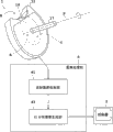
本实施例的乳腺摄影用核医学诊断装置是诊断被检测体(人体)M的乳房组织的装置,具备乳腺摄影用检测器组件(以下简称检测器组件)1、图像处理部3和控制器5。
The nuclear medicine diagnostic apparatus for mammography of this embodiment is a device for diagnosing the breast tissue of the subject (human body) M, and includes a detector unit for mammography (hereinafter referred to as the detector unit) 1, an
检测器组件1具备:检测伽玛射线的检测器11、支承检测器11并使其可旋转的旋转支承部17、用于搁置被检测体M的臂等的搁臂构件18。检测器11呈在具有能够配置乳房的中空部A的闭曲线的一部分形成单一的切口部B而成的形状。换言之,检测器11呈缺少环形状一部分的形状。该检测器11的检测面C形成在检测器11的内侧,以中空部A作为视场。此外,检测器11相当于该发明的伽玛射线检测器。
The
对本实施例的检测器11的具体形状进行说明。所述闭曲线为圆形,检测器11沿切下圆周的一部分而成的曲线设置。作为该中空部A的直径例示为160mm到250mm的范围内的值。另外,作为切口部B的圆弧长度 例示为50mm到150mm。这些尺寸并没有限定于上述范围。
The specific shape of the
检测器11详细而言具有:沿检测器11的形状排列的多个检测器块21;收容上述检测器块21框体23。
Specifically, the
如图3所示,各检测器块21包括:将伽玛射线转换为光的闪烁体31;将光转换为电信号的光电倍增管33;从闪烁体31向光电倍增管33传导光的光导件35等。闪烁体31是将闪烁体片32以行列状(如8行×8列)排列的物体的多层层叠(如两层)的集合体,其上表面为检测器块21的检测面。
As shown in FIG. 3 , each
此外,上述的检测器11的检测面C是通过面向中空部A配置的检测器块21的检测面构成的。因此,严格地说,如图2所示,中空部A是近似于大致圆形的多边形。另外,层叠两层检测器块21时,检测器11成为将闪烁体片32排列16层的多层环。另外,本实施例中,如图2(b)所示,截面观测时,检测面C是相对于中空部A的底面垂直的面。因此,中空部A呈大致圆柱形状(严格地说是多边形状)的柱体。
In addition, the detection surface C of the above-mentioned
旋转支承部17与框体23连结,并且设置有检测器11且使其在任意单轴P的周围旋转自如。本实施例中,单轴P与大致圆形的中空部A的中心轴一致。
The
搁臂构件18是相对于中空部A以凸状弯曲的板状物。搁臂构件18的两端与夹着切口部B的检测器11的两端部连接,闭塞切口部B。搁臂构件18相当于本发明的搁臂构件,另外也相当于本发明的限制构件。
The
检测器组件1还具有闭塞中空部A的一方侧的视场外的板状构件19。此外,板状构件19相当于本发明的板状物。
The
图像处理部3具备放射数据收集部41和RI分布图像生成部43。放射数据收集部41从检测器组件1检测的结果收集放射数据。RI分布图像生成部43根据收集的放射数据进行重建处理,生成二维或三维的RI分布图像。控制器5表示生成的RI分布图像。
The
上述的图像处理部3通过读出规定的程序并执行的中央运算处理装置(CPU)、存储各种信息的RAM(Random-Access Memory)、固定磁盘等存储装置等来实现。
The above-mentioned
接着,对实施例1的乳腺摄影用核医学诊断装置的动作进行说明。图 4是示意地表示检测器11和被检测体M的位置关系的图。
Next, the operation of the nuclear medicine diagnostic apparatus for mammography of the first embodiment will be described. FIG. 4 is a diagram schematically showing the positional relationship between the
如图4所示,在中空部A配置被检测体M的左右的任一个乳房,并且,在切口部B插入与被检测体M的乳房同侧的臂根或/及肩,使臂等与搁臂构件18抵接。由此,检测器11包围乳房的下部,检测器11的一端侧位于直至乳房上方的肩根的范围,检测器11的另一端侧位于直至乳房侧方的腋下的范围。因此,腋下和肩根等乳房周边与乳房被检测器11的检测面C夹持,进入检测器11的视场内。此外,由于具有搁臂构件18,所以臂和肩等的检查对象外部位不会过分进入检测器11的视场。
As shown in FIG. 4, any one of the left and right breasts of the subject M is placed in the hollow part A, and the root of the arm or/and the shoulder on the same side as the breast of the subject M is inserted into the incision part B, and the arms and the like are aligned with the breasts of the subject M. The
其次,向被检测体M投入用正电子放射性同位素标记的放射性药剂。正电子放射性同位素在被检测体M内以彼此成180度向相反方向释放两根伽玛射线。检测器11检测乳房及乳房周边释放并到达检测面C的伽玛射线,并输出电信号。
Next, a radiopharmaceutical labeled with a positron radioactive isotope is administered to the subject M. The positron radioactive isotope emits two gamma rays in opposite directions at 180 degrees to each other in the subject M. The
放射数据收集部41从检测器11的检测结果收集放射数据。具体而言,将从检测器11输出的各电信号暂时与其位置信息和时间信息逐次记录在存储器,当基于检测器11的检测结束时,从存储器读出数据并判定同时计数的事件,计数该事件获得一致数据。
The radiation
RI分布图像生成部43根据收集的放射数据进行重建处理,生成二维或三维的RI分布图像。作为重建处理优选逐次近似图像重建法(例如,OSEM(Ordered Subset Expectation Maximization有序子集最大期望值)算法(参照Takayuki Nakamura,Hiroyuki Kudo IEEE Nuclear Science Symposium Conference Record 2005pp、1950-54))。利用该方法,能够生成充分抑制切口部B的影响的RI分布图像。
The RI
例如,在此虽未图示,但将纵横1.5mm、长4.5mm的闪烁体片在长度方向上层叠4层,将纵横各32根排列的12个检测器块与直径208mm的圆内接并均等配置,在作为切口部B抽出一个检测器块的情况与不抽出的情况下,利用OSEM算法的重建方法进行模拟的结果,可知自视场中心70mm的空间分辨率相对于无切口部B时的1.3mm,在有切口部B时得到1.6mm,从因而利用该重建方法几乎没有切口部B的影响。 For example, although not shown here, four scintillator sheets of 1.5 mm in length and 4.5 mm in length are stacked in the longitudinal direction, and 12 detector blocks arranged 32 in each of length and width are inscribed on a circle with a diameter of 208 mm. Evenly arranged, in the case of extracting one detector block as the notch B and not extracting it, the simulation results using the reconstruction method of the OSEM algorithm show that the spatial resolution of 70mm from the center of the field of view is compared to that of the case without the notch B 1.3 mm, 1.6 mm is obtained with the cutout B, so that there is almost no influence of the cutout B with this reconstruction method. the
此外,参照代替逐次近似图像重建法,也可选择3D傅里叶变换法、3D-FBP法、重组算法(rebinning)、3D再投影法、FORE法(Fourier rebinning) 等现有方法。 In addition, instead of the successive approximation image reconstruction method, existing methods such as 3D Fourier transform method, 3D-FBP method, rebinning algorithm (rebinning), 3D reprojection method, FORE method (Fourier rebinning) can also be selected. the
生成的RI分布图像适当地向控制器5输出。
The generated RI distribution image is appropriately output to the
当左右任一方的乳房诊断结束时,操作者等手动操作旋转支承部17,使检测器11绕单轴P周围旋转。由此,改变检测器11的姿势以容易诊断另一方的乳房。并且,用同样的顺序对另一方乳房进行诊断。
When the diagnosis of either the left or right breast is completed, the operator or the like manually operates the
如上所述,根据实施例1的乳腺摄影用核医学诊断装置,由于具有能够使乳房和乳房周边同时进入视场的检测器11,所以能够检测从乳房和乳房周边释放的伽玛射线。由此,对除乳房之外的乳房周边存在的乳房组织也能够适当地进行肿瘤的有无、位置、恶性度等的诊断。由此,能够更加确切地实施乳癌的诊断。
As described above, the nuclear medicine diagnostic apparatus for mammography according to
另外,通过将中空部A的直径设为160mm到200mm,将切口部B的圆弧长度设为50mm到100mm,能够使乳房合适地配置在中空部A,并且使臂根或肩插入切口部B。 In addition, by setting the diameter of the hollow part A to 160 mm to 200 mm and the arc length of the cutout part B to 50 mm to 100 mm, the breast can be properly placed in the hollow part A, and the base of the arm or the shoulder can be inserted into the cutout part B. . the
另外,检测器组件1具有旋转支承部17,因此能够容易变更分别对左右乳房容易的诊断的检测器11的姿势。
In addition, since the
另外,检测器组件1具有闭塞中空部A的一方侧的视场外的板状构件19,所以能够防止乳房从视场溢出,能够适当地诊断整个乳房。
In addition, since the
另外,检测器组件1具有搁臂构件18,所以能够防止臂等过分进入。另外,被检测体M可获得舒适的姿势,所以能够降低被检测体M的负担。
In addition, since the
另外,通过具有图像处理部3,能够确切地生成RI分布图像。
In addition, by including the
本发明不限于上述实施方式,能够实施下述的变形。 The present invention is not limited to the above-described embodiments, and the following modifications can be implemented. the
(1)上述的各实施例中,闭曲线是位于同一平面上的圆,检测器11沿切下圆周的一部分的曲线设置,但不限于此,只要是在一部分具有切口部的闭曲线或多边形,可适当选择各种形状。从而,也不限定在同一平面上。
(1) In each of the above-mentioned embodiments, the closed curve is a circle located on the same plane, and the
参照图5、图6。图5(a)是变形实施例的检测器的水平剖视图,(b)是其垂直剖视图。图6是示意地表示该变形实施例的检测器12和被检测体M的位置关系的图。如图5、图6所示,将闭曲线作成泪珠形,检测器12呈沿着该泪珠形的轮廓的一部分切下而成的曲线的形状。切口部B形成在泪珠形的轮廓逐渐变细的上端附近。检测器12的视场是中空部A1、 A2的区域。此外,实施例1说明的检测器11与检测器12的横向宽度为相同程度时,检测器11的视场只得到大致相当于中空部A1的视场。如上所述,根据变形实施例的检测器12,除中空部A1之外还能够将中空部A2作为视场,所以能够使乳房周边更加宽广的范围进入视场(在图6中,进入检测器12的视场的乳房及乳房周边用剖面线明示)。
Referring to Fig. 5 and Fig. 6 . Fig. 5(a) is a horizontal sectional view of a detector of a modified embodiment, and (b) is a vertical sectional view thereof. FIG. 6 is a diagram schematically showing the positional relationship between the
另外,除图5、6所示的变形实施例示出的检测器12的形状以外,也能够构成呈马蹄形、U字形或C字形的检测器。另外,也能够代替用图5说明的泪珠形,选择椭圆形、橄榄球形、其他各种轮廓形状,将其切口而成的形状的检测器。
In addition to the shape of the
(2)上述的实施例中,对设置切口部B的位置没有特别地进行说明,但是有适当选择的事项。例如,能够适当地选择闭曲线的轮廓的长度方向、或宽度方向或其他位置。参照图7。图7是变形实施例的检测器的水平剖视图。图示的检测器13呈切下椭圆形的轮廓的一部分而成的形状。这里,切口部B形成在椭圆形的短轴与椭圆形的轮廓交叉的部分。
(2) In the above-mentioned embodiment, the position where the cutout portion B is provided is not particularly described, but there are matters to be selected appropriately. For example, the longitudinal direction, the width direction, or other positions of the contour of the closed curve can be appropriately selected. Refer to Figure 7. Fig. 7 is a horizontal sectional view of a detector of a modified embodiment. The illustrated
(3)另外,在检测器11的形状中,也能够将在夹着切口部B的检测器11的两端部分的曲率半径变更为比检测器11的端部以外的部分大。或者,若检测器11的两端部分不是曲线时,也能够变形为在夹着切口部B的两端部分的各检测面C成为互相平行、或近似平行的关系。由此,能够将乳房周边确切地且广范围地进入视场。具体而言,将用图5、6说明的变形实施例作为一例例举。即,图5等所示的检测器12的夹着切口部B的检测器12的两端部分的曲率半径比检测器12的端部以外的部分大。
(3) In addition, in the shape of the
(4)上述的实施例中,中空部A呈柱体,但不限于此。例如,也能够变更为将检测器11的检测面C相对于中空部A的底面倾斜。具体而言,如图5(b)所示的检测器12也能够构成为具备相对于中空部A1、A2的底面倾斜的检测面。由此,能够使检测面靠近乳房及乳房周边。
(4) In the above-mentioned embodiments, the hollow portion A is a cylinder, but it is not limited thereto. For example, it is also possible to change the detection surface C of the
(5)另外,在上述的实施例中,检测器11是切下了闭曲线的一部分的形状,但不限于此。例如,也能够构成为呈在穹顶形状形成切口部而成的形状,所述穹顶形状具有在内部可收容乳房的空洞部。穹顶形状例示为半球形状。另外,穹顶形状也可以适当地变更为圆锥或角锥等锥体形状。
(5) In addition, in the above-mentioned embodiment, the
图8(a)是变形实施例的检测器的立体图,(b)是其垂直剖视图。该 变形实施例是以中空部A呈锥体(大致圆锥形状)的方式构成检测器14。具体而言,将检测器14的检测面C相对于中空部A的底面倾斜,闭塞中空部A的上方。根据如此构成的检测器14,通过配置为将乳房整体收容于中空部A,能够使乳房整体能够容易地进入视场。另外,可以省略实施例1说明的板状构件19。
Fig. 8(a) is a perspective view of a detector of a modified embodiment, and Fig. 8(b) is a vertical sectional view thereof. In this modified example, the
(6)上述的实施例中,通过检测器11形成的中空部A是固定的,但是也可以构成为中空部A可扩大缩小的检测器。
(6) In the above-mentioned embodiment, the hollow portion A formed by the
参照图9。图9是变形实施例的检测器15的水平剖视图。检测器15划分为两个分割检测器15a和15b。各分割检测器15a、15b具有各个检测器块21和收容检测器块21的框体24a、24b。
Refer to Figure 9. FIG. 9 is a horizontal cross-sectional view of a
各分割检测器15a、15b使这些的一端彼此分离,在其间形成切口部B。另外,在另一端侧,使各分割检测器15a、15b的检测面连续地接合另一端彼此。并且,分割检测器15a、15b通过分割转动保持部51轴支承并转动自如。分割转动保持部51设在分割检测器15a、15b彼此接合的另一端附近的位置,在保持各分割检测器15a、15b的检测面的连续性的同时,保持分割检测器15b并使其转动自如。此外,该转动动作可以无阶梯地转动,也可2个梯度等、阶梯状转动。另外,即使因配置分割转动保持部51的空间的关系,分割检测器15a、15b的检测面不是紧密地连接,但只要使各检测面接近到诊断上无影响的范围即可。另外,即使分割检测器15a、15b的另一端彼此没有紧密连接,在其之间产生微小的间隙,但如上所述只要实质上在另一端侧使检测面连接,就包含于接合状态。
Each of the
并且,通过实施者的手动操作,使分割检测器15转动,可以根据乳房的大小和形状等扩大缩小中空部A。
In addition, the
此外,在上述变形例中,分割检测器15a被固定地设置,但是对于分割检测器15a,也可以构成为被分割转动保持部51转动自如地保持。
In addition, in the modification described above, the
(7)在上述实施例中,旋转支承部17在与中空部A的中心轴一致的单轴P周围使检测器11旋转自如地支承检测器11,但是不限于此。作为单轴P,可以另行选择与中空部A的中心轴平行的轴。另外,作为单轴P,也可以适当地选择与中空部A的中心轴不平行的轴。
(7) In the above-mentioned embodiment, the
(8)在上述实施例中,作为进入检测器11视场的乳房周边,对腋下 或/及肩根为例进行了说明,但是不限于此。即,只要是存在乳房组织的乳房周边的部位,可适当的选择变更。
(8) In the foregoing embodiment, as the breast periphery entering the field of view of the
(9)上述的旋转支承部17或分割转动保持部51均为手动,但是也可以构成为另行设置驱动机构。
(9) Both the above-mentioned
(10)在上述实施例中,搁臂构件18呈弯曲板状,但是不限于此,也可变更为平板状等合适的形状。另外,搁臂构件18闭塞切口部B,但是不限于此,也可以构成为不闭塞。即,只要能够防止臂和肩过分进入,可适当变更搁臂构件18的安装位置和形状,其结果,也可以存在间隙。另外,搁臂构件18用于搁置臂和肩,并没有限定为仅搁置臂。
(10) In the above-mentioned embodiment, the
(11)在上述的实施例中,检测器11的检测器块21为两层层叠,但是不限于此。例如,也可以将检测器块21设为一层或3层以上。另外,检测器11是闪烁体片32多层排列的多层环,但是不限于此,也可以变更为闪烁体片32仅一层排列。
(11) In the above-mentioned embodiment, the
(12)在上述的实施例中,检测器11没有沿中空部A的中心轴P方向扭转(即,从检测器11的一端侧到另一端侧对中心轴P无位移),但是不限于此。参照图10。图10是变形实施例的检测器的立体图。如图所示,也可以使检测器16相对于其中空部A的轴Q位移。换言之,也可变更为呈在中空部A的轴Q方向扭转的形状的检测器16。
(12) In the above-mentioned embodiment, the
(13)上述的实施例是具有圆形的中空部A的检测器11,但是不限于此。也可以根据提供的实际的或统计的被检测体(人体)M的体表、形状,适当地设计检测器的形状。具体而言,也可变更为呈从腋下到乳房的下部下降,沿乳房的下部折回延伸至乳房上方的肩根的形状的检测器。这时,优选为能够沿体表设置检测器的形状。
(13) The above-mentioned embodiment is the
(14)也可以适当地组合上述的各实施例及各变形例说明的各结构,构成乳腺摄影用检测器组件或乳腺摄影用核医学诊断装置。 (14) It is also possible to appropriately combine the structures described in the above-mentioned embodiments and modifications to form a mammography detector unit or a mammography nuclear medicine diagnostic device. the
Claims (30)
Applications Claiming Priority (1)
| Application Number | Priority Date | Filing Date | Title |
|---|---|---|---|
| PCT/JP2006/326131 WO2008081525A1 (en) | 2006-12-27 | 2006-12-27 | Detection unit for mammography and nuclear medicine diagnosis apparatus for mammography provided therewith |
Related Child Applications (1)
| Application Number | Title | Priority Date | Filing Date |
|---|---|---|---|
| CN2011103554195A Division CN102499704A (en) | 2006-12-27 | 2006-12-27 | Mammography detector component and mammography nuclear-medicine diagnostic apparatus with same |
Publications (2)
| Publication Number | Publication Date |
|---|---|
| CN101568854A CN101568854A (en) | 2009-10-28 |
| CN101568854B true CN101568854B (en) | 2012-06-13 |
Family
ID=39588216
Family Applications (1)
| Application Number | Title | Priority Date | Filing Date |
|---|---|---|---|
| CN200680056788.1A Expired - Fee Related CN101568854B (en) | 2006-12-27 | 2006-12-27 | Detector unit for mammography and nuclear medicine diagnostic device for mammography including same |
Country Status (5)
| Country | Link |
|---|---|
| US (1) | US7961840B2 (en) |
| EP (1) | EP2105762B1 (en) |
| JP (1) | JP4905462B2 (en) |
| CN (1) | CN101568854B (en) |
| WO (1) | WO2008081525A1 (en) |
Families Citing this family (9)
| Publication number | Priority date | Publication date | Assignee | Title |
|---|---|---|---|---|
| WO2008072343A1 (en) * | 2006-12-15 | 2008-06-19 | Shimadzu Corporation | Positron ct apparatus |
| JP5622487B2 (en) * | 2009-09-14 | 2014-11-12 | 株式会社東芝 | Radiation diagnostic apparatus and image reconstruction method |
| JP2012013681A (en) * | 2010-06-04 | 2012-01-19 | Toshiba Corp | Nuclear medicine imaging device, method, and program |
| US8842806B2 (en) * | 2012-04-03 | 2014-09-23 | Carestream Health, Inc. | Apparatus and method for breast imaging |
| DE102012208305B4 (en) | 2012-05-16 | 2022-10-20 | Fraunhofer-Gesellschaft zur Förderung der angewandten Forschung e.V. | X-ray detector and X-ray system |
| JP6061157B2 (en) * | 2013-01-31 | 2017-01-18 | 株式会社島津製作所 | Nuclear medicine diagnostic equipment |
| US9326739B2 (en) * | 2014-04-28 | 2016-05-03 | Cheryl A. Galambos McLaughlin | Mammogram table |
| WO2017106838A1 (en) * | 2015-12-18 | 2017-06-22 | University Of Virginia Patent Foundation | Time-of-flight positron emission tomography (tofpet) assembly and related method thereof |
| JP2020085481A (en) * | 2018-11-16 | 2020-06-04 | 株式会社島津製作所 | Nuclear medicine diagnosis device |
Citations (5)
| Publication number | Priority date | Publication date | Assignee | Title |
|---|---|---|---|---|
| CN1041459A (en) * | 1988-09-30 | 1990-04-18 | 株式会社岛津制作所 | The radioscopy camera |
| CN1279594A (en) * | 1997-11-26 | 2001-01-10 | 成象诊断系统公司 | Time-resolved breast imaging device |
| CN1429089A (en) * | 2000-05-09 | 2003-07-09 | 成象诊断系统公司 | Medical optical imaging scanner using multiple wavelength simultaneous data acquisition for breast imaging |
| CN1429088A (en) * | 2000-04-14 | 2003-07-09 | 成象诊断系统公司 | Scanner for medical optical imaging device employing suppression of optical reflections |
| US6794653B2 (en) * | 1999-06-06 | 2004-09-21 | Elgems Ltd. | SPECT for breast cancer detection |
Family Cites Families (10)
| Publication number | Priority date | Publication date | Assignee | Title |
|---|---|---|---|---|
| US5451789A (en) * | 1993-07-19 | 1995-09-19 | Board Of Regents, The University Of Texas System | High performance positron camera |
| JP4230019B2 (en) * | 1998-09-02 | 2009-02-25 | 株式会社日立メディコ | Gamma camera |
| US20040021083A1 (en) * | 2000-06-07 | 2004-02-05 | Nelson Robert Sigurd | Device and system for improved Compton scatter imaging in nuclear medicine {and mammography} |
| JP2004533607A (en) * | 2001-01-16 | 2004-11-04 | ボード・オブ・リージェンツ,ザ・ユニヴァーシティ・オヴ・テキサス・システム | PET camera with individually rotatable detector module and / or individually movable shield part |
| JP3900992B2 (en) * | 2002-04-02 | 2007-04-04 | 株式会社日立製作所 | Radiation detector and radiation inspection apparatus |
| US20030194050A1 (en) | 2002-04-15 | 2003-10-16 | General Electric Company | Multi modality X-ray and nuclear medicine mammography imaging system and method |
| RU2391683C2 (en) * | 2005-02-10 | 2010-06-10 | Конинклейке Филипс Электроникс Н.В. | Portable x-ray detector plate with shock absorption |
| US20100177866A1 (en) | 2005-04-01 | 2010-07-15 | Keizi Shibuya | Mammography Equipment |
| US7492858B2 (en) * | 2005-05-20 | 2009-02-17 | Varian Medical Systems, Inc. | System and method for imaging and treatment of tumorous tissue in breasts using computed tomography and radiotherapy |
| US9072441B2 (en) * | 2006-08-08 | 2015-07-07 | Ge Medical Systems Israel, Ltd. | Method and apparatus for imaging using multiple imaging detectors |
-
2006
- 2006-12-27 CN CN200680056788.1A patent/CN101568854B/en not_active Expired - Fee Related
- 2006-12-27 US US12/521,567 patent/US7961840B2/en not_active Expired - Fee Related
- 2006-12-27 JP JP2008551986A patent/JP4905462B2/en active Active
- 2006-12-27 EP EP06843513.0A patent/EP2105762B1/en not_active Not-in-force
- 2006-12-27 WO PCT/JP2006/326131 patent/WO2008081525A1/en not_active Ceased
Patent Citations (5)
| Publication number | Priority date | Publication date | Assignee | Title |
|---|---|---|---|---|
| CN1041459A (en) * | 1988-09-30 | 1990-04-18 | 株式会社岛津制作所 | The radioscopy camera |
| CN1279594A (en) * | 1997-11-26 | 2001-01-10 | 成象诊断系统公司 | Time-resolved breast imaging device |
| US6794653B2 (en) * | 1999-06-06 | 2004-09-21 | Elgems Ltd. | SPECT for breast cancer detection |
| CN1429088A (en) * | 2000-04-14 | 2003-07-09 | 成象诊断系统公司 | Scanner for medical optical imaging device employing suppression of optical reflections |
| CN1429089A (en) * | 2000-05-09 | 2003-07-09 | 成象诊断系统公司 | Medical optical imaging scanner using multiple wavelength simultaneous data acquisition for breast imaging |
Non-Patent Citations (3)
| Title |
|---|
| JP特开2000-75035A 2000.03.14 |
| JP特开2003-325499A 2003.11.18 |
| JP特表2004-533607A 2004.11.04 |
Also Published As
| Publication number | Publication date |
|---|---|
| EP2105762B1 (en) | 2016-07-13 |
| WO2008081525A1 (en) | 2008-07-10 |
| EP2105762A1 (en) | 2009-09-30 |
| JP4905462B2 (en) | 2012-03-28 |
| JPWO2008081525A1 (en) | 2010-04-30 |
| CN101568854A (en) | 2009-10-28 |
| US7961840B2 (en) | 2011-06-14 |
| EP2105762A4 (en) | 2013-08-21 |
| US20100322379A1 (en) | 2010-12-23 |
Similar Documents
| Publication | Publication Date | Title |
|---|---|---|
| US20250114057A1 (en) | Systems and methods for controlling motion of detectors having moving detector heads | |
| US7521681B2 (en) | Non-rotating transaxial radionuclide imaging | |
| US6225631B1 (en) | Non-invasive radio-imaging analysis, in particular for examining small animals in vivo, and method for using same | |
| EP3449834B1 (en) | Pet imaging device for observing the brain | |
| US20150335301A1 (en) | Diagnostic imaging system and method using multiple types of imaging detectors | |
| JP2006516742A (en) | Single photon emission computed tomography system | |
| CN101568854B (en) | Detector unit for mammography and nuclear medicine diagnostic device for mammography including same | |
| EP2578149B1 (en) | Variable pet device | |
| US8294109B2 (en) | Extracting location information using difference images from a non-parallel hole collimator | |
| CN101176016B (en) | Multi-cap detectors for nuclear medicine | |
| CN113287056A (en) | Medical imaging system based on collimator and detector | |
| US7825383B2 (en) | Mobile camera for organ targeted imaging | |
| US20170273644A1 (en) | Method and system for performing an imaging scan of a subject | |
| US7375338B1 (en) | Swappable collimators method and system | |
| EP3404449A1 (en) | Systems and methods for improved collimation sensitivity | |
| JP4983963B2 (en) | Radiation detector unit | |
| Archer et al. | Implementation and initial characterization of acquisition orbits with a dedicated emission mammotomograph | |
| US11207046B2 (en) | Methods and systems for a multi-modal medical imaging system | |
| JP3135182U (en) | Radiation detector unit | |
| CN102499704A (en) | Mammography detector component and mammography nuclear-medicine diagnostic apparatus with same | |
| EP1996960A2 (en) | Nuclear medicine imaging system with high efficiency transmission measurement | |
| JP2008089396A (en) | Gamma camera system | |
| JP2009042029A (en) | PET equipment | |
| Huber et al. | Characterization of a PET camera optimized for prostate imaging | |
| EP1921467A1 (en) | Tomograph, tomography, tomography program, and computer-readable recording medium where the program is recorded |
Legal Events
| Date | Code | Title | Description |
|---|---|---|---|
| C06 | Publication | ||
| PB01 | Publication | ||
| C10 | Entry into substantive examination | ||
| SE01 | Entry into force of request for substantive examination | ||
| C14 | Grant of patent or utility model | ||
| GR01 | Patent grant | ||
| CF01 | Termination of patent right due to non-payment of annual fee | ||
| CF01 | Termination of patent right due to non-payment of annual fee |
Granted publication date: 20120613 Termination date: 20191227 |