CN101421395A - Cytotoxicity mediation of cells evidencing surface expression of CD63 - Google Patents
Cytotoxicity mediation of cells evidencing surface expression of CD63 Download PDFInfo
- Publication number
- CN101421395A CN101421395A CNA2007800127439A CN200780012743A CN101421395A CN 101421395 A CN101421395 A CN 101421395A CN A2007800127439 A CNA2007800127439 A CN A2007800127439A CN 200780012743 A CN200780012743 A CN 200780012743A CN 101421395 A CN101421395 A CN 101421395A
- Authority
- CN
- China
- Prior art keywords
- monoclonal antibody
- antibody
- preserving number
- cell line
- isolating
- Prior art date
- Legal status (The legal status is an assumption and is not a legal conclusion. Google has not performed a legal analysis and makes no representation as to the accuracy of the status listed.)
- Pending
Links
Images


Classifications
-
- A—HUMAN NECESSITIES
- A61—MEDICAL OR VETERINARY SCIENCE; HYGIENE
- A61K—PREPARATIONS FOR MEDICAL, DENTAL OR TOILETRY PURPOSES
- A61K47/00—Medicinal preparations characterised by the non-active ingredients used, e.g. carriers or inert additives; Targeting or modifying agents chemically bound to the active ingredient
- A61K47/50—Medicinal preparations characterised by the non-active ingredients used, e.g. carriers or inert additives; Targeting or modifying agents chemically bound to the active ingredient the non-active ingredient being chemically bound to the active ingredient, e.g. polymer-drug conjugates
- A61K47/51—Medicinal preparations characterised by the non-active ingredients used, e.g. carriers or inert additives; Targeting or modifying agents chemically bound to the active ingredient the non-active ingredient being chemically bound to the active ingredient, e.g. polymer-drug conjugates the non-active ingredient being a modifying agent
- A61K47/68—Medicinal preparations characterised by the non-active ingredients used, e.g. carriers or inert additives; Targeting or modifying agents chemically bound to the active ingredient the non-active ingredient being chemically bound to the active ingredient, e.g. polymer-drug conjugates the non-active ingredient being a modifying agent the modifying agent being an antibody, an immunoglobulin or a fragment thereof, e.g. an Fc-fragment
- A61K47/6835—Medicinal preparations characterised by the non-active ingredients used, e.g. carriers or inert additives; Targeting or modifying agents chemically bound to the active ingredient the non-active ingredient being chemically bound to the active ingredient, e.g. polymer-drug conjugates the non-active ingredient being a modifying agent the modifying agent being an antibody, an immunoglobulin or a fragment thereof, e.g. an Fc-fragment the modifying agent being an antibody or an immunoglobulin bearing at least one antigen-binding site
- A61K47/6851—Medicinal preparations characterised by the non-active ingredients used, e.g. carriers or inert additives; Targeting or modifying agents chemically bound to the active ingredient the non-active ingredient being chemically bound to the active ingredient, e.g. polymer-drug conjugates the non-active ingredient being a modifying agent the modifying agent being an antibody, an immunoglobulin or a fragment thereof, e.g. an Fc-fragment the modifying agent being an antibody or an immunoglobulin bearing at least one antigen-binding site the antibody targeting a determinant of a tumour cell
-
- G01N33/5759—
-
- A—HUMAN NECESSITIES
- A61—MEDICAL OR VETERINARY SCIENCE; HYGIENE
- A61K—PREPARATIONS FOR MEDICAL, DENTAL OR TOILETRY PURPOSES
- A61K47/00—Medicinal preparations characterised by the non-active ingredients used, e.g. carriers or inert additives; Targeting or modifying agents chemically bound to the active ingredient
- A61K47/50—Medicinal preparations characterised by the non-active ingredients used, e.g. carriers or inert additives; Targeting or modifying agents chemically bound to the active ingredient the non-active ingredient being chemically bound to the active ingredient, e.g. polymer-drug conjugates
- A61K47/51—Medicinal preparations characterised by the non-active ingredients used, e.g. carriers or inert additives; Targeting or modifying agents chemically bound to the active ingredient the non-active ingredient being chemically bound to the active ingredient, e.g. polymer-drug conjugates the non-active ingredient being a modifying agent
- A61K47/68—Medicinal preparations characterised by the non-active ingredients used, e.g. carriers or inert additives; Targeting or modifying agents chemically bound to the active ingredient the non-active ingredient being chemically bound to the active ingredient, e.g. polymer-drug conjugates the non-active ingredient being a modifying agent the modifying agent being an antibody, an immunoglobulin or a fragment thereof, e.g. an Fc-fragment
- A61K47/6835—Medicinal preparations characterised by the non-active ingredients used, e.g. carriers or inert additives; Targeting or modifying agents chemically bound to the active ingredient the non-active ingredient being chemically bound to the active ingredient, e.g. polymer-drug conjugates the non-active ingredient being a modifying agent the modifying agent being an antibody, an immunoglobulin or a fragment thereof, e.g. an Fc-fragment the modifying agent being an antibody or an immunoglobulin bearing at least one antigen-binding site
- A61K47/6849—Medicinal preparations characterised by the non-active ingredients used, e.g. carriers or inert additives; Targeting or modifying agents chemically bound to the active ingredient the non-active ingredient being chemically bound to the active ingredient, e.g. polymer-drug conjugates the non-active ingredient being a modifying agent the modifying agent being an antibody, an immunoglobulin or a fragment thereof, e.g. an Fc-fragment the modifying agent being an antibody or an immunoglobulin bearing at least one antigen-binding site the antibody targeting a receptor, a cell surface antigen or a cell surface determinant
-
- A—HUMAN NECESSITIES
- A61—MEDICAL OR VETERINARY SCIENCE; HYGIENE
- A61K—PREPARATIONS FOR MEDICAL, DENTAL OR TOILETRY PURPOSES
- A61K51/00—Preparations containing radioactive substances for use in therapy or testing in vivo
- A61K51/02—Preparations containing radioactive substances for use in therapy or testing in vivo characterised by the carrier, i.e. characterised by the agent or material covalently linked or complexing the radioactive nucleus
- A61K51/04—Organic compounds
- A61K51/08—Peptides, e.g. proteins, carriers being peptides, polyamino acids, proteins
- A61K51/10—Antibodies or immunoglobulins; Fragments thereof, the carrier being an antibody, an immunoglobulin or a fragment thereof, e.g. a camelised human single domain antibody or the Fc fragment of an antibody
- A61K51/1027—Antibodies or immunoglobulins; Fragments thereof, the carrier being an antibody, an immunoglobulin or a fragment thereof, e.g. a camelised human single domain antibody or the Fc fragment of an antibody against receptors, cell-surface antigens or cell-surface determinants
-
- A—HUMAN NECESSITIES
- A61—MEDICAL OR VETERINARY SCIENCE; HYGIENE
- A61K—PREPARATIONS FOR MEDICAL, DENTAL OR TOILETRY PURPOSES
- A61K51/00—Preparations containing radioactive substances for use in therapy or testing in vivo
- A61K51/02—Preparations containing radioactive substances for use in therapy or testing in vivo characterised by the carrier, i.e. characterised by the agent or material covalently linked or complexing the radioactive nucleus
- A61K51/04—Organic compounds
- A61K51/08—Peptides, e.g. proteins, carriers being peptides, polyamino acids, proteins
- A61K51/10—Antibodies or immunoglobulins; Fragments thereof, the carrier being an antibody, an immunoglobulin or a fragment thereof, e.g. a camelised human single domain antibody or the Fc fragment of an antibody
- A61K51/1045—Antibodies or immunoglobulins; Fragments thereof, the carrier being an antibody, an immunoglobulin or a fragment thereof, e.g. a camelised human single domain antibody or the Fc fragment of an antibody against animal or human tumor cells or tumor cell determinants
-
- A—HUMAN NECESSITIES
- A61—MEDICAL OR VETERINARY SCIENCE; HYGIENE
- A61P—SPECIFIC THERAPEUTIC ACTIVITY OF CHEMICAL COMPOUNDS OR MEDICINAL PREPARATIONS
- A61P35/00—Antineoplastic agents
-
- C—CHEMISTRY; METALLURGY
- C07—ORGANIC CHEMISTRY
- C07K—PEPTIDES
- C07K16/00—Immunoglobulins [IGs], e.g. monoclonal or polyclonal antibodies
- C07K16/18—Immunoglobulins [IGs], e.g. monoclonal or polyclonal antibodies against material from animals or humans
- C07K16/28—Immunoglobulins [IGs], e.g. monoclonal or polyclonal antibodies against material from animals or humans against receptors, cell surface antigens or cell surface determinants
- C07K16/2896—Immunoglobulins [IGs], e.g. monoclonal or polyclonal antibodies against material from animals or humans against receptors, cell surface antigens or cell surface determinants against molecules with a "CD"-designation, not provided for elsewhere
-
- C—CHEMISTRY; METALLURGY
- C07—ORGANIC CHEMISTRY
- C07K—PEPTIDES
- C07K16/00—Immunoglobulins [IGs], e.g. monoclonal or polyclonal antibodies
- C07K16/18—Immunoglobulins [IGs], e.g. monoclonal or polyclonal antibodies against material from animals or humans
- C07K16/28—Immunoglobulins [IGs], e.g. monoclonal or polyclonal antibodies against material from animals or humans against receptors, cell surface antigens or cell surface determinants
- C07K16/30—Immunoglobulins [IGs], e.g. monoclonal or polyclonal antibodies against material from animals or humans against receptors, cell surface antigens or cell surface determinants from tumour cells
-
- C—CHEMISTRY; METALLURGY
- C12—BIOCHEMISTRY; BEER; SPIRITS; WINE; VINEGAR; MICROBIOLOGY; ENZYMOLOGY; MUTATION OR GENETIC ENGINEERING
- C12N—MICROORGANISMS OR ENZYMES; COMPOSITIONS THEREOF; PROPAGATING, PRESERVING, OR MAINTAINING MICROORGANISMS; MUTATION OR GENETIC ENGINEERING; CULTURE MEDIA
- C12N5/00—Undifferentiated human, animal or plant cells, e.g. cell lines; Tissues; Cultivation or maintenance thereof; Culture media therefor
- C12N5/10—Cells modified by introduction of foreign genetic material
- C12N5/12—Fused cells, e.g. hybridomas
-
- G—PHYSICS
- G01—MEASURING; TESTING
- G01N—INVESTIGATING OR ANALYSING MATERIALS BY DETERMINING THEIR CHEMICAL OR PHYSICAL PROPERTIES
- G01N33/00—Investigating or analysing materials by specific methods not covered by groups G01N1/00 - G01N31/00
- G01N33/48—Biological material, e.g. blood, urine; Haemocytometers
- G01N33/50—Chemical analysis of biological material, e.g. blood, urine; Testing involving biospecific ligand binding methods; Immunological testing
- G01N33/53—Immunoassay; Biospecific binding assay; Materials therefor
- G01N33/569—Immunoassay; Biospecific binding assay; Materials therefor for microorganisms, e.g. protozoa, bacteria, viruses
- G01N33/56966—Animal cells
-
- G01N33/5751—
-
- A—HUMAN NECESSITIES
- A61—MEDICAL OR VETERINARY SCIENCE; HYGIENE
- A61K—PREPARATIONS FOR MEDICAL, DENTAL OR TOILETRY PURPOSES
- A61K39/00—Medicinal preparations containing antigens or antibodies
- A61K2039/505—Medicinal preparations containing antigens or antibodies comprising antibodies
-
- C—CHEMISTRY; METALLURGY
- C07—ORGANIC CHEMISTRY
- C07K—PEPTIDES
- C07K2317/00—Immunoglobulins specific features
- C07K2317/20—Immunoglobulins specific features characterized by taxonomic origin
- C07K2317/24—Immunoglobulins specific features characterized by taxonomic origin containing regions, domains or residues from different species, e.g. chimeric, humanized or veneered
-
- C—CHEMISTRY; METALLURGY
- C07—ORGANIC CHEMISTRY
- C07K—PEPTIDES
- C07K2317/00—Immunoglobulins specific features
- C07K2317/70—Immunoglobulins specific features characterized by effect upon binding to a cell or to an antigen
- C07K2317/73—Inducing cell death, e.g. apoptosis, necrosis or inhibition of cell proliferation
- C07K2317/732—Antibody-dependent cellular cytotoxicity [ADCC]
-
- C—CHEMISTRY; METALLURGY
- C07—ORGANIC CHEMISTRY
- C07K—PEPTIDES
- C07K2317/00—Immunoglobulins specific features
- C07K2317/90—Immunoglobulins specific features characterized by (pharmaco)kinetic aspects or by stability of the immunoglobulin
- C07K2317/92—Affinity (KD), association rate (Ka), dissociation rate (Kd) or EC50 value
-
- G—PHYSICS
- G01—MEASURING; TESTING
- G01N—INVESTIGATING OR ANALYSING MATERIALS BY DETERMINING THEIR CHEMICAL OR PHYSICAL PROPERTIES
- G01N2333/00—Assays involving biological materials from specific organisms or of a specific nature
- G01N2333/435—Assays involving biological materials from specific organisms or of a specific nature from animals; from humans
- G01N2333/705—Assays involving receptors, cell surface antigens or cell surface determinants
- G01N2333/70596—Molecules with a "CD"-designation not provided for elsewhere in G01N2333/705
Landscapes
- Health & Medical Sciences (AREA)
- Life Sciences & Earth Sciences (AREA)
- Chemical & Material Sciences (AREA)
- Immunology (AREA)
- General Health & Medical Sciences (AREA)
- Medicinal Chemistry (AREA)
- Engineering & Computer Science (AREA)
- Cell Biology (AREA)
- Proteomics, Peptides & Aminoacids (AREA)
- Organic Chemistry (AREA)
- Molecular Biology (AREA)
- Veterinary Medicine (AREA)
- Public Health (AREA)
- Animal Behavior & Ethology (AREA)
- Pharmacology & Pharmacy (AREA)
- Epidemiology (AREA)
- Biochemistry (AREA)
- Biomedical Technology (AREA)
- Genetics & Genomics (AREA)
- Physics & Mathematics (AREA)
- Bioinformatics & Cheminformatics (AREA)
- Hematology (AREA)
- Urology & Nephrology (AREA)
- Biotechnology (AREA)
- Zoology (AREA)
- Biophysics (AREA)
- Optics & Photonics (AREA)
- Microbiology (AREA)
- Oncology (AREA)
- Pathology (AREA)
- Analytical Chemistry (AREA)
- Food Science & Technology (AREA)
- General Physics & Mathematics (AREA)
- Virology (AREA)
- Tropical Medicine & Parasitology (AREA)
- Wood Science & Technology (AREA)
- General Engineering & Computer Science (AREA)
- Nuclear Medicine, Radiotherapy & Molecular Imaging (AREA)
- General Chemical & Material Sciences (AREA)
- Chemical Kinetics & Catalysis (AREA)
Abstract
Description
发明领域field of invention
本发明涉及癌性疾病的诊断和治疗,尤其涉及肿瘤细胞的细胞毒性的调节;最尤其涉及癌性疾病调节抗体(CDMAB)作为引发细胞毒性反应的手段的用途,所述CDMAB任选地与一种或者多种化疗剂联用。本发明还涉及利用本发明CDMAB的结合分析。The present invention relates to the diagnosis and treatment of cancerous diseases, in particular to the modulation of cytotoxicity of tumor cells; most particularly to the use of cancerous disease-modulating antibodies (CDMABs) as a means of eliciting cytotoxic responses, optionally in combination with a combination of one or more chemotherapeutic agents. The invention also relates to binding assays utilizing the CDMABs of the invention.
发明背景Background of the invention
癌症中的CD63:CD63属于四次跨膜蛋白(tetraspanin)家族中的三型膜蛋白,该家族目前20个成员的特征是均具有四个跨膜片段。几个工作组通过使用针对活化的血小板的、中性粒细胞的和黑色素瘤细胞的全细胞制备物的抗体,独立地确定了CD63的表达。对它们的同源糖蛋白抗原的各自的cDNA进行克隆后确认了这些不同细胞上的抗原为同一种分子。随后,第六次国际白细胞分型研讨会(1996)将这些抗体归类为CD63抗体。在1996年该研讨会之前,CD63曾以多个名称被命名(黑色素瘤1抗原、眼黑色素瘤相关抗原、黑色素瘤相关抗原ME491、溶酶体相关膜糖蛋白3、致密体膜蛋白(granulophysin)、黑色素相关抗原MLA1),这些名称往往与导致发现CD63的部分特性以及确认的抗体有关。因此,CD63也曾被称为ME491抗原(MAb ME491)、神经腺(neuroglandular)抗原(MAbsLS59,LS62,LS76,LS113,LS140,LS152)、Pltgp40(MAbs H5C6,H4F8和H5D2)、人类骨髓基质细胞抗原(MAb 12F12)、骨祖细胞特异性标志物(MAb HOP-26)和整合素相关蛋白(MAb 6H1)。被发现与人类CD63发生交叉反应的其他抗体有8-1H、8-2A(与ME491发生交叉反应)、NKI/C-3和NKI/black-13(Vannegoor and Rumke,1986;Demetrick et al.,1992;Wang et al.,1992)。CD63 in cancer: CD63 belongs to the type III membrane protein in the tetraspanin family, the current 20 members of this family are characterized by four transmembrane segments. Several groups have independently determined CD63 expression by using antibodies against whole cell preparations of activated platelets, neutrophils and melanoma cells. After cloning the respective cDNAs of their cognate glycoprotein antigens, it was confirmed that the antigens on these different cells were the same molecule. Subsequently, the Sixth International Symposium on Leukocyte Typing (1996) classified these antibodies as CD63 antibodies. Prior to the 1996 workshop, CD63 had been named by several names (
利用抗人黑素瘤细胞制备物的众多抗体的一种,即MAb ME491,从黑素瘤cDNA文库最先克隆出CD63。人黑素瘤活检研究表明MAb ME491的反应性与黑素瘤的进展呈负相关。在正常黑色素细胞中,MAb ME491抗体的活性低,在黑色素瘤进展早期阶段(发育异常痣和放射生长期(radial growth phase,RGP)肿瘤)抗体活性升高,在更晚期的黑色素瘤例如垂直生长期(vertialgrowth phase,VGP)和转移性肿瘤中,MAb ME491抗体的活性则下降甚至丧失。应用MAb2.28(抗活化血小板),CD63也在人血小板中被发现并被部分表征,所述MAb 2.28检测活化-依赖的血小板膜53kDa糖蛋白。该分子还与未激活的血小板的内部颗粒膜有关。同一研究中,MAb2.28也用于标记巨核细胞和内皮细胞的内部颗粒,其中它与抗体共定位到组织蛋白酶D,所述组织蛋白酶D是已知的溶酶体小室标志物。关于抗体聚集和表达克隆的后续研究,证明该抗体识别的抗原就是CD63,还证实其存在于溶酶体小室,在那里它与小室-特异性标志物LAMP1和LAMP2共定位。该分子的克隆鉴定其确为CD63,并且属于四次跨膜蛋白家族。CD63 was first cloned from a melanoma cDNA library using one of many antibodies against human melanoma cell preparations, the MAb ME491. Human melanoma biopsy studies demonstrated that MAb ME491 reactivity was inversely correlated with melanoma progression. The activity of the MAb ME491 antibody is low in normal melanocytes, increased in early stages of melanoma progression (dysplastic nevi and radial growth phase (RGP) tumors), and in more advanced melanomas such as vertical growth In vertical growth phase (VGP) and metastatic tumors, the activity of MAb ME491 antibody decreased or even lost. CD63 was also found and partially characterized in human platelets using MAb 2.28 (anti-activated platelets), which detects the activation-dependent platelet membrane 53 kDa glycoprotein. This molecule is also associated with the inner granule membrane of unactivated platelets. In the same study, MAb2.28 was also used to label the internal granules of megakaryocytes and endothelial cells, where it co-localized with antibodies to cathepsin D, a known marker of the lysosomal compartment. Subsequent studies of antibody aggregation and expression clones demonstrated that the antigen recognized by this antibody was CD63 and that it was also present in the lysosomal compartment, where it colocalized with the compartment-specific markers LAMP1 and LAMP2. Cloning of this molecule identified it as CD63 and belonged to the tetraspanin family.
在许多不同组织和细胞中都能够检测到CD63的表达。在细胞水平,人们发现它与质膜结合,也与细胞内晚期内体(late endosomal)的囊状结构结合。某些情况下,细胞活化通过动员细胞内储备的CD63来增加表面表达。在B淋巴细胞,尤其在内体(endosomes)、参与将MHC II类分子复合物输出到表面的外体(exosomes)和分泌囊泡中,还发现CD63与MHCII类分子共定位并发生物理结合。在包括B淋巴细胞和T淋巴细胞、中性粒细胞、乳腺癌和黑素瘤细胞的许多细胞类型中,发现CD63与诸如CD9、CD81、CD11(整合素链αM,L,X)、CD18(整合素链β2)、CD49c(VLA-3或者整合素链α3)、CD49d(整合素链α4)、CD49f(VLA-6或者整合素链α6)和CD29(整合素链β1)的其他四次跨膜蛋白家族的成员相互作用。The expression of CD63 can be detected in many different tissues and cells. At the cellular level, it is found bound to the plasma membrane and also to the sac-like structure of the late endosomal inside the cell. In some cases, cellular activation increases surface expression by mobilizing intracellular reserves of CD63. CD63 has also been found to co-localize and physically associate with MHC class II molecules in B lymphocytes, particularly in endosomes, exosomes and secretory vesicles involved in the export of MHC class II complexes to the surface. In many cell types including B and T lymphocytes, neutrophils, breast cancer and melanoma cells, CD63 is found in association with cells such as CD9, CD81, CD11 (integrin chain α M, L, X ), CD18 (integrin chain β 2 ), CD49c (VLA-3 or integrin chain α 3 ), CD49d (integrin chain α 4 ), CD49f (VLA-6 or integrin chain α 6 ) and CD29 (integrin chain β 1 ) interact with other members of the tetraspanin family.
CD63在癌症中的作用还不清楚。尽管最初几个相互独立的工作组发现CD63参与诸如血小板和粒细胞活化、MHC II类分子依赖的抗原提呈、整合素依赖性细胞粘附和运动以及某些类型癌症的肿瘤进展的多种事件,但其功能还有待进一步充分阐述。虽然目前有证据支持CD63在多种细胞生理事件中的发挥作用,但是还不清楚这些作用是彼此独立的还是具有CD63参与的潜在的共同细胞机制。The role of CD63 in cancer is unclear. Although initially several independent working groups found that CD63 is involved in events such as platelet and granulocyte activation, MHC class II-dependent antigen presentation, integrin-dependent cell adhesion and motility, and tumor progression in some types of cancer , but its function has yet to be fully elucidated. Although current evidence supports the role of CD63 in a variety of cellular physiological events, it is unclear whether these roles are independent of each other or have an underlying common cellular mechanism in which CD63 is involved.
几个工作组已经研究了CD63和某些类型肿瘤进展之间的关系,特别是与黑色素瘤进展之间的关系。除Mab ME491之外,还研发了多种其它的抗CD63单克隆抗体以用于对从携带不同进展阶段肿瘤的患者获得的癌症标本进行免疫组化(immunohistochemical,IHC)染色。观察到着色变浅被作者解释为极可能反映CD63的表达降低,并与肿瘤的晚期进展和转移性特征相关。更近的一项研究还描述了包括CD63在内的几个四次跨膜蛋白家族成员表达水平的明显下调(mRNA定量后)与几种乳腺癌来源的细胞系体外侵袭性的显著相关性。另外一项研究通过差异显示法确定了在去除雌激素的培养乳腺癌细胞中具有CD63。这说明CD63的表达受到类固醇-激素的调节,并且改变的CD63丰度和/或功能也可能与乳腺肿瘤进展有关。Several working groups have investigated the relationship between CD63 and the progression of certain types of tumors, particularly melanoma. In addition to Mab ME491, various other anti-CD63 monoclonal antibodies were developed for immunohistochemical (IHC) staining of cancer specimens obtained from patients with tumors of various advanced stages. The observed lightening of staining was interpreted by the authors as most likely reflecting reduced expression of CD63 and correlating with late progression and metastatic features of tumors. A more recent study also described a marked downregulation (after mRNA quantification) of the expression levels of several tetraspanin family members, including CD63, that correlated significantly with the in vitro invasiveness of several breast cancer-derived cell lines. Another study identified CD63 in estrogen-deprived cultured breast cancer cells by differential display. This suggests that CD63 expression is regulated by steroid-hormones and that altered CD63 abundance and/or function may also be associated with breast tumor progression.
与之相比,利用抗CD63单克隆抗体MAb FC-5.01的研究揭示了其活性表位在不同正常组织中表达不同。尽管该抗体被发现能识别CD63,但它并不能区分早期黑色素瘤和包括转移性黑素瘤在内的更晚期的黑色素瘤(与MAb ME491不同),这表明CD63抗原存在于这些更晚期的肿瘤细胞上,但是在来自不同阶段的肿瘤的细胞中,它的某些表位被掩盖。这可能由于核心CD63多肽的翻译后修饰发生改变,或是由于CD63和其他分子的相互作用,影响了用于抗体识别和结合的特定表位的提供。这些研究结果支持Si和Hersey(1993)所描述的观察结果,即用抗CD63 MAbNKI-C3的染色不能区分来自诸如早期、放射生长期、垂直生长期以及转移性黑素瘤的不同进展阶段的黑素瘤的组织切片。尽管在对来自乳腺癌和非小细胞性肺癌细胞的mRNA的其他研究分析(Adachi et al.,1998;Huang et al.,1998)中,通过定量PCR,发现四次跨膜蛋白分子家族中的两个成员(CD9和CD82)的表达水平与肿瘤进展和患者预后存在明显的相关性,但没有发现CD63存在这种相关性,因为CD63在所有的样本中表达都是相似的。由于这些结果存在明显冲突,因此缺乏能明确证明CD63和肿瘤之间的关系的有力和一致的数据。In contrast, studies using the anti-CD63 monoclonal antibody MAb FC-5.01 revealed that its active epitope is expressed differently in different normal tissues. Although the antibody was found to recognize CD63, it did not distinguish early-stage melanomas from more advanced melanomas, including metastatic melanomas (unlike MAb ME491), suggesting that the CD63 antigen is present in these more advanced tumors cells, but some of its epitopes were masked in cells from tumors of different stages. This may be due to altered post-translational modifications of the core CD63 polypeptide, or due to interactions between CD63 and other molecules that affect the presentation of specific epitopes for antibody recognition and binding. These findings support the observation described by Si and Hersey (1993) that staining with anti-CD63 MAbNKI-C3 cannot distinguish melanoma from different stages of progression such as early, radial growth, vertical growth, and metastatic melanoma. Tumor tissue sections. Although in other research analyzes of mRNA from breast cancer and non-small cell lung cancer cells (Adachi et al., 1998; Huang et al., 1998), by quantitative PCR, it was found that the four-spanin molecular family There was a clear correlation between the expression levels of two members (CD9 and CD82) and tumor progression and patient prognosis, but no such correlation was found for CD63 because CD63 was expressed similarly in all samples. Because of the apparently conflicting nature of these results, robust and consistent data that definitively demonstrate the relationship between CD63 and tumors are lacking.
到目前为止,很少有体内研究试图建立CD63与该分子的晚期肿瘤抑制功能之间的联系。在这些研究中的一项中,经皮下和腹腔注射入无胸腺小鼠的人CD63过度表达的H-RAS转化的NIH-3T3细胞,与亲代无CD63过度表达的小鼠细胞比较,显示出恶性/致瘤性降低的表型,所述表型由减小的肿瘤体积和降低的转移可能性以及增加的生存时间所表明。这表明转化细胞中人CD63的存在会抑制它们的恶性行为。更近期,利用表达人CD63的转基因小鼠模型并诱导对CD63产生耐受的研究工作表明用与牛痘病毒融合的人CD63免疫时,可以抑制注射的人CD63-MHCI型(H-2Kb)共转染鼠黑素瘤细胞的肿瘤生长,并增加生存期。该研究作者指出:由于肿瘤生长的抑制作用仅存在于被注射CD63-MHC-I型共转染细胞,而不是CD63单独转染细胞系的动物中,所以这种治疗作用是T淋巴细胞依赖性的,看起来内源性抗CD63抗体并未参与该保护作用。该解释得到以下事实的支持:经纯化的人CD63预免疫并且显示已产生抗人CD63抗体的野生型动物中,没有抗肿瘤细胞生长的保护作用。Radford等人(1995)利用人CD63转染KM3细胞系(最初认为是人源,后来定性为鼠源)的研究工作表明当将所述细胞皮内注射入无胸腺小鼠时,与使用未转染的KM3亲代细胞观察到的结果比较,该蛋白的表达减慢了这些细胞的生长并降低了其转移潜能,尽管各种转染和未转染细胞系的体外生长率并无显著性差异。这些观察将CD63的潜在作用与其他已知的在体内和体外影响肿瘤细胞生长率的肿瘤抑制基因区分开来。此外,在体外试验中(Radford et al.,1997),抗CD63单克隆抗体ME491的添加不影响相同细胞的体外生长率,所述ME491被发现通过减少细胞的随机运动影响它们的功能。So far, few in vivo studies have attempted to establish a link between CD63 and the molecule's late-stage tumor suppressor function. In one of these studies, human CD63-overexpressing H-RAS-transformed NIH-3T3 cells injected subcutaneously and intraperitoneally into athymic mice displayed malignant / A phenotype of reduced tumorigenicity as evidenced by reduced tumor volume and reduced likelihood of metastasis and increased survival time. This suggests that the presence of human CD63 in transformed cells suppresses their malignant behavior. More recently, work using transgenic mouse models expressing human CD63 and inducing tolerance to CD63 demonstrated that immunization with human CD63 fused to vaccinia virus inhibited the co-transduction of injected human CD63-MHC type I (H-2Kb). Tumor growth and increased survival of melanoma cells stained in mice. The authors noted that since the tumor growth inhibition was only present in animals injected with CD63-MHC-class I co-transfected cells, but not CD63-alone transfected cell lines, this therapeutic effect was T lymphocyte dependent. Yes, it appears that endogenous anti-CD63 antibodies are not involved in this protection. This interpretation is supported by the fact that there was no protection against tumor cell growth in wild type animals pre-immunized with purified human CD63 and shown to have produced anti-human CD63 antibodies. The work of Radford et al. (1995) using human CD63 to transfect the KM3 cell line (initially believed to be of human origin but later characterized as murine) showed that when the cells were injected intradermally into athymic mice, Expression of this protein slowed the growth and reduced metastatic potential of these cells compared to that observed with transfected KM3 parental cells, although the in vitro growth rates of the various transfected and untransfected cell lines did not differ significantly. These observations differentiate the potential role of CD63 from other known tumor suppressor genes that affect tumor cell growth rates in vivo and in vitro. Furthermore, in an in vitro test (Radford et al., 1997), the in vitro growth rate of the same cells was not affected by the addition of the anti-CD63 monoclonal antibody ME491, which was found to affect their function by reducing their random movement.
这项研究还描述了如下观测结果:在诸如层粘连蛋白、纤维连接蛋白、胶原和玻璃粘连蛋白的细胞外基质(ECM)衍生的趋化因子作用下,CD63能促进迁移,并且这种作用可以由β1-型整合素的功能性参与介导,尽管针对整合素的抗体并不能够阻断这些作用。然而,看起来在CD63转染细胞上,玻璃粘连蛋白介导的信号(已知为整合素αvβ5的配体)的作用与由诸如纤维连接蛋白、层粘连蛋白和胶原的其他ECM成分介导的信号的作用之间存在拮抗效应。这提示在在特定条件下,ECM组分存在时,CD63的表达能导致迁移减少,这可能取决于粘附和运动之间的细微平衡。另一项研究中,抗CD63单克隆抗体(MAb 710F)增强了PMA处理的HL-60细胞的粘附性和伸展性,而另一种抗CD63单克隆抗体(MAb 2.28)促进类似作用,但只针对更小部分的细胞群,并且只在加入更大的抗体量时发挥所述作用。这些结果表明尽管目前已经开发了多种抗CD63抗体,但是它们的功能性作用可能存在很大区别。This study also describes the observation that CD63 promotes migration in response to extracellular matrix (ECM)-derived chemokines such as laminin, fibronectin, collagen, and vitronectin, and that this action can Mediated by the functional involvement of β 1 -type integrins, although antibodies against integrins were not able to block these effects. However, it appears that on CD63-transfected cells, vitronectin-mediated signaling (known as a ligand for integrin αvβ5 ) acts in conjunction with other ECM components such as fibronectin, laminin, and collagen. There is an antagonistic effect between the effects of the mediated signals. This suggests that CD63 expression can lead to reduced migration in the presence of ECM components under specific conditions, which may depend on a fine balance between adhesion and motility. In another study, an anti-CD63 mAb (MAb 710F) enhanced the adhesion and extensibility of PMA-treated HL-60 cells, while another anti-CD63 mAb (MAb 2.28) promoted similar effects, but Targets only a smaller fraction of the cell population and only exerts the effect when greater amounts of antibody are added. These results suggest that although a variety of anti-CD63 antibodies have been developed, their functional roles may vary greatly.
四次跨膜蛋白家族可能也参与了细胞增殖。Oren等(1990)描述了识别CD81(TAPA-1)的小鼠MAb 5A6对淋巴瘤细胞系的抗增殖作用。在另一项研究中,人T淋巴细胞的CD37与抗体的结合阻断了CD37诱导的增殖。更近期,用CD37表达缺失(CD37敲除)的小鼠动物模型的研究显示,与来自野生型动物的T淋巴细胞对刀豆蛋白A活化和CD3/T细胞受体结合的响应相比,来自CD37敲除小鼠的T淋巴细胞过度增殖。因此提出:在细胞生长和增殖中的功能性作用可能是四次跨膜蛋白家族的共同特征。最近关于肝母细胞瘤和肝细胞癌细胞的研究显示,这些细胞与抗CD81单克隆抗体的结合导致Erk/MAP激酶通路的活化。已经表明该信号转导通路参与细胞生长和增殖事件。平行研究也表明,过表达人CD81的转染细胞系与模拟转染对照组相比显示增殖增加。因此现有证据已经表明四次跨膜蛋白分子总体,尤其是CD63在细胞生长增殖和细胞粘附/运动相关事件中的作用。因为这两类细胞事件在肿瘤进展和转移过程中起着重要作用,所以这两者是目前热点研究的靶点。The tetraspanin family may also be involved in cell proliferation. Oren et al. (1990) described the antiproliferative effect of the mouse MAb 5A6 recognizing CD81 (TAPA-1) on lymphoma cell lines. In another study, binding of antibodies to CD37 of human T lymphocytes blocked CD37-induced proliferation. More recently, studies using a mouse animal model lacking CD37 expression (CD37 knockout) have shown that T lymphocytes from wild-type animals respond to concanavalin A activation and CD3/T cell receptor binding compared to T lymphocytes from wild-type animals. T lymphocyte hyperproliferation in CD37 knockout mice. Therefore, it is suggested that functional roles in cell growth and proliferation may be a common feature of the tetraspanin family. Recent studies on hepatoblastoma and hepatocellular carcinoma cells have shown that binding of these cells to anti-CD81 monoclonal antibodies leads to activation of the Erk/MAP kinase pathway. This signaling pathway has been shown to be involved in cell growth and proliferation events. Parallel studies also showed that transfected cell lines overexpressing human CD81 showed increased proliferation compared to mock-transfected controls. Existing evidence thus has indicated a role for tetraspanin molecules in general, and CD63 in particular, in cell growth proliferation and cell adhesion/motility related events. Because these two classes of cellular events play important roles in tumor progression and metastasis, they are currently the targets of intense research.
目前为止,还没有报道抗CD63抗体或者特异性针对CD63表达细胞的其它试剂能够同时影响肿瘤细胞的体外和体内生长特性,以及影响肿瘤细胞生长的动物模型的生存期。So far, there has been no report that anti-CD63 antibodies or other agents specific for CD63-expressing cells can affect both the in vitro and in vivo growth characteristics of tumor cells, and the survival of animal models of tumor cell growth.
氨基酸序列测定和分析没有揭示四次跨膜蛋白家族与其它蛋白质家族之间的同源性,或具有任何目前已表征的功能模块,也没有表明其具有任何目前已知的酶活性。结果,人们很难研究该蛋白质家族在信号转导通路中的调节作用。但是,利用四次跨膜蛋白特异性试剂产生的证据表明四次跨膜蛋白具有信号转导特性,所述试剂能改变细胞的生理机能,而这密切依赖于信号转导通路的调节。CD63在物理和/或功能上均显示出与大量分子有联系,所述分子本身是参与第二信使信号的产生的酶,或者在物理和/或功能上与这类酶相关。Amino acid sequence determination and analysis did not reveal homology between the tetraspanin family and other protein families, or possess any currently characterized functional modules, nor did it indicate any currently known enzymatic activity. As a result, it has been difficult to study the regulatory role of this family of proteins in signal transduction pathways. However, evidence that tetraspanins have signaling properties has been generated using tetraspanin-specific agents that alter the physiology of cells, which is closely dependent on the regulation of signal transduction pathways. CD63 has been shown to be physically and/or functionally associated with a number of molecules that are themselves enzymes involved in the generation of second messenger signals, or are physically and/or functionally associated with such enzymes.
设计的仔细分析调控人中性粒细胞与内皮细胞的相互作用的机制的实验表明,预先用几种抗CD63单克隆抗体(AHN-16、AHN-16.1、AHN-16.2、AHN-16.3和AHN-16.5)处理中性粒细胞促进它们与培养内皮细胞层的粘附,所述相互作用是炎症反应的起始步骤之一。此外这种作用强烈地依赖于钙离子(Ca2+)的存在,所述钙离子是公知的多种胞内信号通路的调节因子,它作用于细胞接触刺激性抗体的特定时段。在接触抗体更长时间后,中性粒细胞对内皮细胞的粘附作用开始变得对后来加入的钙离子不敏感,因此提示这是动态和短暂的调节(暂时的)事件。此外,发现CD63在物理上和CD11/CD18蛋白复合物相互作用,特异性靶向作用于此复合物的试剂介导调节信号。在这项研究中还发现,CD63在物理上与包括酪氨酸激酶Lck和Hck的酶的复合物有关,或者其本身就是该复合物的一部分。这些酶是一类蛋白质的成员,这类蛋白质在特定表面受体活化后,在介导细胞内调节信号中发挥重要作用,是导致细胞特异性生理变化的信号转导通路级联反应的一部分。另一项研究表明,四次跨膜蛋白(包括CD63)与单克隆抗体的共连接(co-ligation),能够增强黏着斑激酶(focal adhesion kinase,FAK)的磷酸化或活性,所述FAK由MDA-MB-231乳腺癌细胞对胶原底物的粘附来诱导。这表明CD63(和其它四次跨膜蛋白家族成员)直接参与整合素介导的酪氨酸激酶信号转导通路的调节。其它可以在功能上通过抗CD63单克隆抗体MAb710F与表面CD63的存在和连接(ligation)交叉的信号通路是那些依赖于通过蛋白激酶C(protein kinase C,PKC)磷酸化调节的通路,所述PKC是另外一种公知的细胞内信号转导通路的调节剂。在本文中,骨髓细胞系HL-60的粘附和形态改变经由MAb 710F的增强依赖于豆蔻酰佛波醇乙酯(phorbolmyristate acetate,PMA)对所述细胞的预处理,尽管PKC的短暂参与并没有得到最终证实。然而,一独立工作组的后期研究证实PMA诱导的HL-60分化是PKC活性依赖的,因为Ro31-8220分子(该酶的特异性抑制剂)阻断了PMA的作用。Experiments designed to carefully dissect the mechanisms regulating the interaction of human neutrophils with endothelial cells showed that pretreatment with several anti-CD63 monoclonal antibodies (AHN-16, AHN-16.1, AHN-16.2, AHN-16.3 and AHN- 16.5) Treatment of neutrophils promotes their adhesion to the layer of cultured endothelial cells, said interaction being one of the initial steps of the inflammatory response. Furthermore this effect is strongly dependent on the presence of calcium ions (Ca 2+ ), a well-known regulator of various intracellular signaling pathways, which act on specific periods of time when cells are exposed to stimulatory antibodies. After longer exposure to antibody, neutrophil adhesion to endothelial cells became insensitive to the later addition of calcium ions, thus suggesting a dynamic and transient regulatory (transient) event. In addition, CD63 was found to physically interact with the CD11/CD18 protein complex, and agents specifically targeting this complex mediate regulatory signals. It was also found in this study that CD63 is physically associated with, or is itself part of, a complex of enzymes that includes the tyrosine kinases Lck and Hck. These enzymes are members of a class of proteins that play an important role in mediating intracellular regulatory signals following the activation of specific surface receptors as part of a cascade of signal transduction pathways leading to cell-specific physiological changes. Another study showed that co-ligation of tetraspanins (including CD63) with monoclonal antibodies can enhance the phosphorylation or activity of focal adhesion kinase (FAK), which is produced by Adhesion of MDA-MB-231 breast cancer cells to collagen substrates is induced. This suggests that CD63 (and other tetraspanin family members) are directly involved in the integrin-mediated regulation of tyrosine kinase signaling pathways. Other signaling pathways that can be functionally intersected by the presence and ligation of surface CD63 by the anti-CD63 monoclonal antibody MAb710F are those that depend on the regulation of phosphorylation by protein kinase C (PKC), which is another well-known regulator of intracellular signal transduction pathways. Herein, enhancement of adhesion and morphological changes in the myeloid cell line HL-60 via MAb 710F was dependent on pretreatment of the cells with phorbol myristate acetate (PMA), although transient involvement of PKC and Not conclusively confirmed. However, a later study by an independent group demonstrated that PMA-induced HL-60 differentiation is PKC activity-dependent because the Ro31-8220 molecule, a specific inhibitor of this enzyme, blocked the action of PMA.
支持CD63和其他四次跨膜蛋白家族成员与信号转导通路关联的进一步证据来自下述研究。该研究描述了CD63(以及CD53)分子与酪氨酸磷酸酶活性之间的物理关联(直接结合的或作为超分子复合物的一部分)在该研究中,利用抗CD63抗体分离的免疫沉淀复合物显示与酪氨酸磷酸酶活性有关,尽管与显示与酪氨酸磷酸酶CD45有关的CD53不同,但不可能鉴定CD63相关的磷酸酶。更近期还发现几个四次跨膜蛋白家族的成员与II型磷脂酰肌醇4-激酶(II型PI4-K)有关(Berditchevski et al.,1997)。这种相互作用看起来非常特异,因为其仅在CD9、CD63、CD81、CD1 51和A15/TALLA中被确定,并没有观察到发生于CD37、CD52、CD82或NAG-2。此外,由于每个含有PI-4激酶的复合物限于单个四次跨膜蛋白家族成员,所以四次跨膜蛋白家族成员和PI-4K之间的关联是唯一的。特别发现CD63-PI-4激酶复合物与其它他四次跨膜蛋白成员形成的复合物不同,几乎完全位于脂筏样结构域的细胞内小室中。这一发现表明,与PI-4激酶相互作用的CD63部分会参与涉及或者依赖于磷酸肌醇生物合成途径的特定的细胞内事件(Claas,C,et al,2001),已知所述磷酸肌醇生物合成途径除了作为第二信使分子外,还参与了膜运输(胞吞和胞吐作用)以及细胞骨架重组的调节(Martin,T.,1998)。Further evidence supporting the association of CD63 and other tetraspanin family members with signal transduction pathways comes from the studies described below. This study describes the physical association between CD63 (as well as CD53) molecules and tyrosine phosphatase activity (directly bound or as part of a supramolecular complex). In this study, immunoprecipitated complexes isolated using anti-CD63 antibodies Shown to be associated with tyrosine phosphatase activity, although unlike CD53 which was shown to be associated with tyrosine phosphatase CD45, it was not possible to identify a CD63-associated phosphatase. More recently, several members of the tetraspanin family have also been found to be related to type II phosphatidylinositol 4-kinase (type II PI4-K) (Berditchevski et al., 1997). This interaction appears to be very specific as it was only identified for CD9, CD63, CD81, CD1 51 and A15/TALLA and was not observed for CD37, CD52, CD82 or NAG-2. Furthermore, since each PI-4 kinase-containing complex is restricted to a single tetraspanin family member, the association between tetraspanin family members and PI-4K is unique. Specifically, the CD63-PI-4 kinase complex was found to be located almost exclusively in intracellular compartments of lipid raft-like domains, unlike complexes formed by other tetraspanin members. This finding suggests that the portion of CD63 that interacts with PI-4 kinase is involved in specific intracellular events involving or dependent on the phosphoinositide biosynthetic pathway (Claas, C, et al, 2001), which is known to The alcohol biosynthetic pathway is involved in regulation of membrane trafficking (endocytosis and exocytosis) and cytoskeletal reorganization in addition to its role as a second messenger molecule (Martin, T., 1998).
到目前为止发现的与CD63直接相关的所有酶在信号通路调节中的直接和重要参与提供了支持CD63与信号转导通路调节相关联的进一步的证据,所述CD63作为这些酶活性下游的调节因子或者效应分子。The direct and important involvement of all enzymes directly related to CD63 in the regulation of signaling pathways discovered so far provides further evidence supporting the association of CD63 with the regulation of signal transduction pathways, as a regulator downstream of the activities of these enzymes or effector molecules.
阐明导致肿瘤进展的机制是非常困难和复杂的工作,经常被很多明显的相互矛盾的发现所掩盖,因此很少能够将那些发现成功转化为有效的疗法。从目前已知的关于CD63与肿瘤进展和转移以及信号转导机制之间的关联来看,在肿瘤细胞内改变CD63的功能是可能的。Elucidating the mechanisms that lead to tumor progression is a difficult and complex endeavor, often overshadowed by many apparently contradictory discoveries, so few of those discoveries have been successfully translated into effective therapies. From what is currently known about the association between CD63 and tumor progression and metastasis, as well as signal transduction mechanisms, it is possible to alter the function of CD63 in tumor cells.
开发对肿瘤细胞具有细胞毒作用的抗原特异性试剂作为潜在的治疗或者诊断工具将是极其有益的,所述试剂结合表达识别抗原的细胞,并且通过自身或者与其他分子结合而具有细胞的或者体内的生理活性,以使这些试剂抑制肿瘤细胞生长、进展和转移,且对正常细胞群无显著的损伤作用。It would be extremely beneficial to develop, as potential therapeutic or diagnostic tools, antigen-specific agents that are cytotoxic to tumor cells, that bind to cells expressing the recognized antigen, and that either by themselves or in combination with other molecules have a cellular or in vivo Physiological activity, so that these agents inhibit the growth, progression and metastasis of tumor cells without significant damage to normal cell populations.
用作癌症治疗的单克隆抗体:每个患有癌症的个体都是独特的,正如人的身份有所差异一样,其患有的癌症也不同于其他癌症。尽管这样,现有疗法用相同方式治疗患有同期同类型癌症的所有患者。这种患者中的至少30%的一线治疗是失败的,从而导致更多轮次的治疗并且增加了治疗失败、转移和最终死亡的可能性。优良的治疗方法应该是针对特定个体的个体化疗法。现有唯一的个体化疗法是手术。化疗和放疗无法针对患者量身定做,而手术本身在大部分情况下不足以治愈癌症。Monoclonal Antibodies for Cancer Treatment: Every individual with cancer is unique, and just as people are different, their cancer is different from other cancers. Despite this, existing therapies treat all patients with the same type of cancer at the same time in the same way. First-line therapy fails in at least 30% of such patients, leading to more rounds of therapy and increasing the likelihood of treatment failure, metastasis and ultimately death. A good treatment method should be an individualized therapy for a specific individual. The only individualized therapy available is surgery. Chemotherapy and radiation cannot be tailored to the patient, and surgery alone is not enough to cure the cancer in most cases.
随着单克隆抗体的出现,开发个体化疗法的可能性变得更加现实,因为每种抗体可以针对单一表位。此外,有可能制备抗体的组合,所述组合针对表位的集群(constellation),所述集群唯一地限定了特定个体的肿瘤。With the advent of monoclonal antibodies, the possibility of developing individualized therapies has become more realistic, since each antibody can target a single epitope. Furthermore, it is possible to prepare combinations of antibodies directed against the constellation of epitopes that uniquely define the tumor of a particular individual.
在认识到癌性细胞和正常细胞之间的显著差异是癌性细胞包含对转化细胞特异的抗原后,科学界长期以来认为可以将单克隆抗体设计为通过特异结合这些癌症抗原特异地靶向转化细胞;这样就产生了单克隆抗体可以作为“魔术子弹(Magic Bullets)”消除癌细胞的观点。但是,现在已经广泛意识到没有单一的单克隆抗体可以用于癌症的所有情形,并且单克隆抗体可以按类来开发,用来治疗目标癌症。依照本发明公开的方法分离的单克隆抗体已经显示以有益于患者的方式改善癌性疾病过程,例如通过减轻肿瘤负担,所述分离的单克隆抗体在本文中将分别被称为癌性疾病调节抗体(CDMAB)或者“抗癌”抗体。After realizing that a striking difference between cancerous and normal cells is that cancerous cells contain antigens specific for transformed cells, the scientific community has long believed that monoclonal antibodies could be designed to specifically target transformation by specifically binding to these cancer antigens cells; this led to the idea that monoclonal antibodies could act as "Magic Bullets" to eliminate cancer cells. However, it is now widely recognized that there is no single monoclonal antibody that can be used in all cases of cancer, and that monoclonal antibodies can be developed class by class to treat the cancer of interest. Monoclonal antibodies isolated according to the methods disclosed in the present invention, which have been shown to improve the course of cancerous disease in a manner beneficial to the patient, for example by reducing tumor burden, will be referred to herein as cancerous disease modulating Antibody (CDMAB) or "anti-cancer" antibody.
目前,癌症患者通常可选择的治疗方法很少。癌症治疗的组合方法已经改善了总体存活率和发病率。但是,对于特定个体,这些改善的统计数据和他们个人状况的改善不一定必然相关。Currently, cancer patients often have few treatment options. Combinatorial approaches to cancer treatment have improved overall survival and morbidity. However, for a given individual, these improved statistics do not necessarily correlate with improvements in their personal condition.
因此,如果提出一种方法学,使医师能够独立于相同群组中的其他患者来治疗每例肿瘤,这就会使根据个体进行量身定做的独特治疗成为可能。理论上这种治疗过程将提高治愈率,产生更好的结果,从而满足长期感受到的需要。Therefore, if a methodology is proposed that enables physicians to treat each tumor independently of other patients in the same cohort, this would enable unique treatments tailored to the individual. Theoretically this course of treatment will increase the cure rate, yield better outcomes, and thus meet long-term felt needs.
从历史上来看,多克隆抗体已经被用于人类癌症的治疗,但只取得了有限的成功。已经用人血浆来治疗淋巴瘤和白血病,但很少有延长的症状缓解或者反应。此外,与化疗相比,缺乏重现性,也没有额外的益处。诸如乳腺癌、黑素瘤和肾细胞癌的实体瘤也用人血液、黑猩猩血清、人血浆和马血清进行治疗过,但是相应的结果是不可预见和无效的。Historically, polyclonal antibodies have been used in the treatment of human cancers with limited success. Human plasma has been used to treat lymphoma and leukemia, but with few prolonged remissions or responses. Furthermore, there was a lack of reproducibility and no additional benefit compared with chemotherapy. Solid tumors such as breast cancer, melanoma, and renal cell carcinoma have also been treated with human blood, chimpanzee serum, human plasma, and horse serum, with correspondingly unpredictable and ineffective results.
有很多用单克隆抗体治疗实体肿瘤的临床试验。在20世纪80年代,就有至少四项针对人类乳腺癌的临床试验,所述试验在使用了针对特定抗原或基于组织特异性的抗体的至少47个患者中,只产生了一名应答者。直到1998年,才有了成功的临床试验,该试验联合使用人源化的抗Her2/neu抗体(


研究结肠直肠癌的临床试验涉及同时抗糖蛋白和糖脂靶的抗体。对腺癌具有一定特异性诸如17-1A的抗体,已经进行了超过60例患者的2期临床试验,其中仅有1例患者有部分应答。在其他试验中,在采用另外的环磷酰胺的实验方案的52例患者中,使用17-1A仅产生1例完全应答和2例轻微应答。迄今为止,17-1A作为III期结肠癌辅助治疗的III期临床试验还没有显示出改善的疗效。使用最初被批准用于成像的人源化鼠单克隆抗体也没有使肿瘤消退。Clinical trials investigating colorectal cancer involve antibodies against both glycoprotein and glycolipid targets. Antibodies with certain specificity for adenocarcinoma, such as 17-1A, have been conducted in
只有到了最近,使用单克隆抗体进行的结肠直肠癌的临床试验才有一些阳性结果。在2004年,


因而在瑞士和美国,


对于肺癌、脑癌、卵巢癌、胰腺癌、前列腺癌和胃癌的治疗,疗效仍然很差。对于非小细胞肺癌最有希望的近期结果来自II期临床试验,其中治疗涉及偶联到细胞杀伤药物阿霉素的单克隆抗体(SGN-15;dox-BR96,抗-Sialyl-LeX)与化疗剂泰素帝(Taxotere)联用。泰素帝是唯一经FDA批准用于肺癌二线治疗的化疗剂。原始数据显示相对于单用泰素帝,总体存活率提高。该研究招募的62例患者中,2/3接受SGN-15与泰素帝联合治疗,而剩余的1/3接受单独的泰素帝治疗。对于接受SGN-15与泰素帝联合治疗的患者,中位总体存活时间为7.3个月,与之相对,接受泰素帝单独治疗的患者为5.9个月。接受SNG-15加泰素帝治疗的患者在1年和18个月时的总体存活率分别为29%和18%,与之相比,接受泰素帝单独治疗的患者分别为24%和8%。进一步的临床试验在计划中。Effectiveness remains poor for lung, brain, ovarian, pancreatic, prostate and gastric cancers. The most promising recent results for non-small cell lung cancer come from a phase II trial in which treatment involved a monoclonal antibody conjugated to the cell-killing drug doxorubicin (SGN-15; dox-BR96, anti-Sialyl-LeX) in combination with chemotherapy. Drug Taxotere (Taxotere) in combination. Taxotere is the only chemotherapy agent approved by the FDA for the second-line treatment of lung cancer. Raw data showed improved overall survival relative to Taxotere alone. Of the 62 patients enrolled in the study, 2/3 received SGN-15 in combination with Taxotere, while the remaining 1/3 received Taxotere alone. For patients who received SGN-15 in combination with Taxotere, the median overall survival was 7.3 months, compared to 5.9 months for patients who received Taxotere alone. Overall survival at 1 year and 18 months was 29% and 18%, respectively, for patients treated with SNG-15 plus taxotere, compared to 24% and 8%, respectively, for patients treated with taxotere alone. %. Further clinical trials are planned.
临床前试验中,使用单克隆抗体治疗黑色素瘤取得了有限的成功。这些抗体中,极少有达到临床试验期的,且至今还没有一种得到批准,或在III期临床试验中显示出良好效果。Monoclonal antibodies have been used to treat melanoma with limited success in preclinical trials. Few of these antibodies have reached clinical trials, and so far none have been approved or have shown promising results in phase III clinical trials.
由于对能够明确促进疾病发生的30,000种已知基因产物中的相关靶点缺乏鉴定,使治疗疾病的新药的发现受阻。在肿瘤学研究中,潜在的药物靶点仅因为其在肿瘤细胞中过表达这一事实而经常被选择。随后,这样鉴定的靶点通过与大量化合物的相互作用被筛选。就潜在抗体的治疗而言,这些候选化合物通常由依据Kohler和Milstein提出的基本原理(1975,Nature,256,495-497,Kohler and Milstein)的制备单克隆抗体的传统方法所产生。从抗原(例如,全细胞、细胞成分、纯化的抗原)免疫过的小鼠收集脾细胞,并与永生的杂交瘤细胞伴侣融合。基于与靶抗原密切结合的抗体的分泌来筛选所产生的杂交瘤。包括
尽管乳癌和结肠癌的治疗取得了一些进展,但是有效的抗体治疗的鉴定与开发对所有类型癌症而言都是不够的,不管是作为单一试剂还是联合治疗。Despite some progress in the treatment of breast and colon cancers, the identification and development of effective antibody therapies for all types of cancer is insufficient, either as a single agent or in combination therapy.
现有专利:Existing patents:
US05296348讲述了用于选择特异性针对内化的癌细胞表面抗原的单克隆抗体的方法以及用于鉴定对细胞代谢具有抗转录和/或抗复制作用的单克隆抗体的方法。通过实施例,显示ME491抗体在W9、WM35、WM983黑素瘤细胞和SW948结肠直肠癌细胞中内化。此外,显示了ME491抗体降低了SW948细胞中的转录和细胞增殖。专利申请US20030211498A1(及其相关申请:WO0175177A3,WO0175177A2,AU0153140A5)公开了一种使用与卵巢肿瘤标志多肽结合的抗体来抑制卵巢肿瘤生长或者转移的方法,所述多肽由选自包括CD63抗原的一组的卵巢肿瘤标志基因编码。利用卵巢癌进行了基因表达系列分析,将使CD63被识别的卵巢肿瘤标志基因作为候选基因。专利申请WO02055551A1(及其相关申请CN1364803A)公开了一种新的多肽-人CD63抗原56.87。专利申请CN1326962A公开了一种新的多肽-人CD63抗原14.63。专利申请CN1326951A公开了一种新的多肽-人CD63抗原15.07。专利申请CN1351054A公开了一种新的多肽-人CD63抗原11.11。这些专利和专利申请均鉴定了CD63抗原和抗体,但未公开本发明分离的单克隆抗体,或者本发明分离的单克隆抗体的应用。US05296348 teaches methods for selecting monoclonal antibodies specific for internalized cancer cell surface antigens and for identifying monoclonal antibodies with anti-transcriptional and/or anti-replicative effects on cell metabolism. By way of example, it is shown that the ME491 antibody internalizes in W9, WM35, WM983 melanoma cells and SW948 colorectal cancer cells. Furthermore, the ME491 antibody was shown to reduce transcription and cell proliferation in SW948 cells. Patent application US20030211498A1 (and its related applications: WO0175177A3, WO0175177A2, AU0153140A5) discloses a method for inhibiting the growth or metastasis of ovarian tumors using antibodies that bind to ovarian tumor marker polypeptides selected from a group including CD63 antigens Ovarian tumor marker genes encoding. A serial analysis of gene expression was carried out using ovarian cancer, and the ovarian tumor marker gene that enabled CD63 to be identified was used as a candidate gene. Patent application WO02055551A1 (and its related application CN1364803A) discloses a new polypeptide-human CD63 antigen 56.87. Patent application CN1326962A discloses a new polypeptide-human CD63 antigen 14.63. Patent application CN1326951A discloses a new polypeptide-human CD63 antigen 15.07. Patent application CN1351054A discloses a new polypeptide-human CD63 antigen 11.11. These patents and patent applications all identify the CD63 antigen and antibody, but do not disclose the isolated monoclonal antibody of the present invention, or the use of the isolated monoclonal antibody of the present invention.
编码ME491多肽抗原的基因被克隆,其序列于1988年2月24日被接受公布(Can Res 48:2955,1988年6月1日);编码CD63的基因被克隆并于1991年2月公开了其序列(JBC 266(5):3239-3245,1991),并且所述公开清楚表明了ME491与CD63的同一性。The gene encoding ME491 polypeptide antigen was cloned, and its sequence was accepted for publication on February 24, 1988 (Can Res 48:2955, June 1, 1988); the gene encoding CD63 was cloned and published in February 1991 Its sequence (JBC 266(5):3239-3245, 1991), and said publication clearly shows the identity of ME491 and CD63.
WO2004041170.89(序列识别号:89,优先权申请日:2004年6月29日),WO2003068268-A2(序列识别号:1,优先权申请日:2003年2月13日(2003WO-EP001461);其他优先权日:2002年2月14日(2002GB-00003480)),WO2003057160-A29(序列识别号:40,优先权申请日:2002年12月30日(2002WO-US041798);其他优先权日:2002年1月2日(2002US-0345444P))都公开了与CD63具有100%序列同源性的多肽。WO2004041170.89 (serial identification number: 89, priority application date: June 29, 2004), WO2003068268-A2 (serial identification number: 1, priority application date: February 13, 2003 (2003WO-EP001461); Other priority date: February 14, 2002 (2002GB-00003480)), WO2003057160-A29 (serial identification number: 40, priority application date: December 30, 2002 (2002WO-US041798); other priority date: Both January 2, 2002 (2002US-0345444P)) disclosed polypeptides with 100% sequence homology to CD63.
WO2003016475-A2(序列识别号:9787&12101,优先权申请日:2002年8月14日(2002WO-US025765)其他优先权日:2001年8月14日(2001US-0312147P)公开了与包含CD63的238个氨基酸中的237个氨基酸具有100%序列同源性的多肽。WO2003016475-A2 (serial identification number: 9787&12101, priority filing date: August 14, 2002 (2002WO-US025765) other priority dates: August 14, 2001 (2001US-0312147P) disclosed 238 CD63-containing A polypeptide with 100% sequence homology of 237 amino acids.
WO2003070902-A2(序列识别号:27,优先权申请日:2003年2月18日(2003WO-US004902);其他优先权日:2002年2月20日(2002US-0358279P))公开了与包含CD63的238个氨基酸中的224个氨基酸具有94%序列同源性的多肽。WO2003070902-A2 (Serial Identification Number: 27, Priority Application Date: February 18, 2003 (2003WO-US004902); Other Priority Dates: February 20, 2002 (2002US-0358279P)) discloses a CD63-containing A polypeptide with 94% sequence homology for 224 amino acids out of 238 amino acids.
EP1033401-A2(序列识别号:4168&4913,优先权申请日:2000年2月21日(2000EP-00200610);其他优先权日:1999年2月26日(99US-0122487P))公开了分别与包含CD63的238个氨基酸中的205个氨基酸和94个氨基酸具有100%序列同源性的多肽。EP1033401-A2 (serial identification number: 4168 & 4913, priority filing date: February 21, 2000 (2000EP-00200610); other priority dates: February 26, 1999 (99US-0122487P)) discloses the 205 amino acids and 94 amino acids among the 238 amino acids have 100% sequence homology.
WO200257303-A2(志贺菌属(Shigella)ospG捕获的人猎物蛋白#26,优先权申请日:2002年1月11日(2002WO-EP000777);其他优先权日:2001年1月12日(2001US-0261130P))公开了与包含CD63的238个氨基酸中的130个氨基酸具有100%序列同源性的多肽。WO200257303-A2 (Shigella (Shigella) ospG captured human
WO200055180-A2(序列识别号:756,优先权申请日:2000年3月8日(2000WO-US005918)其他优先权日:1999年3月12日(99US-0124270P))公开了与包含CD63的238个氨基酸中的127个氨基酸具有99%序列同源性的多肽。WO200055180-A2 (Serial Identification Number: 756, Priority Application Date: March 8, 2000 (2000WO-US005918) Other Priority Dates: March 12, 1999 (99US-0124270P)) discloses a combination with 238 containing CD63 127 amino acids in amino acids have 99% sequence homology polypeptide.
WO200200677-A1(序列识别号:3203,优先权申请日:2001年6月7日(2001WO-US018569);其他优先权日:2000年6月7日(2000US-0209467P))公开了与包含CD63的238个氨基酸中的132个氨基酸具有97%序列同源性的多肽。WO200200677-A1 (Serial Identification Number: 3203, Priority Application Date: June 7, 2001 (2001WO-US018569); Other Priority Dates: June 7, 2000 (2000US-0209467P)) discloses a CD63-containing A polypeptide with 97% sequence homology for 132 amino acids out of 238 amino acids.
WO9966027-A1(来自人CD63蛋白的大胞外环(Large extracellularloop)序列,优先权申请日:1999年6月15日(99WO-US013480);其他优先权日:1998年6月15日(98US-0089226P))公开了与包含CD63的238个氨基酸中的99个氨基酸具有100%序列同源性的多肽。WO9966027-A1 (Large extracellular loop sequence from human CD63 protein, priority filing date: June 15, 1999 (99WO-US013480); other priority dates: June 15, 1998 (98US- 0089226P)) discloses polypeptides having 100% sequence homology to 99 of the 238 amino acids comprising CD63.
WO200270539-A2(序列识别号:1207,优先权申请日:2002年3月5日(2002WO-US005095);其他优先权日:2001年3月5日(2001US-00799451))公开了与包含CD63的238个氨基酸中的102个氨基酸具有86%序列同源性的多肽。WO200270539-A2 (Serial Identification Number: 1207, Priority Application Date: March 5, 2002 (2002WO-US005095); Other Priority Dates: March 5, 2001 (2001US-00799451)) discloses a CD63-containing A polypeptide with 86% sequence homology for 102 amino acids out of 238 amino acids.
EP1033401-A2(序列识别号:4169,优先权申请日:2000年2月21日(2000EP-00200610);其他优先权日:1999年2月26日(99US-0122487P)公开了与包含CD63的238个氨基酸中的74个氨基酸具有100%序列同源性的多肽。EP1033401-A2 (serial identification number: 4169, priority filing date: February 21, 2000 (2000EP-00200610); other priority dates: February 26, 1999 (99US-0122487P) discloses a combination with 238 containing CD63 74 amino acids in amino acids have 100% sequence homology polypeptide.
这些专利申请鉴定了与CD63抗原具有不同序列同源性的多肽。在大多数情况下,这些申请还公开了相应多肽及其同系物的抗体和抗体衍生物,但没有公开本发明分离的单克隆抗体,或本发明分离的单克隆抗体的应用。重要的是,所有上述申请都是在编码CD63的多核苷酸序列公开后提交的。These patent applications identify polypeptides with different sequence homology to the CD63 antigen. In most cases, these applications also disclose antibodies and antibody derivatives of the corresponding polypeptides and homologues thereof, but do not disclose the isolated monoclonal antibodies of the present invention, or uses of the isolated monoclonal antibodies of the present invention. Importantly, all of the above applications were filed after the publication of the polynucleotide sequence encoding CD63.
发明概述Summary of the invention
本发明人此前已被授予了“个体化患者特异性抗癌抗体”的美国专利6,180,357,该专利涉及选择个体化定制抗癌抗体的方法,所述抗体可用于治疗癌性疾病。本领域公知可以改变多肽中的某些氨基酸序列而不会对蛋白质的结构或功能产生显著的影响。在抗体的分子重排中,对骨架区的核酸或氨基酸序列的修饰通常是可以耐受的。这些修饰包括但不限于取代(优选保守取代)、删除或添加。此外,本发明还涉及将标准的化疗模式,例如放射性核素,与本发明的CDMAB偶联,从而使所述化疗剂的作用集中。CDMAB还可以与毒素、细胞毒性部分、酶(例如生物素偶联酶)或者造血细胞偶联,从而形成抗体偶联物。The present inventors have previously been granted US Patent 6,180,357 for "Personalized Patient-Specific Anti-Cancer Antibodies," which relates to methods of selecting individualized custom anti-cancer antibodies that can be used to treat cancerous diseases. It is well known in the art that certain amino acid sequences in a polypeptide can be altered without significantly affecting the structure or function of the protein. In molecular rearrangements of antibodies, modifications to the nucleic acid or amino acid sequences of the framework regions are generally tolerated. These modifications include, but are not limited to, substitutions (preferably conservative substitutions), deletions or additions. In addition, the present invention contemplates the conjugation of standard chemotherapeutic modalities, such as radionuclides, to the CDMAB of the present invention, thereby concentrating the action of the chemotherapeutic agent. CDMAB can also be conjugated to toxins, cytotoxic moieties, enzymes (eg, biotin-conjugated enzymes), or hematopoietic cells to form antibody conjugates.
本申请采用6,180,357专利中讲述的制备患者特异性抗癌抗体的方法,以分离杂交瘤细胞系,所述细胞系编码癌性疾病调节单克隆抗体。这些抗体可以被制备为特异性地针对一种肿瘤,由此使得癌症治疗的个体化成为可能。在本申请公开的内容中,具有细胞杀灭(细胞毒素的)或细胞生长抑制(细胞生长抑制的)特性的抗癌抗体将在下文中被称为细胞毒的。这些抗体可以用于帮助癌症的分期和诊断,也可以用于治疗肿瘤转移。这些抗体也可以通过预防性治疗的方式用于癌症预防。与传统的药物发现模式下制备的抗体不同,用本方法制备的抗体可以靶向以前没有表明对恶性组织生长和/或存活必需的分子和途径。而且,这些抗体的结合亲和性满足启动细胞毒性作用事件的要求,所述事件可不顺从于更强的亲和性相互作用。The present application employs the method of preparing patient-specific anti-cancer antibodies taught in the 6,180,357 patent to isolate hybridoma cell lines encoding cancerous disease-modifying monoclonal antibodies. These antibodies can be made to be specific for one tumor, thereby enabling the individualization of cancer treatment. In the context of the present disclosure, anti-cancer antibodies having cell-killing (cytotoxic) or cell-growth-inhibitory (cytostatic) properties will hereinafter be referred to as cytotoxic. These antibodies can be used to aid in the staging and diagnosis of cancer, and can also be used to treat tumor metastasis. These antibodies can also be used in cancer prevention by way of prophylactic therapy. Unlike antibodies produced in the traditional drug discovery paradigm, antibodies produced using this method can target molecules and pathways not previously shown to be essential for malignant tissue growth and/or survival. Furthermore, the binding affinities of these antibodies meet the requirements for initiating cytotoxic events that may not be amenable to stronger affinity interactions.
个体化抗癌治疗的前景将给患者的治疗方式带来改变。可能出现的临床情景是在肿瘤出现时获取肿瘤样本并入库。通过该样本,从预存在的癌性疾病调节抗体组中对肿瘤进行分型。对该患者进行常规分期,而所提供的抗体可用于该患者的进一步分期。用现有的抗体可以直接对该患者进行治疗,且所述肿瘤特异性的抗体组可以通过使用本文中列出的方法来制备或通过噬菌体显示库与本文公开的筛选方法合用来制备。既然其他肿瘤可能携带一些与被治疗肿瘤相同的抗原决定簇,所有制备的抗体都将被加入到抗癌抗体库中。按照本方法制备的抗体可以用于治疗许多患有与这些抗体结合的肿瘤的患者的癌性疾病。The prospect of individualized anticancer therapy will revolutionize the way patients are treated. A possible clinical scenario would be to obtain and bank tumor samples as they arise. From this sample, tumors are typed from the pre-existing panel of cancerous disease-modulating antibodies. The patient is routinely staged and the antibody provided can be used for further staging of the patient. The patient can be directly treated with existing antibodies, and the tumor-specific antibody panel can be prepared by using the methods listed herein or by combining phage display libraries with the screening methods disclosed herein. Since other tumors may carry some of the same antigenic determinants as the treated tumor, all antibodies produced will be added to the anti-cancer antibody library. Antibodies prepared according to this method can be used to treat cancerous diseases in many patients with tumors to which these antibodies bind.
除抗癌抗体之外,患者可以选择接受目前推荐的疗法作为多模式治疗方案的一部分。通过本方法分离的抗体对非癌性细胞是相对无毒的事实允许大剂量抗体组合的使用,所述组合或者单独使用,或者与常规疗法联用。高治疗指数也允许短时程重复治疗,这将降低治疗抗性细胞出现的可能性。Patients can choose to receive currently recommended therapies in addition to anti-cancer antibodies as part of a multimodal treatment regimen. The fact that antibodies isolated by this method are relatively non-toxic to non-cancerous cells allows the use of high-dose antibody combinations, either alone or in combination with conventional therapies. A high therapeutic index also allows repeated treatment over short periods of time, which reduces the likelihood of emergence of treatment-resistant cells.
如果患者的初期治疗产生耐药性或发生了转移,可以重复肿瘤特异性抗体的制备过程进行再次治疗。此外,所述抗癌抗体可以与从该患者体内获得的红细胞偶联,重新输注到患者体内用于治疗转移肿瘤。目前几乎没有有效的治疗转移癌的方法,而转移通常预示着导致死亡的不良结果。然而,转移癌通常血管丰富,并且通过红细胞来递送抗癌抗体可以具有将抗体富集在肿瘤部位的作用。即便在转移之前,大多数癌细胞也要依赖于宿主的血供来存活,因而与红细胞偶联的抗癌抗体对原位肿瘤同样有效。可选择地,所述抗体可以与例如淋巴细胞、巨噬细胞、单核细胞、自然杀伤细胞等其他血细胞偶联。If the patient's initial treatment develops drug resistance or metastases, the preparation process of tumor-specific antibodies can be repeated for re-treatment. In addition, the anti-cancer antibodies can be conjugated to red blood cells obtained from the patient and reinfused into the patient for the treatment of metastatic tumors. Few effective treatments currently exist for metastatic cancer, which often portends a poor outcome leading to death. However, metastatic cancers are often highly vascular, and delivery of anticancer antibodies by erythrocytes may have the effect of enriching antibodies at the tumor site. Even before metastasizing, most cancer cells depend on the host's blood supply for survival, so anticancer antibodies conjugated to red blood cells are also effective against tumors in situ. Alternatively, the antibodies may be conjugated to other blood cells such as lymphocytes, macrophages, monocytes, natural killer cells, and the like.
抗体有五类,每类都与其重链所赋予的功能相关。通常认为通过抗体依赖性细胞的细胞毒性作用或补体依赖性细胞毒性作用来介导裸抗体的癌细胞杀伤功能。例如,鼠IgM和IgG2a抗体能够通过结合补体系统的C1组分来活化人类补体,从而活化导致肿瘤裂解的补体活化的经典途径。对于人类抗体,最有效的补体激活抗体通常是IgM和IgG1。鼠IgG2a和IgG3同种型抗体可以有效地募集具有Fc受体的细胞毒细胞,所述Fc受体会导致单核细胞、巨噬细胞、粒细胞和某些淋巴细胞的细胞杀伤作用。人IgG1和IgG3同种型抗体介导ADCC。There are five classes of antibodies, each related to the function conferred by its heavy chain. It is generally believed that the cancer cell killing function of naked antibodies is mediated through antibody-dependent cellular cytotoxicity or complement-dependent cytotoxicity. For example, murine IgM and IgG2a antibodies are able to activate human complement by binding the C1 component of the complement system, thereby activating the classical pathway of complement activation leading to tumor lysis. For human antibodies, the most potent complement-activating antibodies are usually IgM and IgG1. Murine IgG2a and IgG3 isotype antibodies can effectively recruit cytotoxic cells with Fc receptors that lead to cell killing of monocytes, macrophages, granulocytes and certain lymphocytes. Human IgG1 and IgG3 isotype antibodies mediate ADCC.
抗体介导的癌症杀伤的另一个可能机制可能通过抗体的使用,所述抗体能够催化细胞膜及其结合的糖蛋白或糖脂中的多种化学键的水解,即所谓的催化抗体。Another possible mechanism of antibody-mediated cancer killing may be through the use of antibodies capable of catalyzing the hydrolysis of various chemical bonds in cell membranes and their associated glycoproteins or glycolipids, so-called catalytic antibodies.
抗体介导的癌细胞杀伤还有三种机制。第一种是用抗体作为疫苗以诱导机体产生针对存在于癌细胞的推定抗原的免疫反应。第二种是用抗体靶向生长受体,并干扰它们的功能或下调所述受体,以使受体功能有效丧失。第三种是这类受体对细胞表面部分的直接连接(ligation)作用,这可导致直接的细胞死亡,比如诸如TRAIL R1或TRAIL R2的死亡受体,或是诸如αVβ3及其类似物的整合素分子的连接。There are three other mechanisms of antibody-mediated cancer cell killing. The first is the use of antibodies as vaccines to induce an immune response against putative antigens present in cancer cells. The second is to target the growth receptors with antibodies and interfere with their function or downregulate the receptors so that receptor function is effectively lost. The third is the direct ligation of such receptors to cell surface moieties, which can lead to direct cell death, such as death receptors such as TRAIL R1 or TRAIL R2, or integration such as αVβ3 and its analogues Molecular connections.
癌症药物的临床应用是基于该药物在可接受的风险范围下对患者的益处。在癌症治疗中,存活率通常是追求的最主要的益处。但是,除了延长寿命之外,还有许多其他公认的益处。在治疗对存活率没有负面影响的情况下,这些其他益处包括症状减轻、防止副作用、延长复发或无疾病存活时间以及延长病情进展时间。这些标准是被普遍接受的,且诸如美国食品药品管理局(F.D.A.)等管理机构批准产生这些益处的药物(Hirschfeld et al.Critical Reviews in Oncology/Hematolgy(肿瘤学/血液学的临床综述)42:137-1432002)。除了这些标准之外,人们充分认识到还有一些其他的指标可以预测这些类型的益处。在某种程度上,美国FDA准许的快速审批程序肯定了可能预测患者受益的替代品的存在。到2003年末,有16种药物在这种程序下得到批准,在这些药物中,有四种进入了完全审批,即后续的研究已经显示了直接的患者受益,正如替代指标预测的那样。确定药物对实体肿瘤疗效的一个重要的指标是通过测定对治疗的反应来评价肿瘤负荷(Therasse et al.Journal of the National CancerInstitute 92(3):205-2162000)。这类评价的临床标准(RECIST criteria(RECIST标准))已经由实体肿瘤反应评价标准工作组(Response EvaluationCriteria in Solid Tumors Working Group)公布,该工作组由国际癌症专家组成。正如依据RECIST标准的客观反应显示的,与适当的对照组相比,对肿瘤负荷有确切作用的药物易于最终对患者产生直接的益处。在临床前设置中,肿瘤负荷通常能更直接地被评价和记录。因为临床前研究可以转化成临床设置,所以在临床前模型中延长存活率的药物有最好的预期临床效用。与临床治疗产生的积极作用相似,在临床前设置中减轻肿瘤负荷的药物也对疾病有明显的直接影响。尽管延长生存时间是癌症药物治疗中追求的最主要的临床结果,但仍有其他有临床作用的益处,并且显然,降低肿瘤负荷(其与疾病进展的延迟、生存时间的延长或两者都有关系)也能导致直接的益处,并具有临床作用(Eckhardt et al.Developmental Therapeutics:Successes and Failures of Clinical Trial Designsof Targeted Compounds(发展的治疗学:靶化合物的临床试验设计的成功与失败);ASCO Educational Book,39th Annual Meeting,2003,pages209-219)。The clinical application of a cancer drug is based on the drug's benefit to the patient within an acceptable risk profile. In cancer treatment, survival is often the primary benefit pursued. But there are many other well-recognized benefits beyond extending lifespan. These other benefits include reduction of symptoms, prevention of side effects, prolongation of relapse or disease-free survival, and prolongation of time to disease progression, in cases where treatment has no negative impact on survival. These criteria are generally accepted, and regulatory agencies such as the U.S. Food and Drug Administration (F.D.A.) approve drugs that produce these benefits (Hirschfeld et al. Critical Reviews in Oncology/Hematolgy42: 137-1432002). In addition to these criteria, it is well recognized that there are other indicators that can predict these types of benefits. In a way, the expedited approval process granted by the US FDA affirms the existence of alternatives that may predict patient benefit. By the end of 2003, 16 drugs had been approved under this program, and of these, four had progressed to full approval, ie, subsequent studies had shown immediate patient benefit, as predicted by surrogate indicators. An important index to determine the efficacy of drugs on solid tumors is to evaluate the tumor burden by measuring the response to treatment (Therasse et al. Journal of the National Cancer Institute 92(3):205-2162000). Clinical criteria for such evaluations (RECIST criteria (RECIST criteria)) have been published by the Response Evaluation Criteria in Solid Tumors Working Group, composed of international cancer experts. As shown by objective response according to RECIST criteria, drugs that have a definite effect on tumor burden tend to end up having a direct benefit to the patient compared with an appropriate control group. In a preclinical setting, tumor burden can often be assessed and recorded more directly. Because preclinical studies can be translated into clinical settings, drugs that prolong survival in preclinical models have the best expected clinical utility. Similar to the positive effects produced by clinical treatments, drugs that reduce tumor burden in preclinical settings also have clear direct effects on disease. Although prolonging survival time is the most important clinical outcome pursued in cancer drug therapy, there are other clinically relevant benefits and, clearly, reduction of tumor burden (which is associated with delay of disease progression, prolongation of survival time, or both) relationship) can also lead to direct benefits and have clinical effects (Eckhardt et al. Developmental Therapeutics: Successes and Failures of Clinical Trial Designs of Targeted Compounds (Eckhardt et al. Developmental Therapeutics: Successes and Failures of Clinical Trial Designs of Targeted Compounds); ASCO Educational Book, 39th Annual Meeting, 2003, pages 209-219).
基本上利用US 6,180,357的方法,以及如在美国专利6,657,048和S.N.10/348,231和S.N.60/642,057中所公开的(其中每个的内容通过引用并入本文),用来自患者肺(H460-22-1)或者乳腺(7BDI-58,7BDI-60,7BD-33-11A和1A245.6)的肿瘤活检的细胞免疫小鼠后获得小鼠单克隆抗体7BDI-58、7BDI-60、H460-22-1、1A245.6和7BD-33-11A。H460-22-1、1A245.6和7BD-33-11A抗原在不同组织来源的广泛人细胞系的细胞表面表达。7BDI-58和7BDI-60抗原在乳腺癌细胞的细胞表面表达。乳腺癌细胞系MDA-MB-231(MB-231)和黑素瘤细胞系A2058在体外对H460-22-1的细胞毒作用敏感。乳腺癌细胞系MCF-7和前列腺癌细胞系PC-3在体外对1A245.6和7BD-33-11A的细胞毒作用敏感。乳腺癌细胞系Hs574.T在体外对7BDI-58和7BDI-60的细胞毒作用敏感。Using essentially the method of US 6,180,357, and as disclosed in US Pat. 1) or mammary gland (7BDI-58, 7BDI-60, 7BD-33-11A and 1A245.6) tumor biopsy cells to obtain mouse monoclonal antibodies 7BDI-58, 7BDI-60, H460-22- 1. 1A245.6 and 7BD-33-11A. The H460-22-1, 1A245.6 and 7BD-33-11A antigens are expressed on the cell surface of a wide range of human cell lines of different tissue origin. 7BDI-58 and 7BDI-60 antigens are expressed on the cell surface of breast cancer cells. The breast cancer cell line MDA-MB-231 (MB-231) and the melanoma cell line A2058 were sensitive to the cytotoxic effect of H460-22-1 in vitro. The breast cancer cell line MCF-7 and the prostate cancer cell line PC-3 are sensitive to the cytotoxic effects of 1A245.6 and 7BD-33-11A in vitro. The breast cancer cell line Hs574.T is sensitive to the cytotoxic effects of 7BDI-58 and 7BDI-60 in vitro.
H460-22-1在体内针对乳腺癌症的抗肿瘤活性进一步扩充了H460-16-2针对培养物中乳腺癌细胞的细胞毒性结果(如S.N.11/321,624所公开的)。在人乳腺癌的预防性体内模型中,在移植肿瘤细胞前1天给予小鼠H460-22-1,然后每周注射一次,持续7周。在治疗期间,H460-22-1在抑制肿瘤生长方面比同种型对照抗体明显更为有效(p<0.0001)。在治疗期末,给予H460-22-1的小鼠的肿瘤生长的只有对照组的17.7%。在治疗后的随访期间,H460-22-1的治疗效果仍然持续,治疗组的平均肿瘤体积继续明显小于对照组,直至测量期结束。The antitumor activity of H460-22-1 against breast cancer in vivo further expanded the cytotoxic results of H460-16-2 against breast cancer cells in culture (as disclosed in S.N. 11/321,624). In a preventive in vivo model of human breast cancer, mice were administered H460-22-1 1 day before transplantation of tumor cells, and then injected weekly for 7 weeks. During treatment, H460-22-1 was significantly more effective in inhibiting tumor growth than the isotype control antibody (p<0.0001). At the end of the treatment period, tumors in mice given H460-22-1 had grown only 17.7 percent of those in the control group. During the post-treatment follow-up period, the therapeutic effect of H460-22-1 continued, and the average tumor volume of the treatment group continued to be significantly smaller than that of the control group until the end of the measurement period.
使用存活率作为抗体功效的测量标准,对照组在移植后第74至81天之间达到50%的死亡率。相反,H460-22-1治疗组在研究结束时还没有达到50%的死亡率。H460-22-1与同种型对照治疗组之间的这种差异是显著的(p<0.0015)。这些数据表明,与对照治疗组相比,H460-22-1治疗带来了存活益处。H460-22-1的治疗显示是安全的,因为它没有诱导包括体重降低和临床不良应激在内的任何毒性症状。与对照治疗组相比,H460-22-1在已建立的人乳腺癌模型中延缓了肿瘤生长并提高了存活率,因此,H460-22-1的治疗是有效的。这些结果还是可重现的,因为在另一项该类研究中观察到类似的结果,表明了其对癌症患者治疗的关联和益处。Using survival rates as a measure of antibody efficacy, the control group reached 50% mortality between days 74 and 81 after transplantation. In contrast, the H460-22-1 treatment group had not yet reached 50% mortality at the end of the study. This difference between the H460-22-1 and isotype control treatment groups was significant (p<0.0015). These data suggest that H460-22-1 treatment confers a survival benefit compared to the control treatment group. Treatment with H460-22-1 was shown to be safe as it did not induce any symptoms of toxicity including weight loss and clinical adverse stress. Compared with the control treatment group, H460-22-1 delayed tumor growth and improved survival in established human breast cancer models, therefore, treatment with H460-22-1 is effective. These results were also reproducible, as similar results were observed in another study of this type, indicating its relevance and benefit in the treatment of cancer patients.
除了预防性的乳腺癌体内肿瘤模型外,H460-22-1在建立的体内肿瘤模型中显示了针对MB-231细胞的抗肿瘤活性(如在S.N.11/321,624中所公开的)。在这种异种移植肿瘤模型中,MB-231乳腺癌细胞经皮下植入免疫缺陷小鼠,以使所述肿瘤在抗体治疗前达到临界尺寸。比较了用H460-22-1的治疗和标准的化疗药物顺铂治疗,显示顺铂和H460-22-1治疗组的平均肿瘤体积显著(p<0.001)小于同种型对照抗体治疗组。H460-22-1治疗介导了肿瘤抑制,其约为顺铂化疗介导的肿瘤抑制的2/3,但H460-22-1治疗没有使用顺铂时观察到的显著的体重降低(p<0.003)和临床不良应激(clinical distress)。H460-22-1的抗肿瘤活性及其极小的毒性使其成为有吸引力的抗癌治疗剂。In addition to the prophylactic breast cancer in vivo tumor model, H460-22-1 showed antitumor activity against MB-231 cells in an established in vivo tumor model (as disclosed in S.N. 11/321,624). In this xenograft tumor model, MB-231 breast cancer cells are implanted subcutaneously in immunodeficient mice to allow the tumors to reach a critical size prior to antibody treatment. Treatment with H460-22-1 was compared with the standard chemotherapeutic drug cisplatin, showing that the mean tumor volume in the cisplatin and H460-22-1 treated groups was significantly (p<0.001) smaller than in the isotype control antibody treated group. H460-22-1 treatment mediated tumor suppression, which was about 2/3 of that mediated by cisplatin chemotherapy, but H460-22-1 treatment did not have the significant weight loss observed with cisplatin (p< 0.003) and clinical distress. The antitumor activity of H460-22-1 and its minimal toxicity make it an attractive anticancer therapeutic.
在治疗后的时期,与同种型对照抗体组比较,H460-22-1通过延缓肿瘤生长来维持对肿瘤的抑制。在治疗后31天,与同种型对照组比较,H460-22-1通过减少42%的肿瘤生长限制了肿瘤大小,该减少可与治疗结束时观察到48%的减少相比。在建立的乳腺癌肿瘤模型中,这些结果表明H460-22-1在超出治疗期时仍维持对肿瘤的抑制的潜力,并且证实所述抗体在哺乳动物中减轻肿瘤负荷和提高存活率的能力。In the post-treatment period, H460-22-1 maintained tumor suppression by delaying tumor growth compared to the isotype control antibody group. At 31 days post-treatment, H460-22-1 limited tumor size by reducing tumor growth by 42% compared to the isotype control group, a reduction comparable to the 48% reduction observed at the end of treatment. In an established breast cancer tumor model, these results indicate the potential of H460-22-1 to maintain tumor suppression beyond the treatment period and demonstrate the ability of the antibody to reduce tumor burden and improve survival in mammals.
1A245.6和7BD-33-11A针对培养物中乳腺和前列腺癌细胞的细胞毒性的结果被通过其在体内针对这些癌症的抗肿瘤活性进一步扩展(如在S.N.10/348,231、S.N.10/891,866、S.N.10/603,006和S.N.10/810,751所公开的)。The results of the cytotoxicity of 1A245.6 and 7BD-33-11A against breast and prostate cancer cells in culture were further extended by their antitumor activity against these cancers in vivo (as described in S.N.10/348,231, S.N.10/891,866, S.N. 10/603,006 and S.N. 10/810,751).
7BD-33-11A和1A245.6在MB-231人乳腺癌预防性体内模型中防止肿瘤生长和肿瘤负荷。治疗后300天持续监测。7BD-33-11A从未产生肿瘤,并且在移植后的9个月内7BD-33-11A治疗组的87.5%依然存活(一只小鼠死于非肿瘤相关因素)。相反,同种型对照组在第72天(治疗后23天)的死亡率为100%。1A245.6治疗的小鼠在治疗后151天达到100%的死亡率,这比同种型对照治疗组的时长超过6倍。因此1A245.6在乳腺癌模型中提高存活率并阻止肿瘤生长(从而延缓疾病进展),同时7BD-33-11A在更大程度上提高存活率并阻止肿瘤生长。7BD-33-11A and 1A245.6 prevent tumor growth and tumor burden in the MB-231 human breast cancer preventive in vivo model. Continuous monitoring for 300 days after treatment. 7BD-33-11A never developed tumors, and 87.5% of the 7BD-33-11A treated group remained alive (one mouse died from non-tumor-related causes) at 9 months post-implantation. In contrast, the isotype control group had 100% mortality at day 72 (23 days after treatment). 1A245.6-treated mice reached 100% mortality at 151 days post-treatment, which was more than 6-fold longer than the isotype control treated group. Thus 1A245.6 increased survival and prevented tumor growth (thereby slowing disease progression) in breast cancer models, while 7BD-33-11A increased survival and prevented tumor growth to a greater extent.
7BD-33-11A和1A245.6在建立的人乳腺癌体内模型中还显著地抑制肿瘤生长并减轻肿瘤负荷。到第80天(治疗后23天),7BD-33-11A治疗的小鼠的平均肿瘤体积比同种型对照组低83%(p=0.001)。在该天,1A245.6的治疗减少了35%的平均肿瘤体积,但是,所述减少在该实验中没有达到显著性(p=0.135)。7BD-33-11A and 1A245.6 also significantly inhibited tumor growth and reduced tumor burden in an established in vivo model of human breast cancer. By day 80 (23 days after treatment), the mean tumor volume in 7BD-33-11A treated mice was 83% lower than that of the isotype control group (p=0.001). Treatment with 1A245.6 reduced mean tumor volume by 35% on that day, however, the reduction did not reach significance in this experiment (p=0.135).
使用存活率作为抗体功效的测量标准,估计在治疗后大约60天,7BD-33-11A治疗组中的死亡风险为同种型对照组的约16%(p=0.0006)。同种型对照组在治疗后50天100%死亡。相比之下,1A245.6治疗的小鼠存活至治疗后100天,7BD-33-11A治疗组的60%在治疗后130天仍然存活。这些数据表明,与对照治疗组相比,1A245.6和7BD-33-11A治疗均带来存活益处并减轻肿瘤负荷。Using survival as a measure of antibody efficacy, the risk of death in the 7BD-33-1 IA treated group was estimated to be approximately 16% of the isotype control group at approximately 60 days post-treatment (p=0.0006). 100% of the isotype control group died 50 days after treatment. In contrast, 1A245.6-treated mice survived to 100 days post-treatment, and 60% of the 7BD-33-11A-treated group remained alive at 130 days post-treatment. These data suggest that both 1A245.6 and 7BD-33-11A treatment conferred a survival benefit and reduced tumor burden compared to the control treatment group.
7BD-33-11A和1A245.6的治疗显示是安全的,因为其没有诱导包括体重减轻或者临床不良应激在内的任何毒性症状。与对照治疗组相比,7BD-33-11A和1A245.6治疗在建立的人乳腺癌模型中延缓肿瘤生长并提高存活率,因此,7BD-33-11A和1A245.6治疗是有效的。Treatment with 7BD-33-11A and 1A245.6 was shown to be safe as it did not induce any symptoms of toxicity including weight loss or clinical adverse stress. 7BD-33-11A and 1A245.6 treatment delayed tumor growth and improved survival in an established human breast cancer model compared to control treatment groups, therefore, 7BD-33-11A and 1A245.6 treatment was effective.
在S.N.10/810,751公开的研究(其内容在此引入作为参考)中,在两种不同的已建立的乳腺癌异种移植模型中确定了7BD-33-11A的作用,所述作用被与单独的化疗药物(顺铂)治疗相比或同7BD-33-11A与化疗药物(顺铂)联用相比。In a study published in S.N. 10/810,751, the contents of which are hereby incorporated by reference, the effects of 7BD-33-11A were determined in two different established xenograft models of breast cancer, which were compared with individual Chemotherapy (cisplatin) treatment or 7BD-33-11A in combination with chemotherapy (cisplatin).
在MB-231模型中,第83天(治疗后20天),与缓冲液对照治疗动物相比,7BD-33-11A治疗导致肿瘤生长降低83%(p=0.002)。与对照比较,单独的顺铂治疗导致肿瘤体积减小77%,而顺铂与7BD-33-11A联用导致肿瘤体积减小88%(p=0.006)。In the MB-231 model, at day 83 (20 days after treatment), 7BD-33-11A treatment resulted in an 83% reduction in tumor growth compared to buffer control treated animals (p=0.002). Cisplatin treatment alone resulted in a 77% reduction in tumor volume compared to control, while the combination of cisplatin and 7BD-33-11A resulted in an 88% reduction in tumor volume (p=0.006).
在MDA-MB-468(MB-468)模型中,第62天(治疗后12天),观察到与7BD-33-11A联用的顺铂治疗导致了最大程度的肿瘤生长减少(97%,p=0.001)。与缓冲液对照比较,单独的顺铂治疗产生95%的肿瘤生长减少,而单独的7BD-33-11A治疗显示37%的减少(p=0.046)。In the MDA-MB-468 (MB-468) model, at day 62 (12 days after treatment), cisplatin treatment in combination with 7BD-33-11A was observed to result in the greatest reduction in tumor growth (97%, p=0.001). Cisplatin treatment alone produced a 95% reduction in tumor growth compared to buffer control, while 7BD-33-11A treatment alone showed a 37% reduction (p=0.046).
在MB-231和MB-468模型中,以体重为量度,与顺铂治疗相比,7BD-33-11A治疗导致更好的动物健康状况。这些结果表明,与单独的顺铂治疗相比,7BD-33-11A治疗在MB-231模型中具有更好的效果,并且在两种乳腺癌模型中比顺铂能更好的被耐受,具有较少的诸如体重减轻的副作用。In the MB-231 and MB-468 models, 7BD-33-11A treatment resulted in better animal health as measured by body weight compared with cisplatin treatment. These results demonstrate that 7BD-33-11A treatment is more effective in the MB-231 model than cisplatin alone and is better tolerated than cisplatin in two breast cancer models, Has fewer side effects such as weight loss.
为了确定7BD-33-11A治疗在各种剂量下的作用,在预防性乳腺癌异种移植模型中进行了剂量反应实验(如在S.N.10/810,751中所公开的)。在第55天(治疗后5天),相对于同种型对照治疗组,0.2mg/kg的治疗组使肿瘤生长减少了85%。还是在第55天,2mg/kg和20mg/kg的治疗组均没有发生肿瘤。在第125天(治疗后75天)获得了类似的结果,其中20mg/kg治疗组依然没有发生肿瘤,而2mg/kg治疗组具有一些初期的肿瘤生长。7BD-33-11A的治疗也显示出存活益处。同种型对照组的所有小鼠到第104天(治疗后54天)均死亡,而0.2mg/kg 7BD-33-11A治疗组存活至第197天(治疗后147天)。在使用2.0和20mg/kg 7BD-33-11A的治疗组观察到了更大的存活益处;到第290天(治疗后240天),2.0mg/kg治疗组只有50%死亡,而20mg/kg治疗组到第290天还没有出现死亡。因此,全部三种剂量的7BD-33-11A治疗显示出显著的肿瘤生长抑制和增加的存活率,且最高剂量表现出最高程度的功效。To determine the effect of 7BD-33-11A treatment at various doses, dose response experiments (as disclosed in S.N. 10/810,751) were performed in a preventive breast cancer xenograft model. At day 55 (5 days after treatment), the 0.2 mg/kg treatment group reduced tumor growth by 85% relative to the isotype control treatment group. Also on
除了在建立的乳腺癌体内肿瘤模型中的有益效果,7BD-33-11A和1A245.6治疗在预防性体内前列腺癌模型中也具有针对PC-3细胞的抗肿瘤活性(如在S.N.10/603,006和S.N.10/810,751中所公开,其各自内容在此引入作为参考)。在治疗期后不久,7BD-33-11A和1A245.6治疗比同种型对照抗体更有效抑制肿瘤生长(p值分别为0.001和0.017)。在治疗期末,给予7BD-33-11A或者1A245.6的小鼠的肿瘤生长分别为同种型对照组的仅31%和50%。In addition to beneficial effects in established breast cancer in vivo tumor models, 7BD-33-11A and 1A245.6 treatment also had antitumor activity against PC-3 cells in a prophylactic in vivo prostate cancer model (as described in S.N.10/603,006 and S.N. 10/810,751, the contents of each of which are incorporated herein by reference). Shortly after the treatment period, 7BD-33-11A and 1A245.6 treatment inhibited tumor growth more effectively than the isotype control antibody (p-values 0.001 and 0.017, respectively). At the end of the treatment period, tumor growth in mice dosed with 7BD-33-11A or 1A245.6 was only 31% and 50% of the isotype control group, respectively.
对于PC-3SCID异种移植模型,体重可以用作疾病进展的替代指标。第52天,与同种型对照相比,7BD-33-11A和1A245.6治疗分别显著(p分别为0.002和0.004)阻止了54%和25%的体重减轻。监测治疗后小鼠的存活。治疗后第11天,同种型和缓冲液对照小鼠达到100%的死亡率。相反,7BD-33-11A和1A245.6在治疗后38天达到100%的死亡率,时间比对照组长3倍多。因而,由于7BD-33-11A和1A245.6的治疗与同种型对照治疗组相比,在已建立的人前列腺癌模型中延缓肿瘤生长、防止体重减轻并延长存活时间,7BD-33-11A和1A245.6的治疗是有效的。For the PC-3SCID xenograft model, body weight can be used as a surrogate for disease progression. On day 52, 7BD-33-11A and 1A245.6 treatment significantly (p 0.002 and 0.004, respectively) prevented 54% and 25% body weight loss, respectively, compared to the isotype control. Survival of mice after treatment was monitored. Isotype and buffer control mice reached 100% mortality by
除了预防性的前列腺癌体内肿瘤模型,7BD-33-11A在建立的体内肿瘤模型中显示了针对PC-3细胞的抗肿瘤活性(如在S.N.10/603,006和S.N.10/810,751中所公开的,所述专利的每一个的内容在此引入作为参考)。7BD-33-11A治疗再次与同种型对照进行比较。显示就在治疗后,7BD-33-11A治疗组的平均肿瘤体积显著(p<0.024)小于同种型对照治疗组。与同种型对照组相比,7BD-33-11A的治疗介导了36%的肿瘤抑制。In addition to prophylactic in vivo tumor models of prostate cancer, 7BD-33-11A showed antitumor activity against PC-3 cells in established in vivo tumor models (as disclosed in S.N.10/603,006 and S.N.10/810,751, The contents of each of said patents are hereby incorporated by reference). 7BD-33-11A treatment was again compared to isotype control. It was shown that the mean tumor volume of the 7BD-33-11A treated group was significantly (p<0.024) smaller than the isotype control treated group just after treatment. Treatment with 7BD-33-11A mediated 36% tumor suppression compared to the isotype control group.
除了在乳腺癌和前列腺癌的体内肿瘤模型中的有益作用,7BD-33-11A治疗在预防性体内胰腺癌模型中也具有针对BxPC-3细胞的抗肿瘤活性(如在S.N.11/321,624中所公开的)。在治疗期后不久,7BD-33-11A治疗比缓冲液对照更有效地抑制肿瘤生长(71%,p=0.0009)。此外,与缓冲液对照治疗组相比,7BD-33-11A治疗带来了存活益处。在7BD-33-11A治疗组中,40%的小鼠在缓冲液对照组小鼠全部死亡后2周依然存活。In addition to beneficial effects in in vivo tumor models of breast and prostate cancer, 7BD-33-11A treatment also had antitumor activity against BxPC-3 cells in a prophylactic in vivo pancreatic cancer model (as reported in S.N.11/321,624 public). Shortly after the treatment period, 7BD-33-11A treatment inhibited tumor growth more effectively than buffer control (71%, p=0.0009). Furthermore, 7BD-33-11A treatment resulted in a survival benefit compared to the buffer control treated group. In the 7BD-33-11A treated group, 40% of the mice survived 2 weeks after the buffer control mice had all died.
除了在乳腺癌、前列腺癌和胰腺癌体内肿瘤模型中的有益作用,7BD-33-11A治疗在两个独立的预防性体内黑素瘤模型中也具有针对A2058和A375细胞的抗肿瘤活性(如在S.N.11/321,624中所公开的)。在A2058和A375模型中,7BD-33-11A治疗比缓冲液对照更有效地抑制肿瘤生长(分别为72%,p=0.011和63%,p=0.0006)。7BD-33-11A在黑素瘤以及乳腺癌、前列腺癌和胰腺癌模型中的抗肿瘤活性使其成为有吸引力的抗癌症治疗剂。In addition to the beneficial effects in breast, prostate and pancreatic cancer in vivo tumor models, 7BD-33-11A treatment also had antitumor activity against A2058 and A375 cells in two independent preventive in vivo melanoma models (e.g. as disclosed in S.N. 11/321,624). In both A2058 and A375 models, 7BD-33-11A treatment inhibited tumor growth more effectively than buffer control (72%, p=0.011 and 63%, p=0.0006, respectively). The antitumor activity of 7BD-33-11A in melanoma as well as breast, prostate and pancreatic cancer models makes it an attractive anticancer therapeutic.
除了在人黑素瘤预防性体内模型中显示的有益作用,7BD-33-11A治疗在两个独立的建立的体内黑素瘤模型中也具有针对A2058和A375细胞的抗肿瘤活性(如在S.N.11/321,624中所公开的那样)。在分别针对A2058和A375模型的7BD-33-11A治疗和7BD-33-11A加氮烯唑胺治疗组中,肿瘤生长均被显著抑制。在A2058模型中,平均肿瘤体积是对照组测量的30.87%(p<0.443)。在A375模型中,7BD-33-11A/氮烯唑胺联合治疗组导致39.1天的中位TTE(time-to-endpoint,到终点的时间),相当于肿瘤生长147%的显著延缓(p<0.01)。在任一种模型中均未观察到毒性死亡。因此,看来在乳腺以及目前黑素瘤的建立的人类癌症体内模型中7BD-33-11A的治疗是安全的并显示出功效。In addition to the beneficial effects shown in a prophylactic in vivo model of human melanoma, 7BD-33-11A treatment also had antitumor activity against A2058 and A375 cells in two independent established in vivo melanoma models (as reported in S.N. as disclosed in 11/321,624). Tumor growth was significantly inhibited in both 7BD-33-11A-treated and 7BD-33-11A plus dacarbazine-treated groups for A2058 and A375 models, respectively. In the A2058 model, the mean tumor volume was 30.87% (p<0.443) of that measured in the control group. In the A375 model, the 7BD-33-11A/ dacarbazine combination therapy resulted in a median TTE (time-to-endpoint) of 39.1 days, corresponding to a significant delay in tumor growth of 147% (p< 0.01). No toxic deaths were observed in either model. Thus, treatment with 7BD-33-1 IA appears to be safe and demonstrates efficacy in breast as well as currently established human cancer in vivo models of melanoma.
为了确定7BD-33-11A在体内表现的功效是否全部或者部分归因于ADCC活性,在NOD SCID和SCID小鼠建立的肿瘤模型中检测了7BD-33-11A针对MB-231细胞的抗肿瘤活性。NOD SCID小鼠的天然杀伤(NK)细胞具有功能缺陷,并且缺乏循环补体和功能上未成熟的巨噬细胞群,而SCID小鼠同时具有补体和强有力的NK细胞活性。7BD-33-11A是小鼠IgG2a单克隆抗体,因此在体内具有ADCC活性。将7BD-33-11A的抗肿瘤活性与缓冲液对照和H460-22-1(基于其同种型,不应该通过ADCC显示其活性的小鼠IgG1单克隆抗体)进行比较。第54天(末次治疗后4天),在SCID治疗组中,7BD-33-11A和H460-22-1治疗小鼠发生的肿瘤的体积分别仅为缓冲液对照治疗小鼠的平均肿瘤体积的1.9%和3.6%。相反,在NOD SCID治疗组中,同样在第54天(末次治疗后4天),7BD-33-11A治疗小鼠的肿瘤生长是缓冲液对照治疗小鼠的平均肿瘤体积的67%。H460-22-1治疗小鼠表现出与SCID小鼠类似的效果;肿瘤生长为缓冲液对照治疗小鼠的平均肿瘤体积的1.4%。因此,看起来7BD-33-11A的体内活性部分归因于ADCC活性,而H460-22-1的抗肿瘤作用不依赖于ADCC。To determine whether the in vivo efficacy of 7BD-33-11A is entirely or partially attributable to ADCC activity, the antitumor activity of 7BD-33-11A against MB-231 cells was tested in tumor models established in NOD SCID and SCID mice . Natural killer (NK) cells are functionally defective in NOD SCID mice and lack circulating complement and a functionally immature macrophage population, whereas SCID mice have both complement and potent NK cell activity. 7BD-33-11A is a mouse IgG2a monoclonal antibody and thus possesses ADCC activity in vivo. The antitumor activity of 7BD-33-11A was compared to a buffer control and H460-22-1 (a mouse IgGl monoclonal antibody that should not show activity by ADCC based on its isotype). On day 54 (4 days after the last treatment), in the SCID-treated group, the volumes of tumors developed in 7BD-33-11A and H460-22-1-treated mice were only 2.5% of the average tumor volume of buffer control-treated mice, respectively. 1.9% and 3.6%. In contrast, in the NOD SCID treated group, also on day 54 (4 days after the last treatment), tumor growth in 7BD-33-11A treated mice was 67% of the mean tumor volume of buffer control treated mice. H460-22-1 treated mice showed similar effects to SCID mice; tumor growth was 1.4% of the mean tumor volume of buffer control treated mice. Thus, it appears that the in vivo activity of 7BD-33-11A is partly due to ADCC activity, whereas the antitumor effect of H460-22-1 is independent of ADCC.
为了验证H460-22-1、1A245.6和7BD-33-11A表位作为药物靶点的有效性,测定了它们的靶抗原在正常人组织中的表达。如在S.N.10/603,006和S.N.10/810,751中部分公开的(其中每个的内容通过引用并入本文),确定了7BD-33-11A、H460-22-1和1A245.6对正常人组织的结合。通过IHC染色,大部分组织未能表达7BD-33-11A抗原,包括例如肾、心脏和肺的重要的器官。7BD-33-11A使唾液腺、肝、胰腺、胃、前列腺和十二指肠染色,并使扁桃体强染色。组织染色的结果表明7BD-33-11A显示了对多种细胞类型的受限制的结合,但具有对侵润性巨噬细胞、淋巴细胞和成纤维细胞的结合。对于H460-22-1和1A245.6,更广泛的组织呈现阳性染色。对于大多数情况,染色局限于上皮或者侵润性巨噬细胞、淋巴细胞和成纤维细胞。但是,在心肌和肝细胞上均观察到阳性染色。7BD-33-11A、H460-22-1和1A245.6均显示了膜和细胞质的染色模式。In order to verify the effectiveness of H460-22-1, 1A245.6 and 7BD-33-11A epitopes as drug targets, the expression of their target antigens in normal human tissues was determined. As disclosed in part in S.N.10/603,006 and S.N.10/810,751 (the contents of each of which are incorporated herein by reference), the effects of 7BD-33-11A, H460-22-1 and 1A245.6 on normal human tissue were determined. combined. By IHC staining, most tissues failed to express the 7BD-33-11A antigen, including vital organs such as kidney, heart and lung. 7BD-33-11A stained salivary glands, liver, pancreas, stomach, prostate and duodenum, and strongly stained tonsils. Results from tissue staining indicated that 7BD-33-11A showed restricted binding to various cell types, but had binding to infiltrating macrophages, lymphocytes and fibroblasts. For H460-22-1 and 1A245.6, more extensive tissues showed positive staining. For most cases, staining was limited to epithelial or infiltrating macrophages, lymphocytes, and fibroblasts. However, positive staining was observed on both myocardium and hepatocytes. 7BD-33-11A, H460-22-1 and 1A245.6 all showed membrane and cytoplasmic staining patterns.
如在S.N.10/810,751中所公开的(其内容在此引入作为参考),将7BD-33-11A与商品化供应的抗CD63抗体(RFAC4和H5C6)进行了比较。正常人组织染色的结果表明,7BD-33-11A再次显示出对多种细胞类型的受限制的结合,但具有对侵润性巨噬细胞、淋巴细胞和成纤维细胞的结合。相互比较,RFAC4和H5C6抗体显示了类似的染色模式。但是,RFAC4和H5C6的染色模式非常不同于使用7BD-33-11A观察到的模式。具体来说,RFAC4和H5C6抗体与更广泛的正常组织结合,通常在7BD-33-11A也是阳性的组织中具有较高的染色强度,并且不仅结合于侵润性巨噬细胞、淋巴细胞和成纤维细胞,还结合于大部分组织的上皮细胞。7BD-33-1 IA was compared to commercially available anti-CD63 antibodies (RFAC4 and H5C6) as disclosed in S.N. 10/810,751, the contents of which are incorporated herein by reference. Results of staining of normal human tissue showed that 7BD-33-11A again showed restricted binding to multiple cell types, but had binding to infiltrating macrophages, lymphocytes and fibroblasts. Compared to each other, RFAC4 and H5C6 antibodies showed similar staining patterns. However, the staining patterns of RFAC4 and H5C6 were very different from those observed with 7BD-33-11A. Specifically, the RFAC4 and H5C6 antibodies bound to a wider range of normal tissues, generally with higher staining intensity in tissues where 7BD-33-11A was also positive, and not only to infiltrating macrophages, lymphocytes and adult cells. Fibroblasts are also associated with epithelial cells in most tissues.
H460-22-1、1A245.6和7BD-33-11A抗原的定位以及其在诸如乳腺癌患者的人群中的普遍性的测定在评价这些抗体的治疗用途以及设计有效的临床实验中非常重要。为了描述H460-22-1、1A245.6和7BD-33-11A抗原在癌症患者乳腺肿瘤中的表达,筛检了来自98例个体乳腺癌患者的肿瘤组织样本的7BD-33-11A抗原表达(来自50例患者的结果之前已经在S.N.10/603,006和S.N.10/810,751中进行了描述,所述专利每一个的内容在此引入作为参考),并且筛检了来自50例患者的肿瘤组织样本的1A245.6(如在S.N.10/603,006中所公开的,其内容在此引入作为参考)和H460-22-1抗原表达(如在S.N.11/321,624中所公开的,其内容在此引入作为参考)。The localization of the H460-22-1, 1A245.6 and 7BD-33-11A antigens and determination of their prevalence in populations such as breast cancer patients are important in evaluating the therapeutic use of these antibodies and in designing effective clinical trials. To characterize the expression of H460-22-1, 1A245.6, and 7BD-33-11A antigens in breast tumors of cancer patients, tumor tissue samples from 98 individual breast cancer patients were screened for 7BD-33-11A antigen expression ( Results from 50 patients were previously described in S.N.10/603,006 and S.N.10/810,751, the contents of each of which are incorporated herein by reference), and tumor tissue samples from 50 patients were screened for 1A245.6 (as disclosed in S.N.10/603,006, the contents of which are incorporated herein by reference) and H460-22-1 antigen expression (as disclosed in S.N.11/321,624, the contents of which are incorporated herein by reference ).
这些研究的结果显示,37%的组织样本对7BD-33-11A抗原的染色呈阳性。患者样本中7BD-33-11A的表达显示对癌细胞的特异性,因为其染色局限于恶性细胞。此外,7BD-33-11A使来自乳腺癌患者的20个正常组织样本中的0个染色。另一方面,H460-22-1和1A245.6分别使92%和98%的乳腺癌组织样本染色。H460-22-1和1A245.6还使来自乳腺癌患者的10个正常组织样本中的9个染色。但是,这种染色通常比使用乳腺癌组织样本观察到的染色弱得多,并且一般限于侵润性成纤维细胞。7BD-33-11A、H460-22-1和1A245.6抗原的乳腺肿瘤表达看起来定位于恶性细胞的细胞膜和细胞质,这使CD63成为有吸引力的治疗靶标。The results of these studies showed that 37% of the tissue samples stained positive for the 7BD-33-11A antigen. Expression of 7BD-33-11A in patient samples showed specificity for cancer cells, as its staining was restricted to malignant cells. Furthermore, 7BD-33-11A stained 0 out of 20 normal tissue samples from breast cancer patients. On the other hand, H460-22-1 and 1A245.6 stained 92% and 98% of breast cancer tissue samples, respectively. H460-22-1 and 1A245.6 also stained 9 out of 10 normal tissue samples from breast cancer patients. However, this staining is generally much weaker than that observed using breast cancer tissue samples and is generally limited to invasive fibroblasts. Breast tumor expression of the 7BD-33-11A, H460-22-1 and 1A245.6 antigens appears to localize to the membrane and cytoplasm of malignant cells, making CD63 an attractive therapeutic target.
如在S.N.10/810,751中所公开的(其内容在此引入作为参考),将7BD-33-11A与RFAC4和H5C6以及抗Her2抗体(c-erbB-2)进行了比较。本研究的结果与先前的结果类似,显示36%的肿瘤组织样品对7BD-33-11A抗原的染色呈阳性,而94%和85%的乳腺肿瘤组织分别对H5C6和RFAC4表位的染色呈阳性。患者样本中7BD-33-11A的表达显示出对癌细胞的特异性,因为其染色局限于恶性细胞。此外,7BD-33-11A使来自乳腺癌患者的10个正常组织样品中的0个染色,而H5C6和RFAC4使8个正常乳腺组织样品中的7个染色。与c-erbB-2相比,7BD-33-11A显示了完全不同的染色模式,其中半数的7BD-33-11A抗原表达呈阳性的乳腺肿瘤组织样本的Her2表达呈阴性,表明7BD-33-11A靶向不能用现有抗体疗法治疗的患者群。在对7BD-33-11A和Her2染色都呈阳性的乳腺肿瘤组织切片之间也存在染色强度的差异。c-erbB-2抗体还使一个正常乳腺组织切片阳性染色。7BD-33-11A was compared with RFAC4 and H5C6 and an anti-Her2 antibody (c-erbB-2) as disclosed in S.N. 10/810,751, the contents of which are incorporated herein by reference. The results of the present study were similar to previous results showing that 36% of tumor tissue samples stained positive for 7BD-33-11A antigen, while 94% and 85% of breast tumor tissues stained positive for H5C6 and RFAC4 epitopes, respectively . Expression of 7BD-33-11A in patient samples showed specificity for cancer cells, as its staining was restricted to malignant cells. Furthermore, 7BD-33-11A stained 0 out of 10 normal tissue samples from breast cancer patients, while H5C6 and RFAC4 stained 7 out of 8 normal breast tissue samples. 7BD-33-11A showed a completely different staining pattern compared to c-erbB-2, with half of breast tumor tissue samples positive for 7BD-33-11A antigen expression negative for Her2, suggesting that 7BD-33-11A 11A targets a patient population that cannot be treated with existing antibody therapies. There was also a difference in staining intensity between breast tumor tissue sections that stained positive for both 7BD-33-11A and Her2. The c-erbB-2 antibody also positively stained a section of normal breast tissue.
如在S.N.10/603,006、S.N.10/810,751和S.N.11/321,624中所公开的(所述专利的每一个的内容在此引入作为参考),基于乳腺肿瘤表达的雌激素和黄体酮的受体,进一步评价了7BD-33-11A、H460-22-1和1A245.6的表达,所述雌激素和黄体酮的受体在乳腺肿瘤的发展、治疗和预后中起重要作用。1A245.6抗原的表达与雌激素或黄体酮的受体的表达之间没有明显的相关性。在缺乏雌激素受体并存在黄体酮受体与7BD-33-11A抗原表达之间以及在同时存在雌激素和孕激素受体与H460-22-1抗原表达之间存在轻度相关性。当基于癌症进展的阶段或者程度来分析肿瘤时,对于7BD-33-11A和H460-22-1而言,结果均显示,较高的肿瘤阶段具有较高阳性表达的趋势。使用RFAC4获得了类似的结果。H5C6也显示出与雌激素或者黄体酮受体表达有非常轻微的相关性,但与肿瘤阶段没有明显的相关性,然而,该结论受小样本规模的限制。Based on receptors for estrogen and progesterone expressed by breast tumors, as disclosed in S.N. 10/603,006, S.N. 10/810,751, and S.N. Expression of 7BD-33-11A, H460-22-1 and 1A245.6, receptors for estrogen and progesterone that play important roles in breast tumor development, treatment and prognosis, was further evaluated. There was no significant correlation between the expression of the 1A245.6 antigen and the expression of estrogen or progesterone receptors. There was a mild correlation between the absence of estrogen receptors and the presence of progesterone receptors and the expression of the 7BD-33-11A antigen, and between the presence of both estrogen and progesterone receptors and the expression of the H460-22-1 antigen. When tumors were analyzed based on the stage or extent of cancer progression, for both 7BD-33-11A and H460-22-1, the results showed a trend towards higher positive expression in higher tumor stages. Similar results were obtained using RFAC4. H5C6 also showed a very slight correlation with estrogen or progesterone receptor expression, but no significant correlation with tumor stage, however, this conclusion was limited by the small sample size.
7BD-33-11A抗原的定位及其在前列腺癌患者中的流行在评价7BD-33-11A免疫治疗对前列腺癌患者的益处和设计有效临床试验中是非常重要的。为了描述7BD-33-11A抗原在癌症患者前列腺肿瘤中的表达,筛检了来自51例个体前列腺癌患者的肿瘤组织样本的7BD-33-11A抗原表达(如在S.N.10/810,751中所公开的,其内容在此引入作为参考)。该研究的结果显示,88%的组织样本的7BD-33-11A抗原的染色呈阳性。尽管7BD-33-11A也使正常组织切片高强度染色,但在肿瘤组织样本中与正常样本相比存在更高程度的膜染色。有一个胚胎性横纹肌肉瘤组织样本没有7BD-33-11A抗原的染色。在测试的小样本规模中,肿瘤阶段和7BD-33-11A抗原的存在之间看起来没有直接的相关性。Localization of the 7BD-33-11A antigen and its prevalence in prostate cancer patients are important in evaluating the benefit of 7BD-33-11A immunotherapy in prostate cancer patients and designing effective clinical trials. To characterize the expression of the 7BD-33-11A antigen in prostate tumors of cancer patients, tumor tissue samples from 51 individual prostate cancer patients were screened for 7BD-33-11A antigen expression (as disclosed in S.N. 10/810,751 , the contents of which are hereby incorporated by reference). The results of the study showed that 88% of the tissue samples stained positive for the 7BD-33-11A antigen. Although 7BD-33-11A also highly stained normal tissue sections, there was a higher degree of membrane staining in tumor tissue samples compared to normal samples. One embryonal rhabdomyosarcoma tissue sample did not stain for the 7BD-33-11A antigen. In the small sample size tested, there appeared to be no direct correlation between tumor stage and the presence of the 7BD-33-11A antigen.
7BD-33-11A抗原的定位及其在黑素瘤患者中的流行在评价7BD-33-11A免疫治疗对黑素瘤患者的益处中以及在设计有效临床试验中是非常重要的。为了描述7BD-33-11A抗原在癌症患者黑素瘤中的表达,筛检了来自39例个体黑素瘤患者的肿瘤组织样本的7BD-33-11A抗原的表达(如在S.N.11/321,624中所公开的)。该研究的结果显示,90%的组织样本的7BD-33-11A抗原染色呈阳性。在该小样本中,肿瘤阶段和7BD-33-11A抗原的存在之间看起来也没有直接的相关性。Localization of the 7BD-33-11A antigen and its prevalence in melanoma patients are important in evaluating the benefit of 7BD-33-11A immunotherapy in melanoma patients and in designing effective clinical trials. To characterize the expression of the 7BD-33-11A antigen in cancer patient melanoma, tumor tissue samples from 39 individual melanoma patients were screened for the expression of the 7BD-33-11A antigen (as in S.N. 11/321,624 published). The results of the study showed that 90% of the tissue samples stained positive for the 7BD-33-11A antigen. There also appeared to be no direct correlation between tumor stage and the presence of the 7BD-33-11A antigen in this small sample.
为了进一步扩展7BD-33-11A、H460-22-1和1A245.6的潜在治疗益处,还测定了所述抗原在多种人类癌组织中出现的次数以及定位(如在S.N.10/603,006、S.N.10/810,751和S.N.11/321,624中所公开的,所述专利的每一个的内容在此引入作为参考)。除了乳腺癌和前列腺癌,还有数种癌症类型表达7BD-33-11A抗原。呈现阳性的人类癌症类型包括皮肤(1/2)、肺(3/4)、肝(2/3)、胃(4/5)、甲状腺(2/2)、子宫(4/4)和肾(3/3)。一些癌症不表达所述抗原,这些包括卵巢(0/3)、睾丸(0/1)、脑(0/2)和淋巴结(0/2)。对于H460-22-1和1A245.6,如用正常人组织阵列一样,来自多种人组织类型的许多肿瘤呈现阳性染色。在皮肤、肺、肝、子宫、肾、胃和膀胱的恶性细胞中观察到较深的染色。与在人乳腺癌、前列腺癌和黑素瘤组织中的定位一样,7BD-33-11A、H460-22-1和1A245.6的定位同时存在于这些肿瘤细胞的膜和细胞质内。因此,除了H460-22-1、1A245.6和7BD-33-11A抗体在体外结合癌细胞系外,还有证据表明所述抗原在人类和多种类型癌症中表达。In order to further expand the potential therapeutic benefits of 7BD-33-11A, H460-22-1 and 1A245.6, the frequency and localization of the antigens in various human cancer tissues were also determined (as in S.N.10/603,006, S.N. 10/810,751 and S.N. 11/321,624, the contents of each of which are incorporated herein by reference). In addition to breast and prostate cancer, several cancer types express the 7BD-33-11A antigen. Human cancer types that were positive included skin (1/2), lung (3/4), liver (2/3), stomach (4/5), thyroid (2/2), uterus (4/4), and kidney (3/3). Some cancers did not express the antigen, these included ovary (0/3), testis (0/1), brain (0/2) and lymph nodes (0/2). For H460-22-1 and 1A245.6, as with normal human tissue arrays, many tumors from various human tissue types stained positively. Darker staining was observed in malignant cells of the skin, lung, liver, uterus, kidney, stomach, and bladder. As in human breast, prostate and melanoma tissues, 7BD-33-11A, H460-22-1 and 1A245.6 localized both in the membrane and cytoplasm of these tumor cells. Thus, in addition to the binding of the H460-22-1, 1A245.6 and 7BD-33-11A antibodies to cancer cell lines in vitro, there is evidence that these antigens are expressed in humans and in various types of cancer.
如在S.N.10/810,751(其内容在此引入作为参考)中对7BD-33-11A的描述,以及如在S.N.11/321,624中对1A245.6和H460-22-1的描述,生化数据也表明H460-22-1、1A245.6和7BD-33-11A识别的抗原就是CD63。这也得到了研究支持,所述研究表明针对CD63有反应性的单克隆抗体RFAC4识别通过免测沉淀结合至7BD-33-11A、H460-22-1或1A245.6的蛋白。此外,细菌表达研究表明,H460-22-1、1A245.6和7BD-33-11A与CD63的胞外环2结合。还通过构象依赖性区分了7BD-33-11A、H460-22-1和1A245.6的表位。这些IHC和生化结果证明,H460-22-1、1A245.6和7BD-33-11A结合CD63抗原。因此,此优势证据表明,H460-22-1、1A245.6和7BD-33-11A通过CD63上存在的独特构象表位的连接来介导抗癌作用。为了本发明的目的,所述表位被定义为“CD63抗原部分”,其特征在于具有与由杂交瘤细胞系7BD-33-11A、1A245.6、H460-22-1编码的单克隆抗体、其抗原结合片段或者其抗体偶联物相结合的能力。Biochemical data also indicate that 7BD-33-11A is described in S.N. The antigen recognized by H460-22-1, 1A245.6 and 7BD-33-11A is CD63. This is also supported by studies showing that the monoclonal antibody reactive against CD63, RFAC4, recognizes proteins bound to 7BD-33-11A, H460-22-1 or 1A245.6 by immunoprecipitation. Furthermore, bacterial expression studies showed that H460-22-1, 1A245.6 and 7BD-33-11A bind to the
总体上,该数据表明H460-22-1、1A245.6和7BD-33-11A抗原是癌症相关抗原,并且在人类中表达,是病理相关的癌症靶标。此外,该数据还证实了H460-22-1、1A245.6和7BD-33-11A抗体与人类癌症组织的结合,并可以适用于可以是诊断、治疗预测或者预后的分析。另外,因为该抗原在多数非恶性细胞中的表达相对很少发生,该抗原的细胞膜定位指示了细胞的癌性状态,该发现允许该抗原、其基因或者衍生物、其蛋白或者其变体用于可以是诊断、治疗预测或者预后的分析。Collectively, the data suggest that the H460-22-1, 1A245.6, and 7BD-33-11A antigens are cancer-associated antigens, expressed in humans, and are pathologically relevant cancer targets. In addition, the data also demonstrate the binding of H460-22-1, 1A245.6 and 7BD-33-11A antibodies to human cancer tissues and may be suitable for analysis that may be diagnostic, therapeutic predictive or prognostic. In addition, since expression of this antigen occurs relatively infrequently in most non-malignant cells, the cell membrane localization of the antigen is indicative of the cancerous state of the cell, this finding allows the use of this antigen, its gene or derivative, its protein or its variant The result can be analysis of diagnosis, treatment prediction or prognosis.
本发明描述了H460-22-1、7BD-33-11A和1A245.6的开发和用途,通过美国专利6,180,357中描述的方法来开发,并通过其在细胞毒性分析、在动物模型中未建立和建立的肿瘤生长以及在延长癌性疾病患者存活时间中的作用来进行鉴定。此外,本发明公开了7BD-33-11A两种人源化形式的开发,其中一种在预防性动物模型中显示相似的细胞毒性。本发明还公开了小鼠单克隆抗体AR51A994.1、7BDI-58和7BDI-60的开发和用途。The present invention describes the development and use of H460-22-1, 7BD-33-11A and 1A245.6, developed by the method described in U.S. Patent 6,180,357, and by its use in cytotoxicity assays, not established in animal models and Established tumor growth and its role in prolonging the survival time of patients with cancerous diseases were identified. Furthermore, the present invention discloses the development of two humanized forms of 7BD-33-11A, one of which exhibited similar cytotoxicity in prophylactic animal models. The invention also discloses the development and application of mouse monoclonal antibodies AR51A994.1, 7BDI-58 and 7BDI-60.
本发明代表了癌症治疗领域的进展,因为它描述了与靶分子CD63上存在的一个或者多个表位特异结合的试剂,所述试剂还具有针对恶性肿瘤细胞而不是正常细胞的体外细胞毒特性,其在人类癌症的体内模型中还直接介导肿瘤生长的抑制和存活的延长。既然任何其他先前描述的抗CD63抗体没有一种显示具有相似的性质,这是相对于任何其他先前描述的抗CD63抗体的进展。它也提供了本领域内的进展,因为其首次清楚证明了CD63直接参与某些类型肿瘤的生长和发展相关的事件。它也代表了癌症治疗的进展,因为其能够在人类患者中展示类似的抗癌特性。另一个进展是将这些抗体包含在抗癌症抗体文库中,通过确定不同抗癌抗体的合适组合会提高靶向表达不同抗原标志物的肿瘤的可能性,以实现最有效的靶向和抑制所述肿瘤的生长和进展。The present invention represents an advance in the field of cancer therapy as it describes agents that specifically bind to one or more epitopes present on the target molecule CD63, said agents also possess in vitro cytotoxic properties against malignant tumor cells but not normal cells , which also directly mediates inhibition of tumor growth and prolongation of survival in in vivo models of human cancer. Since none of the other previously described anti-CD63 antibodies were shown to have similar properties, this is an advance over any other previously described anti-CD63 antibodies. It also provides an advance in the field as it clearly demonstrates for the first time that CD63 is directly involved in events related to the growth and development of certain types of tumors. It also represents an advance in cancer therapy, as it can exhibit similar anticancer properties in human patients. Another advance is the inclusion of these antibodies in anti-cancer antibody libraries to achieve the most effective targeting and inhibition of the Tumor growth and progression.
总而言之,本发明讲述了7BD-33-11A抗原作为治疗剂靶标的用途,给予所述治疗剂时能够降低哺乳动物中表达该抗原的癌症的肿瘤负荷,还能够使被治疗的哺乳动物的存活延长。因此,本发明的目的是利用制备抗来源于特定个体的癌细胞、或者一种或多种特定癌细胞系的癌性疾病调节抗体(CDMAB)的方法,以分离杂交瘤细胞系和该杂交瘤细胞系编码的相应的分离的单克隆抗体及其抗原结合片段。所述CDMAB对癌细胞有细胞毒性作用,而同时对非癌细胞相对无毒。In summary, the present invention teaches the use of the 7BD-33-11A antigen as a target for therapeutic agents which, when administered, can reduce the tumor burden of cancers expressing this antigen in mammals and also enable prolonged survival of the treated mammals . Accordingly, it is an object of the present invention to isolate a hybridoma cell line and the hybridoma cell line using a method for preparing a cancer disease-modulating antibody (CDMAB) against cancer cells derived from a specific individual, or one or more specific cancer cell lines. The corresponding isolated monoclonal antibodies and antigen-binding fragments thereof encoded by the cell lines. The CDMAB is cytotoxic to cancer cells while being relatively non-toxic to non-cancer cells.
本发明的另外目的是讲述癌性疾病调节抗体,其配体和其抗原结合分段。A further object of the present invention is to describe cancerous disease modulating antibodies, their ligands and their antigen binding fragments.
本发明的另外目的是产生癌性疾病调节抗体,其细胞毒性通过抗体依赖的细胞毒性介导。It is a further object of the present invention to generate cancerous disease modulating antibodies whose cytotoxicity is mediated through antibody-dependent cytotoxicity.
本发明的另外目的是产生癌性疾病调节抗体,其细胞毒性通过补体依赖的细胞毒性介导。It is a further object of the invention to generate cancerous disease modulating antibodies whose cytotoxicity is mediated through complement dependent cytotoxicity.
本发明的另外目的是产生癌性疾病调节抗体,其细胞毒性是它们催化细胞化学键水解的能力的功能。It is a further object of the present invention to generate cancerous disease modulating antibodies whose cytotoxicity is a function of their ability to catalyze the hydrolysis of cellular chemical bonds.
本发明的另外目的是产生癌性疾病调节抗体和配体,其可用于癌症诊断、预测和监测的结合分析。It is a further object of the present invention to generate cancerous disease modulating antibodies and ligands that can be used in binding assays for cancer diagnosis, prediction and monitoring.
通过本发明下述的说明、举例和一些实施方案的描述,本发明其他的目的和优点将变得显而易见。Other objects and advantages of the invention will become apparent from the following specification, examples and description of some embodiments of the invention.
附图简述Brief description of the drawings
图1比较了杂交瘤上清针对细胞系OCC-1、OVAR-3和CCD-27sk的细胞毒性百分比和结合水平。Figure 1 compares the percent cytotoxicity and binding levels of hybridoma supernatants against the cell lines OCC-1, OVAR-3 and CCD-27sk.
图2是细胞毒性分析中AR51A994.1与阳性对照和阴性对照的比较。Figure 2 is a comparison of AR51A994.1 with positive and negative controls in the cytotoxicity assay.
图3表示AR51A994.1和抗EGFR对照与癌症和正常细胞系的结合。数据制成表格,以高于同种型对照的倍增来表示平均荧光强度。Figure 3 shows the binding of AR51A994.1 and anti-EGFR control to cancer and normal cell lines. Data are tabulated to represent mean fluorescence intensity as fold above isotype control.
图4包括针对数种癌细胞和非癌细胞系的AR51A994.1和抗EGFR抗体的代表性FACS直方图。Figure 4 includes representative FACS histograms of AR51A994.1 and anti-EGFR antibodies against several cancer and non-cancer cell lines.
图5是细胞毒性分析中7BDI-58和7BDI-60与阳性对照和阴性对照的比较。Figure 5 is a comparison of 7BDI-58 and 7BDI-60 with positive and negative controls in the cytotoxicity assay.
图6表示7BDI-58、7BDI-60和抗Her2对照与癌细胞和正常细胞系的结合。数据制成表格,以高于同种型对照的倍增来表示平均荧光强度。Figure 6 shows the binding of 7BDI-58, 7BDI-60 and anti-Her2 control to cancer cells and normal cell lines. Data are tabulated to represent mean fluorescence intensity as fold above isotype control.
图7包括针对数种癌细胞和非癌细胞系的7BDI-58、7BDI-60和抗Her2抗体的代表性FACS直方图。Figure 7 includes representative FACS histograms of 7BDI-58, 7BDI-60 and anti-Her2 antibodies against several cancer and non-cancer cell lines.
图8显示7BDI-58在预防性MDA-MB-231乳腺癌模型中对肿瘤生长的影响。垂直虚线表示给予抗体的时期。数据点表示平均值+/-SEM(均数的标准误)。Figure 8 shows the effect of 7BDI-58 on tumor growth in a prophylactic MDA-MB-231 breast cancer model. Vertical dashed lines indicate the period of antibody administration. Data points represent mean +/- SEM (standard error of the mean).
图9显示7BDI-58在预防性MDA-MB-231乳腺癌模型中对体重的影响。数据点表示平均值+/-SEM。Figure 9 shows the effect of 7BDI-58 on body weight in a prophylactic MDA-MB-231 breast cancer model. Data points represent mean +/- SEM.
图10.获自MDA-MB-231细胞的全细胞膜部分的样本(泳道1)以及获自PC-3(泳道2)和CCD-27sk(泳道3)细胞系的全细胞裂解产物的样本的Western印迹。用如上所述的7BDI-58、7BDI-60、AR51A994.1、7BD-33-11A、1A245.6和H460-22-1作为探针检测印迹。Figure 10. Western blot analysis of samples from the whole cell membrane fraction of MDA-MB-231 cells (lane 1) and samples of whole cell lysates from PC-3 (lane 2) and CCD-27sk (lane 3) cell lines blot. Blots were probed with 7BDI-58, 7BDI-60, AR51A994.1, 7BD-33-11A, 1A245.6 and H460-22-1 as described above.
图11.通过MDA-MB-231细胞系的全细胞膜部分与7BD-33-11A的免疫沉淀制备的免疫复合物。印迹的各条泳道分别用7BDI-58(泳道1)、AR51A994.1(泳道2)、7BD-33-11A(泳道3)以及同种型对照抗体(泳道4和5)作为探针来检测。Figure 11. Immune complexes prepared by immunoprecipitation of whole membrane fractions of the MDA-MB-231 cell line with 7BD-33-11A. The individual lanes of the blot were probed with 7BDI-58 (lane 1), AR51A994.1 (lane 2), 7BD-33-11A (lane 3) and isotype control antibodies (
图12.通过ASPC-1人胰腺癌细胞系的全细胞膜部分与1A245.6的免疫沉淀制备的免疫复合物。印迹的重复泳道分别用7BDI-60(泳道1)、1A245.6(泳道2)、抗CD63克隆H5C6(泳道3)以及用同种型对照抗体(泳道4和5)作为探针来检测。Figure 12. Immune complexes prepared by immunoprecipitation of whole cell membrane fractions of the ASPC-1 human pancreatic carcinoma cell line with 1A245.6. Duplicate lanes of the blot were probed with 7BDI-60 (lane 1), 1A245.6 (lane 2), anti-CD63 clone H5C6 (lane 3) and with an isotype control antibody (
图13.人重组融合构建体GST-EC2(CD63)的Western印迹。印迹的各条泳道分别用7BDI-58(泳道1)、7BDI-60(泳道2)、AR51A994.1(泳道3)、7BD-33-11A(泳道4)、H460-22-1(泳道5)、1A245.6(泳道6)以及用阴性对照H460-22-1(抗CD44;泳道7)和同种型对照抗体(泳道8和9)作为探针来检测。Figure 13. Western blot of human recombinant fusion construct GST-EC2(CD63). The lanes of blot were respectively used with 7BDI-58 (lane 1), 7BDI-60 (lane 2), AR51A994.1 (lane 3), 7BD-33-11A (lane 4), H460-22-1 (lane 5) , 1A245.6 (lane 6) and probed with negative control H460-22-1 (anti-CD44; lane 7) and isotype control antibodies (
图14是7BD-33-11A在人胰腺肿瘤和正常组织微阵列中结合的汇总。Figure 14 is a summary of 7BD-33-11A binding in human pancreatic tumor and normal tissue microarrays.
图15.代表性显微图,显示了在人组织微阵列中用7BD-33-11A(A)或同种型对照抗体(B)在胰腺肿瘤组织上获得的的结合模式以及用7BD-33-11A(C)或同种型对照抗体(D)在非肿瘤胰腺组织上获得的结合模式。7BD-33-11A对肿瘤细胞显示强阳性染色,而对正常组织显示弱-中等染色。放大倍数是200×。Figure 15. Representative micrographs showing binding patterns obtained with 7BD-33-11A (A) or isotype control antibody (B) on pancreatic tumor tissue in a human tissue microarray and with 7BD-33 - Binding patterns obtained for 11A (C) or isotype control antibody (D) on non-neoplastic pancreatic tissue. 7BD-33-11A showed strong positive staining for tumor cells and weak-moderate staining for normal tissues. The magnification is 200×.
图16.由7BD-33-11A引发的小鼠效应细胞抗人乳腺癌细胞的体外细胞毒活性。在不同浓度7BD-33-11A或同种型对照存在的条件下,51Cr标记的MDA-MB-231细胞与非粘附(a)和粘附(b)的小鼠脾效应细胞孵育。Figure 16. In vitro cytotoxic activity of mouse effector cells elicited by 7BD-33-11A against human breast cancer cells. 51 Cr-labeled MDA-MB-231 cells were incubated with non-adherent (a) and adherent (b) mouse spleen effector cells in the presence of different concentrations of 7BD-33-11A or an isotype control.
图17.在用7BD-33-11A或者缓冲液对照实施多种给药方案后,来自MDA-MB-231异种移植物的巨噬细胞数目的汇总。Figure 17. Summary of macrophage numbers from MDA-MB-231 xenografts following various dosing regimens with 7BD-33-11A or buffer control.
图18.7BD-33-11A抗体N端氨基酸的序列。Figure 18.7 The N-terminal amino acid sequence of the BD-33-11A antibody.
图19.7BD-33-11A抗体轻链可变区的cDNA序列(SEQ ID NO:1)。核苷酸序列下面显示的是推导出的氨基酸序列(SEQ ID NO:2)。信号肽序列以斜体表示。CDRs(Kabat命名法)以下划线表示。成熟的轻链起始于天门冬酰胺残基(粗体和双下划线)。Figure 19.7 The cDNA sequence (SEQ ID NO: 1) of the light chain variable region of the BD-33-11A antibody. Shown below the nucleotide sequence is the deduced amino acid sequence (SEQ ID NO: 2). The signal peptide sequence is in italics. CDRs (Kabat nomenclature) are underlined. The mature light chain begins with an asparagine residue (bold and double underlined).
图20.7BD-33-11A抗体重链可变区的cDNA序列(SEQ ID NO:3)。核苷酸序列下面显示的是推导出的氨基酸序列(SEQ ID NO:4)。信号肽序列以斜体表示。CDRs(Kabat命名法)以下划线表示。成熟的重链起始于谷氨酸残基(粗体和双下划线)。Figure 20.7 The cDNA sequence (SEQ ID NO: 3) of the heavy chain variable region of the BD-33-11A antibody. Shown below the nucleotide sequence is the deduced amino acid sequence (SEQ ID NO: 4). The signal peptide sequence is in italics. CDRs (Kabat nomenclature) are underlined. The mature heavy chain starts at a glutamic acid residue (bold and double underlined).
图21.VL区域氨基酸序列的比对(alignment)。7BD-33-11A(Mu33-11A)和(hu)AR7BD-33-11A(Hu33-11A)的VL区域以及人受体1LVE和JK2的氨基酸序列以单字母代码表示。在7BD-33-11A VL序列中以下划线表示CDR序列(Kabat命名法)。图中略去了人VL片段中的CDR序列。(hu)AR7BD-33-11A VL序列中单下划线的氨基酸被预测与CDR序列接触,因此用相应的小鼠残基取代。从顶部开始读起,公开的序列是SEQ IDNO:2的氨基酸残基21-50;SEQ ID NO:6的氨基酸残基22-52;SEQ IDNO:62;SEQ ID NO:2的氨基酸残基51-80;SEQ ID NO:6的氨基酸残基53-82;SEQ ID NO:6的氨基酸残基63-77;SEQ ID NO:2的氨基酸残基81-110;SEQ ID NO:6的氨基酸残基83-112;SEQ ID NO:6的氨基酸残基85-112;SEQ ID NO:2的氨基酸残基111-132;SEQ ID NO:6的氨基酸残基113-134;SEQ ID NO:6的氨基酸残基113-116和SEQ ID NO:6的氨基酸残基125-134。Figure 21. Alignment of VL region amino acid sequences. The amino acid sequences of the VL regions of 7BD-33-11A (Mu33-11A) and (hu)AR7BD-33-11A (Hu33-11A) and human receptors 1LVE and JK2 are shown in single letter codes. CDR sequences are underlined in the 7BD-33-11A VL sequence (Kabat nomenclature). The CDR sequences in the human VL segment are omitted from the figure. (hu) Single underlined amino acids in the AR7BD-33-11A VL sequence are predicted to be in contact with CDR sequences and are therefore substituted with the corresponding mouse residues. Reading from the top, the disclosed sequences are amino acid residues 21-50 of SEQ ID NO: 2; amino acid residues 22-52 of SEQ ID NO: 6; SEQ ID NO: 62; amino acid residue 51 of SEQ ID NO: 2 -80; amino acid residues 53-82 of SEQ ID NO:6; amino acid residues 63-77 of SEQ ID NO:6; amino acid residues 81-110 of SEQ ID NO:2; amino acid residues of SEQ ID NO:6 Bases 83-112; amino acid residues 85-112 of SEQ ID NO:6; amino acid residues 111-132 of SEQ ID NO:2; amino acid residues 113-134 of SEQ ID NO:6; Amino acid residues 113-116 and amino acid residues 125-134 of SEQ ID NO:6.
图22.VH区域氨基酸序列的比对。7BD-33-11A(Mu33-11A)、(hu)AR7BD-33-11A(Hu33-11A)、(hu)AR7BD-33-11A(V11L)的VH区域以及人类受体AAR32409和JH6的氨基酸序列以单字母代码表示。在7BD-33-11A VH序列中以下划线表示CDR序列(Kabat命名法)。图中略去了人VH片段中的CDR序列。(hu)AR7BD-33-11A和(hu)AR7BD-33-11A(V11L)VH序列中单下划线的氨基酸被预测与CDR序列接触,因此用相应的小鼠残基取代。双下划线的氨基酸已经用共有的人残基取代以减少潜在的免疫原性。从顶部开始读起,公开的序列是SEQID NO:4的氨基酸残基20-49;SEQ ID NO:8的氨基酸残基20-49;SEQ IDNO:12的氨基酸残基20-49;SEQ ID NO:63;SEQ ID NO:4的氨基酸残基50-79;SEQ ID NO:8的氨基酸残基50-79;SEQ ID NO:12的氨基酸残基50-79;SEQ ID NO:64;SEQ ID NO:4的氨基酸残基80-109;SEQ IDNO:8的氨基酸残基80-109;SEQ ID NO:12的氨基酸残基80-109;SEQ IDNO:65;SEQ ID NO:4的氨基酸残基110-138;SEQ ID NO:8的氨基酸残基110-138;SEQ ID NO:12的氨基酸残基110-138;SEQ ID NO:66以及SEQ ID NOS:8和12的氨基酸残基128-138。Figure 22. Alignment of VH region amino acid sequences. VH regions of 7BD-33-11A(Mu33-11A), (hu)AR7BD-33-11A(Hu33-11A), (hu)AR7BD-33-11A(V11L) and amino acid sequences of human receptors AAR32409 and JH6 Indicated by a single-letter code. CDR sequences are underlined in the 7BD-33-11A VH sequence (Kabat nomenclature). The CDR sequences in the human VH fragment are omitted from the figure. Single underlined amino acids in the (hu)AR7BD-33-11A and (hu)AR7BD-33-11A(V11L) VH sequences were predicted to be in contact with CDR sequences and were therefore substituted with the corresponding mouse residues. Double underlined amino acids have been substituted with consensus human residues to reduce potential immunogenicity. Reading from the top, the disclosed sequences are amino acid residues 20-49 of SEQ ID NO: 4; amino acid residues 20-49 of SEQ ID NO: 8; amino acid residues 20-49 of SEQ ID NO: 12; Amino acid residues 50-79 of SEQ ID NO: 4; Amino acid residues 50-79 of SEQ ID NO: 8; Amino acid residues 50-79 of SEQ ID NO: 12; SEQ ID NO: 64; SEQ ID NO: 63; Amino acid residues 80-109 of NO: 4; Amino acid residues 80-109 of SEQ ID NO: 8; Amino acid residues 80-109 of SEQ ID NO: 12; SEQ ID NO: 65; Amino acid residues of SEQ ID NO: 4 110-138; amino acid residues 110-138 of SEQ ID NO:8; amino acid residues 110-138 of SEQ ID NO:12; amino acid residues 128-138 of SEQ ID NO:66 and SEQ ID NOS:8 and 12 .
图23.小外显子(mini exon)中(hu)AR7BD-33-11A的轻链可变区的核苷酸序列(SEQ ID NO:5)和推导出的氨基酸序列(SEQ ID NO:6)。信号肽序列以斜体表示。CDRs(Kabat命名法)以下划线表示。成熟的轻链起始于天冬氨酸残基(粗体和双下划线)。该序列侧翼为独特的MluI(ACGCGT)和XbaI(TCTAGA)位点。Figure 23. Nucleotide sequence (SEQ ID NO:5) and deduced amino acid sequence (SEQ ID NO:6) of the light chain variable region of (hu)AR7BD-33-11A in the small exon (mini exon) ). The signal peptide sequence is in italics. CDRs (Kabat nomenclature) are underlined. The mature light chain begins at an aspartic acid residue (bold and double underlined). This sequence is flanked by unique MluI (ACGCGT) and XbaI (TCTAGA) sites.
图24.小外显子中(hu)AR7BD-33-11A(V11L)的重链可变区的核苷酸序列(SEQ ID NO:7)和推导出的氨基酸序列(SEQ ID NO:8)。信号肽序列以斜体表示。CDRs(Kabat命名法)以下划线表示。成熟的重链起始于谷氨酸残基(粗体和双下划线)。显示的序列侧翼为独特的MluI(ACGCGT)和XbaI(TCTAGA)位点。Figure 24. Nucleotide sequence (SEQ ID NO: 7) and deduced amino acid sequence (SEQ ID NO: 8) of the heavy chain variable region of (hu)AR7BD-33-11A (V11L) in the small exon . The signal peptide sequence is in italics. CDRs (Kabat nomenclature) are underlined. The mature heavy chain starts at a glutamic acid residue (bold and double underlined). The sequence shown is flanked by unique MluI (ACGCGT) and XbaI (TCTAGA) sites.
图25.用于7BD-33-11A VL基因构建的引物。从顶部读起,公开的序列是SEQ ID NOS:21-39。Figure 25. Primers used for 7BD-33-11A V L gene construction. Read from the top, the published sequences are SEQ ID NOS: 21-39.
图26.用于7BD-33-11A VH基因构建的引物。从顶部读起,公开的序列是SEQ ID NOS:40-61。Figure 26. Primers used for 7BD-33-11A VH gene construction. Read from the top, the published sequences are SEQ ID NOS: 40-61.
图27.(hu)AR7BD-33-11A VL或者VH小外显子的合成图解。使用一系列的20(对于VL)或22(对于VH;如图所示)个重叠寡核苷酸。将寡核苷酸1-20(对于VL)或者1-22(对于VH)退火并利用Pfu Turbo聚合酶将其延伸。利用5′和3′侧翼寡核苷酸扩增获得的组装双链V基因以产生VL和VH基因片段,将所述片段利用凝胶纯化,用MluI和XbaI消化,并分别亚克隆入pVk和pVg1.D.Tt或pVg2M3.D.T载体。Figure 27. Synthesis scheme of (hu)AR7BD-33-11A VL or VH small exon. A series of 20 (for VL ) or 22 (for VH ; as indicated) overlapping oligonucleotides was used. Oligonucleotides 1-20 (for VL ) or 1-22 (for VH ) were annealed and extended using Pfu Turbo polymerase. The resulting assembled double-stranded V gene was amplified using 5' and 3' flanking oligonucleotides to generate VL and VH gene fragments, which were gel purified, digested with MluI and XbaI, and subcloned into pVk and pVg1.D.Tt or pVg2M3.DT vectors.
图28.FACS竞争实验的汇总。Figure 28. Summary of FACS competition experiments.
图29.通过定点诱变产生单个氨基酸取代的VH突变体的图解。进行两轮独立的PCR。在第一轮PCR中,扩增两个部分的VH基因片段。这两个片段在第二轮PCR中进一步共同扩增,以产生具有单个氨基酸取代的全长VH基因片段。利用侧翼的MluI和XbaI位点,带有所需突变的VH基因被亚克隆入pVg1.D.Tt和pVg2M3.D.T。Figure 29. Schematic representation of VH mutants generated by site-directed mutagenesis for single amino acid substitutions. Two independent rounds of PCR were performed. In the first round of PCR, two partial VH gene segments are amplified. These two fragments were further co-amplified in a second round of PCR to generate a full-length VH gene fragment with a single amino acid substitution. The VH gene with the desired mutation was subcloned into pVg1.D.Tt and pVg2M3.DT using flanking MluI and XbaI sites.
图30.用于(hu)AR7BD-33-11A(V11L)抗体表达的质粒构建体。VL和VH基因被构建为侧翼是MluI和XbaI位点的小外显子。V区域被整合入相应的表达载体。Figure 30. Plasmid constructs for (hu)AR7BD-33-11A(V11L) antibody expression. The VL and VH genes were constructed as small exons flanked by MluI and XbaI sites. The V regions were integrated into corresponding expression vectors.
图31.(hu)AR7BD-33-11Aκ 轻链cDNA(SEQ ID NO:9)和翻译的氨基酸序列(SEQ ID NO:10)。所述氨基酸以单字母代码表示;点(·)表示翻译终止密码子。成熟轻链的第一个氨基酸以双下划线和粗体表示,位于其信号肽序列之前。Figure 31. (hu)AR7BD-33-11A kappa light chain cDNA (SEQ ID NO: 9) and translated amino acid sequence (SEQ ID NO: 10). The amino acids are indicated by the single letter code; dots (·) indicate translation stop codons. The first amino acid of the mature light chain is double underlined and bold and precedes its signal peptide sequence.
图32.(hu)AR7BD-33-11A(V11L)γ1重链cDNA(SEQ ID NO:11)和翻译的氨基酸序列(SEQ ID NO:12)。所述氨基酸以单字母代码表示;点(·)表示翻译终止密码子。成熟重链的第一个氨基酸以双下划线和粗体表示,位于其信号肽序列之前。Figure 32. (hu)AR7BD-33-11A(V11L)
图33.(hu)AR7BD-33-11A(V11L)γ2M3重链cDNA(SEQ ID NO:13)和翻译的氨基酸序列(SEQ ID NO:14)。所述氨基酸以单字母代码表示;点(·)表示翻译终止密码子。成熟重链的第一个氨基酸以双下划线和粗体表示,位于其信号肽序列之前。Figure 33. (hu)AR7BD-33-11A(V11L)γ2M3 heavy chain cDNA (SEQ ID NO: 13) and translated amino acid sequence (SEQ ID NO: 14). The amino acids are indicated by the single letter code; dots (·) indicate translation stop codons. The first amino acid of the mature heavy chain is double underlined and bold and precedes its signal peptide sequence.
图34.在如本文所述的非还原和还原性条件下,进行7BD-33-11A(Mu33-11A)、(hu)AR7BD-33-11A-IgG1(V11L)和(hu)AR7BD-33-11A-IgG2M3(V11L)的SDS-PAGE分析Figure 34. Under non-reducing and reducing conditions as described herein, 7BD-33-11A (Mu33-11A), (hu)AR7BD-33-11A-IgG1 (V11L) and (hu)AR7BD-33- SDS-PAGE analysis of 11A-IgG2M3(V11L)
图35.利用体积排阻色谱法(Size exclusion HPLC)对(A)(hu)AR7BD-33-11A-IgG1(V11L)和(B)(hu)AR7BD-33-11A-IgG2M3(V11L)进行的HPLC分析。Figure 35. Analysis of (A)(hu)AR7BD-33-11A-IgG1(V11L) and (B)(hu)AR7BD-33-11A-IgG2M3(V11L) by size exclusion HPLC HPLC analysis.
图36.FACS竞争实验的汇总。Figure 36. Summary of FACS competition experiments.
图37.FACS竞争,以比较7BD-33-11A、(hu)AR7BD-33-11A-IgG1(V11L)和(hu)AR7BD-33-11A-IgG2M3(V11L)与人CD63的相对结合亲和力。如本文所述,在不同量的竞争剂7BD-33-11A、(hu)AR7BD-33-11A-IgG1(V11L)或(hu)AR7BD-33-11A-IgG2M3(V11L)存在的条件下,对FITC标记的7BD-33-11A与人CD63+PC-3细胞的结合进行分析。Figure 37. FACS competition to compare the relative binding affinities of 7BD-33-11A, (hu)AR7BD-33-11A-IgG1(V11L) and (hu)AR7BD-33-11A-IgG2M3(V11L) to human CD63. As described herein, in the presence of different amounts of the competitor 7BD-33-11A, (hu)AR7BD-33-11A-IgG1(V11L) or (hu)AR7BD-33-11A-IgG2M3(V11L), the Binding of FITC-labeled 7BD-33-11A to human CD63 + PC-3 cells was analyzed.
图38显示在人黑素瘤的小鼠模型中,7BD-33-11A、(hu)AR7BD-33-11A-IgG1和(hu)AR7BD-33-11A-IgG2M3的治疗对肿瘤生长的影响。肿瘤体积表示为组平均值±SEM。垂直虚线表示给药的第一天和最后一天。Figure 38 shows the effect of treatment with 7BD-33-11A, (hu)AR7BD-33-11A-IgG1 and (hu)AR7BD-33-11A-IgG2M3 on tumor growth in a mouse model of human melanoma. Tumor volumes are expressed as group mean ± SEM. Vertical dashed lines indicate the first and last day of dosing.
图39显示研究期间单克隆抗体治疗对体重的影响。体重表示为组平均值±SEM。Figure 39 shows the effect of monoclonal antibody treatment on body weight during the study period. Body weights are expressed as group mean ± SEM.
图40.抗CD63的7BD-33-11A、H460-22-1、1A245.6的结合亲和力以及人源化抗体(hu)AR7BD-111A-IgG1和(hu)AR7BD-11A-IgG2M3的结合亲和力。通过表面等离子体共振(surface plasmon resonance)来测定所述抗体与纯化的重组GST融合构建蛋白GST-EC2(CD63)结合的解离常数。Figure 40. Binding affinity of anti-CD63 7BD-33-11A, H460-22-1, 1A245.6 and binding affinity of humanized antibodies (hu)AR7BD-111A-IgG1 and (hu)AR7BD-11A-IgG2M3. The dissociation constant of the antibody binding to the purified recombinant GST fusion construct protein GST-EC2 (CD63) was determined by surface plasmon resonance.
发明详述Detailed description of the invention
下面用于概述、描述、实施例和权利要求书中的字词或短语通常都有其指定的含义。Words and phrases used in the following summary, description, examples, and claims generally have their assigned meanings.
术语“抗体”使用其最广泛的涵义,且特别包括,例如单克隆抗体(包括激动剂、拮抗剂和中和抗体,脱免疫的、鼠科的、嵌合的或人源化的抗体)、携带有多表位特异性的抗体组合物、单链抗体、抗体的免疫偶联物和抗体片段(见下文)。The term "antibody" is used in its broadest sense and specifically includes, for example, monoclonal antibodies (including agonist, antagonist and neutralizing antibodies, deimmunized, murine, chimeric or humanized antibodies), Antibody compositions, single chain antibodies, immunoconjugates of antibodies and antibody fragments carrying polyepitope specificity (see below).
本文使用的术语“单克隆抗体”指从一群基本上同质的抗体中获得的抗体,也就是说,包括这群抗体的单个抗体是相同的,除了可以少量存在的可能自然发生的变异。单克隆抗体针对单一抗原部位是高度特异性的。而且,与包含针对不同决定基(表位)的不同抗体的多克隆抗体制剂相比,每一种单克隆抗体针对抗原上的单一的决定基。除了特异性之外,单克隆抗体的优势还在于其合成可以不受其他抗体的污染。修饰语“单克隆的”指从基本同质的抗体群中获得的抗体的特征,而不能解释为需要通过任何特定方法来产生该抗体。例如,本文使用的单克隆抗体可以通过由Kohler et al.,Nature,256:495(1975)首次描述的杂交瘤(鼠或人)方法制备或由重组DNA方法制备(参见,例如,美国专利第4,816,567号)。“单克隆抗体”也可以从噬菌体抗体文库中分离,使用的技术在例如Clacksonet al.,Nature,352:624-628(1991)和Marks et al.,J.MoI.Biol,222:581-597(1991)中描述过。The term "monoclonal antibody" as used herein refers to an antibody obtained from a population of substantially homogeneous antibodies, that is, the individual antibodies comprising the population are identical except for possible naturally occurring variations that may be present in minor amounts. Monoclonal antibodies are highly specific against a single antigenic site. Furthermore, each monoclonal antibody is directed against a single determinant on the antigen, in contrast to a polyclonal antibody preparation comprising different antibodies directed against different determinants (epitopes). In addition to specificity, monoclonal antibodies have the advantage of being synthesized without contamination from other antibodies. The modifier "monoclonal" refers to the characteristics of an antibody obtained from a substantially homogeneous population of antibodies and is not to be construed as requiring that the antibody be produced by any particular method. For example, the monoclonal antibodies used herein can be prepared by the hybridoma (mouse or human) method first described by Kohler et al., Nature, 256:495 (1975) or by recombinant DNA methods (see, e.g., U.S. Patent No. 4,816,567). "Monoclonal antibodies" can also be isolated from phage antibody libraries using techniques such as those described in Clackson et al., Nature, 352:624-628 (1991) and Marks et al., J. MoI. Biol, 222:581-597 described in (1991).
“抗体片段”包含完整抗体的一部分,优选包括其抗原结合区域或其可变区。抗体片段的实例包括非全长的抗体、Fab、Fab′、F(ab′)2和Fv片段;双体(diabodies);线形抗体;单链抗体分子;单链抗体,单结构域抗体分子,融合蛋白,重组蛋白和由抗体片段形成的多特异性抗体。An "antibody fragment" comprises a portion of an intact antibody, preferably including its antigen-binding region or its variable region. Examples of antibody fragments include non-full-length antibodies, Fab, Fab', F(ab') 2 and Fv fragments; diabodies; linear antibodies; single chain antibody molecules; Fusion proteins, recombinant proteins and multispecific antibodies formed from antibody fragments.
“完整”抗体指不但包含轻链恒定结构域(CL)和重链恒定结构域CH1、CH2、CH3,还包含结合抗原的可变区的抗体。恒定结构域可以是天然序列的恒定结构域(例如,人天然序列恒定结构域)或其氨基酸序列的变体。优选地,完整抗体有一个或多个效应功能。A "intact" antibody refers to an antibody that comprises not only the light chain constant domain ( CL ) and the heavy chain constant domains CH1 , CH2 , CH3 , but also a variable region that binds the antigen. The constant domain may be a native sequence constant domain (eg, a human native sequence constant domain) or an amino acid sequence variant thereof. Preferably, intact antibodies have one or more effector functions.
依据完整抗体的重链恒定结构域的氨基酸序列,完整抗体可以分为不同的“种类”。主要有五类完整的抗体:IgA、IgD、IgE、IgG和IgM,其中几种可进一步分成“亚类”(同种型),例如,IgG1、IgG2、IgG3、IgG4、IgA和IgA2。与不同种类抗体对应的重链恒定区分别被称为α,δ,∈,γ和μ。不同种类的免疫球蛋白的亚单位结构和三维空间结构是公知的。Depending on the amino acid sequence of the constant domain of their heavy chains, intact antibodies can be assigned to different "classes". There are five main classes of intact antibodies: IgA, IgD, IgE, IgG, and IgM, several of which are further divided into "subclasses" (isotypes), eg, IgGl, IgG2, IgG3, IgG4, IgA, and IgA2. The heavy-chain constant regions that correspond to the different classes of antibodies are called α, δ, ∈, γ, and μ, respectively. The subunit structures and three-dimensional structures of different classes of immunoglobulins are well known.
抗体“效应器功能”指那些由抗体Fc区引起的(天然序列的Fc区或氨基酸序列变体的Fc区)的生物活性。抗体效应器功能的实例包括C1q结合;补体依赖的细胞毒性作用;Fc受体结合;抗体依赖的细胞介导的细胞毒性作用(ADCC);吞噬作用;细胞表面受体的下调(例如B细胞受体;BCR)等。Antibody "effector functions" refer to those biological activities elicited by the Fc region of an antibody (either a native sequence Fc region or an amino acid sequence variant Fc region). Examples of antibody effector functions include CIq binding; complement-dependent cytotoxicity; Fc receptor binding; antibody-dependent cell-mediated cytotoxicity (ADCC); phagocytosis; body; BCR) and so on.
“抗体依赖性细胞介导的细胞毒性作用”和“ADCC”指细胞介导的反应,其中表达Fc受体(FcR)的非特异性的细胞毒细胞(例如天然杀伤(NK)细胞,中性粒细胞和巨噬细胞)识别结合在靶细胞上的抗体,随后导致该靶细胞的裂解。介导ADCC的主要细胞,NK细胞,只表达FcγRIII,而单核细胞表达FcγRI,FcγRII和FcγRIII。关于造血细胞FcR表达的总结见Ravetch and Kinet,Annu.Rev.Immunol 9:457-92(1991)464页的表3。为了测定目标分子的ADCC活性,可以进行例如在美国专利5,500,362或5,821,337中描述的体外ADCC活性分析。用于这类分析的效应细胞包括外周血单核细胞(PBMC)和天然杀伤(NK)细胞。可选择地或附加地,可以在体内评价目标分子的ADCC活性,例如在诸如Clynes et al.PNAS(USA)95:652-656(1998)中公开的动物模型中。"Antibody-dependent cell-mediated cytotoxicity" and "ADCC" refer to cell-mediated responses in which non-specific cytotoxic cells expressing Fc receptors (FcR) (e.g. natural killer (NK) cells, neutrophils cells and macrophages) recognize antibodies bound to the target cell, which subsequently leads to lysis of the target cell. The primary cells that mediate ADCC, NK cells, express FcγRIII only, whereas monocytes express FcγRI, FcγRII and FcγRIII. For a summary of FcR expression in hematopoietic cells see Table 3 on page 464 of Ravetch and Kinet, Annu. Rev. Immunol 9:457-92 (1991). To determine the ADCC activity of a molecule of interest, an in vitro ADCC activity assay such as that described in US Pat. No. 5,500,362 or 5,821,337 can be performed. Effector cells for such assays include peripheral blood mononuclear cells (PBMC) and natural killer (NK) cells. Alternatively or additionally, the ADCC activity of the molecule of interest can be assessed in vivo, for example in an animal model such as disclosed in Clynes et al. PNAS (USA) 95:652-656 (1998).
“效应细胞”指表达一种或多种FcRs、执行效应功能的白细胞。优选地,这些细胞至少表达FcγRIII,并执行ADCC效应器功能。介导ADCC的人类白细胞的实例包括外周血单核细胞(PBMC)、天然杀伤(NK)细胞、单核细胞、细胞毒T细胞和中性粒细胞;优选PBMCs和NK细胞。效应细胞可以从其天然来源中分离,例如从本文中描述的血液或PBMC中分离。"Effector cells"refers to leukocytes expressing one or more FcRs that perform effector functions. Preferably, these cells express at least FcyRIII and perform ADCC effector functions. Examples of human leukocytes that mediate ADCC include peripheral blood mononuclear cells (PBMC), natural killer (NK) cells, monocytes, cytotoxic T cells, and neutrophils; PBMCs and NK cells are preferred. Effector cells can be isolated from their natural source, eg, from blood or PBMC as described herein.
术语“Fc受体”或“FcR”用于描述结合于抗体Fe区的受体。优选的FcR是天然序列的人类FcR。而且,优选的FcR是结合于IgG抗体(γ受体)的受体,并包括FcγRI、FcγRII和FcγRIII亚类的受体,其包括这些受体的等位基因变体和可选择的剪接形式。FcγRII受体包括FcγRIIA(“激活性受体”)和FcγRIIB(“抑制性受体”),两者的氨基酸序列相似,主要不同在于其胞浆结构域。激活性受体FcγRIIA在其胞浆结构域含有基于免疫受体酪氨酸的激活基序(ITAM)。抑制性受体FcγRIIB在其胞浆结构域含有基于免疫受体酪氨酸的抑制基序(ITIM)。(参见在
“补体依赖性细胞毒性作用”或“CDC”指分子在补体存在下,裂解靶细胞的能力。补体激活途径通过补体系统的第一组分(C1q)与交联了同类抗原的分子(比如,抗体)的结合而起始。为了评价补体激活,可以进行如Gazzano-Santoro et al.,J.Immunol.Methods 202:163(1996)中描述的CDC分析。"Complement-dependent cytotoxicity" or "CDC" refers to the ability of a molecule to lyse target cells in the presence of complement. The complement activation pathway is initiated by the binding of the first component of the complement system (Clq) to molecules (eg, antibodies) that cross-link cognate antigens. To assess complement activation, CDC analysis as described in Gazzano-Santoro et al., J. Immunol. Methods 202:163 (1996) can be performed.
术语“可变的”指可变结构域某些部分的序列在抗体间高度不同,并用于每一种特定抗体与其特定抗原的结合及其特异性。然而,变化并不是均匀分布在抗体的可变结构域内。在轻链和重链的可变结构域内,变化集中在所谓的高度可变区的三个片段内。可变结构域内更加高度保守的部分被称为骨架区(FRs)。每个天然的重链和轻链可变结构域,包含主要采用β-片层构型的四个FRs,其由三个高度可变区连接在一起,形成环状连接,在一些情况下形成部分>片层结构。每条链的高度可变区由FR以密切靠近的方式结合一起,并与其他链的高度可变区一起,有助于抗体的抗原结合部位的形成(参见Kabat et al,Sequences of Proteins ofImmunological Interest(免疫学目标蛋白的序列),5th Ed.Public HealthService,National Institutes of Health,Bethesda,Md.(1991))。恒定结构域并不直接参与抗体与抗原的结合,而是呈现各种效应功能,例如参与抗体在抗体依赖性的细胞毒效应(ADCC)中的作用。The term "variable" means that the sequence of some portion of the variable domain is highly variable among antibodies and is used for the binding and specificity of each particular antibody for its particular antigen. However, the changes are not evenly distributed across the variable domains of antibodies. Within the variable domains of the light and heavy chains, the changes are concentrated in three segments called hypervariable regions. The more highly conserved portions of variable domains are called framework regions (FRs). Each of the native heavy and light chain variable domains, comprises four FRs predominantly adopting a β-sheet configuration joined together by three hypervariable regions forming loop junctions, in some cases forming Section > Lamellar Structure. The hypervariable regions of each chain are bound together by FRs in close proximity and, together with the hypervariable regions of the other chains, contribute to the formation of the antigen-binding site of the antibody (see Kabat et al, Sequences of Proteins of Immunological Interest (Sequences of Immunological Target Proteins), 5th Ed. Public Health Service, National Institutes of Health, Bethesda, Md. (1991)). The constant domains are not directly involved in the binding of the antibody to the antigen, but exhibit various effector functions, such as participating in the role of the antibody in antibody-dependent cellular cytotoxicity (ADCC).
本文使用的术语“高度可变区”指抗体上负责与抗原结合的氨基酸残基。高度可变区通常包括“互补决定区”或“CDR”的氨基酸残基(例如轻链可变结构域的氨基酸残基24-34(L1)、50-56(L2)和89-97(L3)及重链可变结构域的氨基酸残基31-35(H1)、50-65(H2)和95-102(H3);Kabat et al,Sequences of Proteins of Immunological Interest(免疫学目标蛋白的序列),5th Ed.Public Health Service,National Institutes of Health,Bethesda,Md.(1991))和/或“高变环”区的那些残基(例如轻链可变结构域的残基2632(L1)、50-52(L2)和91-96(L3)及重链可变结构域的26-32(H1)、53-55(H2)和96-101(H3);Chothia and Lesk J.MoI.Biol.196:901-917(1987))。“骨架区”或“FR”残基指除了本文定义的高变区残基之外的那些可变区的残基。木瓜蛋白酶水解抗体产生了两个相同的被称为“Fab”片段的抗原结合片段和一个残余的“Fc”片段,每个“Fab”片段都有单一的抗原结合部位,“Fc”片段的名称反映了其易结晶的能力。抗体经胃蛋白酶处理后,产生F(ab′)2片段,其有两个抗原结合部位,且仍能够与抗原交联。The term "hypervariable region" as used herein refers to the amino acid residues on an antibody that are responsible for binding to the antigen. Hypervariable regions typically include the amino acid residues of the "complementarity determining regions" or "CDRs" (e.g., amino acid residues 24-34 (L1), 50-56 (L2), and 89-97 (L3) of the light chain variable domain. ) and amino acid residues 31-35 (H1), 50-65 (H2) and 95-102 (H3) of the heavy chain variable domain; Kabat et al, Sequences of Proteins of Immunological Interest (Sequences of Proteins of Immunological Interest ), 5th Ed.Public Health Service, National Institutes of Health, Bethesda, Md. (1991)) and/or those residues of the "hypervariable loop" region (such as residue 2632 (L1) of the light chain variable domain , 50-52(L2) and 91-96(L3) and 26-32(H1), 53-55(H2) and 96-101(H3) of the heavy chain variable domain; Chothia and Lesk J.MoI. Biol. 196:901-917 (1987)). "Framework region" or "FR" residues refer to those variable region residues other than the hypervariable region residues as defined herein. Papain hydrolysis of antibodies yields two identical antigen-binding fragments known as "Fab" fragments and a residual "Fc" fragment, each of which has a single antigen-binding site, the name for the "Fc" fragment Reflecting its ability to readily crystallize. Pepsin treatment of the antibody yields an F(ab') 2 fragment, which has two antigen-binding sites and is still capable of cross-linking antigen.
“Fv”是包含完整的抗原识别和抗原结合部位的最小抗体片段。此区域由以非共价键的方式紧密结合的一条重链和一条轻链可变结构域的二聚体构成。以这种每个可变结构域的三个高变区相互作用的构型决定VH-VL二聚体表面的抗原结合部位。六个高变区域共同赋予了抗体的抗原结合特异性。然而,即便单一的可变结构域(或只含有三个抗原特异性高变区的一半的Fv)仍能够识别和结合抗原,虽然比完整结合位点的亲和性低。Fab片段也含有轻链恒定结构域和重链的第一恒定结构域(CH1)。Fab′片段与Fab片段的不同在于重链CH1结构域的羧基末端多了几个氨基酸残基,包括来自抗体铰链区的一个或多个半胱氨酸。Fab′-SH在本文中指代Fab′,其中恒定结构域的半胱氨酸残基至少携带一个游离的硫基。最初的F(ab′)2抗体片段以Fab′片段对产生,之间有铰链的半胱氨酸。其他的抗体片段的化学偶联也是公知的。"Fv" is the smallest antibody fragment that contains the complete antigen recognition and antigen binding site. This region consists of a dimer of one heavy chain and one light chain variable domain tightly associated in a non-covalent manner. It is this configuration in which the three hypervariable regions of each variable domain interact determines the antigen-binding site on the surface of the VH - VL dimer. Together, the six hypervariable regions confer the antigen-binding specificity of the antibody. However, even a single variable domain (or an Fv containing only half of the three antigen-specific hypervariable regions) is still able to recognize and bind antigen, albeit with a lower affinity than the full binding site. The Fab fragment also contains the constant domain of the light chain and the first constant domain (CH1) of the heavy chain. Fab'fragments differ from Fab fragments in that there are several more amino acid residues at the carboxyl terminus of the CH1 domain of the heavy chain, including one or more cysteines from the antibody hinge region. Fab'-SH refers herein to Fab' in which the cysteine residues of the constant domains bear at least one free sulfur group. The original F(ab')2 antibody fragments were produced as pairs of Fab' fragments with hinge cysteines between them. Chemical coupling of other antibody fragments is also known.
来自任意脊椎动物的抗体的“轻链”都可以归于两种截然不同的所谓κ和λ链中的一种,κ和λ链的分类基于其恒定结构域的氨基酸序列。The "light chains" of antibodies from any vertebrate can be assigned to one of two distinct classes of so-called kappa and lambda chains, which are classified based on the amino acid sequences of their constant domains.
“单链Fv”或“scFv”抗体片段包含抗体的VH和VL结构域,其中这些结构域存在于单一的多肽链上。优选地,Fv多肽进一步包含VH和VL结构域之间的多肽连接子,使scFv形成适于抗原结合的结构。关于scFv的综述参见Plückthun in The Pharmacology of Monoclonal Antibodies(单克隆抗体药理学),vol.113,Rosenburg and Moore eds.,Springer-Verlag,NewYork,pp.269-315(1994)。A "single-chain Fv" or "scFv" antibody fragment comprises the VH and VL domains of an antibody, wherein these domains are present on a single polypeptide chain. Preferably, the Fv polypeptide further comprises a polypeptide linker between the VH and VL domains, allowing the scFv to form a structure suitable for antigen binding. For a review of scFv see Plückthun in The Pharmacology of Monoclonal Antibodies, vol. 113, Rosenburg and Moore eds., Springer-Verlag, New York, pp. 269-315 (1994).
术语“双体(Diabodies)”指带有两个抗原结合位点的小型抗体片段,其在相同多肽链上(VH-VL)含有与轻链可变结构域(VL)结合的重链可变结构域(VH)。通过使用因过短而不能使同一条链上的两个区域配对的连接子,使这些结构域被迫与另一条链上的互补结构域配对,由此产生了两个抗原结合位点。例如在EP 404,097;WO 93/11161;和Hollinger et al.,Proc.Natl Acad.Sci.USA,90:6444-6448(1993)中对双体作了更详细的描述。The term "Diabodies" refers to small antibody fragments with two antigen-binding sites that contain a heavy variable domain (V L ) associated with a light chain variable domain (V L ) on the same polypeptide chain (V H -V L ). chain variable domain ( VH ). By using a linker that is too short to pair the two regions on the same chain, these domains are forced to pair with complementary domains on the other chain, thereby creating two antigen-binding sites. Dimers are described in more detail, for example, in EP 404,097; WO 93/11161; and Hollinger et al., Proc. Natl Acad. Sci. USA, 90:6444-6448 (1993).
“分离”抗体是指从其天然环境的组分中鉴别、分离和/或回收的抗体。其天然环境下的污染成分是指干扰抗体诊断或治疗性用途的物质,可能包括酶、激素和其它的蛋白质或非蛋白质溶质。因为抗体的天然环境中的至少一种组分将不存在,所以分离抗体包括重组细胞内部的原位抗体。但是,通常分离的抗体会经过至少一步的纯化步骤来制备。An "isolated" antibody refers to an antibody that has been identified, separated and/or recovered from a component of its natural environment. Contaminants in their natural environment refer to substances that interfere with the diagnostic or therapeutic use of antibodies, and may include enzymes, hormones, and other protein or non-protein solutes. Isolated antibody includes the antibody in situ inside recombinant cells because at least one component of the antibody's natural environment will not be present. Ordinarily, however, isolated antibody will be prepared by at least one purification step.
“结合”目的抗原(例如,CD63抗原)的抗体是指能够对该抗原有充分亲和力的抗体,以致该抗体在靶向表达该抗原的细胞时,可以用作治疗或诊断试剂。如果抗体是结合CD63部分的抗体,那么它通常会优先结合CD63抗原部分,而不是其它受体,这不包括诸如非特异性的Fc接触的偶然的结合或与其他抗原共有的翻译后修饰成分结合,该抗体不会与其他蛋白有明显的相互作用。检测与目的抗原结合的抗体的方法在本领域中是公知的,可以包括但并不局限于诸如FACS、细胞ELISA和Western印迹等分析。An antibody that "binds" an antigen of interest (eg, CD63 antigen) is one that has sufficient affinity for the antigen such that the antibody, when targeted to cells expressing the antigen, is useful as a therapeutic or diagnostic reagent. If the antibody is one that binds to the CD63 portion, it will usually preferentially bind to the CD63 antigen portion rather than to other receptors, excluding incidental binding such as non-specific Fc contacts or binding to post-translational modification components shared with other antigens, This antibody does not significantly interact with other proteins. Methods for detecting antibodies that bind to an antigen of interest are well known in the art and may include, but are not limited to, analyzes such as FACS, cell ELISA, and Western blot.
如本文使用的,“细胞”、“细胞系”、“细胞培养物”的表述可以交替使用,所有这些用法都包括其子代。也可以理解为由于人为的或非人为的突变,所有的子代在DNA组成上不可能完全相同。在原始转化细胞内筛选的有相同功能或生物学活性的突变子代包括在内。从有意使用不同指代的上下文中,指代是清楚的As used herein, the expressions "cell," "cell line," and "cell culture" are used interchangeably, and all such usages include progeny thereof. It can also be understood that due to artificial or non-artificial mutations, all offspring cannot be completely identical in DNA composition. Mutant progeny screened for the same function or biological activity in the original transformed cells are included. The reference is clear from the context in which the different reference is used intentionally
“治疗”既指治疗性处理,也指预防性或防止的措施,其目的就是预防或减缓(减轻)靶向的病理状态或病症。需要治疗的个体包括那些已经存在所述病症的个体,还包括那些将发展为该病症的或欲对其病症进行预防的个体。因此,本文中欲被治疗的哺乳动物可已经被诊断为患有该病症或可倾向于或易感于该病症。"Treatment" refers to both therapeutic treatment and prophylactic or preventive measures, the purpose of which is to prevent or slow down (lessen) the targeted pathological state or disorder. Those in need of treatment include those already with the disorder as well as those who will develop the disorder or those in which the disorder is to be prevented. Accordingly, the mammal to be treated herein may have been diagnosed with the disorder or may be predisposed or susceptible to the disorder.
术语“癌症”和“癌性”是指或描述哺乳动物的生理状态,其典型特征是细胞生长或死亡不受调控。癌症的例子包括但不限于癌、淋巴瘤、胚细胞瘤、肉瘤和白血病或淋巴恶性肿瘤。这些肿瘤更多具体的实例包括鳞状细胞癌(例如鳞状上皮细胞癌),包括小细胞肺癌、非小细胞肺癌、肺的腺癌和肺的鳞状癌的肺癌,腹膜癌,肝细胞癌,包括胃肠道癌的胃癌,胰腺癌,胶质母细胞瘤,宫颈癌,卵巢癌,肝脏癌症,膀胱癌,肝细胞瘤,乳腺癌,结肠癌,直肠癌,结肠直肠癌,子宫内膜或子宫癌,唾液腺肿瘤,肾癌,前列腺癌,外阴肿瘤,甲状腺癌,肝癌,肛癌,阴茎癌,还有头颈部癌。The terms "cancer" and "cancerous" refer to or describe the physiological state of mammals that is typically characterized by unregulated cell growth or death. Examples of cancer include, but are not limited to, carcinoma, lymphoma, blastoma, sarcoma, and leukemia or lymphoid malignancies. More specific examples of these tumors include squamous cell carcinoma (e.g. squamous cell carcinoma), lung cancer including small cell lung cancer, non-small cell lung cancer, adenocarcinoma of the lung and squamous carcinoma of the lung, peritoneal carcinoma, hepatocellular carcinoma , gastric cancer, pancreatic cancer, glioblastoma, cervical cancer, ovarian cancer, liver cancer, bladder cancer, hepatoma, breast cancer, colon cancer, rectal cancer, colorectal cancer, endometrium cancer, including gastrointestinal cancer Or uterine cancer, salivary gland tumors, kidney cancer, prostate cancer, vulvar tumors, thyroid cancer, liver cancer, anal cancer, penis cancer, and head and neck cancer.
“化疗剂”是用于癌症治疗的化合物。化疗剂的实例包括:诸如噻替派和环磷酰胺(CYTOXANTM)的烷化剂;例如白消安(busulfan)、英丙舒凡(improsulfan)、哌泊舒凡(piposulfan)的烷基磺酸酯类;如苯佐替派(benzodopa)、卡波醌、美妥替哌(meturedopa)和乌瑞替派(uredopa)的氮丙啶类;包括六甲蜜胺、曲他胺(triethylenemelamine)、三亚乙基磷酰胺、三亚乙基硫代磷酰胺和三甲基奥罗蜜胺(trimethylolomelamine)在内的乙烯亚胺类(ethylenimines)和甲基蜜胺类(methylamelamine);如瘤可宁(chlorambucil)、萘氮芥、环磷酰胺、雌莫司汀(estramustine)、异环磷酰胺、氮芥(mechlorethamine)、盐酸甲氧氮芥、美法仑、新氮芥(novembichin)、苯乙酸氮芥(phenesterine)、泼尼莫司汀、曲磷胺、乌拉莫司汀(uracil mustard)等氮芥类药物;如卡氯芥、氯脲霉素、福莫司汀、洛莫司汀、尼莫司汀、雷莫司汀等硝脲类(nitrosureas);如阿克拉霉素类、放线菌素、安曲霉素、重氮丝氨酸、博来霉素、放线菌素C、加里刹霉素、洋红霉素、碳霉素(carnomycin)、嗜癌霉素、色霉素、放线菌素D、柔红霉素、地托咪定、6-重氮-5-氧代-L-正亮氨酸、阿霉素、表阿霉素、依索比星、依达比星、麻西罗霉素、丝裂霉素、霉酚酸、诺加霉素、橄榄霉素、灰霉素、potfiromycin、嘌呤霉素、三铁阿霉素、罗多比星、链黑霉素、链佐星、杀结核菌素、乌苯美司、净司他丁、佐柔比星类的抗生素;甲氨碟呤和5-氟尿嘧啶(5-FU)类抗代谢类药;如二甲叶酸、氨甲喋呤、蝶罗呤、三甲曲沙等叶酸拟似物;如氟达拉滨、6-巯嘌呤、硫米嘌呤、硫鸟嘌呤类嘌呤类似物;如安西他滨、阿扎胞苷、6-氮杂尿苷、卡莫氟、阿糖胞苷、二脱氧尿苷、去氧氟尿苷、依诺他滨、氟尿苷、5-FU等嘧啶类似物;如卡鲁睾酮、屈他雄酮丙酸酯、表硫雄醇、美雄烷、睾内酯酮等男性激素类药物;如氨鲁米特、米托坦、曲洛司坦等抗肾上腺类药物;如亚叶酸等叶酸补充物;醋葡醛内酯;醛磷酰胺糖苷;氨基乙酰丙酸;安吖啶;bestrabucil;比生群;依达曲沙;地磷酰胺;秋水仙胺;地吖醌;依氟鸟氨酸;依利醋铵;依托格鲁;硝酸镓;羟基脲;香菇多糖;氯尼达明;米托胍腙;米托蒽醌;莫哌达醇;尼曲吖啶;喷司他丁;蛋氨氮芥;吡柔比星;足叶草酸;2-乙肼;丙卡巴肼;
用于治疗目的的“哺乳动物”指任何一种可归于哺乳类的动物,包括人类、小鼠、免疫缺陷鼠或裸鼠或品系小鼠、家畜和农场动物及动物园内的动物、体育动物或宠物,比如绵羊、狗、马、猫、母牛等。优选地,本文的哺乳动物指人类。"Mammal" for therapeutic purposes means any animal that can be classified as a mammal, including humans, mice, immunodeficient or nude mice or strains of mice, livestock and farm animals and animals in zoos, sports animals or Pets such as sheep, dogs, horses, cats, cows, etc. Preferably, mammals herein refer to humans.
“寡核苷酸”指短长度、单链或双链多聚脱氧核苷酸,其可利用已知的方法化学合成(例如磷酸三酯、亚磷酸盐、亚磷酰胺化学,利用例如发表于1988年5月4日的EP 266,032描述的固相技术;或利用Froehler et al,Nucl.Acids Res.,14:5399-5407,1986描述的脱氧核苷H-磷酸盐中间物),然后用聚丙烯酰胺凝胶纯化。"Oligonucleotide" refers to a short length, single- or double-stranded polydeoxynucleotide, which can be chemically synthesized using known methods (e.g., phosphotriester, phosphite, phosphoramidite chemistry, using, for example, published in EP 266,032 on May 4th, 1988 described solid-phase technology; or utilize Froehler et al, Nucl.Acids Res., 14:5399-5407, the deoxynucleoside H-phosphate intermediate described in 1986), then use poly Acrylamide gel purification.
除非另外指出,术语“CD63抗原部分”用于本文时是指四次跨膜蛋白家族的III型膜蛋白,其还称为黑色素瘤1抗原、眼黑色素瘤相关抗原、黑色素瘤相关抗原ME491、溶酶体相关膜糖蛋白3、granulophysin、黑色素相关抗原MLA1。Unless otherwise indicated, the term "CD63 antigen portion" as used herein refers to a type III membrane protein of the tetraspanin family, which is also known as
“嵌合”抗体指免疫球蛋白,其部分重链和/或轻链与来源于特定种属或属于特定抗体种类或亚类的抗体的对应序列相同或同源,同时链上余下序列与来源于另一种属或属于另一种类或亚类的抗体的对应序列相同或同源,此外还指这些抗体的片段,只要这些片段能够表现出所需的生物活性(美国专利4,816,567和Morrison et al,Proc.Natl.Acad.Sci USA,81:6851-6855(1984))。A "chimeric" antibody is an immunoglobulin in which part of the heavy and/or light chain is identical or homologous to the corresponding sequence of an antibody derived from a specific species or belonging to a specific antibody class or subclass, while the remaining sequence on the chain is identical to the sequence of origin are identical or homologous to corresponding sequences of antibodies of another genus or belonging to another class or subclass, and also refer to fragments of these antibodies, as long as these fragments are capable of exhibiting the desired biological activity (U.S. Patent 4,816,567 and Morrison et al , Proc. Natl. Acad. Sci USA, 81:6851-6855 (1984)).
非人类(例如鼠科)抗体的“人源化”形式指特殊的嵌合免疫球蛋白、免疫球蛋白链或其片段(例如Fv,Fab,Fab′,F(ab)2或抗体的其他抗原结合序列),其包含源自非人免疫球蛋白的最小序列。在绝大多数情况下,人源化抗体是人的免疫球蛋白(受者抗体),其中,所述受者抗体的互补决定区(CDR)的残基被诸如具有所需的特异性、亲和力、能力的小鼠、大鼠或兔等的非人类抗体(供者抗体)的CDRs的残基所替代。在一些情况下,人类免疫球蛋白的Fv骨架区(FR)残基被相应的非人类FR残基所替代。此外,人源化抗体可以包括既不在受者抗体也不在引入的CDR或FR序列的残基。这些修饰进一步改善和优化了抗体的性能。通常,人源化抗体包括至少一个,通常是两个可变区的基本上全部,其中全部或基本上全部的CDR区对应非人类免疫球蛋白的CDR区,且全部或基本上全部的FR残基是人类免疫球蛋白共有序列的FR残基。人源化抗体也最适宜包含至少一部分的免疫球蛋白恒定区(Fc),通常是人类免疫球蛋白的恒定区。"Humanized" forms of non-human (e.g. murine) antibodies refer to specific chimeric immunoglobulins, immunoglobulin chains or fragments thereof (e.g. Fv, Fab, Fab', F(ab) 2 or other antigenic binding sequence) comprising minimal sequence derived from a non-human immunoglobulin. In most cases, humanized antibodies are human immunoglobulins (recipient antibodies) in which residues from the complementarity determining regions (CDRs) of the recipient antibody have been modified such as to have the desired specificity, affinity, , ability of mouse, rat or rabbit etc. non-human antibody (donor antibody) CDRs replaced. In some instances, Fv framework region (FR) residues of the human immunoglobulin are replaced by corresponding non-human FR residues. In addition, humanized antibodies may comprise residues that are found in neither the recipient antibody nor the imported CDR or FR sequences. These modifications further improve and optimize antibody performance. Typically, a humanized antibody comprises at least one, and usually substantially all of, two variable domains, wherein all or substantially all of the CDR regions correspond to those of a non-human immunoglobulin and all or substantially all of the FR residues The bases are FR residues of the human immunoglobulin consensus sequence. The humanized antibody will optimally also comprise at least a portion of an immunoglobulin constant region (Fc), typically that of a human immunoglobulin.
“去免疫化”抗体是对给定的物种没有免疫原性或免疫原性较差的免疫球蛋白。通过改造抗体的结构可以实现去免疫化。任何本领域技术人员公知的去免疫化技术都可以使用。例如,在发表于2000年6月15日的WO 00/34317中,描述了一项使抗体去免疫原性的适当的技术。"Deimmunized" antibodies are immunoglobulins that are not or less immunogenic for a given species. Deimmunization can be achieved by engineering the structure of antibodies. Any deimmunization technique known to those skilled in the art can be used. For example, in WO 00/34317 published on June 15, 2000, a suitable technique for deimmunizing antibodies is described.
“同源性”被定义为比对序列和必要时引入空位(gaps),以实现最大同源性百分比后,氨基酸序列变体中相同残基的百分比。用于比对的方法和计算机程序是本领域公知的。"Homology" is defined as the percentage of identical residues in amino acid sequence variants after aligning the sequences and introducing gaps, if necessary, to achieve a maximum percentage homology. Methods and computer programs for alignment are well known in the art.
在本说明书自始至终中,杂交瘤细胞系以及从其产生的分离的单克隆抗体可被选择用其内部名称7BDI-58、7BDI-60、7BD-33-11A、1A245.6、H460-22-1或AR51A994.1,或者用保藏号IDAC 141205-01、ATCCPTA-4623、ATCC PTA-4890、ATCC PTA-4889、ATCC PTA-4622或IDAC141205-06来表示。Throughout this specification, hybridoma cell lines and isolated monoclonal antibodies produced therefrom may be selected by their internal designations 7BDI-58, 7BDI-60, 7BD-33-11A, 1A245.6, H460-22-1 Or AR51A994.1, or represented by deposit number IDAC 141205-01, ATCCPTA-4623, ATCC PTA-4890, ATCC PTA-4889, ATCC PTA-4622 or IDAC141205-06.
本文使用的“配体”包含对靶抗原显示结合特异性的部分,其可以是完整的抗体分子和至少具有抗原结合区或者其部分(即,抗体分子的可变部分)的任意分子,例如,Fv分子、Fab分子、Fab′分子、F(ab′)2分子、双特异抗体、融合蛋白或者任意基因工程分子,所述分子特异识别并结合与分离的单克隆抗体结合的抗原,所述单克隆抗体由命名为IDAC141205-01,ATCC PTA-4623,ATCC PTA-4890,ATCC PTA-4889,ATCCPTA-4622或者IDAC 141205-06(IDAC 141205-01,ATCC PTA-4623,ATCC PTA-4890,ATCC PTA-4889,ATCC PTA-4622或者IDAC141205-06抗原)的杂交瘤细胞系产生。"Ligand" as used herein includes a portion exhibiting binding specificity for a target antigen, which may be an intact antibody molecule and any molecule having at least an antigen-binding region or a portion thereof (i.e., a variable portion of an antibody molecule), for example, Fv molecule, Fab molecule, Fab' molecule, F(ab') 2 molecule, bispecific antibody, fusion protein or any genetically engineered molecule, said molecule specifically recognizes and binds to an antigen bound to an isolated monoclonal antibody, said monoclonal antibody Cloned antibodies were designated IDAC141205-01, ATCC PTA-4623, ATCC PTA-4890, ATCC PTA-4889, ATCCPTA-4622 or IDAC 141205-06 (IDAC 141205-01, ATCC PTA-4623, ATCC PTA-4890, ATCC PTA -4889, ATCC PTA-4622 or IDAC141205-06 antigen) hybridoma cell line production.
本文使用的“抗原结合区”表示识别靶抗原的分子的部分。"Antigen-binding region" as used herein means the portion of a molecule that recognizes a target antigen.
本文使用的“竞争性抑制”指使用常规的抗体相互(reciprocal)竞争分析(Belanger L.,Sylvestre C.and Dufour D.(1973),Enzyme linkedimmunoassay for alpha fetoprotein by competitive and sandwichprocedures(竞争和夹心法的甲胎蛋白的酶联免疫测定).Clinica ChimicaActa 48,15)能够识别并结合决定簇位点,其中由命名为IDAC 141205-01,ATCC PTA-4623,ATCC PTA-4890,ATCC PTA-4889,ATCC PTA-4622或IDAC 141205-06的杂交瘤细胞系产生的单克隆抗体(IDAC 141205-01,ATCC PTA-4623,ATCC PTA-4890,ATCC PTA-4889,ATCC PTA-4622或IDAC 141205-06抗体)靶向所述决定簇位点。"Competitive inhibition" as used herein refers to the use of conventional antibody reciprocal competition assays (Belanger L., Sylvestre C. and Dufour D. (1973), Enzyme linkedimmunoassay for alpha fetoprotein by competitive and sandwich procedures (competition and sandwich procedures) enzyme-linked immunoassay for alpha-fetoprotein).
本文使用的“靶抗原”是IDAC 141205-01,ATCC PTA-4623,ATCCPTA-4890,ATCC PTA-4889,ATCC PTA-4622或IDAC 141205-06抗原或其部分。A "target antigen" as used herein is an IDAC 141205-01, ATCC PTA-4623, ATCCPTA-4890, ATCC PTA-4889, ATCC PTA-4622 or IDAC 141205-06 antigen or a portion thereof.
如本文所用,“免疫偶联物”指以化学或生物学方式与细胞毒素、放射性试剂、酶、毒素、抗肿瘤药或治疗剂结合的诸如抗体的任何分子或配体。该抗体可以在所述分子的任何位置与细胞毒素、放射性试剂、抗肿瘤药或治疗剂连接,只要它能够与其靶标结合。免疫偶联物的实例包括抗体毒素化学偶联物和抗体-毒素融合蛋白。As used herein, "immunoconjugate" refers to any molecule or ligand, such as an antibody, that is chemically or biologically bound to a cytotoxin, radioactive agent, enzyme, toxin, antineoplastic drug or therapeutic agent. The antibody can be linked to a cytotoxin, radioactive agent, antineoplastic or therapeutic agent anywhere in the molecule so long as it is capable of binding to its target. Examples of immunoconjugates include antibody toxin chemical conjugates and antibody-toxin fusion proteins.
如本文所用,“融合蛋白”指任何嵌合蛋白,其中,抗原结合区与例如毒素、酶或蛋白药物的生物活性分子连接。As used herein, "fusion protein" refers to any chimeric protein in which an antigen binding region is linked to a biologically active molecule such as a toxin, enzyme or protein drug.
为了更充分理解本文中描述的发明,进行下述说明。In order that the invention described herein may be more fully understood, the following description follows.
本发明提供配体(即,IDAC 141205-01,ATCC PTA-4623,ATCCPTA-4890,ATCC PTA-4889,ATCC PTA-4622或IDAC 141205-06配体),其特异识别并结合IDAC 141205-01,ATCC PTA-4623,ATCC PTA-4890,ATCC PTA-4889,ATCC PTA-4622或IDAC 141205-06抗原。The present invention provides a ligand (i.e., IDAC 141205-01, ATCC PTA-4623, ATCCPTA-4890, ATCC PTA-4889, ATCC PTA-4622 or IDAC 141205-06 ligand) that specifically recognizes and binds IDAC 141205-01, ATCC PTA-4623, ATCC PTA-4890, ATCC PTA-4889, ATCC PTA-4622 or IDAC 141205-06 antigen.
本发明的配体可以采用任何形式,只要它具有抗原结合区,所述抗原结合区竞争性抑制由杂交瘤IDAC 141205-01,ATCC PTA-4623,ATCCPTA-4890,ATCC PTA-4889,ATCC PTA-4622或IDAC 141205-06产生的单克隆抗体与其靶抗原的免疫特异结合。因此,与IDAC 141205-01,ATCC PTA-4623,ATCC PTA-4890,ATCC PTA-4889,ATCC PTA-4622或IDAC 141205-06抗体具有相同结合特异性的任何重组蛋白(例如,融合蛋白,其中所述抗体与诸如淋巴因子或者肿瘤抑制生长因子的另一蛋白结合)也属于本发明的范围。The ligand of the present invention can be in any form, as long as it has an antigen-binding region that competitively inhibits the formation of the antigen produced by hybridoma IDAC 141205-01, ATCC PTA-4623, ATCCPTA-4890, ATCC PTA-4889, ATCC PTA- 4622 or IDAC 141205-06 produces monoclonal antibodies that immunospecifically bind to their target antigens. Accordingly, any recombinant protein (e.g., a fusion protein, wherein the Binding of said antibody to another protein such as a lymphokine or a tumor suppressor growth factor) is also within the scope of the invention.
在本发明的一种实施方案中,所述配体是IDAC 141205-01,ATCCPTA-4623,ATCC PTA-4890,ATCC PTA-4889,ATCC PTA-4622或IDAC141205-06抗体。In one embodiment of the invention, the ligand is the IDAC 141205-01, ATCCPTA-4623, ATCC PTA-4890, ATCC PTA-4889, ATCC PTA-4622 or IDAC141205-06 antibody.
在其他实施方案中,所述配体是抗原结合片段,其可以是具有IDAC141205-01,ATCC PTA-4623,ATCC PTA-4890,ATCC PTA-4889,ATCCPTA-4622或者IDAC 141205-06抗体的抗原-结合区的Fv分子(例如单链Fv分子),Fab分子,Fab′分子,F(ab′)2分子,融合蛋白,双特异抗体,异种抗体或者任何重组分子。本发明的配体针对IDAC 141205-01,ATCCPTA-4623,ATCC PTA-4890,ATCC PTA-4889,ATCC PTA-4622或IDAC141205-06单克隆抗体针对的表位。In other embodiments, the ligand is an antigen-binding fragment, which may be an antigen- Fv molecules (such as single-chain Fv molecules), Fab molecules, Fab' molecules, F(ab') 2 molecules, fusion proteins, diabodies, xenoantibodies or any recombinant molecules of the binding domain. The ligands of the invention are directed against the epitope to which the IDAC 141205-01, ATCCPTA-4623, ATCC PTA-4890, ATCC PTA-4889, ATCC PTA-4622 or IDAC141205-06 monoclonal antibody is directed.
本发明的配体可以被修饰,即,通过分子内的氨基酸修饰,从而产生衍生物分子。也可以是化学修饰。The ligands of the present invention may be modified, ie, by intramolecular amino acid modifications, thereby producing derivative molecules. Chemical modification is also possible.
衍生物分子将保留所述多肽的功能特性,即,具有这类取代的分子将仍然允许所述多肽与IDAC 141205-01,ATCC PTA-4623,ATCC PTA-4890,ATCC PTA-4889,ATCC PTA-4622或IDAC 141205-06抗原或者其部分结合。Derivative molecules will retain the functional properties of the polypeptide, i.e., molecules with such substitutions will still allow the polypeptide to interact with IDAC 141205-01, ATCC PTA-4623, ATCC PTA-4890, ATCC PTA-4889, ATCC PTA- 4622 or IDAC 141205-06 antigen or part thereof binds.
这些氨基酸取代包括在本领域中公知的“保守”氨基酸取代,但并不局限于此。These amino acid substitutions include, but are not limited to, "conservative" amino acid substitutions known in the art.
例如,某些被称为“保守氨基酸取代”的氨基酸取代能够在蛋白中经常出现,但并不改变该蛋白的构象或功能,这是蛋白质化学中已建立的法则。For example, it is an established law in protein chemistry that certain amino acid substitutions, known as "conservative amino acid substitutions," can occur frequently in proteins without changing the conformation or function of that protein.
这些改变包括异亮氨酸(I)、缬氨酸(V)和亮氨酸(L)的任何一种取代任何其他的疏水氨基酸;天门冬氨酸(D)取代谷氨酸(E),反之亦然;谷氨酰胺(Q)取代天冬酰胺(N),反之亦然;以及丝氨酸(S)取代苏氨酸(T),反之亦然。其他的取代也可被认为是保守的,这取决于特定氨基酸的环境和其在蛋白质三维结构中的作用。例如,甘氨酸(G)和丙氨酸(A)可经常互换,丙氨酸和缬氨酸(V)也是。相对疏水的甲硫氨酸(M)可经常与亮氨酸和异亮氨酸互换,有时与缬氨酸互换。赖氨酸(K)和精氨酸(R)经常互换,其发生部位氨基酸残基的明显特征是其电荷,这两种氨基酸残基的不同pK的差别并不明显。在特定情境下,仍有其他的一些变化可以被认为是“保守的”。These changes include substitution of any of isoleucine (I), valine (V), and leucine (L) for any other hydrophobic amino acid; substitution of aspartic acid (D) for glutamic acid (E), Vice versa; glutamine (Q) for asparagine (N) and vice versa; and serine (S) for threonine (T) and vice versa. Other substitutions may also be considered conservative, depending on the environment of the particular amino acid and its role in the three-dimensional structure of the protein. For example, glycine (G) and alanine (A) are often interchangeable, as are alanine and valine (V). The relatively hydrophobic methionine (M) is often interchanged with leucine and isoleucine, and sometimes with valine. Lysine (K) and arginine (R) are often interchanged, and the obvious feature of the amino acid residue at the place where it occurs is its charge. The difference between the different pKs of these two amino acid residues is not obvious. There are still other changes that can be considered "conservative" under certain circumstances.
给定一种抗体,本领域普通技术人员可以制备识别相同表位的竞争性抑制配体,例如竞争性抗体(Belanger et al.,1973)。一种方法是用表达该抗体识别抗原的免疫原产生免疫。该样品可以包括组织、分离的蛋白或细胞系,但并不局限于这些。可以用诸如ELISA、FACS或免疫沉淀的竞争性分析筛选产生的杂交瘤,该分析能鉴定抑制所检测抗体结合的抗体。另一种方法可以利用噬菌体展示文库,淘选识别所述抗原的抗体(Rubinstein et al.,2003)。在任何一种情况下,杂交瘤的选择都是基于它们竞争过原抗体与其靶抗原的结合的能力。因此,这些杂交瘤的特征是识别与所述原抗体相同的抗原,并且这些杂交瘤将更为特异地识别同样的表位。Given an antibody, one of ordinary skill in the art can prepare competitive inhibitory ligands that recognize the same epitope, such as competing antibodies (Belanger et al., 1973). One approach is to immunize with an immunogen expressing the antigen recognized by the antibody. The sample can include, but is not limited to, tissues, isolated proteins or cell lines. Resulting hybridomas can be screened using competitive assays, such as ELISA, FACS, or immunoprecipitation, which identify antibodies that inhibit binding of the tested antibody. Another approach may be to use a phage display library to pan for antibodies that recognize the antigen (Rubinstein et al., 2003). In either case, hybridomas are selected based on their ability to compete for the binding of the passing antibody to its target antigen. Thus, these hybridomas are characterized by recognizing the same antigen as the primary antibody, and these hybridomas will more specifically recognize the same epitope.
实施例1Example 1
杂交瘤制备-杂交瘤细胞系AR51A994.1和7BDI-58Hybridoma preparation - hybridoma cell lines AR51A994.1 and 7BDI-58
依据布达佩斯条约,杂交瘤细胞系7BDI-58和AR51A994.1于2005年12月14日保藏于加拿大国际微生物保藏单位(IDAC),位于加拿大马尼托巴省温尼伯市阿林顿街道1015的加拿大卫生部微生物局,R3E 3R2,保藏号分别为141205-01和141205-06。依据37 CFR 1.808的规定,保藏者保证在专利授权时将不可撤销地取消对公众获取该保藏材料的所有限制。According to the Budapest Treaty, the hybridoma cell lines 7BDI-58 and AR51A994.1 were deposited on December 14, 2005 at the International Depository of Microorganisms of Canada (IDAC), located at 1015 Arlington Street, Winnipeg, Manitoba, Canada Health Canada Microbiology Bureau, R3E 3R2, with accession numbers 141205-01 and 141205-06, respectively. Pursuant to 37 CFR 1.808, the depositor warrants that all restrictions on public access to the deposited material will be irrevocably removed upon grant of the patent.
如在S.N.10/713,642中所公开的那样,制备产生抗癌抗体7BDI-58的杂交瘤。为了制备产生AR51A994.1抗癌抗体的杂交瘤,在PBS中制备冷冻的人卵巢子宫内膜样腺癌肿瘤组织(Genomics Collaborative,Cambridge,MA)的单细胞悬浮液。将IMMUNEASYTM(Qiagen,Venlo,Netherlands)佐剂轻轻混合后备用。通过皮下注射含2百万个细胞的50微升抗原-佐剂来免疫4到6周龄的BALB/c小鼠。初次免疫后2和5周,用新鲜制备的含2百万个细胞的50至60微升抗原-佐剂腹腔注射加强免疫小鼠。末次免疫后第3天,取出脾脏用于融合。将分离的脾细胞与NSO-1骨髓瘤细胞伴侣融合,制备杂交瘤。检测杂交瘤亚克隆融合的上清。Hybridomas producing the anticancer antibody 7BDI-58 were prepared as disclosed in
为了检测杂交瘤细胞分泌的抗体是IgG,还是IgM同种型,进行ELISA分析。将4℃包被缓冲液(0.1M碳酸盐/碳酸氢盐缓冲液,pH9.2-9.6)中的浓度为2.4微克/mL的山羊抗小鼠的IgG+IgM(H+L),按100微升/孔加入ELISA板过夜。该ELISA板用洗涤缓冲液(PBS+0.05%吐温)洗三次。按100微升/孔加入封闭缓冲液(5%奶洗涤缓冲液),室温1小时,随后用洗涤缓冲液洗三次。按100微升/孔加入杂交瘤上清,将该板室温孵育1小时。该板用洗涤缓冲液洗三次,按100微升/孔加入山羊抗小鼠的IgG或IgM辣根过氧化物酶偶联物的1/100,000稀释液(用含5%奶的PBS稀释)。室温孵育1小时后,该板用洗涤缓冲液洗三次。按100微升/孔加入TMB溶液,室温孵育1-3分钟。按50微升/孔加入2M H2SO4,终止颜色反应,用Perkin-Elmer HTS7000酶标仪在450nm波长处读板。如图1所示,AR51A994.1杂交瘤主要分泌IgG同种型抗体。To detect whether the antibody secreted by the hybridoma cells is IgG or IgM isotype, ELISA analysis was performed. Goat anti-mouse IgG+IgM (H+L) at a concentration of 2.4 μg/mL in coating buffer (0.1M carbonate/bicarbonate buffer, pH 9.2-9.6) at 4°C, according to Add 100 μl/well to the ELISA plate overnight. The ELISA plate was washed three times with wash buffer (PBS+0.05% Tween). Add blocking buffer (5% milk washing buffer) at 100 μl/well, room temperature for 1 hour, and then wash three times with washing buffer. Hybridoma supernatant was added at 100 μl/well, and the plate was incubated at room temperature for 1 hour. The plate was washed three times with wash buffer and 100 microliters/well of a 1/100,000 dilution of goat anti-mouse IgG or IgM horseradish peroxidase conjugate (diluted in PBS containing 5% milk) was added. After incubation for 1 hour at room temperature, the plate was washed three times with wash buffer. Add TMB solution at 100 μl/well and incubate at room temperature for 1-3 minutes. Add 2M H 2 SO 4 at 50 μl/well to stop the color reaction, and read the plate with a Perkin-Elmer HTS7000 microplate reader at a wavelength of 450 nm. As shown in Figure 1, the AR51A994.1 hybridoma mainly secretes antibodies of the IgG isotype.
为了确定杂交瘤细胞分泌抗体的亚类,使用小鼠单克隆抗体同种型分型试剂盒(HyCuIt Biotechnology,Frontstraat,Netherlands)进行同种型分型(isotyping)实验。将500微升缓冲溶液加入到包含大鼠抗小鼠亚类特异抗体的检测条(test strip)上。向试管中添加500微升杂交瘤上清,并通过温和搅拌浸没。利用偶联到胶体微粒的大鼠单克隆二抗来直接检测捕获的小鼠免疫球蛋白。这两种蛋白的组合产生了用于分析同种型的可视信号。抗癌抗体AR51A994.1为IgG1,κ同种型。To determine the subclass of antibodies secreted by hybridoma cells, isotyping experiments were performed using the mouse monoclonal antibody isotyping kit (HyCuIt Biotechnology, Frontstraat, Netherlands). 500 microliters of the buffer solution was added to the test strip containing the rat anti-mouse subclass specific antibody. Add 500 µl of hybridoma supernatant to the tube and submerge with gentle agitation. Direct detection of captured mouse immunoglobulins using rat monoclonal secondary antibodies conjugated to colloidal particles. The combination of these two proteins produces a visual signal for the analysis of the isoforms. Anticancer antibody AR51A994.1 is IgG1, kappa isotype.
经过一轮有限稀释,在细胞ELISA分析中,检测杂交瘤上清中与靶细胞结合的抗体。检测了2种人卵巢癌细胞系,和1种人正常皮肤细胞系:分别是OCC-1、OVCAR-3和CCD-27sk。铺板的细胞在使用前先固定。室温下,该板用包含MgCl2和CaCl2的PBS洗涤三次。每孔加入100微升稀释于PBS中的2%多聚甲醛,室温10分钟,然后倒掉。室温下,该板用包含MgCl2和CaCl2的PBS再洗涤三次。每孔加100微升溶于洗涤缓冲液(PBS+0.05%吐温)的5%奶进行封闭,室温1小时。该板用洗涤缓冲液洗三次,按75微升/孔加入杂交瘤上清,室温孵育1小时。用洗涤缓冲液将该板洗三次,按100微升/孔加入辣根过氧化物酶偶联的山羊抗小鼠的IgG抗体的1/25,000稀释液(用含5%奶的PBS稀释)。室温孵育1小时后,该板用洗涤缓冲液洗三次,每孔加入100微升TMB底物,室温孵育1-3分钟。每孔加入50微升2M H2SO4终止反应,并用Perkin-Elmer HTS7000酶标仪在450nm波长处读板。如图1列表所示,结果表示为与内部(in-house)的IgG同种型对照相比,高出背景的倍数,事先已经证明该对照不与所检测的细胞系结合。来源于杂交瘤AR51A994.1的抗体显示出与卵巢癌细胞系OVCAR-3和正常皮肤细胞系CCD-27sk的结合。After one round of limiting dilution, hybridoma supernatants were detected for antibodies bound to target cells in a cellular ELISA assay. Two human ovarian cancer cell lines and one human normal skin cell line were tested: OCC-1, OVCAR-3 and CCD-27sk, respectively. Plated cells are fixed prior to use. The plate was washed three times with PBS containing MgCl2 and CaCl2 at room temperature. 100 microliters of 2% paraformaldehyde diluted in PBS was added per well for 10 minutes at room temperature and then discarded. The plate was washed an additional three times with PBS containing MgCl2 and CaCl2 at room temperature. Add 100 microliters of 5% milk in washing buffer (PBS+0.05% Tween) to each well for blocking, room temperature for 1 hour. The plate was washed three times with washing buffer, 75 μl/well of hybridoma supernatant was added, and incubated at room temperature for 1 hour. The plate was washed three times with wash buffer, and 1/25,000 dilution of horseradish peroxidase-conjugated goat anti-mouse IgG antibody (diluted in PBS containing 5% milk) was added at 100 μl/well. After incubation for 1 hour at room temperature, the plate was washed three times with wash buffer, 100 microliters of TMB substrate was added to each well, and incubated for 1-3 minutes at room temperature. The reaction was terminated by adding 50 microliters of 2M H 2 SO 4 per well, and the plate was read at a wavelength of 450 nm with a Perkin-Elmer HTS7000 microplate reader. As tabulated in Figure 1 , results are expressed as fold over background compared to an in-house IgG isotype control which had previously been shown not to bind to the cell lines tested. Antibodies derived from hybridoma AR51A994.1 showed binding to the ovarian cancer cell line OVCAR-3 and to the normal skin cell line CCD-27sk.
进行抗体结合试验的同时,在OCC-1、OVCAR-3和CCD-27sk这些细胞系上检测了杂交瘤上清的细胞毒性作用。Calcein AM购自Molecular Probes(Eugene,OR)。根据厂商的说明及下述的改动进行分析。所述分析前将细胞以预定的合适密度进行铺板。2天后,将来自杂交瘤微量滴定板的75微升上清转移到细胞板,在5%CO2培养箱中孵育5天。将阳性对照孔吸净,加入100微升叠氮钠(NaN3)或者放线菌酮。经过5天处理后,倒空反应板的液体并吸干。通过多通道挤瓶将室温下包含MgCl2和CaCl2的DPBS分入每个孔中,叩打3次,倒空液体并吸干。将用包含MgCl2和CaCl2的DPBS稀释的50微升荧光钙黄绿素染料添加到每个孔中,在5%CO2培养箱中37℃孵育30分钟。在Perkin-ElmerHTS7000荧光读板仪中读板,数据用Microsoft Excel进行分析。结果列表于图1。来自AR51A994.1杂交瘤的上清分别对OCC-1和OVCAR-3细胞产生了14%和10%的特异细胞毒性。对于OCC-1的细胞毒性分别是从阳性对照叠氮钠和放线菌酮得到的细胞毒性的16%和15%。对于OVCAR-3的细胞毒性是从阳性对照放线菌酮得到的细胞毒性的22%。图1的结果表明AR51A994.1的细胞毒性作用不与其对癌细胞类型的结合水平成正比。尽管OVCAR-3细胞中的结合水平更高,但是与OVCAR-3细胞相比,OCC-1细胞中产生的细胞毒性水平更高。如图1所示,AR51A994.1在CCD-27sk正常细胞系中不产生细胞毒性。已知的非特异性的细胞毒性试剂放线菌酮和NaN3普遍产生了预期的细胞毒性作用。In parallel with the antibody binding assay, the cytotoxic effect of hybridoma supernatants was tested on OCC-1, OVCAR-3 and CCD-27sk cell lines. Calcein AM was purchased from Molecular Probes (Eugene, OR). The analysis was performed according to the manufacturer's instructions with the modifications described below. Cells were plated at predetermined appropriate densities prior to the assay. After 2 days, transfer 75 µl of the supernatant from the hybridoma microtiter plate to the cell plate and incubate for 5 days in a 5% CO2 incubator. Aspirate the positive control wells, and add 100 microliters of sodium azide (NaN 3 ) or cycloheximide. After 5 days of treatment, the reaction plate was emptied of liquid and blotted dry. DPBS containing MgCl2 and CaCl2 at room temperature was dispensed into each well via a multichannel squeeze bottle, tapped 3 times, the liquid was emptied and blotted dry. Add 50 μl of fluorescent calcein dye diluted with DPBS containing MgCl2 and CaCl2 to each well and incubate at 37 °C for 30 min in a 5% CO2 incubator. The plates were read in a Perkin-Elmer HTS7000 fluorescence plate reader, and the data were analyzed with Microsoft Excel. The results are tabulated in Figure 1. The supernatant from AR51A994.1 hybridoma produced 14% and 10% specific cytotoxicity to OCC-1 and OVCAR-3 cells, respectively. Cytotoxicity against OCC-1 was 16% and 15% of that obtained from the positive controls sodium azide and cycloheximide, respectively. The cytotoxicity for OVCAR-3 was 22% of that obtained from the positive control cycloheximide. The results in Figure 1 demonstrate that the cytotoxic effect of AR51A994.1 is not proportional to its level of binding to cancer cell types. Although the level of binding was higher in OVCAR-3 cells, higher levels of cytotoxicity were produced in OCC-1 cells compared to OVCAR-3 cells. As shown in Figure 1, AR51A994.1 did not produce cytotoxicity in CCD-27sk normal cell line. Cycloheximide and NaN 3 , known non-specific cytotoxic agents, generally produced the expected cytotoxic effects.
实施例2Example 2
抗体制备:Antibody preparation:
通过在CL-1000瓶(BD Biosciences,Oakville,ON)中培养杂交瘤来制备AR51A994.1、7BDI-58和7BDI-60单克隆抗体,每周进行两次收集和再接种。根据利用蛋白G琼脂糖凝胶4快流(Protein G Sepharose 4 FastFlow)(Amersham Biosciences,Baie d′Urfé,QC)的标准抗体纯化步骤来纯化抗体。利用去免疫的、人源化的、嵌合的或者鼠科的单克隆抗体也在本发明的范围内。AR51A994.1, 7BDI-58 and 7BDI-60 monoclonal antibodies were prepared by growing hybridomas in CL-1000 flasks (BD Biosciences, Oakville, ON), harvested and re-plated twice a week. Antibodies were purified according to standard antibody purification procedures using
在细胞毒性分析中,将AR51A994.1抗体与许多阳性对照(抗-EGFR抗体(C225,IgG1,κ,5微克/mL,Cedarlane,Hornby,ON),放线菌酮(100微摩尔,Sigma,Oakville,ON),NaN3(0.1%,Sigma,Oakville,ON))和阴性对照(107.3(抗TNP,IgG1,κ,20微克/mL,BD Biosciences,Oakville,ON),以及1B7.1(抗TNP),IgG1,κ,20微克/mL,内部纯化)),以及缓冲稀释液对照进行了比较(图2)。检测了胰腺癌(BxPC-3)、卵巢癌(OCC-1和OVCAR-3)和非癌(CCD-27sk,Hs888.Lu)细胞系(均来自ATCC,Manassas,VA)。钙黄绿素AM购自Molecular Probes(Eugene,OR)。根据厂商的说明及下面列出的改动进行分析。所述分析前将细胞以预定的合适密度进行铺板。2天后,将100微升纯化的抗体或者对照稀释于培养基中,然后转移到所述细胞板,在5%CO2培养箱中孵育5天。然后将板倒空并吸干。通过多通道挤瓶将室温下包含MgCl2和CaCl2的DPBS分入每个孔中,叩打3次,倒空液体,然后吸干。将用包含MgCl2和CaCl2的DPBS稀释的50微升荧光钙黄绿素染料添加到每个孔中,在5%CO2培养箱中37℃孵育30分钟。在Perkin-Elmer HTS7000荧光读板仪内读板,数据用Microsoft Excel进行分析,结果列表于图2。每种抗体得到5至50的分数,其基于以一式三份检测的4次实验观察到的平均细胞毒性,以及得到25至100的分数,其基于分析间所观察到的变异。这两种分数(细胞毒性分数)的总和显示于图2。大于或者等于55的细胞毒性分数被认为对所测细胞系是阳性的。相对于同种型和缓冲液阴性对照,AR51A994.1抗体在OVCAR-3卵巢癌细胞系和BxPC-3胰腺癌细胞系中产生了特异的细胞毒性。这与来自AR51A994.1克隆的杂交瘤上清的数据一致,所述上清也显示出针对OVCAR-3细胞系的特异细胞毒性(参见实施例1)。在OCC-1卵巢癌细胞系中AR51A994.1没有产生阳性的细胞毒性分数。重要的是,与阴性对照相比,AR51A994.1对诸如CCD-27sk或者Hs888.Lu的非癌细胞系没有产生显著的细胞毒性,表明该抗体对癌细胞的细胞毒性是特异的。化学细胞毒性试剂诱导了针对多种细胞系的预期的细胞毒性。In the cytotoxicity assay, the AR51A994.1 antibody was mixed with a number of positive controls (anti-EGFR antibody (C225, IgG1, κ, 5 μg/mL, Cedarlane, Hornby, ON), cycloheximide (100 μM, Sigma, Oakville, ON), NaN 3 (0.1%, Sigma, Oakville, ON)) and negative controls (107.3 (anti-TNP, IgG1, κ, 20 μg/mL, BD Biosciences, Oakville, ON), and 1B7.1 (anti- TNP), IgG1, κ, 20 μg/mL, internally purified)), and a buffered dilution control were compared (Figure 2). Pancreatic cancer (BxPC-3), ovarian cancer (OCC-1 and OVCAR-3) and noncancerous (CCD-27sk, Hs888.Lu) cell lines (all from ATCC, Manassas, VA) were tested. Calcein AM was purchased from Molecular Probes (Eugene, OR). The analysis was performed according to the manufacturer's instructions with the modifications listed below. Cells were plated at predetermined appropriate densities prior to the assay. After 2 days, 100 μl of purified antibody or control was diluted in culture medium, then transferred to the cell plate and incubated in a 5% CO 2 incubator for 5 days. Plates were then emptied and blotted dry. DPBS containing MgCl2 and CaCl2 at room temperature was dispensed into each well via a multichannel squeeze bottle, tapped 3 times, the liquid was emptied, and blotted dry. Add 50 μl of fluorescent calcein dye diluted with DPBS containing MgCl2 and CaCl2 to each well and incubate at 37 °C for 30 min in a 5% CO2 incubator. The plate was read in a Perkin-Elmer HTS7000 fluorescence plate reader, and the data was analyzed with Microsoft Excel. The results are listed in Figure 2. Each antibody was given a score from 5 to 50 based on the average cytotoxicity observed in 4 experiments tested in triplicate, and from 25 to 100 based on the observed variability between assays. The sum of these two scores (cytotoxicity score) is shown in FIG. 2 . A cytotoxicity score greater than or equal to 55 was considered positive for the tested cell line. The AR51A994.1 antibody produced specific cytotoxicity in the OVCAR-3 ovarian cancer cell line and the BxPC-3 pancreatic cancer cell line relative to isotype and buffer negative controls. This is consistent with data from hybridoma supernatants from the AR51A994.1 clone, which also showed specific cytotoxicity against the OVCAR-3 cell line (see Example 1). AR51A994.1 did not produce positive cytotoxicity scores in the OCC-1 ovarian cancer cell line. Importantly, AR51A994.1 did not produce significant cytotoxicity against non-cancer cell lines such as CCD-27sk or Hs888.Lu compared with the negative control, indicating that the antibody is specific for cancer cell cytotoxicity. Chemical cytotoxic agents induce the expected cytotoxicity against a variety of cell lines.
通过流式细胞术(FACS)检测AR51A994.1与胰腺癌(BxPC-3)、卵巢癌(OCC-1和OVCAR-3)和非癌(CCD-27sk,Hs888.Lu)细胞系的结合。首先用DPBS(不含Ca++和Mg++)洗涤细胞单层来制备用于FACS的细胞。然后在37℃使用细胞离解缓冲液(INVITROGEN,Burlington,ON)将细胞从它们的细胞培养板上分离出来。经离心和收集后,将细胞重悬于4℃的包含MgCl2、CaCl2和2%胎牛血清的DPBS(染色培养基)并计数,等分到合适的细胞密度,离心以沉淀所述细胞,并在20μg/mL测试抗体(AR51A994.1)或者对照抗体(同种型对照,抗EGFR)存在的条件下将其重悬于4℃的染色培养基中,冰上置30分钟。在添加Alexa Fluor 546偶联的二抗前,用染色培养基洗涤细胞一次。然后在4℃添加溶于染色培养基的Alexa Fluor546偶联的抗体,孵育30分钟。然后最后一次洗涤细胞,将其重悬于固定培养基(包含1.5%多聚甲醛的染色培养基)。使用FACSarrayTM SystemSoftware(BD Biosciences,Oakville,ON)在FACSarrayTM上运行样本来评价细胞的流式细胞采集。通过调整前向角散射(FSC)和侧向角散射(SSC)探测器上的电压和振幅,设置细胞的FSC和SSC。通过运行未染色的细胞来调节荧光(Alexa-546)通道的检测器,以便细胞具有中位荧光强度约为1至5单位的一致的峰。对于每种样本,获得大约10,000个门事件(染色的固定细胞)用于分析,结果显示于图3。The binding of AR51A994.1 to pancreatic cancer (BxPC-3), ovarian cancer (OCC-1 and OVCAR-3) and non-cancer (CCD-27sk, Hs888.Lu) cell lines was detected by flow cytometry (FACS). Cells were prepared for FACS by first washing the cell monolayer with DPBS (Ca ++ and Mg ++ free). Cells were then detached from their cell culture plates using cell dissociation buffer (INVITROGEN, Burlington, ON) at 37°C. After centrifugation and collection, cells were resuspended in DPBS (staining medium) containing MgCl2 , CaCl2 and 2% fetal bovine serum at 4°C and counted, aliquoted to the appropriate cell density, and centrifuged to pellet the cells , and resuspended in staining medium at 4° C. in the presence of 20 μg/mL test antibody (AR51A994.1) or control antibody (isotype control, anti-EGFR), and placed on ice for 30 minutes. Cells were washed once with staining medium prior to the addition of Alexa Fluor 546-conjugated secondary antibodies. Alexa Fluor546-conjugated antibody dissolved in staining medium was then added and incubated for 30 minutes at 4°C. Cells were then washed a final time and resuspended in fixation medium (staining medium containing 1.5% paraformaldehyde). Flow cytometric acquisition of cells was evaluated by running samples on a FACSarray ™ using FACSarray ™ SystemSoftware (BD Biosciences, Oakville, ON). Set the FSC and SSC of the cell by adjusting the voltage and amplitude on the forward scatter (FSC) and side scatter (SSC) detectors. The detector for the fluorescence (Alexa-546) channel was adjusted by running unstained cells so that the cells had a consistent peak with a median fluorescence intensity of approximately 1 to 5 units. For each sample, approximately 10,000 gated events (stained fixed cells) were obtained for analysis and the results are shown in FIG. 3 .
图3以高于同种型对照的倍增来表示平均荧光强度。图4汇集了AR51A994.1抗体的代表性直方图。AR51A994.1显示了与卵巢癌细胞系OCC-1和OVCAR-3(分别为16和14.8倍)以及非癌肺细胞系Hs888.Lu(24.6倍)的强结合,与胰腺癌细胞系BxPC-3(8.6倍)和非癌皮肤细胞系CCD-27sk(5.1倍)的较弱结合。这些数据表明AR51A994.1显示出功能特异性,因为尽管AR51A994.1与多种被测细胞系存在明显的结合,但其在体外只存在与OVCAR-3卵巢癌和BxPC-3胰腺癌相关的细胞毒性。Figure 3 presents mean fluorescence intensity as fold higher than isotype control. Figure 4 compiles representative histograms for the AR51A994.1 antibody. AR51A994.1 showed strong binding to ovarian cancer cell lines OCC-1 and OVCAR-3 (16- and 14.8-fold, respectively) and to the noncancerous lung cell line Hs888.Lu (24.6-fold), and to the pancreatic cancer cell line BxPC-3 (8.6-fold) and weaker binding to the noncancerous skin cell line CCD-27sk (5.1-fold). These data suggest that AR51A994.1 exhibits functional specificity, as AR51A994.1 is only present in cells associated with OVCAR-3 ovarian cancer and BxPC-3 pancreatic cancer in vitro, despite significant binding to a variety of cell lines tested toxicity.
为了进一步证实上述的体外结合和细胞毒性结果,在细胞毒性分析中,利用肺癌(A549)、其他的胰腺癌(AsPC-1和PL45)和卵巢癌(C-13,ES-2,Hey,OV2008,OVCA-429和OVCAR-3)细胞系(A549,AsPC-1,PL45和OVCAR-3来自ATCC,Manassas,VA;C-13,ES-2,Hey,OV2008和OVCA-429购自Ottawa Regional Cancer Center(Ottawa,Ontario))对AR51A994.1抗体以及上述阳性和阴性对照进行了检测。如上所述进行Live/Dead细胞毒性分析。相对于同种型和缓冲液阴性对照,AR51A994.1抗体在ES-2、OV2008和OVCA-429卵巢癌细胞系和A549肺癌细胞系中产生了特异的细胞毒性(图2)。AR51A994.1抗体也在OVCAR-3卵巢癌细胞系中产生特异的细胞毒性。这与上述OVCAR-3细胞毒性的数据一致。AR51A994.1在C-13和Hey卵巢癌细胞系或AsPC-1和PL45胰腺细胞系中没有产生阳性的细胞毒性分数。化学细胞毒性试剂诱导针对多种细胞系的预期的细胞毒性。To further confirm the above in vitro binding and cytotoxicity results, lung cancer (A549), other pancreatic cancers (AsPC-1 and PL45) and ovarian cancers (C-13, ES-2, Hey, OV2008 , OVCA-429 and OVCAR-3) cell lines (A549, AsPC-1, PL45 and OVCAR-3 were from ATCC, Manassas, VA; C-13, ES-2, Hey, OV2008 and OVCA-429 were purchased from Ottawa Regional Cancer Center (Ottawa, Ontario)) tested the AR51A994.1 antibody as well as the positive and negative controls described above. Live/Dead cytotoxicity assays were performed as described above. The AR51A994.1 antibody produced specific cytotoxicity in ES-2, OV2008 and OVCA-429 ovarian cancer cell lines and A549 lung cancer cell line relative to isotype and buffer negative controls (Figure 2). The AR51A994.1 antibody also produced specific cytotoxicity in the OVCAR-3 ovarian cancer cell line. This is consistent with the OVCAR-3 cytotoxicity data described above. AR51A994.1 did not produce positive cytotoxicity scores in the C-13 and Hey ovarian cancer cell lines or the AsPC-1 and PL45 pancreatic cell lines. Chemical cytotoxic agents induce the desired cytotoxicity against a variety of cell lines.
通过上述流式细胞术(FACS)评估了AR51A994.1与肺癌(A549)、其他的胰腺癌(AsPC-1和PL45)和卵巢癌(C-13、ES-2、Hey、OV2008、OVCA-429和OVCAR-3)细胞系的结合。图3以高于同种型对照的倍增来表示平均荧光强度。图4汇集了AR51A994.1抗体的代表性直方图。AR51A994.1显示了与卵巢癌细胞系ES-2和OV2008(分别为22.2和19.8倍)更强的结合,而与卵巢癌细胞系C-13、Hey、OVCA-429和OVCAR-3(分别为9.8、4.4、3.9和4.3倍)、胰腺细胞系AsPC-1和PL45(分别为4.1和2.4倍)和肺癌细胞系A549(4.6倍)的结合较弱。这些数据表明AR51A994.1显示出功能特异性,因为尽管AR51A994.1与所有的被测细胞系存在明显的结合,但其只存在与一些癌细胞系相关的细胞毒性。The association of AR51A994.1 with lung (A549), other pancreatic (AsPC-1 and PL45) and ovarian (C-13, ES-2, Hey, OV2008, OVCA-429 Combination with OVCAR-3) cell line. Figure 3 presents mean fluorescence intensity as fold higher than isotype control. Figure 4 compiles representative histograms for the AR51A994.1 antibody. AR51A994.1 showed stronger binding to ovarian cancer cell lines ES-2 and OV2008 (22.2 and 19.8-fold, respectively), while binding to ovarian cancer cell lines C-13, Hey, OVCA-429 and OVCAR-3 ( 9.8, 4.4, 3.9, and 4.3-fold), the pancreatic cell lines AsPC-1 and PL45 (4.1 and 2.4-fold, respectively), and the lung cancer cell line A549 (4.6-fold). These data suggest that AR51A994.1 exhibits functional specificity, as despite significant binding of AR51A994.1 to all cell lines tested, it exhibited cytotoxicity associated with only some cancer cell lines.
在细胞毒性分析中,将7BDI-58和7BDI-60抗体与许多阳性对照(抗Her2(IgG1,κ,10微克/mL,Inter Medico,Markham,ON),放线菌酮(100微摩尔,Sigma,Oakville,ON))和阴性对照(107.3(抗TNP,IgG1,κ,20微克/mL,BD Biosciences,Oakville,ON))以及缓冲稀释液对照进行比较(图5)。检测了卵巢癌(OVCAR-3)和乳腺癌(MDA-MB-468(MB-468))以及非癌(Bst549,CCD-27sk,Hs888.Lu)细胞系(均来自ATCC,Manassas,VA)。Live/Dead细胞毒性分析购自Molecular Probes(Eugene,OR)。根据厂商的说明及下面列出的改动进行分析。所述分析前将细胞以预定的合适密度铺板。2天后,将100微升纯化的抗体或者对照稀释于培养基中,然后转移到所述细胞板,在5%CO2培养箱中孵育5天。然后将板倒空并吸干。通过多通道挤瓶将室温下包含MgCl2和CaCl2的DPBS分入每个孔中,叩打3次,倒空液体,然后吸干。将用包含MgCl2和CaCl2的DPBS稀释的50微升荧光钙黄绿素染料添加到每孔中,在5% CO2培养箱中37℃孵育30分钟。在Perkin-Elmer HTS7000荧光读板仪内读板,数据用MicrosoftExcel进行分析,结果列表于图5。每种抗体得到5至50的分数,其基于以一式三份检测的4次实验观察到的平均细胞毒性,以及得到25至100的分数,其基于分析间所观察到的变异。这两种分数(细胞毒性分数)的总和显示于图5。大于或者等于55的细胞毒性分数被认为对所测细胞系是阳性的。相对于同种型和缓冲液阴性对照,7BDI-58抗体在OVCAR-3卵巢癌细胞系中产生特异的细胞毒性。7BDI-58在MB-468乳腺癌细胞系中没有产生阳性的细胞毒性分数。重要的是,与阴性对照相比,7BDI-58对例如Bst549、CCD-27sk或Hs888.Lu的非癌细胞系没有产生显著的细胞毒性,表明该抗体对癌细胞具有特异的细胞毒性。相对于同种型和缓冲液阴性对照,7BDI-60抗体在MB-468乳腺癌细胞系中产生了特异的细胞毒性。7BDI-60在OVCAR-3卵巢癌细胞系中没有产生阳性的细胞毒性分数。重要的是,与阴性对照相比,7BDI-60对例如Bst549、CCD-27sk或者Hs888.Lu的非癌细胞系没有产生显著的细胞毒性,表明该抗体对癌细胞具有特异的细胞毒性。化学细胞毒性试剂诱导针对多种细胞系的预期的细胞毒性。In cytotoxicity assays, 7BDI-58 and 7BDI-60 antibodies were combined with a number of positive controls (anti-Her2 (IgG1, κ, 10 μg/mL, Inter Medico, Markham, ON), cycloheximide (100 μM, Sigma , Oakville, ON)) and a negative control (107.3 (anti-TNP, IgGl, κ, 20 micrograms/mL, BD Biosciences, Oakville, ON)) and a buffer dilution control were compared ( FIG. 5 ). Ovarian (OVCAR-3) and breast (MDA-MB-468 (MB-468)) and noncancerous (Bst549, CCD-27sk, Hs888.Lu) cell lines (all from ATCC, Manassas, VA) were tested. Live/Dead cytotoxicity assay was purchased from Molecular Probes (Eugene, OR). The analysis was performed according to the manufacturer's instructions with the modifications listed below. Cells were plated at predetermined appropriate densities prior to the assay. After 2 days, 100 μl of purified antibody or control was diluted in culture medium, then transferred to the cell plate and incubated in a 5% CO 2 incubator for 5 days. Plates were then emptied and blotted dry. DPBS containing MgCl2 and CaCl2 at room temperature was dispensed into each well via a multichannel squeeze bottle, tapped 3 times, the liquid was emptied, and blotted dry. Add 50 μl of fluorescent calcein dye diluted with DPBS containing MgCl2 and CaCl2 to each well and incubate at 37 °C for 30 min in a 5% CO2 incubator. The plate was read in a Perkin-Elmer HTS7000 fluorescence plate reader, and the data was analyzed with Microsoft Excel. The results are listed in Figure 5. Each antibody was given a score from 5 to 50 based on the average cytotoxicity observed in 4 experiments tested in triplicate, and from 25 to 100 based on the observed variability between assays. The sum of these two scores (cytotoxicity score) is shown in FIG. 5 . A cytotoxicity score greater than or equal to 55 was considered positive for the tested cell line. The 7BDI-58 antibody produced specific cytotoxicity in the OVCAR-3 ovarian cancer cell line relative to isotype and buffer negative controls. 7BDI-58 did not produce positive cytotoxicity scores in the MB-468 breast cancer cell line. Importantly, 7BDI-58 did not produce significant cytotoxicity against non-cancer cell lines such as Bst549, CCD-27sk or Hs888.Lu compared to the negative control, indicating that this antibody is specifically cytotoxic to cancer cells. The 7BDI-60 antibody produced specific cytotoxicity in the MB-468 breast cancer cell line relative to isotype and buffer negative controls. 7BDI-60 did not produce positive cytotoxicity scores in the OVCAR-3 ovarian cancer cell line. Importantly, 7BDI-60 did not produce significant cytotoxicity against non-cancer cell lines such as Bst549, CCD-27sk or Hs888.Lu compared to the negative control, indicating that the antibody has specific cytotoxicity against cancer cells. Chemical cytotoxic agents induce the desired cytotoxicity against a variety of cell lines.
通过流式细胞术(FACS)检测了7BDI-58和7BDI-60与乳腺癌(MB-468)、卵巢癌(OVCAR-3)和非癌(Bst549、CCD-27sk、Hs888.Lu)细胞系的结合。首先用DPBS(不含Ca++和Mg++)洗涤细胞单层来制备用于FACS的细胞。然后在37℃使用细胞离解缓冲液(INVITROGEN,Burlington,ON)将细胞从它们的细胞培养板上分离出来。经离心和收集后,将细胞重悬于4℃的包含MgCl2、CaCl2和25%胎牛血清的Dulbecco′s磷酸盐缓冲液中(洗涤培养基)并计数,等分到合适的细胞密度,离心以沉淀所述细胞,将所述细胞重悬于包含20μg/mL测试抗体(7BDI-58或7BDI-60)或对照抗体(同种型对照或者抗EGFR)的染色培养基(包含MgCl2和CaCl2的DPBS)中,冰上置30分钟。在添加Alexa Fluor 488偶联的二抗前,用洗涤培养基洗涤细胞一次。然后添加溶于染色培养基的AlexaFluor 488偶联抗体20分钟。然后最后一次洗涤细胞,重悬于包含1微克/mL碘化吡啶的染色培养基。使用CellQuest软件(BD Biosciences)在FACScan上运行样本来评价细胞的流式细胞采集。通过调整前向角散射(FSC)和侧向角散射(SSC)探测器上的电压和振幅,设置细胞的FSC和SSC。通过运行使用纯化的同种型对照抗体染色,继之用Alexa Fluor 488偶联的二抗染色的细胞来调节3种荧光通道的检测器(FL1、FL2和FL3),以便细胞具有一致的峰,且中位荧光强度约为1-5单位。通过FSC设门和碘化吡啶拒染法获得活细胞。对于每种样本,获得大约10,000个活细胞用于分析,结果显示于图6。The association between 7BDI-58 and 7BDI-60 and breast cancer (MB-468), ovarian cancer (OVCAR-3) and non-cancer (Bst549, CCD-27sk, Hs888.Lu) cell lines was detected by flow cytometry (FACS). combined. Cells were prepared for FACS by first washing the cell monolayer with DPBS (Ca ++ and Mg ++ free). Cells were then detached from their cell culture plates using cell dissociation buffer (INVITROGEN, Burlington, ON) at 37°C. After centrifugation and harvesting, cells were resuspended in Dulbecco's phosphate buffered saline containing MgCl 2 , CaCl 2 and 25% fetal calf serum (wash medium) at 4°C and counted, aliquoted to the appropriate cell density , centrifuge to pellet the cells, resuspend the cells in staining medium (containing MgCl 2 and CaCl 2 in DPBS) on ice for 30 minutes. Cells were washed once with wash medium prior to the addition of Alexa Fluor 488-conjugated secondary antibodies. AlexaFluor 488-conjugated antibody in staining medium was then added for 20 minutes. Cells were then washed a final time and resuspended in staining medium containing 1 μg/mL pyridium iodide. Flow cytometry acquisition of cells was evaluated by running samples on a FACScan using CellQuest software (BD Biosciences). Set the FSC and SSC of the cell by adjusting the voltage and amplitude on the forward scatter (FSC) and side scatter (SSC) detectors. The detectors for the 3 fluorescence channels (FL1, FL2, and FL3) were adjusted so that the cells had consistent peaks by running cells stained with a purified isotype control antibody followed by Alexa Fluor 488-conjugated secondary antibody, And the median fluorescence intensity is about 1-5 units. Viable cells were obtained by FSC gating and pyridium iodide exclusion. For each sample, approximately 10,000 viable cells were obtained for analysis and the results are shown in FIG. 6 .
图6显示高于每种抗体的同种型对照的平均荧光强度倍增。7BDI-58和7BDI-60抗体的代表性直方图汇集于图7。7BDI-58对卵巢癌细胞系OVCAR-3(13.8倍)、非癌肺细胞系Hs888.Lu(18.3倍)、非癌乳腺细胞系Bst549(10.8倍)和非癌皮肤细胞系CCD-27sk(22.5)显示出较强的结合,而与乳腺癌细胞系MB-468(3.8倍)的结合较弱。这些数据表明7BDI-58显示出功能特异性,因为尽管7BDI-58与多种被检测的细胞系存在明显的结合,但其只存在与OVCAR-3卵巢癌相关的细胞毒性。7BDI-60显示了与卵巢癌细胞系OVCAR-3(5.7倍)、乳腺癌细胞系MB-468(5.1倍)、非癌肺细胞系Hs888.Lu(9.1倍)、非癌乳腺细胞系Bst549(3.7倍)和非癌皮肤细胞系CCD-27sk(8.1倍)的结合。这些数据表明7BDI-60显示出功能特异性,因为尽管7BDI-60与多种被检测的细胞系存在明显的结合,但其只存在与MB-468乳腺癌相关的细胞毒性。Figure 6 shows the mean fluorescence intensity doubling above the isotype control for each antibody. The representative histograms of 7BDI-58 and 7BDI-60 antibodies are assembled in Figure 7. 7BDI-58 has the effect on ovarian cancer cell line OVCAR-3 (13.8 times), non-cancerous lung cell line Hs888.Lu (18.3 times), non-cancerous breast The cell line Bst549 (10.8-fold) and the noncancerous skin cell line CCD-27sk (22.5-fold) showed strong binding, while the breast cancer cell line MB-468 (3.8-fold) showed weaker binding. These data suggest that 7BDI-58 exhibits functional specificity, as 7BDI-58 exhibits only cytotoxicity associated with OVCAR-3 ovarian cancer, despite significant binding to a variety of cell lines tested. 7BDI-60 showed a significant correlation with ovarian cancer cell line OVCAR-3 (5.7 times), breast cancer cell line MB-468 (5.1 times), non-cancer lung cell line Hs888.Lu (9.1 times), non-cancer breast cell line Bst549 ( 3.7-fold) and the non-cancerous skin cell line CCD-27sk (8.1-fold). These data suggest that 7BDI-60 exhibits functional specificity, as 7BDI-60 exhibited only MB-468 breast cancer-associated cytotoxicity despite significant binding to a variety of cell lines tested.
实施例3Example 3
MDA-MB-231细胞的体内肿瘤实验In vivo tumor experiment of MDA-MB-231 cells
参考图8和9,将颈背部皮下注射的100微升生理盐水中的5百万个人乳腺癌细胞(MDA-MB-231)植入到4至6周的雌性SCID小鼠。将所述小鼠随机分成2个治疗组,每组5只。在植入后一天,每组小鼠经腹腔给予稀释后体积为300微升的20mg/kg7BDI-58测试抗体或者缓冲液对照,所述稀释使用含有2.7mM KCl、1mM KH2PO4、137mM NaCl和20mM Na2HPO4的稀释剂从原液浓度稀释。然后在整个研究期间以同样方式每周注射一次所述抗体和对照样本,共8次剂量。利用测径器大约每7天测量一次肿瘤生长。在整个研究期间每周记录一次动物的体重。研究结束时,依据CCAC的指导方针,所有的动物都采取安乐死。在人乳腺癌的MDA-MB-231体内预防模型中,7BDI-58显著降低肿瘤生长。移植后第55天,也是最后一次治疗给药后5天,7BDI-58治疗组的平均肿瘤体积比缓冲液对照治疗组的肿瘤体积小91.2%(图8)。研究结束时7BDI-58治疗组的肿瘤体积与对照的肿瘤体积有显著差异(p=0.0105,t-检验)。Referring to Figures 8 and 9, 5 million human breast cancer cells (MDA-MB-231 ) in 100 microliters of normal saline injected subcutaneously in the nape of the neck were implanted into 4- to 6-week-old female SCID mice. The mice were randomly divided into 2 treatment groups, 5 in each group. One day after implantation, each group of mice was intraperitoneally administered 20 mg/kg 7BDI-58 test antibody or buffer control in a volume of 300 microliters diluted with 2.7 mM KCl, 1 mM KH 2 PO 4 , 137 mM NaCl and 20mM Na 2 HPO 4 diluent from the stock solution concentration. The antibodies and control samples were then injected weekly in the same manner for 8 doses throughout the study period. Tumor growth was measured approximately every 7 days using calipers. Animal body weights were recorded weekly throughout the study period. At the conclusion of the study, all animals were euthanized according to CCAC guidelines. In the MDA-MB-231 in vivo prevention model of human breast cancer, 7BDI-58 significantly reduced tumor growth. On
在整个研究期间没有临床毒性症状。每周测量一次的体重是动物健康和发育停止的指标。如图9所示,在整个研究期间对照或7BDI-58治疗组的体重没有显著差异。治疗周期结束时两组间的体重也没有显著差异。There were no clinical symptoms of toxicity throughout the study period. Body weight measured weekly is an indicator of animal health and developmental arrest. As shown in Figure 9, there were no significant differences in body weight in the control or 7BDI-58 treated groups throughout the study period. There was also no significant difference in body weight between the two groups at the end of the treatment period.
总之,7BDI-58在该人乳腺癌异种移植模型中被良好地并且降低了肿瘤的负荷。In conclusion, 7BDI-58 was well suppressed and reduced tumor burden in this human breast cancer xenograft model.
实施例4Example 4
单克隆抗体7BDI-58、7BDI-60、AR55A994.1和抗CD63抗体之间交叉反应的测定Determination of cross-reactivity between monoclonal antibodies 7BDI-58, 7BDI-60, AR55A994.1 and anti-CD63 antibodies
当利用7BDI-58、7BDI-60和AR51A994.1作为探针时,全细胞膜部分和全细胞裂解产物的Western印迹结果显示与利用ARIUS′抗CD63单克隆抗体7BD-33-11A、1A245.6和H460-22-1获得结果的高相似性(图10)。为了确定前面的抗体是否与CD63交叉反应,在免疫沉淀复合物的Western印迹中使用它们作为探针,所述免疫沉淀复合物利用7BD-33-11A或者1A245.6从培养生长的细胞的全细胞膜部分获得。When 7BDI-58, 7BDI-60 and AR51A994.1 were used as probes, the results of Western blot of the whole cell membrane fraction and whole cell lysates showed similar results with ARIUS' anti-CD63 monoclonal antibodies 7BD-33-11A, 1A245.6 and High similarity of results was obtained with H460-22-1 (Figure 10). To determine whether the preceding antibodies cross-react with CD63, they were used as probes in Western blots of immunoprecipitated complexes using 7BD-33-11A or 1A245.6 from whole cell membranes of cells grown in culture partially obtained.
简单来说,300微克的MDA-MB-231全细胞膜部分(1mg/mL终蛋白浓度)与偶联7BD-33-11A的蛋白G琼脂糖珠在4℃孵育2小时。洗涤后将所述珠在1×非还原性十二烷基硫酸钠聚丙烯酰胺凝胶电泳(SDS-PAGE)样本缓冲液中煮沸,通过10%聚丙烯酰胺凝胶电泳分析样本。在电转到PVDF膜上后,根据标准Western印迹步骤,所述印迹用抗体7BDI-58、AR51A994.1、7BD-33-11A以及IgG1和IgG2a同种型对照作为探针检测。所有一抗在5微克/mL的工作浓度下使用。所获印迹的图像(图11)显示,,7BDI-58和AR51A994.1与7BD-33-11A抗体识别的相同抗原交叉反应,因此它们特异与CD63结合。Briefly, 300 μg of MDA-MB-231 whole cell membrane fraction (1 mg/mL final protein concentration) was incubated with 7BD-33-11A-coupled protein G sepharose beads for 2 hours at 4°C. After washing, the beads were boiled in 1X non-reducing sodium dodecyl sulfate polyacrylamide gel electrophoresis (SDS-PAGE) sample buffer and samples were analyzed by 10% polyacrylamide gel electrophoresis. After electrotransfer onto PVDF membranes, the blots were probed with antibodies 7BDI-58, AR51A994.1, 7BD-33-11A and IgG1 and IgG2a isotype controls according to standard Western blotting procedures. All primary antibodies were used at a working concentration of 5 μg/mL. Images of the blots obtained (FIG. 11) showed that 7BDI-58 and AR51A994.1 cross-reacted with the same antigen recognized by the 7BD-33-11A antibody, thus they specifically bound to CD63.
为了确定抗体7BDI-60是否与CD63交叉反应,从ASPC-1细胞系分离的全细胞膜部分制备人CD63和抗体1A245.5的免疫复合物。通过在非还原性条件下的10%SDS-聚丙烯酰胺凝胶电泳分析免疫复合物以及将蛋白电转到PVDF膜后,利用抗体7BDI-60、1A245.6、抗CD63克隆H5C6和IgG1同种型对照检测所述印迹。所有一抗在5微克/mL的工作浓度下使用。图12显示除了同种型对照外,所有抗体与相同的抗原CD63交叉反应。To determine whether antibody 7BDI-60 cross-reacts with CD63, immune complexes of human CD63 and antibody 1A245.5 were prepared from whole cell membrane fractions isolated from the ASPC-1 cell line. Cloning of H5C6 and IgG1 isotypes using antibodies 7BDI-60, 1A245.6, anti-CD63 after analysis of immune complexes by 10% SDS-polyacrylamide gel electrophoresis under non-reducing conditions and after electrotransfer of proteins to PVDF membranes The blots were detected as controls. All primary antibodies were used at a working concentration of 5 μg/mL. Figure 12 shows that all antibodies cross-react with the same antigen CD63 except for the isotype control.
为了进一步证实7BDI-58、7BDI-60和AR51A994.1之间针对人CD63的交叉反应,在大肠杆菌表达的人CD63最大胞外环重组GST融合构建体的Western印迹中使用所述抗体作为探针。简单来说,通过10%制备性SDS-聚丙烯酰胺凝胶电泳分析5微克纯化的重组GST融合蛋白。转移后,根据标准Western印迹步骤,用7BDI-58、7BDI-60和AR51A994.1以及用抗CD63抗体7BD-33-11A、1A245.6和H460-22-1、抗CD44抗体(克隆H460-16-2)和用IgG1和IgG2a同种型对照抗体作为探针进行检测。所有一抗在5微克/mL的工作浓度下使用。该实验的结果(图13)显示,除了同种型对照外,所有抗体与人CD63最大胞外环的重组GST融合构建体特异性交叉反应,因此表明7BDI-58、7BDI-60和AR51A994.1与人CD63特异结合。To further confirm the cross-reactivity between 7BDI-58, 7BDI-60 and AR51A994.1 against human CD63, the antibodies were used as probes in Western blots of human CD63 maximal extracellular loop recombinant GST fusion constructs expressed in E. coli . Briefly, 5 micrograms of purified recombinant GST fusion protein were analyzed by 10% preparative SDS-polyacrylamide gel electrophoresis. After transfer, according to standard Western blotting procedures, with 7BDI-58, 7BDI-60 and AR51A994.1 and with anti-CD63 antibodies 7BD-33-11A, 1A245.6 and H460-22-1, anti-CD44 antibody (clone H460-16 -2) and detection with IgG1 and IgG2a isotype control antibodies as probes. All primary antibodies were used at a working concentration of 5 μg/mL. The results of this experiment (Figure 13) showed that, except for the isotype control, all antibodies specifically cross-reacted with recombinant GST fusion constructs of the largest extracellular loop of human CD63, thus indicating that 7BDI-58, 7BDI-60 and AR51A994.1 Binds specifically to human CD63.
实施例5Example 5
人胰腺肿瘤组织染色Human pancreatic tumor tissue staining
进行IHC研究以进一步(在S.N.10/603,006中初次公开了胰腺癌的染色)评价7BD-33-11A与人胰腺肿瘤组织的结合。此前进行IHC优化研究以便确定进一步实验的条件。IHC studies were performed to further evaluate the binding of 7BD-33-11A to human pancreatic tumor tissue (staining of pancreatic cancer was first disclosed in S.N. 10/603,006). IHC optimization studies were previously performed in order to define conditions for further experiments.
组织切片在58℃烘箱中干燥1小时脱蜡,通过将组织切片5次浸泡于染色缸中的二甲苯进行脱蜡,每次4分钟。经过一系列梯度乙醇洗涤(100%-75%)处理后,切片在水中重新水合。将载玻片浸泡于pH值为6的10mM柠檬酸盐缓冲液(Dako,Toronto,Ontario)中,随后分别用高、中、低能量设置的微波炉加热载玻片5分钟,最后将其浸没在冷的PBS中。随后将载玻片浸没在3%的过氧化氢溶液中6分钟,用PBS清洗3次,每次5分钟,干燥,并在室温用通用封闭液(Dako,Toronto,Ontario)孵育5分钟。用抗体稀释缓冲液(Dako,Toronto,Ontario)将7BD-33-11A、单克隆小鼠抗肌动蛋白(Dako,Toronto,ON)或同种型对照抗体(靶向黑曲霉(Aspergillus Niger)葡萄糖氧化酶,该酶在哺乳动物组织中不存在或也不可诱导;Dako,Toronto,Ontario)稀释至工作浓度(除了抗肌动蛋白抗体被稀释到2微克/mL外,每种抗体都是5微克/mL),并在室温孵育1小时。用PBS清洗所述载玻片3次,每次5分钟。用提供的HRP偶联的二抗(DakoEnvision System,Toronto,Ontario)来检测/显现一抗的免疫活性,室温孵育30分钟。在该步骤之后,用PBS清洗所述载玻片3次,每次5分钟,随后加入DAB(3,3′-二氨基联苯胺四盐酸盐,Dako,Toronto,Ontario)生色团底物溶液,在室温下进行免疫过氧化物酶染色10分钟,发生颜色反应。在自来水中洗涤所述载玻片以终止显色反应。用Meyer苏木精(SigmaDiagnostics,Oakville,ON)复染后,将所述载玻片用梯度乙醇(75%-100%)脱水并用二甲苯透明。用封固剂(Dako Faramount,Toronto,Ontario)在所述载玻片上盖上盖玻片。使用Axiovert 200(Ziess Canada,Toronto,ON)对载玻片进行显微检查,使用Northern Eclipse成像软件(Mississauga ON)获取并存储数字图像。由组织病理学家对结果进行读取、评分和解释。Tissue sections were dried in an oven at 58°C for 1 hour and dewaxed by soaking the tissue sections in xylene in staining jars five times for 4 minutes each. After a series of graded ethanol washes (100%-75%), sections were rehydrated in water. The slides were soaked in 10 mM citrate buffer (Dako, Toronto, Ontario) with a pH value of 6, then heated in a microwave oven with high, medium, and low energy settings for 5 minutes, and finally immersed in in cold PBS. Slides were then submerged in 3% hydrogen peroxide solution for 6 minutes, washed 3 times with PBS for 5 minutes each, dried and incubated with universal blocking solution (Dako, Toronto, Ontario) for 5 minutes at room temperature. 7BD-33-11A, monoclonal mouse anti-actin (Dako, Toronto, ON), or an isotype control antibody (targeting Aspergillus Niger) glucose was diluted with antibody dilution buffer (Dako, Toronto, Ontario). Oxidase, which is absent or inducible in mammalian tissues; Dako, Toronto, Ontario) was diluted to working concentrations (5 µg of each antibody except for the anti-actin antibody which was diluted to 2 µg/mL /mL) and incubated at room temperature for 1 hour. The slides were washed 3 times with PBS for 5 minutes each. The provided HRP-conjugated secondary antibody (DakoEnvision System, Toronto, Ontario) was used to detect/display the immune activity of the primary antibody, and incubated at room temperature for 30 minutes. After this step, the slides were washed 3 times with PBS for 5 minutes, followed by the addition of DAB (3,3'-diaminobenzidine tetrahydrochloride, Dako, Toronto, Ontario) chromophore substrate solution, immunoperoxidase staining was performed at room temperature for 10 min to allow the color reaction to occur. The slides were washed in tap water to terminate the color reaction. After counterstaining with Meyer's hematoxylin (Sigma Diagnostics, Oakville, ON), the slides were dehydrated with graded ethanol (75%-100%) and cleared with xylene. The slides were covered with a mounting medium (Dako Faramount, Toronto, Ontario). Slides were microscopically examined using an Axiovert 200 (Ziess Canada, Toronto, ON) and digital images were acquired and stored using Northern Eclipse imaging software (Mississauga ON). Results were read, scored and interpreted by a histopathologist.
利用人胰腺的正常和肿瘤组织微阵列(Pentagen,Seoul,Korea)对32种人胰腺肿瘤和4种正常胰腺组织进行抗体结合的检测。图14是人正常和肿瘤胰腺组织阵列的7BD-33-11染色结果的汇总。每种肿瘤样本用两个点代表以克服组织异质性。2个点的平均分数作为最终肿瘤切片的分数。对于4种非肿瘤组织中的每一种只提供1个点。Antibody binding was detected on 32 human pancreatic tumors and 4 normal pancreatic tissues using human pancreatic normal and tumor tissue microarrays (Pentagen, Seoul, Korea). Figure 14 is a summary of 7BD-33-11 staining results of human normal and tumor pancreatic tissue arrays. Each tumor sample is represented by two points to overcome tissue heterogeneity. The average score of the 2 points was used as the score of the final tumor section. Only 1 point is provided for each of the 4 non-neoplastic tissues.
如图14所示,微阵列上7BD-33-11A与被测胰腺癌的总结合是27/32(84%)。抗体在3/32中显示强(+++)染色,在7/32中显示中等(++)染色,在9/32中显示弱(+)染色和以及在8/32中显示疑似(+/-)染色。所述结合限于肿瘤细胞。细胞定位是细胞质和膜的,且具有颗粒状染色模式。染色细胞的百分比表明了7BD-33-11A与肿瘤细胞的不同结合,范围在小于10%至大于50%之间。根据组织微阵列提供的胰腺肿瘤组织学类型,7BD-33-11A对26/30(87%)的导管腺癌和1/2(50%)的内分泌癌存在结合。对4/4(100%)的非肿瘤胰腺组织存在结合;所述结合是针对腺泡上皮细胞和胰岛(图15)。As shown in Figure 14, the total binding of 7BD-33-1 IA to the tested pancreatic cancers on the microarray was 27/32 (84%). Antibody showed strong (+++) staining in 3/32, moderate (++) staining in 7/32, weak (+) staining in 9/32 and doubtful (+) staining in 8/32 /-)dyeing. The binding is restricted to tumor cells. Cellular localization is cytoplasmic and membranous, with a granular staining pattern. The percentage of stained cells indicated variable binding of 7BD-33-11A to tumor cells, ranging from less than 10% to greater than 50%. 7BD-33-11A showed binding to 26/30 (87%) ductal adenocarcinomas and 1/2 (50%) endocrine carcinomas according to pancreatic tumor histology provided by tissue microarray. There was binding to 4/4 (100%) of non-neoplastic pancreatic tissue; the binding was to acinar epithelial cells and islets (Figure 15).
根据胰腺肿瘤的组织学分级,所述抗体分别对切片分级为G1、G1-G2、G2、G2-G3、G3、G4的1/1(100%)、2/3(67%)、9/12(75%)、2/2(100%)、6/6(100%)和1/1(100%)存在结合。对5/5(100%)未知等级的切片存在结合。就胰腺癌的肿瘤TNM期而言,所述抗体分别对I、II、III和IV期的1/1(100%)、14/17(82%)、1/1(100%)和10/11(91%)的切片存在结合。因此,没有在抗体结合和不同的癌参数(组织学肿瘤类型,等级和TNM期)之间发现联系。相关性的这种缺乏可能归因于代表一些癌症分期的小样本规模。According to the histological grading of pancreatic tumors, the antibodies graded the sections as 1/1 (100%), 2/3 (67%), 9/3 of G1, G1-G2, G2, G2-G3, G3, G4, respectively. Binding was present in 12 (75%), 2/2 (100%), 6/6 (100%) and 1/1 (100%). Binding was present for 5/5 (100%) slices of unknown grade. In terms of the tumor TNM stage of pancreatic cancer, the antibody is 1/1 (100%), 14/17 (82%), 1/1 (100%) and 10/10 of stage I, II, III and IV, respectively. Binding was present in 11 (91%) of the sections. Therefore, no link was found between antibody binding and different cancer parameters (histological tumor type, grade and TNM stage). This lack of correlation may be attributed to the small sample size representative of some cancer stages.
7BD-33-11A抗原看起来在胰腺肿瘤组织表达。因此7BD-33-11A具有作为治疗胰腺癌的治疗药物的潜力。The 7BD-33-11A antigen appears to be expressed in pancreatic tumor tissue. Therefore, 7BD-33-11A has potential as a therapeutic drug for pancreatic cancer.
实施例6Example 6
抗体7BD-33-11A体外抗体依赖的细胞的细胞毒(ADCC)活性的证明Demonstration of antibody-dependent cellular cytotoxicity (ADCC) activity of antibody 7BD-33-11A in vitro
先前来自7BD-33-11A在预防性人乳腺癌模型上的体内治疗用途的证据表明,ADCC是该抗体在所述动物模型中的体内活性的可能机制,所述证据通过比较7BD-33-11A在SCID与NOD/SCID小鼠中的功效获得(如在S.N.60/642,057中所公开的)。通过体外细胞毒性分析进一步验证了7BD-33-11A介导针对MDA-MB-231乳腺癌细胞系的抗体依赖的细胞的细胞毒性的能力。从BALB/cAJcl-nu-小鼠的脾脏获得小鼠效应细胞,并用小鼠IL-2(20nM)刺激4天。粘附和非粘附的效应细胞被分离并用于体外细胞毒性分析。从细胞培养板解离MDA-MB-231靶细胞,1000万个细胞用40μCi的Na2 51CrO4(GE Healthcare Amersham Biosciences)标记60分钟,并按每孔104个细胞加到96孔板中。就在以25:1的效应物∶靶标(E:T)比例添加小鼠效应细胞前,将不同终浓度的7BD-33-11A或者IgG2a同种型-配对对照添加到51Cr标记的靶细胞。37℃孵育4小时后,测量溶解细胞释放的51Cr。每次分析重复3次,结果表示为特异性溶解的百分比,其定义为:(实验cpm-自发cpm)×100/(最大cpm-自发cpm)。Previous evidence from the in vivo therapeutic use of 7BD-33-11A in a preventive human breast cancer model suggests that ADCC is a likely mechanism for the antibody's in vivo activity in that animal model, by comparing the 7BD-33-11A Efficacy in SCID and NOD/SCID mice was obtained (as disclosed in SN60/642,057). The ability of 7BD-33-11A to mediate antibody-dependent cellular cytotoxicity against the MDA-MB-231 breast cancer cell line was further validated by in vitro cytotoxicity assays. Mouse effector cells were obtained from the spleens of BALB/cAJcl - nu- mice and stimulated with mouse IL-2 (20 nM) for 4 days. Adherent and non-adherent effector cells were isolated and used for in vitro cytotoxicity assays. MDA-MB-231 target cells were dissociated from cell culture plates, 10 million cells were labeled with 40 μCi of Na 2 51 CrO 4 (GE Healthcare Amersham Biosciences) for 60 minutes, and added to 96-well plates at 10 4 cells per well . Different final concentrations of 7BD-33-11A or an IgG2a isotype-paired control were added to 51Cr -labeled target cells just before the addition of mouse effector cells at an effector:target (E:T) ratio of 25:1 . After 4 hours of incubation at 37°C, 51 Cr released from lysed cells was measured. Each analysis was repeated 3 times, and the results were expressed as the percentage of specific lysis, which was defined as: (experimental cpm-spontaneous cpm) x 100/(maximum cpm-spontaneous cpm).
该实验的结果(图16)清楚地表明7BD-33-11A利用粘附和非粘附的效应细胞诱导特异性和剂量依赖的MDA-MB-231靶细胞溶解,而当靶细胞在相同浓度的同种型-配对对照存在的条件下孵育时,没有观察到这种现象。因此,该数据证明7BD-33-11A能够通过募集效应细胞活性介导ADCC。The results of this experiment (Figure 16) clearly demonstrate that 7BD-33-11A induces specific and dose-dependent lysis of MDA-MB-231 target cells using adherent and non-adherent effector cells, whereas This phenomenon was not observed when incubated in the presence of an isotype-paired control. Thus, this data demonstrates that 7BD-33-11A is capable of mediating ADCC by recruiting effector cell activity.
实施例7Example 7
MDA-MB-231异种移植物中的巨噬细胞积聚Macrophage accumulation in MDA-MB-231 xenografts
关于7BD-33-11A介导针对MDA-MB-231乳腺癌细胞系的抗体依赖的细胞的细胞毒性的能力的其它证据来自利用免疫组织化学的体内研究。Additional evidence for the ability of 7BD-33-11A to mediate antibody-dependent cellular cytotoxicity against the MDA-MB-231 breast cancer cell line comes from in vivo studies using immunohistochemistry.
6至8周大的雌性SCID小鼠经颈背皮下注射植入溶于100微升生理盐水的5百万个人乳腺癌细胞(MDA-MB-231)。每周用测径器测量肿瘤生长。当同组中的大多数达到100mm3(范围80-120mm3)的平均肿瘤体积时,处死3只小鼠,收集它们的肿瘤,其中一部分保存在福尔马林和OCT中。剩余的小鼠被分到治疗或者对照组,每组3只小鼠。分配后1天,每组经腹腔给予稀释后体积为300微升的7BD-33-11A测试抗体(剂量为15mg/kg)或者缓冲液对照,所述稀释使用含有2.7mM KCl、1mMKH2PO4、137mM NaCl和20mM Na2HPO4的稀释剂从原液浓度稀释。在3、6或者10次给予测试抗体或者对照后,每周给予3次,处死小鼠,收集肿瘤并保存在福尔马林和OCT中。将肿瘤样本转移到多伦多综合医院(Toronto General Hospital)(Toronto,ON)的病理学研究实验室(Pathology Research Lab)进行处理。6- to 8-week-old female SCID mice were implanted with 5 million human breast cancer cells (MDA-MB-231 ) in 100 microliters of normal saline by subcutaneous injection in the scruff of the neck. Tumor growth was measured weekly with calipers. When the majority of the cohort reached an average tumor volume of 100 mm 3 (range 80-120 mm 3 ), 3 mice were sacrificed and their tumors were harvested, a portion of which was preserved in formalin and OCT. The remaining mice were divided into treatment or control groups, with 3 mice in each group. One day after distribution, each group was intraperitoneally administered with a volume of 300 microliters of 7BD-33-11A test antibody (dose of 15 mg/kg) or buffer control diluted with 2.7 mM KCl, 1 mM KH 2 PO 4 , 137mM NaCl and 20mM Na2HPO4 were diluted from the stock solution concentration. After 3, 6 or 10 doses of test antibody or control, given 3 times a week, mice were sacrificed and tumors were harvested and preserved in formalin and OCT. Tumor samples were transferred to the Pathology Research Lab at Toronto General Hospital (Toronto, ON) for processing.
组织切片在58℃烘箱中干燥1小时脱蜡,通过将组织切片5次浸泡于染色缸中的二甲苯进行脱蜡,每次4分钟。经过一系列梯度乙醇洗涤(100%-75%)处理后,切片在水中重新水合。将载玻片浸没于pH值为6的10mM柠檬酸盐缓冲液(Dako,Toronto,Ontario)中,随后用高、中、低能量设置的微波炉分别加热载玻片5分钟,最后将其浸没在冷的PBS中。随后将载玻片浸没在3%的过氧化氢溶液中6分钟,用PBS洗涤3次,每次5分钟,干燥,并在室温用通用封闭液(Dako,Toronto,Ontario)孵育5分钟,抗生物素蛋白D封闭液(Vector Laboratories,Burlingame,CA)室温孵育15分钟以及生物素封闭液(Vector Laboratories,Burlingame,CA)室温孵育15分钟。将抗Mac-3(BD Bioscience,Oakville,ON)在抗体稀释缓冲液(Dako,Toronto,Ontario)中稀释到其工作浓度(0.75微克/mL),并在室温孵育1小时。只与抗体稀释缓冲液孵育的载玻片被用作阴性对照。用PBS洗涤所述载玻片3次,每次5分钟。利用生物素化的抗大鼠(BD Bioscience,Oakville,ON)来检测/显现一抗的免疫反应性。利用Vectastain EliteABC试剂(Vector Laboratories,Burlingame,CA)检测颜色反应。在自来水中洗涤所述载玻片以终止显色反应。用Meyer苏木精(Sigma Diagnostics,Oakville,ON)复染后,将所述载玻片用梯度乙醇(75%-100%)脱水并用二甲苯透明。用封固剂(Dako Faramount,Toronto,Ontario)在所述载玻片上盖上盖玻片。使用Axiovert 200(Ziess Canada,Toronto,ON)对载玻片进行显微检查,使用Northern Eclipse成像软件(Mississauga,ON)获取并存储数字图像。由组织病理学家对结果进行读取、评分和解释。在100×放大率(Ziess Axiovert 200M)下进行载玻片的扫描。随机选择5个不同热点部位计数巨噬细胞(Mac-3阳性)。选择肿瘤内区域计数,同时避开外围密集地带。在选择了欲计数的区域后,将放大率调整为400×,并利用QImaging Retiga相机和Northern Eclipse软件(Version 7.0)捕获图像。利用Northern Eclipse的手动计数功能进行阳性细胞的手动计数。计数期间避开坏死区和血管间隙。Tissue sections were dried in an oven at 58°C for 1 hour and dewaxed by soaking the tissue sections in xylene in staining jars five times for 4 minutes each. After a series of graded ethanol washes (100%-75%), sections were rehydrated in water. The slides were immersed in 10 mM citrate buffer (Dako, Toronto, Ontario) at
肿瘤切片的检查显示了肿瘤相关巨噬细胞的3种分布模式。在肿瘤外围和周围结缔组织之间存在条带样模式的外周侵润。所有7BD-33-11A治疗的肿瘤中这种模式都很明显,而缓冲液治疗的肿瘤中只有一些较明显。肿瘤细胞间还存在成群的聚集,最后,在肿瘤细胞之间或者环绕着肿瘤细胞有分散的单个细胞的存在。Examination of tumor sections revealed 3 distribution patterns of tumor-associated macrophages. There was a band-like pattern of peripheral invasion between the tumor periphery and the surrounding connective tissue. This pattern was evident in all 7BD-33-11A treated tumors, but only some in the buffer treated tumors. There are clusters of aggregates between tumor cells, and finally, scattered individual cells exist between or around tumor cells.
如图17所示,与缓冲液治疗相比,所有3个剂量的7BD-33-11A的治疗导致更高的巨噬细胞积聚。最高的积聚发生在6次给药的样本,并且在统计学上是显著的(p=0.047)。这与说明在6次给予7BD-33-11A后观察到肿瘤生长抑制的最大百分比的数据是一致的。此外,当考虑到来自全部3个剂量的数据时,巨噬细胞在7BD-33-11A治疗的肿瘤中的积聚也是显著升高(p=0.037)。只与抗体稀释缓冲液孵育的全部样本均是阴性的。As shown in Figure 17, treatment with all three doses of 7BD-33-1 IA resulted in higher macrophage accumulation compared to buffer treatment. The highest accumulation occurred in the 6-dose sample and was statistically significant (p=0.047). This is consistent with data showing that the greatest percentage inhibition of tumor growth was observed after 6 doses of 7BD-33-11A. Furthermore, macrophage accumulation in 7BD-33-11A treated tumors was also significantly elevated when data from all 3 doses were considered (p=0.037). All samples incubated with antibody dilution buffer only were negative.
因此,在MDA-MB-231异种移植物中,相对于缓冲液治疗的异种移植物,7BD-33-11A治疗的异种移植物存在肿瘤相关巨噬细胞的显著积聚。该数据支持先前关于ADCC作为7BD-33-11A的作用机制的证据。Thus, in MDA-MB-231 xenografts, there was a significant accumulation of tumor-associated macrophages in 7BD-33-11A-treated xenografts relative to buffer-treated xenografts. This data supports previous evidence for ADCC as a mechanism of action for 7BD-33-11A.
实施例8Example 8
7BD-33-11A的人源化Humanization of 7BD-33-11A
由蛋白质设计实验室(Protein Design Labs)(PDL,Fremont,CA)基本上根据Queen等的步骤(1989)进行7BD-33-11A的人源化操作。首先,鉴定与7BD-33-11A的重链和轻链(分别为VH和VL)可变区的氨基酸序列具有高同源性的人可变(V)区。然后,CDR序列以及对于保持CDRs结构重要的骨架氨基酸被移植到选择的人骨架序列。此外,被发现在相应人V区亚群中非典型的人骨架氨基酸被共有氨基酸取代以减少潜在的免疫原性。所获人源化的可变区在小鼠骨髓瘤细胞系Sp2/0中表达于IgG1和IgG2M3形式(分别为(hu)AR7BD-33-11A-IgG1(V11L)和(hu)AR7BD-33-11A-IgG2M3(V11L))中。Humanization of 7BD-33-11A was performed by Protein Design Labs (PDL, Fremont, CA) essentially according to the procedure of Queen et al. (1989). First, human variable (V) regions with high homology to the amino acid sequences of the heavy and light chain ( VH and VL , respectively) variable regions of 7BD-33-11A were identified. Then, the CDR sequences and framework amino acids important for maintaining the structure of the CDRs are grafted to the selected human framework sequences. In addition, atypical human framework amino acids found in corresponding human V region subgroups were substituted with consensus amino acids to reduce potential immunogenicity. The resulting humanized variable regions were expressed in the IgG1 and IgG2M3 formats ((hu)AR7BD-33-11A-IgG1(V11L) and (hu)AR7BD-33- 11A-IgG2M3(V11L)).
产生小鼠抗人CD63单克隆抗体的杂交瘤细胞系7BD-33-11A在DMEM(HyClone,Logan,UT)中培养,所述DMEM包含10% FBS(HyClone,Logan,UT)、1% MEM-非必需氨基酸(BioWhittaker,Walkersville,MD)、0.1%2-巯基乙醇(Sigma,St.Louis,MO)、1%丙酮酸钠(Invitrogen,Carlsbad,CA)、1% L-谷氨酰胺(Invitrogen,Carlsbad,CA)。将小鼠骨髓瘤细胞系Sp2/0-Ag14(ATCC,Manassus,VA;以下称为Sp2/0)在包含10% FBS的DMEM中培养。利用蛋白G琼脂糖柱通过亲和层析从培养上清纯化小鼠单克隆抗体7BD-33-11A。利用FluoReporter荧光素-EX蛋白标记试剂盒(Molecular Probes,Eugene,OR)制备FITC偶联的7BD-33-11A。最初从国立癌症研究所获得的人前列腺癌细胞系PC-3在包含10% FBS的RPMI-1640(BioWhittaker,Walkersville,MD)中培养。所有细胞系均在37℃下于7.5%CO2培养箱中培养。The hybridoma cell line 7BD-33-11A producing mouse anti-human CD63 monoclonal antibody was cultured in DMEM (HyClone, Logan, UT) containing 10% FBS (HyClone, Logan, UT), 1% MEM- Non-essential amino acids (BioWhittaker, Walkersville, MD), 0.1% 2-mercaptoethanol (Sigma, St.Louis, MO), 1% sodium pyruvate (Invitrogen, Carlsbad, CA), 1% L-glutamine (Invitrogen, Carlsbad, CA). The mouse myeloma cell line Sp2/0-Ag14 (ATCC, Manassus, VA; hereinafter referred to as Sp2/0) was cultured in DMEM containing 10% FBS. The mouse monoclonal antibody 7BD-33-11A was purified from the culture supernatant by affinity chromatography using a protein G sepharose column. FITC-conjugated 7BD-33-11A was prepared using the FluoReporter Fluorescein-EX Protein Labeling Kit (Molecular Probes, Eugene, OR). The human prostate cancer cell line PC-3, originally obtained from the National Cancer Institute, was cultured in RPMI-1640 (BioWhittaker, Walkersville, MD) containing 10% FBS. All cell lines were cultured at 37 °C in a 7.5% CO2 incubator.
在Argo BioAnalytica,Inc.(Kenilworth,NJ)进行7BD-33-11A N端氨基酸的测序。图18显示的所测氨基酸的序列与根据小鼠轻链和重轻链可变区基因预测测的序列一致。Sequencing of the N-terminal amino acids of 7BD-33-11A was performed at Argo BioAnalytica, Inc. (Kenilworth, NJ). The amino acid sequence shown in Figure 18 is consistent with the sequence predicted from the mouse light chain and heavy and light chain variable region genes.
利用TRIzol试剂(Invitrogen,Carlsbad,CA,Burlington,ON)从大约107个7BD-33-11A杂交瘤细胞提取总RNA,并根据供应商的操作步骤利用PolyATtract mRNA分离系统(Promega Corporation,Madison,WI)分离poly(A)+RNA。按照供应商的操作步骤,利用SMART RACE cDNA扩增试剂盒(BD Biosciences Clontech,Palo Alto,CA)合成双链cDNA。利用分别与小鼠γ和κ链C区退火的3′引物以及SMART RACE cDNA扩增试剂盒提供的5′通用引物,通过聚合酶链式反应(PCR)扩增重链和轻链的可变区cDNAs。对于VH PCR,3′引物具有序列5′-GCCAGTGGATAGACCGATGG-3′(SEQ ID NO:15)。对于VLPCR,3′引物具有序列5′-GATGGATACAGTTGGTGCAGC-3′(SEQ ID NO:16)。VH和VL cDNAs被亚克隆入pCR4Blunt-TOPO载体(Invitrogen,Carlsbad,CA)以便序列测定。根据厂商的说明书,通过利用荧光双脱氧链终止剂(Applied Biosystems,Foster City,CA)的PCR循环测序反应进行DNA测序。在Model3100基因分析仪(Applied Biosystems,Foster City,CA)上分析测序反应。鉴定出与典型的小鼠轻链和重链可变区同源的独特序列。发现VL和VH序列分别属于亚群I和IIA。编码轻链和重链可变区的cDNA序列分别显示于图19和20。从cDNA序列分析推导的N端20个氨基酸的序列与由对轻链和重链氨基酸测序确定的相应序列一致。Total RNA was extracted from approximately 10 7BD-33-11A hybridoma cells using TRIzol reagent (Invitrogen, Carlsbad, CA, Burlington, ON), and PolyATtract mRNA isolation system (Promega Corporation, Madison, WI) according to the supplier's protocol. ) to isolate poly(A) + RNA. Double-stranded cDNA was synthesized using the SMART RACE cDNA Amplification Kit (BD Biosciences Clontech, Palo Alto, CA) according to the supplier's protocol. The variable regions of the heavy and light chains were amplified by polymerase chain reaction (PCR) using 3′ primers that anneal to the C regions of the mouse γ and κ chains, respectively, and the 5′ universal primer provided with the SMART RACE cDNA Amplification Kit. Region cDNAs. For VH PCR, the 3' primer had the sequence 5'-GCCAGTGGATAGACCGATGG-3' (SEQ ID NO: 15). For VL PCR, the 3' primer had the sequence 5'-GATGGATACAGTTGGTGCAGC-3' (SEQ ID NO: 16). VH and VL cDNAs were subcloned into pCR4Blunt-TOPO vector (Invitrogen, Carlsbad, CA) for sequence determination. DNA sequencing was performed by PCR cycle sequencing reactions using fluorescent dideoxy chain terminators (Applied Biosystems, Foster City, CA) according to the manufacturer's instructions. Sequencing reactions were analyzed on a Model 3100 Genetic Analyzer (Applied Biosystems, Foster City, CA). Unique sequences homologous to typical mouse light and heavy chain variable regions were identified. The VL and VH sequences were found to belong to subgroups I and IIA, respectively. The cDNA sequences encoding the light and heavy chain variable regions are shown in Figures 19 and 20, respectively. The sequence of the N-
按照Queen等人(1989)公开的方法进行人源化抗体V区的设计。根据序列同源性选择用作7BD-33-11A CDRs受体的人V区骨架。计算机程序ABMOD和ENCAD(Levjtt,1983)被用于构建可变区的分子模型。被预测与CDRs接触的人源化V区的氨基酸用7BD-33-11A的相应残基取代。将被发现在相同V区亚群中非典型的人源化V区中的氨基酸改变为共有氨基酸以消除潜在的免疫原性。根据对人V和J区序列的同源性检索,选择人V区AAR32409(Huang et al.,1997)和J片段JH6(Ravetch et al.,1981)作为(hu)AR7BD-33-11A重链可变区的骨架。对于(hu)AR7BD-33-11A轻链可变区,使用人V区1LVE(Miura et al.2003)和J片段JK2(Hieter et al.,1982)。7BD-33-11A的VH和人受体AAR32409/JH6之间的骨架氨基酸的同一性是77%,而7BD-33-11A的VL和人受体1LVE/JK2之间的同一性是88%。The design of the humanized antibody V region was performed according to the method disclosed by Queen et al. (1989). The human V region framework used as the acceptor for the 7BD-33-11A CDRs was selected based on sequence homology. The computer programs ABMOD and ENCAD (Levjtt, 1983) were used to construct molecular models of the variable regions. Amino acids of the humanized V region predicted to be in contact with the CDRs were substituted with the corresponding residues from 7BD-33-11A. Amino acids in humanized V regions that were found to be atypical in the same V region subgroup were changed to consensus amino acids to eliminate potential immunogenicity. According to the homology search of human V and J region sequences, human V region AAR32409 (Huang et al., 1997) and J fragment JH6 (Ravetch et al., 1981) were selected as (hu)AR7BD-33-11A heavy chain The backbone of the variable region. For the (hu)AR7BD-33-11A light chain variable region, human V region ILVE (Miura et al. 2003) and J segment JK2 (Hieter et al., 1982) were used. The identity of the backbone amino acids between the VH of 7BD-33-11A and the human receptor AAR32409/JH6 is 77%, while the identity between the VL of 7BD-33-11A and the human receptor 1LVE/JK2 is 88%. %.
在计算机模型提示与CDRs有显著接触的骨架位置,来源于V区的氨基酸被原始人骨架氨基酸取代。这发生在重链的残基30,48,67,68,70,72,74,97和98。对于轻链,取代发生在残基22。相应的人V区亚群中在其各自位置很少存在的骨架残基被在那些位置的人类共有氨基酸取代。这发生在重链的残基38、40和84。7BD-33-11A、设计的(hu)AR7BD-33-11A以及受体人的VL和VH的V区氨基酸序列的比对分别显示于图21和22。At framework positions where computer modeling suggested significant contacts with CDRs, amino acids derived from the V region were substituted with original human framework amino acids. This occurs at
利用20(对于轻链)或者22(对于重链)个重叠的合成寡核苷酸构建和扩增重链和轻链可变区基因,所述寡核苷酸大约40个碱基对长(Stemmeret al.,1995)。将所述寡核苷酸退火,并利用Pfu Turbo聚合酶(Stratagene,La Jolla,CA)延伸以产生组装的双链全长V基因。利用Pfu Turbo聚合酶通过PCR扩增组装的重链和轻链的V基因片段。用凝胶纯化PCR扩增的片段,并用MluI和XbaI消化,用凝胶纯化,再分别亚克隆入pVg1.D.Tt或pVg2M3.D.Tt(Cole et al.,1997)和pVk(Co et al,1992)。质粒pVg1.D.Tt类似于pVg2M3.D.Tt(Cole et al.,1997),但它包含编码γ1恒定区而不是γ2恒定区的基因组片段。利用Pfu Turbo聚合酶,通过用22个重叠寡核苷酸的基于PCR的一步基因组装法导入单个氨基酸取代(Stemmer et al.,1995),以产生一组(hu)AR7BD-33-11A VH变体(V24A,R38K和V24A,R38K)。使用高保真扩增聚合酶)(Roche Diagnostics,Indianapolis,IN)通过重叠-延伸PCR进行定点诱变以产生另一组(hu)AR7BD-33-11A VH变体(V11L、I20M和Q111A)。编码人源化VL或者VH的基因被设计为小外显子(图23和24),所述小外显子包括信号肽、剪接供体信号以及用于后续克隆入哺乳动物表达载体的适当的限制性内切酶位点。VL和VH小外显子中的剪接供体信号分别来源于相应的人种系JK和JH序列。人源化的VL和VH小外显子中的信号肽序列来源于小鼠抗E/P选择素单克隆抗体EP5C7的VL和VH区(He et al.,1998)。分别通过20和22个重叠的合成寡核苷酸的延伸(图25和26)构建(hu)AR7BD-33-11A VL和VH基因,并进行PCR扩增,如图27所示。将用于VL的寡核苷酸1-20和用于VH的寡核苷酸1-22混合、退火并通过使用Pfu Turbo DNA聚合酶的PCR延伸。通过使用引物1和20(对于VL)或者1和22(对于VH)的PCR扩增获得的V基因双链DNA组装物以并入侧翼的MluI和XbaI位点。获得的VL基因片段被克隆入哺乳动物表达载体pVk(Co et al.,1992)以产生pVk-(hu)AR7BD-33-11A。VH片段分别被克隆入pVg1.D.Tt和pVg2M3.D.Tt(Co et al,1997)以产生pVg1-(hu)AR7BD-33-11A和pVg2M3-(hu)AR7BD-33-11A。The heavy and light chain variable region genes were constructed and amplified using 20 (for the light chain) or 22 (for the heavy chain) overlapping synthetic oligonucleotides approximately 40 base pairs long ( Stemmer et al., 1995). The oligonucleotides were annealed and extended using Pfu Turbo polymerase (Stratagene, La Jolla, CA) to generate an assembled double-stranded full-length V gene. The assembled heavy and light chain V gene fragments were amplified by PCR using Pfu Turbo polymerase. The PCR-amplified fragments were gel-purified, digested with MluI and XbaI, gel-purified, and subcloned into pVg1.D.Tt or pVg2M3.D.Tt (Cole et al., 1997) and pVk (Co et al. al, 1992). Plasmid pVg1.D.Tt is similar to pVg2M3.D.Tt (Cole et al., 1997), but it contains a genomic segment encoding the γ1 constant region rather than the γ2 constant region. Single amino acid substitutions were introduced by PCR-based one-step gene assembly with 22 overlapping oligonucleotides using Pfu Turbo polymerase (Stemmer et al., 1995) to generate a set of (hu)AR7BD-33-11A VH variants (V24A, R38K and V24A, R38K). Site-directed mutagenesis was performed by overlap-extension PCR using High Fidelity Amplification Polymerase (Roche Diagnostics, Indianapolis, IN) to generate another set of (hu)AR7BD-33-11A VH variants (V11L, I20M and Q111A). Genes encoding humanized VL or VH are designed as small exons (Figures 23 and 24) that include a signal peptide, a splice donor signal, and a key for subsequent cloning into a mammalian expression vector. Appropriate restriction enzyme sites. The splice donor signals in the VL and VH minor exons are derived from the corresponding human germline JK and JH sequences, respectively. The signal peptide sequences in the humanized VL and VH minor exons were derived from the VL and VH regions of the mouse anti-E/P-selectin monoclonal antibody EP5C7 (He et al., 1998). The (hu)AR7BD-33-11A V L and V H genes were constructed by extension of 20 and 22 overlapping synthetic oligonucleotides ( FIGS. 25 and 26 ), respectively, and PCR amplified as shown in FIG. 27 . Oligonucleotides 1-20 for VL and oligonucleotides 1-22 for VH were mixed, annealed and extended by PCR using Pfu Turbo DNA polymerase. The resulting V gene double-stranded DNA assembly was amplified by
按照供应商的推荐利用脂质体转染(Lipofectamine)方法将pVg1-(hu)AR7BD-33-11A或pVg2M3-(hu)AR7BD-33-11A以及pVk-(hu)AR7BD-33-11A共转染入293-H细胞以实现瞬时转染,所述293-H细胞在包含2%低Ig FBS(HyClone,Logan,UT)的RPMI-1640中培养。大约7×10⁶个细胞与各15微克的轻链和重链质粒转染,所述质粒已经与70微升的脂质体2000转染试剂形成复合物。细胞在CO2培养箱中孵育5-7天。Co-transfect pVg1-(hu)AR7BD-33-11A or pVg2M3-(hu)AR7BD-33-11A and pVk-(hu)AR7BD-33-11A by lipofectamine method according to the supplier’s recommendation Transient transfection was achieved by transfection into 293-H cells cultured in RPMI-1640 containing 2% low Ig FBS (HyClone, Logan, UT). Approximately 7 x 10⁶ cells were transfected with 15 micrograms each of the light and heavy chain plasmids that had been complexed with 70 microliters of Lipofectamine 2000 transfection reagent. Cells were incubated for 5-7 days in a CO 2 incubator.
通过蛋白A琼脂糖柱层析纯化瞬时表达的(hu)AR7BD-33-11A.IgG1和(hu)AR7BD-33-11A.IgG2M3抗体。在FACS竞争分析中检测这两种抗体对人CD63的亲和力。7BD-33-11A、(hu)AR7BD-33-11A.IgG1和(hu)AR7BD-33-11A.IgG2M3抗体以浓度依赖的方式与FITC-偶联的AR7BD-33-11A竞争。利用计算机软件GraphPad Prism(GraphPadSoftware Inc.,San Diego,CA)获得的7BD-33-11A、(hu)AR7BD-33-11A-IgG1和(hu)AR7BD-33-11A-IgG2M3抗体的IC50值分别是7.02微克/mL、25.3微克/mL和62.3微克/mL(图28)。(hu)AR7BD-33-11A-IgG1的亲和力比7BD-33-11A的亲和力低3.6倍。Transiently expressed (hu)AR7BD-33-11A.IgG1 and (hu)AR7BD-33-11A.IgG2M3 antibodies were purified by protein A Sepharose column chromatography. The affinity of these two antibodies to human CD63 was tested in a FACS competition assay. The 7BD-33-11A, (hu)AR7BD-33-11A.IgG1 and (hu)AR7BD-33-11A.IgG2M3 antibodies competed with FITC-conjugated AR7BD-33-11A in a concentration-dependent manner. The IC values of 7BD-33-11A, (hu)AR7BD-33-11A-IgG1 and (hu)AR7BD-33-11A-IgG2M3 antibodies obtained by computer software GraphPad Prism (GraphPadSoftware Inc., San Diego, CA) were respectively were 7.02 micrograms/mL, 25.3 micrograms/mL and 62.3 micrograms/mL (Figure 28). (hu) The affinity of AR7BD-33-11A-IgG1 was 3.6 times lower than that of 7BD-33-11A.
为了恢复人源化期间7BD-33-11A丧失的抗原结合亲和力,通过22个重叠的合成寡核苷酸的延伸和PCR扩增(V24A和R38K)以及如图29显示的定点诱变(V11L、I20M和Q111A),在VH中实现从人类残基到小鼠残基的数个单氨基酸取代。对于每种突变体,中间的数字指示氨基酸取代的位置,左边和右边的字母分别以单字母代码指示突变前后的氨基酸。V24A和R38K突变体被组合以产生双氨基酸取代突变体(V24A,R38K)。六种VH突变体被克隆入如上所述的pVg1。To restore the antigen-binding affinity of 7BD-33-11A lost during humanization, extension and PCR amplification of 22 overlapping synthetic oligonucleotides (V24A and R38K) and site-directed mutagenesis as shown in Figure 29 (V11L, I20M and Q111A), several single amino acid substitutions were made in the VH from human to mouse residues. For each mutant, the number in the middle indicates the position of the amino acid substitution, and the letters on the left and right indicate the amino acid before and after the mutation in single-letter code, respectively. The V24A and R38K mutants were combined to generate a double amino acid substitution mutant (V24A, R38K). Six VH mutants were cloned into pVg1 as described above.
六种变异的(hu)AR7BD-33-11A IgG1抗体在293-H细胞中瞬时表达,并通过蛋白A琼脂糖柱层析纯化,利用FACS竞争方法分析它们对人CD63的亲和力。所述六种抗体以浓度依赖的方式与FITC-偶联的7BD-33-11A竞争。它们的IC50值显示于图28中。在它们中间,只有V11L变异体与CD63的结合高于野生型和其他变异抗体。在VH((hu)AR7BD-33-11A-IgG1(V11L))中携带V11L取代的(hu)AR7BD-33-11A-IgG1抗体的亲和力在7BD-33-11A的亲和力的3倍以内。Six variant (hu)AR7BD-33-11A IgG1 antibodies were transiently expressed in 293-H cells and purified by protein A sepharose column chromatography, and their affinity to human CD63 was analyzed by FACS competition. The six antibodies competed with FITC-conjugated 7BD-33-11A in a concentration-dependent manner. Their IC50 values are shown in Figure 28. Among them, only the V11L variant bound to CD63 higher than wild-type and other variant antibodies. The affinity of the (hu)AR7BD-33-11A-IgG1 antibody carrying the V11L substitution in the VH ((hu)AR7BD-33-11A-IgG1(V11L)) was within 3 - fold of that of 7BD-33-11A.
如上所述产生携带V11L突变的重链表达载体pVg1-(hu)AR7BD-33-11A(pVg1-(hu)AR7BD-33-11A(V11L))。为了表达(hu)AR7BD-33-11A-IgG2M3(V11L),将携带V11L突变的(hu)AR7BD-33-11A VH基因克隆入如上所述的pVg2M3.D.Tt(Cole et al.,1997),产生pVg2M3-(hu)AR7BD-33-11A.V11L。轻链恒定区来源于人种系κ片段(Hieter et al.,1980)重链分别来源于人种系γ1(Ellison et al.,1982)和人γ2M3(Cole et al,1997)片段。应注意到pVg2M3(hu)AR7BD-33-11A.V11L中编码的γ2M3重链的倒数第二个残基是甘氨酸,甘氨酸是比Cole等人(1997)使用的丝氨酸更典型的残基。人巨细胞病毒主要的立即早期启动子和增强子启动轻链和重链基因的表达。选择标志(gpt基因)的表达由SV40早期启动子启动。如图30所示的最终质粒的总体结构通过限制性酶切图谱进行验证。轻链和重链基因的可变区和恒定区外显子的序列通过核苷酸测序来验证。The heavy chain expression vector pVg1-(hu)AR7BD-33-11A (pVg1-(hu)AR7BD-33-11A(V11L)) carrying the V11L mutation was generated as described above. To express (hu)AR7BD-33-11A-IgG2M3(V11L), the (hu)AR7BD-33-11A VH gene carrying the V11L mutation was cloned into pVg2M3.D.Tt as described above (Cole et al., 1997 ), resulting in pVg2M3-(hu)AR7BD-33-11A.V11L. The light chain constant region was derived from the human germline κ fragment (Hieter et al., 1980) and the heavy chain was derived from the human germline γ1 (Ellison et al., 1982) and human γ2M3 (Cole et al., 1997) fragments, respectively. It should be noted that the penultimate residue of the γ2M3 heavy chain encoded in pVg2M3(hu)AR7BD-33-11A.V11L is glycine, a more typical residue than the serine used by Cole et al. (1997). The human cytomegalovirus major immediate early promoter and enhancer drive expression of the light and heavy chain genes. Expression of the selectable marker (gpt gene) is driven by the SV40 early promoter. The overall structure of the final plasmid as shown in Figure 30 was verified by restriction map. The sequences of the variable and constant region exons of the light and heavy chain genes were verified by nucleotide sequencing.
为了得到稳定产生(hu)AR7BD-33-11A-IgG1(V11L)和(hu)AR7BD-33-11A-IgG2M3(V11L)的细胞系,通过电穿孔将相应的重链和轻链表达载体引入小鼠骨髓瘤细胞系Sp2/0的染色体。基本上按照Co等人(1992)的描述通过电穿孔进行到Sp2/0的稳定转染。转染前,利用FspI使表达载体线性化。将大约107个细胞分别与25微克和50微克的线性化的轻链和重链质粒共转染。在包含10% FBS(HyClone,Logan,UT)的DMEM(BioWhittaker,Walkersville,MD)中悬浮转染的细胞,然后按照100微升/孔接种入几个96孔板。48小时后,每孔加入100微升选择培养基(包含10% FBS、HT培养基添加剂(Sigma,St.Louis,MO)、0.5mg/mL黄嘌呤(Sigma,St.Louis,MO)和2.4微克/mL霉酚酸(Sigma,St.Louis,MO的DMEM)。选择开始后大约10天,通过ELISA分析培养上清中的抗体生成。Immulon 4 HBX免疫分析板(ThermoLabsystems,Franklin,MA)用100微克/孔的1微克/mL的AffiniPure山羊抗人IgG Fcγ-链特异性多克隆抗体(Jackson ImmunoResearch Laboratories,Inc.,West Grove,PA)在4℃包被过夜,所述抗体溶于pH为9.4的0.2M碳酸钠-碳酸氢钠缓冲液中。所述板用洗涤缓冲液(包含0.1%吐温-20的PBS)洗涤,再用300微升/孔的溶于TBS的SuperBlock封闭缓冲液(Pierce Biotechnology,Rockford,IL)室温下封闭30分钟。用洗涤缓冲液洗涤后,将包含(hu)AR7BD-33-11A的样本适当稀释于ELISA缓冲液(包含1%BSA和0.1%吐温20的PBS)中,并按照100微升/孔加入ELISA板。使用人源化的IgG1,κ抗体HuAIP12(Protein Design Labs,Inc.;WO 2004/101511A2)和嵌合的IgG2M3,κ抗体OKT3(Cole et al,1997)作为标准品。室温下孵育所述板1.5小时并用洗涤缓冲液洗涤后,使用100微升/孔的HRP-偶联的AffiniPure山羊抗人IgG Fcγ-链特异性多克隆抗体(JacksonImmunoResearch Laboratories,Inc.,West Grove,PA)的1:1000稀释液检测结合的抗体。室温下孵育1小时并用洗涤缓冲液洗涤后,按照100微升/孔添加ABTS过氧化物酶底物/过氧化物酶溶液B(KPL,Inc.,Gaithersburg,MD)进行显色。室温下孵育4分钟后,通过添加100微升/孔的2%草酸终止显色。利用VersaMax微板读数仪(Molecular DevicesCorporation,Sunnyvale,CA)在415nm处读取吸光度。To obtain cell lines stably producing (hu)AR7BD-33-11A-IgG1(V11L) and (hu)AR7BD-33-11A-IgG2M3(V11L), the corresponding heavy and light chain expression vectors were introduced into small Chromosomes of the murine myeloma cell line Sp2/0. Stable transfection to Sp2/0 was performed by electroporation essentially as described by Co et al. (1992). Before transfection, the expression vector was linearized with FspI. Approximately 107 cells were co-transfected with 25 μg and 50 μg of linearized light and heavy chain plasmids, respectively. Transfected cells were suspended in DMEM (BioWhittaker, Walkersville, MD) containing 10% FBS (HyClone, Logan, UT) and seeded into several 96-well plates at 100 microliters/well. After 48 hours, 100 microliters of selection medium (containing 10% FBS, HT medium supplement (Sigma, St.Louis, MO), 0.5 mg/mL xanthine (Sigma, St.Louis, MO) and 2.4 Microgram/mL mycophenolic acid (DMEM from Sigma, St.Louis, MO). About 10 days after the start of selection, the antibody production in the culture supernatant was analyzed by ELISA.
高产的Sp2/0转染子,Sp2/0-(hu)AR7BD-33-11A-IgG1(V11L)(克隆#18)和Sp2/0-(hu)AR7BD-33-11A-IgG2M3(V11L)(克隆#5),在包含10%FBS的DMEM中扩增,然后在包含1%低Ig FBS(HyClone,Logan,UT)的无蛋白基础培养基-2(Protein-Free Basal Medium-2,PFBM-2)(ProteinDesign Labs,Inc.;Sauer et al.(2000))中适应并扩增生长,一直生长到衰竭,所述PFBM-2添加有无蛋白添加培养基-3(Protein-Free Feed Medium-3,PFFM-3)(Protein Design Labs,Inc.;Sauer et al.(2000))。High-producing Sp2/0 transfectants, Sp2/0-(hu)AR7BD-33-11A-IgG1(V11L) (clone #18) and Sp2/0-(hu)AR7BD-33-11A-IgG2M3(V11L) ( clone #5), expanded in DMEM containing 10% FBS, and then expanded in Protein-Free Basal Medium-2 (Protein-Free Basal Medium-2, PFBM-2) containing 1% low Ig FBS (HyClone, Logan, UT). 2) (ProteinDesign Labs, Inc.; Sauer et al. (2000)) to adapt and expand growth, grow until failure, the PFBM-2 added with or without protein supplement medium-3 (Protein-Free Feed Medium-3 (Protein-Free Feed Medium-3) 3, PFFM-3) (Protein Design Labs, Inc.; Sauer et al. (2000)).
为了证实轻链和重链mRNA序列,从Sp2/0(hu)AR7BD-33-11A-IgG1(V11L)(克隆#18)和Sp2/0(hu)AR7BD-33-11A-IgG2M3(V11L)(克隆#5)分离总RNA。利用总RNA作为模板并且用随机的六脱氧核苷酸作为引物,通过SuperscriptPreamplification System(Invitrogen,Carlsbad,CA)合成第一链cDNA。按照供应商的操作步骤使用Superscript II逆转录酶进行反应。使用分别结合5′和3′非编码区的5′和3′引物,通过PCR扩增包含(hu)AR7BD-33-11A轻链或重链的完整编码区的DNA片段。引物序列显示如下:To confirm the light and heavy chain mRNA sequences, Sp2/0(hu)AR7BD-33-11A-IgG1(V11L) (clone #18) and Sp2/0(hu)AR7BD-33-11A-IgG2M3(V11L) ( Clone #5) Total RNA was isolated. First-strand cDNA was synthesized by the Superscript Preamplification System (Invitrogen, Carlsbad, CA) using total RNA as a template and random hexadeoxynucleotides as primers. Reactions were performed using Superscript II reverse transcriptase following the supplier's protocol. A DNA fragment containing the entire coding region of the (hu)AR7BD-33-11A light or heavy chain was amplified by PCR using 5' and 3' primers that bound the 5' and 3' non-coding regions, respectively. Primer sequences are shown below:
轻链和重链的5′引物:5' primers for light and heavy chains:
mbr35′-CCATAGAAGACACCGGGACC-3′(SEQ ID NO:17)mbr35'-CCATAGAAGACACCGGGACC-3' (SEQ ID NO: 17)
轻链的3′引物:3' primer for light chain:
mc1215′-AGGTGCAAAGATTCACTT-3′(SEQ ID NO:18)mc1215'-AGGTGCAAAGATTCACTT-3' (SEQ ID NO: 18)
重链的3′引物:3' primer for heavy chain:
mc1245′-TCCCGTCGCGACCCACG-3′(SEQ ID NO:19)mc1245'-TCCCGTCGCGACCCACG-3' (SEQ ID NO: 19)
将扩增的片段用凝胶纯化,并利用合适的引物将其进行测序。轻链和重链的测定序列在每个核苷酸位置都与(hu)AR7BD-33-11A-IgG1(V11L)和(hu)AR7BD-33-11A-IgG2M3(V11L)的已知编码序列一致(图31、32和33)。The amplified fragments are gel purified and sequenced using appropriate primers. The determined sequences of the light and heavy chains were identical at every nucleotide position to the known coding sequences of (hu)AR7BD-33-11A-IgG1(V11L) and (hu)AR7BD-33-11A-IgG2M3(V11L) (Figures 31, 32 and 33).
通过在90%FBS(HyClone,Logan,UT)10% DMSO(Sigma,St.Louis,MO)中冷冻Sp2/0-(hu)AR7BD-33-11A.V11L.G1(克隆#18)和Sp2/0-(hu)AR7BD-33-11A.V11L.G2M3(克隆#5)转染子制备了10个小瓶的种子库。每个小瓶包含大约5×106个细胞。每个种子库中的1个小瓶被解冻并在PFBM-2中生长,将细胞培养物送出进行支原体检测(BioniqueTesting Laboratories,Saranac Lake,NY)。DNA荧光染料分析和直接培养法均显示是支原体污染阴性的。Sp2/0-(hu)AR7BD-33-11A.V11L.G1 (clone #18) and Sp2/0-(hu)AR7BD-33-11A.V11L.G1 (clone #18) and Sp2/0 0-(hu)AR7BD-33-11A.V11L.G2M3 (clone #5) transfectants A seed bank of 10 vials was prepared. Each vial contains approximately 5 x 10 6 cells. One vial from each seed bank was thawed and grown in PFBM-2, and the cell culture was sent for mycoplasma testing (Bionique Testing Laboratories, Saranac Lake, NY). Both DNA fluorescent dye analysis and direct culture method were negative for mycoplasma contamination.
(Hu)AR7BD-33-11A-IgG1(V11L)和(hu)AR7BD-33-11A-IgG2M3(V11L)抗体在293-H细胞中瞬时表达,或者在如下所述的Sp2/0细胞中稳定表达。Sp2/0-(hu)AR7BD-33-11A-IgG1(V11L)(克隆#18)和Sp2/0-(hu)AR7BD-33-11AIgG2M3(V11L)(克隆#5)在滚瓶(每个滚瓶400mL)中中扩增至在包含1%低Ig FBS的PFBM-2中的0.8升。在Protein ASepharose(蛋白A琼脂糖)上通过亲和层析从用过的培养上清中纯化(hu)AR7BD-33-11A-IgG1(V11L)和(hu)AR7BD-33-11A-IgG2M3(V11L)单克隆抗体。离心及过滤后,来自瞬时或者稳定转染子的培养上清加载于HiTrap Protein A HP柱(Amersham Biosciences,Piscataway,NJ)上。在用20mM柠檬酸钠缓冲液(pH3.5)洗脱该抗体前,用含有150mM NaCl的20mM柠檬酸钠缓冲液(pH7.0)洗柱。洗脱的混合部分用1.5M柠檬酸钠缓冲液(pH6.5)中和。用PBS透析蛋白,然后在4℃保存前将其经过0.2微米滤器过滤。通过测量280nm处的吸光度确定抗体浓度(1mg/mL=1.4A280)。(hu)AR7BD-33-11A-IgG1(V11L)的产量是50mg,(hu)AR7BD-33-11A-IgG2M3(V11L)的产量是22mg。然后通过按照标准程序进行的SDS-PAGE分析抗体。按照供应商推荐的还原和非还原性条件,在NuPAGE样本还原剂(Invitrogen,Carlsbad,CA)存在或不存在的情况下分别将7BD-33-11A、(hu)AR7BD-33-11A-IgG1(V11L)和(hu)AR7BD-33-11A-IgG2M3(V11L)抗体在70℃加热10分钟。然后,在NuPAGE MES SDS电泳缓冲液(Invitrogen,Carlsbad,CA)中,抗体在4-12%的Bis-Tris NuPAGE凝胶(Invitrogen,Carlsbad,CA)中于200伏特下跑20分钟。作为蛋白标准品,广范围的SDS-PAGE标准品(BIO-RADLaboratories,Hercules,CA)在还原条件下泳动。室温下凝胶用SimplyBlueSafeStain(Invitrogen,Carlsbad,CA)染色过夜,然后在室温下用H2O脱色过夜。非还原条件下的SDS-PAGE分析(图34)表明(hu)AR7BD-33-11A(V11L)抗体的分子量大约为150至160kDa。还原条件下的分析表明(hu)AR7BD-33-11A(V11L)抗体由一条分子量大约为50kDa的重链和一条分子量大约为25kDa的轻链组成。然后通过体积排阻色谱法(Size exclusion HPLC)分析纯度。利用Varion HPLC系统进行体积排阻HPLC,所述系统由Rainin柱加热器CH-1型、Dynamax溶剂递送系统SD200型和Knaver可变波长监视器组成使用Varian Prostar/Dynamax0.24系统控制5.51版软件控制自动进样器、泵和检测器,以及获得、保存和处理数据。利用串联连接的两个TosoHaas TSK-GEL G3000SWXL体积排阻HPLC柱(7.8mm×300mm,5微米粒径,250
通过FACS竞争方法分析(hu)AR7BD-33-11A(V11L)抗体对人CD63的亲和力,所述抗体已经从稳定转染子的培养上清中纯化。用无菌PBS(BioWhittaker,Walkersville,MD)洗涤PC-3细胞三次。在37℃的CO2培养箱中,将所述细胞在包含2.5mM EDTA的HBSS(BioWhittaker,Walkersville,MD)培养基中孵育5-7分钟以使细胞脱离。再用FACS染色缓冲液(FSB)(包含0.5%BSA的PBS(Sigma,St.Louis,MO))洗涤细胞三次。细胞的最后一次洗涤在V形底的96孔分析板(Nalgene NuncInternational,Rochester,NY)中进行,随后弃掉上清。每次试验中每孔含有105个细胞。按照100微升/孔将FITC偶联的7BD-33-11A(15微克/mL终浓度)和竞争抗体(7BD-33-11A或者(hu)AR7BD-33-11A,从终浓度400微克/mL开始,并连续稀释3倍)的混合物加入到分析板中的细胞沉淀,并在4℃孵育1小时。将细胞在FSB中洗涤三次,然后在200微升1%的多聚甲醛溶液中重悬该沉淀,并在双激光FACSCalibur流式细胞仪(BDBiosciences Immunocytometry Systems,San Jose,CA)上通过流式细胞术对所述细胞进行分析。7BD-33-11A、(hu)AR7BD-33-11A-IgG1(V11L)和(hu)AR7BD-33-11A-IgG2M3(V11L)抗体以浓度依赖的方式与FITC偶联的7BD-33-11A竞争。如图36所示,利用计算机软件GraphPad Prism获得的7BD-33-11A、(hu)AR7BD-33-11A-IgG1(V11L)和(hu)AR7BD-33-11A-IgG2M3(V11L)的平均IC50值分别是6.83微克/mL、12.7微克/mL和38.8微克/mL。FACS竞争分析的代表性结果显示于图37中。(hu)AR7BD-33-11A-IgG1(V11L)和(hu)AR7BD-33-11A-IgG2M3(V11L)对人CD63的相对结合比7BD-33-11A对人CD63的相对结合大约低1.9倍和5.7倍。先前已经显示IgG2亚类抗体的亲和力比IgG1亚类抗体的亲和力低2至3倍(Cole et al.,1997;Morelock et al.,1994),在本文观察到(hu)AR7BD-33-11A-IgG1(V11L)和(hu)AR7BD-33-11A-IgG2M3(V11L)抗体之间相同的亲和力差异。人源化的(hu)AR7BD-33-11A-IgG1(V11L)和(hu)AR7BD-33-11A-IgG2M3(V11L)抗体在后面分别被称为(hu)AR7BD-33-11A-IgG1和(hu)AR7BD-33-11A-IgG2M3。The affinity of (hu)AR7BD-33-11A(V11L) antibody to human CD63, which had been purified from culture supernatants of stable transfectants, was analyzed by FACS competition method. PC-3 cells were washed three times with sterile PBS (BioWhittaker, Walkersville, MD). The cells were incubated in HBSS (BioWhittaker, Walkersville, MD) medium containing 2.5 mM EDTA for 5-7 minutes in a CO2 incubator at 37°C to detach the cells. Cells were then washed three times with FACS staining buffer (FSB) (PBS containing 0.5% BSA (Sigma, St. Louis, MO)). A final wash of cells was performed in a V-bottom 96-well assay plate (Nalgene Nunc International, Rochester, NY), after which the supernatant was discarded. Each well contained 105 cells in each assay. According to 100 μl/well, FITC-conjugated 7BD-33-11A (15 μg/mL final concentration) and competing antibody (7BD-33-11A or (hu)AR7BD-33-11A, from a final concentration of 400 μg/mL Start, and serially dilute the 3-fold) mixture to the cell pellet in the assay plate and incubate at 4°C for 1 hour. Cells were washed three times in FSB, and the pellet was resuspended in 200 microliters of 1% paraformaldehyde solution and flow cytometrically analyzed on a dual-laser FACSCalibur flow cytometer (BD Biosciences Immunocytometry Systems, San Jose, CA). The cells were analyzed by technique. 7BD-33-11A, (hu)AR7BD-33-11A-IgG1(V11L) and (hu)AR7BD-33-11A-IgG2M3(V11L) antibodies compete with FITC-conjugated 7BD-33-11A in a concentration-dependent manner . As shown in Figure 36, the average IC50 values of 7BD-33-11A, (hu)AR7BD-33-11A-IgG1(V11L) and (hu)AR7BD-33-11A-IgG2M3(V11L) obtained using the computer software GraphPad Prism They were 6.83 micrograms/mL, 12.7 micrograms/mL and 38.8 micrograms/mL, respectively. Representative results of FACS competition analysis are shown in FIG. 37 . The relative binding of (hu)AR7BD-33-11A-IgG1(V11L) and (hu)AR7BD-33-11A-IgG2M3(V11L) to human CD63 was approximately 1.9-fold lower than that of 7BD-33-11A and 5.7 times. It has previously been shown that antibodies of the IgG2 subclass have
实施例9Example 9
A2058细胞的体内肿瘤实验In vivo tumor experiment of A2058 cells
参考图38和39,4至6周大的雌性SCID小鼠经颈背皮下注射植入溶于100微升生理盐水的500,000个人黑素瘤细胞(A2058)。这些小鼠被随机分成4个治疗组,每组8只小鼠。移植后1天,每组小鼠经腹腔给予稀释后体积为300微升的2mg/kg的7BD-33-11A、(hu)AR7BD-22-11A-IgG1、(hu)AR7BD-22-11A-IgG2M3测试抗体或缓冲液对照,所述稀释使用含有2.7mM KCl、1mM KH2PO4、137mM NaCl和20mM Na2HPO4的稀释剂从原液浓度稀释。然后在整个研究期间以同样方式每周给予一次所述抗体和对照样品。大约每7天用测径器测量一次肿瘤生长。因为抗体的供给问题,用(hu)AR7BD-33-11A-IgG2M3治疗的组总共接受3次给药。该研究在34天后终止,因为动物由于大溃疡性损伤达到CCAC的终点。在这个时间点,对照组、7BD-33-11A和(hu)AR7BD-33-11A-IgG1治疗组已经接受6次给药。在整个研究期间每周一次记录动物的体重。研究结束时,依据CCAC的指导方针,所有的动物都采取安乐死。Referring to Figures 38 and 39, 4- to 6-week-old female SCID mice were implanted with 500,000 human melanoma cells (A2058) in 100 microliters of normal saline by subcutaneous injection in the scruff of the neck. These mice were randomly divided into 4 treatment groups of 8 mice each. One day after transplantation, mice in each group were intraperitoneally administered 2 mg/kg of 7BD-33-11A, (hu)AR7BD-22-11A-IgG1, (hu)AR7BD-22-11A- IgG2M3 test antibody or buffer control was diluted from stock concentration using a diluent containing 2.7 mM KCl, 1 mM KH2PO4, 137 mM NaCl and 20 mM Na2HPO4 . The antibody and control samples were then administered weekly in the same manner throughout the study. Tumor growth was measured approximately every 7 days with calipers. Because of antibody supply issues, the group treated with (hu)AR7BD-33-11A-IgG2M3 received a total of 3 doses. The study was terminated after 34 days as the animals reached the CCAC endpoint due to large ulcerative lesions. At this time point, the control, 7BD-33-11A and (hu)AR7BD-33-11A-IgG1 treated groups had received 6 doses. Animal body weights were recorded weekly throughout the study period. At the conclusion of the study, all animals were euthanized according to CCAC guidelines.
在建立的人黑素瘤A2058体内模型中,小鼠7BD-33-11A和(hu)AR7BD-33-11A-IgG1抑制肿瘤生长。图38显示与缓冲液对照相比,2mg/kg的3种抗体对肿瘤生长的影响。第27天,当治疗组的所有小鼠都存活时,7BD-33-11A降低56%的肿瘤生长(p=0.0086),(hu)AR7BD-33-11A-IgG1降低63%的肿瘤生长(p=0.0016)而(hu)AR7BD-33-11A-IgG2M3对肿瘤生长没有显著的影响(10%的肿瘤抑制)。这些结果证明人源化的IgG1保留了鼠科的抗体的功效,但IgG2M3形式的功效显著下降。这种观察到的降低至少可以部分归因于IgG2M3治疗组接受的较少次数的给药,或者该同种型的较低亲和力(参见上面)。In an established in vivo model of human melanoma A2058, mouse 7BD-33-11A and (hu)AR7BD-33-11A-IgG1 inhibited tumor growth. Figure 38 shows the effect of 2 mg/kg of the three antibodies on tumor growth compared to buffer control. On
在整个研究期间没有临床毒性症状。每周测得的体重用做动物健康和发育停止的指标。图39显示整个研究期间各治疗组的体重结果。,在第27天或者研究结束时(第34天),来自任何抗体治疗组的小鼠的体重与缓冲液对照组的体重相比都没有显著变化。There were no clinical symptoms of toxicity throughout the study period. Body weights measured weekly were used as an indicator of animal health and cessation of development. Figure 39 shows the body weight results for each treatment group throughout the study period. , there was no significant change in body weight of mice from any of the antibody-treated groups compared to the buffer control group on
总之,在A2058黑素瘤模型中,(hu)AR7BD-33-11A-IgG1显示了与鼠科的抗体相同或者比其更高的功效。相反,在该人A2058黑素瘤模型中(hu)AR7BD-33-11A-IgG2M3嵌合抗体不抑制肿瘤生长。此外,鼠科抗体和人源化抗体看起来被小鼠良好地耐受。In conclusion, (hu)AR7BD-33-11A-IgG1 showed equal or higher efficacy than murine antibodies in the A2058 melanoma model. In contrast, the (hu)AR7BD-33-11A-IgG2M3 chimeric antibody did not inhibit tumor growth in this human A2058 melanoma model. Furthermore, murine and humanized antibodies appear to be well tolerated by mice.
实施例10Example 10
7BD-33-11A、1A245.6、H460-22-1、(hu)AR7BD-33-11A-IgG1和(hu)AR7BD-33-11A-IgG2M3对CD63的结合亲和力的测定Determination of the binding affinity of 7BD-33-11A, 1A245.6, H460-22-1, (hu)AR7BD-33-11A-IgG1 and (hu)AR7BD-33-11A-IgG2M3 to CD63
通过测定7BD-33-11A、1A245.6、H460-22-1以及(hu)AR7BD-33-11A-IgG1和(hu)AR7BD-33-11A-IgG2M3在结合到细菌表达和纯化的人CD63细胞外结构域2(GST-EC2)的重组蛋白GST融合构建体后的各自的解离常数的来比较它们的结合亲和力。By measuring the binding of 7BD-33-11A, 1A245.6, H460-22-1 and (hu)AR7BD-33-11A-IgG1 and (hu)AR7BD-33-11A-IgG2M3 to bacterially expressed and purified human CD63 cells The respective dissociation constants of recombinant protein GST fusion constructs of ectodomain 2 (GST-EC2) were used to compare their binding affinities.
使用标准胺偶联步骤固定抗GST抗体。通过注射35mL包含0.05MNHS和0.2M EDC的混合物(溶于H2O中)活化CM5传感器芯片(Biacore,Uppsala,Sweden)的表面。注射浓度为30mg/mL溶于10mM醋酸钠(pH5.0)的抗GST抗体,直到捕获50,000RU至100,000RU。最后,注射35mL1.0M的盐酸乙醇胺以封闭传感器芯片表面的任何活化位点。注射5mg/mL的GST-EC2(25mL),随后注射25至50mL的所述抗体。通过20mM甘氨酸(pH2.2)的两次10mL脉冲的应用实现传感器芯片表面的再生以用于随后的注射。连续地注射浓度从12.5至200nM变动的抗体。作为对照,在捕获GST而不是GST-EC2的表面上注射每种抗体浓度。根据测定的稳态结合水平来计算不同抗体对EC2的亲和力。对于每条传感器曲线(sensorgram),在抗体注射结束前20秒提取报告点(Req)。对于每种抗体浓度,从在GST-EC2上注射抗体获得的Req中减去在GST上注射抗体获得的Req。确定Req/Conc对Req的曲线的斜率,其代表结合常数(KA)。解离常数(KD)计算为KA的倒数。利用Biacore 2000系统(Biacore,Uppsala,Sweden)进行所述实验。该实验得到的7BD-33-11A、H460-22-1和1A245.6的KD值分别为135nM、42nM和10nM(图40),从而表明在该研究使用的抗体中7BD-33-11A的亲和力最低。它也表明人源化抗体(hu)AR7BD-33-11A-IgG1和(hu)AR7BD-33-11A-IgG2M3的亲和力高于亲代的鼠科的7BD-33-11A的亲和力。这些结果不同于实施例8中报道的那些结果。所述结果中的差异可以部分归因于下述因素。首先,使用了FACS对表面等离子体共振的不同的方法学。其次,在实施例8中使用PC-3细胞,而在实施例10中,使用细菌表达的重组CD63。这两个来源会体现CD63的构象或者糖基化形式的轻度差异。Anti-GST antibodies were immobilized using standard amine coupling procedures. The surface of a CM5 sensor chip (Biacore, Uppsala, Sweden) was activated by injecting 35 mL of a mixture containing 0.05M NHS and 0.2M EDC (dissolved in H2O ). Anti-GST antibody dissolved in 10 mM sodium acetate (pH 5.0) was injected at a concentration of 30 mg/mL until 50,000 RU to 100,000 RU were captured. Finally, 35 mL of 1.0 M ethanolamine hydrochloride was injected to block any activated sites on the sensor chip surface. GST-EC2 (25 mL) was injected at 5 mg/mL, followed by 25 to 50 mL of the antibody. Regeneration of the sensor chip surface for subsequent injections was achieved by application of two 10 mL pulses of 20 mM glycine (pH 2.2). Antibody concentrations varying from 12.5 to 200 nM were injected serially. As a control, each antibody concentration was injected on the surface capturing GST but not GST-EC2. The affinity of the different antibodies for EC2 was calculated from the measured steady-state binding levels. For each sensorgram, a reporting point (Req) was extracted 20 seconds before the end of antibody injection. For each antibody concentration, the Req obtained by injecting the antibody on GST was subtracted from the Req obtained by injecting the antibody on GST-EC2. The slope of the curve of Req/Conc versus Req was determined, which represents the binding constant (KA). The dissociation constant (KD) was calculated as the reciprocal of KA. The experiments were performed using the Biacore 2000 system (Biacore, Uppsala, Sweden). The KD values obtained in this experiment for 7BD-33-11A, H460-22-1 and 1A245.6 were 135nM, 42nM and 10nM respectively (Figure 40), thus indicating the affinity of 7BD-33-11A among the antibodies used in this study lowest. It also shows that the affinity of the humanized antibodies (hu)AR7BD-33-11A-IgG1 and (hu)AR7BD-33-11A-IgG2M3 is higher than that of the parental murine 7BD-33-11A. These results differ from those reported in Example 8. The differences in the results can be attributed in part to the following factors. First, a different methodology for FACS versus surface plasmon resonance was used. Next, in Example 8, PC-3 cells were used, while in Example 10, bacterially expressed recombinant CD63 was used. These two sources may reflect slight differences in the conformation or glycosylation patterns of CD63.
此优势证据表明,AR51A994.1、7BDI-58、7BDI-60、H460-22-1、7BD-33-11A、(hu)AR7BD-33-11A-IgG1和1A245.6通过CD63上存在的表位的连接来介导抗癌作用。在实施例4中已经显示,AR51A994.1、7BDI-58、7BDI-60和7BD-33-11A抗体可以用于免疫沉淀来自诸如MDA-MB-231细胞的表达细胞的同源抗原。此外还表明,利用例如但并不限于FACS、细胞ELISA或IHC等技术,AR51A994.1、7BDI-58、7BDI-60、7BD-33-11A、(hu)AR7BD-33-11A-IgG1和(hu)AR7BD-33-11A-IgG2M3抗体可以用于检测表达与之特异性结合的CD63抗原部分的细胞和/或组织。This preponderance of evidence suggests that AR51A994.1, 7BDI-58, 7BDI-60, H460-22-1, 7BD-33-11A, (hu)AR7BD-33-11A-IgG1 and 1A245.6 act via epitopes present on CD63 connection to mediate anticancer effects. It has been shown in Example 4 that the AR51A994.1, 7BDI-58, 7BDI-60 and 7BD-33-11A antibodies can be used to immunoprecipitate cognate antigens from expressing cells such as MDA-MB-231 cells. It has also been shown that AR51A994.1, 7BDI-58, 7BDI-60, 7BD-33-11A, (hu)AR7BD-33-11A-IgG1 and (hu)AR7BD-33-11A-IgG1 and (hu) using techniques such as but not limited to FACS, cell ELISA or IHC ) AR7BD-33-11A-IgG2M3 antibody can be used to detect cells and/or tissues expressing the CD63 antigen portion to which it specifically binds.
因此,利用FACS、细胞ELISA或者IHC分析,可以显示免疫沉淀的AR51A994.1、7BDI-58、7BDI-60和7BD-33-11A抗原能够抑制任一种抗体对这类细胞或者组织的结合。此外,就象AR51A994.1、7BDI-58、7BDI-60和7BD-33-11A抗体一样,其他抗CD63抗体可以用于免疫沉淀和分离其他形式的CD63抗原,并且利用相同类型的分析,所述抗原也可以用于抑制那些抗体与表达该抗原的细胞或者组织的结合。Therefore, immunoprecipitated AR51A994.1, 7BDI-58, 7BDI-60 and 7BD-33-11A antigens can be shown to inhibit the binding of any antibody to such cells or tissues using FACS, cell ELISA or IHC analysis. In addition, just like the AR51A994.1, 7BDI-58, 7BDI-60, and 7BD-33-11A antibodies, other anti-CD63 antibodies can be used to immunoprecipitate and isolate other forms of the CD63 antigen, and using the same type of analysis, the Antigens can also be used to inhibit the binding of those antibodies to cells or tissues expressing the antigen.
本说明书中提及的所有专利和公开出版物代表了本发明所属领域的技术人员的水平。所有专利和公开出版物通过引用的方式并入本文,其引用程度如同每个出版物被特别单独地指明以引用的方式并入本文。All patents and publications mentioned in this specification are representative of the levels of those skilled in the art to which this invention pertains. All patents and publications are herein incorporated by reference to the same extent as if each individual publication was specifically and individually indicated to be incorporated by reference.
应该明白,尽管本发明以某种形式被说明,但这不是要将本发明限定在本文所描述和显示的特定形式或布局。对本领域的技术人员显而易见的是,在不偏离本发明的范围的前提下还可做出各种变化,且不应当认为本发明限于本说明书中显示和描述的内容。本领域的技术人员很容易看出使本发明较好地适合实现所描述的目的并获得提及的结果和益处,以及其中固有的部分。本文中描述的任一寡核苷酸、肽、多肽、生物相关化合物、方法、流程和技术都是优选实施方案中有代表性的,其目的在于示例,并不是限制本发明的范围。本领域的技术人员能够想到包括在本发明精神的范畴内的变化和其它用途,并由所附权利要求书的范围所限定。尽管借助特定的优选实施方案描述了本发明,但是应理解所要求保护的本发明并不仅仅局限于这些特定的实施方案。事实上,所描述的实施本发明的方式的各种变化,对本领域的技术人员来说是显而易见的,均在所附权利要求书的范围内。It should be understood that while the invention has been illustrated in a certain form, it is not intended to limit the invention to the specific form or arrangement described and shown herein. It will be apparent to those skilled in the art that various changes may be made without departing from the scope of the invention and the invention is not to be considered limited to what has been shown and described in the specification. It will be readily apparent to those skilled in the art that the present invention is well suited to carry out the described objects and obtain the stated ends and benefits, as well as parts inherent therein. Any oligonucleotides, peptides, polypeptides, biologically relevant compounds, methods, procedures and techniques described herein are representative of preferred embodiments and are intended to be illustrative and not limiting of the scope of the invention. Those skilled in the art will envision variations and other uses encompassed within the spirit of the invention, as defined by the scope of the appended claims. Although the invention has been described in terms of specific preferred embodiments, it should be understood that the invention as claimed is not limited to such specific embodiments. Indeed, various modifications of the described modes for carrying out the invention which are obvious to those skilled in the art are intended to be within the scope of the appended claims.


序列表sequence listing
<110>阿里乌斯研究公司<110> Arius Research Corporation
<120>显示CD63表面表达的细胞的细胞毒性介导<120> Cytotoxicity Mediated by Cells Showing Surface Expression of CD63
<130>2056I.000059<130>2056I.000059
<150>US 60/642,057<150>
<151>2005-01-03<151>2005-01-03
<150>US 10/713,642<150>
<151>2003-11-13<151>2003-11-13
<150>US 09/727,361<150>US 09/727,361
<151>2000-11-29<151>2000-11-29
<150>US 10/891,866<150>
<151>2004-07-15<151>2004-07-15
<150>US 10/348,231<150>
<151>2003-01-21<151>2003-01-21
<150>US 10/603,006<150>
<151>2003-06-23<151>2003-06-23
<150>US 10/810,751<150>
<151>2004-03-26<151>2004-03-26
<150>US 11/321,624<150>
<151>2005-12-29<151>2005-12-29
<160>66<160>66
<170>PatentIn version 3.3<170>PatentIn version 3.3
<210>1<210>1
<211>396<211>396
<212>DNA<212>DNA
<213>小鼠<213> mouse
<400>1<400>1

<210>2<210>2
<211>132<211>132
<212>PRT<212>PRT
<213>小鼠<213> mouse
<400>2<400>2

<210>3<210>3
<211>414<211>414
<212>DNA<212>DNA
<213>小鼠<213> mouse
<400>3<400>3
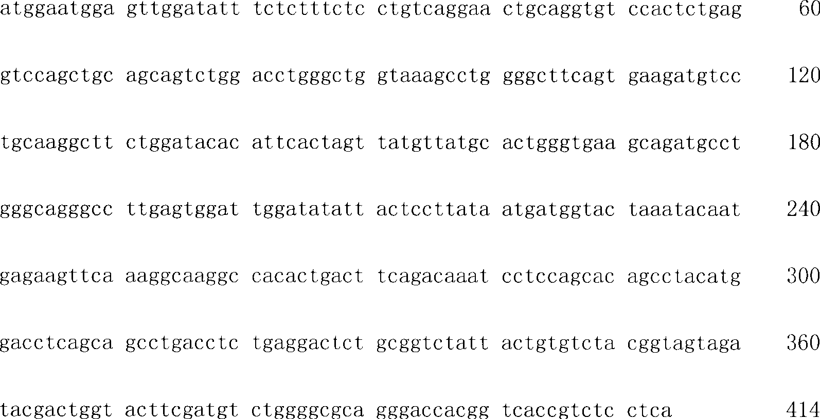
<210>4<210>4
<211>138<211>138
<212>PRT<212>PRT
<213>小鼠<213> mouse
<400>4<400>4
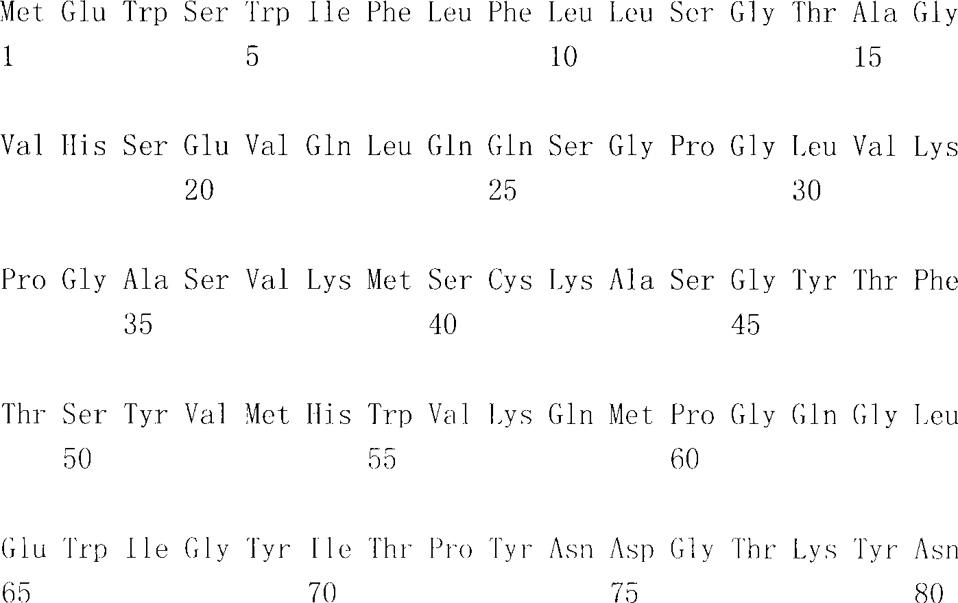

<210>5<210>5
<211>433<211>433
<212>DNA<212>DNA
<213>人工<213> Artificial
<220><220>
<223>人源化轻链可变区<223> Humanized light chain variable region
<400>5<400>5

<210>6<210>6
<211>134<211>134
<212>PRT<212>PRT
<213>人工<213> Artificial
<220><220>
<223>人源化轻链可变区<223> Humanized light chain variable region
<400>6<400>6
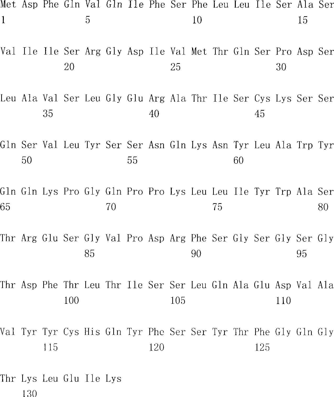
<210>7<210>7
<211>446<211>446
<212>DNA<212>DNA
<213>人工<213> Artificial
<220><220>
<223>人源化重链可变区<223> Humanized heavy chain variable region
<400>7<400>7

<210>8<210>8
<211>138<211>138
<212>PRT<212>PRT
<213>人工<213> Artificial
<220><220>
<223>人源化重链可变区<223> Humanized heavy chain variable region
<400>8<400>8

<210>9<210>9
<211>726<211>726
<212>DNA<212>DNA
<213>人工<213> Artificial
<220><220>
<223>人源化κ轻链<223> Humanized kappa light chain
<400>9<400>9

<210>10<210>10
<211>241<211>241
<212>PRT<212>PRT
<213>人工<213> Artificial
<220><220>
<223>人源化κ轻链<223> Humanized kappa light chain
<400>10<400>10


<210>11<210>11
<211>1404<211>1404
<212>DNA<212>DNA
<213>人工<213> Artificial
<220><220>
<223>人源化重链(V11L)<223> Humanized heavy chain (V11L)
<400>11<400>11
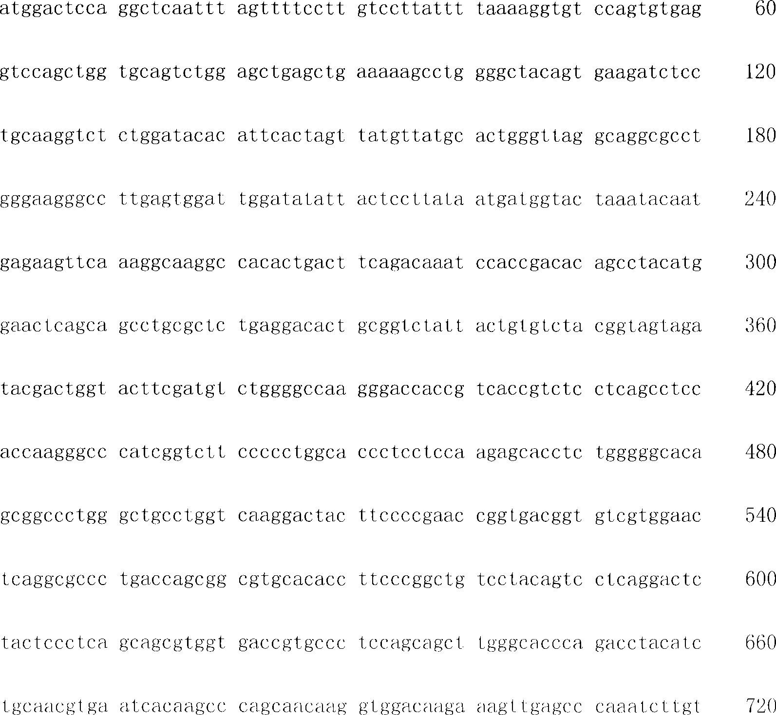
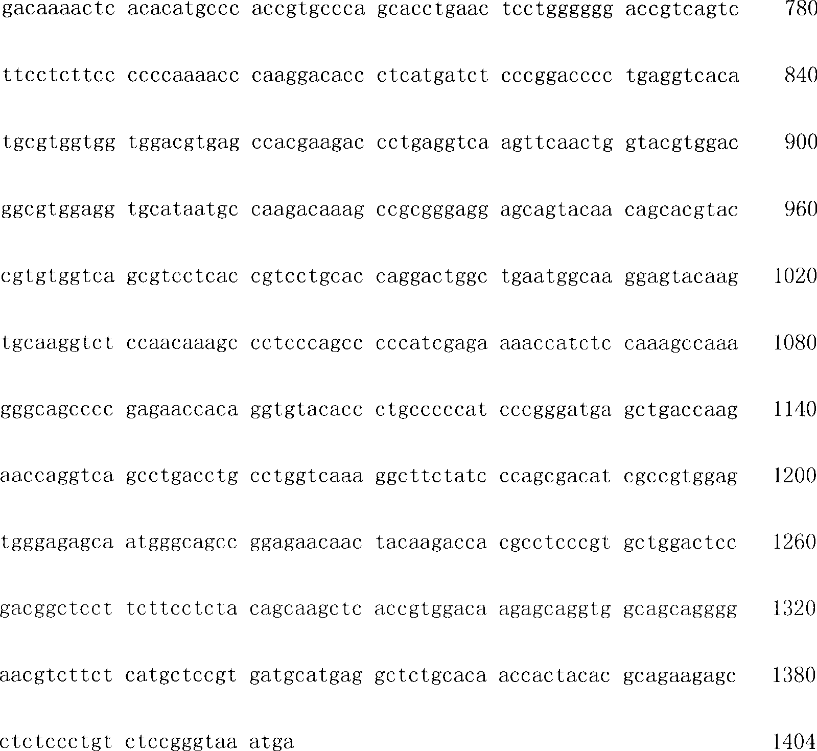
<210>12<210>12
<211>468<211>468
<212>PRT<212>PRT
<213>人工<213> Artificial
<220><220>
<223>人源化重链(V11L)<223> Humanized heavy chain (V11L)
<400>12<400>12


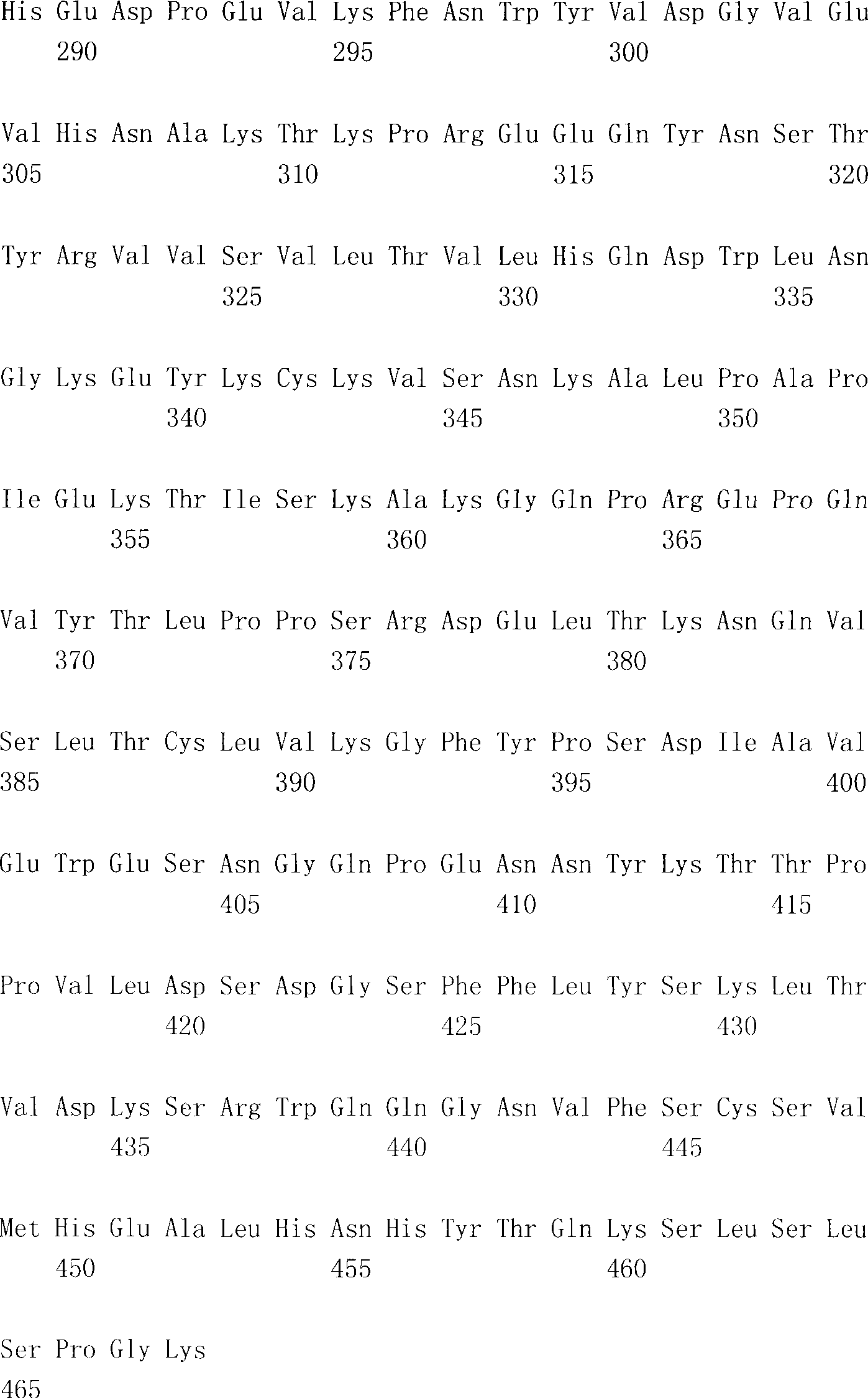
<210>13<210>13
<211>1395<211>1395
<212>DNA<212>DNA
<212>人工<212> Artificial
<220><220>
<223>人源化重链(V11LM3)<223> Humanized heavy chain (V11LM3)
<400>13<400>13

<210>14<210>14
<211>464<211>464
<212>PRT<212>PRT
<213>人工<213> Artificial
<220><220>
<223>人源化重链(V11L M3)<223> Humanized heavy chain (V11L M3)
<400>14<400>14


<210>15<210>15
<211>20<211>20
<212>DNA<212>DNA
<213>人工<213> Artificial
<220><220>
<223>PCR引物<223>PCR primers
<400>15<400>15

<210>16<210>16
<211>21<211>21
<212>DNA<212>DNA
<213>人工<213> Artificial
<220><220>
<223>PCR引物<223>PCR primers
<400>16<400>16

<210>17<210>17
<211>20<211>20
<212>DNA<212>DNA
<213>人工<213> Artificial
<220><220>
<223>PCR引物<223>PCR primers
<400>17<400>17
<210>18<210>18
<211>18<211>18
<212>DNA<212>DNA
<213>人工<213> Artificial
<220><220>
<223>PCR引物<223>PCR primers
<400>18<400>18

<210>19<210>19
<211>17<211>17
<212>DNA<212>DNA
<213>人工<213> Artificial
<220><220>
<223>PCR引物<223>PCR primers
<400>19<400>19

<210>20<210>20
<211>40<211>40
<212>DNA<212>DNA
<213>人工<213> Artificial
<220><220>
<223>PCR引物<223>PCR primers
<400>20<400>20

<210>21<210>21
<211>40<211>40
<212>DNA<212>DNA
<213>人工<213> Artificial
<220><220>
<223>PCR引物<223>PCR primers
<400>21<400>21

<210>22<210>22
<211>40<211>40
<212>DNA<212>DNA
<213>人工<213> Artificial
<220><220>
<223>PCR引物<223>PCR primers
<400>22<400>22

<210>23<210>23
<211>40<211>40
<212>DNA<212>DNA
<213>人工<213> Artificial
<220><220>
<223>PCR引物<223>PCR primers
<400>23<400>23
<210>24<210>24
<211>40<211>40
<212>DNA<212>DNA
<213>人工<213> Artificial
<220><220>
<223>PCR引物<223>PCR primers
<400>24<400>24

<210>25<210>25
<211>40<211>40
<212>DNA<212>DNA
<213>人工<213> Artificial
<220><220>
<223>PCR引物<223>PCR primers
<400>25<400>25

<210>26<210>26
<211>40<211>40
<212>DNA<212>DNA
<213>人工<213> Artificial
<220><220>
<223>PCR引物<223>PCR primers
<400>26<400>26

<210>27<210>27
<211>40<211>40
<212>DNA<212>DNA
<213>人工<213> Artificial
<220><220>
<223>PCR引物<223>PCR primers
<400>27<400>27
<210>28<210>28
<211>40<211>40
<212>DNA<212>DNA
<213>人工<213> Artificial
<220><220>
<223>PCR引物<223>PCR primers
<400>28<400>28

<210>29<210>29
<211>40<211>40
<212>DNA<212>DNA
<213>人工<213> Artificial
<220><220>
<223>PCR引物<223>PCR primers
<400>29<400>29

<210>30<210>30
<211>40<211>40
<212>DNA<212>DNA
<213>人工<213> Artificial
<220><220>
<223>PCR引物<223>PCR primers
<400>30<400>30

<210>31<210>31
<211>40<211>40
<212>DNA<212>DNA
<213>人工<213> Artificial
<220><220>
<223>PCR引物<223>PCR primers
<400>31<400>31
<210>32<210>32
<211>40<211>40
<212>DNA<212>DNA
<213>人工<213> Artificial
<220><220>
<223>PCR引物<223>PCR primers
<400>32<400>32

<210>33<210>33
<211>40<211>40
<212>DNA<212>DNA
<213>人工<213> Artificial
<220><220>
<223>PCR引物<223>PCR primers
<400>33<400>33

<210>34<210>34
<211>40<211>40
<212>DNA<212>DNA
<213>人工<213> Artificial
<220><220>
<223>PCR引物<223>PCR primers
<400>34<400>34

<210>35<210>35
<211>40<211>40
<212>DNA<212>DNA
<213>人工<213> Artificial
<220><220>
<223>PCR引物<223>PCR primers
<400>35<400>35

<210>36<210>36
<211>40<211>40
<212>DNA<212>DNA
<213>人工<213> Artificial
<220><220>
<223>PCR引物<223>PCR primers
<400>36<400>36

<210>37<210>37
<211>40<211>40
<212>DNA<212>DNA
<213>人工<213> Artificial
<220><220>
<223>PCR引物<223>PCR primers
<400>37<400>37

<210>38<210>38
<211>40<211>40
<212>DNA<212>DNA
<213>人工<213> Artificial
<220><220>
<223>PCR引物<223>PCR primers
<400>38<400>38

<210>39<210>39
<211>57<211>57
<212>DNA<212>DNA
<213>人工<213> Artificial
<220><220>
<223>PCR引物<223>PCR primers
<400>39<400>39

<210>40<210>40
<211>40<211>40
<212>DNA<212>DNA
<213>人工<213> Artificial
<220><220>
<223>PCR引物<223>PCR primers
<400>40<400>40

<210>41<210>41
<211>40<211>40
<212>DNA<212>DNA
<213>人工<213> Artificial
<220><220>
<223>PCR引物<223>PCR primers
<400>41<400>41

<210>42<210>42
<211>40<211>40
<212>DNA<212>DNA
<213>人工<213> Artificial
<220><220>
<223>PCR引物<223>PCR primers
<400>42<400>42

<210>43<210>43
<211>40<211>40
<212>DNA<212>DNA
<213>人工<213> Artificial
<220><220>
<223>PCR引物<223>PCR primers
<400>43<400>43

<210>44<210>44
<211>40<211>40
<212>DNA<212>DNA
<213>人工<213> Artificial
<220><220>
<223>PCR引物<223>PCR primers
<400>44<400>44

<210>45<210>45
<211>40<211>40
<212>DNA<212>DNA
<213>人工<213> Artificial
<220><220>
<223>PCR引物<223>PCR primers
<400>45<400>45

<210>46<210>46
<211>40<211>40
<212>DNA<212>DNA
<213>人工<213> Artificial
<220><220>
<223>PCR引物<223>PCR primers
<400>46<400>46

<210>47<210>47
<211>40<211>40
<212>DNA<212>DNA
<213>人工<213> Artificial
<220><220>
<223>PCR引物<223>PCR primers
<400>47<400>47

<210>48<210>48
<211>40<211>40
<212>DNA<212>DNA
<213>人工<213> Artificial
<220><220>
<223>PCR引物<223>PCR primers
<400>48<400>48

<210>49<210>49
<211>40<211>40
<212>DNA<212>DNA
<213>人工<213> Artificial
<220><220>
<223>PCR引物<223>PCR primers
<400>49<400>49

<210>50<210>50
<211>40<211>40
<212>DNA<212>DNA
<213>人工<213> Artificial
<220><220>
<223>PCR引物<223>PCR primers
<400>50<400>50

<210>51<210>51
<211>40<211>40
<212>DNA<212>DNA
<213>人工<213> Artificial
<220><220>
<223>PCR引物<223>PCR primers
<400>51<400>51
<210>52<210>52
<211>40<211>40
<212>DNA<212>DNA
<213>人工<213> Artificial
<220><220>
<223>PCR引物<223>PCR primers
<400>52<400>52

<210>53<210>53
<211>40<211>40
<212>DNA<212>DNA
<213>人工<213> Artificial
<220><220>
<223>PCR引物<223>PCR primers
<400>53<400>53

<210>54<210>54
<211>40<211>40
<212>DNA<212>DNA
<213>人工<213> Artificial
<220><220>
<223>PCR引物<223>PCR primers
<400>54<400>54

<210>55<210>55
<211>40<211>40
<212>DNA<212>DNA
<213>人工<213> Artificial
<220><220>
<223>PCR引物<223>PCR primers
<400>55<400>55

<210>56<210>56
<211>40<211>40
<212>DNA<212>DNA
<213>人工<213> Artificial
<220><220>
<223>PCR引物<223>PCR primers
<400>56<400>56

<210>57<210>57
<211>40<211>40
<212>DNA<212>DNA
<213>人工<213> Artificial
<220><220>
<223>PCR引物<223>PCR primers
<400>57<400>57

<210>58<210>58
<211>40<211>40
<212>DNA<212>DNA
<213>人工<213> Artificial
<220><220>
<223>PCR引物<223>PCR primers
<400>58<400>58

<210>59<210>59
<211>40<211>40
<212>DNA<212>DNA
<213>人工<213> Artificial
<220><220>
<223>PCR引物<223>PCR primers
<400>59<400>59

<210>60<210>60
<211>40<211>40
<212>DNA<212>DNA
<213>人工<213> Artificial
<220><220>
<223>PCR引物<223>PCR primers
<400>60<400>60

<210>61<210>61
<211>34<211>34
<212>DNA<212>DNA
<213>人工<213> Artificial
<220><220>
<223>PCR引物<223>PCR primers
<400>61<400>61

<210>62<210>62
<211>23<211>23
<212>PRT<212>PRT
<213>人工<213> Artificial
<220><220>
<223>人轻链可变区受体<223> Human light chain variable region receptor
<400>62<400>62
<210>63<210>63
<211>30<211>30
<212>PRT<212>PRT
<213>人工<213> Artificial
<220><220>
<223>人重链可变区受体<223> Human heavy chain variable region receptor
<400>63<400>63
<210>64<210>64
<211>14<211>14
<212>PRT<212>PRT
<213>人工<213> Artificial
<220><220>
<223>人重链可变区受体<223> Human heavy chain variable region receptor
<400>64<400>64

<210>65<210>65
<211>24<211>24
<212>PRT<212>PRT
<213>人工<213> Artificial
<220><220>
<223>人重链可变区受体<223> Human heavy chain variable region receptor
<400>65<400>65

<210>66<210>66
<211>8<211>8
<212>PRT<212>PRT
<213>人工<213> Artificial
<220><220>
<223>人重链可变区受体<223> Human heavy chain variable region receptor
<400>66<400>66
Claims (59)
Applications Claiming Priority (2)
| Application Number | Priority Date | Filing Date | Title |
|---|---|---|---|
| US11/362,452 US7442777B2 (en) | 2000-11-29 | 2006-02-24 | Cytotoxicity mediation of cells evidencing surface expression of CD63 |
| US11/362,452 | 2006-02-24 |
Publications (1)
| Publication Number | Publication Date |
|---|---|
| CN101421395A true CN101421395A (en) | 2009-04-29 |
Family
ID=38436880
Family Applications (1)
| Application Number | Title | Priority Date | Filing Date |
|---|---|---|---|
| CNA2007800127439A Pending CN101421395A (en) | 2006-02-24 | 2007-02-22 | Cytotoxicity mediation of cells evidencing surface expression of CD63 |
Country Status (10)
| Country | Link |
|---|---|
| US (2) | US7442777B2 (en) |
| EP (1) | EP1987138A4 (en) |
| JP (1) | JP2009527507A (en) |
| CN (1) | CN101421395A (en) |
| AU (1) | AU2007219010A1 (en) |
| CA (1) | CA2643059A1 (en) |
| GB (2) | GB2449044A (en) |
| MX (1) | MX2008010815A (en) |
| WO (1) | WO2007095729A1 (en) |
| ZA (1) | ZA200807723B (en) |
Cited By (1)
| Publication number | Priority date | Publication date | Assignee | Title |
|---|---|---|---|---|
| CN103502515A (en) * | 2011-01-10 | 2014-01-08 | Opko制药公司 | Antigen surrogates in autoimmune disease |
Families Citing this family (11)
| Publication number | Priority date | Publication date | Assignee | Title |
|---|---|---|---|---|
| CN101687931A (en) * | 2007-07-16 | 2010-03-31 | 霍夫曼-拉罗奇有限公司 | Anti-cancer cell toxicity monoclonal antibody |
| US8617806B2 (en) * | 2008-01-25 | 2013-12-31 | Hansabiomed Ou | Method to measure and characterize microvesicles in the human body fluids |
| JP6055615B2 (en) * | 2011-05-27 | 2016-12-27 | アッヴィ バイオテクノロジー リミテッド | DACHYP Compositions and Methods |
| SG10201709018QA (en) | 2012-03-14 | 2017-11-29 | Regeneron Pharma | Multispecific antigen-binding molecules and uses thereof |
| IL294043B2 (en) * | 2014-09-28 | 2024-10-01 | Univ California | Modulation of stimulatory and non-stimulatory myeloid cells |
| WO2017190079A1 (en) | 2016-04-28 | 2017-11-02 | Regeneron Pharmaceuticals, Inc. | Methods of making multispecific antigen-binding molecules |
| CA3066569A1 (en) | 2017-06-07 | 2018-12-13 | Regeneron Pharmaceuticals, Inc. | Compositions and methods for internalizing enzymes |
| WO2019118513A1 (en) | 2017-12-12 | 2019-06-20 | Pionyr Immunotherapeutics, Inc. | Anti-trem2 antibodies and related methods |
| WO2019152574A1 (en) * | 2018-02-01 | 2019-08-08 | Merck Sharp & Dohme Corp. | Methods for treating cancer or infection using a combination of an anti-pd-1 antibody, an anti-lag3 antibody, and an anti-tigit antibody |
| JP7779653B2 (en) | 2018-02-07 | 2025-12-03 | リジェネロン・ファーマシューティカルズ・インコーポレイテッド | Methods and compositions for therapeutic protein delivery |
| KR20250140631A (en) | 2018-05-17 | 2025-09-25 | 리제너론 파마슈티칼스 인코포레이티드 | Anti-cd63 antibodies, conjugates, and uses thereof |
Family Cites Families (46)
| Publication number | Priority date | Publication date | Assignee | Title |
|---|---|---|---|---|
| US4172124A (en) | 1978-04-28 | 1979-10-23 | The Wistar Institute | Method of producing tumor antibodies |
| US4867973A (en) | 1984-08-31 | 1989-09-19 | Cytogen Corporation | Antibody-therapeutic agent conjugates |
| US4828991A (en) | 1984-01-31 | 1989-05-09 | Akzo N.V. | Tumor specific monoclonal antibodies |
| GB8516853D0 (en) * | 1985-07-03 | 1985-08-07 | British Telecomm | Manufacture of semiconductor structures |
| AU613590B2 (en) | 1986-11-19 | 1991-08-08 | Bristol-Myers Squibb Company | Hybridomas producing monoclonal antibodies to new mucin epitopes |
| US4861581A (en) | 1986-12-05 | 1989-08-29 | Cancer Biologics, Inc. | Detection of necrotic malignant tissue and associated therapy |
| US5171665A (en) | 1989-04-17 | 1992-12-15 | Oncogen | Monoclonal antibody to novel antigen associated with human tumors |
| US5296348A (en) | 1989-05-16 | 1994-03-22 | The Wistar Institute Of Anatomy And Biology | Methods for screening monoclonal antibodies for therapeutic use |
| US6020145A (en) | 1989-06-30 | 2000-02-01 | Bristol-Myers Squibb Company | Methods for determining the presence of carcinoma using the antigen binding region of monoclonal antibody BR96 |
| EP0539970B1 (en) | 1991-10-30 | 1999-05-26 | Idemitsu Kosan Company Limited | Methods for producing human lymphocytes and human monoclonal antibodies, and human monoclonal antibodies produced thereby |
| IL105008A0 (en) | 1992-03-13 | 1993-07-08 | Yeda Res & Dev | Double transfectants of mhc genes as cellular vaccines for immunoprevention of tumor metastasis |
| US5545532A (en) | 1993-02-05 | 1996-08-13 | Epigen, Inc. | Human carcinoma antigen (HCA), HCA antibodies, HCA immunoassays and methods of imaging |
| PT1231268E (en) | 1994-01-31 | 2005-11-30 | Univ Boston | BANKS OF POLYCLONE ANTIBODIES |
| WO1996000084A1 (en) | 1994-06-24 | 1996-01-04 | Torchilin Vladimir P | Use of autoantibodies for tumor therapy and prophylaxis |
| US5783186A (en) | 1995-12-05 | 1998-07-21 | Amgen Inc. | Antibody-induced apoptosis |
| US6783961B1 (en) | 1999-02-26 | 2004-08-31 | Genset S.A. | Expressed sequence tags and encoded human proteins |
| WO1999066027A1 (en) | 1998-06-15 | 1999-12-23 | The Research Foundation Of State University Of New York | Monoclonal antibodies that recognize antigens associated with tumor metastasis |
| DE19832998A1 (en) | 1998-07-22 | 2000-02-03 | Siemens Ag | Method for determining data packets contained in a continuous data stream |
| CA2296792A1 (en) | 1999-02-26 | 2000-08-26 | Genset S.A. | Expressed sequence tags and encoded human proteins |
| AU3395900A (en) | 1999-03-12 | 2000-10-04 | Human Genome Sciences, Inc. | Human lung cancer associated gene sequences and polypeptides |
| US6657048B2 (en) | 1999-10-08 | 2003-12-02 | Arius Research, Inc. | Individualized anti-cancer antibodies |
| US6180357B1 (en) | 1999-10-08 | 2001-01-30 | Arius Research, Inc. | Individualized patient-specific anti-cancer antibodies |
| US20040105816A1 (en) | 1999-10-08 | 2004-06-03 | Young David S. F. | Cancerous disease modifying antibodies |
| US7256271B2 (en) | 2003-01-21 | 2007-08-14 | Arius Research Inc. | Cancerous disease modifying antibodies |
| US20020102638A1 (en) | 2000-01-31 | 2002-08-01 | Rosen Craig A. | Nucleic acids, proteins, and antibodies |
| AU2001253140A1 (en) | 2000-04-03 | 2001-10-15 | The Government Of The United States Of America, As Represented By The Secretary Of The Department Of Health And Human Services | Tumor markers in ovarian cancer |
| CN1326951A (en) | 2000-06-05 | 2001-12-19 | 上海博德基因开发有限公司 | New polypeptide-human CD63 antigen 15.07 and polynucleotide for encoding such polypeptide |
| CN1326962A (en) | 2000-06-05 | 2001-12-19 | 上海博德基因开发有限公司 | New polypeptide-human CD 63 antigen 14.63 and polynucleotide for encoding such polypeptide |
| AU2001266787A1 (en) | 2000-06-07 | 2002-01-08 | Human Genome Sciences, Inc. | Nucleic acids, proteins, and antibodies |
| CN1351054A (en) | 2000-10-26 | 2002-05-29 | 上海博德基因开发有限公司 | Polypeptide-human CD63 antigen 11.11 and polynucleotide for coding it |
| FR2817247B1 (en) * | 2000-11-24 | 2003-02-14 | Valois Sa | FLUID PRODUCT DISPENSING DEVICE |
| US7009040B2 (en) * | 2003-01-21 | 2006-03-07 | Arius Research, Inc. | Cancerous disease modifying antibodies |
| US7361343B2 (en) * | 2003-01-21 | 2008-04-22 | Arius Research Inc. | Cytotoxicity mediation of cells evidencing surface expression of CD63 |
| CN1364803A (en) | 2001-01-10 | 2002-08-21 | 上海博德基因开发有限公司 | New polypeptide-human CD63 antigen 56.87 and polynucleotide for encoding such polypeptide |
| US20030055220A1 (en) | 2001-01-12 | 2003-03-20 | Pierre Legrain | Protein-protein interactions between Shigella flexneri polypeptides and mammalian polypeptides |
| US6783969B1 (en) | 2001-03-05 | 2004-08-31 | Nuvelo, Inc. | Cathepsin V-like polypeptides |
| CA2444675A1 (en) | 2001-04-20 | 2002-10-31 | Chandra S. Arvizu | Secreted proteins |
| WO2003016475A2 (en) | 2001-08-14 | 2003-02-27 | The General Hospital Corporation | Nucleic acid and amino acid sequences involved in pain |
| EP2143438B1 (en) | 2001-09-18 | 2011-07-13 | Genentech, Inc. | Compositions and methods for the diagnosis and treatment of tumors |
| EP1455819A1 (en) | 2001-12-21 | 2004-09-15 | Arius Research, Inc. | Individualized anti-cancer antibodies |
| EP2067472A1 (en) | 2002-01-02 | 2009-06-10 | Genentech, Inc. | Compositions and methods for the diagnosis and treatment of tumor |
| WO2003068268A2 (en) | 2002-02-14 | 2003-08-21 | Bioinvent International Ab | Treatment, diagnosis and imaging of disease |
| US7378492B2 (en) | 2002-02-20 | 2008-05-27 | Incyte Corporation | CD40-related receptor that binds CD40L |
| WO2003086456A2 (en) | 2002-04-05 | 2003-10-23 | Arius Research, Inc. | Anti-ck18 monoclonal antibody and therapeutic and diagnostic uses thereof in cancer |
| WO2004041170A2 (en) | 2002-11-01 | 2004-05-21 | Genentech, Inc. | Compositions and methods for the treatment of immune related diseases |
| US7393531B2 (en) | 2003-01-21 | 2008-07-01 | Arius Research Inc. | Cytotoxicity mediation of cells evidencing surface expression of MCSP |
-
2006
- 2006-02-24 US US11/362,452 patent/US7442777B2/en not_active Expired - Fee Related
-
2007
- 2007-02-22 WO PCT/CA2007/000254 patent/WO2007095729A1/en not_active Ceased
- 2007-02-22 AU AU2007219010A patent/AU2007219010A1/en not_active Abandoned
- 2007-02-22 GB GB0816303A patent/GB2449044A/en not_active Withdrawn
- 2007-02-22 MX MX2008010815A patent/MX2008010815A/en not_active Application Discontinuation
- 2007-02-22 EP EP07701801A patent/EP1987138A4/en not_active Withdrawn
- 2007-02-22 CA CA002643059A patent/CA2643059A1/en not_active Abandoned
- 2007-02-22 CN CNA2007800127439A patent/CN101421395A/en active Pending
- 2007-02-22 JP JP2008555582A patent/JP2009527507A/en not_active Ceased
-
2008
- 2008-09-08 ZA ZA200807723A patent/ZA200807723B/en unknown
- 2008-09-10 GB GBGB0816513.6A patent/GB0816513D0/en not_active Ceased
- 2008-09-18 US US12/284,137 patent/US20090155262A1/en not_active Abandoned
Cited By (2)
| Publication number | Priority date | Publication date | Assignee | Title |
|---|---|---|---|---|
| CN103502515A (en) * | 2011-01-10 | 2014-01-08 | Opko制药公司 | Antigen surrogates in autoimmune disease |
| CN103502515B (en) * | 2011-01-10 | 2015-12-23 | Opko制药公司 | Antigen Surrogates for Autoimmune Diseases |
Also Published As
| Publication number | Publication date |
|---|---|
| WO2007095729A8 (en) | 2009-07-16 |
| MX2008010815A (en) | 2009-03-06 |
| EP1987138A1 (en) | 2008-11-05 |
| US7442777B2 (en) | 2008-10-28 |
| JP2009527507A (en) | 2009-07-30 |
| WO2007095729A1 (en) | 2007-08-30 |
| US20070025912A1 (en) | 2007-02-01 |
| ZA200807723B (en) | 2009-08-26 |
| CA2643059A1 (en) | 2007-08-30 |
| AU2007219010A1 (en) | 2007-08-30 |
| GB2449044A (en) | 2008-11-05 |
| GB0816303D0 (en) | 2008-10-15 |
| EP1987138A4 (en) | 2009-07-22 |
| US20090155262A1 (en) | 2009-06-18 |
| GB2449044A8 (en) | 2008-11-26 |
| GB0816513D0 (en) | 2008-10-15 |
Similar Documents
| Publication | Publication Date | Title |
|---|---|---|
| US8017117B2 (en) | Cytotoxicity mediation of cells evidencing surface expression of CD44 | |
| CN101421395A (en) | Cytotoxicity mediation of cells evidencing surface expression of CD63 | |
| US20080089891A1 (en) | Cancerous disease modifying antibodies | |
| CN101675158A (en) | Cancer Disease Modulating Antibodies | |
| CN101553259A (en) | Cytotoxicity mediation of cells exhibiting surface expression of CD63 | |
| CN101784566A (en) | Demonstrating the mediation of cytotoxicity of cells expressing CD9 on the surface | |
| US20080044408A1 (en) | Cytotoxicity mediation of cells evidencing surface expression of CD63 | |
| MX2008010845A (en) | Anhydrous compositions useful for attaining enhanced sexual wellness. | |
| CN101278055A (en) | Cancer Disease Modulating Antibodies | |
| JP2009528993A (en) | Cancer-like disease-modifying antibody 141205-02 | |
| TW200936757A (en) | Cancerous disease modifying antibodies | |
| JP2010509244A (en) | Cancerous disease modifying antibodies | |
| US20100158801A1 (en) | Methods for the treatment of acute myeloid leukemia | |
| JP2011528010A (en) | Cancerous disease modifying antibodies | |
| JP2010526108A (en) | Cancerous disease modifying antibodies | |
| JP2010509246A (en) | Cancerous disease modifying antibodies | |
| JP2009545533A (en) | Cancer-like disease-modifying antibody | |
| JP2011520923A (en) | Cytotoxic anti-cancer monoclonal antibody | |
| JP2011511767A (en) | Cytotoxic anti-cancer monoclonal antibody | |
| JP2010509245A (en) | Cancerous disease modifying antibodies | |
| CN101278057A (en) | Cancer Disease Modulating Antibodies |
Legal Events
| Date | Code | Title | Description |
|---|---|---|---|
| C06 | Publication | ||
| PB01 | Publication | ||
| C10 | Entry into substantive examination | ||
| SE01 | Entry into force of request for substantive examination | ||
| C02 | Deemed withdrawal of patent application after publication (patent law 2001) | ||
| WD01 | Invention patent application deemed withdrawn after publication |
Open date: 20090429 |