CN101288106B - Automatic generation of optimal views for computed tomography chest diagnosis - Google Patents
Automatic generation of optimal views for computed tomography chest diagnosis Download PDFInfo
- Publication number
- CN101288106B CN101288106B CN2006800334896A CN200680033489A CN101288106B CN 101288106 B CN101288106 B CN 101288106B CN 2006800334896 A CN2006800334896 A CN 2006800334896A CN 200680033489 A CN200680033489 A CN 200680033489A CN 101288106 B CN101288106 B CN 101288106B
- Authority
- CN
- China
- Prior art keywords
- interest
- inertia
- seed points
- structures
- axes
- Prior art date
- Legal status (The legal status is an assumption and is not a legal conclusion. Google has not performed a legal analysis and makes no representation as to the accuracy of the status listed.)
- Expired - Fee Related
Links
Images


Classifications
-
- G—PHYSICS
- G06—COMPUTING OR CALCULATING; COUNTING
- G06T—IMAGE DATA PROCESSING OR GENERATION, IN GENERAL
- G06T7/00—Image analysis
- G06T7/60—Analysis of geometric attributes
-
- A—HUMAN NECESSITIES
- A61—MEDICAL OR VETERINARY SCIENCE; HYGIENE
- A61B—DIAGNOSIS; SURGERY; IDENTIFICATION
- A61B6/00—Apparatus or devices for radiation diagnosis; Apparatus or devices for radiation diagnosis combined with radiation therapy equipment
- A61B6/02—Arrangements for diagnosis sequentially in different planes; Stereoscopic radiation diagnosis
- A61B6/03—Computed tomography [CT]
- A61B6/032—Transmission computed tomography [CT]
-
- A—HUMAN NECESSITIES
- A61—MEDICAL OR VETERINARY SCIENCE; HYGIENE
- A61B—DIAGNOSIS; SURGERY; IDENTIFICATION
- A61B6/00—Apparatus or devices for radiation diagnosis; Apparatus or devices for radiation diagnosis combined with radiation therapy equipment
- A61B6/52—Devices using data or image processing specially adapted for radiation diagnosis
- A61B6/5211—Devices using data or image processing specially adapted for radiation diagnosis involving processing of medical diagnostic data
- A61B6/5223—Devices using data or image processing specially adapted for radiation diagnosis involving processing of medical diagnostic data generating planar views from image data, e.g. extracting a coronal view from a 3D image
-
- G—PHYSICS
- G06—COMPUTING OR CALCULATING; COUNTING
- G06T—IMAGE DATA PROCESSING OR GENERATION, IN GENERAL
- G06T19/00—Manipulating 3D models or images for computer graphics
-
- G—PHYSICS
- G06—COMPUTING OR CALCULATING; COUNTING
- G06T—IMAGE DATA PROCESSING OR GENERATION, IN GENERAL
- G06T2210/00—Indexing scheme for image generation or computer graphics
- G06T2210/41—Medical
Landscapes
- Engineering & Computer Science (AREA)
- Health & Medical Sciences (AREA)
- Life Sciences & Earth Sciences (AREA)
- Physics & Mathematics (AREA)
- Medical Informatics (AREA)
- Theoretical Computer Science (AREA)
- Heart & Thoracic Surgery (AREA)
- Optics & Photonics (AREA)
- Veterinary Medicine (AREA)
- Public Health (AREA)
- General Health & Medical Sciences (AREA)
- Animal Behavior & Ethology (AREA)
- General Physics & Mathematics (AREA)
- Biophysics (AREA)
- High Energy & Nuclear Physics (AREA)
- Computer Vision & Pattern Recognition (AREA)
- Nuclear Medicine, Radiotherapy & Molecular Imaging (AREA)
- Surgery (AREA)
- Pathology (AREA)
- Radiology & Medical Imaging (AREA)
- Biomedical Technology (AREA)
- Molecular Biology (AREA)
- Geometry (AREA)
- Computer Hardware Design (AREA)
- Pulmonology (AREA)
- Computer Graphics (AREA)
- Software Systems (AREA)
- General Engineering & Computer Science (AREA)
- Apparatus For Radiation Diagnosis (AREA)
- Image Processing (AREA)
Abstract
生成计算机断层摄影图像中的一部分的至少一个视图(420)包括:为所述图像内的感兴趣结构选择(310)种子点(410),预处理(320)所述种子点周围的感兴趣区域,为所述感兴趣区域估计(325)至少一个惯性轴,以及从包括所述种子点且垂直于每个惯性轴的三个平面中的一个或多个生成(345)所述至少一个视图。
Generating at least one view (420) of a portion of a computed tomography image includes selecting (310) a seed point (410) for a structure of interest within the image, pre-processing (320) a region of interest around the seed point, estimating (325) at least one axis of inertia for the region of interest, and generating (345) the at least one view from one or more of three planes that include the seed point and are perpendicular to each axis of inertia.
Description
本文公开的实施例涉及为医学成像应用提供细长结构的优化视图。Embodiments disclosed herein relate to providing optimized views of elongated structures for medical imaging applications.
在医疗实践中普遍需要生成解剖结构的优化视图。尤其是,生成细长结构的优化视图与三维(3D)医学成像手段的发展有着特殊关系。示例性应用包括冠状动脉单平面显示和支气管树的可视化。There is a common need in medical practice to generate optimized views of anatomy. In particular, generating optimized views of elongated structures is of particular relevance to the development of three-dimensional (3D) medical imaging modalities. Exemplary applications include single-plane display of coronary arteries and visualization of the bronchial tree.
分析3D数据量常常是一项不顺而耗时的任务,需要用户通过一定的局部视图来分析大量的信息。此外,对于不熟悉处理空间中的旋转和平移的人来说与数据进行交互过程是困难的。Analyzing 3D data volumes is often an awkward and time-consuming task, requiring users to analyze large amounts of information through certain partial views. Furthermore, the process of interacting with the data can be difficult for someone unfamiliar with handling rotations and translations in space.
已经使用过几种方法来简化与3D数据之间的交互,通常这些方法对应用非常依赖。在这些方法中,生成某些解剖结构,尤其是诸如血管、支气管、骨骼等的细长结构的优化视图已经在某些临床实践中取得了成功。Several approaches have been used to simplify interaction with 3D data, often very application dependent. Among these methods, generating optimized views of certain anatomical structures, especially elongated structures such as blood vessels, bronchi, bones, etc., has achieved success in some clinical practices.
这种成功的原因可能在于,当前采用的技术极大地简化了分析靶结构的任务,否则的话,这种任务需要通过操纵有关的六个自由度来浏览一些图像、显示所述靶的局部视图或手动对准结构。这些任务通常需要功能强大的设备和高度的实践经验。这些方法的缺点包括:分割靶结构来产生视图往往使方法相对缓慢而不鲁棒。This success may be due to the fact that currently available technology greatly simplifies the task of analyzing the structure of a target, which would otherwise require manipulating the six degrees of freedom involved to navigate through some images, display partial views of said target, or Align the structures manually. These tasks often require powerful equipment and a high degree of practical experience. Disadvantages of these methods include that segmenting the target structure to generate views tends to make the method relatively slow and not robust.
提供一种单次点击、基于非分割的细长结构显示的自动优化将是有利的。It would be advantageous to provide a single click, automatic optimization based on the display of non-segmented elongated structures.
在一个实施例中,用于生成计算机断层摄影图像中的一部分的至少一个视图的扫描系统包括:具有多个互联装置的控制器,其具有:用于为所述图像内的感兴趣结构选择种子点的装置,用于预处理所述种子点周围的感兴趣区域的装置,用于为所述感兴趣区域估计至少一个惯性轴的装置,其中,用于估计至少一个惯性轴的所述装置:根据每个体素的强度和到所述种子点的距离向所述感兴趣区域中的体素分配密度值;以及利用所述密度值定义主惯性轴和垂直于所述主惯性轴的两个其他惯性轴,以及用于从包括所述种子点且垂直于每个惯性轴的三个平面中的一个或多个生成所述至少一个视图的装置。In one embodiment, a scanning system for generating at least one view of a portion of a computed tomography image comprises: a controller having a plurality of interconnected devices having a means for selecting a seed for a structure of interest within said image means for preprocessing a region of interest around said seed point, means for estimating at least one inertial axis for said region of interest, wherein said means for estimating at least one inertial axis: Assigning density values to voxels in the region of interest according to each voxel's intensity and distance to the seed point; and using the density values to define a primary inertial axis and two other axes perpendicular to the primary inertial axis inertial axes, and means for generating said at least one view from one or more of three planes comprising said seed point and perpendicular to each inertial axis.
一种生成计算机断层摄影图像中的一部分的至少一个视图的方法,包括:为所述图像内的感兴趣结构选择种子点,预处理所述种子点周围的感兴趣区域,为所述感兴趣区域估计至少一个惯性轴,其中,估计至少一个惯性轴包括:根据每个体素的强度和到所述种子点的距离向所述感兴趣区域中的体素分配密度值;以及利用所述密度值定义主惯性轴和垂直于所述主惯性轴的两个其他惯性轴,以及从包括所述种子点且垂直于每个惯性轴的三个平面中的一个或多个生成所述至少一个视图。A method of generating at least one view of a portion of a computed tomography image, comprising: selecting a seed point for a structure of interest within the image, preprocessing a region of interest surrounding the seed point, and creating a region of interest for the region of interest estimating at least one inertial axis, wherein estimating at least one inertial axis comprises: assigning density values to voxels in the region of interest according to each voxel's intensity and distance to the seed point; and using the density values to define A primary inertial axis and two other inertial axes perpendicular to said primary inertial axis, and said at least one view is generated from one or more of three planes comprising said seed point and perpendicular to each inertial axis.
在另一实施例中,一种包括计算机可用介质的计算机程序产品,所述计算机可用介质具有计算机可读程序,其中在计算机上执行所述计算机可读程序时所述计算机可读程序令所述计算机通过如下步骤生成计算机断层摄影图像中的一部分的至少一个视图:为所述图像内的感兴趣结构选择种子点,预处理所述种子点周围的感兴趣区域,为所述感兴趣区域估计至少一个惯性轴,以及从包括所述种子点且垂直于每个惯性轴的三个平面中的一个或多个生成所述至少一个视图。In another embodiment, a computer program product comprising a computer usable medium having a computer readable program, wherein when said computer readable program is executed on a computer, said computer readable program causes said A computer generates at least one view of a portion of a computed tomography image by selecting a seed point for a structure of interest within the image, preprocessing a region of interest around the seed point, estimating at least one inertial axis, and generating said at least one view from one or more of three planes comprising said seed point and perpendicular to each inertial axis.
在下面的说明书中结合附图解释本发明的上述方面和其他特征,在附图中:In the following description, the above-mentioned aspects and other features of the present invention are explained in conjunction with the accompanying drawings, in which:
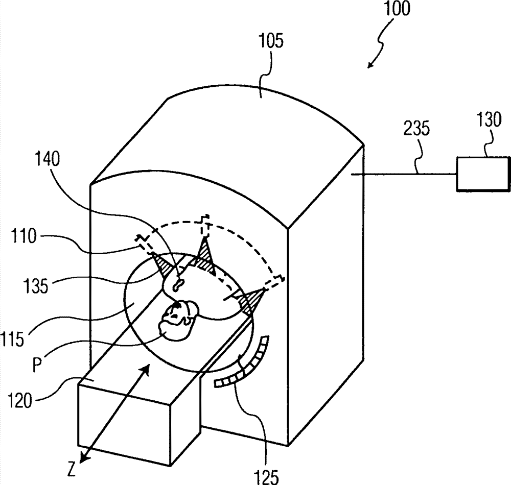
图1A和1B示出了适于实践所公开实施例的成像系统的示意图;1A and 1B show schematic diagrams of imaging systems suitable for practicing the disclosed embodiments;
图2示出了用于操作成像系统的示例性控制器;Figure 2 illustrates an exemplary controller for operating an imaging system;
图3示出了用于自动生成细长结构优化视图的过程的流程图;以及Figure 3 shows a flow diagram of a process for automatically generating an optimized view of an elongated structure; and
图4示出了原始图像和图像选定部分的示例性优化视图。Figure 4 shows an exemplary optimized view of an original image and a selected portion of the image.
图1示出了适用于实践此处公开的实施例的系统100的方框图。虽然所公开的实施例是参考图中所示的实施例描述的,但应当理解本发明可以实施为很多备选形式的实施例。此外,可以使用任何适当尺寸、形状或类型的元件或材料。FIG. 1 shows a block diagram of a
本发明提供了现有技术中当前可用方法的替代方法,基于不需要分割的局部取向估计。此外,本文所述的技术可以利用用户的单次点击生成优化视图。The present invention provides an alternative to the methods currently available in the prior art, based on local orientation estimation that does not require segmentation. Additionally, the techniques described herein can generate optimized views with a single click from the user.
所公开的实施例利用图像处理操作来估计在用户定义的种子点围绕的细长结构的取向。一旦确定了取向,就用其自动生成最佳视图,在最佳视图中将感兴趣结构以其全长显示。The disclosed embodiments utilize image processing operations to estimate the orientation of elongated structures around user-defined seed points. Once the orientation is determined, it is used to automatically generate an optimal view in which the structure of interest is displayed at its full length.
采用该方法,可以在几乎无延迟的单击操作时执行3D CT数据量中细长结构的分析,而不是像当前技术要求的,必须要费力地调节多达六个不同参数来找到最佳视图。With this approach, the analysis of elongated structures in 3D CT data volumes can be performed in a single click with virtually no delay, rather than having to painstakingly adjust up to six different parameters to find the best view, as current technology requires .
所公开实施例的示例性应用包括基于计算机断层摄影(CT)的肺部诊断和分期。在肺部研究中,临床医师可以缓慢浏览一大组CT切片,以查找不同的疾病征兆。这些征兆可以包括支气管的几种可变的树状结构,并不总是容易基于每一切片对这些征兆进行评估。因而,对于这种具体应用而言支气管的优化视图将是很有用的。为此,现有方法依靠的是提取支气管树;不过,这种提取需要一个耗时的预处理阶段,而且当前的分割技术无法提取更精细的支气管。本文所述的技术能够迅速生成在特定感兴趣区域周围的支气管结构的适宜视图。Exemplary applications of the disclosed embodiments include computed tomography (CT) based lung diagnosis and staging. In a lung study, a clinician can slowly scan through a large set of CT slices looking for different signs of disease. These signs can include several variable tree structures of the bronchi, which are not always easy to assess on a per-slice basis. Thus, an optimized view of the bronchi would be useful for this particular application. To this end, existing methods rely on extracting the bronchial tree; however, this extraction requires a time-consuming preprocessing stage, and current segmentation techniques cannot extract finer bronchi. The techniques described herein are capable of rapidly generating a suitable view of bronchial structures surrounding a particular region of interest.
图1A示出了适于实践本文公开的实施例的示例性扫描系统100。扫描系统100包括成像器105,在该实施例中成像器为计算机断层摄影(CT)成像扫描机,其具有X射线源110、检查区域115、患者支持器120和X射线探测器125。扫描系统100还包括用于控制X射线源110、X射线探测器125和患者支持器120并用于处理X射线探测器125获取的数据的控制器130。FIG. 1A illustrates an
扫描系统100可以具有膛式构型,其中包围检查区域。可以控制X射线源110来产生扇形、锥形、楔形或任何其他期望形状的X射线束135。可以引导X射线束135通过检查区域115和患者P,射向X射线探测器125。X射线源110和X射线探测器125可绕由患者支持器120长度界定的轴Z旋转。X射线源110和X射线探测器125可以一起旋转,在一个实施例中可以由机架连接。在另一实施例中,X射线源110或X射线探测器125可以保持固定,而另一个旋转,使得来自X射线源110的X射线冲击到X射线探测器125的连续变化角部分上。
X射线探测器125可以跨越特定角范围,并可以包括用于同时获取图像数据的探测器元件阵列。检查区域115通常环绕着患者P,X射线束从患者通过。患者P被放置在患者支持器120上,患者支持器可以沿Z轴线性移动。
图1B示出了具有成像器153的备选示例性扫描系统150,成像器153具有开放的C臂构型。与成像器105相似,成像器153包括X射线源110、检查区域115、患者支持器120和X射线探测器125。扫描系统150还包括用于控制X射线源110、X射线探测器125和患者支持器120并用于处理X射线探测器125获取的数据的控制器130。扫描系统150利用C臂结构155来连接和支撑X射线源110和X射线探测器125。轭架160容许控制器130围绕垂直于Z轴的轴沿着角方向A调节X射线源110和X射线探测器125。在该构型中,X射线源110和X射线探测器125通常一起旋转。FIG. 1B shows an alternative
图2示出了控制器130的方框图。控制器通常包括实现处理器205、只读存储器210、随机存取存储器215、程序存储器220、用户接口225和网络接口230的电路。FIG. 2 shows a block diagram of the
处理器205通常用于从计算机程序产品(例如计算机可用介质,如只读存储器210、随机存取存储器215和程序存储器220)读取信息和程序。
只读存储器210和随机存取存储器215都可以利用半导体技术和任何其他适当的材料和技术。程序存储器220可以包括计算机可用介质,例如磁盘、计算机硬盘驱动器、紧致盘、数字多用图盘、光盘、芯片、半导体和任何其他能够以计算机可读代码的形式存储程序的装置。Both read-
只读存储器210、随机存取存储器215和程序存储器220的每个或任何组合包括计算机程序产品,该产品包括具有计算机可读程序的计算机可用介质,其中在控制器130执行计算机可读程序时,该计算机可读程序令控制器130控制X射线源110、X射线探测器125和患者支持器120,并处理由成像器105、153获取的数据,以自动生成视图,用于根据本文所述的技术进行CT胸部诊断。Each or any combination of read-
网络接口230一般可以适于通过通信网络235在控制器130和成像器105的部件之间提供接口。通信网络235可以是任何适宜的通信路径,包括公用交换电话网(PSTN)、因特网、无线网络、有线网络、局域网(LAN)、广域网(WAN)、虚拟专用网络(VPN)等,且还可以包括其他类型的网络,包括X.25、TCP/IP、ATM等。在一个实施例中,通信网络120可以是IEEE1349网,也称为“火线”网。
控制器130可以包括与显示器240之间的用户接口225以及诸如键盘的输入装置255和诸如鼠标的定点装置245。可以在处理器205的控制下通过用户接口控制器250操作用户接口。The
回到图1A和1B,在螺旋成像模式下,X射线源110和X射线探测器125可以随着患者支持器120的线性行进同时旋转,以产生X射线源110相对于患者支持器120的大致螺旋轨迹。Returning to FIGS. 1A and 1B , in the helical imaging mode, the
在多切片成像模式下,X射线源110和X射线探测器125可以在患者支持器120保持静止时旋转,以产生X射线源110相对于患者支持器120的大致圆形轨迹,同时由控制器130获取患者P的轴向切片图像。可以在患者支持器120沿Z轴行进过程中以循环方式重复该过程,以沿着Z轴在分立步骤中获取成像数据。In the multi-slice imaging mode, the
因而,控制器130可以控制X射线源110、X射线探测器125和患者支持器120,以沿X射线源110相对于患者支持器120的螺旋或圆形轨迹获得患者P的选定投影视图。可以以控制器130确定的可变帧率收集投影视图。控制器130收集投影视图用于处理,并用于根据公开的实施例调节数据采集过程。Thus,
在采集投影视图期间X射线源110的轨迹优选针对所成像感兴趣区域的每个体积像素或体素提供基本角覆盖,以降低图像伪影。将X射线探测器125收集的投影数据传送到只读存储器210、随机存取存储器215或程序存储器220(单独地或以任何组合),用于进一步处理。The trajectory of the
图3示出了根据本发明的用于生成视图的示例性过程。该过程可以实施为并入如上所述的计算机可用介质中的计算机可读程序。该过程可以实施为多个装置,包括硬件、软件或两者的组合,每种都用来执行每个方框中所述的功能。在一个实施例中,可以将诸装置并入控制器130中。Fig. 3 shows an exemplary process for generating views according to the present invention. The process can be implemented as a computer readable program incorporated in a computer usable medium as described above. The process can be implemented as multiple means, including hardware, software or a combination of both, each designed to perform the function described in each block. In one embodiment, the devices may be incorporated into the
用户通常通过与用户接口225(图2)交互来启动数据采集。用户操作扫描系统100、150以利用例如上述多切片或螺旋成像模式来获取一系列图像。A user typically initiates data collection by interacting with user interface 225 (FIG. 2). A user operates the
来自成像器105、153获取的所有切片的数据表示可以由控制器130处理以生成图像并执行分析的数据量。控制器130用于在显示器240上显示图像,以供用户观察。用户通常观察图像并选择用于进一步分析的那些。The data from all slices acquired by the
再次参照图3,该过程的重要部分是:在图像之内选择用于感兴趣结构的种子点310,预处理种子点周围的感兴趣区域320,为感兴趣区域估计至少一个惯性轴325,以及从包括种子点且垂直于每个惯性轴的三个平面中的一个或多个生成至少一个视图345。Referring again to FIG. 3 , the important parts of the process are: selecting a seed point within the image for the structure of
再次更详细地参照图3,在方框310中,用户为存在于显示器240上的图像中的感兴趣结构确定种子点。在图4中示出了所显示的示例性图像。种子点410可以由用户使用输入装置255或定点装置245激活和定位的十字线415表示。根据所公开的实施例,然后用户可以操作定点装置245以“点击”或选择或指示种子点并激活一个或多个最佳视图的生成。也可以由用户操作输入装置255来选择或指示种子点。Referring again to FIG. 3 in more detail, in
在其他实施例中,种子点的选择可以是半自动或自动的。例如,可以由用户让程序选择图像中供进一步研究的一部分,然后用户在其中选择种子点。或者,可以没有用户的任何指导,程序自己选择种子点,然后可以处理以生成相关结构的一个或多个优化视图。In other embodiments, the selection of seed points may be semi-automatic or automatic. For example, it is possible for the user to let the program select a portion of the image for further study, where the user then selects a seed point. Alternatively, the program may select seed points itself, without any guidance from the user, which may then be processed to generate one or more optimized views of the relevant structure.
在方框315中,该过程自动在用户确定的种子点410周围执行局部搜索以提炼种子点的位置。用户确定的种子点410可能不是用于最佳视图生成的最佳选择。因而,局部搜索涉及分析感兴趣结构内种子点周围的体素以提炼种子点的位置。该分析可以包括与用户所用的标准不同的标准。用于局部搜索的示例性标准可以包括识别可相对于感兴趣结构居于中心的局部最大值、局部最小值或二者。In
在方框320中,可以在以种子点为中心的感兴趣区域上任选地执行预处理技术以提高对噪声、伪影和邻近结构的鲁棒性。在一个实施例中,可以首先将感兴趣区域定义为包括围绕种子点并延伸到感兴趣结构的边界的体素。然后可以在所定义的感兴趣区域上执行自相关变换。通常,自相关变换可以测量体素相关性随时间的平均量以产生期望值。期望值取决于数据随时间变化有多快且可用于滤除噪声和伪影。或者,可以使用其他预处理技术,例如低通滤波、各向异性平滑等。In
如方框325中所示,在一个实施例中,根据每个体素的强度和到种子点的距离向感兴趣区域中的每个体素分配密度值。基于所分配的密度值,为感兴趣区域估计主惯性轴和垂直于主轴的两个其他惯性轴。还可以用其他技术来估计惯性轴,例如线性和非线性滤波、高斯强度建模等。As shown in
参考方框330,在有些情况下,在方框325中获得的轴估计可以任选地要求进一步提炼。一种提炼技术可以包括沿主惯性轴生成一系列2维(2D)正交切片。在方框335中,可以对每张2D切片的交点进行提炼。交点被定义为特定切片内感兴趣结构的中心点。在提炼过程中,可以将特定切片与主惯性轴的交点用作初始估计。然后可以在每一切片中使用简单的梯度下降技术以定位每一切片内感兴趣结构的中心点。然后可以将此用作该切片的新交点。Referring to block 330, in some cases the axis estimates obtained in
在方框340中,可以计算新的主惯性轴。对于包括沿上述主惯性轴生成一系列2D正交切片的技术而言,可以通过将所有切片的所有交点的集合拟合成线来计算主惯性轴。如果需要还可以使用其他技术来生成新的主惯性轴,包括将定义了惯性轴的点拟合成曲面。一旦确定了新的主惯性轴,就相应地旋转其他两个惯性轴以保持它们与新的主惯性轴垂直。在方框345中,利用包括种子点且垂直于每个惯性轴的三个平面中的一个或多个可以生成感兴趣结构的多达三个新的优化视图。In
图4中的小图420示出了图3的过程获得的一个示例性视图。应当注意,可以这样实施图3的过程,使得当用户在图3的方框310中确定种子点并点击或选择或指示种子点时,就以用户很少或没有可觉察延迟地显示优化视图。
尽管将以上实施例作为控制器130中的实施进行了讨论,应当理解该过程和装置还可以实施成独立的工作站,例如远离成像系统100、150的台式机或便携式计算机。只要将上述数据量提供给该过程,该过程就可以在控制器130、独立工作站中工作或可以被分布在多个适于执行本文所述的技术的计算装置之间。Although the above embodiments are discussed as being implemented in the
尽管是在检查支气管结构的背景下进行的讨论,但应当注意所公开的实施例适用于存在细长结构的任何医学成像应用。另一种示例性应用可以包括在CT扫描中生成冠状动脉的最佳视图。Although discussed in the context of examining bronchial structures, it should be noted that the disclosed embodiments are applicable to any medical imaging application where elongated structures are present. Another exemplary application may include generating optimal views of coronary arteries in a CT scan.
应当理解,以上描述仅仅是本发明的例示。本领域的技术人员在不脱离本发明的情况下可以构思出各种替代和变型。因此,本发明旨在涵盖落在所附权利要求范围之内的所有这种替代、变型和变化。It should be understood that the foregoing description is only illustrative of the invention. Various alternatives and modifications can be devised by those skilled in the art without departing from the invention. Accordingly, the present invention is intended to embrace all such alternatives, modifications and changes that fall within the scope of the appended claims.
Claims (12)
Applications Claiming Priority (3)
| Application Number | Priority Date | Filing Date | Title |
|---|---|---|---|
| EP05300745.6 | 2005-09-13 | ||
| EP05300745 | 2005-09-13 | ||
| PCT/IB2006/053214 WO2007031936A2 (en) | 2005-09-13 | 2006-09-11 | Automatic generation of optimal views for computed tomography thoracic diagnosis |
Publications (2)
| Publication Number | Publication Date |
|---|---|
| CN101288106A CN101288106A (en) | 2008-10-15 |
| CN101288106B true CN101288106B (en) | 2012-12-05 |
Family
ID=37865342
Family Applications (1)
| Application Number | Title | Priority Date | Filing Date |
|---|---|---|---|
| CN2006800334896A Expired - Fee Related CN101288106B (en) | 2005-09-13 | 2006-09-11 | Automatic generation of optimal views for computed tomography chest diagnosis |
Country Status (5)
| Country | Link |
|---|---|
| US (1) | US8155418B2 (en) |
| EP (1) | EP1949342A2 (en) |
| JP (1) | JP5009918B2 (en) |
| CN (1) | CN101288106B (en) |
| WO (1) | WO2007031936A2 (en) |
Families Citing this family (38)
| Publication number | Priority date | Publication date | Assignee | Title |
|---|---|---|---|---|
| EP1998698B1 (en) | 2006-03-24 | 2020-12-23 | Neuwave Medical, Inc. | Transmission line with heat transfer ability |
| US7620309B2 (en) * | 2006-04-04 | 2009-11-17 | Adobe Systems, Incorporated | Plenoptic camera |
| US11389235B2 (en) | 2006-07-14 | 2022-07-19 | Neuwave Medical, Inc. | Energy delivery systems and uses thereof |
| US10376314B2 (en) | 2006-07-14 | 2019-08-13 | Neuwave Medical, Inc. | Energy delivery systems and uses thereof |
| US8290358B1 (en) | 2007-06-25 | 2012-10-16 | Adobe Systems Incorporated | Methods and apparatus for light-field imaging |
| WO2009020977A1 (en) | 2007-08-06 | 2009-02-12 | Adobe Systems Incorporated | Method and apparatus for radiance capture by multiplexing in the frequency domain |
| US8189065B2 (en) | 2008-01-23 | 2012-05-29 | Adobe Systems Incorporated | Methods and apparatus for full-resolution light-field capture and rendering |
| US7962033B2 (en) * | 2008-01-23 | 2011-06-14 | Adobe Systems Incorporated | Methods and apparatus for full-resolution light-field capture and rendering |
| US8155456B2 (en) * | 2008-04-29 | 2012-04-10 | Adobe Systems Incorporated | Method and apparatus for block-based compression of light-field images |
| US8244058B1 (en) | 2008-05-30 | 2012-08-14 | Adobe Systems Incorporated | Method and apparatus for managing artifacts in frequency domain processing of light-field images |
| JP5566657B2 (en) * | 2008-10-15 | 2014-08-06 | 株式会社東芝 | 3D image processing apparatus and X-ray diagnostic apparatus |
| US8315476B1 (en) | 2009-01-20 | 2012-11-20 | Adobe Systems Incorporated | Super-resolution with the focused plenoptic camera |
| US8189089B1 (en) | 2009-01-20 | 2012-05-29 | Adobe Systems Incorporated | Methods and apparatus for reducing plenoptic camera artifacts |
| US8708561B2 (en) | 2009-03-20 | 2014-04-29 | Orthoscan, Inc. | Mobile imaging apparatus |
| US8228417B1 (en) | 2009-07-15 | 2012-07-24 | Adobe Systems Incorporated | Focused plenoptic camera employing different apertures or filtering at different microlenses |
| US8345144B1 (en) | 2009-07-15 | 2013-01-01 | Adobe Systems Incorporated | Methods and apparatus for rich image capture with focused plenoptic cameras |
| EP2459096B1 (en) | 2009-07-28 | 2014-10-22 | Neuwave Medical, Inc. | Ablation device |
| US8400555B1 (en) | 2009-12-01 | 2013-03-19 | Adobe Systems Incorporated | Focused plenoptic camera employing microlenses with different focal lengths |
| US8860833B2 (en) | 2010-03-03 | 2014-10-14 | Adobe Systems Incorporated | Blended rendering of focused plenoptic camera data |
| CA2800312C (en) | 2010-05-03 | 2021-01-19 | Neuwave Medical, Inc. | Energy delivery systems and uses thereof |
| US8358366B1 (en) | 2010-05-28 | 2013-01-22 | Adobe Systems Incorporate | Methods and apparatus for high-speed digital imaging |
| FR2963976B1 (en) * | 2010-08-23 | 2013-05-10 | Gen Electric | IMAGE PROCESSING METHOD FOR DETERMINING SUSPECTED ZONES IN A TISSUE MATRIX, AND ITS USE FOR 3D NAVIGATION THROUGH THE TISSUE MATRIX |
| US8749694B2 (en) | 2010-08-27 | 2014-06-10 | Adobe Systems Incorporated | Methods and apparatus for rendering focused plenoptic camera data using super-resolved demosaicing |
| US8724000B2 (en) | 2010-08-27 | 2014-05-13 | Adobe Systems Incorporated | Methods and apparatus for super-resolution in integral photography |
| US8803918B2 (en) | 2010-08-27 | 2014-08-12 | Adobe Systems Incorporated | Methods and apparatus for calibrating focused plenoptic camera data |
| US8665341B2 (en) | 2010-08-27 | 2014-03-04 | Adobe Systems Incorporated | Methods and apparatus for rendering output images with simulated artistic effects from focused plenoptic camera data |
| WO2012082799A1 (en) | 2010-12-13 | 2012-06-21 | Orthoscan, Inc. | Mobile fluoroscopic imaging system |
| US9197798B2 (en) | 2011-03-25 | 2015-11-24 | Adobe Systems Incorporated | Thin plenoptic cameras using microspheres |
| WO2013035026A1 (en) * | 2011-09-07 | 2013-03-14 | Koninklijke Philips Electronics N.V. | Interactive live segmentation with automatic selection of optimal tomography slice |
| WO2013096803A2 (en) | 2011-12-21 | 2013-06-27 | Neuwave Medical, Inc. | Energy delivery systems and uses thereof |
| JP2017526507A (en) | 2014-08-31 | 2017-09-14 | ベレシュトカ,ジョン | System and method for analyzing eyes |
| CN104318507A (en) * | 2014-10-31 | 2015-01-28 | 杭州美诺瓦医疗科技有限公司 | Medical image local multi-parameter single-picture shortcut key control processing and display method |
| CN104408690A (en) * | 2014-10-31 | 2015-03-11 | 杭州美诺瓦医疗科技有限公司 | Processing and display method for local multi-parameter single dynamic image of X-ray medical image |
| WO2017075067A1 (en) | 2015-10-26 | 2017-05-04 | Neuwave Medical, Inc. | Energy delivery systems and uses thereof |
| MX391069B (en) | 2016-04-15 | 2025-03-21 | Neuwave Medical Inc | Systems for energy delivery |
| US11672596B2 (en) | 2018-02-26 | 2023-06-13 | Neuwave Medical, Inc. | Energy delivery devices with flexible and adjustable tips |
| US11832879B2 (en) | 2019-03-08 | 2023-12-05 | Neuwave Medical, Inc. | Systems and methods for energy delivery |
| GB2635586A (en) | 2023-11-07 | 2025-05-21 | Lightfield Medical Inc | Systems and methods for analyzing the eye |
Citations (3)
| Publication number | Priority date | Publication date | Assignee | Title |
|---|---|---|---|---|
| EP1225541A2 (en) * | 2000-11-22 | 2002-07-24 | General Electric Company | Method for automatic segmentation of medical images |
| CN1395713A (en) * | 2000-01-18 | 2003-02-05 | 芝加哥大学 | Method, system and computer readable medium for two-dimensional and three-dimensional detection of lungs nodules in computed tomography image scans |
| CN1518719A (en) * | 2001-04-23 | 2004-08-04 | 西门子共同研究公司 | Method and system for automatic detection of lung tumors from multi-slice high-resolution computed tomography images |
Family Cites Families (5)
| Publication number | Priority date | Publication date | Assignee | Title |
|---|---|---|---|---|
| US7020316B2 (en) | 2001-12-05 | 2006-03-28 | Siemens Corporate Research, Inc. | Vessel-feeding pulmonary nodule detection by volume projection analysis |
| US6839402B2 (en) * | 2002-02-05 | 2005-01-04 | Kimberly-Clark Worldwide, Inc. | Method and apparatus for examining absorbent articles |
| JP2004132709A (en) * | 2002-10-08 | 2004-04-30 | Sony Corp | Image information processing apparatus and method, recording medium, and program |
| JP2005161032A (en) * | 2003-11-10 | 2005-06-23 | Toshiba Corp | Image processing device |
| US20050113664A1 (en) * | 2003-11-26 | 2005-05-26 | Laurent Stefani | Cardiac display methods and apparatus |
-
2006
- 2006-09-11 CN CN2006800334896A patent/CN101288106B/en not_active Expired - Fee Related
- 2006-09-11 JP JP2008529768A patent/JP5009918B2/en not_active Expired - Fee Related
- 2006-09-11 US US12/065,903 patent/US8155418B2/en not_active Expired - Fee Related
- 2006-09-11 WO PCT/IB2006/053214 patent/WO2007031936A2/en not_active Ceased
- 2006-09-11 EP EP06795989A patent/EP1949342A2/en not_active Withdrawn
Patent Citations (3)
| Publication number | Priority date | Publication date | Assignee | Title |
|---|---|---|---|---|
| CN1395713A (en) * | 2000-01-18 | 2003-02-05 | 芝加哥大学 | Method, system and computer readable medium for two-dimensional and three-dimensional detection of lungs nodules in computed tomography image scans |
| EP1225541A2 (en) * | 2000-11-22 | 2002-07-24 | General Electric Company | Method for automatic segmentation of medical images |
| CN1518719A (en) * | 2001-04-23 | 2004-08-04 | 西门子共同研究公司 | Method and system for automatic detection of lung tumors from multi-slice high-resolution computed tomography images |
Also Published As
| Publication number | Publication date |
|---|---|
| US20080247623A1 (en) | 2008-10-09 |
| JP5009918B2 (en) | 2012-08-29 |
| JP2009507551A (en) | 2009-02-26 |
| EP1949342A2 (en) | 2008-07-30 |
| WO2007031936A3 (en) | 2008-06-26 |
| US8155418B2 (en) | 2012-04-10 |
| CN101288106A (en) | 2008-10-15 |
| WO2007031936A2 (en) | 2007-03-22 |
Similar Documents
| Publication | Publication Date | Title |
|---|---|---|
| CN101288106B (en) | Automatic generation of optimal views for computed tomography chest diagnosis | |
| US11116466B2 (en) | Methods, systems, apparatuses, and computer programs for processing tomographic images | |
| CN105074775B (en) | The registration of medical image | |
| JP5438267B2 (en) | Method and system for identifying regions in an image | |
| CN107194909B (en) | Medical image processing apparatus and medical image processing method | |
| US20050113679A1 (en) | Method and apparatus for segmenting structure in CT angiography | |
| CN102132322B (en) | Apparatus for determining modification of size of object | |
| CN106473762A (en) | Iteration X radial imaging optimal method and system | |
| CN107518911A (en) | Medical diagnostic imaging apparatus and medical image-processing apparatus | |
| CN101681507B (en) | Model-based SPECT heart orientation estimation | |
| US20080219527A1 (en) | Cardiac Region Detection From Motion Analysis of Small Scale Reconstruction | |
| JP4447005B2 (en) | Region delineation method and apparatus in computed tomography angiography | |
| CN118177852A (en) | System and method for projection enhancement of 2D synthetic image generation | |
| JP2008104798A (en) | Image processing method | |
| CN115410692A (en) | Apparatus and method for determining tissue boundaries | |
| US20080085057A1 (en) | Robust Segmentation of a Mass Candidate in Digital Mammography Images |
Legal Events
| Date | Code | Title | Description |
|---|---|---|---|
| C06 | Publication | ||
| PB01 | Publication | ||
| C10 | Entry into substantive examination | ||
| SE01 | Entry into force of request for substantive examination | ||
| C14 | Grant of patent or utility model | ||
| GR01 | Patent grant | ||
| CF01 | Termination of patent right due to non-payment of annual fee |
Granted publication date: 20121205 Termination date: 20170911 |
|
| CF01 | Termination of patent right due to non-payment of annual fee |