CN101061510B - Methods and systems for detecting and identifying regions and/or volumes of interest within medical image data - Google Patents
Methods and systems for detecting and identifying regions and/or volumes of interest within medical image data Download PDFInfo
- Publication number
- CN101061510B CN101061510B CN2005800397104A CN200580039710A CN101061510B CN 101061510 B CN101061510 B CN 101061510B CN 2005800397104 A CN2005800397104 A CN 2005800397104A CN 200580039710 A CN200580039710 A CN 200580039710A CN 101061510 B CN101061510 B CN 101061510B
- Authority
- CN
- China
- Prior art keywords
- feature
- cad
- training
- features
- gradient
- Prior art date
- Legal status (The legal status is an assumption and is not a legal conclusion. Google has not performed a legal analysis and makes no representation as to the accuracy of the status listed.)
- Expired - Fee Related
Links
Images


Classifications
-
- G—PHYSICS
- G06—COMPUTING OR CALCULATING; COUNTING
- G06T—IMAGE DATA PROCESSING OR GENERATION, IN GENERAL
- G06T7/00—Image analysis
- G06T7/0002—Inspection of images, e.g. flaw detection
- G06T7/0012—Biomedical image inspection
-
- G—PHYSICS
- G06—COMPUTING OR CALCULATING; COUNTING
- G06V—IMAGE OR VIDEO RECOGNITION OR UNDERSTANDING
- G06V10/00—Arrangements for image or video recognition or understanding
- G06V10/20—Image preprocessing
- G06V10/25—Determination of region of interest [ROI] or a volume of interest [VOI]
-
- G—PHYSICS
- G06—COMPUTING OR CALCULATING; COUNTING
- G06T—IMAGE DATA PROCESSING OR GENERATION, IN GENERAL
- G06T2207/00—Indexing scheme for image analysis or image enhancement
- G06T2207/10—Image acquisition modality
- G06T2207/10072—Tomographic images
- G06T2207/10081—Computed x-ray tomography [CT]
-
- G—PHYSICS
- G06—COMPUTING OR CALCULATING; COUNTING
- G06T—IMAGE DATA PROCESSING OR GENERATION, IN GENERAL
- G06T2207/00—Indexing scheme for image analysis or image enhancement
- G06T2207/20—Special algorithmic details
- G06T2207/20081—Training; Learning
-
- G—PHYSICS
- G06—COMPUTING OR CALCULATING; COUNTING
- G06T—IMAGE DATA PROCESSING OR GENERATION, IN GENERAL
- G06T2207/00—Indexing scheme for image analysis or image enhancement
- G06T2207/30—Subject of image; Context of image processing
- G06T2207/30004—Biomedical image processing
- G06T2207/30061—Lung
Landscapes
- Engineering & Computer Science (AREA)
- Theoretical Computer Science (AREA)
- General Physics & Mathematics (AREA)
- Physics & Mathematics (AREA)
- Nuclear Medicine, Radiotherapy & Molecular Imaging (AREA)
- Computer Vision & Pattern Recognition (AREA)
- General Health & Medical Sciences (AREA)
- Medical Informatics (AREA)
- Multimedia (AREA)
- Radiology & Medical Imaging (AREA)
- Quality & Reliability (AREA)
- Health & Medical Sciences (AREA)
- Apparatus For Radiation Diagnosis (AREA)
- Image Analysis (AREA)
- Image Processing (AREA)
- Investigating, Analyzing Materials By Fluorescence Or Luminescence (AREA)
- Measurement Of The Respiration, Hearing Ability, Form, And Blood Characteristics Of Living Organisms (AREA)
- Magnetic Resonance Imaging Apparatus (AREA)
Abstract
Description
相关申请related application
本申请/专利来源于所提名申请人于2004年11月19日提交的申请号为60/629,750的美国临时专利申请。本申请与共同拥有、共同未决的Philips申请PHUS040505(779361)、PHUS040500(778964)以及PHUS040501(778962)有关。This application/patent is derived from US Provisional Patent Application No. 60/629,750, filed November 19, 2004, by the nominated applicant. This application is related to commonly owned, co-pending Philips applications PHUS040505 (779361), PHUS040500 (778964), and PHUS040501 (778962).
本发明涉及计算机辅助检测系统和方法。本发明更严格地涉及用于对尤其是高分辨率、薄片计算层析成像(HRCT)图像内的例如肺结核这样的临床上或形态学上所关心部位和/或所关心体积进行计算机辅助检测(CAD)中假阳性降低(false positive reduction)的系统和方法,以及支持向量机(SVM)利用新3D特征实现了CAD后分类,产生了很高的特异性(specificity)同时保持了适当的敏感性。The present invention relates to a computer aided detection system and method. The present invention relates more strictly to methods for performing computer-aided detection ( System and method for false positive reduction (false positive reduction) in CAD), and support vector machine (SVM) using new 3D features to achieve post-CAD classification, resulting in high specificity (specificity) while maintaining appropriate sensitivity .
当前与计算机有关的系统的速度和复杂性支持开发更快以及更完善的医学成像系统。用于处理及处理后产生的数据量的相应而生的增大导致创建很多应用程序以自动分析医学图像数据。也就是说,已经开发了各种数据处理软件和系统以便帮助内科医师、临床医师、放射科医师等等评估医学图像以识别和/或诊断以及评估医学图像。例如,已经开发了计算机辅助检测(CAD)算法和系统以从多片CT(multi-slice CT,MSCT)扫描中自动识别“可疑”部位(例如,损伤)。CT或者计算层析成像系统是这样一种成像方式,考虑到它准确地说明解剖结构的尺寸、形状和位置以及异常或损伤的固有能力,其通常用于通过成像来诊断疾病。The speed and complexity of current computer-related systems support the development of faster and more sophisticated medical imaging systems. The corresponding increase in the amount of data used for processing and generated after processing has led to the creation of many applications to automatically analyze medical image data. That is, various data processing software and systems have been developed to assist physicians, clinicians, radiologists, etc. in evaluating medical images for identification and/or diagnosis and evaluation of medical images. For example, computer-aided detection (CAD) algorithms and systems have been developed to automatically identify "suspicious" sites (eg, lesions) from multi-slice CT (MSCT) scans. CT, or computed tomography, is an imaging modality commonly used to diagnose disease by imaging, given its inherent ability to accurately illustrate the size, shape and location of anatomical structures, as well as abnormalities or injuries.
CAD系统能够自动地在形态学上检测(识别)所关心部位(interestingregions)(例如,损伤、结核、微钙化(microcalcifications))以及其他结构上可检测的可能临床相关的状态/部位。当再现并显示医学图像时,CAD系统标记或识别所研究的部位。该标记将引起放射科医师对可疑部位的注意。例如,在寻找可能的癌症结核的肺图像分析过程中,CAD系统将标记所检测的结核。因而,CAD系统结合了放射科医师的专业知识,以自动地提供与在医学图像数据中检测异常有关的第二种见解。通过支持对怀疑是癌症的损伤或结核的早期检测,CAD系统允许较早地介入,理论上会为病人产生较好的预后(prognosis)。CAD systems are able to automatically detect (identify) morphologically interesting regions (eg, lesions, nodules, microcalcifications) and other structurally detectable potentially clinically relevant states/regions. When rendering and displaying medical images, the CAD system marks or identifies the site of interest. This marker will draw the radiologist's attention to the suspicious site. For example, during analysis of a lung image looking for possible cancerous nodules, a CAD system will flag detected nodules. Thus, CAD systems incorporate the expertise of radiologists to automatically provide a second insight related to detecting anomalies in medical image data. By supporting early detection of lesions or tuberculosis suspected to be cancerous, CAD systems allow for earlier intervention, theoretically resulting in a better prognosis for the patient.
CAD及其他机器学习系统的大多数现有工作都遵循观察学习的相同方法。CAD系统从具有已知基础事实(ground truth)的一批数据开始。CAD系统在训练数据上进行“训练”以识别被认为具有足够鉴别能力来区分基础事实(即非训练数据中的结核或非结核)的特征集合。对所属领域技术人员的挑战包括提取便于对类型进行鉴别的特征,理论上找到特征池内特征的最相关子集。一旦被训练,CAD系统然后在非训练数据上进行操作,其中特征从CAD描绘的候选部位被提取并被分类。Most existing work on CAD and other machine learning systems follows the same approach of observational learning. A CAD system starts with a batch of data with known ground truth. The CAD system is "trained" on the training data to recognize a set of features deemed discriminative enough to distinguish the ground truth (ie nodules or non-nodules in the non-training data). Challenges for those skilled in the art include extracting features that facilitate type discrimination, and theoretically finding the most relevant subset of features within a pool of features. Once trained, the CAD system then operates on non-training data, where features are extracted and classified from CAD-delineated candidate parts.
CAD系统可以组合异型的信息(例如具有病人数据的基于图像的特征),或者可以发现例如基于相似性度量的方法。技术人员应当理解,任何计算机驱动决策支持系统的准确性都受到已经通过学习过程被分类的模式集合的可用性(即,训练集合)的影响。诊断扫描处理中所采用的任何计算机学习系统的输出都是建议。因此利用呈现给临床医师的每个建议作为可能的候选结核,临床医师不得不进行研究。也就是说,CAD辅助结果表示所研究部位的底线事实(例如,结核),临床医师会疏忽的是他/她不会更加详细地研究该部位。所属领域技术人员应该理解在诊断环境中,“真阳性”常常指的是所检测的真正恶性的结核。然而,在CAD环境中,即使它指示在良性或钙化结核处,也把标记认为是真阳性标记。于是没有定义“真阴性”,以及在CAD中不能考虑到标准化的特异性。CAD systems can combine heterogeneous information (eg image-based features with patient data), or can discover approaches based eg on similarity measures. The skilled artisan will appreciate that the accuracy of any computer-driven decision support system is affected by the availability of a set of patterns (ie, a training set) that have been classified through a learning process. The output of any computer learning system employed in the processing of diagnostic scans is a suggestion. So with every suggestion presented to the clinician as a possible tuberculosis candidate, the clinician has to do research. That is, CAD-aided results represent the baseline facts of the site under study (eg, tuberculosis), and the clinician will be remiss that he/she will not study the site in more detail. Those skilled in the art will understand that in a diagnostic setting, a "true positive" often refers to a detected truly malignant nodule. However, in the context of CAD, a marker was considered a true positive even if it indicated a benign or calcified nodule. "True negative" is then not defined and normalized specificity cannot be taken into account in CAD.
假阳性标记(从CAD系统中输出的)是根本不指示结核(而是指示于伤痕、支气管壁变厚、运动伪假象、脉管分支等等)的那些标记。因此,CAD性能典型地由每个CT研究中敏感性(检出率)及假阳性率或假阳性标记来限定(qualify)的,其常常在CAD环境中被称为特异性,因而十分希望CAD系统输出最少的假阳性。False positive markers (output from the CAD system) are those markers that do not indicate nodules at all (instead of lesions, bronchial wall thickening, motion artifacts, vessel branches, etc.). Therefore, CAD performance is typically qualified by the sensitivity (detection rate) and the false positive rate or flagging of each CT study, which is often referred to as specificity in the context of CAD, so it is highly desirable that CAD The system outputs the fewest false positives.
完成自动检测处理(有或没有标记)之后,大多数CAD系统会为用户和CAD检测的损伤(部位)的应用自动地调用一个或多个工具以便例如除去多余信息、实现解释工具等等。为此,已知各种技术来降低CAD中的假阳性。例如,W.A.H.Mousa和M.A.U.Khan在Proc.of IEEE ICIP’2002中公开了题为“Lung NoduleClassification Utilizing Support Vector Machines”的他们的假阳性降低技术。K.Suzuki,S.G.Armato III,F.Li,S.Sone,K.Doi在“Massivetraining artificial neural network(MTANN)for reduction of falsepositives in computerized detection of lung nodules in low-dose computedtomography”,Med.Physics 30(7),July 2003,pp.1602-1617中以及Z.Ge,B.Sahiner,H.-P.Chan,L.M.Hadjiski,J.Wei,N.Bogot,P.N.Cascade,E.A.Kazerooni,C.Zhou,“Computer aided detection of lung nodules:false positive reduction using a 3D gradient field method”,MedicalImaging 2004:Image Processing,pp.1076-1082描述了一种最小化假阳性检测的尝试。After completing the automatic detection process (with or without markers), most CAD systems will automatically invoke one or more tools for the user and application of the CAD detected lesion (site) such as to remove redundant information, implement interpretation tools, etc. To this end, various techniques are known to reduce false positives in CAD. For example, W.A.H. Mousa and M.A.U. Khan disclosed their false positive reduction technique entitled "Lung Nodule Classification Utilizing Support Vector Machines" in Proc. of IEEE ICIP'2002. K.Suzuki, S.G.Armato III, F.Li, S.Sone, K.Doi in "Massive training artificial neural network (MTANN) for reduction of false positives in computerized detection of lung nodules in low-dose computedtomography", Med.Physics 30( 7), July 2003, pp.1602-1617 and Z.Ge, B.Sahiner, H.-P.Chan, L.M.Hadjiski, J.Wei, N.Bogot, P.N.Cascade, E.A.Kazerooni, C.Zhou, " Computer aided detection of lung nodules: false positive reduction using a 3D gradient field method", Medical Imaging 2004: Image Processing, pp. 1076-1082 describes an attempt to minimize false positive detections.
传统假阳性降低(FPR)系统常常嵌入在CAD算法中以改进特异性,以及供CAD后处理以便用以改进特异性。例如,R.Wiemker等在他们的COMPUTER-AIDEDSEGMENTATION OF PULMONARY NODULES:AUTOMATED VASCUALTURE CUTOFF INTHIN-AND THICK-SLICE CT,2003 Elsevier Science BV中讨论了最大化CAD算法的敏感性以在薄片CT中把肺结核从结核的周围脉管系统中有效地分离(以弥补部分体积影响)。预定目标是降低分类错误。然而,Wiemker CAD系统和方法不采用完善的机器学习技术,它们也不优化FPR的特征提取和选择方法。例如,虽然Mousa等利用支持向量机来把真正的肺结核从非结核(FP)中区分出来,但是他们的系统是基于可能限制而不是改善特异性的非常简单化的特征提取单元的。Traditional false positive reduction (FPR) systems are often embedded in CAD algorithms to improve specificity, as well as CAD post-processing for specificity improvement. For example, R. Wiemker et al discussed maximizing the sensitivity of the CAD algorithm in their COMPUTER-AIDEDSEGMENTATION OF PULMONARY NODULES: AUTOMATED VASCUALTURE CUTOFF INTHIN-AND THICK-SLICE CT, 2003 Elsevier Science BV to distinguish pulmonary tuberculosis from tuberculosis in thin slice CT. effectively dissociates in the surrounding vasculature (to compensate for some volumetric effects). The intended goal is to reduce classification errors. However, the Wiemker CAD systems and methods do not employ sophisticated machine learning techniques, nor do they optimize feature extraction and selection methods for FPR. For example, while Mousa et al. utilized support vector machines to distinguish true pulmonary nodules from non-nodules (FP), their system was based on a very simplistic feature extraction unit that may limit rather than improve specificity.
它是从所研究的候选部位中识别和提取的特征,在CAD或FPR系统(利用相同的特征进行训练的)中使用该特征可为该系统提供鉴别能力以区分“真正的”发现(例如结核)与非结核(假阳性)。It is a feature identified and extracted from the candidate site under study, and its use in a CAD or FPR system (trained on the same features) can provide the system with discriminative power to distinguish "true" findings (such as tuberculosis ) and non-tuberculous (false positive).
因此本发明的目的是提供与CAD有关的系统和方法,所述系统和方法通过实现一个或多个新颖的3维(3D)特征而显示出在特性方面(即,假阳性降低)的明显改进,所述3D特征在系统训练阶段期间支持对非训练数据的CAD后机器学习以及支持对非训练数据的CAD后处理、产生改进的特异性。It is therefore an object of the present invention to provide CAD-related systems and methods which exhibit significant improvements in performance (i.e., false positive reduction) by implementing one or more novel 3-dimensional (3D) features , the 3D features support post-CAD machine learning on non-training data and support post-CAD processing on non-training data during the system training phase, resulting in improved specificity.
本发明采用了两个新3D特征的集合。当该两个新3D特征集合或其一部分子集与其他众所周知的特征相结合时,用于实现相同的创新3D特征的CAD或FPR系统提供了一种更好地从假阳性中区分出真阳性识别(例如从非结核中区分肺结核)的能力。The present invention employs two sets of new 3D features. When this two new 3D feature sets, or a subset of them, are combined with other well-known features, the CAD or FPR system used to achieve the same innovative 3D features provides a way to better distinguish true positives from false positives. Ability to identify (eg, differentiate pulmonary tuberculosis from non-tuberculosis).
这些新3D特征的第一组基于灰度级分布的直方图测量,排除了附着于该结构的肺壁。另一个3D特征组基于梯度分布、以及特定方向上的梯度场的改变。例如,所提取的3D特征支持训练、以及由支持向量机进行的训练后处理,以在CAD处理中实现薄片胸部CT扫描的假阳性降低系统,产生较高的特异性。本发明中的所提出的基于3D的特征通过提供肺结核以及被误解为结核的结构的3D表征而解决了这个问题。因此,利用这个新颖的3D特征所训练的分类器能更好地把结核从非结核中区分出来以获得比不采用相同特征来进行训练和后续非训练操作的CAD或FPR系统更高的特异性,产生训练系统的鉴别能力上的改进同时保持敏感性。所属领域技术人员应该理解除SVM外的另一个分类器也可与该新3D特征相关地被采用。The first set of these new 3D features was based on histogram measurements of the gray level distribution, excluding the lung wall attached to the structure. Another 3D feature set is based on gradient distributions, and changes in the gradient field in specific directions. For example, the extracted 3D features support training, and post-training processing by support vector machines to implement a false positive reduction system for thin-section chest CT scans in CAD processing, yielding high specificity. The proposed 3D-based features in the present invention solve this problem by providing a 3D representation of pulmonary nodules as well as structures misinterpreted as nodules. Thus, a classifier trained with this novel 3D feature is better able to distinguish nodules from non-nodules to achieve higher specificity than CAD or FPR systems that do not employ the same features for training and subsequent non-training operations , yielding an improvement in the discriminative ability of the trained system while maintaining sensitivity. Those skilled in the art should understand that another classifier other than SVM can also be employed in relation to the new 3D features.
虽然创新系统和方法在此处被描述对在CT或高分辨率CT扫描数据(HRCT)进行操作,但是所属领域技术人员应当理解该描述此处不意味着把本发明的范围限制在对CT或HRCT数据的操作,而是可以在任何所获得的图像数据上进行操作,其仅仅由在此附上的权利要求的范围来限定。Although the innovative systems and methods are described herein to operate on CT or high resolution CT scan data (HRCT), those skilled in the art will understand that the description herein is not meant to limit the scope of the invention to CT or Rather than operating on HRCT data, operations may be performed on any acquired image data, limited only by the scope of the claims appended hereto.
该创新CAD或FPR系统包括机器学习子系统,该子系统包括特征提取器、遗传算法(GA)(用于选择对于所监控的系统训练最相关的特征,包括此处公开的基于3D的创新特征)、以及支持向量机(SVM)。所属领域技术人员应该理解,一旦由GA选择了最相关特征,基于GA的特征选择就不会属于最终FPR子系统的一部分。根据保留所有真阳性识别这样的限制条件,SVM关于一部分基础事实(例如部位/体积实际上是结核还是非结核)限定由CAD所检测的候选部位。The innovative CAD or FPR system includes a machine learning subsystem including a feature extractor, a genetic algorithm (GA) (used to select the most relevant features for the monitored system training, including the innovative 3D-based features disclosed herein ), and Support Vector Machine (SVM). Those skilled in the art will appreciate that once the most relevant features are selected by the GA, the GA-based feature selection will not be part of the final FPR subsystem. SVM defines the candidate sites detected by CAD with respect to a part of the ground truth (eg whether a site/volume is actually nodular or non-nodular) with the constraint that all true positive calls are retained.
CAD后机器学习包括采用遗传算法(GA)以从特征池(包括该新3D特征)中选择特征子集,以利用SVM来对非训练数据内的候选部位进行最佳地分类,所述SVM是关于GA已经确定为具有足够特异性以实际上除去假阳性并保持敏感性的“最佳”特征子集而训练的。Post-CAD machine learning involves employing a genetic algorithm (GA) to select a subset of features from a pool of features (including the new 3D features) to best classify candidate sites within the non-training data using a SVM of The GA has been trained on the "best" subset of features that have been determined to be specific enough to practically remove false positives while maintaining sensitivity.
新特征可根据薄片CT扫描而被提取,并且它们描述了由CAD算法所检测的结构的3D特性。这些新3D特征的一组基于用于排除附着于该结构的肺壁的灰度级分布的一些直方图测量。另一个组特征基于梯度分布和特定方向上的梯度场的改变。基于机器学习的处理后步骤(包括新3D特征),消除了尽可能多的假阳性(高特异性),同时保持了CAD敏感性。更特别的是,当特征必须具有足够的鉴别能力以从假识别中区分出真识别(例如从非结核中区分出结核)时,特征提取是CAD处理的最重要元素之一。New features can be extracted from thin-section CT scans and they describe the 3D properties of structures detected by CAD algorithms. One set of these new 3D features is based on some histogram measurements of the gray level distribution excluding the lung wall attached to the structure. Another set of features is based on gradient distributions and changes in gradient fields in specific directions. Machine learning-based post-processing steps, including new 3D features, eliminate as many false positives as possible (high specificity) while maintaining CAD sensitivity. More specifically, feature extraction is one of the most important elements of CAD processing when features must be discriminative enough to distinguish true from false (eg nodules from non-nodules).
图1是描述一种利用支持向量机(SVMs)在计算机辅助检测(CAD)中从计算机断层(CT)医学图像中降低假阳性(FPR)的系统的示意图;1 is a schematic diagram depicting a system for reducing false positives (FPR) from computed tomography (CT) medical images in computer-aided detection (CAD) using support vector machines (SVMs);
图2是描述特征子集选择的系统级示意图。Figure 2 is a system-level schematic diagram describing feature subset selection.
图3是描述支持向量机的基本思想的示意图;以及Fig. 3 is a schematic diagram describing the basic idea of a support vector machine; and
图4是识别本发明的示范性处理的处理流程图。FIG. 4 is a process flow diagram that identifies an exemplary process of the present invention.
图5a、5b、5c和5d显示了(a)真结核、(c)假结核、(b)和(d)是分割掩码(segmentation mask)。Figures 5a, 5b, 5c and 5d show (a) true nodules, (c) false nodules, (b) and (d) are segmentation masks.
在图像数据集合(例如,CT)中检测肺结核的过程中,计算机辅助(CAD和CADx)的根本目标不是为了指派由机器进行诊断,而是实现一种基于机器的算法或方法以支持放射科医师提出他的/她的判断,即,指出可疑对象的位置以便提高总灵敏度(检出率)。利用CAD或其他临床决策支持系统的主要问题是不可避免假标记(所谓的假阳性)会与真阳性标记一起出现。The fundamental goal of computer-aided (CAD and CADx) in the detection of pulmonary tuberculosis in image data sets (e.g., CT) is not to assign a diagnosis to a machine, but to implement a machine-based algorithm or method to support radiologists Present his/her judgment, ie, point out the location of suspicious objects in order to increase the overall sensitivity (detection rate). A major problem with the use of CAD or other clinical decision support systems is that it is inevitable that false markers (so-called false positives) will appear together with true positive markers.
临床研究支持该所测量的CAD检出率,不同于根据读取放射科医师的数目而由受训练放射科医师所测量的检出率,验证了CAD准确度。参与到读取可疑损伤、微钙化等等的受训练读取者越多,会找到的(图像内)损伤的数目越大。所属领域技术人员应该注意,无论是否由CAD或熟练的从业者进行读取,用于描述绝对灵敏度的任何图形都容易被误解释。也就是说,来自临床研究的数据倾向于支持很多结核更容易由辅助CAD软件检测出来,而没有CAD系统就会被进行读取的放射科医师所忽略。本发明提供了提高的特异性(更好的FPR),同时保持了敏感性(真结核发现)。Clinical studies support this measured CAD detection rate, as opposed to the detection rate measured by trained radiologists based on the number of reading radiologists, validating CAD accuracy. The more trained readers involved in reading suspicious lesions, microcalcifications, etc., the greater the number of lesions (intra-image) that will be found. Those skilled in the art should be aware that any graph used to describe absolute sensitivity, whether read by a CAD or a skilled practitioner, is susceptible to misinterpretation. That said, data from clinical studies tend to support that many nodules are more easily detected by assisted CAD software than would be missed by the radiologist performing the reading without a CAD system. The present invention provides increased specificity (better FPR) while maintaining sensitivity (eukaryotic detection).
包括假阳性降低处理的基于CAD的系统,诸如由Wiemker、Mousa及其他人等等所描述的那些,具有一个重要的工作,那就是识别出医学图像数据中所检测的“可对之进行行动的”结构。一旦被识别和被分割(segment),就由CAD系统或本发明情况的下列FPR子系统获得重要特征的全面集合以便关于一部分基础事实(例如结核或非结核)而对所分割的部位进行分类。所属领域技术人员应当认识到,计算机驱动决策支持、或CAD系统的准确性由用作训练集合的已知病理学的模式或部位集合的可用性来限制。即使诸如由Wiemker,R.,T.Blaffert,在他们的:Options to improve the performance of the computeraided detection of lung nodules in thin-slice CT.2003,Philips ResearchLaboratories:Hamburg中所描述的以及由Wiemker,R.,T.Blaffert在他们的:Computer Aided Tumor Volumetry in CT Data,Invention disclosure.2002,Philips Research,Hamburg中所描述的之类的现有技术CAD算法也会产生许多假阳性,导致有相关风险和较低用户接受性的不必要的介入。CAD-based systems that include false positive reduction processing, such as those described by Wiemker, Mousa, et al., have an important job of identifying "actionable" detected in medical image data. "structure. Once identified and segmented, a comprehensive set of significant features is obtained by the CAD system or the following FPR subsystem in the case of the present invention to classify the segmented site with respect to a part of the ground truth (eg nodular or non-nodular). Those skilled in the art will recognize that the accuracy of computer-driven decision support, or CAD systems, is limited by the availability of a set of patterns or sites of known pathology used as a training set. Even as described by Wiemker, R., T. Blaffert, in their: Options to improve the performance of the computeraided detection of lung nodules in thin-slice CT. 2003, Philips Research Laboratories: Hamburg and by Wiemker, R. , T. Blaffert in their: Computer Aided Tumor Volume in CT Data, Invention disclosure.2002, Philips Research, Hamburg, and the like described in the prior art CAD algorithm will also produce many false positives, resulting in associated risks and relatively high Unnecessary intervention with low user acceptance.
此外,当前假阳性降低算法常常是为了胸部X光照片图像或厚片CT扫描而开发的,未必会在源自HRCT的数据上最佳地执行。Furthermore, current false positive reduction algorithms are often developed for chest radiograph images or thick-slice CT scans and may not perform optimally on data derived from HRCT.
为此,此处描述的创新FPR系统和方法包括一种CAD子系统或处理以识别出候选部位并分割该部位。在训练期间,训练数据集合内的所分割部位被传递给特征提取器,或者实现特征提取处理的处理器。特征提取获得了包含来自所检测结构的3D和2D特征的特征池,包括据此所公开并请求保护的新3D特征。该特征池被传递给遗传算法(GA)子系统或者GA处理器(CAD后),所述处理器对特征池进行处理以实现最佳特征子集。最佳特征子集包括在创新CAD或FPR系统内为SVM提供足够的鉴别能力以识别候选部位/体积的那些特征。To this end, the innovative FPR systems and methods described herein include a CAD subsystem or process to identify candidate sites and segment the sites. During training, the segmented regions within the training data set are passed to a feature extractor, or processor that implements the feature extraction process. Feature extraction obtains a feature pool containing 3D and 2D features from the detected structure, including the new 3D features disclosed and claimed hereby. This feature pool is passed to a Genetic Algorithm (GA) subsystem or GA processor (post-CAD) which processes the feature pool to achieve an optimal feature subset. The optimal subset of features includes those features that provide sufficient discriminative power for the SVM to identify candidate sites/volumes within the innovative CAD or FPR system.
在训练期间,由GA进行的CAD后处理确定了由SVM所采用的特征的最佳子集以肯定地识别出结核。这个处理确定了特征的最佳子集以供CAD后使用。此后,CAD处理“新”图像数据,其对在非训练数据中发现的候选部位进行了分割。特征子集(如训练期间所确定的)从候选部位中被提取,并由“已训练”分类器(SVM)来使用以便利用具有适当特异性的适当类别对候选对象进行分类。创新FPR或CAD系统能够借此准确地(并且具有足够的特异性)在高分辨率薄片CT(HRCT)中检测小肺结核,在特征上类似于包含训练集合、并且包括新且新颖的3D特征的那些。例如,片厚度<=1mm的HRCT数据足够详细地提供了允许检测很小结核的数据。检测较小结核的能力需要新方法以可靠地检测和区别候选部位,如下文在权利要求中所阐述的那样。During training, post-CAD processing by the GA determined the best subset of features employed by the SVM to positively identify nodules. This process determines the optimal subset of features for post-CAD use. Thereafter, CAD processes "new" image data, which segments candidate parts found in non-training data. A subset of features (as determined during training) is extracted from the candidate parts and used by a "trained" classifier (SVM) to classify the candidate object with the appropriate class with appropriate specificity. Innovative FPR or CAD systems can thereby accurately (and with sufficient specificity) detect small pulmonary nodules in high-resolution thin-section CT (HRCT), characteristically similar to those containing the training set, and including new and novel 3D features Those ones. For example, HRCT data with slice thickness <= 1 mm provides sufficient detail to allow detection of very small nodules. The ability to detect smaller nodules requires new methods to reliably detect and distinguish candidate sites, as set forth in the claims below.
参考图1来大致地描述本发明的FPR系统400的优选实施例。FPR系统400包括CAD子系统420,其用于识别和分割满足特定标准的所关心部位或体积,以及包括FPR子系统430。优选地,该CAD子系统420包括CAD处理器410,以及可以更进一步包括分割单元430,以便对医学图像数据执行低水平处理,以及对该医学图像数据进行分割。所属领域技术人员应当理解CAD系统必须执行分割功能以描绘候选部位以进行更进一步分析,无论分割功能被实现成CAD子系统或是实现成单独的分割单元以支持CAD处理(诸如分割单元430)。CAD子系统420提供对所关心的候选部位或体积(例如,结核)的分割,无论在训练数据上进行操作或是对“新”候选部位进行研究,以及该CAD子系统420指导参数调节处理以实现稳定的分割。A preferred embodiment of an FPR system 400 of the present invention is generally described with reference to FIG. 1 . The FPR system 400 includes a CAD subsystem 420 for identifying and segmenting regions or volumes of interest that meet certain criteria, and an FPR subsystem 430 . Preferably, the CAD subsystem 420 includes a CAD processor 410, and may further include a segmentation unit 430 to perform low-level processing on the medical image data and to segment the medical image data. Those skilled in the art will understand that a CAD system must perform a segmentation function to delineate candidate regions for further analysis, whether the segmentation function is implemented as a CAD subsystem or as a separate segmentation unit to support CAD processing (such as segmentation unit 430). The CAD subsystem 420 provides segmentation of candidate sites or volumes of interest (e.g., nodules), whether operating on training data or investigating "new" candidate sites, and directs the parameter adjustment process to Achieve stable segmentation.
已分割数据被提供到包括FPR子系统430的特征提取单元440。特征池被提取或产生以便限定每个已分割部位/体积,所述特征池包括如在此所教导的基于3D特征的两个“新”集合。然后由遗传算法处理器450对特征池进行操作以识别“最佳的”特征池子集。GA处理背后的意图是为了由所训练的CAD系统来最大化特异性为基础事实,如由SVM 460所预测的那样。也就是说,GA处理器450产生或识别特征子集,当由SVM进行操作时所述特征子集提高了非训练数据中的特异性。相对于特征的选择和特征的数目而确定GA识别的特征子集,其中所述特征由SVM来使用以便以足够的特异性最小化假阳性识别。一旦由GA使用训练数据集确定了最佳特征子集,当所训练的CAD系统对非训练数据操作时就不需要GA处理器。The segmented data is provided to a feature extraction unit 440 comprising an FPR subsystem 430 . A feature pool is extracted or generated to define each segmented region/volume, the feature pool comprising two "new" sets based on 3D features as taught herein. The feature pool is then operated on by the genetic algorithm processor 450 to identify the "best" subset of the feature pool. The intention behind the GA process is to maximize the specificity ground truth by the trained CAD system, as predicted by the SVM 460. That is, the GA processor 450 generates or identifies a subset of features that, when operated on by the SVM, improve specificity in the non-training data. The subset of features identified by the GA is determined with respect to the choice of features and the number of features used by the SVM to minimize false positive identifications with sufficient specificity. Once the optimal subset of features is determined by the GA using the training data set, the GA processor is not needed when the CAD system being trained operates on non-training data.
CAD子系统420通过产生二元或三元图像来从背景中描绘出候选结核(包括非结核),所述图像中标记了结核部位、背景部位和肺壁(或“切断”)部位。当收到灰度级和已标记的候选部位或体积时,特征提取器440计算(提取)所有相关的特征,诸如2D和3D形状特征、基于直方图的特征、基于梯度的特征等等,来作为特征池。在训练模式中,特征提取是关键的,因为它大大地影响了FPR系统的总体性能。如果没有对特征的整个集合或池进行适当地提取,GA处理器450可能不会准确地确定具有最佳鉴别能力和最小尺寸的最佳特征子集(以便避免过度拟合(over-fitting)并提高概括能力)。The CAD subsystem 420 delineates candidate nodules (including non-nodules) from the background by generating binary or triplet images in which nodule sites, background sites, and lung wall (or "cut") sites are marked. When receiving grayscale and labeled candidate parts or volumes, the feature extractor 440 calculates (extracts) all relevant features, such as 2D and 3D shape features, histogram-based features, gradient-based features, etc., to as a feature pool. In training mode, feature extraction is critical as it greatly affects the overall performance of the FPR system. If the entire set or pool of features is not properly extracted, the GA processor 450 may not accurately determine the best subset of features with the best discriminative power and smallest size (in order to avoid over-fitting and Improve generalization ability).
基于GA的特征选择处理由共同拥有共同未决的Philips申请号US040120(ID公开#779446)教导,通过引用将其内容引入于此。通过创建其包括多个“基因”的若干“染色体”而开始GA的特征子集选择。每个基因表示一个所选特征。由染色体所表示的特征集合用来在训练数据上训练SVM。染色体的适合度(fi tness)由最终的SVM执行得有多好来评价。在本发明中,有三种所采用的适合度功能:敏感性、特异性以及包含在染色体中的特征数目。这三种适合度功能被排序成具有不同的优先级,换句话说,敏感性具有第一优先级、特异性第二、特征数目第三。这称作是分级适合度功能。开始这个处理时,通过随机选择特征以形成染色体来产生染色体的总数。然后该算法(即,GA)迭代地搜索执行最佳(高适合度)的那些染色体。The GA-based feature selection process is taught by commonly owned co-pending Philips Application No. US040120 (ID Publication #779446), the contents of which are incorporated herein by reference. Feature subset selection for GA starts by creating several "chromosomes" which include multiple "genes". Each gene represents a selected trait. The set of features represented by chromosomes is used to train the SVM on the training data. The fitness of a chromosome is evaluated by how well the final SVM performs. In the present invention, there are three fitness functions employed: sensitivity, specificity, and the number of features contained in a chromosome. These three fitness functions are ordered with different priorities, in other words, sensitivity has first priority, specificity second, and number of features third. This is known as the graded fitness function. At the beginning of this process, the total number of chromosomes is generated by randomly selecting features to form chromosomes. The algorithm (ie, GA) then iteratively searches for those chromosomes that perform best (high fitness).
在每次产生时,GA在对象总体中评价每个染色体的适合度以及从两个主要的改良操作(变异和交叉)的开始到结束地从当前染色体中创建新染色体。“好”染色体中的基因很可能被保留以进行下一次产生,而性能差的那些很可能被丢弃。最终通过适者生存的这种处理找到最佳解(即,一批特征)。确定了包括新3D特征的特征子集之后,把它用于训练分类器,例如SVM。图2表示了基于GA的特征子集选择处理的一般工作流程。At each generation, GA evaluates the fitness of each chromosome in the object population and creates new chromosomes from the current chromosomes from the beginning to the end of the two main improvement operations (mutation and crossover). Genes in "good" chromosomes are likely to be retained for the next generation, while those with poor performance are likely to be discarded. Ultimately the best solution (ie, a batch of features) is found through this process of survival of the fittest. After determining the subset of features including the new 3D features, it is used to train a classifier, such as an SVM. Figure 2 represents the general workflow of the GA-based feature subset selection process.
所属领域技术人员应该理解SVM把“原”特征空间映射到一些较高维数的特征空间,在所述较高维数的特征空间中可由超平面来分离训练集合,如图3所示。基于SVM的分类器具有若干内部参数,其可能影响它的性能。这种参数被凭经验地优化以得到最佳可能总准确度。此外,在由SVM采用之前对该特征值进行标准化以避免使具有大数值范围的特征比具有较小数值范围的特征有优势,这是该创新系统和处理的焦点,由共同拥有且共同未决的Philips申请号US 040499(ID公开号778965)所教导。Those skilled in the art should understand that SVM maps the "original" feature space to some higher-dimensional feature space, in which the training set can be separated by a hyperplane, as shown in FIG. 3 . An SVM based classifier has several internal parameters which may affect its performance. Such parameters are empirically optimized for the best possible overall accuracy. Furthermore, normalizing the feature values prior to adoption by the SVM to avoid giving an advantage to features with large value ranges over features with smaller value ranges is the focus of this innovative system and process, jointly owned and co-pending by taught by Philips Application No. US 040499 (ID Publication No. 778965).
利用包括已知其病理学的训练数据或部位的肺结核数据集来验证该创新FPR系统。该验证被实现,以及采用新3D特征的创新FPR系统显示出降低了大多数假结核同时实际上保留了所有真结核。The innovative FPR system was validated using a tuberculosis dataset comprising training data or sites of known pathology. This validation was achieved, and the innovative FPR system employing the new 3D features was shown to reduce most false nodules while preserving virtually all true nodules.
图4是描述一种根据本发明而实现的处理的流程图。在图4中,框500表示在医学图像训练数据集合上训练分类器,对于所述医学图像训练数据来说关于所关心的具体部位或体积的临床基础事实是已知的。该步骤包括在被选择以包括若干真假结核并被自动分割的医学图像训练数据集合上训练分类器。从训练数据内的每个所分割部位和体积而识别/提取特征池,所述特征池优选包括在此所教导的新3D特征。特征池由遗传算法处理器来处理以识别最佳特征子集,接着对支持向量机进行训练。Figure 4 is a flow chart describing a process implemented in accordance with the present invention. In FIG. 4, block 500 represents training a classifier on a set of medical image training data for which the clinical ground truth about a specific site or volume of interest is known. This step involves training a classifier on a training data set of medical images selected to include several true and false nodules and automatically segmented. A feature pool is identified/extracted from each segmented region and volume within the training data, said feature pool preferably comprising the new 3D features taught herein. Feature pooling is processed by a genetic algorithm processor to identify the best subset of features, followed by training of support vector machines.
框520表示在新或非训练医学图像数据内检测对关于基础事实进行识别来说是候选者的部位或体积(例如结核或非结核)的训练后步骤。框530表示分割候选部位的步骤,以及框540表示处理该分割候选部位以提取由GA确定为适当分类的最相关特征的那些特征(即特征子集)的步骤。然后,如模块550所示,支持向量机以改进的特异性并保持敏感性来确定非训练候选部位的假阳性识别。
在此所提出的新基于3D的特征包括基于3D直方图的特征的集合和基于3D梯度的特征的集合。从包含由CAD算法检测为结核的结构的所关心体积(VOI)来计算新特征的两个集合中的每一个。因为肺结核或所误解释的结构常常附着于肺壁,所以把被识别为肺壁的像素从直方图计算中排除。如果没有完成这一点,该特征就不能可靠地表征包含必需彼此相区别的结核和非结核的候选者。壁去除过程基于图像处理。然后,某些统计数值(例如2阶以及更高阶的)可从直方图中被计算出。The new 3D-based features proposed here include a set of 3D histogram-based features and a set of 3D gradient-based features. Each of the two sets of new features is calculated from a volume of interest (VOI) containing structures detected as nodules by the CAD algorithm. Because tuberculosis or misinterpreted structures are often attached to the lung wall, pixels identified as lung wall were excluded from the histogram calculation. If this is not done, the signature cannot reliably characterize candidates comprising nodules and non-nodules which must be distinguished from each other. The wall removal process is based on image processing. Then, certain statistics (eg, order 2 and higher) can be calculated from the histogram.
基于3D梯度的特征基于3D梯度场,所述3D梯度场在结核或结构的表面上被计算。然后该梯度场在某些方向上被分析,诸如对象的主方向。在所提出的基于梯度的特征中利用了沿着主方向在结核和非结核表面上梯度场的不同。当在基于SVM的假阳性降低系统中实现新特征时,它产生了假阳性的高度降低,同时保持了CAD算法的敏感性。The 3D gradient based feature is based on a 3D gradient field calculated on the surface of the nodule or structure. This gradient field is then analyzed in certain directions, such as the main directions of the object. The difference in gradient fields along the main directions on nodule and non-nodule surfaces is exploited in the proposed gradient-based feature. When the new features are implemented in an SVM-based false positive reduction system, it yields a high reduction in false positives while maintaining the sensitivity of the CAD algorithm.
特征提取feature extraction
来自VOI图像的至少二十三(23)个特征可以从该部位/体积中被提取以形成特征池。这些2D和3D特征(表1)表征了来自利用由薄片CT扫描所提供的机会的不同方面(例如灰度级分布、形状)的所检测结构。At least twenty-three (23) features from the VOI image can be extracted from the part/volume to form a feature pool. These 2D and 3D features (Table 1 ) characterize the detected structures from different aspects (eg gray level distribution, shape) from exploiting the opportunities offered by thin section CT scanning.
表1Table 1
表1中的头四个特征描述了所描绘对象内部的3D灰度级分布,因为肺结核常常具有比被误识别成结核的脉管部分更高的灰度值。特征#_5-9试图通过表征所描绘对象的三维形状而从假结核中区分出真结核,因为例如脉管部分这样的误识别结构会具有比真结核更加细长的形状。特征#_6-8被计算为根据所描绘的3D对象而计算的惯性矩阵的不同特征值之间的比例。特征#_10基于下述观测,即真结核和假结核在描绘对象内部及描绘对象周围会具有不同的3D对比度。The first four features in Table 1 describe the 3D grayscale distribution inside the depicted object, since pulmonary nodules often have higher grayscale values than vessel parts misidentified as nodules. Features #_5-9 attempt to distinguish true nodules from false nodules by characterizing the three-dimensional shape of the depicted object, since misrecognized structures such as vessel portions would have a more elongated shape than true nodules. Features #_6-8 are calculated as ratios between different eigenvalues of the inertia matrix calculated from the depicted 3D object. Feature #_10 is based on the observation that true and false nodules will have different 3D contrast inside and around the delineated object.
因为对于小结核来说基于形状的特征常常难以计算,我们开发了某些不需要进行对象的事先分割的基于直方图的特征(#_11-15)。它们被计算为以所关心的体积为中心但是排除了附着于该结构的壁像素的立方形的中心矩。特征#_15-23基于梯度分布和特定方向上的梯度场的改变。通过分析所检测结构表面上的梯度场,我们能够从非结核中区分出结核。尤其是沿着结核和非结核主方向的梯度是不同的,因为在非结核中梯度不会显著地变化,象脉管延续那样。Because shape-based features are often computationally intractable for small nodules, we developed certain histogram-based features (#_11-15) that do not require prior segmentation of objects. They are calculated as the central moment of a cube centered on the volume of interest but excluding wall pixels attached to the structure. Features #_15-23 are based on gradient distributions and changes in the gradient field in specific directions. By analyzing the gradient field on the surface of the detected structure, we were able to distinguish nodules from non-nodules. In particular the gradient along the main direction of nodules and non-nodules is different, since the gradient does not change significantly in non-nodules, as does the vessel continuation.
为了防止不必要的计算和过度适合以及为了确保可靠的分类器,此处公开的本发明包括基于GA的使用的特征子集选择处理。尤其是,由于它的鲁棒性的关系所以采用了称作CHC的GA。CHC自动地确定最佳尺寸以及这种特征的集合。To prevent unnecessary computation and overfitting and to ensure a reliable classifier, the invention disclosed here includes a feature subset selection process based on the use of GAs. In particular, a GA called CHC is employed due to its robustness. CHC automatically determines the optimal size and set of such features.
图5a-d显示了一个真结核(5a)和一个实际上是脉管的一部分的错误地检测的结核(5c)。图5b和5d显示了相应的分割掩码。因为结核或结构常常附于肺壁,所以很重要的是把这些像素排除于更进一步的计算之中。该壁及其他像素可由分割算法来识别,所述壁及其他像素在结构的描述期间被删去。这些算法可提供三元分割掩码,其中级别之一(例如中间级)表明哪个是壁和“删去”像素。在所建议的发明中,我们采用了图像处理技术以识别壁像素。注意,我们不想从我们的计算中排除例如脉管这样的“删去”像素,因为它们对于区分结核与非结核是很重要的。如果所有它的8个最相近的邻近元素也具有中间级标记,则我们把像素识别成壁像素。为了防止在相对薄的结构中的“洞(holes)”,像脉管那样,我们应用一个处理后步骤以在所关心的整个体积上扫描了壁像素之后填满这些洞。尤其是如果它的邻近元素具有如图6所示的标记,则被声明成壁像素的像素会变回非壁像素。表2显示了我们在这个操作中所采用的过滤掩码(filter mask):这个图应该放在最后。Figures 5a-d show a true nodule (5a) and a falsely detected nodule (5c) that is actually part of a vessel. Figures 5b and 5d show the corresponding segmentation masks. Because nodules or structures are often attached to the lung wall, it is important to exclude these pixels from further calculations. This wall and other pixels can be identified by a segmentation algorithm, which are deleted during the description of the structure. These algorithms may provide a ternary segmentation mask, where one of the levels (such as the middle level) indicates which is a wall and "cuts out" pixels. In the proposed invention, we employ image processing techniques to identify wall pixels. Note that we do not want to exclude "deleted" pixels such as vessels from our calculations, as they are important in distinguishing nodules from non-nodules. We identify a pixel as a wall pixel if all of its 8 closest neighbors also have intermediate-level labels. To prevent "holes" in relatively thin structures, like vessels, we apply a post-processing step to fill these holes after scanning the wall pixels over the entire volume of interest. In particular, a pixel declared as a wall pixel changes back to a non-wall pixel if its neighboring elements have a markup as shown in Figure 6 . Table 2 shows the filter mask we employ in this operation: this image should be placed last.
XXX XYX XXY YXXXXX XYX XXY YXX
YOY XOX XOX XOXYOY XOX XOX XOX
XXX XYX YXX XXYXXX XYX YXX XXY
表2:用于填满壁去除过程中出现的可能的洞的过滤掩码:O:当前像素:,X:中间级标记;,Y:任意标记Table 2: Filter masks used to fill possible holes that arise during wall removal: O: current pixel: , X: intermediate-level markers; , Y: arbitrary markers
由于假阳性结核对于计算某些关于形状和质地的特征来说常常太小,所以我们代之以计算基于所关心体积(VOI)的灰度级分布且不需要进行分割的量。尤其是,我们计算诸如在以VOI的中心为中心的立方形中的矩以及中心矩之类的基于直方图的特征,但是排除了上面描述的所有壁像素。表3显示了这些特征的定义。Since false positive nodules are often too small to compute certain features about shape and texture, we instead compute quantities based on the gray level distribution of the volume of interest (VOI) without segmentation. In particular, we compute histogram-based features such as moments in a cube centered at the center of the VOI and central moments, but exclude all wall pixels described above. Table 3 shows the definitions of these features.
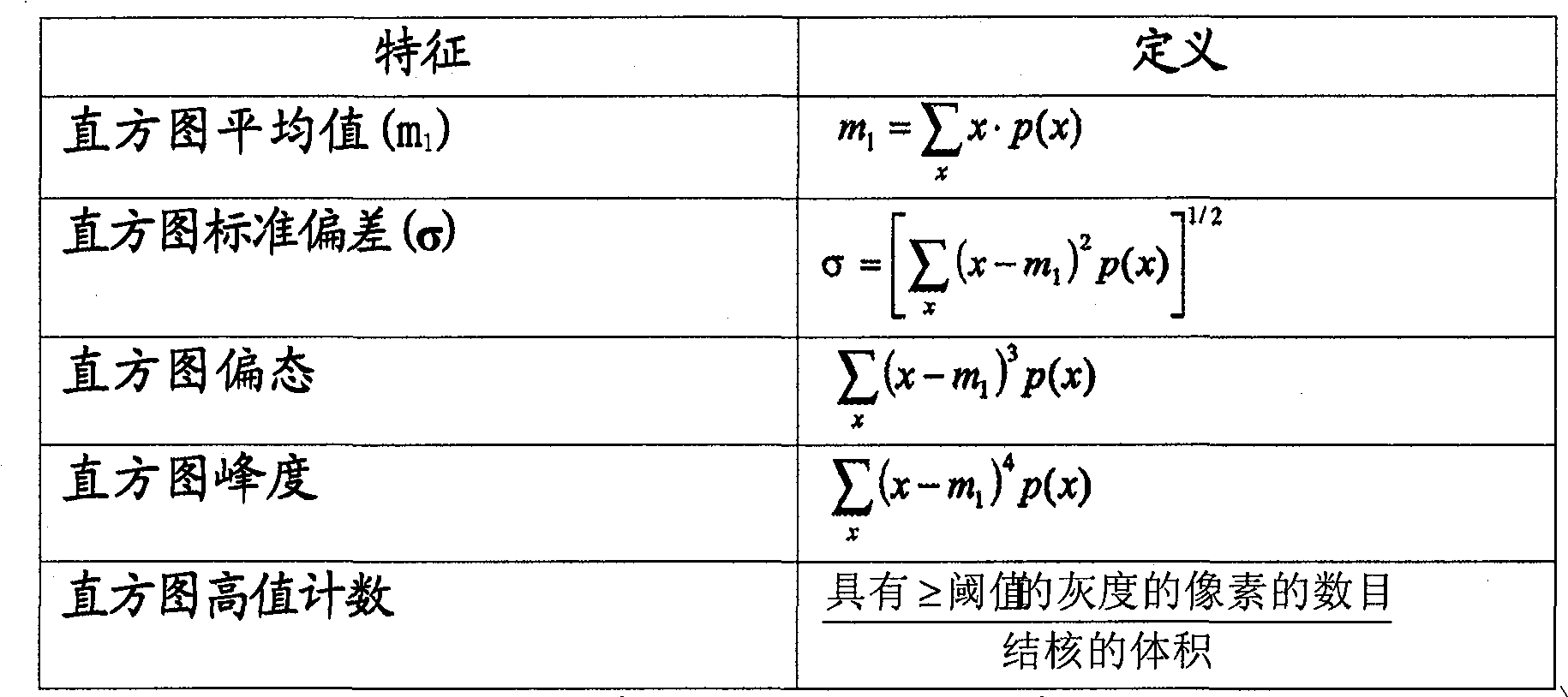
表3基于直方图的特征Table 3 Histogram-based features
在表3中,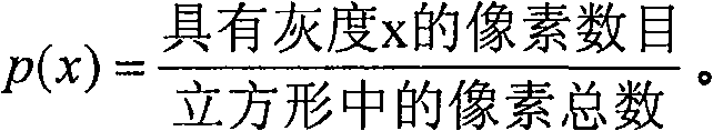
在薄片CT扫描中CAD算法常常把脉管的一部分检测为结核。通过分析所检测及描绘结构表面上的梯度场,我们能够从非结核中区分出结核。尤其是沿着结核和非结核主方向的梯度是不同的,因为在非结核中梯度不会显著地变化,因为脉管会延续。在实际结核的情况中,改变更显著,因为结核更紧密。CAD algorithms often detect a portion of a vessel as a nodule in thin-section CT scans. By analyzing the gradient fields on the surface of the detected and delineated structures, we were able to distinguish nodules from non-nodules. In particular the gradient along the main direction of nodules and non-nodules is different since the gradient does not change significantly in non-nodules because the vessels continue. In the case of actual nodules, the change is more pronounced because the nodules are more compact.
开始,利用3D梯度核心来计算所描绘对象3D表面上的梯度场。然后对表面的每个点处梯度的幅值进行计算。以及计算基于3D矩的对象的主方向,沿着基于第二阶中心矩的结核的惯性矩阵:Initially, a 3D gradient kernel is utilized to compute the gradient field on the 3D surface of the depicted object. The magnitude of the gradient at each point of the surface is then computed. As well as computing the principal directions of the object based on the 3D moments, along the inertia matrix of the nodules based on the second order central moments:
其中Mijk是(i,j,k)个中心矩且根据“标记”数据L被计算为:where Mijk are the (i,j,k) central moments and are computed from the "labeled" data L as:
Mijk=∑(sl-csl)i(row-cr)j(col-cc)kL(sl,row,col) (2)M ijk =∑(sl-c sl ) i (row-c r ) j (col-c c ) k L(sl, row, col) (2)
其中c=(cs1,cr,cc)表示结核的重心。重心的坐标被定义为:Where c=(c s1 , cr , c c ) represents the center of gravity of the nodule. The coordinates of the center of gravity are defined as:
惯性矩阵的特征向量确定了物体的主轴。尤其我们使用了对应于最大特征值的特征向量。沿着这个方向,我们把梯度幅值的分布用作特征,其能够在结核和像脉管部分这样的非结核之间进行区分。The eigenvectors of the inertia matrix define the principal axes of the object. In particular we use the eigenvector corresponding to the largest eigenvalue. Along this direction, we use the distribution of gradient magnitudes as a feature capable of distinguishing between nodules and non-nodules like vascular sections.
重要的是要注意执行该创新方法或驱动该创新FPR分类器所需要的软件可以包括用于实现逻辑功能的可执行指令的有序列表。因而,该软件能够以由或结合指令执行系统、装置、或设备来使用的任何计算机可读介质来体现,诸如基于计算机的系统、包含处理器的系统、或可把指令从指令执行系统、装置、或设备中取出并且执行该指令的其他系统。在本文件的上下文中,“计算机可读介质”可以是能够包含、存储、通信、传播、或传送由或结合指令执行系统、装置、或设备来使用的程序的任何装置。It is important to note that the software required to implement the inventive method or drive the inventive FPR classifier may include an ordered list of executable instructions for implementing logical functions. Thus, the software can be embodied on any computer-readable medium used by or in connection with an instruction execution system, apparatus, or device, such as a computer-based system, a system containing a processor, or instructions from an instruction execution system, apparatus, or , or other systems that take out and execute the instruction from the device. In the context of this document, a "computer-readable medium" may be any device capable of containing, storing, communicating, propagating, or transferring a program for use by or in connection with an instruction execution system, apparatus, or device.
计算机可读介质可以是例如但不限于电、磁、光、电磁、红外,或半导体系统,装置,设备,或传播介质。计算机可读介质的更加具体的例子(非穷举列表)包括以下:具有一个或多个线路的电连接(电子)、便携式计算机盘(磁性)、随机存取存储器(RAM)(磁性)、只读存储器(ROM)(磁性)、可擦除可编程只读存储器(EPROM或闪速存储器)(磁性)、光纤(光)、以及便携式光盘只读存储器(CDROM)(光)。要注意的是计算机可读介质甚至可以是其上打印了程序的纸或别的适当的介质,因为该程序可例如经由光学扫描纸或其他介质而被电子地获取、然后编译、解释或者否则如有必要以适当的方式进行处理、然后存储在计算机存储器中。A computer readable medium can be, for example and without limitation, an electrical, magnetic, optical, electromagnetic, infrared, or semiconductor system, apparatus, device, or propagation medium. More specific examples (non-exhaustive list) of computer readable media include the following: electrical connection with one or more wires (electronic), portable computer disk (magnetic), random access memory (RAM) (magnetic), Read Memory (ROM) (magnetic), Erasable Programmable Read Only Memory (EPROM or Flash Memory) (magnetic), Optical Fiber (optical), and Compact Disc Read Only Memory (CDROM) (optical). It is to be noted that the computer readable medium may even be paper or other suitable medium on which the program is printed, as the program may be retrieved electronically, for example via optically scanned paper or other medium, and then compiled, interpreted, or otherwise It is necessary to process in an appropriate manner and then store in computer memory.
应当强调的是本发明的上述实施例,尤其是任何“优选”实施例,仅仅是用于仅仅阐述对本发明原理的清楚理解的实现的可能例子。此外,在基本上不脱离本发明的精神和原理下可对本发明的上述实施例做出许多变化和修改。所有这种修改和变化用来由本发明的范畴内所包括的且由以下权利要求所保护的当前公开所教导。It should be emphasized that the above-described embodiments of the present invention, particularly, any "preferred" embodiments, are merely possible examples of implementations merely set forth for a clear understanding of the principles of the invention. Furthermore, many variations and modifications may be made to the above-described embodiments of the present invention without departing substantially from the spirit and principles of the invention. All such modifications and variations are intended to be taught by the present disclosure as being included within the scope of the present invention and protected by the following claims.
Claims (15)
- One kind be used in computer aided detection (CAD) medical image be concerned about the method for position or volume, it comprises that CAD handles with detection and machine learning is to maximize specificity and to reduce and handle the false-positive number that non-training data is reported afterwards after describing candidate position and the CAD in the training stage, and described method comprises step:Be selected to training classifier on the medical image training dataset that comprises the some positions that are known as true or false for basic fact, utilize described CAD to handle to discern and cut apart this position, extract feature and limit this position to create feature pool, described feature comprises at least one based on the histogrammic feature of 3D, uses the genetic algorithm processor to determine being used for minimal feature subset with the candidate position in the non-training data of improved specific recognition by support vector machine (SVM) to feature pool;In non-training data, detect the candidate position;Cut apart the candidate position in the non-training data;Extract and each relevant characteristic set of cutting apart in candidate position; AndUtilize characteristic set to be classified in the candidate position by SVM,It is characterized in that, described at least one comprise the high value counting of histogram based on the histogrammic feature of 3D, wherein to count be the ratio of number that has the pixel of the above gray-scale value of threshold value in the described position to the high value of histogram.
- 2. the method for setting forth in the claim 1, wherein, training step further comprises each candidate position of determining in the training data in the size of training period by the character subset of GA optimization, and determines to comprise this subclass so that the actual characteristic to being optimized to " the best " suitable SVM mapping.
- 3. the method as being set forth in the claim 1, wherein training step comprises that further the feature pool of being discerned in each position in training data is defined as chromosome, feature of each gene representation wherein, and wherein genetic algorithm at first by selecting feature to breed chromosome at random, and iteratively the search have higher grade of fit those chromosomes, wherein utilize variation and intersect each generation is repeated to assess, during the training stage, produce new and chromosome that be more suitable for.
- 4. the method as being set forth in the claim 3 is wherein determined to be included in two stages and is used GA, and wherein said method comprises:A.) about its characteristic set and the number of feature and discern each chromosome; AndB.) characteristic set of discerning for each chromosome analysis and the number of features of being discerned are determined the optimum dimension of feature with the number of the number that occurs based on the coloured differently body and average error.
- 5. the method set forth of claim 1, wherein training step comprises utilizing and filters mask and discern the wall pixel.
- 6. the method set forth of claim 1, wherein based on 3D histogrammic set further comprise following at least one: histogram mean value (mi), histogram standard deviation (σ), histogram skewness and histogram kurtosis.
- 7. the method set forth of claim 1, wherein produce set based on the 3D gradient by at first utilizing 3D gradient core to calculate the lip-deep gradient fields of rendered object 3D, gradient magnitude in each point of gauging surface, and calculate based on the object principal direction of 3D square with based on the tuberculosis inertial matrix of the second rank central moment.
- 8. the method set forth of claim 1, wherein training step comprises that the grey level distribution based on volume of interest in the medical image produces at least one based on the histogrammic feature of 3D, wherein said generation step comprises step:Calculate the high value counting of histogram at least, wherein calculation procedure comprises the described step of being concerned about all the wall pixels in the position of eliminating.
- 9. the method set forth of claim 8, wherein calculation procedure comprises and utilizes function p (x), it is defined as having the sum of the number of pixels of gray level x divided by pixel in the histogram case.
- 10. the method set forth of claim 1, wherein training step comprises that producing at least one classifies to the object of studying that is detected in the volume of being concerned about with support based on the feature of 3D gradient in medical image, and wherein said generation step comprises step:Rendered object;The compute gradient core;Utilize this gradient core to come compute gradient field on the border of institute's rendered object;In borderline each some place compute gradient amplitude; AndCalculate main gradient direction.
- 11. the method for claim 10, the step of wherein calculating main gradient direction comprises the main shaft of calculating object, and wherein Zui Da institute's calculating main shaft is used to define gradient magnitude calculated direction that distributes.
- 12. as the method for being set forth in the claim 10, wherein calculation procedure comprises that identification comes the principal direction of the maximum principal axis of calculating object based on 3D square and inertial matrix, wherein inertial matrix is based on second-order moment around mean, and the proper vector of inertial matrix has defined main shaft.
- 13. one kind is used for the interior position of being concerned about of detection and Identification medical image and/or the system of volume, wherein this system comprises that CAD subsystem and false positive reduce (FPR) subsystem, this CAD subsystem be used to discern with delineate in examine then be concerned about the position, this false positive reduces (FPR) subsystem and is used for improved specificity the position being categorized into one of two kinds of basic fact states, minimize false-positive number whereby by System ReportsThis false positive reduces subsystem and the CAD subsystem communicates, and it at first closes at training dataset and trains, and with improved specificity the candidate position in the non-training data is operated then, and wherein this false positive reduction subsystem comprises:Feature extractor is used to extract the corresponding feature pool of describing with each CAD in candidate position, comprises that at least one is based on the histogrammic character subset of 3D or from the feature of this subclass;The genetic algorithm equipment of communicating by letter with feature extractor, the CAD that adopted describes to determine optimal feature subset in the feature pool at position from training; AndWith the support vector machine (SVM) that feature extractor is communicated by letter with GA, classify in the candidate position that its each CAD that non-training data, training back is detected according to optimal feature subset describes;Wherein by from each segmentaion position, extracting feature pool by feature extractor, utilize GA to discern the best subset of the feature of extracting, and utilize known basic fact on the view data that comprises the candidate position, system to be trained, so that thereby system is carrying out the enough distinguishing abilities of operating period demonstration with improved specificity classification candidate position to non-training dataIt is characterized in that, described at least one comprise the high value counting of histogram based on the histogrammic character subset of 3D or from the feature of this subclass, wherein to count be the ratio of number that has the pixel of the above gray-scale value of threshold value in the described position to the high value of histogram.
- 14. the medical image categorizing system of setting forth in the claim 13, wherein the CAD subsystem further comprises and cuts apart subsystem, and it provides the reader to import position to describe better to be used to train at training period.
- 15. as the medical image categorizing system that claim 13 is set forth, wherein GA operates the suitable example of classification in the process of training and non-training data being operated.
Applications Claiming Priority (3)
| Application Number | Priority Date | Filing Date | Title |
|---|---|---|---|
| US62975004P | 2004-11-19 | 2004-11-19 | |
| US60/629,750 | 2004-11-19 | ||
| PCT/IB2005/053835 WO2006054271A2 (en) | 2004-11-19 | 2005-11-21 | False positive reduction in computer -assisted detection ( cad) |
Publications (2)
| Publication Number | Publication Date |
|---|---|
| CN101061510A CN101061510A (en) | 2007-10-24 |
| CN101061510B true CN101061510B (en) | 2010-09-08 |
Family
ID=36407532
Family Applications (1)
| Application Number | Title | Priority Date | Filing Date |
|---|---|---|---|
| CN2005800397104A Expired - Fee Related CN101061510B (en) | 2004-11-19 | 2005-11-21 | Methods and systems for detecting and identifying regions and/or volumes of interest within medical image data |
Country Status (7)
| Country | Link |
|---|---|
| US (1) | US7840062B2 (en) |
| EP (1) | EP1815431B1 (en) |
| JP (1) | JP2008520322A (en) |
| CN (1) | CN101061510B (en) |
| AT (1) | ATE506661T1 (en) |
| DE (1) | DE602005027600D1 (en) |
| WO (1) | WO2006054271A2 (en) |
Families Citing this family (45)
| Publication number | Priority date | Publication date | Assignee | Title |
|---|---|---|---|---|
| US7088872B1 (en) * | 2002-02-14 | 2006-08-08 | Cogent Systems, Inc. | Method and apparatus for two dimensional image processing |
| JP2007520278A (en) * | 2004-01-26 | 2007-07-26 | コーニンクレッカ フィリップス エレクトロニクス エヌ ヴィ | Diagnosis decision support apparatus and method based on examples |
| US7616788B2 (en) * | 2004-11-12 | 2009-11-10 | Cogent Systems, Inc. | System and method for fast biometric pattern matching |
| US8131477B2 (en) | 2005-11-16 | 2012-03-06 | 3M Cogent, Inc. | Method and device for image-based biological data quantification |
| EP2052355A2 (en) * | 2006-08-11 | 2009-04-29 | Koninklijke Philips Electronics N.V. | Methods and apparatus to integrate systematic data scaling into genetic algorithm-based feature subset selection |
| JP2010504578A (en) * | 2006-09-22 | 2010-02-12 | コーニンクレッカ フィリップス エレクトロニクス エヌ ヴィ | A method for feature selection based on genetic algorithm using classifier ensemble |
| US8275179B2 (en) * | 2007-05-01 | 2012-09-25 | 3M Cogent, Inc. | Apparatus for capturing a high quality image of a moist finger |
| US8411916B2 (en) * | 2007-06-11 | 2013-04-02 | 3M Cogent, Inc. | Bio-reader device with ticket identification |
| US20090100105A1 (en) * | 2007-10-12 | 2009-04-16 | 3Dr Laboratories, Llc | Methods and Systems for Facilitating Image Post-Processing |
| US8224057B2 (en) * | 2007-10-18 | 2012-07-17 | Siemens Aktiengesellschaft | Method and system for nodule feature extraction using background contextual information in chest x-ray images |
| US20100014755A1 (en) * | 2008-07-21 | 2010-01-21 | Charles Lee Wilson | System and method for grid-based image segmentation and matching |
| US9123095B2 (en) | 2008-09-29 | 2015-09-01 | Koninklijke Philips N.V. | Method for increasing the robustness of computer-aided diagnosis to image processing uncertainties |
| WO2010076668A1 (en) * | 2009-01-05 | 2010-07-08 | Freescale Semiconductor, Inc. | System and method for efficient image feature extraction |
| US8295637B2 (en) | 2009-01-07 | 2012-10-23 | Seiko Epson Corporation | Method of classifying red-eye objects using feature extraction and classifiers |
| US8346800B2 (en) * | 2009-04-02 | 2013-01-01 | Microsoft Corporation | Content-based information retrieval |
| CN101807256B (en) * | 2010-03-29 | 2013-03-20 | 天津大学 | Object identification detection method based on multiresolution frame |
| CN101826160B (en) * | 2010-03-31 | 2012-11-14 | 北京航空航天大学 | Hyperspectral image classification method based on immune evolutionary strategy |
| CN101866416A (en) * | 2010-06-18 | 2010-10-20 | 山东大学 | A Fingerprint Image Segmentation Method Based on Transductive Learning |
| US20140010433A1 (en) * | 2010-11-03 | 2014-01-09 | Ajay Divekar | System and method for improved detection of objects of interest in image data by management of false positives |
| TWI439951B (en) * | 2010-11-08 | 2014-06-01 | Inst Information Industry | Facial gender identification system and method and computer program products thereof |
| US9691395B1 (en) * | 2011-12-31 | 2017-06-27 | Reality Analytics, Inc. | System and method for taxonomically distinguishing unconstrained signal data segments |
| US9189746B2 (en) * | 2012-01-12 | 2015-11-17 | Microsoft Technology Licensing, Llc | Machine-learning based classification of user accounts based on email addresses and other account information |
| CN102842132B (en) * | 2012-07-12 | 2015-12-09 | 上海联影医疗科技有限公司 | A kind of CT pulmonary nodule detection method |
| CN103892855A (en) * | 2012-12-28 | 2014-07-02 | 上海联影医疗科技有限公司 | Digital medical image processing method and device |
| KR20160037023A (en) * | 2014-09-26 | 2016-04-05 | 삼성전자주식회사 | Apparatus and Method for supporting a Computer Aided Diagnosis |
| US9595103B2 (en) * | 2014-11-30 | 2017-03-14 | Case Western Reserve University | Textural analysis of lung nodules |
| US9842390B2 (en) | 2015-02-06 | 2017-12-12 | International Business Machines Corporation | Automatic ground truth generation for medical image collections |
| CN105260744B (en) * | 2015-10-08 | 2018-08-14 | 北京航空航天大学 | The automatic on-line diagnostic method and system of a kind of goods train coupler yoke key position failure |
| KR102433384B1 (en) * | 2016-01-05 | 2022-08-18 | 한국전자통신연구원 | Apparatus and method for processing texture image |
| EP3264322A1 (en) * | 2016-06-30 | 2018-01-03 | Deutsches Krebsforschungszentrum Stiftung des Öffentlichen Rechts | Machine learning-based quantitative photoacoustic tomography (pat) |
| JP6657132B2 (en) * | 2017-02-27 | 2020-03-04 | 富士フイルム株式会社 | Image classification device, method and program |
| CN107945875A (en) * | 2017-11-17 | 2018-04-20 | 合肥工业大学 | Pulmonary nodule detection method and system based on data enhancing |
| WO2020012414A1 (en) * | 2018-07-11 | 2020-01-16 | Advenio Tecnosys Pvt. Ltd. | Framework for reduction of hard mimics in medical images |
| US10943681B2 (en) * | 2018-11-21 | 2021-03-09 | Enlitic, Inc. | Global multi-label generating system |
| US10813612B2 (en) | 2019-01-25 | 2020-10-27 | Cleerly, Inc. | Systems and method of characterizing high risk plaques |
| CN111062977B (en) * | 2019-12-13 | 2021-05-04 | 推想医疗科技股份有限公司 | Sample data generation method and device, computer equipment and storage medium |
| US11969280B2 (en) | 2020-01-07 | 2024-04-30 | Cleerly, Inc. | Systems, methods, and devices for medical image analysis, diagnosis, risk stratification, decision making and/or disease tracking |
| US20220392065A1 (en) | 2020-01-07 | 2022-12-08 | Cleerly, Inc. | Systems, methods, and devices for medical image analysis, diagnosis, risk stratification, decision making and/or disease tracking |
| KR20220124217A (en) | 2020-01-07 | 2022-09-13 | 클리어리, 인크. | Systems, methods and devices for medical image analysis, diagnosis, risk stratification, decision-making and/or disease tracking |
| US12136484B2 (en) | 2021-11-05 | 2024-11-05 | Altis Labs, Inc. | Method and apparatus utilizing image-based modeling in healthcare |
| TWI806425B (en) * | 2022-02-14 | 2023-06-21 | 宏碁股份有限公司 | Feature selection method |
| US20250217981A1 (en) | 2022-03-10 | 2025-07-03 | Cleerly, Inc. | Systems, methods, and devices for image-based plaque analysis and risk determination |
| US20250143657A1 (en) | 2022-03-10 | 2025-05-08 | Cleerly, Inc. | Systems, devices, and methods for non-invasive image-based plaque analysis and risk determination |
| US12440180B2 (en) | 2022-03-10 | 2025-10-14 | Cleerly, Inc. | Systems, devices, and methods for non-invasive image-based plaque analysis and risk determination |
| US12406365B2 (en) | 2022-03-10 | 2025-09-02 | Cleerly, Inc. | Systems, devices, and methods for non-invasive image-based plaque analysis and risk determination |
Citations (2)
| Publication number | Priority date | Publication date | Assignee | Title |
|---|---|---|---|---|
| CN1395713A (en) * | 2000-01-18 | 2003-02-05 | 芝加哥大学 | Method, system and computer readable medium for two-dimensional and three-dimensional detection of lungs nodules in computed tomography image scans |
| CN1405734A (en) * | 2002-10-28 | 2003-03-26 | 武汉大学 | A Method of Edge Enhancement for Medical Image |
Family Cites Families (7)
| Publication number | Priority date | Publication date | Assignee | Title |
|---|---|---|---|---|
| US6996549B2 (en) * | 1998-05-01 | 2006-02-07 | Health Discovery Corporation | Computer-aided image analysis |
| AU2001251539A1 (en) * | 2000-04-11 | 2001-10-23 | Cornell Research Foundation Inc. | System and method for three-dimensional image rendering and analysis |
| US6724856B2 (en) * | 2002-04-15 | 2004-04-20 | General Electric Company | Reprojection and backprojection methods and algorithms for implementation thereof |
| US6707878B2 (en) * | 2002-04-15 | 2004-03-16 | General Electric Company | Generalized filtered back-projection reconstruction in digital tomosynthesis |
| US7218766B2 (en) * | 2002-04-15 | 2007-05-15 | General Electric Company | Computer aided detection (CAD) for 3D digital mammography |
| EP1716514A2 (en) | 2004-02-10 | 2006-11-02 | Koninklijke Philips Electronics N.V. | Genetic algorithms for optimization of genomics-based medical diagnostic tests |
| US7756313B2 (en) * | 2005-11-14 | 2010-07-13 | Siemens Medical Solutions Usa, Inc. | System and method for computer aided detection via asymmetric cascade of sparse linear classifiers |
-
2005
- 2005-11-21 AT AT05817730T patent/ATE506661T1/en not_active IP Right Cessation
- 2005-11-21 CN CN2005800397104A patent/CN101061510B/en not_active Expired - Fee Related
- 2005-11-21 WO PCT/IB2005/053835 patent/WO2006054271A2/en not_active Ceased
- 2005-11-21 DE DE602005027600T patent/DE602005027600D1/en not_active Expired - Lifetime
- 2005-11-21 EP EP05817730A patent/EP1815431B1/en not_active Expired - Lifetime
- 2005-11-21 JP JP2007542446A patent/JP2008520322A/en active Pending
- 2005-11-21 US US11/719,634 patent/US7840062B2/en not_active Expired - Fee Related
Patent Citations (2)
| Publication number | Priority date | Publication date | Assignee | Title |
|---|---|---|---|---|
| CN1395713A (en) * | 2000-01-18 | 2003-02-05 | 芝加哥大学 | Method, system and computer readable medium for two-dimensional and three-dimensional detection of lungs nodules in computed tomography image scans |
| CN1405734A (en) * | 2002-10-28 | 2003-03-26 | 武汉大学 | A Method of Edge Enhancement for Medical Image |
Non-Patent Citations (2)
| Title |
|---|
| MILLER M T ET AL.Feature selection for computer-aided polyp detection usinggenetic algorithms.The international society for optical engineering5031.2003,5031第102页摘要、第103页1.3、第105页2.1、附图1. * |
| 基于多尺度对比度塔的图像融合方法及性能评价.1339页的3. * |
Also Published As
| Publication number | Publication date |
|---|---|
| WO2006054271A2 (en) | 2006-05-26 |
| JP2008520322A (en) | 2008-06-19 |
| US7840062B2 (en) | 2010-11-23 |
| CN101061510A (en) | 2007-10-24 |
| EP1815431B1 (en) | 2011-04-20 |
| DE602005027600D1 (en) | 2011-06-01 |
| ATE506661T1 (en) | 2011-05-15 |
| EP1815431A2 (en) | 2007-08-08 |
| US20090148010A1 (en) | 2009-06-11 |
| WO2006054271A3 (en) | 2007-04-05 |
Similar Documents
| Publication | Publication Date | Title |
|---|---|---|
| CN101061510B (en) | Methods and systems for detecting and identifying regions and/or volumes of interest within medical image data | |
| Boroczky et al. | Feature subset selection for improving the performance of false positive reduction in lung nodule CAD | |
| EP1815399B1 (en) | A stratification method for overcoming unbalanced case numbers in computer-aided lung nodule false positive reduction | |
| Campadelli et al. | A fully automated method for lung nodule detection from postero-anterior chest radiographs | |
| US8265355B2 (en) | System and method for automated detection and segmentation of tumor boundaries within medical imaging data | |
| Jain et al. | Lung nodule segmentation using salp shuffled shepherd optimization algorithm-based generative adversarial network | |
| US20090175531A1 (en) | System and method for false positive reduction in computer-aided detection (cad) using a support vector macnine (svm) | |
| JP4388121B2 (en) | Polyp detection method in 3D image volume | |
| US20100183210A1 (en) | Computer-assisted analysis of colonic polyps by morphology in medical images | |
| US20150065868A1 (en) | System, method, and computer accessible medium for volumetric texture analysis for computer aided detection and diagnosis of polyps | |
| Mondal et al. | Automated diagnosis of pulmonary emphysema using multi-objective binary thresholding and hybrid classification | |
| Dawoud | Lung segmentation in chest radiographs by fusing shape information in iterative thresholding | |
| Sheikhy et al. | State of the art review of AI in renal imaging | |
| Mukherjee et al. | A soft-computing based approach towards automatic detection of pulmonary nodule | |
| CN101061490A (en) | System and method for false positive reduction in computer-aided detection (CAD) using a support vector machine (SVM) | |
| Khalil et al. | Deep learning-enhanced brain tumor prediction via Entropy-coded BPSO in CIELAB Color Space | |
| Kumar et al. | A hybrid model for kidney stone detection using deep learning | |
| Naveen et al. | An approach for classification of lung nodules | |
| Summers et al. | Future directions: computer-aided diagnosis | |
| Pulagam et al. | Automated pulmonary lung nodule detection using an optimal manifold statistical based feature descriptor and SVM classifier | |
| Liu et al. | Computer aided detection of lung nodules based on voxel analysis utilizing support vector machines | |
| Admane et al. | Multi-stage lung cancer detection and prediction using image processing techniques | |
| Khomkham | Pulmonary lesion classification using convolutional neural network for endobronchial ultrasonogram | |
| Sithiq et al. | HCL Net: Deep Learning for Accurate Classification of Honeycombing Lung and Ground Glass Opacity in CT Images | |
| Natarajan et al. | Contour‐Detected Normalized Residual Model for Kidney Stone Classification. |
Legal Events
| Date | Code | Title | Description |
|---|---|---|---|
| C06 | Publication | ||
| PB01 | Publication | ||
| C10 | Entry into substantive examination | ||
| SE01 | Entry into force of request for substantive examination | ||
| C14 | Grant of patent or utility model | ||
| GR01 | Patent grant | ||
| CF01 | Termination of patent right due to non-payment of annual fee | ||
| CF01 | Termination of patent right due to non-payment of annual fee |
Granted publication date: 20100908 Termination date: 20201121 |