Ribosomal protein SA
40S ribosomal protein SA is a ribosomal protein that in humans is encoded by the RPSA gene.[5][6] It also acts as a cell surface receptor, in particular for laminin, and is involved in several pathogenic processes.
Function
[edit]Laminins, a family of extracellular matrix glycoproteins, are the major noncollagenous constituent of basement membranes. They have been implicated in a wide variety of biological processes including cell adhesion, differentiation, migration, signaling, neurite outgrowth and metastasis. Many of the effects of laminin are mediated through interactions with cell surface receptors. These receptors include members of the integrin family, as well as non-integrin laminin-binding proteins. The RPSA gene encodes a multifunctional protein, which is both a ribosomal protein and a high-affinity, non-integrin laminin receptor. This protein has been variously called Ribosomal protein SA; RPSA; LamR; LamR1; 37 kDa Laminin Receptor Precursor; 37LRP; 67 kDa Laminin Receptor; 67LR; 37/67 kDa Laminin Receptor; LRP/LR; LBP/p40; and p40 ribosome-associated protein. Ribosomal protein SA and RPSA are the approved name and symbol. The amino acid sequence of RPSA is highly conserved through evolution, suggesting a key biological function. It has been observed that the level of RPSA transcript is higher in colon carcinoma tissue and lung cancer cell lines than their normal counterparts. Also, there is a correlation between the upregulation of this polypeptide in cancer cells and their invasive and metastatic phenotype. Multiple copies of the RPSA gene exist; however, most of them are pseudogenes thought to have arisen from retropositional events. Two alternatively spliced transcript variants encoding the same protein have been found for this gene.[7]
Structure and stability
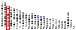
[edit]The complementary DNA (cDNA) of the RPSA gene is formed by the assembly of seven exons, six of which correspond to the coding sequence.[6] The amino acid sequence of RPSA, deduced from the sequence of its cDNA, includes 295 residues. RPSA can be sub-divided in two main domains: an N-domain (residues 1–209), which corresponds to exons 2-5 of the gene, and a C-domain (residues 210–295), which corresponds to exons 6–7. The N-domain of RPSA is homologous to the ribosomal protein S2 (RPS2) of prokaryotes. It contains a palindromic sequence 173LMWWML178 which is conserved in all metazoans. Its C-domain is highly conserved in vertebrates. The amino acid sequence of RPSA is 98% identical in all mammals. RPSA is a ribosomal protein which has acquired the function of laminin receptor during evolution.[8][9] The structure of the N-domain of RPSA is similar to those of prokaryotic RPS2.[10] The C-domain is intrinsically disordered in solution. The N-domain is monomeric in solution and unfolds according to a three state equilibrium. The folding intermediate is predominant at 37 °C.[11]
Interactions
[edit]Several interactions of RPSA that had originally been discovered by methods of cellular biology, have subsequently been confirmed by using recombinant derivatives and in vitro experiments. The latter have shown that the folded N-domain and disordered C-domain of RPSA have both common and specific functions.[12]
- RPSA binds to proteins that are involved in the translation of the genetic code. (i) Yeast two-hybrid screens have shown that RPSA binds to Ribosomal protein S21 of the 40S small ribosomal subunit.[13][14] (ii) Serial deletions of RPSA have shown that the segment of residues 236–262, included in the C-domain, is involved in the interaction between RPSA and the 40S subunit of ribosome.[15] (iii) Studies that were based on nuclear magnetic resonance spectroscopy (NMR), have shown that the anticodon binding domain of Lysyl-tRNA synthetase binds directly to the C-domain of RPSA.[16]
- RPSA was initially identified as a laminin binding protein.[17][18] Both recombinant N-domain and C-domain of RPSA bind laminin in vitro, and they bind with similar dissociation constants (300 nM).[10][12]
- Both RPSA and laminin belong to the heparin/heparan sulfate interactome.[19] Heparin binds in vitro to the N-domain of RPSA but not to its C-domain. Moreover, the C-domain of RPSA and heparin compete for binding to laminin, which shows that the highly acidic C-domain of RPSA mimicks heparin (and potentially heparan sulfates) for the binding to laminin.[12]
- RPSA is a potential cellular receptor for several pathogenic Flaviviruses, including the dengue virus (DENV),[20][21] and Alphaviruses, including the Sindbis virus (SINV).[22] The N-domain of RPSA includes a binding site for SINV in vitro.[10] The N-domain also includes weak binding sites for recombinant domain 3 (ED3, residues 296–400) from the envelope proteins of two Flaviviruses, West-Nile virus and serotype 2 of DENV. The C-domain includes weak binding sites for domain 3 of the yellow fever virus (YFV) and of serotypes 1 and 2 of DENV. In contrast, domain 3 from the Japanese encephalitis virus does not appear to bind RPSA in vitro.[12]
- RPSA is also a receptor for small molecules. (i) RPSA binds aflatoxin B1 both in vivo and in vitro.[23] (ii) RPSA is a receptor for epigallocatechin-gallate (EGCG), which is a major constituent of green tea and has many health related effects.[24][25] EGCG binds only to the N-domain of RPSA in vitro, with a dissociation constant of 100 nM, but not to its C-domain.[12]
References
[edit]- ^ a b c GRCh38: Ensembl release 89: ENSG00000168028 – Ensembl, May 2017
- ^ a b c GRCm38: Ensembl release 89: ENSMUSG00000032518 – Ensembl, May 2017
- ^ "Human PubMed Reference:". National Center for Biotechnology Information, U.S. National Library of Medicine.
- ^ "Mouse PubMed Reference:". National Center for Biotechnology Information, U.S. National Library of Medicine.
- ^ Satoh K, Narumi K, Sakai T, Abe T, Kikuchi T, Matsushima K, Sindoh S, Motomiya M (Jul 1992). "Cloning of 67-kDa laminin receptor cDNA and gene expression in normal and malignant cell lines of the human lung". Cancer Lett. 62 (3): 199–203. doi:10.1016/0304-3835(92)90096-E. PMID 1534510.
- ^ a b Jackers P, Minoletti F, Belotti D, Clausse N, Sozzi G, Sobel ME, Castronovo V (Sep 1996). "Isolation from a multigene family of the active human gene of the metastasis-associated multifunctional protein 37LRP/p40 at chromosome 3p21.3". Oncogene. 13 (3): 495–503. PMID 8760291.
- ^ DiGiacomo, Vincent; Meruelo, Daniel (May 2016). "Looking into laminin receptor: critical discussion regarding the non-integrin 37/67-kDa laminin receptor/RPSA protein". Biological Reviews. 91 (2): 288–310. doi:10.1111/brv.12170. PMC 5249262. PMID 25630983.
- ^ Ardini E, Pesole G, Tagliabue E, Magnifico A, Castronovo V, Sobel ME, Colnaghi MI, Ménard S (August 1998). "The 67-kDa laminin receptor originated from a ribosomal protein that acquired a dual function during evolution". Molecular Biology and Evolution. 15 (8): 1017–25. doi:10.1093/oxfordjournals.molbev.a026000. PMID 9718729.
- ^ Nelson J, McFerran NV, Pivato G, Chambers E, Doherty C, Steele D, Timson DJ (February 2008). "The 67 kDa laminin receptor: structure, function and role in disease". Bioscience Reports. 28 (1): 33–48. doi:10.1042/BSR20070004. PMID 18269348.
- ^ a b c Jamieson KV, Wu J, Hubbard SR, Meruelo D (February 2008). "Crystal structure of the human laminin receptor precursor". The Journal of Biological Chemistry. 283 (6): 3002–5. doi:10.1074/jbc.C700206200. PMID 18063583.
- ^ Ould-Abeih, MB; Petit-Topin, I; Zidane, N; Baron, B; Bedouelle, Hugues (Jun 2012). "Multiple folding states and disorder of ribosomal protein SA, a membrane receptor for laminin, anticarcinogens, and pathogens". Biochemistry. 51 (24): 4807–4821. doi:10.1021/bi300335r. PMID 22640394.
- ^ a b c d e Zidane, N; Ould-Abeih, MB; Petit-Topin, I; Bedouelle, H (2012). "The folded and disordered domains of human ribosomal protein SA have both idiosyncratic and shared functions as membrane receptors". Biosci. Rep. 33 (1): 113–124. doi:10.1042/BSR20120103. PMC 4098866. PMID 23137297.
- ^ Stelzl U, Worm U, Lalowski M, Haenig C, Brembeck FH, Goehler H, Stroedicke M, Zenkner M, Schoenherr A, Koeppen S, Timm J, Mintzlaff S, Abraham C, Bock N, Kietzmann S, Goedde A, Toksöz E, Droege A, Krobitsch S, Korn B, Birchmeier W, Lehrach H, Wanker EE (Sep 2005). "A human protein-protein interaction network: a resource for annotating the proteome". Cell. 122 (6): 957–968. doi:10.1016/j.cell.2005.08.029. hdl:11858/00-001M-0000-0010-8592-0. PMID 16169070. S2CID 8235923.
- ^ Sato M, Saeki Y, Tanaka K, Kaneda Y (Mar 1999). "Ribosome-associated protein LBP/p40 binds to S21 protein of 40S ribosome: analysis using a yeast two-hybrid system". Biochem. Biophys. Res. Commun. 256 (2): 385–390. doi:10.1006/bbrc.1999.0343. PMID 10079194.
- ^ Malygin, AA; Babaylova, ES; Loktev, VB; Karpova, GG (2011). "A region in the C-terminal domain of ribosomal protein SA required for binding of SA to the human 40S ribosomal subunit". Biochimie. 93 (3): 612–617. doi:10.1016/j.biochi.2010.12.005. PMID 21167900.
- ^ Cho, HY; Ul Mushtaq, A; Lee, JY; Kim, DG; Seok, MS; Jang, M; Han, BW; Kim, S; Jeon, YH (2014). "Characterization of the interaction between lysyl-tRNA synthetase and laminin receptor by NMR". FEBS Lett. 588 (17): 2851–2858. doi:10.1016/j.febslet.2014.06.048. PMID 24983501. S2CID 36128866.
- ^ Rao, NC; Barsky, SH; Terranova, VP; Liotta, LA (1983). "Isolation of a tumor cell laminin receptor". Biochem. Biophys. Res. Commun. 111 (3): 804–808. doi:10.1016/0006-291X(83)91370-0. PMID 6301485.
- ^ Lesot, H; Kühl, U; Mark, K (1983). "Isolation of a laminin-binding protein from muscle cell membranes". EMBO J. 2 (6): 861–865. doi:10.1002/j.1460-2075.1983.tb01514.x. PMC 555201. PMID 16453457.
- ^ Ori, A; Wilkinson, MC; Fernig, DG (2011). "A systems biology approach for the investigation of the heparin/heparan sulfate interactome". J. Biol. Chem. 286 (22): 19892–19904. doi:10.1074/jbc.M111.228114. PMC 3103365. PMID 21454685.
- ^ Thepparit, C; Smith, DR (2004). "Serotype-specific entry of dengue virus into liver cells: identification of the 37-kilodalton/67-kilodalton high-affinity laminin receptor as a dengue virus serotype 1 receptor". J. Virol. 78 (22): 12647–12656. doi:10.1128/jvi.78.22.12647-12656.2004. PMC 525075. PMID 15507651.
- ^ Tio, PH; Jong, WW; Cardosa, MJ (2005). "Two dimensional VOPBA reveals laminin receptor (LAMR1) interaction with dengue virus serotypes 1, 2 and 3". Virol. J. 2: 25. doi:10.1186/1743-422X-2-25. PMC 1079963. PMID 15790424.
- ^ Wang, KS; Kuhn, RJ; Strauss, EG; Ou, S; Strauss, JH (1992). "High-affinity laminin receptor is a receptor for Sindbis virus in mammalian cells". J. Virol. 66 (8): 4992–5001. doi:10.1128/JVI.66.8.4992-5001.1992. PMC 241351. PMID 1385835.
- ^ Zhuang, Z; Huang, Y; Yang, Y; Wang, S (2016). "Identification of AFB1-interacting proteins and interactions between RPSA and AFB1". J. Hazard. Mater. 301: 297–303. doi:10.1016/j.jhazmat.2015.08.053. PMID 26372695.
- ^ Tachibana, H; Koga, K; Fujimura, Y; Yamada, K (2004). "A receptor for green tea polyphenol EGCG". Nat. Struct. Mol. Biol. 11 (4): 380–381. doi:10.1038/nsmb743. PMID 15024383. S2CID 27868813.
- ^ Tachibana, H (2011). "Green tea polyphenol sensing". Proc. Jpn. Acad. Ser. B Phys. Biol. Sci. 87 (3): 66–80. Bibcode:2011PJAB...87...66T. doi:10.2183/pjab.87.66. PMC 3066547. PMID 21422740.
Further reading
[edit]- Belkin AM, Stepp MA (2000). "Integrins as receptors for laminins". Microsc. Res. Tech. 51 (3): 280–301. doi:10.1002/1097-0029(20001101)51:3<280::AID-JEMT7>3.0.CO;2-O. PMID 11054877. S2CID 45941383.
- Wewer UM, Liotta LA, Jaye M, Ricca GA, Drohan WN, Claysmith AP, Rao CN, Wirth P, Coligan JE, Albrechtsen R (1986). "Altered levels of laminin receptor mRNA in various human carcinoma cells that have different abilities to bind laminin". Proc. Natl. Acad. Sci. U.S.A. 83 (19): 7137–7141. Bibcode:1986PNAS...83.7137W. doi:10.1073/pnas.83.19.7137. PMC 386670. PMID 2429301.
- Van den Ouweland AM, Van Duijnhoven HL, Deichmann KA, Van Groningen JJ, de Leij L, Van de Ven WJ (1989). "Characteristics of a multicopy gene family predominantly consisting of processed pseudogenes". Nucleic Acids Res. 17 (10): 3829–3843. doi:10.1093/nar/17.10.3829. PMC 317862. PMID 2543954.
- Yow HK, Wong JM, Chen HS, Lee CG, Davis S, Steele GD, Chen LB (1988). "Increased mRNA expression of a laminin-binding protein in human colon carcinoma: complete sequence of a full-length cDNA encoding the protein" (PDF). Proc. Natl. Acad. Sci. U.S.A. 85 (17): 6394–6398. Bibcode:1988PNAS...85.6394Y. doi:10.1073/pnas.85.17.6394. PMC 281978. PMID 2970639.
- Selvamurugan N, Eliceiri GL (1996). "The gene for human E2 small nucleolar RNA resides in an intron of a laminin-binding protein gene". Genomics. 30 (2): 400–1. PMID 8586453.
- Vladimirov SN, Ivanov AV, Karpova GG, Musolyamov AK, Egorov TA, Thiede B, Wittmann-Liebold B, Otto A (1996). "Characterization of the human small-ribosomal-subunit proteins by N-terminal and internal sequencing, and mass spectrometry". Eur. J. Biochem. 239 (1): 144–149. doi:10.1111/j.1432-1033.1996.0144u.x. PMID 8706699.
- Clausse N, Jackers P, Jarès P, Joris B, Sobel ME, Castronovo V (1997). "Identification of the active gene coding for the metastasis-associated 37LRP/p40 multifunctional protein". DNA Cell Biol. 15 (12): 1009–1023. doi:10.1089/dna.1996.15.1009. PMID 8985115.
- Daidone MG, Silvestrini R, Benini E, Grigioni WF, D'Errico A (1997). "Expression of high-affinity 67-kDa laminin receptors in primary breast cancers and metachronous metastatic lesions or contralateral cancers". Br. J. Cancer. 76 (1): 52–3. doi:10.1038/bjc.1997.335. PMC 2223804. PMID 9218732.
- Kenmochi N, Kawaguchi T, Rozen S, Davis E, Goodman N, Hudson TJ, Tanaka T, Page DC (1998). "A map of 75 human ribosomal protein genes". Genome Res. 8 (5): 509–23. doi:10.1101/gr.8.5.509. PMID 9582194.
- de Manzoni G, Guglielmi A, Verlato G, Tomezzoli A, Pelosi G, Schiavon I, Cordiano C (1998). "Prognostic significance of 67-kDa laminin receptor expression in advanced gastric cancer". Oncology. 55 (5): 456–460. doi:10.1159/000011895. PMID 9732225. S2CID 46799166.
- Sato M, Saeki Y, Tanaka K, Kaneda Y (1999). "Ribosome-associated protein LBP/p40 binds to S21 protein of 40S ribosome: analysis using a yeast two-hybrid system". Biochem. Biophys. Res. Commun. 256 (2): 385–390. doi:10.1006/bbrc.1999.0343. PMID 10079194.
- Canfield SM, Khakoo AY (1999). "The nonintegrin laminin binding protein (p67 LBP) is expressed on a subset of activated human T lymphocytes and, together with the integrin very late activation antigen-6, mediates avid cellular adherence to laminin". J. Immunol. 163 (6): 3430–40. doi:10.4049/jimmunol.163.6.3430. PMID 10477615. S2CID 25203285.
- Donaldson EA, McKenna DJ, McMullen CB, Scott WN, Stitt AW, Nelson J (2000). "The expression of membrane-associated 67-kDa laminin receptor (67LR) is modulated in vitro by cell-contact inhibition". Mol. Cell Biol. Res. Commun. 3 (1): 53–59. doi:10.1006/mcbr.2000.0191. PMID 10683318.
- Pedraza C, Geberhiwot T, Ingerpuu S, Assefa D, Wondimu Z, Kortesmaa J, Tryggvason K, Virtanen I, Patarroyo M (2000). "Monocytic cells synthesize, adhere to, and migrate on laminin-8 (alpha 4 beta 1 gamma 1)". J. Immunol. 165 (10): 5831–8. doi:10.4049/jimmunol.165.10.5831. PMID 11067943.
- Vande Broek I, Vanderkerken K, De Greef C, Asosingh K, Straetmans N, Van Camp B, Van Riet I (2001). "Laminin-1-induced migration of multiple myeloma cells involves the high-affinity 67 kD laminin receptor". Br. J. Cancer. 85 (9): 1387–1395. doi:10.1054/bjoc.2001.2078. PMC 2375239. PMID 11720479.
- Waltregny D, de Leval L, Coppens L, Youssef E, de Leval J, Castronovo V (2002). "Detection of the 67-kD laminin receptor in prostate cancer biopsies as a predictor of recurrence after radical prostatectomy". Eur. Urol. 40 (5): 495–503. doi:10.1159/000049825. PMID 11752855. S2CID 22778829.


