[go: up one dir, main page]

Next Issue
Volume 9, October
Previous Issue
Volume 9, June


Non-Coding RNA, Volume 9, Issue 4 (August 2023) – 12 articles

Cover Story (view full-size image): 
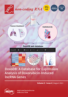
Doxorubicin causes serious side effects, including acute ventricular dysfunction, cardiomyopathy, and heart failure. We hypothesize that comparing the expression profiles of cells and tissues treated with doxorubicin may yield new insights into the adverse effects of the drug on cellular activities. We analyzed published RNA sequencing (RNA-seq) data from doxorubicin-treated cells to identify commonly differentially expressed genes, including long non-coding RNAs (lncRNAs) as they are known to be dysregulated in diseased tissues and cells. From our systematic analysis, we identified several doxorubicin-induced genes. We treated human cardiac fibroblasts with doxorubicin to record expression changes in the selected doxorubicin-induced genes and performed a loss-of-function experiment of the lncRNA MAP3K4-AS1. To further disseminate the analyzed data, we built the web database DoxoDB. View this paper
  • Issues are regarded as officially published after their release is announced to the table of contents alert mailing list.
  • You may sign up for e-mail alerts to receive table of contents of newly released issues.
  • PDF is the official format for papers published in both, html and pdf forms. To view the papers in pdf format, click on the "PDF Full-text" link, and use the free Adobe Reader to open them.
Order results
Result details
Section
Select all
Export citation of selected articles as:
15 pages, 1969 KiB  
Article
Investigation into the Role of Long-Non-Coding RNA MIAT in Leukemia
by Alessia Ostini and Mirna Mourtada-Maarabouni
Non-Coding RNA 2023, 9(4), 47; https://doi.org/10.3390/ncrna9040047 - 11 Aug 2023
Cited by 2 | Viewed by 2520
Abstract
Myocardial Infarction Associated Transcript (MIAT) is a nuclear long non-coding RNA (LncRNA) with four different splicing variants. MIAT dysregulation is associated with carcinogenesis, mainly acting as an oncogene regulating cellular growth, invasion, and metastasis. The aim of the current study is [...] Read more.
Myocardial Infarction Associated Transcript (MIAT) is a nuclear long non-coding RNA (LncRNA) with four different splicing variants. MIAT dysregulation is associated with carcinogenesis, mainly acting as an oncogene regulating cellular growth, invasion, and metastasis. The aim of the current study is to investigate the role of MIAT in the regulation of T and chronic myeloid leukemic cell survival. To this end, MIAT was silenced using MIAT-specific siRNAs in leukemic cell lines, and functional assays were performed thereafter. This investigation also aims to investigate the effects of MIAT silencing on the expression of core genes involved in cancer. Functional studies and gene expression determination confirm that MIAT knockdown not only affects short- and long-term survival and the apoptosis of leukemic cells but also plays a pivotal role in the alteration of key genes involved in cancer, including c-MYC and HIF-1A. Our observations suggest that MIAT could act as an oncogene and it has the potential to be used not only as a reliable biomarker for leukemia, but also be employed for prognostic and therapeutic purposes. Full article
Show Figures

Figure 1

Figure 1
<p><span class="html-italic">MIAT</span>-specific silencing inhibits short- and long-term survival and increases the apoptosis of Jurkat cells. Jurkat cells were transfected with the negative siRNA (NC) or one of the three <span class="html-italic">MIAT</span>-specific siRNAs (M1, M2, M3) using Nucleofection, and were assessed 24 h/48 h post-replating. Relative gene expression of <span class="html-italic">MIAT</span> was measured by Real-Time PCR 24 h post-transfection and confirmed the silencing of <span class="html-italic">MIAT</span> gene for all three siRNAs (<b>A</b>). Total and viable cell counts, as determined by vital blue staining, were reduced in Jurkat cells at 48 h post-transfection (<b>B</b>). The results were further confirmed utilizing flow cytometry, showing a decrease in total and viable cell count at 48 h (<b>C</b>). The rate of apoptosis, determined by acridine orange staining, is increased in <span class="html-italic">MIAT</span>-specific siRNAs transfected cells at both 24 h (<b>D</b>). Representations of cells undergoing apoptosis are depicted by white arrows (<b>E</b>). Long-term survival of Jurkat cells was reduced upon <span class="html-italic">MIAT</span>-specific silencing, as demonstrated by clonogenic assay (<b>F</b>). Data are represented with bar graphs depicting the means ± SEM from independent experiments. * Indicates a <span class="html-italic">p</span>-value &lt; 0.05; ** indicate a <span class="html-italic">p</span>-value &lt; 0.01; ***/**** indicate a <span class="html-italic">p</span>-value &lt; 0.001, as measured by One-way and Two-Way ANOVA tests and Dunnett’s multiple comparison test (MCT).</p>
Full article ">Figure 2
<p><span class="html-italic">MIAT</span>-specific silencing inhibits short-term survival and increases the apoptosis of CEM-C7 cells. CEM-C7 cells were transfected with the negative siRNA (NC) or one of the three <span class="html-italic">MIAT</span>-specific siRNAs (M1, M2, M3) using Nucleofection, and were assessed 24 h and 48 h post-replating. Relative gene expression of <span class="html-italic">MIAT</span> was measured by Real-Time PCR 24 h post-transfection and confirmed the silencing of <span class="html-italic">MIAT</span> for all three siRNAs (<b>A</b>). Total and viable cell count, as determined by vital blue staining, was reduced in CEM-C7 cells at 24 h post-transfection (<b>B</b>). Further confirmation by flow cytometry showed a decrease in total and viable cell count at 48 h (<b>C</b>). The rate of apoptosis, determined by acridine orange staining, is increased in <span class="html-italic">MIAT</span>-specific siRNAs transfected cells at 24 h (<b>D</b>). Representation of cells undergoing apoptosis are depicted by white arrows (<b>E</b>). Data are represented with bar graphs depicting the means ± SEM from independent experiments. * Indicates a <span class="html-italic">p</span>-value &lt; 0.05; ** indicates a <span class="html-italic">p</span>-value &lt; 0.01; **** indicates a <span class="html-italic">p</span>-value &lt; 0.001 as measured by One-way and Two-Way ANOVA tests and Dunnett’s multiple comparison test (MCT).</p>
Full article ">Figure 3
<p><span class="html-italic">MIAT</span>-specific silencing inhibits short- and long-term survival and increases the apoptosis of K562 cells. K562 cells were transfected with the negative siRNA (NC) or one of the two <span class="html-italic">MIAT</span>-specific siRNAs (M1, M2) using Nucleofection, and were assessed 24 h and 48 h post-replating. Relative gene expression of <span class="html-italic">MIAT</span> was measured by Real-Time PCR 24 h post-transfection and confirmed the silencing of <span class="html-italic">MIAT</span> gene for M1 and M2 siRNAs (<b>A</b>). Total and viable cell counts, as determined by vital blue staining, were reduced in K562 cells at 48 h post-transfection (<b>B</b>). The rate of apoptotic cells, determined by acridine orange staining, is increased in <span class="html-italic">MIAT</span>-specific siRNAs transfected cells at 24 h (<b>C</b>). Representation of cells undergoing apoptosis is depicted by white arrows (<b>D</b>). Long-term survival of K562 cells was reduced upon <span class="html-italic">MIAT</span>-specific silencing as demonstrated by clonogenic assay (<b>E</b>). Data are represented with bar graphs depicting the means ± SEM from independent experiments. * Indicates a <span class="html-italic">p</span>-value &lt; 0.05; ** indicate a <span class="html-italic">p</span>-value &lt; 0.01; ***/**** indicate a <span class="html-italic">p</span>-value &lt; 0.001 as measured by One-way and Two-Way ANOVA tests and Dunnett’s multiple comparison test (MCT).</p>
Full article ">Figure 4
<p><span class="html-italic">MIAT</span> silencing induced dysregulation of several genes in SH-SY5Y neuroblastoma cells. RNA sequencing previously performed showed differentially expressed (<b>D</b>,<b>E</b>) genes upon <span class="html-italic">MIAT</span> silencing [<a href="#B14-ncrna-09-00047" class="html-bibr">14</a>]. Data are the difference in expression between SH-SY5Y neuroblastoma cells transfected with negative siRNA and cells transfected with <span class="html-italic">MIAT</span>-specific siRNA (M2), expressed as a normalized log2 fold change (log2FC). Blue bars represent downregulated genes, red bars represent upregulated genes. (<b>A</b>) <span class="html-italic">CDK6</span>: Cyclin-dependent kinase 6; (<b>B</b>) <span class="html-italic">XIAP</span>: X-Linked Inhibitor of Apoptosis; (<b>C</b>) <span class="html-italic">GADD45A</span>: Growth Arrest and DNA Damage Inducible Alpha; (<b>D</b>) <span class="html-italic">CBL</span>: Casitas B-lineage Lymphoma; (<b>E</b>) <span class="html-italic">FLT1</span>: Fms Related Receptor Tyrosine Kinase 1; (<b>F</b>) <span class="html-italic">HIF-1A</span>: Hypoxia Inducible Factor 1 Alpha Subunit; (<b>G</b>) <span class="html-italic">c-MYC</span>: MYC Proto-Oncogene; (<b>H</b>) <span class="html-italic">RELA</span>: <span class="html-italic">RELA</span> Proto-Oncogene, NF-KB Subunit; (<b>I</b>) <span class="html-italic">NOD1</span>: Nucleotide Binding Oligomerization Domain Containing 1.</p>
Full article ">Figure 5
<p><span class="html-italic">MIAT</span> knockdown induces changes in the relative expression of leukemic-related genes. Jurkat T cells were transfected with negative siRNA (NC) or one of the three <span class="html-italic">MIAT</span>-specific siRNAs (M1, M2 or M3) using nucleofection. The relative gene expression was measured by Real-Time PCR 24 h post-transfection <span class="html-italic">CDK</span>6 (<b>A</b>); <span class="html-italic">XIAP</span> (<b>B</b>); <span class="html-italic">GADD45</span>A (<b>C</b>); <span class="html-italic">CBL</span> (<b>D</b>); <span class="html-italic">FLT</span>-1 (<b>E</b>); <span class="html-italic">HIF-1A</span> (<b>F</b>); <span class="html-italic">c-MYC</span> (<b>G</b>); <span class="html-italic">RELA</span> (<b>H</b>); <span class="html-italic">NOD1</span> (<b>I</b>). Data are represented with bar graphs depicting the means ± SEM from independent experiments. * Indicates a <span class="html-italic">p</span>-value &lt; 0.05; ** indicates a <span class="html-italic">p</span>-value &lt; 0.01; ***/**** indicates a <span class="html-italic">p</span>-value &lt; 0.001 as measured by One-Way ANOVA tests and Dunnett’s multiple comparison test (MCT). <span class="html-italic">CDK6</span>: Cyclin-dependent kinase 6; <span class="html-italic">XIAP</span>: X-Linked Inhibitor of Apoptosis; <span class="html-italic">GADD45A</span>: Growth Arrest and DNA Damage Inducible Alpha; <span class="html-italic">CBL</span>: Casitas B-lineage Lymphoma; <span class="html-italic">FLT1</span>: Fms Related Receptor Tyrosine Kinase 1; <span class="html-italic">HIF-1A</span>: Hypoxia Inducible Factor 1 Alpha Subunit; <span class="html-italic">c-MYC</span>: MYC Proto-Oncogene; <span class="html-italic">RELA</span>: RELA Proto-Oncogene, NF-KB Subunit; <span class="html-italic">NOD1</span>: Nucleotide Binding Oligomerization Domain Containing 1.</p>
Full article ">Figure 6
<p><span class="html-italic">MIAT</span> knockdown induces changes in the relative expression of leukemic-related genes. CEM-C7 cells were transfected with negative siRNA (NC) or one of the three <span class="html-italic">MIAT</span>-specific siRNAs (M1, M2 or M3) using nucleofection. The relative gene expression was measured by Real-Time PCR 24 h post-transfection <span class="html-italic">CDK6</span> (<b>A</b>); <span class="html-italic">XIAP</span> (<b>B</b>); <span class="html-italic">GADD45A</span> (<b>C</b>); <span class="html-italic">CBL</span> (<b>D</b>); <span class="html-italic">FLT-1</span> (<b>E</b>); <span class="html-italic">HIF-1A</span> (<b>F</b>); <span class="html-italic">c-MYC</span> (<b>G</b>); RELA (<b>H</b>); NOD1 (<b>I</b>). Data are represented with bar graphs depicting the means ± SEM from independent experiments. * Indicates a <span class="html-italic">p</span>-value &lt; 0.05; ** indicates a <span class="html-italic">p</span>-value &lt; 0.01; ***/**** indicates a <span class="html-italic">p</span>-value &lt; 0.001 as measured by One-way ANOVA tests and Dunnett’s multiple comparison test (MCT). <span class="html-italic">CDK6</span>: Cyclin-dependent kinase 6; <span class="html-italic">XIAP</span>: X-Linked Inhibitor of Apoptosis; <span class="html-italic">GADD45A</span>: Growth Arrest and DNA Damage Inducible Alpha; <span class="html-italic">CBL</span>: Casitas B-lineage Lymphoma; <span class="html-italic">FLT1</span>: Fms Related Receptor Tyro-sine Kinase 1; <span class="html-italic">HIF-1A</span>: Hypoxia Inducible Factor 1 Alpha Subunit; <span class="html-italic">c-MYC</span>: MYC Proto-Oncogene; <span class="html-italic">RELA</span>: RELA Proto-Oncogene, NF-KB Subunit; <span class="html-italic">NOD1</span>: Nucleotide Binding Oligomerization Domain Containing 1.</p>
Full article ">
3 pages, 165 KiB  
Editorial
Methods and Tools in RNA Biology
by Mirolyuba Ilieva and Shizuka Uchida
Non-Coding RNA 2023, 9(4), 46; https://doi.org/10.3390/ncrna9040046 - 10 Aug 2023
Cited by 1 | Viewed by 1515
Abstract
Breakthroughs in innovative techniques and instruments have driven the exploration of non-coding RNAs (ncRNAs), including microRNAs (miRNAs) and long non-coding RNAs (lncRNAs) [...] Full article
(This article belongs to the Special Issue Methods and Tools in RNA Biology)
14 pages, 1790 KiB  
Communication
Structural Modifications and Novel Protein-Binding Sites in Pre-miR-675—Explaining Its Regulatory Mechanism in Carcinogenesis
by Abhishek Dey
Non-Coding RNA 2023, 9(4), 45; https://doi.org/10.3390/ncrna9040045 - 10 Aug 2023
Cited by 2 | Viewed by 2644
Abstract
Pre-miR-675 is a microRNA expressed from the exon 1 of H19 long noncoding RNA, and the atypical expression of pre-miR-675 has been linked with several diseases and disorders including cancer. To execute its function inside the cell, pre-miR-675 is folded into a particular [...] Read more.
Pre-miR-675 is a microRNA expressed from the exon 1 of H19 long noncoding RNA, and the atypical expression of pre-miR-675 has been linked with several diseases and disorders including cancer. To execute its function inside the cell, pre-miR-675 is folded into a particular conformation, which aids in its interaction with several other biological molecules. However, the exact folding dynamics of pre-miR-675 and its protein-binding motifs are currently unknown. Moreover, how H19 lncRNA and pre-miR-675 crosstalk and modulate each other’s activities is also unclear. The detailed structural analysis of pre-miR-675 in this study determines its earlier unknown conformation and identifies novel protein-binding sites on pre-miR-675, thus making it an excellent therapeutic target against cancer. Co-folding analysis between H19 lncRNA and pre-miR-675 determine structural transformations in pre-miR-675, thus describing the earlier unknown mechanism of interaction between these two molecules. Comprehensively, this study details the conformation of pre-miR-675 and its protein-binding sites and explains its relationship with H19 lncRNA, which can be interpreted to understand the role of pre-miR-675 in the development and progression of tumorigenesis and designing new therapeutics against cancers. Full article
Show Figures

Graphical abstract

Graphical abstract
Full article ">Figure 1
<p>In vitro architecture and statistical analysis of pre-miR-675 secondary structure. (<b>A</b>) Normalized 5-NIA reactivity profile for in vitro synthesized pre-miR-675 replicate 1. (<b>B</b>) Normalized 5-NIA reactivity profile for in vitro synthesized pre-miR-675 replicate 2. Some of the nucleotides were found to have high preference for 5-NIA as compared to others in both the replicates. (<b>C</b>) Overlapped normalized 5-NIA reactivity profile of in vitro synthesized pre-miR-675 replicates. The pre-miR-675-replicate 1 is represented in red, while pre-miR-675-Replicate 2 is represented as blue. The reactivity profile was found to be highly similar for both the replicates. (<b>D</b>) Scatter plot with Pearson’s correlation coefficient between the normalized reactivity profiles of pre-miR-675 replicates, R = 0.98 illustrates identical and reproducible reactivity profiles between both the replicates of pre-miR-675. (<b>E</b>) Overlapped RNA arcplot (depicting secondary structure) with Scorer results for pre-miR-675, Replicate 1 and Replicate 2. Both pre-miR-675 replicates fold into identical conformation with PPV = 100% and SENS = 100%. (<b>F</b>) Normalized 5-NIA reactivity profile generated after averaging the reactivity profiles of pre-miR-675 replicate 1 and replicate 2. Also shown is the overlapped arc profile representing the secondary structure of pre-miR-675. (<b>G</b>) Secondary structure model of pre-miR-675 was obtained after feeding superfold with obtained SHAPE constraints. Black colour represents 5-NIA reactivity &lt; 0.4 which means nucleotides are base paired, orange represents 5-NIA reactivity between 0.4 and 0.8 meaning these nucleotides are either unpaired or base paired and red represents 5-NIA reactivity &gt; 0.8 for nucleotides which means these nucleotides are unpaired and not involved in base pairing.</p>
Full article ">Figure 2
<p>Pre-miR-675 protein-binding sites of different RNA-binding proteins identified through RBPsuite. Pre-miR-675 binding sequences identified for proteins. (<b>A</b>) Fused in Sarcoma (FUS). (<b>B</b>) Serine/arginine-rich splicing factor 1 (SRSF1). (<b>C</b>) FMR1 autosomal Homolog 2 (FXR2). (<b>D</b>) Serine/arginine-rich splicing factor 9 (SRSF9). (<b>E</b>) Lin-28 homolog B (Lin28B). (<b>F</b>) Human Antigen R (HuR). For better representation, nucleotides in the sequence logo is represented by different colours with Adenine as green, Guanine as orange, Uracil as red and Cytosine as blue. (<b>G</b>) Protein-binding sites in pre-miR-675. Most of the protein-binding sites in pre-miR-675 include the nucleotides from the loop region thus making pre-miR-675 an excellent target of therapeutic importance.</p>
Full article ">Figure 3
<p>RNA co-fold and base pair conservation analysis between pre-miR-675 and H19-exons. (<b>A</b>) Exon 1 (Model 1). (<b>B</b>) Exon 2 (Model 2). (<b>C</b>) Exon 3 (Model 1). (<b>D</b>) Exon 4 (Model 2) of precursor H19 lncRNA. Upper panel in each figure represents the folding dynamics of individual H19-exons, pre-miR-675 and their co-folding pattern analysed through RNAcofold in dot–bracket notation. The “&amp;” in the co-fold pattern of the hybrid structure is the separator between two RNA molecules. The middle panel represents the H19-exons and pre-miR-675 base pairs, which were either modified or remained unchanged during the co-folding of both the RNA molecules. The lower panel explains the meaning of each symbol/notations used to determine the base pair conservation during the co-folding process of H19-exons and pre-miR-675. pre-miR-675 has been shown to prefer exon 1 and exon 3 of H19 lncRNA, resulting in the opening of its canonical stem–loop helical structure, while its interaction with exon 2 and exon 4 results more in localized rearrangements in base-pairing between nucleotides.</p>
Full article ">Figure 4
<p>Structural modifications in pre-miR-675 enables it to bind H19-exon1/exon3. Hybrid RNA conformer between (<b>A</b>) H19-exon 1 and pre-miR-675, (<b>B</b>) H19-exon 2 and pre-miR-675 and (<b>C</b>) H19-exon 3 and pre-miR-675. The hybrid conformation between H19-exon 1 and exon 3 with pre-miR-675 (red line) is formed due to the disruption of pre-miR-675 stem–loop helical conformation and formation of new base pairs between two RNA molecules, while the pre-miR-675 (red line) retains its canonical conformation, when interacting with exon 2 of H19 lncRNA, thus retaining all its bulges and loops, including the apical loop. These hybrid conformations explain the negative feedback regulatory mechanism exerted by H19 lncRNA and pre-miR-675 on each other during the development and progression of cancer.</p>
Full article ">
21 pages, 3821 KiB  
Review
Long Non-Coding RNA Signatures in Lymphopoiesis and Lymphoid Malignancies
by Hamed Baghdadi, Reza Heidari, Mahdi Zavvar, Nazanin Ahmadi, Mehdi Shakouri Khomartash, Mahmoud Vahidi, Mojgan Mohammadimehr, Davood Bashash and Mahdi Ghorbani
Non-Coding RNA 2023, 9(4), 44; https://doi.org/10.3390/ncrna9040044 - 1 Aug 2023
Cited by 4 | Viewed by 2032
Abstract
Lymphoid cells play a critical role in the immune system, which includes three subgroups of T, B, and NK cells. Recognition of the complexity of the human genetics transcriptome in lymphopoiesis has revolutionized our understanding of the regulatory potential of RNA in normal [...] Read more.
Lymphoid cells play a critical role in the immune system, which includes three subgroups of T, B, and NK cells. Recognition of the complexity of the human genetics transcriptome in lymphopoiesis has revolutionized our understanding of the regulatory potential of RNA in normal lymphopoiesis and lymphoid malignancies. Long non-coding RNAs (lncRNAs) are a class of RNA molecules greater than 200 nucleotides in length. LncRNAs have recently attracted much attention due to their critical roles in various biological processes, including gene regulation, chromatin organization, and cell cycle control. LncRNAs can also be used for cell differentiation and cell fate, as their expression patterns are often specific to particular cell types or developmental stages. Additionally, lncRNAs have been implicated in lymphoid differentiation, such as regulating T-cell and B-cell development, and their expression has been linked to immune-associated diseases such as leukemia and lymphoma. In addition, lncRNAs have been investigated as potential biomarkers for diagnosis, prognosis, and therapeutic response to disease management. In this review, we provide an overview of the current knowledge about the regulatory role of lncRNAs in physiopathology processes during normal lymphopoiesis and lymphoid leukemia. Full article
(This article belongs to the Special Issue Non-Coding RNA in the Immune System)
Show Figures

Figure 1

Figure 1
<p>An overview of lncRNA biogenesis. At the chromatin level, lncRNA promoters have higher levels of H3K4me3, H3K27ac, and H3K9ac, which are more strongly repressed by chromatin remodeling complexes such as Swr1, Isw2, Rsc, and Ino80. The transcription of lncRNA starts when the SWI/SNF chromatin remodeling complex facilitate access of RNA polymerase II (Pol II) to the core promoter, while the elongation stage is controlled by MYC and DICER1, and its synthesis is complete by 3′-polyadenylation. Then, synthesized lncRNAs should be spliced, capped, and/or polyadenylated by recruiting proteins. This procedure is mediated by regions within the immature lncRNAs such as polyadenylation signal regions. Many Pol II-transcribed lncRNAs are not efficiently processed and are retained in the nucleus. Binding to the nuclear retention element is another mechanism leading to the retention of lncRNAs in the nucleus. Nuclear organization, regulation of gene transcription, alternative splicing, and chromatin remodulation are the main functions of lncRNAs in the nucleus. Other lncRNAs are spliced and exported to the cytoplasm by nuclear RNA export factor 1 (NXF1). In the cytoplasm, lncRNAs typically interact with a variety of RBPs. Some of them are sorted into mitochondria, some of them are associated with ribosomes, and others reside in organelles, such as exosomes. As one of the main mechanisms of action of lncRNA-mediated mRNA stability regulation, lncRNAs can directly bind to their target mRNAs and avoid interactions with ribosomes.</p>
Full article ">Figure 2
<p>LncRNAs can regulate the differentiation of B and T lineages from hematopoietic stem cells into mature cells, as well as the subsequent activation of these lymphocytes. <b>HSC</b>: Hematopoietic stem cell; <b>CLP</b>: common lymphoid progenitor; <b>DN</b>: double negative; <b>DP</b>: double positive; <b>MZB</b>: marginal zone B cell; <b>CB</b>: centroblast; <b>CC</b>: centrocyte.</p>
Full article ">Figure 3
<p>Alterations in lnsRNAs in different NHLs. <b>CLL/SLL</b>: Chronic lymphocytic leukemia/small lymphocytic lymphoma; <b>MCL</b>: mantle cell lymphoma; <b>BL</b>: burkitt lymphoma; <b>FL</b>: follicular lymphoma; <b>DLBCL</b>: diffuse large B-cell lymphoma; <b>SHM</b>: somatic hyper mutation; <b>CSR</b>: class switch recombination.</p>
Full article ">
13 pages, 1407 KiB  
Article
Effects of Controlled Ozone Exposure on Circulating microRNAs and Vascular and Coagulation Biomarkers: A Mediation Analysis
by Hao Chen, Syed Masood, Ana G. Rappold, David Diaz-Sanchez, James M. Samet and Haiyan Tong
Non-Coding RNA 2023, 9(4), 43; https://doi.org/10.3390/ncrna9040043 - 1 Aug 2023
Viewed by 1472
Abstract
Exposure to ozone (O3) is associated with adverse respiratory and cardiovascular outcomes. Alterations in circulating microRNAs (miRNAs) may contribute to the adverse vascular effects of O3 exposure through inter-cellular communication resulting in post-transcriptional regulation of messenger RNAs by miRNAs. In [...] Read more.
Exposure to ozone (O3) is associated with adverse respiratory and cardiovascular outcomes. Alterations in circulating microRNAs (miRNAs) may contribute to the adverse vascular effects of O3 exposure through inter-cellular communication resulting in post-transcriptional regulation of messenger RNAs by miRNAs. In this study, we investigated whether O3 exposure induces alterations in circulating miRNAs that can mediate effects on downstream vascular and coagulation biomarkers. Twenty-three healthy male adults were exposed on successive days to filtered air and 300 ppb O3 for 2 h. Circulating miRNA and protein biomarkers were quantified after each exposure session. The data were subjected to mixed-effects model and mediation analyses for the statistical analyses. The results showed that the expression level of multiple circulating miRNAs (e.g., miR-19a-3p, miR-34a-5p) was significantly associated with O3 exposure. Pathway analysis showed that these miRNAs were predictive of changing levels of downstream biomarkers [e.g., D-dimer, C-reactive protein, tumor necrosis factor α (TNFα)]. Mediation analysis showed that miR-19a-3p may be a significant mediator of O3-exposure-induced changes in blood TNFα levels [0.08 (0.01, 0.15), p = 0.02]. In conclusion, this preliminary study showed that O3 exposure of healthy male adults resulted in changes in circulating miRNAs, some of which may mediate vascular effects of O3 exposure. Full article
(This article belongs to the Special Issue Non-coding RNA in the USA: Latest Advances and Perspectives)
Show Figures

Figure 1

Figure 1
<p>Schematic showing the mediation model employed in this study. Letter “a” indicates the coefficient of X derived from the statistical analysis for “X → M”, “b” indicates the coefficient of M in the model “M → Y”, and “c” indicates the coefficient of X in the model “X  +  M → Y”.</p>
Full article ">Figure 2
<p>Changes in circulating miRNAs for participants in control (CTL), fish oil (FO), and olive oil (OO) groups immediately after exposure to filtered air or 300 ppb O<sub>3</sub>. Shown are log2-transformed fold change values in <span class="html-italic">miR-122-5p</span>, <span class="html-italic">miR-125b-5p</span>, <span class="html-italic">miR-144-5p</span>, <span class="html-italic">miR-155-5p</span>, <span class="html-italic">miR-19a-3p</span>, <span class="html-italic">miR-342-3p</span>, and <span class="html-italic">miR-34a-5p</span>. Bars with whiskers indicate means with their respective 95% confidence interval. * <span class="html-italic">p</span> &lt; 0.05 as “significant” between two conditions.</p>
Full article ">Figure 3
<p>Pathway analysis showing the link between O<sub>3</sub> exposure-induced miRNAs and vascular proteins in the plasma and their related biological pathways. Four miRNAs (in red) that were associated with O<sub>3</sub> exposure and their matched mRNA/protein targets (in dark blue) and their associated downstream inflammatory signaling pathways (in gray) were identified by the Ingenuity Pathway Analysis software.</p>
Full article ">
23 pages, 41554 KiB  
Review
Crosstalk between Long Non-Coding RNA and Spliceosomal microRNA as a Novel Biomarker for Cancer
by Maram Arafat and Ruth Sperling
Non-Coding RNA 2023, 9(4), 42; https://doi.org/10.3390/ncrna9040042 - 31 Jul 2023
Cited by 2 | Viewed by 2066
Abstract
Non-coding RNAs (ncRNAs) play diverse roles in regulating cellular processes and have been implicated in pathological conditions, including cancer, where interactions between ncRNAs play a role. Relevant here are (i) microRNAs (miRNAs), mainly known as negative regulators of gene expression in the cytoplasm. [...] Read more.
Non-coding RNAs (ncRNAs) play diverse roles in regulating cellular processes and have been implicated in pathological conditions, including cancer, where interactions between ncRNAs play a role. Relevant here are (i) microRNAs (miRNAs), mainly known as negative regulators of gene expression in the cytoplasm. However, identification of miRNAs in the nucleus suggested novel nuclear functions, and (ii) long non-coding RNA (lncRNA) regulates gene expression at multiple levels. The recent findings of miRNA in supraspliceosomes of human breast and cervical cancer cells revealed new candidates of lncRNA targets. Here, we highlight potential cases of crosstalk between lncRNA and supraspliceosomal miRNA expressed from the same genomic region, having complementary sequences. Through RNA:RNA base pairing, changes in the level of one partner (either miRNA or lncRNA), as occur in cancer, could affect the level of the other, which might be involved in breast and cervical cancer. An example is spliceosomal mir-7704 as a negative regulator of the oncogenic lncRNA HAGLR. Because the expression of spliceosomal miRNA is cell-type-specific, the list of cis-interacting lncRNA:spliceosomal miRNA presented here is likely just the tip of the iceberg, and such interactions are likely relevant to additional cancers. We thus highlight the potential of lncRNA:spliceosomal miRNA interactions as novel targets for cancer diagnosis and therapies. Full article
(This article belongs to the Special Issue Recent Advances in Chemical Biology to Study and Target ncRNAs)
Show Figures

Graphical abstract

Graphical abstract
Full article ">Figure 1
<p>Negative regulation of HAGLR by spliceosomal mir-7704 in breast and cervical cancer cells. (<b>A</b>) UCSC Genome browser view indicates the overlap of mir-7704 with HOXD1 and the lncRNA HAGLR. (<b>B</b>) Average expression levels (RNA-Seq) and standard error of mir-7704 from the spliceosome fractions from MCF-10A, MCF-7, and MDA-MB-231 cells. Pair statistics are marked for &lt;0.1 (*) and &lt;0.001 (***). (<b>C</b>) Expression of HAGLR (RT-PCR) measured from total RNA and nuclear RNA in the three tested breast cell lines. (<b>D</b>) Inhibition of mir-7704 expression upregulates the expression of HAGLR. Results of quantitative real-time PCR (qRT-PCR) analysis of the effect of mir-7704 inhibition on the nuclear expression of HAGLR in MCF-10A, MCF-7, and MDA-MB-231 cells. Transfection with Anti-miR-7704 inhibitor resulted in the downregulation of mir-7704 (upper panel) and an increase in the expression level of HAGLR mRNA (lower panel) relative to the control (non-silencing anti-mir). HAGLR expression level was normalized to the internal control of ß-actin expressed from the same preparation. (<b>E</b>) Overexpression of mir-7704 leads to increase in the level of mir-7704 in HeLa cells, as measured by qRT-PCR. This increase in the level of mir-7704 is accompanied by significant decrease in the level of HAGLR mRNA compared to control (empty vector). (<b>F</b>) Downregulation of mir-7704 in HeLa cells leads to increase in the expression level of HAGLR mRNA compared to control (non-silencing anti-mir). The expression levels of HAGLR were normalized to ß-actin expression from the same preparation. (<b>G</b>) Survival curve analyses of HAGLR in breast and cervical cancer, based on data from GEPIA [<a href="#B41-ncrna-09-00042" class="html-bibr">41</a>], The solid line represents the survival curve, and the dashed lines represent the 95% confidence interval. Data of (<b>B</b>–<b>D</b>) adapted from ref. [<a href="#B5-ncrna-09-00042" class="html-bibr">5</a>], and (<b>E</b>,<b>F</b>), from ref. [<a href="#B18-ncrna-09-00042" class="html-bibr">18</a>].</p>
Full article ">Figure 2
<p>UCSC Genome browser view, indicating the overlap between the discussed miRNAs and their potential lncRNA crosstalk targets.</p>
Full article ">Figure 3
<p>Proposed types of crosstalk between lncRNA and respective spliceosomal miRNA, schematic drawing. (<b>A</b>) Complementarity between spliceosomal miRNA (red) and different regions along the sequence of an lncRNA (pastel colors). lncRNA colored blocks—exons; and lines—introns. (<b>B</b>) A scheme portraying potential interactions through base pairing between lncRNA (blue) and spliceosomal miRNA (red) in the nucleus, which can lead to promotion or inhibition of cancer. Created with <a href="http://BioRender.com" target="_blank">BioRender.com</a> (accessed on 17 June 2023).</p>
Full article ">
15 pages, 1507 KiB  
Systematic Review
Conservation and Targets of miR-71: A Systematic Review and Meta-Analysis
by Devin Naidoo, Ryan Brennan and Alexandre de Lencastre
Non-Coding RNA 2023, 9(4), 41; https://doi.org/10.3390/ncrna9040041 - 26 Jul 2023
Cited by 1 | Viewed by 1841
Abstract
MicroRNAs (miRNAs) perform a pivotal role in the regulation of gene expression across the animal kingdom. As negative regulators of gene expression, miRNAs have been shown to function in the genetic pathways that control many biological processes and have been implicated in roles [...] Read more.
MicroRNAs (miRNAs) perform a pivotal role in the regulation of gene expression across the animal kingdom. As negative regulators of gene expression, miRNAs have been shown to function in the genetic pathways that control many biological processes and have been implicated in roles in human disease. First identified as an aging-associated gene in C. elegans, miR-71, a miRNA, has a demonstrated capability of regulating processes in numerous different invertebrates, including platyhelminths, mollusks, and insects. In these organisms, miR-71 has been shown to affect a diverse range of pathways, including aging, development, and immune response. However, the exact mechanisms by which miR-71 regulates these pathways are not completely understood. In this paper, we review the identified functions of miR-71 across multiple organisms, including identified gene targets, pathways, and the conditions which affect regulatory action. Additionally, the degree of conservation of miR-71 in the evaluated organisms and the conservation of their predicted binding sites in target 3′ UTRs was measured. These studies may provide an insight on the patterns, interactions, and conditions in which miR-71 is able to exert genotypic and phenotypic influence. Full article
(This article belongs to the Special Issue ncRNAs to Target Molecular Pathways)
Show Figures

Figure 1

Figure 1
<p>miR-71 and target 3′ UTR sequence conservation amongst multiple species. (<b>A</b>) A consensus sequence was created from the Clustal Omega alignment of 112 miR-71 sequences compiled after primary screening. Letters in blue indicate over 95% identity across sequences. (<b>B</b>) A consensus sequence was created from Clustal Omega alignment of 14 target 3′ UTR sequences after secondary screening. Letters in blue indicate over 95% identity across sequences. (<b>C</b>) Consensus sequences from (<b>A</b>,<b>B</b>) were aligned for miRNA-UTR binding showing an 8-mer A1 seed site the strongest binding capacity. Binding was favorable with a maximum free energy of −21.9 kcal/mol between the consensus miR-71 sequence and the consequence 3′ UTR sequence.</p>
Full article ">Figure 2
<p>Major invertebrate targets of miR-71. Major pathways and genes regulated by miR-71 include Argonaute proteins insulin-like signaling (IIS) pathway and TIR-1.</p>
Full article ">Figure 3
<p>Mammalian targets of miR-71. miR-71 is believed to play a role in the pathogenicity of parasitic invertebrates. Parasitic invertebrates excrete extracellular vesicles that have been shown to contain miR-71 and may influence the pathogenicity of parasites. miR-71 has been shown to upregulate Argonaute proteins which ultimately leads to a decrease in nitric oxide production by macrophages. In a separate pathway miR-71 has been shown to negatively regulate IRF4 and suppress the immune response in mammals by decreasing cytokine response and inflammation.</p>
Full article ">Figure 4
<p>Preferred Reporting Items for Systematic Reviews and Meta-Analyses (PRISMA). A systematic search was performed using PubMed Scopus and PubMed Central databases to identify studies that experimentally investigate the targets of miR-71. The search terms used were “microRNA-71” “miR-71” and “miR-71 targets”.</p>
Full article ">
4 pages, 1191 KiB  
Commentary
Phosphate Deficiency: A Tale from the End of PILNCR2
by Santosh Kumar Upadhyay
Non-Coding RNA 2023, 9(4), 40; https://doi.org/10.3390/ncrna9040040 - 25 Jul 2023
Viewed by 2101
Abstract
A deficiency in inorganic phosphate (Pi) induces the expression of miRNA399 and the accumulation of its target Pi transporters (PHT1s) mRNA, which is contrary to the goal of miRNA-mediated gene regulation. Recently, a novel mechanism of RNA/RNA-duplex formation between the transcripts [...] Read more.
A deficiency in inorganic phosphate (Pi) induces the expression of miRNA399 and the accumulation of its target Pi transporters (PHT1s) mRNA, which is contrary to the goal of miRNA-mediated gene regulation. Recently, a novel mechanism of RNA/RNA-duplex formation between the transcripts of a Pi deficiency-induced long non-coding RNA (PILNCR2) and PHT1s has been reported, which prevents the binding and cleavage of miRNA399 to PHT1 mRNAs, thereby providing tolerance of Pi-deficient conditions. Moreover, the way in which ribosomes move through the RNA/RNA-duplex for the translation of PHT1 transporter proteins remains elusive. Full article
(This article belongs to the Section Small Non-Coding RNA)
Show Figures

Figure 1

Figure 1
<p><span class="html-italic">PLNCR2</span>-mediated regulation of Pi deficiency. (<b>A</b>) shows the ZmmiRNA399 and its target site (indicated by arrows) in <span class="html-italic">ZmPHT1</span> mRNAs. (<b>B</b>) RNA/RNA duplex formed between the transcripts of <span class="html-italic">PLNCR2</span> and <span class="html-italic">ZmPHT1</span> genes, which inhibit binding of ZmmiRNA399 to <span class="html-italic">ZmPHT1</span> mRNAs. (<b>C</b>) A model that demonstrates the normal expression of <span class="html-italic">PHT1</span> genes and <span class="html-italic">PLNCR2</span> during conditions of Pi sufficiency, while Pi deficiency induces the expression of <span class="html-italic">PHT1</span> genes and ZmmiRNA399, along with the <span class="html-italic">PLNCR2</span> from the opposite strand of <span class="html-italic">PHT1;1.</span> All transcripts translocate to the cytoplasm, where the <span class="html-italic">PLNCR2</span> transcripts form RNA/RNA duplexes with the mRNAs of <span class="html-italic">PHT1s</span> due to their complementary sequences. The RNA duplex prevents the binding of ZmmiRNA399 to mRNAs of <span class="html-italic">PHT1s</span>, protecting them from cleavage. These results highlight the increased accumulation of <span class="html-italic">PHT1</span> transcripts that form more PHT1s used for the transportation of Pi into the cytoplasm, thereby proving low Pi tolerance. The figure is adapted and modified from the study of Wang et al. [<a href="#B7-ncrna-09-00040" class="html-bibr">7</a>], with permission given by Elsevier through Rightslink, licence number 5580761357750.</p>
Full article ">
24 pages, 4590 KiB  
Article
DoxoDB: A Database for the Expression Analysis of Doxorubicin-Induced lncRNA Genes
by Rebecca Distefano, Mirolyuba Ilieva, Jens Hedelund Madsen, Sarah Rennie and Shizuka Uchida
Non-Coding RNA 2023, 9(4), 39; https://doi.org/10.3390/ncrna9040039 - 13 Jul 2023
Cited by 3 | Viewed by 2332
Abstract
Cancer and cardiovascular disease are the leading causes of death worldwide. Recent evidence suggests that these two life-threatening diseases share several features in disease progression, such as angiogenesis, fibrosis, and immune responses. This has led to the emergence of a new field called [...] Read more.
Cancer and cardiovascular disease are the leading causes of death worldwide. Recent evidence suggests that these two life-threatening diseases share several features in disease progression, such as angiogenesis, fibrosis, and immune responses. This has led to the emergence of a new field called cardio-oncology. Doxorubicin is a chemotherapy drug widely used to treat cancer, such as bladder and breast cancer. However, this drug causes serious side effects, including acute ventricular dysfunction, cardiomyopathy, and heart failure. Based on this evidence, we hypothesize that comparing the expression profiles of cells and tissues treated with doxorubicin may yield new insights into the adverse effects of the drug on cellular activities. To test this hypothesis, we analyzed published RNA sequencing (RNA-seq) data from doxorubicin-treated cells to identify commonly differentially expressed genes, including long non-coding RNAs (lncRNAs) as they are known to be dysregulated in diseased tissues and cells. From our systematic analysis, we identified several doxorubicin-induced genes. To confirm these findings, we treated human cardiac fibroblasts with doxorubicin to record expression changes in the selected doxorubicin-induced genes and performed a loss-of-function experiment of the lncRNA MAP3K4-AS1. To further disseminate the analyzed data, we built the web database DoxoDB. Full article
(This article belongs to the Special Issue Methods and Tools in RNA Biology)
Show Figures

Figure 1

Figure 1
<p>RNA-seq data analysis of DOX-treated PAAFs compared to the control cells. (<b>A</b>) Counts per million (CPM) values were used to draw a violin plot for each sample by distinguishing between protein-coding and lncRNA genes based on the annotation (biotype) provided by the Ensembl database (GRCh38.109). A small boxplot indicates the median and quartile range of expressions in CPM values. (<b>B</b>) Volcano plot of differentially expressed genes derived using 2-fold change and FDR &lt; 0.05. (<b>C</b>) Number of differentially expressed lncRNA, protein-coding, and other genes (e.g., pseudogenes, rRNAs, miRNAs, and other small RNAs) based on the biotypes provided by the Ensembl database. (<b>D</b>) Top 10 enriched GO terms and (<b>E</b>) KEGG pathways for the list of up-regulated protein-coding genes. (<b>F</b>) Enriched GO terms for the list of down-regulated protein-coding genes. (<b>G</b>) Enriched GO terms (top 10) and (<b>H</b>) KEGG pathways for the top-correlated protein-coding genes in their expression to the set of up-regulated lncRNA genes.</p>
Full article ">Figure 2
<p>RNA-seq data analysis of DOX-treated sgP130_siCTRL, sgP130_siP107, sgP130+sgRB1_siCTRL, and sgP130+sgRB1_siP107 HFF cells compared to the control cells. (<b>A</b>) Volcano plot of differentially expressed genes among four comparisons. The threshold values of 2-fold and FDR &lt; 0.05 were used to select differentially expressed genes. (<b>B</b>) Number of differentially expressed lncRNA, protein-coding, and other types of genes.</p>
Full article ">Figure 3
<p>RNA-seq data analysis of DOX-induced senescent PD-L1+ and PD-L1- cells compared to the control cells. (<b>A</b>) Volcano plot of differentially expressed genes derived using the threshold values of 2-fold change and FDR &lt; 0.05. (<b>B</b>) Number of differentially expressed lncRNA, protein-coding, and other types of genes. (<b>C</b>) Venn diagrams of shared up- and down-regulated lncRNA and protein-coding genes between DOX-induced senescent PD-L1+ and PD-L1- cells compared to the control cells (<span class="html-italic">p</span>-value &lt; 0.0001). (<b>D</b>) Top 10 enriched GO terms and (<b>E</b>) KEGG pathways for the shared up-regulated protein-coding genes. (<b>F</b>) Top 10 enriched GO terms and (<b>G</b>) KEGG pathways for the shared down-regulated protein-coding genes.</p>
Full article ">Figure 4
<p>RNA-seq data analysis of DOX-treated IBC cells (FCIBC02 and SUM149) compared to control cells. (<b>A</b>) Volcano plot of differentially expressed genes. The threshold values of 2-fold change and FDR &lt; 0.05 were applied. (<b>B</b>) Number of differentially expressed lncRNA, protein-coding, and other types of genes. (<b>C</b>) Venn diagrams of shared up- and down-regulated lncRNA and protein-coding genes between DOX-treated IBC FCIBC02 and SUM149 cells compared to the control cells (<span class="html-italic">p</span>-value &lt; 0.0001). (<b>D</b>) Top 10 enriched GO terms for the shared up-regulated protein-coding genes. (<b>E</b>) Top 10 enriched GO terms and (<b>F</b>) KEGG pathways for the most significantly correlated protein-coding genes to the shared up-regulated lncRNA genes.</p>
Full article ">Figure 5
<p>UpSet plots of shared genes. (<b>A</b>) Up-regulated lncRNA genes and (<b>B</b>) protein-coding genes shared among the different datasets analyzed (GSE154101, GSE135842, GSE198396, and GSE163361).</p>
Full article ">Figure 6
<p>DOX treatment of CFs. (<b>A</b>) Bright field images of DOX-treated CFs after 24 h. The size of the scale bar is 200 μm. The inlet image in 100 nM DOX sample shows intrinsic fluorescence by DOX. (<b>B</b>) Expression profiles of DOX-regulated genes. <span class="html-italic">n</span> = 6 biological replicates. * (<span class="html-italic">p</span> &lt; 0.05) and *** (<span class="html-italic">p</span> &lt; 0.005). (<b>C</b>) Expression of <span class="html-italic">MAP3K4-AS1</span>. Compared to the control siRNA against scrambled sequence (siScr), all three siRNAs against <span class="html-italic">MAP3K4-AS1</span> showed statistically significant down-regulation of <span class="html-italic">MAP3K4-AS1</span>. <span class="html-italic">n</span> = 6 biological replicates. (<b>D</b>–<b>F</b>) Triplex assay to measure (<b>D</b>) cellular viability; (<b>E</b>) cytotoxicity; and (<b>F</b>) apoptosis measured by the activation of caspase-3/7. RFU stands for relative fluorescence units. <span class="html-italic">n</span> = 8 biological replicates.</p>
Full article ">Figure 7
<p>The DoxoDB web application. (<b>A</b>) Home page of DoxoDB. (<b>B</b>) “Explore Results” page. By setting the threshold values, the users can select the study and the condition to focus on. The results can then be dynamically visualized in the “Result Table” and in the adjacent Volcano plot. (<b>C</b>) KEGG pathway analysis of the DEGs. (<b>D</b>) Venn diagram to visualize the shared DEGs among the different conditions within the specified study. (<b>E</b>) “Explore the lncRNAs” page. The users can dynamically explore each differentially expressed lncRNA gene in each study and comparison from the lncRNA Table.</p>
Full article ">
28 pages, 1490 KiB  
Review
Functions of Circular RNA in Human Diseases and Illnesses
by Alison Gu, Dabbu Kumar Jaijyan, Shaomin Yang, Mulan Zeng, Shaokai Pei and Hua Zhu
Non-Coding RNA 2023, 9(4), 38; https://doi.org/10.3390/ncrna9040038 - 4 Jul 2023
Cited by 13 | Viewed by 4476
Abstract
Circular RNAs (circRNAs) represent single-stranded RNA species that contain covalently closed 3′ and 5′ ends that provide them more stability than linear RNA, which has free ends. Emerging evidence indicates that circRNAs perform essential functions in many DNA viruses, including coronaviruses, Epstein–Barr viruses, [...] Read more.
Circular RNAs (circRNAs) represent single-stranded RNA species that contain covalently closed 3′ and 5′ ends that provide them more stability than linear RNA, which has free ends. Emerging evidence indicates that circRNAs perform essential functions in many DNA viruses, including coronaviruses, Epstein–Barr viruses, cytomegalovirus, and Kaposi sarcoma viruses. Recent studies have confirmed that circRNAs are present in viruses, including DNA and RNA viruses, and play various important functions such as evading host immune response, disease pathogenesis, protein translation, miRNA sponges, regulating cell proliferation, and virus replication. Studies have confirmed that circRNAs can be biological signatures or pathological markers for autoimmune diseases, neurological diseases, and cancers. However, our understanding of circRNAs in DNA and RNA viruses is still limited, and functional evaluation of viral and host circRNAs is essential to completely understand their biological functions. In the present review, we describe the metabolism and cellular roles of circRNA, including its roles in various diseases and viral and cellular circRNA functions. Circular RNAs are found to interact with RNA, proteins, and DNA, and thus can modulate cellular processes, including translation, transcription, splicing, and other functions. Circular RNAs interfere with various signaling pathways and take part in vital functions in various biological, physiological, cellular, and pathophysiological processes. We also summarize recent evidence demonstrating cellular and viral circRNA’s roles in DNA and RNA viruses in this growing field of research. Full article
(This article belongs to the Section Evolution of Non-Coding RNA)
Show Figures

Figure 1

Figure 1
<p>Biogenesis of different types of circRNAs. Transcription of genes produces pre-mRNA molecules. Pre-mRNA undergoes a splicing process to generate circular RNAs and mature mRNA. The circularization process is driven by different factors, including intron-paring, RBP, lariat, and others. Different types of circRNAs, such as single-exon (circRNA that contains a single exon), multi-exon (circRNA that contains multiples exons), exonic-intronic (circRNA that contains exons and introns), and intronic circRNAs (circRNA composed of introns), can be formed.</p>
Full article ">Figure 2
<p>Circular RNAs have been associated with various diseases. CircRNAs have been found to play important roles in diseases, including diabetes, cardiovascular diseases, age-related diseases, osteoarthritis, cancer, stress, viral diseases, and others.</p>
Full article ">Figure 3
<p>An overview of functions and biomedical importance of circular RNAs in different cellular processes.</p>
Full article ">
13 pages, 718 KiB  
Review
Impacts of MicroRNA-483 on Human Diseases
by Katy Matson, Aaron Macleod, Nirali Mehta, Ellie Sempek and Xiaoqing Tang
Non-Coding RNA 2023, 9(4), 37; https://doi.org/10.3390/ncrna9040037 - 28 Jun 2023
Cited by 5 | Viewed by 2188
Abstract
MicroRNAs (miRNAs) are short non-coding RNA molecules that regulate gene expression by targeting specific messenger RNAs (mRNAs) in distinct cell types. This review provides a com-prehensive overview of the current understanding regarding the involvement of miR-483-5p and miR-483-3p in various physiological and pathological [...] Read more.
MicroRNAs (miRNAs) are short non-coding RNA molecules that regulate gene expression by targeting specific messenger RNAs (mRNAs) in distinct cell types. This review provides a com-prehensive overview of the current understanding regarding the involvement of miR-483-5p and miR-483-3p in various physiological and pathological processes. Downregulation of miR-483-5p has been linked to numerous diseases, including type 2 diabetes, fatty liver disease, diabetic nephropathy, and neurological injury. Accumulating evidence indicates that miR-483-5p plays a crucial protective role in preserving cell function and viability by targeting specific transcripts. Notably, elevated levels of miR-483-5p in the bloodstream strongly correlate with metabolic risk factors and serve as promising diagnostic markers. Consequently, miR-483-5p represents an appealing biomarker for predicting the risk of developing diabetes and cardiovascular diseases and holds potential as a therapeutic target for intervention strategies. Conversely, miR-483-3p exhibits significant upregulation in diabetes and cardiovascular diseases and has been shown to induce cellular apoptosis and lipotoxicity across various cell types. However, some discrepancies regarding its precise function have been reported, underscoring the need for further investigation in this area. Full article
(This article belongs to the Special Issue Women’s Special Issue Series: Noncoding RNAs and Diseases)
Show Figures

Figure 1

Figure 1
<p>The genomic location of miR-483 and Ins-Igf2-H19 gene clusters on human chromosome 11. The sequences of the stem-loop precursor miRNA (pre-miR-483) and two mature miRNAs, miR-483-5p (highlighted in blue) and miR-483-3p (highlighted in red) are from the miRBase database (<a href="https://www.mirbase.org/" target="_blank">https://www.mirbase.org/</a>, accessed on 15 May 2023).</p>
Full article ">Figure 2
<p>Impacts of miR-483-5p in human diseases. The cartoon highlights the involvement of miR-483-5p in numerous human diseases through its regulation of specific targets in various cell types, including liver, pancreas, adipose, kidney, heart, and brain.</p>
Full article ">
16 pages, 1223 KiB  
Review
LncRNA Functional Screening in Organismal Development
by Yang Li, Huicong Zhai, Lingxiu Tong, Cuicui Wang, Zhiming Xie and Ke Zheng
Non-Coding RNA 2023, 9(4), 36; https://doi.org/10.3390/ncrna9040036 - 28 Jun 2023
Viewed by 1799
Abstract
Controversy continues over the functional prevalence of long non-coding RNAs (lncRNAs) despite their being widely investigated in all kinds of cells and organisms. In animals, lncRNAs have aroused general interest from exponentially increasing transcriptomic repertoires reporting their highly tissue-specific and developmentally dynamic expression, [...] Read more.
Controversy continues over the functional prevalence of long non-coding RNAs (lncRNAs) despite their being widely investigated in all kinds of cells and organisms. In animals, lncRNAs have aroused general interest from exponentially increasing transcriptomic repertoires reporting their highly tissue-specific and developmentally dynamic expression, and more importantly, from growing experimental evidence supporting their functionality in facilitating organogenesis and individual fitness. In mammalian testes, while a great multitude of lncRNA species are identified, only a minority of them have been shown to be useful, and even fewer have been demonstrated as true requirements for male fertility using knockout models to date. This noticeable gap is attributed to the virtual existence of a large number of junk lncRNAs, the lack of an ideal germline culture system, difficulty in loss-of-function interrogation, and limited screening strategies. Facing these challenges, in this review, we discuss lncRNA functionality in organismal development and especially in mouse testis, with a focus on lncRNAs with functional screening. Full article
(This article belongs to the Special Issue Recent Advances in Chemical Biology to Study and Target ncRNAs)
Show Figures

Figure 1

Figure 1
<p>Physiologically functional lncRNAs in mouse testis. The physiological role of a few lncRNAs in mouse spermatogenesis has been studied by creating knockout mouse models. Most lncRNA knockouts delete the entire genomic locus. Other knockouts delete the functional element in the lncRNA locus, such as the ORF region in the <span class="html-italic">Gm9999</span> locus. So far, only four lncRNAs have been demonstrated as functional in mouse testis. <span class="html-italic">Tsx</span> is highly expressed in pachytene spermatocytes and <span class="html-italic">Tesh1</span> is mainly expressed in elongated spermatids. <span class="html-italic">Tug1</span>, <span class="html-italic">Gm9999</span>, and <span class="html-italic">Tesh1</span> knockouts exhibit teratospermia and impaired male fertility. Mechanistically, <span class="html-italic">Tsx</span> acts in <span class="html-italic">cis</span> and <span class="html-italic">Tesh1</span> in <span class="html-italic">trans</span>. <span class="html-italic">Tug1</span> could act in <span class="html-italic">cis</span>, in <span class="html-italic">trans</span>, or by an encoded protein. <span class="html-italic">Gm9999</span> executes its function through its two encoded polypeptides.</p>
Full article ">Figure 2
<p>Schematic of present and future strategies to screen functional lncRNAs at the organismal level. We used mammalian testis as a model to depict these strategies. The present strategies tend to select lncRNA candidates based on tissue-specific, cell-type-predominant, or developmental dynamic expression patterns, co-expression with critical regulators, sequence conservation across species, genomic locus proximity to functional genes, and epigenetic marks for transcriptional activity (<a href="#ncrna-09-00036-t001" class="html-table">Table 1</a>). Future strategies call for more attention to the molecular roles of lncRNAs including their subcellular localization, functional sequence, and structure, and interaction with other regulators or functional elements.</p>
Full article ">
Previous Issue
Next Issue
Back to TopTop