[go: up one dir, main page]

Next Issue
Volume 4, December
Previous Issue
Volume 4, June


Non-Coding RNA, Volume 4, Issue 3 (September 2018) – 8 articles

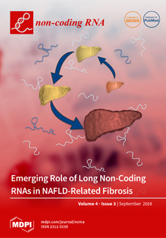
Cover Story (view full-size image): Despite a comprehensive understanding of cellular changes that occur during the development and progression of nonalcoholic fatty liver disease (NAFLD) fibrosis, the molecular mechanisms underlying these changes remain poorly understood. Long non-coding RNAs (lncRNAs) are emerging as key contributors to biological processes, underpinning the initiation and progression of NAFLD fibrosis. Delineating the mechanisms by which lncRNAs mediate NAFLD fibrosis, is a critical first step toward identifying novel therapeutic targets for drug development and improved, noninvasive methods for disease diagnosis.
  • Issues are regarded as officially published after their release is announced to the table of contents alert mailing list.
  • You may sign up for e-mail alerts to receive table of contents of newly released issues.
  • PDF is the official format for papers published in both, html and pdf forms. To view the papers in pdf format, click on the "PDF Full-text" link, and use the free Adobe Reader to open them.
Order results
Result details
Section
Select all
Export citation of selected articles as:
4 pages, 227 KiB  
Editorial
The Non-Coding RNA Journal Club: Highlights on Recent Papers—6
by Hua Xiao, Patrick K. T. Shiu, Jun Shu, Gaetano Santulli, Mohammad K. Gheybi, Simon J. Conn, Baptiste Bogard, Florent Hubé, Joseph H. Taube, Sendurai A. Mani, Luo Song, George A. Calin and Shuxing Zhang
Non-Coding RNA 2018, 4(3), 23; https://doi.org/10.3390/ncrna4030023 - 18 Sep 2018
Cited by 1 | Viewed by 3655
Abstract
We are delighted to share with you our sixth Journal Club and highlight some of the most interesting papers published recently [...] Full article
(This article belongs to the Collection The Non-Coding RNA Journal Club: Highlights on Recent Papers)
21 pages, 2091 KiB  
Review
Novel Roles of Non-Coding RNAs in Opioid Signaling and Cardioprotection
by Zesergio Melo, Cecilia Ishida, Maria De la Paz Goldaraz, Rocio Rojo and Raquel Echavarria
Non-Coding RNA 2018, 4(3), 22; https://doi.org/10.3390/ncrna4030022 - 17 Sep 2018
Cited by 19 | Viewed by 5524
Abstract
Cardiovascular disease (CVD) is a significant cause of morbidity and mortality across the world. A large proportion of CVD deaths are secondary to coronary artery disease (CAD) and myocardial infarction (MI). Even though prevention is the best strategy to reduce risk factors associated [...] Read more.
Cardiovascular disease (CVD) is a significant cause of morbidity and mortality across the world. A large proportion of CVD deaths are secondary to coronary artery disease (CAD) and myocardial infarction (MI). Even though prevention is the best strategy to reduce risk factors associated with MI, the use of cardioprotective interventions aimed at improving patient outcomes is of great interest. Opioid conditioning has been shown to be effective in reducing myocardial ischemia-reperfusion injury (IRI) and cardiomyocyte death. However, the molecular mechanisms behind these effects are under investigation and could provide the basis for the development of novel therapeutic approaches in the treatment of CVD. Non-coding RNAs (ncRNAs), which are functional RNA molecules that do not translate into proteins, are critical modulators of cardiac gene expression during heart development and disease. Moreover, ncRNAs such as microRNAs (miRNAs) and long non-coding RNAs (lncRNAs) are known to be induced by opioid receptor activation and regulate opioid signaling pathways. Recent advances in experimental and computational tools have accelerated the discovery and functional characterization of ncRNAs. In this study, we review the current understanding of the role of ncRNAs in opioid signaling and opioid-induced cardioprotection. Full article
(This article belongs to the Collection Regulatory RNAs in Cardiovascular Development and Disease)
Show Figures

Graphical abstract

Graphical abstract
Full article ">Figure 1
<p>Classification and function of coding and non-coding RNAs (ncRNAs). The array of RNA molecules is diverse in structure and function across species. Messenger RNAs (mRNAs) are coding transcripts that undergo extensive post-transcriptional processing, 5′capping (m<sup>7</sup>G) and 3′polyadenylation (poly(A)), and can be translated into proteins. Long non-coding RNAs (lncRNAs) possess little, if any, coding potential and, after their transcription from genic and intergenic regions, they can suffer different types of post-transcriptional processing. MicroRNAs (miRNAs) are transcribed into primary miRNA transcripts (pri-miRNA) by Pol II or are generated through alternative splicing. They are further processed into pre-miRNA and mature miRNA before being incorporated into the RNA-induced silencing complex (RISC)to silence their target genes. Small nucleolar RNAs (snoRNAs) are usually transcribed by Pol II and belong to the translational machinery of the cell and process ribosomal RNAs. Circular RNAs (circRNAs) are produced from back splicing of exons and possess unique features given by the formation of a 3′,5′phosphodiester bond.</p>
Full article ">Figure 2
<p>miRNAs in control of mu opioid receptor (MOR) translation. Multiple miRNAs able to bind to regulatory elements located in MOR 3′ UTR have been identified. Expression of some of these miRNAs (miR-339-3p, let-7, miR-23b-5p, miR-212/132, and miR-103/107) is induced by opioid agonists as a negative feedback mechanism. In contrast, morphine can also reduce miR-16-5p expression to increase MOR translation.</p>
Full article ">Figure 3
<p>ncRNAs and opioid-induced cardioprotection in healthy and failing hearts. Experimental evidence shows that opioids can reduce cardiomyocyte apoptosis caused by myocardial IRI through miR-133b-5p and its target Fas in both healthy and failing hearts. In healthy hearts, fentanyl activation of DOR inhibits MALAT1 expression and a ceRNA for miR-145-5p, which allows miR-145-5p to target Bnip3 and reduce apoptosis. Interestingly, heart failure induces expression of MOR in cardiomyocytes, which could have important implications for opioid-mediated cardioprotection in patients with MI.</p>
Full article ">Figure 4
<p>Morphine and fentanyl induction of ncRNAs involved in opioid signaling and cardioprotection. A schematic summary of miRNAs known to be induced by opioids in a variety of cells and animal models as well as its mechanisms of opioid signaling regulation and its potential implications for cardioprotection by reducing apoptosis.</p>
Full article ">
11 pages, 1776 KiB  
Article
Targeted Genomic Screen Reveals Focal Long Non-Coding RNA Copy Number Alterations in Cancer Cell Lines
by Pieter-Jan Volders, Steve Lefever, Shalina Baute, Justine Nuytens, Katrien Vanderheyden, Björn Menten, Pieter Mestdagh and Jo Vandesompele
Non-Coding RNA 2018, 4(3), 21; https://doi.org/10.3390/ncrna4030021 - 13 Sep 2018
Cited by 6 | Viewed by 4167
Abstract
The landscape of somatic copy-number alterations (SCNAs) affecting long non-coding RNAs (lncRNAs) in human cancers remains largely unexplored. While the majority of lncRNAs remain to be functionally characterized, several have been implicated in cancer development and metastasis. Considering the plethora of lncRNAs genes [...] Read more.
The landscape of somatic copy-number alterations (SCNAs) affecting long non-coding RNAs (lncRNAs) in human cancers remains largely unexplored. While the majority of lncRNAs remain to be functionally characterized, several have been implicated in cancer development and metastasis. Considering the plethora of lncRNAs genes that have been currently reported, it is conceivable that many more lncRNAs might function as oncogenes or tumor suppressor genes. We devised a strategy to detect focal lncRNA SCNAs using a custom DNA microarray platform probing 10,519 lncRNA genes. By screening a panel of 80 cancer cell lines, we detected numerous focal aberrations targeting one or multiple lncRNAs without affecting neighboring protein-coding genes. These focal aberrations are highly suggestive for a tumor suppressive or oncogenic role of the targeted lncRNA gene. Although functional validation remains an essential step in the further characterization of the involved candidate cancer lncRNAs, our results provide a direct way of prioritizing candidate lncRNAs that are involved in cancer pathogenesis. Full article
(This article belongs to the Collection Non-Coding RNA Methods)
Show Figures

Figure 1

Figure 1
<p>Overview of the long non-coding RNA (lncRNA) genes affected by focal somatic copy-number alterations (SCNAs) after extensive filtering. Red represents the copy number loss (log-ratio &lt; 1.5) in that cell line, while blue corresponds to copy number gain (log-ratio &gt; 1.5). Dark red and blue correspond to copy number changes with absolute log-ratios of above 2.5.</p>
Full article ">Figure 2
<p>Quantitative polymerase chain reaction validation of the putative focal SCNAs. The Cq value of the aberration is normalized to the Cq value of each of the flanking regions. A copy number gain (blue) is considered as confirmed and focal when the relative quantity to both flanking regions is higher than one. Similarly, a copy number loss (red) is considered as confirmed and focal when the relative quantity to both flanking regions is less than one. Red crosses represent Cq values &gt; 35, corresponding to a homozygous deletion of the flanking regions. Stars represent significant (<span class="html-italic">p</span>-value &lt; 0.05) differences from one.</p>
Full article ">Figure 3
<p>A comparison word cloud shows strong enrichment of the word “cancer” in abstracts of publications associated with the lncRNAs affected by SCNAs. The size of each word corresponds to the deviation of its frequency in abstracts associated with the lncRNAs affected by SCNAs from the average occurrence frequency. Green words are more prevalent in abstracts on lncRNAs affected by SCNAs, while orange words are more prevalent in the abstracts on lncRNAs unaffected by SCNAs.</p>
Full article ">
10 pages, 3149 KiB  
Article
Z Probe, An Efficient Tool for Characterizing Long Non-Coding RNA in FFPE Tissues
by Manish K. Tripathi, Chidi Zacheaus, Kyle Doxtater, Fatemeh Keramatnia, Cuilan Gao, Murali M. Yallapu, Meena Jaggi and Subhash C. Chauhan
Non-Coding RNA 2018, 4(3), 20; https://doi.org/10.3390/ncrna4030020 - 5 Sep 2018
Cited by 8 | Viewed by 6048
Abstract
Formalin-fixed paraffin embedded (FFPE) tissues are a valuable resource for biomarker discovery in order to understand the etiology of different cancers and many other diseases. Proteins are the biomarkers of interest with respect to FFPE tissues as RNA degradation is the major challenge [...] Read more.
Formalin-fixed paraffin embedded (FFPE) tissues are a valuable resource for biomarker discovery in order to understand the etiology of different cancers and many other diseases. Proteins are the biomarkers of interest with respect to FFPE tissues as RNA degradation is the major challenge in these tissue samples. Recently, non-protein coding transcripts, long non-coding RNAs (lncRNAs), have gained significant attention due to their important biological actions and potential involvement in cancer. RNA sequencing (RNA-seq) or quantitative reverse transcription-polymerase chain reaction (qRT-PCR) are the only validated methods to evaluate and study lncRNA expression and neither of them provides visual representation as immunohistochemistry (IHC) provides for proteins. We have standardized and are reporting a sensitive Z probe based in situ hybridization method to visually identify and quantify lncRNA in FFPE tissues. This assay is highly sensitive and identifies transcripts visible within different cell types and tumors. We have detected a scarcely expressed tumor suppressor lncRNA NRON (non-coding repressor of nuclear factor of activated T-cells (NFAT)), a moderately expressed oncogenic lncRNA UCA1 (urothelial cancer associated 1), and a highly studied and expressed lncRNA MALAT1 (metastasis associated lung adenocarcinoma transcript 1) in different cancers. High MALAT1 staining was found in colorectal, breast and pancreatic cancer. Additionally, we have observed an increase in MALAT1 expression in different stages of colorectal cancer. Full article
(This article belongs to the Collection Non-Coding RNA Methods)
Show Figures

Figure 1

Figure 1
<p>A schematic representation of the Z probe based RNAScope assay for long non-coding RNA (lncRNA) analysis. Starting with Z probes hybridizing with the target sequence creating double ZZs with up to 20 groups side by side. The pre-amplifier then binds to the complementary sequence on the 28-base tail (top of the ZZ). Pre-amplifiers contain multiple binding sites for amplifiers to bind to and the amplifiers also have multiple binding sites for labeled probes to bind. Upon chromogenic stain, the labeled probes fluoresce with a red color.</p>
Full article ">Figure 2
<p>Validation and optimization of the Z-probe staining. Paraffin embedded, and sectioned, pancreatic cancer tissue, colorectal cancer tissue and HeLa cell pellet were stained with a negative control probe <span class="html-italic">DapB</span> (full form) and a positive control probe <span class="html-italic">PPIB</span> (full form). The <span class="html-italic">PPIB</span> stained well with pancreatic, colorectal and HeLa cells (bottom panel (<b>a</b>–<b>c</b>). 20× (inset) and 80× magnification using CaseViewer 2.2 software (3DHistech Ltd., Budapest, Hungary) scanned and analyzed on Pannoramic 250 Flash III (3DHistech Ltd.). Arrows point at specific staining.</p>
Full article ">Figure 3
<p>Paraffin embedded different human cancer tissues that have been Z probe stained for different lncRNAs. (<b>a</b>) Tumor suppressor lncRNA NRON (very low expression, non-coding repressor of NFAT) (<b>i</b>) and oncogenic lncRNA UCA1 (moderately expressed, urothelial cancer associated 1) (<b>ii</b>) stained in colorectal cancer tissue. (<b>b</b>) LncRNA MALAT1 (metastasis associated lung adenocarcinoma transcript 1) stained using specific Z-probes in paraffin embedded (<b>i</b>) colorectal cancer, (<b>ii</b>) breast cancer and (<b>iii</b>) pancreatic cancer tissues. 20× (inset) and 80× magnification using CaseViewer 2.2 software, scanned and analyzed on Pannoramic 250 Flash III. Arrows point at specific lncRNA signals.</p>
Full article ">Figure 4
<p>LncRNA-MALAT in different stages of colorectal cancer (CRC). Paraffin embedded different stages of colorectal cancer tissues were stained for lncRNA MALAT1 and quantitated for staining intensity. 60 CRC tissues, Stage I = 15; Stage II = 16; Stage III = 20; Stage IV = 9, were stained for lncRNA-MALAT1 (<b>a</b>) Stages I–IV CRC tissues show a differential stain for lncRNA MALAT1. Stain intensity correlates with the progression. (<b>b</b>) Quantitation of the lncRNA MALAT1 staining intensity was performed using Image J software. 10× (inset) and 80× magnification using CaseViewer 2.2 software, scanned and analyzed on Pannoramic 250 Flash III. Statistical analysis: One-way ANOVA and Tukey’ multiple comparison tests compare the mean of each column with the mean of other columns.</p>
Full article ">Figure 5
<p>LncRNA MALAT1 expression in breast cancer. BioMax TMA BR243w containing 12 normal adjacent tumor (NAT) and 12 invasive breast carcinomas were stained and analyzed. (<b>a</b>) Matched breast cancer tissues (NAT vs. Invasive) from two different patients, 1 and 2, were stained for lncRNA MALAT1 using Z probe. The invasive breast cancer tissues have higher staining for lncRNA MALAT1 as compared to normal adjacent tumors. (<b>b</b>) Quantitation of the lncRNA MALAT1 staining intensity was performed using Image J software. 10× (inset) and 80× magnification using CaseViewer 2.2 software, scanned and analyzed on Pannoramic 250 Flash III. Statistical analysis: Unpaired <span class="html-italic">t</span>-test, ** <span class="html-italic">p</span> &lt; 0.01, *** <span class="html-italic">p</span> &lt; 0.001.</p>
Full article ">
17 pages, 702 KiB  
Review
Long Non-Coding RNAs in Obesity-Induced Cancer
by Mabel Yin-Chun Yau, Lu Xu, Chien-Ling Huang and Chi-Ming Wong
Non-Coding RNA 2018, 4(3), 19; https://doi.org/10.3390/ncrna4030019 - 28 Aug 2018
Cited by 20 | Viewed by 4745
Abstract
Many mechanisms of obesity-induced cancers have been proposed. However, it remains unclear whether or not long non-coding RNAs (lncRNAs) play any role in obesity-induced cancers. In this article, we briefly discuss the generally accepted hypotheses explaining the mechanisms of obesity-induced cancers, summarize the [...] Read more.
Many mechanisms of obesity-induced cancers have been proposed. However, it remains unclear whether or not long non-coding RNAs (lncRNAs) play any role in obesity-induced cancers. In this article, we briefly discuss the generally accepted hypotheses explaining the mechanisms of obesity-induced cancers, summarize the latest evidence for the expression of a number of well-known cancer-associated lncRNAs in obese subjects, and propose the potential contribution of lncRNAs to obesity-induced cancers. We hope this review can serve as an inspiration to scientists to further explore the regulatory roles of lncRNAs in the development of obesity-induced cancers. Those findings will be fundamental in the development of effective therapeutics or interventions to combat this life-threatening adverse effect of obesity. Full article
(This article belongs to the Special Issue Genomic Instability and Non-Coding RNA)
Show Figures

Figure 1

Figure 1
<p>The epigenetic state of imprinting control region on the <span class="html-italic">Igf2</span>-<span class="html-italic">H19</span> locus determines the expression pattern. Regulation of maternal and paternal expression in the <span class="html-italic">Igf2</span>-<span class="html-italic">H19</span> imprinted domain is controlled by genomic DNA methylation. The open boxes represent the genes <span class="html-italic">Igf2</span> and <span class="html-italic">H19</span>, and the blue boxes represent the imprinting control region (ICR). The close lollipops represent methylated CpG islands. The yellow and red circles represent the CCCTC binding factor (CTCF) insulator protein and enhancer, respectively. The arrows from the boxes indicate expression of the genes. <span class="html-italic">Igf2</span> and <span class="html-italic">H19</span> genes are activated by the shared downstream enhancer, and their activations are dependent on the DNA methylation of the ICR. CCCTC binding factor (CTCF) is recruited to unmethylated ICR on the maternal allele that promotes the enhancer to activate the expression of <span class="html-italic">H19</span> gene, but not of <span class="html-italic">Igf2</span> gene. In contract, on paternal allele, ICR is hypermethylated that prevents the binding of CTCF to ICR. The overall outcomes are that the expression of <span class="html-italic">H19</span> is repressed, but the expression of <span class="html-italic">Igf2</span> is induced, from the paternal allele.</p>
Full article ">Figure 2
<p>Proposed roles of long non-coding RNAs (lncRNAs) in obese-induced cancers. Many obese-related physiological changes such as nutrient availability, oxygen level, inflammatory cytokines, and metabolic hormones may affect the expression level (1) and post-transcriptional processing (2) of endogenous lncRNAs. Many lncRNAs can be transported to circulation and may serve as biomarkers or molecular diagnostic applications (3). Whether or not the exogenous lncRNAs contribute to the cancer progression is just emerging. In contrast, there is plentiful evidence showing that endogenous lncRNAs can regulate gene expression by diverse mechanisms. lncRNAs may act as scaffolds or molecular decoys, which directly interact with RNA binding proteins (RBPs; red circle) such as transcription factors and chromatin-modifying complexes to regulate the expression of proto-oncogenes and/or tumor suppressor genes (4). lncRNAs may act as endogenous sponges regulating gene expression via modulating microRNAs (miRNAs) availability (5 and 6).</p>
Full article ">
15 pages, 465 KiB  
Review
The Role of Long Non-Coding RNAs (lncRNAs) in the Development and Progression of Fibrosis Associated with Nonalcoholic Fatty Liver Disease (NAFLD)
by Amanda Hanson, Danielle Wilhelmsen and Johanna K. DiStefano
Non-Coding RNA 2018, 4(3), 18; https://doi.org/10.3390/ncrna4030018 - 21 Aug 2018
Cited by 46 | Viewed by 7423
Abstract
Nonalcoholic fatty liver disease (NAFLD) encompasses a spectrum of conditions ranging from hepatic steatosis to inflammation (nonalcoholic steatohepatitis or NASH) with or without fibrosis, in the absence of significant alcohol consumption. The presence of fibrosis in NASH patients is associated with greater liver-related [...] Read more.
Nonalcoholic fatty liver disease (NAFLD) encompasses a spectrum of conditions ranging from hepatic steatosis to inflammation (nonalcoholic steatohepatitis or NASH) with or without fibrosis, in the absence of significant alcohol consumption. The presence of fibrosis in NASH patients is associated with greater liver-related morbidity and mortality; however, the molecular mechanisms underlying the development of fibrosis and cirrhosis in NAFLD patients remain poorly understood. Long non-coding RNAs (lncRNAs) are emerging as key contributors to biological processes that are underpinning the initiation and progression of NAFLD fibrosis. This review summarizes the experimental findings that have been obtained to date in animal models of liver fibrosis and NAFLD patients with fibrosis. We also discuss the potential applicability of circulating lncRNAs to serve as biomarkers for the diagnosis and prognosis of NAFLD fibrosis. A better understanding of the role played by lncRNAs in NAFLD fibrosis is critical for the identification of novel therapeutic targets for drug development and improved, noninvasive methods for disease diagnosis. Full article
(This article belongs to the Special Issue Non-Coding RNA, Fibrogenesis, and Fibrotic Disease)
Show Figures

Figure 1

Figure 1
<p>Stages of nonalcoholic fatty liver disease (NAFLD). Liver appearance in the various stages of the disease is schematized to represent the physical changes that accompany disease progression. The percentage of patients progressing from one stage to the subsequent stage is depicted below the arrows. NASH: nonalcoholic steatohepatitis, HCC: hepatocellular carcinoma.</p>
Full article ">
12 pages, 302 KiB  
Perspective
The State of Long Non-Coding RNA Biology
by John S. Mattick
Non-Coding RNA 2018, 4(3), 17; https://doi.org/10.3390/ncrna4030017 - 10 Aug 2018
Cited by 69 | Viewed by 8419
Abstract
Transcriptomic studies have demonstrated that the vast majority of the genomes of mammals and other complex organisms is expressed in highly dynamic and cell-specific patterns to produce large numbers of intergenic, antisense and intronic long non-protein-coding RNAs (lncRNAs). Despite well characterized examples, their [...] Read more.
Transcriptomic studies have demonstrated that the vast majority of the genomes of mammals and other complex organisms is expressed in highly dynamic and cell-specific patterns to produce large numbers of intergenic, antisense and intronic long non-protein-coding RNAs (lncRNAs). Despite well characterized examples, their scaling with developmental complexity, and many demonstrations of their association with cellular processes, development and diseases, lncRNAs are still to be widely accepted as major players in gene regulation. This may reflect an underappreciation of the extent and precision of the epigenetic control of differentiation and development, where lncRNAs appear to have a central role, likely as organizational and guide molecules: most lncRNAs are nuclear-localized and chromatin-associated, with some involved in the formation of specialized subcellular domains. I suggest that a reassessment of the conceptual framework of genetic information and gene expression in the 4-dimensional ontogeny of spatially organized multicellular organisms is required. Together with this and further studies on their biology, the key challenges now are to determine the structure–function relationships of lncRNAs, which may be aided by emerging evidence of their modular structure, the role of RNA editing and modification in enabling epigenetic plasticity, and the role of RNA signaling in transgenerational inheritance of experience. Full article
(This article belongs to the Special Issue Non-Coding RNA in the Nervous System)
9 pages, 2830 KiB  
Commentary
Formation of a Family of Long Intergenic Noncoding RNA Genes with an Embedded Translocation Breakpoint Motif in Human Chromosomal Low Copy Repeats of 22q11.2—Some Surprises and Questions
by Nicholas Delihas
Non-Coding RNA 2018, 4(3), 16; https://doi.org/10.3390/ncrna4030016 - 20 Jul 2018
Cited by 6 | Viewed by 3837
Abstract
A family of long intergenic noncoding RNA (lincRNA) genes, FAM230 is formed via gene sequence duplication, specifically in human chromosomal low copy repeats (LCR) or segmental duplications. This is the first group of lincRNA genes known to be formed by segmental duplications and [...] Read more.
A family of long intergenic noncoding RNA (lincRNA) genes, FAM230 is formed via gene sequence duplication, specifically in human chromosomal low copy repeats (LCR) or segmental duplications. This is the first group of lincRNA genes known to be formed by segmental duplications and is consistent with current views of evolution and the creation of new genes via DNA low copy repeats. It appears to be an efficient way to form multiple lincRNA genes. But as these genes are in a critical chromosomal region with respect to the incidence of abnormal translocations and resulting genetic abnormalities, the 22q11.2 region, and also carry a translocation breakpoint motif, several intriguing questions arise concerning the presence and function of the translocation breakpoint sequence in RNA genes situated in LCR22s. Full article
(This article belongs to the Section Long Non-Coding RNA)
Show Figures

Graphical abstract

Graphical abstract
Full article ">Figure 1
<p>A schematic of 5′ and 3′ sections of the long intergenic noncoding RNA (lincRNA) gene <span class="html-italic">FAM230C</span> present in chr13 that form two distinct groups of long noncoding RNA (lncRNA) genes. Based on reference [<a href="#B13-ncrna-04-00016" class="html-bibr">13</a>]. Abbreviations: chr, chromosome; TBTA, translocation breakpoint type A; LCR22, low copy repeats in chr22.</p>
Full article ">Figure 2
<p>A segment of the secondary structural model of the TBTA (GenBank sequence ID: AB261997.1) showing two long stem loops: the 294 bp palindromic AT-rich repeat sequences (PATRR) and the 104 bp stem loop formed by AT sequences from AT-rich region #2. The PATRR has a significant number of G:C bonds in the lower portion of the stem but is A:T base pair rich in the upper portion close to the loop side. The AT-rich region #2 stem loop consists entirely of A:T bonds with the exception of two G:T bonds. The Zuker DNA folding program [<a href="#B37-ncrna-04-00016" class="html-bibr">37</a>] was used to generate the secondary structure.</p>
Full article ">Figure 3
<p>(<b>Top</b>) Schematic of PATRR stem loop. The section between arrows denote sequences from the 5’ half missing in gene <span class="html-italic">LINC01660.</span> (<b>Bottom</b>) Alignment of the nucleotide sequences of the TBTA and L<span class="html-italic">INC01660</span>. The alignment was determined by the Emboss Needle Pairwise Sequence Alignment program (<a href="https://www.ebi.ac.uk/Tools/psa/emboss_needle/nucleotide.html" target="_blank">https://www.ebi.ac.uk/Tools/psa/emboss_needle/nucleotide.html</a>) [<a href="#B38-ncrna-04-00016" class="html-bibr">38</a>]. Green color highlights the TBTA nucleotide sequence missing in <span class="html-italic">LINC01660</span> equivalent to TBTA nucleotide positions 1489–1690. This sequence has a large number of G and C residues that form a number of G:C pairs at the base of the 5′ side of the double stranded stem of the PATRR that stabilizes the stem. The G:C pairs as well as other base pairs are missing in <span class="html-italic">LINC01660</span> that has the PATRR 5’ section deleted.</p>
Full article ">Figure 4
<p>The TBTA sequence, highlighting the various elements it harbors, including the PATRR and AT-rich region #2 and AT-rich region #1. The sequence is from National Center of Biotechnology information (NCBI) GenBank: AB261997.1. The figure is modified from reference [<a href="#B13-ncrna-04-00016" class="html-bibr">13</a>]. Two arrows (bottom) delineate the regions deleted in the RNA gene <span class="html-italic">LINC01658</span> sequence that is equivalent to TBTA positions 930–1880, and encompasses the AluYm transposable element (in yellow), the entire AT-rich region #2 (in red), the 5’ side of the PATRR and includes all of the PATRR-associated AT-rich sequences up to position 1880 (in green). The AT-rich region #1 is also deleted (delineated by arrows at positions 305 and 359) with the exception of the very small AT sequence, positions 360–366. These deletions essentially result in RNA gene <span class="html-italic">LINC01658</span> having no AT-rich sequences.</p>
Full article ">
Previous Issue
Next Issue
Back to TopTop