[go: up one dir, main page]

Next Issue
Volume 10, March
Previous Issue
Volume 10, January


Toxics, Volume 10, Issue 2 (February 2022) – 58 articles

Cover Story (view full-size image): 
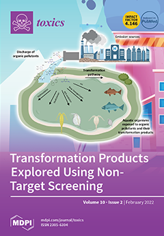
The occurrence of transformation products (TPs) of emerging pollutants has been of potential concern in aquatic environments. Nonetheless, it is unclear how TPs are formed and affect organisms in aquatic ecosystem. Thanking to recent advances in analytics and biotechnology, identification of TPs and their formation mechanisms can be explored by non-target screening (NTS) using high-resolution mass spectrometry (HRMS) and in vivo/vitro bioassay using model organisms. In this review, recent progress in TP research including their exposure and effect assessments is summarized. In particular, the review covers prospective research efforts on TP, such as identification of the products using NTS, and assessment of potential risk. View this paper
  • Issues are regarded as officially published after their release is announced to the table of contents alert mailing list.
  • You may sign up for e-mail alerts to receive table of contents of newly released issues.
  • PDF is the official format for papers published in both, html and pdf forms. To view the papers in pdf format, click on the "PDF Full-text" link, and use the free Adobe Reader to open them.
Order results
Result details
Section
Select all
Export citation of selected articles as:
13 pages, 1259 KiB  
Article
DNA Methylation and Detoxification in the Earthworm Lumbricus terrestris Exposed to Cadmium and the DNA Demethylation Agent 5-aza-2′-deoxycytidine
by Gerhard P. Aigner, Pamela Nenning, Birgit Fiechtner, Maja Šrut and Martina Höckner
Toxics 2022, 10(2), 100; https://doi.org/10.3390/toxics10020100 - 21 Feb 2022
Cited by 7 | Viewed by 3108
Abstract
Earthworms are well-established model organisms for testing the effects of heavy metal pollution. How DNA methylation affects cadmium (Cd) detoxification processes such as the expression of metallothionein 2 (MT2), however, is largely unknown. We therefore exposed Lumbricus terrestris to 200 mg concentrations of [...] Read more.
Earthworms are well-established model organisms for testing the effects of heavy metal pollution. How DNA methylation affects cadmium (Cd) detoxification processes such as the expression of metallothionein 2 (MT2), however, is largely unknown. We therefore exposed Lumbricus terrestris to 200 mg concentrations of Cd and 5-aza-2′-deoxycytidine (Aza), a demethylating agent, and sampled tissue and coelomocytes, cells of the innate immune system, for 48 h. MT2 transcription significantly increased in the Cd- and Cd-Aza-treated groups. In tissue samples, a significant decrease in MT2 in the Aza-treated group was detected, showing that Aza treatment inhibits basal MT2 gene activity but has no effect on Cd-induced MT2 levels. Although Cd repressed the gene expression of DNA-(cytosine-5)-methyltransferase-1 (DNMT1), which is responsible for maintaining DNA methylation, DNMT activity was unchanged, meaning that methylation maintenance was not affected in coelomocytes. The treatment did not influence DNMT3, which mediates de novo methylation, TET gene expression, which orchestrates demethylation, and global levels of hydroxymethylcytosine (5hmC), a product of the demethylation process. Taken together, this study indicates that Aza inhibits basal gene activity, in contrast to Cd-induced MT2 gene expression, but does not affect global DNA methylation. We therefore conclude that Cd detoxification based on the induction of MT2 does not relate to DNA methylation changes. Full article
(This article belongs to the Special Issue Earthworm Model to Study the Effects of Environmental Pollutants)
Show Figures

Figure 1

Figure 1
<p>(<b>a</b>) mRNA copy numbers of Metallothionein 2 (MT2) in <span class="html-italic">L. terrestris</span> coelomocytes and (<b>b</b>) tissue of control (C) and exposed individuals (5-aza-2′-deoxycytidine (A); 200 mg/kg CdCl<sub>2</sub> (Cd); 5-aza-2′-deoxycytidine and 200 mg/kg CdCl<sub>2</sub> (CdA)). Stars indicate significant differences between treatments and the control (<span class="html-italic">p</span> &lt; 0.05 (*); <span class="html-italic">p</span> &lt; 0.01 (**)). Replicates per treatment analyzed in coelomocytes: C = 5, A = 6, Cd = 5, CdA = 3. Replicates per treatment analyzed in tissue: C = 7, A = 5, Cd = 8, CdA = 12.</p>
Full article ">Figure 2
<p>mRNA copy numbers of DNA-methyltransferase 1 (DNMT1) and ten–eleven translocation (TET) in <span class="html-italic">L. terrestris</span> coelomocytes and tissue of control (C) and exposed individuals (5-aza-2′-deoxycytidine (A); 200 mg/kg CdCl<sub>2</sub> (Cd); 5-aza-2′-deoxycytidine and 200 mg/kg CdCl<sub>2</sub> (CdA)). Stars indicate significant differences between treatments and control (<span class="html-italic">p</span> &lt; 0.05 (*)). Replicates per treatment analyzed in coelomocytes: C = 5, A = 6, Cd = 5, CdA = 3. Replicates per treatment analyzed in tissue: C = 6, A = 4, Cd = 6, CdA = 6.</p>
Full article ">Figure 3
<p>(<b>a</b>) Global DNA methylation levels in coelomocytes extruded from control (C, <span class="html-italic">n</span> = 4) and exposed <span class="html-italic">L. terrestris</span> specimens (5-aza-2′-deoxycytidine (A), <span class="html-italic">n</span> = 4; 200 mg/kg Cd (Cd) <span class="html-italic">n</span> = 5; 5-aza-2′-deoxycytidine and 200 mg/kg CdCl<sub>2</sub> (CdA), <span class="html-italic">n</span> = 4) after 48 h of exposure. (<b>b</b>) Total DNMT protein activity (DNMT1 and DNMT3) of coelomocytes extruded from control (C, <span class="html-italic">n</span> = 3) and Cd-exposed (200 mg/kg CdCl<sub>2</sub> (Cd, <span class="html-italic">n</span> = 6)) <span class="html-italic">L. terrestris</span> individuals after 48 h of exposure.</p>
Full article ">Figure 4
<p>(<b>a</b>) MDS analysis using tissue samples and variables DNMT1, TET, MT2 and 5mC. Significance in group separation is based on the MDS2 dimension (shown by letters). The ellipse around the treatment groups indicates a confidence of 90%. (<b>b</b>) MDS analysis using coelomocyte samples and variables DNMT1, TET, MT2 and 5mC. Significance in group separation is based on the MDS1 dimension (shown by letters). The ellipse around treatment groups indicates a confidence of 90%.</p>
Full article ">
20 pages, 4384 KiB  
Article
Nonylphenol Polyethoxylates Enhance Adipose Deposition in Developmentally Exposed Zebrafish
by Christopher D. Kassotis, Matthew K. LeFauve, Yu-Ting Tiffany Chiang, Megan M. Knuth, Stacy Schkoda and Seth W. Kullman
Toxics 2022, 10(2), 99; https://doi.org/10.3390/toxics10020099 - 20 Feb 2022
Cited by 7 | Viewed by 3459 | Correction
Abstract
Alkylphenol polyethoxylates (APEOs), such as nonylphenol ethoxylates (NPEOs), are high-production-volume surfactants used in laundry detergents, hard-surface cleaners, pesticide formulations, textile production, oils, paints, and other products. NPEOs comprise −80% of the total production of APEOs and are widely reported across diverse environmental matrices. [...] Read more.
Alkylphenol polyethoxylates (APEOs), such as nonylphenol ethoxylates (NPEOs), are high-production-volume surfactants used in laundry detergents, hard-surface cleaners, pesticide formulations, textile production, oils, paints, and other products. NPEOs comprise −80% of the total production of APEOs and are widely reported across diverse environmental matrices. Despite a growing push for replacement products, APEOs continue to be released into the environment through wastewater at significant levels. Research into related nonionic surfactants from varying sources has reported metabolic health impacts, and we have previously demonstrated that diverse APEOs and alcohol polyethoxylates promote adipogenesis in the murine 3T3-L1 pre-adipocyte model. These effects appeared to be independent of the base alkylphenol and related to the ethoxylate chain length, though limited research has evaluated NPEO exposures in animal models. The goals of this study were to assess the potential of NPEOs to promote adiposity (Nile red fluorescence quantification) and alter growth and/or development (toxicity, length, weight, and energy expenditure) of developmentally exposed zebrafish (Danio rerio). We also sought to expand our understanding of the ability to promote adiposity through evaluation in human mesenchymal stem cells. Herein, we demonstrated consistent adipogenic effects in two separate human bone-marrow-derived mesenchymal stem cell models, and that nonylphenol and its ethoxylates promoted weight gain and increased adipose deposition in developmentally exposed zebrafish. Notably, across both cell and zebrafish models we report increasing adipogenic/obesogenic activity with increasing ethoxylate chain lengths up to maximums around NPEO-6 and then decreasing activity with the longest ethoxylate chain lengths. This research suggests metabolic health concerns for these common obesogens, suggesting further need to assess molecular mechanisms and better characterize environmental concentrations for human health risk assessments. Full article
Show Figures

Figure 1

Figure 1
<p>Nonylphenol and polyethoxylates promote adipogenesis in human mesenchymal stem cell models. Zenbio and Lonza human bone marrow–derived mesenchymal stem cell models were differentiated as described in Methods and assessed for adipocyte differentiation (Nile Red staining of lipid accumulation) and cell proliferation (Hoechst staining) after 14/21 (respectively) days of differentiation while exposed to controls chemicals as well as nonylphenol and its ethoxylates. Percent normalized triglyceride accumulation per cell relative to maximal rosiglitazone response (normalized to DNA content) (<b>A</b>,<b>C</b>). increase (cell proliferation) or decrease (potential cytotoxicity) in DNA content relative to vehicle control (<b>B</b>,<b>D</b>). Zenbio–sourced cell data provided in (<b>A</b>,<b>B</b>), and Lonza–sourced cell data provided in (<b>C</b>,<b>D</b>). Data presented as mean ± SEM from three independent experiments. * indicates lowest concentration with significant increase in triglyceride over vehicle control or cell proliferation/cytotoxicity relative to vehicle control, <span class="html-italic">p</span> &lt; 0.05, as per Kruskal–Wallis in GraphPad Prism 9. X–axis format is provided in log scale. Panel (<b>E</b>) provides a summary plot of maximal effects on triglyceride accumulation based on ethoxylate chain length across cell models, comparing results from panels (<b>A</b>,<b>C</b>) with previously published effects in 3T3–L1 cells (PMID: 29106673). NPEO = nonylphenol polyethoxylated (with varying average ethoxylate chain lengths).</p>
Full article ">Figure 2
<p>Lethality of nonylphenol and polyethoxylates in developmentally exposed zebrafish. Zebrafish were developmentally exposed to control chemicals, nonylphenol, or nonylphenol polyethoxylates from one through six days post-fertilization. Following exposures, fish were aged to 30 days post-fertilization. Lethality was measured twice daily to determine survivorship across test chemicals and treatments for each test chemical (<b>A</b>). <span class="html-italic">n</span> = 15 replicate fish in each biological replicate (spawning event) for each test chemical and concentration, and four spawns were performed (four biological replicates) and averaged for responses depicted here, for approximately 60 fish evaluated per experimental group. Lethality is depicted in survivorship curves, with decreasing response on the Y axis depicting greater lethality of the test chemicals. Survival percent relative to DMSO vehicle control exposed fish at 30 days (<b>B</b>). * indicates significant change in survival compared to vehicle control fish, <span class="html-italic">p</span> &lt; 0.05, as per Kruskal–Wallis test with Dunn’s multiple comparisons. TBT 10 nM, NP 10 μM, NPEO-2 10 μM, NPEO-4 10 μM, NPEO-6 10 μM, and NPEO-10 10 μM promoted absolute toxicity (no surviving fish) and thus are not depicted here for the purposes of clarity. DMSO = dimethylsulfoxide, vehicle control; TBT = tributyltin chloride; NPEO = nonylphenol polyethoxylated (with varying average ethoxylate chain lengths).</p>
Full article ">Figure 3
<p>Metabolic activity in zebrafish developmentally exposed to nonylphenol and polyethoxylates. Zebrafish were developmentally exposed to control chemicals, nonylphenol, or nonylphenol polyethoxylates. Immediately following exposure, at six days post-fertilization, metabolic activity was measured using the alamar blue assay. Two groups of three replicate fish were transferred into wells of a 24-well black clear-bottom plate, media removed, and alamar blue solution added to wells. Plates were immediately read for fluorescence, then incubated in the dark for several hours, before measuring fluorescence again. The increase in fluorescence is correlated with increased metabolic activity in the fish. Chemical exposure treated fish responses were compared with dimethylsulfoxide (0.1%, vehicle control) treated fish to determine significant differences. <span class="html-italic">n</span> = 6–9 replicate fish in each biological replicate (spawning event), and three spawns were performed for approximately 24 fish per exposure group. * indicates significant change in arbitrary fluorescence compared to vehicle control fish, <span class="html-italic">p</span> &lt; 0.05, as per Kruskal–Wallis test with Dunn’s multiple comparisons. DMSO = dimethylsulfoxide, vehicle control; TBT = tributyltin chloride; NPEO = nonylphenol polyethoxylated (with varying average ethoxylate chain lengths). # represents <span class="html-italic">p</span> &lt; 0.10, as per statistics described above. Box and whisker plots depict the following metrics: whiskers represent 10–90th percentiles, box bounds represent the 25th to 75th percentiles, and the middle line represents the median.</p>
Full article ">Figure 4
<p>Growth trajectories in zebrafish developmentally exposed to nonylphenol and polyethoxylates. Zebrafish were developmentally exposed to control chemicals, nonylphenol, or nonylphenol polyethoxylates; aged to 30 days post−fertilization; then measured and stained with Nile Red (0.5 μg/mL) for 30 min. Zebrafish were imaged and standard length of each fish was measured (<b>A</b>) using the integrated point−to−point measurement tool within the Leica software, which scales by magnification. Following imaging, zebrafish were blotted with kim wipes and weighed on a microbalance to obtain total body weights (<b>B</b>) for each fish and then averaged across test chemicals and concentrations. Zebrafish body mass index (<b>C</b>) was calculated by dividing the calculated standard length and weights and correcting measurement units to g/m<sup>2</sup>. <span class="html-italic">n</span> = 24 (DMSO), 16, 21, 25, 15, 20, 24, 14, 21, 25, 15, 25, 27, 22, 28, 25, 22, 27, 26, 25, 23, 26, and 25 across four spawning events (biological replicates) for exposure groups listed below, respectively. * indicates significant increase in total body fluorescence quantification over vehicle control fish, <span class="html-italic">p</span> &lt; 0.05, as per Kruskal–Wallis test with Dunn’s multiple comparisons. DMSO = dimethylsulfoxide, vehicle control; TBT = tributyltin chloride; NPEO = nonylphenol polyethoxylated (with varying average ethoxylate chain lengths). # represents <span class="html-italic">p</span> &lt; 0.10, as per statistics described above. Box and whisker plots depict the following metrics: whiskers represent 10–90th percentiles, box bounds represent the 25th to 75th percentiles, and the middle line represents the median.</p>
Full article ">Figure 5
<p>Adipose Deposition in Zebrafish Developmentally Exposed to Nonylphenol and Polyethoxylates. Zebrafish were developmentally exposed to control chemicals, nonylphenol, or nonylphenol polyethoxylates; aged to 30 days post-fertilization; then measured and stained with Nile Red (0.5 μg/mL) for 30 min. Total body fluorescence (<b>A</b>) was imaged at 16× magnification using a yellow fluorescent protein filter (representative images in <a href="#toxics-10-00099-f006" class="html-fig">Figure 6</a>) and fluorescence was quantified for each fish and then biological replicates were averaged. <span class="html-italic">n</span> = 24 (DMSO), 16, 21, 25, 15, 20, 24, 14, 21, 25, 15, 25, 27, 22, 28, 25, 22, 27, 26, 25, 23, 26, and 25 across four spawning events (biological replicates) for exposure groups listed below, respectively. * indicates significant increase in total body fluorescence quantification over vehicle control fish, <span class="html-italic">p</span> &lt; 0.05, as per Kruskal–Wallis test with Dunn’s multiple comparisons. # represents <span class="html-italic">p</span> &lt; 0.10, as per statistics described above. Box and whisker plots depict the following metrics: whiskers represent 10–90th percentiles, box bounds represent the 25th to 75th percentiles, and the middle line represents the median. Developmental trajectory of adipose depots across the zebrafish (<b>B</b>). Relative proportions of fish exhibiting fluorescing adipocytes in each depot in the DMSO fish were set as “normal” and the heat map depicts increased proportions of fish in each group with visible adipocytes in each depot. Adipose depots labeled as per the developmental guides provided in Minchin and Rawls, 2017 (PMID: 28348140) and grouped based on anatomical classifications. A value of 9 represents a 9X increase in the proportion of fish in an exposure group PVAT = pancreatic visceral adipose tissue; AVAT = abdominal visceral adipose tissue; RVAT = renal visceral adipose tissue; aCVAT = anterior cardiac visceral adipose tissue; cPOS = central paraosseal non-visceral adipose tissue; OES = esophageal non-visceral adipose tissue; LSAT = lateral truncal adipose tissue; pOCU = posterior ocular adipose tissue; aOCU = anterior ocular adipose tissue; vOCU = ventral ocular adipose tissue; BHD = basihyal hyoid adipose tissue; CHD = ceratohyal hyoid adipose tissue; vOPC = ventral opercular adipose tissue; dOPC = dorsal opercular adipose tissue; and UHD = urihyal hyoid adipose tissue. DMSO = dimethylsulfoxide, vehicle control; TBT = tributyltin chloride; NPEO = nonylphenol polyethoxylated (with varying average ethoxylate chain lengths).</p>
Full article ">Figure 6
<p>Adipose Patterning in Zebrafish Developmentally Exposed to Nonylphenol and Polyethoxylates. Representative fluorescent images of developmentally exposed zebrafish exposed to control chemicals, nonylphenol, and the nonylphenol polyethoxylates. Anesthetized fish imaged at 30 days post-fertilization, following a 30 min stain (0.5 μg/mL Nile Red). Images obtained at 16× magnification using a yellow fluorescent protein filter. DMSO = dimethylsulfoxide, vehicle control; TBT = tributyltin chloride; NPEO = nonylphenol polyethoxylated (with varying average ethoxylate chain lengths).</p>
Full article ">
13 pages, 7602 KiB  
Article
Removal of Hexavalent Chromium(VI) from Wastewater Using Chitosan-Coated Iron Oxide Nanocomposite Membranes
by Jung Eun Park, Jun-Ho Shin, Wonzin Oh, Sang-June Choi, Jeongju Kim, Chorong Kim and Jongho Jeon
Toxics 2022, 10(2), 98; https://doi.org/10.3390/toxics10020098 - 19 Feb 2022
Cited by 27 | Viewed by 3999
Abstract
Chromium is a toxic and carcinogenic heavy metal that originates from various human activities. Therefore, the effective removal of chromium from aqueous solutions is an extremely important global challenge. Herein, we report a chitosan-coated iron oxide nanoparticle immobilized hydrophilic poly(vinylidene) fluoride membrane (Chi@Fe [...] Read more.
Chromium is a toxic and carcinogenic heavy metal that originates from various human activities. Therefore, the effective removal of chromium from aqueous solutions is an extremely important global challenge. Herein, we report a chitosan-coated iron oxide nanoparticle immobilized hydrophilic poly(vinylidene) fluoride membrane (Chi@Fe2O3–PVDF) which can potentially be used for efficient removal of hexavalent chromium(VI) by a simple filtration process. Membrane filtration is an easy and efficient method for treating large volumes of water in a short duration. The adsorption experiments were conducted by batch and continuous in-flow systems. The experimental data showed rapid capture of hexavalent chromium (Cr(VI)) which can be explained by the pseudo-second-order kinetic and Langmuir isotherm model. The nanocomposite membrane exhibited high adsorption capacity for Cr(VI) (14.451 mg/g in batch system, 14.104 mg/g in continuous in-flow system). Moreover, its removal efficiency was not changed significantly in the presence of several competing ions, i.e., Cl, NO3, SO42−, and PO43−. Consequently, the Chi@Fe2O3-PVDF-based filtration process is expected to show a promising direction and be developed as a practical method for wastewater treatment. Full article
(This article belongs to the Special Issue Advanced Research on the Removal of Pollutants by Nanomaterials)
Show Figures

Figure 1

Figure 1
<p>Schematic illustration of the desalinization procedure using a chitosan-coated iron oxide nanocomposite poly(vinylidene fluoride) membrane (Chi@Fe<sub>2</sub>O<sub>3</sub>–PVDF).</p>
Full article ">Figure 2
<p>Scanning electron microscope images of (<b>a</b>) poly(vinylidene fluoride) (PVDF); (<b>b</b>) Chi@Fe<sub>2</sub>O<sub>3</sub>–PVDF. White dots indicate Chi@Fe<sub>2</sub>O<sub>3</sub> NPs deposited on the membrane.</p>
Full article ">Figure 3
<p>Fourier-transform infrared spectra (FT-IR) of poly(vinylidene fluoride) (PVDF) (black), Chi@Fe<sub>2</sub>O<sub>3</sub> NPs (blue), and Chi@Fe<sub>2</sub>O<sub>3</sub>–PVDF (red).</p>
Full article ">Figure 4
<p>Energy-dispersive X-ray spectroscopy analysis of (<b>a</b>) Chi@Fe<sub>2</sub>O<sub>3</sub>–PVDF and (<b>b</b>) Cr(VI) captured Chi@Fe<sub>2</sub>O<sub>3</sub>–PVDF.</p>
Full article ">Figure 5
<p>(<b>a</b>) Comparison of removal efficiency of PVDF, Chi@Fe<sub>2</sub>O<sub>3</sub>–PVDF, and Chi@Fe<sub>2</sub>O<sub>3</sub>–PVDF after acid treatment; (<b>b</b>) Effect of pH on the adsorption of Cr(VI) using Chi@Fe<sub>2</sub>O<sub>3</sub>–PVDF.</p>
Full article ">Figure 6
<p>(<b>a</b>) Scanning electron microscope image of Cr(VI) captured Chi@Fe<sub>2</sub>O<sub>3</sub>–PVDF; (<b>b–e</b>) Energy-dispersive X-ray spectroscopy elemental mapping patterns of Cr(VI) captured Chi@Fe<sub>2</sub>O<sub>3</sub>–PVDF (scale bar = 1 μm).</p>
Full article ">Figure 7
<p>The adsorption isotherms of Cr(VI) fitted on the basis of (<b>a</b>) the Langmuir model; (<b>b</b>) the Freundlich model; (<b>c</b>) the pseudo-first-order adsorption kinetic; and (<b>d</b>) the pseudo-second-order adsorption kinetic.</p>
Full article ">Figure 8
<p>Isotherm data for the removal of Cr(VI) using Chi@Fe<sub>2</sub>O<sub>3</sub>–PVDF in the continuous in-flow system.</p>
Full article ">Figure 9
<p>(<b>a</b>) Effect of coexisting ions on the removal of Cr(VI) using Chi@Fe<sub>2</sub>O<sub>3</sub>–PVDF with a volume of 50 mL, pH value of 4, and Cr(VI) concentration of 1 ppm; (<b>b</b>) Real water experiments for the removal of Cr(VI) using Chi@Fe<sub>2</sub>O<sub>3</sub>–PVDF with a volume of 50 mL, pH value of 4, and Cr(VI) concentration of 0.1 ppm.</p>
Full article ">
15 pages, 2047 KiB  
Article
PBDEs Found in House Dust Impact Human Lung Epithelial Cell Homeostasis
by Antonio Zandona, Karla Jagić, Marija Dvoršćak, Josip Madunić, Darija Klinčić and Maja Katalinić
Toxics 2022, 10(2), 97; https://doi.org/10.3390/toxics10020097 - 19 Feb 2022
Cited by 6 | Viewed by 2205
Abstract
The toxicity of eight polybrominated diphenyl ethers (PBDEs) congeners detected in environmental and biological samples (BDE-28, -47, -99, -100, -153, -154, -183, and -209) was evaluated on the epithelial lung cells. Exposure to these PBDEs increased membrane disruption and a release of lactate [...] Read more.
The toxicity of eight polybrominated diphenyl ethers (PBDEs) congeners detected in environmental and biological samples (BDE-28, -47, -99, -100, -153, -154, -183, and -209) was evaluated on the epithelial lung cells. Exposure to these PBDEs increased membrane disruption and a release of lactate dehydrogenase, accompanied by oxidative stress in cells through the formation of reactive oxygen species (ROS) and a decrease in mitochondrial membrane potential. Interestingly, some of the tested PBDEs increased apoptotic markers as well. For several congeners, the observed toxicity was time dependent, meaning that even smaller concentrations of these compounds will have negative effects over time. Such time-dependent toxicity was also confirmed for cell treatment with a real house dust sample extract. This could be indicative with regard to the constant exposure to a mixture of PBDE congeners through different pathways in the organism and thereby presenting a risk for human health. As such, our findings point to the importance of further studies on the negative effects of PBDEs to understand their mechanism of action in detail. Full article
(This article belongs to the Section Toxicology)
Show Figures

Figure 1

Figure 1
<p>The general structure of polybrominated diphenyl ethers and the structure of the eight environmentally relevant congeners tested in this study.</p>
Full article ">Figure 2
<p>Viability of A549 cells after prolonged (24, 48 and 72 h) exposure to PBDEs at concentrations from 1.5 to 48 μg mL<sup>−1</sup> determined with the MTS assay; 0 μg mL<sup>−1</sup> represents untreated cells. The results are expressed as percentage of corresponding control, untreated cells and given as means ± SD (<span class="html-italic">n</span> = 6). The dashed frame highlights a time-dependent effect. &amp; <span class="html-italic">p</span> &lt; 0.05; # <span class="html-italic">p</span> &lt; 0.01; * <span class="html-italic">p</span> &lt; 0.0001 vs. untreated control.</p>
Full article ">Figure 3
<p>Cytotoxicity curves of BDE-99 and -209 at concentrations from 1.5 to 48 μg mL<sup>−1</sup> in A549 cells over 24, 48 and 72 h. The results of the MTS assay are given as means ± SD (<span class="html-italic">n</span> = 6).</p>
Full article ">Figure 4
<p>Prolonged exposure of A549 cells to a house dust sample extract. A549 cells were exposed to ∑PBDE in house dust at concentrations from 0.375 to 24 μg mL<sup>−1</sup> for 24, 48 and 72 h, and cell viability was determined with the MTS assay. The results are given as means ± SD (<span class="html-italic">n</span> = 6). The dashed frame highlights the concentration found in the house dust sample. &amp; <span class="html-italic">p</span> &lt; 0.05; # <span class="html-italic">p</span> &lt; 0.01; vs. untreated control.</p>
Full article ">Figure 5
<p>LOAEL cytotoxicity of BDE-99 (7.5 μg mL<sup>−1</sup>) and BDE-209 (15 μg mL<sup>−1</sup>), alone and combined, in A549 cells treated for 24, 48 and 72 h. The results of the MTS assay are given as means ± SE (<span class="html-italic">n</span> = 6). # <span class="html-italic">p</span> &lt; 0.01; <span>$</span> <span class="html-italic">p</span> &lt; 0.001 vs. untreated control.</p>
Full article ">Figure 6
<p>Levels of LDH release after 4 h exposure of A549 cells to PBDE congeners and house dust sample extract. Triton (0.08%) was used as the positive control. The results are presented as percentage of LDH release and given as means ± SD (<span class="html-italic">n</span> = 3). # <span class="html-italic">p</span> &lt; 0.01; <span>$</span> <span class="html-italic">p</span> &lt; 0.001; * <span class="html-italic">p</span> &lt; 0.0001 vs. untreated control.</p>
Full article ">Figure 7
<p>ROS levels after the 4 h exposure of A549 cells to PBDE congeners, house dust sample extract and H<sub>2</sub>O<sub>2</sub> (100 µM) as positive control. The results of DCF-fluorescence signal measurements are presented as relative fluorescence units (RFU) and given as means ± SD (<span class="html-italic">n</span> = 6). &amp; <span class="html-italic">p</span> &lt; 0.05; # <span class="html-italic">p</span> &lt; 0.01; <span>$</span> <span class="html-italic">p</span> &lt; 0.001; * <span class="html-italic">p</span> &lt; 0.0001 vs. untreated control.</p>
Full article ">Figure 8
<p>GSH levels after the 4 h exposure of A549 cells to single PBDEs and house dust sample extract at 3, 6 and 12 µg mL<sup>−1</sup> and to tBOOH (100 µM) as positive control. The results of MCB-fluorescence measurements are presented as relative fluorescence units (RFU) and given as means ± SD (<span class="html-italic">n</span> = 6). &amp; <span class="html-italic">p</span> &lt; 0.05; # <span class="html-italic">p</span> &lt; 0.01; <span>$</span> <span class="html-italic">p</span> &lt; 0.001; * <span class="html-italic">p</span> &lt; 0.0001 vs. untreated control.</p>
Full article ">Figure 9
<p>Mitochondrial membrane potential after 4 h exposure of A549 cells to single BDE congeners and house dust sample extract at 3, 6 and 12 µg mL<sup>−1</sup> and CCCP (50 µM) as positive control. The results are presented as relative fluorescence units (RFU) and given as means ± SD (<span class="html-italic">n</span> = 6). &amp; <span class="html-italic">p</span> &lt; 0.5, # <span class="html-italic">p</span> &lt; 0.01, * <span class="html-italic">p</span> &lt; 0.001 vs. untreated control.</p>
Full article ">Figure 10
<p>Percentage of different stages of apoptosis of A549 cells after 4 h exposure to single PBDE congener and house dust sample extract at 12 µg mL<sup>−1</sup> and PFA (0.08%, <span class="html-italic">v</span>/<span class="html-italic">v</span>) as a positive control. The results are presented as percentage of total apoptotic cells compared to vehicle control (2.4% methanol) and given as means ± SD (<span class="html-italic">n</span> = 6). * <span class="html-italic">p</span> &lt; 0.001 vs. control.</p>
Full article ">
14 pages, 297 KiB  
Article
Using Human Biomonitoring Data to Support Risk Assessment of Cosmetic Ingredients—A Case Study of Benzophenone-3
by Christophe Rousselle, Matthieu Meslin, Tamar Berman, Marjolijn Woutersen, Wieneke Bil, Jenna Wildeman and Qasim Chaudhry
Toxics 2022, 10(2), 96; https://doi.org/10.3390/toxics10020096 - 19 Feb 2022
Cited by 8 | Viewed by 3777
Abstract
Safety assessment of UV filters for human health by the Scientific Committee on Consumer Safety (SCCS) is based on the estimation of internal dose following external (skin) application of cosmetic products, and comparison with a toxicological reference value after conversion to internal dose. [...] Read more.
Safety assessment of UV filters for human health by the Scientific Committee on Consumer Safety (SCCS) is based on the estimation of internal dose following external (skin) application of cosmetic products, and comparison with a toxicological reference value after conversion to internal dose. Data from human biomonitoring (HBM) could be very useful in this regard, because it is based on the measurement of real-life internal exposure of the human population to a chemical. UV filters were included in the priority list of compounds to be addressed under the European Human Biomonitoring Initiative (HBM4EU), and risk assessment of benzophenone-3 (BP-3) was carried out based on HBM data. Using BP-3 as an example, this study investigated the benefits and limitations of the use of external versus internal exposure data to explore the usefulness of HBM to support the risk assessment of cosmetic ingredients. The results show that both approaches did indicate a risk to human health under certain levels of exposure. They also highlight the need for more robust exposure data on BP-3 and other cosmetic ingredients, and a standardized framework for incorporating HBM data in the risk assessment of cosmetic products. Full article
44 pages, 42980 KiB  
Article
Neuro-Particle Swarm Optimization Based In-Situ Prediction Model for Heavy Metals Concentration in Groundwater and Surface Water
by Kevin Lawrence M. De Jesus, Delia B. Senoro, Jennifer C. Dela Cruz and Eduardo B. Chan
Toxics 2022, 10(2), 95; https://doi.org/10.3390/toxics10020095 - 18 Feb 2022
Cited by 17 | Viewed by 3012
Abstract
Limited monitoring activities to assess data on heavy metal (HM) concentration contribute to worldwide concern for the environmental quality and the degree of toxicants in areas where there are elevated metals concentrations. Hence, this study used in-situ physicochemical parameters to the limited data [...] Read more.
Limited monitoring activities to assess data on heavy metal (HM) concentration contribute to worldwide concern for the environmental quality and the degree of toxicants in areas where there are elevated metals concentrations. Hence, this study used in-situ physicochemical parameters to the limited data on HM concentration in SW and GW. The site of the study was Marinduque Island Province in the Philippines, which experienced two mining disasters. Prediction model results showed that the SW models during the dry and wet seasons recorded a mean squared error (MSE) ranging from 6 × 10⁻⁷ to 0.070276. The GW models recorded a range from 5 × 10⁻⁸ to 0.045373, all of which were approaching the ideal MSE value of 0. Kling–Gupta efficiency values of developed models were all greater than 0.95. The developed neural network-particle swarm optimization (NN-PSO) models for SW and GW were compared to linear and support vector machine (SVM) models and previously published deterministic and artificial intelligence (AI) models. The findings indicated that the developed NN-PSO models are superior to the developed linear and SVM models, up to 1.60 and 1.40 times greater than the best model observed created by linear and SVM models for SW and GW, respectively. The developed models were also on par with previously published deterministic and AI-based models considering their prediction capability. Sensitivity analysis using Olden’s connection weights approach showed that pH influenced the concentration of HM significantly. Established on the research findings, it can be stated that the NN-PSO is an effective and practical approach in the prediction of HM concentration in water resources that contributes a solution to the limited HM concentration monitored data. Full article
Show Figures

Figure 1

Figure 1
<p>Major rivers and its tributaries in the province of Marinduque.</p>
Full article ">Figure 2
<p>The architecture of the heavy metal prediction models.</p>
Full article ">Figure 3
<p>The hN-PSO system.</p>
Full article ">Figure 4
<p>Block diagram of ANN weights optimization using PSO.</p>
Full article ">Figure 5
<p>Pearson’s correlation matrix plots for the surface water physicochemical parameters and HM concentrations during (<b>a</b>) DS; and (<b>b</b>) WS.</p>
Full article ">Figure 6
<p>Pearson’s correlation matrix plots for the groundwater physicochemical parameters and HM concentrations during (<b>a</b>) DS; and (<b>b</b>) WS.</p>
Full article ">Figure 7
<p>Effect of the hidden neurons on the heavy metal model performance measured using AIC: (<b>a</b>) surface water—dry season; (<b>b</b>) surface water—wet season; (<b>c</b>) groundwater—dry season; (<b>d</b>) groundwater—wet season.</p>
Full article ">Figure 7 Cont.
<p>Effect of the hidden neurons on the heavy metal model performance measured using AIC: (<b>a</b>) surface water—dry season; (<b>b</b>) surface water—wet season; (<b>c</b>) groundwater—dry season; (<b>d</b>) groundwater—wet season.</p>
Full article ">Figure 8
<p>KGE values for SW and GW Models during the dry and wet season.</p>
Full article ">Figure 9
<p>Radar graph showing the performance of the (<b>a</b>) Cr; (<b>b</b>) Cd; (<b>c</b>) Fe; (<b>d</b>) Mn; (<b>e</b>) Zn; (<b>f</b>) Ni; (<b>g</b>) Pb; (<b>h</b>) Cu surface water models during the dry season.</p>
Full article ">Figure 9 Cont.
<p>Radar graph showing the performance of the (<b>a</b>) Cr; (<b>b</b>) Cd; (<b>c</b>) Fe; (<b>d</b>) Mn; (<b>e</b>) Zn; (<b>f</b>) Ni; (<b>g</b>) Pb; (<b>h</b>) Cu surface water models during the dry season.</p>
Full article ">Figure 10
<p>Radar graph showing the performance of the (<b>a</b>) Cr; (<b>b</b>) Cd; (<b>c</b>) Fe; (<b>d</b>) Mn; (<b>e</b>) Zn; (<b>f</b>) Ni; (<b>g</b>) Pb; (<b>h</b>) Cu surface water models during the wet season.</p>
Full article ">Figure 10 Cont.
<p>Radar graph showing the performance of the (<b>a</b>) Cr; (<b>b</b>) Cd; (<b>c</b>) Fe; (<b>d</b>) Mn; (<b>e</b>) Zn; (<b>f</b>) Ni; (<b>g</b>) Pb; (<b>h</b>) Cu surface water models during the wet season.</p>
Full article ">Figure 11
<p>Radar graph showing the performance of the (<b>a</b>) Cr; (<b>b</b>) Cd; (<b>c</b>) Fe; (<b>d</b>) Mn; (<b>e</b>) Zn; (<b>f</b>) Ni; (<b>g</b>) Pb; (<b>h</b>) Cu groundwater models during the dry season.</p>
Full article ">Figure 11 Cont.
<p>Radar graph showing the performance of the (<b>a</b>) Cr; (<b>b</b>) Cd; (<b>c</b>) Fe; (<b>d</b>) Mn; (<b>e</b>) Zn; (<b>f</b>) Ni; (<b>g</b>) Pb; (<b>h</b>) Cu groundwater models during the dry season.</p>
Full article ">Figure 12
<p>Radar graph showing the performance of the (<b>a</b>) Cr; (<b>b</b>) Cd; (<b>c</b>) Fe; (<b>d</b>) Mn; (<b>e</b>) Zn; (<b>f</b>) Ni; (<b>g</b>) Pb; (<b>h</b>) Cu groundwater models during the wet season.</p>
Full article ">Figure 12 Cont.
<p>Radar graph showing the performance of the (<b>a</b>) Cr; (<b>b</b>) Cd; (<b>c</b>) Fe; (<b>d</b>) Mn; (<b>e</b>) Zn; (<b>f</b>) Ni; (<b>g</b>) Pb; (<b>h</b>) Cu groundwater models during the wet season.</p>
Full article ">Figure 13
<p>The relative importance of the physicochemical parameters to the heavy metal concentration in (<b>a</b>) SW during the DS; (<b>b</b>) SW during the WS; (<b>c</b>) GW during the DS; (<b>d</b>) GW during the DS.</p>
Full article ">Figure 14
<p>Comparison of the performance of the published models and the developed models for (<b>a</b>) DS surface water; (<b>b</b>) WS surface water; (<b>c</b>) DS groundwater; (<b>d</b>) WS groundwater.</p>
Full article ">Figure 14 Cont.
<p>Comparison of the performance of the published models and the developed models for (<b>a</b>) DS surface water; (<b>b</b>) WS surface water; (<b>c</b>) DS groundwater; (<b>d</b>) WS groundwater.</p>
Full article ">Figure A1
<p>Spatial maps of temperature for (<b>a</b>) SW during the DS; (<b>b</b>) SW during the WS; (<b>c</b>) GW during the DS; (<b>d</b>) GW during the WS.</p>
Full article ">Figure A2
<p>Spatial maps of pH for (<b>a</b>) SW during the DS; (<b>b</b>) SW during the WS; (<b>c</b>) GW during the DS; (<b>d</b>) GW during the WS.</p>
Full article ">Figure A3
<p>Spatial maps of EC for (<b>a</b>) SW during the DS; (<b>b</b>) SW during the WS; (<b>c</b>) GW during the DS; (<b>d</b>) GW during the WS.</p>
Full article ">Figure A4
<p>Spatial maps of TDS for (<b>a</b>) SW during the DS; (<b>b</b>) SW during the WS; (<b>c</b>) GW during the DS; (<b>d</b>) GW during the WS.</p>
Full article ">Figure A5
<p>Spatial maps of Cr for (<b>a</b>) SW during the DS; (<b>b</b>) SW during the WS; (<b>c</b>) GW during the DS; (<b>d</b>) GW during the WS.</p>
Full article ">Figure A6
<p>Spatial maps of Cd for (<b>a</b>) SW during the DS; (<b>b</b>) SW during the WS; (<b>c</b>) GW during the DS; (<b>d</b>) GW during the WS.</p>
Full article ">Figure A7
<p>Spatial maps of Fe for (<b>a</b>) SW during the DS; (<b>b</b>) SW during the WS; (<b>c</b>) GW during the DS; (<b>d</b>) GW during the WS.</p>
Full article ">Figure A8
<p>Spatial maps of Mn for (<b>a</b>) SW during the DS; (<b>b</b>) SW during the WS; (<b>c</b>) GW during the DS; (<b>d</b>) GW during the WS.</p>
Full article ">Figure A9
<p>Spatial maps of Zn for (<b>a</b>) SW during the DS; (<b>b</b>) SW during the WS; (<b>c</b>) GW during the DS; (<b>d</b>) GW during the WS.</p>
Full article ">Figure A10
<p>Spatial maps of Ni for (<b>a</b>) SW during the DS; (<b>b</b>) SW during the WS; (<b>c</b>) GW during the DS; (<b>d</b>) GW during the WS.</p>
Full article ">Figure A11
<p>Spatial maps of Pb for (<b>a</b>) SW during the DS; (<b>b</b>) SW during the WS; (<b>c</b>) GW during the DS; (<b>d</b>) GW during the WS.</p>
Full article ">Figure A12
<p>Spatial maps of Cu for (<b>a</b>) SW during the DS; (<b>b</b>) SW during the WS; (<b>c</b>) GW during the DS; (<b>d</b>) GW during the WS.</p>
Full article ">Figure A13
<p>Correlation plots for NN-PSO simulations of Cr for (<b>a</b>) SW during the DS; (<b>b</b>) SW during the WS; (<b>c</b>) GW during the DS; (<b>d</b>) GW during the WS.</p>
Full article ">Figure A14
<p>Correlation plots for NN-PSO simulations of Cd for (<b>a</b>) SW during the DS; (<b>b</b>) SW during the WS; (<b>c</b>) GW during the DS; (<b>d</b>) GW during the WS.</p>
Full article ">Figure A15
<p>Correlation plots for NN-PSO simulations of Fe for (<b>a</b>) SW during the DS; (<b>b</b>) SW during the WS; (<b>c</b>) GW during the DS; (<b>d</b>) GW during the WS.</p>
Full article ">Figure A15 Cont.
<p>Correlation plots for NN-PSO simulations of Fe for (<b>a</b>) SW during the DS; (<b>b</b>) SW during the WS; (<b>c</b>) GW during the DS; (<b>d</b>) GW during the WS.</p>
Full article ">Figure A16
<p>Correlation plots for NN-PSO simulations of Mn for (<b>a</b>) SW during the DS; (<b>b</b>) SW during the WS; (<b>c</b>) GW during the DS; (<b>d</b>) GW during the WS.</p>
Full article ">Figure A17
<p>Correlation plots for NN-PSO simulations of Zn for (<b>a</b>) SW during the DS; (<b>b</b>) SW during the WS; (<b>c</b>) GW during the DS; (<b>d</b>) GW during the WS.</p>
Full article ">Figure A18
<p>Correlation plots for NN-PSO simulations of Ni for (<b>a</b>) SW during the DS; (<b>b</b>) SW during the WS; (<b>c</b>) GW during the DS; (<b>d</b>) GW during the WS.</p>
Full article ">Figure A19
<p>Correlation plots for NN-PSO simulations of Pb for (<b>a</b>) SW during the DS; (<b>b</b>) SW during the WS; (<b>c</b>) GW during the DS; (<b>d</b>) GW during the WS.</p>
Full article ">Figure A20
<p>Correlation plots for NN-PSO simulations of Cu for (<b>a</b>) SW during the DS; (<b>b</b>) SW during the WS; (<b>c</b>) GW during the DS; (<b>d</b>) GW during the WS.</p>
Full article ">Figure A20 Cont.
<p>Correlation plots for NN-PSO simulations of Cu for (<b>a</b>) SW during the DS; (<b>b</b>) SW during the WS; (<b>c</b>) GW during the DS; (<b>d</b>) GW during the WS.</p>
Full article ">
9 pages, 1228 KiB  
Article
Chromosome Damage in Relation to Recent Radiation Exposure and Radiation Quality in Nuclear Power Plant Workers
by Yang Jee Kim, Joong Won Lee, Yoon Hee Cho, Young Joo Choi, Younghyun Lee and Hai Won Chung
Toxics 2022, 10(2), 94; https://doi.org/10.3390/toxics10020094 - 18 Feb 2022
Cited by 4 | Viewed by 3095
Abstract
Ionizing radiation is a well-known carcinogen that causes genomic instability. However, the biological and carcinogenetic effects of occupational radiation exposure at low doses have not been extensively studied. The aim of this study was to assess chromosomal instability in power plant workers exposed [...] Read more.
Ionizing radiation is a well-known carcinogen that causes genomic instability. However, the biological and carcinogenetic effects of occupational radiation exposure at low doses have not been extensively studied. The aim of this study was to assess chromosomal instability in power plant workers exposed to occupational radiation at low doses in South Korea. Chromosomal aberrations in the lymphocytes of 201 nuclear power plant workers and 59 sex-matched controls were measured. Chromosomal aberrations in the lymphocytes of 201 nuclear power plant workers (mean age: 41.4 ± 10.0 years) and 59 sex-matched controls (mean age: 47.2 ± 6.0 years) were measured. A total of 500 metaphases for each subject were scored randomly. The means of recent 1.5-year, recent 5.5-year, and cumulative exposed radiation doses among workers were 8.22 ± 7.0 mSv, 30.7 ± 22.0 mSv, and 158.8 ± 86.1 mSv, respectively. The frequency of chromosome-type and chromatid-type aberrations was significantly higher in workers than that in the control group (p < 0.001), and the frequency of chromosome-type aberrations among workers increased in a radiation dose-dependent manner (τ = 0.16, p = 0.005). Poisson regression analyses revealed that chromosome-type aberrations were significantly associated with recent 1.5-year dose after adjusting for confounding variables such as age, smoking, and alcohol intake, even when only the exposed worker was considered. Frequency of multi-aberrant cells (two or more chromosome aberrations within a cell) increased according to cumulative neutron exposure. Our study demonstrates that chromosome damage can be induced in nuclear power plant workers occupationally exposed to ionizing radiation at low doses below the occupational permissible dose limit. Furthermore, an increase in multi-aberrant cells may provide evidence for chronic neutron exposure in nuclear power plant workers. This study was performed to obtain baseline data for a surveillance program of workers occupationally exposed to ionizing radiation long-term. Full article
Show Figures

Figure 1

Figure 1
<p>Correlation between years of employment and cumulative dose (<b>A</b>), recent 5.5-year dose (<b>B</b>) and recent 1.5-year dose (<b>C</b>).</p>
Full article ">Figure 2
<p>Association of recent 1.5-y dose with chromosome-type aberration. Error bars represent the standard deviation of the mean. *, <span class="html-italic">p</span> &lt; 0.05 (<span class="html-italic">p</span> for trend).</p>
Full article ">Figure 3
<p>Association of multi-aberrant cells with neutron exposure. Error bars represent the standard deviation of the mean. *, <span class="html-italic">p</span> &lt; 0.05 (<span class="html-italic">p</span> for trend).</p>
Full article ">
14 pages, 3001 KiB  
Article
Pollution Characteristics and Risk Prediction of Endocrine Disruptors in Lakes of Wuhan
by Yurui Zhang, Jun Cao, Tan Ke, Yue Tao, Wanyin Wu, Panpan Wang, Min Zhou and Lanzhou Chen
Toxics 2022, 10(2), 93; https://doi.org/10.3390/toxics10020093 - 18 Feb 2022
Cited by 9 | Viewed by 2233
Abstract
As a new and ubiquitous trace organic pollutant, endocrine-disrupting compounds (EDCs) can cause endocrine-disrupting effects on organisms even at low levels. However, little information is available on the resource and assessment of EDC risks in the water environment. The study area was selected [...] Read more.
As a new and ubiquitous trace organic pollutant, endocrine-disrupting compounds (EDCs) can cause endocrine-disrupting effects on organisms even at low levels. However, little information is available on the resource and assessment of EDC risks in the water environment. The study area was selected based on the paucity of information on the pollution status of inland lakes. Wuhan has numerous and diverse types of lakes which receive micropollutants from different pathways. In this study, the spatial distribution, occurrence, quantity and ecological risks of EDCs in 12 lakes were investigated. Five EDCs, including 17-alpha-ethinylestradiol (17α-EE2), estrone (E1), β-estradiol (β-E2), estriol (E3) and bisphenol A (BPA) were detected in surface waters. The distribution of EDC content in the lakes was ordered as follows: exurban zone < suburban area < urban areas. The pollution sources in remote lakes mainly included agricultural and aquaculture wastewater, while those in suburban and urban areas included domestic or industrial wastewater. Areas with higher EDC content were frequently related to agricultural activities, aquaculture water or dense populations. Water quality parameters, including dissolved oxygen, pH and water temperature, were significantly related to the occurrence and distribution of EDCs in the lakes. Risk assessment demonstrated that the occurrence of EDCs posed minimum to medium risk to aquatic organisms in the lakes. The results showed that the lakes faced a threat hormone pollution though it was at lower doses and, thus, the ecological risk of EDCs should be considered in future environmental policies and decisions in China. Full article
(This article belongs to the Section Ecotoxicology)
Show Figures

Figure 1

Figure 1
<p>Map with the location of the sampling points in Wuhan Lakes.</p>
Full article ">Figure 2
<p>Concentrations of EDCs in the samples.</p>
Full article ">Figure 3
<p>Boxplot of EDCs in 12 lakes. Discrete trends in EDCs measured at each sampling site in 12 lakes. The × reveals the minimum and maximum values of a data set.</p>
Full article ">Figure 4
<p>Distribution maps of EDCs in Wuhan Lakes.</p>
Full article ">Figure 5
<p>RDA analysis of the relations between EDCs and environmental factor contents.</p>
Full article ">Figure 6
<p>Risk quotients (RQs) calculated for detected compounds in each sampling site.</p>
Full article ">
14 pages, 2125 KiB  
Article
Triiodothyronine or Antioxidants Block the Inhibitory Effects of BDE-47 and BDE-49 on Axonal Growth in Rat Hippocampal Neuron-Glia Co-Cultures
by Hao Chen, Rhianna K. Carty, Adrienne C. Bautista, Keri A. Hayakawa and Pamela J. Lein
Toxics 2022, 10(2), 92; https://doi.org/10.3390/toxics10020092 - 18 Feb 2022
Cited by 3 | Viewed by 1997
Abstract
We previously demonstrated that polybrominated diphenyl ethers (PBDEs) inhibit the growth of axons in primary rat hippocampal neurons. Here, we test the hypothesis that PBDE effects on axonal morphogenesis are mediated by thyroid hormone and/or reactive oxygen species (ROS)-dependent mechanisms. Axonal growth and [...] Read more.
We previously demonstrated that polybrominated diphenyl ethers (PBDEs) inhibit the growth of axons in primary rat hippocampal neurons. Here, we test the hypothesis that PBDE effects on axonal morphogenesis are mediated by thyroid hormone and/or reactive oxygen species (ROS)-dependent mechanisms. Axonal growth and ROS were quantified in primary neuronal-glial co-cultures dissociated from neonatal rat hippocampi exposed to nM concentrations of BDE-47 or BDE-49 in the absence or presence of triiodothyronine (T3; 3–30 nM), N-acetyl-cysteine (NAC; 100 µM), or α-tocopherol (100 µM). Co-exposure to T3 or either antioxidant prevented inhibition of axonal growth in hippocampal cultures exposed to BDE-47 or BDE-49. T3 supplementation in cultures not exposed to PBDEs did not alter axonal growth. T3 did, however, prevent PBDE-induced ROS generation and alterations in mitochondrial metabolism. Collectively, our data indicate that PBDEs inhibit axonal growth via ROS-dependent mechanisms, and that T3 protects axonal growth by inhibiting PBDE-induced ROS. These observations suggest that co-exposure to endocrine disruptors that decrease TH signaling in the brain may increase vulnerability to the adverse effects of developmental PBDE exposure on axonal morphogenesis. Full article
(This article belongs to the Special Issue Developmental Exposure to Environmental Contaminants)
Show Figures

Graphical abstract

Graphical abstract
Full article ">Figure 1
<p>T3 supplementation prevented BDE-47 and BDE-49 inhibition of axonal growth in primary hippocampal neurons. Primary neuron-glia co-cultures dissociated from the hippocampi of P0-1 rats were exposed to vehicle (DMSO diluted 1:1000) or varying concentrations of BDE-47 or BDE-49 in the absence or presence of T3 beginning 3 h after plating. After 48 h exposure, cultures were fixed and immunostained for the axon-selective cytoskeletal protein tau-1. (<b>A</b>) Representative photomicrographs of DIV 2 hippocampal neurons exposed to vehicle, BDE 47 at 2 nM ± exogenous T3 at 3 nM. Scale bar = 25 µm. (<b>B</b>) Quantification of axon length in tau-1 immunopositive neurons. Data presented as the mean ± SE (<span class="html-italic">n</span> = 70–90 neurons from three independent dissections). *** Significantly different from vehicle at <span class="html-italic">p</span> &lt; 0.001; # significantly different from the corresponding BDE treatment in the absence of T3 at <span class="html-italic">p</span> &lt; 0.05 as determined by one-way ANOVA followed by Tukey’s post hoc test. (<b>C</b>) Fold-change in transcript levels of <span class="html-italic">Klf9</span> (as a % of vehicle control). Data are presented as the mean ± SE of <span class="html-italic">Klf9</span> expression normalized to the average of the reference genes <span class="html-italic">Ppia</span> and <span class="html-italic">Hprt1</span>. * Significantly different from vehicle at <span class="html-italic">p</span> &lt; 0.05 as determined by REST 2009 pairwise randomization test.</p>
Full article ">Figure 2
<p>T3 did not influence axonal growth. Primary neuron-glia co-cultures dissociated from the hippocampi of P0-1 rat hippocampi were exposed to vehicle (DMSO diluted 1:1000) or T3 and/or BDE-47 or BDE-49 beginning 3 h after plating. After 48 h exposure, cultures were fixed and immunostained for tau-1. Representative photomicrographs (<b>A</b>) and quantification of axon length (<b>B</b>) in tau-1 immunopositive neurons at DIV 2. Data are presented as the mean ± SE (<span class="html-italic">n</span> = 30–40 neurons per group from one dissection; results repeated in 3 independent dissections). There were no significant differences between neurons exposed to vehicle vs. T3 as determined by one-way ANOVA (<span class="html-italic">p</span> &lt; 0.05). Scale bar = 25 µm.</p>
Full article ">Figure 3
<p>Antioxidants prevented BDE-47 and BDE-49 inhibition of axonal growth and production of ROS. Primary neuron-glia co-cultures dissociated from the hippocampi of P0-1 rat pups were exposed to vehicle, BDE-47 or BDE-49 in the absence or presence of N-acetyl cysteine (NAC) or α-tocopherol. After 48 h exposure, cultures were fixed and immunostained for tau-1. (<b>A</b>) Representative photomicrographs of DIV 2 hippocampal neurons from different experimental groups. Scale bar = 25 µm. (<b>B</b>) Quantification of axon length in tau-1 immunopositive cells (<span class="html-italic">n</span> = 70–90 neurons from three independent dissections). Quantification of ROS levels following exposure to vehicle, BDE-47 or BDE-49 alone (<b>C</b>) or in the presence of an antioxidant (<b>D</b>) (<span class="html-italic">n</span> = three independent dissections). H<sub>2</sub>O<sub>2</sub> was included as a positive technical control for the ROS-Glo assay per the manufacturer’s instructions. Data presented as the mean ± SE. * Significantly different from vehicle at * <span class="html-italic">p</span> &lt; 0.05, ** <span class="html-italic">p</span> &lt; 0.01, *** <span class="html-italic">p</span> &lt; 0.001, **** <span class="html-italic">p</span> &lt; 0.0001; # significantly different from PBDE treatment alone at # <span class="html-italic">p</span> &lt; 0.05, ## <span class="html-italic">p</span> &lt; 0.01, as determined by one-way ANOVA followed by Tukey’s post hoc test; †significantly different from individual PBDE treatment at <span class="html-italic">p</span> &lt; 0.05 as determined by Student’s <span class="html-italic">t</span>-test.</p>
Full article ">Figure 4
<p>T3 normalized ROS levels and mitochondrial substrate metabolism in cultures exposed to BDE-47 or BDE-49. Hippocampal neuron-glia co-cultures were exposed to vehicle, T3, BDE-47 and/or BDE-49 for 1 h on DIV 2. (<b>A</b>) Quantification of ROS production following co-exposure to T3 and PBDEs. (<b>B</b>) Mitochondrial substrate metabolism kinetics immediately following PBDE exposure alone and in the presence of T3. Data presented as the mean ± SE (<span class="html-italic">n</span> = three independent dissections). * Significantly different from vehicle at * <span class="html-italic">p</span> &lt; 0.05, ** <span class="html-italic">p</span> &lt; 0.01, *** <span class="html-italic">p</span> &lt; 0.001; # significantly different from T3 at # <span class="html-italic">p</span> &lt; 0.05, ## <span class="html-italic">p</span> &lt; 0.01, ### <span class="html-italic">p</span> &lt; 0.001, #### <span class="html-italic">p</span> &lt; 0.0001 as determined by one-way ANOVA followed by Dunnett’s post hoc test; †significantly different from individual PBDE treatment at † <span class="html-italic">p</span> &lt; 0.05, ††† <span class="html-italic">p</span> &lt; 0.001 as determined by Student’s <span class="html-italic">t</span>-test.</p>
Full article ">
12 pages, 1267 KiB  
Article
Early Warnings by Liver Organoids on Short- and Long-Chain PFAS Toxicity
by Stefano Palazzolo, Isabella Caligiuri, Andrea Augusto Sfriso, Matteo Mauceri, Rossella Rotondo, Davide Campagnol, Vincenzo Canzonieri and Flavio Rizzolio
Toxics 2022, 10(2), 91; https://doi.org/10.3390/toxics10020091 - 18 Feb 2022
Cited by 19 | Viewed by 4241
Abstract
Short-chain per-fluoroalkyl substances (PFAS) have replaced long-chains in many applications, however the toxicity and its mode of action and interactions due to the large number of these compounds and their mixtures is still poorly understood. The paper aims to compare the effects on [...] Read more.
Short-chain per-fluoroalkyl substances (PFAS) have replaced long-chains in many applications, however the toxicity and its mode of action and interactions due to the large number of these compounds and their mixtures is still poorly understood. The paper aims to compare the effects on mouse liver organoids (target organ for bioaccumulation) of two long-chain PFAS (perfluorooctane sulfonate -PFOS-, perfluorooctanoic acid -PFOA) and two short-chain PFAS commonly utilized in the industry (heptafluorobutyric acid -HFBA-, Pentafluoropropionic anhydride-PFPA) to identify the mode of action of these classes of contaminants. Cytomorphological aberrations and ALT/GDH enzyme disruption were identified but no acute toxicity endpoint neither apoptosis was detected by the two tested short-chain PFAS. After cytomorphological analysis, it is evident that short-chain PFAS affected organoid morphology inducing a reduction of cytostructural complexity and aberrant cytological features. Conversely, EC50 values of 670 ± 30 µM and 895 ± 7 µM were measured for PFOS and PFOA, respectively, together with strong ALT/GDH enzyme disruption, caspase 3 and 7 apoptosis activation and deep loss of architectural complexity of organoids in the range of 500–1000 µM. Eventually, biochemical markers and histology analysis confirmed the sensitivity of organoid tests that could be used as a fast and reproducible platform to test many PFAS and mixtures saving time and at low cost in comparison with in vivo tests. Organoids testing could be introduced as an innovative platform to assess the toxicity to fast recognize potentially dangerous pollutants. Full article
Show Figures

Graphical abstract

Graphical abstract
Full article ">Figure 1
<p>Morphological changes of mouse liver organoids treated with different concentrations of PFAS. Mouse liver organoids treated with PFOS and PFOA for 6, 24 and 48 h at 1 mM, 500 µM and 100 µM. Images show morphological changes of organoids in bright field and H/E staining. Arrows indicate dead or suffering organoids. Empty panels are due to the toxicity of the treatment that induced organoids death. Zoom areas highlight organoids cellular and nuclear damages. H/E magnification 20×; bright field magnification 4×.</p>
Full article ">Figure 2
<p>Morphological changes of mouse liver organoids treated with different concentrations of PFAS. Mouse liver organoids treated with HFBA and PFPA for 6, 24 and 48 h with 1 mM, 500 µM and 100 µM. Images show morphological changes of organoids in bright field and H/E staining. Arrows indicate dead or suffering organoids. Zoom areas highlight organoids cellular and nuclear damages. H/E magnification 20×; bright field magnification 4×.</p>
Full article ">Figure 3
<p>Evaluation of hepatotoxicity of PFAS by measuring the enzyme activity of ALT and GDH biochemical markers on mouse liver organoids treated for 1 and 3 h with 1 mM, 500 µM and 100 µM. (<b>A</b>) ALT activity increased starting from the lowest concentration (100 µM) for both long and short PFASs. (<b>B</b>) GDH activity followed the same trend showing an increase from the lowest concentration of treatment. <span class="html-italic">p</span>-value was calculated vs. (NT) untreated samples (* indicates <span class="html-italic">p</span>-value &lt; 0.01).</p>
Full article ">Figure 4
<p>Activation of Caspase 3/7 as markers of apoptosis after 3 h of treatment with PFAS. Mouse liver organoids were treated for 3 with 1 mM, 500 µM and 100 µM. Results showed that only long chain PFAS induced apoptosis on organoids. <span class="html-italic">p</span>-value was calculated vs. (NT) untreated samples (* indicates <span class="html-italic">p</span>-value &lt; 0.05).</p>
Full article ">
11 pages, 2321 KiB  
Article
Effect of Combined Soil Amendment on Immobilization of Bioavailable As and Pb in Paddy Soil
by Young-Kyu Hong, Jin-Wook Kim, Sang-Phil Lee, Jae-E. Yang and Sung-Chul Kim
Toxics 2022, 10(2), 90; https://doi.org/10.3390/toxics10020090 - 16 Feb 2022
Cited by 9 | Viewed by 2647
Abstract
Heavy metal pollution in soil can have detrimental effects on soil ecosystems and human health. In situ remediation techniques are widely used to reduce the bioavailable fractions of heavy metals in soil. The main objective of this study was to examine the reduction [...] Read more.
Heavy metal pollution in soil can have detrimental effects on soil ecosystems and human health. In situ remediation techniques are widely used to reduce the bioavailable fractions of heavy metals in soil. The main objective of this study was to examine the reduction of the bioavailable fractions of As and Pb in paddy soil with artificial lightweight material (ALM) manufactured from recycled materials. A total of four treatments, including a control (no amendment), ALM10 (10% of ALM in soil), ALM10+L (10% ALM combined with 0.5% lime), and ALM10+FeO (10% ALM combined with 0.5% FeO), were applied to paddy fields, and rice (Oryza sativa L.) was cultivated after 32 weeks. The highest reduction efficiencies for the bioavailable fractions of As and Pb in soil were observed in the ALM10+FeO (52.8%) and ALM10+L treatments (65.7%), respectively. The uptake of As decreased by 52.1% when ALM10+FeO was applied to paddy soil, and that of Pb decreased by 79.7% when ALM10+L was applied. Correlation analysis between bioavailable heavy metals in soil and soil chemical properties showed that soil pH, electrical conductivity (EC), P2O5, and soil organic matter (SOM) were the main factors controlling the mobility and bioavailability of As and Pb. Overall, the efficiencies of As and Pb reduction increased synergistically in both soil and plants when FeO and lime were combined with the ALM. In future studies, long-term monitoring is necessary to examine the longevity of soil amendments. Full article
(This article belongs to the Special Issue Toxicity and Bioaccumulation of Contaminants in Soil and Wastewater)
Show Figures

Figure 1

Figure 1
<p>Reduction efficiency of (<b>a</b>) As and (<b>b</b>) Pb in aqueous solution for determining optimum application rate of amendments.</p>
Full article ">
24 pages, 773 KiB  
Review
Male Lower Urinary Tract Dysfunction: An Underrepresented Endpoint in Toxicology Research
by Nelson T. Peterson and Chad M. Vezina
Toxics 2022, 10(2), 89; https://doi.org/10.3390/toxics10020089 - 16 Feb 2022
Cited by 2 | Viewed by 6285
Abstract
Lower urinary tract dysfunction (LUTD) is nearly ubiquitous in men of advancing age and exerts substantial physical, mental, social, and financial costs to society. While a large body of research is focused on the molecular, genetic, and epigenetic underpinnings of the disease, little [...] Read more.
Lower urinary tract dysfunction (LUTD) is nearly ubiquitous in men of advancing age and exerts substantial physical, mental, social, and financial costs to society. While a large body of research is focused on the molecular, genetic, and epigenetic underpinnings of the disease, little research has been dedicated to the influence of environmental chemicals on disease initiation, progression, or severity. Despite a few recent studies indicating a potential developmental origin of male LUTD linked to chemical exposures in the womb, it remains a grossly understudied endpoint in toxicology research. Therefore, we direct this review to toxicologists who are considering male LUTD as a new aspect of chemical toxicity studies. We focus on the LUTD disease process in men, as well as in the male mouse as a leading research model. To introduce the disease process, we describe the physiology of the male lower urinary tract and the cellular composition of lower urinary tract tissues. We discuss known and suspected mechanisms of male LUTD and examples of environmental chemicals acting through these mechanisms to contribute to LUTD. We also describe mouse models of LUTD and endpoints to diagnose, characterize, and quantify LUTD in men and mice. Full article
(This article belongs to the Special Issue Developmental Exposure to Environmental Contaminants)
Show Figures

Figure 1

Figure 1
<p>General anatomy of the male urinary tract and effects of chemical insults on the male lower urinary tract. (<b>A</b>) A general depiction of the male lower urinary tract. (<b>B</b>) Known effects of environmental chemicals on the lower urinary tract of either the man or male mouse.</p>
Full article ">
12 pages, 2514 KiB  
Article
Analysis of the Aerosol Generated from Tetrahydrocannabinol, Vitamin E Acetate, and Their Mixtures
by Vladimir B. Mikheev and Alexander Ivanov
Toxics 2022, 10(2), 88; https://doi.org/10.3390/toxics10020088 - 15 Feb 2022
Cited by 3 | Viewed by 2516
Abstract
E-cigarette, or vaping, product use–associated lung injury (EVALI) outbreak was linked to vitamin E acetate (VEA) used as a solvent for tetrahydrocannabinol (THC). Several studies were conducted to assess the products of VEA (and THC/VEA mixtures) thermal degradation as a result of vaporizing/aerosolizing [...] Read more.
E-cigarette, or vaping, product use–associated lung injury (EVALI) outbreak was linked to vitamin E acetate (VEA) used as a solvent for tetrahydrocannabinol (THC). Several studies were conducted to assess the products of VEA (and THC/VEA mixtures) thermal degradation as a result of vaporizing/aerosolizing from a traditional type (coil—cotton wick) and ceramic type coil vape pens. The particle size distribution (PSD) of VEA aerosol and the temperature VEA and THC/VEA mixtures are heated to were also measured for a few types of traditional and ceramic vape pens. The current study assessed the PSD of the aerosol generated from THC, VEA, and a number of THC/VEA mixtures using a dab-type vape pen under two different temperature settings and two puffing flow rates. Thermal degradation of THC, VEA, and THC/VEA mixtures were also assessed, and coil temperature was measured. Results showed the dependence of the PSD upon the chemical content of the aerosolized mixture as well as upon the puffing flow rate. Minimal thermal degradation was observed. Flaws in the vape pen’s design, which most likely affected results, were detected. The suitability of VEA, THC, and THC/VEA mixtures with certain types of vape pens was discussed. Full article
Show Figures

Figure 1

Figure 1
<p>Kind Dream vape pen.</p>
Full article ">Figure 2
<p>Kind Dream heating element (<b>left</b>—coils, <b>right</b>—heated coils).</p>
Full article ">Figure 3
<p>IR temperature measurements (not all data shown, just two eight puffs sessions per test condition).</p>
Full article ">Figure 4
<p>Examples of DMS500 spectrums for all testing conditions. Each plot shows one 5 s puff.</p>
Full article ">Figure 5
<p>Count Median Diameter vs. Chemical Composition at two temperature settings and two flow rates. Each test was replicated three times. Series number indicates test number.</p>
Full article ">Figure 6
<p>Particle Number Concentration vs. Chemical Composition at two temperature settings and two flow rates. Each test was replicated three times. Series number indicates test number.</p>
Full article ">
13 pages, 3549 KiB  
Article
Uptake, Elimination and Effects of Cosmetic Microbeads on the Freshwater Gastropod Biomphalaria glabrata
by Ying Wang, Alice Baynes, Kofi O. Renner, Mingxing Zhang, Mark D. Scrimshaw and Edwin J. Routledge
Toxics 2022, 10(2), 87; https://doi.org/10.3390/toxics10020087 - 14 Feb 2022
Cited by 14 | Viewed by 3093
Abstract
The presence of plastic cosmetic microbeads in the environment due to their extensive use in society and inevitable dispersal into wastewater is concerning. Therefore, it is vital to understand the processes of microplastic uptake and elimination by aquatic organisms, and to further assess [...] Read more.
The presence of plastic cosmetic microbeads in the environment due to their extensive use in society and inevitable dispersal into wastewater is concerning. Therefore, it is vital to understand the processes of microplastic uptake and elimination by aquatic organisms, and to further assess their potential to cause harmful effects and wider impacts. We therefore investigated the short-term (48-h) and long-term (21-d) uptake, elimination, and effects of exposure to polyethylene microbeads (a mixture of fragments and spheres extracted from commercially available facial scrubs) on the freshwater snail, Biomphalaria glabrata. We found fast uptake in the short-term (75 μg/g/h) and the long-term (6.94 μg/g/h) in B. glabrata exposed to 800 particles/200-mL and 80 particles/200-mL, respectively. Irregular fragments were more easily ingested and egested compared to spheres (ANOVA, p < 0.05) in both 48-h and 21-d exposures. The mean size of the fragments in B. glabrata tissues (413 ± 16 μm) after 48-h exposure was significantly larger than that of the standard sample (369 ± 26 μm) (ANOVA, F3,20 = 3.339, p = 0.033), suggesting that aggregation in the gut may occur. Floating feces containing microbeads were observed in the long-term exposure, which could alter the fate, behavior, and bioavailability of egested microbeads. No significant effects on survival and growth were shown within 48-h or 21-d exposure periods. Thus, further studies on the specific features of microplastics (e.g., their shape and size) influencing uptake and elimination, as well as toxic molecular mechanisms, should be explored in future ecotoxicological studies. Full article
(This article belongs to the Special Issue Underexplored Chemical Interactions in Humans and Wildlife)
Show Figures

Graphical abstract

Graphical abstract
Full article ">Figure 1
<p>Schematic diagram of the whole process to extract microbeads from facial scrub for use in the exposure studies. More details are given in <a href="#sec2dot1-toxics-10-00087" class="html-sec">Section 2.1</a>.</p>
Full article ">Figure 2
<p>Feces of <span class="html-italic">B. glabrata</span> with fragments and spheres upon exposures to 4 particles/mL polyethylene microbeads for 48 h (<b>A</b>) and 0.4 particles/mL polyethylene microbeads for 21 d (<b>B</b>).</p>
Full article ">Figure 3
<p>Microbead mass (mean ± standard deviation; <span class="html-italic">n</span> = 6) and its percentage in different media over 48-h exposure.</p>
Full article ">Figure 4
<p>Shape (<b>A</b>) and size (<b>B</b>) distribution of microplastics in different media (tissue, feces, and exposure medium) over 48-h exposure. Typical extracted microplastics in tissue (<b>C</b>) and feces (<b>D</b>) exposure medium samples are shown. Straight lines in <a href="#toxics-10-00087-f004" class="html-fig">Figure 4</a>B show medians and interquartile. Differences between groups were analyzed for significance using Tukey’s multiple comparison test. Different letters above columns in the histogram and one side in the violin plot indicate significant differences (<span class="html-italic">p</span> &lt; 0.05).</p>
Full article ">Figure 5
<p>Shape (<b>A</b>) and size (<b>B</b>) distribution of microplastics in different media during the 21-d exposure. Typical photographs of typical extracted microbeads in tissue (<b>C</b>) and feces (<b>D</b>) over the 21-d exposure are shown. Differences between groups were analyzed for significance using Tukey’s multiple comparison test. Different letters above columns in subgraph (<b>A</b>) indicate significant differences (ANOVA, <span class="html-italic">p</span> &lt; 0.05). Straight lines in subgraph (<b>B</b>) show medians and interquartile. No significant differences in the size of fragments and spheres exist in <a href="#toxics-10-00087-f005" class="html-fig">Figure 5</a>B using Tukey’s multiple comparison test (ANOVA, <span class="html-italic">p</span> &gt; 0.05).</p>
Full article ">Figure 6
<p>Number of particles in feces collected at Day 2, 4, 6, 8, 16, and 21 during the 21-d exposure. Different letters above columns indicate significant differences (ANOVA, <span class="html-italic">p</span> &lt; 0.05).</p>
Full article ">
13 pages, 4046 KiB  
Article
Bisphenol A-Related Effects on Bone Morphology and Biomechanical Properties in an Animal Model
by Tobias Prasse, Ioannis Stratos, Anja Niehoff, Hildegard Christ, Vincent Heck, Carolin Meyer and Thomas Mittlmeier
Toxics 2022, 10(2), 86; https://doi.org/10.3390/toxics10020086 - 14 Feb 2022
Cited by 5 | Viewed by 2292
Abstract
Bisphenol A (BPA), which is contained in numerous plastic products, is known to act as an endocrine-disruptive, toxic, and carcinogenic chemical. This experimental series sought to determine the influence of BPA exposure on the femoral bone architecture and biomechanical properties of male and [...] Read more.
Bisphenol A (BPA), which is contained in numerous plastic products, is known to act as an endocrine-disruptive, toxic, and carcinogenic chemical. This experimental series sought to determine the influence of BPA exposure on the femoral bone architecture and biomechanical properties of male and female Wistar rats. BPA was applied subcutaneously by using osmotic pumps. After 12 weeks, the bones were analyzed by micro-computed tomography (micro-CT) and a three-point bending test. Comparing the female low- and high-dose groups, a significantly greater marrow area (p = 0.047) was identified in the group exposed to a higher BPA concentration. In addition, the trabecular number tended to be higher in the female high-dose group when compared to the low-dose group (p > 0.05). The area moment of inertia also tended to be higher in the male high-dose group when compared to the male low-dose group (p > 0.05). Considering our results, BPA-related effects on the bone morphology in female Wistar rats are osteoanabolic after high-dose exposure, while, in male rats, a tendency toward negative effects on the bone morphology in terms of a reduced cross-sectional cortical area and total area could be demonstrated. Full article
(This article belongs to the Special Issue Plasticizer Exposure: Harmful Impact on Human Health)
Show Figures

Figure 1

Figure 1
<p>Timeline of the experimental setup.</p>
Full article ">Figure 2
<p>Cross-sectional micro-CT images. The left image (<b>a</b>) shows the femoral metaphysis with the white arrow pointing to one of the trabeculae; FP = facies patellaris femoris. The right image (<b>b</b>) pictures the femoral diaphysis. The area within the red circle is equal to the total cross-sectional area (Tt.Ar) and consists of the cortical area (Ct.Ar., area in between the red and yellow circle) and the marrow area (area within the yellow circle). The top of the figure is the anterior aspect, while the bottom of the figure is the posterior aspect of the bone.</p>
Full article ">Figure 3
<p>Bone volume fraction (BV/TV) of female (-f) and male rats (-m) 12 weeks after administration of the control solution (CTL; <span class="html-italic">n</span> = 6 CTL-f, <span class="html-italic">n</span> = 6 CTL-m), low-dose BPA (LD; <span class="html-italic">n</span> = 5 LD-f, <span class="html-italic">n</span> = 5 LD-f), and high-dose BPA (HD; <span class="html-italic">n</span> = 5 HD-f, <span class="html-italic">n</span> = 7 HD-m). The low-dose group was administered 10.2 µg BPA per kg bodyweight per day, and the high-dose group was administered 156 µg BPA per kg bodyweight per day. The data are visualized with boxplots; the mean is marked with a cross (+) and tendencies (<span class="html-italic">p</span> &lt; 0.1, but ≥ 0.05) are marked with a pound (#). Differences between male and female rats are not marked.</p>
Full article ">Figure 4
<p>Cross-sectional bone marrow area of the femoral bone of female (-f) and male rats (-m) in mm<sup>2</sup> 12 weeks after administration of the control solution (CTL; <span class="html-italic">n</span> = 6 CTL-f, <span class="html-italic">n</span> = 6 CTL-m), low-dose BPA (LD; <span class="html-italic">n</span> = 5 LD-f, <span class="html-italic">n</span> = 5 LD-f), and high-dose BPA (HD; <span class="html-italic">n</span> = 5 HD-f, <span class="html-italic">n</span> = 7 HD-m). The low-dose group was administered 10.2 µg BPA per kg bodyweight per day, and the high-dose group was administered 156 µg BPA per kg bodyweight per day. The data are visualized with boxplots; the mean is marked with a cross (+) and significant differences are marked with an asterisk (*). Differences between male and female rats are not marked.</p>
Full article ">Figure 5
<p>Ultimate displacement of female (-f) and male rats (-m) in mm 12 weeks after administration of the control solution (CTL; <span class="html-italic">n</span> = 6 CTL-f, <span class="html-italic">n</span> = 6 CTL-m), low-dose BPA (LD; <span class="html-italic">n</span> = 5 LD-f, <span class="html-italic">n</span> = 5 LD-f), and high-dose BPA (HD; <span class="html-italic">n</span> = 5 HD-f, <span class="html-italic">n</span> = 7 HD-m). The low-dose group was administered 10.2 µg BPA per kg bodyweight per day, and the high-dose group was administered 156 µg BPA per kg bodyweight per day. The data are visualized with boxplots; the mean is marked with a cross (+) and tendencies (<span class="html-italic">p</span> &lt; 0.1, but ≥ 0.05) are marked with a pound (#). Differences between male and female rats are not marked.</p>
Full article ">Figure 6
<p>Ultimate strain of female (-f) and male rats (-m) 12 weeks after administration of the control solution (CTL; <span class="html-italic">n</span> = 6 CTL-f, <span class="html-italic">n</span> = 6 CTL-m), low-dose BPA (LD; <span class="html-italic">n</span> = 5 LD-f, <span class="html-italic">n</span> = 5 LD-f), and high-dose BPA (HD; <span class="html-italic">n</span> = 5 HD-f, <span class="html-italic">n</span> = 7 HD-m). The low-dose group was administered 10.2 µg BPA per kg bodyweight per day, and the high-dose group was administered 156 µg BPA per kg bodyweight per day. The data are visualized with boxplots; the mean is marked with a cross (+) and tendencies (<span class="html-italic">p</span> &lt; 0.1, but ≥0.05) are marked with a pound (#). Differences between male and female rats are not marked.</p>
Full article ">Figure 7
<p>Area moment of inertia of female (-f) and male rats (-m) in mm<sup>4</sup> 12 weeks after administration of the control solution (CTL; <span class="html-italic">n</span> = 6 CTL-f, <span class="html-italic">n</span> = 6 CTL-m), low-dose BPA (LD; <span class="html-italic">n</span> = 5 LD-f, <span class="html-italic">n</span> = 5 LD-f), and high-dose BPA (HD; <span class="html-italic">n</span> = 5 HD-f, <span class="html-italic">n</span> = 7 HD-m). The low-dose group was administered 10.2 µg BPA per kg bodyweight per day, and the high-dose group was administered 156 µg BPA per kg bodyweight per day. The data are visualized with boxplots; the mean is marked with a cross (+) and tendencies (<span class="html-italic">p</span> &lt; 0.1, but ≥ 0.05) are marked with a pound (#). Differences between male and female rats are not marked.</p>
Full article ">
20 pages, 1211 KiB  
Article
Sorption/Desorption and Kinetics of Atrazine, Chlorfenvinphos, Endosulfan Sulfate and Trifluralin on Agro-Industrial and Composted Organic Wastes
by Raquel Rojas, Guillermo Repetto, José Morillo and José Usero
Toxics 2022, 10(2), 85; https://doi.org/10.3390/toxics10020085 - 14 Feb 2022
Cited by 7 | Viewed by 2594
Abstract
The use of pesticides presents a risk to terrestrial and aquatic ecosystems. For this reason, the development of strategies to prevent and restore pollution is of the greatest interest, including the adsorption to organic matter. The aim of the present study was to [...] Read more.
The use of pesticides presents a risk to terrestrial and aquatic ecosystems. For this reason, the development of strategies to prevent and restore pollution is of the greatest interest, including the adsorption to organic matter. The aim of the present study was to investigate the sorption/desorption and kinetics of atrazine, chlorfenvinphos, endosulfan sulfate, and trifluralin onto several raw organic wastes by batch experiments. Three kinetic models were used to fit the obtained sorption kinetics data and two to fit the obtained adsorption isotherm data; both the Freundlich and pseudo-second-order kinetic models described the sorption isotherms well. The desorption study revealed hysteresis in all cases, showing strong, and not completely reversible, adsorption in most cases, with the exception of atrazine-sawdust and chlorfenvinphos-sawdust and chicken manure combinations, for which responses were weak and irreversible. The best kinetic, adsorption and desorption constants were achieved for the hydrophobic pesticides. With respect to sorption-desorption rates, orujillo was found to be the best adsorbent for atrazine, while composted urban solid waste was more suitable for trifluralin and endosulfan sulfate. Sorption constants and simple correlations indicated that, not only the organic matter content, but also the nature of the organic matter itself, and the pesticide and adsorbent properties, determine pesticide sorption-desorption. The use of wastes as efficient and cheap adsorbents for reducing the risk of pesticide pollution is proposed. Full article
(This article belongs to the Section Environmental Chemistry)
Show Figures

Graphical abstract

Graphical abstract
Full article ">Figure 1
<p>Sorption capacity of the sorbents (OR1—sawdust, OR2—chicken manure, OR3—olive oil solid waste “orujillo” and OR4—composted urban solid waste) for pesticides.</p>
Full article ">Figure 2
<p>Amount of sorbed pesticides at several times on the four studied organic wastes (OR1—sawdust, OR2—chicken manure, OR3—olive oil solid waste orujillo and OR4—composted urban solid waste).</p>
Full article ">
19 pages, 1284 KiB  
Article
New Models to Reduce the Health Risks of Informal WEEE Recyclers in MTN Phone Village, Rumukurushi, Port Harcourt, Nigeria
by Ogechukwu Okwu, Andrew Hursthouse, Evi Viza and Linus Idoko
Toxics 2022, 10(2), 84; https://doi.org/10.3390/toxics10020084 - 12 Feb 2022
Cited by 5 | Viewed by 2800
Abstract
Waste electrical and electronic equipment (WEEE) management in Port Harcourt, an oil-producing city in Nigeria, has become an environmental challenge for the location. WEEE recycling is predominantly managed by informal recyclers, who lack the skills to perform risk-free recycling, hence raising health risks [...] Read more.
Waste electrical and electronic equipment (WEEE) management in Port Harcourt, an oil-producing city in Nigeria, has become an environmental challenge for the location. WEEE recycling is predominantly managed by informal recyclers, who lack the skills to perform risk-free recycling, hence raising health risks to individuals in associated communities and degrading the environment. Formal recycling, which embraces the best practices for effective WEEE management, is faced with several limitations, such as a lack of detailed guidelines on waste recycling, reuse, and final disposal techniques, with no opportunities for landfilling. A qualitative approach was adopted for this study. Data were gathered via questionnaires and analysed graphically. A background literature review of the assessment of informal recycling methods and associated challenges was performed. Hence, a new concept for the local management of WEEE processing was introduced. This concept limits the role of informal recyclers to WEEE collection. In this case, informal recyclers are paid for WEEE collection; they no longer engage in further WEEE processing. The results show that 48% and 40% agree to partner and collaborate with government agencies, respectively. Conversely, 52% and 40% agree and strongly agree, respectively, to limit their activities to WEEE collection only if the government is willing to pay for the services. Full article
(This article belongs to the Section Exposome Analysis and Risk Assessment)
Show Figures

Figure 1

Figure 1
<p>Channels pollutants in WEEE migrate to biological and environmental receptors.</p>
Full article ">Figure 2
<p>Global generation of WEEE in 2016.</p>
Full article ">Figure 3
<p>Hierarchy of waste.</p>
Full article ">Figure 4
<p>A plot of informal WEEE recyclers’ willingness to partner with government agencies.</p>
Full article ">Figure 5
<p>A plot of the readiness of informal recyclers to restrict their activities to WEEE gathering only and be paid.</p>
Full article ">
13 pages, 1677 KiB  
Article
Vanadium Toxicity Monitored by Fertilization Outcomes and Metal Related Proteolytic Activities in Paracentrotus lividus Embryos
by Roberto Chiarelli, Chiara Martino, Maria Carmela Roccheri and Fabiana Geraci
Toxics 2022, 10(2), 83; https://doi.org/10.3390/toxics10020083 - 10 Feb 2022
Cited by 5 | Viewed by 2368
Abstract
Metal pharmaceutical residues often represent emerging toxic pollutants of the aquatic environment, as wastewater treatment plants do not sufficiently remove these compounds. Recently, vanadium (V) derivatives have been considered as potential therapeutic factors in several diseases, however, only limited information is available about [...] Read more.
Metal pharmaceutical residues often represent emerging toxic pollutants of the aquatic environment, as wastewater treatment plants do not sufficiently remove these compounds. Recently, vanadium (V) derivatives have been considered as potential therapeutic factors in several diseases, however, only limited information is available about their impact on aquatic environments. This study used sea urchin embryos (Paracentrotus lividus) to test V toxicity, as it is known they are sensitive to V doses from environmentally relevant to very cytotoxic levels (50 nM; 100 nM; 500 nM; 1 µM; 50 µM; 100 µM; 500 µM; and 1 mM). We used two approaches: The fertilization test (FT) and a protease detection assay after 36 h of exposure. V affected the fertilization percentage and increased morphological abnormalities of both egg and fertilization envelope, in a dose-dependent manner. Moreover, a total of nine gelatinases (with apparent molecular masses ranging from 309 to 22 kDa) were detected, and their proteolytic activity depended on the V concentration. Biochemical characterization shows that some of them could be aspartate proteases, whereas substrate specificity and the Ca2+/Zn2+ requirement suggest that others are similar to mammalian matrix metalloproteinases (MMPs). Full article
(This article belongs to the Topic Hazard Assessment of Endocrine Disrupting Chemicals)
Show Figures

Figure 1

Figure 1
<p>Effects of V exposure on the percentage of fertilization events in the sea urchin <span class="html-italic">Paracentrotus lividus</span>. Upper panel: images of representative eggs captured by light microscopy. Egg with normal morphology and normal fertilization membrane (<b>A</b>). Egg with normal morphology and abnormal fertilization membrane (<b>B</b>). Egg with abnormal morphology and normal fertilization membrane (<b>C</b>). Egg with abnormal morphology and abnormal fertilization membrane (<b>D</b>). Unfertilized egg with normal morphology (<b>E</b>). Unfertilized egg with abnormal morphology (<b>F</b>). Bar = 50 µm. Lower panel: histogram bars showing the percentage of the number of eggs with each morphology per total of eggs used in each treatment. % of eggs with normal morphology and normal fertilization membrane (<b>a</b>). % of eggs with normal morphology and abnormal fertilization membrane (<b>b</b>). % of eggs with abnormal morphology and normal fertilization membrane (<b>c</b>). % of eggs with abnormal morphology and abnormal fertilization membrane (<b>d</b>). % of unfertilized eggs with normal morphology (<b>e</b>). % of unfertilized eggs with abnormal morphology (<b>f</b>). The statistical significances was set to <span class="html-italic">p</span> &lt; 0.05 (*), <span class="html-italic">p</span> &lt; 0.01 (**) and <span class="html-italic">p</span> &lt; 0.001 (***).</p>
Full article ">Figure 2
<p>Proteolytic activities analyzed by gelatin substrate gel zimography. (<b>A</b>) Zimogram showing gelatinase bands in lysates of embryos at 36 h of growth from: control and V-treated embryos. M = protein molecular weight marker. (<b>B</b>) The pie charts display the percentages of each gelatinase activity (309, 225, 177, 79, 59, 34, 30, 25, and 22 kDa), for control and treated embryos after 36 h of development.</p>
Full article ">Figure 3
<p>Relative gelatinase activities. The histograms show the percentage of relative gelatinase activity for each gelatinase in control and V-treated embryos after 36 h of development. All values were normalized with respect to the gelatinases activity of the control samples (fixed to 100%). The statistical significances were set to <span class="html-italic">p</span> ≤ 0.05 (*), <span class="html-italic">p</span> ≤ 0.01 (**), and <span class="html-italic">p</span> ≤ 0.0005 (***).</p>
Full article ">Figure 4
<p>Proteolytic activities analyzed by gelatin substrate gel zimography for control (C) and V-treated (V) embryos. The analysis was conducted in the absence (NA) or in the presence of a variety of protease inhibitors: Ethylenediaminetetraacetic acid (EDTA); ethylene glycol-bis (β-aminoethyl ether)-N,N,N′,N′-tetraacetic acid (EGTA); 1,10 phenanthroline (1,10-Phe); Dithiothreitol (DTT); Phenylmethylsulfonyl fluoride (PMSF); pepstatin A (peps A).</p>
Full article ">
10 pages, 673 KiB  
Article
Toxicokinetics of Chromium in Enchytraeus crypticus (Oligochaeta)
by Fátima C. F. Santos, Rudo A. Verweij, Cornelis A. M. van Gestel and Mónica J. B. Amorim
Toxics 2022, 10(2), 82; https://doi.org/10.3390/toxics10020082 - 9 Feb 2022
Cited by 3 | Viewed by 2263
Abstract
Chromium is naturally occurring, but emission from anthropogenic sources can lead to increased soil concentrations. Information on its toxicokinetics is essential in order to understand the time needed to reach toxicity and the mechanisms of uptake/elimination. In this study the toxicokinetics of Cr(III) [...] Read more.
Chromium is naturally occurring, but emission from anthropogenic sources can lead to increased soil concentrations. Information on its toxicokinetics is essential in order to understand the time needed to reach toxicity and the mechanisms of uptake/elimination. In this study the toxicokinetics of Cr(III) was evaluated using the soil standard species Enchytraeus crypticus. The animals were exposed to 180 mg Cr/kg dry soil, a sublethal concentration, in LUFA 2.2 natural soil. OECD guideline 317 was followed, with a 14-day uptake phase in spiked soil followed by a 14-day elimination in clean soil. Exposure to Cr led to fast uptake and elimination, with Ku = 0.012 kgsoil/kgorganism/day and Ke = 0.57 day−1. The bioaccumulation factor was 0.022, and DT50 for elimination was 1.2 days. The concentration of Cr reached an internal equilibrium in the animals after 10 days. Transfer to clean soil allowed body Cr concentrations to return to background levels after approximately 7 days. E. crypticus seemed able to efficiently regulate internal Cr concentrations by actively eliminating Cr (an essential element). Although Ku and Ke deviated from the values reported in other studies for other soil invertebrates, the bioaccumulation factors were similar. These findings show the importance of toxicokinetic studies in evaluating toxicity based on internal metal concentrations that can more accurately represent the bioavailable concentration. Full article
(This article belongs to the Special Issue Current Developments in Soil Ecotoxicology)
Show Figures

Graphical abstract

Graphical abstract
Full article ">Figure 1
<p>Dry body mass of <span class="html-italic">Enchytraeus crypticus</span> during test duration, including a 14-day uptake phase in LUFA 2.2 soil spiked at 180 mg Cr/kg dry soil followed by 14 days in clean LUFA 2.2 soil. Each dot represents an individual enchytraeid (<span class="html-italic">n</span> = 5 per sampling time). The lines show the fit of a linear regression model to the data of the uptake and elimination phases. Please note the logarithmic scale on the Y-axis.</p>
Full article ">Figure 2
<p>Uptake and elimination of Cr in <span class="html-italic">Enchytraeus crypticus</span> when exposed for 14 days in LUFA 2.2 soil spiked with 180 mg Cr/kg soil DW and then transferred to non-spiked soil, for a 14-day elimination period. The red line is the first-order one-compartment model fit (Equation (1)) based on total soil concentration (145 mg Cr/kg soil DW). The fit to the data included the animals’ growth rate, R<sup>2</sup> = 0.24. Each dot is a measured replicate individual enchytraeid.</p>
Full article ">
10 pages, 1760 KiB  
Article
Environmental Risk Assessment of Oxaliplatin Exposure on Early Life Stages of Zebrafish (Danio rerio)
by Davide Di Paola, Fabiano Capparucci, Jessica Maria Abbate, Marika Cordaro, Rosalia Crupi, Rosalba Siracusa, Ramona D’Amico, Roberta Fusco, Tiziana Genovese, Daniela Impellizzeri, Salvatore Cuzzocrea, Nunziacarla Spanò, Enrico Gugliandolo and Alessio Filippo Peritore
Toxics 2022, 10(2), 81; https://doi.org/10.3390/toxics10020081 - 9 Feb 2022
Cited by 27 | Viewed by 3216
Abstract
Pharmaceuticals are actually identified as a threat to the ecosystem. Nowadays, the growing consumption of antineoplastic agents has been related to their continuous input in natural environments. These substances can interfere with physiological and biochemical processes of aquatic species over their entire life [...] Read more.
Pharmaceuticals are actually identified as a threat to the ecosystem. Nowadays, the growing consumption of antineoplastic agents has been related to their continuous input in natural environments. These substances can interfere with physiological and biochemical processes of aquatic species over their entire life cycle. Oxaliplatin (OXA) is a widely used chemotherapeutic agent to treat colon or rectal cancer. This study was aimed to evaluate the developmental toxicity of the OXA exposure. To this end, zebrafish embryos were incubated with 0.001, 0.1, 0.5 mg/L OXA. At different timepoints mortality rate, hatching rate, developmental abnormalities, histological analysis, oxidative stress and mRNA expression of gene related to oxidative stress were evaluated. Our results showed that OXA exposure can induce increased mortality and developmental abnormalities reducing the hatching rate. Histological analysis demonstrated that OXA induced liver, intestine, muscle and heart injury. Superoxide dismutase and catalase activities were significantly increased after OXA exposure demonstrating its oxidative effects. The mRNA expression levels of apoptosis-related genes (caspase-3, bax and bcl-2) were significantly upregulated by OXA exposure. In conclusion, we highlighted that OXA exposure led to a dose-related developmental toxicity, oxidative stress and apoptosis. Full article
(This article belongs to the Special Issue Toxicity of Contaminants on Aquatic Organisms)
Show Figures

Figure 1

Figure 1
<p>Embryo phenotypes, mortality and hatching rate after 24 to 96 h of exposure to OXA. (<b>A</b>) The embryo phenotypes in the unexposed and OXA-exposed groups. (<b>B</b>) The mortality rate in zebrafish embryos exposed to OXA. (<b>C</b>) The hatching rate in zebrafish embryos exposed to OXA. The asterisk denotes a statistically significant difference when compared with the CTRL: *** <span class="html-italic">p</span> &lt; 0.001 versus control.</p>
Full article ">Figure 2
<p>Effects of OXA exposure on activities of SOD (<b>A</b>) and CAT (<b>B</b>), in the larval zebrafish. Embryonic zebrafish was exposed to OXA for 96 hpf. Data are expressed as the mean ± SEM of three replicates (about 10 larvae per replicate). The asterisk denotes a statistically significant difference when compared with the CTRL: * <span class="html-italic">p</span> &lt; 0.05, *** <span class="html-italic">p</span> &lt; 0.001 versus control.</p>
Full article ">Figure 3
<p>Histopathological changes in the hearts, livers, intestines and muscles of zebrafish larvae exposed to OXA at 96 hpf. Data are presented as means ± SEM, or median with interquartile range for non-parametric data of 10 larvae for each group. Scale bars 40× magnification.</p>
Full article ">Figure 4
<p>The OXA exposure effects on cell death zebrafish embryos. At 96 hpf, OXA 0.001, 0.1, 0.5 mg/L exposure, the levels of cell death were observed and photographed by a fluorescence microscope after staining with acridine orange (<b>A</b>). Percentage of cell death histogram (<b>B</b>). The results are expressed as mean of three independent experiment data. Related gene expression levels of apoptotic pathway in zebrafish embryos exposed to OXA at concentrations of 0.001, 0.1, 0.5 mg/L at 48 hpf (<b>C</b>). The fold change from the CTRL group is used to reflect the mRNA expression levels. * <span class="html-italic">p</span> &lt; 0.05, ** <span class="html-italic">p</span> &lt; 0.01, *** <span class="html-italic">p</span> &lt; 0.001 versus control. Scale bars 4× magnification.</p>
Full article ">
13 pages, 973 KiB  
Article
Screening of a Novel Solvent for Optimum Extraction of Anionic Surfactants in Water
by Jung-Hwan Yoon, Yong Geon Shin, Hyuck Soo Kim, M. B. Kirkham and Jae E. Yang
Toxics 2022, 10(2), 80; https://doi.org/10.3390/toxics10020080 - 8 Feb 2022
Cited by 3 | Viewed by 2539
Abstract
Anionic surfactants (AS) are detrimental aquatic pollutants due to their well-characterized toxicity to aquatic organisms. The concentration of AS in aquatic environments is increasing because of their extensive use in many industries and households. The standard reference method for AS analysis is to [...] Read more.
Anionic surfactants (AS) are detrimental aquatic pollutants due to their well-characterized toxicity to aquatic organisms. The concentration of AS in aquatic environments is increasing because of their extensive use in many industries and households. The standard reference method for AS analysis is to determine a methylene blue active substance (MBAS) complex formed between AS and the methylene blue (MB) cation by using chloroform. However, chloroform has a low AS extraction efficiency and other limiting properties, such as a high density and volatility, which make the conventional AS analytical method time-consuming and labor-intensive. In an effort to replace the use of chloroform, this study was carried out to screen novel solvents for their ability to extract AS in water samples. Criteria were based on AS extraction efficiency, physicochemical properties, and the stability of the solvent under different environmental conditions. Organic solvents, such as methyl isobutyl ketone (MIBK), 1,2-dichloroethane (DCE), dichloromethane, benzene, and n-hexane, were assessed. In extraction of the anionic surfactant sodium dodecyl sulfate (SDS), the mixture of MIBK-DCE (3:1) proved to be an optimum solvent as an alternative to chloroform. It not only enhanced SDS extractability but also improved properties, such as having a lower volatility, a lower density than water, and a quicker phase separation. Among solvents screened, no one single solvent in SDS extraction could meet such criteria. The performance of the MIBK-DCE (3:1) mixture in SDS extraction was stable, irrespective of pH and ionic strength of the SDS solution, washing process, and presence of cations. Anionic interference from halogen and polyatomic and organic anions in SDS extraction by MIBK-DCE (3:1) existed only at an elevated concentration, which is not occurring in the natural aquatic environment. Results demonstrated that a MIBK-DCE (3:1) mixture solvent could be used in AS analysis for a wide range of aquatic samples and it could be the basis for the development of a new analytical method to replace conventional chloroform. Full article
(This article belongs to the Special Issue Advanced Studies on Toxic Chemicals: Properties and Characteristics)
Show Figures

Graphical abstract

Graphical abstract
Full article ">Figure 1
<p>Comparison of washing and not washing on the extractability of sodium dodecyl sulfate (SDS) by (<b>a</b>) methyl isobutyl ketone (MIBK), (<b>b</b>) 1,2-dichloroethane (DCE), and (<b>c</b>) chloroform.</p>
Full article ">Figure 1 Cont.
<p>Comparison of washing and not washing on the extractability of sodium dodecyl sulfate (SDS) by (<b>a</b>) methyl isobutyl ketone (MIBK), (<b>b</b>) 1,2-dichloroethane (DCE), and (<b>c</b>) chloroform.</p>
Full article ">
11 pages, 991 KiB  
Article
Ex Vivo Model to Assess the Exposure of Patients to Plasticizers from Medical Devices during Pre-CAR-T Cells’ Apheresis
by Raphaëlle Lautraite, Lise Bernard, Pascale Halle, Philip Chennell, Yoann Le Basle, Justyna Kanold and Valérie Sautou
Toxics 2022, 10(2), 79; https://doi.org/10.3390/toxics10020079 - 8 Feb 2022
Cited by 1 | Viewed by 2467
Abstract
Background: The treatment of relapsed or refractory leukemia remains a major problem. Among the new therapeutic approaches, the use of modified T lymphocytes, called chimeric antigen receptor T cells (CAR-T cells), seems promising. The first step of their preparation is leukapheresis, which involves [...] Read more.
Background: The treatment of relapsed or refractory leukemia remains a major problem. Among the new therapeutic approaches, the use of modified T lymphocytes, called chimeric antigen receptor T cells (CAR-T cells), seems promising. The first step of their preparation is leukapheresis, which involves the collection of mononuclear cells from the patient. This medical procedure requires numerous medical devices (MDs) made of plasticized polyvinylchloride (PVC). These compounds can leach out of the devices during contact with the patient’s blood. The aim of our study was to evaluate the migration of the plasticizers contained in the MD during a simulated pre-CAR-T cell leukapheresis procedure, and to measure the patient’s and their lymphocytes’ exposure to them. Methods: The qualitative and quantitative composition of the MD used for pre-CAR-T cell apheresis was determined by gas chromatography–mass spectrometry (GC–MS). Then, an ex vivo leukapheresis model using an ethanol/water simulant was performed to evaluate the plasticizers’ migration under simulated clinical conditions of pre-CAR-T cells’ cytapheresis. The plasticizers released into the simulant were quantified by GC–MS. Results: Diethylhexylphthalate (DEHP) was found in the apheresis kit, with amounts ranging from 25% to 59% (g/100 g of PVC). Bis(2-ethylhexyl) adipate was detected at trace levels. A total of 98.90 ± 11.42 mg of DEHP was released into the simulant, corresponding to an exposure dose of 1.4 mg/kg for a 70 kg patient. Conclusions: Patients undergoing a pre-CAR-T cell apheresis are mainly exposed to DEHP, which can impact their health because of its endocrine disruption effect, but could also lead to a decrease in CAR-T cells’ efficiency/quality. Full article
(This article belongs to the Section Toxicology)
Show Figures

Figure 1

Figure 1
<p>DEHP contents in the apheresis kit, and details of the sections for its determination. 1: Inlet line; 2: diversion bag; 3: inlet line manifold; 4: inlet saline line clamp (red); 5: anticoagulant (AC) check valve; 6: return line; 7: return line manifold; 8: return saline line clamp (blue); 9: AC line (orange); 10: saline line (green); 11: inlet line trap; 12: inlet pressure sensor diaphragm; 13: centrifuge pressure sensor diaphragm; 14: reservoir; 15: return pressure sensor diaphragm; 16: collect pressure sensor diaphragm; 17: centrifuge loop; 18: channel; 19: connector; 20: collection Bag; 21: collect line; 22: sample bulb assembly; 23: accessory line; 24: manifold to channel.</p>
Full article ">Figure 2
<p>The apheresis model used in the study.</p>
Full article ">
10 pages, 1154 KiB  
Article
Interspecies-Extrapolated Biotic Ligand Model to Predict Arsenate Toxicity to Terrestrial Plants with Consideration of Cell Membrane Surface Electrical Potential
by Jinsung An
Toxics 2022, 10(2), 78; https://doi.org/10.3390/toxics10020078 - 8 Feb 2022
Cited by 1 | Viewed by 2095
Abstract
Arsenic is a metalloid that is highly toxic to living organisms in the environment. In this study, toxicity caused by inorganic arsenate (As(V)) to terrestrial plants, such as barley Hordeum vulgare and wheat Triticum aestivum, was predicted using the existing biotic ligand [...] Read more.
Arsenic is a metalloid that is highly toxic to living organisms in the environment. In this study, toxicity caused by inorganic arsenate (As(V)) to terrestrial plants, such as barley Hordeum vulgare and wheat Triticum aestivum, was predicted using the existing biotic ligand model (BLM) for bioluminescent Aliivibrio fischeri via interspecies extrapolation. Concurrently, the concept of cell plasma membrane electrical potential (Ψ0) was incorporated into the extrapolated BLM to improve the model predictability in the presence of major cations such as Ca2+. The 50% effective As(V) toxicity (EC50{HAsO42−}) to H. vulgare decreased from 45.1 ± 4.34 to 15.0 ± 2.60 µM as Ca2+ concentration increased from 0.2 to 20 mM owing to the accumulation of H2AsO4 and HAsO42− on the cell membrane surface. The extrapolated BLM, which only considered inherent sensitivity, explained well the alteration of As(V) toxicity to H. vulgare and T. aestivum by Ca2+ with in an order of magnitude, when considering a linear relationship between Ψ0 and EC50{HAsO42−}. Full article
(This article belongs to the Section Ecotoxicology)
Show Figures

Figure 1

Figure 1
<p>Measured EC50 values expressed as HAsO<sub>4</sub><sup>2−</sup> activity (left <span class="html-italic">y</span>-axis) obtained from (<b>a</b>) root elongation tests using <span class="html-italic">Hordeum vulgare</span> and (<b>b</b>) bioluminescence inhibition tests using <span class="html-italic">Aliivibrio fischeri</span> (data obtained from An et al. [<a href="#B14-toxics-10-00078" class="html-bibr">14</a>]). Calculated cell membrane surface electrical potential (Ψ<sub>0</sub>) (right <span class="html-italic">y</span>-axis) for (<b>a</b>) <span class="html-italic">H. vulgare</span> and (<b>b</b>) <span class="html-italic">A. fischeri</span> with varying Ca<sup>2+</sup> concentrations. Solid bars and error bars indicate the EC50 values and their standard deviations (<span class="html-italic">n</span> = 3), respectively. Open circles represent the calculated Ψ<sub>0</sub> values. The <span class="html-italic">p</span>-value of the ANOVA test was &gt;0.05, indicating that Ca<sup>2+</sup> addition did not significantly affect As(V) toxicity.</p>
Full article ">Figure 2
<p>Measured EC50 values expressed as HAsO<sub>4</sub><sup>2−</sup> activity from root elongation tests using (<b>a</b>) <span class="html-italic">H. vulgare</span> conducted in this study and (<b>b</b>) <span class="html-italic">T. aestivum</span> obtained from Wang et al. [<a href="#B22-toxics-10-00078" class="html-bibr">22</a>] plotted against calculated PM surface electrical potential (Ψ<sub>0</sub>). Normalized EC50 calculated by dividing the measured EC<sub>50</sub>{HAsO<sub>4</sub><sup>2−</sup>} by the <span class="html-italic">y</span>-axis intercept (i.e., a situation where Ψ<sub>0</sub> = 0) of the negative linear relationship of each species presented in <a href="#toxics-10-00078-f002" class="html-fig">Figure 2</a>a,b is also plotted against Ψ<sub>0</sub> (<b>c</b>) to quantify the effect of Ψ<sub>0</sub> by eliminating species sensitivity. The solid line represents the linear regression curve.</p>
Full article ">Figure 3
<p>Comparison of measured EC<sub>50</sub>{HAsO<sub>4</sub><sup>2−</sup>} and predicted EC<sub>50</sub>{HAsO<sub>4</sub><sup>2−</sup>} by the extrapolated BLM considering Ψ<sub>0</sub>. Open and closed circles indicate the EC<sub>50</sub>{HAsO<sub>4</sub><sup>2−</sup>} values for <span class="html-italic">T. aestivum</span> and <span class="html-italic">H. vulgare</span>, respectively. The solid line represents a perfect match between the measured and predicted EC50 values. The dashed lines indicate the difference between the measured and predicted values within an order of magnitude.</p>
Full article ">
9 pages, 9812 KiB  
Article
Polyacrylamide Functionalized Graphene Oxide/Alginate Beads for Removing Ciprofloxacin Antibiotics
by Jung-Weon Choi and Sang-June Choi
Toxics 2022, 10(2), 77; https://doi.org/10.3390/toxics10020077 - 7 Feb 2022
Cited by 8 | Viewed by 2498
Abstract
Ciprofloxacin (CPX), a widely used antibiotic, was removed by synthesizing graphene oxide/calcium alginate–polyacrylamide (GO/Ca-Alg2–PAM) beads, a three-dimensional double-network complex. The synthesis of GO/Ca-Alg2–PAM beads was performed by crosslinking and cation exchange mechanisms with graphene oxide (GO), sodium alginate (Na-Alg), [...] Read more.
Ciprofloxacin (CPX), a widely used antibiotic, was removed by synthesizing graphene oxide/calcium alginate–polyacrylamide (GO/Ca-Alg2–PAM) beads, a three-dimensional double-network complex. The synthesis of GO/Ca-Alg2–PAM beads was performed by crosslinking and cation exchange mechanisms with graphene oxide (GO), sodium alginate (Na-Alg), and polyacrylamide (PAM). The properties of GO/Ca-Alg2–PAM beads were confirmed using field emission scanning electron microscopy, Fourier transform infrared spectroscopy, and a thermogravimetric analysis. Furthermore, isothermal adsorption experiments were performed and fitted using three isothermal adsorption models (Langmuir, Freundlich, and Temkin). The adsorption isotherm experimental data fit well with the Langmuir isotherm model with a qm value of 6.846 mg/g. In addition, the spontaneous reaction of the CPX adsorption using GO/Ca-Alg2–PAM was confirmed by temperature-dependent experiments. Full article
(This article belongs to the Special Issue Advanced Research on the Removal of Pollutants by Nanomaterials)
Show Figures

Figure 1

Figure 1
<p>Fourier transform infrared spectra of (<b>a</b>) graphene oxide (GO), (<b>b</b>) GO/Ca-Alg<sub>2</sub>, and (<b>c</b>) GO/Ca-Alg<sub>2</sub>–PAM.</p>
Full article ">Figure 2
<p>(<b>a</b>) GO/Ca-Alg<sub>2</sub>–PAM beads, (<b>b</b>) scanning electron microscopy image, and (<b>c</b>) scanning electron microscopy and energy dispersive X-ray spectroscopy spectra.</p>
Full article ">Figure 3
<p>Thermogravimetric analysis curves of (<b>a</b>) graphene oxide, (<b>b</b>) GO/Ca-Alg<sub>2</sub>, and (<b>c</b>) GO/Ca-Alg<sub>2</sub>–PAM.</p>
Full article ">Figure 4
<p>Adsorption isotherm and nonlinear fitting models of ciprofloxacin onto GO/Ca-Alg<sub>2</sub>–PAM beads.</p>
Full article ">Figure 5
<p>Ciprofloxacin adsorption onto GO/Ca-Alg<sub>2</sub>–PAM beads depending on temperature.</p>
Full article ">Figure 6
<p>The plots of ln(<span class="html-italic">Kc</span>) vs. 1/T for ciprofloxacin adsorption on to GO/Ca-Alg<sub>2</sub>–PAM at 283, 298, and 313 K.</p>
Full article ">
14 pages, 5385 KiB  
Article
Size Effects of Microplastics on Embryos and Observation of Toxicity Kinetics in Larvae of Grass Carp (Ctenopharyngodon idella)
by Chaonan Zhang, Zhiheng Zuo, Qiujie Wang, Shaodan Wang, Liqun Lv and Jixing Zou
Toxics 2022, 10(2), 76; https://doi.org/10.3390/toxics10020076 - 7 Feb 2022
Cited by 21 | Viewed by 3306
Abstract
Microplastics have caused great concern in recent years. However, few studies have compared the toxicity of different sizes of microplastics in fishes, especially commercial fishes, which are more related to human health. In the present study, we revealed the effects of varying sizes [...] Read more.
Microplastics have caused great concern in recent years. However, few studies have compared the toxicity of different sizes of microplastics in fishes, especially commercial fishes, which are more related to human health. In the present study, we revealed the effects of varying sizes of microplastics on grass carp embryos and larvae using scanning electron microscopy (SEM) and fluorescence imaging. Embryos were exposed to 80 nm and 8 μm microplastics at concentrations of 5, 15, and 45 mg/L. Toxicity kinetics of various sizes of fluorescent microplastics were analyzed through microscopic observation in the larvae. Results found that nanoplastics could not penetrate the embryo’s chorionic membrane, instead they conglutinated or aggregated on the chorion. Our results are the first to explore the defense mechanisms of commercial fish embryos against microplastics. Larvae were prone to ingesting their own excrement, resulting in microplastic flocculants winding around their mouth. For the first time, it was found that excreted microplastics could be reconsumed by fish and reaccumulated in the oral cavity. Microplastics of a certain size (1 μm) could be accumulated in the nasal cavity. We speculate that the presence of a special groove structure in the nasal cavity of grass carp larvae may manage to seize the microplastics with a particular size. As far as we know, this is the first report of microplastics being found in the nasal passages of fish. Fluorescence images clearly recorded the toxicity kinetics of microplastics in herbivorous fish. Full article
Show Figures

Figure 1

Figure 1
<p>High-definition enlarged images of chorion membranes of grass carp. (<b>A</b>–<b>D</b>) show different parts of chorion membranes.</p>
Full article ">Figure 2
<p>SEM images of the out-membrane surface of grass carp embryo after exposed to 80 nm microplastics. (<b>A</b>–<b>C</b>) show different status of microplastics on membranes. (<b>D</b>) is a larger version of (<b>C</b>).</p>
Full article ">Figure 3
<p>The pore structures of the out-membrane surface of grass carp embryo after exposed to microplastics. (<b>A</b>–<b>C</b>) show different pore structures. (<b>D</b>) is a larger version of (<b>C</b>).</p>
Full article ">Figure 4
<p>The larvae of grass carp after exposure to 5 μm green fluorescent microplastics. Photographs were taken under a brightfield microscope (capital letters <b>A</b>–<b>H</b>) and green fluorescent microscope (lowercase letters <b>a</b>–<b>h</b>). Observation time was labeled in the figure. Scale bar = 2 mm.</p>
Full article ">Figure 5
<p>The larvae of grass carp after exposure to 5 μm red fluorescent microplastics. Photographs were taken under a brightfield microscope (capital letters <b>A</b>–<b>H</b>) and red fluorescent microscope (lowercase letters <b>a</b>–<b>h</b>). Observation time was labeled in the figure. Scale bar = 2 mm.</p>
Full article ">Figure 6
<p>The larvae of grass carp after exposure to 1 μm orange fluorescent microplastics. Photographs were taken under a brightfield microscope (capital letters <b>A</b>–<b>H</b>) and red fluorescent microscope (lowercase letters <b>a</b>–<b>h</b>). Observation time was labeled in the figure. Scale bar = 2 mm.</p>
Full article ">Figure 7
<p>The larvae of grass carp after exposure to 1 μm red fluorescent microplastics. Photographs were taken under a brightfield microscope (capital letters <b>A</b>,<b>B</b>) and red fluorescent microscope (lowercase letters <b>a</b>,<b>b</b>). <b>B/b</b> is a larger version of <b>A/a</b>. Scale bar = 0.5 mm.</p>
Full article ">
20 pages, 4893 KiB  
Article
Isolation of DiNP-Degrading Microbes from the Mouse Colon and the Influence DiNP Exposure Has on the Microbiota, Intestinal Integrity, and Immune Status of the Colon
by Karen K. Chiu, Shah Tauseef Bashir, Ahmed M. Abdel-Hamid, Lindsay V. Clark, Mary J. Laws, Isaac Cann, Romana A. Nowak and Jodi A. Flaws
Toxics 2022, 10(2), 75; https://doi.org/10.3390/toxics10020075 - 6 Feb 2022
Cited by 4 | Viewed by 3269
Abstract
Di-isononyl phthalate (DiNP) is a plasticizer used to impart flexibility or stability in a variety of products including polyvinyl chloride, cable coatings, artificial leather, and footwear. Previous studies have examined the impact of DiNP on gut integrity and the colonic immune microenvironment, but [...] Read more.
Di-isononyl phthalate (DiNP) is a plasticizer used to impart flexibility or stability in a variety of products including polyvinyl chloride, cable coatings, artificial leather, and footwear. Previous studies have examined the impact of DiNP on gut integrity and the colonic immune microenvironment, but this study further expands the research by examining whether DiNP exposure alters the colonic microbiota and various immune markers. Previous studies have also revealed that environmental microbes degrade various phthalates, but no studies have examined whether anaerobic gut bacteria can degrade DiNP. Thus, this study tested the hypothesis that DiNP exposure alters the gut microbiota and immune-related factors, and that anaerobic bacteria in the gut can utilize DiNP as the sole carbon source. To test this hypothesis, adult female mice were orally dosed with corn oil or various doses of DiNP for 10–14 consecutive days. After the treatment period, mice were euthanized during diestrus. Colonic contents were collected for full-length 16S rRNA gene sequencing to identify the bacteria in the colon contents. Sanger sequencing of the 16S rRNA gene was used to identify bacteria that were able to grow in Bacteroides minimal media with DiNP as the sole carbon source. Colon tissues were collected for immunohistochemistry of immune(-related) factors. An environmentally relevant dose of DiNP (200 µg/kg) significantly increased a Lachnoclostridium taxon and decreased Blautia compared to the control. Collectively, minimal changes in the colonic microbiota were observed as indicated by non-significant beta-diversities between DiNP treatments and control. Furthermore, three strains of anaerobic bacteria derived from the colon were identified to use DiNP as the sole carbon source. Interestingly, DiNP exposure did not alter protein levels of interleukin-6, tumor necrosis factor alpha, claudin-1, and mucin-1 compared to the control. Collectively, these findings show that DiNP exposure alters the gut microbiota and that the gut contains DiNP-degrading microbes. Full article
(This article belongs to the Topic Hazard Assessment of Endocrine Disrupting Chemicals)
Show Figures

Figure 1

Figure 1
<p>Phyla quantification. Overall relative abundance composition of colonic bacteria for phyla are identified in each sample (<b>A</b>), and the relative abundance of each phylum by treatment group is summarized in (<b>B</b>).</p>
Full article ">Figure 2
<p>Overall family abundance composition of each sample in control and treatment groups (<b>A</b>) and relative abundance of each family by treatment group (<b>B</b>). <span class="html-italic">n</span> = 5/treatment group.</p>
Full article ">Figure 3
<p>Overall genus abundance composition of each sample in control and treatment groups (<b>A</b>) and relative abundance of each family by treatment group (<b>B</b>). <span class="html-italic">n</span> = 5/treatment group.</p>
Full article ">Figure 3 Cont.
<p>Overall genus abundance composition of each sample in control and treatment groups (<b>A</b>) and relative abundance of each family by treatment group (<b>B</b>). <span class="html-italic">n</span> = 5/treatment group.</p>
Full article ">Figure 4
<p>Abundance of <span class="html-italic">Lachnoclostridum</span> (<b>A</b>) and <span class="html-italic">Blautia</span> (<b>B</b>) in control and treatment groups.</p>
Full article ">Figure 4 Cont.
<p>Abundance of <span class="html-italic">Lachnoclostridum</span> (<b>A</b>) and <span class="html-italic">Blautia</span> (<b>B</b>) in control and treatment groups.</p>
Full article ">Figure 5
<p>Interleukin-6 (IL-6) immunohistochemistry in the distal colon. Representative immunohistological images are displayed below at 5X objective. All DiNP treatment groups were compared to control. Quantification of IL-6 is in the graph on the right. The data are presented as means ± standard error of the mean (SEM). <span class="html-italic">n</span> = 4–6 samples/group.</p>
Full article ">Figure 6
<p>Tumor necrosis factor alpha immunohistochemistry in the distal colon. Representative immunohistological images are displayed below at 5X objective. All DiNP treatment groups were compared to control. Quantification of TNF is in the graph on the right. The data are presented as means ± standard error of the mean (SEM). <span class="html-italic">n</span> = 4–6 samples/group. The asterisk (*) indicates <span class="html-italic">p</span> &lt; 0.05.</p>
Full article ">Figure 7
<p>Claudin1 immunohistochemistry in the distal colon. Representative immunohistological images are displayed below at 20X objective. All DiNP treatment groups were compared to control. Quantification of CLDN1 is in the graph on the right. The data are presented as means ± standard error of the mean (SEM). <span class="html-italic">n</span> = 4–6 samples/group.</p>
Full article ">Figure 8
<p>Mucin1 immunohistochemistry in the distal colon. Representative immunohistological images are displayed below at 20X objective. All DiNP treatment groups were compared to control. Quantification of MUC1 is in the graph on the right. The data are presented as means ± standard error of the mean (SEM). <span class="html-italic">n</span> = 4–6 samples/group.</p>
Full article ">
10 pages, 288 KiB  
Review
The Effects of E-Cigarette Aerosol on Oral Cavity Cells and Tissues: A Narrative Review
by Paweł Szumilas, Aleksandra Wilk, Kamila Szumilas and Beata Karakiewicz
Toxics 2022, 10(2), 74; https://doi.org/10.3390/toxics10020074 - 6 Feb 2022
Cited by 10 | Viewed by 5977
Abstract
A wealth of research has comprehensively documented the harmful effects of traditional cigarette smoking and nicotine on human health. The lower rate of exposure to harmful chemicals and toxic substances offered by alternative electronic smoking devices (e-cigarettes, vaping, etc.) has made these methods [...] Read more.
A wealth of research has comprehensively documented the harmful effects of traditional cigarette smoking and nicotine on human health. The lower rate of exposure to harmful chemicals and toxic substances offered by alternative electronic smoking devices (e-cigarettes, vaping, etc.) has made these methods of smoking popular, especially among adolescents and young adults, and they are regarded frequently as safer than regular cigarettes. During vaporization of these so-called e-liquids, toxins, carcinogens and various other chemical substances may be released and inhaled by the user. Data on the potential human health effect attendant on exposure to e-vapor are based mainly on animal and in vitro studies. The oral tissues are the first locus of direct interaction with the components of the inhaled vapor. However, the short-term as well as long-term effects of the exposure are not known. The aim of the review is to briefly present data on the effects of the chemical components and toxins of e-cigarette vapor on oral cavity cells and tissues of oral health. Full article
(This article belongs to the Special Issue Advantages and Disadvantages of Electronic Cigarettes)
Show Figures

Graphical abstract

Graphical abstract
Full article ">
11 pages, 280 KiB  
Article
Development of an LC-MS/MS Method for Non-Invasive Biomonitoring of Neonicotinoid and Systemic Herbicide Pesticide Residues in Bat Hair
by Sarah E. Hooper, Sybill K. Amelon and Chung-Ho Lin
Toxics 2022, 10(2), 73; https://doi.org/10.3390/toxics10020073 - 5 Feb 2022
Cited by 8 | Viewed by 3413
Abstract
With over a quarter of the world’s bats species facing extinction, there is a need for ecotoxicological studies to assess if acute and sublethal exposure to newer pesticides such as neonicotinoids and carbonates contribute to population declines. Pesticide exposure studies in bats have [...] Read more.
With over a quarter of the world’s bats species facing extinction, there is a need for ecotoxicological studies to assess if acute and sublethal exposure to newer pesticides such as neonicotinoids and carbonates contribute to population declines. Pesticide exposure studies in bats have been limited to terminal sampling methods, therefore we developed a non-invasive liquid chromatography-tandem mass spectrometry (LC-MS/MS) method utilizing hair trimmings. The hair of big brown bats (Eptesicus fuscus) was collected and pooled by county to assess the best extraction solvent and solid-phase-extraction (SPE) clean-up cartridges. Using the best performing extraction solvent, methanol, and the best performing SPE cartridge, Chromabond HR-X, we developed an optimized multiple reaction monitoring (MRM) LC-MS/MS method for simultaneous determination of 3 neonicotinoids, clothianidin, imidacloprid, and thiamethoxam; 1 carbonate, carbaryl; and 4 systemic herbicides, 2,4-D, atrazine, dicamba, and glyphosate. The optimized protocol yielded the detection of 3–8 of the compounds in the county-level bat hair pools. 2,4-D, glyphosate, and imidacloprid were found in all samples with two of the county-level hair samples having glyphosate concentrations of over 3500 pg/mg of hair. This approach has great potential to facilitate non-terminal ecotoxicological studies assessing the effects of subacute (chronic) pesticide exposure in threatened and endangered bat species and other species experiencing population declines. Full article
(This article belongs to the Special Issue Wildlife Toxicology: An Update on Contaminant Exposure and Effects)
Show Figures

Graphical abstract

Graphical abstract
Full article ">
13 pages, 739 KiB  
Systematic Review
Are In Vitro Cytotoxicity Assessments of Environmental Samples Useful for Characterizing the Risk of Exposure to Multiple Contaminants at the Workplace? A Systematic Review
by Carla Viegas, Pedro Pena, Bianca Gomes, Marta Dias, Liliana Aranha Caetano and Susana Viegas
Toxics 2022, 10(2), 72; https://doi.org/10.3390/toxics10020072 - 5 Feb 2022
Cited by 3 | Viewed by 2229
Abstract
In some occupational environments risk characterization is challenging or impossible to achieve due to the presence of multiple pollutants and contaminants. Thus, in vitro testing using the most relevant cell lines will provide information concerning health effects due to the co-exposure to multiple [...] Read more.
In some occupational environments risk characterization is challenging or impossible to achieve due to the presence of multiple pollutants and contaminants. Thus, in vitro testing using the most relevant cell lines will provide information concerning health effects due to the co-exposure to multiple stressors. The aim of this review article is to identify studies where the cytotoxicity assessment was performed in environmental samples, as well as to describe the main outputs and challenges regarding risk characterization and management. This study is based on a study of the available information/data on cytotoxicity assessment performed on environmental samples following the PRISMA methodology. Different cell lines were used depending on the environment assessed and exposure routes implicated. The A549 alveolar epithelial cell line was applied in four studies for occupational exposure in the waste sorting industry and for outdoor environments; lymphocytes were used in two studies for occupational and outdoor environments; swine kidney cells were used in three studies performed in the waste industry and hepatocellular/Hep G2 in one study in the waste industry. Cytotoxicity assessments in environmental samples should have a more prominent role due to their contribution for identifying and better understanding the associations between co-exposure to environmental contaminants and adverse human health effects as a prioritization for risk management. Full article
(This article belongs to the Section Exposome Analysis and Risk Assessment)
Show Figures

Figure 1

Figure 1
<p>PRISMA methodology of selection of papers and other sources.</p>
Full article ">
16 pages, 1762 KiB  
Article
A Preliminary Assessment of Size-Fractionated Microplastics in Indoor Aerosol—Kuwait’s Baseline
by Saif Uddin, Scott W. Fowler, Nazima Habibi, Sufiya Sajid, Sam Dupont and Montaha Behbehani
Toxics 2022, 10(2), 71; https://doi.org/10.3390/toxics10020071 - 4 Feb 2022
Cited by 35 | Viewed by 3779
Abstract
The omnipresence of microplastic (MP) in various environmental samples, including aerosols, has raised public health concerns; however, there is presently very limited information on MPs in indoor aerosol. This paper presents a unique dataset where smaller MPs have been sampled using a six-stage [...] Read more.
The omnipresence of microplastic (MP) in various environmental samples, including aerosols, has raised public health concerns; however, there is presently very limited information on MPs in indoor aerosol. This paper presents a unique dataset where smaller MPs have been sampled using a six-stage cascade impactor from indoor environments in Kuwait. The MP concentration in the indoor air varied between 3.2 and 27.1 particles m−3, and the relative MP concentration decreased linearly from the lowest to the highest size fraction. A significant effect of location was observed for the total number of MPs (F2,14 = 5.80, p = 0.02) and the inhalable fraction (F2,14 = 8.38, p = 0.005), while location had no effect on the respirable fraction (F2,14 = 0.54, p = 0.60). A significant effect of the type of air conditioning used was also observed for the total number of MPs (F2,19 = 5.58, p = 0.01) and the inhalable fraction (F2,19 = 6.45, p = 0.008), while location had no effect on the respirable fraction (F2,19 = 1.30, p = 0.30). For the total number of MPs and the inhalable fraction, the concentration was significantly higher for the split unit air-conditioning as compared to the central air-conditioning plants. The presence/absence of carpets had no significant effect on the MP concentrations (total: F1,19 = 4.08, p = 0.06; inhalable: F1,19 = 3.03, p = 0.10; respirable: F1,19 = 4.27, p = 0.05). The shape was dominantly fibers, with few fragments in lower size fractions. These datasets represent the first baseline information for Kuwait, and the smaller MPs in all the samples further underscore the need to develop standardized protocols of MP collection in the ≤2.5 µm fraction that can have more conspicuous health implications. Full article
(This article belongs to the Section Environmental Chemistry)
Show Figures

Figure 1

Figure 1
<p>Relative concentration of MP in each size category.</p>
Full article ">Figure 2
<p>MP concentrations at three different locations (KISR, government building, and high-density residential building) for the inhalable, respirable, and total size fractions.</p>
Full article ">Figure 3
<p>MP concentrations for three types of air conditioning (central plant, split unit, and packer unit) for the inhalable, respirable, and total size fractions.</p>
Full article ">Figure 4
<p>MP concentrations in presence/absence of carpet (yes/no) for the inhalable, respirable, and total size fractions.</p>
Full article ">Figure 5
<p>Relationship between the occupancy (number of people present) and the MP concentration.</p>
Full article ">
Previous Issue
Next Issue
Back to TopTop