[go: up one dir, main page]

Next Issue
Volume 9, December
Previous Issue
Volume 9, October


Antibiotics, Volume 9, Issue 11 (November 2020) – 114 articles

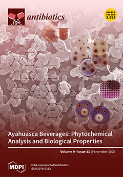
Cover Story (view full-size image): Ayahuasca is a South American beverage commonly prepared from the leaves of the Psychotria viridis shrub and stem scraps from the Banisteriopsis caapi vine or another plants. The resulting brew may contain N,N-dimethyltryptamine (DMT), a powerful psychedelic drug, and harmala alkaloids, which allow making DMT orally active. Ayahuasca is used largely as an aid to spiritual practices, and some recent investigations have pointed to its potential therapeutic effects. This work allowed a more in-depth knowledge of the phytochemical composition of Ayahuasca samples. Their ability to inhibit lipid peroxidation and the capability to scavenge free radicals were demonstrated, indicating antioxidant properties. Anti-inflammatory and antimicrobial activities were also tested. To the best of our knowledge, this is the first study of this kind. View this paper.
  • Issues are regarded as officially published after their release is announced to the table of contents alert mailing list.
  • You may sign up for e-mail alerts to receive table of contents of newly released issues.
  • PDF is the official format for papers published in both, html and pdf forms. To view the papers in pdf format, click on the "PDF Full-text" link, and use the free Adobe Reader to open them.
Order results
Result details
Section
Select all
Export citation of selected articles as:
15 pages, 2165 KiB  
Article
Time Trends and Factors Associated with Antibiotic Prescribing in Swiss Primary Care (2008 to 2020)
by Nahara Anani Martínez-González, Stefania Di Gangi, Giuseppe Pichierri, Stefan Neuner-Jehle, Oliver Senn and Andreas Plate
Antibiotics 2020, 9(11), 837; https://doi.org/10.3390/antibiotics9110837 - 23 Nov 2020
Cited by 19 | Viewed by 3691
Abstract
Antibiotic resistance (ABR) is a major threat to public health, and the majority of antibiotics are prescribed in the outpatient setting, especially in primary care. Monitoring antibiotic consumption is one key measure in containing ABR, but Swiss national surveillance data are limited. We [...] Read more.
Antibiotic resistance (ABR) is a major threat to public health, and the majority of antibiotics are prescribed in the outpatient setting, especially in primary care. Monitoring antibiotic consumption is one key measure in containing ABR, but Swiss national surveillance data are limited. We conducted a retrospective cross-sectional study to characterise the patterns of antibiotic prescriptions, assess the time trends, and identify the factors associated with antibiotic prescribing in Swiss primary care. Using electronic medical records data, we analysed 206,599 antibiotic prescriptions from 112,378 patients. Based on 27,829 patient records, respiratory (52.1%), urinary (27.9%), and skin (4.8%) infections were the commonest clinical indications for antibiotic prescribing. The most frequently prescribed antibiotics were broad-spectrum penicillins (BSP) (36.5%), fluoroquinolones (16.4%), and macrolides/lincosamides (13.8%). Based on the WHO AWaRe classification, antibiotics were 57.9% Core-Access and 41.7% Watch, 69% of which were fluoroquinolones and macrolides. Between 2008 and 2020, fluoroquinolones and macrolides/lincosamides prescriptions significantly declined by 53% and 51%; BSP prescriptions significantly increased by 54%. Increasing patients’ age, volume, and employment level were significantly associated with antibiotic prescribing. Our results may inform future antibiotic stewardship interventions to improve antibiotic prescribing. Full article
(This article belongs to the Special Issue Antibiotics Use in Primary Care)
Show Figures

Figure 1

Figure 1
<p>Event flow chart. Number of patients with antibiotic prescriptions in Swiss primary care from the FIRE database. FIRE, Family Medicine ICPC-Research using EMR; EMR, Electronic Medical Records; ICPC, International Classification of Primary Care (-2) code system. #Date issues: when the date of a first antibiotic prescription coincided with the date of a first consultation; a prescription could have been carried over from a previous consultation prior to the patient’s inclusion in the database.</p>
Full article ">Figure 2
<p>Time trends in antibiotic prescribing in primary care. Points are yearly observed numbers and lines are smoothed curves. (<b>a</b>) Antibiotics with &gt;10,000 prescriptions; (<b>b</b>) Antibiotics with &gt;1000 prescriptions. * This group contains prescriptions from the ATC group J01XB-J01XX. The prescriptions reported in this group consist of &gt;98% nitrofurantoin and fosfomycin prescriptions.</p>
Full article ">Figure 2 Cont.
<p>Time trends in antibiotic prescribing in primary care. Points are yearly observed numbers and lines are smoothed curves. (<b>a</b>) Antibiotics with &gt;10,000 prescriptions; (<b>b</b>) Antibiotics with &gt;1000 prescriptions. * This group contains prescriptions from the ATC group J01XB-J01XX. The prescriptions reported in this group consist of &gt;98% nitrofurantoin and fosfomycin prescriptions.</p>
Full article ">Figure 3
<p>Distribution of antibiotic prescribing in Swiss primary care. Points are observed numbers and lines are smoothed curves; 95% Confidence Interval band is shown in grey. (<b>a</b>) Seasonal variation of antibiotic prescriptions by month: 1 = January, 12 = December; (<b>b</b>) Fluctuation of antibiotic prescriptions over 12 years.</p>
Full article ">Figure 4
<p>Inequality in prescriptions across Swiss general practitioners: Lorenz curve of antibiotic prescriptions in a subgroup of 240 general practitioners (GPs) with 26,595 antibiotic prescriptions issued in 2018. Index of inequality by Gini’s coefficient = 0.31; “0” = perfect equality (straight line) and “1” = total inequality. The further the Lorenz curve deviates from perfect equality, the higher the Gini coefficient and the inequality.</p>
Full article ">
13 pages, 4837 KiB  
Article
Inhibition of Campylobacter jejuni Biofilm Formation by D-Amino Acids
by Bassam A. Elgamoudi, Taha Taha and Victoria Korolik
Antibiotics 2020, 9(11), 836; https://doi.org/10.3390/antibiotics9110836 - 23 Nov 2020
Cited by 20 | Viewed by 3593
Abstract
The ability of bacterial pathogens to form biofilms is an important virulence mechanism in relation to their pathogenesis and transmission. Biofilms play a crucial role in survival in unfavorable environmental conditions, acting as reservoirs of microbial contamination and antibiotic resistance. For intestinal pathogen [...] Read more.
The ability of bacterial pathogens to form biofilms is an important virulence mechanism in relation to their pathogenesis and transmission. Biofilms play a crucial role in survival in unfavorable environmental conditions, acting as reservoirs of microbial contamination and antibiotic resistance. For intestinal pathogen Campylobacter jejuni, biofilms are considered to be a contributing factor in transmission through the food chain and currently, there are no known methods for intervention. Here, we present an unconventional approach to reducing biofilm formation by C. jejuni by the application of D-amino acids (DAs), and L-amino acids (LAs). We found that DAs and not LAs, except L-alanine, reduced biofilm formation by up to 70%. The treatment of C. jejuni cells with DAs changed the biofilm architecture and reduced the appearance of amyloid-like fibrils. In addition, a mixture of DAs enhanced antimicrobial efficacy of D-Cycloserine (DCS) up to 32% as compared with DCS treatment alone. Unexpectedly, D-alanine was able to reverse the inhibitory effect of other DAs as well as that of DCS. Furthermore, L-alanine and D-tryptophan decreased transcript levels of peptidoglycan biosynthesis enzymes alanine racemase (alr) and D-alanine-D-alanine ligase (ddlA) while D-serine was only able to decrease the transcript levels of alr. Our findings suggest that a combination of DAs could reduce biofilm formation, viability and persistence of C. jejuni through dysregulation of alr and ddlA. Full article
Show Figures

Graphical abstract

Graphical abstract
Full article ">Figure 1
<p>Effect of 100 mM D-amino acids (DAs) and L-amino acids (LAs) on <span class="html-italic">C. jejuni</span> 11168-O biofilm. Inhibition of biofilm formation in the presence of 100 mM of; L-alanine (L-ala), D-alanine (D-ala), L-serine (L-ser), D-serine (D-ser), L-methionine (L-met), D-methionine (D-met), L-tryptophan (L-trp), or D-tryptophan (D-trp). All data are mean ± Standard errors and were analyzed using an unpaired, two-tailed Student’s <span class="html-italic">t</span>-test, <span class="html-italic">p</span> &lt; 0.05. The asterisk (*) indicates a statistically significant difference compared to untreated control.</p>
Full article ">Figure 2
<p>Inhibition and dispersion response of <span class="html-italic">C. jejuni</span> 11168-O biofilms in the presence of LAs and DAs at different concentrations. (<b>A</b>) Inhibition of biofilm formation by different concentrations of LAs and DAs, (<b>B</b>) Dispersion of the existing biofilm induced by different concentrations of LAs and DAs. The data is presented as Mean ± Standard errors of Percentage of inhibition (Normalized to untreated control).</p>
Full article ">Figure 3
<p>Quantitative analysis of biofilm inhibition of (<b>A</b>) <span class="html-italic">C. jejuni</span> 11168-O, (<b>B</b><span class="html-italic">) C. jejuni</span> 81-176, and (<b>C</b>) <span class="html-italic">C. coli</span> NCTC 11366 in the presence of 10 mM of DAs. The data is presented as Mean ± Standard errors of Percentage of inhibition (Normalized to untreated control).</p>
Full article ">Figure 4
<p>Effect of the equimolar mixture of DAs and LAs on <span class="html-italic">C. jejuni</span> 11168-O biofilm. All data are mean ± Standard errors and were analyzed using an unpaired, two-tailed Student’s <span class="html-italic">t</span>-test, <span class="html-italic">p</span> &lt; 0.05. The asterisk (*) indicates a statistically significant difference compared to untreated control.</p>
Full article ">Figure 5
<p>The mature biofilm of <span class="html-italic">C. jejuni</span> 11168-O and amyloid-like fibres. <span class="html-italic">C. jejuni</span> biofilm imaged using dual fluorescence labelling by confocal laser scanning microscopy (CLSM). (<b>a</b>–<b>c</b>) Bacterial cells within the biofilm (4′,6-diamidino-2-phenylindole (DAPI), blue) and red arrow indicates for amyloid-like <span class="html-italic">fibrils</span> (Thioflavin T (ThioT), green). (Scale bar = 10 µm).</p>
Full article ">Figure 6
<p>Confocal scanning laser microscopy images of <span class="html-italic">C. jejuni</span> 11168-O biofilm in presence of 25 mM of DAs. <span class="html-italic">C. jejuni</span> biofilm at 48 h, imaged using dual fluorescence labelling by confocal laser scanning microscopy (CLSM). (<b>a</b>) Untreated, (<b>b</b>) D-ala, (<b>c</b>) L-ala, (<b>d</b>) D-ser, (<b>e</b>) D-met, (<b>f</b>) D-trp. Cells were stained with 4′,6-diamidino-2-phenylindole (DAPI, blue) and amyloid fibrils by Thioflavin T (ThioT, green) (Scale bar = 20 µm).</p>
Full article ">Figure 7
<p>Reversal of <span class="html-italic">C. jejuni</span> 11168-O biofilm inhibition by (<b>A</b>) D-Cycloserine (DCS), (<b>B</b>) L-ala and D-ser in presence of <span class="html-small-caps">D</span>-alanine (D-ala). The data is presented as Mean ± Standard errors of Percentage of inhibition (Normalized to untreated control).</p>
Full article ">Figure 7 Cont.
<p>Reversal of <span class="html-italic">C. jejuni</span> 11168-O biofilm inhibition by (<b>A</b>) D-Cycloserine (DCS), (<b>B</b>) L-ala and D-ser in presence of <span class="html-small-caps">D</span>-alanine (D-ala). The data is presented as Mean ± Standard errors of Percentage of inhibition (Normalized to untreated control).</p>
Full article ">Figure 8
<p>Effect of DCS on <span class="html-italic">C. jejuni</span> 11168-O biofilm when combined with a mixture of L-ala, D-ser, D-met, D-trp (5:5:2:5 mM). The data is presented as Mean ± Standard errors of Percentage of inhibition (Normalized to untreated control).</p>
Full article ">
19 pages, 2267 KiB  
Review
Peri-Implant Diseases: Diagnosis, Clinical, Histological, Microbiological Characteristics and Treatment Strategies. A Narrative Review
by Ioannis Kormas, Chantal Pedercini, Alessandro Pedercini, Michail Raptopoulos, Hatem Alassy and Larry F. Wolff
Antibiotics 2020, 9(11), 835; https://doi.org/10.3390/antibiotics9110835 - 22 Nov 2020
Cited by 40 | Viewed by 10864
Abstract
Since the use of dental implants is continuously increasing, it is imperative for dental practitioners to understand the nature and treatment of peri-implant diseases. The purpose of this manuscript is to comprehensively review peri-implant diseases, their characteristics, as well as their non-surgical and [...] Read more.
Since the use of dental implants is continuously increasing, it is imperative for dental practitioners to understand the nature and treatment of peri-implant diseases. The purpose of this manuscript is to comprehensively review peri-implant diseases, their characteristics, as well as their non-surgical and surgical treatment. To that end, the current literature was searched and a narrative review was conducted. It is essential that the case definitions described in the 2017 World Workshop on the Classification of Periodontal and Peri-implant Diseases and Conditions are used to diagnose and classify peri-implant health, peri-implant mucositis and peri-implantitis. While recent epidemiologic studies on peri-implant diseases exist, there is great heterogeneity in the definition of these conditions. Several risk factors and indicators are reported in the literature, with smoking and diabetes being the most universally accepted. In peri-implant mucositis, non-surgical treatment seems to be sufficient. However, for the treatment of peri-implantitis, a surgical approach, which includes open-flap debridement, apically positioned flap and guided bone regeneration, is considered more appropriate. A great variety of adjuncts to mechanical treatment have been reported with controversial results. Finally, studies comparing results from different peri-implantitis treatments are warranted in randomized controlled clinical trials in order to provide stronger evidence-based approaches. Full article
Show Figures

Figure 1

Figure 1
<p>Peri-implant health/disease status: (<b>a</b>) Peri-implant health; (<b>b</b>) Peri-implant mucositis; (<b>c</b>) Peri-implantitis.</p>
Full article ">Figure 2
<p>Risk factors and indicators of peri-implant diseases.</p>
Full article ">Figure 3
<p>Open-flap debridement: (<b>a</b>) Pre-operative; (<b>b</b>) Intra-operative; (<b>c</b>) Flap closure.</p>
Full article ">Figure 4
<p>Apically position flap with implantoplasty: (<b>a</b>) Pre-operative; (<b>b</b>) Intra-operative; (<b>c</b>) Flap closure.</p>
Full article ">Figure 5
<p>Guided bone regeneration: (<b>a</b>) Pre-operative; (<b>b</b>) Intra-operative; (<b>c</b>) Flap closure.</p>
Full article ">
17 pages, 1937 KiB  
Article
Antimicrobial Resistance Patterns in Organic and Conventional Dairy Herds in Sweden
by Karin Sjöström, Rachel A. Hickman, Viktoria Tepper, Gabriela Olmos Antillón, Josef D. Järhult, Ulf Emanuelson, Nils Fall and Susanna Sternberg Lewerin
Antibiotics 2020, 9(11), 834; https://doi.org/10.3390/antibiotics9110834 - 21 Nov 2020
Cited by 12 | Viewed by 3384
Abstract
Monitoring antimicrobial resistance (AMR) and use (AMU) is important for control. We used Escherichia coli from healthy young calves as an indicator to evaluate whether AMR patterns differ between Swedish organic and conventional dairy herds and whether the patterns could be related to [...] Read more.
Monitoring antimicrobial resistance (AMR) and use (AMU) is important for control. We used Escherichia coli from healthy young calves as an indicator to evaluate whether AMR patterns differ between Swedish organic and conventional dairy herds and whether the patterns could be related to AMU data. Samples were taken twice, in 30 organic and 30 conventional dairy herds. Selective culturing for Escherichia coli, without antibiotics and with nalidixic acid or tetracycline, was used to estimate the proportions of resistant isolates. Microdilution was used to determine the minimum inhibitory concentrations (MICs) for thirteen antimicrobial substances. AMU data were based on collection of empty drug packages. Less than 8% of the bacterial growth on non-selective plates was also found on selective plates with tetracycline, and 1% on plates with nalidixic acid. Despite some MIC variations, resistance patterns were largely similar in both periods, and between organic and conventional herds. For most substances, only a few isolates were classified as resistant. The most common resistances were against ampicillin, streptomycin, sulfamethoxazole, and tetracycline. No clear association with AMU could be found. The lack of difference between organic and conventional herds is likely due to a generally good animal health status and consequent low AMU in both categories. Full article
Show Figures

Figure 1

Figure 1
<p>Minimum inhibitory concentration (MIC) distribution of <span class="html-italic">Escherichia coli</span> isolates from calves, sampled in period 1, in 30 (148 isolates) organic (Org) and 30 (145 isolates) conventional (Conv) Swedish dairy herds, in a panel of 13 antimicrobial agents. Blue color indicates the range of concentrations tested. Vertical black lines are the epidemiological cut-off points, according to EUCAST. <span class="html-italic">p</span> values test the difference between organic and conventional herds with Fisher’s exact test. Yellow color denotes isolates from conventional herds and pink color shows MIC values.</p>
Full article ">Figure 2
<p>Minimum inhibitory concentration (MIC) distributions of <span class="html-italic">Escherichia coli</span> isolates from calves, sampled in period 2, in 27 (127 isolates) organic (Org) and 27 (131 isolates) conventional (Conv) Swedish dairy herds, in a panel of 13 antimicrobial agents. Blue color indicates the range of concentrations tested. Vertical black lines are the epidemiologic cut-off points according to EUCAST. <span class="html-italic">p</span> values test the difference between organic and conventional herds with Fisher’s exact test. Yellow color denotes isolates from conventional herds and pink color shows MIC values.</p>
Full article ">Figure 3
<p>Minimum inhibitory concentration (MIC) distributions of <span class="html-italic">Escherichia coli</span> isolates from environmental samples of farm manure (drainage and manure pit) at the beginning of period 2, in 27 (50 isolates) organic (Org) and 27 (53 isolates) conventional (Conv) Swedish dairy herds, in a panel of 13 antimicrobial agents. Blue color indicates the range of concentrations tested. Vertical thicker black lines are the epidemiologic cut-off points according to EUCAST. <span class="html-italic">p</span> values test the difference between organic and conventional herds with Fisher’s exact test. Yellow color denotes isolates from conventional herds and pink color shows MIC values.</p>
Full article ">Figure 4
<p>Number of sampled herds with zero (0), low (1–25%), medium (26–60%) and high (&gt;60%) proportions of <span class="html-italic">Escherichia coli</span> with antimicrobial resistance to any antimicrobial class (AMR) or to three or more antimicrobial classes (MDR) in Swedish organic and conventional dairy herds. Isolates from calf samples in period 1 (per 1) and period 2 (per 2) and environmental samples in period 2 (env).</p>
Full article ">Figure 5
<p>Heatmaps showing the patterns of resistant and susceptible <span class="html-italic">Escherichia coli</span> isolates from Swedish organic and conventional dairy herds sampled in two different time periods. (<b>A</b>) conventional herds 1st sampling (<b>B</b>) conventional herds 2nd sampling (<b>C</b>) organic herds 1st sampling (<b>D</b>) organic herds 2nd sampling. Color scale illustrates average value for each herd and each antimicrobial substance tested, where 1 = resistant and 0 = susceptible. AMX = ampicillin, CAZ = ceftazidime, CHL = chloramphenicol, CIP = ciprofloxacin, CST = colistin, CTX = cefotaxime, FLOR = florfenicol, GEN = gentamycin, NAL = nalidixic acid, STR = streptomycin, SXT = sulfamethoxazole, TET = tetracycline, and TMP = trimethoprim.</p>
Full article ">
17 pages, 5475 KiB  
Review
Can We Exploit β-Lactamases Intrinsic Dynamics for Designing More Effective Inhibitors?
by Eleonora Gianquinto, Donatella Tondi, Giulia D'Arrigo, Loretta Lazzarato and Francesca Spyrakis
Antibiotics 2020, 9(11), 833; https://doi.org/10.3390/antibiotics9110833 - 21 Nov 2020
Cited by 6 | Viewed by 2895
Abstract
β-lactamases (BLs) represent the most frequent cause of antimicrobial resistance in Gram-negative bacteria. Despite the continuous efforts in the development of BL inhibitors (BLIs), new BLs able to hydrolyze the last developed antibiotics rapidly emerge. Moreover, the insurgence rate of effective mutations is [...] Read more.
β-lactamases (BLs) represent the most frequent cause of antimicrobial resistance in Gram-negative bacteria. Despite the continuous efforts in the development of BL inhibitors (BLIs), new BLs able to hydrolyze the last developed antibiotics rapidly emerge. Moreover, the insurgence rate of effective mutations is far higher than the release of BLIs able to counteract them. This results in a shortage of antibiotics that is menacing the effective treating of infectious diseases. The situation is made even worse by the co-expression in bacteria of BLs with different mechanisms and hydrolysis spectra, and by the lack of inhibitors able to hit them all. Differently from other targets, BL flexibility has not been deeply exploited for drug design, possibly because of the small protein size, for their apparent rigidity and their high fold conservation. In this mini-review, we discuss the evidence for BL binding site dynamics being crucial for catalytic efficiency, mutation effect, and for the design of new inhibitors. Then, we report on identified allosteric sites in BLs and on possible allosteric inhibitors, as a strategy to overcome the frequent occurrence of mutations in BLs and the difficulty of competing efficaciously with substrates. Nevertheless, allosteric inhibitors could work synergistically with traditional inhibitors, increasing the chances of restoring bacterial susceptibility towards available antibiotics. Full article
(This article belongs to the Special Issue Novel Targets and Mechanisms in Antimicrobial Drug Discovery)
Show Figures

Graphical abstract

Graphical abstract
Full article ">Figure 1
<p>Alignment of class A BLs in cartoon representation: SHV-1 (gray), TEM-1 (dark yellow), CTX-M-15 (green), KPC-2 (pink) (PDB IDs: 4zam, 1ero, 4hbt, 3rxw, respectively). A close-up on the Ω-loop is reported in the right panel: residues are labeled and shown as capped sticks. Each residue number has been linked to four residue names, one for each aligned BL, with a consistent color code. Residues are numbered according to the standard numbering scheme [<a href="#B28-antibiotics-09-00833" class="html-bibr">28</a>].</p>
Full article ">Figure 2
<p>Flipped-in and flipped-out conformations of Trp105 in KPC-2 (PDB ID: 5ul8). (<b>a</b>) Overall view of KPC-2. (<b>b</b>) Close-up of KPC-2 active site. In the most stable conformation (green), Trp105 side chain is close to Thr216, while in the flipped-in conformation (orange), the indole ring approaches Leu167. Protein is represented in pink cartoon and gray surface, important active site residues are labelled and shown as sticks. Relevant waters are depicted as blue spheres, and labelled: the deacylation water (Wat1) is coordinated by Glu166 and Asn170, while Wat2 occupies the oxyanion hole created by backbone nitrogens of Ser70 and Thr237.</p>
Full article ">Figure 3
<p>Structural comparison among subclass B1 BLs: VIM-2 (yellow, PDB ID: 4nq2), NDM-1 (pink, PDB ID: 3spu), IMP-1(blue, PDB ID: 1ddk). (<b>a</b>) Superposition highlighting loops L3 and L10. Proteins are shown as cartoon, zinc atoms in the active site are represented as spheres. (<b>b</b>) Close-up of loop L3. Relevant residues are highlighted in sticks, the residue numbering follows the general class B BL scheme. Residue labels are aligned for each residue identifier.</p>
Full article ">Figure 4
<p>Allosteric site A1 in SHV-1 (gray, PDB ID: 1shv) and TEM-1 (pink, PDB ID: 1pzo) class A BLs.</p>
Full article ">Figure 5
<p>Allosteric effectors in site A1 of BLs SHV-1 and TEM-1. (<b>a</b>) SHV-1 X-ray structure (PDB IDs: 1shv) with Cymal-6 (green) bound to the cryptic site A1. (<b>b</b>) Superposition among two holo and an apo TEM-1 X-ray structures (PDB IDs: 1pzp (teal), 1pzo (pink), and 1yt4 (light orange), respectively). Proteins are represented in cartoon, while ligands are depicted in bright-colored sticks (Cymal-6, green; FTA, cyan; CBT, magenta). Catalytic Ser70 is highlighted in red as a reference for the position of the active site, while the cryptic site A1 area is approximated by an oval shape. For each X-ray structure in both (<b>a</b>,<b>b</b>), a close-up of ligand binding modes in the cryptic site is reported, with residues labelled with one letter code and depicted in capped sticks. For clarity, water molecules have been omitted. Residues are numbered according to the standard numbering scheme [<a href="#B28-antibiotics-09-00833" class="html-bibr">28</a>].</p>
Full article ">Figure 6
<p>PWP triad in class A BLs. Pro226, Trp229, Pro252 and the catalytic serine are shown in capped sticks. Helix 10 is displayed in cartoon. SHV-1 (gray), TEM-1 (dark yellow), CTX-M-15 (green), KPC-2 (pink) (PDB IDs: 4zam, 1ero, 4hbt, 3rxw, respectively).</p>
Full article ">
10 pages, 1274 KiB  
Article
Sequential Hypertonic-Hypotonic Treatment Enhances Efficacy of Antibiotic against Acinetobacter baumannii Biofilm Communities
by Azeza Falghoush, Haluk Beyenal and Douglas R. Call
Antibiotics 2020, 9(11), 832; https://doi.org/10.3390/antibiotics9110832 - 21 Nov 2020
Cited by 4 | Viewed by 2499
Abstract
Infections with bacterial biofilm communities are highly tolerant of antibiotics. This protection is attributed, in part, to a hydrated extracellular polymeric substance (EPS) that surrounds the bacterial community and that limits antibiotic diffusion. In this study, we evaluated whether it is possible to [...] Read more.
Infections with bacterial biofilm communities are highly tolerant of antibiotics. This protection is attributed, in part, to a hydrated extracellular polymeric substance (EPS) that surrounds the bacterial community and that limits antibiotic diffusion. In this study, we evaluated whether it is possible to dehydrate and then re-hydrate a biofilm as a means to increase antibiotic penetration and efficacy. Acinetobacter baumannii biofilms (24 h) were exposed to hypertonic concentrations of maltodextrin, sucrose or polyethylene glycol (PEG) as the dehydration step. These biofilms were then washed with deionized water containing 10 times the concentration of antibiotics needed to kill these bacteria in broth culture (50 µg/mL tobramycin, 300 µg/mL chloramphenicol, 20 µg/mL ciprofloxacin or 100 µg/mL erythromycin) as the rehydration step. Biofilms were then harvested, and the number of viable cells was determined. Sequential treatment with PEG and tobramycin reduced cell counts 4 to 7 log (p < 0.05) relative to combining PEG and tobramycin in a single treatment, and 3 to 7 log relative to tobramycin treatment alone (p < 0.05). Results were variable for other osmotic compounds and antibiotics depending on the concentrations used, likely related to mass and hydrophobicity. Our findings support future clinical evaluation of sequential regimens of hypertonic and hypotonic solutions to enhance antibiotic efficacy against chronic biofilm infections. Full article
Show Figures

Figure 1

Figure 1
<p>Effects of antibiotic alone, combined (+) or sequential (→) treatment with osmotic compounds maltodextrin (MD), sucrose (S), polyethylene glycol (PEG 3350 or PEG 400) and tobramycin (Tob, 50 µg/mL) against <span class="html-italic">Acinetobacter baumannii</span> pre-formed biofilms. Error bars represent the standard error of the means calculated from three biological replicates. Statistical significance was calculated using one-way ANOVA with paired comparisons (Tukey’s test) for individual antibiotics with or without osmotic agent (* <span class="html-italic">p</span> &lt; 0.05; ns = not significant).</p>
Full article ">Figure 2
<p><span class="html-italic">Acinetobacter baumannii</span> pre-formed biofilms exposed to antibiotic alone, combined (+) or sequential (→) treatment with osmotic compounds maltodextrin (MD), sucrose (S), polyethylene glycol (PEG 3350 or PEG 400) followed by (<b>A</b>) erythromycin (Ery,100 µg/mL) or (<b>B</b>) chloramphenicol (Chl 300 µg/mL). Error bars represent the standard error of the means calculated from three biological replicates.</p>
Full article ">Figure 3
<p>Treatment of <span class="html-italic">Acinetobacter baumannii</span> biofilms with individual, combined (+) or sequential (→) treatment of osmotic compound polyethylene glycol (PEG 400) followed by (<b>A</b>) erythromycin (Ery, 200 µg/mL) or (<b>B</b>) chloramphenicol (Chl 600 µg/mL). Three biological replicates were used to calculate the error bars that represent the standard error of the means (* <span class="html-italic">p</span> &lt; 0.05).</p>
Full article ">Figure 4
<p>Effects of individual combined (+) or sequential (→) treatment with increased concentrations of osmotic compounds (<b>A</b>) maltodextrin (40 or 60 mM) and (<b>B</b>) PEG 3350 Da (25.8 or 105.6 mM), followed by tobramycin (Tob, 50 µg/mL). Error bars represent the standard error of the means calculated from three biological replicates. Statistical significance was calculated using a one-way ANOVA and Tukey’s test for tobramycin with or without the osmotic compound (* <span class="html-italic">p</span> &lt; 0.05).</p>
Full article ">Scheme 1
<p>Represents how hypertonic solution of an osmotic compound dehydrates biofilm. De-ionized (DI) water then draws antibiotic into biofilm.</p>
Full article ">
16 pages, 3299 KiB  
Article
Scorpion-Venom-Derived Antimicrobial Peptide Css54 Exerts Potent Antimicrobial Activity by Disrupting Bacterial Membrane of Zoonotic Bacteria
by Jonggwan Park, Jun Hee Oh, Hee Kyoung Kang, Moon-Chang Choi, Chang Ho Seo and Yoonkyung Park
Antibiotics 2020, 9(11), 831; https://doi.org/10.3390/antibiotics9110831 - 20 Nov 2020
Cited by 13 | Viewed by 3490
Abstract
Antibiotic resistance is an important issue affecting humans and livestock. Antimicrobial peptides are promising alternatives to antibiotics. In this study, the antimicrobial peptide Css54, isolated from the venom of C. suffuses, was found to exhibit antimicrobial activity against bacteria such as Listeria [...] Read more.
Antibiotic resistance is an important issue affecting humans and livestock. Antimicrobial peptides are promising alternatives to antibiotics. In this study, the antimicrobial peptide Css54, isolated from the venom of C. suffuses, was found to exhibit antimicrobial activity against bacteria such as Listeria monocytogenes, Streptococcus suis, Campylobacter jejuni, and Salmonella typhimurium that cause zoonotic diseases. Moreover, the cytotoxicity and hemolytic activity of Css54 was lower than that of melittin isolated from bee venom. Circular dichroism assays showed that Css54 has an α-helix structure in an environment mimicking that of bacterial cell membranes. We examined the effect of Css54 on bacterial membranes using N-phenyl-1-naphthylamine, 3,3′-dipropylthiadicarbbocyanine iodides, SYTOX green, and propidium iodide. Our findings suggest that the Css54 peptide kills bacteria by disrupting the bacterial membrane. Moreover, Css54 exhibited antibiofilm activity against L. monocytogenes. Thus, Css54 may be useful as an alternative to antibiotics in humans and animal husbandry. Full article
Show Figures

Graphical abstract

Graphical abstract
Full article ">Figure 1
<p>Structure analysis of Css54. (<b>A</b>) Helical wheel diagram of Css54 was obtained from the HeliQuest site (<a href="https://heliquest.ipmc.cnrs.fr/cgi-bin/ComputParams.py" target="_blank">https://heliquest.ipmc.cnrs.fr/cgi-bin/ComputParams.py</a>). (<b>B</b>) Three-dimensional structure of Css54 was predicted by PEP-FOLD3 and displayed by PyMOL.</p>
Full article ">Figure 2
<p>Circular dichroism (CD) spectra of Css54 measured in (<b>A</b>) 10 mM sodium phosphate, (<b>B</b>) TFE at 20% and 40%, and (<b>C</b>) SDS at 5 and 10 mM. Peptide concentration was fixed at 40 μM.</p>
Full article ">Figure 3
<p>Hemolysis and cytotoxicity of antimicrobial peptides (AMPs). (<b>A</b>) Hemolytic activities were determined by measuring the release of hemoglobin from sRBCs at an absorbance wavelength of 414 nm. Percentage of hemolysis in sheep RBCs incubated with peptides at increased concentrations. (<b>B</b>) Cytotoxicity assay of AMPs. PK(15) cells isolated from porcine kidney were treated with Css54, and melittin at different concentrations in DMEM supplemented 10% FBS. Cell survival rates were measured by MTT assay.</p>
Full article ">Figure 4
<p>Antimicrobial activity of Css54 in different environments. (<b>A</b>) Effect of different pH values, (<b>B</b>) salt concentrations, and (<b>C</b>) temperatures on antimicrobial activity of Css54. (<b>D</b>) To evaluate thermal stability, Css54 at 1× MIC was incubated for 20, 40, 60, 80, and 100 min and then <span class="html-italic">L. monocytogenes</span> was added to the solution for incubation overnight at 37 °C.</p>
Full article ">Figure 5
<p>Inhibition of <span class="html-italic">L. monocytogenes</span> biofilm formation by Css54. (<b>A</b>) <span class="html-italic">L. monocytogenes</span> (KCTC 3710), (<b>B</b>) <span class="html-italic">L. monocytogenes</span> (KCCM 43155), and (<b>C</b>) <span class="html-italic">L. monocytogenes</span> (KCCM 40307) strains were mixed with AMPs for 24 h. The mass of each biofilm was determined by crystal violet staining. Fluorescence microscopy image of biofilms formed by (<b>D</b>) <span class="html-italic">L. monocytogenes</span> (KCTC 3710), (<b>E</b>) <span class="html-italic">L. monocytogenes</span> (KCCM 43155), and (<b>F</b>) <span class="html-italic">L. monocytogenes</span> (KCCM 40307) strains after treatment with AMPs. Live bacteria were stained with SYTO9 dye and analyzed with an EVOS FL Auto 2 imaging system (Invitrogen).</p>
Full article ">Figure 6
<p>Outer membrane permeability and membrane depolarization assay. (<b>A</b>) <span class="html-italic">S. typhimurium</span> (ATCC 14028) outer membrane permeability induced by peptides. The outer membrane permeability of <span class="html-italic">S. typhimurium</span> induced by Css54 was measured using the fluorescent dye NPN (<span class="html-italic">N</span>-phenyl-1-naphthylamine. (<b>B</b>) <span class="html-italic">S. typhimurium</span> and (<b>C</b>) <span class="html-italic">L. monocytogenes</span> cytoplasmic membrane depolarization assay. Bacterial cytoplasmic membrane depolarization was analyzed using the potential-sensitive dye DiSC<sub>3</sub>(5). Various concentrations of Css54 were added to the bacteria.</p>
Full article ">Figure 7
<p>Analysis of <span class="html-italic">L. monocytogenes</span> and <span class="html-italic">S. typhimurium</span> membrane integrity after treatment with Css54. Css54 at 0.5×, 1×, and 2× MIC were added to (<b>A</b>) <span class="html-italic">S. typhimurium</span> and (<b>B</b>) <span class="html-italic">L. monocytogenes</span>. Fluorescence intensity of SYTOX green was measured at emission and excitation wavelengths of 520 and 485 nm, respectively. Flow cytometry analysis using propidium iodide staining against bacterial membrane integrity. Changes in (<b>C</b>) <span class="html-italic">S. typhimurium</span> and (<b>D</b>) <span class="html-italic">L. monocytogenes</span> after treatment with peptide at 0.5× and 1× MIC for 10 min were measured by flow cytometry using propidium iodide (PI).</p>
Full article ">
30 pages, 4285 KiB  
Review
African Plant-Based Natural Products with Antivirulence Activities to the Rescue of Antibiotics
by Christian Emmanuel Mahavy, Pierre Duez, Mondher ElJaziri and Tsiry Rasamiravaka
Antibiotics 2020, 9(11), 830; https://doi.org/10.3390/antibiotics9110830 - 19 Nov 2020
Cited by 12 | Viewed by 4181
Abstract
The worldwide emergence of antibiotic-resistant bacteria and the thread of widespread superbug infections have led researchers to constantly look for novel effective antimicrobial agents. Within the past two decades, there has been an increase in studies attempting to discover molecules with innovative properties [...] Read more.
The worldwide emergence of antibiotic-resistant bacteria and the thread of widespread superbug infections have led researchers to constantly look for novel effective antimicrobial agents. Within the past two decades, there has been an increase in studies attempting to discover molecules with innovative properties against pathogenic bacteria, notably by disrupting mechanisms of bacterial virulence and/or biofilm formation which are both regulated by the cell-to-cell communication mechanism called ‘quorum sensing’ (QS). Certainly, targeting the virulence of bacteria and their capacity to form biofilms, without affecting their viability, may contribute to reduce their pathogenicity, allowing sufficient time for an immune response to infection and a reduction in the use of antibiotics. African plants, through their huge biodiversity, present a considerable reservoir of secondary metabolites with a very broad spectrum of biological activities, a potential source of natural products targeting such non-microbicidal mechanisms. The present paper aims to provide an overview on two main aspects: (i) succinct presentation of bacterial virulence and biofilm formation as well as their entanglement through QS mechanisms and (ii) detailed reports on African plant extracts and isolated compounds with antivirulence properties against particular pathogenic bacteria. Full article
Show Figures

Figure 1

Figure 1
<p>Schematic representation of bacterial invasion processes (<b>A</b>) <span class="html-italic">P. aeruginosa</span>, (<b>B</b>) <span class="html-italic">E. coli</span>, and (<b>C</b>) <span class="html-italic">S. aureus</span>. Bacterial dissemination is done thanks to active and/or passive motilities (swimming, swarming, twitching, gliding, sliding, and darting). In presence of appropriate surfaces and environment, the invasion is initiated by adhesion steps and microcolony formation which lead to the development of mature biofilms in four major steps (adhesion, microcolonies development, biofilm maturation, and dispersion). Under modulation by QS, the bacterial community deploys an arsenal of virulence factors (pyocyanin, heat labile (LT) and heat stable (ST)-enterotoxin, hemolysins, and leukotoxins) that undermine nearby cells (including immune defense cells) and stimulate the production of exogenous biofilm matrix for protection purposes.</p>
Full article ">Figure 2
<p>Model of antibiotherapy combined with an antivirulence agent. (<b>A</b>) Schematic representation of bacterial invasion processes. (<b>B</b>) Scenario presenting the use of antibiotherapy alone and the difficulty of immune defense cells to clear biofilm-encapsulated bacteria even at high doses of antibiotic (e.g., four to 100-fold MICs [<a href="#B82-antibiotics-09-00830" class="html-bibr">82</a>,<a href="#B83-antibiotics-09-00830" class="html-bibr">83</a>]). (<b>C</b>) Scenario presenting the simultaneous use of antibiotic and antivirulence agents; antibiotics at MICs kill a majority of bacteria gathered inside an unstructured biofilm which can be easily cleared by immune defense cells.</p>
Full article ">
16 pages, 3355 KiB  
Article
Antimicrobial Resistance Risk Assessment Models and Database System for Animal-Derived Pathogens
by Xinxing Li, Buwen Liang, Ding Xu, Congming Wu, Jianping Li and Yongjun Zheng
Antibiotics 2020, 9(11), 829; https://doi.org/10.3390/antibiotics9110829 - 19 Nov 2020
Cited by 7 | Viewed by 3393
Abstract
(1) Background: The high use of antibiotics has made the issue of antimicrobial resistance (AMR) increasingly serious, which poses a substantial threat to the health of animals and humans. However, there remains a certain gap in the AMR system and risk assessment models [...] Read more.
(1) Background: The high use of antibiotics has made the issue of antimicrobial resistance (AMR) increasingly serious, which poses a substantial threat to the health of animals and humans. However, there remains a certain gap in the AMR system and risk assessment models between China and the advanced world level. Therefore, this paper aims to provide advanced means for the monitoring of antibiotic use and AMR data, and take piglets as an example to evaluate the risk and highlight the seriousness of AMR in China. (2) Methods: Based on the principal component analysis method, a drug resistance index model of anti-E. coli drugs was established to evaluate the antibiotic risk status in China. Additionally, based on the second-order Monte Carlo methods, a disease risk assessment model for piglets was established to predict the probability of E. coli disease within 30 days of taking florfenicol. Finally, a browser/server architecture-based visualization database system for animal-derived pathogens was developed. (3) Results: The risk of E. coli in the main area was assessed and Hohhot was the highest risk area in China. Compared with the true disease risk probability of 4.1%, the result of the disease risk assessment model is 7.174%, and the absolute error was 3.074%. Conclusions: Taking E. coli as an example, this paper provides an innovative method for rapid and accurate risk assessment of drug resistance. Additionally, the established system and assessment models have potential value for the monitoring and evaluating AMR, highlight the seriousness of antimicrobial resistance, advocate the prudent use of antibiotics, and ensure the safety of animal-derived foods and human health. Full article
(This article belongs to the Special Issue Multidrug-Resistant Bacteria in Animals)
Show Figures

Figure 1

Figure 1
<p>The framework of the second-order Monte Carlo model.</p>
Full article ">Figure 2
<p>The overall design scheme of the system.</p>
Full article ">Figure 3
<p>The framework of the system.</p>
Full article ">Figure 4
<p>Data collection interface.</p>
Full article ">Figure 5
<p>Multiple drugs query (sample data).</p>
Full article ">Figure 6
<p>Drug resistance spectrum query (sample data).</p>
Full article ">Figure 7
<p>Visual analysis of the drug resistance index (DRI) using doxycycline as an example.</p>
Full article ">
8 pages, 216 KiB  
Communication
Impact of Tigecycline’s MIC in the Outcome of Critically Ill Patients with Carbapenemase-Producing Klebsiella pneumoniae Bacteraemia Treated with Tigecycline Monotherapy—Validation of 2019′s EUCAST Proposed Breakpoint Changes
by Matthaios Papadimitriou-Olivgeris, Christina Bartzavali, Alexandra Nikolopoulou, Fevronia Kolonitsiou, Virginia Mplani, Iris Spiliopoulou, Myrto Christofidou, Fotini Fligou and Markos Marangos
Antibiotics 2020, 9(11), 828; https://doi.org/10.3390/antibiotics9110828 - 19 Nov 2020
Cited by 4 | Viewed by 2959
Abstract
Background: Tigecycline is a therapeutic option for carbapenemase-producing Klebsiella pneumoniae (CP-Kp). Our aim was to evaluate the impact of the tigecycline’s minimum inhibitory concentration (MIC) in the outcome of patients with CP-Kp bacteraemia treated with tigecycline monotherapy. Methods: Patients with monomicrobial bacteraemia due [...] Read more.
Background: Tigecycline is a therapeutic option for carbapenemase-producing Klebsiella pneumoniae (CP-Kp). Our aim was to evaluate the impact of the tigecycline’s minimum inhibitory concentration (MIC) in the outcome of patients with CP-Kp bacteraemia treated with tigecycline monotherapy. Methods: Patients with monomicrobial bacteraemia due to CP-Kp that received appropriate targeted monotherapy or no appropriate treatment were included. Primary outcome was 30-day mortality. MICs of meropenem, tigecycline, and ceftazidime/avibactam were determined by Etest, whereas for colistin, the broth microdilution method was applied. PCR for blaKPC, blaVIM, blaNDM, and blaOXA genes was applied. Results: Among 302 CP-Kp bacteraemias, 32 isolates (10.6%) showed MICs of tigecycline ≤ 0.5 mg/L, whereas 177 (58.6%) showed MICs that were 0.75–2 mg/L. Colistin and aminoglycoside susceptibility was observed in 43.0% and 23.8% of isolates, respectively. The majority of isolates carried blaKPC (249; 82.5%), followed by blaVIM (26; 8.6%), both blaKPC and blaVIM (16; 5.3%), and blaNDM (11; 3.6%). Fifteen patients with tigecycline MIC ≤ 0.5 mg/L and 55 with MIC 0.75–2 mg/L were treated with tigecycline monotherapy; 30-day mortality was 20.0% and 50.9%, respectively (p = 0.042). Mortality of 150 patients that received other antimicrobials was 24.7%; among 82 patients that received no appropriate treatment, mortality was 39.0%. No difference in 30-day mortality was observed between patients that received tigecycline (MIC ≤ 0.5 mg/L) or other antimicrobials. Conclusion: Tigecycline monotherapy was as efficacious as other antimicrobials in the treatment of bloodstream infections due to CP-Kp isolates with a tigecycline’s MIC ≤ 0.5 mg/L. Full article
(This article belongs to the Special Issue Antimicrobial Resistance: The Final Frontier)
7 pages, 216 KiB  
Perspective
Phage Therapy: Towards a Successful Clinical Trial
by Andrzej Górski, Jan Borysowski and Ryszard Międzybrodzki
Antibiotics 2020, 9(11), 827; https://doi.org/10.3390/antibiotics9110827 - 19 Nov 2020
Cited by 68 | Viewed by 6894
Abstract
While phage therapy carried out as compassionate use (experimental therapy) has recently flourished, providing numerous case reports of supposedly healed patients, clinical trials aiming to formally prove their value in accord with current regulatory requirements have failed. In light of the current issue [...] Read more.
While phage therapy carried out as compassionate use (experimental therapy) has recently flourished, providing numerous case reports of supposedly healed patients, clinical trials aiming to formally prove their value in accord with current regulatory requirements have failed. In light of the current issue of increasing antibiotic resistance, the need for a final say regarding the place of phage therapy in modern medicine is evident. We analyze the possible factors that may favor success or lead to the failure of phage therapy: quality of phage preparations, their titer and dosage, as well as external factors that could also contribute to the outcome of phage therapy. Hopefully, better control of these factors may eventually bring about long-awaited positive results. Full article
17 pages, 977 KiB  
Article
Plasmid-Mediated Ampicillin, Quinolone, and Heavy Metal Co-Resistance among ESBL-Producing Isolates from the Yamuna River, New Delhi, India
by Mohammad Tahir Siddiqui, Aftab Hossain Mondal, Firdoos Ahmad Gogry, Fohad Mabood Husain, Ali Alsalme and Qazi Mohd. Rizwanul Haq
Antibiotics 2020, 9(11), 826; https://doi.org/10.3390/antibiotics9110826 - 19 Nov 2020
Cited by 25 | Viewed by 3235
Abstract
Antibiotic resistance is one of the major current global health crises. Because of increasing contamination with antimicrobials, pesticides, and heavy metals, the aquatic environment has become a hotspot for emergence, maintenance, and dissemination of antibiotic and heavy metal resistance genes among bacteria. The [...] Read more.
Antibiotic resistance is one of the major current global health crises. Because of increasing contamination with antimicrobials, pesticides, and heavy metals, the aquatic environment has become a hotspot for emergence, maintenance, and dissemination of antibiotic and heavy metal resistance genes among bacteria. The aim of the present study was to determine the co-resistance to quinolones, ampicillin, and heavy metals among the bacterial isolates harboring extended-spectrum β-lactamases (ESBLs) genes. Among 73 bacterial strains isolated from a highly polluted stretch of the Yamuna River in Delhi, those carrying blaCTX-M, blaTEM, or blaSHV genes were analyzed to detect the genetic determinants of resistance to quinolones, ampicillin, mercury, and arsenic. The plasmid-mediated quinolone resistance (PMQR) gene qnrS was found in 22 isolates; however, the qnrA, B, C, and qnrD genes could not be detected in any of the bacteria. Two variants of CMY, blaCMY-2 and blaCMY-42, were identified among eight and seven strains, respectively. Furthermore, merB, merP, merT, and arsC genes were detected in 40, 40, 44, and 24 bacterial strains, respectively. Co-transfer of different resistance genes was also investigated in a transconjugation experiment. Successful transconjugants had antibiotic and heavy metal resistance genes with similar tolerance toward antibiotics and heavy metals as did their donors. This study indicates that the aquatic environment is a major reservoir of bacteria harboring resistance genes to antibiotics and heavy metals and emphasizes the need to study the genetic basis of resistant microorganisms and their public health implications. Full article
(This article belongs to the Section Mechanism and Evolution of Antibiotic Resistance)
Show Figures

Figure 1

Figure 1
<p>Antibiotic susceptibility pattern of extended-spectrum β-lactamases (ESBL) producing bacterial strains used in this study. Note 1: AMP, ampicillin; CAZ, ceftazidime; CTX, cefotaxime; CZ, cefazolin; CTR, ceftriaxone; AT, aztreonam; CX, cefoxitin; TR, trimethoprim; P/T-piperacillin/tazobactam, PB, polymyxin B; CIP, ciprofloxacin; A/S, ampicillin/sulbactam; LE, levofloxacin; AK, amikacin; TE, tetracycline; C, chloramphenicol; IPM-imipenem. Note 2: “Resistant”—Bacteria which could resist the effect of prescribed dosage concentration of antibiotic. “Intermediate”—Bacteria on its course to be resistant to prescribed concentration of antibiotic. “Sensitive”—Bacteria, which could not resist the prescribed dosage concentration of antibiotic.</p>
Full article ">Figure 2
<p>Percentage of bacterial strains harboring different resistance genes.</p>
Full article ">
8 pages, 987 KiB  
Brief Report
Absence of mgrB Alleviates Negative Growth Effects of Colistin Resistance in Enterobacter cloacae
by Jessie E. Wozniak, Aroon T. Chande, Eileen M. Burd, Victor I. Band, Sarah W. Satola, Monica M. Farley, Jesse T. Jacob, I. King Jordan and David S. Weiss
Antibiotics 2020, 9(11), 825; https://doi.org/10.3390/antibiotics9110825 - 19 Nov 2020
Cited by 4 | Viewed by 2171
Abstract
Colistin is an important last-line antibiotic to treat highly resistant Enterobacter infections. Resistance to colistin has emerged among clinical isolates but has been associated with a significant growth defect. Here, we describe a clinical Enterobacter isolate with a deletion of mgrB, a [...] Read more.
Colistin is an important last-line antibiotic to treat highly resistant Enterobacter infections. Resistance to colistin has emerged among clinical isolates but has been associated with a significant growth defect. Here, we describe a clinical Enterobacter isolate with a deletion of mgrB, a regulator of colistin resistance, leading to high-level resistance in the absence of a growth defect. The identification of a path to resistance unrestrained by growth defects suggests colistin resistance could become more common in Enterobacter. Full article
Show Figures

Figure 1

Figure 1
<p>Mu471 exhibits high-level colistin resistance in the absence of a growth delay. (<b>A</b>) Etest of Mu471. (<b>B</b>) Etest of Mu471 with pBAV P<span class="html-italic">mgrB</span>. Agar contains kanamycin (90 μg/mL) for plasmid retention. (<b>C</b>) Growth curves of Mu471 with pBAV P<span class="html-italic">mgrB</span> and empty vector control. Growth curves were performed in Mueller–Hinton broth with kanamycin (90 μg/mL) for plasmid retention.</p>
Full article ">Figure 2
<p>Deletion of <span class="html-italic">mgrB</span> causes high-level colistin resistance in the absence of a growth delay. (<b>A</b>) Etest of ATCC13047. (<b>B</b>) Etest of ATCC13047Δ<span class="html-italic">mgrB</span>. (<b>C</b>) Growth curve of ATCC13047 and ATCC13047 Δ<span class="html-italic">mgrB</span>. Growth curves were performed in Mueller–Hinton broth.</p>
Full article ">
14 pages, 1658 KiB  
Article
Potential Inhibitory Effect of Apis mellifera’s Venom and of Its Two Main Components—Melittin and PLA2—on Escherichia coli F1F0-ATPase
by Hala Nehme, Helena Ayde, Dany El Obeid, Jean Marc Sabatier and Ziad Fajloun
Antibiotics 2020, 9(11), 824; https://doi.org/10.3390/antibiotics9110824 - 18 Nov 2020
Cited by 10 | Viewed by 3123
Abstract
Bacterial resistance has become a worrying problem for human health, especially since certain bacterial strains of Escherichia coli (E. coli) can cause very serious infections. Thus, the search for novel natural inhibitors with new bacterial targets would be crucial to overcome [...] Read more.
Bacterial resistance has become a worrying problem for human health, especially since certain bacterial strains of Escherichia coli (E. coli) can cause very serious infections. Thus, the search for novel natural inhibitors with new bacterial targets would be crucial to overcome resistance to antibiotics. Here, we evaluate the inhibitory effects of Apis mellifera bee venom (BV-Am) and of its two main components -melittin and phospholipase A2 (PLA2)- on E. coli F1F0-ATPase enzyme, a crucial molecular target for the survival of these bacteria. Thus, we optimized a spectrophotometric method to evaluate the enzymatic activity by quantifying the released phosphate from ATP hydrolysis catalyzed by E. coli F1F0-ATPase. The protocol developed for inhibition assays of this enzyme was validated by two reference inhibitors, thymoquinone (IC50 = 57.5 μM) and quercetin (IC50 = 30 μM). Results showed that BV-Am has a dose-dependent inhibitory effect on E. coli F1F0-ATPase with 50% inhibition at 18.43 ± 0.92 μg/mL. Melittin inhibits this enzyme with IC50 = 9.03 ± 0.27 µM, emphasizing a more inhibitory effect than the two previous reference inhibitors adopted. Likewise, PLA2 inhibits E. coli F1F0-ATPase with a dose-dependent effect (50% inhibition at 2.11 ± 0.11 μg/mL) and its combination with melittin enhanced the inhibition extent of this enzyme. Crude venom and mainly melittin and PLA2, inhibit E. coli F1F0-ATPase and could be considered as important candidates for combating resistant bacteria. Full article
(This article belongs to the Special Issue Peptide Antibiotics from Microbes and Venomous Animals)
Show Figures

Graphical abstract

Graphical abstract
Full article ">Figure 1
<p>(<b>a</b>) Optimization of H<sub>2</sub>SO<sub>4</sub> concentration for the P<sub>i</sub> dosage. Standard P<sub>i</sub> solutions of 5 µM and 50 µM prepared in tris-HCl buffer (pH = 8.5; 50 mM). Incubation time: 10 min. λ = 700 nm. Error bars show the standard deviation obtained from experiments done in triplicate. (<b>b</b>) Spectra showing the OD of standard solutions of P<sub>i</sub>, quercetin and a mixture of P<sub>i</sub> and quercetin. P<sub>i</sub> = 5 µM; Quercetin = 30 µM.</p>
Full article ">Figure 2
<p>(<b>a</b>) Dose-response curve representing the enzymatic activity (%) of <span class="html-italic">E. coli</span> F<sub>1</sub>F<sub>0</sub>-ATPase as a function of the log of the concentration of thymoquinone, quercetin and melittin, respectively. Error bars show the standard deviation obtained from experiments done in triplicate; (<b>b</b>) Comparison between the IC<sub>50</sub> values of the reference inhibitors obtained by the developed method and from the literature.</p>
Full article ">Figure 3
<p>Curve showing the enzymatic activity (%) of the BV-<span class="html-italic">Am</span> venom on the membrane-bound <span class="html-italic">E. coli</span> F<sub>1</sub>F<sub>0</sub>-ATPase. Error bars show the standard deviation obtained from experiments done in triplicate.</p>
Full article ">Figure 4
<p>(<b>a</b>) Curve showing the effect of different concentrations of PLA<sub>2</sub> on the membrane-bound <span class="html-italic">E. coli</span> F<sub>1</sub>F<sub>0</sub>-ATPase. Error bars show the standard deviation obtained from experiments done in triplicate. Incubation time: 10 min; (<b>b</b>) Graph showing the activity of membrane-bound <span class="html-italic">E. coli</span> F<sub>1</sub>F<sub>0</sub>-ATPase as a function of different pre-incubation times of the enzyme in presence of the PLA<sub>2</sub> before the addition of ATP. (ATP) = 100 µM. (PLA<sub>2</sub>) = 1.5 µg/mL. Error bars show the standard deviation obtained from experiments done in triplicate.</p>
Full article ">Figure 5
<p>Lineweaver-Burk representation. The points of intersection with the <span class="html-italic">x</span>-axis of the linear regressions obtained without and with melittin correspond to −1/K<sub>m</sub> and −1/K′<sub>m</sub>, respectively while the points of intersection of these lines with the y-axes correspond to 1/V<sub>max</sub> and 1/V′<sub>max</sub>, respectively. Error bars show the standard deviation obtained from experiments done in triplicate.</p>
Full article ">Figure 6
<p>Synergistic action of melittin and PLA<sub>2</sub> on the membrane <span class="html-italic">E. coli</span> F<sub>1</sub>F<sub>0</sub>-ATPase. Reference shows maximal enzymatic activity (100%) obtained for an enzymatic reaction in optimal conditions and in the absence of inhibitor. (ATP) = 100 µM, (Melittin) = 9 µM, and (PLA<sub>2</sub>) = 2.5 µg/mL. Error bars show the standard deviation obtained from experiments done in triplicate.</p>
Full article ">
11 pages, 1638 KiB  
Article
Antimicrobial Susceptibility Patterns and Wild-Type MIC Distributions of Anaerobic Bacteria at a German University Hospital: A Five-Year Retrospective Study (2015–2019)
by Mohamed Tarek Badr, Benjamin Blümel, Sandra Baumgartner, Johanna M. A. Komp and Georg Häcker
Antibiotics 2020, 9(11), 823; https://doi.org/10.3390/antibiotics9110823 - 18 Nov 2020
Cited by 7 | Viewed by 3533
Abstract
Local antimicrobial susceptibility surveys are crucial for optimal empirical therapy guidelines and for aiding in antibiotic stewardship and treatment decisions. For many laboratories, a comprehensive overview of local antimicrobial susceptibility patterns of anaerobic bacteria is still lacking due to the long incubation time [...] Read more.
Local antimicrobial susceptibility surveys are crucial for optimal empirical therapy guidelines and for aiding in antibiotic stewardship and treatment decisions. For many laboratories, a comprehensive overview of local antimicrobial susceptibility patterns of anaerobic bacteria is still lacking due to the long incubation time and effort involved. The present study investigates the antimicrobial susceptibility patterns and related clinical and demographic data of 2856 clinical isolates of anaerobic bacteria that were submitted for analysis to the Institute for Medical Microbiology and Hygiene of the Freiburg University Medical Center (a tertiary university medical center in Southern Germany) between 2015 and 2019. Antimicrobial susceptibility testing has been carried out according to the European Committee on Antimicrobial Susceptibility Testing (EUCAST) guideline. Minimum inhibitory concentration (MIC)50 and MIC90 for penicillin, metronidazole, moxifloxacin, and clindamycin were established for Gram-positive anaerobes and for ampicillin-sulbactam, meropenem, metronidazole, moxifloxacin, and clindamycin for Gram-negative anaerobes. The distribution of MIC-values for various antibiotics against anaerobic bacteria was also established, especially for those having no specific breakpoints according to EUCAST guidelines. Most clinically relevant anaerobic bacteria originated from general surgery, neurological, and orthopedic wards. A high proportion of isolates were resistant to moxifloxacin and clindamycin indicating the importance of their susceptibility testing before administration. Based on our study metronidazole and other β-lactam/β-lactamase inhibitor combinations such as ampicillin-sulbactam remain suitable for empirical treatment of infections with anaerobic bacteria. Full article
(This article belongs to the Section Antibiotics Use and Antimicrobial Stewardship)
Show Figures

Figure 1

Figure 1
<p>Heatmap of the isolate number of anaerobic bacteria distributed across (<b>a</b>) wards or (<b>b</b>) samples. The color represents normalized Z-score value where higher scores indicate higher number of isolates.</p>
Full article ">Figure 2
<p>Percentage of susceptibility of selected antibiotics against tested (<b>A</b>) Gram-positive and (<b>B</b>) Gram-negative anaerobes.</p>
Full article ">Figure 3
<p>Estimation of wild-type minimum inhibitory concentration (MIC) distributions and ECOFFs for rifampicin against <span class="html-italic">Cutibacterium.</span></p>
Full article ">Figure 4
<p>Estimation of wild-type MIC distributions for moxifloxacin against (<b>A</b>) Gram-positive and (<b>B</b>) Gram-negative bacteria.</p>
Full article ">
9 pages, 511 KiB  
Article
Impact of a Rapid Diagnostic Meningitis/Encephalitis Panel on Antimicrobial Use and Clinical Outcomes in Children
by Danielle McDonald, Christina Gagliardo, Stephanie Chiu and M. Cecilia Di Pentima
Antibiotics 2020, 9(11), 822; https://doi.org/10.3390/antibiotics9110822 - 18 Nov 2020
Cited by 10 | Viewed by 2780
Abstract
Rapid molecular diagnostic assays are increasingly used to guide effective antimicrobial therapy. Data on their effectiveness to decrease antimicrobial use in children have been limited and varied. We aimed to assess the impact of the implementation of the FilmArray Meningitis Encephalitis Panel (MEP) [...] Read more.
Rapid molecular diagnostic assays are increasingly used to guide effective antimicrobial therapy. Data on their effectiveness to decrease antimicrobial use in children have been limited and varied. We aimed to assess the impact of the implementation of the FilmArray Meningitis Encephalitis Panel (MEP) on antimicrobial use and outcomes in children. In an observational retrospective study performed at Atlantic Health System (NJ), we sought to evaluate the duration of intravenous antibiotic treatment (days of therapy (DoT)) for patients <21 years of age hospitalized and evaluated for presumptive meningitis or encephalitis before and after the introduction of the MEP. A secondary analysis was performed to determine if recovery of a respiratory pathogen influenced DoT. The median duration of antibiotic therapy prior to the implementation of the MEP was 5 DoT (interquartile range (IQR): 3–6) versus 3 DoT (IQR: 1–5) (p < 0.001) when MEP was performed. The impact was greatest on intravenous third-generation cephalosporin and ampicillin use. We found a reduction in the number of inpatient days associated with the MEP. In the regression analysis, a positive respiratory pathogen panel (RPP) was not a significant predictor of DoT (p = 0.08). Furthermore, we found no significant difference between DoT among patients with negative and positive RPP (p = 0.12). Our study supports the implementation of rapid diagnostics to decrease the utilization of antibiotic therapy among pediatric patients admitted with concerns related to meningitis or encephalitis. Full article
(This article belongs to the Special Issue Antimicrobial Prescribing and Stewardship, 1st Volume)
Show Figures

Figure 1

Figure 1
<p>Patient-specific empiric antimicrobial utilization.</p>
Full article ">
13 pages, 803 KiB  
Review
Knowledge, Attitudes and Perceptions of Medical Students on Antimicrobial Stewardship
by Panagiotis Efthymiou, Despoina Gkentzi and Gabriel Dimitriou
Antibiotics 2020, 9(11), 821; https://doi.org/10.3390/antibiotics9110821 - 17 Nov 2020
Cited by 27 | Viewed by 4805
Abstract
Antimicrobial Resistance (AMR) is an ongoing threat to modern medicine throughout the world. The World Health Organisation has emphasized the importance of adequate and effective training of medical students in wise prescribing of antibiotics Furthermore, Antimicrobial Stewardship (AMS) has been recognized as a [...] Read more.
Antimicrobial Resistance (AMR) is an ongoing threat to modern medicine throughout the world. The World Health Organisation has emphasized the importance of adequate and effective training of medical students in wise prescribing of antibiotics Furthermore, Antimicrobial Stewardship (AMS) has been recognized as a rapidly growing field in medicine that sets a goal of rational use of antibiotics in terms of dosing, duration of therapy and route of administration. We undertook the current review to systematically summarize and present the published data on the knowledge, attitudes and perceptions of medical students on AMS. We reviewed all studies published in English from 2007 to 2020. We found that although medical students recognize the problem of AMR, they lack basic knowledge regarding AMR. Incorporating novel and effective training methods on all aspects of AMS and AMR in the Medical Curricula worldwide is of paramount importance. Full article
(This article belongs to the Special Issue Antimicrobial Prescribing and Stewardship, 1st Volume)
Show Figures

Figure 1

Figure 1
<p>Flowchart of methodology and included studies.</p>
Full article ">
19 pages, 2524 KiB  
Article
Extended Spectrum Beta-Lactamase-Resistant Determinants among Carbapenem-Resistant Enterobacteriaceae from Beef Cattle in the North West Province, South Africa: A Critical Assessment of Their Possible Public Health Implications
by Lungisile Tshitshi, Madira Coutlyne Manganyi, Peter Kotsoana Montso, Moses Mbewe and Collins Njie Ateba
Antibiotics 2020, 9(11), 820; https://doi.org/10.3390/antibiotics9110820 - 17 Nov 2020
Cited by 19 | Viewed by 3686
Abstract
Carbapenems are considered to be the last resort antibiotics for the treatment of infections caused by extended-spectrum beta-lactamase (ESBL)-producing strains. The purpose of this study was to assess antimicrobial resistance profile of Carbapenem-resistant Enterobacteriaceae (CRE) isolated from cattle faeces and determine the presence [...] Read more.
Carbapenems are considered to be the last resort antibiotics for the treatment of infections caused by extended-spectrum beta-lactamase (ESBL)-producing strains. The purpose of this study was to assess antimicrobial resistance profile of Carbapenem-resistant Enterobacteriaceae (CRE) isolated from cattle faeces and determine the presence of carbapenemase and ESBL encoding genes. A total of 233 faecal samples were collected from cattle and analysed for the presence of CRE. The CRE isolates revealed resistance phenotypes against imipenem (42%), ertapenem (35%), doripenem (30%), meropenem (28%), cefotaxime, (59.6%) aztreonam (54.3%) and cefuroxime (47.7%). Multidrug resistance phenotypes ranged from 1.4 to 27% while multi antibiotic resistance (MAR) index value ranged from 0.23 to 0.69, with an average of 0.40. Escherichia coli (E. coli), Klebsiella pneumoniae (K. pneumoniae), Proteus mirabilis (P. mirabilis) and Salmonella (34.4, 43.7, 1.3 and 4.6%, respectively) were the most frequented detected species through genus specific PCR analysis. Detection of genes encoding carbapenemase ranged from 3.3% to 35% (blaKPC, blaNDM, blaGES, blaOXA-48, blaVIM and blaOXA-23). Furthermore, CRE isolates harboured ESBL genes (blaSHV (33.1%), blaTEM (22.5%), blaCTX-M (20.5%) and blaOXA (11.3%)). In conclusion, these findings indicate that cattle harbour CRE carrying ESBL determinants and thus, proper hygiene measures must be enforced to mitigate the spread of CRE strains to food products. Full article
(This article belongs to the Special Issue Carbapenemase-Producing Enterobacterales)
Show Figures

Figure 1

Figure 1
<p>Antimicrobial susceptibility profile of <span class="html-italic">Enterobacteriaceae</span> isolated from cattle faeces.</p>
Full article ">Figure 2
<p>Antimicrobial susceptibility pattern of carbapenem-resistant <span class="html-italic">Enterobacteriaceae</span>. A = Amoxicillin, AMC = Amoxicillin-clavulanate, ATM = Aztreonam, CPM = Cefepime, CTX = Cefotaxime, FOX = Cefoxitin, CAZ = Ceftazidime, CXM = Cefuroxime, EFT = Ceftiofur, KF = Cephalothin, CIP = Ciprofloxacin, PRL = Piperacillin and TC = Ticarcillin.</p>
Full article ">Figure 3
<p>Frequency of multidrug resistance pattern in CRE isolated from cattle faeces.</p>
Full article ">Figure 4
<p>Mean frequency of multiple antibiotic resistance profiles of CRE strains.</p>
Full article ">Figure 5
<p>Number of carbapenemase-encoding genes detected in CRE isolated from cattle faeces.</p>
Full article ">Figure 6
<p>Frequency of ESBL-encoding genes detected in CRE isolated from cattle faeces.</p>
Full article ">
14 pages, 3741 KiB  
Article
Pangenome Analysis of Mycobacterium tuberculosis Reveals Core-Drug Targets and Screening of Promising Lead Compounds for Drug Discovery
by Hamza Arshad Dar, Tahreem Zaheer, Nimat Ullah, Syeda Marriam Bakhtiar, Tianyu Zhang, Muhammad Yasir, Esam I. Azhar and Amjad Ali
Antibiotics 2020, 9(11), 819; https://doi.org/10.3390/antibiotics9110819 - 17 Nov 2020
Cited by 16 | Viewed by 4331
Abstract
Tuberculosis, caused by Mycobacterium tuberculosis (M. tuberculosis), is one of the leading causes of human deaths globally according to the WHO TB 2019 report. The continuous rise in multi- and extensive-drug resistance in M. tuberculosis broadens the challenges to control tuberculosis. [...] Read more.
Tuberculosis, caused by Mycobacterium tuberculosis (M. tuberculosis), is one of the leading causes of human deaths globally according to the WHO TB 2019 report. The continuous rise in multi- and extensive-drug resistance in M. tuberculosis broadens the challenges to control tuberculosis. The availability of a large number of completely sequenced genomes of M. tuberculosis has provided an opportunity to explore the pangenome of the species along with the pan-phylogeny and to identify potential novel drug targets leading to drug discovery. We attempt to calculate the pangenome of M. tuberculosis that comprises a total of 150 complete genomes and performed the phylo-genomic classification and analysis. Further, the conserved core genome (1251 proteins) is subjected to various sequential filters (non-human homology, essentiality, virulence, physicochemical parameters, and pathway analysis) resulted in identification of eight putative broad-spectrum drug targets. Upon molecular docking analyses of these targets with ligands available at the DrugBank database shortlisted a total of five promising ligands with projected inhibitory potential; namely, 2′deoxy-thymidine-5′-diphospho-alpha-d-glucose, uridine diphosphate glucose, 2′-deoxy-thymidine-beta-l-rhamnose, thymidine-5′-triphosphate, and citicoline. We are confident that with further lead optimization and experimental validation, these lead compounds may provide a sound basis to develop safe and effective drugs against tuberculosis disease in humans. Full article
Show Figures

Figure 1

Figure 1
<p>Pan-core plot of 150 <span class="html-italic">Mycobacterium tuberculosis</span> genomes. X-axis shows the number of genomes while the y-axis represents the number of gene families. With the addition of every genome, the pangenome size increased while the core genome size declined. The pangenome curve (shown in brown) has almost flattened, i.e., reached plateau. This suggests that the global gene repertoire of this species is unlikely to change significantly in the future and the pangenome is almost closed.</p>
Full article ">Figure 2
<p>Pan-phylogeny tree of 150 complete genomes of <span class="html-italic">Mycobacterium tuberculosis</span>. Colored strips show the continental origin of strains. Overall, the strains originating from different geographical regions of the World clustered into different clades. This pan-phylogeny has been constructed based on accessory gene presence/absence data in different strains.</p>
Full article ">Figure 3
<p>The docked complex of 2′deoxy-thymidine-5′-diphospho-alpha-<span class="html-small-caps">d</span>-glucose and glucose-1-phosphate thymidylyltransferase.</p>
Full article ">Figure 4
<p>The docked complex of uridine diphosphate glucose and glucose-1-phosphate thymidylyltransferase.</p>
Full article ">Figure 5
<p>The docked complex of 2′-deoxy-thymidine-beta-<span class="html-small-caps">l</span>-rhamnose and glucose-1-phosphate thymidylyltransferase.</p>
Full article ">Figure 6
<p>The docked complex of thymidine-5′-triphosphate and glucose-1-phosphate thymidylyltransferase.</p>
Full article ">Figure 7
<p>The docked complex of citicoline and glucose-1-phosphate thymidylyltransferase.</p>
Full article ">Figure 8
<p>The methodology adopted in the study to find lead compounds (drugs) against <span class="html-italic">Mycobacterium tuberculosis</span>.</p>
Full article ">
14 pages, 1469 KiB  
Article
Increased Intraspecies Diversity in Escherichia coli Biofilms Promotes Cellular Growth at the Expense of Matrix Production
by Andreia S. Azevedo, Gislaine P. Gerola, João Baptista, Carina Almeida, Joana Peres, Filipe J. Mergulhão and Nuno F. Azevedo
Antibiotics 2020, 9(11), 818; https://doi.org/10.3390/antibiotics9110818 - 17 Nov 2020
Cited by 7 | Viewed by 2268
Abstract
Intraspecies diversity in biofilm communities is associated with enhanced survival and growth of the individual biofilm populations. Studies on the subject are scarce, namely, when more than three strains are present. Hence, in this study, the influence of intraspecies diversity in biofilm populations [...] Read more.
Intraspecies diversity in biofilm communities is associated with enhanced survival and growth of the individual biofilm populations. Studies on the subject are scarce, namely, when more than three strains are present. Hence, in this study, the influence of intraspecies diversity in biofilm populations composed of up to six different Escherichia coli strains isolated from urine was evaluated in conditions mimicking the ones observed in urinary tract infections and catheter-associated urinary tract infections. In general, with the increasing number of strains in a biofilm, an increase in cell cultivability and a decrease in matrix production were observed. For instance, single-strain biofilms produced an average of 73.1 µg·cm−2 of extracellular polymeric substances (EPS), while six strains biofilms produced 19.9 µg·cm−2. Hence, it appears that increased genotypic diversity in a biofilm leads E. coli to direct energy towards the production of its offspring, in detriment of the production of public goods (i.e., matrix components). Apart from ecological implications, these results can be explored as another strategy to reduce the biofilm burden, as a decrease in EPS matrix production may render these intraspecies biofilms more sensitive to antimicrobial agents. Full article
(This article belongs to the Special Issue New Insights on Biofilm Antimicrobial Strategies)
Show Figures

Graphical abstract

Graphical abstract
Full article ">Figure 1
<p>Biofilm formation profiles exhibited by the combination of <span class="html-italic">Escherichia coli</span> strains during 48 h, at 37 °C in AUM (artificial urine medium). Example of calculation of the area under the curve for cultivability (<b>a</b>) and total biomass (<b>b</b>). Cultivability areas showing a relationship between the number of cultivable cells and the number of strains within the biofilm (<b>c</b>). CV (crystal violet) areas show a trend of biomass reduction as the number of different strains within the biofilm increases (<b>d</b>). The results represent three independent experiments. Results are presented as the mean ± standard deviation. A—partial area; T—time; V—Log (CFUs·cm<sup>−2</sup>) value or O.D.620 nm value.</p>
Full article ">Figure 2
<p>Cluster analysis for cultivability areas versus CV areas (<b>a</b>) and for cultivability areas versus EPS (extracellular polymeric substances) matrix (<b>b</b>) for 48 h-aged biofilms. Clusters are observable according to the number of strains in consortia. A trend for multi-strain biofilms to produce less EPS matrix is noticeable. The graph (<b>a</b>) is subdivided in three clusters: low cell numbers and biomass (1); high cell numbers and low biomass (2); low cell numbers and high biomass (3). The graph (<b>b</b>) is subdivided in four clusters: low cell numbers and EPS matrix (1); high cell numbers and low EPS matrix (2); high cell numbers and EPS matrix (3) and low cell numbers and high EPS matrix (4). The results represent three independent experiments. Results are presented as the mean.</p>
Full article ">Figure 3
<p>Cluster analysis for cultivability areas versus CV areas for each time point of the experiments: 0–2 h (<b>a</b>), 2–6 h (<b>b</b>), 6–24 h (<b>c</b>) and 24–48 h (<b>d</b>). The clustering phenomenon is more evident during the first hours of biofilm development, namely up to 24 h of growth. The graph (<b>a</b>) is subdivided in three clusters: low cell numbers and biomass (1); high cell numbers and low biomass (2); low cell numbers and high biomass (3). The results represent the mean of three independent experiments.</p>
Full article ">Figure 4
<p>Heat maps showing the phylogenetic differences between the <span class="html-italic">E. coli</span> urinary isolates in percentage (<b>a</b>) and their biofilm-forming ability when combined with each other in pairs (<b>b</b>,<b>c</b>). Phylogenetic percentages were calculated using the different nucleotides in the 7 housekeeping genes between the six <span class="html-italic">E. coli</span> strains, based in the neighbor-joining method and pairwise distance. The biofilm-forming ability of the combined consortia was scored in terms of total produced biomass (<b>b</b>) and cultivability (<b>c</b>) according to the ∑ Combinatorial biofilm index (∑ Cb<sub>I</sub> scoring: cooperative (&lt;0.875), neutral (0.875 &lt; ∑ Cb<sub>I</sub> &lt; 1.125) and antagonistic (&gt;1.125)] for 0–48 h period of biofilm formation. This analysis on total biomass suggests that both high and low phylogenetic distance between the strains can be accompanied with an increase in the produced biomass, namely, when UI3, the most distant, was paired with all the remaining, as well as when related strains (UI1 with UI2; UI4 with UI5) were combined. All the pairs in terms of cultivability were scored as neutral.</p>
Full article ">Figure 5
<p>Three-dimensional organization of 6 h-aged biofilm formed in AUM and in polystyrene coupons by a consortium of three <span class="html-italic">E. coli</span> strains (UI3—blue, UI5—green and UI6—red). (<b>a</b>) Examples of CLSM (confocal laser scanning microscopy) images obtained of the layers within the biofilm at different heights (<b>a</b> = 0 μm; <b>b</b> = 0.7 μm; <b>c</b> = 2.1 μm; <b>d</b> = 3.5 μm). (<b>e</b>) Cross-section of the biofilm.</p>
Full article ">
15 pages, 3890 KiB  
Article
Biofilm-Induced Antibiotic Resistance in Clinical Acinetobacter baumannii Isolates
by Abebe Mekuria Shenkutie, Mian Zhi Yao, Gilman Kit-hang Siu, Barry Kin Chung Wong and Polly Hang-mei Leung
Antibiotics 2020, 9(11), 817; https://doi.org/10.3390/antibiotics9110817 - 17 Nov 2020
Cited by 61 | Viewed by 4995
Abstract
In order to understand the role of biofilm in the emergence of antibiotic resistance, a total of 104 clinical Acinetobacter baumannii strains were investigated for their biofilm-forming capacities and genes associated with biofilm formation. Selected biofilm-formers were tested for antibiotic susceptibilities when grown [...] Read more.
In order to understand the role of biofilm in the emergence of antibiotic resistance, a total of 104 clinical Acinetobacter baumannii strains were investigated for their biofilm-forming capacities and genes associated with biofilm formation. Selected biofilm-formers were tested for antibiotic susceptibilities when grown in biofilm phase. Reversibility of antibiotic susceptibility in planktonic cells regrown from biofilm were investigated. We found 59.6% of the strains were biofilm-formers, among which, 66.1% were non-multidrug resistant (MDR) strains. Presence of virulence genes bap, csuE, and abaI was significantly associated with biofilm-forming capacities. When strains were grown in biofilm state, the minimum biofilm eradication concentrations were 44, 407, and 364 times higher than the minimum bactericidal concentrations (MBC) for colistin, ciprofloxacin, and imipenem, respectively. Persisters were detected after treating the biofilm at 32–256 times the MBC of planktonic cells. Reversibility test for antibiotic susceptibility showed that biofilm formation induced reversible antibiotic tolerance in the non-MDR strains but a higher level of irreversible resistance in the extensively drug-resistant (XDR) strain. In summary, we showed that the non-MDR strains were strong biofilm-formers. Presence of persisters in biofilm contributed to the reduced antibiotic susceptibilities. Biofilm-grown Acinetobacter baumannii has induced antibiotic tolerance in non-MDR strains and increased resistance levels in XDR strains. To address the regulatory mechanisms of biofilm-specific resistance, thorough investigations at genome and transcription levels are warranted. Full article
(This article belongs to the Special Issue New Insights on Biofilm Antimicrobial Strategies)
Show Figures

Figure 1

Figure 1
<p>CLSM images of <span class="html-italic">A. baumannii</span> ST1894 biofilm treated with bactericidal antibiotics. (<b>A</b>) Untreated biofilm cells, (<b>B</b>) biofilm treated with 512 µg/mL imipenem, (<b>C</b>) biofilm treated with 128 µg/mL colistin, (<b>D</b>) biofilm treated with 512 µg/mL ciprofloxacin. Biofilm was incubated with antibiotics at 37 °C for 48 h, which was followed by costaining with propidium iodide (PI) and SYTO 9, and examined under CLSM. Dead cells were stained with PI and appeared red. Viable cells were stained with SYTO 9 and appeared green in color.</p>
Full article ">Figure 2
<p>Detection of persisters from biofilm and planktonic cells of <span class="html-italic">A. baumannii</span> ST 1894. The number of viable biofilm and planktonic cells at different time points after treatment with 2048 μg/mL ciprofloxacin. Red solid and dotted lines represent untreated biofilm and planktonic cells, respectively. Blue solid and dotted lines represent biofilm and planktonic cells treated with ciprofloxacin, respectively.</p>
Full article ">Figure 3
<p>CLSM image of <span class="html-italic">A. baumannii</span> ST1894 biofilm treated with ciprofloxacin at 1024 × MIC. Viable bacterial cells in the biofilm was detected by a Live/Dead Biofilm Viability Kit. Persister cells appear green in color, dead cells appear red in color.</p>
Full article ">
11 pages, 1020 KiB  
Article
Impact of the COVID-19 Pandemic on Antimicrobial Consumption and Hospital-Acquired Candidemia and Multidrug-Resistant Bloodstream Infections
by Ana Belen Guisado-Gil, Carmen Infante-Domínguez, Germán Peñalva, Julia Praena, Cristina Roca, María Dolores Navarro-Amuedo, Manuela Aguilar-Guisado, Nuria Espinosa-Aguilera, Manuel Poyato-Borrego, Nieves Romero-Rodríguez, Teresa Aldabó, Sonsoles Salto-Alejandre, Maite Ruiz-Pérez de Pipaón, José Antonio Lepe, Guillermo Martín-Gutiérrez, María Victoria Gil-Navarro, José Molina, Jerónimo Pachón, José Miguel Cisneros and On behalf of the PRIOAM Team
Antibiotics 2020, 9(11), 816; https://doi.org/10.3390/antibiotics9110816 - 17 Nov 2020
Cited by 46 | Viewed by 7232
Abstract
During the COVID-19 pandemic, the implementation of antimicrobial stewardship strategies has been recommended. This study aimed to assess the impact of the COVID-19 pandemic in a tertiary care Spanish hospital with an active ongoing antimicrobial stewardship programme (ASP). For a 20-week period, we [...] Read more.
During the COVID-19 pandemic, the implementation of antimicrobial stewardship strategies has been recommended. This study aimed to assess the impact of the COVID-19 pandemic in a tertiary care Spanish hospital with an active ongoing antimicrobial stewardship programme (ASP). For a 20-week period, we weekly assessed antimicrobial consumption, incidence density, and crude death rate per 1000 occupied bed days of candidemia and multidrug-resistant (MDR) bacterial bloodstream infections (BSI). We conducted a segmented regression analysis of time series. Antimicrobial consumption increased +3.5% per week (p = 0.016) for six weeks after the national lockdown, followed by a sustained weekly reduction of −6.4% (p = 0.001). The global trend for the whole period was stable. The frequency of empirical treatment of patients with COVID-19 was 33.7%. No change in the global trend of incidence of hospital-acquired candidemia and MDR bacterial BSI was observed (+0.5% weekly; p = 0.816), nor differences in 14 and 30-day crude death rates (p = 0.653 and p = 0.732, respectively). Our work provides quantitative data about the pandemic effect on antimicrobial consumption and clinical outcomes in a centre with an active ongoing institutional and education-based ASP. However, assessing the long-term impact of the COVID-19 pandemic on antimicrobial resistance is required. Full article
(This article belongs to the Section Antibiotics Use and Antimicrobial Stewardship)
Show Figures

Figure 1

Figure 1
<p>Changes in occupied bed days (OBD) before the national lockdown (baseline) and the pandemic period.</p>
Full article ">Figure 2
<p>Segmented regression analysis of the hospital antimicrobial consumption for the period before the national lockdown (baseline) and the pandemic period. Red boxes represent the observed value for DDD/1000 OBD, and solid lines are the modelled regression trend segments characterised by the segmented joinpoint regression analysis. DDD, defined daily doses; ATC group J01 (antibacterials for systemic use) and J02 (antifungals); OBD, occupied bed days.</p>
Full article ">Figure 3
<p>Segmented regression analysis of antimicrobial consumption for the COVID-19 ward and the hospital without the COVID-19 ward during the pandemic period. Red boxes (non COVID-19 wards) and red triangles (COVID-19 ward) represent the observed value for DDD/1000 OBD, and solid lines (non COVID-19 wards) and dashed lines (COVID-19 ward) are the modelled regression trend segments characterised by the joinpoint regression analysis. DDD, defined daily doses; ATC group J01 (antibacterials for systemic use) and J02 (antifungals); OBD, occupied bed days.</p>
Full article ">
8 pages, 218 KiB  
Brief Report
The Case for Intermittent Carbapenem Dosing in Stable Haemodialysis Patients
by Vanda Ho, Felecia Tay, Jia En Wu, Lionel Lum and Paul Tambyah
Antibiotics 2020, 9(11), 815; https://doi.org/10.3390/antibiotics9110815 - 16 Nov 2020
Cited by 2 | Viewed by 2535
Abstract
Purpose: Antimicrobial resistant infections are common in patients on haemodialysis, often needing long courses of carbapenems. This results in a longer hospital stay and risk of iatrogenic complications. However, carbapenems can be given intermittently to allow for earlier discharge. We aim to describe [...] Read more.
Purpose: Antimicrobial resistant infections are common in patients on haemodialysis, often needing long courses of carbapenems. This results in a longer hospital stay and risk of iatrogenic complications. However, carbapenems can be given intermittently to allow for earlier discharge. We aim to describe the clinical outcomes of intermittent versus daily meropenem in stable, intermittently haemodialysed patients. Methods: In total, 103 records were examined retrospectively. Data collected include demographics, clinical interventions and outcomes such as hospital length of stay (LOS), 30-day readmission rates and adverse events. Findings: Mean age 61.6 ± 14.2 years, 57.3% male. Most common bacteria cultured were Klebsiella pneumoniae (16.5%). The most common indication was pneumonia (27.2%). Mean duration of therapy on meropenem was 12.4 ± 14.4 days; eight patients needed more than 30 days of meropenem. In total, 55.3% did not have intervention for source control; 86.4% received daily dosing of meropenem; 7.8% patients received intermittent dosing of meropenem only, and 5.8 patients received both types of dosing regimens. LOS of the index admission was shorter for the intermittent arm (15.5 ± 7.6 days versus daily: 30.2 ± 24.5 days), though 30-day readmission was higher (50% versus daily: 38.2%). Implications: We recommend further rigorous randomised controlled trials to investigate the clinical utility of intermittent meropenem dosing in patients on stable haemodialysis. Full article
(This article belongs to the Special Issue Interventions to Improve Antibiotic Use)
12 pages, 1573 KiB  
Article
Genetic Profiles and Antimicrobial Resistance Patterns of Salmonella Infantis Strains Isolated in Italy in the Food Chain of Broiler Meat Production
by Patrizia Casagrande Proietti, Valentina Stefanetti, Laura Musa, Alessia Zicavo, Anna Maria Dionisi, Sara Bellucci, Agnese La Mensa, Laura Menchetti, Raffaella Branciari, Roberta Ortenzi and Maria Pia Franciosini
Antibiotics 2020, 9(11), 814; https://doi.org/10.3390/antibiotics9110814 - 16 Nov 2020
Cited by 22 | Viewed by 3217
Abstract
This work aimed to evaluate the antimicrobial susceptibility of 87 Salmonella Infantis strains isolated in Italy from 2016 to 2019 along the food chain of broiler meat production and in humans and to determine the genetic profiles of the strains in order to [...] Read more.
This work aimed to evaluate the antimicrobial susceptibility of 87 Salmonella Infantis strains isolated in Italy from 2016 to 2019 along the food chain of broiler meat production and in humans and to determine the genetic profiles of the strains in order to establish a possible correlation with the antimicrobial pattern. All isolates were tested by the disk diffusion method to evaluate antimicrobial susceptibility toward sixteen antimicrobials, and the broth microdilution method was used to confirm extended spectrum β-lactamase (ESBL) production. PCR and pulsed field gel electrophoresis (PFGE) were applied to characterize ESBL-encoding and AmpC β-lactamase genes and to analyze the S. Infantis strains genetic profiles respectively. S. Infantis isolates showed high prevalence of resistance, in particular toward nalidixic acid (97.7%), tetracycline (96.5%), sulphamethoxazole/trimethoprim (91%) and cefepime (72.4%). The 80.5% of isolates were ESBL, cefotaxime-resistant, carrying the blaCTX-M1 gene. The most prevalent PFGE profile was XbaI.0126 (35.6%). The remaining strains had a genetic homology from 81% to 97% with the XbaI.0126 profile. The strains belonging to these profiles were isolated from different matrices collected along the broiler food chain independently on the year and from the region and there was no correlation between the PFGE profiles and resistance patterns. We found two ESBL-producing S. Infantis strains with the same XbaI.2621 profile isolated from humans and from poultry feces, not yet reported in Italy. Our findings confirmed the diffusion of ESBL-multi drug resistant (MDR) S. Infantis along the broiler food chain and in humans and underlined the importance of continuous monitoring to control and to reduce the prevalence of this bacterium, applying a global One Health approach. Full article
(This article belongs to the Special Issue Antimicrobial-Resistance of Food-Borne Pathogens)
Show Figures

Figure 1

Figure 1
<p>Number of <span class="html-italic">S</span>. Infantis isolates resistant, intermediate and susceptible to ampicillin (AMP, Panel (<b>a</b>)), ciprofloxacin (CIP Panel (<b>b</b>)), ampicillin/sulbactam (AMS, Panel (<b>c</b>)), cephalexin (CL, Panel (<b>d</b>)) and cefotaxime (CTX, Panel (<b>e</b>)) over the four-year study period.</p>
Full article ">Figure 2
<p>XbaI PFGE macrorestriction cluster analysis and antimicrobial resistance pattern of 87 <span class="html-italic">S</span>. Infantis strains. * R-type: ampicillin (A), amoxicillin/clavulanic acid (Amc), ampicillin/sulbactam (Ams), ceftazidime (Caz), ciprofloxacin (Cp), cefalexin (Cl), cloramphenicol (C), cefotaxime (Ctx), cefepime (Fp), cefoxitin (Fox), gentamicin (Gn), imipenem (I), meropenem (M), nalidixic Acid (Na), trimethoprim/sulphametoxazole (Sxt) and tetracycline (Te).</p>
Full article ">
19 pages, 667 KiB  
Review
Marine Bioactive Compounds against Aspergillus fumigatus: Challenges and Future Prospects
by Chukwuemeka Samson Ahamefule, Blessing C. Ezeuduji, James C. Ogbonna, Anene N. Moneke, Anthony C. Ike, Bin Wang, Cheng Jin and Wenxia Fang
Antibiotics 2020, 9(11), 813; https://doi.org/10.3390/antibiotics9110813 - 16 Nov 2020
Cited by 4 | Viewed by 3006
Abstract
With the mortality rate of invasive aspergillosis caused by Aspergillus fumigatus reaching almost 100% among some groups of patients, and with the rapidly increasing resistance of A. fumigatus to available antifungal drugs, new antifungal agents have never been more desirable than now. Numerous [...] Read more.
With the mortality rate of invasive aspergillosis caused by Aspergillus fumigatus reaching almost 100% among some groups of patients, and with the rapidly increasing resistance of A. fumigatus to available antifungal drugs, new antifungal agents have never been more desirable than now. Numerous bioactive compounds were isolated and characterized from marine resources. However, only a few exhibited a potent activity against A. fumigatus when compared to the multitude that did against some other pathogens. Here, we review the marine bioactive compounds that display a bioactivity against A. fumigatus. The challenges hampering the discovery of antifungal agents from this rich habitat are also critically analyzed. Further, we propose strategies that could speed up an efficient discovery and broaden the dimensions of screening in order to obtain promising in vivo antifungal agents with new modes of action. Full article
(This article belongs to the Section The Global Need for Effective Antibiotics)
Show Figures

Graphical abstract

Graphical abstract
Full article ">Figure 1
<p>Chemical structures of compounds from marine bacteria with antifungal activity against <span class="html-italic">A. fumigatus.</span> (<b>a</b>) Isoikarugamycin, (<b>b</b>) 28-<b><span class="html-italic">N</span></b>-methylikaguramycin, (<b>c</b>) 4,4′-oxybis [3-phenylpropionic acid] and (<b>d</b>) Caniferolide A–D.</p>
Full article ">Figure 2
<p>Chemical structures of compounds from marine sponges with antifungal activities against <span class="html-italic">A. fumigatus.</span> (<b>a</b>) Aurantosides A &amp; B, (<b>b</b>) Aurantosides D &amp; E, and (<b>c</b>) Microsclerodermin A &amp; B.</p>
Full article ">Figure 3
<p>Bromophenol compounds from marine algae with antifungal activities against <span class="html-italic">A. fumigatus.</span> (<b>a</b>) 2,20,3,30-tetrabromo-4,40,5,50 tetrahydroxydiphenylmethane and (<b>b</b>) 3-bromo-4-(2,3-dibromo-4,5-dihydroxybenzyl)-5 methoxymethylpyrocatechol.</p>
Full article ">Figure 4
<p>Chemical structure of compounds from sea cucumbers with effective antifungal activities against <span class="html-italic">A. fumigatus</span>. (<b>a</b>) Impatienside B, (<b>b</b>) Marmoratoside A (1); 17α-hydroxy impatienside (2); Impatienside A (3); &amp; Bivittoside D (4), and (<b>c</b>) Holothurin B.</p>
Full article ">Figure 5
<p>Flow chart of possible approaches to enhance the chances of discovering potent antifungal agents against <span class="html-italic">A. fumigatus.</span></p>
Full article ">Figure 6
<p>Suggested culture approaches to increase the discovery of antifungal bioactive compounds.</p>
Full article ">
29 pages, 1332 KiB  
Perspective
Antifungal Drug Repurposing
by Jong H. Kim, Luisa W. Cheng, Kathleen L. Chan, Christina C. Tam, Noreen Mahoney, Mendel Friedman, Mikhail Martchenko Shilman and Kirkwood M. Land
Antibiotics 2020, 9(11), 812; https://doi.org/10.3390/antibiotics9110812 - 15 Nov 2020
Cited by 36 | Viewed by 7509
Abstract
Control of fungal pathogens is increasingly problematic due to the limited number of effective drugs available for antifungal therapy. Conventional antifungal drugs could also trigger human cytotoxicity associated with the kidneys and liver, including the generation of reactive oxygen species. Moreover, increased incidences [...] Read more.
Control of fungal pathogens is increasingly problematic due to the limited number of effective drugs available for antifungal therapy. Conventional antifungal drugs could also trigger human cytotoxicity associated with the kidneys and liver, including the generation of reactive oxygen species. Moreover, increased incidences of fungal resistance to the classes of azoles, such as fluconazole, itraconazole, voriconazole, or posaconazole, or echinocandins, including caspofungin, anidulafungin, or micafungin, have been documented. Of note, certain azole fungicides such as propiconazole or tebuconazole that are applied to agricultural fields have the same mechanism of antifungal action as clinical azole drugs. Such long-term application of azole fungicides to crop fields provides environmental selection pressure for the emergence of pan-azole-resistant fungal strains such as Aspergillus fumigatus having TR34/L98H mutations, specifically, a 34 bp insertion into the cytochrome P450 51A (CYP51A) gene promoter region and a leucine-to-histidine substitution at codon 98 of CYP51A. Altogether, the emerging resistance of pathogens to currently available antifungal drugs and insufficiency in the discovery of new therapeutics engender the urgent need for the development of new antifungals and/or alternative therapies for effective control of fungal pathogens. We discuss the current needs for the discovery of new clinical antifungal drugs and the recent drug repurposing endeavors as alternative methods for fungal pathogen control. Full article
(This article belongs to the Special Issue Drugs Repurposing for the Treatment of Bacterial Infections)
Show Figures

Figure 1

Figure 1
<p>Yeast dilution bioassay showing differential susceptibility of <span class="html-italic">S. cerevisiae slt2</span>Δ, <span class="html-italic">bck1</span>Δ, and <span class="html-italic">glr1</span>Δ mutants to cinnamic acid analogs (0.5 mM) (adapted from [<a href="#B243-antibiotics-09-00812" class="html-bibr">243</a>]). Numbers 10<sup>0</sup>, 10<sup>−1</sup>, 10<sup>−2</sup>, 10<sup>−3</sup>, 10<sup>−4</sup>, and 10<sup>−5</sup> indicate the cell dilution rate for yeast spotting; growth scores 10<sup>1</sup>, 10<sup>2</sup>, 10<sup>3</sup>, 10<sup>4</sup>, 10<sup>5</sup>, and 10<sup>6</sup> denote cell numbers which appeared following incubation. <span class="html-italic">slt2</span>Δ, mitogen-activated protein kinase (MAPK) mutant; <span class="html-italic">bck1</span>Δ, MAPK kinase kinase (MAPKKK) mutant; <span class="html-italic">glr1</span>Δ, glutathione reductase mutant.</p>
Full article ">Figure 2
<p>(<b>a</b>) <span class="html-italic">A. fumigatus</span> MAPK mutant showing tolerance to the repurposed benzoic ingredient (Kim et al., unpublished observation); (<b>b</b>) scheme describing structural modifications of cinnamates or benzoates that could overcome the tolerance of <span class="html-italic">S. cerevisiae glr1</span>Δ or <span class="html-italic">A. fumigatus</span> MAPK mutants, respectively, to the repurposed compounds (see <a href="#antibiotics-09-00812-f001" class="html-fig">Figure 1</a> and [<a href="#B243-antibiotics-09-00812" class="html-bibr">243</a>] for cinnamates).</p>
Full article ">
29 pages, 362 KiB  
Review
Antimicrobial Activity of Bee-Collected Pollen and Beebread: State of the Art and Future Perspectives
by Nikos Asoutis Didaras, Katerina Karatasou, Tilemachos G Dimitriou, Grigoris D. Amoutzias and Dimitris Mossialos
Antibiotics 2020, 9(11), 811; https://doi.org/10.3390/antibiotics9110811 - 14 Nov 2020
Cited by 69 | Viewed by 9256
Abstract
Bee-collected pollen (BCP) is a well-known functional food. Honey bees process the collected pollen and store it in the hive, inside the comb cells. The processed pollen is called bee- bread or ambrosia and it is the main source of proteins, lipids, vitamins, [...] Read more.
Bee-collected pollen (BCP) is a well-known functional food. Honey bees process the collected pollen and store it in the hive, inside the comb cells. The processed pollen is called bee- bread or ambrosia and it is the main source of proteins, lipids, vitamins, macro-and micro-elements in honey bee nutrition. During storage, beebread undergoes solid state fermentation which preserves it and increases the bioavailability of nutrients. Research on beebread has been rather limited until now. In recent years, there is an increasing interest regarding the antimicrobial properties of BCP and beebread, due to emerging antimicrobial resistance by pathogens. Both BCP and beebread exhibit antimicrobial properties against diverse pathogens, like bacteria and fungi. As is the case with other bee products, lack of antimicrobial resistance might be attributed to the synergy of more than one antimicrobial compounds within BCP and beebread. Furthermore, BCP and bee bread exert targeted activity against pathogens and affect the host microbiome in a prebiotic manner. This review aims to present up to date research findings regarding these aspects as well as to discuss current challenges and future perspectives in the field. Full article
9 pages, 1161 KiB  
Perspective
Healthcare Challenges and Future Solutions in Dental Practice: Assessing Oral Antibiotic Resistances by Contemporary Point-Of-Care Approaches
by Georgios N. Belibasakis, Bodil K. Lund, Carina Krüger Weiner, Benita Johannsen, Desirée Baumgartner, Daniel Manoil, Margareta Hultin and Konstantinos Mitsakakis
Antibiotics 2020, 9(11), 810; https://doi.org/10.3390/antibiotics9110810 - 14 Nov 2020
Cited by 9 | Viewed by 3602
Abstract
Antibiotic resistance poses a global threat, which is being acknowledged at several levels, including research, clinical implementation, regulation, as well as by the World Health Organization. In the field of oral health, however, the issue of antibiotic resistances, as well as of accurate [...] Read more.
Antibiotic resistance poses a global threat, which is being acknowledged at several levels, including research, clinical implementation, regulation, as well as by the World Health Organization. In the field of oral health, however, the issue of antibiotic resistances, as well as of accurate diagnosis, is underrepresented. Oral diseases in general were ranked third in terms of expenditures among the EU-28 member states in 2015. Yet, the diagnosis and patient management of oral infections, in particular, still depend primarily on empiric means. On the contrary, on the global scale, the field of medical infections has more readily adopted the integration of molecular-based systems in the diagnostic, patient management, and antibiotic stewardship workflows. In this perspective review, we emphasize the clinical significance of supporting in the future antibiotic resistance screening in dental practice with novel integrated and point-of-care operating tools that can greatly support the rapid, accurate, and efficient administration of oral antibiotics. Full article
Show Figures

Figure 1

Figure 1
<p>Flow chart indicating the concept of the ‘diagnostic and resistance monitoring hub’ in dental practice, adapting to the One Health solution.</p>
Full article ">Figure 2
<p>Oral diagnostic and resistance screening workflow at the dental POC.</p>
Full article ">Figure 3
<p>LabDisk cartridge integrating all necessary biochemical components for a fully automated sample-to-answer microbiological analysis. (Figure: Hahn-Schickard, Bernd Müller Fotografie. Reprinted from [<a href="#B44-antibiotics-09-00810" class="html-bibr">44</a>], Copyright (2016), with permission from IOS Press. The publication is available at IOS Press through <a href="http://dx.doi.org/10.3233/978-1-61499-653-8-61" target="_blank">http://dx.doi.org/10.3233/978-1-61499-653-8-61</a>).</p>
Full article ">
17 pages, 319 KiB  
Article
Species Distribution, Antimicrobial Resistance, and Enterotoxigenicity of Non-aureus Staphylococci in Retail Chicken Meat
by Soo In Lee, Sun Do Kim, Ji Heon Park and Soo-Jin Yang
Antibiotics 2020, 9(11), 809; https://doi.org/10.3390/antibiotics9110809 - 13 Nov 2020
Cited by 16 | Viewed by 2710
Abstract
Non-aureus staphylococci (NAS), including coagulase-negative staphylococci, have emerged as important causes of opportunistic infections in humans and animals and a potential cause of staphylococcal food poisoning. In this study, we investigated (i) the staphylococcal species profiles of NAS in in retail chicken [...] Read more.
Non-aureus staphylococci (NAS), including coagulase-negative staphylococci, have emerged as important causes of opportunistic infections in humans and animals and a potential cause of staphylococcal food poisoning. In this study, we investigated (i) the staphylococcal species profiles of NAS in in retail chicken meat, (ii) the phenotypic and genotypic factors associated with antimicrobial resistance in the NAS isolates, and (iii) the prevalence of classical and newer staphylococcal enterotoxin (SE) genes. A total of 58 NAS of nine different species were isolated from retail raw chicken meat samples. The occurrence of multidrug resistance in the NAS, particularly S. agnetis and S. chromogenes, with high resistance rates against tetracycline or fluoroquinolones were confirmed. The tetracycline resistance was associated with the presence of tet(L) in S. chromogenes and S. hyicus or tet(K) in S. saprophyticus. The occurrence of fluoroquinolone resistance in S. agnetis and S. chromogenes was usually associated with mutations in the quinolone resistance determining regions (QRDR) of gyrA and parC. In addition, the frequent presence of SE genes, especially seh, sej, and sep, was detected in S. agnetis and S. chromogenes. Our findings suggest that NAS in raw chicken meat can have potential roles as reservoirs for antimicrobial resistance and enterotoxin genes. Full article
(This article belongs to the Special Issue Antimicrobial Resistance: The Final Frontier)
15 pages, 3024 KiB  
Article
Stress-Based High-Throughput Screening Assays to Identify Inhibitors of Cell Envelope Biogenesis
by Maurice Steenhuis, Corinne M. ten Hagen-Jongman, Peter van Ulsen and Joen Luirink
Antibiotics 2020, 9(11), 808; https://doi.org/10.3390/antibiotics9110808 - 13 Nov 2020
Cited by 13 | Viewed by 3256
Abstract
The structural integrity of the Gram-negative cell envelope is guarded by several stress responses, such as the σE, Cpx and Rcs systems. Here, we report on assays that monitor these responses in E. coli upon addition of antibacterial compounds. Interestingly, compromised [...] Read more.
The structural integrity of the Gram-negative cell envelope is guarded by several stress responses, such as the σE, Cpx and Rcs systems. Here, we report on assays that monitor these responses in E. coli upon addition of antibacterial compounds. Interestingly, compromised peptidoglycan synthesis, outer membrane biogenesis and LPS integrity predominantly activated the Rcs response, which we developed into a robust HTS (high-throughput screening) assay that is suited for phenotypic compound screening. Furthermore, by interrogating all three cell envelope stress reporters, and a reporter for the cytosolic heat-shock response as control, we found that inhibitors of specific envelope targets induce stress reporter profiles that are distinct in quality, amplitude and kinetics. Finally, we show that by using a host strain with a more permeable outer membrane, large-scaffold antibiotics can also be identified by the reporter assays. Together, the data suggest that stress profiling is a useful first filter for HTS aimed at inhibitors of cell envelope processes. Full article
Show Figures

Graphical abstract

Graphical abstract
Full article ">Figure 1
<p>Overview of the Rcs cell envelope stress response. Please see the text for detailed information on the Rcs signal transduction cascade.</p>
Full article ">Figure 2
<p>Activation of the Rcs and Cpx reporter assay using known stressors. (<b>A</b>) <span class="html-italic">E. coli</span> TOP10F’ cells were grown in a 96-well plate and DjlA was expressed from pBAD22 using different concentrations of L-arabinose for induction, as indicated. The Rcs response was determined using the compatible P<span class="html-italic">rprA</span>-mNG reporter construct in the same cells. mNG fluorescence was measured after 2 h, corrected for growth (OD<sub>600</sub>) and plotted as fold-change of signal compared to untreated cells (set to 1). (<b>B</b>) <span class="html-italic">E. coli</span> TOP10F’ cells, either containing pEH3-Hbp or pEH3-Hbp110C/348C, were grown in a 96-well plate and protein expression was induced with 40 µM IPTG. The Cpx response was determined using the compatible P<span class="html-italic">cpxP</span>-mNG reporter construct in the same cells. After 2 h, mNG fluorescence was measured, corrected for growth (OD<sub>600</sub>) and plotted as fold-change of signal compared to cells expressing Hbp-WT (set to 1). Error bars represent the standard deviation of duplicate samples. The figure shows a representative example of three independent experiments.</p>
Full article ">Figure 3
<p>Real-time monitoring of Rcs stress activation in response to selected agents. The graph displays the kinetics of the Rcs response upon addition of the antibacterial agents that are listed in <a href="#antibiotics-09-00808-t001" class="html-table">Table 1</a>. <span class="html-italic">E. coli</span> TOP10F’ cells, harboring the P<span class="html-italic">rprA</span>-mNG reporter construct, were grown in a 96-well plate and exposed to 0.5× MIC of the indicated agents. mNG fluorescence was measured in time, corrected for growth (OD<sub>600</sub>) and plotted as fold-change of signal compared to untreated cells (set to 1, dashed line), with agents that show (<b>A</b>) a fast response and (<b>B</b>) a slow response, as indicated. Error bars represent the standard deviation of duplicate samples. The figure shows a representative example of three independent experiments.</p>
Full article ">Figure 4
<p>Rcs reporter assay reliability for HTS (high-throughput screening). <span class="html-italic">E. coli</span> TOP10F’ cells, harboring the P<span class="html-italic">rprA</span>-mNG reporter construct, were grown in a 96-well plate. The cells were either treated with an antibiotic at 0.5× MIC (positive control) or not treated (negative control) in an interleaved-signal format. By measuring the OD<sub>600</sub>-corrected mNG fluorescence plotted as fold-change compared to untreated cells (set to 1), the Z’ factor of the reporter assay was determined at multiple time points for (<b>A</b>) polymyxin B (PMB), (<b>B</b>) ampicillin (<b>C</b>) VUF15259 and (<b>D</b>) globomycin. The Z’ factor could not be calculated at early time points for some of the antibiotics (ampicillin, globomycin and VUF15259). The dashed line indicates a Z’ factor of 0.5. Error bars represent the standard deviation of triplicate samples. The figure shows a representative example of three independent experiments.</p>
Full article ">Figure 4 Cont.
<p>Rcs reporter assay reliability for HTS (high-throughput screening). <span class="html-italic">E. coli</span> TOP10F’ cells, harboring the P<span class="html-italic">rprA</span>-mNG reporter construct, were grown in a 96-well plate. The cells were either treated with an antibiotic at 0.5× MIC (positive control) or not treated (negative control) in an interleaved-signal format. By measuring the OD<sub>600</sub>-corrected mNG fluorescence plotted as fold-change compared to untreated cells (set to 1), the Z’ factor of the reporter assay was determined at multiple time points for (<b>A</b>) polymyxin B (PMB), (<b>B</b>) ampicillin (<b>C</b>) VUF15259 and (<b>D</b>) globomycin. The Z’ factor could not be calculated at early time points for some of the antibiotics (ampicillin, globomycin and VUF15259). The dashed line indicates a Z’ factor of 0.5. Error bars represent the standard deviation of triplicate samples. The figure shows a representative example of three independent experiments.</p>
Full article ">Figure 5
<p>Adapting <span class="html-italic">E. coli</span> to detect Rcs stress responses to large-scaffold antibiotics. <span class="html-italic">E. coli</span> MC4100 cells, harboring an empty pABCON2 (empty vector) or pABCON2-fhuA ∆C/∆4L plasmid in combination with pUA66-PrprA-mNG were grown in a 96-well plate and exposed to an increasing concentration of (<b>A</b>,<b>B</b>) vancomycin and (<b>B</b>,<b>D</b>) ampicillin. Fluorescence was determined 2.5 h after exposure and plotted as OD<sub>600</sub>-corrected fold-change of signal compared to untreated cells (set to 1) as shown in the top panels. The OD<sub>600</sub>-based measurement of cell viability is displayed in the bottom panels, with untreated cells set to 100% cell viability. Error bars represent the standard deviation of duplicate samples. The figure shows a representative example of three independent experiments.</p>
Full article ">Figure 6
<p>Potential screening strategy to identify cell-envelope-specific antibiotics. Compounds that are tested in a primary screen for Rcs stress activation using P<span class="html-italic">rprA</span>-mNG can be categorized into agents that induce a fast Rcs response (detectable within 30–90 min after exposure) or a slow Rcs response (detectable &gt;90 min after exposure). Subsequently, activation of σ<sup>E</sup>, Cpx and heat-shock responses using the P<span class="html-italic">rpoE</span>-mNG, P<span class="html-italic">cpxP</span>-mNG and P<span class="html-italic">groES</span>-mNG reporter constructs, respectively, can be used for counter-screening and subsequent selection of suitable hits. The outcome of the response profiling may give a first indication on the potential targets of hits, which then need to be verified using target-specific approaches.</p>
Full article ">
Previous Issue
Next Issue
Back to TopTop