[go: up one dir, main page]

Next Issue
Volume 6, July
Previous Issue
Volume 6, May
 
 
jcm-logo

Journal Browser

Journal Browser

J. Clin. Med., Volume 6, Issue 6 (June 2017) – 7 articles

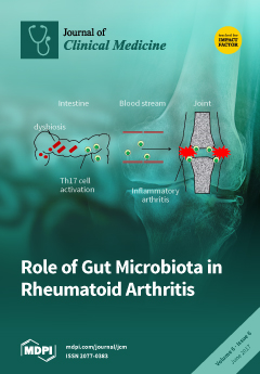
Cover Story (view full-size image): In this paper, we have reviewed the previous findings in animal and human studies with regard to the role of gut microbiota in rheumatoid arthritis (RA). We and others have reported that the increased abundance of Prevotella copri was found in some early RA patients. We produced gut microbiota-humanized mice and showed that Prevotella copri –dominated microbiota directly contributes to the aggravation of arthritis. View the paper here.
  • Issues are regarded as officially published after their release is announced to the table of contents alert mailing list.
  • You may sign up for e-mail alerts to receive table of contents of newly released issues.
  • PDF is the official format for papers published in both, html and pdf forms. To view the papers in pdf format, click on the "PDF Full-text" link, and use the free Adobe Reader to open them.
Order results
Result details
Section
Select all
Export citation of selected articles as:
5437 KiB  
Review
Fibrotic Hypersensitivity Pneumonitis: Key Issues in Diagnosis and Management
by Vasileios Kouranos, Joseph Jacob, Andrew Nicholson and Elizabetta Renzoni
J. Clin. Med. 2017, 6(6), 62; https://doi.org/10.3390/jcm6060062 - 15 Jun 2017
Cited by 38 | Viewed by 14971
Abstract
The diagnosis of hypersensitivity pneumonitis (HP) relies on the clinical evaluation of a number of features, including a history of significant exposure to potentially causative antigens, physical examination, chest CT scan appearances, bronchoalveolar lavage lymphocytosis, and, in selected cases, histology. The presence of [...] Read more.
The diagnosis of hypersensitivity pneumonitis (HP) relies on the clinical evaluation of a number of features, including a history of significant exposure to potentially causative antigens, physical examination, chest CT scan appearances, bronchoalveolar lavage lymphocytosis, and, in selected cases, histology. The presence of fibrosis is associated with higher morbidity and mortality. Differentiating fibrotic HP from the idiopathic interstitial pneumonias can be a challenge. Furthermore, even in the context of a clear diagnosis of fibrotic HP, the disease behaviour can parallel that of idiopathic pulmonary fibrosis in a subgroup, with inexorable progression despite treatment. We review the current knowledge on the diagnosis, management, and prognosis of HP with particular focus on the fibrotic phenotype. Full article
(This article belongs to the Special Issue Chronic Respiratory Diseases)
Show Figures

Figure 1

Figure 1
<p>Features suggestive of chronic hypersensitivity pneumonitis in different patients. (<b>A</b>) Spared pulmonary lobules are visible within non-fibrotic lung bilaterally (arrows); background fibrosis is evidenced by peripheral reticulation and traction bronchiectasis (black arrowheads). (<b>B</b>,<b>C</b>) A predilection towards bronchocentricity of the fibrosis in the upper lobes is evident on axial (arrowheads) and coronal (arrows) images. (<b>D</b>) An upper and midzone predominance to the fibrosis, characterised by reticulation and traction bronchiectasis, is seen on a coronal CT. Incidentally, volume loss in the right lung is noticeable by slight tenting and elevation of the right hemidiaphragm (arrow). (<b>E</b>) A UIP pattern with honeycomb cysts is visible in the left midzone of the lung (arrowhead) in a 74-year-old male ex-smoker. A surgical biopsy performed a few years before this CT had demonstrated findings compatible with fibrotic hypersensitivity pneumonitis.</p>
Full article ">Figure 1 Cont.
<p>Features suggestive of chronic hypersensitivity pneumonitis in different patients. (<b>A</b>) Spared pulmonary lobules are visible within non-fibrotic lung bilaterally (arrows); background fibrosis is evidenced by peripheral reticulation and traction bronchiectasis (black arrowheads). (<b>B</b>,<b>C</b>) A predilection towards bronchocentricity of the fibrosis in the upper lobes is evident on axial (arrowheads) and coronal (arrows) images. (<b>D</b>) An upper and midzone predominance to the fibrosis, characterised by reticulation and traction bronchiectasis, is seen on a coronal CT. Incidentally, volume loss in the right lung is noticeable by slight tenting and elevation of the right hemidiaphragm (arrow). (<b>E</b>) A UIP pattern with honeycomb cysts is visible in the left midzone of the lung (arrowhead) in a 74-year-old male ex-smoker. A surgical biopsy performed a few years before this CT had demonstrated findings compatible with fibrotic hypersensitivity pneumonitis.</p>
Full article ">Figure 2
<p>(<b>A</b>) A case of hypersensitivity pneumonitis shows peribronchiolar chronic inflammation of varying intensity around bronchovascular bundles. (<b>B</b>) A single, small, poorly-formed granuloma is found within the interstitium</p>
Full article ">Figure 3
<p>(<b>A</b>–<b>C</b>) A case of chronic hypersensitivity pneumonitis shows (<b>A</b>) areas of patchy subpleural dense fibrosis and (<b>B</b>) focal honeycomb change, typical of a pattern of usual interstitial pneumonia. (<b>C</b>) Rare peribronchiolar granulomas are also seen.</p>
Full article ">Figure 3 Cont.
<p>(<b>A</b>–<b>C</b>) A case of chronic hypersensitivity pneumonitis shows (<b>A</b>) areas of patchy subpleural dense fibrosis and (<b>B</b>) focal honeycomb change, typical of a pattern of usual interstitial pneumonia. (<b>C</b>) Rare peribronchiolar granulomas are also seen.</p>
Full article ">
458 KiB  
Review
Renin Inhibition with Aliskiren: A Decade of Clinical Experience
by Nikolaos-Dimitrios Pantzaris, Evangelos Karanikolas, Konstantinos Tsiotsios and Dimitrios Velissaris
J. Clin. Med. 2017, 6(6), 61; https://doi.org/10.3390/jcm6060061 - 9 Jun 2017
Cited by 24 | Viewed by 8185
Abstract
The renin-angiotensin-aldosterone system (RAAS) plays a key role in the pathophysiology of arterial hypertension as well as in more complex mechanisms of cardiovascular and renal diseases. RAAS-blocking agents like angiotensin-converting enzyme (ACE) inhibitors and angiotensin II receptor blockers, have long been key components [...] Read more.
The renin-angiotensin-aldosterone system (RAAS) plays a key role in the pathophysiology of arterial hypertension as well as in more complex mechanisms of cardiovascular and renal diseases. RAAS-blocking agents like angiotensin-converting enzyme (ACE) inhibitors and angiotensin II receptor blockers, have long been key components in the treatment of essential hypertension, heart failure, diabetic nephropathy, and chronic kidney disease, showing benefits well beyond blood pressure reduction. Renin blockade as the first step of the RAAS cascade finally became possible in 2007 with the approval of aliskiren, the first orally active direct renin inhibitor available for clinical use and the newest antihypertensive agent on the market. In the last decade, many clinical trials and meta-analyses have been conducted concerning the efficacy and safety of aliskiren in comparison to other antihypertensive agents, as well as the efficacy and potential clinical use of various combinations. Large trials with cardiovascular and renal endpoints attempted to show potential benefits of aliskiren beyond blood pressure lowering, as well as morbidity and mortality outcomes in specific populations such as diabetics, heart failure patients, and post-myocardial infarction individuals. The purpose of this review is to present the currently available data regarding established and future potential clinical uses of aliskiren. Full article
Show Figures

Figure 1

Figure 1
<p>RAAS and its pharmacological blockade.</p>
Full article ">
393 KiB  
Review
Role of Gut Microbiota in Rheumatoid Arthritis
by Yuichi Maeda and Kiyoshi Takeda
J. Clin. Med. 2017, 6(6), 60; https://doi.org/10.3390/jcm6060060 - 9 Jun 2017
Cited by 169 | Viewed by 21642
Abstract
Rheumatoid arthritis (RA) is a systemic autoimmune disease, caused by both genetic and environmental factors. Recently, investigators have focused on the gut microbiota, which is thought to be an environmental agent affecting the development of RA. Here we review the evidence from animal [...] Read more.
Rheumatoid arthritis (RA) is a systemic autoimmune disease, caused by both genetic and environmental factors. Recently, investigators have focused on the gut microbiota, which is thought to be an environmental agent affecting the development of RA. Here we review the evidence from animal and human studies that supports the role of the gut microbiota in RA. We and others have demonstrated that the abundance of Prevotella copri is increased in some early RA. We have also used gnotobiotic experiments to show that dysbiosis in RA patients contributed to the development of Th17 cell-dependent arthritis in intestinal microbiota-humanized SKG mice. On the other hand, Prevotella histicola from human gut microbiota suppressed the development of arthritis. In summary, Prevotella species are involved in the pathogenesis of arthritis. Full article
(This article belongs to the Special Issue Th17 Cell in Autoimmune and Inflammatory Diseases)
Show Figures

Figure 1

Figure 1
<p>Environmental factors such as smoking, hormones, gut microbiota, and periodontal bacteria are important in the development of rheumatoid arthritis. <span class="html-italic">Prevotella copri</span>-dominated microbiota may contribute to the development of arthritis, whereas <span class="html-italic">P. histicola</span> suppresses the onset of arthritis.</p>
Full article ">
1878 KiB  
Article
Social Factors Determine the Emergency Medical Admission Workload
by Seán Cournane, Richard Conway, Declan Byrne, Deirdre O’Riordan, Seamus Coveney and Bernard Silke
J. Clin. Med. 2017, 6(6), 59; https://doi.org/10.3390/jcm6060059 - 9 Jun 2017
Cited by 3 | Viewed by 4903
Abstract
We related social factors with the annual rate of emergency medical admissions using census small area statistics. All emergency medical admissions (70,543 episodes in 33,343 patients) within the catchment area of St. James’s Hospital, Dublin, were examined between 2002 and 2016. Deprivation Index, [...] Read more.
We related social factors with the annual rate of emergency medical admissions using census small area statistics. All emergency medical admissions (70,543 episodes in 33,343 patients) within the catchment area of St. James’s Hospital, Dublin, were examined between 2002 and 2016. Deprivation Index, Single-Parent status, Educational level and Unemployment rates were regressed against admission rates. High deprivation areas had an approximately fourfold (Incidence Rate Ratio (IRR) 4.0 (3.96, 4.12)) increase in annual admission rate incidence/1000 population from Quintile 1(Q1), from 9.2/1000 (95% Confidence Interval (CI): 9.0, 9.4) to Q5 37.3 (37.0, 37.5)). Single-Parent families comprised 40.6% of households (95% CI: 32.4, 49.7); small areas with more Single Parents had a higher admission rate-IRR (Q1 vs. for Q5) of 2.92 (95% CI: 2.83, 3.01). The admission incidence rate was higher for Single-Parent status (IRR 1.50 (95% CI: 1.46, 1.52)) where the educational completion level was limited to primary level (Incidence Rate Ratio 1.45 (95% CI: 1.43, 1.47)). Small areas with higher educational quintiles predicted lower Admission Rates (IRR 0.85 (95% CI: 0.84, 0.86)). Social factors strongly predict the annual incidence rate of emergency medical admissions. Full article
Show Figures

Figure 1

Figure 1
<p>Relationship between Deprivation status and Hospital Admission Incidence rates for emergency medical conditions. The predicted probabilities for each were derived from the zero-truncated Poisson model. We used margins to estimate the average marginal effect. Graphed is Admission Rates/1000 local area population between 2002–2016 for the first or any subsequent admission.</p>
Full article ">Figure 2
<p>Spatial variation in Emergency Hospital Admission Incidence rates for medical conditions. There was a total of 74 EDs in the catchment area with a median population per ED of 2845 (IQR 2020, 3399). These areas have a high element of Deprivation, being ranked nationally as Deprivation Quintile I (<span class="html-italic">n</span> = 13), Quintile III (<span class="html-italic">n</span> = 5), Quintile IV (<span class="html-italic">n</span> = 4) and Quintile V (<span class="html-italic">n</span> = 49). ED = Electoral Division.</p>
Full article ">Figure 3
<p>Relationship between Single-Parent status and Admission Incidence Rate of an emergency medical admission. The predicted probabilities were derived from the multi-variable truncated Poisson model. We used margins to estimate and interpret adjusted predictions for sub-groups, while controlling for other variables, using computations of average marginal effects. Single-Parent small area frequency predicted an increased admission rate, adjusted for Deprivation status.</p>
Full article ">Figure 4
<p>Interaction between small area Single Parent and unemployment rates and the Admission Incidence Rate of an emergency medical admission. The predicted probabilities were derived from the multi-variable truncated Poisson model. We used margins to estimate and interpret adjusted predictions for sub-groups, while controlling for other variables, using computations of average marginal effects. As the Quintiles (Q1, Q3, Q3) of unemployment increased, the strength of the association of Single-Parent status with the admission rate increased.</p>
Full article ">
2058 KiB  
Article
An Effective, Versatile, and Inexpensive Device for Oxygen Uptake Measurement
by Paule Bénit, Dominique Chrétien, Mathieu Porceddu, Constantin Yanicostas, Malgorzata Rak and Pierre Rustin
J. Clin. Med. 2017, 6(6), 58; https://doi.org/10.3390/jcm6060058 - 8 Jun 2017
Cited by 9 | Viewed by 6921
Abstract
In the last ten years, the use of fluorescent probes developed to measure oxygen has resulted in several marketed devices, some unreasonably expensive and with little flexibility. We have explored the use of the effective, versatile, and inexpensive Redflash technology to determine oxygen [...] Read more.
In the last ten years, the use of fluorescent probes developed to measure oxygen has resulted in several marketed devices, some unreasonably expensive and with little flexibility. We have explored the use of the effective, versatile, and inexpensive Redflash technology to determine oxygen uptake by a number of different biological samples using various layouts. This technology relies on the use of an optic fiber equipped at its tip with a membrane coated with a fluorescent dye (www.pyro-science.com). This oxygen-sensitive dye uses red light excitation and lifetime detection in the near infrared. So far, the use of this technology has mostly been used to determine oxygen concentration in open spaces for environmental studies, especially in aquatic media. The oxygen uptake determined by the device can be easily assessed in small volumes of respiration medium and combined with the measurement of additional parameters, such as lactate excretion by intact cells or the membrane potential of purified mitochondria. We conclude that the performance of by this technology should make it a first choice in the context of both fundamental studies and investigations for respiratory chain deficiencies in human samples. Full article
Show Figures

Figure 1

Figure 1
<p>The optode device. The tip of the optic fiber (in red) is covered by a polymer membrane coated with a fluorescent oxygen-sensitive dye (in green) fixed to the optic fiber with silicone glue. The optic fiber receives red light (excitation) from, and re-emits infrared light (emission) to, an analyzer box that can be connected to a personal computer. The fluorescence of the dye is proportional to its oxygen-dependent oxidation state, which is fully reversible. Time-dependent variation of the infrared emission reflects variation of the oxygen at the membrane surface. By inserting the tip of the optic fiber into any aerated medium, it appears possible to determine at any time the oxygen tension in the medium and so to estimate oxygen consumption in the medium.</p>
Full article ">Figure 2
<p>In vitro oxygen uptake by tissue sample and cells in suspension. (<b>A</b>) Both the Hansatech polarographic device (bottom) and the FireSting optode work simultaneously allowing to record strictly similar rates of oxygen uptake (about 25% resistant to 0.6 mM cyanide) by a mouse brain hemisphere place on a nylon net at mid-height in 400 μL of respiratory medium A (see Material and Methods). (<b>B</b>) Fully cyanide-sensitive human primary fibroblast respiration (about 1 × 10<sup>6</sup> cells for 50 μM O<sub>2</sub>/min) recorded in 200 μL of respiratory medium A. Numbers along the traces are nmol/min/mg protein.</p>
Full article ">Figure 3
<p>Respiration of Zebrafish embryos, and oxygen tension coupled to membrane potential determination by rat liver mitochondria. (<b>A</b>) Cyanide-sensitive respiration of Zebrafish embryos (1 and 5) measured at ambient temperature (20 °C) using a micro-optode in 30 μL of PBS (linear rates observed for 10 min). Notice the spherical magnetic stirrer avoiding to hurt the embryos. (<b>B</b>) Using an open layout, oxygen level changes linked to mitochondrial substrate oxidation was measured concomitantly to the membrane potential. Oxygen was measured using the macro-optode placed in a 3 mL quartz cell thermostated at 38 °C and magnetically stirred. Oxygen uptake (red trace) was started by the addition of succinate followed by the addition of a limiting amount of ADP (decreased level of oxygen, due to high rate of consumption; oxidation state 3 [<a href="#B20-jcm-06-00058" class="html-bibr">20</a>]), the exhaustion of which exhaustion in a higher oxygen level (oxidation state 4). The addition of malonate (a long established inhibitor of the succinate dehydrogenase [<a href="#B21-jcm-06-00058" class="html-bibr">21</a>]) fully inhibited oxygen uptake, and the level of oxygenation of the medium came back to initial value by re-equilibration with air. The membrane potential measured simultaneously (blue trace) rose upon succinate addition (quenching of rhodamine fluorescence) to drop down upon the ADP addition. After ADP exhaustion, the membrane potential rose again, while adding malonate worked to abolish most of it. Numbers along the traces are nmol/min/mg protein.</p>
Full article ">Figure 4
<p>Respiration and lactate excretion by mouse-cultured astrocytes. (<b>A</b>) The macro-optode fitted to a magnetically stirred, 37.5 °C-thermostated quartz-cell by a closed cap (yet allowing for the addition of substrates and inhibitors) measures oxygen uptake due to cyanide-sensitive respiration (red traces) by control astrocytes or astrocytes prepared from the CI-defective <span class="html-italic">Harlequin</span> mice. The concomitant fluorometric determination of NADH (blue traces) allows for a determination of the rate of the excretion of lactate, thanks to its conversion to pyruvate brought about by added lactate dehydrogenase in the presence of added NAD<sup>+</sup>. Numbers along the traces are nmol/min/mg protein. (<b>B</b>) The additional presence of glutamate and glutamate transaminase avoided inhibition of the LDH reaction by accumulated pyruvate.</p>
Full article ">Figure 4 Cont.
<p>Respiration and lactate excretion by mouse-cultured astrocytes. (<b>A</b>) The macro-optode fitted to a magnetically stirred, 37.5 °C-thermostated quartz-cell by a closed cap (yet allowing for the addition of substrates and inhibitors) measures oxygen uptake due to cyanide-sensitive respiration (red traces) by control astrocytes or astrocytes prepared from the CI-defective <span class="html-italic">Harlequin</span> mice. The concomitant fluorometric determination of NADH (blue traces) allows for a determination of the rate of the excretion of lactate, thanks to its conversion to pyruvate brought about by added lactate dehydrogenase in the presence of added NAD<sup>+</sup>. Numbers along the traces are nmol/min/mg protein. (<b>B</b>) The additional presence of glutamate and glutamate transaminase avoided inhibition of the LDH reaction by accumulated pyruvate.</p>
Full article ">
585 KiB  
Review
Minimal Residual Disease in Acute Myeloid Leukemia: Still a Work in Progress?
by Federico Mosna, Debora Capelli and Michele Gottardi
J. Clin. Med. 2017, 6(6), 57; https://doi.org/10.3390/jcm6060057 - 3 Jun 2017
Cited by 27 | Viewed by 7585
Abstract
Minimal residual disease evaluation refers to a series of molecular and immunophenotypical techniques aimed at detecting submicroscopic disease after therapy. As such, its application in acute myeloid leukemia has greatly increased our ability to quantify treatment response, and to determine the chemosensitivity of [...] Read more.
Minimal residual disease evaluation refers to a series of molecular and immunophenotypical techniques aimed at detecting submicroscopic disease after therapy. As such, its application in acute myeloid leukemia has greatly increased our ability to quantify treatment response, and to determine the chemosensitivity of the disease, as the final product of the drug schedule, dose intensity, biodistribution, and the pharmakogenetic profile of the patient. There is now consistent evidence for the prognostic power of minimal residual disease evaluation in acute myeloid leukemia, which is complementary to the baseline prognostic assessment of the disease. The focus for its use is therefore shifting to individualize treatment based on a deeper evaluation of chemosensitivity and residual tumor burden. In this review, we will summarize the results of the major clinical studies evaluating minimal residual disease in acute myeloid leukemia in adults in recent years and address the technical and practical issues still hampering the spread of these techniques outside controlled clinical trials. We will also briefly speculate on future developments and offer our point of view, and a word of caution, on the present use of minimal residual disease measurements in “real-life” practice. Still, as final standardization and diffusion of the methods are sorted out, we believe that minimal residual disease will soon become the new standard for evaluating response in the treatment of acute myeloid leukemia. Full article
(This article belongs to the Special Issue Role of Minimal Residual Disease Assessment in Hematological Cancers)
Show Figures

Graphical abstract

Graphical abstract
Full article ">Figure 1
<p>Paradigmatic scenarios of AML evolution after therapy considering MRD results. Nine paradigmatic scenarios (Cases 1 to 9) are displayed. Trends are simplified as compared to real life, for explicative reasons. Intermittent line at 1% represent the usual sensitivity limit of morphological examination; dotted line at 0.01% represents the usual sensitivity limit of MRD assessment. Vertical arrows represent chemotherapy cycles. Vertical lines represent common time-points for evaluation, i.e., after induction therapy and consolidation. (PANEL <b>A</b>): in Case 1, a quick response to chemotherapy cycles (white arrows) allows the patients to reach morphological CR after induction and the disappearance of measurable MRD after the first consolidation cycle. MRD remains undetectable during follow-up and the patient stays long-term in clinical remission. In Case 2, MRD negativity is reached within the end of consolidation therapy, so no further treatment (e.g., with allo-HSCT) is decided, and the patient remains in stable MRD negativity and clinical remission in the long term thereafter. In Case 3 the patient retains MRD-detectable disease at the end of consolidation, and allo-HSCT (lightning bolt) is decided; this enables us to obtain MRD negativity and long-term remission. (PANEL <b>B</b>): in Cases 4, 5 and 6 morphological remission is obtained following induction therapy, but no MRD is obtained by the end of consolidation in either cases. Case 4 is then consolidated by allo-HSCT, which enables the patient to reach MRD-negative leukemia levels briefly, but ultimately fails to eradicate the disease, with eventual relapse. In Case 5, no allo-HSCT is performed, and leukemia rapidly evolves from MRD persistence into overt clinical relapse. In Case 6, MRD identifies leukemia persistence just below the level of morphological detection (1%), and allo-HSCT, though inducing a brief reduction of residual disease, does not manage to obtain MRD negativity nor to prevent ultimate relapse. (PANEL <b>C</b>): in the scenario represented by Case 7 the patient never achieves a proper control over the disease, which results primary refractory; allo-HSCT is used with little efficacy, and ultimately clinical progression is unavoidable. Finally, Case 8 and 9 experience deep early response, achieving MRD negativity within the end of consolidation. In both cases reappearance of AML is detected by MRD monitoring during follow-up before clinical relapse: Case 9 is treated with additional therapy (either experimental treatments—i.e., grey arrow—or, less likely, allo-HSCT, lightning bolts) and restored to MRD negativity. Case 8 does not receive such treatment and ultimately relapse. In Case 9 the possibility of a molecular relapse spontaneously reverting to MRD-negativity also without the need for a clinical intervention, although increasingly rare with modern MRD measurement technique, cannot be ruled out, especially in the case of CBF AML.</p>
Full article ">
870 KiB  
Review
Use of the Ketogenic Diet to Treat Intractable Epilepsy in Mitochondrial Disorders
by Eleni Paleologou, Naila Ismayilova and Maria Kinali
J. Clin. Med. 2017, 6(6), 56; https://doi.org/10.3390/jcm6060056 - 26 May 2017
Cited by 30 | Viewed by 11043
Abstract
Mitochondrial disorders are a clinically heterogeneous group of disorders that are caused by defects in the respiratory chain, the metabolic pathway of the adenosine tri-phosphate (ATP) production system. Epilepsy is a common and important feature of these disorders and its management can be [...] Read more.
Mitochondrial disorders are a clinically heterogeneous group of disorders that are caused by defects in the respiratory chain, the metabolic pathway of the adenosine tri-phosphate (ATP) production system. Epilepsy is a common and important feature of these disorders and its management can be challenging. Epileptic seizures in the context of mitochondrial disease are usually treated with conventional anti-epileptic medication, apart from valproic acid. However, in accordance with the treatment of intractable epilepsy where there are limited treatment options, the ketogenic diet (KD) has been considered as an alternative therapy. The use of the KD and its more palatable formulations has shown promising results. It is especially indicated and effective in the treatment of mitochondrial disorders due to complex I deficiency. Further research into the mechanism of action and the neuroprotective properties of the KD will allow more targeted therapeutic strategies and thus optimize the treatment of both epilepsy in the context of mitochondrial disorders but also in other neurodegenerative disorders. Full article
Show Figures

Graphical abstract

Graphical abstract
Full article ">Figure 1
<p>Potential mechanisms of epileptogenesis in mitochondrial disease. (ROS: Reactive Oxygen Species; ATP: Adenosine Tri-Phosphate; Na<sup>+</sup>: Sodium; K<sup>+</sup>: Potassium; MtDNA: mitochondrial DNA; OXPHOS: oxidative phosphorylation).</p>
Full article ">Figure 2
<p>Mitochondri in hepatocyte. Ketone bodies alter mitochondrial metabolism at the cellular level; ketones reduce mitochondrial NAD coupling, oxidize Co-enzyme Q, increase the rate of ATP hydrolysis and increase metabolic efficiency. Oxidation of Co-enzyme Q decreases the major source reactive oxygen species (ROS). Increased rate of ATP hydrolysis widens the extra/intracellular ionic gradients, leading to hyperpolarization of cells. Potassium (K<sup>+</sup>): Hyperpolarization of cell membrane reducing excitotoxicity; CAT: carnitine-acylcarnitine translocase; BHB: β-hydroxybutyrate; ACA: acetoacetate; I–V: electron transport chain complexes; ADP: Adenosine Di-Phosphate; ATP: Adenosine Tri-Phosphate; CoQ10: Co-enzyme Q10; K<sub>ATP</sub>: ATP-sensitive potassium channels.</p>
Full article ">
Previous Issue
Next Issue
Back to TopTop