
Early and Mid-Term Results of Endovascular Aneurysm Repair with the Cordis Incraft Ultra-Low Profile Endograft: A High-Volume Center Experience

<p>Five years overall survival estimate calculated by the Kaplan–Meier method.</p> "> Figure 2
<p>Five years of freedom from any reintervention calculated by the Kaplan–Meier method.</p> "> Figure 3
<p>Five years of freedom from open surgical conversion calculated by the Kaplan–Meier method.</p> "> Figure 4
<p>Five years of freedom from iliac leg occlusion calculated by the Kaplan–Meier method.</p> "> Figure 5
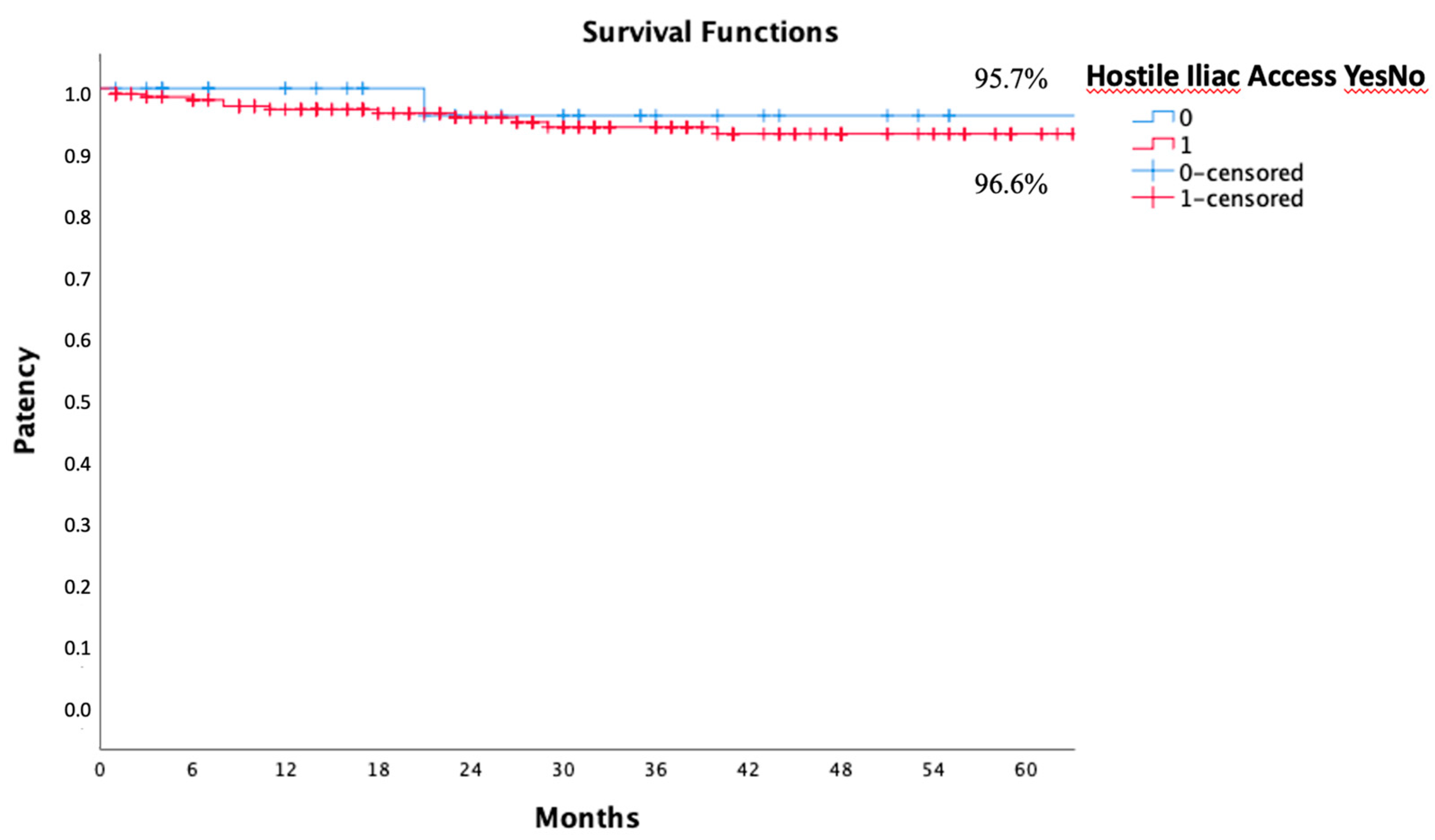
<p>Five years of freedom from iliac leg occlusion in hostile (red) and non-hostile (blue) anatomy calculated by the Kaplan–Meier method.</p> "> Figure 6
<p>Five years of freedom from iliac leg occlusion in grade 1 (blue), grade 2 (red), and grade 3 (green) hostile anatomy were calculated by the Kaplan–Meier method.</p> "> Figure 7
<p>Pre-operative CT-angiogram of a patient with AAA and chronic total occlusion of the right common iliac artery (<b>A</b>); final intraoperative angiography and post-operative CT-angiogram demonstrating effective Incraft implantation with sac exclusion and complete right common iliac artery recanalization (<b>B</b>,<b>C</b>).</p> ">
Abstract
:1. Introduction
2. Materials and Methods
Statistical Analysis
3. Results
3.1. Intra-Procedural Outcomes
3.2. Thirty-Day Results
3.3. Mid-Term Results
4. Discussion
5. Conclusions
Author Contributions
Funding
Institutional Review Board Statement
Informed Consent Statement
Data Availability Statement
Conflicts of Interest
References
- Parodi, J.C.; Palmaz, J.C.; Barone, H.D. Transfemoral intraluminal graft implantation for abdominal aortic aneurysms. Ann. Vasc. Surg. 1991, 5, 491–499. [Google Scholar] [CrossRef]
- Wanhainen, A.; Van Herzeele, I.; Bastos Goncalves, F.; Bellmunt Montoya, S.; Berard, X.; Boyle, J.R.; D’Oria, M.; Prendes, C.F.; Karkos, C.D.; Kazimierczak, A.; et al. Editor’s Choice—European Society for Vascular Surgery (ESVS) 2024 Clinical Practice Guidelines on the Management of Abdominal Aorto-Iliac Artery Aneurysms. Eur. J. Vasc. Endovasc. Surg. 2024, 67, 192–331. [Google Scholar] [CrossRef] [PubMed]
- Harris, P.L.; Vallabhaneni, S.R.; Desgranges, P.; Becquemin, J.P.; van Marrewijk, C.; Laheij, R.J. Incidence and risk factors of late rupture, conversion, and death after endovascular repair of infrarenal aortic aneurysms: The EUROSTAR experience. European Collaborators on Stent/graft techniques for aortic aneurysm repair. J. Vasc. Surg. 2000, 32, 739–749. [Google Scholar] [CrossRef] [PubMed]
- Carroccio, A.; Faries, P.L.; Morrissey, N.J.; Teodorescu, V.; Burks, J.A.; Gravereaux, E.C.; Hollier, L.H.; Marin, M.L. Predicting iliac limb occlusions after bifurcated aortic stent grafting: Anatomic and device related causes. J. Vasc. Surg. 2002, 36, 679–684. [Google Scholar] [CrossRef]
- AbuRahma, A.F.; Campbell, J.E.; Mousa, A.Y.; Hass, S.M.; Stone, P.A.; Jain, A.; Nanjundappa, A.; Dean, L.S.; Keiffer, T.; Habib, J. Clinical outcomes for hostile versus favorable aortic neck anatomy in endovascular aortic aneurysm repair using modular devices. J. Vasc. Surg. 2011, 54, 13–21. [Google Scholar] [CrossRef]
- Oderich, G.S.; Forbes, T.L.; Chaer, R.; Davies, M.G.; Lindsay, T.F.; Mastracci, T.; Singh, M.J.; Timaran, C.; Woo, E.Y.; Writing Committee Group. Reporting standards for endovascular aortic repair of aneurysms involving the renal-mesenteric arteries. J. Vasc. Surg. 2021, 73 (Suppl. S1), 4S–52S. [Google Scholar] [CrossRef]
- Gallitto, E.; Gargiulo, M.; Faggioli, G.; Pini, R.; Mascoli, C.; Freyrie, A.; Ancetti, S.; Stella, A. Impact of iliac artery anatomy on the outcome of fenestrated and branched endovascular aortic repair. J. Vasc. Surg. 2017, 66, 1659–1667. [Google Scholar] [CrossRef]
- Becquemin, J.P.; Pillet, J.C.; Lescalie, F.; Sapoval, M.; Goueffic, Y.; Lermusiaux, P.; Steinmetz, E.; Marzelle, J.; ACE trialists. A randomized controlled trial of endovascular aneurysm repair versus open surgery for abdominal aortic aneurysms in low- to moderate-risk patients. J. Vasc. Surg. 2011, 53, 1167–1173.e1. [Google Scholar] [CrossRef]
- Lederle, F.A.; Freischlag, J.A.; Kyriakides, T.C.; Padberg, F.T., Jr.; Matsumura, J.S.; Kohler, T.R.; Lin, P.H.; Jean-Claude, J.M.; Cikrit, D.F.; Swanson, K.M.; et al. Outcomes following endovascular vs open repair of abdominal aortic aneurysm: A randomized trial. JAMA 2009, 302, 1535–1542. [Google Scholar] [CrossRef]
- EVAR trial participants. Endovascular aneurysm repair versus open repair in patients with abdominal aortic aneurysm (EVAR trial 1): Randomised controlled trial. Lancet 2005, 365, 2179–2186. [Google Scholar] [CrossRef]
- Blankensteijn, J.D.; de Jong, S.E.; Prinssen, M.; van der Ham, A.C.; Buth, J.; van Sterkenburg, S.M.; Verhagen, H.J.; Buskens, E.; Grobbee, D.E.; Dutch Randomized Endovascular Aneurysm Management (DREAM) Trial Group. Two-year outcomes after conventional or endovascular repair of abdominal aortic aneurysms. N. Engl. J. Med. 2005, 352, 2398–2405. [Google Scholar] [CrossRef] [PubMed]
- United Kingdom EVAR Trial Investigators; Greenhalgh, R.M.; Brown, L.C.; Powell, J.T.; Thompson, S.G.; Epstein, D.; Sculpher, M.J. Endovascular versus open repair of abdominal aortic aneurysm. N. Engl. J. Med. 2010, 362, 1863–1871. [Google Scholar] [CrossRef] [PubMed]
- Lederle, F.A.; Freischlag, J.A.; Kyriakides, T.C.; Matsumura, J.S.; Padberg, F.T., Jr.; Kohler, T.R.; Kougias, P.; Jean-Claude, J.M.; Cikrit, D.F.; Swanson, K.M.; et al. Long-term comparison of endovascular and open repair of abdominal aortic aneurysm. N. Engl. J. Med. 2012, 367, 1988–1997. [Google Scholar] [CrossRef]
- De Bruin, J.L.; Baas, A.F.; Buth, J.; Prinssen, M.; Verhoeven, E.L.; Cuypers, P.W.; van Sambeek, M.R.; Balm, R.; Grobbee, D.E.; Blankensteijn, J.D.; et al. Long-term outcome of open or endovascular repair of abdominal aortic aneurysm. N. Engl. J. Med. 2010, 362, 1881–1889. [Google Scholar] [CrossRef]
- Giles, K.A.; Landon, B.E.; Cotterill, P.; O’Malley, A.J.; Pomposelli, F.B.; Schermerhorn, M.L. Thirty-day mortality and late survival with reinterventions and readmissions after open and endovascular aortic aneurysm repair in Medicare beneficiaries. J. Vasc. Surg. 2011, 53, 6–13.e1. [Google Scholar] [CrossRef]
- Stokmans, R.A.; Teijink, J.A.; Forbes, T.L.; Böckler, D.; Peeters, P.J.; Riambau, V.; Hayes, P.D.; van Sambeek, M.R. Early results from the ENGAGE registry: Real-world performance of the Endurant Stent Graft for endovascular AAA repair in 1262 patients. Eur. J. Vasc. Endovasc. Surg. 2012, 44, 369–375. [Google Scholar] [CrossRef]
- Verzini, F.; Isernia, G.; De Rango, P.; Simonte, G.; Parlani, G.; Loschi, D.; Cao, P. Abdominal aortic endografting beyond the trials: A 15-year single-center experience comparing newer to older generation stent-grafts. J. Endovasc. Ther. 2014, 21, 439–447. [Google Scholar] [CrossRef]
- Antoniou, G.A.; Antoniou, S.A.; Torella, F. Editor’s Choice—Endovascular vs. Open Repair for Abdominal Aortic Aneurysm: Systematic Review and Meta-analysis of Updated Peri-operative and Long Term Data of Randomised Controlled Trials. Eur. J. Vasc. Endovasc. Surg. 2020, 59, 385–397. [Google Scholar] [CrossRef]
- Murray, D.; Ghosh, J.; Khwaja, N.; Murphy, M.O.; Baguneid, M.S.; Walker, M.G. Access for endovascular aneurysm repair. J. Endovasc. Ther. 2006, 13, 754–761. [Google Scholar] [CrossRef]
- Kristmundsson, T.; Sonesson, B.; Dias, N.; Malina, M.; Resch, T. Anatomic suitability for endovascular repair of abdominal aortic aneurysms and possible benefits of low profile delivery systems. Vascular 2014, 22, 112–115. [Google Scholar] [CrossRef]
- Pratesi, G.; Pratesi, C.; Chiesa, R.; Coppi, G.; Scheinert, D.; Brunkwall, J.S.; van der Meulen, S.; Torsello, G. The Innovation Trial: Four-year safety and effectiveness of the INCRAFT® AAA Stent-Graft System for endovascular repair. J. Cardiovasc. Surg. 2017, 58, 650–657. [Google Scholar] [CrossRef] [PubMed]
- Torsello, G.; Pratesi, G.; van der Meulen, S.; Ouriel, K.; INNOVATION trial collaborators. Aortoiliac remodeling and 5-year outcome of an ultralow-profile endograft. J. Vasc. Surg. 2019, 69, 1747–1757. [Google Scholar] [CrossRef] [PubMed]
- Zavatta, M.; Squizzato, F.; Balestriero, G.; Bonvini, S.; Perkmann, R.; Milite, D.; Veraldi, G.F.; Antonello, M.; Triveneto Incraft Registry Collaborators. Early and midterm outcomes of endovascular aneurysm repair with an ultra-low-profile endograft from the Triveneto Incraft Registry. J. Vasc. Surg. 2021, 73, 1950–1957.e2. [Google Scholar] [CrossRef] [PubMed]
- Gill, H.L.; Doonan, R.J.; Altoijry, A.; Obrand, D.I.; Mackenzie, K.S.; Steinmetz, O.K. Early North American experience with the INCRAFT device. J. Vasc. Surg. 2019, 70, 102–106. [Google Scholar] [CrossRef]
- Pratesi, C.; Piffaretti, G.; Pratesi, G.; Castelli, P.; ITalian Excluder Registry Investigators. ITalian Excluder Registry and results of Gore Excluder endograft for the treatment of elective infrarenal abdominal aortic aneurysms. J. Vasc. Surg. 2014, 59, 52–57.e1. [Google Scholar] [CrossRef]
- van Zeggeren, L.; Bastos Gonçalves, F.; van Herwaarden, J.A.; Zandvoort, H.J.; Werson, D.A.; Vos, J.A.; Moll, F.L.; Verhagen, H.J.; de Vries, J.P. Incidence and treatment results of Endurant endograft occlusion. J. Vasc. Surg. 2013, 57, 1246–1254; discussion 1254. [Google Scholar] [CrossRef]
- Greaves, N.S.; Moore, A.; Seriki, D.; Ghosh, J. Outcomes of Endovascular Aneurysm Repair using the Ovation Stent Graft System in Adverse Anatomy. Eur. J. Vasc. Endovasc. Surg. 2018, 55, 512–517. [Google Scholar] [CrossRef]


| Mean ± SD age (y) Male Smoker Hypertension Chronic obstructive pulmonary disease Ischaemic heart disease Hyperlipemia Chronic renal failure Peripheral artery disease Obesity Diabetes Previous aortic surgery Symptomatic Ruptured | 75.3 ± 7.7 122 (91.7) 74 (55.6) 114 (85.7) 89 (66.9) 75 (56.4) 85 (63.9) 54 (40.6) 64 (48.1) 33 (24.8) 24 (18.0) 0 (0.0) 9 (6.8) 1 (0.8) |
| AAA diameter (mm) PAU, n (%) Proximal neck diameter (mm) Proximal neck length (mm) Aortic bifurcation diameter (mm) Hostility of at least 1 iliac access, n (%) Hostility of both iliac accesses, n (%) | 54.6 ± 8.7 14 (10.5) 21.6 ± 3.1 30.9 ± 1.1 22.1 ± 5.9 120 (90.2) 104 (78.2) |
| Iliac angulation > 90° Severe calcification External iliac diameter < 7 mm Iliac stenosis Iliac occlusion Common iliac aneurysm Iliac tortuosity index, mean ± SD Grade 1 hostility Grade 2 hostility Grade 3 hostility | 48 (18.0) 133 (50.0) 127 (47.7) 125 (47.0) 9 (3.4) 8 (3.0) 1.44 ± 0.23 135 (50.8) 63 (23.7) 68 (25.6) |
| Technical success Intraoperative death Intraoperative conversion to OSR Intraoperative endoleak Type 1A endoleak Type 1B endoleak Type II endoleak Type III endoleak Type IV endoleak Type V endoleak Other complications Access surgical conversion Lower limb ischemia Iliac rupture Iliac branch occlusion | 104 (78.2) 0 (0.0) 0 (0.0) 69 (51.9) 27 (20.3) 1 (0.8) 41 (30.8) 0 (0.0) 0 (0.0) 0 (0.0) 9 (6.7) 6 (4.5) 1 (0.8) 1 (0.8) 1 (0.8) |
| Clinical success Death Reintervention Conversion to open surgical repair Endoleak Type I endoleak Type II endoleak Type III endoleak Type IV endoleak Type V endoleak | 132 (99.3) 0 (0.0) 1 (0.8) 0 (0.0) 25 (18.8) 0 (0.0) 25 (18.8) 0 (0.0) 0 (0.0) 0 (0.0) |
Disclaimer/Publisher’s Note: The statements, opinions and data contained in all publications are solely those of the individual author(s) and contributor(s) and not of MDPI and/or the editor(s). MDPI and/or the editor(s) disclaim responsibility for any injury to people or property resulting from any ideas, methods, instructions or products referred to in the content. |
© 2024 by the authors. Licensee MDPI, Basel, Switzerland. This article is an open access article distributed under the terms and conditions of the Creative Commons Attribution (CC BY) license (https://creativecommons.org/licenses/by/4.0/).
Share and Cite
Baccani, L.; Parlani, G.; Isernia, G.; Lenti, M.; Terpin, A.M.; Simonte, G. Early and Mid-Term Results of Endovascular Aneurysm Repair with the Cordis Incraft Ultra-Low Profile Endograft: A High-Volume Center Experience. J. Clin. Med. 2024, 13, 5413. https://doi.org/10.3390/jcm13185413
Baccani L, Parlani G, Isernia G, Lenti M, Terpin AM, Simonte G. Early and Mid-Term Results of Endovascular Aneurysm Repair with the Cordis Incraft Ultra-Low Profile Endograft: A High-Volume Center Experience. Journal of Clinical Medicine. 2024; 13(18):5413. https://doi.org/10.3390/jcm13185413
Chicago/Turabian StyleBaccani, Luigi, Gianbattista Parlani, Giacomo Isernia, Massimo Lenti, Andrea Maria Terpin, and Gioele Simonte. 2024. "Early and Mid-Term Results of Endovascular Aneurysm Repair with the Cordis Incraft Ultra-Low Profile Endograft: A High-Volume Center Experience" Journal of Clinical Medicine 13, no. 18: 5413. https://doi.org/10.3390/jcm13185413