[go: up one dir, main page]

Next Issue
Volume 13, May
Previous Issue
Volume 13, March


Pathogens, Volume 13, Issue 4 (April 2024) – 77 articles

Cover Story (view full-size image): 
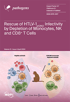
HTLV-1 establishes a persistent lifelong infection by evading the host immune response, in part through the functions of the viral p12 protein. Viruses that do not express p12 (HTLV-1p12KO) cannot persistently infect macaques, and the transient depletion of monocytes prior to virus exposure allows early seroconversion, which is not sustained over time. To test whether HTLV-1 persistence depends on a monocyte reservoir or if monocyte depletion provides a transient immune evasion benefit, we simultaneously depleted NK, CD8+ T cells, and monocytes (triple depletion) prior to exposure to HTLV-1p12KO. Triple depletion resulted in the exacerbation of infection and the complete rescue of HTLV-1p12KO infectivity. This underscores the role of monocytes in orchestrating anti-viral immunity and the importance of p12 in hijacking host immunity. View this paper
  • Issues are regarded as officially published after their release is announced to the table of contents alert mailing list.
  • You may sign up for e-mail alerts to receive table of contents of newly released issues.
  • PDF is the official format for papers published in both, html and pdf forms. To view the papers in pdf format, click on the "PDF Full-text" link, and use the free Adobe Reader to open them.
Order results
Result details
Section
Select all
Export citation of selected articles as:
10 pages, 233 KiB  
Communication
Seroprevalence and Risk Factors Associated with Phleboviruses and Crimean–Congo Hemorrhagic Fever Virus among Blood Donors in Central Tunisia
by Rym Ayari, Houda Chaouch, Stephen Findlay-Wilson, Wissem Hachfi, Nadia Ben Lasfar, Foued Bellazreg, Stuart Dowall, Neila Hannachi and Amel Letaief
Pathogens 2024, 13(4), 348; https://doi.org/10.3390/pathogens13040348 - 22 Apr 2024
Viewed by 1274
Abstract
The aim of this study was to determine the prevalence of six viruses, from two families of the order Bunyavirales, in the general population of central Tunisia. Sera collected from 377 asymptomatic blood donors were serologically assayed for Rift Valley fever virus [...] Read more.
The aim of this study was to determine the prevalence of six viruses, from two families of the order Bunyavirales, in the general population of central Tunisia. Sera collected from 377 asymptomatic blood donors were serologically assayed for Rift Valley fever virus (RVFV), Crimean–Congo hemorrhagic fever virus (CCHFV), and four sandfly-borne phleboviruses: Toscana virus (TOSV), sandfly fever Naples virus (SFNV), sandfly fever Sicilian virus (SFSV), and sandfly fever Cyprus virus (SFCV). Of the 377 subjects enrolled in this study, 17.3% were IgG positive for at least one of the viruses tested. The most frequently detected antibodies were against TOSV (13.3%), followed by SFCV (2.9%), RVFV (1.9%), SFSV (1.3%), and SFNV (1.1%). Only one sample was IgG positive for CCHFV. Dual reactivity was observed in nine cases: SFSV + SFCV in three cases (0.8%) and TOSV + SFNV, TOSV + SFCV, and TOSV + RVFV in two cases (0.5%) each. 15.9% of donors were IgG positive against sandfly-borne phleboviruses. Among the 65 donors IgG positive for phleboviruses, 50.8% were from rural areas compared to 12.3% from urban areas (p < 0.001); 92.3% had animals in their living quarters (p = 0.009); and 70.8% lived in the vicinity of stagnant water (p = 0.062). Seroprevalence was significantly higher among donors living with chronic diseases (p = 0.039). Furthermore, the seroprevalence of phleboviruses was higher in Kairouan, the central governorate, than in the two coastal governorates: Monastir and Sousse, with 33.4%, 24.2%, and 14.9%, respectively. The presence of antibodies in the general population needs further investigation to better assess the extent of these viruses. Only TOSV was known to have an extensive circulation in Tunisia and in North Africa. Continued surveillance and interventions are necessary to detect the emergence of all arboviruses and to prevent further transmission. Full article
(This article belongs to the Special Issue Diagnostics of Emerging and Re-emerging Pathogens)
16 pages, 924 KiB  
Article
Spatial Distribution of Dermanyssus gallinae Infestations in Greece and Their Association with Ambient Temperature, Humidity, and Altitude
by Georgios Sioutas, Athanasios I. Gelasakis and Elias Papadopoulos
Pathogens 2024, 13(4), 347; https://doi.org/10.3390/pathogens13040347 - 22 Apr 2024
Viewed by 1085
Abstract
Dermanyssus gallinae, the poultry red mite (PRM), is the most prevalent and harmful ectoparasite of laying hens globally. Although prevalence and risk factor studies can help veterinarians make decisions regarding farm treatments, relevant data are scarce. The present study investigated the prevalence [...] Read more.
Dermanyssus gallinae, the poultry red mite (PRM), is the most prevalent and harmful ectoparasite of laying hens globally. Although prevalence and risk factor studies can help veterinarians make decisions regarding farm treatments, relevant data are scarce. The present study investigated the prevalence and infestation severity of PRM in poultry farms across Greece and examined potential risk factors. AviVet traps were used to sample 84 farms (51 backyard, 33 industrial) over three years. Farm altitude, temperature, humidity, region, and production systems were assessed as potential risk factors with chi-square tests, initially for all the studied farms and then exclusively for backyard farms. The overall prevalence was 75.0% and was higher in backyard farms (80.4%) compared with industrial ones (66.7%), varying regionally from 66.7 to 90.9%. Altitude and temperature were not significant risk factors, but farms with humidity <60% had a lower infestation risk. Infestation severity did not significantly differ by risk factors. The poultry red mite is highly prevalent across Greek poultry production systems and regions. In the future, global warming, reduced acaricide options, and a ban on cage systems will all threaten a wider spatio-temporal distribution of the PRM, justifying the urgent need for effective monitoring and control methods to protect hen production and welfare and workers’ health. Full article
(This article belongs to the Special Issue Spatio-Temporal Analysis of Veterinary Infectious Diseases)
Show Figures

Figure 1

Figure 1
<p>Sampling locations of poultry farms for <span class="html-italic">Dermanyssus gallinae</span> in different geographical regions of Greece. Green pins represent the location of backyard laying hen farms that were sampled, and blue pins represent the location of industrial laying hen farms that were sampled.</p>
Full article ">
11 pages, 622 KiB  
Review
Exploring Extended-Spectrum Beta-Lactamase (ESBL)-Producing Escherichia coli in Food-Producing Animals and Animal-Derived Foods
by Laryssa Freitas Ribeiro, Natália Maramarque Nespolo, Gabriel Augusto Marques Rossi and John Morris Fairbrother
Pathogens 2024, 13(4), 346; https://doi.org/10.3390/pathogens13040346 - 22 Apr 2024
Cited by 3 | Viewed by 2060
Abstract
Antimicrobials serve as crucial treatments in both veterinary and human medicine, aiding in the control and prevention of infectious diseases. However, their misuse or overuse has led to the emergence of antimicrobial resistance, posing a significant threat to public health. This review focuses [...] Read more.
Antimicrobials serve as crucial treatments in both veterinary and human medicine, aiding in the control and prevention of infectious diseases. However, their misuse or overuse has led to the emergence of antimicrobial resistance, posing a significant threat to public health. This review focuses on extended-spectrum beta-lactamase (ESBL)-producing Escherichia coli in animals and their associated food products, which contribute to the proliferation of antimicrobial-resistant strains. Recent research has highlighted the presence of ESBL-producing E. coli in animals and animal-derived foods, with some studies indicating genetic similarities between these isolates and those found in human infections. This underscores the urgent need to address antimicrobial resistance as a pressing public health issue. More comprehensive studies are required to understand the evolving landscape of ESBLs and to develop strategic public health policies grounded in the One Health approach, aiming to control and mitigate their prevalence effectively. Full article
(This article belongs to the Special Issue Microbial Resistance, a Worldwide Concern a Global Sight)
Show Figures

Figure 1

Figure 1
<p>The epidemiology of extended-spectrum beta-lactamase (ESBL)-producing <span class="html-italic">E. coli</span> encompasses a multifaceted landscape, elucidating intricate transmission pathways implicated in antimicrobial use within animal husbandry settings, spanning from farms to food industries. Moreover, this complex web extends to encompass the involvement of food handlers, thus amplifying the potential dissemination routes of ESBL-producing <span class="html-italic">E. coli</span> strains. Additionally, environmental contamination emerges as a critical facet, with the aquatic ecosystem serving as a reservoir for ESBL dissemination, fueled by a confluence of factors including farm, industrial, and hospital sewage. This comprehensive understanding underscores the far-reaching ramifications of ESBL prevalence and underscores the imperative for holistic intervention strategies targeting various nodes along the transmission continuum to effectively mitigate the burgeoning threat posed by antimicrobial resistance.</p>
Full article ">
11 pages, 702 KiB  
Article
Molecular Detection of HPV, EBV, HSV-1, HCMV, and H. pylori Pathogens: An Evaluation among Polish Children with Molar Incisor Hypomineralization (MIH)
by Wojciech Tynior, Agata Świętek, Dorota Hudy, Danuta Ilczuk-Rypuła and Joanna Katarzyna Strzelczyk
Pathogens 2024, 13(4), 345; https://doi.org/10.3390/pathogens13040345 - 22 Apr 2024
Viewed by 968
Abstract
Molar incisor hypomineralization (MIH) is a congenital disorder of the enamel tissue, characterized by a quantitative deficiency. In childhood, infections such as EBV, HSV-1, HCMV, or H. pylori may occur and cause various diseases. This study aimed to investigate the prevalence of HPV, [...] Read more.
Molar incisor hypomineralization (MIH) is a congenital disorder of the enamel tissue, characterized by a quantitative deficiency. In childhood, infections such as EBV, HSV-1, HCMV, or H. pylori may occur and cause various diseases. This study aimed to investigate the prevalence of HPV, EBV, HSV-1, HCMV, and H. pylori infections in two groups of children: children with molar incisor hypomineralization (MIH) and a control group, using molecular methods. The study group included 47 children aged between 6–13 years who had been diagnosed with MIH. The control group consisted of 42 children. The study found that, in the MIH group, the prevalence of HPV-16 was 6.38%, HPV-18 was 4.26%, EBV was 31.91%, HSV-1 was 4.26%, HCMV was 4.26%, and H. pylori was 12.77%. There were no significant differences in the prevalence of any of tested pathogens between the study and the control group (p > 0.05). However, the study found a higher prevalence of EBV infection in children who had smallpox/pneumonia by the age of 3 years. Ten children were found to have at least two pathogens present. Moreover, both groups had a high prevalence and activity of EBV. These findings provide new insights into the carriage of pathogens among children with MIH, providing new information for parents, scientists, and healthcare professionals. Full article
Show Figures

Figure 1

Figure 1
<p>Percentage of patients with detected pathogens in the MIH and control group.</p>
Full article ">Figure 2
<p>Number of patients with different number of detected pathogens.</p>
Full article ">
12 pages, 1729 KiB  
Article
Trends in Non-Tuberculous Mycobacterial Lung Disease and Treatment Outcomes in a Low-Tuberculosis Prevalence Setting: A Retrospective Analysis
by Biplob Kumar Mohanty, Tomas Mikal Lind Eagan, Bernt Bøgvald Aarli, Dag Harald Skutlaberg and Tehmina Mustafa
Pathogens 2024, 13(4), 344; https://doi.org/10.3390/pathogens13040344 - 22 Apr 2024
Viewed by 1244
Abstract
Background: Information on the management of non-tuberculous mycobacterial (NTM) lung infection and disease is scarce. The aim of this study was to investigate the trends in NTM lung infections, and the factors associated with the initiation of treatment and treatment outcomes. Methods: A [...] Read more.
Background: Information on the management of non-tuberculous mycobacterial (NTM) lung infection and disease is scarce. The aim of this study was to investigate the trends in NTM lung infections, and the factors associated with the initiation of treatment and treatment outcomes. Methods: A retrospective analysis was carried out on patient medical records from Haukeland University Hospital, Bergen, Norway, from 2000 to 2021. Results: Among 154 patients with NTM lung infection, the majority (70%) were older than 65 years, and 49% had an underlying pulmonary comorbidity. The most frequently observed mycobacterial species was M. avium complex (MAC), followed by M. malmoense and M. abscessus. In total, 72 (47%) patients received antibiotic treatment. Patients with high symptom scores, aged below 65, and with MAC infection had more than three times the odds of receiving antibiotic treatment. A favourable response and culture conversion was observed in 53 of 72 (74%) patients. However, 17 (32%) of them had a relapse. Out of 82 patients who did not receive treatment, 45 (55%) had spontaneous culture conversion, and 8 (18%) of them had a relapse. No factor was identified to be significantly associated with a favourable treatment response. Conclusion: A favourable response to treatment was seen in 74% of patients with a high relapse rate. Full article
(This article belongs to the Section Epidemiology of Infectious Diseases)
Show Figures

Figure 1

Figure 1
<p>Flow chart showing the inclusion and exclusion of patients in the study.</p>
Full article ">Figure 2
<p>(<b>A</b>,<b>B</b>) Proportion of different species in NTM lung infection and NTM lung disease from the year 2000 to 2021. Total amount of NTM lung infections and NTM lung disease for each year shown at the top of the bars.</p>
Full article ">Figure 3
<p>Trends in different species in NTM lung infection in 5 year periods from the year 2000 to 2019.</p>
Full article ">Figure 4
<p>Comparison of culture conversion between patients with NTM lung infection according to their treatment status.</p>
Full article ">
12 pages, 1242 KiB  
Article
Staphylococcus hsinchuensis sp. nov., Isolated from Soymilk
by Yu-Ting Wang, Yu-Chun Lin, Yi-Huei Hsieh, Yu-Tzu Lin, Moriyuki Hamada, Chih-Chieh Chen, Jong-Shian Liou, Ai-Yun Lee, Wei-Ling Zhang, Yung-Tsung Chen and Chien-Hsun Huang
Pathogens 2024, 13(4), 343; https://doi.org/10.3390/pathogens13040343 - 21 Apr 2024
Viewed by 1171
Abstract
A novel coagulase-negative Staphylococcus strain (H164T) was isolated from soymilk in Taiwan. Comparative sequence analysis of the 16S rRNA gene revealed that the H164T strain is a member of the genus Staphylococcus. We used multilocus sequence analysis (MLSA) and [...] Read more.
A novel coagulase-negative Staphylococcus strain (H164T) was isolated from soymilk in Taiwan. Comparative sequence analysis of the 16S rRNA gene revealed that the H164T strain is a member of the genus Staphylococcus. We used multilocus sequence analysis (MLSA) and phylogenomic analyses to demonstrate that the novel strain was closely related to Staphylococcus gallinarum, Staphylococcus nepalensis, Staphylococcus cohnii, and Staphylococcus urealyuticus. The average nucleotide identity and digital DNA-DNA hybridization values between H164T and its closest relatives were <95% and <70%, respectively. The H164T strain could also be distinguished from its closest relatives by the fermentation of d-fructose, d-maltose, d-trehalose, and d-mannitol, as well as by the activities of α-glucosidase and alkaline phosphatase. The major cellular fatty acids were C15:0 iso and C15:0 anteiso, and the predominant menaquinones were MK-7 and MK-8, respectively. The major cellular fatty acids and predominant menaquinones were C15:0 iso and C15:0 anteiso and MK-7 and MK-8, respectively. In conclusion, this strain represents a novel species, named Staphylococcus hsinchuensis sp. nov., with the type strain H164T (=BCRC 81404T = NBRC 116174T). Full article
Show Figures

Figure 1

Figure 1
<p>Phylogenetic tree based on 16S rRNA gene sequences showing the relationship of <span class="html-italic">Staphylococcus hsinchuensis</span> sp. nov. H164<sup>T</sup> with strains of closely related species. The tree was constructed by the neighbor-joining and minimum evolution methods based on a comparison of approximately 1450 bp, and <span class="html-italic">Macrococcus caseolyticus</span> ATCC 13548<sup>T</sup> was used as the outgroup. Bootstrap values (&gt;60%) based on 1000 replicates are shown at branch nodes. Bar, 0.5% sequence divergence.</p>
Full article ">Figure 2
<p>Phylogenetic tree based on the concatenated housekeeping gene sequences (<span class="html-italic">dnaJ</span>, <span class="html-italic">gap</span>, <span class="html-italic">hsp60</span>, <span class="html-italic">rpoB</span>, <span class="html-italic">sodA</span>, and <span class="html-italic">tuf</span>) showing the relationship of <span class="html-italic">Staphylococcus hsinchuensis</span> sp. nov. H164<sup>T</sup> with strains of closely related species. The tree was constructed by the neighbor-joining and minimum evolution methods based on a comparison of approximately 9600 bp, and <span class="html-italic">Macrococcus caseolyticus</span> FDAARGOS_868<sup>T</sup> was used as an outgroup. Bootstrap values based on 1000 replicates are shown at branch nodes. Bar, 5% sequence divergence.</p>
Full article ">Figure 3
<p>A UBCG tree based on 92 bacterial core genes of <span class="html-italic">Staphylococcus hsinchuensis</span> sp. nov. H164<sup>T</sup> and the type strains of closely related species. Bootstrap values greater than 60% are shown at each node, and <span class="html-italic">Macrococcus caseolyticus</span> FDAARGOS_868<sup>T</sup> was used as an outgroup. Bootstrap values based on 1000 replicates are shown at branch nodes. Bar, 5% sequence divergence.</p>
Full article ">
11 pages, 1552 KiB  
Article
Antimicrobial Activity of Methylene Blue Associated with Photodynamic Therapy: In Vitro Study in Multi-Species Oral Biofilm
by Bruno Bueno-Silva, Javier Parma-Garcia, Lucio Frigo, Lina J. Suárez, Tatiane Tiemi Macedo, Fábio Hideaki Uyeda, Marcelo Augusto Ruiz da Cunha Melo, Roberto Sacco, Carlos Fernando Mourão, Magda Feres, Jamil Awad Shibli and Luciene Cristina Figueiredo
Pathogens 2024, 13(4), 342; https://doi.org/10.3390/pathogens13040342 - 21 Apr 2024
Viewed by 1771
Abstract
The control of infectious diseases caused by biofilms is a continuing challenge for researchers due to the complexity of their microbial structures and therapeutic implications. Photodynamic therapy as an adjunctive anti-infective treatment has been described as a possible valid approach but has not [...] Read more.
The control of infectious diseases caused by biofilms is a continuing challenge for researchers due to the complexity of their microbial structures and therapeutic implications. Photodynamic therapy as an adjunctive anti-infective treatment has been described as a possible valid approach but has not been tested in polymicrobial biofilm models. This study evaluated the effect of photodynamic therapy in vitro with methylene blue (MB) 0.01% and red LEDs (λ = 660 nm, power density ≈ 330 mW/cm2, 2 mm distance from culture) on the metabolic activity and composition of a multispecies subgingival biofilm. Test Groups LED and MB + LED showed a more significant reduction in metabolic activity than the non-LED application group (~50 and 55%, respectively). Groups LED and MB equally affected (more than 80%) the total bacterial count in biofilms. No differences were noted in the bacterial biofilm composition between the groups. In vitro LED alone or the MB + LED combination reduced the metabolic activity of bacteria in polymicrobial biofilms and the total subgingival biofilm count. Full article
(This article belongs to the Special Issue Innovative Strategies to Counteract Microbial Biofilm Growth)
Show Figures

Figure 1

Figure 1
<p>Metabolic activity of biofilms treated with methylene blue 0.01% (MB), with LED, methylene blue associated with LED (MB + LED), and treated with culture media (CONTROL). The results were normalized to those of the control group. Different letters represent statistically significant differences among groups MB and control (letter “a”) and groups LED and MB + LED (letter “b”) by Kruskal–Wallis and Dunn’s posthoc tests (<span class="html-italic">p</span> ≤ 0.05).</p>
Full article ">Figure 2
<p>Total counts (×10<sup>5</sup>) of biofilms treated with methylene blue 0.01% (MB), with LED, methylene blue associated with LED (MB + LED), and treated with culture media (CONTROL). Different letters represent statistically significant differences among the control group (letter “b”) and the tested groups (letter “a”) by Kruskal–Wallis and Dunn’s posthoc tests (<span class="html-italic">p</span> ≤ 0.05).</p>
Full article ">Figure 3
<p>Statistical analysis performed using the Kruskal–Wallis test, followed by Dunn’s post hoc test (<span class="html-italic">p</span> ≤ 0.05). The letter “a” means the statistical difference between the biofilm of the blue group and the biofilm of the control group. The letter “b” represents the statistical difference between the biofilm of the blue group + laser for the biofilm of the control group. Finally, the letter “c” represents the statistical difference between the biofilm of the laser group and the control group biofilm.</p>
Full article ">
16 pages, 3759 KiB  
Article
Simultaneous Detection of Porcine Respiratory Coronavirus, Porcine Reproductive and Respiratory Syndrome Virus, Swine Influenza Virus, and Pseudorabies Virus via Quadruplex One-Step RT-qPCR
by Yan Ma, Kaichuang Shi, Zhenhai Chen, Yuwen Shi, Qingan Zhou, Shenglan Mo, Haina Wei, Liping Hu and Meilan Mo
Pathogens 2024, 13(4), 341; https://doi.org/10.3390/pathogens13040341 - 19 Apr 2024
Viewed by 1176
Abstract
Porcine respiratory coronavirus (PRCoV), porcine reproductive and respiratory syndrome virus (PRRSV), swine influenza virus (SIV), and pseudorabies virus (PRV) are significant viruses causing respiratory diseases in pigs. Sick pigs exhibit similar clinical symptoms such as fever, cough, runny nose, and dyspnea, making it [...] Read more.
Porcine respiratory coronavirus (PRCoV), porcine reproductive and respiratory syndrome virus (PRRSV), swine influenza virus (SIV), and pseudorabies virus (PRV) are significant viruses causing respiratory diseases in pigs. Sick pigs exhibit similar clinical symptoms such as fever, cough, runny nose, and dyspnea, making it very difficult to accurately differentially diagnose these diseases on site. In this study, a quadruplex one-step reverse-transcription real-time quantitative PCR (RT-qPCR) for the detection of PRCoV, PRRSV, SIV, and PRV was established. The assay showed strong specificity, high sensitivity, and good repeatability. It could detect only PRCoV, PRRSV, SIV, and PRV, without cross-reactions with TGEV, PEDV, PRoV, ASFV, FMDV, PCV2, PDCoV, and CSFV. The limits of detection (LODs) for PRCoV, PRRSV, SIV, and PRV were 129.594, 133.205, 139.791, and 136.600 copies/reaction, respectively. The intra-assay and inter-assay coefficients of variation (CVs) ranged from 0.29% to 1.89%. The established quadruplex RT-qPCR was used to test 4909 clinical specimens, which were collected in Guangxi Province, China, from July 2022 to September 2023. PRCoV, PRRSV, SIV, and PRV showed positivity rates of 1.36%, 10.17%, 4.87%, and 0.84%, respectively. In addition, the previously reported RT-qPCR was also used to test these specimens, and the agreement between these methods was higher than 99.43%. The established quadruplex RT-qPCR can accurately detect these four porcine respiratory viruses simultaneously, providing an accurate and reliable detection technique for clinical diagnosis. Full article
(This article belongs to the Special Issue Veterinary Viral Infections and Host Immune Responses)
Show Figures

Figure 1

Figure 1
<p>The standard curves of the multiplex RT-qPCR. (<b>A</b>–<b>D</b>): Amplification curves of the standard plasmid constructs p-PRCoV (<b>A</b>), p-PRRSV (<b>B</b>), p-SIV (<b>C</b>), and p-PRV (<b>D</b>) at final reaction concentrations ranging from 1.50 × 10<sup>8</sup> to 1.50 × 10<sup>2</sup> copies/μL. (<b>E</b>) Standard curves.</p>
Full article ">Figure 2
<p>Specificity analysis. The quadruplex RT-qPCR can specifically detect PRCoV (<b>A</b>), PRRSV (<b>B</b>), SIV (<b>C</b>), and PRV (<b>D</b>). 1: p-PRCoV; 2: p-PRRSV; 3: p-SIV; 4: p-PRV; 5: PRCoV; 6: PRRSV; 7: PRRSV CH-1R strain; 8: PRRSV HuN4-F112 strain; 9: PRRSV R98 strain; 10: PRRSV GXFS2022129 strain; 11: SIV; 12: SIV TJ strain; 13: PRV; 14: PRV Bartha-k61 strain; 15: PRV HB-98 strain; 16: PRV HB2000 strain; 17: PRV EA strain; 18: TGEV H strain; 19: PEDV CV777 strain; 20: PRoV G5-type NX strain; 21: FMDV O/Mya98/XJ/2010 strain; 22: PCV2 ZJ/C strain; 23: CSFV CVCC AV1412 strain; 24: ASFV; 25: PDCoV; 26: clinical negative sample; 27: nuclease-free distilled water.</p>
Full article ">Figure 3
<p>Sensitivity analysis. The amplification curves generated from the standard plasmid constructs p-PRCoV (<b>A</b>), p-PRRSV (<b>B</b>), p-SIV (<b>C</b>), and p-PRV (<b>D</b>). 1–10: The final reaction concentrations ranged from 1.50 × 10<sup>8</sup> to 1.50 × 10<sup>−1</sup> copies/μL. 11: Nuclease-free distilled water.</p>
Full article ">Figure 4
<p>Determination of the sensitivity using PROBIT regression analysis. The LODs of p-PRCoV (<b>A</b>), p-PRRSV (<b>B</b>), p-SIV (<b>C</b>), and p-PRV (<b>D</b>) were determined to be 129.594 (95% CI of 116.689–152.319), 133.205 (95% CI of 120.653–156.152), 139.791 (95% CI of 127.124–166.855), and 136.600 (95% CI of 124.105–161.057) copies/reaction, respectively.</p>
Full article ">
16 pages, 4006 KiB  
Article
Descriptive Epidemiology of Pathogens Associated with Acute Respiratory Infection in a Community-Based Study of K–12 School Children (2015–2023)
by Cristalyne Bell, Maureen Goss, Derek Norton, Shari Barlow, Emily Temte, Cecilia He, Caroline Hamer, Sarah Walters, Alea Sabry, Kelly Johnson, Guanhua Chen, Amra Uzicanin and Jonathan Temte
Pathogens 2024, 13(4), 340; https://doi.org/10.3390/pathogens13040340 - 19 Apr 2024
Viewed by 1150
Abstract
School-based outbreaks often precede increased incidence of acute respiratory infections in the greater community. We conducted acute respiratory infection surveillance among children to elucidate commonly detected pathogens in school settings and their unique characteristics and epidemiological patterns. The ORegon CHild Absenteeism due to [...] Read more.
School-based outbreaks often precede increased incidence of acute respiratory infections in the greater community. We conducted acute respiratory infection surveillance among children to elucidate commonly detected pathogens in school settings and their unique characteristics and epidemiological patterns. The ORegon CHild Absenteeism due to Respiratory Disease Study (ORCHARDS) is a longitudinal, laboratory-supported, school-based, acute respiratory illness (ARI) surveillance study designed to evaluate the utility of cause-specific student absenteeism monitoring for early detection of increased activity of influenza and other respiratory viruses in schools from kindergarten through 12th grade. Eligible participants with ARIs provided demographic, epidemiologic, and symptom data, along with a nasal swab or oropharyngeal specimen. Multipathogen testing using reverse-transcription polymerase chain reaction (RT-PCR) was performed on all specimens for 18 respiratory viruses and 2 atypical bacterial pathogens (Chlamydia pneumoniae and Mycoplasma pneumoniae). Between 5 January 2015 and 9 June 2023, 3498 children participated. Pathogens were detected in 2455 of 3498 (70%) specimens. Rhinovirus/enteroviruses (36%) and influenza viruses A/B (35%) were most commonly identified in positive specimens. Rhinovirus/enteroviruses and parainfluenza viruses occurred early in the academic year, followed by seasonal coronaviruses, RSV, influenza viruses A/B, and human metapneumovirus. Since its emergence in 2020, SARS-CoV-2 was detected year-round and had a higher median age than the other pathogens. A better understanding of the etiologies, presentations, and patterns of pediatric acute respiratory infections can help inform medical and public health system responses. Full article
(This article belongs to the Special Issue Recent Advances in Pediatric Infectious Diseases)
Show Figures

Figure 1

Figure 1
<p>(<b>a</b>) Number of children—aged 4–18 years—recruited into the ORegon CHild Absenteeism due to Respiratory Disease Study (ORCHARDS) each month over the period from 5 January 2015 to 9 June 2023. Total participants = 3498. Box shows period from 2020 through 9 June 2023. (<b>b</b>) Detail showing number of children—aged 4–18 years—recruited into ORCHARDS each week from 2020 through 9 June 2023. Total participants in this time period = 1602. Boxes demonstrate periods when school was in session along with countermeasures employed due to the COVID-19 pandemic. Arrows depict school closure (March 2020) and end of mask requirement in schools (March 2022).</p>
Full article ">Figure 2
<p>Number of enrollment inquiry calls and enrollments into the ORegon CHild Absenteeism due to Respiratory Disease Study (ORCHARDS) by month over the period from 5 January 2015 to 9 June 2023.</p>
Full article ">Figure 3
<p>Number of pathogen detections in students from kindergarten through 12th grade in ORCHARDS over period from 5 January 2015 to 9 June 2023. Detections for virus subtypes are depicted by stippled bars. * The first detection of SARS-CoV-2 in ORCHARDS occurred on 17 March 2020; all archived specimens, starting on 1 September 2019, were tested for SARS-CoV-2.</p>
Full article ">Figure 4
<p>Number of respiratory pathogens detected by a commercial multiplex RT-PCR test per week across nine academic years occurring between 5 January 2015 and 9 June 2023, in students from kindergarten through 12th grade with acute respiratory infections in the Oregon School District, Dane County, Wisconsin (<span class="html-italic">n</span> = 3498). The bacteria included in the multiplex RT-PCR and detected in the samples were <span class="html-italic">M. pneumoniae</span> and <span class="html-italic">C. pneumoniae</span>.</p>
Full article ">Figure 5
<p>Number of respiratory pathogen detections by month from 5 January 2015 through 9 June 2023. * SARS-CoV-2 was not detected until March 2020. All strains of influenza A viruses, influenza B viruses, parainfluenza viruses, RSV, and seasonal coronaviruses are grouped together into a single category. Bacteria comprise <span class="html-italic">M. pneumoniae</span> and <span class="html-italic">C. pneumoniae</span>. (<b>a</b>) Violin plot of children’s ages for the detected respiratory pathogens. All strains of influenza A viruses, influenza B viruses, parainfluenza viruses, RSV, and seasonal coronaviruses are grouped together into a single category. Data are also shown for the bacterial pathogens <span class="html-italic">M. pneumoniae</span> and <span class="html-italic">C. pneumoniae</span>, grouped together as bacteria. (<b>b</b>) Box plot of participant age distributions for the detected respiratory pathogens, demonstrating the medians, lower quartiles, and upper quartiles of the participants’ ages associated with the detected pathogens.</p>
Full article ">Figure 5 Cont.
<p>Number of respiratory pathogen detections by month from 5 January 2015 through 9 June 2023. * SARS-CoV-2 was not detected until March 2020. All strains of influenza A viruses, influenza B viruses, parainfluenza viruses, RSV, and seasonal coronaviruses are grouped together into a single category. Bacteria comprise <span class="html-italic">M. pneumoniae</span> and <span class="html-italic">C. pneumoniae</span>. (<b>a</b>) Violin plot of children’s ages for the detected respiratory pathogens. All strains of influenza A viruses, influenza B viruses, parainfluenza viruses, RSV, and seasonal coronaviruses are grouped together into a single category. Data are also shown for the bacterial pathogens <span class="html-italic">M. pneumoniae</span> and <span class="html-italic">C. pneumoniae</span>, grouped together as bacteria. (<b>b</b>) Box plot of participant age distributions for the detected respiratory pathogens, demonstrating the medians, lower quartiles, and upper quartiles of the participants’ ages associated with the detected pathogens.</p>
Full article ">
12 pages, 688 KiB  
Review
Chronic Hepatitis C Virus Infection, Extrahepatic Disease and the Impact of New Direct-Acting Antivirals
by Nahum Méndez-Sánchez, Carlos E. Coronel-Castillo and Mariana Michelle Ramírez-Mejía
Pathogens 2024, 13(4), 339; https://doi.org/10.3390/pathogens13040339 - 19 Apr 2024
Viewed by 1303
Abstract
Chronic hepatitis C virus infection is an important cause of liver cirrhosis, hepatocellular carcinoma and death. Furthermore, it is estimated that about 40–70% of patients develop non-hepatic alterations in the course of chronic infection. Such manifestations can be immune-related conditions, lymphoproliferative disorders and [...] Read more.
Chronic hepatitis C virus infection is an important cause of liver cirrhosis, hepatocellular carcinoma and death. Furthermore, it is estimated that about 40–70% of patients develop non-hepatic alterations in the course of chronic infection. Such manifestations can be immune-related conditions, lymphoproliferative disorders and metabolic alterations with serious adverse events in the short and long term. The introduction of new Direct-Acting Antivirals has shown promising results, with current evidence indicating an improvement and remission of these conditions after a sustained virological response. Full article
(This article belongs to the Special Issue Elimination Strategies for Viral Hepatitis in Latin America)
Show Figures

Figure 1

Figure 1
<p>Chronic hepatitis C virus infection and extrahepatic diseases. This figure illustrates the complex relationship between chronic hepatitis C virus (HCV) infection and the development of various extrahepatic diseases. It highlights the main non-hepatic manifestations associated with chronic HCV infection, including immune-related conditions, lymphoproliferative disorders and metabolic alterations. (1) HCV infection disrupts lipoprotein homeostasis, including low-density lipoprotein cholesterol (VLDL). Viral proteins (NS4B and NS5A) induce the formation of lipid droplets (LDs), by altering the endoplasmic reticulum (ER) structure through cellular lipid lipase activation, and ipoviral particles are incorporated into LDs in the VLDL within the ER. In addition, viral proteins downregulate proliferator-activated receptor gamma (PPARγ), which, alongside PPARα, promotes insulin sensitivity and adipogenesis. (2) The latter processes, hepatic steatosis and chronic inflammation characterized by high titers of TNF-α, induce Ser phosphorylation, reducing the activity of insulin receptor substrate 1 (IRS-1) and therefore the expression of glucose transporters (GLUTs), hence inducing insulin resistance and cardiovascular risk by atherosclerosis. (3) Metabolic alterations lead to the development of chronic kidney disease, which may be exacerbated by the local activity of cytokines and deposits of immunocomplexes. Regarding immune mechanisms, the following should be noted: (I) Core and non-structural proteins form cryoglobulins, since they serve as the main ligand for IgM CD5+ B-cells. (II) Chronic antigenic stimulation by the immunocomplexes that are formed between IgG or IgM isotypes and viral antigens are part of the rheumatoid factor origin in the development of SSJ, RA and vasculitis. (III) Furthermore, chronic antigen simulation leads to B-cell clonal expansion; the B-cells in both HCV-MC and lymphomas use the VH1-69 gene and the VkA27 segment, which are at the same time used by anti-E2 antibodies elicited by HCV, supporting the theory of an antigenic selection-driven process underlying lymphoma development. (IV) Immunocomplexes serve as activators for the classical complement pathway by the union of the C1q to CH2 domain of IgG and the CH3 and CH4 of IgM. Even more, the C1q and HCV core bind to gC1qR, with gC1qR/HCV core complexes contributing to C4 deposits in small vessels; this viral interaction may explain renal injury, since the HCV core protein is present within the glomerular and tubulo-interstitial capillaries.</p>
Full article ">
11 pages, 382 KiB  
Article
Higher Prevalence of the Periodontal Pathogen Selenomonas noxia among Pediatric and Adult Patients May Be Associated with Overweight and Obesity
by Austin Williams, Jace Porter, Karl Kingsley and Katherine M. Howard
Pathogens 2024, 13(4), 338; https://doi.org/10.3390/pathogens13040338 - 19 Apr 2024
Cited by 2 | Viewed by 1275
Abstract
New evidence has suggested that oral and gut microflora may have significant impacts on the predisposition, development, and stability of obesity in adults over time—although less is known about this phenomenon in children. Compared with healthy-weight controls, overweight and obese adult patients are [...] Read more.
New evidence has suggested that oral and gut microflora may have significant impacts on the predisposition, development, and stability of obesity in adults over time—although less is known about this phenomenon in children. Compared with healthy-weight controls, overweight and obese adult patients are now known to harbor specific pathogens, such as Selenomonas noxia (S. noxia), that are capable of digesting normally non-digestible cellulose and fibers that significantly increase caloric extraction from normal dietary intake. To evaluate this phenomenon, clinical saliva samples (N = 122) from subjects with a normal BMI (18–25) and a BMI over 25 (overweight, obese) from an existing biorepository were screened using qPCR. The prevalence of S. noxia in samples from normal-BMI participants were lower (21.4%) than in overweight-BMI (25–29; 46.1%) and obese-BMI (30 and above; 36.8%) samples—a strong, positive correlation that was not significantly affected by age or race and ethnicity. These data strongly suggest that S. noxia may be intricately associated with overweight and obesity among patients, and more research will be needed to determine the positive and negative feedback mechanisms that may be responsible for these observations as well as the interventions needed to remove or reduce the potential effects of this oral pathogen. Full article
(This article belongs to the Special Issue Oral Microbiome and Human Systemic Health)
Show Figures

Figure 1

Figure 1
<p>Screening for the presence of <span class="html-italic">S. noxia</span> using qPCR. <span class="html-italic">S. noxia</span> was found in <span class="html-italic">n</span> = 6/28 or 21.4% of samples with a normal BMI (18 to 24), <span class="html-italic">n</span> = 12/26 or 46.1% of samples with an overweight BMI (25 to 29), and <span class="html-italic">n</span> = 25/68 or 36.8% in the obese BMI (30 and above) category. In total, <span class="html-italic">n</span> = 43/122 or 35.2% of study samples tested positive. NC = negative control, PC = positive control, CT = qPCR cycle threshold.</p>
Full article ">
30 pages, 7697 KiB  
Article
Isospora and Lankesterella Parasites (Eimeriidae, Apicomplexa) of Passeriform Birds in Europe: Infection Rates, Phylogeny, and Pathogenicity
by Carina Keckeisen, Alžbeta Šujanová, Tanja Himmel, Julia Matt, Nora Nedorost, Carolina R. F. Chagas, Herbert Weissenböck and Josef Harl
Pathogens 2024, 13(4), 337; https://doi.org/10.3390/pathogens13040337 - 18 Apr 2024
Viewed by 1606
Abstract
Wild birds are common hosts to numerous intracellular parasites such as single-celled eukaryotes of the family Eimeriidae (order Eucoccidiorida, phylum Apicomplexa). We investigated the infection rates, phylogeny, and pathogenicity of Isospora and Lankesterella parasites in wild and captive passerine birds. Blood and tissue [...] Read more.
Wild birds are common hosts to numerous intracellular parasites such as single-celled eukaryotes of the family Eimeriidae (order Eucoccidiorida, phylum Apicomplexa). We investigated the infection rates, phylogeny, and pathogenicity of Isospora and Lankesterella parasites in wild and captive passerine birds. Blood and tissue samples of 815 wild and 15 deceased captive birds from Europe were tested using polymerase chain reaction and partial sequencing of the mitochondrial cytochrome b and cytochrome c oxidase I and the nuclear 18S rRNA gene. The infection rate for Lankesterella in wild birds was 10.7% compared to 5.8% for Isospora. Chromogenic in situ hybridization with probes targeting the parasites’ 18S rRNA was employed to identify the parasites’ presence in multiple organs, and hematoxylin–eosin staining was performed to visualize the parasite stages and assess associated lesions. Isospora parasites were mainly identified in the intestine, spleen, and liver. Extraintestinal tissue stages of Isospora were accompanied by predominantly lymphohistiocytic inflammation of varying severity. Lankesterella was most frequently detected in the spleen, lung, and brain; however, infected birds presented only a low parasite burden without associated pathological changes. These findings contribute to our understanding of Isospora and Lankesterella parasites in wild birds. Full article
(This article belongs to the Collection Pathology and Parasitic Diseases of Animals)
Show Figures

Graphical abstract

Graphical abstract
Full article ">Figure 1
<p>Schematic illustration of the life cycle of <span class="html-italic">Isospora</span> parasites in Passeriformes based on descriptions in [<a href="#B13-pathogens-13-00337" class="html-bibr">13</a>,<a href="#B17-pathogens-13-00337" class="html-bibr">17</a>,<a href="#B18-pathogens-13-00337" class="html-bibr">18</a>]. The life cycle of <span class="html-italic">Isospora</span> in avian hosts consists of an endogenous and exogenous part whereby transmission occurs by the ingestion of infectious sporulated oocysts (tetrasporozoic diplosporocystic) from the environment via the fecal–oral route. Excystation takes place in the bird’s intestine, releasing sporozoites that invade epithelial cells of the mucosa. Subsequently, merogony occurs, producing merozoites which invade other epithelial cells and undergo replication. Gametogony is initiated by the merozoites from the last merogony generation. Micro- and macrogamonts fuse during fertilization to form a zygote (enteric isosporosis). Unsporulated oocysts are excreted with the feces to sporulate in the environment within a few days to become infectious for the next avian host. In systemic isosporosis, formerly known as atoxoplasmosis, merozoites produced in intestinal epithelial cells invade mononuclear cells and leave the intestine, leading to extraintestinal merogony.</p>
Full article ">Figure 2
<p>Schematic illustration of the proposed heteroxenous life cycle of <span class="html-italic">Lankesterella</span> parasites in Passeriformes based on descriptions in published studies on birds [<a href="#B6-pathogens-13-00337" class="html-bibr">6</a>] and amphibians [<a href="#B31-pathogens-13-00337" class="html-bibr">31</a>]. Transmission of <span class="html-italic">Lankesterella</span> parasites in birds presumably occurs by predation of infected invertebrate vectors, followed by merogony, gametogony, and sporogony, likely in intestinal epithelial cells and/or cells of the mononuclear phagocytic system, producing sporozoites which invade leucocytes and thrombocytes. Infected circulating blood cells are then ingested by hematophagous vectors without further development. After their uptake by the vectors, the parasites’ sporozoites can persist within them for a long time and were seen up to 42 days post-infection in <span class="html-italic">Aedes aegypti</span>. In this illustration, a mosquito is shown as a potential vector, but it is still not known which vectors are actually capable of transmitting <span class="html-italic">Lankesterella</span>.</p>
Full article ">Figure 3
<p>Bayesian Inference tree of partial <span class="html-italic">cytochrome b</span> sequences (827 bp) of avian <span class="html-italic">Isospora</span> parasites. Posterior probabilities and maximum likelihood bootstrap values are indicated at all nodes. The scale bar indicates the expected mean number of substitutions per site according to the model of sequence evolution applied. A sequence of <span class="html-italic">Lankesterella</span> sp. kSYLATR01 from <span class="html-italic">Sylvia atricapilla</span> was included as an outgroup. Different colors are provided for the host species investigated in the present study to enhance comparability.</p>
Full article ">Figure 4
<p>Bayesian Inference tree of partial <span class="html-italic">cytochrome b</span> sequences (827 bp) of avian and amphibian <span class="html-italic">Lankesterella</span> parasites. Posterior probabilities and maximum likelihood bootstrap values are indicated at all nodes. The scale bar indicates the expected mean number of substitutions per site according to the model of sequence evolution applied. A sequence of <span class="html-italic">Isospora</span> sp. iPASDOM02 from <span class="html-italic">Passer domesticus</span> was included as an outgroup. Different colors are provided for the host species investigated in the present study to enhance comparability.</p>
Full article ">Figure 5
<p>Histological sections of the spleen (<b>A</b>–<b>D</b>) of a captive <span class="html-italic">Passerina ciris</span> (B1276/12), intestine (<b>E</b>–<b>H</b>) of a wild <span class="html-italic">E. rubecula</span> (AH1980), and liver (<b>I</b>–<b>R</b>) of a captive <span class="html-italic">Padda oryzivora</span> (B727/17). The images in the right-hand column show higher magnifications of the regions marked by rectangles in the left columns. Note that the CISH images in the right column show a smaller field of view than contained in the rectangles. CISH using an <span class="html-italic">Isospora</span>-specific probe showed distinct purple signals, confirming the parasites’ presence (<b>A</b>,<b>D</b>,<b>E</b>,<b>H</b>,<b>I</b>,<b>L</b>,<b>M</b>,<b>P</b>,<b>R</b>). Multiple <span class="html-italic">Isospora</span> parasites (white arrowheads, presumably merozoites) were seen in the spleen (<b>C</b>) and liver (<b>K</b>,<b>O</b>,<b>Q</b>) in corresponding HE-stained sections. Note the <span class="html-italic">Isospora</span> parasites (white arrowheads) within the cytoplasm of mononuclear cells indenting the nuclei (<b>K</b>,<b>O</b>). Multifocal necrosis (black asterisks) in the spleen (<b>B</b>) and liver (<b>J</b>,<b>N</b>) was associated with <span class="html-italic">Isospora</span> parasites. With CISH, <span class="html-italic">Isospora</span> parasites were easily detected by their purple staining within enterocytes (<b>H</b>, white arrow). The corresponding HE-stained section showed <span class="html-italic">Isospora</span> parasites of different shapes and sizes (<b>G</b>, white arrow, presumably macrogamont). Inflammatory response (<b>F</b>, white asterisks) was observed in the lamina propria mucosae of the intestine. Scale bars: (<b>A</b>,<b>B</b>,<b>E</b>,<b>F</b>,<b>I</b>,<b>J</b>,<b>M</b>,<b>N</b>) 50 µm; (<b>C</b>,<b>D</b>,<b>G</b>,<b>H</b>,<b>K</b>,<b>L</b>,<b>O</b>,<b>R</b>) 10 µm.</p>
Full article ">Figure 6
<p>Parasite stages of <span class="html-italic">Lankesterella</span> spp. in infected wild birds, identified by CISH in brain (<b>A</b>–<b>C</b>), heart (<b>D</b>–<b>F</b>), liver (<b>G</b>–<b>I</b>), and lung (<b>J</b>–<b>L</b>) sections. The tissue sections originate from a <span class="html-italic">Motacilla alba</span> (AH1897) with the exception of (<b>C</b>,<b>F</b>). (<b>C</b>) belongs to the brain of a <span class="html-italic">Hirundo rustica</span> (AH2003) and (<b>F</b>) to the heart of another <span class="html-italic">M. alba</span> (AH1969). (<b>A</b>,<b>B</b>) Relatively small parasites within capillaries were discernible by their purple staining. (<b>C</b>,<b>E</b>–<b>G</b>,<b>I</b>,<b>K</b>,<b>L</b>) Roundish to oval signals appeared to be closely associated with endothelia of different-sized blood vessels. (<b>D</b>,<b>H</b>,<b>J</b>) Single signals of varying size and color intensity, in which the exact cellular localization of the parasite could not be precisely determined in the respective organ. Scale bars: 10 µm.</p>
Full article ">
5 pages, 193 KiB  
Communication
Pradofloxacin for Treatment of Bartonella henselae in Experimentally Inoculated Cats
by Michael R. Lappin and Ronan Fitzgerald
Pathogens 2024, 13(4), 336; https://doi.org/10.3390/pathogens13040336 - 18 Apr 2024
Viewed by 1281
Abstract
Bartonella henselae is associated with numerous clinical syndromes in people. Cats are the definitive hosts for B. henselae, develop high levels of bacteremia, and are associated with human infections, particularly in the presence of Ctenocephalides felis. Several antibiotic protocols used for [...] Read more.
Bartonella henselae is associated with numerous clinical syndromes in people. Cats are the definitive hosts for B. henselae, develop high levels of bacteremia, and are associated with human infections, particularly in the presence of Ctenocephalides felis. Several antibiotic protocols used for the treatment of B. henselae infection in cats have failed to clear bacteremia. The purpose of this study was to assess the safety and efficacy of a high-dose pradofloxacin protocol to eliminate B. henselae bacteremia. Bartonella henselae infection was initiated in 8 cats by intravenous inoculation of infected feline blood and then pradofloxacin was administered at 7.5 mg/kg, PO, twice daily for 28 days, starting 12 weeks after inoculation. Complete blood cell counts were performed prior to pradofloxacin administration and then every 2 weeks for 10 weeks. Bartonella PCR assay was performed prior to pradofloxacin administration and approximately every 2 weeks for 10 weeks and then weekly for 4 weeks. Methylprednisolone acetate (5 mg/kg) was administered by intramuscular injection to all cats on week 10. The cats remained normal and none developed a hematocrit, platelet count, lymphocyte count, or neutrophil count outside of the normal reference ranges. In the one month prior to pradofloxacin administration, all cats were PCR-positive for Bartonella DNA on at least two of four sample dates; after pradofloxacin administration, all cats were negative for B. henselae DNA in blood on all nine sample dates. The protocol appears to be safe and failure to amplify B. henselae DNA from the blood after the administration of pradofloxacin and one dose of methylprednisolone acetate suggests either an antibiotic effect or the organism was cleared spontaneously. Full article
(This article belongs to the Special Issue The Expanding Clinical Spectrum of Bartonelloses)
10 pages, 217 KiB  
Article
Development of a Targeted NGS Assay for the Detection of Respiratory Pathogens including SARS-CoV-2 in Felines
by Jobin J. Kattoor, Mothomang Mlalazi-Oyinloye, Sarah M. Nemser and Rebecca P. Wilkes
Pathogens 2024, 13(4), 335; https://doi.org/10.3390/pathogens13040335 - 17 Apr 2024
Cited by 1 | Viewed by 1783
Abstract
Acute respiratory diseases in felines can be attributed to a diverse range of pathogens. The recent emergence of novel viruses, particularly SARS-CoV-2 and its variants, has also been associated with respiratory ailments in cats and other pets, underscoring the need for a highly [...] Read more.
Acute respiratory diseases in felines can be attributed to a diverse range of pathogens. The recent emergence of novel viruses, particularly SARS-CoV-2 and its variants, has also been associated with respiratory ailments in cats and other pets, underscoring the need for a highly sensitive diagnostic assay capable of concurrently detecting multiple respiratory pathogens. In this study, we developed a targeted next generation sequencing panel using Ion Torrent Ampliseq technology to detect multiple respiratory pathogens, including recent SARS-CoV-2 variants and Feline herpesvirus-1, Feline calicivirus, Bordetella bronchiseptica, Mycoplasmopsis (previously Mycoplasma) felis, and Chlamydia felis. A PCR amplification-based library preparation, employing primers designed for pathogen target regions, was synthesized and divided into two pools, followed by sequencing and assembly to a repertoire of target pathogen genomes. Analytical sensitivity was assessed based on Ct values from real-time PCR for the corresponding pathogens, indicating an equivalent detection limit. Most of the pathogens under study were positively identified to a limit of approximately Ct 36, whereas for Feline herpesvirus-1 and SARS-CoV-2, positive reads were observed in samples with a Ct of 37. Based on a limited number of samples, the diagnostic sensitivity values for the SARS-CoV-2, Feline herpesvirus-1, and M. felis samples were 100% with no false negative results. The diagnostic specificity of SARS-CoV-2, Feline herpesvirus-1, Feline calicivirus, and C. felis were 100%. Importantly, none of the target primers exhibited non-specific amplification, ensuring the absence of false positive results for other pathogens within the study. Additionally, the assay’s specificity was validated by cross-referencing the raw sequencing data with established databases like BLAST, affirming the high specificity of the targeted Next-Generation Sequencing (tNGS) assay. Variations in the sequencing reads of different pathogens were observed when subjected to diverse extraction methods. Rigorous assessment of the assay’s reliability involved reproducibility across testing personnel and repeated runs. The developed assay’s clinical applicability was tested using samples submitted to the diagnostic laboratory from cat shelters and suspected cases. The developed targeted next-generation sequencing methodology empowers the detection of multiple respiratory pathogens manifesting similar clinical symptoms while offering confirmation of results through genome sequencing. Full article
(This article belongs to the Special Issue Diagnostics of Animal Viral Infectious Diseases)
13 pages, 1748 KiB  
Article
Phylodynamic and Evolution of the Hemagglutinin (HA) and Neuraminidase (NA) Genes of Influenza A(H1N1) pdm09 Viruses Circulating in the 2009 and 2023 Seasons in Italy
by Fabio Scarpa, Leonardo Sernicola, Stefania Farcomeni, Alessandra Ciccozzi, Daria Sanna, Marco Casu, Marco Vitale, Alessia Cicenia, Marta Giovanetti, Chiara Romano, Francesco Branda, Massimo Ciccozzi and Alessandra Borsetti
Pathogens 2024, 13(4), 334; https://doi.org/10.3390/pathogens13040334 - 17 Apr 2024
Viewed by 1488
Abstract
The influenza A(H1N1) pdm09 virus, which emerged in 2009, has been circulating seasonally since then. In this study, we conducted a comprehensive genome-based investigation to gain a detailed understanding of the genetic and evolutionary characteristics of the hemagglutinin (HA) and neuraminidase (NA) surface [...] Read more.
The influenza A(H1N1) pdm09 virus, which emerged in 2009, has been circulating seasonally since then. In this study, we conducted a comprehensive genome-based investigation to gain a detailed understanding of the genetic and evolutionary characteristics of the hemagglutinin (HA) and neuraminidase (NA) surface proteins of A/H1N1pdm09 strains circulating in Italy over a fourteen-year period from 2009 to 2023 in relation to global strains. Phylogenetic analysis revealed rapid transmission and diversification of viral variants during the early pandemic that clustered in clade 6B.1. In contrast, limited genetic diversity was observed during the 2023 season, probably due to the genetic drift, which provides the virus with a constant adaptability to the host; furthermore, all isolates were split into two main groups representing two clades, i.e., 6B.1A.5a.2a and its descendant 6B.1A.5a.2a.1. The HA gene showed a faster rate of evolution compared to the NA gene. Using FUBAR, we identified positively selected sites 41 and 177 for HA and 248, 286, and 455 for NA in 2009, as well as sites 22, 123, and 513 for HA and 339 for NA in 2023, all of which may be important sites related to the host immune response. Changes in glycosylation acquisition/loss at prominent sites, i.e., 177 in HA and 248 in NA, should be considered as a predictive tool for early warning signs of emerging pandemics, and for vaccine and drug development. Full article
(This article belongs to the Special Issue Advance in Influenza A and Influenza B Viruses)
Show Figures

Figure 1

Figure 1
<p>Time-scaled phylogenetic tree of the Italian A(H1N1) pdm09 HA sequences sampled between 2009 and 2023. All nodes are highly supported (posterior probabilities &gt; 0.95). The scale below is expressed in years before 2023. Terminals constituting genetically monophyletic groups have been collapsed, and the labels on the right indicate the sampling years. <a href="#app1-pathogens-13-00334" class="html-app">Figure S1</a> represents the tree in its fully extended form.</p>
Full article ">Figure 2
<p>Phylogenetic reconstruction of Italian isolates of influenza A H1N1pdm09 HA and NA gene sequences circulating in Italy in 2009 and 2023.</p>
Full article ">Figure 3
<p>Principal Coordinates Analysis of HA in 2009 and 2023 with groups set a priori. Symbols indicate each sequence depicted as a data point in 2009 and 2023. Bidimensional plot shows the genetic differentiation among sequences due to the nucleotide substitutions per site found in the dataset. The cumulative variability explained by the first three axes amounts to 83.82% (Axe 1: 77.15; Axe 2: 4.23; Axe 3: 2.44).</p>
Full article ">Figure 4
<p>Principal Coordinates Analysis of NA in 2009 and 2023 with groups set a priori. Symbols indicate each sequence depicted as a data point in 2009 and 2023. Bidimensional plot shows the genetic differentiation among sequences due to the nucleotide substitutions per site found in the dataset. The cumulative variability explained by the first three axes amounts to 79.09% (Axe 1: 73.22; Axe 2: 3.17; Axe 3: 2.71).</p>
Full article ">Figure 5
<p>(<b>a</b>) HA 2009; (<b>b</b>) HA 2023; (<b>c</b>) NA 2009; (<b>d</b>) NA 2023. Graph showing the posterior rate distribution across the discretized rate grid of the ω parameter, which assesses natural selection within molecular sequences (indicated by the colored bar on the right of each graph). ω &gt; 1: positive selection; ω = 1: neutrality; and ω &lt; 1: negative selection. X axis indicates synonymous substitution rate. Y axis indicates non-synonymous substitution rate. The dot’s size corresponds to the posterior weight assigned to each grid point, while the color indicates the strength of selection. Sites under positive selection are highlighted in green, while those under negative selection are highlighted in black.</p>
Full article ">
21 pages, 1517 KiB  
Article
Molecular Characterization of Non-H5 and Non-H7 Avian Influenza Viruses from Non-Mallard Migratory Waterbirds of the North American Flyways, 2006–2011
by Shahan Azeem, John Baroch, Deepanker Tewari, Kristy L. Pabilonia, Mary Killian, Birgit Bradel-Tretheway, Dong Sun, Sara Ghorbani-Nezami and Kyoung-Jin Yoon
Pathogens 2024, 13(4), 333; https://doi.org/10.3390/pathogens13040333 - 17 Apr 2024
Viewed by 1275
Abstract
The surveillance of migratory waterbirds (MWs) for avian influenza virus (AIV) is indispensable for the early detection of a potential AIV incursion into poultry. Surveying AIV infections and virus subtypes in understudied MW species could elucidate their role in AIV ecology. Oropharyngeal–cloacal (OPC) [...] Read more.
The surveillance of migratory waterbirds (MWs) for avian influenza virus (AIV) is indispensable for the early detection of a potential AIV incursion into poultry. Surveying AIV infections and virus subtypes in understudied MW species could elucidate their role in AIV ecology. Oropharyngeal–cloacal (OPC) swabs were collected from non-mallard MWs between 2006 and 2011. OPC swabs (n = 1158) that molecularly tested positive for AIV (Cts ≤ 32) but tested negative for H5 and H7 subtypes were selected for virus isolation (VI). The selected samples evenly represented birds from all four North American flyways (Pacific, Central, Mississippi, and Atlantic). Eighty-seven low pathogenic AIV isolates, representing 31 sites in 17 states, were recovered from the samples. All isolates belonged to the North American lineage. The samples representing birds from the Central Flyway had the highest VI positive rate (57.5%) compared to those from the other flyways (10.3–17.2%), suggesting that future surveillance can focus on the Central Flyway. Of the isolates, 43.7%, 12.6%, and 10.3% were obtained from blue-winged teal, American wigeon, and American black duck species, respectively. Hatch-year MWs represented the majority of the isolates (70.1%). The most common H and N combinations were H3N8 (23.0%), H4N6 (18.4%), and H4N8 (18.4%). The HA gene between non-mallard and mallard MW isolates during the same time period shared 85.5–99.5% H3 identity and 89.3–99.7% H4 identity. Comparisons between MW (mallard and non-mallard) and poultry H3 and H4 isolates also revealed high similarity (79.0–99.0% and 88.7–98.4%), emphasizing the need for continued AIV surveillance in MWs. Full article
(This article belongs to the Special Issue Pathogenesis, Epidemiology, and Control of Animal Influenza Viruses)
Show Figures

Figure 1

Figure 1
<p>Phylogenetic relationship among H3 low pathogenic avian influenza virus isolates from non-mallard and mallard migratory waterbirds and poultry in the United States (2006–2011) based on HA sequences. Phylogenies were inferred using the maximum likelihood method with 100 bootstrap replicates for the viruses. Numbers at the nodes represent bootstrap values. Poultry isolates are indicated by a black triangle. Two H4 sequences were used to root the phylogenetic tree.</p>
Full article ">Figure 2
<p>Phylogenetic relationships among H4 low pathogenic avian influenza virus isolates from non-mallard and mallard migratory waterbirds and poultry in the United States (2006–2011) based on HA sequences. Phylogenies were inferred using the maximum likelihood method with 100 bootstrap replicates for the viruses. Numbers at the nodes represent bootstrap values. Poultry isolates are indicated by a black triangle. Two H3 sequences were used to root the phylogenetic tree.</p>
Full article ">Figure 3
<p>Phylogenetic relationship among N6 low pathogenic avian influenza virus isolates from non-mallard and mallard migratory waterbirds in the United States (2006–2011) based on NA sequences. Phylogenies were inferred using the maximum likelihood method with 100 bootstrap replicates for the viruses. Numbers at the nodes represent bootstrap values. Two N8 sequences were used to root the phylogenetic tree.</p>
Full article ">Figure 4
<p>Phylogenetic relationship among N8 low pathogenic avian influenza virus isolates from non-mallard and mallard migratory waterbirds in the United States (2006–2011) based on NA sequences. Phylogenies were inferred using the maximum likelihood method with 100 bootstrap replicates for the viruses. Numbers at the nodes represent bootstrap values. Two N6 sequences were used to root the phylogenetic tree.</p>
Full article ">
15 pages, 1318 KiB  
Review
Dengue-Associated Hemophagocytic Lymphohistiocytosis: A Narrative Review of Its Identification and Treatment
by Kay Choong See
Pathogens 2024, 13(4), 332; https://doi.org/10.3390/pathogens13040332 - 17 Apr 2024
Cited by 2 | Viewed by 2190
Abstract
Dengue’s lack of specific treatments beyond supportive care prompts a focus on uncovering additional pathophysiological factors. Dengue-associated hemophagocytic lymphohistiocytosis (HLH), characterized by dysregulated macrophage activation and cytokine storm, remains underexplored despite its potential to worsen disease severity and mortality. While rare, dengue-associated HLH [...] Read more.
Dengue’s lack of specific treatments beyond supportive care prompts a focus on uncovering additional pathophysiological factors. Dengue-associated hemophagocytic lymphohistiocytosis (HLH), characterized by dysregulated macrophage activation and cytokine storm, remains underexplored despite its potential to worsen disease severity and mortality. While rare, dengue-associated HLH disproportionately affects severe cases, significantly impacting mortality rates. To mitigate high mortality, early identification and familiarity with dengue-associated HLH are imperative for prompt treatment by clinicians. This narrative review therefore aims to examine the current clinical and therapeutic knowledge on dengue-associated HLH, and act as a resource for clinicians to improve their management of HLH associated with severe dengue. Dengue-associated HLH should be considered for all cases of severe dengue and may be suspected based on the presence of prolonged or recurrent fever for >7 days, or anemia without intravascular hemolysis or massive bleeding. Diagnosis relies on fulfilling at least five of the eight HLH-2004 criteria. Treatment predominantly involves short courses (3–4 days) of high-dose steroids (e.g., dexamethasone 10 mg/m2), with additional therapies considered in more severe presentations. Notably, outcomes can be favorable with steroid therapy alone. Full article
Show Figures

Figure 1

Figure 1
<p>Pathophysiology of dengue-associated hemophagocytic lymphohistiocytosis. Dengue virus infection triggers the activation of cytotoxic T-cells, leading to cytokine release, macrophage activation, and subsequent dysfunction of vital organs. Images from Science Figures (open license) (accessed from <a href="https://sciencefigures.org" target="_blank">https://sciencefigures.org</a> on 30 March 2024). Figure constructed based on information from Hines et al. [<a href="#B35-pathogens-13-00332" class="html-bibr">35</a>] and Keenan et al. [<a href="#B36-pathogens-13-00332" class="html-bibr">36</a>].</p>
Full article ">Figure 2
<p>Bone marrow smear with hemophagocytosis. The large cell in the center of the image is a macrophage that has phagocytosed several red blood cells and platelets. Image by Petter Quist-Paulsen (license CC BY SA 3.0) (accessed from <a href="https://sml.snl.no/hemofagocytisk_lymfohistiocytose_-_HLH" target="_blank">https://sml.snl.no/hemofagocytisk_lymfohistiocytose_-_HLH</a> on 30 March 2024).</p>
Full article ">
20 pages, 391 KiB  
Review
Biomarkers in Detection of Hepatitis C Virus Infection
by Jungreem Woo and Youkyung Choi
Pathogens 2024, 13(4), 331; https://doi.org/10.3390/pathogens13040331 - 17 Apr 2024
Cited by 1 | Viewed by 2382
Abstract
The hepatitis C virus (HCV) infection affects 58 million people worldwide. In the United States, the incidence rate of acute hepatitis C has doubled since 2014; during 2021, this increased to 5% from 2020. Acute hepatitis C is defined by any symptom of [...] Read more.
The hepatitis C virus (HCV) infection affects 58 million people worldwide. In the United States, the incidence rate of acute hepatitis C has doubled since 2014; during 2021, this increased to 5% from 2020. Acute hepatitis C is defined by any symptom of acute viral hepatitis plus either jaundice or elevated serum alanine aminotransferase (ALT) activity with the detection of HCV RNA, the anti-HCV antibody, or hepatitis C virus antigen(s). However, most patients with acute infection are asymptomatic. In addition, ALT activity and HCV RNA levels can fluctuate, and a delayed detection of the anti-HCV antibody can occur among some immunocompromised persons with HCV infection. The detection of specific biomarkers can be of great value in the early detection of HCV infection at an asymptomatic stage. The high rate of HCV replication (which is approximately 1010 to 1012 virions per day) and the lack of proofreading by the viral RNA polymerase leads to enormous genetic diversity, creating a major challenge for the host immune response. This broad genetic diversity contributes to the likelihood of developing chronic infection, thus leading to the development of cirrhosis and liver cancer. Direct-acting antiviral (DAA) therapies for HCV infection are highly effective with a cure rate of up to 99%. At the same time, many patients with HCV infection are unaware of their infection status because of the mostly asymptomatic nature of hepatitis C, so they remain undiagnosed until the liver damage has advanced. Molecular mechanisms induced by HCV have been intensely investigated to find biomarkers for diagnosing the acute and chronic phases of the infection. However, there are no clinically verified biomarkers for patients with hepatitis C. In this review, we discuss the biomarkers that can differentiate acute from chronic hepatitis C, and we summarize the current state of the literature on the useful biomarkers that are detectable during acute and chronic HCV infection, liver fibrosis/cirrhosis, and hepatocellular carcinoma (HCC). Full article
(This article belongs to the Special Issue Host Immune Responses to RNA Viruses, Volume II)
15 pages, 3109 KiB  
Article
MinION Sequencing of Fungi in Sub-Saharan African Air and a Novel LAMP Assay for Rapid Detection of the Tropical Phytopathogenic Genus Lasiodiplodia
by Kevin M. King, Gail G. M. Canning and Jonathan S. West
Pathogens 2024, 13(4), 330; https://doi.org/10.3390/pathogens13040330 - 17 Apr 2024
Viewed by 1246
Abstract
To date, there have been no DNA-based metabarcoding studies into airborne fungi in tropical Sub-Saharan Africa. In this initial study, 10 air samples were collected onto Vaseline-coated acrylic rods mounted on drones flown at heights of 15–50 meters above ground for 10–15 min [...] Read more.
To date, there have been no DNA-based metabarcoding studies into airborne fungi in tropical Sub-Saharan Africa. In this initial study, 10 air samples were collected onto Vaseline-coated acrylic rods mounted on drones flown at heights of 15–50 meters above ground for 10–15 min at three sites in Ghana. Purified DNA was extracted from air samples, the internal transcribed spacer (ITS) region was amplified using fungal-specific primers, and MinION third-generation amplicon sequencing was undertaken with downstream bioinformatics analyses utilizing GAIA cloud-based software (at genus taxonomic level). Principal coordinate analyses based on Bray–Curtis beta diversity dissimilarity values found no clear evidence for the structuring of fungal air communities, nor were there significant differences in alpha diversity, based on geographic location (east vs. central Ghana), underlying vegetation type (cocoa vs. non-cocoa), or height above ground level (15–23 m vs. 25–50 m), and despite the short flight times (10–15 min), ~90 operational taxonomic units (OTUs) were identified in each sample. In Ghanaian air, fungal assemblages were skewed at the phylum taxonomic level towards the ascomycetes (53.7%) as opposed to basidiomycetes (24.6%); at the class level, the Dothideomycetes were predominant (29.8%) followed by the Agaricomycetes (21.8%). The most common fungal genus in Ghanaian air was cosmopolitan and globally ubiquitous Cladosporium (9.9% of reads). Interestingly, many fungal genera containing economically important phytopathogens of tropical crops were also identified in Ghanaian air, including Corynespora, Fusarium, and Lasiodiplodia. Consequently, a novel loop-mediated isothermal amplification (LAMP) assay, based on translation elongation factor-1α sequences, was developed and tested for rapid, sensitive, and specific detection of the fungal phytopathogenic genus Lasiodiplodia. Potential applications for improved tropical disease management are considered. Full article
(This article belongs to the Special Issue Fungal Pathogens of Crops)
Show Figures

Figure 1

Figure 1
<p>Schematic representation of Ghana air sampling onto Vaseline-coated acrylic rods mounted onto a Mavic air 3 drone (DJI, China).</p>
Full article ">Figure 2
<p>Principal coordinate analysis (PCoA) plots based on Bray–Curtis dissimilarity values (beta diversity) for fungal OTUs. These were assigned (at genus taxonomic level) with GAIA software using MinION third-generation amplicon sequencing (ITS) reads obtained using air samples collected in Ghana (Aburi, Assin Fosu, and Koforidua regions). Data were analyzed based on (<b>A</b>) geographic location (east vs. central Ghana), (<b>B</b>) crop above which air was sampled (cocoa vs. non-cocoa), and (<b>C</b>) height above ground (15–23 m vs. 25–50 m).</p>
Full article ">Figure 3
<p>Fungal community structure for air samples from Ghana (<span class="html-italic">n</span> = 10) as determined via GAIA software of third-generation MinION amplicon (ITS) sequence reads. Shown are the percentages of reads from the UK and Ghana assigned to (<b>A</b>) phylum, (<b>B</b>) class, and (<b>C</b>) genus taxonomic levels.</p>
Full article ">
9 pages, 401 KiB  
Article
A Set of Multiresistant Isolates of Mycoplasma bovis Subtype ST-1 with a Variable Susceptibility to Quinolones Are Also Circulating in Spain
by Juan Carlos Corrales, Antonio Sánchez, Xóchitl Hernández, Joaquín Amores-Iniesta, Antón Esnal and Christian de la Fe
Pathogens 2024, 13(4), 329; https://doi.org/10.3390/pathogens13040329 - 16 Apr 2024
Viewed by 1045
Abstract
Mycoplasma bovis (M. bovis) is one of the worldwide most important infectious agents involved in respiratory complex diseases (RCD). In Spain, the endemic presence of subtypes ST-2 and ST-3 with phenotypic differences linked to their susceptibility to fluoroquinolones opened the way [...] Read more.
Mycoplasma bovis (M. bovis) is one of the worldwide most important infectious agents involved in respiratory complex diseases (RCD). In Spain, the endemic presence of subtypes ST-2 and ST-3 with phenotypic differences linked to their susceptibility to fluoroquinolones opened the way to develop control strategies focused on previous diagnosis of the subtype and the use of directed therapies when M. bovis were involved in RCD. Surprisingly, microbiological studies conducted during 2023 evidenced for the first time the presence of Spanish isolates of a new polC-subtype, previously classified as ST-1, recovered from calves with respiratory symptoms and pneumonia in different areas of the country (n = 16). Curiously, the minimum inhibitory concentration (MIC) to a panel of antimicrobials revealed phenotypic differences between these ST-1 isolates when using fluoroquinolones (FLQ). There is no geographical correlation between MIC profiles even for a set of 8 isolates recovered from different animals in the same flock. Sequencing of 4 genes (gyrA, gyrB, parC and parE) encoding quinolone resistance-determining regions (QRDR) evidenced the presence of accumulate mutations in 2 ST-1 isolates with high FLQ MICs, but not in all them (n = 3), thus suggesting that, as previously recorded for ST-2 isolates, other mechanisms should be involved in the acquisition of resistence to these antimicrobials. Additionally, as previously detected in the Spanish ST-2 and ST-3, subtype ST-1 isolates are also resistant to macrolides or lincosamides. Full article
(This article belongs to the Special Issue Mycoplasmas in Respiratory Tract Infections of Cattle)
Show Figures

Figure 1

Figure 1
<p>Phylogenetic tree of <span class="html-italic">polC</span> gene biotypes analysis. The tree was constructed by using the neighbor-joining method, the Tamura-Nei genetic distance model. Evolutionary analyses was conducted in MEGA X.</p>
Full article ">
9 pages, 2341 KiB  
Case Report
Mycobacteriosis in a Pet Ferret (Mustela putorius furo) Caused by Mycobacterium xenopi: A Case Report on Neglected Risk of Zoonotic Transmission
by Željko Mihaljević, Irena Reil, Josipa Habuš, Zrinka Štritof, Šimun Naletilić, Gabrijela Jurkić Krsteska, Tajna Kovač, Maja Zdelar-Tuk, Sanja Duvnjak and Silvio Špičić
Pathogens 2024, 13(4), 328; https://doi.org/10.3390/pathogens13040328 - 16 Apr 2024
Cited by 1 | Viewed by 1329
Abstract
Ferrets are highly susceptible to a wide range of mycobacteria, mainly M. bovis, M. avium, and M. triplex. Therefore, ferrets pose a risk of transmission of mycobacteriosis, especially zoonotically relevant tuberculosis. The aim of this study was to describe the [...] Read more.
Ferrets are highly susceptible to a wide range of mycobacteria, mainly M. bovis, M. avium, and M. triplex. Therefore, ferrets pose a risk of transmission of mycobacteriosis, especially zoonotically relevant tuberculosis. The aim of this study was to describe the findings of M. xenopi mycobacteriosis in a pet ferret and emphasize its zoonotic potential. A pet ferret had a history of weight loss, apathy, hyporexia, and hair loss. Abdominal ultrasound revealed splenomegaly with two solid masses and cystic lesions of the liver. Fine-needle aspiration cytology revealed numerous acid-fast bacilli in epithelioid cells, thus leading to the suspicion of mycobacterial infection. Because of its poor general condition, the ferret was euthanized. Necropsy examination revealed generalized granulomatous lymphadenitis, pneumonia, myocarditis, splenitis, and hepatitis. Histologically, in all organs, there were multifocal to coalescing areas of inflammatory infiltration composed of epithelioid macrophages, a low number of lymphocytes, and plasma cells, without necrosis nor multinucleated giant cells. Ziehl–Neelsen staining detected the presence of numerous (multibacillary) acid-fast bacteria, which were PCR-typed as M. xenopi. This is the first study showing the antimicrobial susceptibility testing of M. xenopi in veterinary medicine, describing the resistance to doxycycline. Overall, our results could facilitate further diagnosis and provide guidelines for the treatment protocols for such infections. Full article
(This article belongs to the Special Issue One Health and Neglected Zoonotic Diseases)
Show Figures

Figure 1

Figure 1
<p>Abdominal ultrasound (<span class="html-italic">E</span>-<span class="html-italic">Cube 7</span>, <span class="html-italic">Alpinion</span>, Microconvex–array ultrasound transducer <span class="html-italic">C5</span>-<span class="html-italic">8N</span>) revealing two hyperechoic round masses measuring 1.56 × 1.31 cm and 1.47 × 1.5 cm in the pancreaticoduodenal area.</p>
Full article ">Figure 2
<p>Gross appearance of enlarged lymph nodes and spleen.</p>
Full article ">Figure 3
<p>Gross appearance of the liver showing multifocal to coalescing gray to yellow areas and severely enlarged spleen (splenomegaly) (<b>A</b>). Diffuse thickening and multifocal ulcerations of gastric mucosa (<b>B</b>).</p>
Full article ">Figure 4
<p>Mesenteric lymph node, ferret, and H&amp;E stains. Large multifocal to coalescing area of epithelioid macrophage infiltration that disrupts lymph node architectures (<b>A</b>). Higher magnification shows epithelioid macrophages with large, bright cytoplasm that infiltrate the lymph node (<b>B</b>).</p>
Full article ">Figure 5
<p>Pancreaticoduodenal lymph node, ferret. Abundant (multibacillary) slender rod-shaped intracytoplasmic acid-fast <span class="html-italic">M. xenopi</span> within infiltrating macrophages. Ziehl–Neelsen, original magnification 200×.</p>
Full article ">
14 pages, 4218 KiB  
Article
Antibiotic Augmentation of Thermal Eradication of Staphylococcus epidermidis Biofilm Infections
by Haydar A. S. Aljaafari, Nadia I. Abdulwahhab and Eric Nuxoll
Pathogens 2024, 13(4), 327; https://doi.org/10.3390/pathogens13040327 - 16 Apr 2024
Viewed by 912
Abstract
Staphylococcus epidermidis is a major contributor to bacterial infections on medical implants, currently treated by surgical removal of the device and the surrounding infected tissue at considerable morbidity and expense. In situ hyperthermia is being investigated as a non-invasive means of mitigating these [...] Read more.
Staphylococcus epidermidis is a major contributor to bacterial infections on medical implants, currently treated by surgical removal of the device and the surrounding infected tissue at considerable morbidity and expense. In situ hyperthermia is being investigated as a non-invasive means of mitigating these bacterial biofilm infections, but minimizing damage to the surrounding tissue requires augmenting the thermal shock with other approaches such as antibiotics and discerning the minimum shock required to eliminate the biofilm. S. epidermidis biofilms were systematically shocked at a variety of temperatures (50–80 °C) and durations (1–10 min) to characterize their thermal susceptibility and compare it to other common nosocomial pathogens such as Staphylococcus aureus and Pseudomonas aeruginosa. Biofilms were also exposed to three classes of antibiotics (ciprofloxacin, tobramycin and erythromycin) separately at concentrations ranging from 0 to 128 μg mL−1 to evaluate their impact on the efficacy of thermal shock and the subsequent potential regrowth of the biofilm. S. epidermidis biofilms were shown to be more thermally susceptible to hyperthermia than other common bacterial pathogens. All three antibiotics substantially decreased the duration and/or temperature needed to eliminate the biofilms, though this augmentation did not meet the criteria of synergism immediately following thermal shock. Subsequent reincubation, however, revealed strong synergism on a longer timescale. Full article
(This article belongs to the Section Bacterial Pathogens)
Show Figures

Figure 1

Figure 1
<p>Effect of thermal shock and reincubation on <span class="html-italic">S. epidermidis</span> biofilms. Error bars indicate standard deviation for at least 6 pegs. Numbers above each bar indicate the percentage of pegs with detectable bacteria after the corresponding procedure.</p>
Full article ">Figure 2
<p>Effect of antibiotics on <span class="html-italic">S. epidermidis</span> in planktonic (panels (<b>A</b>,<b>C</b>,<b>E</b>)) and biofilm (panels (<b>B</b>,<b>D</b>,<b>F</b>)) phenotypes. Error bars indicate standard deviation for at least 4 pegs.</p>
Full article ">Figure 3
<p>Combined effect of thermal shock and ciprofloxacin on <span class="html-italic">S. epidermidis</span> biofilms. Error bars indicate standard deviation for at least 6 pegs. Numbers above each bar indicate the percentage of pegs with detectable bacteria after the corresponding procedure. Each panel (<b>A</b>–<b>F</b>) represents a different temperature/exposure time combination, as indicated.</p>
Full article ">Figure 4
<p>Combined effect of thermal shock and tobramycin on <span class="html-italic">S. epidermidis</span> biofilms. Error bars indicate standard deviation for at least 6 pegs. Numbers above each bar indicate the percentage of pegs with detectable bacteria after the corresponding procedure. Each panel (<b>A</b>–<b>F</b>) represents a different temperature/exposure time combination, as indicated.</p>
Full article ">Figure 5
<p>Combined effect of thermal shock and erythromycin on <span class="html-italic">S. epidermidis</span> biofilms. Error bars indicate standard deviation for at least 6 pegs. Numbers above each bar indicate the percentage of pegs with detectable bacteria after the corresponding procedure. Each panel (<b>A</b>–<b>F</b>) represents a different temperature/exposure time combination, as indicated.</p>
Full article ">Figure 6
<p>Comparison of <span class="html-italic">S. aureus</span> and <span class="html-italic">S. epidermidis</span> biofilm thermal susceptibilities. Error bars indicate standard deviation for at least 6 pegs. Numbers above each bar indicate the percentage of pegs with detectable bacteria after the corresponding procedure. <span class="html-italic">S. aureus</span> results have been taken from ref. [<a href="#B23-pathogens-13-00327" class="html-bibr">23</a>].</p>
Full article ">Figure 7
<p>Comparison of <span class="html-italic">P. aeruginosa</span> and <span class="html-italic">S. epidermidis</span> biofilm thermal susceptibilities. Error bars indicate standard deviation for at least 6 pegs. Numbers above each bar indicate the percentage of pegs with detectable bacteria after the corresponding procedure. <span class="html-italic">P. aeruginosa</span> results have been taken from ref. [<a href="#B24-pathogens-13-00327" class="html-bibr">24</a>].</p>
Full article ">
19 pages, 2813 KiB  
Article
Cold Plasma Deposition of Tobramycin as an Approach to Localized Antibiotic Delivery to Combat Biofilm Formation
by Beatrice Olayiwola, Fiona O’Neill, Chloe Frewen, Darren F. Kavanagh, Rosemary O’Hara and Liam O’Neill
Pathogens 2024, 13(4), 326; https://doi.org/10.3390/pathogens13040326 - 16 Apr 2024
Viewed by 1055
Abstract
Hospital-acquired infections (HAIs) remain a significant factor in hospitals, with implant surfaces often becoming contaminated by highly resistant strains of bacteria. Recent studies have shown that electrical plasma discharges can reduce bacterial load on surfaces, and this approach may help augment traditional antibiotic [...] Read more.
Hospital-acquired infections (HAIs) remain a significant factor in hospitals, with implant surfaces often becoming contaminated by highly resistant strains of bacteria. Recent studies have shown that electrical plasma discharges can reduce bacterial load on surfaces, and this approach may help augment traditional antibiotic treatments. To investigate this, a cold atmospheric plasma was used to deposit tobramycin sulphate onto various surfaces, and the bacterial growth rate of K. pneumoniae in its planktonic and biofilm form was observed to probe the interactions between the plasma discharge and the antibiotic and to determine if there were any synergistic effects on the growth rate. The plasma-deposited tobramycin was still active after passing through the plasma field and being deposited onto titanium or polystyrene. This led to the significant inhibition of K. pneumoniae, with predictable antibiotic dose dependence. Separate studies have shown that the plasma treatment of the biofilm had a weak antimicrobial effect and reduced the amount of biofilm by around 50%. Combining a plasma pre-treatment on exposed biofilm followed by deposited tobramycin application proved to be somewhat effective in further reducing biofilm growth. The plasma discharge pre-treatment produced a further reduction in the biofilm load beyond that expected from just the antibiotic alone. However, the effect was not additive, and the results suggest that a complex interaction between plasma and antibiotic may be at play, with increasing plasma power producing a non-linear effect. This study may contribute to the treatment of infected surgical sites, with the coating of biomaterial surfaces with antibiotics reducing overall antibiotic use through the targeted delivery of therapeutics. Full article
(This article belongs to the Special Issue Innovative Strategies to Counteract Microbial Biofilm Growth)
Show Figures

Figure 1

Figure 1
<p>The figure shows the system diagram (<b>left</b>) and image (<b>right</b>) of the BioDep plasma deposition system [<a href="#B4-pathogens-13-00326" class="html-bibr">4</a>,<a href="#B21-pathogens-13-00326" class="html-bibr">21</a>]. Unit powered by high-voltage power supply that provides RF voltage at 20 kHz to the two electrodes creating electric field. The nebulized antibiotic particles deposited onto titanium coupons using pneumatic nebulizer (left image provided by Theradep Ltd.).</p>
Full article ">Figure 2
<p>The figure shows susceptibility testing of tobramycin sulphate against <span class="html-italic">K. pneumoniae</span> culture (10<sup>6</sup> CFU/mL) measured in bacterial growth (%) acquired by obtaining the absorbance (630 nm) after 24 h incubation.</p>
Full article ">Figure 3
<p>The figure shows <span class="html-italic">Klebsiella pneumoniae</span> growth (CFU/mL) over the span of 6 h on coated 96-well polystyrene plates; blank wet deposition (non-plasma, activated plate (positive control)), non-activated wet deposit (NAWD), and plasma deposition (activated plate (positive control). Tobramycin deposition at 15 mg/mL, 3 layers and plasma deposition at 90 V. See <a href="#pathogens-13-00326-t001" class="html-table">Table 1</a> for <span class="html-italic">t</span>-test analysis.</p>
Full article ">Figure 4
<p>The figure shows the bacterial growth of <span class="html-italic">K. pneumoniae</span> over a period of 6 h based on absorbance at 630 nm on coated 96-well plates. The samples were as follows: blank (no antibiotic), non-activated wet deposited (antibiotic deposited without plasma on a non-activated plate (positive control)), wet deposited (antibiotic sprayed without plasma on an activated plate), and finally, plasma-deposited (combination of plasma and antibiotic (positive control)). Tobramycin deposition at 15 mg/mL, 3 layers (<span class="html-italic">n</span> = 16).</p>
Full article ">Figure 5
<p>The figure shows tobramycin-coated coupons (plasma and wet deposit) at varying concentrations (10, 15, and 20 mg/mL) plated onto 10<sup>6</sup> CFU/mL of <span class="html-italic">K. pneumoniae</span> and the zones of inhibition (ZOI) created measured in cm (<span class="html-italic">n</span> = 3).</p>
Full article ">Figure 6
<p>The figure shows tobramycin-coated coupons at varying concentrations through increasing the number of passes of tobramycin (10 mg/mL) over the coupon surface. Zones of inhibition (ZOI) created through plating the coupons on the lawn if 10<sup>6</sup> CFU/mL of <span class="html-italic">K. pneumoniae</span> on MHA (<span class="html-italic">n</span> = 3).</p>
Full article ">Figure 7
<p><span class="html-italic">K. pneumoniae</span> biofilm growth (%) after tobramycin (15 mg/mL) wet deposition at 60 µL/min, 80 µL/min, and 100 µL/min (<span class="html-italic">n</span> = 32). <span class="html-italic">p</span>-value results of the treatments when compared to the control sample that consisted of no treatment included in top right corner.</p>
Full article ">Figure 8
<p>The figure shows <span class="html-italic">K. pneumoniae</span> biofilm formation (%) after plasma treatment using 90, 110, and 120 V on the <span class="html-italic">K. pneumoniae</span> biofilms (<span class="html-italic">n</span> = 32). <span class="html-italic">p</span>-value results of the treatments when compared to the control sample that consisted of no treatment included in top right corner.</p>
Full article ">Figure 9
<p>The figure shows <span class="html-italic">K. pneumoniae</span> biofilm growth (%) after plasma treatment using 90,110 and 120 V followed by the wet deposition of tobramycin (15 mg/mL) at 60 µL/min (<span class="html-italic">n</span> = 32). <span class="html-italic">p</span>-value results of the treatments when compared to the control sample that consisted of no treatment included in top right corner.</p>
Full article ">Figure 10
<p>The figure shows FTIR analysis of the tobramycin sulphate deposited at a (top) plasma deposition of 60 µL/min using 90 V and (bottom) wet deposition using a 60 µL/min flow rate and no plasma.</p>
Full article ">
13 pages, 1059 KiB  
Review
Are Kidneys Affected by SARS-CoV-2 Infection? An Updated Review on COVID-19-Associated AKI
by Fabrizio Fabrizi, Luca Nardelli, Anna Regalia, Francesca Zanoni and Giuseppe Castellano
Pathogens 2024, 13(4), 325; https://doi.org/10.3390/pathogens13040325 - 16 Apr 2024
Viewed by 1317
Abstract
Background: Human kidneys are an important target of SARS-CoV-2 infection, and many renal abnormalities have been found in patients with SARS-CoV-2 infection, including proteinuria, hematuria, and acute kidney injury. Acute kidney injury is now considered a common complication of COVID-19, and the epidemiology [...] Read more.
Background: Human kidneys are an important target of SARS-CoV-2 infection, and many renal abnormalities have been found in patients with SARS-CoV-2 infection, including proteinuria, hematuria, and acute kidney injury. Acute kidney injury is now considered a common complication of COVID-19, and the epidemiology of AKI in SARS-CoV-2-infected patients continues to be controversial. Aim and Methods: We have carried out a narrative review to evaluate the frequency and risk factors for AKI among patients hospitalized due to COVID-19, and the latest surveys on this topic have been included. The mechanisms by which AKI occurs in COVID-19 patients have also been reviewed. Results: Multiple risk factors for the development of AKI in patients with SARS-CoV-2 infection have been identified; these have been classified in various groups (management and background factors, among others). SARS-CoV-2 targets the kidneys by indirect activity, but SARS-CoV-2 infects tubular epithelial cells and podocytes. We retrieved 24 reports (n = 502,593 unique patients with SARS-CoV-2 infection) and found an incidence of AKI of 31.8% (range, 0.5% to 56.9%). Only a minority (n = 2) of studies had a prospective design. We found that the AKI risk was greater in SARS-CoV-2 patients who underwent in-hospital deaths vs. those who survived; the summary estimate of the unadjusted RR of AKI was 2.63 (95% CI, 2.37; 2.93) (random-effects model). A stratified analysis showed that the incidence of AKI was greater in those reports where the frequency of COVID-19-positive patients having comorbidities (diabetes mellitus, arterial hypertension, and advanced age) was high. The unadjusted relative risk (aRR) of AKI was greater in SARS-CoV-2 patients who underwent ICU admission vs. those who did not; the pooled estimate of AKI risk was 2.64 (95% CI, 1.96; 3.56) according to the random-effects model. Conclusions: AKI is a common complication of hospitalized SARS-CoV-2-infected patients, and some comorbidities are important risk factors for it. The direct activity of the virus on the kidneys has been mentioned in the pathogenesis of AKI in SARS-CoV-2 patients. Further studies are ongoing in order to identify the mechanisms underlying the kidney injury in this population. The role of AKI on survival in SARS-CoV-2-infected patients is another area of active investigation. Full article
(This article belongs to the Section Viral Pathogens)
Show Figures

Figure 1

Figure 1
<p>Pathogenesis of AKI in COVID-19-positive patients. Some agents (such as vaccines towards SARS-CoV-2) lower the incidence of AKI in SARS-CoV-2 patients, and others (i.e., factors associated with the viral disease per se, factors linked to SARS-CoV-2 therapy, or background parameters) appear to increase the AKI rate in the same cohort.</p>
Full article ">Figure 2
<p>Molecular mechanisms of AKI in patients with SARS-CoV-2 infection.</p>
Full article ">Figure 3
<p>Relative risk of AKI in hospitalized patients with SARS-CoV-2 infection who underwent in-hospital mortality vs. those who survived (<span class="html-italic">n</span> = 15 studies) (fixed- and random-effects model).</p>
Full article ">Figure 4
<p>Relative risk of AKI in hospitalized patients with SARS-CoV-2 infection who underwent ICU admission vs. those who did not (<span class="html-italic">n</span> = 8 studies) (fixed- and random-effects model) (Odd man out).</p>
Full article ">
23 pages, 5889 KiB  
Article
Scientometrics Evaluation of Published Scientific Papers on the Use of Proteomics Technologies in Mastitis Research in Ruminants
by Maria V. Bourganou, Dimitris C. Chatzopoulos, Daphne T. Lianou, George Th. Tsangaris, George C. Fthenakis and Angeliki I. Katsafadou
Pathogens 2024, 13(4), 324; https://doi.org/10.3390/pathogens13040324 - 15 Apr 2024
Viewed by 1402
Abstract
The objective of this study was the presentation of quantitative characteristics regarding the scientific content and bibliometric details of the relevant publications. In total, 156 papers were considered. Most papers presented original studies (n = 135), and fewer were reviews (n [...] Read more.
The objective of this study was the presentation of quantitative characteristics regarding the scientific content and bibliometric details of the relevant publications. In total, 156 papers were considered. Most papers presented original studies (n = 135), and fewer were reviews (n = 21). Most original articles (n = 101) referred to work involving cattle. Most original articles described work related to the diagnosis (n = 72) or pathogenesis (n = 62) of mastitis. Most original articles included field work (n = 75), whilst fewer included experimental (n = 31) or laboratory (n = 30) work. The tissue assessed most frequently in the studies was milk (n = 59). Milk was assessed more frequently in studies on the diagnosis (61.1% of relevant studies) or pathogenesis (30.6%) of the infection, but mammary tissue was assessed more frequently in studies on the treatment (31.0%). In total, 47 pathogens were included in the studies described; most were Gram-positive bacteria (n = 34). The three bacteria most frequently included in the studies were Staphylococcus aureus (n = 55 articles), Escherichia coli (n = 31) and Streptococcus uberis (n = 19). The proteomics technology employed more often in the respective studies was liquid chromatography-tandem mass spectrometry (LC-MS/MS), either on its own (n = 56) or in combination with other technologies (n = 40). The median year of publication of articles involving bioinformatics or LC-MS/MS and bioinformatics was the most recent: 2022. The 156 papers were published in 78 different journals, most frequently in the Journal of Proteomics (n = 16 papers) and the Journal of Dairy Science (n = 12). The median number of cited references in the papers was 48. In the papers, there were 1143 co-authors (mean: 7.3 ± 0.3 co-authors per paper, median: 7, min.–max.: 1–19) and 742 individual authors. Among them, 15 authors had published at least seven papers (max.: 10). Further, there were 218 individual authors who were the first or last authors in the papers. Most papers were submitted for open access (n = 79). The median number of citations received by the 156 papers was 12 (min.–max.: 0–339), and the median yearly number of citations was 2.0 (min.–max.: 0.0–29.5). The h-index of the papers was 33, and the m-index was 2. The increased number of cited references in papers and international collaboration in the respective study were the variables associated with most citations to published papers. This is the first ever scientometrics evaluation of proteomics studies, the results of which highlighted the characteristics of published papers on mastitis and proteomics. The use of proteomics in mastitis research has focused on the elucidation of pathogenesis and diagnosis of the infection; LC-MS/MS has been established as the most frequently used proteomics technology, although the use of bioinformatics has also emerged recently as a useful tool. Full article
(This article belongs to the Section Bacterial Pathogens)
Show Figures

Figure 1

Figure 1
<p>Number of papers on mastitis and proteomics published annually up to 2023 (dashed line indicates trendline).</p>
Full article ">Figure 2
<p>Median year of publication of papers on mastitis and proteomics from 12 countries with most published papers (≥5) on study topic (abbreviations of country names according to International Naming Convention ISO 3166 [<a href="#B11-pathogens-13-00324" class="html-bibr">11</a>]).</p>
Full article ">Figure 3
<p>Number of scientific establishments, from which originated papers on mastitis and proteomics, among 12 countries with most published papers (≥5) on study topic (blue color palette in accord with number of scientific establishments within country).</p>
Full article ">Figure 4
<p>Number of original articles on mastitis and proteomics, in accord with mammalian species in which work was performed in respective studies.</p>
Full article ">Figure 5
<p>Mammalian species on which focused work described in majority of original articles on mastitis and proteomics.</p>
Full article ">Figure 6
<p>Venn diagram of mastitis aspect in respective studies among original articles on mastitis and proteomics (note: two papers on aetiology and pathogenesis concurrently and one paper on diagnosis and treatment concurrently could not be visualized).</p>
Full article ">Figure 7
<p>Trendlines for number of original articles on mastitis and proteomics describing experimental (blue) or field (green) work in respective studies, in accord with year of publication.</p>
Full article ">Figure 8
<p>Number of original articles on mastitis and proteomics, in accord with country of origin and mastitis aspect in respective studies (abbreviations of country names according to International Naming Convention ISO 3166 [<a href="#B11-pathogens-13-00324" class="html-bibr">11</a>]; gray bars: papers on aetiology, blue bars: papers on diagnosis, red bars: papers on pathogenesis, green bars: papers on treatment of mastitis).</p>
Full article ">Figure 9
<p>Number of original articles on mastitis and proteomics, in accord with material assessed and mastitis aspect studied in respective studies.</p>
Full article ">Figure 10
<p>Box and whisker plot for year of publication of original articles on mastitis and proteomics, in accord with material assessed in respective studies.</p>
Full article ">Figure 11
<p>Original articles on mastitis and proteomics describing various proteomics methodological approaches employed, in accord with material assessed in respective studies.</p>
Full article ">Figure 12
<p>Bi-plot of results of principal component analysis for proteomics methodological approach, mammalian species, mastitis aspect, material assessed and year of publication in original articles on mastitis and proteomics.</p>
Full article ">Figure 13
<p>Association between journals and countries of origin of published papers on mastitis and proteomics (abbreviations of country names according to International Naming Convention ISO 3166 [<a href="#B11-pathogens-13-00324" class="html-bibr">11</a>]; abbreviations of journals from left to right: <span class="html-italic">Animals</span>, <span class="html-italic">Data in Brief</span>, <span class="html-italic">International Journal of Molecular Sciences</span>, <span class="html-italic">Journal of Dairy Science</span>, <span class="html-italic">Journal of Proteome Research</span>, <span class="html-italic">Journal of Proteomics</span>, <span class="html-italic">Veterinary Microbiology</span>, <span class="html-italic">Veterinary Research</span>.</p>
Full article ">Figure 14
<p>Violin plot for number of cited references in reviews and original articles on mastitis and proteomics.</p>
Full article ">Figure 15
<p>Change in median number of co-authors per year in papers on mastitis and proteomics through years (dashed line is trendline).</p>
Full article ">Figure 16
<p>Yearly proportion of published papers on mastitis and proteomics, in accord with type of accessibility selection, i.e., submitted for subscription-only (blue) or for open-access (yellow) publication.</p>
Full article ">Figure 17
<p>Box and whisker plot for yearly citations received by original articles (blue) or reviews (gray) on mastitis and proteomics.</p>
Full article ">Figure 18
<p>Trendlines for cross-plot of number of cited references and respective yearly citations for published papers on mastitis and proteomics, with (green-blue) or without (gray-pink) international collaboration.</p>
Full article ">
12 pages, 3374 KiB  
Review
The Last Mile in Polio Eradication: Program Challenges and Perseverance
by Rocio Lopez Cavestany, Martin Eisenhawer, Ousmane M. Diop, Harish Verma, Arshad Quddus and Ondrej Mach
Pathogens 2024, 13(4), 323; https://doi.org/10.3390/pathogens13040323 - 15 Apr 2024
Cited by 1 | Viewed by 2202
Abstract
As the Global Polio Eradication Initiative (GPEI) strategizes towards the final steps of eradication, routine immunization schedules evolve, and high-quality vaccination campaigns and surveillance systems remain essential. New tools are consistently being developed, such as the novel oral poliovirus vaccine to combat outbreaks [...] Read more.
As the Global Polio Eradication Initiative (GPEI) strategizes towards the final steps of eradication, routine immunization schedules evolve, and high-quality vaccination campaigns and surveillance systems remain essential. New tools are consistently being developed, such as the novel oral poliovirus vaccine to combat outbreaks more sustainably, as well as non-infectiously manufactured vaccines such as virus-like particle vaccines to eliminate the risk of resurgence of polio on the eve of a polio-free world. As the GPEI inches towards eradication, re-strategizing in the face of evolving challenges and preparing for unknown risks in the post-certification era are critical. Full article
(This article belongs to the Special Issue Human Poliovirus)
Show Figures

Figure 1

Figure 1
<p>2023 global overview of positive poliovirus isolates, including types 1, 2, and 3 wild poliovirus (WPV) and circulating vaccine-derived poliovirus (cVDPV). Clinical acute flaccid paralysis (AFP) and environmental surveillance (ES) poliovirus detections are depicted. On the left, there is a per-country list of the latest detections of AFP and ES. Data source: from WHO HQ as of 27 February 2024.; figure adapted from internal WHO HQ figures; map creation 5 April 2024; map produced by WHO GIS Centre for Health, DNA/DDI. Disclaimer: The designations employed and the presentation of the material in this publication do not imply the expression of any opinion whatsoever on the part of the WHO concerning the legal status of any country, territory, city, or area or of its authorities or concerning the delimitation of its frontiers or boundaries. Dotted and dashed lines on maps represent approximate border lines for which there may not yet be full agreement.</p>
Full article ">Figure 2
<p>Case count of global clinical AFP poliovirus cases in the past decade (2013–2023) including WPV1 and cVDPV types 1, 2, and 3. Data from WHO HQ as of 27 February 2024. Abbreviations: cVDPV, circulating vaccine-derived poliovirus; WPV, wild-type poliovirus; AFP, acute flaccid paralysis.</p>
Full article ">Figure 3
<p>Routine polio immunization schedules of countries using inactivated poliovirus vaccines (IPVs) or IPVs plus oral poliovirus vaccines (OPVs) administered sequentially. Data source: WHO as of December 2023; map creation date 05 April 2024; map production by WHO GIS Centre for Health, DNA/DDI. Disclaimer: The designations employed and the presentation of the material in this publication do not imply the expression of any opinion whatsoever on the part of WHO concerning the legal status of any country, territory, city, or area or of its authorities or concerning the delimitation of its frontiers or boundaries. Dotted and dashed lines on maps represent approximate border lines for which there may not yet be full agreement.</p>
Full article ">Figure 4
<p>Emergences of cVDPV2 in 2016–2023 after the tOPV-to-bOPV switch linked to OPV use. The figure depicts the use of Sabin OPV (mOPV2 and tOPV) for outbreak response and the introduction of nOPV2 in March 2021. The data are from 1 July 2016, to 31 December 2023. Figure from Bandyopadhyay et al., 2024 [<a href="#B40-pathogens-13-00323" class="html-bibr">40</a>]. Abbreviations: cVDPV2, type 2 circulating vaccine-derived poliovirus; tOPV, trivalent oral poliovirus vaccine; bOPV, bivalent oral poliovirus vaccine; mOPV2, type 2 monovalent oral poliovirus vaccine; nOPV2, novel type 2 oral poliovirus vaccine.</p>
Full article ">
13 pages, 1705 KiB  
Article
Effect of Live and Fragmented Saccharomyces cerevisiae in the Feed of Pigs Challenged with Mycoplasma hyopneumoniae
by Gabriela Vega-Munguía, Alejandro Vargas Sánchez, Juan E. Camacho-Medina, Luis Suárez-Vélez, Gabriela Bárcenas-Morales, David Quintar Guerrero, Abel Ciprian-Carrasco and Susana Mendoza Elvira
Pathogens 2024, 13(4), 322; https://doi.org/10.3390/pathogens13040322 - 14 Apr 2024
Viewed by 1406
Abstract
Currently, the responsible use of antimicrobials in pigs has allowed the continuous development of alternatives to these antimicrobials. In this study, we describe the impact of treatments with two probiotics, one based on live Saccharomyces cerevisiae (S. cerevisiae) and another based [...] Read more.
Currently, the responsible use of antimicrobials in pigs has allowed the continuous development of alternatives to these antimicrobials. In this study, we describe the impact of treatments with two probiotics, one based on live Saccharomyces cerevisiae (S. cerevisiae) and another based on fragmented S. cerevisiae (beta-glucans), that were administered to piglets at birth and at prechallenge with Mycoplasma hyopneumoniae. Thirty-two pigs were divided into four groups of eight animals each. The animals had free access to water and food. The groups were as follows: Group A, untreated negative control; Group B, inoculated by nebulization with M. hyopneumoniae positive control; Group C, first treated with disintegrated S. cerevisiae (disintegrated Sc) and inoculated by nebulization with M. hyopneumoniae; and Group D, treated with live S. cerevisiae yeast (live Sc) and inoculated by nebulization with M. hyopneumoniae. In a previous study, we found that on Days 1 and 21 of blood sampling, nine proinflammatory cytokines were secreted, and an increase in their secretion occurred for only five of them: TNF-α, INF-α, INF-γ, IL-10, and IL-12 p40. The results of the clinical evolution, the degree of pneumonic lesions, and the productive parameters of treated Groups C and D suggest that S. cerevisiae has an immunomodulatory effect in chronic proliferative M. hyopneumoniae pneumonia characterized by delayed hypersensitivity, which depends on the alteration or modulation of the respiratory immune response. The data presented in this study showed that S. cerevisiae contributed to the innate resistance of infected pigs. Full article
Show Figures

Figure 1

Figure 1
<p>Average temperature of the experimental pig groups. Group B [ ]: aerosolized pigs during the first three days of the experiment with <span class="html-italic">Mycoplasma hyopneumoniae</span> strain 194. Note: bimodal hyperthermia in this group. Group C [ ] and Group D [ ] remained within normality. The continuous horizontal line represents the mean temperature (39.27 °C) of untreated or challenged Group A, and the dashed lines are located two standard deviations above (2S) and below (2S) the normal.</p>
Full article ">Figure 2
<p>Evolution of respiratory signs. Group B was challenged with <span class="html-italic">Mycoplasma hyopneumoniae</span> only. Panel (<b>A</b>) shows the temporal duration and severity of the clinical signs of cough, while Panel (<b>B</b>) shows the temporal duration and severity of dyspnea.</p>
Full article ">Figure 3
<p>Evolution of respiratory signs. Group C was challenged with <span class="html-italic">Mycoplasma hyopneumoniae</span> and treated with the disintegrated yeast <span class="html-italic">Saccharomyces cerevisiae</span>. Panel (<b>A</b>) shows the temporal duration and severity of the clinical signs of cough, while Panel (<b>B</b>) shows the temporal duration and severity of dyspnea.</p>
Full article ">Figure 3 Cont.
<p>Evolution of respiratory signs. Group C was challenged with <span class="html-italic">Mycoplasma hyopneumoniae</span> and treated with the disintegrated yeast <span class="html-italic">Saccharomyces cerevisiae</span>. Panel (<b>A</b>) shows the temporal duration and severity of the clinical signs of cough, while Panel (<b>B</b>) shows the temporal duration and severity of dyspnea.</p>
Full article ">Figure 4
<p>Evolution of respiratory signs. Group D was challenged with <span class="html-italic">Mycoplasma hyopneumoniae</span> and treated with the live yeast <span class="html-italic">Saccharomyces cerevisiae</span>. Panel (<b>A</b>) shows the temporal duration and severity of the clinical signs of cough, while Panel (<b>B</b>) shows the temporal duration and severity of dyspnea.</p>
Full article ">Figure 5
<p>Extension of the pneumonic lesions found in the groups challenged with <span class="html-italic">Mycoplasma hyopneumoniae</span> and treated with disintegrated <span class="html-italic">Saccharomyces cerevisiae</span> and with live <span class="html-italic">Saccharomyces cerevisiae</span>. The pneumonic lesions found in Groups A and C were less extensive than those in Groups B and D (a: <span class="html-italic">p =</span> 0.05), while in Group D (b: <span class="html-italic">p =</span> 0.05), the lesions were less extensive than those in Group B (c: <span class="html-italic">p</span> = 0.01).</p>
Full article ">Figure 6
<p>Daily weight gain (DWG), determined for the groups of experimental pigs: Group A, untreated and unchallenged. Group B, untreated and challenged pigs; Group C pigs treated with disintegrated and challenged <span class="html-italic">Sc</span>; and Group D, pigs treated with live and challenged <span class="html-italic">Sc</span>. The pigs in Groups B, C and D were aerosolized in the first three days of the experiment with <span class="html-italic">Mycoplasma hyopneumoniae</span> strain 194. The DWG was determined during the experiment in five stages: 1. previous; 2. start; 3. defined; 4. evolution and 5. final. <span class="html-italic">Sc</span>: <span class="html-italic">Saccharomyces cerevisiae</span>.</p>
Full article ">
12 pages, 859 KiB  
Article
Hepatitis C Prevalence and Birth Outcomes among Pregnant Women in the United States: A 2010–2020 Population Study
by Paul Wasuwanich, Songyos Rajborirug, Robert S. Egerman, Tony S. Wen and Wikrom Karnsakul
Pathogens 2024, 13(4), 321; https://doi.org/10.3390/pathogens13040321 - 14 Apr 2024
Viewed by 1287
Abstract
Background: The rates of hepatitis C virus (HCV) infection have increased in the pregnant population. We aim to describe the age-stratified clinical outcomes and trends for inpatient pregnant women with HCV in the U.S. Methods: We utilized hospitalization data from the 2010–2020 National [...] Read more.
Background: The rates of hepatitis C virus (HCV) infection have increased in the pregnant population. We aim to describe the age-stratified clinical outcomes and trends for inpatient pregnant women with HCV in the U.S. Methods: We utilized hospitalization data from the 2010–2020 National Inpatient Sample. Pregnancy and HCV were identified according to their ICD-9/ICD-10 codes. Demographic and clinical data including cirrhosis, mortality, preterm birth, and stillbirth were extracted. The age groups were defined as ≤18, 19–25, 26–34, and ≥35 years. Results: We identified 195,852 inpatient pregnant women with HCV, among whom 0.7% were ≤18, 26.7% were 19–25, 57.9% were 26–34, and 14.8% were ≥35 years of age. The hospitalization rates of pregnant women with HCV increased overall between 2010 and 2020, with the highest velocity in the 26–34 age group. The 26–34 age group had the highest HCV burden, with an age-standardized hospitalization rate of 660 per 100,000 in 2020. The rates of mortality and cirrhosis were significantly higher in the HCV cohort and increased further with age (p < 0.05). Among the HCV pregnant cohort, 151,017 (77.1%) delivered during hospitalization. Preterm births and stillbirths were significantly higher in the HCV pregnant cohort compared to the controls across multiple age groups (p < 0.05). Minority race/ethnicity was associated with increased mortality, cirrhosis, preterm birth, and stillbirth (p < 0.001). HIV co-infection, hepatitis B co-infection, and diabetes increased the odds of cirrhosis (p < 0.001). Conclusions: Hospitalizations of pregnant women with HCV are escalating, and these women are at increased risk of mortality, cirrhosis, preterm birth, and stillbirth with modifying factors, exacerbating risks further. Full article
(This article belongs to the Section Viral Pathogens)
Show Figures

Figure 1

Figure 1
<p>Nationwide hospitalization rates of pregnant women with hepatitis C from 2010 to 2020, stratified by age group.</p>
Full article ">Figure 2
<p>Comparative frequencies of mortality and liver cirrhosis among inpatient pregnant women and preterm birth and stillbirth among inpatient pregnant women who delivered, stratified by hepatitis C virus (HCV) infection status, 2010–2020.</p>
Full article ">
52 pages, 1537 KiB  
Review
Emerging Approaches for Mitigating Biofilm-Formation-Associated Infections in Farm, Wild, and Companion Animals
by Daniela Araújo, Ana Rita Silva, Rúben Fernandes, Patrícia Serra, Maria Margarida Barros, Ana Maria Campos, Ricardo Oliveira, Sónia Silva, Carina Almeida and Joana Castro
Pathogens 2024, 13(4), 320; https://doi.org/10.3390/pathogens13040320 - 13 Apr 2024
Cited by 4 | Viewed by 2522
Abstract
The importance of addressing the problem of biofilms in farm, wild, and companion animals lies in their pervasive impact on animal health and welfare. Biofilms, as resilient communities of microorganisms, pose a persistent challenge in causing infections and complicating treatment strategies. Recognizing and [...] Read more.
The importance of addressing the problem of biofilms in farm, wild, and companion animals lies in their pervasive impact on animal health and welfare. Biofilms, as resilient communities of microorganisms, pose a persistent challenge in causing infections and complicating treatment strategies. Recognizing and understanding the importance of mitigating biofilm formation is critical to ensuring the welfare of animals in a variety of settings, from farms to the wild and companion animals. Effectively addressing this issue not only improves the overall health of individual animals, but also contributes to the broader goals of sustainable agriculture, wildlife conservation, and responsible pet ownership. This review examines the current understanding of biofilm formation in animal diseases and elucidates the complex processes involved. Recognizing the limitations of traditional antibiotic treatments, mechanisms of resistance associated with biofilms are explored. The focus is on alternative therapeutic strategies to control biofilm, with illuminating case studies providing valuable context and practical insights. In conclusion, the review highlights the importance of exploring emerging approaches to mitigate biofilm formation in animals. It consolidates existing knowledge, highlights gaps in understanding, and encourages further research to address this critical facet of animal health. The comprehensive perspective provided by this review serves as a foundation for future investigations and interventions to improve the management of biofilm-associated infections in diverse animal populations. Full article
(This article belongs to the Special Issue Innovative Strategies to Counteract Microbial Biofilm Growth)
Show Figures

Figure 1

Figure 1
<p>A schematic representation of two models of the formation of a biofilm. The different steps of biofilm formation of the old model or 5-step model (<b>A</b>): (a) Attachment; (b) Formation of micro-colonies; (c) Maturation; (d) Dispersion and Detachment. The different steps of a new model or inclusive model (<b>B</b>): (a) Attachment; (b) Growth; (c) Dispersion and Detachment.</p>
Full article ">Figure 2
<p>A scheme of the different mechanisms associated with biofilm antimicrobial resistance. (<b>A</b>) Prevent access to the target; (<b>B</b>) Environmentally adapted responses; (<b>C</b>) Physiological and metabolic heterogeneity with nutritional limitations; and (<b>D</b>) Genetic mechanisms.</p>
Full article ">
9 pages, 2433 KiB  
Communication
Recombinant Vaccine Strain ASFV-G-Δ9GL/ΔUK Produced in the IPKM Cell Line Is Genetically Stable and Efficacious in Inducing Protection in Pigs Challenged with the Virulent African Swine Fever Virus Field Isolate Georgia 2010
by Elizabeth Ramirez-Medina, Ayushi Rai, Nallely Espinoza, Edward Spinard, Ediane Silva, Leeanna Burton, Jason Clark, Amanda Meyers, Alyssa Valladares, Lauro Velazquez-Salinas, Cyril G. Gay, Douglas P. Gladue and Manuel V. Borca
Pathogens 2024, 13(4), 319; https://doi.org/10.3390/pathogens13040319 - 13 Apr 2024
Viewed by 1181
Abstract
We have previously reported that the recombinant African Swine Fever (ASF) vaccine candidate ASFV-G-Δ9GL/ΔUK efficiently induces protection in domestic pigs challenged with the virulent strain Georgia 2010 (ASFV-G). As reported, ASFV-G-Δ9GL/ΔUK induces protection, while intramuscularly (IM), administered at doses of 104 HAD [...] Read more.
We have previously reported that the recombinant African Swine Fever (ASF) vaccine candidate ASFV-G-Δ9GL/ΔUK efficiently induces protection in domestic pigs challenged with the virulent strain Georgia 2010 (ASFV-G). As reported, ASFV-G-Δ9GL/ΔUK induces protection, while intramuscularly (IM), administered at doses of 104 HAD50 or higher, prevents ASF clinical disease in animals infected with the homologous ASFV g strain. Like other recombinant vaccine candidates obtained from ASFV field isolates, ASFV-G-Δ9GL/ΔUK stocks need to be produced in primary cultures of swine macrophages, which constitutes an important limitation in the production of large virus stocks at the industrial level. Here, we describe the development of ASFV-G-Δ9GL/ΔUK stocks using IPKM (Immortalized Porcine Kidney Macrophage) cells, which are derived from swine macrophages. We show that ten successive passages of ASFV-G-Δ9GL/ΔUK in IPKM cells induced small changes in the virus genome. The produced virus, ASFV-G-Δ9GL/ΔUKp10, presented a similar level of replication in swine macrophages cultures to that of the original ASFV-G-Δ9GL/ΔUK (ASFV-G-Δ9GL/ΔUKp0). The protective efficacy of ASFV-G-Δ9GL/ΔUKp10 was evaluated in pigs that were IM-inoculated with either 104 or 106 HAD50 of ASFV-G-Δ9GL/ΔUKp10. While animals inoculated with 104 HAD50 present a partial protection against the experimental infection with the virulent parental virus ASFV-G, those inoculated with 106 HAD50 were completely protected. Therefore, as was just recently reported for another ASF vaccine candidate, ASFV-G-ΔI177L, IPKM cells are an effective alternative to produce stocks for vaccine strains which only grow in swine macrophages. Full article
(This article belongs to the Special Issue Emergence and Control of African Swine Fever)
Show Figures

Figure 1

Figure 1
<p>Virus yields of ASFV-G-Δ9GL/ΔUK in sequential passages in IPKM cells. ASFV-G-Δ9GL/ΔUK and ASFV g were sequentially passed 10 times (MOI = 1) in IPKM cell cultures. Viral titers in each passage were evaluated in primary swine macrophages and values expressed as HAD<sub>50</sub>/mL. Data represent averages and SD of two experiments.</p>
Full article ">Figure 2
<p>In vitro growth kinetics of ASFV-G-Δ9GL/ΔUkp10 in primary swine macrophages (MOI = 0.01). Samples were taken at the indicated time points and titrated in swine macrophages. Titrations were performed in swine macrophages. Data represent means and standard deviations from two independent experiments. The sensitivity of virus detection is ≥log10 1.8 HAD<sub>50</sub>/mL. The symbol (*) indicates significant differences between ASFV-G-Δ9GL/ΔUKp0 and ASFV-G-Δ9GL/ΔUKp10 at specific time points. Differences were inferred by the unpaired <span class="html-italic">t</span> test using the two-stage set up (Benjamine, Krieger and Yekutieli) method. The reliability of multiple comparisons was evaluated by the false discovery rate method (FDR), considering a q-value &lt; 0.05.</p>
Full article ">Figure 3
<p>Body temperature in pigs (n = 5) IM inoculated (or Mock inoculated) with either 10<sup>4</sup> or 10<sup>6</sup> HAD<sub>50</sub> of ASFV-G-Δ9GL/ΔUKp10 and challenged 28 days later with 10<sup>2</sup> HAD<sub>50</sub> of parental virulent ASFV-G. Data represent individual animals.</p>
Full article ">Figure 4
<p>Viremias observed in pigs (n = 5) IM-inoculated (or Mock inoculated) with either 10<sup>4</sup> or 10<sup>6</sup> HAD<sub>50</sub> of ASFV-G-Δ9GL/ΔUKp10 or mock-inoculated and challenged 28 days later with 10<sup>2</sup> HAD<sub>50</sub> of ASFV-G. Data represent individual animals. Sensitivity of virus detection: ≥10<sup>1.8</sup> TCID<sub>50</sub>/mL.</p>
Full article ">Figure 5
<p>Mortality in animals (n = 5) IM-inoculated (or mock-inoculated) with either 10<sup>4</sup> or 10<sup>6</sup> HAD<sub>50</sub> of ASFV-G-Δ9GL/ΔUKp10 and challenged 28 days later with 10<sup>2</sup> HAD<sub>50</sub> of ASFV-G.</p>
Full article ">
Previous Issue
Next Issue
Back to TopTop