[go: up one dir, main page]

Next Issue
Volume 13, February
Previous Issue
Volume 12, December


Pathogens, Volume 13, Issue 1 (January 2024) – 96 articles

Cover Story (view full-size image): 
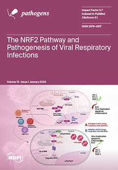
Respiratory viruses target the human respiratory system and cause various clinical symptoms in humans. Although several host factors have been found to play a crucial role in the pathogenesis of respiratory viral infections, the interaction between respiratory viruses and the host cellular response remains poorly understood. We focused on the impact of Nrf2 activation on the replication of respiratory viruses and summarized the scientific evidence on how certain respiratory viruses dysregulate the Nrf2 activation pathway. Obtaining insights into the crosstalk between respiratory viruses and the Nrf2 pathway will set the foundation for the use of established Nrf2 activators as therapeutics for viral infections. View this paper
  • Issues are regarded as officially published after their release is announced to the table of contents alert mailing list.
  • You may sign up for e-mail alerts to receive table of contents of newly released issues.
  • PDF is the official format for papers published in both, html and pdf forms. To view the papers in pdf format, click on the "PDF Full-text" link, and use the free Adobe Reader to open them.
Order results
Result details
Section
Select all
Export citation of selected articles as:
15 pages, 2969 KiB  
Article
Characterization of Variant RNAs Encapsidated during Bromovirus Infection by High-Throughput Sequencing
by Sarah Dexheimer, Nipin Shrestha, Bandana Sharma Chapagain, Jozef J. Bujarski and Yanbin Yin
Pathogens 2024, 13(1), 96; https://doi.org/10.3390/pathogens13010096 - 22 Jan 2024
Viewed by 1254
Abstract
Previously, we described the RNA recombinants accumulating in tissues infected with the bromoviruses BMV (Brome mosaic virus) and CCMV (Cowpea chlorotic mottle virus). In this work, we characterize the recombinants encapsidated inside the purified virion particles of BMV and CCMV. By using a [...] Read more.
Previously, we described the RNA recombinants accumulating in tissues infected with the bromoviruses BMV (Brome mosaic virus) and CCMV (Cowpea chlorotic mottle virus). In this work, we characterize the recombinants encapsidated inside the purified virion particles of BMV and CCMV. By using a tool called the Viral Recombination Mapper (ViReMa) that detects recombination junctions, we analyzed a high number of high-throughput sequencing (HTS) short RNA sequence reads. Over 28% of BMV or CCMV RNA reads did not perfectly map to the viral genomes. ViReMa identified 1.40% and 1.83% of these unmapped reads as the RNA recombinants, respectively, in BMV and CCMV. Intra-segmental crosses were more frequent than the inter-segmental ones. Most intra-segmental junctions carried short insertions/deletions (indels) and caused frameshift mutations. The mutation hotspots clustered mainly within the open reading frames. Substitutions of various lengths were also identified, whereas a small fraction of crosses occurred between viral and their host RNAs. Our data reveal that the virions can package detectable amounts of multivariate recombinant RNAs, contributing to the flexible nature of the viral genomes. Full article
Show Figures

Figure 1

Figure 1
<p>Flow chart of bioinformatics data analyses. The reference genomes for Bowtie mapping and ViReMa analysis include not only the viral genomes but also plant hosts’ transcriptomes (mRNAs and rRNAs). Bowtie version 1 was used before ViReMa analysis because Bowtie1 allows a perfect match without mismatches, and we aimed to collect unmapped reads with all kinds of variations for ViReMa analysis. Only reads indicated to have one mismatch from single alignment reads were fed into Bowtie2, which allows mismatches for SNP identification. The different categories of read outputs from ViReMa are explained in <a href="#sec3-pathogens-13-00096" class="html-sec">Section 3</a>.</p>
Full article ">Figure 2
<p>Schematic examples of potential types of recombinant reads. The thicker lines symbolize reference RNAs (black for viral RNAs and grey for host RNAs). The thinner blue and red lines represent recombinant reads, with blue (5′) and red (3′) representing two recombined portions of the reference RNAs. Please note that since the non-stranded sequencing library was prepared, cross types E and H cannot be assigned to the replicating + or − viral RNA strands.</p>
Full article ">Figure 3
<p>Circos plot of mutation events in BMV RNAs. From outside to inside: SNPs (gray bars), multi-base substitution events (≥10 supporting reads, purple histogram), inter-genome recombination (≥10 supporting reads, pink histogram), intra-genome recombination (≥10 supporting reads, orange histogram), genome ideogram (with ticks marking the nucleotide position), protein coding region shown as thick black lines, and encoded protein product labels, lines connecting two positions within an RNA (intra-genome crosses) and between two RNAs (inter-genome crosses). These recombination lines are directional, being color-coded according to which RNA the line starts from. For example, lines starting from NC_002026 are coded in dark blue, representing that the recombination has its 5′ end in this RNA. The table in the top left shows the height and spacing of the four histograms, representing the number of events found in each position. Higher means more events or hot mutation spots.</p>
Full article ">Figure 4
<p>Circos plot of mutation events in CCMV RNAs. From outside to inside: SNPs (gray bars), multi-base substitution events (≥10 supporting reads, purple histogram), inter-genome recombination (≥10 supporting reads, pink histogram), intra-genome recombination (≥10 supporting reads, orange histogram), genome ideogram (with ticks marking the nucleotide position), protein coding region shown as thick black lines and encoded protein product labels, lines connecting two positions within an RNA (intra-genome crosses) and between two RNAs (inter-genome crosses). These recombination lines are directional, being color-coded according to which RNA the line starts from. For example, lines starting from AF325739 are coded in dark blue, representing that the recombination has its 5′ end in this RNA. The table in the top left shows the height and spacing of the four histograms, representing the number of events found in each position. Higher means more events or hot mutation spots.</p>
Full article ">Figure 5
<p>Length distribution of substitutions in BMV and CCMV. The x-axis is the length starting at 2 nts. The y-axis is the frequency (i.e., the number of events having that length of substitution) shown in log10 scale.</p>
Full article ">
13 pages, 2409 KiB  
Article
Differing Expression and Potential Immunological Role of C-Type Lectin Receptors of Two Different Chicken Breeds against Low Pathogenic H9N2 Avian Influenza Virus
by Sungsu Youk, Dong-Hun Lee and Chang-Seon Song
Pathogens 2024, 13(1), 95; https://doi.org/10.3390/pathogens13010095 - 22 Jan 2024
Viewed by 1426
Abstract
Diverse immune responses in different chicken lines can result in varying clinical consequences following avian influenza virus (AIV) infection. We compared two widely used layer breeds, Lohmann Brown (LB) and Lohmann White (LW), to examine virus replication and immune responses against H9N2 AIV [...] Read more.
Diverse immune responses in different chicken lines can result in varying clinical consequences following avian influenza virus (AIV) infection. We compared two widely used layer breeds, Lohmann Brown (LB) and Lohmann White (LW), to examine virus replication and immune responses against H9N2 AIV infection. The transcription profile in the spleen of H9N2-infected chickens was compared using a microarray. Confirmatory real-time RT-PCR was used to measure the expression of C-type lectin, OASL, and MX1 genes. Additionally, to investigate the role of chicken lectin receptors in vitro, two C-type lectin receptors (CLRs) were expressed in DF-1 cells, and the early growth of the H9N2 virus was evaluated. The LB chickens shed a lower amount of virus from the cloaca compared with the LW chickens. Different expression levels of C-type lectin-like genes were observed in the transcription profile, with no significant differences in OASL or MX gene expression. Real-time RT-PCR indicated a sharp decrease in C-type lectin levels in the spleen of H9N2-infected LW chickens. In vitro studies demonstrated that cells overexpressing CLR exhibited lower virus replication, while silencing of homeostatic CLR had no effect on AIV replication. This study demonstrated distinct immune responses to H9N2 avian influenza in LB and LW chickens, particularly with differences in C-type lectin expression, potentially leading to lower virus shedding in LB chickens. Full article
(This article belongs to the Special Issue Pathogenesis, Epidemiology, and Control of Animal Influenza Viruses)
Show Figures

Figure 1

Figure 1
<p>Scatter plot of oropharyngeal (<b>A</b>) and cloacal (<b>B</b>) virus shedding detected by rRT-PCR from LB and LW chickens inoculated with H9N2 LPAI virus. Virus titers are expressed as log10 with error bars. For statistical purposes, rRT-PCR-negative samples were given a value of the limit of detection (1.8 log<sub>10</sub> EID<sub>50</sub>/mL). The circle (●) and square (■) plots indicate viral shedding from LB and LW chickens, respectively. The asterisk indicates the significance of the mean virus titer of 5 dpi oropharyngeal swab between LB and LW (<span class="html-italic">p</span> &lt; 0.001). The numbers of rRT-PCR-positive samples are indicated in parenthesis (virus shedding positive/total).</p>
Full article ">Figure 2
<p>Relative transcriptional gene expression profile in spleen of H9N2 infected LB and LW by microarray. H9N2 virus was inoculated into LB and LW layers. Spleen samples were collected from three chickens of each breed at each time point (1, 3, and 5 dpi). Relative gene expression of LB was measured by calculating LB/LW fold change. Differentially expressed genes were considered when criteria for fold change and <span class="html-italic">p</span>-value (fold change greater than 2 or smaller than 0.5 with <span class="html-italic">p</span>-value less than 0.05) were met. The number of differentially expressed genes was counted. C-type lectin-like, chOASL, and chMx mRNA are colored in blue, fuchsia, and black, respectively.</p>
Full article ">Figure 3
<p>Differential mRNA level of chC-lectin, chOASL, and chMx. Quantitative real-time reverse transcriptase polymerase chain reaction was conducted to confirm relative mRNA expression revealed by microarray. The relative gene expression levels were quantified by calculating the mean fold change; gene expression levels at a given time point were compared to that of uninfected chickens. The asterisk indicates the significance of the gene expression between LB and LW (<span class="html-italic">p</span> &lt; 0.001).</p>
Full article ">Figure 4
<p>Amino acid sequences of cloned CLR1 and CLR2. C-type lectin receptor sequences were cloned into expression vectors. Amino acid sequences of cloned pcDNA-CLR1 and pcDNA-CLR2 were aligned and compared with two references, <span class="html-italic">YLEC8</span> (NM_0013937243.2) and <span class="html-italic">YLEC17</span> (NM_001393744). The letters in sequences are highlighted based on polarity of amino acids. Potential cytoplasmic, transmembrane, and extracellular regions were annotated with a prediction tool implemented in Geneious Prime v. 2023.0.4. Highlighted areas in pcDNA-CLR2 and <span class="html-italic">YLEC17</span> are different sequences with amino acid abbreviations. Dots indicate consensus sequences. Dashed areas are blank. For extracellular carbohydrate-binding domains, two conserved C-type lectin domains across eukaryotic cells were predicted using position-specific scoring matrix (PSSM ID: 153,057 and 153063). Gray arrows indicate predicted putative ligand binding sites.</p>
Full article ">Figure 5
<p>Expression of CLRs in DF-1 cells. Expression of CLRs were analyzed by Western blotting (<b>A</b>) and immunofluorescence assay (<b>B</b>). DF-1 cells were transfected with pcDNA-CLR1 or pcDNA-CLR2. Cell lysate and medium were then harvested at 12 h after transfection and analyzed by Western blot assay. Separately, localization of CLRs was demonstrated using mouse anti-6X His tag antibody and Alex Fluor 488 anti-mouse secondary antibody (green). Cell nuclei were stained with 4,6-diamidino-2-phenylindole (blue).</p>
Full article ">Figure 6
<p>H9N2 virus growth under overexpression or downregulation of CLR in DF-1 cells. Transfection of CLR expressing plasmids (<b>A</b>) or siRNA targeting CLR mRNA (<b>B</b>) was conducted 12 h before inoculating virus. Supernatant virus titers were measured at 12 h after transfection using MDCK cells in triplicate. The asterisk indicates statistical significance by one-way ANOVA and Tukey’s post hoc comparison (<span class="html-italic">p</span> &lt; 0.05).</p>
Full article ">
14 pages, 258 KiB  
Article
The Assessment of Infection Risk in Patients with Vitiligo Undergoing Dialysis for End-Stage Renal Disease: A Retrospective Cohort Study
by Pearl Shah, Mitchell Hanson, Jennifer L. Waller, Sarah Tran, Stephanie L. Baer, Varsha Taskar and Wendy B. Bollag
Pathogens 2024, 13(1), 94; https://doi.org/10.3390/pathogens13010094 - 21 Jan 2024
Viewed by 1734
Abstract
Vitiligo is an autoimmune condition that causes patchy skin depigmentation. Although the mechanism by which vitiligo induces immunocompromise is unclear, other related autoimmune diseases are known to predispose those affected to infection. Individuals with vitiligo exhibit epidermal barrier disruption, which could potentially increase [...] Read more.
Vitiligo is an autoimmune condition that causes patchy skin depigmentation. Although the mechanism by which vitiligo induces immunocompromise is unclear, other related autoimmune diseases are known to predispose those affected to infection. Individuals with vitiligo exhibit epidermal barrier disruption, which could potentially increase their susceptibility to systemic infections; patients with renal disease also show a predisposition to infection. Nevertheless, there is little research addressing the risk of infection in dialysis patients with vitiligo in comparison to those without it. A retrospective analysis was performed on patients with end-stage renal disease (ESRD) in the United States Renal Data System who started dialysis between 2004 and 2019 to determine if ESRD patients with vitiligo are at an increased risk of bacteremia, cellulitis, conjunctivitis, herpes zoster, or septicemia. Multivariable logistic regression modeling indicated that female sex, black compared to white race, Hispanic ethnicity, hepatitis C infection, and tobacco use were associated with an enhanced risk of vitiligo, whereas increasing age and catheter, versus arteriovenous fistula, and access type were associated with a decreased risk. After controlling for demographics and clinical covariates, vitiligo was found to be significantly associated with an increased risk of bacteremia, cellulitis, and herpes zoster but not with conjunctivitis and septicemia. Full article
(This article belongs to the Section Bacterial Pathogens)
16 pages, 1241 KiB  
Review
The Roles and Interactions of Porphyromonas gingivalis and Fusobacterium nucleatum in Oral and Gastrointestinal Carcinogenesis: A Narrative Review
by Bing Wang, Juan Deng, Valentina Donati, Nabeel Merali, Adam E. Frampton, Elisa Giovannetti and Dongmei Deng
Pathogens 2024, 13(1), 93; https://doi.org/10.3390/pathogens13010093 - 20 Jan 2024
Cited by 4 | Viewed by 2784
Abstract
Epidemiological studies have spotlighted the intricate relationship between individual oral bacteria and tumor occurrence. Porphyromonas gingivalis and Fusobacteria nucleatum, which are known periodontal pathogens, have emerged as extensively studied participants with potential pathogenic abilities in carcinogenesis. However, the complex dynamics arising from [...] Read more.
Epidemiological studies have spotlighted the intricate relationship between individual oral bacteria and tumor occurrence. Porphyromonas gingivalis and Fusobacteria nucleatum, which are known periodontal pathogens, have emerged as extensively studied participants with potential pathogenic abilities in carcinogenesis. However, the complex dynamics arising from interactions between these two pathogens were less addressed. This narrative review aims to summarize the current knowledge on the prevalence and mechanism implications of P. gingivalis and F. nucleatum in the carcinogenesis of oral squamous cell carcinoma (OSCC), colorectal cancer (CRC), and pancreatic ductal adenocarcinoma (PDAC). In particular, it explores the clinical and experimental evidence on the interplay between P. gingivalis and F. nucleatum in affecting oral and gastrointestinal carcinogenesis. P. gingivalis and F. nucleatum, which are recognized as keystone or bridging bacteria, were identified in multiple clinical studies simultaneously. The prevalence of both bacteria species correlated with cancer development progression, emphasizing the potential impact of the collaboration. Regrettably, there was insufficient experimental evidence to demonstrate the synergistic function. We further propose a hypothesis to elucidate the underlying mechanisms, offering a promising avenue for future research in this dynamic and evolving field. Full article
(This article belongs to the Special Issue Opportunistic Oral Pathogens in Oral and Systemic Diseases)
Show Figures

Figure 1

Figure 1
<p>Multiple pathways employed by <span class="html-italic">P. gingivalis</span> in tumor induction: (1) P2X7 activation via ATP is blocked, leading to the stimulation of IL-1β, which promotes tumorigenesis, and the induction of ROS, which fosters a pro-inflammatory microenvironment. (2) Facilitation of immune evasion occurs through the activation of B7-H1 and B7-DC receptors, contributing to a (partial) circumvention of the immune system. Immune evasion is facilitated through the activation of B7-H1 and B7-DC receptors, contributing to a (partial) evasion of the immune system. (3) Activation of FimA results in the downregulation of p53, enhancing the host cell’s cell cycle while simultaneously suppressing apoptosis. The JAK/STAT axis is also implicated in the downregulation of apoptosis. (4) Additionally, <span class="html-italic">P. gingivalis</span> stimulates invasion through PAR2 activation via gingipains, activating NF-κB signaling, which leads to the formation of MMP-9, thereby enhancing <span class="html-italic">P. gingivalis</span> invasion. Upon invasion, pro-MMP-9 undergoes upregulation facilitated by ERK1/2 and ETS1, along with activation of p38 and HSP27. Abbreviations: ATP—adenosine triphosphate; ERK1/2—extracellular signal-regulated kinase 1/2; ETS1—protein; FimA—protein; HSP27—heat shock protein 27; IL-1β—interleukin-1β; JAK—Janus kinase 1; MMP-9—matrix metalloproteinase-9; NF-κB—nuclear factor kappa B; P2X7—purinergic receptor; pro-MMP-9—pro-matrix metalloproteinase-9; p38, p53—protein; ROS—reaction oxygen species; STAT—signal transducer and activator of transcription.</p>
Full article ">Figure 2
<p>The potential mechanisms of <span class="html-italic">F. nucleatum</span> in cancer: (1) The virulence factor FadA binds to E-cadherin, subsequently activating β-catenin. This activation, in turn, triggers the transcription factor Myc, leading to the activation of cyclin-D. The activation of cyclin-D stimulates host cell survival and proliferation. (2) <span class="html-italic">F. nucleatum</span> enhances invasion by activating p38, Etk, p70, and RhoA, resulting in the upregulation of MMP-13. Abbreviations: Etk—tyrosine kinase; FadA—<span class="html-italic">Fusobacterium</span> adhesin A; MMP-13—matrix metalloproteinase-13; p38—protein kinase; p70—S6 kinase; RhoA—kinase.</p>
Full article ">
6 pages, 183 KiB  
Brief Report
Mpox Virus in the Pharynx of Men Having Sex with Men: A Case Series
by Silvia Limonta, Giuseppe Lapadula, Luca Mezzadri, Laura Corsico, Francesca Rovida, Alice Ranzani, Fausto Baldanti and Paolo Bonfanti
Pathogens 2024, 13(1), 92; https://doi.org/10.3390/pathogens13010092 - 20 Jan 2024
Cited by 1 | Viewed by 1316
Abstract
The recent Mpox virus (MPV) outbreak in Europe and North America, primarily among men who have sex with men (MSM), raised concerns about various transmission sources. We examined patients with Mpox from an urban STI center in Lombardy, Italy, between May and August [...] Read more.
The recent Mpox virus (MPV) outbreak in Europe and North America, primarily among men who have sex with men (MSM), raised concerns about various transmission sources. We examined patients with Mpox from an urban STI center in Lombardy, Italy, between May and August 2022. Demographic, transmission, and clinical data were collected using a standardized form. Initial and subsequent tests were conducted using the RealStar Orthopoxvirus PCR Kit 1.0 (Altona Diagnostics, Hamburg, Germany) for skin lesions and oropharyngeal swabs. A total of 15 patients were recruited, all MSM, with 40% being HIV-positive. Almost all reported recent unprotected sexual activity. Oropharyngeal symptoms were observed in a minority, and oral cavity lesions were present in 20% of cases. MPV DNA was detected in skin lesions of 93% of patients and in oropharyngeal swabs of 87%. Skin samples exhibited a higher viral load than pharyngeal samples, with the latter persisting longer. Prospective follow-up of 11 individuals revealed an average pharyngeal persistence of 5.3 days beyond skin lesion clearance, reaching up to 80 days in an immunosuppressed case. Our findings indicate that MPV replication can persist in the pharynx asymptomatically and for an extended period. Full article
(This article belongs to the Special Issue Current Epidemic of Mpox)
14 pages, 3175 KiB  
Article
Impact of Plasmodium relictum Infection on the Colonization Resistance of Bird Gut Microbiota: A Preliminary Study
by Justė Aželytė, Apolline Maitre, Lianet Abuin-Denis, Elianne Piloto-Sardiñas, Alejandra Wu-Chuang, Rita Žiegytė, Lourdes Mateos-Hernández, Dasiel Obregón, Alejandro Cabezas-Cruz and Vaidas Palinauskas
Pathogens 2024, 13(1), 91; https://doi.org/10.3390/pathogens13010091 - 20 Jan 2024
Viewed by 1338
Abstract
Avian malaria infection has been known to affect host microbiota, but the impact of Plasmodium infection on the colonization resistance in bird gut microbiota remains unexplored. This study investigated the dynamics of Plasmodium relictum infection in canaries, aiming to explore the hypothesis that [...] Read more.
Avian malaria infection has been known to affect host microbiota, but the impact of Plasmodium infection on the colonization resistance in bird gut microbiota remains unexplored. This study investigated the dynamics of Plasmodium relictum infection in canaries, aiming to explore the hypothesis that microbiota modulation by P. relictum would reduce colonization resistance. Canaries were infected with P. relictum, while a control group was maintained. The results revealed the presence of P. relictum in the blood of all infected canaries. Analysis of the host microbiota showed no significant differences in alpha diversity metrics between infected and control groups. However, significant differences in beta diversity indicated alterations in the microbial taxa composition of infected birds. Differential abundance analysis identified specific taxa with varying prevalence between infected and control groups at different time points. Network analysis demonstrated a decrease in correlations and revealed that P. relictum infection compromised the bird microbiota’s ability to resist the removal of taxa but did not affect network robustness with the addition of new nodes. These findings suggest that P. relictum infection reduces gut microbiota stability and has an impact on colonization resistance. Understanding these interactions is crucial for developing strategies to enhance colonization resistance and maintain host health in the face of parasitic infections. Full article
(This article belongs to the Section Parasitic Pathogens)
Show Figures

Figure 1

Figure 1
<p>The dynamics of <span class="html-italic">P. relictum</span> parasitemia. Individual parasitemia values (% of infected erythrocytes) of <span class="html-italic">P. relictum</span> based on microscopy are presented. Different colors represent individual birds. DPI—days post-infection.</p>
Full article ">Figure 2
<p>Comparison of microbial diversity to assess the impact of <span class="html-italic">P. relictum</span> infection on the microbiota in birds between infected and control groups at 22 and 38 days post-infection (DPI). (<b>A</b>) Observed features, (<b>B</b>) Faith’s phylogenetic diversity (PD), and (<b>C</b>) Pielou’s evenness index. Comparison of beta–diversity with Bray–Curtis dissimilarity index for infected (triangle) and control (circle) groups at 22 (<b>D</b>) and 38 DPI (<b>E</b>), represented in PCoA plot obtained by Betadisper function. * <span class="html-italic">p</span> &lt; 0.05.</p>
Full article ">Figure 3
<p>Microbial community assemblies in <span class="html-italic">P. relictum</span>-infected and uninfected birds. Co-occurrence networks (<b>A</b>) were extrapolated from the microbiota of <span class="html-italic">P. relictum</span>-infected and control birds at 22 DPI and 38 DPI. Bacterial taxa with at least one connection are symbolized by nodes, whilst connected edges represent a significant correlation between them. The width of the edges corresponds to the level of co-occurrence correlation (SparCC, weight ≥ 0.5 or ≤−0.5). Green edges represent positive correlations. The colors of nodes specify clusters and modules in which taxa occur. The size of nodes is related to their eigenvector centrality. Venn diagrams displaying the number of shared and unique taxa detected within <span class="html-italic">P. relictum</span>-infected and control birds at 22 DPI (<b>B</b>) and 38 DPI (<b>C</b>).</p>
Full article ">Figure 4
<p>Core association networks inferred between <span class="html-italic">P. relictum</span>-infected and control groups at 22 DPI and 38 DPI. Positive (green) or negative (red) correlations are shown by the color of the edges. Nodes represent bacterial taxa.</p>
Full article ">Figure 5
<p>(<b>A</b>,<b>B</b>) Network robustness to node removal with cascading attack. Values of connectivity loss in <span class="html-italic">P. relictum</span>-infected (red) and control (blue) birds at 22 DPI and 38 DPI were compared. (<b>C</b>–<b>F</b>) Comparison of network robustness to node addition. The values of the largest connected component (LCC) (<b>C</b>,<b>D</b>) and average path length (<b>E</b>,<b>F</b>) are presented.</p>
Full article ">
15 pages, 365 KiB  
Article
High Genetic Diversity in Third-Generation Cephalosporin-Resistant Escherichia coli in Wastewater Systems of Schleswig-Holstein
by Laura Carlsen, Matthias Grottker, Malika Heim, Birte Knobling, Sebastian Schlauß, Kai Wellbrock and Johannes K. Knobloch
Pathogens 2024, 13(1), 90; https://doi.org/10.3390/pathogens13010090 - 20 Jan 2024
Viewed by 1376
Abstract
The spread of multidrug-resistant bacteria from humans or livestock is a critical issue. However, the epidemiology of resistant pathogens across wastewater pathways is poorly understood. Therefore, we performed a detailed comparison of third-generation cephalosporin-resistant Escherichia coli (3GCREC) from wastewater treatment plants (WWTPs) to [...] Read more.
The spread of multidrug-resistant bacteria from humans or livestock is a critical issue. However, the epidemiology of resistant pathogens across wastewater pathways is poorly understood. Therefore, we performed a detailed comparison of third-generation cephalosporin-resistant Escherichia coli (3GCREC) from wastewater treatment plants (WWTPs) to analyze dissemination pathways. A total of 172 3GCREC isolated from four WWTPs were characterized via whole genome sequencing. Clonal relatedness was determined using multi-locus sequence typing (MLST) and core genome MLST. Resistance genotypes and plasmid replicons were determined. A total of 68 MLST sequence types were observed with 28 closely related clusters. Resistance genes to eight antibiotic classes were detected. In fluoroquinolone-resistant isolates, resistance was associated with three-or-more point mutations in target genes. Typing revealed high genetic diversity with only a few clonal lineages present in all WWTPs. The distribution paths of individual lines could only be traced in exceptional cases with a lack of enrichment of certain lineages. Varying resistance genes and plasmids, as well as fluoroquinolone resistance-associated point mutations in individual isolates, further corroborated the high diversity of 3GCREC in WWTPs. In total, we observed high diversity of 3GCREC inside the tested WWTPs with proof of resistant strains being released into the environment even after treatment processes. Full article
Show Figures

Figure 1

Figure 1
<p>Subdivision of all isolates into phenotypic resistance types. If available, ten isolates per day from each WWTP and sampling site were analyzed for their resistance type (R01–R18). Resistance types displayed in red shades (R01 through R07) show phenotypic resistance to fluoroquinolones. Missing isolates were either identified as non-<span class="html-italic">E. coli</span> (influent), or there were no 10 β-glucuronidase-producing colonies (effluent). Letters and numbers mark the sampling location (i: influent; e: effluent) and sampling day within the sampling week.</p>
Full article ">Figure 2
<p>Resistance genes detected in all <span class="html-italic">E. coli</span>. Genes encoding resistance against β-lactams, fluoroquinolones (FQ), point mutations associated with FQ resistance (<b>A</b>), aminogylcosides (AMG), sulfonamides (SUL) (<b>B</b>), trimethoprim (TMP), tetracyclines (TET), MLS (macrolides, lincosamides, streptogramines) and phenicols (PHEN) (<b>C</b>) are displayed. Different colors represent the different WWTPs. The first bar represents the total number of analyzed isolates. Only genes/mutations that occurred at least five times are displayed.</p>
Full article ">Figure 3
<p>cgMLST analysis of clonal relations in <span class="html-italic">E. coli</span> isolates from all WWTPs. Isolates are colored depending on their WWTP origin. Grey-shaded clusters show clonally related subgroups according to cgMLST. Single locus variants of ST10 and ST38 are marked with a star. Additional information about phenotypic resistance types and genetic distances is displayed in <a href="#app1-pathogens-13-00090" class="html-app">Supplementary Figure S1</a>.</p>
Full article ">
10 pages, 1105 KiB  
Review
The Impact of Pathogens on Sepsis Prevalence and Outcome
by Birte Dyck, Matthias Unterberg, Michael Adamzik and Björn Koos
Pathogens 2024, 13(1), 89; https://doi.org/10.3390/pathogens13010089 - 20 Jan 2024
Cited by 3 | Viewed by 3295
Abstract
Sepsis, a severe global healthcare challenge, is characterized by significant morbidity and mortality. The 2016 redefinition by the Third International Consensus Definitions Task Force emphasizes its complexity as a “life-threatening organ dysfunction caused by a dysregulated host response to infection”. Bacterial pathogens, historically [...] Read more.
Sepsis, a severe global healthcare challenge, is characterized by significant morbidity and mortality. The 2016 redefinition by the Third International Consensus Definitions Task Force emphasizes its complexity as a “life-threatening organ dysfunction caused by a dysregulated host response to infection”. Bacterial pathogens, historically dominant, exhibit geographic variations, influencing healthcare strategies. The intricate dynamics of bacterial immunity involve recognizing pathogen-associated molecular patterns, triggering innate immune responses and inflammatory cascades. Dysregulation leads to immunothrombosis, disseminated intravascular coagulation, and mitochondrial dysfunction, contributing to the septic state. Viral sepsis, historically less prevalent, saw a paradigm shift during the COVID-19 pandemic, underscoring the need to understand the immunological response. Retinoic acid-inducible gene I-like receptors and Toll-like receptors play pivotal roles, and the cytokine storm in COVID-19 differs from bacterial sepsis. Latent viruses like human cytomegalovirus impact sepsis by reactivating during the immunosuppressive phases. Challenges in sepsis management include rapid pathogen identification, antibiotic resistance monitoring, and balancing therapy beyond antibiotics. This review highlights the evolving sepsis landscape, emphasizing the need for pathogen-specific therapeutic developments in a dynamic and heterogeneous clinical setting. Full article
Show Figures

Figure 1

Figure 1
<p>Pathogen prevalence in positive isolates in different cohorts over time, based on the publications of Vincent et al. 2006 [<a href="#B12-pathogens-13-00089" class="html-bibr">12</a>], Moreno et al. 2008 [<a href="#B13-pathogens-13-00089" class="html-bibr">13</a>], and Umemura et al. 2021 [<a href="#B8-pathogens-13-00089" class="html-bibr">8</a>].</p>
Full article ">Figure 2
<p>Simplified immune response during sepsis upon infection with a pathogen. Created with BioRender.com. PAMP = pathogen-associated molecular pattern; PRR = pattern-recognition receptor; DAMP = damage-associated molecular pattern; ROS = reactive oxygen species; NETosis = neutrophil extracellular trap formation; DIC = disseminated intravascular coagulation; ARDS = Acute Respiratory Distress Syndrome; MODS = Multiple Organ Dysfunction Syndrome; CRP = C-reactive protein, red upward arrow indicate elevated concentration.</p>
Full article ">
18 pages, 355 KiB  
Review
Antibiotic Resistance to Molecules Commonly Prescribed for the Treatment of Antibiotic-Resistant Gram-Positive Pathogens: What Is Relevant for the Clinician?
by Gianpiero Tebano, Irene Zaghi, Francesco Baldasso, Chiara Calgarini, Roberta Capozzi, Caterina Salvadori, Monica Cricca and Francesco Cristini
Pathogens 2024, 13(1), 88; https://doi.org/10.3390/pathogens13010088 - 19 Jan 2024
Cited by 1 | Viewed by 1577
Abstract
Antibiotic resistance in Gram-positive pathogens is a relevant concern, particularly in the hospital setting. Several antibiotics are now available to treat these drug-resistant pathogens, such as daptomycin, dalbavancin, linezolid, tedizolid, ceftaroline, ceftobiprole, and fosfomycin. However, antibiotic resistance can also affect these newer molecules. [...] Read more.
Antibiotic resistance in Gram-positive pathogens is a relevant concern, particularly in the hospital setting. Several antibiotics are now available to treat these drug-resistant pathogens, such as daptomycin, dalbavancin, linezolid, tedizolid, ceftaroline, ceftobiprole, and fosfomycin. However, antibiotic resistance can also affect these newer molecules. Overall, this is not a frequent phenomenon, but it is a growing concern in some settings and can compromise the effectiveness of these molecules, leaving few therapeutic options. We reviewed the available evidence about the epidemiology of antibiotic resistance to these antibiotics and the main molecular mechanisms of resistance, particularly methicillin-resistant Sthaphylococcus aureus, methicillin-resistant coagulase-negative staphylococci, vancomycin-resistant Enterococcus faecium, and penicillin-resistant Streptococcus pneumoniae. We discussed the interpretation of susceptibility tests when minimum inhibitory concentrations are not available. We focused on the risk of the emergence of resistance during treatment, particularly for daptomycin and fosfomycin, and we discussed the strategies that can be implemented to reduce this phenomenon, which can lead to clinical failure despite appropriate antibiotic treatment. The judicious use of antibiotics, epidemiological surveillance, and infection control measures is essential to preserving the efficacy of these drugs. Full article
(This article belongs to the Special Issue Antimicrobial Resistance of Pathogens Causing Nosocomial Infections)
17 pages, 14185 KiB  
Review
HTLV-1 Tax Tug-of-War: Cellular Senescence and Death or Cellular Transformation
by Marcia Bellon and Christophe Nicot
Pathogens 2024, 13(1), 87; https://doi.org/10.3390/pathogens13010087 - 19 Jan 2024
Cited by 3 | Viewed by 1868
Abstract
Human T cell leukemia virus type 1 (HTLV-1) is a retrovirus associated with a lymphoproliferative disease known as adult T cell leukemia/lymphoma (ATLL). HTLV-1 infection efficiently transforms human T cells in vivo and in vitro. The virus does not transduce a proto-oncogene, nor [...] Read more.
Human T cell leukemia virus type 1 (HTLV-1) is a retrovirus associated with a lymphoproliferative disease known as adult T cell leukemia/lymphoma (ATLL). HTLV-1 infection efficiently transforms human T cells in vivo and in vitro. The virus does not transduce a proto-oncogene, nor does it integrate into tumor-promoting genomic sites. Instead, HTLV-1 uses a random mutagenesis model, resulting in cellular transformation. Expression of the viral protein Tax is critical for the immortalization of infected cells by targeting specific cellular signaling pathways. However, Tax is highly immunogenic and represents the main target for the elimination of virally infected cells by host cytotoxic T cells (CTLs). In addition, Tax expression in naïve cells induces pro-apoptotic signals and has been associated with the induction of non-replicative cellular senescence. This review will explore these conundrums and discuss the mechanisms used by the Tax viral oncoprotein to influence life-and-death cellular decisions and affect HTLV-1 pathogenesis. Full article
(This article belongs to the Special Issue New Directions in HTLV-1 Research)
Show Figures

Figure 1

Figure 1
<p>Control of apoptosis pathways by HTLV-1 Tax. Tax-mediated activation of the IKK complex results in IκB degradation and nuclear translocation of RelA/p65 to activate the expression of pro-survival genes. Increased expression of survivin and IAPs (inhibitors of apoptosis) efficiently inhibits caspases while cFLIP inhibits death receptor FAS/FASL signaling. In addition, BclxL can translocate to the mitochondria where it blocks Bax- and Bak-mediated release of cytochrome C and activation of caspases. Activation of NF-κB is also associated with the release of cytokines resulting in an autocrine/paracrine activation loop engaging receptor tyrosine kinase and activation of the JAK/STAT pathway. Tax also prevents pro-apoptotic functions of p53.</p>
Full article ">Figure 2
<p>Cellular senescence pathways affected by Tax. Schematic representation of different physiological processes associated with induction of cellular senescence. While cellular division is associated with telomere shortening and replicative senescence, Tax can reactivate hTERT expression and stimulate its activity. These effects are sufficient to prevent telomere-induced foci (TIF) and activation of the DDR. Tax expression is associated with rapid cell proliferation and various metabolic stress progression, such as the production of reactive oxygen species (ROS). In turn, ROS induces DNA damage and activation of ATM/p53 response. Tax expression impairs DNA replication fork, resulting in stalling and fallout, creating an accumulation of double DNA strand breaks and activation of the DDR. Activation of the DDR can stimulate ATM/p53 or p16INK pathways, leading to irreversible cellular senescence.</p>
Full article ">Figure 3
<p>Schematic representation of cellular fates in response to various stress stimuli. Evolution of infected cells relative to the levels of Tax and HBZ expression. Upon stress signals, induced by replicative senescence, metabolic stress, high expression and oncogene-induced senescence, genotoxic agent exposure, and genomic DNA damage or chronic inflammation, the cell may undergo transient cell cycle arrest to repair and/or prevent accumulation of more cellular damages. If cell cycle checkpoints are not functioning properly or the damage is too extensive to be repaired, the cell may undergo apoptosis. Cells that undergo early senescence may progress to the irreversible late senescence stage in the presence of an active p53 signaling pathway or transformation if p53 is inactivated.</p>
Full article ">
15 pages, 3272 KiB  
Article
Epidemiological Investigation of Tick-Borne Bacterial Pathogens in Domestic Animals from the Qinghai–Tibetan Plateau Area, China
by Yihong Ma, Yingna Jian, Geping Wang, Iqra Zafar, Xiuping Li, Guanghua Wang, Yong Hu, Naoaki Yokoyama, Liqing Ma and Xuenan Xuan
Pathogens 2024, 13(1), 86; https://doi.org/10.3390/pathogens13010086 - 19 Jan 2024
Viewed by 1509
Abstract
The Qinghai–Tibetan Plateau area (QTPA) features a unique environment that has witnessed the selective breeding of diverse breeds of domestic livestock exhibiting remarkable adaptability. Nevertheless, Anaplasma spp., Rickettsia spp., Coxiella spp., and Borrelia spp. represent tick-borne bacterial pathogens that pose a global threat [...] Read more.
The Qinghai–Tibetan Plateau area (QTPA) features a unique environment that has witnessed the selective breeding of diverse breeds of domestic livestock exhibiting remarkable adaptability. Nevertheless, Anaplasma spp., Rickettsia spp., Coxiella spp., and Borrelia spp. represent tick-borne bacterial pathogens that pose a global threat and have substantial impacts on both human and animal health, as well as on the economy of animal husbandry within the Qinghai–Tibetan plateau area. In this study, a total of 428 samples were systematically collected from 20 distinct areas within the Qinghai Plateau. The samples included 62 ticks and 366 blood samples obtained from diverse animal species to detect the presence of Anaplasma spp., Rickettsia spp., Coxiella spp., and Borrelia spp. The prevalence of infection in this study was determined as follows: Anaplasma bovis accounted for 16.4% (70/428), A. capra for 4.7% (20/428), A. ovis for 5.8% (25/428), Borrelia burgdorferi sensu lato for 6.3% (27/428), Coxiella burnetii for 0.7% (3/428), and Rickettsia spp. for 0.5% (2/428). Notably, no cases of A. marginale and A. phagocytophilum infections were observed in this study. The findings revealed an elevated presence of these pathogens in Tibetan sheep and goats, with no infections detected in yaks, Bactrian camels, donkeys, and horses. To the best of our knowledge, this study represents the first investigation of tick-borne bacterial pathogens infecting goats, cattle, horses, and donkeys within the Qinghai Plateau of the Qinghai–Tibetan Plateau area. Consequently, our findings contribute valuable insights into the distribution and genetic diversity of Anaplasma spp., Rickettsia spp., Coxiella spp., and Borrelia spp. within China. Full article
(This article belongs to the Special Issue Parasites: Epidemiology, Treatment and Control: 2nd Edition)
Show Figures

Figure 1

Figure 1
<p>A map of the Qinghai Plateau highlighting the various sampling sites and animals included. The figure was created and adjusted using map data in Excel.</p>
Full article ">Figure 2
<p>Morphological identification of ticks in QTPA. (<b>a</b>) <span class="html-italic">H. qinghaiensis</span> detected in QTPA; (<b>b</b>) <span class="html-italic">D. nuttalli</span> from QTPA in this study.</p>
Full article ">Figure 3
<p>Phylogenetic tree of ticks using the Maximum Likelihood method. The numbers indicated at the nodes represent the percentage of occurrence of clades based on 1000 bootstrap replications of the data. The sequences of isolates obtained in this study, along with their corresponding accession numbers, are highlighted in red.</p>
Full article ">Figure 4
<p>Phylogenetic trees of <span class="html-italic">Anaplasma</span> spp. constructed using the Maximum Likelihood method in MEGA X, employing the Kimura 2-parameter model. (<b>a</b>) The phylogenetic tree of <span class="html-italic">A. bovis</span> based on 16s rRNA gene. (<b>b</b>) The phylogenetic tree of <span class="html-italic">A. capra</span> based on <span class="html-italic">gltA</span> genes. (<b>c</b>) The phylogenetic of <span class="html-italic">A. ovis</span> based on <span class="html-italic">msp4</span> gene. The numbers assigned to the nodes represent the percentage of occurrence of clades, determined through 1000 bootstrap replications of the data. Isolates from this study, along with their corresponding accession numbers, are highlighted in red.</p>
Full article ">Figure 5
<p>The phylogenetic tree of <span class="html-italic">B. burgdorferi</span> s.l., based on 16s rRNA partial sequences obtained from Tibetan sheep and goats in this study, as well as sequences retrieved from the GenBank database, was constructed using the Maximum Likelihood method in MEGA X and Kimura 2-parameter model. The numbers assigned to the nodes indicate the percentage of occurrence of clades, determined through 1000 bootstrap replications of the data. Isolates from this study, along with their corresponding accession numbers, are highlighted in red.</p>
Full article ">Figure 6
<p>The phylogenetic tree of <span class="html-italic">Rickettsia</span> spp., constructed using the Maximum Likelihood method in MEGA X and employing the Tamura 3-parameter model, includes numbers assigned to the nodes representing the percentage of occurrence of clades and was determined through 1000 bootstrap replications of the data. Isolates from this study, along with their corresponding accession numbers, are highlighted in red.</p>
Full article ">Figure 7
<p>The phylogenetic tree of <span class="html-italic">C. burnetii</span>, constructed using the Maximum Likelihood method in MEGA X and employing the Kimura 2-parameter model, includes numbers assigned to the nodes representing the percentage of occurrence of clades and was determined through 1000 bootstrap replications of the data. Isolates from this study, along with their corresponding accession numbers, are highlighted in red.</p>
Full article ">
9 pages, 1875 KiB  
Communication
Presence and Characterisation of Porcine Respirovirus 1 (PRV1) in Northern Italy
by Enrica Sozzi, Gabriele Leo, Cristina Bertasio, Giovanni Loris Alborali, Cristian Salogni, Matteo Tonni, Nicoletta Formenti, Davide Lelli, Ana Moreno, Tiziana Trogu, Sabrina Canziani, Clara Tolini, Monica Pierangela Cerioli and Antonio Lavazza
Pathogens 2024, 13(1), 85; https://doi.org/10.3390/pathogens13010085 - 18 Jan 2024
Viewed by 1321
Abstract
Porcine Respirovirus 1 (PRV1) is an enveloped, single-stranded, negative-sense RNA virus belonging to the genus Respirovirus within the Paramyxoviridae family. Since its first detection in China in 2013, PRV1 has been identified in several American and European countries. Although its pathogenicity is uncertain, [...] Read more.
Porcine Respirovirus 1 (PRV1) is an enveloped, single-stranded, negative-sense RNA virus belonging to the genus Respirovirus within the Paramyxoviridae family. Since its first detection in China in 2013, PRV1 has been identified in several American and European countries. Although its pathogenicity is uncertain, recent studies have suggested that it may play a role in the Porcine Respiratory Disease Complex (PRDC) because of its capacity to replicate in the upper and lower respiratory tracts. This study aimed to determine the spread of PRV1 in Northern Italy and the phylogeny of the isolates. Therefore, PRV1 was investigated using real-time RT-PCR in 902 samples collected from September 2022 to September 2023 from pigs with respiratory symptoms in North Italy. Fourteen (1.55%) samples tested as PRV1-positive. The full-length fusion (F) gene, which codifies for a major surface protein, was amplified and used for phylogenetic analysis to help carry out molecular epidemiological studies on this virus. In addition, swine influenza virus (SIV) and porcine reproductive and respiratory syndrome virus (PRRSV) infections were detected in most of the PRV1-positive samples. In conclusion, we report the detection of PRV1 in Italy and discuss its potential role as a co-factor in causing the Porcine Respiratory Disease Complex. Full article
(This article belongs to the Special Issue Molecular Detection and Surveillance of Veterinary Infectious Disease)
Show Figures

Figure 1

Figure 1
<p>Geographical distribution of the pig farms where PRV1 was identified. Farms for which F gene sequencing was obtained or not are shown with red and green dots, respectively.</p>
Full article ">Figure 2
<p>Two percent agarose gel showing the end-point RT-PCR amplification product of ten samples tested. M, marker 100 bp DNA Ladder (Invitrogen Inc., Carlsbad, CA, USA). From these results, it can be seen that samples 3, 4, 5, 6, 8, and 9 tested positive for the F-gene of interest. Samples 1, 2, 7, and 10 were negative.</p>
Full article ">Figure 3
<p>The phylogenetic tree is based on the F-gene sequencing of the PRV1s detected in the survey and the viral strains deposited in GenBank. Molecular analyses were performed with MEGA 10 software with bootstrap analysis (1000 replicates) using the Maximum Likelihood method based on the General Time Reversible + G + I model. Bootstrap values &gt; 60% are shown. Sequences generated in this study are indicated with a black rhombus. Note that the F sequence of one strain (334725-2/2022) was excluded since it was incomplete. Published sequences are identified by strain and GenBank accession number. The F gene sequence of HRV1 (GenBank acc.: NC003461) was used as an outgroup.</p>
Full article ">
19 pages, 1836 KiB  
Article
Markers of Inflammation, Tissue Damage, and Fibrosis in Individuals Diagnosed with Human Immunodeficiency Virus and Pneumonia: A Cohort Study
by Katherine Peña-Valencia, Will Riaño, Mariana Herrera-Diaz, Lucelly López, Diana Marín, Sandra Gonzalez, Olga Agudelo-García, Iván Arturo Rodríguez-Sabogal, Lázaro Vélez, Zulma Vanessa Rueda and Yoav Keynan
Pathogens 2024, 13(1), 84; https://doi.org/10.3390/pathogens13010084 - 18 Jan 2024
Cited by 1 | Viewed by 1555
Abstract
Previous studies have noted that persons living with human immunodeficiency virus (HIV) experience persistent lung dysfunction after an episode of community-acquired pneumonia (CAP), although the underlying mechanisms remain unclear. We hypothesized that inflammation during pneumonia triggers increased tissue damage and accelerated pulmonary fibrosis, [...] Read more.
Previous studies have noted that persons living with human immunodeficiency virus (HIV) experience persistent lung dysfunction after an episode of community-acquired pneumonia (CAP), although the underlying mechanisms remain unclear. We hypothesized that inflammation during pneumonia triggers increased tissue damage and accelerated pulmonary fibrosis, resulting in a gradual loss of lung function. We carried out a prospective cohort study of people diagnosed with CAP and/or HIV between 2016 and 2018 in three clinical institutions in Medellín, Colombia. Clinical data, blood samples, and pulmonary function tests (PFTs) were collected at baseline. Forty-one patients were included, divided into two groups: HIV and CAP (n = 17) and HIV alone (n = 24). We compared the concentrations of 17 molecules and PFT values between the groups. Patients with HIV and pneumonia presented elevated levels of cytokines and chemokines (IL-6, IL-8, IL-18, IL-1RA, IL-10, IP-10, MCP-1, and MIP-1β) compared to those with only HIV. A marked pulmonary dysfunction was evidenced by significant reductions in FEF25, FEF25-75, and FEV1. The correlation between these immune mediators and lung function parameters supports the connection between pneumonia-associated inflammation and end organ lung dysfunction. A low CD4 cell count (<200 cells/μL) predicted inflammation and lung dysfunction. These results underscore the need for targeted clinical approaches to mitigate the adverse impacts of CAP on lung function in this population. Full article
(This article belongs to the Special Issue Immunity to Respiratory Infections)
Show Figures

Figure 1

Figure 1
<p>Flow diagram of the process of selection and exclusion of participants in the study.</p>
Full article ">Figure 2
<p>Plasma concentrations (natural logarithm) of (<b>A</b>) CCL11/eotaxin; (<b>B</b>) IL-1β; (<b>C</b>) IL-1Ra/IL1; (<b>D</b>) IL-6; (<b>E</b>) IL-8; (<b>F</b>) IL-10; (<b>G</b>) IL-13; (<b>H</b>) IL-17A; (<b>I</b>) IL-18; (<b>J</b>) IP10/CXCL10; (<b>K</b>) MCP1/CCL2; (<b>L</b>) MIP-1α/CCL3; (<b>M</b>) MIP-1β/CCL4; (<b>N</b>) VEGF/VPF; (<b>O</b>) RANTES/CCL5; (<b>P</b>) PAI1; and (<b>Q</b>) sCD14 measured in HIV and CAP compared to those with HIV but no pneumonia. Immunoassays were performed on plasma samples from all 41 participants included in the study at baseline. The vertical axis indicates the measured plasma concentration of each molecule in pg/mL. Each symbol represents a data point from a single individual (a circle indicates individual data points from the HIV and CAP group; a triangle indicates individual data points from the HIV group). At the time of admission, the concentrations of IL-6, IL-8, IL-10, IL-18, IL-1Ra/IL1, IP10/CXCL10, MCP1/CCL2, and MIP-1β/CCL4 were significantly higher in the HIV and CAP group than in the HIV group. The concentrations of the remaining molecules were not statistically different between the two groups at the time of the baseline. * Statistically significant if <span class="html-italic">p</span>-value &lt; 0.05.</p>
Full article ">Figure 2 Cont.
<p>Plasma concentrations (natural logarithm) of (<b>A</b>) CCL11/eotaxin; (<b>B</b>) IL-1β; (<b>C</b>) IL-1Ra/IL1; (<b>D</b>) IL-6; (<b>E</b>) IL-8; (<b>F</b>) IL-10; (<b>G</b>) IL-13; (<b>H</b>) IL-17A; (<b>I</b>) IL-18; (<b>J</b>) IP10/CXCL10; (<b>K</b>) MCP1/CCL2; (<b>L</b>) MIP-1α/CCL3; (<b>M</b>) MIP-1β/CCL4; (<b>N</b>) VEGF/VPF; (<b>O</b>) RANTES/CCL5; (<b>P</b>) PAI1; and (<b>Q</b>) sCD14 measured in HIV and CAP compared to those with HIV but no pneumonia. Immunoassays were performed on plasma samples from all 41 participants included in the study at baseline. The vertical axis indicates the measured plasma concentration of each molecule in pg/mL. Each symbol represents a data point from a single individual (a circle indicates individual data points from the HIV and CAP group; a triangle indicates individual data points from the HIV group). At the time of admission, the concentrations of IL-6, IL-8, IL-10, IL-18, IL-1Ra/IL1, IP10/CXCL10, MCP1/CCL2, and MIP-1β/CCL4 were significantly higher in the HIV and CAP group than in the HIV group. The concentrations of the remaining molecules were not statistically different between the two groups at the time of the baseline. * Statistically significant if <span class="html-italic">p</span>-value &lt; 0.05.</p>
Full article ">Figure 3
<p>Lung function measures showing differences between groups.</p>
Full article ">
16 pages, 1134 KiB  
Article
Genetic Analysis of H5N1 High-Pathogenicity Avian Influenza Virus following a Mass Mortality Event in Wild Geese on the Solway Firth
by Craig S. Ross, Alexander M. P. Byrne, Sahar Mahmood, Saumya Thomas, Scott Reid, Lorna Freath, Larry R. Griffin, Marco Falchieri, Paul Holmes, Nick Goldsmith, Jessica M. Shaw, Alastair MacGugan, James Aegerter, Rowena Hansen, Ian H. Brown and Ashley C. Banyard
Pathogens 2024, 13(1), 83; https://doi.org/10.3390/pathogens13010083 - 17 Jan 2024
Cited by 1 | Viewed by 3132
Abstract
The United Kingdom (UK) and Europe have seen successive outbreaks of H5N1 clade 2.3.4.4b high-pathogenicity avian influenza virus (HPAIV) since 2020 peaking in the autumn/winter periods. During the 2021/22 season, a mass die-off event of Svalbard Barnacle Geese (Branta leucopsis) was [...] Read more.
The United Kingdom (UK) and Europe have seen successive outbreaks of H5N1 clade 2.3.4.4b high-pathogenicity avian influenza virus (HPAIV) since 2020 peaking in the autumn/winter periods. During the 2021/22 season, a mass die-off event of Svalbard Barnacle Geese (Branta leucopsis) was observed on the Solway Firth, a body of water on the west coast border between England and Scotland. This area is used annually by Barnacle Geese to over-winter, before returning to Svalbard to breed. Following initial identification of HPAIV in a Barnacle Goose on 8 November 2021, up to 32% of the total Barnacle Goose population may have succumbed to disease by the end of March 2022, along with other wild bird species in the area. Potential adaptation of the HPAIV to the Barnacle Goose population within this event was evaluated. Whole-genome sequencing of thirty-three HPAIV isolates from wild bird species demonstrated that there had been two distinct incursions of the virus, but the two viruses had remained genetically stable within the population, whilst viruses from infected wild birds were closely related to those from poultry cases occurring in the same region. Analysis of sera from the following year demonstrated that a high percentage (76%) of returning birds had developed antibodies to H5 AIV. This study demonstrates genetic stability of this strain of HPAIV in wild Anseriformes, and that, at the population scale, whilst there is a significant impact on survival, a high proportion of birds recover following infection. Full article
(This article belongs to the Special Issue Avian Virus Infection)
Show Figures

Figure 1

Figure 1
<p>Location of wild birds testing positive for H5N1 HPAIV on the Solway Firth—Mapped position of H5 HPAI positive samples submitted to APHA for analysis. Regions of Barnacle Geese counts are shown with total counts for each region over the winter of 2021/2022 (adapted from [<a href="#B7-pathogens-13-00083" class="html-bibr">7</a>]).</p>
Full article ">Figure 2
<p>Barnacle Geese counts and samples submitted from Solway Firth region (<b>A</b>) total count of Barnacle geese from the Solway Firth region for 2022/23 and comparison to counts from previous years, (<b>B</b>) cumulative Barnacle Geese mortalities counted on the Solway Firth for 2021/2022, (<b>C</b>) positive and negative samples submitted to APHA for H5 HPAI analysis during winter 2021/2022 (A and B adapted from [<a href="#B7-pathogens-13-00083" class="html-bibr">7</a>]).</p>
Full article ">Figure 3
<p>The HA of H5N1 HPAI isolated from birds on the Solway Firth are closely linked. A maximum-phylogenetic tree of the HA gene from H5N1 HPAI samples from United Kingdom between 2020 and 2022. The tips of UK samples are coloured according to genotype and Solway Firth bird type.</p>
Full article ">
11 pages, 3089 KiB  
Article
Formulating Parasiticidal Fungi in Dried Edible Gelatins to Reduce the Risk of Infection by Trichuris sp. among Continuous Grazing Bison
by Rami Salmo, Cándido Viña, Izaro Zubiria, José Ángel Hernández Malagón, Jaime M. Sanchís, Cristiana Cazapal, María Sol Arias, Rita Sánchez-Andrade and Adolfo Paz-Silva
Pathogens 2024, 13(1), 82; https://doi.org/10.3390/pathogens13010082 - 17 Jan 2024
Viewed by 1029
Abstract
Control of infection by gastrointestinal nematodes remains a big problem in ruminants under continuous grazing. For the purpose of decreasing the risk of infection by Trichuris sp. in captive bison (Bison bison) always maintained in the same plot, dried gelatins having [...] Read more.
Control of infection by gastrointestinal nematodes remains a big problem in ruminants under continuous grazing. For the purpose of decreasing the risk of infection by Trichuris sp. in captive bison (Bison bison) always maintained in the same plot, dried gelatins having ≥106 chlamydospores of both Mucor circinelloides and Duddingtonia flagrans were given to them for one week, and at the end, fecal samples (FF) collected each week for four weeks were analyzed immediately. Feces taken one week prior to gelatin administration served as controls (CF). Eggs of Trichuris sp. were sorted into non-viable and viable, then classified into viable undeveloped (VU), viable with cellular development (VCD), or viable infective (VI). Ovistatic and ovicidal effects were determined throughout the study. In FF, viability of Trichuris eggs decreased between 9% (first week) and 57% (fourth week), egg development was delayed during the first two weeks, and VI percentages were significantly lower than in CF (p = 0.001). It is concluded that the preparation of gelatins with chlamydospores of parasiticidal fungi and their subsequent dehydration offer an edible formulation that is ready to use, stress-free to supply, and easy to store, as well as being well-accepted by ruminants and highly efficient to reduce the risk of Trichuris sp. infection among animals under continuous grazing regimes. Full article
Show Figures

Figure 1

Figure 1
<p>Gelatins were made from a blend of 10<sup>7</sup> chlamydospores of both <span class="html-italic">M. circinelloides</span> and <span class="html-italic">D. flagrans,</span> and then dehydrated.</p>
Full article ">Figure 2
<p>Captive bison under continuous grazing and passing eggs of <span class="html-italic">Trichuris</span> sp. in their feces received dried gelatins containing a blend of 10<sup>6</sup> chlamydospores of both <span class="html-italic">M. circinelloides</span> and <span class="html-italic">D. flagrans</span> each (Marcelle Natureza Zoological Park, NW Spain).</p>
Full article ">Figure 3
<p>Evolution of eggs of <span class="html-italic">Trichuris</span> sp. in feces of bison kept under continuous grazing. CF (controls): samples collected prior to providing bison fungal chlamydospores. (<b>a</b>) Viable unembryonated, with two nuclei; (<b>b</b>) viable with cellular development (VCD); (<b>c</b>) viable with cylindrical larva (VI, infective). FF: samples taken after giving bison a blend of chlamydospores. (<b>d</b>) Viable undeveloped (VU); (<b>e</b>) cytoplasm vacuolization (non-viable); (<b>f</b>) empty, with eggshell disrupted (non-viable).</p>
Full article ">Figure 4
<p>Percentages of viable eggs of <span class="html-italic">Trichuris</span> sp. in feces of bison under continuous pasturing. FF: samples taken after giving bison a blend of chlamydospores; CF (controls): samples collected prior to providing bison fungal chlamydospores. Points represent the percentage average ± 2 SD.</p>
Full article ">Figure 5
<p>Stages of viable eggs of <span class="html-italic">Trichuris</span> sp. in feces of bison under rotational grazing (CF). VU: unembryonated (zygote); VCD: with cellular development; VI: infective. Points represent the percentage average ± 2 SD.</p>
Full article ">Figure 6
<p>Stages of viable eggs of <span class="html-italic">Trichuris</span> sp. in feces of bison kept under continuous pasturing, after receiving dry gelatins containing a blend of chlamydospores of <span class="html-italic">M. circinelloides</span> and <span class="html-italic">Duddingtonia flagrans</span> (FF). VU: unembryonated (zygote); VCD: with cellular development; VI: infective. Points represent the percentage average ± 2 SD.</p>
Full article ">
22 pages, 423 KiB  
Review
Coinfection of Babesia and Borrelia in the Tick Ixodes ricinus—A Neglected Public Health Issue in Europe?
by Thomas G. T. Jaenson, Jeremy S. Gray, Per-Eric Lindgren and Peter Wilhelmsson
Pathogens 2024, 13(1), 81; https://doi.org/10.3390/pathogens13010081 - 17 Jan 2024
Cited by 1 | Viewed by 3157
Abstract
Ixodes ricinus nymphs and adults removed from humans, and larvae and nymphs from birds, have been analysed for infection with Babesia species and Borrelia species previously in separately published studies. Here, we use the same data set to explore the coinfection pattern of [...] Read more.
Ixodes ricinus nymphs and adults removed from humans, and larvae and nymphs from birds, have been analysed for infection with Babesia species and Borrelia species previously in separately published studies. Here, we use the same data set to explore the coinfection pattern of Babesia and Borrelia species in the ticks. We also provide an overview of the ecology and potential public health importance in Sweden of I. ricinus infected both with zoonotic Babesia and Borrelia species. Among 1952 nymphs and adult ticks removed from humans, 3.1% were PCR-positive for Babesia spp. Of these Babesia-positive ticks, 43% were simultaneously Borrelia-positive. Among 1046 immatures of I. ricinus removed from birds, 2.5% were Babesia-positive, of which 38% were coinfected with Borrelia species. This study shows that in I. ricinus infesting humans or birds in Sweden, potentially zoonotic Babesia protozoa sometimes co-occur with human-pathogenic Borrelia spp. Diagnostic tests for Babesia spp. infection are rarely performed in Europe, and the medical significance of this pathogen in Europe could be underestimated. Full article
10 pages, 1842 KiB  
Article
Evaluation of Antibacterial and Antibiofilm Activity of Rice Husk Extract against Staphylococcus aureus
by Gloria Burlacchini, Angela Sandri, Adele Papetti, Ilaria Frosi, Federico Boschi, Maria M. Lleo and Caterina Signoretto
Pathogens 2024, 13(1), 80; https://doi.org/10.3390/pathogens13010080 - 16 Jan 2024
Cited by 1 | Viewed by 2216
Abstract
Infections caused by Staphylococcus aureus are particularly difficult to treat due to the high rate of antibiotic resistance. S. aureus also forms biofilms that reduce the effects of antibiotics and disinfectants. Therefore, new therapeutic approaches are increasingly required. In this scenario, plant waste [...] Read more.
Infections caused by Staphylococcus aureus are particularly difficult to treat due to the high rate of antibiotic resistance. S. aureus also forms biofilms that reduce the effects of antibiotics and disinfectants. Therefore, new therapeutic approaches are increasingly required. In this scenario, plant waste products represent a source of bioactive molecules. In this study, we evaluated the antimicrobial and antibiofilm activity of the rice husk extract (RHE) on S. aureus clinical isolates. In a biofilm inhibition assay, high concentrations of RHE counteracted the formation of biofilm by S. aureus isolates, both methicillin-resistant (MRSA) and -sensitive (MSSA). The observation of the MRSA biofilm by confocal laser scanning microscopy using live/dead cell viability staining confirmed that the bacterial viability in the RHE-treated biofilm was reduced. However, the extract showed no or little biofilm disaggregation ability. An additive effect was observed when treating S. aureus with a combination of RHE and oxacillin/cefoxitin. In Galleria mellonella larvae treated with RHE, the extract showed no toxicity even at high concentrations. Our results support that the rice husk has antimicrobial and antibiofilm properties and could potentially be used in the future in topical solutions or on medical devices to prevent biofilm formation. Full article
(This article belongs to the Special Issue Bacterial Biofilm Infections and Treatment)
Show Figures

Figure 1

Figure 1
<p>Growth curves of MSSA (<b>a</b>) and MRSA (<b>b</b>) isolates grown in absence/presence of RHE 70×. Colony-forming units (CFU) were counted after plating serial dilutions collected from the cultures every hour for the first 9 h. Growth rate (<b>c</b>) was calculated from the exponential phase of the growth curves. Each value represents the mean ± SD of three experiments. Growth rate in absence vs. presence of RHE 70× was analyzed by one-tailed <span class="html-italic">t</span>-test; * <span class="html-italic">p</span> &lt; 0.05.</p>
Full article ">Figure 2
<p>Biofilms formed by the MRSA isolate in (<b>a</b>) absence and (<b>b</b>) presence of RHE 70×. Bacterial cells are stained with SYTO9 green fluorescent dye. Damaged cells are counter-stained with propidium iodide (PI) red fluorescent nucleic acid dye.</p>
Full article ">
26 pages, 2847 KiB  
Review
Polyamine Metabolism for Drug Intervention in Trypanosomatids
by Yolanda Pérez-Pertejo, Carlos García-Estrada, María Martínez-Valladares, Sankaranarayanan Murugesan, Rosa M. Reguera and Rafael Balaña-Fouce
Pathogens 2024, 13(1), 79; https://doi.org/10.3390/pathogens13010079 - 16 Jan 2024
Cited by 3 | Viewed by 1925
Abstract
Neglected tropical diseases transmitted by trypanosomatids include three major human scourges that globally affect the world’s poorest people: African trypanosomiasis or sleeping sickness, American trypanosomiasis or Chagas disease and different types of leishmaniasis. Different metabolic pathways have been targeted to find antitrypanosomatid drugs, [...] Read more.
Neglected tropical diseases transmitted by trypanosomatids include three major human scourges that globally affect the world’s poorest people: African trypanosomiasis or sleeping sickness, American trypanosomiasis or Chagas disease and different types of leishmaniasis. Different metabolic pathways have been targeted to find antitrypanosomatid drugs, including polyamine metabolism. Since their discovery, the naturally occurring polyamines, putrescine, spermidine and spermine, have been considered important metabolites involved in cell growth. With a complex metabolism involving biosynthesis, catabolism and interconversion, the synthesis of putrescine and spermidine was targeted by thousands of compounds in an effort to produce cell growth blockade in tumor and infectious processes with limited success. However, the discovery of eflornithine (DFMO) as a curative drug against sleeping sickness encouraged researchers to develop new molecules against these diseases. Polyamine synthesis inhibitors have also provided insight into the peculiarities of this pathway between the host and the parasite, and also among different trypanosomatid species, thus allowing the search for new specific chemical entities aimed to treat these diseases and leading to the investigation of target-based scaffolds. The main molecular targets include the enzymes involved in polyamine biosynthesis (ornithine decarboxylase, S-adenosylmethionine decarboxylase and spermidine synthase), enzymes participating in their uptake from the environment, and the enzymes involved in the redox balance of the parasite. In this review, we summarize the research behind polyamine-based treatments, the current trends, and the main challenges in this field. Full article
(This article belongs to the Special Issue Leishmaniasis: Transmission, Pathogenesis and Treatment)
Show Figures

Graphical abstract

Graphical abstract
Full article ">Figure 1
<p>Chemical structure of DFMO (eflornithine) and mechanism of action as irreversible inhibitor of ornithine decarboxylase (ODC) in <span class="html-italic">T. brucei</span>.</p>
Full article ">Figure 2
<p>Schematic representation of a bloodstream <span class="html-italic">T. brucei</span> trypomastigote (<b>left picture</b>) and an intracellular <span class="html-italic">T. cruzi</span> and <span class="html-italic">Leishmania</span> amastigote (<b>right picture</b>). The major metabolic pathways that have been considered as potential drug targets are indicated. Some organelles are also represented: C: cytosol; G: glycosome; K: kinetoplast; M: mitochondrion; N: nucleus.</p>
Full article ">Figure 3
<p>The canonic polyamine biosynthetic pathway of mammals (hosts of trypanosomatids). The enzymes involved in polyamine biosynthesis are shown in black boxes, and the metabolites are in empty circles. The full polyamine metabolic pathway includes biosynthesis and interconversion routes that starts from the essential amino acid L-methionine and from the semi-essential L-arginine. Putrescine, spermidine and spermine are synthesized from L-ornithine and AdoMetDc after decarboxylation, the activity of both enzymes being closely regulated by the cells. Both enzymes have been addressed as druggable targets for therapeutic intervention. Note that regardless of trypanosomatids, there is not any connection between polyamines and glutathione in mammals. Abbreviations of the enzymes and transporters and their corresponding EC numbers are given in alphabetic order: AdoMetDC, S-adenosylmethione decarboxylase (EC 4.1.1.50); APAO: N 1-acetylpolyamine oxidase (EC 1.5.3.13); Arginase (EC 3.5.3.1); DHS, deoxyhypusine synthase (EC: 2.5.1.46); DOHH deoxyhypusine hydroxylase (EC 1.14.99.29); MAT, methionine adenosyltransferase (EC 2.5.1.6); ODC, ornithine decarboxylase (EC 4.1.1.17); POT1, putrescine transport 1 (EC 7.6.2.16); SpdS, spermidine synthase (EC 2.5.1.16); SpmS, spermine synthase (EC 2.5.1.22). Abbreviations for the metabolites are also given in alphabetic order: AdoMet, S-adenosylmethionine; dcAdoMet, decarboxylated S-adenosylmethionine; eIF5A, eukaryotic translation initiation factor 5A; dh-eIF5A, deoxyhypusine-eIF5A; h-eIF5A, hypusine-eIF5A.</p>
Full article ">Figure 4
<p>The polyamine biosynthetic pathway of trypanosomatid (African, American trypanosomes and <span class="html-italic">Leishmania</span>) is much simpler than the host pathway and lacks spermine and the whole interconversion pathway. However, it is in part devoted to ROS detoxification by means of the conjugation of spermidine to glutathione to form trypanothione (see <a href="#pathogens-13-00079-f003" class="html-fig">Figure 3</a>). Polyamine biosynthesis has some specific peculiarities involving: the lack of a true arginase in <span class="html-italic">T. brucei</span> and <span class="html-italic">T. cruzi</span> and the lack of the <span class="html-italic">odc</span> encoding gene in <span class="html-italic">T. cruzi</span> that make this parasite auxotrophic for putrescine and/or spermidine. Abbreviations of the enzymes and transporters and their corresponding EC numbers are given in alphabetic order: AdoMetDC, S-adenosylmethione decarboxylase (EC 4.1.1.50); Arginase (EC 3.5.3.1); DHS, deoxyhypusine synthase (EC: 2.5.1.46); DOHH deoxyhypusine hydroxylase (EC 1.14.99.29); MAT, methionine adenosyltransferase (EC 2.5.1.6); ODC, ornithine decarboxylase (EC 4.1.1.17); POT1, putrescine transport 1 (EC 7.6.2.16); SpdS, spermidine synthase (EC 2.5.1.16); Abbreviations for the metabolites are also given in alphabetic order: AdoMet, S-adenosylmethionine; dcAdoMet, decarboxylated S-adenosylmethionine; eIF5A, eukaryotic translation initiation factor 5A; dh-eIF5A, deoxyhypusine-eIF5A; h-eIF5A, hypusine-eIF5A.</p>
Full article ">Figure 5
<p>Host–parasite interplay in <span class="html-italic">Leishmania</span> infections. L-arginine plays a key role in macrophage activation and parasite survival during infection. Within the host macrophage, L-arginine is substrate of inducible nitric oxide synthase (iNOS) and arginase. iNOS/arginase balance is transcriptionally controlled by interleukins and is modulated at a biochemical level too. On the one hand, the classic M1 proinflammatory activation of macrophages responds to Th1 cytokines such as TNFa and IFNg and IL1, IL2 and IL10 interleukins. Alternative M2 anti-inflammatory activation of macrophages responds to Th2 cytokines such as TGFb, IL4, IL10 and IL13. M1 activation induces iNOS in macrophages, responsible for L-arginine cleavage to NO and citrulline byproduct. NO will promote the cascade production of nitrogen reactive species (RNOS) such as peroxynitrite radical (ONOO-). On the other hand, L-arginine cleavage by iNOS is a two-step enzymatic process that produces an important intermediate <span class="html-italic">N</span>-hydroxy-L-arginine (NOHA), which, before its complete hydrolysis to NO, can interfere arginase activity, preventing L-ornithine and polyamine production. Th2 response implies an increase in arginase activity, resulting in the formation of L-ornithine and polyamines, which can be used by the parasite. Inside the parasitophorous vacuole, <span class="html-italic">Leishmania</span> amastigotes can obtain L-arginine and polyamine from the host using the corresponding active transporters. <span class="html-italic">Leishmania</span> can synthesize putrescine and spermidine from L-arginine and L-methionine, which are essentials for the parasite, but <span class="html-italic">T. cruzi</span> is auxothroph for putrescine since it lacks genes encoding for both, a true arginase and ODC. * Absent in <span class="html-italic">T. brucei</span> and <span class="html-italic">T. cruzi</span>; ** Absent in <span class="html-italic">T. cruzi</span>.</p>
Full article ">Figure 6
<p>Trypanothione biosynthetic pathway and redox balance in trypanosomatids. Reduced trypanothione is a complex formed by two molecules of reduced glutathione bridged by their glycine residues with spermidine. Trypanothione can be oxidized by ROS to the oxidized form, establishing a sulfur redox balance enzymatically controlled by trypanothione reductase. The singularity of this ROS scavenger in trypanosomatids is an interesting druggable target for drug intervention in trypanosomatids. Abbreviations of the enzymes and transporters and their corresponding EC numbers are given in alphabetic order: <span class="html-italic">γ</span>GCS, <span class="html-italic">γ</span>-glutamylcysteine synthetase (EC 6.3.2.2); GS, glutathione synthase (EC 6.3.2.3); GSS, glutathionyl spermidine synthetase (EC 6.3.1.8); TryR, trypanothione reductase (EC 1.8.1.12); TryS, trypanothione synthase (EC 6.3.1.9). Abbreviations for the metabolites are also given in alphabetic order: <span class="html-italic">γ</span>GC, <span class="html-italic">γ</span>-glutamylcysteine; Gsp, glutathionylspermidine; GSH, reduced glutathione; ROS, reactive oxygen species; T(SH)2, reduced trypanothione; TS2, oxidized trypanothione.</p>
Full article ">Figure 7
<p>ODC inhibitors. DFMO (<b>1</b>) and other different chemical scaffolds inhibiting <span class="html-italic">Trypanosoma</span> ODC.</p>
Full article ">Figure 8
<p>Polyamine uptake inhibitors. MGBG (<b>5</b>), the diamidines CGP40215A (<b>6</b>) and CGP48664A (<b>7</b>), MDL 73811 (<b>8</b>), UTSam568 (<b>9</b>).</p>
Full article ">Figure 9
<p>AdoMetDC inhibitors. Triclabendazole (<b>10</b>), isotretinoin (<b>11</b>)<b>,</b> promazine (<b>12</b>), chlorpromazine (<b>13</b>) and chlomipramine (<b>14</b>).</p>
Full article ">Figure 10
<p>Polyamine transport system inhibitors. Ant4 (<b>15</b>), GW5074 (<b>16</b>).</p>
Full article ">
10 pages, 916 KiB  
Article
Mycoplasma gallisepticum and Mycoplasma synoviae in Turkeys in Poland
by Olimpia Kursa, Grzegorz Tomczyk, Agata Sieczkowska, Sylwia Kostka and Anna Sawicka-Durkalec
Pathogens 2024, 13(1), 78; https://doi.org/10.3390/pathogens13010078 - 15 Jan 2024
Viewed by 1793
Abstract
The pathogenic mycoplasmas are among the bacteria causing significant losses in the poultry industry worldwide. Mycoplasma gallisepticum (MG) and M. synoviae (MS) are economically important pathogens causing chronic respiratory disease, decreased growth, egg production and hatchability rates, and significant downgrading of carcasses. Effective [...] Read more.
The pathogenic mycoplasmas are among the bacteria causing significant losses in the poultry industry worldwide. Mycoplasma gallisepticum (MG) and M. synoviae (MS) are economically important pathogens causing chronic respiratory disease, decreased growth, egg production and hatchability rates, and significant downgrading of carcasses. Effective diagnosis of infection with these species in poultry is highly requisite considering their two routes of spreading—horizontal and vertical. Their prevalence and molecular epidemiology were investigated in 184 turkey flocks in Poland. Tracheal samples were selected from 144 broiler flocks and 40 turkey breeder flocks collected in 2015–2023. The prevalence of MG was determined by real-time PCR targeting the 16S rRNA gene and PCR targeting the mgc2 gene, and MS was determined by a 16–23S rRNA real-time PCR and a vlhA gene PCR. Further identification and molecular characterization were carried out using PCR and sequencing. M. gallisepticum and M. synoviae were found in 8.33% and 9.72% of turkey broiler flocks respectively. The phylogenetic analysis of MG isolates in most cases showed high similarity to the ts-11-like strains. MS isolates showed high similarity to strains isolated from flocks of laying hens causing EAA. Additional tests detected Ornithobacterium rhinotracheale, Gallibacterium anatis, Enterococcus faecalis and Enterococcus faecium, Staphylococcus aureus and Riemerella anatipestifer. These secondary pathogens could have significantly heightened the pathogenicity of the mycoplasma infections studied. Full article
(This article belongs to the Section Bacterial Pathogens)
Show Figures

Figure 1

Figure 1
<p>Phylogenetic tree constructed based on <span class="html-italic">mgc2</span> gene of MG strains isolated from turkey flocks and reference strains.</p>
Full article ">Figure 2
<p>Phylogenetic tree created based on the <span class="html-italic">vlhA</span> gene sequences of MS strains isolated from turkey flocks, the reference strain and the vaccine strain.</p>
Full article ">
10 pages, 1251 KiB  
Article
First Report of Benzimidazole Resistance in Field Population of Haemonchus contortus from Sheep, Goats and Cattle in Bosnia and Herzegovina
by Naida Kapo, Jasmin Omeragić, Šejla Goletić, Emina Šabić, Adis Softić, Ahmed Smajlović, Indira Mujezinović, Vedad Škapur and Teufik Goletić
Pathogens 2024, 13(1), 77; https://doi.org/10.3390/pathogens13010077 - 15 Jan 2024
Cited by 1 | Viewed by 1238
Abstract
Haemonchus contortus is a globally significant parasitic nematode in ruminants, with widespread resistance to benzimidazole due to its excessive and prolonged use. Given the extensive use of benzimidazole anthelmintics in Bosnia and Herzegovina, we hypothesized that resistance is prevalent. The aim of this [...] Read more.
Haemonchus contortus is a globally significant parasitic nematode in ruminants, with widespread resistance to benzimidazole due to its excessive and prolonged use. Given the extensive use of benzimidazole anthelmintics in Bosnia and Herzegovina, we hypothesized that resistance is prevalent. The aim of this study was to identify the presence of anthelmintic resistance to benzimidazole in H. contortus from naturally infected sheep, goats and cattle in Bosnia and Herzegovina through the detection of the Phe/Tyr polymorphism in the amino acid at position 200 of the β-tubulin protein. From 19 locations in Bosnia and Herzegovina, a total of 83 adult H. contortus were collected from the abomasum of ruminants. Among these, 45 H. contortus specimens were isolated from sheep, 19 from goats and 19 from cattle. Results showed that 77.8% of H. contortus in sheep exhibited homozygous resistant genotypes at position 200 of the β-tubulin gene, with 15.5% being heterozygous. In goats, all tested H. contortus (100%) were homozygous resistant, and no heterozygous resistant or homozygous sensitive genotypes were found. Cattle had 94.7% homozygous resistant H. contortus, with no heterozygous resistant genotypes detected. In H. contortus from sheep and cattle, 6.7% and 5.3%, respectively, displayed homozygous sensitive genotypes. This study, for the first time, highlights the presence of a resistant population of H. contortus in sheep, goats and cattle in Bosnia and Herzegovina, using the rt-qPCR method. The resistance likely spread from sheep or goats to cattle, facilitated by shared pastures and the practice of transhumance, indicating a widespread and growing issue of anthelmintic resistance. Full article
(This article belongs to the Special Issue Parasitic Helminths: Drug Resistance, Control and Immune Evasion)
Show Figures

Figure 1

Figure 1
<p>Sampling locations across B&amp;H, from which abomasum samples were collected from sheep, goats and cattle.</p>
Full article ">Figure 2
<p>The Tm (melting temperature) values of the amplicons were obtained by analyzing their dissociation characteristics, with continuous heating and fluorescence measurement in a temperature range of 65 °C to 95 °C, following the completion of the EvaGreen<sup>®</sup> rt-qPCR method. Clear peaks of the tested sample amplicons and significantly different Tm values of the samples (Tm = 83.2–84.3 °C) compared to the PCR water used as a negative control (70.2 °C) confirm the specificity of the amplification process.</p>
Full article ">
28 pages, 363 KiB  
Review
Biofilm Producing Methicillin-Resistant Staphylococcus aureus (MRSA) Infections in Humans: Clinical Implications and Management
by Ashlesha Kaushik, Helen Kest, Mangla Sood, Bryan W. Steussy, Corey Thieman and Sandeep Gupta
Pathogens 2024, 13(1), 76; https://doi.org/10.3390/pathogens13010076 - 15 Jan 2024
Cited by 1 | Viewed by 4915
Abstract
Since its initial description in the 1960s, methicillin-resistant Staphylococcus aureus (MRSA) has developed multiple mechanisms for antimicrobial resistance and evading the immune system, including biofilm production. MRSA is now a widespread pathogen, causing a spectrum of infections ranging from superficial skin issues to [...] Read more.
Since its initial description in the 1960s, methicillin-resistant Staphylococcus aureus (MRSA) has developed multiple mechanisms for antimicrobial resistance and evading the immune system, including biofilm production. MRSA is now a widespread pathogen, causing a spectrum of infections ranging from superficial skin issues to severe conditions like osteoarticular infections and endocarditis, leading to high morbidity and mortality. Biofilm production is a key aspect of MRSA’s ability to invade, spread, and resist antimicrobial treatments. Environmental factors, such as suboptimal antibiotics, pH, temperature, and tissue oxygen levels, enhance biofilm formation. Biofilms are intricate bacterial structures with dense organisms embedded in polysaccharides, promoting their resilience. The process involves stages of attachment, expansion, maturation, and eventually disassembly or dispersion. MRSA’s biofilm formation has a complex molecular foundation, involving genes like icaADBC, fnbA, fnbB, clfA, clfB, atl, agr, sarA, sarZ, sigB, sarX, psm, icaR, and srtA. Recognizing pivotal genes for biofilm formation has led to potential therapeutic strategies targeting elemental and enzymatic properties to combat MRSA biofilms. This review provides a practical approach for healthcare practitioners, addressing biofilm pathogenesis, disease spectrum, and management guidelines, including advances in treatment. Effective management involves appropriate antimicrobial therapy, surgical interventions, foreign body removal, and robust infection control practices to curtail spread within healthcare environments. Full article
(This article belongs to the Special Issue Advances in Treatment of Biofilm Infections)
19 pages, 1840 KiB  
Review
SARS-CoV-2 ORF3a Protein as a Therapeutic Target against COVID-19 and Long-Term Post-Infection Effects
by Jiantao Zhang, Kellie Hom, Chenyu Zhang, Mohamed Nasr, Volodymyr Gerzanich, Yanjin Zhang, Qiyi Tang, Fengtian Xue, J. Marc Simard and Richard Y. Zhao
Pathogens 2024, 13(1), 75; https://doi.org/10.3390/pathogens13010075 - 14 Jan 2024
Cited by 2 | Viewed by 3344
Abstract
The COVID-19 pandemic caused by SARS-CoV-2 has posed unparalleled challenges due to its rapid transmission, ability to mutate, high mortality and morbidity, and enduring health complications. Vaccines have exhibited effectiveness, but their efficacy diminishes over time while new variants continue to emerge. Antiviral [...] Read more.
The COVID-19 pandemic caused by SARS-CoV-2 has posed unparalleled challenges due to its rapid transmission, ability to mutate, high mortality and morbidity, and enduring health complications. Vaccines have exhibited effectiveness, but their efficacy diminishes over time while new variants continue to emerge. Antiviral medications offer a viable alternative, but their success has been inconsistent. Therefore, there remains an ongoing need to identify innovative antiviral drugs for treating COVID-19 and its post-infection complications. The ORF3a (open reading frame 3a) protein found in SARS-CoV-2, represents a promising target for antiviral treatment due to its multifaceted role in viral pathogenesis, cytokine storms, disease severity, and mortality. ORF3a contributes significantly to viral pathogenesis by facilitating viral assembly and release, essential processes in the viral life cycle, while also suppressing the body’s antiviral responses, thus aiding viral replication. ORF3a also has been implicated in triggering excessive inflammation, characterized by NF-κB-mediated cytokine production, ultimately leading to apoptotic cell death and tissue damage in the lungs, kidneys, and the central nervous system. Additionally, ORF3a triggers the activation of the NLRP3 inflammasome, inciting a cytokine storm, which is a major contributor to the severity of the disease and subsequent mortality. As with the spike protein, ORF3a also undergoes mutations, and certain mutant variants correlate with heightened disease severity in COVID-19. These mutations may influence viral replication and host cellular inflammatory responses. While establishing a direct link between ORF3a and mortality is difficult, its involvement in promoting inflammation and exacerbating disease severity likely contributes to higher mortality rates in severe COVID-19 cases. This review offers a comprehensive and detailed exploration of ORF3a’s potential as an innovative antiviral drug target. Additionally, we outline potential strategies for discovering and developing ORF3a inhibitor drugs to counteract its harmful effects, alleviate tissue damage, and reduce the severity of COVID-19 and its lingering complications. Full article
(This article belongs to the Section Vaccines and Therapeutic Developments)
Show Figures

Figure 1

Figure 1
<p>(<b>A</b>) Structure and unique features of SARS-CoV-2 ORF3a protein (adapted from [<a href="#B18-pathogens-13-00075" class="html-bibr">18</a>,<a href="#B28-pathogens-13-00075" class="html-bibr">28</a>]). The nucleotide sequence of <span class="html-italic">ORF3a</span> is 825 bp in length and encodes a 31 kD protein of 275 amino acids. It shows an extracellular N-terminal signal peptide (aa1–13), 3 transmembrane (TM) domains (aa40–128) that are across the cellular membrane and 8 antiparallel β-sheets (β1–β8, aa145–235) at the cytoplasmic C-terminus. Other functional motifs include 3 caveolin-binding motifs (aa69–77, aa107–114 and aa141–149) [<a href="#B32-pathogens-13-00075" class="html-bibr">32</a>]; a YXXΦ motif (aa160–163) and a diG motif (aa177–178) [<a href="#B31-pathogens-13-00075" class="html-bibr">31</a>,<a href="#B33-pathogens-13-00075" class="html-bibr">33</a>,<a href="#B34-pathogens-13-00075" class="html-bibr">34</a>], and a PBM (aa272–275) [<a href="#B18-pathogens-13-00075" class="html-bibr">18</a>,<a href="#B35-pathogens-13-00075" class="html-bibr">35</a>]. A putative YXXΦ motif (aa233–236) is also indicated [<a href="#B36-pathogens-13-00075" class="html-bibr">36</a>]. (<b>B</b>) Cellular pathways of ORF3a-mediated effects that lead to the NLRP3 inflammasome activation and cytokine storm (<b>a</b>) [<a href="#B37-pathogens-13-00075" class="html-bibr">37</a>,<a href="#B38-pathogens-13-00075" class="html-bibr">38</a>,<a href="#B39-pathogens-13-00075" class="html-bibr">39</a>], NF-κB-mediated cytokine production, apoptosis and necrosis leading to kidney injury (<b>b</b>) [<a href="#B40-pathogens-13-00075" class="html-bibr">40</a>], and disruption of autophagy-lysosomal pathway by ORF3a that causes reactive microglia and astrocyte activation leading to neuroinflammation and brain damage (<b>c</b>) [<a href="#B41-pathogens-13-00075" class="html-bibr">41</a>]. Elevation of kidney injury molecule 1 (KIM-1) is a specific biomarker for kidney injury [<a href="#B42-pathogens-13-00075" class="html-bibr">42</a>]; elevation of glial fibrillary acidic protein (GFAP) is a well-accepted biomarker for brain damage [<a href="#B43-pathogens-13-00075" class="html-bibr">43</a>,<a href="#B44-pathogens-13-00075" class="html-bibr">44</a>].</p>
Full article ">
13 pages, 1704 KiB  
Article
Complement Suppresses the Initial Type 1 Interferon Response to Ocular Herpes Simplex Virus Type 1 Infection in Mice
by Daniel J. J. Carr, Adrian Filiberti and Grzegorz B. Gmyrek
Pathogens 2024, 13(1), 74; https://doi.org/10.3390/pathogens13010074 - 13 Jan 2024
Viewed by 1857
Abstract
The complement system (CS) contributes to the initial containment of viral and bacterial pathogens and clearance of dying cells in circulation. We previously reported mice deficient in complement component 3 (C3KO mice) were more sensitive than wild-type (WT) mice to ocular HSV-1 infection, [...] Read more.
The complement system (CS) contributes to the initial containment of viral and bacterial pathogens and clearance of dying cells in circulation. We previously reported mice deficient in complement component 3 (C3KO mice) were more sensitive than wild-type (WT) mice to ocular HSV-1 infection, as measured by a reduction in cumulative survival and elevated viral titers in the nervous system but not the cornea between days three and seven post infection (pi). The present study was undertaken to determine if complement deficiency impacted virus replication and associated changes in inflammation at earlier time points in the cornea. C3KO mice were found to possess significantly (p < 0.05) less infectious virus in the cornea at 24 h pi that corresponded with a decrease in HSV-1 lytic gene expression at 12 and 24 h pi compared to WT animals. Flow cytometry acquisition found no differences in the myeloid cell populations residing in the cornea including total macrophage and neutrophil populations at 24 h pi with minimal infiltrating cell populations detected at the 12 h pi time point. Analysis of cytokine and chemokine content in the cornea measured at 12 and 24 h pi revealed that only CCL3 (MIP-1α) was found to be different between WT and C3KO mice with >2-fold increased levels (p < 0.05, ANOVA and Tukey’s post hoc t-test) in the cornea of WT mice at 12 h pi. C3KO mouse resistance to HSV-1 infection at the early time points correlated with a significant increase in type I interferon (IFN) gene expression including IFN-α1 and IFN-β and downstream effector genes including tetherin and RNase L (p < 0.05, Mann–Whitney rank order test). These results suggest early activation of the CS interferes with the induction of the type I IFN response and leads to a transient increase in virus replication following corneal HSV-1 infection. Full article
Show Figures

Figure 1

Figure 1
<p><b>HSV-1 replication is reduced in the cornea of C3KO mice</b>. (<b>A</b>) Titers of HSV-1 in the cornea of WT and C3KO mice at 24 h PI as determined by plaque assay. (<b>B</b>–<b>D</b>) HSV-1 lytic gene expression including (<b>B</b>) ICP27, (<b>C</b>) TK, and (<b>D</b>) gB at the indicated time point PI as determined by real-time RT-PCR. The mean ± SEM (n = 6–8 samples/time point) is plotted for each time point. Each graph is a summary of the results from 2–3 experiments. **** <span class="html-italic">p</span> &lt; 0.0001, ** <span class="html-italic">p</span> &lt; 0.01, * <span class="html-italic">p</span> &lt; 0.05 comparing the two groups/time point as determined by Bonferroni–Dunn <span class="html-italic">t</span>-test method.</p>
Full article ">Figure 2
<p><b>C3 levels peak in the cornea of WT mice at 12 h PI.</b> WT and C3KO mice were infected with HSV-1 (500 PFU/cornea). At 12 or 24 h PI, the mice were exsanguinated and the corneas were removed and processed for C3 content by ELISA. The results are depicted as mean + SEM (n = 4–8 samples/time point) and plotted for each time point. The 0 h PI time point represents uninfected mice to serve as background control. The graph is a summary of the results from 2–3 experiments. ** <span class="html-italic">p</span> &lt; 0.01, * <span class="html-italic">p</span> &lt; 0.05 comparing the indicated groups as determined by the Holm–Sidak multiple <span class="html-italic">t</span>-test method.</p>
Full article ">Figure 3
<p><b>Myeloid cell infiltration into the cornea of WT and C3KO mice does not differ at 24 h PI.</b> WT and C3KO mice were infected with HSV-1 (500 PFU/cornea). At 24 h PI, the mice were exsanguinated, and the cornea was removed and processed for myeloid cell content by flow cytometry. The gating strategy is shown for monocyte/macrophage and neutrophil populations in panel (<b>A</b>). Panel (<b>B</b>) shows the summary of myeloid cell types, n = 5–6/group, * <span class="html-italic">p</span> &lt; 0.05 comparing the indicated group as determined by Mann–Whitney rank order test.</p>
Full article ">Figure 4
<p><b>MIP-1α/CCL3 is elevated early in the cornea of WT mice in response to HSV-1 infection.</b> WT and C3KO mice were infected with HSV-1 (500 PFU/cornea). At 12 or 24 h PI, the mice were exsanguinated, and the cornea was removed, homogenized, and clarified supernatant was assessed for analyte content by suspension array. Bars represent mean ± SEM, n = 8–10/group/time point from 3 experiments. * <span class="html-italic">p</span> &lt; 0.05 comparing the indicated group as determined by ANOVA and Tukey’s post-hoc <span class="html-italic">t</span>-test.</p>
Full article ">Figure 5
<p><b>IFN-β and RNase L expression are elevated in the cornea of C3KO mice early post HSV-1 infection.</b> C57BL/6 WT and C3KO male and female mice (n = 6–10/group/time point) were infected with HSV-1 McKrae (500–1000 PFU/cornea). At the indicated time PI, the mice were exsanguinated and the corneas were processed for mRNA analysis by real-time RT-PCR for relative expression of (<b>A</b>) IFN-α1, (<b>B</b>) IFN-β, (<b>C</b>) PKR, (<b>D</b>) RNase L. The results are expressed as the mean ± SEM relative value as determined using the ∆∆C<sub>t</sub> method. ** <span class="html-italic">p</span> &lt; 0.01, * <span class="html-italic">p</span> &lt; 0.05 comparing the indicated groups at the indicated time point as determined by Holm–Sidak multiple <span class="html-italic">t</span>-test method.</p>
Full article ">
9 pages, 231 KiB  
Editorial
Biting Back: Advances in Fighting Ticks and Understanding Tick-Borne Pathogens
by Anastasia Diakou
Pathogens 2024, 13(1), 73; https://doi.org/10.3390/pathogens13010073 - 13 Jan 2024
Cited by 1 | Viewed by 1376
Abstract
Ticks are blood-feeding arthropods and obligate ectoparasites of virtually all animal species (except fish) and humans [...] Full article
20 pages, 5728 KiB  
Article
Myxoma Virus Combination Therapy Enhances Lenalidomide and Bortezomib Treatments for Multiple Myeloma
by Alpay Yeşilaltay, Dilek Muz, Berna Erdal, Türker Bilgen, Bahadır Batar, Burhan Turgut, Birol Topçu, Bahar Yılmaz and Burcu Altındağ Avcı
Pathogens 2024, 13(1), 72; https://doi.org/10.3390/pathogens13010072 - 12 Jan 2024
Viewed by 1679
Abstract
This study aimed to explore the effectiveness and safety of Myxoma virus (MYXV) in MM cell lines and primary myeloma cells obtained from patients with multiple myeloma. Myeloma cells were isolated from MM patients and cultured. MYXV, lenalidomide, and bortezomib were used in [...] Read more.
This study aimed to explore the effectiveness and safety of Myxoma virus (MYXV) in MM cell lines and primary myeloma cells obtained from patients with multiple myeloma. Myeloma cells were isolated from MM patients and cultured. MYXV, lenalidomide, and bortezomib were used in MM cells. The cytotoxicity assay was investigated using WST-1. Apoptosis was assessed through flow cytometry with Annexin V/PI staining and caspase-9 concentrations using ELISA. To explore MYXV entry into MM cells, monoclonal antibodies were used. Moreover, to explore the mechanisms of MYXV entry into MM cells, we examined the level of GFP-labeled MYXV within the cells after blocking with monoclonal antibodies targeting BCMA, CD20, CD28, CD33, CD38, CD56, CD86, CD117, CD138, CD200, and CD307 in MM cells. The study demonstrated the effects of treating Myxoma virus with lenalidomide and bortezomib. The treatment resulted in reduced cell viability and increased caspase-9 expression. Only low-dose CD86 blockade showed a significant difference in MYXV entry into MM cells. The virus caused an increase in the rate of apoptosis in the cells, regardless of whether it was administered alone or in combination with drugs. The groups with the presence of the virus showed higher rates of early apoptosis. The Virus, Virus + Bortezomib, and Virus + Lenalidomide groups had significantly higher rates of early apoptosis (p < 0.001). However, the measurements of late apoptosis and necrosis showed variability. The addition of MYXV resulted in a statistically significant increase in early apoptosis in both newly diagnosed and refractory MM patients. Our results highlight that patient-based therapy should also be considered for the effective management of MM. Full article
(This article belongs to the Section Vaccines and Therapeutic Developments)
Show Figures

Figure 1

Figure 1
<p>Fluorescence microscopy of growth of MYXV in the BHK-21 cell line (×10) (<b>A</b>), fluorescence microscopy of growth of MYXV in the Vero cell line (×10) (<b>B</b>), fluorescence microscopy of growth of MYXV in the Vero cell line. (<b>C</b>,<b>D</b>): ×20; (<b>E</b>): ×10.</p>
Full article ">Figure 2
<p>The appearance of virus growth foci under the fluorescence microscope in the microtitration test using the BHK-21 control cell line of MYXV. (<b>A</b>) Virus dilution ×10, (<b>B</b>,<b>C</b>) virus dilution ×20, (<b>D</b>) control. The appearance of virus growth foci under the fluorescence microscope in the microtitration test using the Vero control cell line of MYXV. (<b>E</b>–<b>G</b>) Virus dilution ×10. (<b>H</b>) Control.</p>
Full article ">Figure 3
<p>Flow cytometry results. Scatter plots (<b>A</b>) and histograms of cell counts (<b>B</b>) for negative control. Scatter plots (<b>C</b>) and histograms of cell counts (<b>D</b>) for positive control.</p>
Full article ">Figure 4
<p>Annexin V/PI-staining flow cytometry image of apoptosis in myeloma cells from a newly diagnosed patient with MM. (<b>A</b>) MYXV-infected cell, (<b>B</b>) MYXV-infected cell combined with bortezomib, (<b>C</b>) MYXV-infected cell combined with lenalidomide, (<b>D</b>) MYXV-infected cell combined with bortezomib and lenalidomide, (<b>E</b>) control, (<b>F</b>) bortezomib-administered cell, (<b>G</b>) lenalidomide-administered cell, (<b>H</b>) bortezomib- and lenalidomide-administered cell. Live cells were visualized in the red part and apoptotic cells were blue part.</p>
Full article ">Figure 5
<p>Annexin V/PI-staining flow cytometry image of apoptosis in myeloma cells from a refractory patient with MM. (<b>A</b>) MYXV-infected cell, (<b>B</b>) MYXV-infected cell combined with bortezomib, (<b>C</b>) MYXV-infected cell combined with lenalidomide, (<b>D</b>) MYXV-infected cell combined with bortezomib and lenalidomide, (<b>E</b>) control, (<b>F</b>) bortezomib-administered cell, (<b>G</b>) lenalidomide-administered cell, (<b>H</b>) bortezomib- and lenalidomide-administered cell. Live cells were visualized in the red part and apoptotic cells were blue part.</p>
Full article ">
17 pages, 673 KiB  
Article
A Genome-Wide Association Study for Resistance to Tropical Theileriosis in Two Bovine Portuguese Autochthonous Breeds
by Diana Valente, Octávio Serra, Nuno Carolino, Jacinto Gomes, Ana Cláudia Coelho, Pedro Espadinha, José Pais and Inês Carolino
Pathogens 2024, 13(1), 71; https://doi.org/10.3390/pathogens13010071 - 12 Jan 2024
Viewed by 1555
Abstract
The control of Tropical Theileriosis, a tick-borne disease with a strong impact on cattle breeding, can be facilitated using marker-assisted selection in breeding programs. Genome-wide association studies (GWAS) using high-density arrays are extremely important for the ongoing process of identifying genomic variants associated [...] Read more.
The control of Tropical Theileriosis, a tick-borne disease with a strong impact on cattle breeding, can be facilitated using marker-assisted selection in breeding programs. Genome-wide association studies (GWAS) using high-density arrays are extremely important for the ongoing process of identifying genomic variants associated with resistance to Theileria annulata infection. In this work, single-nucleotide polymorphisms (SNPs) were analyzed in the Portuguese autochthonous cattle breeds Alentejana and Mertolenga. In total, 24 SNPs suggestive of significance (p ≤ 10−4) were identified for Alentejana cattle and 20 SNPs were identified for Mertolenga cattle. The genomic regions around these SNPs were further investigated for annotated genes and quantitative trait loci (QTLs) previously described by other authors. Regarding the Alentejana breed, the MAP3K1, CMTM7, SSFA2, and ATG13 genes are located near suggestive SNPs and appear as candidate genes for resistance to Tropical Theileriosis, considering its action in the immune response and resistance to other diseases. On the other hand, in the Mertolenga breed, the UOX gene is also a candidate gene due to its apparent link to the pathogenesis of the disease. These results may represent a first step toward the possibility of including genetic markers for resistance to Tropical Theileriosis in current breed selection programs. Full article
(This article belongs to the Section Epidemiology of Infectious Diseases)
Show Figures

Figure 1

Figure 1
<p>Manhattan plot of the association study for resistance to tropical theileriosis in two Portuguese autochthonous bovine breeds. The genome-wide significance threshold is indicated by the dashed line (<span class="html-italic">p</span> &lt; 10<sup>−4</sup>). The red dots above the threshold line represent the SNPs considered as significant in this work. The position of the bovine chromosome is shown on the <span class="html-italic">x</span>-axis. The strength of association for a GWAS single-locus mixed model is shown on the <span class="html-italic">y</span>-axis. (<b>a</b>) Data from Alentejana breed animals. (<b>b</b>) Data from Mertolenga breed animals.</p>
Full article ">
17 pages, 1962 KiB  
Article
Chromosome-Level Assemblies for the Pine Pitch Canker Pathogen Fusarium circinatum
by Lieschen De Vos, Magriet A. van der Nest, Quentin C. Santana, Stephanie van Wyk, Kyle S. Leeuwendaal, Brenda D. Wingfield and Emma T. Steenkamp
Pathogens 2024, 13(1), 70; https://doi.org/10.3390/pathogens13010070 - 12 Jan 2024
Cited by 1 | Viewed by 1503
Abstract
The pine pitch canker pathogen, Fusarium circinatum, is globally regarded as one of the most important threats to commercial pine-based forestry. Although genome sequences of this fungus are available, these remain highly fragmented or structurally ill-defined. Our overall goal was to provide [...] Read more.
The pine pitch canker pathogen, Fusarium circinatum, is globally regarded as one of the most important threats to commercial pine-based forestry. Although genome sequences of this fungus are available, these remain highly fragmented or structurally ill-defined. Our overall goal was to provide high-quality assemblies for two notable strains of F. circinatum, and to characterize these in terms of coding content, repetitiveness and the position of telomeres and centromeres. For this purpose, we used Oxford Nanopore Technologies MinION long-read sequences, as well as Illumina short sequence reads. By leveraging the genomic synteny inherent to F. circinatum and its close relatives, these sequence reads were assembled to chromosome level, where contiguous sequences mostly spanned from telomere to telomere. Comparative analyses unveiled remarkable variability in the twelfth and smallest chromosome, which is known to be dispensable. It presented a striking length polymorphism, with one strain lacking substantial portions from the chromosome’s distal and proximal regions. These regions, characterized by a lower gene density, G+C content and an increased prevalence of repetitive elements, contrast starkly with the syntenic segments of the chromosome, as well as with the core chromosomes. We propose that these unusual regions might have arisen or expanded due to the presence of transposable elements. A comparison of the overall chromosome structure revealed that centromeric elements often underpin intrachromosomal differences between F. circinatum strains, especially at chromosomal breakpoints. This suggests a potential role for centromeres in shaping the chromosomal architecture of F. circinatum and its relatives. The publicly available genome data generated here, together with the detailed metadata provided, represent essential resources for future studies of this important plant pathogen. Full article
(This article belongs to the Special Issue Plant Pathogenic Fungi)
Show Figures

Figure 1

Figure 1
<p>(<b>A</b>) Sequence comparison between the set of 12 pseudomolecules compiled for each genome. MUMmer revealed high levels of synteny across the <span class="html-italic">F. circinatum</span> FSP34 and KS17 assemblies. Forward matches are indicated with purple dots and reverse matches with blue dots. A close up of the black box is shown in (<b>B</b>). (<b>B</b>) Close-up of the comparison between the twelfth pseudomolecule from the two genome assemblies.</p>
Full article ">Figure 2
<p>Schematic overview of the relative positions of centromeres (blue bars) and telomeres (orange bars) identified and/or predicted for each of the 12 pseudomolecules in the FSP34 and KS17 genome assemblies generated in this study (pink bar in FSP34 pseudomolecule 12 is indicative that the centromere and telomere are both positioned distally in close proximity to each other).</p>
Full article ">Figure 3
<p>Schematic representation of the reciprocal translocation between chromosomes 8 and 11 in various American clade species of the FFSC (<a href="#app1-pathogens-13-00070" class="html-app">Table S1</a>) relative to <span class="html-italic">F. verticillioides</span>. Portions of chromosomes originating from chromosome 8 in <span class="html-italic">F. verticillioides</span> are indicated in blue, whilst those originating from chromosome 11 in <span class="html-italic">F. verticillioides</span> are indicated in green. The centromeric regions are indicated in orange. The yellow line represents chromosomal breakpoints not involving centromeric regions. Only chromosomal arrangements larger than 200,000 bp are indicated in this representation. Arrangement A represents the translocation in <span class="html-italic">F. circinatum</span> strains KS17, FFRA, UG10, UG27, CMWF560, CMWF1803 and GL1327, as well as <span class="html-italic">F. pilosicola</span>, <span class="html-italic">F. temperatum</span>, <span class="html-italic">F. marasasianum</span> and <span class="html-italic">F. sororula</span>. Arrangement B represents the translocation in <span class="html-italic">F. circinatum</span> FSP34. Arrangement C represents the translocation in <span class="html-italic">F. circinatum</span> strains FSOR and CMWF567, as well as <span class="html-italic">F. fracticaudum</span>, while arrangement D represents the one in <span class="html-italic">F. pininemorale</span>.</p>
Full article ">Figure 4
<p>Classes and orders of transposable elements (TEs) identified in FSP34<sub>current</sub> (<b>A</b>), KS17<sub>current</sub> (<b>B</b>) and FSP34<sub>previous</sub> (<b>C</b>), based on the classification of Wicker et al. [<a href="#B51-pathogens-13-00070" class="html-bibr">51</a>]. The TEs are given as Class I (TRIM—Terminal Repeat transposons in Miniature, LARD—Large Retrotransposon derivatives, LINE—Long Interspersed Nuclear Elements and unclassified—non-autonomous retrotransposon), and Class II (TIR—Terminal Inverted Repeats).</p>
Full article ">
16 pages, 1116 KiB  
Review
From Stress Tolerance to Virulence: Recognizing the Roles of Csps in Pathogenicity and Food Contamination
by Evieann Cardoza and Harinder Singh
Pathogens 2024, 13(1), 69; https://doi.org/10.3390/pathogens13010069 - 11 Jan 2024
Cited by 1 | Viewed by 1429
Abstract
Be it for lab studies or real-life situations, bacteria are constantly exposed to a myriad of physical or chemical stresses that selectively allow the tolerant to survive and thrive. In response to environmental fluctuations, the expression of cold shock domain family proteins (Csps) [...] Read more.
Be it for lab studies or real-life situations, bacteria are constantly exposed to a myriad of physical or chemical stresses that selectively allow the tolerant to survive and thrive. In response to environmental fluctuations, the expression of cold shock domain family proteins (Csps) significantly increases to counteract and help cells deal with the harmful effects of stresses. Csps are, therefore, considered stress adaptation proteins. The primary functions of Csps include chaperoning nucleic acids and regulating global gene expression. In this review, we focus on the phenotypic effects of Csps in pathogenic bacteria and explore their involvement in bacterial pathogenesis. Current studies of csp deletions among pathogenic strains indicate their involvement in motility, host invasion and stress tolerance, proliferation, cell adhesion, and biofilm formation. Through their RNA chaperone activity, Csps regulate virulence-associated genes and thereby contribute to bacterial pathogenicity. Additionally, we outline their involvement in food contamination and discuss how foodborne pathogens utilize the stress tolerance roles of Csps against preservation and sanitation strategies. Furthermore, we highlight how Csps positively and negatively impact pathogens and the host. Overall, Csps are involved in regulatory networks that influence the expression of genes central to stress tolerance and virulence. Full article
(This article belongs to the Section Bacterial Pathogens)
Show Figures

Figure 1

Figure 1
<p>Contribution of Csps in bacterial pathogenesis. The possible molecular mechanisms underlying Csp-mediated virulence are rooted in its chaperone activity. Csps contribute to stress tolerance by increasing mRNA stability and transcript levels, chaperoning structured RNA, and regulating stress gene expression. Csps contribute to bacterial pathogenicity through its regulatory activity by influencing gene expression and pathways related to motility, biofilm formation, host invasion, survival, and proliferation.</p>
Full article ">Figure 2
<p>Csps—the good or bad guys? Expression of Csps in pathogens positively influences their survival and proliferation in the environment, on treated food items, and within a host. Csps also contribute to bacterial pathogenicity and give them an upper hand in the host system. On the other hand, bacteria expressing Csps negatively impact the host by increasing the ability of bacteria to promote virulence, food contamination, and infection. However, the <span class="html-italic">csp</span> deletion of pathogens acts as a positive factor for the host through their diminished invasion and suppressed tolerance to the stresses usually faced in a host environment. These factors ultimately result in reduced pathogenesis and infectivity. <span class="html-italic">csp</span> deletion negatively affects pathogens by weakening virulence and survival in a host. This eventually makes them vulnerable to host defenses. Csps are therefore the good guys for pathogens expressing them and bad guys for the host.</p>
Full article ">
16 pages, 1695 KiB  
Review
Metabolic Dysfunction-Associated Fatty Liver Disease and Chronic Viral Hepatitis: The Interlink
by Cornelius J. Fernandez, Mohammed Alkhalifah, Hafsa Afsar and Joseph M. Pappachan
Pathogens 2024, 13(1), 68; https://doi.org/10.3390/pathogens13010068 - 10 Jan 2024
Cited by 2 | Viewed by 2597
Abstract
Metabolic dysfunction-associated fatty liver disease (MAFLD) has now affected nearly one-third of the global population and has become the number one cause of chronic liver disease in the world because of the obesity pandemic. Chronic hepatitis resulting from hepatitis B virus (HBV) and [...] Read more.
Metabolic dysfunction-associated fatty liver disease (MAFLD) has now affected nearly one-third of the global population and has become the number one cause of chronic liver disease in the world because of the obesity pandemic. Chronic hepatitis resulting from hepatitis B virus (HBV) and hepatitis C virus (HCV) remain significant challenges to liver health even in the 21st century. The co-existence of MAFLD and chronic viral hepatitis can markedly alter the disease course of individual diseases and can complicate the management of each of these disorders. A thorough understanding of the pathobiological interactions between MAFLD and these two chronic viral infections is crucial for appropriately managing these patients. In this comprehensive clinical review, we discuss the various mechanisms of chronic viral hepatitis-mediated metabolic dysfunction and the impact of MAFLD on the progression of liver disease. Full article
(This article belongs to the Special Issue Viral Hepatitis in Europe: The Unresolved Issues)
Show Figures

Figure 1

Figure 1
<p>Pathobiological interlink between CHB and metabolic dysfunction and the impact of MAFLD on HBV replication. CHB—chronic hepatitis B, HCC—hepatocellular carcinoma, HBx—hepatitis B protein X, STAT3—signal transducer and activator of transcription 3, NF-kβ—nuclear factor kappa B subunit, PI3K/AKT—phosphoinositide 3-kinase/protein kinase B, <span class="html-italic">PPAR</span>—peroxisome proliferator-activated receptor gene, <span class="html-italic">SREBP</span>—sterol regulatory element-binding protein gene, <span class="html-italic">FABP1</span>—fatty acid-binding protein 1 gene, <span class="html-italic">LXR</span>—liver X receptor gene, <span class="html-italic">FATP2</span>—fatty acid transport protein 2 gene, IL13—interleukin 13, TGF-β1—transforming growth factor beta 1, JAK-STAT-6—Janus kinase-signal transducer and activator of transcription 6, CCL11—C-C motif ligand 11 (eosinophil chemotactic protein or eotaxin-1), IL4—interleukin 4, IL6—interleukin 6, G-CSF—granulocyte colony-stimulating factor, NCTP—sodium taurocholate cotransporting polypeptide, Th17—T helper 17 cell, IL21—interleukin 21, TLR4/Myd88—Toll-like receptor-myeloid differentiation factor 88, IFN-β—interferon beta, KCs—Kupffer cells, HSCs—hepatic stellate cells, IL8—interleukin 8, TNF-α—tumor necrosis factor alpha, Fas or FasR—Fas receptor (apoptosis antigen 1), DCs—dendritic cells, HbsAg—hepatitis B surface antigen, ROS—reactive oxygen species, p38-MAPK—p38-mitogen-activated protein kinase, NASH—nonalcoholic steatohepatitis.</p>
Full article ">Figure 2
<p>Pathobiological interlink between CHC and metabolic dysfunction and the impact of MAFLD on HCV replication. KCs—Kupffer cells, CCL5—C-C motif ligand 5, HSCs—hepatic stellate cells, TNF-α—tumor necrosis factor alpha, NASH—nonalcoholic steatohepatitis, ERK/NF-kβ—extracellular signal-regulated kinase/nuclear factor kappa B subunit, HCC—hepatocellular carcinoma, CHC—chronic hepatitis C, SERBP1—Serpine1 mRNA-binding protein 1, FAS—fatty acid synthase, MTTP—microsomal triglyceride transfer protein, VLDL—very low-density lipoprotein, TG—triglyceride, <span class="html-italic">PPAR</span>—peroxisome proliferator-activated receptor, CPT1A—carnitine palmitoyl acyl-CoA transferase 1A, ROS—reactive oxygen species, <span class="html-italic">PTEN</span>—phosphatase and tensin gene, IRS1/2—insulin receptor substrates 1 and 2, PI3K/AKT—phosphoinositide 3-kinase/protein kinase B, STAT3—signal transducer and activator of transcription 3, CD36—cluster of differentiation 36 (fatty acid translocase), AP1—activator protein 1, SFA—saturated fatty acid, MUFA—monounsaturated fatty acid.</p>
Full article ">Figure 3
<p>Impact of CHB and CHC on various stages of MAFLD progression. HCV—hepatitis C virus, HBV—hepatitis B virus, MAFLD—metabolic dysfunction-associated fatty liver disease, NASH—nonalcoholic steatohepatitis, HCC—hepatocellular carcinoma, NCTP—sodium taurocholate cotransporting polypeptide.</p>
Full article ">
19 pages, 3405 KiB  
Article
Paracrine Signaling Mediated by the Cytosolic Tryparedoxin Peroxidase of Trypanosoma cruzi
by María Laura Chiribao, Florencia Díaz-Viraqué, María Gabriela Libisch, Carlos Batthyány, Narcisa Cunha, Wanderley De Souza, Adriana Parodi-Talice and Carlos Robello
Pathogens 2024, 13(1), 67; https://doi.org/10.3390/pathogens13010067 - 10 Jan 2024
Viewed by 1541
Abstract
Peroxiredoxins are abundant and ubiquitous proteins that participate in different cellular functions, such as oxidant detoxification, protein folding, and intracellular signaling. Under different cellular conditions, peroxiredoxins can be secreted by different parasites, promoting the induction of immune responses in hosts. In this work, [...] Read more.
Peroxiredoxins are abundant and ubiquitous proteins that participate in different cellular functions, such as oxidant detoxification, protein folding, and intracellular signaling. Under different cellular conditions, peroxiredoxins can be secreted by different parasites, promoting the induction of immune responses in hosts. In this work, we demonstrated that the cytosolic tryparedoxin peroxidase of Trypanosoma cruzi (cTXNPx) is secreted by epimastigotes and trypomastigotes associated with extracellular vesicles and also as a vesicle-free protein. By confocal microscopy, we show that cTXNPx can enter host cells by an active mechanism both through vesicles and as a recombinant protein. Transcriptomic analysis revealed that cTXNPx induces endoplasmic reticulum stress and interleukin-8 expression in epithelial cells. This analysis also suggested alterations in cholesterol metabolism in cTXNPx-treated cells, which was confirmed by immunofluorescence showing the accumulation of LDL and the induction of LDL receptors in both epithelial cells and macrophages. BrdU incorporation assays and qPCR showed that cTXNPx has a mitogenic, proliferative, and proinflammatory effect on these cells in a dose–dependent manner. Importantly, we also demonstrated that cTXNPx acts as a paracrine virulence factor, increasing the susceptibility to infection in cTXNPx-pretreated epithelial cells by approximately 40%. Although the results presented in this work are from in vitro studies and likely underestimate the complexity of parasite–host interactions, our work suggests a relevant role for this protein in establishing infection. Full article
(This article belongs to the Section Parasitic Pathogens)
Show Figures

Figure 1

Figure 1
<p>(<b>A</b>) Immunoelectron microscopy of extracellular vesicles purified from epimastigote supernatants (scale bar 0.5 μm). An anti-cTXNPx antibody was used at a dilution of 1/100. (<b>B</b>) Western blot using an anti cTXNPx antibody (1/5000) of extracellular vesicles and the free fraction of purified vesicles from epimastigote (E) or trypomastigote (T).</p>
Full article ">Figure 2
<p>Interaction of cTXNPx with cells. (<b>A</b>) HeLa cells were incubated with epimastigote extracellular vesicles for 10 min, 1 h, or 6 h. Internalization was visualized by immunofluorescence with an anti-cTXNPx antibody, nuclei were stained with DAPI, and the dotted line represents the cell outline. The graph shows the mean fluorescence intensity of cTXNPx at the different incubation times and in the control. (<b>B</b>) HeLa cells were incubated with 1 µM recombinant Atto647-labeled cTXNPx for 1 h at 37 °C or 4 °C, the cytoskeleton was visualized with an anti-tubulin antibody, and the nuclei were stained with DAPI. The graph shows the mean fluorescence intensity of cTXNPx inside the cell at 37 °C and at 4 °C. (** <span class="html-italic">p</span> ≤ 0.01, *** <span class="html-italic">p</span> ≤ 0.001).</p>
Full article ">Figure 3
<p>cTXNPx is present within lysosomal compartments. HeLa cells were incubated for 1 h with 1 µM Atto-labeled cTXNPx. Cells were stained with anti-LAMP1 antibodies and DAPI for lysosomal and nuclear staining, respectively, and visualized with a superresolution microscope. The image shows a representative Z stack, the scale bar in the main image is 10 µm, and in the inset is is 2 µm. A color map co-localization plugin showed a correlation index of 0.45. Green: LAMP1; Pink: cTXNPx. The dashed line represents the cell boundary.</p>
Full article ">Figure 4
<p>cTXNPx induces cell proliferation and activates the ERK pathway. (<b>A</b>) Incorporation of BrdU in cTXNPx-treated epithelial cells. (<b>B</b>) Western blot on epithelial cell extracts treated with cTXNPx for 5, 15, 30, 60, and 120 min. LPS was used as an endotoxin control (* <span class="html-italic">p</span> ≤ 0.05, *** <span class="html-italic">p</span>≤ 0.001).</p>
Full article ">Figure 5
<p>Quantification of IL1A, IL6, and IL1A expression by qPCR in HeLa cells after 6 h of interaction with cTXNPx 1 µM or LPS. Quantification was performed with ΔΔt Ct using GAPDH as a housekeeping gene (* <span class="html-italic">p</span> ≤ 0.05, ** <span class="html-italic">p</span> ≤ 0.01, *** <span class="html-italic">p</span>≤ 0.001).</p>
Full article ">Figure 6
<p>Quantification of rLDL expression and LDL uptake of HeLa and J774 cells after 6 h of incubation with 1 µM cTXNPx. The mean fluorescence intensity (MFI) at 550 (for LDL uptake) and 480 nm (for rLDL expression) was quantified with Fiji software. The graph shows a representative result of three biological replicates performed (** <span class="html-italic">p</span> ≤ 0.05, *** <span class="html-italic">p</span> ≤ 0.01, **** <span class="html-italic">p</span>≤ 0.001).</p>
Full article ">Figure 7
<p>Evaluation of the invasive and infective capacity of trypomastigotes to HeLa cells pre-incubated with 1 µM cTXNPx for 2 h. The percentage of infected cells (<b>left</b>) after 4 h of parasite–cell interaction (t = 0 h post-infection) and the number of amastigotes per infected cell (<b>right</b>) at 48 h post-infection were evaluated by microscopy. The graphs show the results of three independent biological replicates (** <span class="html-italic">p</span> ≤ 0.01).</p>
Full article ">
Previous Issue
Next Issue
Back to TopTop