[go: up one dir, main page]

Next Issue
Volume 16, June-1
Previous Issue
Volume 16, May-1
 
 
polymers-logo

Journal Browser

Journal Browser

Polymers, Volume 16, Issue 10 (May-2 2024) – 143 articles

Cover Story (view full-size image): 
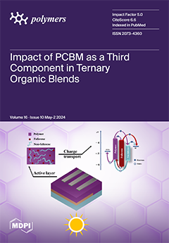
This study investigates donor:acceptor1:acceptor2 (D:A1:A2) matrix blends with photovoltaic potential. Optical analysis shows the spectral complementarity of the component materials, and AFM analysis shows that the addition of fullerene improves and enhances morphological attributes. In terms of charge carrier electrical mobility, the A1 = A2 sample has the highest recorded value. This comprehensive study highlights the critical role of the third component in influencing the intrinsic factors like electrical mobility and reveals the important relationship between acceptor ratios and the final blend's properties. These findings provide important new information for the optimization of ternary organic solar cells. View this paper
  • Issues are regarded as officially published after their release is announced to the table of contents alert mailing list.
  • You may sign up for e-mail alerts to receive table of contents of newly released issues.
  • PDF is the official format for papers published in both, html and pdf forms. To view the papers in pdf format, click on the "PDF Full-text" link, and use the free Adobe Reader to open them.
Order results
Result details
Section
Select all
Export citation of selected articles as:
17 pages, 2389 KiB  
Article
Selection and Optimization of Carbon-Reinforced Polyether Ether Ketone Process Parameters in 3D Printing—A Rotating Component Application
by Raja Subramani, Praveenkumar Vijayakumar, Maher Ali Rusho, Anil Kumar, Karthik Venkitaraman Shankar and Arun Kumar Thirugnanasambandam
Polymers 2024, 16(10), 1443; https://doi.org/10.3390/polym16101443 - 20 May 2024
Cited by 1 | Viewed by 1631
Abstract
The selection of process parameters is crucial in 3D printing for product manufacturing. These parameters govern the operation of production machinery and influence the mechanical properties, production time, and other aspects of the final product. The optimal process parameter settings vary depending on [...] Read more.
The selection of process parameters is crucial in 3D printing for product manufacturing. These parameters govern the operation of production machinery and influence the mechanical properties, production time, and other aspects of the final product. The optimal process parameter settings vary depending on the product and printing application. This study identifies the most suitable cluster of process parameters for producing rotating components, specifically impellers, using carbon-reinforced Polyether Ether Ketone (CF-PEEK) thermoplastic filament. A mathematical programming technique using a rating method was employed to select the appropriate process parameters. The research concludes that an infill density of 70%, a layer height of 0.15 mm, a printing speed of 60 mm/s, a platform temperature of 195 °C, an extruder temperature of 445 °C, and an extruder travel speed of 95 mm/s are optimal process parameters for manufacturing rotating components using carbon-reinforced PEEK material. Full article
(This article belongs to the Special Issue Polymeric Materials and Their Application in 3D Printing)
Show Figures

Figure 1

Figure 1
<p>Schematic of MEx Process Parameters.</p>
Full article ">Figure 2
<p>Experimental specimens.</p>
Full article ">Figure 3
<p>Experimental specimens [<a href="#B45-polymers-16-01443" class="html-bibr">45</a>].</p>
Full article ">Figure 4
<p>Concept of this research.</p>
Full article ">Figure 5
<p>Scale conversion using the triangular membership function.</p>
Full article ">Figure 6
<p>Morphology analysis of printed alternatives (<b>a</b>) A 1 (<b>b</b>) A 2 (<b>c</b>) A 3 (<b>d</b>) A 4 (<b>e</b>) A 5 (<b>f</b>) Geometric of gold-sputter-coated samples.</p>
Full article ">
13 pages, 3388 KiB  
Article
Study on Nutrient Carrier of Mulch Based on Hydrogel @SiO2
by Dan Qin, Yujie Ma, Mei Wang and Zhihua Shan
Polymers 2024, 16(10), 1442; https://doi.org/10.3390/polym16101442 - 20 May 2024
Viewed by 880
Abstract
Soil conservation is one of the best methods to improve soil fertility and enhance crop growth efficiency. Replacing plastic mulch with biomass is an environmentally friendly strategy. Innovative encapsulated soil granules (ESGs) were developed using PVA/PC film as the wall material and rural [...] Read more.
Soil conservation is one of the best methods to improve soil fertility and enhance crop growth efficiency. Replacing plastic mulch with biomass is an environmentally friendly strategy. Innovative encapsulated soil granules (ESGs) were developed using PVA/PC film as the wall material and rural soil as the core. The PVA/PC was synthesized using 60% protein polypeptide (PC) from leather waste scrap and 35% poly (vinyl alcohol) (PVA), which was optimized for water absorption expansion and water retention performance. The ESG-10 granulated with 10% PVA/PC exhibited good water absorption, moisture retention, and resistance to water solubility. As an auxiliary material for soil improvement, the amount of ESGs mixed with the topsoil at ratios of 0 g/m2, 200 g/m2, and 400 g/m2 was proportional to the soil insulation and moisture retention. In rapeseed cultivation, the experimental results indicated that the soil mulched with ESG-10 can maintain seedling vitality for a long time under low water content conditions. Full article
(This article belongs to the Section Smart and Functional Polymers)
Show Figures

Figure 1

Figure 1
<p>Scheme of applied granulation process.</p>
Full article ">Figure 2
<p>Equilibrium moisture content (<b>a</b>) and solubility (<b>b</b>) of four kinds of PVA/PC films.</p>
Full article ">Figure 3
<p>Awater-T curve (<b>a</b>) and expansion of PVA/PC-60 film (<b>b</b>).</p>
Full article ">Figure 4
<p>Water loss-T of PVA/PC-60 film.</p>
Full article ">Figure 5
<p>DSC analysis of films.</p>
Full article ">Figure 6
<p>Performance of ESGs as a mulching film (Before rain (<b>a</b>), after rain (<b>b</b>)).</p>
Full article ">Figure 7
<p>Shape change of ESGs during immersion period.</p>
Full article ">Figure 8
<p>Water retention over time using ESGs.</p>
Full article ">Figure 9
<p>Cycles and maximum water absorption of two ESGs.</p>
Full article ">Figure 10
<p>The temperature and humidity of soil mulched by ESG<sub>o</sub> (the environmental RH: 57.3~68.3%).</p>
Full article ">Figure 11
<p>Growth of rapeseed under ESG mulching.</p>
Full article ">
14 pages, 1897 KiB  
Article
Clay Tailings Flocculated in Seawater and Industrial Water: Analysis of Aggregates, Sedimentation, and Supernatant Quality
by Williams H. Leiva, Norman Toro, Pedro Robles, Gonzalo R. Quezada, Iván Salazar and Ricardo Jeldres
Polymers 2024, 16(10), 1441; https://doi.org/10.3390/polym16101441 - 20 May 2024
Viewed by 1021
Abstract
High-molecular-weight anionic polyacrylamide was used to analyze the effect of kaolin on the structure of particle aggregates formed in freshwater and seawater. Batch flocculation experiments were performed to determine the size of the flocculated aggregates over time by using focused beam reflectance measurements. [...] Read more.
High-molecular-weight anionic polyacrylamide was used to analyze the effect of kaolin on the structure of particle aggregates formed in freshwater and seawater. Batch flocculation experiments were performed to determine the size of the flocculated aggregates over time by using focused beam reflectance measurements. Sedimentation tests were performed to analyze the settling rate of the solid–liquid interface and the turbidity of the supernatant. Subsequently, a model that relates the hindered settling rate to the aggregate size was used to determine the mass fractal dimension (Df). Flocculation kinetics revealed that greater amounts of kaolin generated larger aggregates because of its lamellar morphology. The maximum size was between 10 and 20 s of flocculation under all conditions. However, the presence of kaolin reduced the settling rate. The fractal dimension decreased with the increase in the kaolin content, resulting in the formation of irregular and porous aggregates. By contrast, factors such as the flocculation time, water quality, and quartz size had limited influences on the fractal dimension. Seawater produced a clearer supernatant because of its higher ionic strength and precoagulation of particles. Notably, the harmful effect of clays in seawater was reduced. Full article
(This article belongs to the Special Issue Status and Progress of Soluble Polymers II)
Show Figures

Figure 1

Figure 1
<p>X-ray diffraction spectrum of kaolin particles.</p>
Full article ">Figure 2
<p>Volume-weighted size distribution of quartz and kaolin particles.</p>
Full article ">Figure 3
<p>Flocculation kinetics of synthetic tailings flocculated at 25 g/t of SNF604, pH 7.5, stirring at 180 rpm, and solids concentration of 10% weight for various kaolin contents: (<b>A</b>) fine quartz in process water, (<b>B</b>) fine quartz in seawater, (<b>C</b>) coarse quartz in process water, and (<b>D</b>) coarse quartz in seawater. The flocculant solution is added 30 s after starting the measurement.</p>
Full article ">Figure 3 Cont.
<p>Flocculation kinetics of synthetic tailings flocculated at 25 g/t of SNF604, pH 7.5, stirring at 180 rpm, and solids concentration of 10% weight for various kaolin contents: (<b>A</b>) fine quartz in process water, (<b>B</b>) fine quartz in seawater, (<b>C</b>) coarse quartz in process water, and (<b>D</b>) coarse quartz in seawater. The flocculant solution is added 30 s after starting the measurement.</p>
Full article ">Figure 4
<p>Initial settling rates of synthetic tailings flocculated at 25 g/t of SNF604, pH 7.5, stirring at 180 rpm, and solids concentration of 10% weight for various kaolin contents: (<b>A</b>) fine quartz in process water, (<b>B</b>) fine quartz in seawater, (<b>C</b>) coarse quartz in process water, and (<b>D</b>) coarse quartz in seawater.</p>
Full article ">Figure 5
<p>Turbidity of the supernatant of synthetic tailings flocculated at 25 g/t of SNF604, pH 7.5, stirring at 180 rpm, and solids concentration of 10% weight for various kaolin contents: (<b>A</b>) fine quartz in process water, (<b>B</b>) fine quartz in seawater, (<b>C</b>) coarse quartz in process water, and (<b>D</b>) coarse quartz in seawater.</p>
Full article ">Figure 6
<p>Mass fractal dimension of synthetic tailing aggregates flocculated at 25 g/t SNF604, pH 7.5, 180 rpm stirring, and 10 wt% solids concentration for various flocculation times and kaolin contents: (<b>A</b>) fine quartz in process water, (<b>B</b>) fine quartz in seawater, (<b>C</b>) coarse quartz in process water, and (<b>D</b>) coarse quartz in seawater.</p>
Full article ">Figure 6 Cont.
<p>Mass fractal dimension of synthetic tailing aggregates flocculated at 25 g/t SNF604, pH 7.5, 180 rpm stirring, and 10 wt% solids concentration for various flocculation times and kaolin contents: (<b>A</b>) fine quartz in process water, (<b>B</b>) fine quartz in seawater, (<b>C</b>) coarse quartz in process water, and (<b>D</b>) coarse quartz in seawater.</p>
Full article ">
16 pages, 9672 KiB  
Article
Experimental Evaluation of a Granular Damping Element
by Sanel Avdić, Marko Nagode, Jernej Klemenc and Simon Oman
Polymers 2024, 16(10), 1440; https://doi.org/10.3390/polym16101440 - 19 May 2024
Viewed by 1458
Abstract
Due to their advantages—longer internal force delay compared to bulk materials, resistance to harsh conditions, damping of a wide frequency spectrum, insensitivity to ambient temperature, high reliability and low cost—granular materials are seen as an opportunity for the development of high-performance, lightweight vibration-damping [...] Read more.
Due to their advantages—longer internal force delay compared to bulk materials, resistance to harsh conditions, damping of a wide frequency spectrum, insensitivity to ambient temperature, high reliability and low cost—granular materials are seen as an opportunity for the development of high-performance, lightweight vibration-damping elements (particle dampers). The performance of particle dampers is affected by numerous parameters, such as the base material, the size of the granules, the flowability, the initial prestress, etc. In this work, a series of experiments were performed on specimens with different combinations of influencing parameters. Energy-based design parameters were used to describe the overall vibration-damping performance. The results provided information for a deeper understanding of the dissipation mechanisms and their mutual correlation, as well as the influence of different parameters (base material, granule size and flowability) on the overall damping performance. A comparison of the performance of particle dampers with carbon steel and polyoxymethylene granules and conventional rubber dampers is given. The results show that the damping performance of particle dampers can be up to 4 times higher compared to conventional bulk material-based rubber dampers, even though rubber as a material has better vibration-damping properties than the two granular materials in particle dampers. However, when additional design features such as mass and stiffness are introduced, the results show that the overall performance of particle dampers with polyoxymethylene granules can be up to 3 times higher compared to particle dampers with carbon steel granules and conventional bulk material-based rubber dampers. Full article
(This article belongs to the Section Polymer Processing and Engineering)
Show Figures

Figure 1

Figure 1
<p>Angle of repose.</p>
Full article ">Figure 2
<p>(<b>a</b>) Reference damping element, (<b>b</b>) Granular material container.</p>
Full article ">Figure 3
<p>(<b>a</b>) Granular material, (<b>b</b>) specimen preparation.</p>
Full article ">Figure 4
<p>Servo-hydraulic load frames: (<b>a</b>) 100 kN load frame, (<b>b</b>) 25 kN load frame.</p>
Full article ">Figure 5
<p>Angle of repose experiment setup.</p>
Full article ">Figure 6
<p>Experiment setup: (<b>a</b>) uniaxial compression, (<b>b</b>) damping element evaluation.</p>
Full article ">Figure 7
<p>Uniaxial compression results: Stiffness (blue) and Stiffness per unit mass (purple).</p>
Full article ">Figure 8
<p>Damping element evaluation results: (<b>a</b>) stiffness, (<b>b</b>) stiffness per unit mass.</p>
Full article ">Figure 9
<p>Damping performance in terms of specific damping capacity.</p>
Full article ">Figure 10
<p>Damping performance in terms of design parameter: (<b>a</b>) <span class="html-italic">η<sub>k</sub></span>, (<b>b</b>) <span class="html-italic">η<sub>d</sub></span>.</p>
Full article ">Figure A1
<p>Experimental results for the angle of repose: (<b>a</b>) carbon steel, d = 2 mm, (<b>b</b>) carbon steel, d = 1.5 mm, (<b>c</b>) carbon steel, d = 1 mm, (<b>d</b>) POM, d = 2 mm, (<b>e</b>) POM, d = 1.5 mm, (<b>f</b>) POM, d = 1 mm.</p>
Full article ">Figure A2
<p>Force-displacement curves for uniaxial compression tests.</p>
Full article ">Figure A3
<p>Force-displacement curves recorded for all specimens during static and dynamic tests.</p>
Full article ">
15 pages, 7087 KiB  
Article
Preparation and Performance of a Novel ZnO/TM/PET Composite Negative Ion Functional Fiber
by Mengxin Zhang, Jishu Zhang, Xin Lu, Jianbing Wu, Jiajia Peng, Wei Wang and Jin Tao
Polymers 2024, 16(10), 1439; https://doi.org/10.3390/polym16101439 - 19 May 2024
Viewed by 943
Abstract
Using zinc oxide (ZnO), tourmaline (TM), and polyethylene terephthalate (PET) as main raw materials, a novel ZnO/TM/PET negative ion functional fiber was created. The rheological properties of a ZnO/TM/PET masterbatch were investigated; the morphology, XRD, and FT-IR of the fibers were observed; and [...] Read more.
Using zinc oxide (ZnO), tourmaline (TM), and polyethylene terephthalate (PET) as main raw materials, a novel ZnO/TM/PET negative ion functional fiber was created. The rheological properties of a ZnO/TM/PET masterbatch were investigated; the morphology, XRD, and FT-IR of the fibers were observed; and the mechanical properties, thermal properties, and negative ion release properties of the new fiber were tested. The results showed that the average particle size of the ZnO/TM composite is nearly 365 nm, with an increase in negative ion emission efficiency by nearly 50% compared to the original TM. The apparent viscosity of fiber masterbatch decreases with the increase in the addition of the ZnO/TM composite, and the rheological properties of the PET fiber masterbatch are not significantly effected, still showing shear thinning characteristics when the amount of addition reaches 10%. The ZnO/TM composite disperses well in the interior and surface of the ZnO/TM/PET fiber matrix. The prepared ZnO/TM/PET fiber has excellent properties, such as fineness of 1.54 dtex, glass transition temperature of 122.4 °C, fracture strength of 3.31 cN/dtex, and negative ion release of 1640/cm3, which shows great industrialization potential. Full article
(This article belongs to the Special Issue Advances in Polymer Fiber and Textiles)
Show Figures

Figure 1

Figure 1
<p>Application diagram of negative air ion functional fabric in industry and human life.</p>
Full article ">Figure 2
<p>Process diagram of ZnO/TM/PET negative ion functional fiber.</p>
Full article ">Figure 3
<p>Particle size distribution of TM and ZnO/TM composite powder.</p>
Full article ">Figure 4
<p>Rheological curves of ZnO/TM/PET functional masterbatch with different contents.</p>
Full article ">Figure 5
<p>SEM and ESD of the sample: (<b>a</b>) raw tourmaline powder, (<b>b</b>) zinc oxide compound tourmaline powder, and (<b>c</b>) ESD atlas of the composite powder.</p>
Full article ">Figure 6
<p>SEM photos of ZnO/TM/PET composite functional fibers: (<b>a</b>) 5%–2.54 dtex; (<b>b</b>) 5%–1.46 dtex; (<b>c</b>) 7.5%–2.54 dtex; (<b>d</b>) 7.5%–1.46 dtex; (<b>e</b>) 10%–2.54 dtex; (<b>f</b>) 10%–1.46 dtex.</p>
Full article ">Figure 7
<p>XRD patterns of the samples: (<b>a</b>) raw tourmaline and ZnO/TM composite; (<b>b</b>) ZnO/TM/PET fibers.</p>
Full article ">Figure 8
<p>Infrared spectrum of a fiber sample.</p>
Full article ">Figure 9
<p>Mechanical properties curves of ZnO/TM/PET fiber samples: (<b>a</b>) breaking strength, (<b>b</b>) breaking strength, (<b>c</b>) elongation at break, and (<b>d</b>) elastic modulus.</p>
Full article ">Figure 10
<p>DSC and TG curve of the samples: (<b>a</b>) DSC curve of raw TM and ZnO/TM composite power; (<b>b</b>) DSC curve of PET and ZnO/TM/PET fibers; (<b>c</b>) TG curve of PET and ZnO/TM/PET fibers.</p>
Full article ">Figure 11
<p>NAI release of different ZnO/TM/PET fiber samples: (<b>a</b>) static method, (<b>b</b>) dynamic method.</p>
Full article ">
19 pages, 4192 KiB  
Article
Cellulose-Based Polyurethane Foams of Low Flammability
by Marzena Szpiłyk, Renata Lubczak and Jacek Lubczak
Polymers 2024, 16(10), 1438; https://doi.org/10.3390/polym16101438 - 19 May 2024
Cited by 1 | Viewed by 1139
Abstract
Decreasing oil resources creates the need to search for raw materials in the biosphere, which can be converted into polyols suitable for obtaining polyurethane foams (PUF). One such low-cost and reproducible biopolymer is cellulose. There are not many examples of cellulose-derived polyols due [...] Read more.
Decreasing oil resources creates the need to search for raw materials in the biosphere, which can be converted into polyols suitable for obtaining polyurethane foams (PUF). One such low-cost and reproducible biopolymer is cellulose. There are not many examples of cellulose-derived polyols due to the sluggish reactivity of cellulose itself. Recently, cellulose and its hydroxypropyl derivatives were applied as source materials to obtain polyols, further converted into biodegradable rigid polyurethane foams (PUFs). Those PUFs were flammable. Here, we describe our efforts to modify such PUFs in order to decrease their flammability. We obtained an ester from diethylene glycol and phosphoric(III) acid and used it as a reactive flame retardant in the synthesis of polyol-containing hydroxypropyl derivative of cellulose. The cellulose-based polyol was characterized by infrared spectra (IR) and proton nuclear magnetic resonance (1H-NMR) methods. Its properties, such as density, viscosity, surface tension, and hydroxyl numbers, were determined. Melamine was also added to the foamed composition as an additive flame retardant, obtaining PUFs, which were characterized by apparent density, water uptake, dimension stability, heat conductance, compressive strength, and heat resistance at 150 and 175 °C. Obtained rigid PUFs were tested for flammability by determining oxygen index, horizontal flammability test, and calorimetric analysis. Obtained rigid PUFs showed improved flammability resistance in comparison with non-modified PUFs and classic PUFs. Full article
(This article belongs to the Special Issue Biopolymers and Bio-Based Polymer Composites)
Show Figures

Figure 1

Figure 1
<p>IR spectrum of phosphoric acid(III) and DEG ester.</p>
Full article ">Figure 2
<p>IR spectrum of polyol H<sub>3</sub>PO<sub>3</sub>-DEG + HPC-GL-EC.</p>
Full article ">Figure 3
<p>H-NMR spectrum of phosphoric acid(III) ester and DEG.</p>
Full article ">Figure 4
<p><sup>1</sup>H-NMR spectrum of polyol obtained from H<sub>3</sub>PO<sub>3</sub>-DEG -HPC-GL-EC.</p>
Full article ">Figure 5
<p>IR spectrum of unheated foam (<span class="html-fig-inline" id="polymers-16-01438-i001"><img alt="Polymers 16 01438 i001" src="/polymers/polymers-16-01438/article_deploy/html/images/polymers-16-01438-i001.png"/></span>) and foam exposed to temperatures 150 °C (<span class="html-fig-inline" id="polymers-16-01438-i002"><img alt="Polymers 16 01438 i002" src="/polymers/polymers-16-01438/article_deploy/html/images/polymers-16-01438-i002.png"/></span>) and 175 °C (<span class="html-fig-inline" id="polymers-16-01438-i003"><img alt="Polymers 16 01438 i003" src="/polymers/polymers-16-01438/article_deploy/html/images/polymers-16-01438-i003.png"/></span>).</p>
Full article ">Figure 6
<p>Thermal stability of the polyurethane foam as the mass loss after heating at 150 °C (<b>a</b>) and 175 °C (<b>b</b>) obtained from polyols H<sub>3</sub>PO<sub>3</sub>-DEG-HPC-GL-EC (1), HPC-GL-EG-EC (2), and HPC-TEG-GL-EC (3).</p>
Full article ">Figure 7
<p>Optical microscopy images of foam obtained from polyols H<sub>3</sub>PO<sub>3</sub>-DEG-HPC-GL-EC (<b>a</b>), HPC-GL-EG-EC (<b>b</b>), and HPC-GL-TEG-EC (<b>c</b>).</p>
Full article ">Figure 8
<p>Thermal analysis of foams obtained from polyols H<sub>3</sub>PO<sub>3</sub>-DEG- HPC-GL-EC (1), HPC-GL-EG-EC (2), and HPC-TEG-GL-EC (3): mass change as a function of temperature (<b>a</b>); and differential mass change as a function of temperature (<b>b</b>).</p>
Full article ">Figure 9
<p>DSC thermogram of foam obtained from polyol H<sub>3</sub>PO<sub>3</sub>-GDE-HPC-GL-EC.</p>
Full article ">Figure 10
<p>Photographs of PUF samples obtained from polyol H<sub>3</sub>PO<sub>3</sub>-DEG-HPC-GL-EC flamed at horizontal test.</p>
Full article ">Figure 11
<p>Total heat release vs. time during combustion of polyurethane foams obtained from polyols H<sub>3</sub>PO<sub>3</sub>-DEG-HPC-GL-EC (1), HPC-GL-EG-EC (2), and HPC-TEG-GL-EC (3).</p>
Full article ">Figure 12
<p>Heat release rate vs. time during combustion of polyurethane foams obtained from polyols H<sub>3</sub>PO<sub>3</sub>-DEG-HPC-GL-EC (1), HPC-GL-EG-EC (2), and HPC-TEG-GL-EC (3).</p>
Full article ">Scheme 1
<p>Obtaining polyols from HPC in the glycol medium.</p>
Full article ">Scheme 2
<p>Obtaining polyols from cellulose.</p>
Full article ">Scheme 3
<p>Preparation of DEG ester and phosphoric acid (III).</p>
Full article ">Scheme 4
<p>Formation of an ester bond in reaction with ethylene carbonate.</p>
Full article ">
20 pages, 3926 KiB  
Article
Three-Dimensional Bioprinting of GelMA Hydrogels with Culture Medium: Balancing Printability, Rheology and Cell Viability for Tissue Regeneration
by Laura Mendoza-Cerezo, Jesús M. Rodríguez-Rego, Antonio Macías-García, Antuca Callejas-Marín, Luís Sánchez-Guardado and Alfonso C. Marcos-Romero
Polymers 2024, 16(10), 1437; https://doi.org/10.3390/polym16101437 - 19 May 2024
Cited by 2 | Viewed by 1251
Abstract
Three-dimensional extrusion bioprinting technology aims to become a fundamental tool for tissue regeneration using cell-loaded hydrogels. These biomaterials must have highly specific mechanical and biological properties that allow them to generate biosimilar structures by successive layering of material while maintaining cell viability. The [...] Read more.
Three-dimensional extrusion bioprinting technology aims to become a fundamental tool for tissue regeneration using cell-loaded hydrogels. These biomaterials must have highly specific mechanical and biological properties that allow them to generate biosimilar structures by successive layering of material while maintaining cell viability. The rheological properties of hydrogels used as bioinks are critical to their printability. Correct printability of hydrogels allows the replication of biomimetic structures, which are of great use in medicine, tissue engineering and other fields of study that require the three-dimensional replication of different tissues. When bioprinting cell-loaded hydrogels, a small amount of culture medium can be added to ensure adequate survival, which can modify the rheological properties of the hydrogels. GelMA is a hydrogel used in bioprinting, with very interesting properties and rheological parameters that have been studied and defined for its basic formulation. However, the changes that occur in its rheological parameters and therefore in its printability, when it is mixed with the culture medium necessary to house the cells inside, are unknown. Therefore, in this work, a comparative study of GelMA 100% and GelMA in the proportions 3:1 (GelMA 75%) and 1:1 (GelMA 50%) with culture medium was carried out to determine the printability of the gel (using a device of our own invention), its main rheological parameters and its toxicity after the addition of the medium and to observe whether significant differences in cell viability occur. This raises the possibility of its use in regenerative medicine using a 3D extrusion bioprinter. Full article
(This article belongs to the Special Issue 3D and 4D Printing of Polymers: Modeling and Experimental Approaches)
Show Figures

Graphical abstract

Graphical abstract
Full article ">Figure 1
<p>Work planning.</p>
Full article ">Figure 2
<p>Test device for the optimisation of 3D bioprinting registered under patent number U202300008.</p>
Full article ">Figure 3
<p>(<b>A</b>) Analysis of G′ and G″ at different temperatures for GelMA hydrogel. (<b>B</b>) Performance of GelMA hydrogel in the shear rate versus shear viscosity rheology test at 25 °C, at 28 °C and at 34 °C. (<b>C</b>) Oscillatory test of GelMA hydrogel to determine G′ and G″ at different frequencies at a set point temperature of 30 °C.</p>
Full article ">Figure 4
<p>Behaviour of PBS, complete medium (DMEM + FBS + Pen/Strep) and medium without additives (DMEM) in the shear rate versus shear viscosity rheology test at 37 °C.</p>
Full article ">Figure 5
<p>(<b>A</b>) Analysis of G′ and G″ at different temperatures for GelMA 75% and 50% hydrogel. (<b>B</b>) Oscillatory test to determine G′ and G″ at different frequencies for GelMA 75% and GelMA 50% hydrogel. (<b>C</b>) Analysis of G′ and G″ at different temperatures for GelMA 100%, 75% and 50% hydrogel. (<b>D</b>) Oscillatory test to determine G′ and G″ at different frequencies for GelMA 100%, 75% and GelMA 50% hydrogel.</p>
Full article ">Figure 6
<p>Comparative study of printability. Image from the INMA group. Image produced by the INMA group.</p>
Full article ">Figure 7
<p>(<b>A</b>) CELLINK Start with Pr = 1 and appropriate gel state. (<b>B</b>) GelMA 100% with Pr &gt; 1 and appropriate/high gel state. (<b>C</b>) GelMA 75% with Pr &gt; 1 and appropriate/high gel state. (<b>D</b>) GelMA 50% with Pr &gt; 1 and appropriate/high gelling state.</p>
Full article ">Figure 8
<p>Measuring the collapsed area.</p>
Full article ">Figure 9
<p>Collapse test. The image on the left shows the GelMA hydrogel without culture medium, and the image on the right shows the GelMA 50%.</p>
Full article ">Figure 10
<p>Viability test performed on GelMA 100%, GelMA 75% and GelMA 50% hydrogels.</p>
Full article ">
18 pages, 9298 KiB  
Article
Anionic Effect on Electrical Transport Properties of Solid Co2+/3+ Redox Mediators
by Ravindra Kumar Gupta, Ahamad Imran and Aslam Khan
Polymers 2024, 16(10), 1436; https://doi.org/10.3390/polym16101436 - 19 May 2024
Viewed by 836
Abstract
In a solid-state dye-sensitized solar cell, a fast-ion conducting (σ25°C > 10−4 S cm−1) solid redox mediator (SRM; electrolyte) helps in fast dye regeneration and back-electron transfer inhibition. In this work, we synthesized solid Co2+/3+ redox mediators using [...] Read more.
In a solid-state dye-sensitized solar cell, a fast-ion conducting (σ25°C > 10−4 S cm−1) solid redox mediator (SRM; electrolyte) helps in fast dye regeneration and back-electron transfer inhibition. In this work, we synthesized solid Co2+/3+ redox mediators using a [(1 − x)succinonitrile: x poly(ethylene oxide)] matrix, LiX, Co(tris-2,2′-bipyridine)3(bis(trifluoromethyl) sulfonylimide)2, and Co(tris-2,2′-bipyridine)3(bis(trifluoromethyl) sulfonylimide)3 via the solution-cast method, and the results were compared with those of their acetonitrile-based liquid counterparts. The notation x is a weight fraction (=0, 0.5, and 1), and X represents an anion. The anion was either bis(trifluoromethyl) sulfonylimide [TFSI; ionic size, 0.79 nm] or trifluoromethanesulfonate [Triflate; ionic size, 0.44 nm]. The delocalized electrons and a low value of lattice energy for the anions made the lithium salts highly dissociable in the matrix. The electrolytes exhibited σ25°C ≈ 2.1 × 10⁻³ (1.5 × 10⁻³), 7.2 × 10⁻⁴ (3.1 × 10⁻⁴), and 9.7 × 10⁻⁷ (6.3 × 10⁻⁷) S cm−1 for x = 0, 0.5, and 1, respectively, with X = TFSI (Triflate) ions. The log σ–T−1 plot portrayed a linear curve for x = 0 and 1, and a downward curve for x = 0.5. The electrical transport study showed σ(TFSI) > σ(Triflate), with lower activation energy for TFSI ions. The anionic effect increased from x = 0 to 1. This effect was explained using conventional techniques, such as Fourier transform infrared spectroscopy (FT-IR), X-ray diffractometry (XRD), X-ray photoelectron spectroscopy (XPS), scanning electron microscopy (SEM), UV–visible spectroscopy (UV-vis), differential scanning calorimetry (DSC), and thermogravimetric analysis (TGA). Full article
(This article belongs to the Special Issue Advanced Polymers for Solar Cells Applications)
Show Figures

Figure 1

Figure 1
<p>Nyquist curves of SRM [(1 − <span class="html-italic">x</span>)SN: <span class="html-italic">x</span>PEO]-LiX-Co salts (<span class="html-italic">x</span> = 0, 0.5, and 1) along with their liquid counterparts (LRMs) at 25 °C for anions, X = TFSI<sup>−</sup> (solid symbols), and Triflate<sup>−</sup> (open symbols). (I) and (II) correspond to low- and high-frequency regions, respectively.</p>
Full article ">Figure 2
<p>Log σ vs. <span class="html-italic">T</span><sup>−1</sup> curves of SRM [(1 − <span class="html-italic">x</span>)SN: <span class="html-italic">x</span>PEO]-LiX-Co salts with <span class="html-italic">x</span> = 0, 0.5, and 1. Inset: Vogel–Tamman–Fulcher (VTF) plots for SRMs with <span class="html-italic">x</span> = 0.5. X = TFSI<sup>−</sup> ions (filled symbols) and Triflate<sup>−</sup> ions (open symbols). LRMs, ACN-based liquid counterparts. For regions I and II, please see the text.</p>
Full article ">Figure 3
<p>FT-IR spectra of SRM [(1 − <span class="html-italic">x</span>)SN: <span class="html-italic">x</span>PEO]-LiX-Co salts (<span class="html-italic">x</span> = 0, 0.5, and 1). LRM, ACN-based liquid counterpart. X = TFSI<sup>−</sup> (solid lines) or Triflate<sup>−</sup> (dotted lines). This also includes vibrational peaks of matrices, solvent, and ionic salts (vertical lines).</p>
Full article ">Figure 4
<p>Relative intensity, Δ<span class="html-italic">I</span>, of SRM [(1 − <span class="html-italic">x</span>)SN: <span class="html-italic">x</span>PEO]-LiX-Co salts with <span class="html-italic">x</span> = 0, 0.5, and 1, and X = TFSI<sup>−</sup> and Triflate<sup>−</sup>, along with those of their liquid counterparts (LRMs).</p>
Full article ">Figure 5
<p>XRD patterns of SRM [(1 − <span class="html-italic">x</span>)SN: <span class="html-italic">x</span>PEO]-LiX-Co salts, where <span class="html-italic">x</span> = 0, 0.5, and 1. X = TFSI<sup>−</sup> (solid line) and Triflate<sup>−</sup> (dotted line).</p>
Full article ">Figure 6
<p>XPS spectra of different elements of SRM [(1 − <span class="html-italic">x</span>)SN: <span class="html-italic">x</span>PEO]-LiX-Co salts, where <span class="html-italic">x</span> = (<b>a</b>) 0, (<b>b</b>) 0.5, and (<b>c</b>) 1. X = TFSI<sup>−</sup> (solid line) and Triflate<sup>−</sup> (dotted line).</p>
Full article ">Figure 7
<p>Plot of ratio (R) of intensity with width vs. peak position for different elements of SRM [(1 − <span class="html-italic">x</span>)SN: <span class="html-italic">x</span>PEO]-LiX-Co salts, where <span class="html-italic">x</span> = 0, 0.5, and 1. X = TFSI<sup>−</sup> (solid symbols) and Triflate<sup>−</sup> (open symbols).</p>
Full article ">Figure 8
<p>SEM images for the surface of SRM [(1 − <span class="html-italic">x</span>)SN: <span class="html-italic">x</span>PEO]-LiX-Co salts with <span class="html-italic">x</span> = 0, 0.5, and 1, infiltrated in the mesoporous TiO<sub>2</sub>. X = (<b>a</b>) TFSI<sup>−</sup> and (<b>b</b>) Triflate<sup>−</sup>. Scale bar, 1 μm.</p>
Full article ">Figure 9
<p>Transmittance spectra of SRM [(1 − <span class="html-italic">x</span>)SN: <span class="html-italic">x</span>PEO]-LiX-Co salts with <span class="html-italic">x</span> = 0, 0.5, and 1. LRM, ACN-based liquid counterpart. X = TFSI<sup>−</sup> (filled symbols) and Triflate<sup>−</sup> (open symbols).</p>
Full article ">Figure 10
<p>DSC curves of SRM [(1 − <span class="html-italic">x</span>)SN: <span class="html-italic">x</span>PEO]-LiX- Co salts with <span class="html-italic">x</span> = 0, 0.5, and 1. X = TFSI<sup>−</sup> (filled symbols) and Triflate<sup>−</sup> (open symbols).</p>
Full article ">Figure 11
<p>TGA curves of SRM [(1 − <span class="html-italic">x</span>)SN: <span class="html-italic">x</span>PEO]-LiX-Co salts with <span class="html-italic">x</span> = 0, 0.5, and 1. X = TFSI<sup>−</sup> ions (filled symbols) and Triflate<sup>−</sup> ions (open symbols).</p>
Full article ">
14 pages, 6060 KiB  
Article
Catalytic Pyrolysis of Polypropylene for Cable Semiconductive Buffer Layers
by Xiaokai Meng, Hua Yu, Zhumao Lu and Tao Jin
Polymers 2024, 16(10), 1435; https://doi.org/10.3390/polym16101435 - 19 May 2024
Viewed by 791
Abstract
With the progress of the power grid system, the coverage area of cables is widening, and the problem of cable faults is gradually coming to affect people’s daily lives. While the vast majority of cable faults are caused by the ablation of the [...] Read more.
With the progress of the power grid system, the coverage area of cables is widening, and the problem of cable faults is gradually coming to affect people’s daily lives. While the vast majority of cable faults are caused by the ablation of the cable buffer layer, polypropylene (PP), as a common cable buffer material, has pyrolysis properties that critically impact cable faults. Studying the semiconductive buffer layer of polypropylene (PP) and its pyrolysis properties allows us to obtain a clearer picture of the pyrolysis products formed during PP ablation. This understanding aids in the accurate diagnosis of cable faults and the identification of ablation events. In this study, the effects of temperature and catalyst (H-Zeolite Standard Oil Corporation Of New York (Socony) Mobil-Five (HZSM-5)) content on the PP thermolysis product distribution were studied by using an online tubular pyrolysis furnace-mass spectrometry (MS) experimental platform. The results showed that PP/40% HZSM-5 presented the highest thermolytic efficiency and relative yield of the main products at 400 °C. Full article
(This article belongs to the Special Issue Polymer Combustion and Pyrolysis Kinetics)
Show Figures

Figure 1

Figure 1
<p>Schematic diagram of the pyrolytic single-photon ionization (SPI)-TOF-MS system.</p>
Full article ">Figure 2
<p>(<b>a</b>) SEM image and (<b>b</b>) EDS with elements O, Si, Al, and C of HZSM-5.</p>
Full article ">Figure 3
<p>PP/20% HZSM-5, PP/30% HZSM-5, and PP/40% HZSM-5 of volatile pyrolysis products under different temperatures of mass spectrum: (<b>a</b>,<b>e</b>,<b>i</b>) 300 °C, (<b>b</b>,<b>f</b>,<b>j</b>) 400 °C, (<b>c</b>,<b>g</b>) 500 °C, and (<b>d</b>,<b>h</b>,<b>k</b>,<b>l</b>) 600 °C (the red line represents olefins, the blue line represents diolefins, and the green line represents aromatic hydrocarbons).</p>
Full article ">Figure 4
<p>Peak evolution versus time curves of molecular ions from main products at 300 °C as (<b>a</b>–<b>c</b>): pure PP; (<b>d</b>–<b>f</b>): PE/50% HZSM-5.</p>
Full article ">Figure 5
<p>Peak evolution versus time curves of molecular ions from main products at 400 °C as (<b>a</b>–<b>c</b>): pure PP; (<b>d</b>–<b>f</b>): PE/50% HZSM-5.</p>
Full article ">Figure 6
<p>Time evolution curves of main pyrolysis products of PP with different HZSM-5 contents at 500 °C: (<b>a</b>,<b>d</b>) PP/20% HZSM-5, (<b>b</b>,<b>e</b>) PP/30% HZSM-5, (<b>c</b>,<b>f</b>) PP/40% HZSM-5.</p>
Full article ">Figure 7
<p>Time evolution curves of main pyrolysis products of PP with different HZSM-5 contents at 600 °C: (<b>a</b>,<b>d</b>) PP/20% HZSM-5, (<b>b</b>,<b>e</b>) PP/30% HZSM-5, (<b>c</b>,<b>f</b>) PP/40% HZSM-5.</p>
Full article ">Figure 8
<p>Schematic diagram of thermal degradation mechanism of PP/catalyst.</p>
Full article ">
35 pages, 6165 KiB  
Review
Polyelectrolytes for Environmental, Agricultural, and Medical Applications
by Martina Zuñiga Delgado, Francisca L. Aranda, Fabian Hernandez-Tenorio, Karla A. Garrido-Miranda, Manuel F. Meléndrez and Daniel A. Palacio
Polymers 2024, 16(10), 1434; https://doi.org/10.3390/polym16101434 - 18 May 2024
Viewed by 1489
Abstract
In recent decades, polyelectrolytes (PELs) have attracted significant interest owing to a surge in research dedicated to the development of new technologies and applications at the biological level. Polyelectrolytes are macromolecules of which a substantial portion of the constituent units contains ionizable or [...] Read more.
In recent decades, polyelectrolytes (PELs) have attracted significant interest owing to a surge in research dedicated to the development of new technologies and applications at the biological level. Polyelectrolytes are macromolecules of which a substantial portion of the constituent units contains ionizable or ionic groups. These macromolecules demonstrate varied behaviors across different pH ranges, ionic strengths, and concentrations, making them fascinating subjects within the scientific community. The aim of this review is to present a comprehensive survey of the progress in the application studies of polyelectrolytes and their derivatives in various fields that are vital for the advancement, conservation, and technological progress of the planet, including agriculture, environmental science, and medicine. Through this bibliographic review, we seek to highlight the significance of these materials and their extensive range of applications in modern times. Full article
(This article belongs to the Section Polymer Applications)
Show Figures

Figure 1

Figure 1
<p>(<b>A</b>) poly(vinyl sulfate) potassium, (<b>B</b>) poly[(3(methacryloylamino)propyl)dimethyl(3-sulfopropyl)ammonium hydroxide], and (<b>C</b>) poly(allylamine hydrochloride).</p>
Full article ">Figure 2
<p>(<b>A</b>) Trend of increasing publications on polyelectrolytes and (<b>B</b>) leading countries in polyelectrolyte publications and their applications, based on Scopus reports.</p>
Full article ">Figure 3
<p>(<b>A</b>) Map of co-authorship of countries in publications on polyelectrolytes and their applications. Note: seven groups of countries: red, green, light blue, purple, yellow, blue, and dark red and (<b>B</b>) co-occurrence map of author keywords in publications on polyelectrolytes and their applications. Note: seven thematic groups: yellow, blue, green, purple, red, orange, and light blue.</p>
Full article ">Figure 4
<p>Contingency matrix on the relationship between journals and countries in polyelectrolyte publications and their applications (cells with x indicate that the model deviation is not statistically significant).</p>
Full article ">Figure 5
<p>Sankey diagram of author keywords in polyelectrolyte publications and their applications.</p>
Full article ">Figure 6
<p>Historical map of keyword co-occurrence in polyelectrolyte publications and their applications.</p>
Full article ">Figure 7
<p>(<b>A</b>) Poly (vinylphosponic acid), (<b>B</b>) Poly(N-methacryloyl-4-aminosalicylic acid), (<b>C</b>) Poly(2-acrylamido-2-methyl-1-propanesulfonic acid), and (<b>D</b>) Poly(acrylamide).</p>
Full article ">Figure 8
<p>(<b>A</b>) poly (acrylic acid), (<b>B</b>) poly (sodium styrenesulfonate), and (<b>C</b>) poly (vinylpyrrolidone).</p>
Full article ">Figure 9
<p>Formation of polyelectrolyte complexes by the interaction of polymers with opposite charges.</p>
Full article ">Figure 10
<p>Schematic of the substrate adsorption and double layer film formation in polycation and polyanion solution [<a href="#B151-polymers-16-01434" class="html-bibr">151</a>].</p>
Full article ">Figure 11
<p>Injectable hydrogel for personalized cancer treatment [<a href="#B164-polymers-16-01434" class="html-bibr">164</a>].</p>
Full article ">Figure 12
<p>Conceptual scheme of 3D printing of customized thick and perfusable cardiac patches [<a href="#B182-polymers-16-01434" class="html-bibr">182</a>].</p>
Full article ">Figure 13
<p>Hybrid scaffolds for cardiac valve tissue engineering based on type I collagen and hyaluronic acid [<a href="#B204-polymers-16-01434" class="html-bibr">204</a>].</p>
Full article ">Figure 14
<p>Conceptual scheme of the composite sponge structure derived from quaternized chitin, egg white protein, and montmorillonite [<a href="#B211-polymers-16-01434" class="html-bibr">211</a>].</p>
Full article ">Figure 15
<p>Schematic process of formation and drug release of MXene-based hydrogel system for application in infected wounds [<a href="#B216-polymers-16-01434" class="html-bibr">216</a>].</p>
Full article ">Figure 16
<p>Block copolymer as contrast agent for in vivo tumor magnetic resonance micelle imaging.</p>
Full article ">
18 pages, 6423 KiB  
Article
A Numerical Model to Predict the Relaxation Phenomena in Thermoset Polymers and Their Effects on Residual Stress during Curing—Part I: A Theoretical Formulation and Numerical Evaluation of Relaxation Phenomena
by Raffaele Verde, Alberto D’Amore and Luigi Grassia
Polymers 2024, 16(10), 1433; https://doi.org/10.3390/polym16101433 - 18 May 2024
Cited by 1 | Viewed by 848
Abstract
This paper analyzes the effect of crosslinking reactions on a thermoset polymer’s viscoelastic properties. In particular, a numerical model to predict the evolution of epoxy’s mechanical properties during the curing process is proposed and implemented in an Ansys APDL environment. A linear viscoelastic [...] Read more.
This paper analyzes the effect of crosslinking reactions on a thermoset polymer’s viscoelastic properties. In particular, a numerical model to predict the evolution of epoxy’s mechanical properties during the curing process is proposed and implemented in an Ansys APDL environment. A linear viscoelastic behavior is assumed, and the scaling of viscoelastic properties in terms of the temperature and degree of conversion is modeled using a modified version of the TNM (Tool–Narayanaswamy–Mohynian) model. The effects of the degree of conversion and structural relaxation on epoxy’s relaxation times are simultaneously examined for the first time. This formulation is based on the thermo-rheological and chemo-rheological simplicities hypothesis and can predict the evolution of epoxy’s relaxation phenomena. The thermal–kinetic reactions of curing are implemented in a homemade routine written in APDL language, and the structural module of Ansys is used to predict the polymer’s creep and stress relaxation curves at different temperatures and degrees of conversion. Full article
(This article belongs to the Special Issue Relaxation Phenomena in Polymers)
Show Figures

Figure 1

Figure 1
<p>Definition of fictive temperature. (<b>a</b>) Schematic of a generic property P vs. temperature during the glass transition (<b>b</b>) Temperature derivative of volume during the glass transition.</p>
Full article ">Figure 2
<p>Glass transition temperature versus the degree of conversion.</p>
Full article ">Figure 3
<p>Evolution of shear relaxation modulus at the reference state.</p>
Full article ">Figure 4
<p>Relaxation time versus the temperature for various degrees of conversion.</p>
Full article ">Figure 5
<p>Shift factor’s parameters versus the degree of conversion. (<b>a</b>) Activation energy. (<b>b</b>) Non-linearity parameters.</p>
Full article ">Figure 6
<p>Memory function at the glass transition temperature.</p>
Full article ">Figure 7
<p>Temperature and degree of conversion profiles. (<b>a</b>) Temperature profiles. (<b>b</b>) Degree of conversion profiles.</p>
Full article ">Figure 8
<p>Evolution of temperature, fictive temperature, and glass transition temperature for different curing profiles: (<b>a</b>) Curing temperature Tcure = 115 °C. (<b>b</b>) Curing temperature Tcure = 125 °C. (<b>c</b>) Curing temperature Tcure = 135 °C. (<b>d</b>) Curing temperature Tcure = 145 °C. (<b>e</b>) Curing Temperature Tcure = 160 °C.</p>
Full article ">Figure 9
<p>Evolution of fictive temperature during the cooling from liquid to glassy state for different cooling rates: (<b>a</b>) degree of conversion α = 0.57; (<b>b</b>) degree of conversion α = 0.69; (<b>c</b>) degree of conversion α = 0.80; (<b>d</b>) degree of conversion α = 0.89; (<b>e</b>) degree of conversion α = 0.98.</p>
Full article ">Figure 10
<p>The fictive temperature at the end of cooling versus the logarithmic of the cooling rate.</p>
Full article ">Figure 11
<p>Stress relaxation and creep curves at T = 30 °C and T = 60 °C for different conversion degrees. (<b>a</b>) Stress relaxation (<b>b</b>) Creep. Continuing lines represent the relaxation phenomena at T = 30 °C. The dashed lines represent the relaxation phenomena at T = 60 °C.</p>
Full article ">Figure 12
<p>Stress relaxation and creep curves at T = 90 °C and T = 120 °C for different conversion degrees. (<b>a</b>) Stress relaxation (<b>b</b>) Creep. Continuing lines represent the relaxation phenomena at T = 90 °C. The dashed lines represent the relaxation phenomena at T = 120 °C.</p>
Full article ">Figure 13
<p>Fictive temperature and Poisson relaxation curves at T = 30°C and T = 60 °C for different conversion degrees. (<b>a</b>) Fictive temperature (<b>b</b>) Poisson. Continuing lines represent the relaxation phenomena at T = 30 °C. The dashed lines represent the relaxation phenomena at T = 60 °C.</p>
Full article ">Figure 14
<p>Fictive temperature and Poisson ratio evolution curves at T = 90 °C and T = 120 °C for different conversion degrees. (<b>a</b>) Fictive temperature (<b>b</b>) Poisson. Continuing lines represent the relaxation phenomena at T = 90 °C. The dashed lines represent the relaxation phenomena at T = 120 °C.</p>
Full article ">
21 pages, 1126 KiB  
Article
Development and Validation of a 1D Dynamic Model of an Injection Moulding Process and Design of a Model-Based Nozzle Pressure Controller
by Rasmus Aagaard Hertz, Ole Therkelsen, Søren Kristiansen, Jesper Kjærsgaard Christensen, Frederik Agervig Hansson and Lasse Schmidt
Polymers 2024, 16(10), 1432; https://doi.org/10.3390/polym16101432 - 18 May 2024
Cited by 1 | Viewed by 852
Abstract
A 1D model describing the dynamics of an injection moulding machine and the injection process is presented. The model describes an injection cylinder actuated by a dual-pump electro–hydraulic speed-variable drive and the filling, holding and cooling phases of the injection moulding process utilising [...] Read more.
A 1D model describing the dynamics of an injection moulding machine and the injection process is presented. The model describes an injection cylinder actuated by a dual-pump electro–hydraulic speed-variable drive and the filling, holding and cooling phases of the injection moulding process utilising amorphous polymers. The model is suggested as the foundation for the design of model-based pressure controllers of, e.g., the nozzle pressure. The focus is on using material, mould and machine properties to construct the model, making it possible to analyse and design the dynamic system prior to manufacturing hardware or conducting experiments. Both the presented model and the developed controller show good agreement with experimental results. The proposed method is general in nature and enables the design, analysis and evaluation of the machine, material and mould dynamics for controller design based solely on the physical properties of the system. Full article
(This article belongs to the Special Issue Recent Advances in Injection Molding of Polymers)
Show Figures

Figure 1

Figure 1
<p>Structure of the presented model.</p>
Full article ">Figure 2
<p>Flamebar element used for modelling. The volumes are numbered with the mathematical operator “<span class="html-italic">i</span>”. The modelled pressure is at the centre of each section, denoted by <math display="inline"><semantics> <msub> <mover accent="true"> <mi>p</mi> <mo>^</mo> </mover> <mi>i</mi> </msub> </semantics></math>. The physical pressure sensors in the mould are marked as <math display="inline"><semantics> <msub> <mi>p</mi> <mrow> <mi>i</mi> <mo>,</mo> <mi>j</mi> </mrow> </msub> </semantics></math>. The arrows above the flows indicate the positive direction.</p>
Full article ">Figure 3
<p>Simplified schematic of the hydraulic and mechanical subsystem.</p>
Full article ">Figure 4
<p>Open loop Bode plots for the (<b>a</b>) plant and the (<b>b</b>) controller and plant.</p>
Full article ">Figure 5
<p>Simulation performance compared to experimental data of the described machine and mould: <math display="inline"><semantics> <mover> <mo>•</mo> <mo>¯</mo> </mover> </semantics></math> is the average pressure; <math display="inline"><semantics> <mover accent="true"> <mo>•</mo> <mo>^</mo> </mover> </semantics></math> is the simulation result; <math display="inline"><semantics> <msup> <mo>•</mo> <mo>*</mo> </msup> </semantics></math> is the velocity reference.</p>
Full article ">Figure 6
<p>Performance of nozzle pressure controller for two set points and two temperatures evaluated on test machine and nonlinear simulation model. Reference pressure is denoted <math display="inline"><semantics> <msup> <mo>•</mo> <mo>*</mo> </msup> </semantics></math>, and simulation results are denoted with <math display="inline"><semantics> <mover accent="true"> <mo>•</mo> <mo>^</mo> </mover> </semantics></math>.</p>
Full article ">Figure A1
<p>Geometry of the sprue.</p>
Full article ">
26 pages, 3679 KiB  
Article
A “2-in-1” Bioanalytical System Based on Nanocomposite Conductive Polymers for Early Detection of Surface Water Pollution
by Anna S. Kharkova, Anastasia S. Medvedeva, Lyubov S. Kuznetsova, Maria M. Gertsen, Vladimir V. Kolesov, Vyacheslav A. Arlyapov and Anatoly N. Reshetilov
Polymers 2024, 16(10), 1431; https://doi.org/10.3390/polym16101431 - 17 May 2024
Cited by 2 | Viewed by 1079
Abstract
This work proposes an approach to the formation of receptor elements for the rapid diagnosis of the state of surface waters according to two indicators: the biochemical oxygen demand (BOD) index and toxicity. Associations among microorganisms based on the bacteria P. yeei and [...] Read more.
This work proposes an approach to the formation of receptor elements for the rapid diagnosis of the state of surface waters according to two indicators: the biochemical oxygen demand (BOD) index and toxicity. Associations among microorganisms based on the bacteria P. yeei and yeast S. cerevisiae, as well as associations of the yeasts O. polymorpha and B. adeninivorans, were formed to evaluate these indicators, respectively. The use of nanocomposite electrically conductive materials based on carbon nanotubes, biocompatible natural polymers—chitosan and bovine serum albumin cross-linked with ferrocenecarboxaldehyde, neutral red, safranin, and phenosafranin—has made it possible to expand the analytical capabilities of receptor systems. Redox polymers were studied by IR spectroscopy and Raman spectroscopy, the contents of electroactive components were determined by atomic absorption spectroscopy, and electrochemical properties were studied by electrochemical impedance and cyclic voltammetry methods. Based on the proposed kinetic approach to modeling individual stages of bioelectrochemical processes, the chitosan–neutral red/CNT composite was chosen to immobilize the yeast association between O. polymorpha (ks = 370 ± 20 L/g × s) and B. adeninivorans (320 ± 30 L/g × s), and a bovine serum albumin (BSA)–neutral composite was chosen to immobilize the association between the yeast S. cerevisiae (ks = 130 ± 10 L/g × s) and the bacteria P. yeei red/CNT (170 ± 30 L/g × s). After optimizing the composition of the receptor systems, it was shown that the use of nanocomposite materials together with associations among microorganisms makes it possible to determine BOD with high sensitivity (with a lower limit of 0.6 mg/dm3) and detect the presence of a wide range of toxicants of both organic and inorganic origin. Both receptor elements were tested on water samples, showing a high correlation between the results of biosensor analysis of BOD and toxicity and the results of standard analytical methods. The results obtained show broad prospects for creating sensitive and portable bioelectrochemical sensors for the early warning of environmentally hazardous situations based on associations among microorganisms and nanocomposite materials. Full article
(This article belongs to the Section Polymer Composites and Nanocomposites)
Show Figures

Graphical abstract

Graphical abstract
Full article ">Figure 1
<p>The biosensor formation approach used in this study.</p>
Full article ">Figure 2
<p>Scheme of the synthesis of a redox-active polymer based on CHIT-SFR, where 1—chitosan; 2—glutaraldehyde; 3—safranin O; and 4—CHIT–SFR redox-active polymer.</p>
Full article ">Figure 3
<p>Scheme of the synthesis of a redox-active polymer based on BSA-SFR.</p>
Full article ">Figure 4
<p>Modification of a redox-active polymer with glutaraldehyde, where 1—modified BSA, 2—glutaraldehyde, and 3—water-insoluble redox-active polymer BSA-FC.</p>
Full article ">Figure 5
<p>Scheme of the synthesis of a network redox-active polymer based on chitosan and ferrocenecarboxaldehyde (CHIT-FC), where 1 denotes linear modified Chitosan, 2—glutaraldehyde, and 3—networked redox-active polymer CHIT-FC.</p>
Full article ">Figure 6
<p>BOD toxicity biosensor design. Insert: the current increasing is associated with the adding of a model solution into the system (GGS for BOD analysis, Glucose and Cu<sup>2+</sup> solution is for toxicity analysis). For the water sample BOD assessment, the GGS solution is replaced with the sample. For toxicity assessment, Cu<sup>2+</sup> is replaced with the sample (glucose was diluted in the sample at the same concentration for the control solution test).</p>
Full article ">Figure 7
<p>Formation of receptor elements for assessing toxicity and BOD<sub>5</sub>. (<b>A</b>) Experimental design for assessing the stability of the associations. (<b>B</b>) Results of the CFU determination after <span class="html-italic">S. cerevisiae</span> and <span class="html-italic">P. yeei</span> formation and toxicity assessment of Cu<sup>2+</sup> ion as a model toxicant. (<b>C</b>) Dependence of the analytical signal or decrease in metabolic activity on the concentration of a model toxicant of a bioreceptor element based on the mediator ferrocene and the association of the bacteria <span class="html-italic">P. yeei</span> and yeast <span class="html-italic">S. cerevisiae</span> in the presence of Cd<sup>2+</sup> ions. (<b>D</b>) Dependence of the association-based biosensor response on the BOD<sub>5</sub> of the model sample.</p>
Full article ">Figure 8
<p>Redox-active polymer for association immobilization. (<b>A</b>) IR spectra of safranin O, chitosan, and a redox-active polymer, where v –NH<sub>2</sub> are stretching vibrations of –NH<sub>2</sub>, v C=N are stretching vibrations of C=N. (<b>B</b>) SEM images of BSA-FC-CNT/COOH nanocomposite. (<b>C</b>) SEM images of CHIT-FC redox-active polymer. (<b>D</b>) SEM images of BSA-FC redox-active polymer.</p>
Full article ">Figure 9
<p>The electrochemical properties of the redox-active polymers and their nanocomposites. (<b>A</b>) CV curves of GP-electrode; BSA-NR redox-active polymer; BSA-NR-CNT/COOH nanocomposite at 100 mV/s. (<b>B</b>) CV curves of the BSA-NR redox-active polymer for a range of scan rates from 10 to 100 mV/s. (<b>C</b>) Determination of the limiting stages of the electronic process in the redox-active polymer BSA-NR. (<b>D</b>) Impedance spectroscopy of the studied redox-active polymers based on BSA. (<b>E</b>) Impedance spectroscopy of the studied redox-active polymers based on CHIT. (<b>F</b>) Cyclic voltammograms were recorded before (line 1) and after (line 2) the addition of glucose in order to determine the rate constant for the interaction between enzyme systems at 10 mV/s. (<b>G</b>) Dependence of the ratio of limiting currents in the presence and absence of a substrate on the reciprocal value of the root of the scan rate.</p>
Full article ">
13 pages, 3201 KiB  
Article
Dual Semi-Interpenetrating Networks of Water-Soluble Macromolecules and Supramolecular Polymer-like Chains: The Role of Component Interactions
by Anna L. Makarova, Alexander L. Kwiatkowski, Alexander I. Kuklin, Yuri M. Chesnokov, Olga E. Philippova and Andrey V. Shibaev
Polymers 2024, 16(10), 1430; https://doi.org/10.3390/polym16101430 - 17 May 2024
Viewed by 882
Abstract
Dual networks formed by entangled polymer chains and wormlike surfactant micelles have attracted increasing interest in their application as thickeners in various fields since they combine the advantages of both polymer- and surfactant-based fluids. In particular, such polymer-surfactant mixtures are of great interest [...] Read more.
Dual networks formed by entangled polymer chains and wormlike surfactant micelles have attracted increasing interest in their application as thickeners in various fields since they combine the advantages of both polymer- and surfactant-based fluids. In particular, such polymer-surfactant mixtures are of great interest as novel hydraulic fracturing fluids with enhanced properties. In this study, we demonstrated the effect of the chemical composition of an uncharged polymer poly(vinyl alcohol) (PVA) and pH on the rheological properties and structure of its mixtures with a cationic surfactant erucyl bis(hydroxyethyl)methylammonium chloride already exploited in fracturing operations. Using a combination of several complementary techniques (rheometry, cryo-transmission electron microscopy, small-angle neutron scattering, and nuclear magnetic resonance spectroscopy), we showed that a small number of residual acetate groups (2–12.7 mol%) in PVA could significantly reduce the viscosity of the mixed system. This result was attributed to the incorporation of acetate groups in the corona of the micellar aggregates, decreasing the molecular packing parameter and thereby inducing the shortening of worm-like micelles. When these groups are removed by hydrolysis at a pH higher than 7, viscosity increases by five orders of magnitude due to the growth of worm-like micelles in length. The findings of this study create pathways for the development of dual semi-interpenetrating polymer-micellar networks, which are highly desired by the petroleum industry. Full article
(This article belongs to the Section Polymer Networks)
Show Figures

Figure 1

Figure 1
<p>Scheme of alkaline hydrolysis of PVA acetate.</p>
Full article ">Figure 2
<p>Frequency dependencies of the storage modulus G′ (full symbols) and loss modulus G″ (open symbols) for aqueous solutions containing (<b>A</b>) 26 mM EHAC and 9.1 mM NaSal at pH 5.5 (squares) and pH 11 (circles); (<b>B</b>) 26 mM EHAC, 9.1 mM NaSal and 890 monomol/L PVA with 12.7 mol% of acetate groups at pH 5.5 (squares), pH 8.1 (diamonds), and pH 11 (circles). Temperature: 20 °C.</p>
Full article ">Figure 2 Cont.
<p>Frequency dependencies of the storage modulus G′ (full symbols) and loss modulus G″ (open symbols) for aqueous solutions containing (<b>A</b>) 26 mM EHAC and 9.1 mM NaSal at pH 5.5 (squares) and pH 11 (circles); (<b>B</b>) 26 mM EHAC, 9.1 mM NaSal and 890 monomol/L PVA with 12.7 mol% of acetate groups at pH 5.5 (squares), pH 8.1 (diamonds), and pH 11 (circles). Temperature: 20 °C.</p>
Full article ">Figure 3
<p>Dependencies of zero shear viscosity in pH for aqueous solutions containing 26 mM EHAC, 9.1 mM NaSal in the absence of PVA (circles) and in the presence of 890 monomol/L PVA with 12.7 mol% of acetate groups (squares).</p>
Full article ">Figure 4
<p>Cryo-TEM micrographs of aqueous solutions containing 26 mM EHAC and 9.1 mM NaSal: in the absence of PVA at pH 5.5 (<b>A</b>); in the absence of PVA at pH 10 (<b>B</b>); in the presence of 890 monomol/L PVA at pH 5.5 (<b>C</b>); in the presence of 890 monomol/L PVA at pH 10 (<b>D</b>). PVA contains 12.7 mol% of acetate groups.</p>
Full article ">Figure 5
<p>SANS scattering curves for aqueous solutions containing 26 mM EHAC and 9.1 mM NaSal: (<b>A</b>) in the absence of PVA at pD = 11.5 (diamonds, <a href="#polymers-16-01430-f004" class="html-fig">Figure 4</a>A) and with 890 monomol/L PVA at pD = 11.5 (circles, <a href="#polymers-16-01430-f004" class="html-fig">Figure 4</a>A,B); (<b>B</b>) with 890 monomol/L PVA at pD = 6 (squares, <a href="#polymers-16-01430-f004" class="html-fig">Figure 4</a>B) and at pD=11.5 (circles, <a href="#polymers-16-01430-f004" class="html-fig">Figure 4</a>A,B). Solid lines represent fits of the scattering curves with a cylinder form factor (parameter fits are summarised in <a href="#polymers-16-01430-t001" class="html-table">Table 1</a>). PVA contains 12.7 mol% of acetate groups.</p>
Full article ">Figure 6
<p><sup>1</sup>H NMR spectra of 4 wt.% PVA (12.7 mol% of acetate groups) solutions in D<sub>2</sub>O at pH 5.5 (<b>A</b>) and pH 11 (<b>B</b>).</p>
Full article ">Figure 7
<p>pH dependence of the molar ratio of polymer acetate units (squares) and free sodium acetate (circles) to PVA monomer units in aqueous solutions of PVA (12 mol% of acetate groups) derived from NMR data.</p>
Full article ">Figure 8
<p>Ratios of zero-shear viscosities of micellar solutions (26 mM EHAC and 9.1 mM NaSal) with 890 monomol/L PVA and without polymer vs. amount of residual acetate groups in PVA at pH 5.5. Temperature: 200 °C.</p>
Full article ">Figure 9
<p>Schematic representation of the interaction between PVA and EHAC WLMs at low and high pH. Surfactant molecules are represented by red and grey. PVA molecules are indicated by green lines. Residual PVA acetate units and sodium acetate formed due to hydrolysis are indicated by blue short lines.</p>
Full article ">Figure 10
<p>Frequency dependencies of the storage modulus G° (full symbols) and loss modulus G″ (open symbols) for aqueous solutions containing (<b>A</b>) 6.2 mM EHAC and 2.2 mM NaSal; (<b>B</b>) 26 mM EHAC and 9.1 mM NaSal in the absence of PVA (squares) and in the presence of 890 monomol/L fully hydrolysed PVA (circles) at pH 5.5. Temperature: 20 °C.</p>
Full article ">
24 pages, 17897 KiB  
Article
Researching on the Effect of Input Parameters on the Quality and Manufacturability of 3D-Printed Cellular Samples from Nylon 12 CF in Synergy with Testing Their Behavior in Bending
by Martin Koroľ, Jozef Török, Peter Pavol Monka, Petr Baron, Beata Mrugalska and Katarina Monkova
Polymers 2024, 16(10), 1429; https://doi.org/10.3390/polym16101429 - 17 May 2024
Viewed by 807
Abstract
The study of cellular structures and their properties represents big potential for their future applications in real practice. The article aims to study the effect of input parameters on the quality and manufacturability of cellular samples 3D-printed from Nylon 12 CF in synergy [...] Read more.
The study of cellular structures and their properties represents big potential for their future applications in real practice. The article aims to study the effect of input parameters on the quality and manufacturability of cellular samples 3D-printed from Nylon 12 CF in synergy with testing their bending behavior. Three types of structures (Schwarz Diamond, Shoen Gyroid, and Schwarz Primitive) were selected for investigation that were made via the fused deposition modeling technique. As part of the research focused on the settings of input parameters in terms of the quality and manufacturability of the samples, input parameters such as volume fraction, temperature of the working space, filament feeding method and positioning of the sample on the printing pad were specified for the combination of the used material and 3D printer. During the experimental investigation of the bending properties of the samples, a three-point bending test was performed. The dependences of force on deflection were mathematically described and the amount of absorbed energy and ductility were evaluated. The results show that among the investigated structures, the Schwarz Diamond structure appears to be the most suitable for bending stress applications. Full article
(This article belongs to the Special Issue Polymer-Based Biomaterials for Tissue Engineering Applications)
Show Figures

Figure 1

Figure 1
<p>Categories of cellular structures.</p>
Full article ">Figure 2
<p>Investigated structures’ arrangement with their basic cell<span class="html-italic">s</span>: (<b>a</b>) Schwarz Diamond; (<b>b</b>) Schwarz Primitive; (<b>c</b>) Schoen Gyroid.</p>
Full article ">Figure 3
<p>Samples for experimental testing: (<b>a</b>) Schwarz Diamond, (<b>b</b>) Schoen Gyroid and (<b>c</b>) Schwarz Primitive.</p>
Full article ">Figure 4
<p>Test set-up; (<b>a</b>) Zwick 1456 machine; (<b>b</b>) a specimen positioning within bending test.</p>
Full article ">Figure 5
<p>Filament moisture measurement: (<b>a</b>) Halogen HR83 moisture measuring instrument; (<b>b</b>) the material that was tested in the weighing pan.</p>
Full article ">Figure 6
<p>Measurement of moisture content of material Nylon 12 CF.</p>
Full article ">Figure 7
<p>Tensile testing of filament: (<b>a</b>) tensile test set up; (<b>b</b>) measured force-displacement dependences.</p>
Full article ">Figure 8
<p>Filament feeding method: (<b>a</b>) placement of the material in the tray at the bottom of the printer<span class="html-italic">;</span> (<b>b</b>) placement of the material in the rack at the top of the printer.</p>
Full article ">Figure 9
<p>Results of improved technological conditions (Nylon 12 Carbon Fiber).</p>
Full article ">Figure 10
<p>Annealing process of a sample inside of the workspace of the 3D printer machine.</p>
Full article ">Figure 11
<p>(<b>a</b>) Saving the sample in software as part of preprocessing before printing; (<b>b</b>) surface of the sample after printing under a Celestrom microscope at 40× magnification and with a 2 MP CMOS resolution.</p>
Full article ">Figure 11 Cont.
<p>(<b>a</b>) Saving the sample in software as part of preprocessing before printing; (<b>b</b>) surface of the sample after printing under a Celestrom microscope at 40× magnification and with a 2 MP CMOS resolution.</p>
Full article ">Figure 12
<p>Representative force versus deflection curves obtained from experimental tests of specimens with cellular structures under bending loading.</p>
Full article ">Figure 13
<p>Specification of the area moment of inertia, <span class="html-italic">I</span>.</p>
Full article ">Figure 14
<p>(<b>a</b>) Energy absorption during bending; (<b>b</b>) energy absorption during bending, elastic; ductility indexes, <span class="html-italic">µ<sub>d</sub></span>, based on the deflection value at the proportionality limit. <span class="html-italic">µ<sub>E</sub></span> is expressed as the quotient of the total and elastic energy.</p>
Full article ">Figure 15
<p>Ductility indexes, <span class="html-italic">µ<sub>d</sub></span>, based on the deflection value at the proportionality limit. <span class="html-italic">µ<sub>E</sub></span> is expressed as the quotient of the total and elastic energy.</p>
Full article ">
18 pages, 7906 KiB  
Article
Brucine Sulfate, a Novel Bacteriostatic Agent in 3D Printed Bone Scaffold Systems
by Jinying Li, Shi Hu, Pei Feng, Yang Xia, Zihan Pei, Jiaxuan Tian, Kun Jiang, Liang Liu, Xiong Cai and Ping Wu
Polymers 2024, 16(10), 1428; https://doi.org/10.3390/polym16101428 - 17 May 2024
Viewed by 1124
Abstract
Bacterial infection is a common complication in bone defect surgery, in which infection by clinically resistant bacteria has been a challenge for the medical community. Given this emerging problem, the discovery of novel natural-type inhibitors of drug-resistant bacteria has become imperative. Brucine, present [...] Read more.
Bacterial infection is a common complication in bone defect surgery, in which infection by clinically resistant bacteria has been a challenge for the medical community. Given this emerging problem, the discovery of novel natural-type inhibitors of drug-resistant bacteria has become imperative. Brucine, present in the traditional Chinese herb Strychnine semen, is reported to exert analgesic and anti-inflammatory effects. Brucine’s clinical application was limited because of its water solubility. We extracted high-purity BS by employing reflux extraction and crystallization, greatly improved its solubility, and evaluated its antimicrobial activity against E. coli and S. aureus. Importantly, we found that BS inhibited the drug-resistant strains significantly better than standard strains and achieved sterilization by disrupting the bacterial cell wall. Considering the safety concerns associated with the narrow therapeutic window of BS, a 3D BS-PLLA/PGA bone scaffold system was constructed with SLS technology and tested for its performance, bacteriostatic behaviors, and biocompatibility. The results have shown that the drug-loaded bone scaffolds had not only long-term, slow-controlled release with good cytocompatibility but also demonstrated significant antimicrobial activity in antimicrobial testing. The above results indicated that BS may be a potential drug candidate for the treatment of antibiotic-resistant bacterial infections and that scaffolds with enhanced antibacterial activity and mechanical properties may have potential applications in bone tissue engineering. Full article
(This article belongs to the Section Polymer Applications)
Show Figures

Figure 1

Figure 1
<p>(<b>A</b>) The fabrication process of BS; (<b>B</b>) preparation of BS-PLLA/PGA drug-loaded bone scaffold powders; (<b>C</b>) fabrication of the scaffolds via SLS; (<b>D</b>) The three orthographic views of the BS-PLLA/PGA scaffolds.</p>
Full article ">Figure 2
<p>(<b>A</b>) HPLC chromatograms. Strychnine and brucine standard mixture representatively (a), total alkaloid extracts (b), brucine standard (c), and extracted BS (d); (<b>B</b>) Inhibition zone of BS against standard <span class="html-italic">E. coli</span> and <span class="html-italic">S. aureus</span> at different concentrations of 5, 7.5, 10, 12.5, 15, and 30 mg/mL; (<b>C</b>) The time-courses of BS at 5, 10, or 20 mg/mL against standard and antibiotic-resistant <span class="html-italic">E. coli</span> and <span class="html-italic">S. aureus</span>; (<b>D</b>) BS maximum bactericidal concentration simulation curve; (<b>E</b>) The turbidity images of <span class="html-italic">E. coli</span> and <span class="html-italic">S. aureus</span> at 24 h of incubation. (<b>F</b>) Tube LB: no bacteria (blank medium control); Tubes 1–5: BS at 30, 20, 10, 5, and 2.5 mg/mL; Tube 6: bacterial medium control; and Tubes a–e: DMSO controls (corresponding to the concentrations used in BS solutions).</p>
Full article ">Figure 3
<p>(<b>A</b>) The SEM morphology of the scaffolds; (<b>B</b>) The XRD patterns; (<b>C</b>) Cumulative release curve of BS-PLLA/PGA scaffolds containing different BS contents.</p>
Full article ">Figure 4
<p>(<b>A</b>) The turbidity image of <span class="html-italic">E. coli</span> and <span class="html-italic">S. aureus</span> in co-culture with the scaffolds after 24 h, 48 h, and 72 h; (<b>B</b>) Antibacterial rates of drug-loaded scaffolds with different concentrations of BS against standard <span class="html-italic">E. coli</span> and <span class="html-italic">S. aureus</span> at 24 h, 48 h, and 72 h; (<b>C</b>) The adhesion morphology of standard <span class="html-italic">E. coli</span> and <span class="html-italic">S. aureus</span> on the scaffolds of different concentrations at 24 h and 72 h.</p>
Full article ">Figure 5
<p>(<b>A</b>,<b>B</b>) SEM and TEM images of standard <span class="html-italic">E. coli</span> and <span class="html-italic">S. aureus</span> treated by BS for 3 h; (<b>C</b>) Schematic diagram of possible antibacterial mechanisms of the drug-loaded scaffold.</p>
Full article ">Figure 6
<p>(<b>A</b>) The adhesion morphology of MG63 cells in co-culture with the scaffolds after 3 days; (<b>B</b>) Fluorescence microscope images of MG63 cells co-cultured with the scaffold for 24, 48, and 72 days (nuclei shown as blue dots); (<b>C</b>) The total number of MG63 cells after co-culture with the scaffolds.</p>
Full article ">
25 pages, 14550 KiB  
Article
Impact Response Features and Penetration Mechanism of UHMWPE Subjected to Handgun Bullet
by Yihui Zhu, Yang Song, Wei Wu, Jie Ma, Zhuangqing Fan, Yaoke Wen, Cheng Xu, Min Xia and Weifeng Da
Polymers 2024, 16(10), 1427; https://doi.org/10.3390/polym16101427 - 17 May 2024
Viewed by 874
Abstract
Ensuring military and police personnel protection is vital for urban security. However, the impact response mechanism of the UHMWPE laminate used in ballistic helmets and vests remains unclear, making it hard to effectively protect the head, chest, and abdomen. This study utilized 3D-DIC [...] Read more.
Ensuring military and police personnel protection is vital for urban security. However, the impact response mechanism of the UHMWPE laminate used in ballistic helmets and vests remains unclear, making it hard to effectively protect the head, chest, and abdomen. This study utilized 3D-DIC technology to analyze UHMWPE laminate’s response to 9 mm lead-core pistol bullets traveling at 334.93 m/s. Damage mode and response characteristics were revealed, and an effective numerical calculation method was established that could reveal the energy conversion process. The bullet penetrated by 1.03 mm, causing noticeable fiber traction, resulting in cross-shaped failure due to fiber compression and aggregation. Bulge transitioned from circular to square, initially increasing rapidly, then slowing. Maximum in-plane shear strain occurred at ±45°, with values of 0.0904 and −0.0928. Model accuracy was confirmed by comparing strain distributions. The investigation focused on bullet-laminate interaction and energy conversion. Bullet’s kinetic energy is converted into laminate’s kinetic and internal energy, with the majority of erosion energy occurring in the first four equivalent sublaminates and the primary energy change in the system occurring at 75 μs in the fourth equivalent sublayer. The results show the damage mode and energy conversion of the laminate, providing theoretical support for understanding the impact response mechanism and improving the efficiency of protective energy absorption. Full article
(This article belongs to the Special Issue Mechanical Behaviors and Properties of Polymer Materials)
Show Figures

Figure 1

Figure 1
<p>Schematic diagram of the testing system.</p>
Full article ">Figure 2
<p>The site layout of the testing system. (<b>a</b>) Test System Panorama Diagram; (<b>b</b>) Firing Mechanism.</p>
Full article ">Figure 2 Cont.
<p>The site layout of the testing system. (<b>a</b>) Test System Panorama Diagram; (<b>b</b>) Firing Mechanism.</p>
Full article ">Figure 3
<p>The positions of P<sub>0</sub>, L<sub>AB</sub>, L<sub>CD</sub>. (<b>a</b>) The positions of DIC; (<b>b</b>) The feature points on the side view.</p>
Full article ">Figure 4
<p>The influence of the bracket displacement and whole laminate displacement on the bulge height.</p>
Full article ">Figure 5
<p>History of the maximum height of the bulge.</p>
Full article ">Figure 6
<p>The impacted surface’s morphology. (<b>a</b>) The front-side morphology; (<b>b</b>) Bullet hole of Experiment; (<b>c</b>,<b>d</b>) The front-side morphology of repeat experiments; (<b>e</b>,<b>f</b>) The side-view and back side of the impacted laminate.</p>
Full article ">Figure 6 Cont.
<p>The impacted surface’s morphology. (<b>a</b>) The front-side morphology; (<b>b</b>) Bullet hole of Experiment; (<b>c</b>,<b>d</b>) The front-side morphology of repeat experiments; (<b>e</b>,<b>f</b>) The side-view and back side of the impacted laminate.</p>
Full article ">Figure 7
<p>The bulge shape in z-direction (W). (<b>a</b>) 100 μs; (<b>b</b>) 200 μs; (<b>c</b>) 300 μs; (<b>d</b>) 400 μs.</p>
Full article ">Figure 7 Cont.
<p>The bulge shape in z-direction (W). (<b>a</b>) 100 μs; (<b>b</b>) 200 μs; (<b>c</b>) 300 μs; (<b>d</b>) 400 μs.</p>
Full article ">Figure 8
<p>The profile of the bulge and its width. (<b>a</b>) The bulge profile in the x-direction along L<sub>AB</sub>; (<b>b</b>) The bulge profile in the y-direction along L<sub>CD</sub>.</p>
Full article ">Figure 9
<p>Displacement, velocity, and acceleration curves of the maximum point of the bulge height.</p>
Full article ">Figure 10
<p>The displacement in x-direction (U) of the back face. (<b>a</b>) 100 μs; (<b>b</b>) 200 μs; (<b>c</b>) 300 μs; (<b>d</b>) 400 μs.</p>
Full article ">Figure 11
<p>The displacement in y-direction (V) of the back face. (<b>a</b>) 100 μs; (<b>b</b>) 200 μs; (<b>c</b>) 300 μs; (<b>d</b>) 400 μs.</p>
Full article ">Figure 12
<p>Evolution of the variable exx. (<b>a</b>) 100 μs; (<b>b</b>) 200 μs; (<b>c</b>) 300 μs; (<b>d</b>) 400 μs.</p>
Full article ">Figure 13
<p>Evolution of the variable eyy. (<b>a</b>) 100 μs; (<b>b</b>) 200 μs; (<b>c</b>) 300 μs; (<b>d</b>) 400 μs.</p>
Full article ">Figure 14
<p>Evolution of the variable exy. (<b>a</b>) 100 μs; (<b>b</b>) 200 μs; (<b>c</b>) 300 μs; (<b>d</b>) 400 μs.</p>
Full article ">Figure 15
<p>The evolution of the variable von Mises strain. (<b>a</b>) 100 μs; (<b>b</b>) 200 μs; (<b>c</b>) 300 μs; (<b>d</b>) 400 μs.</p>
Full article ">Figure 15 Cont.
<p>The evolution of the variable von Mises strain. (<b>a</b>) 100 μs; (<b>b</b>) 200 μs; (<b>c</b>) 300 μs; (<b>d</b>) 400 μs.</p>
Full article ">Figure 16
<p>The evolution of the shear strain rate. (<b>a</b>) 100 μs; (<b>b</b>) 200 μs; (<b>c</b>) 300 μs; (<b>d</b>) 400 μs.</p>
Full article ">Figure 17
<p>Numerical models. (<b>a</b>) Full model of the laminate; (<b>b</b>) 25 equivalent sublaminates; (<b>c</b>) full model of the bullet.</p>
Full article ">Figure 18
<p>The evolution of the bulge morphology on the back surface: (<b>a</b>) 120 μs; (<b>b</b>) 220 μs; (<b>c</b>) 320 μs; (<b>d</b>) 420 μs.</p>
Full article ">Figure 19
<p>The evolution of the equivalent stress on the back surface: (<b>a</b>) 120 μs; (<b>b</b>) 220 μs; (<b>c</b>) 320 μs; (<b>d</b>) 420 μs.</p>
Full article ">Figure 20
<p>The stress of the back face.</p>
Full article ">Figure 21
<p>The shear strain exy represented by the deformation of the tiny unit.</p>
Full article ">Figure 22
<p>The evolution of the bulge width in the x and y directions.</p>
Full article ">Figure 23
<p>The morphology of the numerical model’s bullet hole at 400 μs. (<b>a</b>) 1/2 model sides; (<b>b</b>) front-side view; (<b>c</b>) back-side view.</p>
Full article ">Figure 24
<p>The deformation process of the bullet. (<b>a</b>) 50 μs; (<b>b</b>) 70 μs; (<b>c</b>) 90 μs.</p>
Full article ">Figure 25
<p>The top-down view morphology of the bullet at 400 μs: (<b>a</b>) side view; (<b>b</b>) upward view.</p>
Full article ">Figure 26
<p>The evolution of the penetration response: (<b>a</b>) 100 μs; (<b>b</b>) 200 μs; (<b>c</b>) 300 μs; (<b>d</b>) 400 μs.</p>
Full article ">Figure 26 Cont.
<p>The evolution of the penetration response: (<b>a</b>) 100 μs; (<b>b</b>) 200 μs; (<b>c</b>) 300 μs; (<b>d</b>) 400 μs.</p>
Full article ">Figure 27
<p>The evolution of energy: (<b>a</b>) The evolution of total energy; (<b>b</b>) The evolution of the laminate and bullet energy.</p>
Full article ">Figure 28
<p>Energy of the equivalent sublaminate: (<b>a</b>) Eroded energy of the first 4 equivalent sublaminates; (<b>b</b>) Energy of the first 4 equivalent sublaminates.</p>
Full article ">
19 pages, 16063 KiB  
Article
Comparison of Printable Biomaterials for Use in Neural Tissue Engineering: An In Vitro Characterization and In Vivo Biocompatibility Assessment
by Miguel Etayo-Escanilla, Noelia Campillo, Paula Ávila-Fernández, José Manuel Baena, Jesús Chato-Astrain, Fernando Campos, David Sánchez-Porras, Óscar Darío García-García and Víctor Carriel
Polymers 2024, 16(10), 1426; https://doi.org/10.3390/polym16101426 - 17 May 2024
Cited by 1 | Viewed by 1793
Abstract
Nervous system traumatic injuries are prevalent in our society, with a significant socioeconomic impact. Due to the highly complex structure of the neural tissue, the treatment of these injuries is still a challenge. Recently, 3D printing has emerged as a promising alternative for [...] Read more.
Nervous system traumatic injuries are prevalent in our society, with a significant socioeconomic impact. Due to the highly complex structure of the neural tissue, the treatment of these injuries is still a challenge. Recently, 3D printing has emerged as a promising alternative for producing biomimetic scaffolds, which can lead to the restoration of neural tissue function. The objective of this work was to compare different biomaterials for generating 3D-printed scaffolds for use in neural tissue engineering. For this purpose, four thermoplastic biomaterials, ((polylactic acid) (PLA), polycaprolactone (PCL), Filaflex (FF) (assessed here for the first time for biomedical purposes), and Flexdym (FD)) and gelatin methacrylate (GelMA) hydrogel were subjected to printability and mechanical tests, in vitro cell–biomaterial interaction analyses, and in vivo biocompatibility assessment. The thermoplastics showed superior printing results in terms of resolution and shape fidelity, whereas FD and GelMA revealed great viscoelastic properties. GelMA demonstrated a greater cell viability index after 7 days of in vitro cell culture. Moreover, all groups displayed connective tissue encapsulation, with some inflammatory cells around the scaffolds after 10 days of in vivo implantation. Future studies will determine the usefulness and in vivo therapeutic efficacy of novel neural substitutes based on the use of these 3D-printed scaffolds. Full article
Show Figures

Figure 1

Figure 1
<p>Design of the scaffolds used for (<b>a</b>) printability tests, (<b>b</b>) mechanical tests, (<b>c</b>) in vitro cell–biomaterial interaction analyses, and (<b>d</b>) in vivo assays.</p>
Full article ">Figure 2
<p>Scheme of the materials and methods employed in the printability tests, mechanical characterization, the in vitro cell–biomaterial interaction analyses, and the in vivo assay. HE refers to hematoxylin and eosin staining and PS to Picrosirius staining.</p>
Full article ">Figure 3
<p>Macroscopic images of 3D-printed scaffolds of (<b>a</b>) PLA, (<b>b</b>) PCL, (<b>c</b>) FF, (<b>d</b>) FD, and (<b>e</b>) GelMA. Bright and contrast have been edited to improve their visualization.</p>
Full article ">Figure 4
<p>Graphic representation of tensile test results of PLA, PCL, FF, FD, and GelMA (G). (<b>a</b>) Young’s modulus (Mpa), (<b>b</b>) charge at fracture (N), and (<b>c</b>) strain at fracture (%). The results corresponding to each mechanical parameter are shown as mean ± standard deviation values. Statistically significant differences were determined with the Mann–Whitney test and represented as follows: ‘*’ indicates statistically significant differences (<span class="html-italic">p</span> &lt; 0.05) between all biomaterials, ‘a’ indicates statistically significant differences (<span class="html-italic">p</span> &lt; 0.05) between all biomaterials except “PCL,” and ‘b’ indicates statistically significant differences (<span class="html-italic">p</span> &lt; 0.05) between all biomaterials except “FF” and “G.”</p>
Full article ">Figure 5
<p>In vitro biocompatibility tests. (<b>a</b>) Representative panel of L/D assay of SK-N-AS seeded with PLA, PCL, FF, FD, and GelMA (G), and in 2D cultures as technical controls, after 72 h and 7 days of culture. Scale bar = 100 µm. (<b>b</b>) Graphic representation of WST-1 results of PLA, PCL, FF, FD, and GelMA (G). Statistically significant differences were determined with the Mann–Whitney test and are represented as follows: “NS” indicates no significant differences (<span class="html-italic">p</span> ≥ 0.05) between 72 h and 7 days; “2 or 1” indicates the number of biomaterials that obtained a significantly inferior cell viability (<a href="#polymers-16-01426-f005" class="html-fig">Figure 5</a>a) or absorbance value (<a href="#polymers-16-01426-f005" class="html-fig">Figure 5</a>b); and ‘a’ indicates no significant differences (<span class="html-italic">p</span> ≥ 0.05) with the CTR+ group.</p>
Full article ">Figure 6
<p>Representative panel of the hematoxylin and eosin (HE) and Picrosirius (PS) staining of the in vivo samples of PLA, PCL, FF, FD, GelMA (G), and healthy rat skin tissue (CTR). The PS images distinguish an external fibrotic layer (EFL) and an inner cellular layer (ICL). The black arrows indicate syncytial formations. Scale bar of the first column indicates 500 µm, while the scale bar of the second and third columns indicates 50 µm.</p>
Full article ">Figure 7
<p>Representative panel of CD45 immunohistochemistry of the in vivo samples of PLA, PCL, FF, FD, GelMA (G), and healthy rat skin tissue (incubated with CD45 antibody (CTR+) and without CD45 antibody (CTR−). The black arrows indicate signs of perivascular infiltration. Scale bar = 100 µm.</p>
Full article ">
18 pages, 8533 KiB  
Article
Liquid Crystal Orientation and Shape Optimization for the Active Response of Liquid Crystal Elastomers
by Jorge Luis Barrera, Caitlyn Cook, Elaine Lee, Kenneth Swartz and Daniel Tortorelli
Polymers 2024, 16(10), 1425; https://doi.org/10.3390/polym16101425 - 17 May 2024
Cited by 1 | Viewed by 1242
Abstract
Liquid crystal elastomers (LCEs) are responsive materials that can undergo large reversible deformations upon exposure to external stimuli, such as electrical and thermal fields. Controlling the alignment of their liquid crystals mesogens to achieve desired shape changes unlocks a new design paradigm that [...] Read more.
Liquid crystal elastomers (LCEs) are responsive materials that can undergo large reversible deformations upon exposure to external stimuli, such as electrical and thermal fields. Controlling the alignment of their liquid crystals mesogens to achieve desired shape changes unlocks a new design paradigm that is unavailable when using traditional materials. While experimental measurements can provide valuable insights into their behavior, computational analysis is essential to exploit their full potential. Accurate simulation is not, however, the end goal; rather, it is the means to achieve their optimal design. Such design optimization problems are best solved with algorithms that require gradients, i.e., sensitivities, of the cost and constraint functions with respect to the design parameters, to efficiently traverse the design space. In this work, a nonlinear LCE model and adjoint sensitivity analysis are implemented in a scalable and flexible finite element-based open source framework and integrated into a gradient-based design optimization tool. To display the versatility of the computational framework, LCE design problems that optimize both the material, i.e., liquid crystal orientation, and structural shape to reach a target actuated shapes or maximize energy absorption are solved. Multiple parameterizations, customized to address fabrication limitations, are investigated in both 2D and 3D. The case studies are followed by a discussion on the simulation and design optimization hurdles, as well as potential avenues for improving the robustness of similar computational frameworks for applications of interest. Full article
(This article belongs to the Special Issue Modeling and Simulations of Smart and Responsive Polymers)
Show Figures

Figure 1

Figure 1
<p>Design domain with boundary conditions (<b>left</b>) and a zoomed in region that shows the orientation of the LC in the unactuated/nematic (<b>center</b>) and actuated/isotropic (<b>right</b>) states.</p>
Full article ">Figure 2
<p>Graph for the forward analysis including the Newton solver block to compute the LCE structure’s cost and constraint functions.</p>
Full article ">Figure 3
<p>Graph for the adjoint sensitivity analysis.</p>
Full article ">Figure 4
<p>Soft gripper LC orientation design: (<b>left</b>) LCE gripper in both the initially unactuated (nematic) and actuated (isotropic) states; and (<b>right</b>) design domain and desired deformation.</p>
Full article ">Figure 5
<p>Designs corresponding to optimization iterates <math display="inline"><semantics> <mrow> <msub> <mi mathvariant="script">D</mi> <mrow> <mi>i</mi> <mi>t</mi> </mrow> </msub> <mo>=</mo> <mrow> <mo>[</mo> <mn>1</mn> <mo>,</mo> <mn>5</mn> <mo>,</mo> <mn>15</mn> <mo>,</mo> <mn>30</mn> <mo>,</mo> <mn>50</mn> <mo>]</mo> </mrow> </mrow> </semantics></math> showing LC orientation (colored horizontal strip), unactuated (light grey) and actuated (dark gray) configurations.</p>
Full article ">Figure 6
<p>Initial design of leaping structure with two distinct LC orientation regions and its desired deformation.</p>
Full article ">Figure 7
<p>(<b>Left</b>) Snapshots of the evolution of leaping structure colored by angle alignment as described in <a href="#polymers-16-01425-f006" class="html-fig">Figure 6</a>; and (<b>right</b>) their unactuated and actuated configurations in light gray and dark gray, respectively.</p>
Full article ">Figure 8
<p>A quarter of the <math display="inline"><semantics> <mrow> <mn>3</mn> <mo>×</mo> <mn>3</mn> </mrow> </semantics></math> LCE lattice design domain (<b>left</b>) is simulated. The onsets at the <b>center</b> and <b>right</b> depict the boundary conditions for the analysis for <math display="inline"><semantics> <mi mathvariant="bold-italic">u</mi> </semantics></math> and shape perturbation for <math display="inline"><semantics> <mover accent="true"> <mrow> <mo>Δ</mo> <mi mathvariant="bold-italic">X</mi> </mrow> <mo>˜</mo> </mover> </semantics></math>.</p>
Full article ">Figure 9
<p>Evolution of lattice structure design. The LC orientation and domain shape are optimized concurrently.</p>
Full article ">Figure 10
<p>Initial (<b>left</b>) and optimized (<b>right</b>) lattice structures in the unactuated and actuated states.</p>
Full article ">Figure 11
<p>Compliant mechanism design problem: planes of symmetry in 3D domain (<b>top left</b>), cross-section (<b>bottom left</b>), and onsets detailing the boundary conditions for the analysis for <math display="inline"><semantics> <mi mathvariant="bold-italic">u</mi> </semantics></math> (<b>top right</b>) and shape perturbation for <math display="inline"><semantics> <mover accent="true"> <mrow> <mo>Δ</mo> <mi mathvariant="bold-italic">X</mi> </mrow> <mo>˜</mo> </mover> </semantics></math> (<b>bottom right</b>).</p>
Full article ">Figure 12
<p>Evolution of the objective and constraint for the compliant mechanism design problem.</p>
Full article ">Figure 13
<p>Unactuated and actuated states for initial (<b>left</b>), LC orientation and shape optimal (<b>center</b>), and LC orientation only optimal (<b>right</b>) designs.</p>
Full article ">
13 pages, 4117 KiB  
Article
The Air Permeability and the Porosity of Polymer Materials Based on 3D-Printed Hybrid Non-Woven Needle-Punched Fabrics
by Victor Nazarov, Alexander Dedov, Fedor Doronin, Mikhail Savel’ev, Andrey Evdokimov and Georgy Rytikov
Polymers 2024, 16(10), 1424; https://doi.org/10.3390/polym16101424 - 17 May 2024
Viewed by 896
Abstract
The possibility of controlling the porosity and, as a result, the permeability of fibrous non-woven fabrics was studied. Modification of experimental samples was performed on equipment with adjustable heating and compression. It was found that the modification regimes affected the formation of the [...] Read more.
The possibility of controlling the porosity and, as a result, the permeability of fibrous non-woven fabrics was studied. Modification of experimental samples was performed on equipment with adjustable heating and compression. It was found that the modification regimes affected the formation of the porous structure. We found that there was a relationship between the permeability coefficient and the porosity coefficient of the materials when the modification speed and temperature were varied. A model is proposed for predicting the permeability for modified material with a given porosity. As the result, a new hybrid composite material with reversible dynamic color characteristics that changed under the influence of ultraviolet and/or thermal exposure was produced. The developed technology consists of: manufacture of the non-woven needle-punched fabrics, surface structuring, material extrusion, additive manufacturing (FFF technology) and the stencil technique of ink-layer adding. In our investigation, we (a) obtained fibrous polymer materials with a porosity gradient in thickness, (b) determined the dependence of the material’s porosity coefficient on the speed and temperature of the modification and (c) developed a model for calculating the porosity coefficient of the materials with specified technological parameters. Full article
(This article belongs to the Special Issue Functional Hybrid Polymeric Composites)
Show Figures

Figure 1

Figure 1
<p>The technology and the stages (<b>A</b>–<b>C</b>) of the hybrid experimental samples (based on the non-woven needle-punched fabric (NNF) and material extrusion additive TPU filament) manufacturing. 1—Modified material; 2—heated shaft; 3—guide shafts; 4—conveyor belt (a thin arrow shows the direction of movement of the conveyor belt and a thickened arrow shows the movement of the fabric); T—heat.</p>
Full article ">Figure 2
<p>The reversible change in the color characteristics of an experimental TPU/NNF sample under the influence of the environmental factors (heating for 20 s + UV radiation). The TPU/NNF-sample returned to its initial state within ~2 min (<a href="#polymers-16-01424-t001" class="html-table">Table 1</a>).</p>
Full article ">Figure 3
<p>The porosity coefficient δ dependences on the non-woven webs (with surface densities of 0.15 (<b>a</b>), 0.25 (<b>b</b>) and 0.40 (<b>c</b>) kg/m<sup>2</sup>), processing speed w (m/min), at shaft temperatures 110 (1), 130 (2), 150 (3), 160 (4), 170 (5), 185 (6) and 200 (7) °C.</p>
Full article ">Figure 4
<p>The dependences of A coefficient (3) on the shaft temperature when processing the non-woven webs with a surface density of 0.15 (1), 0.25 (2) and 0.40 (3) kg/m<sup>2</sup>.</p>
Full article ">Figure 5
<p>The dependences of B coefficient (4) on the shaft temperature when processing the non-woven webs with a surface density of 0.15 (1), 0.25 (2) and 0.40 (3) kg/m<sup>2</sup>.</p>
Full article ">Figure 6
<p>The dependence of the modified materials’ porosity coefficient on the shaft temperature and on the processing speed for the non-woven webs with surface densities of 0.15 (1), 0.25 (2) and 0.40 (3) kg/m<sup>2</sup>.</p>
Full article ">Figure 7
<p>The permeability on the porosity dependences for the modified materials based on the non-woven webs with the surface densities of 0.15 (<b>a</b>), 0.25 (<b>b</b>) and 0.40 (<b>c</b>) kg/m<sup>2</sup>.</p>
Full article ">Figure 8
<p>Micrograph of the modified materials with a surface density of 0.15 kg/m<sup>2</sup> with a porosity coefficient of less than (<b>a</b>) and more than (<b>b</b>) 0.86 (0.82 and 0.88, respectively).</p>
Full article ">Figure 9
<p>Micrographs of the modified materials with a surface density of 0.15 kg/m<sup>2</sup> and a porosity coefficient of 0.72 (<b>a</b>) and 0.90 (<b>b</b>) (the modified layers are located above line 1; (<b>a</b>) shows a transition layer between lines 1 and 2).</p>
Full article ">
15 pages, 7414 KiB  
Article
A New Approach to Carbon Nanotube Filament Nanostructuring for Additive Manufacturing
by Fedor Doronin, Mikhail Savel’ev, Georgy Rytikov, Andrey Evdokimov and Victor Nazarov
Polymers 2024, 16(10), 1423; https://doi.org/10.3390/polym16101423 - 17 May 2024
Viewed by 930
Abstract
A new technique of additive prototyping filament volumetric nanostructuring based on the high-speed mechanical mixing of acrylonitrile-butadiene-styrene (ABS) copolymer granules and single-walled carbon nanotube (CNT) powder (without prior dispersion in solvents) is considered. The morphological spectra of scanning electron microscopy (SEM) images of [...] Read more.
A new technique of additive prototyping filament volumetric nanostructuring based on the high-speed mechanical mixing of acrylonitrile-butadiene-styrene (ABS) copolymer granules and single-walled carbon nanotube (CNT) powder (without prior dispersion in solvents) is considered. The morphological spectra of scanning electron microscopy (SEM) images of nanostructured filament slice surfaces were obtained and characterized with the original mathematical simulation. The relations of structural changes in the “ingredient-matrix” polymer system with dielectric and mechanical properties of the ABS-based filaments were established. The supplementation of 1.5 mass.% of CNT powder to the ABS filament composition leads to the tensile strength increasing from 36 ± 2 to 42 ± 2 MPa. It is shown that the greater the average biharmonic amplitude and the morphological spectrum localization radius of the slice surfaces’ SEM images, the lower the electrical resistance of the corresponding nanostructured filaments. The possibility of carbon nanotube-modified filament functional layers forming using the extrusion additive prototyping technique (FFF) on the surface of plasma-chemically modified PET substrates (for the creation of load cell elements) is experimentally demonstrated. Full article
Show Figures

Figure 1

Figure 1
<p>The scheme of the CNT-volume-modified ABS filament thread fabrication. The letters a–g indicate the technological operations described in the text. a—mixing composition (ABS pellets and CNT) before grinding; b—grinding the composition; c—stirring the composition with methylene chloride; d—dry composition after stirring; e—grinding dry composition; f—extrusion of filament; g—ABS+CNT filament.</p>
Full article ">Figure 2
<p>Installation for directed physicochemical design through the plasma-chemical treatment of the surface of polymer substrates used in extrusion 3D prototyping (<b>A</b>); configuration of strain gauge elements (<b>B</b>) and schematic diagram [<a href="#B43-polymers-16-01423" class="html-bibr">43</a>] of extrusion FFF 3D printing (<b>C</b>).</p>
Full article ">Figure 3
<p>Scheme of the test object’s adhesion determining (<b>A</b>) a “filament–polymer substrate” pair using a specialized bursting machine (<b>B</b>). Measurement steps (I–IV) of the filament’s adhesion to the polymer substrate: 1—metal cylinder with an adhesive layer; 2—test object; 3—PET substrate; 4—Kevlar twisted thread; 5—tested sample; 6—double-sided adhesive tape; 7—rubber gasket; 8—upper steel plate with a hole for the metal cylinder; 9—lower steel plate; 10—nut; 11—bolt [<a href="#B16-polymers-16-01423" class="html-bibr">16</a>].</p>
Full article ">Figure 4
<p>SEM images (<b>a</b>,<b>b</b>) and planar distribution of elements (C—(<b>c</b>); N—(<b>d</b>); and O—(<b>e</b>)) over the surface of the CNT-volume-modified ABS filament thread cross-section.</p>
Full article ">Figure 5
<p>SEM images of the carbon nanotube-filled ABS filament (<b>a</b>) with 0.5 mass.% (<b>b</b>); 1.5 mass.% (<b>c</b>); 3.0 mass.% (<b>d</b>); and 5.0 mass.% (<b>e</b>,<b>f</b>) of CNTs. Yellow arrows show the carbon nanotubes.</p>
Full article ">Figure 6
<p>Chemical element (C, N, O, Fe, and Si) plane distribution over the CNT-modified ABS filament thread cross-section surface.</p>
Full article ">Figure 7
<p>The effect of the carbon nanotube content onto the tensile strength (<b>a</b>) and electrical resistance (<b>b</b>) of the CNT-modified ABS filament thread (diameter—1.75 ± 0.02 mm; length—4 cm).</p>
Full article ">Figure 8
<p>The morphological spectra A<sub>kl</sub> (<b>A</b>–<b>C</b>), their projections onto the spatial plane of the lattice wave vectors k and l (<b>A1</b>,<b>B1</b>,<b>C1</b>), and their profilograms A<sub>kl</sub> (k) (<b>A2</b>,<b>B2</b>,<b>C2</b>) for the ABS filaments: unfilled (<b>A</b>) and nanostructured with 3 mass.% (<b>B</b>) and 5 mass.% (<b>C</b>) of CNTs.</p>
Full article ">Figure 9
<p>The morphological spectrum Gaussian models (<b>A1</b>,<b>B1</b>,<b>C1</b>) and their projections onto the spatial plane of the lattice wave vectors k and l (<b>A2</b>,<b>B2</b>,<b>C2</b>) with the electrical resistance values R for the obtained ABS filaments: unfilled (<b>A</b>) and nanostructured with 3 mass.% (<b>B</b>) and 5 mass.% (<b>C</b>) of CNTs.</p>
Full article ">Figure 10
<p>Printing defects caused by filament exfoliation due to insufficient adhesion to the initial (unmodified) PET substrate.</p>
Full article ">Figure 11
<p>SEM and EDS analysis (C—carbon; N—nitrogen; and O—oxygen) of the surface of the original (<b>A</b>) and plasma-chemically treated PET substrates for 15 (<b>B</b>); 30 (<b>C</b>); 45 (<b>D</b>); and 60 (<b>E</b>) seconds [<a href="#B38-polymers-16-01423" class="html-bibr">38</a>].</p>
Full article ">Figure 12
<p>Values of surface energy (γ<sub>s</sub>) and its polar (γ<sub>s</sub><sup>P</sup>) and dispersive (γ<sub>s</sub><sup>D</sup>) components depending on the distance between the plasma source and the PET substrate with a treatment duration of 60 s; 0—initial (unmodified) PET; 1–6—distance between the plasma source and the PET surface (cm).</p>
Full article ">Figure 13
<p>The dependences of the surface energy γ<sub>s</sub> (1) and its polar γ<sub>s</sub><sup>P</sup> (2) and dispersive γ<sub>s</sub><sup>D</sup> (3) components for PET substrates on the duration of plasma-chemical treatment and the tensile strength (4) for the separation of the ABS filament test objects from the PET surface.</p>
Full article ">Figure 14
<p>Strain gauge element on the surface of a modified PET substrate with reliable fixation of ABS filament layers (no filament peeling; for comparison, see <a href="#polymers-16-01423-f010" class="html-fig">Figure 10</a>).</p>
Full article ">
13 pages, 1593 KiB  
Article
Lyophilized Emulsions of Thymol and Eugenol Essential Oils Encapsulated in Cellulose
by Koranit Shlosman, Dmitry M. Rein, Rotem Shemesh and Yachin Cohen
Polymers 2024, 16(10), 1422; https://doi.org/10.3390/polym16101422 - 17 May 2024
Viewed by 961
Abstract
Efforts to tap into the broad antimicrobial, insecticidal, and antioxidant activities of essential oils (EOs) are limited due to their strong odor and susceptibility to light and oxidation. Encapsulation of EOs and subsequent drying overcome these limitations and extend their applications. This study [...] Read more.
Efforts to tap into the broad antimicrobial, insecticidal, and antioxidant activities of essential oils (EOs) are limited due to their strong odor and susceptibility to light and oxidation. Encapsulation of EOs and subsequent drying overcome these limitations and extend their applications. This study characterized freeze-dried (lyophilized) emulsions of eugenol (EU) and thymol (TY) EOs, encapsulated by chemically unmodified cellulose, a sustainable and low-cost resource. High-resolution scanning electron microscopy showed successful lyophilization. While the observed “flake-like” structure of the powders differed significantly from that of the emulsified microcapsules, useful properties were retained. Fourier transform infrared spectroscopy confirmed the presence of EOs in their corresponding powders and thermo-gravimetric analysis demonstrated high encapsulation efficiency (87–88%), improved thermal stability and resistance to evaporation, and slow EO release rates in comparison to their free forms. The lightweight and low-cost cellulose encapsulation, together with the results showing retained properties of the dried powder, enable the use of EOs in applications requiring high temperatures, such as EO incorporation into polymer films, that can be used to protect agricultural crops from microbial infections. Full article
(This article belongs to the Special Issue Polysaccharide-Based Materials: Developments and Properties)
Show Figures

Figure 1

Figure 1
<p>SEM (<b>a</b>–<b>c</b>) and light microscope (<b>d</b>–<b>i</b>) images of lyophilized samples of (<b>a</b>,<b>d</b>,<b>g</b>) hydrogel_lyo, (<b>b</b>,<b>e</b>,<b>h</b>) EU-1:8-10k_lyo and (<b>c</b>,<b>f</b>,<b>i</b>) TY-1:8-10k_lyo. Lyophilized specimens (<b>d</b>–<b>f</b>) before and (<b>g</b>–<b>i</b>) after light milling.</p>
Full article ">Figure 2
<p>FTIR-ATR spectra of Hydrogel_lyo together with (<b>a</b>) free EU and EU-1:8-10k_lyo and (<b>b</b>) free TY and TY-1:8-10k_lyo.</p>
Full article ">Figure 3
<p>Thermal analyses of Hydrogel_lyo, free EU, free TY, EU-1:8-10k_lyo and TY-1:8-10k_lyo: (<b>a</b>) TGA and (<b>b</b>) DTG thermographs Inset: zoom-in of DTG in the range of 25–400 °C.</p>
Full article ">Figure 4
<p>Static TGA of free TY performed at 40, 50 and 60 °C. Each point represents an average of two measurements; the error bars represent the upper and lower values. Inset—zoom-in of the measurements at 40 °C.</p>
Full article ">Figure 5
<p>Static TGA; % weight loss over time for (<b>a</b>) Hydrogel_lyo performed at 40 °C, (<b>b</b>) free EU and EU-1:8-10k_lyo performed at 40 °C, and (<b>c</b>) free TY and TY-1:8-10k_lyo performed at 50 °C. An average of two measurements is presented. The error bars connect the measured values, when shown, otherwise the measurements were identical. In (<b>b</b>,<b>c</b>), for comparison purpose with free EO, the weight loss of lyophilized samples is presented after water subtraction and normalization to EO content, as explained in the text and SI.</p>
Full article ">
29 pages, 1587 KiB  
Review
Organic Nanoparticles in Progressing Cardiovascular Disease Treatment and Diagnosis
by Alexandru Scafa Udriște, Alexandra Cristina Burdușel, Adelina-Gabriela Niculescu, Marius Rădulescu, Paul Cătălin Balaure and Alexandru Mihai Grumezescu
Polymers 2024, 16(10), 1421; https://doi.org/10.3390/polym16101421 - 16 May 2024
Viewed by 1136
Abstract
Cardiovascular diseases (CVDs), the world’s most prominent cause of mortality, continue to be challenging conditions for patients, physicians, and researchers alike. CVDs comprise a wide range of illnesses affecting the heart, blood vessels, and the blood that flows through and between them. Advances [...] Read more.
Cardiovascular diseases (CVDs), the world’s most prominent cause of mortality, continue to be challenging conditions for patients, physicians, and researchers alike. CVDs comprise a wide range of illnesses affecting the heart, blood vessels, and the blood that flows through and between them. Advances in nanomedicine, a discipline focused on improving patient outcomes through revolutionary treatments, imaging agents, and ex vivo diagnostics, have created enthusiasm for overcoming limitations in CVDs’ therapeutic and diagnostic landscapes. Nanomedicine can be involved in clinical purposes for CVD through the augmentation of cardiac or heart-related biomaterials, which can be functionally, mechanically, immunologically, and electrically improved by incorporating nanomaterials; vasculature applications, which involve systemically injected nanotherapeutics and imaging nanodiagnostics, nano-enabled biomaterials, or tissue-nanoengineered solutions; and enhancement of sensitivity and/or specificity of ex vivo diagnostic devices for patient samples. Therefore, this review discusses the latest studies based on applying organic nanoparticles in cardiovascular illness, including drug-conjugated polymers, lipid nanoparticles, and micelles. Following the revised information, it can be concluded that organic nanoparticles may be the most appropriate type of treatment for cardiovascular diseases due to their biocompatibility and capacity to integrate various drugs. Full article
Show Figures

Figure 1

Figure 1
<p>Diagrammatic illustration of the pathophysiological processes behind ischemic heart disease. Abbreviations: CAD: coronary artery disease; AMI: acute myocardial infarction; PCI: percutaneous coronary intervention; TNFα: tumor necrosis factor alpha; CRP: C-reactive protein; IL-6R: interleukin-6 receptor; SARS-CoV-2: severe acute respiratory syndrome coronavirus 2; RAAS: renin–angiotensin–aldosterone system; MINOCA: myocardial infarction with non-obstructive coronary arteries; INOCA: ischemia with non-obstructive coronary arteries; H<sub>2</sub>O<sub>2</sub>: hydrogen peroxide; K<sub>ATP</sub>: ATP-sensitive potassium channel; K<sub>v</sub>: voltage-gated potassium channel; Na<sub>v</sub>: voltage-gated sodium channel; LOX-1: oxidized low-density lipoprotein receptor 1; Ox-LDL: oxidized low-density lipoprotein; ROS: reactive oxygen species; NO: nitric oxide; Symbols: ↑: increase; ↓: decrease. Reprinted from an open-access source [<a href="#B8-polymers-16-01421" class="html-bibr">8</a>].</p>
Full article ">Figure 2
<p>Types of organic nanoparticles (NPs) that are frequently employed in biomedical applications. Adapted from open-access sources [<a href="#B19-polymers-16-01421" class="html-bibr">19</a>,<a href="#B27-polymers-16-01421" class="html-bibr">27</a>].</p>
Full article ">
15 pages, 4070 KiB  
Article
How the Digital Product Passport Can Lead the Plastics Industry towards a Circular Economy—A Case Study from Bottle Caps to Frisbees
by Thomas Rumetshofer, Klaus Straka and Jörg Fischer
Polymers 2024, 16(10), 1420; https://doi.org/10.3390/polym16101420 - 16 May 2024
Viewed by 1046
Abstract
The Digital Product Passport (DPP) as a product-specific data set is a powerful tool that provides information on the origin or composition of products and increases transparency and traceability. This recycling case study accompanies the production of 2192 frisbees, which originated from collected [...] Read more.
The Digital Product Passport (DPP) as a product-specific data set is a powerful tool that provides information on the origin or composition of products and increases transparency and traceability. This recycling case study accompanies the production of 2192 frisbees, which originated from collected beverage bottle caps. In total, 486.7 kg of feedstock was collected and transformed into 363.2 kg of final product with verified traceability through all process steps via a DPP, provided by the R-Cycle initiative and based on the GS1 standard. This demanded a generally agreed dataset, the availability of technical infrastructure, and additional effort in the processing steps to collect and process the data. R-Cycle offers a one-layer DPP where the data structure is lean and information is visible to everyone. This is beneficial to a variety of stakeholders in terms of transparency. However, it does not allow the sharing of sensitive information. On the one hand, the DPP has a high potential to be an enabler for customer engagement, origin verification, or as a starting point for more efficient and advanced recycling of plastics. On the other hand, the DPP involves a certain effort in data generation and handling, which must be justified by the benefits. For small, simple packaging items, the DPP may not be the perfect solution for all problems. However, with a broader societal mindset and legislative push, the DPP can become a widely used and trusted declaration tool. This can support the plastics industry in its journey towards a circular economy. Full article
(This article belongs to the Section Circular and Green Polymer Science)
Show Figures

Figure 1

Figure 1
<p>Performed process steps from collection to the final product for this case study [<a href="#B31-polymers-16-01420" class="html-bibr">31</a>,<a href="#B32-polymers-16-01420" class="html-bibr">32</a>].</p>
Full article ">Figure 2
<p>Partial example of Python code for the transfer of information [<a href="#B31-polymers-16-01420" class="html-bibr">31</a>].</p>
Full article ">Figure 3
<p>Example of product data and product tree for the collection step of Lot 11 on the R-Cycle platform, reprinted from [<a href="#B31-polymers-16-01420" class="html-bibr">31</a>].</p>
Full article ">Figure 4
<p>Product tree for Lot 11 as an example of the sorting step on the R-Cycle platform, reprinted from [<a href="#B31-polymers-16-01420" class="html-bibr">31</a>].</p>
Full article ">Figure 5
<p>Product tree for the grinding step on the R-Cycle platform, reprinted from [<a href="#B31-polymers-16-01420" class="html-bibr">31</a>].</p>
Full article ">Figure 6
<p>Product tree for the regranulation step on the R-Cycle platform, reprinted from [<a href="#B31-polymers-16-01420" class="html-bibr">31</a>].</p>
Full article ">Figure 7
<p>Product tree for the injection molding step on the R-Cycle platform, reprinted from [<a href="#B31-polymers-16-01420" class="html-bibr">31</a>].</p>
Full article ">Figure 8
<p>Product tree for the final DPP of the case study, reprinted from [<a href="#B31-polymers-16-01420" class="html-bibr">31</a>].</p>
Full article ">Figure 9
<p>Mass balance for final DPP data of this case study as Sankey diagram [<a href="#B31-polymers-16-01420" class="html-bibr">31</a>].</p>
Full article ">
14 pages, 4043 KiB  
Article
Progress toward the Definition of X-ray Computed Tomography Accuracy in the Characterization of Polymer-Based Lattice Structures
by Daniel Gallardo, Lucía-Candela Díaz, José Antonio Albajez and José Antonio Yagüe-Fabra
Polymers 2024, 16(10), 1419; https://doi.org/10.3390/polym16101419 - 16 May 2024
Viewed by 959
Abstract
Lattice structures have become an innovative solution for the improvement of part design, as they are able to substitute solid regions, maintain mechanical capabilities, and reduce material usage; however, dimensional quality control of these geometries is challenging. X-ray computed tomography (XCT) is the [...] Read more.
Lattice structures have become an innovative solution for the improvement of part design, as they are able to substitute solid regions, maintain mechanical capabilities, and reduce material usage; however, dimensional quality control of these geometries is challenging. X-ray computed tomography (XCT) is the most suitable non-destructive metrological technique as it is capable of characterizing internal features and hidden elements. Uncertainty estimation of XCT is still in development, and studies typically use high-resolution calibrated devices such as focal variation microscopes (FVMs) as a reference, focusing on certain parts of the lattice but not the whole structure. In this paper, an estimation of the accuracy of XCT evaluation of a complete lattice structure in comparison to a higher-resolution reference device (FVM) is presented. Experimental measurements are taken on ad hoc designed test objects manufactured in polyamide 12 (PA12) using selective laser sintering (SLS), optimized for the evaluation on both instruments using different cubic-based lattice typologies. The results confirm higher precision on XCT evaluation in both qualitative and quantitative analysis. Even with a lower resolution, XCT is able to characterize details of the surface such as re-entrant features; as well, standard deviations and uncertainties in strut diameter evaluation remain more stable in all cells in XCT, identifying on the other hand reconstruction problems on FVM measurements. Moreover, it is shown that, using XCT, no additional evaluation errors were found in inner cells, suggesting that the measurement of external elements could be representative of the whole structure for metrological purposes. Full article
(This article belongs to the Special Issue Polymeric Materials in 3D Printing)
Show Figures

Figure 1

Figure 1
<p>(<b>a</b>) Individual probes of each cell typology: body-centered cubic (BCC), body-centered cubic with vertical struts (BCCZ), and face-centered cubic (FCC); (<b>b</b>) 4 × 4 × 4 assembly (BCCZ).</p>
Full article ">Figure 2
<p>(<b>a</b>) FCC assembly (circled in red, detailed in lower left corner) mounted in the XCT platform; (<b>b</b>) BCCZ individual probe placed in the accessory rotary plate of FVM.</p>
Full article ">Figure 3
<p>STL files obtained for each lattice typology in FVM measurements. Outer surface (blue) and holes (yellow) are displayed. (<b>a</b>) BCC; (<b>b</b>) BCCZ; (<b>c</b>) FCC.</p>
Full article ">Figure 4
<p>STL files obtained for each lattice typology in XCT measurements. (<b>a</b>) BCC; (<b>b</b>) BCCZ; (<b>c</b>) FCC.</p>
Full article ">Figure 5
<p>A 2D slice comparison of FVM (orange) and XCT (red) surface reconstruction over grayscale map. (<b>a</b>) BCCZ vertical strut; (<b>b</b>) FCC transversal cut of a node; (<b>c</b>) 3D details of the FVM surface of a BCC strut (yellow areas represent holes); (<b>d</b>) 3D details of the XCT surface of a BCC strut.</p>
Full article ">Figure 6
<p>Strut diameter comparison for each lattice typology with standard deviations.</p>
Full article ">Figure 7
<p>Comparison of uncertainties in strut diameters for each lattice typology.</p>
Full article ">Figure 8
<p>Cell position groups divided by colors.</p>
Full article ">Figure 9
<p>Comparison of the XCT deviations of the struts’ diameters from FVM for each cell location along the structure.</p>
Full article ">
13 pages, 1770 KiB  
Article
Synthesis of Room Temperature Curable Polymer Binder Mixed with Polymethyl Methacrylate and Urethane Acrylate for High-Strength and Improved Transparency
by Ju-Hong Lee, Won-Bin Lim, Jin-Gyu Min, Jae-Ryong Lee, Ju-Won Kim, Ji-Hong Bae and Pil-Ho Huh
Polymers 2024, 16(10), 1418; https://doi.org/10.3390/polym16101418 - 16 May 2024
Cited by 1 | Viewed by 1020
Abstract
Urethane acrylate (UA) was synthesized from various di-polyols, such as poly(tetrahydrofuran) (PTMG, Mn = 1000), poly(ethylene glycol) (PEG, Mn = 1000), and poly(propylene glycol) (PPG, Mn = 1000), for use as a polymer binder for paint. Polymethyl methacrylate (PMMA) and UA were blended [...] Read more.
Urethane acrylate (UA) was synthesized from various di-polyols, such as poly(tetrahydrofuran) (PTMG, Mn = 1000), poly(ethylene glycol) (PEG, Mn = 1000), and poly(propylene glycol) (PPG, Mn = 1000), for use as a polymer binder for paint. Polymethyl methacrylate (PMMA) and UA were blended to form an acrylic resin with high transmittance and stress-strain curve. When PMMA was blended with UA, a network structure was formed due to physical entanglement between the two polymers, increasing the mechanical properties. UA was synthesized by forming a prepolymer using di-polyol and hexamethylene diisocyanate, which were chain structure monomers, and capping them with 2-hydroxyethyl methacrylate to provide an acryl group. Fourier transform infrared spectroscopy was used to observe the changes in functional groups, and gel permeation chromatography was used to confirm that the three series showed similar molecular weight and PDI values. The yellowing phenomenon that appears mainly in the curing reaction of the polymer binder was solved, and the mechanical properties according to the effects of the polyol used in the main chain were compared. The content of the blended UA was quantified using ultravioletvisible spectroscopy at a wavelength of 370 nm based on 5, 10, 15, and 20 wt%, and the shear strength and tensile strength were evaluated using specimens in a suitable mode. The ratio for producing the polymer binder was optimized. The mechanical properties of the polymer binder with 5–10 wt% UA were improved in all series. Full article
(This article belongs to the Special Issue Progress in Polyurethane and Composites)
Show Figures

Figure 1

Figure 1
<p>FT-IR spectra of the urethane acrylate according to the type of polyol. The (<b>a</b>) shows the hydroxyl group of the synthesized urethane acrylate, (<b>b</b>) representatives the isocyanate group, and (<b>c</b>,<b>d</b>) show the C=O and C=C bond peaks, respectively.</p>
Full article ">Figure 2
<p>GPC curves of urethane acrylate binders synthesized with a similar MW formulation.</p>
Full article ">Figure 3
<p>Shear strength test according to the type and content of urethane acrylate.</p>
Full article ">Figure 4
<p>UV-vis transmittance according to the difference in the content of urethane acrylate.</p>
Full article ">Figure 5
<p>Specimen specification figures of PMMA/urethane acrylate binder for tensile strength.</p>
Full article ">Figure 6
<p>Tensile strength of the urethane acrylate-polymethyl methacrylate polymer binder.</p>
Full article ">Scheme 1
<p>Synthetic procedure to form the di-urethane acrylate/polymethyl methacrylate binder. Arrows indicate composite and blend paths.</p>
Full article ">
18 pages, 6315 KiB  
Article
Numerical Simulation of Mold Filling of Polymeric Materials with Friction Effect during Hot Embossing Process at Micro Scale
by Faleh Rabhi, Gang Cheng and Thierry Barriere
Polymers 2024, 16(10), 1417; https://doi.org/10.3390/polym16101417 - 16 May 2024
Viewed by 724
Abstract
The filling efficiency during the hot embossing process at micro scale is essential for micro-component replication. The presence of the unfilled area is often due to the inadequate behavior law applied to the embossed materials. This research consists of the identification of viscoplastic [...] Read more.
The filling efficiency during the hot embossing process at micro scale is essential for micro-component replication. The presence of the unfilled area is often due to the inadequate behavior law applied to the embossed materials. This research consists of the identification of viscoplastic law (two-layer viscoplastic model) of polymers and the optimization of processing parameters. Mechanical tests have been performed for two polymers at 20 °C and 30 °C above their glass transition temperature. The viscoplastic parameters are characterized based on stress–strain curves from the compression tests. The influences of imposed displacement, temperature, and friction on mold filling are investigated. The processing parameters are optimized to achieving the complete filling of micro cavities. The replication of a micro-structured cavity has been effectuated using this process and the experimental observations validate the results in the simulation, which confirms the efficiency of the proposed numerical approach. Full article
(This article belongs to the Section Polymer Processing and Engineering)
Show Figures

Graphical abstract

Graphical abstract
Full article ">Figure 1
<p>Schematic diagram of HE to replicate micro/nano patterns on polymeric film.</p>
Full article ">Figure 2
<p>SEM images of (<b>a</b>) circular, (<b>b</b>) threadlike, and (<b>c</b>) hexagonal replicated microstructures obtained via HE.</p>
Full article ">Figure 3
<p>Representation of TLVP model.</p>
Full article ">Figure 4
<p>Description of uniaxial compression test with cylindrical polymer specimen (the length of the specimen is 18 mm and its diameter is 10 mm).</p>
Full article ">Figure 5
<p>Flowchart to analyze mold filling in HE at micro scale.</p>
Full article ">Figure 6
<p>Description of 2D simulation model in HE.</p>
Full article ">Figure 7
<p>Finite element meshes of the contact zone between tool and polymer used in the simulation model.</p>
Full article ">Figure 8
<p>Evolution of true stress as a function of strain in compression tests for PMMA at different temperatures.</p>
Full article ">Figure 9
<p>Evolution of true stress as a function of strain in compression tests for PC at different temperatures.</p>
Full article ">Figure 10
<p>Evolution of true stress vs. time for PC under different strains at Tg + 20 °C.</p>
Full article ">Figure 11
<p>Plastic stain vs. equilibrium stress curves at six strains to identify A and <math display="inline"><semantics> <mrow> <msub> <mrow> <mi mathvariant="normal">n</mi> </mrow> <mrow> <mn>2</mn> </mrow> </msub> </mrow> </semantics></math>.</p>
Full article ">Figure 12
<p>Evolution of von Mises stress in PC at Tg + 20 °C with different imposed displacements: (<b>a</b>) 0.05 mm, (<b>b</b>) 0.1 mm, (<b>c</b>) 0.15 mm, and (<b>d</b>) 0.2 mm.</p>
Full article ">Figure 13
<p>Evolution of von Mises stress in PC using elastoplastic model at Tg + 20 °C with different imposed displacements: (<b>a</b>) 0.05 mm, (<b>b</b>) 0.1 mm, (<b>c</b>) 0.15 mm, and (<b>d</b>) 0.2 mm.</p>
Full article ">Figure 14
<p>Displacement results for the PC substrate specimen with elastic–viscoplastic behavior at Tg + 20 °C, (<b>a</b>) without friction and (<b>b</b>) with friction (µ = 0.4).</p>
Full article ">Figure 15
<p>Description of HE equipment and profile of the micro mold manufacturing insert cavity.</p>
Full article ">Figure 16
<p>Evolution of imposed displacement and temperature vs. time during HE at micro scale.</p>
Full article ">Figure 17
<p>Comparison of profiles between the mold insert and polymer replica obtained at Tg + 20 °C, with an imposed displacement of 0.2 mm.</p>
Full article ">
17 pages, 14472 KiB  
Article
Polydimethylsiloxane Surface Modification of Microfluidic Devices for Blood Plasma Separation
by Margarida Gonçalves, Inês Maia Gonçalves, Joel Borges, Vera Faustino, Delfim Soares, Filipe Vaz, Graça Minas, Rui Lima and Diana Pinho
Polymers 2024, 16(10), 1416; https://doi.org/10.3390/polym16101416 - 16 May 2024
Cited by 1 | Viewed by 1447
Abstract
Over the last decade, researchers have developed a variety of new analytical and clinical diagnostic devices. These devices are predominantly based on microfluidic technologies, where biological samples can be processed and manipulated for the collection and detection of important biomolecules. Polydimethylsiloxane (PDMS) is [...] Read more.
Over the last decade, researchers have developed a variety of new analytical and clinical diagnostic devices. These devices are predominantly based on microfluidic technologies, where biological samples can be processed and manipulated for the collection and detection of important biomolecules. Polydimethylsiloxane (PDMS) is the most commonly used material in the fabrication of these microfluidic devices. However, it has a hydrophobic nature (contact angle with water of 110°), leading to poor wetting behavior and issues related to the mixing of fluids, difficulties in obtaining uniform coatings, and reduced efficiency in processes such as plasma separation and molecule detection (protein adsorption). This work aimed to consider the fabrication aspects of PDMS microfluidic devices for biological applications, such as surface modification methods. Therefore, we studied and characterized two methods for obtaining hydrophilic PDMS surfaces: surface modification by bulk mixture and the surface immersion method. To modify the PDMS surface properties, three different surfactants were used in both methods (Pluronic® F127, polyethylene glycol (PEG), and polyethylene oxide (PEO)) at different percentages. Water contact angle (WCA) measurements were performed to evaluate the surface wettability. Additionally, capillary flow studies were performed with microchannel molds, which were produced using stereolithography combined with PDMS double casting and replica molding procedures. A PDMS microfluidic device for blood plasma separation was also fabricated by soft lithography with PDMS modified by PEO surfactant at 2.5% (v/v), which proved to be the best method for making the PDMS hydrophilic, as the WCA was lower than 50° for several days without compromising the PDMS’s optical properties. Thus, this study indicates that PDMS surface modification shows great potential for enhancing blood plasma separation efficiency in microfluidic devices, as it facilitates fluid flow, reduces cell aggregations and the trapping of air bubbles, and achieves higher levels of sample purity. Full article
Show Figures

Figure 1

Figure 1
<p>Experimental procedure of PDMS samples with bulk modification using 1, 2.5, 5, and 10% (<span class="html-italic">w</span>/<span class="html-italic">v</span>) of non-ionic surfactants: PEO, Pluronic<sup>®</sup> F127, and PEG.</p>
Full article ">Figure 2
<p>Experimental procedure for surface modification of PDMS samples by the immersion method with 1, 2.5, 5, and 10% (<span class="html-italic">w</span>/<span class="html-italic">v</span>) of non-ionic surfactants: PEO, Pluronic<sup>®</sup> F127, and PEG.</p>
Full article ">Figure 3
<p>Designs and dimensions of the different molds for the capillary microchannels. Four channels were drawn: (A) a straight rectangular channel; (B) a channel with spiral-shaped geometry; (C) a channel with a main channel that bifurcates into two equal branch channels; and (D) a channel with bifurcation-confluence geometry.</p>
Full article ">Figure 4
<p>Steps of the double casting procedure. After making the PDMS molds using the 10:1 mixture (2), they were subjected to a 72 h heat treatment at 100 °C. Afterward, a new PDMS mixture was poured over the PDMS microchannels (4) (weights were employed to prevent the molds from floating) (5), and the replica mold procedure was performed followed by a thermal aging treatment. A new PDMS mold was obtained (7) and used for replica molding with modified PDMS.</p>
Full article ">Figure 5
<p>Schematic view of the microchannel design and its main dimensions. (<b>A</b>) Device 1, (<b>B</b>) Device 2, and (<b>C</b>) outlets (2.5× objective). Regions I, II, and III have cross-flow filtration barriers. The blue dashed line represents the axis of symmetry of the multi-step microfluidic device. Adapted from [<a href="#B30-polymers-16-01416" class="html-bibr">30</a>].</p>
Full article ">Figure 6
<p>Schematic representation of the experimental set-up (inverted microscope coupled with a controllable syringe pump and a high-speed camera acquisition system). (<b>A</b>) Visual representation of sample collection system and (<b>B</b>) numbering of the outlets 1 to 7 (in green).</p>
Full article ">Figure 7
<p>Water contact angle results from control and bulk-modified PDMS samples with (<b>A</b>) 10:1 ratio and (<b>B</b>) 5:1 ratio. Error bars represent the mean standard deviation at 95%, h –hours, w- weeks and m-months.</p>
Full article ">Figure 8
<p>Water contact angle results from control and surface-immersion-modified PDMS samples: (<b>A</b>) 10:1 ratio and (<b>B</b>) 5:1 ratio. Error bars represent the mean standard deviation at 95%, h –hours, w- weeks and m-months.</p>
Full article ">Figure 9
<p>Comparison between the samples with ratios of 10:1 (black boxes, (<b>A</b>–<b>D</b>)) and 5:1 (red boxes, (<b>E</b>–<b>H</b>)). (<b>A</b>,<b>E</b>) are the control samples for each ratio. The PEO-modified samples are arranged from the highest to the lowest level of visibility.</p>
Full article ">Figure 10
<p>Spiral channels of 10:1 ratio control devices (<b>A</b>) without O<sub>2</sub> plasma treatment and (<b>B</b>) with O<sub>2</sub> plasma treatment, both with a 100 µL drop (light blue arrow).</p>
Full article ">Figure 11
<p>Images captured using a 10× objective lens plus a zoom of 1.6×. PDMS control Device 1: (<b>A</b>) zone I, (<b>B</b>) zone II, and (<b>C</b>) zone III; PDMS control Device 2: (<b>D</b>) zone I, (<b>E</b>) zone II, and (<b>F</b>) zone III. Clusters of cells are indicated with red arrows. All the representations are displayed at the 20 min mark with a flow rate of 200 µL/min.</p>
Full article ">Figure 12
<p>Images captured using a 10× objective lens plus a zoom of 1.6×. PEO Device 1: (<b>A</b>) zone I, (<b>B</b>) zone II, and (<b>C</b>) zone III; PEO Device 2: (<b>D</b>) zone I, (<b>E</b>) zone II, and (<b>F</b>) zone III. Clusters of cells are indicated with red arrows. All the representations are displayed at the 20 min mark with a flow rate of 200 µL/min.</p>
Full article ">Figure 13
<p>Captured images of the microtubes with the samples collected from each outlet of the devices (O1, O2, O3, O4, O5, O6, O7). Samples from (<b>A</b>) Device 1, control sample; (<b>B</b>) Device 2, control sample; (<b>C</b>) Device 1 modified with PEO 2.5%; and (<b>D</b>) Device 2 modified with PEO 2.5%.</p>
Full article ">Figure 14
<p>Representation of the mean pixel intensity value of the final samples from the device outlets obtained by the <span class="html-italic">Plot Profile</span> plugin for (<b>A</b>) Device 1 and (<b>B</b>) Device 2. The color intensity of the initial 2% blood sample (yellow line) and PSS (grey line) are also presented.</p>
Full article ">
17 pages, 5769 KiB  
Article
Evaluating the Effects of Metallic Waste on the Structural and Gamma-Ray Shielding Properties of Epoxy Composites
by Sitah Alanazi, Mohammad Hanfi, Mohammad W. Marashdeh, Mamduh J. Aljaafreh and Karem A. Mahmoud
Polymers 2024, 16(10), 1415; https://doi.org/10.3390/polym16101415 - 16 May 2024
Cited by 1 | Viewed by 783
Abstract
The objective of the research is to develop novel materials that are both inexpensive and have a low density, while also being able to endure the transportation of γ-photons with low-to-medium energy levels. The outcome consisted of four epoxy resins that were strengthened [...] Read more.
The objective of the research is to develop novel materials that are both inexpensive and have a low density, while also being able to endure the transportation of γ-photons with low-to-medium energy levels. The outcome consisted of four epoxy resins that were strengthened with different quantities of heavy metallic waste. The density of the formed composites improved from 1.134 ± 0.022 g/cm3 to 1.560 ± 0.0312 g/cm3 when the waste content was raised from 0 to 40 weight percent. The theoretical investigation was determined using Monte Carlo (MCNP) simulation software, and the results of linear attenuation coefficient were justified experimentally in a low and medium energy range of 15–662 keV. The mass attenuation coefficient results in a low gamma energy range (15–122 keV) varied in between 3.175 and 0.159 cm2/g (for E-MW0 composite) and in between 8.212 and 0.164 cm2/g (for E-MW40 composite). The decrease in mass attenuation coefficient was detected in a medium gamma photon energy range (122–662 keV) with 0.123–0.082 cm2/g (for E-MW0 composite) and 0.121–0.080 cm2/g (for E-MW40 composite). The density of the enhanced composites influenced these parameters. As the metallic waste composition increased, the fabricated composites’ half-value thickness decreased. At 15 keV, the half-value thickness decreased from 0.19 to 0.05 cm. At 59 keV, it fell from 2.70 to 1.41 cm. At 122 keV, it fell from 3.90 to 2.72 cm. At 662 keV, it fell from 7.45 to 5.56 cm. This decrease occurred as the heavy metal waste concentration increased from 0 to 40 wt.%. The study indicates that as metallic waste concentrations rise, there is a rise in the effective atomic number and a decline in the buildup factors. Full article
(This article belongs to the Special Issue Resin-Based Polymer Materials and Related Applications: Volume 2)
Show Figures

Figure 1

Figure 1
<p>(<b>a</b>) Diagram illustrating the procedure of measuring the linear attenuation coefficient using the narrow beam transmission technique and (<b>b</b>) a three-dimensional representation of the MCNP-5 input file.</p>
Full article ">Figure 2
<p>SEM images for the E-MW0, E-MW10, E-MW20, and E-MW40 fabricated samples.</p>
Full article ">Figure 3
<p>Mass attenuation coefficient variation of the fabricated samples vs. incident gamma energy of photons at photon energy.</p>
Full article ">Figure 4
<p>Variation of dependence of the radiation protections efficiency (RPE, %) on the γ-photon energy.</p>
Full article ">Figure 5
<p>Relationship between the effective atomic number (Z<sub>eff</sub>) and the energy of γ-photon.</p>
Full article ">Figure 6
<p>Dependence of effective atomic number (Z<sub>eff</sub>) on the metallic waste concentration.</p>
Full article ">Figure 7
<p>Reliance impact of weight percentage concentrations of metallic waste on the mass attenuation coefficient at a given γ-photon energy.</p>
Full article ">Figure 8
<p>Dependence influence of the heavy metallic waste concentrations on half-value thickness (Δ0.5, cm) at some fixed γ-photon energy.</p>
Full article ">Figure 9
<p>The fabricated composites’ radiation protection effectiveness (RPE,%) as a function of metallic waste.</p>
Full article ">Figure 10
<p>The relationship between the γ-photon energy and the equivalent atomic number (Z<sub>eq</sub>).</p>
Full article ">Figure 11
<p>Dependence of exposure buildup factor (EBF) on the γ-photon energy.</p>
Full article ">Figure 12
<p>Dependence of absorption buildup factor (EABF) on the γ-photon energy.</p>
Full article ">
24 pages, 5335 KiB  
Article
Biomass Waste Utilization as Nanocomposite Anodes through Conductive Polymers Strengthened SiO2/C from Streblus asper Leaves for Sustainable Energy Storages
by Thanapat Autthawong, Natthakan Ratsameetammajak, Kittiched Khunpakdee, Mitsutaka Haruta, Torranin Chairuangsri and Thapanee Sarakonsri
Polymers 2024, 16(10), 1414; https://doi.org/10.3390/polym16101414 - 16 May 2024
Viewed by 913
Abstract
Sustainable anode materials, including natural silica and biomass-derived carbon materials, are gaining increasing attention in emerging energy storage applications. In this research, we highlighted a silica/carbon (SiO2/C) derived from Streblus asper leaf wastes using a simple method. Dried Streblus asper leaves, [...] Read more.
Sustainable anode materials, including natural silica and biomass-derived carbon materials, are gaining increasing attention in emerging energy storage applications. In this research, we highlighted a silica/carbon (SiO2/C) derived from Streblus asper leaf wastes using a simple method. Dried Streblus asper leaves, which have plenty of biomass in Thailand, have a unique leaf texture due to their high SiO2 content. We can convert these worthless leaves into SiO2/C nanocomposites in one step, producing eco-materials with distinctive microstructures that influence electrochemical energy storage performance. Through nanostructured design, SiO2/C is thoroughly covered by a well-connected framework of conductive hybrid polymers based on the sodium alginate–polypyrrole (SA-PPy) network, exhibiting impressive morphology and performance. In addition, an excellent electrically conductive SA-PPy network binds to the SiO2/C particle surface through crosslinker bonding, creating a flexible porous space that effectively facilitates the SiO2 large volume expansion. At a current density of 0.3 C, this synthesized SA-PPy@Nano-SiO2/C anode provides a high specific capacity of 756 mAh g−1 over 350 cycles, accounting for 99.7% of the theoretical specific capacity. At the high current of 1 C (758 mA g−1), a superior sustained cycle life of over 500 cycles was evidenced, with over 93% capacity retention. The research also highlighted the potential for this approach to be scaled up for commercial production, which could have a significant impact on the sustainability of the lithium-ion battery industry. Overall, the development of green nanocomposites along with polymers having a distinctive structure is an exciting area of research that has the potential to address some of the key challenges associated with lithium-ion batteries, such as capacity degradation and safety concerns, while also promoting sustainability and reducing environmental impact. Full article
(This article belongs to the Special Issue Carbon/Polymer Composite Materials)
Show Figures

Figure 1

Figure 1
<p>(<b>a</b>) XRD patterns and (<b>b</b>) FTIR spectra of synthesized products: Nano-SiO<sub>2</sub>, Nano-SiO<sub>2</sub>/C, PPy, and PPy@Nano-SiO<sub>2</sub>/C nanocomposites; (<b>c</b>) Raman spectra; and (<b>d</b>) thermogravimetric analysis curves under an air atmosphere for synthesized Nano-SiO<sub>2</sub>/C and PPy@Nano-SiO<sub>2</sub>/C nanocomposites.</p>
Full article ">Figure 2
<p>SEM images of the as-prepared products: (<b>a</b>) Nano-SiO<sub>2</sub>, (<b>b</b>) Nano-SiO<sub>2</sub>/C, and (<b>c</b>) PPy@Nano-SiO<sub>2</sub>/C nanocomposites; (<b>d</b>) the SEM-EDS mapping area corresponded to the elemental mapping of the PPy@Nano-SiO<sub>2</sub>/C nanocomposite: the corresponding elemental mapping of (<b>e</b>) C, (<b>f</b>) Si, (<b>g</b>) O, and (<b>h</b>) N, respectively; (<b>i</b>) schematic diagram of morphology in as-synthesized materials.</p>
Full article ">Figure 3
<p>CV curves of prepared electrodes at the first three cycles of the PPy@Nano-SiO<sub>2</sub>/C electrode between 0.01 and 3.0 V at a scan rate of 0.1 mV s<sup>−1</sup>.</p>
Full article ">Figure 4
<p>TEM images corresponding SAED patterns (inset) of pre-cycled (<b>a</b>,<b>b</b>) and post-cycled (<b>d</b>,<b>e</b>) PPy@Nano-SiO<sub>2</sub>/C electrodes and particle size histograms of SiO<sub>2</sub> NPs (<b>c</b>) and Si QDs (<b>f</b>), HRTEM image of a single Si-QDs (<b>g</b>) showing the (111) lattice (inset), HAADF-STEM image (<b>h</b>), and EDS elemental mapping images of SA-PPy@Nano-SiO<sub>2</sub>/C: overlay of C K edge (<b>i</b>), O K edge (<b>j</b>), Si K edge (<b>k</b>), and N K edge (<b>l</b>).</p>
Full article ">Figure 5
<p>Battery performances of prepared electrodes: (<b>a</b>) rate cycle capability at different current densities in the range of 0.1C−1.0C, (<b>b</b>) comparative cycle performance and the corresponding Coulombic efficiency at a current density of 0.3C, (<b>c</b>) long-term cycle stability at a charging state of 1.0C for 500 cycles.</p>
Full article ">Figure 6
<p>(<b>a</b>) Nyquist plots with a fitted equivalent circuit inset of prepared electrodes, and (<b>b</b>) Warburg coefficient plots for the initial state.</p>
Full article ">Figure 7
<p>Dynamic analysis of PPy@Nano-SiO<sub>2</sub>/C electrode in a half-cell configuration: (<b>a</b>) CV curves recorded at different scan rates; (<b>b</b>) b-value of a relationship between the log (sweep rate, mV s<sup>−1</sup>) and log (peak current, mA) in the discharge and charge processes (marked as peak A and peak B in (<b>a</b>)), (<b>c</b>) CV curves at the scan rate of 2.0 mV s<sup>−1</sup> with capacitive-controlled (green region) contribution; and (<b>d</b>) variation of capacitive and diffusion contribution at different scan rates.</p>
Full article ">Figure 8
<p>(<b>a</b>) Visualization dissolution tests of PPy@Nano-SiO<sub>2</sub>/C electrode in 1 M LiPF<sub>6</sub> EC/DMC electrolyte, (<b>b</b>) Raman and (<b>c</b>) FTIR spectra of the collected electrolyte after being stored in the Ar-filled glovebox at 25 °C for 7 days.</p>
Full article ">Figure 9
<p>Schematic diagram of SA-PPy@Nano-SiO<sub>2</sub>/C electrode and adhesive polymer network of PPy-SA-PPy.</p>
Full article ">
Previous Issue
Next Issue
Back to TopTop