[go: up one dir, main page]

Next Issue
Volume 12, January
Previous Issue
Volume 11, November
 
 
polymers-logo

Journal Browser

Journal Browser

Polymers, Volume 11, Issue 12 (December 2019) – 219 articles

Cover Story (view full-size image): 
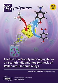
Sustainable star-like octahedron or decahedron Pd/Pt nanoparticles were synthesized by a novel and selective green approach using a bioconjugate of chitosan and polyhydroxybutyrate (Cs–PHB). The bimetallic Pd/Pt nanoparticle alloys synthesized with various Pd/Pt molar ratios were successfully applied in the catalytic reduction of 4-nitrophenol to 4-aminophenol by borohydride. The calculated κc values (estimated ratio between pseudo-first-order kinetic rate constants (s−1) values and the catalyst concentrations (g/L)) revealed that the decahedron nanoparticles exhibited excellent catalytic activity compared to other bimetallic nanoparticles reported in the literature. View this paper.
  • Issues are regarded as officially published after their release is announced to the table of contents alert mailing list.
  • You may sign up for e-mail alerts to receive table of contents of newly released issues.
  • PDF is the official format for papers published in both, html and pdf forms. To view the papers in pdf format, click on the "PDF Full-text" link, and use the free Adobe Reader to open them.
Order results
Result details
Section
Select all
Export citation of selected articles as:
15 pages, 3238 KiB  
Article
Understanding the Polymerization of Polyfurfuryl Alcohol: Ring Opening and Diels-Alder Reactions
by Gianluca Tondi, Nicola Cefarin, Thomas Sepperer, Francesco D’Amico, Raphael J. F. Berger, Maurizio Musso, Giovanni Birarda, Andreas Reyer, Thomas Schnabel and Lisa Vaccari
Polymers 2019, 11(12), 2126; https://doi.org/10.3390/polym11122126 - 17 Dec 2019
Cited by 44 | Viewed by 6568
Abstract
Polyfurfuryl alcohol (PFA) is one of the most intriguing polymers because, despite its easy polymerization in acid environment, its molecular structure is definitely not obvious. Many studies have been performed in recent decades, and every time, surprising aspects came out. With the present [...] Read more.
Polyfurfuryl alcohol (PFA) is one of the most intriguing polymers because, despite its easy polymerization in acid environment, its molecular structure is definitely not obvious. Many studies have been performed in recent decades, and every time, surprising aspects came out. With the present study, we aim to take advantage of all of the findings of previous investigations and exploit them for the interpretation of the completely cured PFA spectra registered with three of the most powerful techniques for the characterization of solid, insoluble polymers: Solid-State 13C-NMR, Attenuated Total Reflectance (ATR), Fourier Transform Infrared (FTIR) spectroscopy, and UV-resonant Raman spectroscopy at different excitation wavelengths, using both an UV laser source and UV synchrotron radiation. In addition, the foreseen structures were modeled and the corresponding 13C-NMR and FTIR spectra were simulated with first-principles and semi-empiric methods to evaluate their matching with experimental ones. Thanks to this multi-technique approach, based on complementary analytical tools and computational support, it was possible to conclude that, in addition to the major linear unconjugated polymerization, the PFA structure consists of Diels-Alder rearrangements occurring after the opening of some furanic units, while the terminal moieties of the chain involves γ-lactone arrangements. The occurrence of head-head methylene ether bridges and free hydroxyl groups (from unreacted furfuryl alcohol, FA, or terminal chains) could be excluded, while the conjugated systems could be considered rather limited. Full article
(This article belongs to the Special Issue State-of-the-Art Polymer Science and Technology in Italy (2019,2020))
Show Figures

Graphical abstract

Graphical abstract
Full article ">Figure 1
<p>Solid-state <sup>13</sup>C-NMR spectrum of hardened PFA.</p>
Full article ">Figure 2
<p>Diels-Alder crosslinking reaction between linear (<b>1</b>) and ring opened (<b>2</b>) PFA structures.</p>
Full article ">Figure 3
<p>FTIR spectrum of hardened PFA in the spectral region 1850–450 cm<sup>−1</sup>. In the inset, the spectral region 3550–2750 cm<sup>−1</sup> is plotted, keeping the original intensity scale.</p>
Full article ">Figure 4
<p>UV-Raman spectra of hardened PFA collected with UV laser excitation at 266 nm (black curve), and with synchrotron radiation excitation at 249 nm and 226 nm (blue and green curves respectively). Red dotted line pinpoints the 1650 cm<sup>−1</sup> wavenumber.</p>
Full article ">Figure 5
<p>Comparison of experimental and calculated <sup>13</sup>C-NMR spectrum of PFA. With * the bands due to the artifact of the model.</p>
Full article ">Figure 6
<p>Comparison of experimental and calculated FTIR spectrum of PFA. With * the bands due to the artifact of the model.</p>
Full article ">
13 pages, 2937 KiB  
Article
A Methodology Towards Mechanical Properties Optimization of Three-Component Polymers by the Gradual Variation of Feed Composition in Semi-Continuous Emulsion-Free Radical Polymerization
by Francisco J. Rivera-Gálvez, Luis J. González-Ortiz, Miguel A. López-Manchado, María E. Hernández-Hernández and Carlos F. Jasso-Gastinel
Polymers 2019, 11(12), 2125; https://doi.org/10.3390/polym11122125 - 17 Dec 2019
Cited by 2 | Viewed by 2501
Abstract
In this work, a new methodology for the synthesis of three-component polymers (TCPs) was developed using a seeded, semi-continuous free-radical emulsion polymerization towards the optimization of the moduli–ultimate deformation performance and energy dissipation capacity for a styrene (S), n-butyl acrylate (BA), and 4-vinylbenzyl [...] Read more.
In this work, a new methodology for the synthesis of three-component polymers (TCPs) was developed using a seeded, semi-continuous free-radical emulsion polymerization towards the optimization of the moduli–ultimate deformation performance and energy dissipation capacity for a styrene (S), n-butyl acrylate (BA), and 4-vinylbenzyl chloride (VBC) system. The three components were sequentially fed in pairs, varying feed composition along the conversion using S as the common monomer. To prepare a reference material, an industrial method was utilized with those monomers, using an equivalent global composition in a two-stage batch process (TS). Nanophase formation in the particles was observed by transmission electron microscopy (TEM), while the separation of the phases in the solid samples was observed by atomic force microscopy (AFM). The changes in glass transition temperature were determined by differential scanning calorimetry (DSC) and dynamic mechanical analysis (DMA). The latter was primarily used to compare mechanodynamic properties as a function of temperature for the two synthesis methods used. Thus, the higher toughness of the forced composition three-component polymeric materials was evaluated by means of their energy dissipation capacity, toughness, and stress–strain measurements at several temperatures. Full article
(This article belongs to the Section Polymer Analysis and Characterization)
Show Figures

Graphical abstract

Graphical abstract
Full article ">Figure 1
<p>Profiles of BA, S, and VBC monomers used to prepare the three-component polymers (TCPs): (<b>a</b>) profile I; (<b>b</b>) profile II. For materials code, see the previous paragraph.</p>
Full article ">Figure 2
<p>TEM images of polymer particles for TS, IPS, and IIPS at 400 kx (<b>a</b>–<b>c</b>), respectively, and similarly (<b>d</b>–<b>f</b>) at 40 kx.</p>
Full article ">Figure 3
<p>AFM images of (<b>a</b>) TS, (<b>b</b>) IPS, (<b>c</b>) IIPS, and (<b>d</b>) IIPBA.</p>
Full article ">Figure 4
<p>Differential scanning calorimetry analysis for TS polymer and TCPs in nitrogen atmosphere and heating rate of 20 °C/min. For materials code see <a href="#sec2dot2dot2-polymers-11-02125" class="html-sec">Section 2.2.2</a>.</p>
Full article ">Figure 5
<p>(<b>a</b>) Storage and (<b>b</b>) loss moduli versus temperature for TS polymer and TCP. Frequency of 1 Hz. Heating rate of 1.5 °C/min. For materials code see <a href="#sec2dot2dot2-polymers-11-02125" class="html-sec">Section 2.2.2</a>.</p>
Full article ">Figure 6
<p>Strain–stress curves for TS polymer and TCPs at a crosshead speed of 5 mm/min and (<b>a</b>) 23, (<b>b</b>) 40, and (<b>c</b>) 50 ± 2 °C. For materials code see <a href="#sec2dot2dot2-polymers-11-02125" class="html-sec">Section 2.2.2</a>.</p>
Full article ">
16 pages, 2144 KiB  
Article
Selected Physicochemical and Pharmaceutical Properties of Poly-ε-caprolactone and Poly(d,l-lactide-co-ε-caprolactone) Conjugates of Lamivudine Synthesized via Ring-Opening Polymerization
by Tomasz Urbaniak and Witold Musiał
Polymers 2019, 11(12), 2124; https://doi.org/10.3390/polym11122124 - 17 Dec 2019
Cited by 1 | Viewed by 2886
Abstract
The modification of drug fate after administration may be achieved by the covalent coupling of active pharmaceutical ingredients with macromolecules. To prolong or delay the release, slowly degrading polymers such as polyesters may be applied for conjugation. The detachment of a covalently conjugated [...] Read more.
The modification of drug fate after administration may be achieved by the covalent coupling of active pharmaceutical ingredients with macromolecules. To prolong or delay the release, slowly degrading polymers such as polyesters may be applied for conjugation. The detachment of a covalently conjugated drug from the polymeric matrix relies mostly on the material degradation profile and barely on the weak interaction between the drug and macromolecules. In the present study, lamivudine was conjugated via ring-opening polymerization with poly-ε-caprolactone and poly(d,l-lactide-co-ε-caprolactone). The influence of the reaction parameters on the course of the polymerization and physicochemical properties of obtained conjugates were investigated. Subsequently, selected reaction products were formulated into submicron particles, and drug release profiles in physiological-like conditions were investigated. The course of the reaction was monitored via gel permeation chromatography. The structure and physicochemical properties of products were evaluated via spectroscopic, calorimetric, and diffractometric methods. The profile of the drug release from particles prepared by the slow evaporation of conjugate solution from o/w emulsion was monitored with high-performance liquid chromatography. Both an elevated reaction temperature and higher catalyst concentration increased the polymerization rate and simultaneously promoted the side reactions, resulting in a broad molecular weight distribution of products in the range from 1.30 to 2.15. The physicochemical properties of conjugates obtained in different conditions varied and had a direct influence on the drug release. The release curve of lamivudine from particles based on low molecular weight conjugates achieved a plateau between 18.9 and 22.2 μg per mg of conjugate within a month. Drug detachment from particles composed of high molecular weight conjugates exhibited a distinct delay period preceded by a drug burst release at a maximal level of 13.3 μg per mg of conjugate. Conjugate chemical composition and the degree of crystallinity were also found to influence the release. Full article
(This article belongs to the Special Issue Biodegradable Polymers for Biomedical Application)
Show Figures

Graphical abstract

Graphical abstract
Full article ">Figure 1
<p>Monomer conversion versus reaction time for (<b>a</b>) <span class="html-italic">ε</span>-caprolactone (<span class="html-italic">ε</span>-CL) homopolymerization and (<b>b</b>) LA-<span class="html-italic">ε</span>-CL copolymerization in varying temperatures ranging from 100 °C to 145 °C; (<b>c</b>) <span class="html-italic">ε</span>-CL homopolymerization and (<b>d</b>) LA-<span class="html-italic">ε</span>-CL copolymerization in varying 2-ethylhexanoate (SO) concentrations expressed as SO:LV ratios; a detailed explanation of the synthesis abbreviations is included in the <a href="#polymers-11-02124-t001" class="html-table">Table 1</a> header.</p>
Full article ">Figure 2
<p>Monomer conversion vs. time vs. weight average molecular weight for (<b>a</b>) <span class="html-italic">ε</span>-CL homopolymerization and (<b>b</b>) LA-<span class="html-italic">ε</span>-CL copolymerization reactions employing an SO:LV ratio of 1:7, conducted in temperatures of 100 °C, 115 °C, 130 °C, and 145 °C; a detailed explanation of the synthesis abbreviations is included in the <a href="#polymers-11-02124-t001" class="html-table">Table 1</a> header.</p>
Full article ">Figure 3
<p>Monomer conversion vs. time vs. weight average molecular weight for (<b>a</b>) <span class="html-italic">ε</span>-CL homopolymerization and (<b>b</b>) LA-<span class="html-italic">ε</span>-CL copolymerization reactions conducted in 115 °C and varying SO concentrations expressed as the SO:LV ratio; a detailed explanation of the synthesis abbreviations is included in the <a href="#polymers-11-02124-t001" class="html-table">Table 1</a> header.</p>
Full article ">Figure 4
<p>Molecular weight polydispersity index versus reaction time for: (<b>a</b>) <span class="html-italic">ε</span>-CL homopolymerization and (<b>b</b>) LA-<span class="html-italic">ε</span>-CL copolymerization in varying temperatures ranging from 100 °C to 145 °C; (<b>c</b>) <span class="html-italic">ε</span>-CL homopolymerization and (<b>d</b>) LA-<span class="html-italic">ε</span>-CL copolymerization in varying SO concentrations expressed as SO:LV ratio; a detailed explanation of the synthesis abbreviations is included in the <a href="#polymers-11-02124-t001" class="html-table">Table 1</a> header.</p>
Full article ">Figure 4 Cont.
<p>Molecular weight polydispersity index versus reaction time for: (<b>a</b>) <span class="html-italic">ε</span>-CL homopolymerization and (<b>b</b>) LA-<span class="html-italic">ε</span>-CL copolymerization in varying temperatures ranging from 100 °C to 145 °C; (<b>c</b>) <span class="html-italic">ε</span>-CL homopolymerization and (<b>d</b>) LA-<span class="html-italic">ε</span>-CL copolymerization in varying SO concentrations expressed as SO:LV ratio; a detailed explanation of the synthesis abbreviations is included in the <a href="#polymers-11-02124-t001" class="html-table">Table 1</a> header.</p>
Full article ">Figure 5
<p>Ln(M<sub>t</sub>/M<sub>0</sub>) versus reaction time plot for <span class="html-italic">ε</span>-CL-LA copolymerization and <span class="html-italic">ε</span>-CL homopolymerization reactions conducted in (<b>a</b>) various temperatures and (<b>b</b>) SO:LV ratios; a detailed explanation of the synthesis abbreviations is included in the <a href="#polymers-11-02124-t001" class="html-table">Table 1</a> header.</p>
Full article ">Figure 6
<p>Arrhenius plot for LV-initiated homopolymerization (○) and copolymerization (□) reactions.</p>
Full article ">Figure 7
<p>(<b>a</b>) LV release in phosphate-buffered saline at 37 ± 2 °C for period of 51 days and (<b>b</b>) corresponding changes of release media pH during release experiment.</p>
Full article ">
24 pages, 13466 KiB  
Article
Investigation of Piezoelectricity and Resistivity of Surface Modified Barium Titanate Nanocomposites
by Udhay Sundar, Zichen Lao and Kimberly Cook-Chennault
Polymers 2019, 11(12), 2123; https://doi.org/10.3390/polym11122123 - 17 Dec 2019
Cited by 15 | Viewed by 4411
Abstract
Polymer-ceramic nanocomposite piezoelectric and dielectric films are of interest because of their possible application to advanced embedded energy storage devices for printed wired electrical boards. The incompatibility of the two constituent materials; hydrophilic ceramic filler, and hydrophobic epoxy limit the filler concentration, and [...] Read more.
Polymer-ceramic nanocomposite piezoelectric and dielectric films are of interest because of their possible application to advanced embedded energy storage devices for printed wired electrical boards. The incompatibility of the two constituent materials; hydrophilic ceramic filler, and hydrophobic epoxy limit the filler concentration, and thus, their piezoelectric properties. This work aims to understand the role of surfactant concentration in establishing meaningful interfacial layers between the epoxy and ceramic filler particles by observing particle surface morphology, piezoelectric strain coefficients, and resistivity spectra. A comprehensive study of nanocomposites, comprising non-treated and surface treated barium titanate (BTO), embedded within an epoxy matrix, was performed. The surface treatments were performed with two types of coupling agents: Ethanol and 3-glycidyloxypropyltrimethoxysilan. The observations of particle agglomeration, piezoelectric strain coefficients, and resistivity were compared, where the most ideal properties were found for concentrations of 0.02 and 0.025. This work demonstrates that the interfacial core-shell processing layer concentration influences the macroscopic properties of nanocomposites, and the opportunities for tuning interfacial layers for desirable characteristics of specific applications. Full article
Show Figures

Graphical abstract

Graphical abstract
Full article ">Figure 1
<p>General structure of a silane coupling agent, where R is an organic functional group.</p>
Full article ">Figure 2
<p>Surface treatment of BaTiO<sub>3</sub> nanoparticles with KH-560. The mixture was magnetically stirred for 12 h and 70 °C followed by drying in air at 50 °C for an additional 12 h.</p>
Full article ">Figure 3
<p>A Schematic diagram of the composite preparation is provided. The contents, i.e., surface-treated BaTiO<sub>3</sub>, epoxy was mixed in a beaker and ultra-sonicated for 1 h, and the subsequent mixture was spin-coated using a multi-step method to ensure uniform dispersion by accelerating and decelerating gradually.</p>
Full article ">Figure 4
<p>EDS micrograph images of the BT powder surface treated using ethanol for (<b>A</b>) 1 h, (<b>B</b>) 2 h, (<b>C</b>) 3 h, (<b>D</b>) 4 h and (<b>E</b>) 5 h. It is evident that there is better particle dispersion with longer sonication time in ethanol. However, the optimal sonication time based on the images is for four hours. (Note: The different colors all represent BT nanoparticles, but since they are on different SEM studs, the machine treats them as different sample sets, and hence the change in color).</p>
Full article ">Figure 5
<p>SEM micrograph images of BaTiO<sub>3</sub> nanoparticles that were surface treated in ethanol for 4 h (<b>A</b>) at a scale of 100 µm and (<b>B</b>) at a scale of 20 µm. From the images we can see charging of the samples even after applying a gold coating of 30 nm, which could be attributed to the surface charges produced on the BaTiO<sub>3</sub> nanoparticle due to its reaction with ethanol. This electrostatic charge keeps the BaTiO<sub>3</sub> nanoparticles from agglomerating as can be seen by the reduction in mean particle size i.e., from ~60 µm to 40 µm.</p>
Full article ">Figure 6
<p>SEM micrograph images of (<b>A</b>) pristine BaTiO<sub>3</sub> particles and (<b>B</b>) BaTiO<sub>3</sub> particles surface treated using SCA 0.01. The images represent the cluster sizes of the of BaTiO<sub>3</sub> nanoparticles on a carbon tape. The average cluster size of the pristine BaTiO<sub>3</sub> particles is ~60 µm and that of the BaTiO<sub>3</sub> particles surface treated using SCA 0.01 is ~30 µm.</p>
Full article ">Figure 7
<p>Particle distributions of (<b>a</b>) pristine BaTiO<sub>3</sub> which shows an average cluster size of ~60 µm, (<b>b</b>) ethanol surface treated for 4 h with an average cliuster size of ~40 µm, (<b>c</b>) 0.01 silane coupling agent treated BaTiO<sub>3</sub> with an average cluster size of ~30 µm. It can observed that the agglomerates reduce with surface treatment. (<b>d</b>) 0.015 silane coupling agent with an average cluster size of &lt;~30 µm, (<b>e</b>) 0.020 silane coupling agent with an average cluster size of &lt;~25 µm, and (<b>f</b>) 0.025 silane coupling agent with an average cluster size of ~25–30 µm.</p>
Full article ">Figure 8
<p>EDS micrograph images of the fractured surface of the pristine (not surface treated) BT(0.5)/epoxy composite film where (<b>A</b>) overall surface of the composite, (<b>B</b>) distribution of Ba and (<b>C</b>) Ti within the overlaying epoxy matrix.</p>
Full article ">Figure 9
<p>EDS micrograph images of the fractured surface of the SCA 0.010 surface treated BT (0.5)—Epoxy composite film where (<b>A</b>) is the overall surface of the composite, (<b>B</b>) distribution of Ba and (<b>C</b>) Ti particles within the overlaying epoxy matrix.</p>
Full article ">Figure 10
<p>EDS micrograph images of SCA 0.015 surface modified BaTiO<sub>3</sub> powder showing (<b>A</b>) the EDS layered image and the distribution of (<b>B</b>) Ti, (<b>C</b>) Ba and (<b>D</b>) Si particles.</p>
Full article ">Figure 11
<p>EDS micrograph images of BaTiO<sub>3</sub> nanoparticles that were surface treated using SCA 0.020 taken at a scale of 100 µm. It can be seen that Si was successfully coated on to the surface of the BaTiO<sub>3</sub> nanoparticles as seen in (<b>A</b>). The images in (<b>B</b>–<b>D</b>) represent the particle distributions of Ba, Ti and Si, respectively. Compared to the samples surface treated using SCA 0.015, we notice that there is a greater correspondence between the Ba atoms in (<b>B</b>) and the Si atoms in (<b>D</b>).</p>
Full article ">Figure 12
<p>(<b>A</b>) EDS micrograph images of the BaTiO<sub>3</sub> nanoparticles that were surface treated using SCA 0.025. The green color depicts the (<b>B</b>) particle distribution of silicon atoms and the red denotes (<b>C</b>) the barium atoms.</p>
Full article ">Figure 13
<p>SEM micrograph images of the cross-section of the fractured surface of the composite that was prepared using 0.5 volume fraction 0.020 silane coupling agent BaTiO<sub>3</sub> nanoparticles at (<b>A</b>) 129 X and (<b>B</b>) 93 X.</p>
Full article ">Figure 14
<p>SEM micrograph images of (<b>A</b>) SCA 0.025 BT (0.5)/epoxy composite at 280 X and (<b>B</b>) SCA 0.025 BT (0.6)/epoxy at 151 X; both showing significant delamination of the film from the composite. It is interesting to note that there is no cracking on the composite film, it delaminates as a whole.</p>
Full article ">Figure 15
<p>(<b>a</b>) The piezoelectric strain coefficient, d<sub>33</sub> for the BaTiO<sub>3</sub>-epoxy composite fabricated using BaTiO<sub>3</sub> nanoparticles that were surface modified using ethanol for 4 h is plotted as a function of volume fraction of BaTiO<sub>3</sub>. The maximum value is 0.58 pC/N at 0.6 vol. fraction of BaTiO<sub>3</sub>. (<b>b</b>) The piezoelectric strain coefficient, d<sub>31</sub> for the BaTiO<sub>3</sub>-epoxy composite fabricated using BaTiO<sub>3</sub> nanoparticles that were surface modified using ethanol for 4 h is plotted as a function of volume fraction of BaTiO<sub>3</sub>. The maximum value is 1.49 pC/N at 0.6 volume fraction of BaTiO<sub>3</sub>.</p>
Full article ">Figure 16
<p>The piezoelectric strain coefficient, d33, for the BaTiO<sub>3</sub>-epoxy composite that were fabricated using BaTiO<sub>3</sub> nanoparticles which were untreated, surface modified by 0.01 SCA, 0.015 SCA, 0.02 SCA and 0.025 SCA. The maximum d33 values are 0.44, 0.71, 0.87, 1.16 and 0.84 pC/N at volume fractions of 0.6, 0.6, 0.5, 0.5 and 0.4, respectively.</p>
Full article ">Figure 17
<p>The piezoelectric strain coefficient, <span class="html-italic">d</span><sub>31</sub>, for the BaTiO<sub>3</sub>-epoxy composite that were fabricated using BaTiO<sub>3</sub> nanoparticles which were untreated, surface modified by 0.01 SCA, 0.015 SCA, 0.02 SCA and 0.025% SCA. The maximum <span class="html-italic">d</span><sub>31</sub> values are 0.86, 2.18, 1.89, 3.11 and 2.38 pC/N at volume fractions of 0.6, 0.6, 0.5, 0.5 and 0.4, respectively.</p>
Full article ">Figure 18
<p>The resistivity of the BaTiO<sub>3</sub>-epoxy composites that were fabricated using BaTiO<sub>3</sub> nanoparticles which were not surface treated (pristine) is plotted as a function of frequency. The maximum resistivity recorded at 20 MHz was 4.20 ohm-m which corresponds to the composites containing 10% of BaTiO<sub>3</sub>.</p>
Full article ">Figure 19
<p>(<b>a</b>) The resistivity of the BaTiO<sub>3</sub>-epoxy composite that were fabricated using BaTiO<sub>3</sub> nanoparticles, which were surface-modified using 0.010 SCA, which is plotted as a function of frequency. The maximum resistivity recorded at 20 MHz was 22.08 ohm-m which corresponds to the composites containing 0.1 of BaTiO<sub>3</sub>. (<b>b</b>) The resistivity of the BaTiO<sub>3</sub>-epoxy composite that were fabricated using BaTiO<sub>3</sub> nanoparticles, surface-modified using 0.015 SCA, is plotted as a function of frequency. The maximum resistivity recorded at 20 MHz was 24.26 ohm-m which corresponds to the composites containing 0.5 of BaTiO<sub>3</sub>.</p>
Full article ">Figure 20
<p>(<b>a</b>) The resistivity of the BaTiO<sub>3</sub>-epoxy composite that were fabricated using BaTiO<sub>3</sub> nanoparticles. These nanoparticles were surface modified using 0.02 SCA is plotted as a function of frequency. The maximum resistivity recorded at 20 MHz was 39.76 ohm-m which corresponds to the composites containing 0.5 of BaTiO<sub>3</sub>. (<b>b</b>) The resistivity of the BaTiO<sub>3</sub>-epoxy composite that were fabricated using BaTiO<sub>3</sub> nanoparticles, which were surface modified using 0.02 SCA is plotted as a function of frequency. The maximum resistivity recorded at 20 MHz was 58.22 ohm-m which corresponds to the composites containing 0.4 of BaTiO<sub>3</sub>.</p>
Full article ">Figure 21
<p>Substitution of the hydroxyl groups due to the surface modification with the silane coupling agent. This leads to a decrease in conductivity as there is no more water adsorbed on the surface of the BaTiO<sub>3</sub> nanoparticle.</p>
Full article ">
13 pages, 2450 KiB  
Article
Giving a Second Opportunity to Tire Waste: An Alternative Path for the Development of Sustainable Self-Healing Styrene–Butadiene Rubber Compounds Overcoming the Magic Triangle of Tires
by Javier Araujo-Morera, Marianella Hernández Santana, Raquel Verdejo and Miguel Angel López-Manchado
Polymers 2019, 11(12), 2122; https://doi.org/10.3390/polym11122122 - 17 Dec 2019
Cited by 50 | Viewed by 7616
Abstract
Current regulations demand tires with long lifetime and reduced fuel consumption without sacrificing car safety. However, tire technology still needs to reach a suitable balance between these three indicators. Here, we address them by developing a self-healing tire compound using styrene–butadiene rubber (SBR) [...] Read more.
Current regulations demand tires with long lifetime and reduced fuel consumption without sacrificing car safety. However, tire technology still needs to reach a suitable balance between these three indicators. Here, we address them by developing a self-healing tire compound using styrene–butadiene rubber (SBR) as the matrix and reclaimed tire waste as the sustainable filler. The addition of ground tire rubber (GTR) to the matrix simultaneously improved the rolling resistance and maintained both wet grip and healing ability. We provide an in-depth analysis of the healing behavior of the material at a scale close to the relevant molecular processes through a systematic dynamic-mechanical and dielectric analysis. We found that SBR and SBR/GTR compounds show a complete recovery of stiffness and relaxation dynamics after being damaged by cyclic deformation, resulting in a heterogeneous repaired rubber network. This new development could well overcome the so-called magic triangle of tires, which is certainly one of the key objectives of the tire industry. Full article
(This article belongs to the Special Issue Advances in Rubber Composites)
Show Figures

Graphical abstract

Graphical abstract
Full article ">Figure 1
<p>(<b>a</b>) Storage modulus (E′); (<b>b</b>) Damping factor (tanδ) of SBR/GTR compounds.</p>
Full article ">Figure 2
<p>Damping factor (tan δ) of SBR/GTR compounds at low (−10 °C) and high (60 °C) temperatures.</p>
Full article ">Figure 3
<p>Dielectric loss (ε″) as a function of frequency in the temperature range from −45 to −5 °C of: (<b>a</b>) SBR compound; (<b>b</b>) SBR/10 GTR compound, and (<b>c</b>) GTR. (<b>d</b>) Dielectric loss (ε″) as a function of GTR content at a selected temperature T= −25 °C.</p>
Full article ">Figure 4
<p>Storage modulus (E′) as a function of temperature for: (<b>a</b>) SBR and (<b>b</b>) SBR/10GTR compound.</p>
Full article ">Figure 5
<p>Dielectric magnitudes (ε″ and σ′) as a function of the frequency at a temperature of −25 °C for the virgin, damaged, and repaired states of: (<b>a</b>) SBR; (<b>b</b>,<b>c</b>) SBR/10GTR compound.</p>
Full article ">Figure 6
<p>Damping factor (tan δ) of SBR and SBR/10GTR compounds under virgin, damaged, and repaired conditions at: (<b>a</b>) low temperatures (−10 °C); (<b>b</b>) elevated temperatures (60 °C).</p>
Full article ">Figure 7
<p>Rolling resistance, wet grip, and healing ability of SBR and SBR/10GTR compounds.</p>
Full article ">
10 pages, 1448 KiB  
Article
A Complexed Initiating System AlCl3·Phenetole/TiCl4·H2O with Dominant Synergistic Effect for Efficient Synthesis of High Molecular Weight Polyisobutylene
by Yulong Jin, Liang Chen, Xing Guo, Linfeng Xu, Zhihua Zhu, Zhen Liu, Ruihua Cheng and Boping Liu
Polymers 2019, 11(12), 2121; https://doi.org/10.3390/polym11122121 - 17 Dec 2019
Cited by 2 | Viewed by 2891
Abstract
A complexed initiating system AlCl3·phenetole/TiCl4·H2O was prepared by simply compounding AlCl3/phenetole and TiCl4/H2O and used for cationic polymerization of isobutylene. It was found AlCl3·phenetole/TiCl4·H2O exhibited [...] Read more.
A complexed initiating system AlCl3·phenetole/TiCl4·H2O was prepared by simply compounding AlCl3/phenetole and TiCl4/H2O and used for cationic polymerization of isobutylene. It was found AlCl3·phenetole/TiCl4·H2O exhibited activities 1.2–3 times higher than those of AlCl3/phenetole, and more than an order of magnitude higher than those of TiCl4/H2O, which indicated a notable synergistic effect produced in the complexed system. In addition, due to the higher activity of AlCl3·phenetole/TiCl4·H2O, lower coinitiator concentration and polymerization temperature, as well as higher monomer concentration were more favored for this complexed initiating system to produce polyisobutylene (PIB) with reasonable molecular weight (Mw) and molecular weight distribution (MWD). Furthermore, high molecular weight polyisobutylene (HPIB) with Mw = 1–3 × 10⁵ g·mol−1 could be successfully produced by the complexed catalyst system at Tp = −60 to −40 °C. As a whole, the high activity as well as the simple preparation procedures of the complexed initiating system offer us a unique approach for the production of HPIB with improved efficiency. Full article
(This article belongs to the Section Polymer Chemistry)
Show Figures

Graphical abstract

Graphical abstract
Full article ">Figure 1
<p>The gel permeation chromatography (GPC) curves of the PIB produced by (<b>A</b>) AlCl<sub>3</sub>/phenetole and (<b>B</b>) AlCl<sub>3</sub>·phenetole /TiCl<sub>4</sub>·H<sub>2</sub>O at various [AlCl<sub>3</sub> + TiCl<sub>4</sub>] concentrations. The other reaction conditions are listed in <a href="#polymers-11-02121-t001" class="html-table">Table 1</a>.</p>
Full article ">Figure 2
<p>The GPC curves of PIB produced by AlCl<sub>3</sub>/phenetole, TiCl<sub>4</sub>/H<sub>2</sub>O and AlCl<sub>3</sub>·phenetole/TiCl<sub>4</sub>·H<sub>2</sub>O, (<b>A</b>) the GPC curves of PIB produced from run 3, 4 and 11 in <a href="#polymers-11-02121-t001" class="html-table">Table 1</a>; (<b>B</b>) the GPC curves of PIB produced from run 5, 6 and 11 in <a href="#polymers-11-02121-t001" class="html-table">Table 1</a> and (<b>C</b>) the GPC curves of PIB produced from run 7, 8 and 11 in <a href="#polymers-11-02121-t001" class="html-table">Table 1</a>.</p>
Full article ">Figure 3
<p>The GPC curves of the PIB produced by (<b>A</b>) AlCl<sub>3</sub>/phenetole and (<b>B</b>) AlCl<sub>3</sub>·phenetole/ TiCl<sub>4</sub>·H<sub>2</sub>O at different T<sub>p</sub>. The other reaction conditions are listed in <a href="#polymers-11-02121-t002" class="html-table">Table 2</a>.</p>
Full article ">
27 pages, 9337 KiB  
Article
Influence of the Design Solutions of Extruder Screw Mixing Tip on Selected Properties of Wheat Bran-Polyethylene Biocomposite
by Emil Sasimowski, Łukasz Majewski and Marta Grochowicz
Polymers 2019, 11(12), 2120; https://doi.org/10.3390/polym11122120 - 17 Dec 2019
Cited by 11 | Viewed by 4820
Abstract
The study investigated the impact of the extruder screw design solution—the intensive mixing tip used—on the course of the extrusion process and the properties of the obtained biocomposite extrudate. A lignocellulosic wheat bran biocomposite based on a low-density polyethylene matrix was extruded. Three [...] Read more.
The study investigated the impact of the extruder screw design solution—the intensive mixing tip used—on the course of the extrusion process and the properties of the obtained biocomposite extrudate. A lignocellulosic wheat bran biocomposite based on a low-density polyethylene matrix was extruded. Three mixing tips of the screw were used interchangeably: apineapple tip, a cut rings tip, and a Maddock tip. The experimental tests carried out included the production of an extrudate with a mass content of bran altered within the range from 10% to 50%. Processing properties such as the melt flow rate (MFR) and mass flow rate of the extruded biocomposite were determined. Selected physical, mechanical, and structural properties of the biocomposite extrudate obtained with the use of the three tested mixing tips at five bran contents were tested. Full article
(This article belongs to the Section Polymer Processing and Engineering)
Show Figures

Figure 1

Figure 1
<p>View of intensive mixing tips: (<b>a</b>) pineapple tip (<b>b</b>) cut rings tip, and (<b>c</b>) Maddock’s tip.</p>
Full article ">Figure 2
<p>View of wheat bran powder: (<b>a</b>) immediately after grinding, and (<b>b</b>) after separation of fractions with a grain size of less than 0.4 mm.</p>
Full article ">Figure 3
<p>Microscopic image of wheat bran powder, fractions with a grain size less than 0.4 mm.</p>
Full article ">Figure 4
<p>View of the breakthrough of the extrudate with a bran content of (<b>a</b>) 10% and (<b>b</b>) 50% made using the pineapple mixing tip.</p>
Full article ">Figure 5
<p>View of the breakthrough of the extrudate with a bran content of (<b>a</b>) 10% and (<b>b</b>) 50% made using the Maddock mixing tip.</p>
Full article ">Figure 6
<p>View of the breakthrough of the extrudate with a bran content of (<b>a</b>) 10% and (<b>b</b>) 50% made using the cut rings mixing tip.</p>
Full article ">Figure 7
<p>Density of the extrudate obtained with the screw mixing tips tested, depending on bran content (mean values with 95% confidence intervals).</p>
Full article ">Figure 8
<p>Water absorption of the extrudate obtained with the screw mixing tips tested, depending on bran content (mean values with 95% confidence intervals).</p>
Full article ">Figure 9
<p>Moisture content of the extrudate obtained with the use of the screw mixing tips tested depending on the time and bran content.</p>
Full article ">Figure 10
<p>Young modulus of the extrudate obtained with the screw mixing tips tested, depending on bran content (mean values with 95% confidence intervals).</p>
Full article ">Figure 11
<p>Tensile strength of the extrudate obtained with the screw mixing tips tested, depending on bran content (mean values with 95% confidence intervals).</p>
Full article ">Figure 12
<p>Strain at tensile strength of the extrudate obtained with the screw mixing tips tested, depending on bran content (mean values with 95% confidence intervals).</p>
Full article ">Figure 13
<p>Variability of the L, a, and b color parameters of the extrudate produced using the pineapple mixing tip, depending on bran content.</p>
Full article ">Figure 14
<p>Variability of the L, a, and b color parameters of the extrudate produced using the Maddock mixing tip, depending on bran content.</p>
Full article ">Figure 15
<p>Variability of the L, a, and b color parameters of the extrudate produced using the cut rings mixing tip, depending on bran content.</p>
Full article ">Figure 16
<p>Melt flow rate (150 °C; 5 kg) of the extrudate obtained with the screw mixing tips tested, depending on bran content (mean values with 95% confidence intervals).</p>
Full article ">Figure 17
<p>Mass flow rate of the extruded plastic obtained with the screw mixing tips tested, depending on bran content (mean values with 95% confidence intervals).</p>
Full article ">Figure 18
<p>FTIR spectra of polyethylene (PE), wheat bran (WB), and composites containing 50% of wheat bran made using the Maddock (PE-M), pineapple (PE-P), and cut rings (PE-C) tips.</p>
Full article ">Figure 19
<p>DSC curves of components and composites with 50% bran content obtained in an inert atmosphere.</p>
Full article ">Figure 20
<p>TG and DTG curves obtained in oxidizing atmosphere for polyethylene (PE), wheat bran, and composites containing 50% of wheat bran made using the Maddock, pineapple, and cut rings tips.</p>
Full article ">Figure 21
<p>3D FTIR spectra of gaseous decomposition products; (<b>A</b>)—PE samples, (<b>B</b>)—composite containing 50% of wheat bran extruded with the pineapple tip, (<b>C</b>)—bran samples.</p>
Full article ">
11 pages, 5055 KiB  
Article
Microfibrillated Cellulose Suspension and Its Electrorheology
by Kisuk Choi, Jae Do Nam, Seung Hyuk Kwon, Hyoung Jin Choi, Md Sakinul Islam and Nhol Kao
Polymers 2019, 11(12), 2119; https://doi.org/10.3390/polym11122119 - 17 Dec 2019
Cited by 17 | Viewed by 3409
Abstract
Microfibrillated cellulose (MFC) particles were synthesized by a low-pressure alkaline delignification process, and their shape and chemical structure were investigated by SEM and Fourier transformation infrared spectroscopy, respectively. As a novel electrorheological (ER) material, the MFC particulate sample was suspended in insulating oil [...] Read more.
Microfibrillated cellulose (MFC) particles were synthesized by a low-pressure alkaline delignification process, and their shape and chemical structure were investigated by SEM and Fourier transformation infrared spectroscopy, respectively. As a novel electrorheological (ER) material, the MFC particulate sample was suspended in insulating oil to fabricate an ER fluid. Its rheological properties—steady shear stress, shear viscosity, yield stress, and dynamic moduli—under electric field strength were characterized by a rotational rheometer. The MFC-based ER fluid demonstrated typical ER characteristics, in which the shear stresses followed the Cho–Choi–Jhon model well under electric field strength. In addition, the solid-like behavior of the ER fluid was investigated with the Schwarzl equation. The elevated value of both dynamic and elastic yield stresses at applied electric field strengths was well described using a power law model (~E1.5). The reversible and quick response of the ER fluid was also illustrated through the on–off test. Full article
(This article belongs to the Special Issue Cellulose and Renewable Materials)
Show Figures

Graphical abstract

Graphical abstract
Full article ">Figure 1
<p>SEM images of microfibrillated cellulose (MFC) with at low (<b>a</b>) and high (<b>b</b>) magnification.</p>
Full article ">Figure 2
<p>FT-IR spectra of MFC.</p>
Full article ">Figure 3
<p>Flow curve of shear stress (<b>a</b>) and shear viscosity (<b>b</b>) of the MFC-based electrorheological (ER) fluid under various electric filed strengths. The line in (<b>a</b>) is fitted by the Cho–Choi–Jhon (CCJ) model.</p>
Full article ">Figure 4
<p>The storage modulus (<b>a</b>) and loss modulus (<b>b</b>) of the MFC-based ER fluid as a function of strain under various electric field strengths.</p>
Full article ">Figure 5
<p>Elastic stresses of the MFC-based ER fluid under various electric field strengths.</p>
Full article ">Figure 6
<p>The storage modulus (<b>a</b>) and loss modulus (<b>b</b>) of the MFC-based ER fluid as a function of angular frequency at fixed strain under various electric field strengths.</p>
Full article ">Figure 7
<p>Relaxation modulus of the MFC-based ER fluid.</p>
Full article ">Figure 8
<p>Shear viscosity versus shear stress measured in the controlled shear stress (CSS) mode for MFC particle-based ER fluid under various electric field strengths.</p>
Full article ">Figure 9
<p>Yield stress dependency on the electric field strength of the MFC-based ER fluid.</p>
Full article ">Figure 10
<p>On–off test results of the MFC-based ER fluid at a constant shear rate (= 1 s<sup>−1</sup>) under various electric field strengths with the unit of kV/mm.</p>
Full article ">
26 pages, 12095 KiB  
Article
The Potential of Agglomerated Cork for Sandwich Structures: A Systematic Investigation of Physical, Thermal, and Mechanical Properties
by Claudia Sergi, Jacopo Tirillò, Fabrizio Sarasini, Enrique Barbero Pozuelo, Sonia Sanchez Saez and Christoph Burgstaller
Polymers 2019, 11(12), 2118; https://doi.org/10.3390/polym11122118 - 17 Dec 2019
Cited by 15 | Viewed by 4152
Abstract
Considering the major role played by sandwich structures in many fields where high stiffness-to-weight ratio is required, the selection of a suitable core material is of paramount importance. In order to face the environmental problems related to waste disposal, the selection of an [...] Read more.
Considering the major role played by sandwich structures in many fields where high stiffness-to-weight ratio is required, the selection of a suitable core material is of paramount importance. In order to face the environmental problems related to waste disposal, the selection of an eco-friendly core material is now included in the design criteria of sandwich structures. Agglomerated cork is recognized as a good solution that combines satisfactory mechanical performances and eco-sustainability. Many research studies individually addressed cork’s morphological, thermal, and mechanical features without providing a comprehensive overview of the relationships that exist between them. In this work, the investigation of the peculiar cork morphology allowed learning more about its good insulation capacity and its impressive recovery capability. The use of dynamic mechanical analysis (DMA) and thermogravimetric analysis (TGA) clarified the influence of temperature on both flexural and compressive performances. The effect of testing parameters such as temperature and speed on agglomerated cork properties was validated through statistical analysis. Moreover, to highlight agglomerated cork advantages and drawbacks, the work provides also a comparison with more traditional polyvinylchloride (PVC) foams commonly used in industrial applications. Full article
(This article belongs to the Section Polymer Applications)
Show Figures

Figure 1

Figure 1
<p>Schematic representation of specimen sampling points for thermal conductivity analysis.</p>
Full article ">Figure 2
<p>NL20 micrograph in the radial direction (<b>A</b>); NL25 micrograph in the tangential/axial direction (<b>B</b>).</p>
Full article ">Figure 3
<p>Hexagonal prism base from a NL10 micrograph (<b>A</b>); heptagonal and pentagonal base prisms from a NL20 micrograph (<b>B</b>).</p>
Full article ">Figure 4
<p>Evaluation of cell-wall thickness of NL10 (<b>A</b>), NL20 (<b>B</b>), and NL25 (<b>C</b>).</p>
Full article ">Figure 5
<p>NL10 micrographs of the boundary region between two granules (<b>A</b>) with an enlargement of the polymeric binder (<b>B</b>).</p>
Full article ">Figure 6
<p>HP250 (<b>A</b>) and HP200 (<b>B</b>) closed-cell microstructure.</p>
Full article ">Figure 7
<p>HP130 closed-cell microstructure in two different areas of the sample (<b>A</b>) and (<b>B</b>).</p>
Full article ">Figure 8
<p>Agglomerated cork and polyvinylchloride (PVC) foam contact angles.</p>
Full article ">Figure 9
<p>NL20 (<b>A</b>) and HP130 (<b>B</b>) water droplets.</p>
Full article ">Figure 10
<p>Agglomerated cork and PVC foam thermal conductivity in as-received and dried conditions.</p>
Full article ">Figure 11
<p>NL10 thermal conductivity evaluated at five different points of the sample.</p>
Full article ">Figure 12
<p>Agglomerated cork and PVC foam <span class="html-italic">R</span>-values in as-received and dried conditions.</p>
Full article ">Figure 13
<p>Agglomerated cork and PVC foam heat capacity in as-received and dried conditions.</p>
Full article ">Figure 14
<p>Agglomerated cork and PVC foam thermal diffusivity in as-received and dried conditions.</p>
Full article ">Figure 15
<p>Thermogravimetric analysis (TGA) and DTG curves of NL10, NL20, and NL25.</p>
Full article ">Figure 16
<p>TGA and DTG curves of HP130, HP200, and HP250.</p>
Full article ">Figure 17
<p>Storage modulus (blue), loss modulus (green), and loss factor (red) of NL10, NL20, and NL25.</p>
Full article ">Figure 18
<p>Storage modulus (blue), loss modulus (green), and loss factor (red) of HP130, HP200, and HP250.</p>
Full article ">Figure 19
<p>Crack propagation during NL25 shear test.</p>
Full article ">Figure 20
<p>NL10 intergranular fracture after shear test.</p>
Full article ">Figure 21
<p>Typical stress—strain curves in bending for core materials at room temperature.</p>
Full article ">Figure 22
<p>Flexural modulus and strength as a function of temperature for agglomerated corks.</p>
Full article ">Figure 23
<p>Flexural modulus and strength as a function of temperature for PVC foams.</p>
Full article ">Figure 24
<p>Failure modes in bending for PVC foams as a function of temperature.</p>
Full article ">Figure 25
<p>Flexural strength dataset of agglomerated corks and PVC foams, and fitting curves with constant density in the three cases.</p>
Full article ">Figure 26
<p>Typical compression curves for PVC foams and agglomerated corks.</p>
Full article ">Figure 27
<p>Agglomerated cork compressive modulus and plateau stress as a function of test speed.</p>
Full article ">Figure 28
<p>PVC foam compressive modulus, plateau stress, and yield strength as a function of test speed.</p>
Full article ">Figure 29
<p>Agglomerated cork instantaneous recovery compared to final test deformation, and residual deformation compared to final test deformation as a function of test speed.</p>
Full article ">Figure 30
<p>PVC foam instantaneous recovery compared to final test deformation, and residual deformation compared to final test deformation as a function of test speed.</p>
Full article ">Figure 31
<p>Agglomerated cork compressive modulus and plateau stress as a function of temperature.</p>
Full article ">Figure 32
<p>PVC foam compressive modulus and plateau stress as a function of temperature.</p>
Full article ">
11 pages, 2072 KiB  
Article
Improving Performance of Electrospun Nylon 6,6 Nanofiber Membrane for Produced Water Filtration via Solvent Vapor Treatment
by Nur Syakinah Abd Halim, Mohd Dzul Hakim Wirzal, Muhammad Roil Bilad, Nik Abdul Hadi Md Nordin, Zulfan Adi Putra, Nonni Soraya Sambudi and Abdull Rahim Mohd Yusoff
Polymers 2019, 11(12), 2117; https://doi.org/10.3390/polym11122117 - 17 Dec 2019
Cited by 36 | Viewed by 6225
Abstract
Electrospun nanofiber membrane (NFM) has a high potential to be applied as a filter for produced water treatment due to its highly porous structure and great permeability. However, it faces fouling issues and has low mechanical properties, which reduces the performance and lifespan [...] Read more.
Electrospun nanofiber membrane (NFM) has a high potential to be applied as a filter for produced water treatment due to its highly porous structure and great permeability. However, it faces fouling issues and has low mechanical properties, which reduces the performance and lifespan of the membrane. NFM has a low integrity and the fine mat easily detaches from the sheet. In this study, nylon 6,6 was selected as the polymer since it offers great hydrophilicity. In order to increase mechanical strength and separation performance of NFM, solvent vapor treatment was implemented where the vapor induces the fusion of fibers. The fabricated nylon 6,6 NFMs were treated with different exposure times of formic acid vapor. Results show that solvent vapor treatment helps to induce the fusion of overlapping fibers. The optimum exposure time for solvent vapor is 5 h to offer full retention of dispersed oil (100% of oil rejection), has 62% higher in tensile strength (1950 MPa) compared to untreated nylon 6,6 NFM (738 MPa), and has the final permeability closest to the untreated nylon 6,6 NFM (733 L/m2.h.bar). It also took more time to get fouled (220 min) compared to untreated NFM (160 min). Full article
(This article belongs to the Special Issue Electrospun Nanofibers: Theory and Its Applications)
Show Figures

Graphical abstract

Graphical abstract
Full article ">Figure 1
<p>FESEM images for untreated nylon 6,6 NFM and treated nylon 6,6 NFM at treatment times of 5, 12, 24 and 48 h with magnification of 10,000×.</p>
Full article ">Figure 2
<p>Fiber diameter (<b>a</b>) and pore size (<b>b</b>) as function of solvent vapor exposure time. The lines in the bars of represent the standard deviation.</p>
Full article ">Figure 3
<p>AFM images for untreated nylon 6,6 NFM and treated nylon 6,6 NFM at treatment times of 5, 12, 24 and 48 h.</p>
Full article ">Figure 4
<p>Dynamic contact angle vs. time for untreated nylon 6,6 NFM and treated nylon 6,6 NFM with treatment time at 5, 12, 24 and 48 h.</p>
Full article ">Figure 5
<p>Tensile strength of nylon 6,6 NFMs.</p>
Full article ">Figure 6
<p>Pure water permeability of nylon 6,6 NFMs.</p>
Full article ">Figure 7
<p>Produced Water permeability for nylon 6, 6 NFMs.</p>
Full article ">Figure 8
<p>Steady-state pure water and PW permeability for nylon 6,6 NFMs.</p>
Full article ">
9 pages, 10300 KiB  
Article
Modified Epoxy Resin Synthesis from Phosphorus—Containing Polyol and Physical Changes Studies in the Synthesized Products
by Jeong Beom Jang, Tae Hee Kim, Taeyoon Kim, Hye Jin Kim, Bongkuk Seo, Choong-Sun Lim and Wonjoo Lee
Polymers 2019, 11(12), 2116; https://doi.org/10.3390/polym11122116 - 16 Dec 2019
Cited by 5 | Viewed by 4924
Abstract
Epoxy resins are commonly used to manufacture the molding compounds, reinforced plastics, coatings, or adhesives required in various industries. However, the demand for new epoxy resins has increased to satisfy diverse industrial requirements such as enhanced mechanical properties, thermal stability, or electrical properties. [...] Read more.
Epoxy resins are commonly used to manufacture the molding compounds, reinforced plastics, coatings, or adhesives required in various industries. However, the demand for new epoxy resins has increased to satisfy diverse industrial requirements such as enhanced mechanical properties, thermal stability, or electrical properties. Therefore, in this study, we synthesized new epoxy resin (PPME) by modifying phosphorous-containing polyol. The prepared resin was analyzed and added to epoxy compositions in various quantities. The compositions were cured at high temperatures to obtain plastics to further test the mechanical and thermal properties of the epoxy resin. The measured tensile and flexural strength of epoxy compositions were similar to the composition without synthesized epoxy resin. However, the heat release rates of the compositions exhibited tendencies of a decrease proportional to the amount of PPME. Full article
(This article belongs to the Special Issue Thermosets II)
Show Figures

Graphical abstract

Graphical abstract
Full article ">Figure 1
<p>Reaction scheme of polyol synthesis.</p>
Full article ">Figure 2
<p><sup>1</sup>H-NMR data of produced polyol.</p>
Full article ">Figure 3
<p>FT-IR spectrum of DGEBF, P-polyol, and PPME.</p>
Full article ">Figure 4
<p>Reaction scheme of PPME.</p>
Full article ">Figure 5
<p>Reaction scheme among bisphenol A epoxy resin, PPME, and D-230.</p>
Full article ">Figure 6
<p>(<b>a</b>) DSC curves of epoxy compositions), (<b>b</b>) HRR vs. temperature curves, (<b>c</b>) HRR data, (<b>d</b>) THR of the epoxy compositions with PPME.</p>
Full article ">Figure 7
<p>Tensile (<b>a</b>) and flexural (<b>b</b>) strength of the cured compositions.</p>
Full article ">Figure 8
<p>Impact strength of the compositions.</p>
Full article ">Figure 9
<p>DMA curves, (<b>a</b>) tan curves, (<b>b</b>) storage modulus curves of epoxy polymers.</p>
Full article ">Figure 10
<p>FE-SEM images of the fractured surfaces: (<b>a</b>) binder, (<b>b</b>) PPME-5, (<b>c</b>) PPME-10, (<b>d</b>) PPME-15.</p>
Full article ">
16 pages, 4496 KiB  
Article
Experimental, Numerical, and Analytical Study on The Effect of Graphene Oxide in The Mechanical Properties of a Solvent-Free Reinforced Epoxy Resin
by Sergio Horta Muñoz, María del Carmen Serna Moreno, José Miguel González-Domínguez, Pablo Antonio Morales-Rodríguez and Ester Vázquez
Polymers 2019, 11(12), 2115; https://doi.org/10.3390/polym11122115 - 16 Dec 2019
Cited by 8 | Viewed by 3322
Abstract
This paper presents a methodology for manufacturing nanocomposites from an epoxy resin reinforced with graphene oxide (GO) nanoparticles. A scalable and sustainable fabrication process, based on a solvent-free method, is proposed with the objective of achieving a high level of GO dispersion, while [...] Read more.
This paper presents a methodology for manufacturing nanocomposites from an epoxy resin reinforced with graphene oxide (GO) nanoparticles. A scalable and sustainable fabrication process, based on a solvent-free method, is proposed with the objective of achieving a high level of GO dispersion, while maintaining matrix performance. The results of three-point bending tests are examined by means of an analytical technique which allows determining the mechanical response of the material under tension and compression from flexural data. As result, an increase of 39% in the compressive elastic modulus of the nanocomposite is found with the addition of 0.3 wt % GO. In parallel, we described how the strain distribution and the failure modes vary with the amount of reinforcement based on digital image correlation (DIC) techniques and scanning electron microscopy (SEM). A novel analytical model, capable of predicting the influence of GO content on the elastic properties of the material, is obtained. Numerical simulations considering the experimental conditions are carried out. the full strain field given by the DIC system is successfully reproduced by means of the finite element method (FEM). While, the experimental failure is explained by the crack growth simulations using the eXtended finite element method (XFEM). Full article
(This article belongs to the Special Issue Processing and Molding of Polymers)
Show Figures

Graphical abstract

Graphical abstract
Full article ">Figure 1
<p>Specimen manufacturing: (<b>a</b>) Graphene-oxide (GO)-epoxy dispersion; (<b>b</b>) Neat resin, 0.3wt % GO- and graphite-reinforced specimens for bending testing.</p>
Full article ">Figure 2
<p>Experimental facility: (<b>a</b>) Electromechanical testing machine and digital image correlation (DIC) system; (<b>b</b>) Specimen image obtained by DIC system during testing.</p>
Full article ">Figure 3
<p>Bending specimen shape and dimensions.</p>
Full article ">Figure 4
<p>Normal strain and stress distributions on the cross-section under bending moment with <span class="html-italic">E<sub>t</sub></span> ≠ <span class="html-italic">E<sub>c</sub></span>.</p>
Full article ">Figure 5
<p>Bar chart showing evolution of tensile, compressive and flexural elastic moduli measured via bending testing.</p>
Full article ">Figure 6
<p>Stress-strain plots obtained from three-point bending tests at the midspan upper face.</p>
Full article ">Figure 7
<p>Fitting of evolution of moduli ratio with reinforcement load.</p>
Full article ">Figure 8
<p>Compressive stress-strain behaviour of the material measured by cylinder compressive testing of blank specimen.</p>
Full article ">Figure 9
<p>Comparison of strain contour maps obtained: (<b>a</b>) experimentally through DIC and (<b>b</b>) numerically by FEM.</p>
Full article ">Figure 10
<p>Comparison of applied force versus maximum deflection plot from eXtended finite element method (XFEM) simulations and three-point bending test of blank specimen.</p>
Full article ">Figure 11
<p>Normal strain contour plot from XFEM simulations corresponding to (<b>a</b>) crack initiation; and (<b>b</b>) end of propagation; and (<b>c</b>) experimental fracture.</p>
Full article ">Figure 12
<p>Scanning electron microscopy (SEM) fractographies: (<b>a</b>) and (<b>b</b>) neat resin; (<b>c</b>) and (<b>d</b>) 0.3 wt % GO reinforced epoxy.</p>
Full article ">
21 pages, 8163 KiB  
Article
Compression-Responsive Photonic Crystals Based on Fluorine-Containing Polymers
by Julia Kredel and Markus Gallei
Polymers 2019, 11(12), 2114; https://doi.org/10.3390/polym11122114 - 16 Dec 2019
Cited by 10 | Viewed by 4252
Abstract
Fluoropolymers represent a unique class of functional polymers due to their various interesting and important properties such as thermal stability, resistance toward chemicals, repellent behaviors, and their low refractive indices in comparison to other polymeric materials. Based on the latter optical property, fluoropolymers [...] Read more.
Fluoropolymers represent a unique class of functional polymers due to their various interesting and important properties such as thermal stability, resistance toward chemicals, repellent behaviors, and their low refractive indices in comparison to other polymeric materials. Based on the latter optical property, fluoropolymers are particularly of interest for the preparation of photonic crystals for optical sensing application. Within the present study, photonic crystals were prepared based on core-interlayer-shell particles focusing on fluoropolymers. For particle assembly, the melt-shear organization technique was applied. The high order and refractive index contrast of the individual components of the colloidal crystal structure lead to remarkable reflection colors according to Bragg’s law of diffraction. Due to the special architecture of the particles, consisting of a soft core, a comparably hard interlayer, and again a soft shell, the resulting opal films were capable of changing their shape and domain sizes upon applied pressure, which was accompanied with a (reversible) change of the observed reflection colors as well. By the incorporation of adjustable amounts of UV cross-linking agents into the opal film and subsequent treatment with different UV irradiation times, stable and pressure-sensitive opal films were obtained. It is shown that the present strategy led to (i) pressure-sensitive opal films featuring reversibly switchable reflection colors and (ii) that opal films can be prepared, for which the written pattern—resulting from the compressed particles—could be fixed upon subsequent irradiation with UV light. The herein described novel fluoropolymer-containing photonic crystals, with their pressure-tunable reflection color, are promising candidates in the field of sensing devices and as potential candidates for anti-counterfeiting materials. Full article
(This article belongs to the Section Polymer Chemistry)
Show Figures

Graphical abstract

Graphical abstract
Full article ">Figure 1
<p>Stepwise synthesis of core-interlayer-shell particles. The seed and core particles consist of poly (benzyl acrylate-<span class="html-italic">co</span>-allyl methacrylate) (BzA and ALMA), the interlayer contains styrene and allyl methacrylate (ALMA) and the shell material is formed by 2-((1, 1, 2-trifluoro-2-(perfluoropropoxy) ethyl) thio) ethylacrylate (fluoro-acrylate) and ethyl acrylate (EA).</p>
Full article ">Figure 2
<p>TEM measurements of the two different particle batches for herein described investigations: seed particles (<b>a</b>); core particles (<b>b</b>); core-interlayer 1 particles (<b>c</b>); core-interlayer 1-shell 1 particles (<b>d</b>); core-interlayer 2 particles (<b>e</b>) and core-interlayer 2-shell 2 particles (<b>f</b>). For both types of core-interlayer-shell particles, the same core was used.</p>
Full article ">Figure 3
<p>DLS measurements for the different particle dispersions in water for the two particle batches type 1 (<b>a</b>) and 2 (<b>b</b>).</p>
Full article ">Figure 4
<p>Fabrication of opal films using the melt-shear organization technique.</p>
Full article ">Figure 5
<p>Differential scanning calorimetry (DSC) measurements of core material, both types of core-interlayer-shell particles and of the pure poly (fluoro-acrylate) of the shell material.</p>
Full article ">Figure 6
<p>Photography of Opal Film 1 (<b>a</b>) and corresponding SEM topography image (<b>b</b>) of the surface of the same opal showing the underlying core particles inside a matrix.</p>
Full article ">Figure 7
<p>Angle-dependent UV-Vis reflection spectra of the Opal Film 1 (<b>a</b>) and Opal Film 2 (<b>b</b>) prepared from soft core-rigid interlayer-soft shell particles.</p>
Full article ">Figure 8
<p>Schematically presentation of the cross-linking reaction for the preparation of pressure-sensitive opal films.</p>
Full article ">Figure 9
<p>Photography of Opal Film 1 after application of a pressure of approximately 2 bar (left side).</p>
Full article ">Figure 10
<p>Original UV/Vis spectrum of Opal Film 1. The reflection peak is shifted through pressure by 70 nm into the green reflection regime (<b>a</b>); UV-Vis spectrum of the repeatedly compressed and relaxed opal film (<b>b</b>).</p>
Full article ">Figure 11
<p>Schematic presentation of the cross-linking strategy for a reversible pressure-sensitive opal film and after a second cross-linking step, leading to irreversible shape transformation of the opal film upon fixation of the pattern.</p>
Full article ">Figure 12
<p>UV/Vis spectrum of Opal Film 2. The reflection peak shifted upon pressure by 50 nm into the green regime of the spectrum (<b>a</b>); Fully reversible shift of the reflection peak back to the original wavelength and corresponding spectrum after the same pressure again followed by freezing upon application of UV irradiation for 5 min from both sides of the opal film (<b>b</b>).</p>
Full article ">Figure 13
<p>SEM Images of the untreated and pressed Opal Film 2. Surface (<b>a</b>) and cross-section (<b>b</b>) of untreated opal film. Surface (<b>c</b>) and cross-section (<b>d</b>) of the pressed opal film.</p>
Full article ">Figure 14
<p>Photography of the Opal Film 2 after a pattern that was obtained followed by fixation upon UV irradiation for 5 min from each side.</p>
Full article ">
26 pages, 5925 KiB  
Article
Poly(Glycerol Sebacate)–Poly(l-Lactide) Nonwovens. Towards Attractive Electrospun Material for Tissue Engineering
by Piotr Denis, Michał Wrzecionek, Agnieszka Gadomska-Gajadhur and Paweł Sajkiewicz
Polymers 2019, 11(12), 2113; https://doi.org/10.3390/polym11122113 - 16 Dec 2019
Cited by 33 | Viewed by 7315
Abstract
Two types of poly(glycerol sebacate) (PGS) prepolymers were synthesized and electrospun with poly(l-lactic acid) (PLA), resulting in bicomponent nonwovens. The obtained materials were pre-heated in a vacuum, at different times, to crosslink PGS and investigate morphological and structural dependencies in that [...] Read more.
Two types of poly(glycerol sebacate) (PGS) prepolymers were synthesized and electrospun with poly(l-lactic acid) (PLA), resulting in bicomponent nonwovens. The obtained materials were pre-heated in a vacuum, at different times, to crosslink PGS and investigate morphological and structural dependencies in that polymeric, electrospun system. As both PGS and PLA are sensitive to pre-heating (crosslinking) conditions, research concerns both components. More interest is focused on the properties of PGS, considering further research for mechanical properties and subsequent experiments with PGS synthesis. Electrospinning of PGS blended with PLA does not bring difficulties, but obtaining elastomeric properties of nonwovens is problematic. Even though PGS has many potential advantages over other polyesters when soft tissue engineering is considered, its full utilization via the electrospinning process is much harder in practice. Further investigations are ongoing, especially with the promising PGS prepolymer with a higher esterification degree and its variations. Full article
(This article belongs to the Special Issue Biodegradable Polymer Scaffolds for Tissue Engineering)
Show Figures

Graphical abstract

Graphical abstract
Full article ">Figure 1
<p>Scheme for two-step poly(glycerol sebacate) (PGS) synthesis.</p>
Full article ">Figure 2
<p>FTIR spectrum for LprePGS and HprePGS. PGS, poly(glycerol sebacate).</p>
Full article ">Figure 3
<p><sup>1</sup>H signals for each spin system in poly(glycerol sebacate).</p>
Full article ">Figure 4
<p><sup>1</sup>H spectrum for H-prePGS (L-prePGS is very similar).</p>
Full article ">Figure 5
<p>SEM images of electrospun nonwovens, in left column straight after electrospinning, in the right column after 24 h of crosslinking (heating at 135 °C, 10 mBar vacuum).</p>
Full article ">Figure 6
<p>Gauss approximation of fibers’ diameters distributions for investigated samples. (<b>a</b>) For samples consisting of PLA only; (<b>b</b>) for samples consisting of PLA and LprePGS; (<b>c</b>) for samples consisting of PLA and HprePGS. Arrows indicate average values. Measurements for samples with highest PGS content (75%) neglect areas overflowed by PGS and should be considered as a very approximate representation of the whole nonwoven.</p>
Full article ">Figure 6 Cont.
<p>Gauss approximation of fibers’ diameters distributions for investigated samples. (<b>a</b>) For samples consisting of PLA only; (<b>b</b>) for samples consisting of PLA and LprePGS; (<b>c</b>) for samples consisting of PLA and HprePGS. Arrows indicate average values. Measurements for samples with highest PGS content (75%) neglect areas overflowed by PGS and should be considered as a very approximate representation of the whole nonwoven.</p>
Full article ">Figure 7
<p>Sol-gel analysis vs. crosslinking time for bicomponent, PL49, and LprePGS or HprePGS nonwovens. The absolute values of the error are plotted and range from 2% to 4%.</p>
Full article ">Figure 8
<p>Contact angle measurements for electrospun nonwovens without any additional treatment.</p>
Full article ">Figure 9
<p>Contact angle measurements for electrospun nonwovens subjected to 24 h long crosslinking in 135 °C and 10 mBar vacuum.</p>
Full article ">Figure 10
<p>Contact angle measurements for electrospun nonwovens consisting of 50% PLA and 50% PGS (Lpre or Hpre) subjected to various times of crosslinking (from 3 h to 48 h) in 135 °C and 10 mBar vacuum. In the figure, 0 h corresponds to samples not subjected to crosslinking.</p>
Full article ">Figure 11
<p>DSC first heating, fresh and “relaxed” (heated for 3 h at 135 °C after electrospinning) PLA fibers.</p>
Full article ">Figure 12
<p>DSC first heating of nonwovens with 75% PLA and 25% PGS (Lpre or Hpre), crosslinked for 0 h (initial state), 6 h, and 48 h.</p>
Full article ">Figure 13
<p>DSC first heating of nonwovens with 50% PLA and 50% PGS (Lpre or Hpre), crosslinked for 0 h (initial state), 6 h, and 48 h.</p>
Full article ">Figure 14
<p>DSC first heating of nonwovens with 25% PLA and 75% PGS (Lpre or Hpre), crosslinked for 0 h (initial state), 6 h, and 48 h.</p>
Full article ">Figure 15
<p>DSC first cooling of nonwovens with 75% PLA and 25% PGS (Lpre or Hpre), crosslinked for 0 h (initial state), 6 h, and 48 h.</p>
Full article ">Figure 16
<p>DSC first cooling of nonwovens with 50% PLA and 50% PGS (Lpre or Hpre), crosslinked for 0 h (initial state), 6 h, and 48 h.</p>
Full article ">Figure 17
<p>DSC first cooling of nonwovens with 25% PLA and 75% PGS (Lpre or Hpre), crosslinked for 0 h (initial state), 6 h, and 48 h.</p>
Full article ">Figure 18
<p>FTIR spectrum of nonwovens with 75% PLA and 25% PGS (Lpre or Hpre), crosslinked for 0 h (initial state), 6 h, and 48 h.</p>
Full article ">Figure 19
<p>FTIR spectrum of nonwovens with 50% PLA and 50% PGS (Lpre or Hpre), crosslinked for 0 h (initial state), 6 h, and 48 h.</p>
Full article ">Figure 20
<p>FTIR spectrum of nonwovens with 25% PLA and 75% PGS (Lpre or Hpre), crosslinked for 0 h (initial state), 6 h, and 48 h.</p>
Full article ">
15 pages, 1297 KiB  
Article
Hydrofilm Polyurethane Films Reduce Radiation Dermatitis Severity in Hypofractionated Whole-Breast Irradiation: An Objective, Intra-Patient Randomized Dual-Center Assessment
by Leonard Christopher Schmeel, David Koch, Frederic Carsten Schmeel, Bettina Bücheler, Christina Leitzen, Birgit Mahlmann, Dorothea Kunze, Martina Heimann, Dilini Brüser, Alina-Valik Abramian, Felix Schoroth, Thomas Müdder, Fred Röhner, Stephan Garbe, Brigitta Gertrud Baumert, Hans Heinz Schild and Timo Martin Wilhelm-Buchstab
Polymers 2019, 11(12), 2112; https://doi.org/10.3390/polym11122112 - 16 Dec 2019
Cited by 26 | Viewed by 10921
Abstract
Radiation-induced skin injury represents the most frequent side effect in breast cancer patients undergoing whole-breast irradiation (WBI). Numerous clinical studies on systemic and topical treatments for radiation dermatitis have failed to provide sustainable treatment strategies. While protective skin products such as dressings are [...] Read more.
Radiation-induced skin injury represents the most frequent side effect in breast cancer patients undergoing whole-breast irradiation (WBI). Numerous clinical studies on systemic and topical treatments for radiation dermatitis have failed to provide sustainable treatment strategies. While protective skin products such as dressings are undoubtedly the standard of care in wound care management, their utilization as preventive treatment in radiotherapy has been somewhat neglected in recent years. In this prospective, intra-patient randomized observational study, Hydrofilm polyurethane films were prophylactically applied to either the medial or lateral breast-half of 74 patients with breast cancer undergoing hypofractionated whole-breast irradiation following breast-preserving surgery. Maximum radiation dermatitis severity was assessed using Common Terminology Criteria for Adverse Events (CTCAE) v4.03 toxicity scores, photospectrometric erythema and pigmentation measurements and patient-assessed modified Radiation-Induced Skin Reaction Assessment Scale (RISRAS) scale. Phantom studies revealed a clinically negligible dose build-up of less than 0.1% with Hydrofilm. Compared to the control compartments physician-assessed radiation dermatitis severity was reduced in the hydrofilm compartments (mean 0.54 vs. 1.34; p = < 0.001). Objective photospectrometric skin measurements showed decreased erythema (p = 0.0001) and hyperpigmentation (p = 0.002) underneath Hydrofilm. Hydrofilm also completely prevented moist desquamation, and significantly reduced patients’ treatment-related symptoms of itching, burning, pain, and limitations of day-to-day-activities. Significant beneficial effects were observed in terms of radiation dermatitis severity, erythema, hyperpigmentation as well as subjective treatment-related symptom experiences, while adverse reactions were rare and minor. Therefore, a prophylactic application of Hydrofilm polyurethane films can be suggested in hypofractionated WBI. Full article
(This article belongs to the Special Issue Advances in Functional Polyurethanes)
Show Figures

Graphical abstract

Graphical abstract
Full article ">Figure 1
<p>Locations of spectrophotometric skin readings within a breast half. Measurement locations are indicated as stars and were performed analogously within the other breast half.</p>
Full article ">Figure 2
<p>Exemplary photographs, each taken from two different angles, of five (<b>A</b>–<b>E</b>) different patients following completion of the whole-breast irradiation. Hydrofilm was applied to the lateral breast compartment in patients <b>A</b> + <b>D</b> and to the medial breast compartment in patients <b>B</b>, <b>C</b>, and <b>E</b> during the entire therapy period.</p>
Full article ">Figure 3
<p>Box- and whisker-plots showing the mean (indicated as X) and median (indicated as band inside) differences, and the range of measured L*-values as indicator of skin pigmentation, a*-values as indicator of erythema severity and b* values in both the Hydrofilm-covered and control breast compartments. Increased L*-values indicate increased skin luminance/less pigmentation; increased a*-values indicate increased erythema; b* values describe the color position on a scale ranging from blue to yellow.</p>
Full article ">
13 pages, 2094 KiB  
Article
Removal of Pb(II) Ions Using Polymer Inclusion Membranes Containing Calix[4]resorcinarene Derivative as Ion Carrier
by Iwona Zawierucha, Anna Nowik-Zajac and Cezary A. Kozlowski
Polymers 2019, 11(12), 2111; https://doi.org/10.3390/polym11122111 - 16 Dec 2019
Cited by 11 | Viewed by 2691
Abstract
Stricter environmental regulations regarding the discharge of toxic metals require developing various technologies for the removal of these metals from polluted effluents. The removal of toxic metal ions using immobilized membranes with doped ligands is a promising approach for enhancing environmental quality, because [...] Read more.
Stricter environmental regulations regarding the discharge of toxic metals require developing various technologies for the removal of these metals from polluted effluents. The removal of toxic metal ions using immobilized membranes with doped ligands is a promising approach for enhancing environmental quality, because of the high selectivity and removal efficiency, high stability, and low energy requirements of the membranes. Cellulose triacetate-based polymer inclusion membranes (PIMs), with calix[4]resorcinarene derivative as an ion carrier, were analyzed to determine their ability for removal of Pb(II) ions from aqueous solutions. The effects of ion carrier concentration, plasticizer amount, pH of source aqueous phase, and receiving agents on the effective transport of Pb(II) were determined. All studied parameters were found to be important factors for the transport of Pb(II) ions. The PIM containing calix[4]resorcinarene derivative as an ion carrier showed high stability and excellent transport activity for selective removal of Pb(II) from the battery industry effluent, with a separation efficiency of 90%. Full article
(This article belongs to the Special Issue Polymer-Based Membrane Technology and Applications)
Show Figures

Graphical abstract

Graphical abstract
Full article ">Figure 1
<p>Structure of calix[4]resorcinarene.</p>
Full article ">Figure 2
<p>The profile of Pb(II) concentrations in the source, membrane, and receiving phases during the transport process across the polymer inclusion membrane (PIM). Source phase: 1.0∙10<sup>−3</sup> M Pb(NO<sub>3</sub>)<sub>2</sub> (pH = 4); membrane: 2.0 cm<sup>3</sup> <span class="html-italic">o</span>-nitrophenyl octyl ether (<span class="html-italic">o</span>-NPOE)/1.0 g cellulose triacetate (CTA); 0.2 M carrier; receiving phase: 0.1 M HCl.</p>
Full article ">Figure 3
<p>Relationship of the initial ion flux transport Pb(II) across PIM vs. the pH of the source phase. Source phase: 1.0∙10<sup>−3</sup> M Pb(NO<sub>3</sub>)<sub>2</sub>; membrane: 2.0 cm<sup>3</sup> <span class="html-italic">o</span>-NPOE/1.0 g CTA; 0.3 M carrier; receiving phase: 0.1 M HCl.</p>
Full article ">Figure 4
<p>Pb(II) transport fluxes vs. ion carrier concentration by PIM. Source phase: 1.0∙10<sup>−3</sup> M Pb(NO<sub>3</sub>)<sub>2</sub>, pH = 5.0; membrane: 2.0 cm<sup>3</sup> <span class="html-italic">o</span>-NPOE/1.0 g CTA; receiving phase: 0.1 M HCl.</p>
Full article ">Figure 5
<p>Relationship of recovery factor (RF) of Pb(II) ions vs. carrier concentration. Source phase: 1.0∙10<sup>−3</sup> M Pb(NO<sub>3</sub>)<sub>2</sub>, pH = 5.0; membrane: 2.0 cm<sup>3</sup> <span class="html-italic">o</span>-NPOE/1.0 g CTA; receiving phase: 0.1 M HCl.</p>
Full article ">Figure 6
<p>Change in transport fluxes with the amount of o-NPOE. Source phase: 1.0∙10<sup>−3</sup>M Pb(NO<sub>3</sub>)<sub>2</sub>, pH = 5.0; membrane: 2.0 cm<sup>3</sup> <span class="html-italic">o</span>-NPOE/1.0 g CTA, 0.3 M carrier; receiving phase: 0.1 M HCl.</p>
Full article ">Figure 7
<p>Three-dimensional image of atomic force microscopy (AFM) for polymer inclusion membrane containing 0.3 M carrier and 2.0 cm<sup>3</sup> <span class="html-italic">o</span>-NPOE/1.0 g CTA.</p>
Full article ">Figure 8
<p>Flux values in consecutive transport experiments performed on the same membrane. Source phase: 1.0∙10<sup>−3</sup> M Pb(NO<sub>3</sub>)<sub>2</sub>, pH = 5.0; membrane: 2.0 cm<sup>3</sup> <span class="html-italic">o</span>-NPOE/1.0 g CTA, 0.3 M carrier; receiving phase: 0.1 M HCl.</p>
Full article ">Figure 9
<p>Arrhenius plot of Pb(II) ion transport across PIM with calix[4]resorcinarene. Source phase: 1.0∙10−3 M Pb(NO3)2, pH = 5.0; membrane: 2.0 cm3 o-NPOE/1.0 g CTA; 0.3 M carrier, receiving phase: 0.1 M HCl.</p>
Full article ">Figure 10
<p>The RF values obtained in competitive PIM transport of metal ions from the battery industrial effluent. Membrane: 2.0 cm<sup>3</sup> <span class="html-italic">o</span>-NPOE/1.0 g CTA; 0.3 M carrier, receiving phase: 0.1 M HCl.</p>
Full article ">
14 pages, 1940 KiB  
Article
Synthesis and Application of H-ZSM-5 Zeolites with Different Levels of Acidity as Synergistic Agents in Flame Retardant Polymeric Materials
by Felipe Reis Bernardes, Michelle Jakeline Cunha Rezende, Victor de Oliveira Rodrigues, Regina Sandra Veiga Nascimento and Simone Pereira da Silva Ribeiro
Polymers 2019, 11(12), 2110; https://doi.org/10.3390/polym11122110 - 16 Dec 2019
Cited by 9 | Viewed by 3228
Abstract
Several studies show a synergistic effect between intumescent formulations and aluminosilicates, such as zeolites and clays, but little is known about the effect of acidity of these additives on the synergistic action. In this work, H-ZSM-5 zeolite was submitted to desilication treatments for [...] Read more.
Several studies show a synergistic effect between intumescent formulations and aluminosilicates, such as zeolites and clays, but little is known about the effect of acidity of these additives on the synergistic action. In this work, H-ZSM-5 zeolite was submitted to desilication treatments for 30 min and for 2 h, and silicalite-1 was synthesized. The objective was to obtain samples of equivalent crystalline structure, but with different amounts of acid sites, in order to evaluate the effect of acid concentration of H-ZSM-5 zeolites on the synergistic action with an intumescent formulation composed by ammonium polyphosphate and pentaerythritol in polypropylene. H-ZSM-5 zeolites and silicalite were characterized by X-ray diffraction, nitrogen adsorption analysis and temperature-programmed desorption of ammonia. The desilication produced H-ZSM-5 zeolites with similar volumes of mesopores in both treatments, but the zeolite resulting from 2 h of desilication presented a higher concentration of acid sites than the zeolite from 30 min. The flame-retardant properties were evaluated by UL-94 classification, limiting oxygen index, glow-wire, thermogravimetric analysis and heating microscopy. The results showed that increasing the concentration and accessibility of the acid sites of H-ZSM-5 zeolites the flame-retardant properties of the studied composites improved. It is suggested that the increase of acid site concentration positively influences the catalysis of the reaction between ammonium polyphosphate and pentaerythritol, favoring the production of the precursors of the intumescent layer. Full article
(This article belongs to the Section Polymer Applications)
Show Figures

Graphical abstract

Graphical abstract
Full article ">Figure 1
<p>XDR patterns of the samples of H-ZSM-5 zeolites and silicalite.</p>
Full article ">Figure 2
<p>Physisorption isotherms for H-ZSM-5 zeolites and silicalite.</p>
Full article ">Figure 3
<p>Temperature-programmed desorption of ammonia (NH<sub>3</sub>-TPD) profiles and total acidity of H-ZSM-5 zeolites and silicalite.</p>
Full article ">Figure 4
<p>TGA curves of composites of polypropylene with intumescent layer.</p>
Full article ">Figure 5
<p>Images of the formation of the intumescent layer by heating microscopy technique.</p>
Full article ">
17 pages, 3468 KiB  
Article
Plasma Treatment of Polymer Powder as an Effective Tool to Functionalize Polymers: Case Study Application on an Amphiphilic Polyurethane
by Rossella Laurano, Monica Boffito, Alessandro Torchio, Claudio Cassino, Valeria Chiono and Gianluca Ciardelli
Polymers 2019, 11(12), 2109; https://doi.org/10.3390/polym11122109 - 16 Dec 2019
Cited by 20 | Viewed by 4640
Abstract
Plasma treatment is a widely applied, easy, fast, and highly reproducible surface modification technique. In this work powder plasma treatment was exploited to expose carboxylic groups along the backbone of a water soluble polymer. Specifically, a custom-made amphiphilic poly(ether urethane) containing Poloxamer® [...] Read more.
Plasma treatment is a widely applied, easy, fast, and highly reproducible surface modification technique. In this work powder plasma treatment was exploited to expose carboxylic groups along the backbone of a water soluble polymer. Specifically, a custom-made amphiphilic poly(ether urethane) containing Poloxamer® 407 blocks (Mw = 54,000 Da) was first synthesized and its powders were plasma treated in the presence of Acrylic Acid vapor. To maximize –COOH group exposure while preventing polymer degradation, different Ar gas flow rates (i.e., 10, 30, and 50 sccm) were investigated. Upon gas flow increase, significant polymer degradation was observed, with a 35% molecular weight reduction at 50 sccm Ar flow rate. On the other hand, the highest number of exposed carboxylic groups (5.3 × 10¹⁸ ± 5.5 × 10¹⁷ units/gpolymer) was obtained by setting gas flow at 10 sccm. Hence, a gas flow of 10 sccm turned out to be the best set-up to maximize –COOH exposure while preventing degradation phenomena. Additionally, upon plasma treatment, no detrimental effects were observed in the thermoresponsiveness of polymer aqueous solutions, which was ensured by Poloxamer® 407 blocks. Therefore, the newly developed technology here applied on an amphiphilic poly(ether urethane) could pave the way to the tailored design of a plethora of different multifunctional hydrogels. Full article
(This article belongs to the Special Issue Stimuli Responsive Polymers II)
Show Figures

Graphical abstract

Graphical abstract
Full article ">Figure 1
<p>Schematic representation of the two main steps needed for the design of an amphiphilic poly(ether urethane) exposing a tunable amount of carboxylic groups: (I) polyurethane synthesis; (II) powder plasma treatment.</p>
Full article ">Figure 2
<p>Schematic representation of the plasma treatment process: (I) etching phase in the presence of Ar gas to create free radicals on powder surface and (II) grafting step in the presence of Ar gas and Acrylic Acid vapor to expose carboxylic groups.</p>
Full article ">Figure 3
<p>Attenuated total reflectance Fourier transform infrared (ATR-FTIR) spectra of Poloxamer<sup>®</sup> 407 (green) and CHP407 (red). The bands that proved the success of the synthesis (i.e., urethane bond formation) are highlighted in bold.</p>
Full article ">Figure 4
<p>(<b>A</b>) ATR-FTIR spectra of untreated (CHP407) and plasma treated (CHP407_10, CHP407_30, and CHP407_50) samples; (<b>B</b>) bands proving the success of the plasma treatment are highlighted in the red boxes and reported as magnifications.</p>
Full article ">Figure 5
<p>Molecular weight distribution profiles (normalized refractive index (RI) vs. molecular weight of each polymeric component composing the sample (<span class="html-italic">M</span><sub>i</sub>)) of untreated (CHP407) and plasma treated poly(ether urethane) powder (CHP407_X, X = 10, 30, or 50 sccm). Each profile resulted from the average of three different analyses.</p>
Full article ">Figure 6
<p>Toluidine Blue O (TBO) colorimetric assay performed on CHP407 powder (control condition) and on samples plasma treated with acrylic acid vapor at different Ar gas flow rates (i.e., 10, 30, and 50 sccm). The light blue color in CHP407 sample marks adsorbed TBO molecules, while dark blue in CHP407_X indicates the presence of TBO molecules grafted to –COOH groups, in addition to adsorbed ones.</p>
Full article ">Figure 7
<p>Quantification of exposed carboxylic groups on CHP407_X samples through Toluidine Blue O colorimetric assay. Analyses were performed in triplicate on samples belonging to three different batches. * <span class="html-italic">p</span> &lt; 0.05.</p>
Full article ">Figure 8
<p>Proton nuclear magnetic resonance (<sup>1</sup>H-NMR) spectra of two benzylamine (BA)-grafted CHP407_10 batches (CHP407_10_BA_A and CHP407_10_BA_B, green and blue spectra, respectively) and a control sample (CHP407_BA, red spectrum). Magnified insert highlights differences between the spectra.</p>
Full article ">Figure 9
<p>UV/Vis spectra of CHP407 (left) and CHP407_10 (right) solutions (0.1% <span class="html-italic">w/V</span>) added with 1,6-diphenyl-1,3,5-hexatriene (DPH) fluorescent dye recorded in the 330–400 nm spectral range upon heating up to 40 °C.</p>
Full article ">
13 pages, 4620 KiB  
Article
Allylamine PECVD Modification of PDMS as Simple Method to Obtain Conductive Flexible Polypyrrole Thin Films
by Robert Texidó and Salvador Borrós
Polymers 2019, 11(12), 2108; https://doi.org/10.3390/polym11122108 - 15 Dec 2019
Cited by 5 | Viewed by 4260
Abstract
In this paper, we report a one-step method to obtain conductive polypyrrole thin films on flexible substrates. To do this, substrates were modified through allylamine plasma grafting to create a high amount of reactive amine groups on PDMS surface. These groups are used [...] Read more.
In this paper, we report a one-step method to obtain conductive polypyrrole thin films on flexible substrates. To do this, substrates were modified through allylamine plasma grafting to create a high amount of reactive amine groups on PDMS surface. These groups are used during polypyrrole particle synthesis as anchoring points to immobilize the polymeric chains on the substrate during polymerization. Surface morphology of polypyrrole thin films are modified, tailoring the polyelectrolyte used in the polypyrrole synthesis obtaining different shapes of nanoparticles that conform to the film. Depending on the polyelectrolyte molecular weight, the shape of polypyrrole particles go from globular (500 nm diameter) to a more constructed and elongated shape. The films obtained with this methodology reflected great stability under simple bending as well as good conductivity values (between 2.2 ± 0.7 S/m to 5.6 ± 0.2 S/cm). Full article
(This article belongs to the Special Issue Conducting Polymers for Advanced Applications)
Show Figures

Graphical abstract

Graphical abstract
Full article ">Figure 1
<p>Schematics of allylamine grafting modification of PDMS substrates through PECVD. First, an active carrier gas in contact with the substrate forms radical groups in the surface of PDMS. Afterwards, vaporized allylamine reacts with radical groups, forming an amine modified surface.</p>
Full article ">Figure 2
<p>PDMS-Allylamine grafting modified characterization. (<b>A</b>) IR-ATR spectrum of PDMS and Allylamine grafting PDMS modified; (<b>B</b>,<b>C</b>) water contact angle of a water drop on PDMS before and after the allylamine plasma grafting.</p>
Full article ">Figure 3
<p>PPy nanosuspension characterization. (<b>A</b>) PPy nanosuspension. FE-SEM images of PPy:PSS nanosuspension prepared with PSS of two molecular weight; (<b>B</b>) Mw 70,000 and (<b>C</b>) Mw 200,000. (<b>D</b>) DLS size distribution of PPy:PSS nanosuspensions. (<b>E</b>) Conductivity values of PPy:PSS nanosuspension deposited by drop casting on silicon wafer.</p>
Full article ">Figure 4
<p>PPy:PSS nanoparticles immobilization on allylamine modified PDMS scheme.</p>
Full article ">Figure 5
<p>Allylamine modified PDMS films after incubation. Evaluation of nanosuspension adhesion: IR spectra of Allylamine modified PDMS after incubation (<b>A</b>,<b>B</b>).</p>
Full article ">Figure 6
<p>FE-SEM images of allylamine modified PDMS after nanosuspension immobilization at different magnifications: Incubated only with pyrrole (Al–PPy) (<b>A</b>,<b>B</b>); incubated with pyrrole and PSS Mw 70,000 (Al–PPy:PSS 1) (<b>C</b>,<b>D</b>); incubated with pyrrole and PSS Mw 200,000 (Al–PPy:PSS 2) (<b>E</b>,<b>F</b>).</p>
Full article ">Figure 7
<p>Images of flexibility behaviour of PPy:PSS films formed on Alylamine modified PDMS (<b>A</b>–<b>C</b>) and conductivity behavior of the films after stretch (<b>D</b>).</p>
Full article ">
16 pages, 4101 KiB  
Article
Surface Modification of Bamboo Fibers to Enhance the Interfacial Adhesion of Epoxy Resin-Based Composites Prepared by Resin Transfer Molding
by Dong Wang, Tian Bai, Wanli Cheng, Can Xu, Ge Wang, Haitao Cheng and Guangping Han
Polymers 2019, 11(12), 2107; https://doi.org/10.3390/polym11122107 - 15 Dec 2019
Cited by 41 | Viewed by 5227
Abstract
Bamboo fibers (BFs)-reinforced epoxy resin (EP) composites are prepared by resin transfer molding (RTM). The influence of BFs surface modification (NaOH solution or coupling agents, i.e., KH550 and KH560) on interfacial properties of BFs/EP composites is systematically investigated. The synergistic effect of hydrolysis, [...] Read more.
Bamboo fibers (BFs)-reinforced epoxy resin (EP) composites are prepared by resin transfer molding (RTM). The influence of BFs surface modification (NaOH solution or coupling agents, i.e., KH550 and KH560) on interfacial properties of BFs/EP composites is systematically investigated. The synergistic effect of hydrolysis, peeling reaction of BFs, and the condensation reaction of hydrolyzed coupling agents are confirmed by FTIR. Scanning electron microscopy (SEM) and atomic force microscopy (AFM) reveal that the interfacial compatibility of NaOH- and silane-modified BFs/EP composites was significantly improved. KH550-modified BFs/EP composite renders optimal tensile, flexural, and impact strength values of 68 MPa, 86 MPa, and 226 J/m. The impact resistance mechanism at the interface of BFs/EP composites was proposed. Moreover, the dynamic mechanical properties, creep behavior, and differential scanning calorimetry of BFs/EP composites have also been carried out to understand thermal stabilities. Overall, the surface-modified BFs-reinforced EP composites exhibited superior interfacial bonding. Full article
(This article belongs to the Special Issue Epoxy Resins and Composites)
Show Figures

Figure 1

Figure 1
<p>Schematic illustration of the bamboo fibers (BFs)-reinforced epoxy resin (EP) composite fabrication by resin transfer molding (RTM).</p>
Full article ">Figure 2
<p>FTIR spectra of (<b>a</b>) NaOH-treated BFs and (<b>b</b>) coupling agents treated BFs.</p>
Full article ">Figure 3
<p>Schematic illustration and reaction mechanisms of different surface modifications.</p>
Full article ">Figure 4
<p>SEM images of surface-modified BFs and BFs/EP composites: (<b>a</b>) as-received BFs, (<b>b</b>) 2% NaOH-BFs, (<b>c</b>) 5% NaOH-BFs, (<b>d</b>) KH550 BFs, (<b>e</b>) KH560 BFs, (<b>f</b>) raw-BFs/EP, (<b>g</b>) 2% NaOH-BFs/EP, (<b>h</b>) KH550-BFs/EP, and (<b>i</b>) KH560-BFs/EP.</p>
Full article ">Figure 5
<p>AFM images, height curves, and surface roughness of as-received and surface-modified BFs: (<b>a</b>) as-received BFs, (<b>a</b>′) 2% NaOH-BFs, (<b>b</b>) as-received BFs, (<b>b</b>′) 2% KH550-BFs, (<b>c</b>) as-received BFs, and (<b>c</b>′) 2% KH560-BFs.</p>
Full article ">Figure 6
<p>The mechanical properties of BFs/EP composites: (<b>a</b>) tensile strength and modulus, (<b>b</b>) flexural strength and modulus, and (<b>c</b>) impact strength.</p>
Full article ">Figure 7
<p>Schematic illustration of the impact resistance of (<b>a</b>) raw-BFs/EP, (<b>b</b>) NaOH-BFs/EP, (<b>c</b>) KH550-BFs/EP, and (<b>d</b>) KH560-BFs/EP composites.</p>
Full article ">Figure 8
<p>Dynamic mechanical properties of BFs/EP composites: (<b>a</b>) storage modulus (E’) and (<b>b</b>) loss factor (tan δ).</p>
Full article ">Figure 9
<p>Creep-recovery curves of BFs/EP composites at (<b>a</b>) 30 °C and (<b>b</b>) 60 °C.</p>
Full article ">Figure 10
<p>DSC curves of the BFs/EP composites in the temperature range of 30–240 °C.</p>
Full article ">
31 pages, 7630 KiB  
Review
Fundamentals of Global Modeling for Polymer Extrusion
by Krzysztof Wilczyński, Andrzej Nastaj, Adrian Lewandowski, Krzysztof J. Wilczyński and Kamila Buziak
Polymers 2019, 11(12), 2106; https://doi.org/10.3390/polym11122106 - 15 Dec 2019
Cited by 50 | Viewed by 19456
Abstract
A review paper is presented on modeling for polymer extrusion for both single screw and twin-screw extrusion. An issue of global modeling is discussed, which includes modeling for solid conveying, melting, melt flow, and co-operation of the screw/die system. The classical approach to [...] Read more.
A review paper is presented on modeling for polymer extrusion for both single screw and twin-screw extrusion. An issue of global modeling is discussed, which includes modeling for solid conveying, melting, melt flow, and co-operation of the screw/die system. The classical approach to global modeling of the extrusion process, which is based on separate models for each section of the screw, i.e., solid transport section, melting and pre-melting sections, and the melt flow section is presented. In this case, the global model consists of the elementary models. A novel continuous concept of global modeling based on CFD (Computational Fluids Dynamics) computations is also presented, and a concept of using the DEM (Discrete Element Method) computation coupled with CFD computations is discussed. Full article
Show Figures

Graphical abstract

Graphical abstract
Full article ">Figure 1
<p>Scheme of the extrusion process: 1—solid polymer, 2—hopper, 3—barrel, 4—screw, 5—heaters, 6—die, 7—extrudate, A—solid conveying zone, B—pre-melting zone (delay zone), C—melting zone, D—melt conveying zone, E—melt flow zone in the die, X—width of the solid bed, W—width of the screw channel, H—height of the screw channel, h<sub>f</sub>—clearance between the screw flights and the barrel, and h—polymer melt thickness.</p>
Full article ">Figure 2
<p>CSM melting mechanism (Contiguous Solid Melting) observed for flood fed single screw extrusion of polypropylene [<a href="#B59-polymers-11-02106" class="html-bibr">59</a>].</p>
Full article ">Figure 3
<p>Melting mechanism for counter-rotating twin-screw extrusion [<a href="#B120-polymers-11-02106" class="html-bibr">120</a>].</p>
Full article ">Figure 4
<p>Melting mechanism for starve fed extrusion of polypropylene [<a href="#B59-polymers-11-02106" class="html-bibr">59</a>].</p>
Full article ">Figure 5
<p>Melting of a wood-plastic composite of polypropylene PP and wood flour WF of a different composition in the single screw extrusion: (<b>a</b>) 25% WF, (<b>b</b>) 50% WF, (<b>c</b>) 75% WF, A—molten material, B—solid material [<a href="#B129-polymers-11-02106" class="html-bibr">129</a>] (with permission from Int. Polym. Process. 2015, 30, 113-120, by Wilczyński, K.; Nastaj, A., Lewandowski, A., Wilczyński, K.J., Buziak, K. © Carl Hanser Verlag GmbH &amp; Co. KG, Muenchen).</p>
Full article ">Figure 6
<p>Melting of polyblends: (<b>a</b>) high density polyethylene/polystyrene blend (HDPE/PS)—starve fed extrusion, (<b>b</b>) polypropylene/polymethyl methacrylate blend (PP/PMMA)—starve fed extrusion, and (<b>c</b>) polypropylene/polystyrene blend (PP/PS)—flood fed extrusion [<a href="#B131-polymers-11-02106" class="html-bibr">131</a>].</p>
Full article ">Figure 7
<p>Melting of polymer blends in starve fed single-screw extrusion: (<b>a</b>) melting visualization, (<b>b</b>) melting model: A—major component of polyblen (HDPE), B—minor component of polyblend (PS), A/B—polyblend (HDPE/PS), MELTING_I—by heat conduction, MELTING_II—by energy dissipation [<a href="#B131-polymers-11-02106" class="html-bibr">131</a>].</p>
Full article ">Figure 8
<p>Melting mechanism for injection molding [<a href="#B141-polymers-11-02106" class="html-bibr">141</a>].</p>
Full article ">Figure 9
<p>Screw pumping characteristics [<a href="#B128-polymers-11-02106" class="html-bibr">128</a>,<a href="#B206-polymers-11-02106" class="html-bibr">206</a>]: single-screw extrusion, (<b>a</b>) conventional screw, (<b>b</b>) mixing section, (<b>c</b>) Maddock section, counter-rotating twin-screw extrusion, (<b>d</b>) thick flighted section, (<b>e</b>) shearing section, and (<b>f</b>) thin flighted section.</p>
Full article ">Figure 10
<p>Screw flow simulations: pressure/velocity distributions for the power law model at slip/no slip conditions [<a href="#B251-polymers-11-02106" class="html-bibr">251</a>].</p>
Full article ">Figure 11
<p>Slip effects and melting: (<b>a</b>) velocity distribution without a slip, and with slipping, and (<b>b</b>) possible melting mechanisms.</p>
Full article ">Figure 12
<p>Screw flow simulations: pressure/velocity distributions for Bingham model [<a href="#B251-polymers-11-02106" class="html-bibr">251</a>].</p>
Full article ">Figure 13
<p>Modeling concepts: (<b>a</b>) classical modeling: A—solid conveying model, B—pre-melting model, C—melting model, D—melt conveying model, E—die flow model, (<b>b</b>) continuum modeling: G—continuous model.</p>
Full article ">Figure 14
<p>A forward scheme of computations for flood fed extrusion, and a backward scheme of computations.</p>
Full article ">Figure 15
<p>Scheme of computation for flood fed single screw extrusion: (<b>a</b>) Δ<span class="html-italic">p</span><sub>die</sub> &lt; 0, (<b>b</b>) Δ<span class="html-italic">p</span><sub>die</sub> &gt; 0, (<b>c</b>) ׀Δ<span class="html-italic">p</span><sub>die</sub>׀ &lt; δ<sub>p</sub>, 1—start of melting, 2—start of compression section, 3—pressure max, 4—end of pressure drop, 5—end of melting, 6—pressure at screw exit (die inlet), <span class="html-italic">Q</span>—flow rate, <span class="html-italic">Q<sub>i</sub></span><sub>+1</sub>—next iteration flow rate, <span class="html-italic">p</span>—pressure, Δ<span class="html-italic">p</span><sub>die</sub>—die pressure at die exit, δ<sub>p</sub>—accuarcy of pressure computation, <span class="html-italic">T</span>—temperature, <span class="html-italic">T</span><sub>m</sub>—melting point, <span class="html-italic">T</span><sub>die</sub>—die melt temperature, <span class="html-italic">M</span>—solid fraction (melting), <span class="html-italic">E</span>—power consumption.</p>
Full article ">Figure 16
<p>Computation scheme: (<b>a</b>) one-stage melting mechanism, computation discrepancy, (<b>b</b>) two-stage melting mechanism, computation discrepancy, (<b>c</b>) computation convergency: (I), forward computations in the melting section, (II) forward computations in the die section, (III), backward computations in the melt conveying section, (<span class="html-italic">M</span>), melting (<span class="html-italic">SF</span>), (<span class="html-italic">F</span>), filling (fill factor), (<span class="html-italic">T</span>), temperature, (<span class="html-italic">P</span>), pressure, (1), start of melting, (2) end of melting, (3) transfer of computations to the die, (4) start of die melt temperature computation, (5) start of die pressure computation, (6) zero pressure location, (7) beginning of filling computation (partly filled region starts), ΔP<sub>DIEi</sub>, die pressure, <span class="html-italic">T</span><sub>DIEi</sub>, presumed melt temperature, <span class="html-italic">T</span><sub>m</sub>, melting point, <span class="html-italic">i</span>, number of iterations, Δ<span class="html-italic">T</span> = |<span class="html-italic">T</span><sub>m</sub> − <span class="html-italic">T</span><sub>i</sub>|, convergency checking, and δ<span class="html-italic">T</span>, computation accuracy [<a href="#B206-polymers-11-02106" class="html-bibr">206</a>].</p>
Full article ">Figure 17
<p>Conventional and non-conventional screw configurations: (<b>a</b>) conventional section, (<b>b</b>) Maddock section, (<b>c</b>) mixing section, (<b>d</b>) Maillefer section, (<b>e</b>) Barr section, and (<b>f</b>) Rheotoc section.</p>
Full article ">Figure 18
<p>Example of modeling: (<b>a</b>) geometrical model of the melting mechanism, and (<b>b</b>) temperature and velocity distribution in the cross-section of the screw channel.</p>
Full article ">Figure 19
<p>Various concepts of implementation of screw characteristics into the global model of the extrusion process: (<b>a</b>) screw pumping characteristics implemented into the melt region, (<b>b</b>) total (continuous) screw characteristics implemented into the entire area of the screw. Example of modeling.</p>
Full article ">
20 pages, 7594 KiB  
Article
Modification of Poly(Ethylene 2,5-Furandicarboxylate) with Poly(Ethylene glycol) for Biodegradable Copolyesters with Good Mechanical Properties and Spinnability
by Peng Ji, Danping Lu, Shengming Zhang, Wanying Zhang, Chaosheng Wang and Huaping Wang
Polymers 2019, 11(12), 2105; https://doi.org/10.3390/polym11122105 - 14 Dec 2019
Cited by 20 | Viewed by 5035
Abstract
Using 2,5-furandicarboxylic acid, ethylene glycol, and poly(ethylene glycol) as raw materials and ethylene glycol antimony as a catalyst, poly(ethylene furandicarboxylate) (PEF) and polyethylene glycol (PEG) copolymers (PEGFs) were synthesized by transesterification by changing the molecular weight of PEG (from 600 to 10,000 g/mol) [...] Read more.
Using 2,5-furandicarboxylic acid, ethylene glycol, and poly(ethylene glycol) as raw materials and ethylene glycol antimony as a catalyst, poly(ethylene furandicarboxylate) (PEF) and polyethylene glycol (PEG) copolymers (PEGFs) were synthesized by transesterification by changing the molecular weight of PEG (from 600 to 10,000 g/mol) and the PEG content (from 10 to 60 wt %). The thermal, hydrophilic, degradation, and spinnility characteristics of these copolymers were then investigated. Thermogravimetric analysis shows that PEGF is thermally stable at 62 °C, much lower than the temperature for PEF. The intrinsic viscosity of the obtained copolyester was between 0.67 and 0.99 dL/g, which is higher than the viscosity value of PEF. The contact angle experiment shows that the hydrophilicity of PEGFs is improved (the surface contact angle is reduced from 91.9 to 63.3°), which gives PEGFs a certain degradability, and the maximum mass loss can reach approximately 15%. Melt spinning experiments show that the PEGF polymer has poor spinnability, but the mechanical properties of the polymer monofilament are better. Full article
(This article belongs to the Special Issue Synthesis and Characterization of Bio-Based Polymers)
Show Figures

Graphical abstract

Graphical abstract
Full article ">Figure 1
<p>Melt spinning experimental device diagram.</p>
Full article ">Figure 2
<p>Polymerization scheme of PEF copolyester synthesized by transesterification.</p>
Full article ">Figure 3
<p><sup>1</sup>H NMR spectra of PEGFs with different PEG contents (PEG <span class="html-italic">M</span>n = 2000 g/mol).</p>
Full article ">Figure 4
<p><sup>1</sup>H NMR spectra of PEGFs with PEG of different molecular weights (mass content: 40%).</p>
Full article ">Figure 5
<p>FTIR of PEGFs with different PEG contents (PEG <span class="html-italic">M</span>n = 2000 g/mol).</p>
Full article ">Figure 6
<p>ATR FTIR of PEGFs with PEG of different molecular weights (mass content: 40%).</p>
Full article ">Figure 7
<p>TGA of PEGFs with different PEG contents (PEG <span class="html-italic">M</span>n =2000 g/mol).</p>
Full article ">Figure 8
<p>TGA of PEGFs with PEG of different molecular weights (mass content: 40%).</p>
Full article ">Figure 9
<p>DSC of PEGFs with different PEG contents (PEG <span class="html-italic">M</span>n = 2000 g/mol). (<b>a</b>) Cooling crystallization curve of copolyester prepared for different molecular weight PEG. (<b>b</b>) Melting curve of copolyester prepared for different molecular weight PEG.</p>
Full article ">Figure 10
<p>DSC of PEGFs with PEG of different molecular weights (mass content: 40%). (<b>a</b>) Cooling crystallization curve of copolyester prepared for different molecular weight of PEG. (<b>b</b>) Melting curve of copolyester prepared for different molecular weight of PEG.</p>
Full article ">Figure 11
<p>Contact angle of copolyester with different PEG contents (PEG <span class="html-italic">M</span>n = 2000 g/mol).</p>
Full article ">Figure 12
<p>Contact angle of PEGFs with different PEG molecular weights (mass content: 40%).</p>
Full article ">Figure 13
<p>Mass change of PEF and PEGF copolyester (<b>a</b>: Different content of PEG (<span class="html-italic">M</span>n = 2000 g/mol) copolyester; <b>b</b>: Different molecular weights of PEG (mass content: 40%) copolyester).</p>
Full article ">Figure 14
<p>The change in the pH value of PEF and its copolyester degradation solution. (<b>a</b>: Different PEG contents (Mn = 2000 g/mol); <b>b</b>: Different PEG molecular weights (content: 40%))</p>
Full article ">Figure 15
<p>Copolyester hydrolysis mechanism diagram.</p>
Full article ">Figure 16
<p>NMR of PEGF-60%-2000 degradation samples.</p>
Full article ">Figure 17
<p>PEGF-60%-2000 degradation product (<b>a</b>) and nuclear magnetic resonance curve (<b>b</b>).</p>
Full article ">Figure 18
<p>NMR of PEGF-40%-10000 degradation samples.</p>
Full article ">Figure 19
<p>SEM images of PEF and its copolyester degradation samples.</p>
Full article ">Figure 20
<p>PEF and PEGF-40%-2000 monofilament.</p>
Full article ">Figure 21
<p>Tensile curve of the PEF and PEGF monofilaments.</p>
Full article ">
17 pages, 2488 KiB  
Article
Chemically Denatured Structures of Porcine Pepsin using Small-Angle X-ray Scattering
by Yecheol Rho, Jun Ha Kim, Byoungseok Min and Kyeong Sik Jin
Polymers 2019, 11(12), 2104; https://doi.org/10.3390/polym11122104 - 14 Dec 2019
Cited by 13 | Viewed by 4167
Abstract
Porcine pepsin is a gastric aspartic proteinase that reportedly plays a pivotal role in the digestive process of many vertebrates. We have investigated the three-dimensional (3D) structure and conformational transition of porcine pepsin in solution over a wide range of denaturant urea concentrations [...] Read more.
Porcine pepsin is a gastric aspartic proteinase that reportedly plays a pivotal role in the digestive process of many vertebrates. We have investigated the three-dimensional (3D) structure and conformational transition of porcine pepsin in solution over a wide range of denaturant urea concentrations (0–10 M) using Raman spectroscopy and small-angle X-ray scattering. Furthermore, 3D GASBOR ab initio structural models, which provide an adequate conformational description of pepsin under varying denatured conditions, were successfully constructed. It was shown that pepsin molecules retain native conformation at 0–5 M urea, undergo partial denaturation at 6 M urea, and display a strongly unfolded conformation at 7–10 M urea. According to the resulting GASBOR solution models, we identified an intermediate pepsin conformation that was dominant during the early stage of denaturation. We believe that the structural evidence presented here provides useful insights into the relationship between enzymatic activity and conformation of porcine pepsin at different states of denaturation. Full article
(This article belongs to the Section Polymer Analysis and Characterization)
Show Figures

Graphical abstract

Graphical abstract
Full article ">Figure 1
<p>Purification and asymmetrical flow field flow fractionation with multi-angle light scattering (AF4-MALS) of pepsin in solution. (<b>a</b>) SDS-PAGE electropherogram of pepsin in solution in the absence of denaturant. M: molecular weight size marker. (<b>b</b>) Asymmetrical flow field-flow fractionation coupled with multi-angle light scattering (AF4-MALS) for pepsin in solution in the absence of denaturant. The thick line represents the determined molecular weight according to the Zimm model.</p>
Full article ">Figure 2
<p>(<b>a</b>) Raman spectra of pepsin solutions as a function of urea concentration, which was measured at room temperature (approximately 25 °C) and (<b>b</b>) effect of increasing urea concentrations on the integrated area of amide III bands in the helical conformation.</p>
Full article ">Figure 3
<p><span class="html-italic">I</span>(<span class="html-italic">q</span>) versus <span class="html-italic">q</span> as linear-log plots of pepsin in solution as a function of urea concentration. Open shapes represent the experimental data, and solid lines represent the X-ray scattering profiles obtained from the dummy residue models using GASBOR. The dashed yellow line shows the theoretical SAXS curve calculated from the crystal structure of monomeric pepsin (PDB:3PEP) (χ<sup>2</sup> = 0.170). For clarity, each plot is shifted along the log <span class="html-italic">I</span>(<span class="html-italic">q</span>) axis.</p>
Full article ">Figure 4
<p>Guinier plots of X-ray scattering profiles of pepsin in solution at various urea concentrations. (<b>a</b>,<b>b</b>) Guinier plots of ln[<span class="html-italic">I</span>(<span class="html-italic">q</span>)] versus <span class="html-italic">q</span><sup>2</sup> of the scattering profiles of pepsin protein molecules in solutions at (<b>a</b>) 0–5 M and (<b>b</b>) 5–10 M urea. The Guinier fits (yellow lines) were obtained from the linearity of the scattering data in the <span class="html-italic">q</span><sup>2</sup> region for <span class="html-italic">qR<sub>g</sub></span> &lt; 1.3. Open shapes represent data beyond the Guinier region. For clarity, each plot is shifted along the ln <span class="html-italic">I</span>(<span class="html-italic">q</span>) axis. (<b>c</b>) Effect of urea on the experimental radius of gyration of pepsin protein at pH 4.5. The solid line is a guide for ease of comparison.</p>
Full article ">Figure 5
<p>Kratky plots of <span class="html-italic">q</span><sup>2</sup><span class="html-italic">I</span>(<span class="html-italic">q</span>) versus <span class="html-italic">q</span> of the scattering profiles of pepsin in solution with (<b>a</b>) 0–5 M and (<b>b</b>) 5–10 M urea.</p>
Full article ">Figure 6
<p>The pair distance distribution <span class="html-italic">p</span>(<span class="html-italic">r</span>) functions of pepsin in solution with various urea concentrations. (<b>a</b>,<b>b</b>) <span class="html-italic">P</span>(<span class="html-italic">r</span>) versus <span class="html-italic">r</span> profiles of pepsin in solution with (<b>a</b>) 0–5 M and (<b>b</b>) 5–10 M urea as a function of urea concentration, based on an analysis of the experimental SAXS data through GNOM. The areas under the curves were normalized to equal areas for ease of comparison. (<b>c</b>) Effect of urea on the <span class="html-italic">p</span>(<span class="html-italic">r</span>)-based radius of gyration (<span class="html-italic">R</span><sub>g,p(r)</sub>) and maximum dimension (<span class="html-italic">D</span><sub>max</sub>) of pepsin at pH 4.5. The solid line is a guide for ease of comparison.</p>
Full article ">Figure 7
<p>Structural models of pepsin in solution under various denaturing conditions with 0–10 M urea (<b>a–k</b>, respectively) using the ab initio shape method program GASBOR. Surface rendering in the structural model was achieved using the program PyMOL. For the comparison of overall shapes and dimensions, the ribbon diagrams of the atomic crystal structure of pepsin were superimposed on the reconstructed dummy residues models using SUPCOMB (NSD = 1.802).</p>
Full article ">Figure 8
<p>Distributions of <span class="html-italic">R</span><sub>g</sub> and <span class="html-italic">D</span><sub>max</sub> pools and selected ensembles of pepsin using ensemble optimization method (EOM). (<b>a</b>) X-ray scattering profile of pepsin in solution with 10 M urea. Open shapes represent the experimental data, and the solid line shows the fit obtained from EOM. The discrepancy (χ<sup>2</sup>) between the experimental and theoretical curves was calculated as 0.079. (<b>b</b>,<b>c</b>) Size distribution functions for pepsin in solution at 10 M urea using the EOM program. (<b>b</b>) <span class="html-italic">R</span><sub>g</sub> parameter distribution, (<b>c</b>) <span class="html-italic">D</span><sub>max</sub> parameter distribution. The distributions corresponding to a large pool of 10,000 randomized conformations are shown as solid gray lines. The distributions of optimized ensembles of <span class="html-italic">N</span> = 50 samples selected by the generic algorithm are shown as blue (<span class="html-italic">R</span><sub>g</sub>) and red (<span class="html-italic">D</span><sub>max</sub>) bars, respectively. R<sub>flex (random)</sub>/R<sub>sigma</sub> = ~86.53% (~85.90%)/1.10.</p>
Full article ">Figure 9
<p>A subset of fully unfolded conformations selected from the generated ensembles for pepsin at 10 M urea through the ensemble optimization method. (<b>a</b>) <span class="html-italic">R</span><sub>g</sub> = 59.31 Å, <span class="html-italic">D</span><sub>max</sub> = 184.37 Å, volume fraction = 36%, (<b>b</b>) <span class="html-italic">R</span><sub>g</sub> = 49.68 Å, <span class="html-italic">D</span><sub>max</sub> = 168.99 Å, volume fraction = 29%, (<b>c</b>) <span class="html-italic">R</span><sub>g</sub> = 78.23 Å, <span class="html-italic">D</span><sub>max</sub> = 246.74 Å, volume fraction = 21%, (<b>d</b>) <span class="html-italic">R</span><sub>g</sub> = 48.76 Å, <span class="html-italic">D</span><sub>max</sub> = 148.60 Å, volume fraction = 14%.</p>
Full article ">
15 pages, 5923 KiB  
Article
Evolution of Interfacial Friction Angle and Contact Area of Polymer Pellets during the Initial Stage of Ultrasonic Plasticization
by Bingyan Jiang, Yang Zou, Guomeng Wei and Wangqing Wu
Polymers 2019, 11(12), 2103; https://doi.org/10.3390/polym11122103 - 14 Dec 2019
Cited by 6 | Viewed by 3206
Abstract
Interfacial friction heating is one of the leading heat generation mechanisms during the initial stage of ultrasonic plasticization of polymer pellets, which has a significant influence on the subsequent viscoelastic heating according to our previous study. The interfacial friction angle and contact area [...] Read more.
Interfacial friction heating is one of the leading heat generation mechanisms during the initial stage of ultrasonic plasticization of polymer pellets, which has a significant influence on the subsequent viscoelastic heating according to our previous study. The interfacial friction angle and contact area of polymer pellets are critical boundary conditions for the analysis of interfacial frictional heating of polymer pellets. However, the duration of the interfacial friction heating is extremely short in ultrasonic plasticization, and the polymer pellets are randomly distributed in the cylindrical barrel, resulting in the characterization of the distribution of the interfacial friction angle and contact area to be a challenge. In this work, the interfacial friction angle of the polymer pellets in the partially plasticized samples of polymethyl methacrylate (PMMA), polypropylene (PP), and nylon66 (PA66) were characterized by a super-high magnification lens zoom 3D microscope. The influence of trigger pressure, plasticizing pressure, ultrasonic amplitude, and vibration time on the interfacial friction angle and the contact area of the polymer pellets were studied by a single factor experiment. The results show that the compaction degree of the plasticized samples could be enhanced by increasing the level of the process parameters. With the increasing parameter level, the proportion of interfacial friction angle in the range of 0–10° and 80–90° increased, while the proportion in the range of 30–60° decreased accordingly. The proportion of the contact area of the polymer pellets was increased up to 50% of the interfacial friction area which includes the upper, lower, and side area of the cylindrical plasticized sample. Full article
(This article belongs to the Special Issue Tribology of Polymers)
Show Figures

Graphical abstract

Graphical abstract
Full article ">Figure 1
<p>Process diagram of ultrasonic plasticization micro-injection molding: (<b>a</b>) Feeding; (<b>b</b>) plasticizing and injection; (<b>c</b>) holding; (<b>d</b>) take out parts.</p>
Full article ">Figure 2
<p>Working principle of the UPMIM machine.</p>
Full article ">Figure 3
<p>Schematic diagram of the measuring method for the interfacial friction angle of pellets: (<b>a</b>) Select the friction angle to be measured; (<b>b</b>) remove the pellet; (<b>c</b>) measuring; (<b>d</b>) statistical analysis.</p>
Full article ">Figure 4
<p>Process and result of friction angle measurement: (<b>a</b>) Measuring friction angle; (<b>b</b>) Remove measured pellets; (<b>c</b>) Results of measurement.</p>
Full article ">Figure 5
<p>Influence of trigger pressure on the height of the plasticized sample.</p>
Full article ">Figure 6
<p>Influence of plasticizing pressure on the height of the plasticized sample (UA = 25 μm, VT = 1.5 s).</p>
Full article ">Figure 7
<p>Influence of ultrasonic amplitude on the height of the plasticized sample (UT = 1.5 s, PPe = 20 MPa).</p>
Full article ">Figure 8
<p>Influence of vibration time on the height of the plasticized sample (UA = 25 μm, PPe = 20 MPa).</p>
Full article ">Figure 9
<p>Influence of trigger pressure on the distribution of the inter-pellets friction angle of pellets: (<b>a</b>) PMMA; (<b>b</b>) PP; (<b>c</b>) PA66; (<b>d</b>) Evolution of the inter-pellets friction angle.</p>
Full article ">Figure 10
<p>Influence of plasticized pressure on the distribution of inter-pellets friction angle of plasticized samples (UA = 25 μm, VT = 1.5 s): (<b>a</b>) PMMA; (<b>b</b>) PP; (<b>c</b>) PA66; (<b>d</b>) Evolution of the inter-pellets friction angle.</p>
Full article ">Figure 11
<p>Influence of ultrasonic amplitude on the distribution of inter-pellets friction angle of plasticized samples (VT = 1.5 s, PPe = 20 MPa): (<b>a</b>) PMMA; (<b>b</b>) PP; (<b>c</b>) PA66; (<b>d</b>) Evolution of the inter-pellets friction angle.</p>
Full article ">Figure 12
<p>Influence of vibration time on the distribution of inter-pellets friction angle of plasticized samples (UA = 25 μm, PPe = 20 MPa): (<b>a</b>) PMMA; (<b>b</b>) PP; (<b>c</b>) PA66; (<b>d</b>) Evolution of the inter-pellets friction angle.</p>
Full article ">Figure 13
<p>Influence of trigger pressure on the distribution of the contact area of the polymer pellets.</p>
Full article ">Figure 14
<p>Influence of plasticizing pressure on the distribution of the contact area of the polymer pellets (VT = 1.5 s, PPe = 20 MPa).</p>
Full article ">Figure 15
<p>Influence of ultrasonic amplitude on the distribution of the contact area of the polymer pellets (VT = 1.5 s, PPe = 20 MPa).</p>
Full article ">Figure 16
<p>Influence of vibration time on the distribution of the contact area of the polymer pellets (VT = 1.5 s, PPe = 20 MPa).</p>
Full article ">
15 pages, 4864 KiB  
Article
Thiol–Ene Click Reaction Initiated Rapid Gelation of PEGDA/Silk Fibroin Hydrogels
by Jianwei Liang, Xiaoning Zhang, Zhenyu Chen, Shan Li and Chi Yan
Polymers 2019, 11(12), 2102; https://doi.org/10.3390/polym11122102 - 14 Dec 2019
Cited by 18 | Viewed by 5266
Abstract
In this work, poly(ethylene glycol) diacrylate (PEGDA) molecules were grafted to silk fibroin (SF) molecules via a thiol–ene click reaction under 405 nm UV illumination for the fabrication of a PEGDA/SF composite hydrogel. The composite hydrogels could be prepared in a short and [...] Read more.
In this work, poly(ethylene glycol) diacrylate (PEGDA) molecules were grafted to silk fibroin (SF) molecules via a thiol–ene click reaction under 405 nm UV illumination for the fabrication of a PEGDA/SF composite hydrogel. The composite hydrogels could be prepared in a short and controllable gelation time without the use of a photoinitiator. Features relevant to the drug delivery of the PEGDA/SF hydrogels were assessed, and the hydrogels were characterized by various techniques. The results showed that the prepared PEGDA/SF hydrogels demonstrated a good sustained-release performance with limited swelling behavior. It was found that a prior cooling step can improve the compressive strength of the hydrogels effectively. Additionally, the MTT assay indicated the prepared PEGDA/SF hydrogel is non-cytotoxic. Subcutaneous implantation of the PEGDA/SF hydrogel in Kunming mice did not induce an obvious inflammation, which revealed that the prepared PEGDA/SF hydrogel possessed good biocompatibility. Furthermore, the mechanism of the gelation process was discussed. Full article
(This article belongs to the Section Biobased and Biodegradable Polymers)
Show Figures

Graphical abstract

Graphical abstract
Full article ">Figure 1
<p>Photographs of mold used for sample preparation (<b>a</b>); samples after 30 s UV light illumination (<b>b</b>), 1 min of UV light illumination (<b>c</b>), 2 min of UV light illumination (<b>d</b>), and 3 min UV light illumination. (<b>e</b>) Figures (<b>f</b>–<b>j</b>) are the side views of Figure (<b>a</b>–<b>e</b>).</p>
Full article ">Figure 2
<p>Compressive test results of as-prepared hydrogels and the hydrogels stored in a refrigerator at 4 °C for 24 h before testing. The data point for the sample illuminated by UV light for 1 min without a low-temperature treatment was missing, as the sample was too soft to be tested by the universal testing machine.</p>
Full article ">Figure 3
<p>The swelling ratio of the prepared hydrogel in 6 h. After 1 h, the swelling ratio reached equilibrium (9.5%).</p>
Full article ">Figure 4
<p>Fourier transform infrared (FTIR) spectra of silk fibroin (SF), reduced glutathione (GSH)-SF, and samples from 500 cm<sup>−1</sup> to 700 cm<sup>−1</sup> (<b>a</b>) and from 1600 cm<sup>−1</sup> to 1650 cm<sup>−1</sup> (<b>b</b>) i. SF, ii. GSH-SF, iii. sample after 30 s UV illumination, iv. sample after 1 min UV illumination, v. sample after 2 min UV illumination, vi. sample after 3 min UV illumination.</p>
Full article ">Figure 5
<p>SEM images of hydrogel samples with UV light treatment for 1 min (<b>a</b>), 2 min (<b>b</b>), and 3 min (<b>c</b>).</p>
Full article ">Figure 6
<p>The release profiles of rhodamine B (RB) from the prepared hydrogel.</p>
Full article ">Figure 7
<p>Release of RB from the prepared hydrogel with curve fitting corresponding to the Ritger–Peppas equation. The adjust R-square is 0.982, indicating that the curve is reliable.</p>
Full article ">Figure 8
<p>Viability of human embryonic kidney (HEK) 293 cells cultured for 24 h.</p>
Full article ">Figure 9
<p>The representative cell morphology of 3-(4,5-dimethythiazol-2-yl)-2,5-diphenyl tetrazolium bromide (MTT) assay at 24 h. (<b>a</b>) Blank, (<b>b</b>) experimental group, and (<b>c</b>) positive control (scale bar 100 μm).</p>
Full article ">Figure 10
<p>(<b>a</b>) Representative photograph of the poly(ethylene glycol) diacrylate (PEGDA)/SF hydrogel at the time of subcutaneous implantation. (<b>b</b>) Tissue appearance of mice subcutis three days after implantation of the PEGDA/SF hydrogel. (Arrow: PEGDA/SF hydrogel).</p>
Full article ">Figure 11
<p>(<b>a</b>) Histological sections of mice subcutis three days after the implantation of PEGDA/SF hydrogel. The hydrogel embedded within the tissue exhibited a porous structure, which is consistent with SEM observation. The black box is the zone shown in (<b>b</b>) (H: Hydrogel; V: Blood vessel; P: Plasma cell; Arrow: Fibroblast).</p>
Full article ">Scheme 1
<p>Schematic of PEGDA/SF hydrogel preparation procedure: SF was modified by GSH at first, then PEGDA was grafted to GSH-SF under UV light exposition. With an increase in the exposure time to UV light, the material undergoes a phase change from liquid to hydrogel due to the crosslinking of PEGDA. Then, the prepared PEGDA/SF hydrogels were stored in the refrigerator at 4 °C for hydrogen bond formation.</p>
Full article ">
9 pages, 1905 KiB  
Article
Development of Multi-Functional Graphene Polymer Composites Having Electromagnetic Interference Shielding and De-Icing Properties
by Ji-Hwan Ha, Soon-Kook Hong, Jae-Kwan Ryu, Joonwon Bae and Sung-Hoon Park
Polymers 2019, 11(12), 2101; https://doi.org/10.3390/polym11122101 - 14 Dec 2019
Cited by 39 | Viewed by 4288
Abstract
We developed a multi-functional graphene composite with electromagnetic interference (EMI) shielding and de-icing properties. Two-dimensional graphene fillers were homogeneously dispersed in a polymer by three-roll milling. The electrical properties and percolation threshold of the graphene composites were measured with various graphene contents. The [...] Read more.
We developed a multi-functional graphene composite with electromagnetic interference (EMI) shielding and de-icing properties. Two-dimensional graphene fillers were homogeneously dispersed in a polymer by three-roll milling. The electrical properties and percolation threshold of the graphene composites were measured with various graphene contents. The variation in the EMI shielding properties of the graphene composites with respect to the filler content was measured. The shielding efficiency improved with increasing graphene filler content. Furthermore, we conducted electrical heating tests on the graphene composites. The composites could be heated rapidly to 200 °C by electrical Joule heating with low electric power because of the high electrical conductivity of the composite. Moreover, the composite film was suitable for application in a de-icing unit because of its rapid and homogenous heating performance. Full article
(This article belongs to the Special Issue Carbon Nanomaterial-Modified Polymer Composites)
Show Figures

Figure 1

Figure 1
<p>Schematic showing the fabrication of a graphene polydimethylsiloxane (G-PDMS) composite using the three-roll milling method. The 2D carbon nano fillers are dispersed by the mechanical shear forces of the rolls.</p>
Full article ">Figure 2
<p>SEM image of (<b>a</b>) graphene. Cross-section SEM view of (<b>b</b>) low graphene (LG)-PDMS, (<b>c</b>) middle graphene (MG)-PDMS, and (<b>d</b>) high graphene (HG)-PDMS. Graphene is dispersed homogeneously in the polymer.</p>
Full article ">Figure 3
<p>Electrical conductivity of the G-PDMS composite as a function of graphene vol% and electrical percolation threshold of a G-PDMS composite. Inset: log-log plot of the conductivity of the composite according to the relation ((P − P<sub>c</sub>)/P<sub>c</sub>); (P<sub>c</sub> = 0.48 vol%, β = 3.9)<b>.</b></p>
Full article ">Figure 4
<p>EMI shielding effectiveness (SE) of G-PDMS composites with various filler contents (1.1 vol%, 2.3 vol%, 3 vol%, and pure PDMS); the frequency range is 1.0–3.0 GHz.</p>
Full article ">Figure 5
<p>Electrical heating performance of G-PDMS according to filler contents at constant voltage (10 V). (<b>a</b>) MG-PDMS film can induce rapid heating performance from room temperature by electrical power. (<b>b</b>) De-icing properties of the HG-PDMS heating unit are superior because of its rapid heating properties.</p>
Full article ">
13 pages, 4422 KiB  
Article
Examination of Poly (Styrene-Butadiene-Styrene)-Modified Asphalt Performance in Bonding Modified Aggregates Using Parallel Plates Method
by Xiangbing Gong, Zejiao Dong, Zhiyang Liu, Huanan Yu and Kaikai Hu
Polymers 2019, 11(12), 2100; https://doi.org/10.3390/polym11122100 - 14 Dec 2019
Cited by 2 | Viewed by 2678
Abstract
Although asphalt-aggregate bonding provides contacting strength for hot mix asphalt (HMA), it is still ignorant in dynamic shear test, due to the only use of metal parallel plate. Modified parallel plates cored from different types of aggregate were provided to simulate aggregate-asphalt-aggregate (AAA) [...] Read more.
Although asphalt-aggregate bonding provides contacting strength for hot mix asphalt (HMA), it is still ignorant in dynamic shear test, due to the only use of metal parallel plate. Modified parallel plates cored from different types of aggregate were provided to simulate aggregate-asphalt-aggregate (AAA) sandwich in HMA, aiming at the comprehensive interpretation on bonding’s influence. This study began with an experimental design, aggregate plates, and joint clamps were processed to be installed into the rheometer. Aggregate type and loading conditions were set as essential variables. Subsequently, microscopic tests were utilized to obtain chemical components of aggregate, micro morphology of interface, and roughness of plates. The shearing tests for poly (styrene-butadiene-styrene)-modified asphalt were conducted in bonding with aggregate plates. Meanwhile, contrasting groups adopting metal plates followed the same experimental procedures. The results indicate that the influence of aggregate type on binder’s rheological characteristics is dependent on the experimental variables, and microscopic characteristics and component differences should be taken into consideration when selecting aggregates in designing asphalt mixtures. Full article
Show Figures

Figure 1

Figure 1
<p>Schematic image to illustrate the purpose of aggregate-asphalt-aggregate dynamic shear rheometer (DSR) tests.</p>
Full article ">Figure 2
<p>Experimental procedures: (<b>a</b>) rock cylinders (<span class="html-italic">Φ</span> 8 mm and <span class="html-italic">Φ</span> 25 mm) and bases after the glue operation, (<b>b</b>) installation in the Discovery Hybrid Rheometer (DHR) for the <span class="html-italic">Φ</span> 8 mm cylinder, and (<b>c</b>) installation in the DHR for the <span class="html-italic">Φ</span> 25 mm cylinder.</p>
Full article ">Figure 3
<p>Microscopic characteristics: (<b>a</b>) Scanning Electron Microscope (SEM) images of basalt (BS), (<b>b</b>) SEM images of limestone (LS), (<b>c</b>) atomic force microscopy (AFM) image of BS, and (<b>d</b>) AFM images of LS.</p>
Full article ">Figure 4
<p>Strain sweep curves at 0 °C with different parallel plates (2000 μm, gap).</p>
Full article ">Figure 5
<p>Master curves at 15 °C with different types of aggregates and plates (2000 μm, gap).</p>
Full article ">Figure 6
<p>Master curves at 60 °C with different types of aggregates and plates (1000 μm, gap).</p>
Full article ">Figure 7
<p>Relaxation modulus curves at 0 °C with different parallel plates: metal-metal (MM) plates, basalt-aggregate-aggregate (BS-AA) plates, and limestone-AA (LS-AA) plates (2000 μm, gap).</p>
Full article ">Figure 8
<p>Strain illustration for the multiple stress creep recovery test (MSCR) test and the initial strain is assumed to be zero for the first cycle.</p>
Full article ">Figure 9
<p>Strain versus time plots of different types of aggregates and plates (1000 μm, gap): (<b>a</b>) 100 Pa, and (<b>b</b>) 3200 Pa.</p>
Full article ">Figure 10
<p>Two parameters calculated from the MSCR test: (<b>a</b>) 45 °C, (<b>b</b>) 50 °C, (<b>c</b>) 55 °C, and (<b>d</b>) 60 °C, the error bar is equal to the standard deviation, 100 means 100 Pa, and 3200 represents 3200 Pa.</p>
Full article ">
19 pages, 2430 KiB  
Article
Development of Injection-Molded Polylactide Pieces with High Toughness by the Addition of Lactic Acid Oligomer and Characterization of Their Shape Memory Behavior
by Diego Lascano, Giovanni Moraga, Juan Ivorra-Martinez, Sandra Rojas-Lema, Sergio Torres-Giner, Rafael Balart, Teodomiro Boronat and Luis Quiles-Carrillo
Polymers 2019, 11(12), 2099; https://doi.org/10.3390/polym11122099 - 14 Dec 2019
Cited by 26 | Viewed by 4280
Abstract
This work reports the effect of the addition of an oligomer of lactic acid (OLA), in the 5–20 wt% range, on the processing and properties of polylactide (PLA) pieces prepared by injection molding. The obtained results suggested that the here-tested OLA mainly performs [...] Read more.
This work reports the effect of the addition of an oligomer of lactic acid (OLA), in the 5–20 wt% range, on the processing and properties of polylactide (PLA) pieces prepared by injection molding. The obtained results suggested that the here-tested OLA mainly performs as an impact modifier for PLA, showing a percentage increase in the impact strength of approximately 171% for the injection-molded pieces containing 15 wt% OLA. A slight plasticization was observed by the decrease of the glass transition temperature (Tg) of PLA of up to 12.5 °C. The OLA addition also promoted a reduction of the cold crystallization temperature (Tcc) of more than 10 °C due to an increased motion of the biopolymer chains and the potential nucleating effect of the short oligomer chains. Moreover, the shape memory behavior of the PLA samples was characterized by flexural tests with different deformation angles, that is, 15°, 30°, 60°, and 90°. The obtained results confirmed the extraordinary effect of OLA on the shape memory recovery (Rr) of PLA, which increased linearly as the OLA loading increased. In particular, the OLA-containing PLA samples were able to successfully recover over 95% of their original shape for low deformation angles, while they still reached nearly 70% of recovery for the highest angles. Therefore, the present OLA can be successfully used as a novel additive to improve the toughness and shape memory behavior of compostable packaging articles based on PLA in the new frame of the Circular Economy. Full article
Show Figures

Graphical abstract

Graphical abstract
Full article ">Figure 1
<p>Field emission scanning electron microscopy (FESEM) images of the fracture surfaces of the of the polylactide (PLA) pieces with different weight contents of oligomer of lactic acid (OLA): (<b>a</b>) 0 wt%; (<b>b</b>) 5 wt%; (<b>c</b>) 10 wt%; (<b>d</b>) 15 wt%; and (<b>e</b>) 20 wt%. Images were taken at 1000× and scale markers are of 10 µm.</p>
Full article ">Figure 2
<p>Differential scanning calorimetry (DSC) thermograms corresponding to the polylactide (PLA) pieces with different weight contents of oligomer of lactic acid (OLA).</p>
Full article ">Figure 3
<p>(<b>a</b>) Thermogravimetric analysis (TGA) and (<b>b</b>) first derivate thermogravimetric (DTG) curves corresponding to the polylactide (PLA) pieces with different weight contents of oligomer of lactic acid (OLA).</p>
Full article ">Figure 4
<p>A comparative plot of the complex viscosity (|η<span class="html-italic">*</span>|) of the polylactide (PLA) sheets with different weight contents of oligomer of lactic acid (OLA) at a constant temperature of 200 °C as a function of increasing angular frequency.</p>
Full article ">Figure 5
<p>Evolution as a function of temperature of the (<b>a</b>) storage modulus (E’) and (<b>b</b>) dynamic damping factor (<span class="html-italic">tan δ</span>) of the polylactide (PLA) pieces with different weight contents of oligomer of lactic acid (OLA).</p>
Full article ">Figure 6
<p>Photographs of the qualitative study of the shape memory recovery capacity of the polylactide (PLA) sheets with different weight contents of oligomer of lactic acid (OLA): (<b>a</b>) initial deformation of the sheets by introducing them into a glass tube and (<b>b</b>) recovered shape of the sheets after heating at 70 °C.</p>
Full article ">Figure 7
<p>Evolution of the percentage of shape memory recovery (%<span class="html-italic">R<sub>r</sub></span>) of polylactide (PLA) sheets with different weight contents of oligomer of lactic acid (OLA) at different initial deformation angles: 15°, 30°, 60°, and 90°.</p>
Full article ">
20 pages, 4262 KiB  
Review
Progress in the Synthesis of Bifunctionalized Polyhedral Oligomeric Silsesquioxane
by Mingyue Wang, Hong Chi, Joshy K.S. and Fuke Wang
Polymers 2019, 11(12), 2098; https://doi.org/10.3390/polym11122098 - 14 Dec 2019
Cited by 51 | Viewed by 6699
Abstract
Polyhedral oligomeric silsesquioxane (POSS) has been considered as one of the most promising nanofillers in academic and industrial research due to its unique multifunctional nanostructure, easy functionalization, hybrid nature, and high processability. The progress of POSS has been extensive, particularly applications based on [...] Read more.
Polyhedral oligomeric silsesquioxane (POSS) has been considered as one of the most promising nanofillers in academic and industrial research due to its unique multifunctional nanostructure, easy functionalization, hybrid nature, and high processability. The progress of POSS has been extensive, particularly applications based on single- or multiple-armed POSS. In polymer hybrids, in order to enhance the properties, bifunctional POSS has been incorporated into the backbone chain of the polymer. This review summarizes recent developments in the synthesis, modification, and application of bifunctional POSS-containing composite materials. This includes amino-POSS, hydroxyl-POSS, aromatic ring-POSS, ether-POSS, and vinyl groups-POSS and their applications, exemplified by polyurethanes (PUs) and polyimides (PIs). In addition, the review highlights the enhancement of thermal, mechanical, and optical properties of the composites. Full article
(This article belongs to the Collection Silicon-Containing Polymeric Materials)
Show Figures

Graphical abstract

Graphical abstract
Full article ">Figure 1
<p>Schematic illustration of reaction types of bifunctional polyhedral oligomeric silsesquioxanes (B-POSSs) including octaphenyldicycloocatasiloxane tetrasodium Silanolate (<b>1</b>), 3,13-dihydrooctaphenyl B-POSS (<b>2</b>), 3,13-divinyl B-POSS (<b>3</b>), para-aminophenol anhydride B-POSS (<b>6</b>), di(ethylene glycol) POSS (<b>8</b>), 3,13-Dihydroxypropyloctaphenyl B-POSS (<b>10</b>), 3,13-diglycidyloxypropyloctaphenyl B-POSS (<b>13</b>), 3,13-diphenylethenyl B-POSS (<b>14</b>), 3,13-diazidopropyloctaphenyl B-POSS (<b>18</b>). Numbers denote compounds discussed in the paper.</p>
Full article ">Figure 2
<p>Synthesis of 3,13-dianilino B-POSS (<b>4</b>). (Reprinted with permission from Liu et al. [<a href="#B68-polymers-11-02098" class="html-bibr">68</a>]. Copyright 2016 Royal Society of Chemistry).</p>
Full article ">Figure 3
<p>Synthesis of organic–inorganic polybenzoxazine-B-POSS (PBZ-B-POSS) copolymers based on compound <b>4</b>. (Reprinted with permission from Liu et al. [<a href="#B70-polymers-11-02098" class="html-bibr">70</a>]. Copyright 2017 Elsevier).</p>
Full article ">Figure 4
<p>Synthesis of compounds B-POSS-ND-<span class="html-italic">p</span>-OH (<b>6</b>) and B-POSS-ND-<span class="html-italic">o</span>-OH (<b>7</b>) from <b>5</b>. (Reprinted with permission from Chen et al. [<a href="#B73-polymers-11-02098" class="html-bibr">73</a>]. Copyright 2018 American Chemical Society).</p>
Full article ">Figure 5
<p>Synthesis of 4OH-B-POSS (<b>9</b>) of four hydrogen double-decker silsesquioxane (4H-DDSQ) with di(ethylene glycol) vinyl ether via hydrosilylation. (Reproduced with permission from Kucuk et al. [<a href="#B76-polymers-11-02098" class="html-bibr">76</a>]. Copyright 2011 Elsevier).</p>
Full article ">Figure 6
<p>Synthesis of 3,13-dihydroxylpropyloctaphenyl B-POSS (<b>10</b>). (Reproduced with permission from Wei et al. [<a href="#B78-polymers-11-02098" class="html-bibr">78</a>]. Copyright 2012, Royal Society of Chemistry).</p>
Full article ">Figure 7
<p>(<b>A</b>) Synthesis of para-, meta-, and ortho-T<sub>8</sub>X<sub>6</sub>(OH)<sub>2</sub> (X = V, F, A, N, or P) with hydroxyl groups. (<b>B</b>) Experimental and simulated <sup>1</sup>H NMR spectra of the products. Letters indicate chemically inequivalent vinyl groups on the POSS cage. (Reprinted with permission from Han et al. [<a href="#B80-polymers-11-02098" class="html-bibr">80</a>]. Copyright 2016 John Wiley and Sons).</p>
Full article ">Figure 8
<p>Hydrosilylation polymerization of B-POSS with diynes. (Reproduced with permission from Seino et al. [<a href="#B69-polymers-11-02098" class="html-bibr">69</a>]. Copyright 2006 American Chemical Society).</p>
Full article ">Figure 9
<p>Synthesis of dialkenylfunctional B-POSS (<b>12</b>) via condensation reaction. (Reprinted with permission from Mituła et al. [<a href="#B87-polymers-11-02098" class="html-bibr">87</a>]. Copyright 2017 Creative Commons CC BY license).</p>
Full article ">Figure 10
<p>Asymmetric side-capped B-POSS by using a combination of dichloro- and trichlorosilane capping agents. (Reprinted with permission from Vogelsang et al. [<a href="#B92-polymers-11-02098" class="html-bibr">92</a>]. Copyright 2018 Elsevier).</p>
Full article ">Figure 11
<p>Asymmetric functional B-POSS by the selective protection of silanols with boronic acid. B represents the boron atom and the R on the boron atom represents 4-Me-C<sub>6</sub>H<sub>4</sub>, which are more clearly to show selective-protected location of the tetraol B-POSS. (Reprinted with permission from Barry et al. [<a href="#B93-polymers-11-02098" class="html-bibr">93</a>]. Copyright 2019 Royal Society of Chemistry).</p>
Full article ">Figure 12
<p>Synthesis of phosphonic-acid-containing B-POSS (PHOS-B-POSS) (<b>17</b>). (Reproduced with permission from Kucuk et al. [<a href="#B98-polymers-11-02098" class="html-bibr">98</a>]. Copyright 2012 Royal Society of Chemistry).</p>
Full article ">Figure 13
<p>Synthesis of POSS-containing sulfonated polyimide in the main chain. (Reproduced with permission from Wu et al. [<a href="#B130-polymers-11-02098" class="html-bibr">130</a>]. Copyright 2015 Elsevier).</p>
Full article ">
14 pages, 2839 KiB  
Article
A Novel Class of Cost Effective and High Performance Composites Based on Terephthalate Salts Reinforced Polyether Ether Ketone
by Franco Dominici, Debora Puglia, Francesca Luzi, Fabrizio Sarasini, Marco Rallini and Luigi Torre
Polymers 2019, 11(12), 2097; https://doi.org/10.3390/polym11122097 - 14 Dec 2019
Cited by 10 | Viewed by 2869
Abstract
Poly(ether ether ketone) (PEEK)-based nanocomposites have been realized with incorporation (0–30 wt %) of anhydrous calcium terephthalate salts (CATAS), synthetized by reaction of terephtalic acid with the metal (Ca) oxide, by means of a melt processing. Their structure, morphology, thermal, and mechanical properties [...] Read more.
Poly(ether ether ketone) (PEEK)-based nanocomposites have been realized with incorporation (0–30 wt %) of anhydrous calcium terephthalate salts (CATAS), synthetized by reaction of terephtalic acid with the metal (Ca) oxide, by means of a melt processing. Their structure, morphology, thermal, and mechanical properties have been investigated. Scanning electron microscopy observations confirmed homogeneous dispersion of nanometer-sized fillers and a toughened fracture morphology even at the higher content, while thermal characterization confirmed an unvaried thermal stability and unmodified crystalline structure of the reference PEEK matrix. A negligible nucleating effect was evidenced, while a blocking effect of the amorphous phase fraction provide composites with increased stiffness, confirmed by enhanced values of G’ and shifts of glass transition peak to higher temperatures, for restriction in chain mobility imposed by CATAS. The proposed solutions aimed to enlarge the application range of high performance costly PEEK-based composites, by using thermally stable nanofillers with limited costs and easily controllable synthesis phase. Full article
(This article belongs to the Section Polymer Analysis and Characterization)
Show Figures

Graphical abstract

Graphical abstract
Full article ">Figure 1
<p>Chemical structure of calcium terephthalate salts before (<b>left</b>) and after (<b>right</b>) thermal treatment (evidence of structural rearrangement with indication of bond length) reprinted from [<a href="#B16-polymers-11-02097" class="html-bibr">16</a>].</p>
Full article ">Figure 2
<p>Field emission scanning electron microscope (FESEM) micrograph of calcium terephthalate trihydrate salts (CATS) (<b>a</b>) and thermogravimetric/derivative of mass loss (TG/DTG) curves for CATS (<b>b</b>); FESEM micrograph of CATAS (calcium terephthalate anhydrous salts)(<b>c</b>) and TG/DTG curves for CATAS (<b>d</b>).</p>
Full article ">Figure 3
<p>Fourier transform infrared (FT-IR) (<b>a</b>) and X-ray diffraction (XRD) (<b>b</b>) spectra of CATS and CATAS.</p>
Full article ">Figure 4
<p>FESEM micrographs of neat PEEK and PEEK nanocomposites with 10, 20, and 30 wt % of CATAS at different magnification.</p>
Full article ">Figure 5
<p>(<b>a</b>) XRD patterns; (<b>b</b>) TG/DTG curves; (<b>c</b>) G’ and (<b>d</b>) G” curves; (<b>e</b>) complex viscosity; and (<b>f</b>) storage (open symbol) and loss moduli (closed symbol) at 400 °C for PEEK and PEEK_CATAS nanocomposites at different CATAS content.</p>
Full article ">Figure 6
<p>Differential scanning calorimetry (DSC) scans: (<b>a</b>) first heating; (<b>b</b>) cooling; (<b>c</b>) and (<b>d</b>) second heating scan (arrow for double melting peaks in (<b>c</b>) and zoom for <span class="html-italic">T</span><sub>g</sub> in (<b>d</b>)) of PEEK and PEEK composites at different CATAS content (symbols every 1000th points).</p>
Full article ">
Previous Issue
Next Issue
Back to TopTop