
Interaction of Substrates with γ-Secretase at the Level of Individual Transmembrane Helices—A Methodological Approach
 , , , , , and
<p>Comparing both approaches used here to investigate substrate/γ secretase TMD−TMD interactions. (<b>a</b>) In Approach 1, we manually tested four substrate TMDs against most γ secretase TMDs. LD<sub>50</sub> values represent relative affinities. (<b>b</b>) In Approach 2, 28 substrate TMDs were run simultaneously against presenilin TMD2, which corresponds to the γ secretase TMD implied by other studies in substrate recognition, using a highly efficient screening technique. Sequence abundance under selective conditions is equivalent to affinity and encoded by the colors of the heatmap (yellow: lowest affinity; dark green: highest affinity). WT = wild type; NC = negative control. Soluble domains extending into the bacterial periplasmic space represent β-lactamase domains; GFP and ToxR domains pointing to the cytoplasm are annotated.</p> "> Figure 2
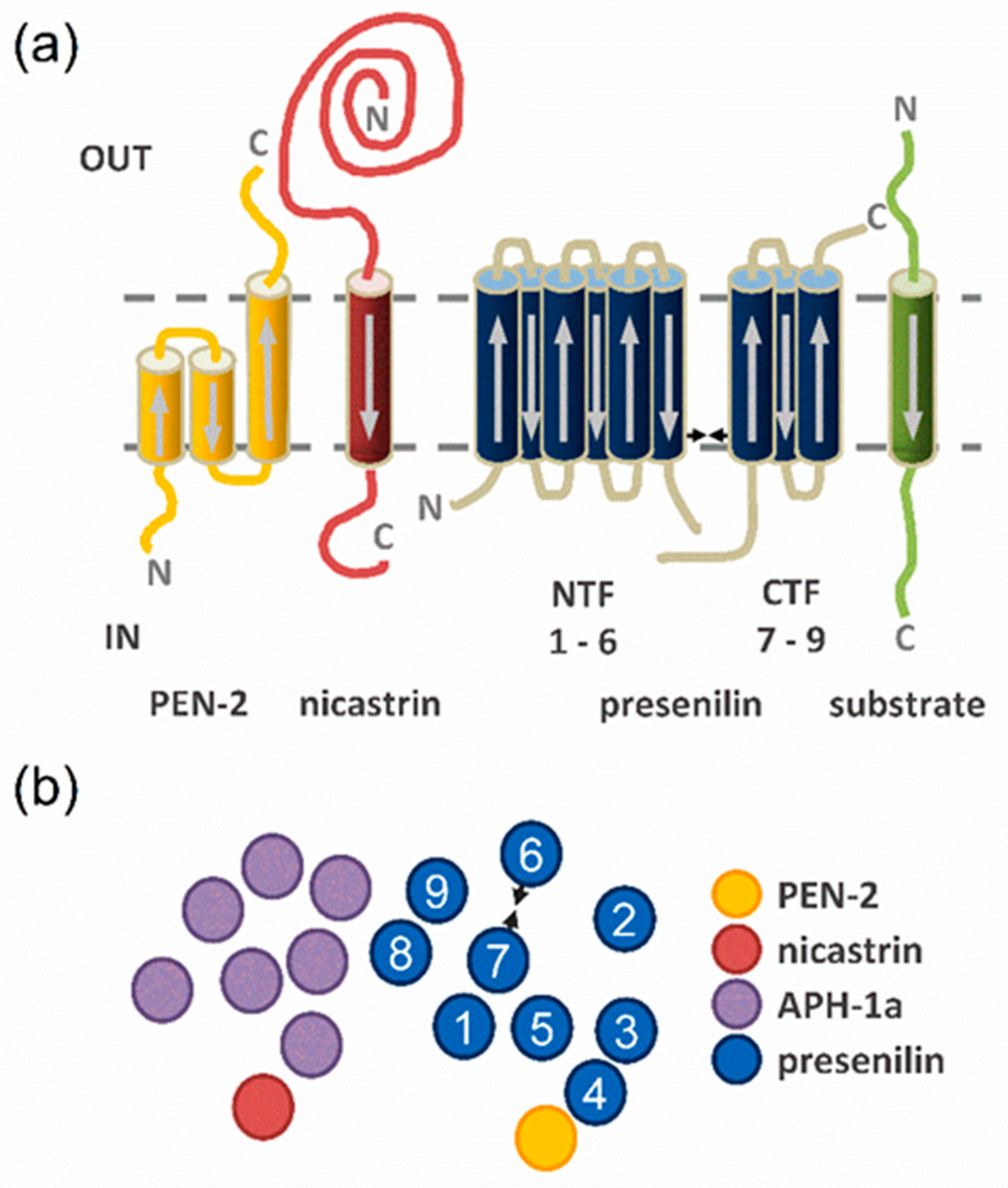
<p>Overview of γ-secretase and substrate TMDs. (<b>a</b>) Transmembrane topologies of the γ-secretase subunits presenilin (blue), PEN-2 (yellow), nicastrin (red), APH-1 (purple), and a substrate (green). Arrows correspond to the direction of the sequences. (<b>b</b>) Top view onto the γ-secretase TMDs. The two catalytic aspartates in TMD6 and TMD7 of presenilin are represented by arrowheads and the TMDs are numbered from N- to C-terminus.</p> "> Figure 3
<p>TMD−TMD interactions of PS1 with substrates and the non-substrate ITGB1. (<b>a</b>) Strength of parallel heterotypic interactions normalized to the homodimerization signal of GpA used as a reference. Three different variants of PS1 TMD2 were tested in combination with APP. (<b>b</b>) Antiparallel heterotypic interactions normalized to the homodimerization signal of EmrE. The shown data correspond to the combination of TMD frames showing the strongest interactions in any given case (see <a href="#app1-ijms-24-14396" class="html-app">Figure S2</a> A-T, where the GFP expression controls are also given). Means ± SEM, n > 3. The positive and negative controls (grey bars) were included in every single round of experiments.</p> "> Figure 4
<p>TMD−TMD interactions of nicastrin or PEN-2 with substrates and the non-substrate ITGB1. (<b>a</b>) Strength of parallel heterotypic interactions to the nicastrin TMD normalized to the homodimerization signal of GpA. (<b>b</b>) Strength of antiparallel heterotypic interactions to the PEN-2 TMD normalized to the homodimerization signal of EmrE TMD4. The shown data correspond to the TMD pairs showing the strongest interactions, as shown in <a href="#app1-ijms-24-14396" class="html-app">Figure S2</a>, which also contains the GFP expression controls. Means ± SEM, n > 3. The positive and negative controls (grey bars) were included in every single round of experiments. The color coding given in the inset of panel (<b>b</b>) also applies to panel (<b>a</b>).</p> "> Figure 5
<p>Mutational analysis of the APP TMD in its pairwise interactions with different PS1 TMDs or itself. (<b>a</b>) Strength of the interaction of APP TMD (frame 2) mutants vs. PS1 TMD4 (frame 3) normalized to the signal of wt APP TMD. Residues whose mutation reduced the signal to <50% of wt are colored. (<b>b</b>) Strength of interaction of APP TMD (frame 2) mutants vs. PS1 <sup>125–146</sup>TMD2 (frame 0) normalized to the signal of wt. We note that mutant V46A more than tripled the LD<sub>50</sub> in this case, for reasons that are unclear. For technical reasons, a lower LD<sub>50</sub> limits the potential impact of mutations. (<b>c</b>) Strength of homodimerization of APP TMD (frame 2) mutants normalized to the signal of the wt APP TMD. The same residues as in (<b>a</b>) are highlighted in (<b>b</b>,<b>c</b>). (<b>d</b>) Mapping the mutation-sensitive residue positions onto a helical wheel or the NMR structure of the helix (pdb: 6hyf) model suggests that the amino acids colored in yellow or green, respectively, may correspond to two separate helix−helix interfaces formed by the APP TMD. Single, double, or triple asterisks denote statistical significance at the 0.05, 0.01, or 0.001 confidence levels (relative to wt APP). Means ± SEM, n = 4–5. The pairs used as references (dark grey bars) were included in every single round of the respective experiments.</p> "> Figure 6
<p>TMD−TMD interactions of various parts of PS1 TMD2 with substrate TMDs, as identified by the BLaTM library screening approach. Candidate pairs (shown in yellow) were identified by next generation sequencing and the resulting full dataset of 1344 pairs (see <a href="#app1-ijms-24-14396" class="html-app">Figure S7</a>) was filtered for abundances exceeding 40% of the signal of GpA wt. Homotypic interactions of positive and negative controls are given by grey bars; heterotypic TMD pairs also covered by approach 1 (<a href="#ijms-24-14396-f003" class="html-fig">Figure 3</a>) are shown in dark green and orange. The TMD sequences of the pair A4_HUMAN_2|PS1_TMD2_0 (identified by approach 2) are equivalent to those of the APP | PS1_TMD2_0 pair (approach 1) (both colored in green).</p> ">
Abstract
:1. Introduction
2. Results
2.1. Determining TMD−TMD Interactions by Manual Testing of Candidate Pairs
2.2. TMD−TMD Interactions Determined by Library Screening
3. Discussion
4. Materials and Methods
4.1. Plasmid Design and Construction
4.2. Determining Ampicillin LD50 Values
4.3. Determining GFP Expression
4.4. Design and Screening of Combinatorial TMD Libraries
5. Conclusions
Supplementary Materials
Author Contributions
Funding
Data Availability Statement
Acknowledgments
Conflicts of Interest
References
- Langosch, D.; Arkin, I.T. Interaction and Conformational Dynamics of Membrane-Spanning Protein Helices. Protein Sci. 2009, 18, 1343–1358. [Google Scholar] [CrossRef]
- Neumann, J.; Klein, N.; Otzen, D.E.; Schneider, D. Folding energetics and oligomerization of polytopic alpha-helical transmembrane proteins. Arch. Biochem. Biophys. 2014, 564, 281–296. [Google Scholar] [CrossRef] [PubMed]
- Schneider, D.; Engelman, D.M. GALLEX: A measurement of heterologous association of transmembrane helices in a biological membrane. J. Biol. Chem. 2003, 278, 3105–3111. [Google Scholar] [CrossRef] [PubMed]
- Petschnigg, J.; Groisman, B.; Kotlyar, M.; Taipale, M.; Zheng, Y.; Kurat, C.F.; Sayad, A.; Sierra, J.R.; Usaj, M.M.; Snider, J.; et al. The mammalian-membrane two-hybrid assay (MaMTH) for probing membrane-protein interactions in human cells. Nat. Methods 2014, 11, 585–592. [Google Scholar] [CrossRef]
- Karimova, G.; Dautin, N.; Ladant, D. Interaction network among Escherichia coli membrane proteins involved in cell division as revealed by bacterial two-hybrid analysis. J. Bacteriol. 2005, 187, 2233–2243. [Google Scholar] [CrossRef] [PubMed]
- Sawma, P.; Roth, L.; Blanchard, C.; Bagnard, D.; Cremel, G.; Bouveret, E.; Duneau, J.P.; Sturgis, J.N.; Hubert, P. Evidence for new homotypic and heterotypic interactions between transmembrane helices of proteins involved in receptor tyrosine kinase and neuropilin signaling. J. Mol. Biol. 2014, 426, 4099–4111. [Google Scholar] [CrossRef]
- Steindorf, D.; Schneider, D. In vivo selection of heterotypically interacting transmembrane helices: Complementary helix surfaces, rather than conserved interaction motifs, drive formation of transmembrane hetero-dimers. Biochim. Biophys. Acta 2017, 1859, 245–256. [Google Scholar] [CrossRef]
- Duart, G.; Grau, B.; Mingarro, I.; Martinez-Gil, L. Methodological approaches for the analysis of transmembrane domain interactions: A systematic review. Biochim. Biophys. Acta-Biomembr. 2021, 1863, 183712. [Google Scholar] [CrossRef]
- Schanzenbach, C.; Schmidt, F.C.; Breckner, P.; Teese, M.G.; Langosch, D. BLaTM—A genetic tool to measure heterotypic interactions of transmembrane helices. Sci. Rep. 2017, 7, 43476. [Google Scholar] [CrossRef]
- Julius, A.; Laur, L.; Schanzenbach, C.; Langosch, D. BLaTM 2.0, a Genetic Tool Revealing Preferred Antiparallel Interaction of Transmembrane Helix 4 of the Dual-Topology Protein EmrE. J. Mol. Biol. 2017, 11, 1630–1637. [Google Scholar] [CrossRef]
- Steiner, H.; Fukumori, A.; Tagami, S.; Okochi, M. Making the final cut: Pathogenic amyloid-β peptide generation by γ-secretase. Cell Stress 2018, 2, 292–310. [Google Scholar] [CrossRef] [PubMed]
- Hardy, J.; Selkoe, D.J. The amyloid hypothesis of Alzheimer’s disease: Progress and problems on the road to therapeutics. Science 2002, 297, 353–356. [Google Scholar] [CrossRef] [PubMed]
- Wolfe, M.S. Processive proteolysis by gamma-secretase and the mechanism of Alzheimer’s disease. Biol. Chem. 2012, 393, 899–905. [Google Scholar] [CrossRef] [PubMed]
- Guner, G.; Lichtenthaler, S.F. The substrate repertoire of gamma-secretase/presenilin. Semin. Cell Dev. Biol. 2020, 105, 27–42. [Google Scholar] [CrossRef] [PubMed]
- Lichtenthaler, S.F.; Haass, C.; Steiner, H. Regulated intramembrane proteolysis—Lessons from amyloid precursor protein processing. J. Neurochem. 2011, 117, 779–796. [Google Scholar] [CrossRef] [PubMed]
- Lu, P.; Bai, X.C.; Ma, D.; Xie, T.; Yan, C.; Sun, L.; Yang, G.; Zhao, Y.; Zhou, R.; Scheres, S.H.W.; et al. Three-dimensional structure of human γ-secretase. Nature 2014, 512, 166–170. [Google Scholar] [CrossRef] [PubMed]
- Bolduc, D.M.; Montagna, D.R.; Gu, Y.; Selkoe, D.J.; Wolfe, M.S. Nicastrin functions to sterically hinder gamma-secretase-substrate interactions driven by substrate transmembrane domain. Proc. Natl. Acad. Sci. USA 2016, 113, E509–E518. [Google Scholar] [CrossRef]
- Zhou, R.; Yang, G.; Guo, X.; Zhou, Q.; Lei, J.; Shi, Y. Recognition of the amyloid precursor protein by human gamma-secretase. Science 2019, 363, eaaw0930. [Google Scholar] [CrossRef]
- Yang, G.; Zhou, R.; Zhou, Q.; Guo, X.; Yan, C.; Ke, M.; Lei, J.; Shi, Y. Structural basis of Notch recognition by human γ-secretase. Nature 2019, 565, 192–197. [Google Scholar] [CrossRef]
- Petit, D.; Hitzenberger, M.; Lismont, S.; Zoltowska, K.M.; Ryan, N.S.; Mercken, M.; Bischoff, F.; Zacharias, M.; Chavez-Gutierrez, L. Extracellular interface between APP and Nicastrin regulates A beta length and response to gamma-secretase modulators. EMBO J. 2019, 38, e101494. [Google Scholar] [CrossRef]
- Kornilova, A.Y.; Bihel, F.; Das, C.; Wolfe, M.S. The initial substrate-binding site of γ-secretase is located on presenilin near the active site. Proc. Natl. Acad. Sci. USA 2005, 102, 3230–3235. [Google Scholar] [CrossRef] [PubMed]
- Fukumori, A.; Steiner, H. Substrate recruitment of gamma-secretase and mechanism of clinical presenilin mutations revealed by photoaffinity mapping. EMBO J. 2016, 35, 1628–1643. [Google Scholar] [CrossRef]
- Tomita, T.; Iwatsubo, T. Structural Biology of Presenilins and Signal Peptide Peptidases. J. Biol. Chem. 2013, 288, 14673–14680. [Google Scholar] [CrossRef] [PubMed]
- Liu, X.Y.; Zhao, J.; Zhang, Y.K.; Ubarretxena-Belandia, I.; Forth, S.; Lieberman, R.L.; Wang, C.Y. Substrate-Enzyme Interactions in Intramembrane Proteolysis: Gamma-Secretase as the Prototype. Front. Mol. Neurosci. 2020, 13, 65. [Google Scholar] [CrossRef] [PubMed]
- Bai, X.C.; Yan, C.; Yang, G.; Lu, P.; Ma, D.; Sun, L.; Zhou, R.; Scheres, S.H.; Shi, Y. An atomic structure of human gamma-secretase. Nature 2015, 525, 212–217. [Google Scholar] [CrossRef]
- Aguayo-Ortiz, R.; Dominguez, L. Simulating the gamma-secretase enzyme: Recent advances and future directions. Biochimie 2018, 147, 130–135. [Google Scholar] [CrossRef]
- Hitzenberger, M.; Gotz, A.; Menig, S.; Brunschweiger, B.; Zacharias, M.; Scharnagl, C. The dynamics of gamma-secretase and its substrates. Semin. Cell Dev. Biol. 2020, 105, 86–101. [Google Scholar] [CrossRef]
- Lemmon, M.A.; Flanagan, J.M.; Treutlein, H.R.; Zhang, J.; Engelman, D.M. Sequence specificity in the dimerization of transmembrane alpha-helices. Biochemistry 1992, 31, 12719–12725. [Google Scholar] [CrossRef]
- Bai, X.C.; Rajendra, E.; Yang, G.; Shi, Y.; Scheres, S.H. Sampling the conformational space of the catalytic subunit of human gamma-secretase. eLife 2015, 4, e1182. [Google Scholar] [CrossRef]
- Gurezka, R.; Laage, R.; Brosig, B.; Langosch, D. A Heptad Motif of Leucine Residues Found in Membrane Proteins Can Drive Self-Assembly of Artificial Transmembrane Segments. J. Biol. Chem. 1999, 274, 9265–9270. [Google Scholar] [CrossRef]
- Chavez-Gutierrez, L.; Bammens, L.; Benilova, I.; Vandersteen, A.; Benurwar, M.; Borgers, M.; Lismont, S.; Zhou, L.; Van Cleynenbreugel, S.; Esselmann, H.; et al. The mechanism of gamma-Secretase dysfunction in familial Alzheimer disease. EMBO J. 2012, 31, 2261–2274. [Google Scholar] [CrossRef] [PubMed]
- Hemming, M.L.; Elias, J.E.; Gygi, S.P.; Selkoe, D.J. Proteomic profiling of gamma-secretase substrates and mapping of substrate requirements. PLoS Biol. 2008, 6, e257. [Google Scholar] [CrossRef] [PubMed]
- Silber, M.; Hitzenberger, M.; Zacharias, M.; Muhle-Goll, C. Altered Hinge Conformations in APP Transmembrane Helix Mutants May Affect Enzyme-Substrate Interactions of gamma-Secretase. ACS Chem. Neurosci. 2020, 11, 4426–4433. [Google Scholar] [CrossRef]
- Barrett, P.J.; Song, Y.; Van Horn, W.D.; Hustedt, E.J.; Schafer, J.M.; Hadziselimovic, A.; Beel, A.J.; Sanders, C.R. The amyloid precursor protein has a flexible transmembrane domain and binds cholesterol. Science 2012, 336, 1168–1171. [Google Scholar] [CrossRef] [PubMed]
- Watanabe, N.; Image, I., II; Takagi, S.; Tominaga, A.; Image Image, I.; Tomita, T.; Iwatsubo, T. Functional analysis of the transmembrane domains of presenilin 1: Participation of transmembrane domains 2 and 6 in the formation of initial substrate-binding site of γ-secretase. J. Biol. Chem. 2010, 285, 19738–19746. [Google Scholar] [CrossRef]
- Takagi-Niidome, S.; Sasaki, T.; Osawa, S.; Sato, T.; Morishima, K.; Cai, T.; Iwatsubo, T.; Tomita, T. Cooperative roles of hydrophilic loop 1 and the C-terminus of presenilin 1 in the substrate-gating mechanism of gamma-secretase. J. Neurosci. 2015, 35, 2646–2656. [Google Scholar] [CrossRef]
- Beel, A.J.; Sanders, C.R. Substrate specificity of gamma-secretase and other intramembrane proteases. Cell. Mol. Life Sci. 2008, 65, 1311–1334. [Google Scholar] [CrossRef]
- Sato, C.; Takagi, S.; Tomita, T.; Iwatsubo, T. The C-terminal PAL motif and transmembrane domain 9 of presenilin 1 are involved in the formation of the catalytic pore of the gamma-secretase. J. Neurosci. 2008, 28, 6264–6271. [Google Scholar] [CrossRef]
- Kong, R.; Chang, S.; Xia, W.; Wong, S.T. Molecular dynamics simulation study reveals potential substrate entry path into gamma-secretase/presenilin-1. J. Struct. Biol. 2015, 191, 120–129. [Google Scholar] [CrossRef]
- Somavarapu, A.K.; Kepp, K.P. The dynamic mechanism of presenilin-1 function: Sensitive gate dynamics and loop unplugging control protein access. Neurobiol. Dis. 2016, 89, 147–156. [Google Scholar] [CrossRef]
- Li, S.; Zhang, W.; Han, W. Initial Substrate Binding of gamma-Secretase: The Role of Substrate Flexibility. ACS Chem. Neurosci. 2017, 8, 1279–1290. [Google Scholar] [CrossRef]
- Aguayo-Ortiz, R.; Chavez-Garcia, C.; Straub, J.E.; Dominguez, L. Characterizing the structural ensemble of gamma-secretase using a multiscale molecular dynamics approach. Chem. Sci. 2017, 8, 5576–5584. [Google Scholar] [CrossRef] [PubMed]
- Tominaga, A.; Cai, T.; Takagi-Niidome, S.; Iwatsubo, T.; Tomita, T. Conformational Changes in Transmembrane Domain 4 of Presenilin 1 Are Associated with Altered Amyloid-beta 42 Production. J. Neurosci. 2016, 36, 1362–1372. [Google Scholar] [CrossRef]
- Hogel, P.; Gotz, A.; Kuhne, F.; Ebert, M.; Stelzer, W.; Rand, K.D.; Scharnagl, C.; Langosch, D. Glycine Perturbs Local and Global Conformational Flexibility of a Transmembrane Helix. Biochemistry 2018, 57, 1326–1337. [Google Scholar] [CrossRef] [PubMed]
- Teese, M.G.; Langosch, D. Role of GxxxG Motifs in Transmembrane Domain Interactions. Biochemistry 2015, 54, 5125–5135. [Google Scholar] [CrossRef] [PubMed]
- Munter, L.M.; Voigt, P.; Harmeier, A.; Kaden, D.; Gottschalk, K.E.; Weise, C.; Pipkorn, R.; Schaefer, M.; Langosch, D.; Multhaup, G. GxxxG motifs within the amyloid precursor protein transmembrane sequence are critical for the etiology of Aβ42. EMBO J. 2007, 26, 1702–1712. [Google Scholar] [CrossRef]
- Sato, T.; Tang, T.C.; Reubins, G.; Fei, J.Z.; Fujimoto, T.; Kienlen-Campard, P.; Constantinescu, S.N.; Octave, J.N.; Aimoto, S.; Smith, S.O. A helix-to-coil transition at the epsilon-cut site in the transmembrane dimer of the amyloid precursor protein is required for proteolysis. Proc. Natl. Acad. Sci. USA 2009, 106, 1421–1426. [Google Scholar] [CrossRef] [PubMed]
- Chen, W.; Gamache, E.; Rosenman, D.J.; Xie, J.; Lopez, M.M.; Li, Y.M.; Wang, C. Familial Alzheimer’s mutations within APPTM increase Abeta42 production by enhancing accessibility of epsilon-cleavage site. Nat. Commun. 2014, 5, 3037. [Google Scholar] [CrossRef]
- Nadezhdin, K.D.; Bocharova, O.V.; Bocharov, E.V.; Arseniev, A.S. Dimeric Structure of Transmembrane Domain of Amyloid Precursor Protein in Micellar Environment. FEBS Lett. 2012, 586, 1687–1692. [Google Scholar] [CrossRef]
- Dominguez, L.; Meredith, S.C.; Straub, J.E.; Thirumalai, D. Transmembrane fragment structures of amyloid precursor protein depend on membrane surface curvature. J. Am. Chem. Soc. 2014, 136, 854–857. [Google Scholar] [CrossRef]
- Winkler, E.; Julius, A.; Steiner, H.; Langosch, D. Homodimerization Protects the Amyloid Precursor Protein C99 Fragment from Cleavage by gamma-Secretase. Biochemistry 2015, 54, 6149–6152. [Google Scholar] [CrossRef]
- Mall, S.; Broadbridge, R.; Sharma, R.P.; Lee, A.G.; East, J.M. Effects of aromatic residues at the ends of transmembrane alpha-helices on helix interactions with lipid bilayers. Biochemistry 2000, 39, 2071–2078. [Google Scholar] [CrossRef] [PubMed]
- Garcia-Murria, M.J.; Duart, G.; Grau, B.; Diaz-Beneitez, E.; Rodriguez, D.; Mingarro, I.; Martinez-Gil, L. Viral Bcl2s’ transmembrane domain interact with host Bcl2 proteins to control cellular apoptosis. Nat. Commun. 2020, 11, 6056. [Google Scholar] [CrossRef] [PubMed]
- Duart, G.; Elazar, A.; Weinstein, J.Y.; Gadea-Salom, L.; Ortiz-Mateu, J.; Fleishman, S.J.; Mingarro, I.; Martinez-Gil, L. Computational design of BclxL inhibitors that target transmembrane domain interactions. Proc. Natl. Acad. Sci. USA 2023, 120, e2219648120. [Google Scholar] [CrossRef]
- Vincent, M.S.; Comas Hervada, C.; Sebban-Kreuzer, C.; Le Guenno, H.; Chabalier, M.; Kosta, A.; Guerlesquin, F.; Mignot, T.; McBride, M.J.; Cascales, E.; et al. Dynamic proton-dependent motors power type IX secretion and gliding motility in Flavobacterium. PLoS Biol. 2022, 20, e3001443. [Google Scholar] [CrossRef] [PubMed]


Disclaimer/Publisher’s Note: The statements, opinions and data contained in all publications are solely those of the individual author(s) and contributor(s) and not of MDPI and/or the editor(s). MDPI and/or the editor(s) disclaim responsibility for any injury to people or property resulting from any ideas, methods, instructions or products referred to in the content. |
© 2023 by the authors. Licensee MDPI, Basel, Switzerland. This article is an open access article distributed under the terms and conditions of the Creative Commons Attribution (CC BY) license (https://creativecommons.org/licenses/by/4.0/).
Share and Cite
Pauli, T.M.; Julius, A.; Costa, F.; Eschrig, S.; Moosmüller, J.; Fischer, L.; Schanzenbach, C.; Schmidt, F.C.; Ortner, M.; Langosch, D. Interaction of Substrates with γ-Secretase at the Level of Individual Transmembrane Helices—A Methodological Approach. Int. J. Mol. Sci. 2023, 24, 14396. https://doi.org/10.3390/ijms241814396
Pauli TM, Julius A, Costa F, Eschrig S, Moosmüller J, Fischer L, Schanzenbach C, Schmidt FC, Ortner M, Langosch D. Interaction of Substrates with γ-Secretase at the Level of Individual Transmembrane Helices—A Methodological Approach. International Journal of Molecular Sciences. 2023; 24(18):14396. https://doi.org/10.3390/ijms241814396
Chicago/Turabian StylePauli, Theresa M., Ayse Julius, Francesco Costa, Sabine Eschrig, Judith Moosmüller, Lea Fischer, Christoph Schanzenbach, Fabian C. Schmidt, Martin Ortner, and Dieter Langosch. 2023. "Interaction of Substrates with γ-Secretase at the Level of Individual Transmembrane Helices—A Methodological Approach" International Journal of Molecular Sciences 24, no. 18: 14396. https://doi.org/10.3390/ijms241814396