Academia.edu no longer supports Internet Explorer.
To browse Academia.edu and the wider internet faster and more securely, please take a few seconds to upgrade your browser.
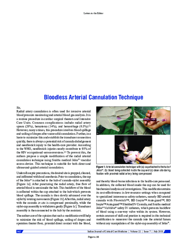
2018, Indian Journal of Critical Care Medicine
European Journal of Anaesthesiology
Feasibility and safety of antegrade radial artery cannulation2009 •
IP innovative publication pvt. ltd
Comparative evaluation of safety and efficacy of radial arterial cannulation using conventional blind palpation technique with ultrasound guided Technique perioperatively -Raccuet studyIntroduction: Radial cannulation is needed in perioperative management of many patients requiring invasive hemodynamic monitoring during surgery and may be not only time consuming and at times difficult to attain in untrained centers. Objectives: Comparative evaluation of safety and efficacy of Radial Arterial Cannulation using Conventional blind palpation technique with Ultrasound guided Technique perioperatively-Raccuet Study. Materials and Methods: This randomized controlled, prospective, single blind comparative study was conducted on 142 adult ASA I to IV patients of either sex, scheduled for elective major surgery requiring general anesthesia of which 71 patients underwent radial cannulation by classical palpatory method (group P) and the other 71 underwent radial cannulation under ultrasound guidance (Group U). All procedures were evaluated using Siemens Acuson X500 ultrasound machine with 5-13Hz linear array transducer. All patients were evaluated for efficacy by measuring time to cannulation (TTC), mean time to first attempt cannulation (mTFA), and number of attempts for cannulation (NA). Safety was assessed by evaluating incidence of hematoma and spasm in both the groups. Statistical analysis was done using (SPSS) Version 22.0. Independent t test and Mann Whitney test have been used for carrying out significant P value. Results: Patients were demographically similar in both the groups. TTC in group U (37.97±18.14sec) was significantly less than that in Group P (58.38±21.45sec) (P =0.007).mTFA was 34.81±15.77 seconds in group U compared with 55.58±19.29 seconds with group P (P=0.01). There was trend towards lower NA in group U as compared to group P (P=0.06).Incidence of hematoma formation and vasospasm were similar amongst both groups but study was underpowered to evaluate the same. Conclusion: Ultrasound guided radial artery cannulation improves the success rate of cannulation in lesser time with similar complication rates as compared to classical palpatory method of cannulation.
Turkish Journal of Thoracic and Cardiovascular Surgery
Is axillary arterial cannulation better than femoral arterial cannulation?2012 •
Catheterization and Cardiovascular Interventions
Subcutaneous administration of nitroglycerin to facilitate radial artery cannulation2006 •
Objectives: To study the effect of sublingual versus subcutaneous nitroglycerin on radial artery spasm caused by failed access attempts. Background: Radial artery spasm is the leading reason for failed radial access. We studied the efficacy of systemic versus local nitroglycerin in relieving radial artery spasm caused by needle entry resulting in failed cannulation. Methods: Fifty-two consecutive patients were studied. All patients had failed attempt at radial artery cannulation, resulting in loss of radial pulse. Patients were divided in three groups, group I (n = 11), observed without additional treatment, group II (n = 20), administered 400 mcg of sublingual nitroglycerin, and group III (n = 21), administered 400 mcg of subcutaneous nitroglycerin at the site of the lost radial pulse. All patients were monitored for the return of radial pulse. Demographics, hemodynamics, and time to return of radial pulse as well as ability to successfully cannulate the radial artery were recorded. Results: Seventy-two percent of group I patients, 90% of group II patients, and 100% of group III patients had re-establishment of radial pulse. The time to return of radial pulse was significantly shorter for group III compared with that for group II (3 ± 1 min vs. 8 ± 1 min respectively, P < 0.001). Re-establishment of radial pulse was faster in group II and group III compared with that in group I (18 ± 5 min, P < 0.001). Systolic blood pressure changes and headaches were less common in group III. Conclusion: Subcutaneous administration of nitroglycerin is superior in facilitating radial artery cannulation after initial failed attempt. © 2006 Wiley-Liss, Inc.
Cardiac Surgery Procedures [Working Title]
Cannulation for Cardiopulmonary BypassEuropean Journal of Cardio-Thoracic Surgery
Technical problems and complications of axillary artery cannulation2005 •
Turkish Journal of Thoracic and Cardiovascular Surgery
Axillary artery cannulation in ascending aortic pathologies2011 •
Per Jacopo De Grossi Mazzorin, Piccolo libro bianco - Ricordi di amici, archeozoologi e non, Prato 2024, a cura di M. Masseti, C. Minniti e C.A. Corbino
Jacopo, una poesia, in: Per Jacopo De Grossi Mazzorin - Piccolo libro bianco, Prato 2024, pp. 63-72.Ricordo personale del caro amico Jacopo De Grossi Mazzorin, venuto a mancare all'affetto dei suoi cari il 5 febbraio 2023.
isara solutions
WOMEN EMPOWERMENT ITS PRACTICE AND IMPORTANCEAt-Tajdid : Jurnal Pendidikan dan Pemikiran Islam
Kepemimpinan dalam Perspektif Islam2020 •
Philosophical Transactions of the Royal Society B: Biological Sciences
From hominins to humans: howsapiensbecame behaviourally modern2011 •
2016 •
Selçuk Üniversitesi Selçuklu Araştırmaları Dergisi
Türkiye Tarihi Selçuklular Devri2018 •
Lusitania Romana: del pasado al presente de la investigación
O Sul da Lusitânia Romana nos últimos 25 anos: avanços e novas perspectivas de investigação2017 •
2019 •
2013 •
Scientific Reports
N-glycosylation of α1D-adrenergic receptor N-terminal domain is required for correct trafficking, function, and biogenesis2020 •
2020 •
Griya Journal of Mathematics Education and Application
Analisis Kemampuan Pemecahan Masalah Matematika pada Materi Bilangan Ditinjau dari Kemampuan Number Sense Siswa2023 •
International Journal of Anatomy and Research
Reconstruction of Total Length of Femur from Its Proximal and Distal Fragments2015 •
2011 •
 Dr. Pushkar Desai