Monte-Carlo-Based Scatter Correction
for Quantitative SPECT Reconstruction
Realization and Evaluation
Rolf Bippus1 , Andreas Goedicke1 , Henrik Botterweck2
1
2
Philips Research Laboratories, Aachen
Fachhochschule Lübeck, Bildgebende Verfahren in der Medizintechnik
rolf.bippus@philips.com
Abstract. Quantitative SPECT as well as simultaneous acquisition of
multiple isotopes with SPECT in the clinical field, although clinically
interesting, are still limited by reconstruction artifacts and computing
power. As a considerable step in this direction, we have implemented an
efficient reconstructor with variance reduced Monte-Carlo-simulation in
the forward and/or backward projection of an OS-EM iteration. Apart
from a quantitatively accurate scatter estimation the integrated MC simulation allows us to include all effects that are relevant for the multiple
isotope task. The reconstruction problem is explicitly formulated as a
combined maximum-likelihood estimation for all unknowns in one step
and implemented efficiently on the basis of the OS-EM algorithm using
the Effective Scatter Source approach. The algorithm has been evaluated
on simulated as well as measured phantom data. Two isotopes (Tc99m,
Tl201) were tested on the voxelized NCAT phantom and evaluated quantitatively. In addition we performed phantom measurements to evaluate
the method on a Philips CardioMDT M with a VantageT M line source
for attenuation correction. A clear advantage of the proposed approach
is its robustness and generalizability. It is currently being evaluated in
clinical applications like simultaneous dual isotope cardiac imaging.
1
Introduction
Absolute quantification as well as simultaneous acquisition of multiple isotopes
are receiving increasing interest in clinical applications (e.g. [1], [2]). However
patient scatter, down-scatter plus collimator effects such as penetration and Pbfluorescence result in data contamination.
Basically one can distiguish between energy and/or spatial distribution based
scatter estimation and correction from reconstruction based methods [3]. The
OS-EM (Ordered Subset Expectation Maximization) approach we follow with
our implementation falls into the second category. Scatter is estimated based on
the current activity estimate in the forward projection step. Using the approximative scheme of effective scatter source (ESS) Estimation, originally published
by Frey et al. [4], scatter estimation is effectively separated from other effects
in the algorithm. The original approach uses precomputed scatter kernels to
�Quantitative SPECT Reconstruction
371
obtain the ESS via convolution. In contrast we apply variance reduced MonteCarlo simulation to estimate the ESS. An attenuation map is used as a physical
model of the patient.
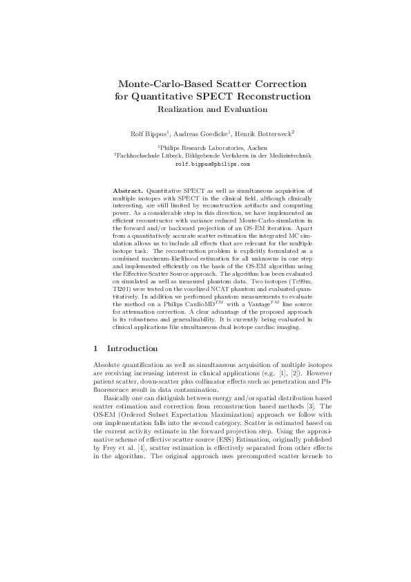
Using one of the reconstruction based scatter estimation methods one can differentiate sequential and simultaneous reconstruction as depicted in Fig. 1. We
follow the scheme of simultaneous reconstruction (Fig. 1(b)) where the cross contamination estimation is integrated into the forward projector and all estimates
isotope activities are forward projected into all energy windows and updated in
each sub-iteration of the OS-EM algorithm.
2
Methods
The full update equations for the ordered subset EM algorithm for all isotope
activities (typicall 1 or 2) fǫ,i (isotope i in voxel ǫ) on all available data are given
by equations (1) and (2).
The forward projection step computes (1) the contributions of isotopes i to all
projection pixels p in each energy-window e, based on the recent activity estimate
(k)
ǫ,i
is the normalized system operator. In the backprojection step
fǫ,i , where Hp,e
(2), the correction factors are obtained from the ratio of the observed and the
estimated projection values.
X (k)
X X (k)
X
ǫ,i
ǫ,i
ν (k) (p, e) =
νi (p, e) =
,
fǫ,i Hp,e
Hp,e
=1
(1)
i
(k+1)
fǫ,i
2.1
(k)
= fǫ,i
i
ǫ
1 X X ǫ,i ν(p, e)
Hp,e (k)
nǫ,i p e
ν (p, e)
p,e
,
nǫ,i =
X
ǫ,i
Hp,e
(2)
p,e
Approximations / Implementation
Using brute force counting statistics with MC-simulation to obtain the ν (k) (p, e)
in equation (1) is not feasible. Therefore we applied the principle of convolution
based forced detection.
Simulating the photon tracks, at each interaction of the photons with the
patient the effective cross section is calculated for the photon being redirected
towards the detector for a set of intermediary energy intervals. These are accumulated in 3D and are treated as secondary emission distributions, the ESS.
These are then projected onto all camera positions and energy windows, taking
(c)
(d)
Fig. 1. Sequential (a) vs. simultaneous (b) dual isotope reconstruction scheme.
�372
Bippus, Goedicke & Botterweck
into account attenuation and collimator response functions (CRF). The CRFs,
simulated beforehand and stored, are taking into account all major physical
degradation effects. The principle is illustrated in Fig. 2.
According to the Dual-Matrix aproach, the backprojector is approximated
ǫ
) as well as the relative weights
by the reversed PSFs and the attenuation (Bp,e
ωi (p, e) of the isotope i contributing to pixel (p, e) in the forward projection,
thus ignoring the spatial distribution of the scatter.
(k+1)
fǫ,i
2.2
(k)
= fǫ,i
1 X X ǫ (k)
ν(p, e)
B ω (p, e) (k)
nǫ,i p e p,e i
ν (p, e)
(3)
Activity Estimation
Quantitative results are obtained by defining volumes of interest (VOI) and calculating the mean activity within the VOI. We evaluated quantitative activities
after 30 to 50 full iterations of the OS-EM algorithm. Corresponding images
are not suited for visual inspection or VOI delineation due to high noise. Thus
images for visual inspection or delineation use 4 full iterations of the OS-EM algorithm. For the simulated data we defined the VOIs directly from true activity
images.
3
3.1
Results
NCAT Phantom Results
The voxelized NCAT phantom was simulated such that the activities represent
a patient dose of 25 mCi Tc99m and 3.5 mCi of Tl201 . In the Tl201 distribution a
heart lesion was included, which was not present in the Tc99m image. The lesion
activity was chosen 15 % of the normal myocardial Tl201 activity.
Fig. 2. Principle of down-scatter estimation in the forward projection step.
�Quantitative SPECT Reconstruction
373
For quantitative comparison of true and reconstructed activity, we measured
absolute activity in the 17 segments of a standard 17 segment polar plot representation. Heart re-orientation and segmentation were performed on the true
activity image and kept constant for the reconstructed image. As before the
values represent absolute activity estimates averaged over each segment of the
heart.
3.2
Phantom Measurement Results
Phantom measurements were performed using a Data Spectrum Cooperation
Anthropomorphic Torso PhantomT M with a fillable heart insert. The phantom
was filled according to an estimated biodistribution resulting from 90 MBq Tl201
and 650 MBq Tc99m administered to an adult. The heart contained 2 lesions of
5.6 ml (lesion 1) and 12 ml (lesion 2), each filled with Tc99m only, which gives
no Tl201 and approx. 65% Tc99m (due to the walls of the lesions) in the lesions.
Resulting images and polar plots are shown in Fig. 4.
4
Conclusion
Quantitative MC reconstruction for SPECT and multiple isotopes shows to be
feasible. The more exact modeling of scatter within the object and collimator results in reduced image artifacts. The computational effort is reasonable,
especially compared to other state of the art scatter corrections like ESSE.
An advantage of the proposed approach is its robustness and generalizability:
Fast adaptation to new isotope combinations is possible. Next steps will include
the evaluation of its potential in clinical application of dual isotope cardiac imaging.
(a) truth
(b) 30 iterations
Fig. 3. NCAT, T c99m -T l201 SDI, T l201 mean activity in kBq/ml in each of 17 segments
of standard polar plot.
�374
Bippus, Goedicke & Botterweck
Fig. 4. Phantom measurements on the CardioMD. Cardiac SDI filled with Tl201 and
Tc99m and two lesions filled with Tc99m only.
(a)
(b)
References
1. Berman D, Kang X, Tamarappoo B, et al. stress thallium-201/rest technetium- 99m
sequential dual isotope high-speed myocardial perfusion imaging. JACC Cardiovasc
Imaging. 2009;2(3):273–82.
2. Sgouros G. Dosimetry of internal emitters. J Nucl Med. 2005;46(1):273–82.
3. King J M A ad Glick, Pretorius PH, Wells RG, et al. Attenuation, Scatter and
Spatial Resolution Compensation in SPECT. In: Wernick M, Aarsvold JS, editors.
Emission Tomography. The Fundamentals of PET and SPECT. Acedemic Press;
2004.
4. Frey EC, Tsui BMW. A new method for modelling the spatially variant, objectdependent scatter response function in SPECT. In: Proc IEEE Nucl Sci Symp;
1996. p. 1082–6.
�
 Andreas Goedicke