Virology 283, 197–206 (2001)
doi:10.1006/viro.2000.0890, available online at http://www.idealibrary.com on
The Long Repeat Region Is Dispensable for Fowl Adenovirus Replication in Vitro
Davor Ojkic and Éva Nagy 1
Department of Pathobiology, Ontario Veterinary College, University of Guelph, Guelph, Ontario N1G 2W1, Canada
Received December 8, 2000; returned to author for revision January 8, 2001; accepted February 26, 2001
Two regions containing tandemly repeated sequences are present in the fowl adenovirus 9 (FAdV-9) genome. The longer
repeat region (TR-2) is composed of 13 contiguous 135-bp-long direct repeats, the function of which is unknown. An infectious
FAdV-9 genomic clone, constructed by homologous recombination in Escherichia coli, was used for engineering of recombinant viruses. The enhanced green fluorescence protein (EGFP) coding sequence was cloned in both rightward and leftward
orientations so as to replace TR-2. Replication-competent recombinant FAdVs were recovered, demonstrating that TR-2 was
dispensable for FAdV-9 propagation in vitro. The expression of EGFP in infected cells was demonstrated by fluorescence
microscopy, immunoprecipitation, and RT-PCR. © 2001 Academic Press
Key Words: fowl adenovirus; tandem repeats; recombinant virus.
AdV replication in mammalian AdV-infected cells could
not be determined because mammalian AdV genes implicated in the process have a role in other essential
steps of virus replication. However, it was possible to
demonstrate that the induction of a heat-shock response
in FAdV-infected cells is an essential step in AdV replication since CELO virus utilizes a single gene, Gam1, to
induce the heat-shock response in infected cells (Glotzer
et al., 2000).
Early regions 1, 3, and 4 are the common sites to
accommodate foreign genes in recombinant mammalian
AdVs. Since FAdVs lack these regions, alternative strategies had to be explored for the construction of FAdVbased vectors. Mutational analysis demonstrated that
CELO virus could tolerate deletions/insertions into the
right end of its genome, which allowed the generation of
replication-competent recombinant viruses (Michou et
al., 1999). However, deletions in the right end of FAdV
genomes can have a negative impact on virus growth,
and depending on the location and the extent of deletions, these may result in a severe drop in titers of
recombinant viruses (Johnson et al., 2000). The left end of
the CELO virus genome contains regions that could be
deleted and supplied in trans, but since no complementing cell lines are available to support propagation of
replication-defective FAdVs, only replication-competent
recombinant FAdVs have been characterized in more
detail.
An unusual feature of the FAdV-9 genome is the
presence of two regions of tandemly repeated sequences (Cao et al., 1998). Imperfect repeats found in
the early region 4 of mouse adenovirus type 1 appear
to have an impact on virus pathogenicity (Ball et al.,
1991). Tandem reiterations have also been described
INTRODUCTION
The family Adenoviridae includes viruses that have
been isolated from many mammalian (genus Mastadenovirus) and avian (genus Aviadenovirus) species. Avian
adenoviruses (AAdVs) are further subdivided into three
serological groups (McFerran, 1997). Fowl adenoviruses
(FAdV 1–12) belong to group I AAdVs and share a common group antigen with viruses isolated from geese,
ducks, and turkeys. Several features distinguish FAdVs
from their mammalian counterparts. Two fibers protruding from the penton base were observed when 14 FAdV
strains, representing 11 serotypes, were examined by
electron microscopy (Gelderblom and Maichle-Lauppe,
1982), whereas almost all mastadenoviruses, with the
exception of subgroup F human adenoviruses (HAdVs),
have only one fiber per penton base (Kidd et al., 1993;
Yeh et al., 1994). FAdV genomes are much larger than
those of other adenoviruses (AdVs), and FAdV-9 has the
longest AdV genome whose complete nucleotide sequence has been determined so far, 45,063 bp. Moreover, FAdV-1 (CELO virus) and FAdV-9 do not have recognizable early regions 1, 3, and 4, and although most
late genes are well conserved, protein V coding sequences could also not be identified (Chiocca et al.,
1996; Ojkic and Nagy, 2000). Certain FAdV genes have
very specialized functions and one of these facilitated
the evaluation of an important aspect of the AdV replication cycle. The importance of heat-shock response to
1
To whom correspondence and reprint requests should be addressed at Department of Pathobiology, Ontario Veterinary College,
University of Guelph, Guelph, Ontario, N1G 2W1, Canada. Fax: 519-824–
5930. E-mail: enagy@ovc.uoguelph.ca.
197
0042-6822/01 $35.00
Copyright © 2001 by Academic Press
All rights of reproduction in any form reserved.
�198
OJKIC AND NAGY
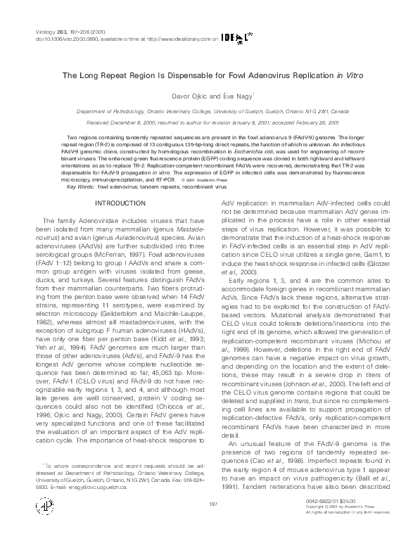
FIG. 1. (A) The strategy for construction of pPacFAdV9. FAdV-9 terminal fragments in pWE-PacITR were extended by inserting the SmaI–SmaI
fragment from pF-ApaI into SmaI-digested pWE-PacITR so that it now contains the complete ApaI left 4.9-kb and right 6.7-kb terminal fragments,
generating pFAP2. Plasmid pFAP2 was then linearized with ApaI and cotransformed into competent BJ5183 E. coli along with FAdV-9 DNA,
generating pPacFAdV9. Thick black lines represent FAdV-9 sequences and hatched blocks represent TR-2. Thin and other gray shaded lines
represent vector sequences. (B) The strategy for construction of pFDTR2-EGFP and pFDTR2-EGFPinv. pAC-EGFP and pAC-EGFPinv were
generated by inserting the EGFP-coding sequence (excised from pEGFP-N1 with Bsp120I and NotI) into Eco52I-digested pAC8, deleting TR-2.
ApaI–XbaI fragment in pFDSal was replaced with ApaI–XbaI fragments from pAC-EGFP and pAC-EGFPinv, respectively. Recombinant plasmids
containing modified viral genomes (pFDTR2-EGFP and pFDTR2-EGFPinv) were generated by recombination between SalI-linearized pFDSalEGFPinv, pFDSal-EGFP, and FAdV-9 DNA. Thick black lines represent FAdV-9 sequences and the hatched blocks are TR-2 sequences. Thin and
other gray shaded lines represent vector sequences. (C) Tandem repeat region as EGFP insertion site. TR-2 contains 13 repeated subunits and
each subunit has two Eco52I recognition sites. TR-2 was deleted by digestion with Eco52I, and EGFP coding sequences were inserted in place
of deleted TR-2. The recombinant virus containing EGFP sequence in leftward orientation was designated rFDTR2-EGFP, whereas the
recombinant virus containing the EGFP sequence in rightward orientation was designated rFDTR2-EGFPinv.
in DNA from canine adenovirus 1 vaccine strain CLL
(Sira et al., 1987) and a mutant human adenovirus 34
(Chen and Horwitz, 1990) as part of the inverted terminal repeats. On the other hand, FAdV-9 tandem
repeats are located on the right end of the viral genome and are incorporated within a region rich in
open reading frames. The shorter repeat region (TR-1)
is composed of five 33-bp-long direct repeats located
between nt 37,648 and 37,812, whereas the longer
repeat region (TR-2), positioned between nt 38,807 and
40,561, contains 13 tandemly repeated 135-bp-long
subunits (Ojkic and Nagy, 2000). Interestingly, sequences similar to FAdV-9 TR-2 were detected in field
isolates of fowl adenoviruses from Ontario, India (unpub-
�TR-2 IS NOT REQUIRED FOR FAdV REPLICATION
199
FIG. 1—Continued
lished data), and Australia (Johnson et al., 2000), but the
function(s) of these repeats, if any, is unknown.
The objectives of this study were to determine whether
TR-2 was dispensable for FAdV-9 propagation in vitro
and to examine the suitability of TR-2 as a site for
insertion of foreign genes. We inserted the enhanced
green fluorescence protein (EGFP) coding sequences in
both rightward and leftward orientations as marker gene
in place of the deleted TR-2, developed a system for the
construction of TR-2-deleted recombinant FAdVs, generated recombinant FAdVs, and examined their potential
as gene expression vectors in vitro.
�200
OJKIC AND NAGY
FIG. 2. Restriction enzyme digestion and Southern blot analysis of
recombinant viruses and FAdV-9. (A) BamHI digestion of DNA extracted
from FAdV-9 (lanes 1), rFDTR2-EGFP (lanes 2), and rFDTR2-EGFPinv
(lanes 3). Lanes 4: 1-kb ladder. (B) DNA from the gel shown in A was
transferred onto a Nytran membrane and probed with a DIG-labeled
EGFP probe.
RESULTS
Construction of recombinant FAdV-9 viruses
The construction of a plasmid containing the entire
FAdV-9 genome was central to the overall strategy since
generation and rescue of recombinant FAdV-9 viruses
depended on a full-length, infectious genomic clone of
the viral DNA. The construction of the FAdV-9 genomic
clone, designated pPacFAdV9, was carried out in a series of cloning steps coupled with recombination between a plasmid containing cloned FAdV-9 end fragments (pFAP2) and FAdV-9 DNA in Escherichia coli, as
depicted in Fig. 1A. When the PacI-digested pPacFAdV9
DNA was transfected into CH-SAH cells, infectious viruses were recovered.
A three-step cloning/recombination approach (see Materials and Methods for detailed explanations) was utilized
to construct recombinant plasmids containing modified viral genomes. The EGFP coding sequence was inserted in
rightward (pFDTR2-EGFPinv) and leftward (pFDTR2-EGFP)
orientations in place of deleted TR-2, as shown in Figs. 1B
and 1C. A cytopathic effect was observed between 5 and 7
days after transfections of CH-SAH cells with PacI-digested
pFDTR2-EGFPinv and pFDTR2-EGFP DNA. The recombinant virus generated after transfection with the former was
designated rFDTR2-EGFPinv, whereas the virus generated
after transfection with the latter was designated rFDTR2EGFP.
The DNA extracted from rFDTR2-EGFP and rFDTR2EGFPinv were digested with BamHI and separated by
agarose gel electrophoresis. The estimated sizes of
BamHI fragments concurred with those predicted by
computer sequence analysis of FAdV-9 DNA (Fig. 2). The
presence of EGFP coding sequences in DNA extracted
from recombinant viruses was also confirmed by South-
ern blot analysis with an EGFP probe. The difference in
sizes of BamHI DNA fragments that contained EGFP
sequences (1.4 kb in rFDTR2-EGFP; 1.8 kb in rFDTR2EGFPinv) is due to different orientations of the EGFP
coding sequences (Fig. 1C). The integrity of the junctions
between viral sequences and cloned EGFP DNA was
also confirmed by sequencing of the 1.5-kb ApaI/XbaI
fragments obtained from recombinant viruses.
To examine the stability of the modified viral genomes,
recombinant viruses were plaque-purified through four
passages in cultured cells. DNA was extracted from 12
plaque-purified recombinant viruses and subjected to
restriction enzyme analysis. No detectable differences in
the restriction enzyme pattern were observed between
DNA extracted from recombinant viruses representing
the first and fourth passages. The plaque morphology of
the rFDTR2-EGFP and rFDTR2-EGFPinv was also indistinguishable from that of FAdV-9 (data not shown).
Analysis of EGFP expression
Fluorescence microscopy was utilized for rapid detection of EGFP expression in CH-SAH and QT-35 cells that
were infected with recombinant viruses. Green fluorescence was observed in cells infected with both rFDTR2EGFP and rFDTR2-EGFPinv. However, rFDTR2-EGFPinv
induced much higher levels of EGFP expression than
rFDTR2-EGFP, as judged by the intensity of green fluorescence in infected cells (Fig. 3), and was used in all
subsequent experiments.
EGFP expression in rFDTR2-EGFPinv-infected CHSAH cells was also evaluated by immunoprecipitation.
CH-SAH cells were labeled with [ 35S]methionine at vari-
FIG. 3. Detection of EGFP expression 24 h.p.i. by fluorescence
microscopy in cells infected at an m.o.i. of 1. (A) CH-SAH cells infected
with FAdV-9. (B) rFDTR2-EGFP-infected CH-SAH cells. (C) rFDTR2EGFPinv-infected CH-SAH cells. (D) rFDTR2-EGFPinv-infected QT-35
cells.
�TR-2 IS NOT REQUIRED FOR FAdV REPLICATION
201
lication in cultured cells was lower than that of its wildtype counterpart between 12 and 30 h.p.i. However, between 36 and 48 h.p.i., rFDTR2-EGFPinv titers were similar to those obtained for FAdV-9.
The yield of recombinant virus was routinely 1–2 3 10 8
plaque-forming units/ml (p.f.u./ml), which is similar to
titers normally obtained for FAdV-9 (3 3 10 8 p.f.u./ml).
Temporal analysis of EGFP mRNA expression
FIG. 4. Detection of EGFP by immunoprecipitation. CH-SAH cells
were either mock-infected (lane 1) or infected with rFDTR2-EGFPinv
(lanes 2, 3, and 5) or FAdV-9 (lane 4) and labeled with [ 35S]methionine.
Cell lysates were obtained from infected cells at different times p.i.
(lanes 1 and 2: 18 h; lane 3: 24 h; lanes 4 and 5: 36 h) and immunoprecipitated with an EFGP-specific monoclonal antibody. Immunoprecipitated proteins were separated by SDS–PAGE and visualized by
fluorography. Lane M: marker.
ous times postinfection (p.i.). Proteins from lysates of
radiolabeled cells were immunoprecipitated with an
EGFP-specific monoclonal antibody, separated by SDS–
PAGE, and visualized by fluorography. A major band at a
molecular mass of 27 kDa was observed in cells infected
with rFDTR2-EGFPinv, but not in mock-infected or FAdV9-infected CH-SAH cells and was in agreement with the
expected molecular mass of EGFP, 27 kDa (Fig. 4). EGFP
expression in infected cells could be detected by fluorescence microscopy and immunoprecipitation only during the late times of rFDTR2-EGFPinv infection.
One-step growth curve
Importantly, the one-step growth curve experiment results indicated that rFDTR2-EGFPinv grew as efficiently
as FAdV-9 (Fig. 5). The kinetics of rFDTR2-EGFPinv rep-
The temporal transcription profile of EGFP mRNA in
rFDTR2-EGFPinv-infected cells was examined by RT-PCR
at various times p.i. Although both fluorescence microscopy and immunoprecipitation results suggested that
EGFP expression occurred during the late phase of infection, mRNA transcription may have started earlier. A
low level of EGFP mRNA was detected as early as 2 h.p.i.
but then decreased to barely detectable levels between
4 and 10 h.p.i. The level of EGFP mRNA expression
increased detectably between 10 and 12 h.p.i. and continued throughout the late phase of infection (Fig. 6).
Identification of FAdV-9 leader sequences, promoters,
and poly(A) site
59-RACE analysis was carried out with EGFP-specific
primers to examine the structure of EGFP mRNA 24 h.p.i.
and to eventually identify the promoter used to initiate
EGFP expression in cells infected with rFDTR2-EGFPinv.
Sequence analysis of the 59-RACE product of the untranslated 59 portion of EGFP mRNA revealed the presence of bipartite leader sequences, which were also
found in the untranslated 59 regions of CELO virus late
transcripts (Payet et al., 1998). The first leader was located between nt 8385 and 8414 and the second leader
FIG. 5. One-step growth curves for rFDTR2-EGFPinv and FAdV-9. Confluent monolayers of CH-SAH cells in six-well dishes were infected with
rFDTR2-EGFPinv and FAdV-9 at an m.o.i. of 5. Supernatants and cells were harvested at various times p.i. and the production of total virus was
monitored by plaque assays. The data represents two replicates at each time point.
�202
OJKIC AND NAGY
FIG. 6. Kinetics of EGFP mRNA expression in rFDTR2-EGFPinv infected cells. CH-SAH cells were either mock-infected (lane N) or infected with rFDTR2-EGFPinv or FAdV-9. Total RNA was extracted at
various times p.i., indicated by the numbers above the lanes, reverse
transcribed, and amplified by PCR with primers internal to EGFP mRNA.
PCR products were separated by agarose gel electrophoresis and
visualized by ethidium bromide staining. Lane M: 1-kb ladder.
was between nt 12,299 and 12,421. Following the bipartite
leader the first nucleotide upstream of the EGFP was at
nt 38,503, 287 nts from the EGFP ATG initiation codon.
The presence of these leader sequences on the EGFP
mRNA suggested that EGFP was expressed from the
major late promoter (MLP). The location of FAdV-9 MLP
was predicted between nt 8173 and 8361, with a putative
TATA box located between nt 8345 and 8351 (Ojkic and
Nagy, 2000). 39-RACE with EGFP-specific primers was
also carried out and the nucleotide sequence of the
cloned 39-RACE product was analyzed. The EGFP mRNA
was polyadenylated and the terminal end was at nt
40,178. This suggests that the polyadenylation signal
used for EGFP mRNA was the one located between nt
40,155 and 40,159 (Fig. 7).
DISCUSSION
Our results demonstrated that TR-2 was dispensable
for in vitro virus replication since deletion of TR-2 and
insertion of the EGFP coding sequence in either orientation did not have a significant impact on FAdV-9 growth
characteristics. In contrast to HAdV vectors where expression cassettes containing exogenous promoters and
polyadenylation sites are often used, the EGFP gene
inserted in place of TR-2 lacked additional transcriptional
elements. Nevertheless, EGFP expression was under the
control of the endogenous FAdV-9 MLP. Moreover, late in
infection, high levels of transgene expression were de-
tected by RT-PCR, fluorescence microscopy, and radioimmunoprecipitation.
The role of TR-2, if any, during the natural course of
FAdV-9 infection is unknown. The hypervirulent FAdV-8
strain CFA40 genome has seven 145-bp-long tandem
repeats, showing 80.4% similarity to FAdV-9 TR-2 and in
a similar location as in FAdV-9. Although the development of FAdV-8 strain CFA40-based vectors has been
reported, the role of these sequences was not investigated (Johnson et al., 2000). Interestingly, in our laboratory Southern hybridization revealed sequences similar
to FAdV-9 TR-2 in numerous FAdV field isolates associated with outbreaks of inclusion body hepatitis in chickens in Ontario. Partial DNA sequence analysis of two
field isolates demonstrated that, although other parts of
the genomes of these viruses showed relatively weak
similarities to FAdV-9, they also contained repeats with
98% identity to FAdV-9 TR-2. Furthermore, the DNA obtained from an FAdV field isolate from India contained
sequences which were identical to FAdV-9 TR-2 (data not
shown). The presence of TR-2-like sequences in FAdV
field isolates originating from three continents suggests
that TR-2-related sequences might play an important role
during the natural course of infection. Since several
different FAdV serotypes have been associated with inclusion body hepatitis, the genotype might play a more
important role than the serotype in the pathogenicity of
the particular group I AAdV strain (McFerran, 1997). Inverted and direct repeat sequences, playing various
roles in viral replication cycles, have also been found in
retroviruses, poxviruses, and herpesviruses. In most
cases, the exact role of tandem repeats is still elusive
and their overall implications and precise role in virus
replication are not well understood. However, it has been
reported that tandemly repeated sequences could be
involved in modulation of viral virulence and oncogenicity. For example, Marek’s disease virus (MDV) contains a
variable number of copies of a 132-bp-long direct repeat.
An increase in the copy number of this repeat has been
positively correlated with viral attenuation, but it is not
the only determinant of MDV virulence (Tulman et al.,
2000). The sequence similarity between FAdV-9 TR-2 and
the MDV 132-bp repeat is relatively low, only 35.9% at the
FIG. 7. The structure of EGFP mRNA in cells infected with rFDTR2-EGFPinv. Total RNA extracted from cells infected with rFDTR2-EGFPinv was
reverse transcribed and used for 59- and 39-RACE. The sequence analysis of cloned cDNAs revealed the presence of bipartite leaders.
�TR-2 IS NOT REQUIRED FOR FAdV REPLICATION
nucleotide level, and these repeats do not appear to be
related. However, a possible role of TR-2 as a genotypeassociated factor which might be involved in modulating
FAdV virulence is intriguing and future studies are required to examine the role of TR-2 in vivo.
Since TR-2 is dispensable, it can be used as an alternative site for insertions of foreign genes. The development of transfer plasmids in which TR-2 is replaced with
a gene of interest, and our strategy to generate a recombinant virus in two cloning steps followed by recombination in E. coli, simplifies the time-consuming and laborintensive process of production and purification of recombinant viruses. These FAdV-9 vectors will be
developed as recombinant vaccines for poultry, where
use of replication-competent viruses could be advantageous in inducing a protective immune response. Wellcharacterized HAdVs have been extensively used in recombinant vector development for both humans and animals. Nonetheless, we note that FAdV-9-based vectors
have several characteristics that could make them even
more attractive for applications in mammalian species.
The genome of FAdVs is much larger (;45 kb) than
genomes from mammalian AdVs (;30–36 kb). This property might allow for insertion of larger genes into FAdVbased vectors compared to HAdV-based vectors. While
the presence of HAdV neutralizing antibodies can severely limit their usefulness for human applications, it is
less likely that a preexisting immune response to FAdVs
exists. HAdV infections are common in humans, so that
even helper-dependent HAdV vectors might revert to
replication-competent viruses by in vivo recombination
with naturally occurring HAdVs. FAdVs are naturally replication-defective in human cells, which provides an additional safety feature (Cotten et al., 1993). However, they
can still be used for gene delivery and expression in
nonpermissive mammalian systems. For example, CELO
virus is capable of transducing several types of human
cells in culture at levels comparable to HAdV-5 (Michou
et al., 1999).
We have found that rFDTR2-EGFPinv could also transduce human lung carcinoma cells (A549) and bovine
kidney (MDBK) cells (data not shown). However, transduction efficiency (judged by the number of infected cells
expressing EGFP) and EGFP expression levels in the
mammalian cells (judged by the intensity of green fluorescence in transduced cells) were lower than those
observed in cells of avian origin, even though the cells
were infected with the same m.o.i. It is clear that more
detailed characterization of FAdV molecular biology and
evaluation of viral gene expression are required before
FAdV vectors could be used in nonpermissive systems.
The work reported here lays the foundation for further
structural and functional studies of FAdV-9 and facilitates
our efforts in developing this virus as a recombinant
vector.
203
MATERIALS AND METHODS
Virus
FAdV-9 was obtained from ATCC as avian adenovirus
type 8, strain A-2A (ATCC VR-833). In the older North
American literature this strain is often called avian adenovirus type 8, but in the more recent North American
literature the same strain is called fowl adenovirus type
8 and fowl adenovirus serotype 9. In the older European
literature the strain A2 is called fowl adenovirus serotype
9, whereas in the newer European literature the same
strain is sometimes designated serotype 10. In our previous publications (Clavijo et al., 1996; Alexander et al.,
1998; Cao et al., 1998, 2000; Ojkic and Nagy, 2000) the
strain A-2A is called avian/fowl adenovirus type 8. However, in the Seventh Report of the International Committee on Taxonomy of Viruses (Benkö et al., 2000) strain
A-2A is now listed as FAdV-9 and therefore in this paper
we followed the official designation.
Cell lines
The virus was propagated in a chicken hepatoma cell
line (CH-SAH), kindly provided by Solway Animal Health
(Mendota Heights, MN). Quail tumor cells (QT-35) were a
generous gift from C. Moscovici. The cells were maintained in DMEM-F12 medium (Life Technologies) supplemented with 10% fetal bovine serum (FBS; Cansera), 2
mM L-glutamine, 100 U/ml penicillin, and 100 mg/ml
streptomycin.
Viral DNA extraction
The viral DNA was extracted from purified virus. Infected cells were harvested at maximum CPE, freezethawed 3 times, and centrifuged at 6000 rpm in a Beckman 16.250 rotor for 30 min at 4°C to remove cellular
debris. The virus was pelleted by ultracentrifugation and
resuspended in TE buffer (pH 8.0). The 30-ml centrifuge
tube was filled to 2/3 of its volume with the virus suspension, and 1/3 of the volume of ice-cold sucrose solution (30% w/w in TE buffer) was then layered beneath
the virus suspension. The tube was centrifuged at 24,000
rpm in a Beckman SW-28 rotor for 90 min at 4°C; the
supernatant was removed and the pellet was resuspended in 1 ml TE buffer. The virus was disrupted by
addition of an equal volume of 23 lysis buffer [0.01 M
Tris–HCl (pH 7.5) 0.01 M EDTA, 1% SDS, 1 mg/ml proteinase K in 0.01 M Tris–HCl (pH 8)] and incubated overnight
at 37°C. The lysed virus was then extracted with phenol,
the aqueous phase was transferred to a fresh centrifuge
tube, and viral DNA was recovered by ethanol precipitation.
Construction of FAdV-9 genomic clones
The strategy for the construction of a full-length FAdV-9
genomic plasmid clone is depicted in Fig. 1A. A plasmid
�204
OJKIC AND NAGY
(pITR) containing the FAdV-9 left 2.5-kb and right 1.5-kb
SmaI-terminal fragments in opposite orientation was initially constructed. The terminal fragments, contained in a
BamHI fragment, were then transferred into pWE-Amp, a
modified cosmid vector derived from pWE-15 (Clontech)
and PacI-restriction sites were introduced into the ends
of the cloned viral DNA by linker addition (pWE-PacITR).
FAdV-9 DNA lacks PacI sites and PacI sites were introduced to enable linearization of cloned viral DNA from
the plasmid prior to transfections. The FAdV-9 terminal
fragments in pWE-PacITR were then extended by inserting the SmaI–SmaI fragment from pF-ApaI so that it now
contains the complete ApaI left 4.9-kb and right 6.7-kb
terminal fragments (pFAP2). Recombination in E. coli was
carried out as first described by Chartier et al. (1996).
pFAP2 was linearized with ApaI and cotransformed into
competent BJ5183 E. coli (kindly provided by D. Hanahan)
along with FAdV-9 DNA, allowing for recombination between overlapping fragments to occur and generating
pPacFAdV9, a plasmid containing the entire cloned
FAdV-9 genome. Restriction enzyme analysis verified the
correct structure of the plasmid.
into CH-SAH cells by Lipofectamin (Life Technologies) as
suggested by the manufacturer. Approximately 1 mg of
DNA and 5 ml of Lipofectamin were used for each transfection. Cells were exposed to the DNA–liposome complexes overnight and 1 ml of DMEM-F12 containing 20%
FBS was added the next morning. The medium was
replaced with fresh, complete DMEM-F12 24 h after
transfection. CPE was typically observed between 5 and
7 days after transfections.
Generation of recombinant FAdV-9 viruses
Fluorescence microscopy
A three-step strategy was developed for the construction of two recombinant viruses. Plasmid pAC-Not(2)
contains ApaI FAdV-9 terminal fragment between nt
38,288 and 45,063, including the TR-2. In the first step the
EGFP coding sequence was excised from pEGFP-N1
(Clontech) with Bsp120I and NotI and cloned into Eco52Idigested pAC8 (see Fig. 1A) in both rightward (pACEGFPinv) and leftward (pAC-EGFP) orientations, deleting
TR-2. In the second step the ApaI–XbaI fragments (containing the EGFP coding sequence in place of the deleted TR-2) were excised from pAC-EGFP and pAC-EGFPinv and used to replace the ApaI–XbaI fragment in the
plasmid pFDSal (constructed by digesting pPacFAdV9
with SalI to completion followed by religation resulting in
deletion of all internal SalI fragments from pPacFAdV9).
Two transfer vectors were constructed, containing the
EGFP coding sequence in rightward (pFDSal-EGFPinv)
and leftward (pFDSal-EGFP) orientations. In the third
step, the transfer vectors pFDSal-EGFPinv and pFDSalEGFP were linearized with SalI and cotransformed into
competent BJ5183 E. coli along with FAdV-9 DNA. Resultant recombinant plasmids contained modified viral genomes with EGFP coding sequences replacing deleted
TR-2 in rightward (pFDTR2-EGFPinv) and leftward
(pFDTR2-EGFP) orientations (Fig. 1B). Restriction enzyme
analysis verified the correct structure of the cloned viral
DNAs.
For the fluorescence microscopy analysis cells were
grown in chamber slides and infected with FAdV-9,
rFDTR2-EGFP, and rFDTR2-EGFPinv at a multiplicity of
infection (m.o.i.) of 1. The cells were washed twice with
PBS and covered with a coverslip. EGFP expression in
infected cells was observed by an Olympus Provis AX-70
microscope equipped with FITC optics.
Transfections
PacI-digested cloned viral DNAs (pPacFAdV9,
pFDTR2-EGFP, and pFDTR2-EGFPinv) were transfected
Southern blotting
Viral DNA extracted from purified virions was digested
with restriction enzymes and separated by agarose gel
electrophoresis. Transfer of DNA from agarose gels to
nylon membranes (Schleicher and Schuell) was carried
out as described elsewhere (Southern, 1975). DIG DNA
Labeling and Detection Kit (Roche) was used for the
labeling of gel-purified EGFP DNA (BamHI–NotI fragment
from pEGFP-N1). The manufacturer’s instructions were
followed for the hybridization of DIG-labeled EGFP probe
to immobilized nucleic acids as well as for the colorimetric detection of hybridized DNA sequences.
Immunoprecipitation
CH-SAH cells were grown in 60-mm dishes, infected
with FAdV-9 and rFDTR2-EGFPinv at an m.o.i. of 5. The
cells were starved for 1 h in a methionine-deficient medium for 1 h before labeling. At various times p.i. the cells
were labeled with 50 mCi/ml of Easy Tag Express
[ 35S]methionine (NEN) for 2 h. After labeling, the cells
were washed twice with cold PBS and disrupted in 500
ml of lysis buffer (PBS containing 0.5% Triton X-100 and
0.1 mM PMSF). The lysate was cleared by centrifugation
at 14,000 rpm in an Eppendorf 5415C microcentrifuge for
20 min at 4°C and the supernatant was transferred to a
clean tube. Fifty-microliter aliquots of the lysate was
used for immunoprecipitations. In the preclearing step to
remove nonspecifically bound material the lysate was
mixed with 30 ml of Pansorbin cells (Calbiochem), incubated for 1 h on ice, and centrifuged at 14,000 rpm in an
Eppendorf 5415C microcentrifuge for 1 min at 4°C. The
supernatant was transferred into a fresh tube and 2 ml of
Living Colors (JL-8) monoclonal antibody (Clontech) was
added and kept on ice for 2 h, while mixing regularly. In
the precipitation step, 30 ml of Pansorbin cells were
added and kept on ice, mixing regularly. Immune com-
�TR-2 IS NOT REQUIRED FOR FAdV REPLICATION
plexes were pelleted by centrifugation at 14,000 rpm in
an Eppendorf 5415C microcentrifuge for 1 min at 4°C.
The immunoprecipitates were solubilized in SDS sample
buffer, subjected to electrophoresis in 10% SDS–polyacrylamide gels, and analyzed by fluorography.
One-step growth curve
CH-SAH cells were grown in 35-mm tissue culture plates
and infected with FAdV-9 and rFDTR2-EGFPinv at an m.o.i.
of 5. At various times p.i. the medium was removed and
frozen at 270°C, the cells were washed 3 times with PBS,
1 ml of fresh medium was added to each plate, and the
plates were frozen at 270°C. The cells were freeze-thawed
3 times before titrations. Plaque assays were carried out for
both intracellular and extracellular virus as described elsewhere (Alexander et al., 1998).
RT-PCR
Total RNA from CH-SAH cells infected with FAdV-9
and rFDTR2-EGFPinv at an m.o.i. of 5 was extracted at
various times p.i. by Trizol (Life Technologies). The
total RNA was first treated with DNaseI (Life Technologies) to remove any traces of viral DNA. First-strand
cDNA synthesis was carried out in a total reaction
volume of 20 ml. For the first-strand cDNA synthesis 2
mg of total RNA was mixed with 100 ng of random
primers (Life Technologies), heated at 70°C, and then
chilled on ice. After addition of 1 ml of 10 mM dNTP
mix, 4 ml of 53 first-strand buffer [250 mM Tris–HCl
(pH 8.3), 375 mM KCl, 15 mM MgCl 2 ], and 2 ml of 0.1
mM DTT, the reactions were incubated for 10 min at
25°C. Two-hundred units of SuperScript II RNaseH 2
reverse transcriptase (Life Technologies) was added,
mixed gently, and incubated at 45°C for 45 min. After
heat inactivation (70°C for 15 min) the reactions were
treated with 2 U of E. coli RNaseH (Life Technologies)
at 37°C for 20 min to remove the RNA. Two-microliter
aliquots of the first-strand reactions were taken for
PCR amplification in a total reaction volume of 50 ml.
Reactions without reverse transcriptase were the negative control used to ascertain that no residual viral
DNA was present. Two sets of primers were used for
PCR amplification to evaluate EGFP mRNA expression:
TR-F2 (59-GTTACGCTCCACCCCGAATCCAG-39) and
TR-R2 (59-GGGACAAGGTTTACACGGACGAGA-39),
flanking the EGFP ORF. The results of RT-PCR analysis
obtained by using internal EGFP primers, 3EGFP (59AAGCAGAAGAACGGCATCAAGGTG-39), and EGFP-R
(59-CACGAACTCCAGCAGGACCATG-39) are not
shown.
59 and 39 rapid amplification of cDNA ends (59-RACE)
59- and 39-RACE kits (Life Technologies) were used
according to the manufacturer’s recommendations. Total
RNA was extracted from cells infected with rFDTR2-
205
EGFPinv at 8 and 24 h.p.i., pooled, and reverse transcribed as described above. 59-RACE was done with two
EGFP specific primers: 5EGFP (59GGGTCTTGTAGTTGCCGTCGTCCT39) and nested EGFP-E (59TTGAATTCATCGCCCTCGCCCTCGCCG39). 39-RACE was also
carried out with two EGFP specific primers: 3EGFP
(59AAGCAGAAGAACGGCATCAAGGTG39) and nested
EGFP-C (59CATGGTCCTGCTGGAGTTCGTG39). The 59and 39-RACE amplification products were gel purified
and cloned in pUC19. The nucleotide sequence of cloned
cDNA was determined as described below.
Sequencing and nucleotide sequence analysis
All sequencing reactions were carried out with an
Applied Biosystems 377 automated sequencer located at
the University of Guelph Molecular Supercentre. Sequence analysis, plasmid map drawing, and oligonucleotide design were done by the Vector NTI 5.0 software
package (Informax Inc.).
ACKNOWLEDGMENTS
The authors acknowledge the editorial comments on this manuscript
by Peter J. Krell. This work was supported by the Natural Sciences and
Engineering Research Council of Canada and the Ontario Ministry of
Agriculture, Food, and Rural Affairs.
REFERENCES
Alexander, H. S., Huber, P., Cao, J. X., Krell, P. J., and Nagy, É. (1998).
Growth characteristics of fowl adenovirus type 8 in a chicken hepatoma cell line. J. Virol. Meth. 74, 9–14.
Ball, O. B., Clayton, W. B., Villegas, P., and Spindler, K. R. (1991). Early
region 4 sequence and biological comparison of two isolates of
mouse adenovirus type 1. Virology 180, 257–265.
Benkö, M., Harrach, B., and Russell, W. C. (2000). Family Adenoviridae.
In “Virus Taxonomy: Classification and Nomenclature of Viruses.
Seventh Report of the International Committee on Taxonomy of
Viruses” (M. H. V. van Regenmortel, C. M. Fauquet, D. H. L. Bishop,
E. B. Carstens, M. K. Estes, S. M. Lemon, J. Maniloff, M. A. Mayo, D. J.
McGeoch, C. R. Pringle, and R. B. Wickner, Eds.), pp. 227–238.
Academic Press, San Diego, CA.
Cao, J. X., Krell, P. J., and Nagy, É. (1998). Sequence and transcriptional
analysis of terminal regions of the fowl adenovirus type 8 genome.
J. Gen. Virol. 79, 2507–2516.
Cao, J. X., Krell, P. J., and Nagy, É. (2000). The ORF RTL1 transcript of
fowl adenovirus type-8 is spliced and truncated at late stages of the
virus replication cycle. Virus Genes 20, 135–137.
Chartier, C., Degryse, E., Gantzer, M., Dieterle, A., Pavirani, A., and
Mehtali, M. (1996). Efficient generation of recombinant adenovirus
vectors by homologous recombination in Escherichia coli. J. Virol. 70,
4805–4810.
Chen, M., and Horwitz, M. S. (1990). Replication of an adenovirus type
34 mutant DNA containing tandem reiterations of the inverted terminal repeat. Virology 179, 567–575.
Chiocca, S., Kurzbauer, R., Schaffner, G., Baker, A., Mautner, V., and
Cotten, M. (1996). The complete DNA sequence and genomic organization of the avian adenovirus CELO. J. Virol. 70, 2939–2949.
Clavijo, A., Krell P. J., and Nagy, É. (1996). Molecular cloning and
restriction enzyme mapping of avian adenovirus type 8 DNA. Virus
Res. 45, 93–99.
Cotten, M., Wagner, E., Zatloukal, K., and Birnstiel, M. L. (1993). Chicken
�206
OJKIC AND NAGY
adenovirus (CELO virus) particles augment receptor-mediated DNA
delivery to mammalian cells and yield exceptional levels of stable
transformants. J. Virol. 67, 3777–3785.
Gelderblom, H., and Maichle-Lauppe, I. (1982). The fibers of fowl adenoviruses. Arch. Virol. 72, 289–298.
Glotzer, J. B., Saltik, M., Chiocca, S., Michou, A. I., Moseley, P., and
Cotten, M. (2000). Activation of heat-shock response by an adenovirus is essential for virus replication. Nature 407, 207–211.
Johnson, M. A., Pooley, C., and Lowenthal, J. W. (2000). Delivery of avian
cytokines by adenovirus vectors. Dev. Comp. Immunol. 24, 343–354.
Kidd, A. H., Chroboczek, J., Cusack, S., and Ruigrok, R. W. (1993). Adenovirus type 40 virions contain two distinct fibers. Virology 192, 73–84.
McFerran, J. B. (1997). Group I adenovirus infections. In “Diseases of
Poultry” (B. W. Calnek, Ed.), pp. 608–620. Iowa State Univ. Press,
Ames, IA.
Michou, A. I., Lehrmann, H., Saltik, M., and Cotten, M. (1999). Mutational
analysis of the avian adenovirus CELO, which provides a basis for
gene delivery vectors. J. Virol. 73, 1399–1410.
Ojkic, D., and Nagy, É. (2000). The complete nucleotide sequence of
fowl adenovirus type 8. J. Gen. Virol. 81, 1833–1837.
Payet, V., Arnauld, C., Picault, J. P., Jestin, A., and Langlois, P. (1998).
Transcriptional analysis of avian adenovirus CELO. J. Virol. 72, 9278–
9285.
Sira, S., Abouhaidar, M. G., Liu, Y. C., and Campbell, J. B. (1987).
Multiple reiteration of a 40-bp nucleotide sequence in the inverted
terminal repeat of the genome of a canine adenovirus. Virology
159, 76–83.
Southern, E. M. (1975). Detection of specific sequences among DNA
fragments separated by gel electrophoresis. J. Mol. Biol. 98,
503–517.
Tulman, E. R., Afonso, C. L., Lu, Z., Zsák, L., Rock, D. L., and Kutish, G. F.
(2000). The genome of a very virulent Marek’s disease virus. J. Virol.
74, 7980–7988.
Yeh, H. Y., Pieniazek, N., Pieniazek, D., Gelderblom, H., and Luftig, R. B.
(1994). Human adenovirus type 41 contains two fibers. Virus Res. 33,
179–198.
�
 Eva Nagy