Journal of Nano Research
ISSN: 1661-9897, Vol. 40, pp 146-157
doi:10.4028/www.scientific.net/JNanoR.40.146
© 2016 Trans Tech Publications, Switzerland
Submitted: 2015-11-03
Revised: 2016-01-21
Accepted: 2016-01-27
Silica Functionalized Magnesium Ferrite Nanocomposites for Potential
Biomedical Applications: Preparation, Characterization and Enhanced
Colloidal Stability Studies
Ehi-Eromosele C.O.1,a*, Ita B.I.1 & 2,b, Iweala E.EJ.3,c, Ogunniran K.O. 1,d,
Adekoya J.A.1,e, Siyanbola T.O. 1,f
1
Department of Chemistry, Covenant University, PMB 1023, Ota, Nigeria.
Department of Pure and Applied Chemistry, University of Calabar, Calabar, Nigeria.
3
Department of Biological Sciences, Covenant University, PMB 1023, Ota, Nigeria.
a
cyril.ehi-eromosele@covenantuniversity.edu.ng, biserom2001@yahoo.com,
c
emeka.iweala@covenantuniversity.edu.ng, dkehinde.ogunniran@covenantuniversity.edu.ng,
e
joseph.adekoya@covenantuniversity.edu.ng, ftolutope.siyanbola@covenantuniversity.edu.ng
2
Keywords: Silica; biomedical applications; colloidal stability; combustion synthesis; magnetic
nanoparticles.
Abstract. Magnetic nanocomposite material composed of silica coated MgFe2O4 for potential
biomedical applications were synthesized by a two-step chemical method including solution
combustion synthesis, followed by silica coatings of the ferrite nanoparticles. The effects of silica
coatings on the structural, morphological and magnetic properties were comprehensively
investigated using powder X-ray diffraction (XRD), Field Emission Scanning Electron Microscope
(FESEM), energy dispersive absorption x-ray (EDAX), Fourier Transform Infrared spectroscopy
(FTIR), thermogravimetric analysis and differential thermal analysis (TG–DTA) and vibrating
sample magnetometer (VSM). The colloidal behaviour of coated MNPs in physiological saline
medium like water or phosphate buffer saline (PBS) was also studied by zeta potential
measurements. The XRD patterns indicate that the crystalline structure is single cubic spinel phase
and the spinel structure is retained after silica coating. Also, after silica coating, the crystallite size
(from Scherrer formula) decreases from 53 to 47 nm. The magnetic results show that MgFe2O4
MNPs (bare and silica coated) is ferrimagnetic at room temperature. Zeta potential studies revealed
that there is enhanced colloidal stability of MgFe2O4 MNPs after silica coating in aqueous media
which is an applicable potential in biomedical applications.
1.0 Introduction
In the last two decades, a number of nanoparticle-based therapeutic and diagnostic agents
have been developed for the treatment of cancer, diabetes, pain, asthma, allergy, infections, and so
on [1,2] (Brannon-Peppas and Blanchette, 2004; Kawasaki and Player, 2005). Magnetic
nanoparticles (MNPs) have attracted great interest in a number of biomedical applications due to
their inherent magnetic properties and biocompatibility [3]. The functional properties of these
MNPs can be tailored for specific biological functions, such as drug delivery [4,5], hyperthermia or
magnetic targeting [6,7], magnetic resonance imaging (MRI) [8,9], cell labeling and sorting [10,11],
and immunoassays [12].
The spinel ferrite ferromagnetic or superparamagnetic nanomaterials with general formula
MFe2O4 (M = Mn, Fe, Ni, Co, Zn, Mg) are currently under extensive development in advanced
therapeutics and diagnosis of a wide range of diseases. Typically, they have been used as heating
foci in hyperthermia, contrast agents in MRI and magnetic field-guided drug delivery [5,13-15].
The structural and magnetic properties of spinel ferrites strongly depend on magnetic moment,
particle size and distribution, shape and crystallinity which are highly sensitive to method of
preparation [16,17]. Various methods of synthesis such as ball milling, co-precipitation, sol-gel,
reverse micelle, hydrothermal and combustion methods have been used for the synthesis of
MgFe2O4 nanoparticles [18-22]. Most of the wet chemical methods like chemical co-precipitation
and hydrothermal require careful control of pH of the solution, temperature, time and concentration
All rights reserved. No part of contents of this paper may be reproduced or transmitted in any form or by any means without the written permission of Trans
Tech Publications, www.ttp.net. (ID: 197.210.200.118-04/02/16,15:27:01)
�Journal of Nano Research Vol. 40
147
like parameters for formation of particles. Combustion method offers mass production, low
processing time, cost effectiveness, good stoichiometric control and ultrafine particle formation
with narrow size distribution, which has an important influence on the magnetic properties of the
ferrite.
MNPs tend to aggregate due to the strong dipole–dipole interaction and lack of surfactants.
They are chemically very active and, in most cases, become surface oxidized when exposed to air.
Hence, the modification of the surface of the MNPs with biocompatible and biodegradable
materials (inorganic, organic or polymeric) is required for biomedical applications [23,24].
Different materials like polyethylene glycol, polyvinyl alcohol, oleic acid, dextran, chitosan, gold,
silica etc. have been used for the surface modification of MNPs, in order to improve their
biocompatibility and colloidal stability. Silica has been widely used as a coating material for MNPs
used in biomedical applications [25,26]. Its biocompatibility, stability against degradation, and easy
surface modification due to the abundant silanol groups, is making silica microspheres of particular
interest for use in biomedicine and bioengineering [27].
Magnesium-based nanoparticles have been used as a potential agent in several biological
applications. They have been shown to have antibacterial [28,29] and antitumoral activities [30].
Nanocrystalline MgFe2O4 has also been investigated as potential heating agents in magnetic
hyperthermia [31]. None of these reports have investigated the surface modification of
nanocrystalline MgFe2O4 even though it is a requirement for biological applications. Even though
there is a multitude of known magnetic materials with potentials for biomedical applications, their
biomedical applicability has been restricted by the strict demand of biocompatibility. Also, the
magnetic structure of the surface layer usually is greatly different from that in the body of
nanoparticle, and the magnetic interactions in the surface layer could have a notable effect on the
magnetic properties of nanoparticles [32]. Hence, the interaction between the surfactant and the
nanoparticle is critical and essential to synthesis and application of nanoparticles [33]. Therefore,
the current study is about the solution combustion synthesis of nanocrystalline MgFe2O4 using a
mixture of fuel (urea and ammonium acetate) approach, subsequently subjected to a size selection
process, and coated with silica. The effect of silica coatings on the structural, morphological and
magnetic properties are discussed in detail. The colloidal stability of bare and silica coated
MgFe2O4 MNPs in water was examined. The colloidal stability of silica coated MgFe2O4 MNPs in
phosphate buffer solution (PBS) at pH 7.4 (physiological pH) and pH 5.0 (cancer cell endosomal
pH) was also studied to test its colloidal stability under biorevelant conditions highlighting its
potential in-vivo biomedical applications e.g., magnetic hyperthermia and targeted drug delivery.
2.0 Experimental
2.1 Materials
Analytical grade Mg(NO3).6H2O (99% purity of Alfar Aesar), Fe(NO3)3.9H2O (99% purity of
Sigma Aldrich), urea (U, CH4N2O) and ammonium acetate (AA, CH3COONH4) obtained from SD
Fine Chem. Ltd., Mumbai were used as starting materials. Tetraethoxy silane (TEOS), ethanol and
ammonia solution (ammonium hydroxide solution, ca 25% NH3) were Sigma Aldrich, Germany
products. Double distilled water was used throughout the experiments. All reagents were used
without further purification.
2.2 Synthesis of MgFe2O4 MNPs
MgFe2O4 MNPs were prepared by the solution combustion method using urea and ammonium
acetate as fuels. The optimization of the crystallinity and particle size of MgFe2O4 MNPs using a
mixture of fuels has been studied in detail in our recent publication [34]. Stoichiometric amounts of
Mg(NO3).6H2O, Fe(NO3)3.9H2O, CH4N2O and CH3COONH4 were dissolved in 20 ml of de-ionised
water. Then the solutions were heated to 80oC to form a viscuous gel of precursors under magnetic
stirring. Secondly, the gel is transferred to a pre-heated coil (300oC). Finally, after a short moment,
�148
Journal of Nano Research Vol. 40
the solution precursors boiled, swelled, evolved a large amount of gases and ignited, followed by
the yielding of puffy black products. Part of this final product (auto-combustion powder) was
annealed at 900oC for 2hrs each to obtain a pure nanocrystalline ferrite phase. In order to
disaggregate the MNPs and select the smaller ones, a procedure reported by Villanueva et al [26]
was performed with slight modifications. The MNPs (900 mg) were dispersed in ethanol (200 mL),
and the solution was ultrasonicated at 60oC for 2 hrs. Then, the suspension was taken out from the
bath and left at 100oC with reflux for 24 hrs. The solution was allowed to settle at room temperature
for 24 hrs. The largest particles tended to aggregate and settled at the bottom of container and they
were collected with the help of a magnet. The particles that still remain dispersed in the solution
which are the smallest ones were collected by centrifugation and dried at 60oC and then used for
further silica coating.
2.3 Synthesis of Silica Coated MNPs
The MNPs were coated with silica following the Stober method [35]. The MNPs (100 mg) were
added to a solution of 150 mL of ethanol that contained distilled water (10 mL) and ammonium
hydroxide (2 mL). The solution was maintained in an ultrasonic bath for 1 hr. Then, tetraethoxy
silane (TEOS) (2 mL) was added to the solution and sonicated for 15 min. This process was
repeated twice. Finally, the mixture was allowed to stand for 24 hrs. The solution was filtered and
the NPs were washed with ethanol five times and dried at 60oC in the oven.
2.4 Physico-Chemical Characterization
The X-ray diffractograms of the bare and silica coated MNPs were recorded using an X-ray
diffractometer (D8 Advance, Bruker, Germany), equipped with a Cu Kα radiation source (λ =
1.5406 A˚) and the crystallite size was calculated by the well-known Debye-Scherrer relation.
D=
0.9λ
β Cosθ
(1)
where β is the full-width at half maxima (in radians) of the strongest intensity diffraction peak
(311), λ is the wavelength of the radiation and θ is the angle of the strongest characteristic peak. Eq.
2. was employed to calculate the lattice parameter (a) using the value of d-spacing of the strongest
intensity diffraction peak.
a = d hkl h 2 + k 2 + l 2
(2)
where, h, k, l are the Miller indices of the crystal planes and dhkl is the separation of lattice planes
X-ray density (Dx) was calculated using equation 3.
8M
DX =
Na 3
(3)
Where, M is the molecular weight, N is the Avogadro’s number, and a, is the lattice constant. The
surface morphology and elemental detection were examined with a Field Emission-Scanning
Electron Microscopes, Nova Nano SEM 600 (FEI Co., Netherlands). Thermal decomposition
behavior of silica coated MNPs was carried out in a temperature range of 30-1000oC in argon
atmosphere with a heating rate of 10oC/min using STA 409 PC Luxx from NETZSCH-Geratebau
(Germany). The silica coating was investigated by using Fourier Transform Infrared spectroscopy
(ALPHA, Bruker) in the range of 400 to 4000 cm−1. The magnetic characterizations were carried
out with a Vibrating Scanning Magnetometer (Lake Shore cryotronics-7400 series) under the
applied field of ±20,000 G at room temperature. Zeta potential measurements were performed using
a zeta sizer (Nano Zs, Nano series Malvern instruments). Measurements were taken in water and in
PBS. Zeta potential measurements were done thrice for each sample at 30 electrode cycles.
�Journal of Nano Research Vol. 40
149
3.0 Results and Discussion
3.1 Silica Coating of Polycrystalline MgFe2O4
In order to improve the safety aspects of their biomedical applications, surface modification
of MNPs is necessary. The coating of MgFe2O4 core with a biocompatible inorganic material was
used to passivate the MNP surface and also to improve the colloidal stability. Silica coatings on
nanoparticles provide a rich surface chemistry, high biocompatibility and an anomalously high
stability, especially in aqueous media. It is assumed that silica adsorbed on the surface of magnetic
core of MgFe2O4 MNPs and forms a shell. The graphical representation of the size selection and
silica coating procedure is shown in Fig. 1. Before coating of the MgFe2O4 core, a size selection of
the agglomerated polycrystalline MgFe2O4 MNPs was done. The particles obtained by the
combustion method using both urea and a mixture of fuel produced agglomerates due to the dipolar
magnetic interaction and the lack of surfactants used in the synthesis. In spite of the lack of
homogeneity in size, the combustion method assures chemical homogeneity of the sample. Since
dipolar magnetic interactions decreases with increase in temperature, this is used to disaggregate the
sample. When particles are dispersed in ethanol, sonicated and heated, aggregates are broken,
producing more isolated nanoparticles of smaller sizes [26].
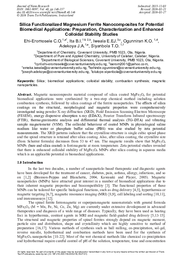
3.2 Structural and Phase Analysis
XRD was performed on the bare (sample not coated with silica i.e. the powder before size
selection was done) and silica coated samples of nanocrystalline MgFe2O4 and it is shown in Fig. 2.
The effects of size selection and silica coating on the structural properties of MgFe2O4 MNPs are
presented in Table 1. Like the XRD of the bare sample, the coated sample showed all the
characteristic peaks of spinel cubic structure (JCPDS card no. 73-1720) in the diffraction pattern.
This clearly showed that the sample retained the spinel structure even after coating by silica but
with a slight suppression of diffraction peaks. Therefore, the XRD data suggests that the silica shell
consists mainly of amorphous phase rather than polycrystalline one [36] since there is the absence
of silica-derived diffraction peaks. There is a pronounced change in the calculated structural
properties of the coated sample compared to the bare sample with the coated sample recording
lesser values of lattice parameter (a) and unit cell volume (V) but higher X-ray density (Dx) value
than the bare sample. The calculated crystallite sizes (D) for the bare sample and the coated sample
are 53 nm and 47 nm, respectively (Table 1). The reduction in the calculated crystallite size which
had also caused the reduction in other structural properties might be due to the size selection
process done to disaggregate the particles. Some researchers have reported the reduction in the
crystallite size of nanoparticles after coating [37]. The changes in the structural properties of the
coated sample might also be due to the silica coating.
�150
Journal of Nano Research Vol. 40
(440)
(533)
(511)
(422)
(400)
(220)
Intensity (A.U)
(311)
Fig. 3 shows typical FE-SEM images of bare and silica coated MgFe2O4 MNPs. From Fig.
3, it can be observed that the bare sample is in highly agglomerated form whereas the coated sample
displays better dispersion. The image for the coated sample shows clusters of MNPs as silica like
most coatings can coat single particles and aggregates. However, there is reduced agglomeration,
confirming the presence of silica coating on the MNPs; which helps to reduce the magnetic
interactions between nanoparticles. The nanoparticles after silica coatings retained the faceted
structure of uncoated samples but had a fairly lesser regular near-spherical structure compared with
the bare sample. In addition, Fig. 4 shows the EDAX spectrum for the silica coated MgFe2O4
MNPs. The spectrum contained four peaks (carbon peak is probably due to sample holder), which
were assigned to Mg, Fe, O, and Si. The peak of Si confirms the association of silica on the surface
of MgFe2O4 MNPs. Therefore, the EDAX analysis suggests Mg, Fe, O, and Si are the main
constituents in the nanocomposite.
(a)
(b)
30
40
50
60
70
80
2 Theta (degrees)
Fig. 2: X-ray diffraction patterns of polycrystalline MgFe2O4 (a) bare and (b) silica coated
samples
Table 1: Effects of Size Selection and Silica Coating on the Structural Properties of MgFe2O4
MNPs
MgFe2O4
Crystallite size,
D, (nm)
Lattice
constant, a,
(nm)
Unit cell
volume, V, nm3
X-ray density,
Dx, g/cm3
Bare
53
0.838
0.5885
4.5147
Coated
47
0.837
0.5864
4.5309
�Journal of Nano Research Vol. 40
151
Fig. 3: FESEM images of polycrystalline MgFe2O4 (a) bare and (b) silica coated samples
Fig. 4: EDAX spectra of silica coated MgFe2O4 nanocomposite
Fig. 5 shows the FTIR spectra of the bare and the coated sample. FTIR is an appropriate
technique to confirm the attachment of silica to the surface of the MNPs. In case of the bare sample,
the band observed at 560 cm-1 corresponds to stretching vibrations of Fe−O which is a typical
metal–oxygen absorption band for the spinel structure of the ferrite [38,39]. In the coated sample,
there is a shift of the stretching vibrations of Fe−O to 574 cm-1 which might be due to the silica
coating and confirms the presence of the ferrite nanoparticles in the silica nanocomposite. The
characteristic absorption band at 430 cm-1 and 1077 cm-1 corresponds to the bending and stretching
vibrations of Si-O-Si, respectively which confirm the formation of SiO2 [40,41]. Therefore, the
formation of ferrite and attachment of silica onto MgFe2O4 MNPs surface is confirmed and
supported by FTIR analysis.
�152
Journal of Nano Research Vol. 40
1.05
1.00
1077
(b)
0.95
430
Transmittance (%)
560
(a)
574
0.90
500
1000
1500
2000
2500
3000
3500
4000
-1
Wavenumber (cm )
Fig. 5: FTIR spectra of (a) bare MgFe2O4 (b) silica coated MgFe2O4
3.3 Thermal analysis
Thermogravimetric analysis (TGA) can provide additional quantitative evidence on the
structure of the nanoparticle coatings. It is an extremely valuable technique for surface
characterization of nanoparticles. TGA allows us to determine the bonding strength of the ligand to
the nanoparticle surface and its chemical stability [37]. The results of simultaneous thermal analysis
- TGA and DTA (differential thermal analysis), on the silica coated MgFe2O4 MNPs are presented
in Fig. 6. The weight loss process is observed in two stages. In the first, ~20% weight loss (which
might be due to the vapourisation of residual moisture) in the temperatute range of 30 - 120oC
corresponds to the endothermic peak (appearing as a kink) at about 120oC. However, in the 120 155oC temperature range, ~75% weight loss (corresponding to a sharp endothermic peak at ~150oC
in the DTA curve) is noticed in the second stage which was attributed to the detachment of coated
silica layer from the surface. It is well known that SiO2 synthesized by the Stöber process possess
high amounts of water and ethanol adsorbed on the surface, and both are removed by heating up to
150-200oC [42,43]. This might have accounted for the high weight loss associated with the
detachment of silica coatings. It can also be seen that in the 155-1000oC temperature range, no
significant weight loss is observed confirming the presence of pure MgFe2O4 phase. From this
analysis, a high amount of silica is attached to nanoparticles’ surfaces which also confirm the
presence of silica on the surface of MNPs. The results also show the potential stability of the silica
coated sample in applications less than 155oC.
Fig 6: TG-DTA curves silica coated MgFe2O4 MNPs
�Journal of Nano Research Vol. 40
153
Magnetisation (emu/g)
3.4 Magnetic Studies
The hysteresis loops measured at room temperature for the bare and silica coated MgFe2O4
samples are shown in Fig. 7. The magnetic results show that MgFe2O4 MNPs (bare and silica
coated) is ferrimagnetic at room temperature. The saturation magnetization (Ms), remanence (Mr),
coercivity (Hc) and loop squareness ratio (Mr/Ms) of the bare and coated sample were summarized
in Table 2. It can be seen that the Ms of the coated sample (22 emu/g) is smaller when compared to
the bare sample (26 emu/g) at an applied field of ±20,000 G at 300 K. However, the magnetization
for both samples is close to that of the bulk MgFe2O4 MNPs (~30 emu/g) [44]. MgFe2O4 is a mixed
type spinel ferrite with the Mg2+ and Fe3+ metal ions distributed over the tetrahedral and octahedral
sites. MgFe2O4 is an interesting magnetic material where magnetic couplings purely originate from
the magnetic moment of Fe cations and may be relatively weaker due to non magnetic Mg2+ metal
ions [45]. The reduction in magnetization for the coated sample may be attributed to the presence of
non-magnetic silica layer on the surface of MNPs which reduces the particle-particle interaction and
lowers the exchange coupling energy which in turn reduces the magnetization [37]. The reduction
in magnetization might also be due to the lesser amount of magnetic substance per gram in the silica
coated sample compared with the bare sample [5]. A reduction in magnetization was also reported
for silica coated LSMO particles [25]. The coated sample had a lesser Mr, but a higher Hc and
Mr/Ms values than the bare sample. Ms, Mr, and Hc are important magnetic properties critical to
biomedical applications. The Mr/Ms for both the bare and coated samples is found to be higher than
0.5 which is the expected value for randomly packed single domain particles [46]. The alternating
current susceptibility measurements of the coated sample showed that the magnetic responses are
frequency dependent which is an important parameter in hyperthermia and targeted drug delivery
applications [7,47].
-2 0 0 0 0
-1 0 0 0 0
30
(a)
(b )
20
10
0
0
-1 0
10000
20000
F ield (G )
-2 0
-3 0
Fig. 7: Magnetic hysteresis curves of MgFe2O4 measured at room temperature for (a) the bare
sample (b) silica coated sample
Table 2: Magnetic Properties of the Uncoated and the Silica Coated MgFe2O4
Sample
Bare sample
Silica
coated
sample
Saturation
Magnetisation, Ms
(emu/g)
26
22
Remanence
Magnetisation, Mr
(emu/g)
15
13
Coercivity
(Gauss)
Mr/ Ms
198
215
0.58
0.59
�154
Journal of Nano Research Vol. 40
3.5 Colloidal Stability of Silica Coated MgFe2O4 MNPs
For biomedical applications, MNPs should form stable dispersion in physiological saline
medium like water or phosphate buffer saline (PBS). The stabilization of the MNPs is crucial to
obtain magnetic colloidal ferrofluids that are stable against aggregation both in a biological medium
and in a magnetic field [48]. The colloidal stabilities of the bare and silica coated sample in water
and the colloidal stabilities of the coated samples in PBS (pH 5.0 and 7.4) were evaluated by the
zeta potential measurements. Colloidal stability in physiological media like PBS is also useful to
evaluate the strength of coating [49]. The zeta potential value in distilled water observed for the
coated sample (-15.50 mV) is higher than the bare sample (-2.45 mV). The results imply that the
aggregation of the coated sample in water is far less than the uncoated sample, which improves
colloidal stability with increasing zeta potential values [7]. Also, enhanced zeta potential values for
coated particles suggest that the silica particles have been successfully bound with surfaces of
uncoated particles [50]. The negative zeta potential helps to repel each particle in the suspension,
ensuring long-term stability and avoiding particle agglomeration [51]. The pH dependent zeta
potentials of the coated sample is -29.15 mV at pH 7.4 (physiological pH) and -19.35 mV at pH 5.0
(cancer cell endosomal pH). The results show that the silica coated MgFe2O4 MNPs are colloidally
stable both in physiological and inside the cancer cell environments. Colloidal stability is a very
important requirement for MNPs used in biomedical applications because aggregates can cause
serious harm to the patient e.g., by clogging blood vessels. These results imply that the silica coated
MgFe2O4 MNPs could maintain their dispersion stability and heating capacity in various
physiological environments and thus have great potential to be used in magnetic fluid hyperthermia
as a heating mediator and as a drug delivery vehicle. However, further experiments like
hemocompatibility assay, cytotoxicity tests and magnetic hyperthermia measurements have to be
carried out to test their real therapeutic potentials.
4.0 Conclusion
In conclusion, the single cubic spinel phase of nanocrystalline MgFe2O4 MNPs was obtained
by the solution combustion synthesis using a combination of urea and ammonium acetate fuels
followed by annealing at 900oC for 2hrs. The surfaces of the synthesized MNPs were modified with
silica for the purpose of enhanced colloidal stability for potential use in biomedical applications.
FESEM, EDAX, FTIR and thermal analysis showed that the MgFe2O4 MNPs were successfully
coated by silica. XRD revealed that the cubic spinel crystalline structure was retained in the coated
samples but with slight suppression of the peaks; and there was a reduction of the crystallite sizes in
the coated sample (47 nm) compared with the bare sample (53 nm). This decrease in crystallite size
was generally attributed to lesser agglomeration of particles due to silica coating and to the size
selection done before silica coating. The magnetic results show that MgFe2O4 MNPs (bare and silica
coated) is ferrimagnetic at room temperature. A reduction in the magnetic properties of all the silica
coated samples was observed and it was attributed to the presence of diamagnetic silica coatings
and the lesser amount of magnetic substance per gram in the silica coated sample compared with the
bare sample. The magnetic measurements of MgFe2O4 MNPs showed that the magnetic responses
are frequency dependent even with silica coatings and this is an important parameter in
hyperthermia and targeted drug delivery applications. Zeta potential studies revealed that there is
enhanced colloidal stability of MgFe2O4 MNPs after silica coating in aqueous media. Also, the
colloidal stability of silica coated MgFe2O4 MNPs in physiological media (PBS) highlights their
potential applications in biomedical field.
Acknowledgements
This work would not have been possible without the visiting research grant given to Dr. EhiEromosele C.O. by the International Centre for Materials Science, Jawarharlal Nehru Centre for
Advanced Scientific Research, Bangalore, India. The corresponding author would like to thank
Professor Vikram Jayaram, Chairman of the Department of Materials Engineering, Indian Institute
of Science (IISc), Bangalore for giving him access to their VSM and TG-DTA facilities.
�Journal of Nano Research Vol. 40
155
References
[1] L. Brannon-Peppas, J.O. Blanchette, Nanoparticle and targeted systems for cancer therapy,
Advanced Drug Delivery Reviews 56 (2004) 1649-1659.
[2] E.S. Kawasaki, A. Player, Nanotechnology, Nanomedicine and the Developmentof New
Effective Therapies for Cancer, Nanomedicine 1 (2005) 101-109.
[3] M.M. Yallapu, F.S. Othman, E.T. Curtis, B.K. Gupta, M. Jaggi, S.C. Chauhan, Multifunctional
Magnetic Nanoparticles for Magnetic Resonance Imaging and Cancer Therapy, Biomaterials
32 (2011) 1890-1905.
[4] M. Namdeo, S. Saxena, R. Tankhiwale, M. Bajpai, Y.M. Mohan, S.K. Bajpai, Magnetic
Nanoparticles for Drug Delivery Applications, Journal of Nanoscience and Nanotechnology 8
(2008) 3247-3271.
[5] S.A. Shah, M.H. Asdi, M.U. Hashmi, M.F. Umar, S. Awan, Thermoresponsive Copolymer
Coated MnFe2O4 Magnetic Nanoparticles for Hyperthermia Therapy and Controlled Drug
Delivery, Materials Chemistry and Physics 137 (2012) 365-371.
[6] C. Wilhelm, J.P. Fortin, F. Gazeau, Tumour Cell Toxicity of Intracellular Hyperthermia
Mediated by Magnetic Nanoparticles, Journal of Nanoscience and Nanotechnology 7 (2007)
2933-2937.
[7] N.D. Thorat, S.V. Otari, R.A. Bohara, H.M. Yadav, V.M. Khot, A. B. Salunkhe, M.R. Phdatre,
A.I. Prasad, R.S. Ningthoujam, S.H. Pawar, Structured Superparamagnetic Nanoparticles for
High Performance Mediator of Magnetic FluidHyperthermia: Synthesis, Colloidal Stability and
Biocompatibility Evaluation. Materials Science and Engineering C 42 (2014) 637-646.
[8] N. Kohler, C. Sun, A. Fichtenholtz, J. Gunn, C. Fang, M. Zhang, Methotrexate-Immobilised
Poly (Ethylene Glycol) Magnetic Nanoparticles in MR Imaging and Drug Delivery, Small 2
(2006) 785-792.
[9] C. Sun, J.S. Lee, M. Zhang, Magnetic Nanoparticles in MR Imaging and Drug Delivery,
Advanced Drug Delivery Reviews 60 (2008) 1252-1265.
[10] I.J. Bruce, T. Sen, Surface Modification of Magnetic Nanoparticles with Alkoxysilanes and
Their Application in Magnetic Bio-separations, Langmuir 21 (2005) 7029-7035.
[11] C. Wilhelm, F. Gazeau, Universal Cell Labelling with Anionic Magnetic Nanoparticles,
Biomaterials 29 (2008) 3161-3174.
[12] T. Osaka, T. Matsunaga, T. Nakanishi, A. Arakaki, D. Niwa, H. Iida, Synthesis of Magnetic
Nanoparticles and their Application to Bioassays, Analytical and Bioanalytical Chemistry 384
(2006) 593-600.
[13] D.K. Kim, Characterization and MRI study of surfactant-coated superparamagnetic
nanoparticles administrated into the rat brain, J. Magn. Magn. Mater. 225 (2001) 256–261.
[14] K. Cheng, S. Peng, C. Xu, S. Sun, Porous hollow Fe(3)O(4) nanoparticles for targeted delivery
and controlled release of cisplatin, J. Am. Chem. Soc. 131 (2009) 10637– 10644.
[15] A. Makridis, K. Topouridou, M. Tziomaki, D. Sakellari, K. Simeonidis, M. Angelakeris, M.P.
Yavropoulou, J. G. Yovos, O. Kalogirou, In vitro application of Mn-ferrite nanoparticles as
novel magnetic hyperthermia agents, J. Mater. Chem. B 2 (2014) 8390-8398.
[16] R. Hiergeist, W. Andra, N. Buske, R. Hergt, I. Hilger, U. Richter, W. Kaiser, Application of
magnetite ferrofluids for hyperthermia, Journal of Magnetism and Magnetic Materials 201
(1999) 420-422.
[17] P. Sivakumar, R. Ramesh, A. Ramanand, S. Ponnusamy, C. Muthamizhchelvan, Preparation of
sheet like polycrystalline NiFe2O4 nanostructure with PVA matrices and their properties,
Materials Letters, 65 (9) 2011 1438–1440.
[18] M. Rabanal, A. Várez, B. Levenfeld, J. Torralba, Magnetic properties of Mg-ferrite after
milling process, J. Mater. Process. Technol., 143 (2003) 470-474.
[19] Q. Chen, A. J. Rondinone, B. C. Chakoumakos, Z. John Zhang, Synthesis of
superparamagnetic MgFe2O4 nanoparticles by coprecipitation, Journal of Magnetism and
Magnetic Materials, 194(1) (1999) 1–7.
�156
Journal of Nano Research Vol. 40
[20] N. Kaur, M. Kaur, Comparative studies on impact of synthesis methods on structural and
magnetic properties of magnesium ferrite nanoparticles, Processing and Application of
Ceramics 8(3) (2014) 137–143.
[21] J. Chandradass, A. H. Jadhav, K. H. Kim, H. Kim, Influence of processing methodology on the
structural and magnetic behavior of MgFe2O4 nanopowders, Journal of Alloys and Compounds,
517 (2012) 164–169.
[22] T. Sasaki, S. Ohara, T. Naka, J. Vejpravova, V. Sechovsky, M. Umetsu, S. Takami, B.
Jeyadevan, T. Adschiri, Continuous synthesis of fine MgFe2O4 nanoparticles by supercritical
hydrothermal reaction, J. Supercrit. Fluids, 53(3) (2010) 92-94.
[23] P. Tartaj, M. Morales, S. Veintemillas-Verdaguer, T. Gonzalez-Carreno, C.J. Serna, The
Preparation of Magnetic Nanoparticles for Applications in Biomedicine, Journal of Physics D:
Applied Physics 36 (2003) R182-R197.
[24] M.R. Phadatare, V.M. Khot, A.B. Salunkhe, N.D. Thorat, S.H. Pawar, Studies on Polyethylene
Glycol Coating on NiFe2O4 Nanoparticles for Biomedical Applications, Journal of Magnetism
and Magnetic Materials 324 (2012) 770–772.
[25] V. Uskokovic, A. Kosak, M. Drofenik, Preparation of Silica-Coated Lanthanum Strontium
Manganite Particles with Designable Curie Point for Application in Hyperthermia Treatments,
International Journal of Applied Ceramic Technology 3 (2): (2006) 134–143.
[26] A. Villanueva, P. de la Presa, J.M. Alonso, T. Rueda, A. Martınez, P. Crespo, M.P. Morales,
M.A. Gonzalez-Fernandez, J. Valde´s, G. Rivero, Hyperthermia HeLa Cell Treatment with
Silica-Coated Manganese Oxide Nanoparticles, The Journal of Physical Chemistry C 114
(2010) 1976–1981.
[27] B. Mojic, K.P. Giannakopoulos, Z. Cvejic, V.V. Srdic, Silica coated ferrite nanoparticles:
Influence of citrate functionalization procedure on final particle morphology. Ceramics
International 38 (2012) 6635 – 6641.
[28] S. Makhluf, R. Dror, Y. Nitzan, Y. Abramovich, R. Jelinek, A. Gedanken, Microwave-Assisted
Synthesis of Nanocrystalline MgO and its use as a Bacteriocide. Advanced Functional
Materials 15 (10) (2005) 1708-1715.
[29] K. Krishnamoorthy, G. Manivannan, S.J. Kim, K. Jeyasubramanian, M.J. Premanathan,
Antibacterial Activity of MgO Nanoparticles Based on Lipid Peroxidation by Oxygen Vacancy,
Journal of Nanoparticle Research 14 (2012) 1063-1068.
[30] S. Kanagesan, M. Hashim, S. Tamilselvan, N.B. Alitheen, I. Ismail, G. Bahmanrokh, Cytotoxic
Effect of Nanocrystalline MgFe2O4 Particles for Cancer Cure, Journal of Nanomaterials (2013)
1-8.
[31] V.M. Khot, A.B. Salunkhe, N.D. Thorat, M.R. Phadatare, S.H. Pawar, Induction heating
studies of combustion synthesized MgFe2O4 nanoparticles for hyperthermia applications,
Journal of Magnetism and Magnetic Materials 332 (2013) 48–51.
[32] C.R. Vestal, Z.J. Zhang, Effects of surface coordination chemistry on the magnetic properties
of MnFe2O4 spinel ferrite nanoparticles, J. Am. Chem. Soc., 125 (2003) 9828–9833.
[33] M. Mahdavi, F. Namvar, M.B. Ahmad, R. Mohamad, Green Biosynthesis and Characterization
of Magnetic Iron Oxide (Fe3O4) Nanoparticles Using Seaweed (Sargassum muticum) Aqueous
Extract, Molecules 18 (2013) 5954–596.
[34] C.O. Ehi-Eromosele, B.I. Ita, E.E.J. Iweala, Synthesis, Microstructure and Magnetic Properties
of Nanocrystalline MgFe2O4 Particles: Effect of Mixture of Fuels and Sintering Temperature,
Science of Sintering (Accepted for publication).
[35] W. Stober, A. Fink, E. Bohn, Controlled growth of monodisperse silica spheres in micron size
range, J. Colloid Interface Sci., 26 (1968) 62–69.
[36] J. Choi, J.C. Kim, Y.B. Lee, I.S. Kim, Y.K. Park, N.H. Hur, Fabrication of Silica-Coated
Magnetic Nanoparticles with Highly Photoluminescent Lanthanide Probes. Chemical
Communications 16 (2007) 1644-1646.
�Journal of Nano Research Vol. 40
157
[37] A.B. Salunkhe, V.M. Khot, N.D. Thorat, M.R. Phadatare, C.I. Sathish, D.S. Dhawale, S.H.
Pawar, Polyvinyl Alcohol Functionalized Cobalt Ferrite Nanoparticles for Biomedical
Applications, Applied Surface Science 264 (2013) 598-604.
[38] B. Zhou, Y.W. Zhang, C.S. Liao, C.H. Yan, L.Y. Chen, S.Y. Wang, Rare Earth Mediated
Magnetism and Magneto-Optical Kerr Effects in Nanocrystalline CoFeMn0.9RE0.1O4 Thin
Films, Journal of Magnetism and Magnetic Materials 280 (2004) 327–333.
[39] H.M. Zaki, H.A. Dawoud, Far-infrared Spectra for Copper–Zinc Mixed Ferrites, Physica B 405
(2010) 4476–4479.
[40] A. Venkateswara Rao, P.B. Wagh, D. Haranath, P.P. Risbud, S.D. Kumbhare, Influence of
Temperature on the Physical Properties of TEOS Silica Xerogels, Ceramic International 25 (6):
(1999) 505–509.
[41] K.H. Wu, W.C. Huang, Effect of Varying the Acid to Metal Ion Ratio R on the Structural and
Magnetic Properties of SiO2-Doped Ni–Zn Ferrite, Journal of Solid State Chemistry 177 (2004)
3052–3057.
[42] B. Topuz, D. Şimşek, M. Çiftçioğlu, Preparation of monodisperse silica spheres and
determination of their densification behaviour. Ceramics International 41(1) (2015) 43-52.
[43] S. Zellmer, M. Lindenau, S. Michel, G. Garnweitner, C. Schilde, Influence of surface
modification on structure formation and micromechanical properties of spray-dried silica
aggregates. J Colloid Interface Sci. 464 (2016) 183-190.
[44] C.P. Liu, M.W. Li, Z. Cui, J.R. Huang, Y.L. Tian, T. Lin, W.B. Mi, Comparative study of
magnesium ferrite nanocrystallites prepared by sol–gel and coprecipitation methods, J. Mater.
Sci. 42 (2007) 6133-6138.
[45] V.M. Khot, A.B. Salunkhe, M.R. Phadatare, S.H. Pawar, Formation, Microstructure and
Magnetic Properties of Nanocrystalline MgFe2O4, Materials Chemistry and Physics 132 (2012)
782–787.
[46] A.B. Salunkhe, V.M. Khot, M.R. Phadatare, N.D. Thorat, R.S. Joshi, H.M. Yadav, S.H. Pawar,
Low Temperature Combustion Synthesis and Magnetostructural Properties of Co-Mn
Nanoferrites, Journal of Magnetism and Magnetic Materials 352 (2014) 91-98.
[47] M. Rahimi, P. Kameli, M. Ranjbar, H. Salamati, The Effect of Polyvinyl Alcohol (PVA)
Coating on Structural, Magnetic Properties and Spin Dynamics of Ni0.3Zn0.7Fe2O4 Ferrite
Nanoparticles, Journal of Magnetism and Magnetic Materials 347 (2013) 139–145.
[48] E. Umut, Surface Modification of Nanoparticles used in Biomedical Applications In: Modern
Surface Engineering Treatments, Ed. by Mahmood Aliofkhazraei, InTech (2013) 185-208.
[49] N.D. Thorat, V.M. Khot, A.B. Salunkhe, A. Prasad, R.S. Ningthoujam, S.H. Pawar, Surface
Functionalized LSMO Nanoparticles with Improved ColloidalStability for Hyperthermia
Applications, Journal of Physics D: Applied Physics 46 105003 (2013) 1-11.
[50] S.V. Jadhav, D.S. Nikam, V.M. Khot, S.S. Mali, C.K. Hong, S.H. Pawar, PVA and PEG
Functionalised LSMO Nanoparticles for Magnetic Fluid Hyperthermia Application, Materials
Characterization 102 (2015) 209–220.
[51] P. Smirnov, Cellular Magnetic Resonance Imaging using Superparamagnetic Anionic Iron
Oxide Nanoparticles: Applications to In Vivo Trafficking of Lymphocytes and Cell-Based
Anticancer Therapy, Methods in Molecular Biology 512 (2009) 333-353.
�
 Tolutope Siyanbola