J Neurol (2009) 256:1307–1313
DOI 10.1007/s00415-009-5119-1
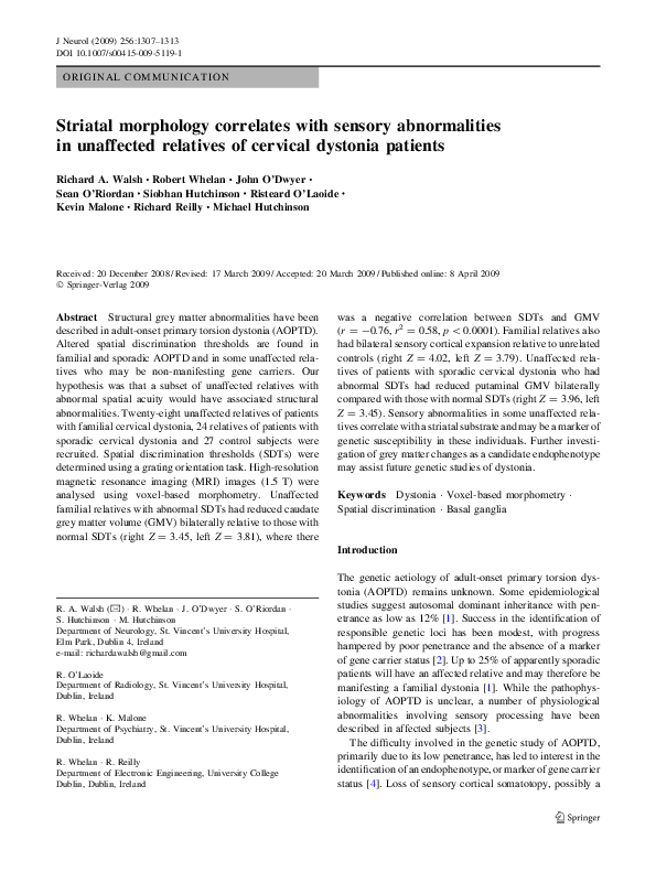
ORIGINAL COMMUNICATION
Striatal morphology correlates with sensory abnormalities
in unaffected relatives of cervical dystonia patients
Richard A. Walsh Æ Robert Whelan Æ John O’Dwyer Æ
Sean O’Riordan Æ Siobhan Hutchinson Æ Risteard O’Laoide Æ
Kevin Malone Æ Richard Reilly Æ Michael Hutchinson
Received: 20 December 2008 / Revised: 17 March 2009 / Accepted: 20 March 2009 / Published online: 8 April 2009
Ó Springer-Verlag 2009
Abstract Structural grey matter abnormalities have been
described in adult-onset primary torsion dystonia (AOPTD).
Altered spatial discrimination thresholds are found in
familial and sporadic AOPTD and in some unaffected relatives who may be non-manifesting gene carriers. Our
hypothesis was that a subset of unaffected relatives with
abnormal spatial acuity would have associated structural
abnormalities. Twenty-eight unaffected relatives of patients
with familial cervical dystonia, 24 relatives of patients with
sporadic cervical dystonia and 27 control subjects were
recruited. Spatial discrimination thresholds (SDTs) were
determined using a grating orientation task. High-resolution
magnetic resonance imaging (MRI) images (1.5 T) were
analysed using voxel-based morphometry. Unaffected
familial relatives with abnormal SDTs had reduced caudate
grey matter volume (GMV) bilaterally relative to those with
normal SDTs (right Z = 3.45, left Z = 3.81), where there
was a negative correlation between SDTs and GMV
(r = -0.76, r2 = 0.58, p \ 0.0001). Familial relatives also
had bilateral sensory cortical expansion relative to unrelated
controls (right Z = 4.02, left Z = 3.79). Unaffected relatives of patients with sporadic cervical dystonia who had
abnormal SDTs had reduced putaminal GMV bilaterally
compared with those with normal SDTs (right Z = 3.96, left
Z = 3.45). Sensory abnormalities in some unaffected relatives correlate with a striatal substrate and may be a marker of
genetic susceptibility in these individuals. Further investigation of grey matter changes as a candidate endophenotype
may assist future genetic studies of dystonia.
Keywords Dystonia � Voxel-based morphometry �
Spatial discrimination � Basal ganglia
Introduction
R. A. Walsh (&) � R. Whelan � J. O’Dwyer � S. O’Riordan �
S. Hutchinson � M. Hutchinson
Department of Neurology, St. Vincent’s University Hospital,
Elm Park, Dublin 4, Ireland
e-mail: richardawalsh@gmail.com
R. O’Laoide
Department of Radiology, St. Vincent’s University Hospital,
Dublin, Ireland
R. Whelan � K. Malone
Department of Psychiatry, St. Vincent’s University Hospital,
Dublin, Ireland
R. Whelan � R. Reilly
Department of Electronic Engineering, University College
Dublin, Dublin, Ireland
The genetic aetiology of adult-onset primary torsion dystonia (AOPTD) remains unknown. Some epidemiological
studies suggest autosomal dominant inheritance with penetrance as low as 12% [1]. Success in the identification of
responsible genetic loci has been modest, with progress
hampered by poor penetrance and the absence of a marker
of gene carrier status [2]. Up to 25% of apparently sporadic
patients will have an affected relative and may therefore be
manifesting a familial dystonia [1]. While the pathophysiology of AOPTD is unclear, a number of physiological
abnormalities involving sensory processing have been
described in affected subjects [3].
The difficulty involved in the genetic study of AOPTD,
primarily due to its low penetrance, has led to interest in the
identification of an endophenotype, or marker of gene carrier
status [4]. Loss of sensory cortical somatotopy, possibly a
123
�1308
J Neurol (2009) 256:1307–1313
physiological correlate of a structural abnormality, has been
proposed as a candidate endophenotype and is supported by
the finding of abnormal spatial acuity both in AOPTD and in
unaffected relatives [5–7]. Assessment of spatial acuity
peripherally as a marker of structural and organisational
changes in central structures relies on the integrity of the
peripheral nervous system and subject attention during
examination. Direct examination of cortical and subcortical
structures may therefore be preferable.
Our objective was to look for a structural CNS correlate of abnormal spatial acuity previously identified in
unaffected relatives of patients with sporadic and familial
AOPTD. Our hypothesis was that relatives with abnormal acuity would have grey matter changes affecting the
primary sensory cortex that have been previously
described in affected subjects [8]. We also specifically
looked for structural changes involving the caudate and
putamen given their position as part of the striato-thalamo-cortical motor control loop and the prominence of
the putamen in particular in previous imaging studies of
AOPTD [9–11].
Methods
Unaffected relatives
Twenty-eight unaffected members of five multiplex
AOPTD families (pedigrees 5, 6, 8, 10 and 26; Table 1)
with mean age 38.1 ± 8.8 years were recruited. Fifteen
were first-degree relatives of an affected family member
and 13 were second-degree relatives. Of these familial
relatives, 24 were right-handed and 4 were left-handed.
Twenty-four unaffected first-degree relatives of patients
with sporadic cervical dystonia (sporadic relatives) were
also recruited with mean age of 38.6 ± 9.2 years; 22 were
right-handed and 2 were left-handed.
Control subjects
Twenty-seven healthy control subjects were recruited from
amongst hospital staff and members of the public. Mean
age was 39.8 ± 11.8 years; 23 were right-handed and 4
were left-handed. Exclusion criteria included history of
neurological illness, neuropathic symptoms or significant
head trauma. Dystonia was excluded using a standardised
examination [12].
Sensory testing
A grating orientation task was performed using Johnson–van
Boven–Phillips domes applied to the index finger bilaterally
as previously reported [6]. Spatial discrimination threshold
(SDT) was defined as the grating width that would be
expected to achieve a 75% level of accuracy for a given
subject. Age-related control group means were established
by the examination of 141 healthy control subjects during an
earlier study [7]. Mean SDTs (±SD) for these healthy subjects were: for age group 20–29 years 1.172 ± 0.315 mm,
for 30–39 years 1.051 ± 0.286 mm, for 40–49 years
1.465 ± 0.500 mm and for 50–64 years 1.851 ± 0.578
mm. The upper limit of normal for subjects in this study was
set at a Z-score of 2.5 (control group mean ?2.5 SD), which
was 1.959 mm, 1.766 mm, 2.716 mm and 3.297 mm for
each of the four age groups, respectively.
Of the 28 unaffected familial relatives, 12 were known to
have abnormal SDTs (the familial abnormal SDT group) and
16 had normal SDTs (the familial normal SDT group). Of the
24 unaffected sporadic relatives, 13 were known to have
abnormal SDTs (sporadic abnormal SDT group) and 11 had
normal SDTs (sporadic normal SDT group). Z-scores of
unaffected relatives and those of the 141 controls used to
establish normative values for spatial acuity are shown in
Fig. 1. All 27 control subjects participating in this imaging
study had normal spatial acuity.
Table 1 Summary of affected family members in multiplex and singleton AOPTD families from which all unaffected relatives were recruited
Number affected
Mean age at onset (years)a
AOPTD phenotypes
Multiplex families
Pedigree 5
4
47.0
3 cervical dystonia, 1 blepharospasm
Pedigree 6
3
35.7
2 cervical dystonia, 1 spasmodic dysphonia
Pedigree 8
5
45.0
3 cervical dystonia, 1 FHD, 1 spasmodic dysphonia
Pedigree 10
4
47.5
All cervical dystonia
Pedigree 26
All 5 pedigrees
Sporadic families
3
51.5
All cervical dystonia
19
45.3
–
10
46.8
One family member with cervical dystonia
FHD focal hand dystonia
a
Age at onset data unavailable for two patients with familial AOPTD and one patient with sporadic AOPTD
123
�J Neurol (2009) 256:1307–1313
Fig. 1 SDT Z-scores in unrelated control subjects and unaffected
relatives of patients with cervical dystonia. The dashed line represents
the chosen cutoff value between normal and abnormal SDTs at a Z-score
of 2.5
Voxel-based morphometry
Image acquisition
All MRI scans were obtained at 1.5 T on the same scanner
(Siemens Avanto, Erlangen, Germany). A high-resolution
three-dimensional T1-weighted magnetization-prepared
rapid-acquisition gradient echo (MPRAGE) sequence was
acquired (TR = 1160 ms; TE = 4.21 ms, TI = 600 ms,
flip angle = 15°) with a sagittal orientation, 256 9 256
matrix size and 0.9 mm isotropic voxels. A radiologist
blinded to clinical status reviewed all images. Subjects
were eliminated from further analysis if any macroscopic
structural brain abnormality was identified or if there was
movement artefact affecting image quality.
Pre-processing and statistical analysis
of structural data
Statistical parametric mapping software (SPM5; Wellcome
Centre for Neuroimaging, London, UK), running under
Matlab 6.5 (Mathworks, Sherborn, MA, USA), was used to
pre-process and analyse the MRI data obtained. Pre-processing incorporated image registration and classification
into a single generative model [13]. Segmented grey matter
data were modulated in order to preserve total grey matter
volume. The spatially normalised and modulated grey
matter partitions were smoothed using a 12 mm full-width
at half-maximum Gaussian kernel allowing parametric
statistical analysis to be performed in every analysis. Total
grey matter volume, age, sex and handedness were entered
as nuisance covariates in all analyses.
1309
Each analysis was restricted to the predefined regions of
interest using anatomically defined masks (Wake Forest
University PickAtlas) [14]. This software employs SPM5’s
small volume correct feature, reducing the number of multiple comparisons. Type I errors were controlled using false
discovery rate (FDR) of 0.05, controlling the expected proportion of false positives among supra-threshold voxels for
each analysis performed [15]. For the purpose of this study
we restricted our analysis to the primary sensory cortex, the
caudate nucleus and the putamen bilaterally. We further
restricted our findings to regions in which at least a conservative threshold of 100 contiguous voxels were found to be
significant after FDR correction. The locations of significant
voxels were summarised by their local maxima separated by
at least 8 mm, and by converting the maxima coordinates
from Montreal Neurological Institute (MNI) to Talairach
coordinate space. These coordinates were assigned neuroanatomic labels using the Talairach Daemon brain atlas [16].
Correlations were calculated using individual voxel values at
the local maxima of grey matter intensity in each predetermined region of interest to examine the relationship with
spatial acuity. Z-scores are given in the ‘‘Results’’ section for
each inter-group comparison of statistical significance. A
summary of voxel-based morphometry (VBM) results and
coordinates of voxels where peak GMV differences were
found are given in Table 2.
Results
Unaffected relatives compared with unrelated healthy
controls
Sensory cortical volume was increased bilaterally when
comparing unaffected familial relatives with unrelated healthy control subjects (right Z = 4.02, left Z = 3.79; Fig. 2).
No grey matter change in sub-cortical structures was noted in
this comparison. This sensory cortical finding was not replicated in the sporadic relative group when analysed separately.
Unaffected relatives with abnormal SDTs compared
with those with normal SDTs
In all 52 unaffected relatives of sporadic and familial
dystonia subjects, putaminal volume was greater bilaterally
in the normal SDT group (Fig. 3a). This, however, was
significant at FDR \ 0.06 rather than the predetermined
FDR of 0.05. Amongst familial relatives alone, putaminal
volume did not differ between normal SDT and abnormal
SDT groups, but those with normal SDTs had significantly
larger caudate volume bilaterally (right Z = 3.45, left
Z = 3.81; Fig. 3b). Amongst the 24 sporadic relatives
alone, normal SDT relatives had a bilateral increase in
123
�1310
J Neurol (2009) 256:1307–1313
Table 2 Summary of results including coordinates of peak GMV differences
Grey matter comparison
Region
Left
Z
Unaffected familial relatives [ controls
Right
Coordinates (mm)
x
y
Z
z
Coordinates (mm)
x
y
z
Bilateral post-central gyrus
3.79
-38
-40
57
4.02
14
-31
70
Normal SDT (S ? F) [ abnormal SDT (S ? F)a
Bilateral putamen
3.19
-24
14
7
3.27
24
18
1
Normal SDT (F) [ abnormal SDT (F)
Normal SDT (S) [ abnormal SDT (S)
Bilateral caudate
Bilateral putamen
3.81
3.45
-14
-22
5
16
16
7
3.45
3.96
18
24
9
19
18
-4
Grey matter correlating significantly with SDT
Z-scores for all 52 relatives
Left caudate
3.8
-12
-7
19
–
–
–
–
Grey matter and SDT correlation for 28 unaffected
familial relatives
Bilateral caudate
4.14
-14
-7
19
4.08
18
-11
19
F familial relatives, S sporadic relatives, Z Z-score
a
FDR 0.06
Fig. 2 Bilateral sensory
cortical grey matter increase in
all 28 unaffected familial
relatives compared with 27
unrelated healthy controls
shown on a three-dimensional
(3D) surface render (peak
difference Z = 4.02 at 14, -31,
70; cluster size threshold 100
voxels, FDR = 0.05)
123
�J Neurol (2009) 256:1307–1313
Fig. 3 Voxels in which grey matter volume in unaffected relatives
with normal spatial acuity was greater than in those with abnormal
spatial acuity. a All 52 sporadic and familial relatives combined
1311
(Z = 3.27 at 24, 18, 1), b All 28 unaffected familial relatives
(Z = 3.81 at -14, 5, 16) and c All 24 sporadic relatives (Z = 3.96 at
24, 19, -4; cluster size threshold 100 voxels, FDR = 0.05)
putaminal volume in comparison with abnormal SDT relatives (right Z = 3.96, left Z = 3.45; Fig. 3c).
Correlation of spatial discrimination thresholds
with grey matter intensity
The following correlations were estimated between the
local maximum of intensity in the left caudate (Talairach x,
y, z coordinates in parentheses) and SDT Z-scores, although
significant correlations were identified for voxels in the
caudate bilaterally. For all 52 unaffected relatives, greater
SDT Z-scores (or greater impairment of spatial acuity)
were associated with reduced grey matter intensities
(r = -0.53, r2 = 0.28, p \ 0.0001; -12, -7, 19). The
strongest correlation between grey matter intensity and
SDT Z-scores was seen in the 28 unaffected familial relatives (r = -0.76, r2 = 0.58, p \ 0.0001; -14, -17, 19;
Fig. 4a, b). In the 25 sporadic and familial relatives with
abnormal SDTs, there was also a correlation between grey
matter intensity and SDT Z-score (r = -0.62, r2 = 0.38,
p = 0.001; -14, -11, -19). This was observed in the
12 familial relatives with abnormal SDT Z-scores taken
alone (r = -0.75, r2 = 0.56, p = 0.005; -14, -7, 19) and
in the 13 sporadic relatives alone there was a trend towards
a correlation (r = -0.47, r2 = 0.22, p = 0.1; -14, -11,
19). In the unrelated control group no voxels within the
putamen or caudate correlated significantly with SDT
Z-scores. There were therefore no supra-threshold voxels
with which to similarly perform a correlation.
Discussion
Morphological grey matter changes involving sensorimotor
circuits have been previously reported in patients with
Fig. 4 a Grey matter voxels correlating significantly with spatial
acuity in the 28 unaffected familial relatives (Z = 4.14 at -14, -7,
19; cluster size threshold 100 voxels, FDR = 0.05) and b correlation
between SDT Z-scores and caudate grey matter intensity at the local
maxima for the familial relative group
123
�1312
AOPTD. Although the basal ganglia, in particular the
putamen, have been frequently highlighted in anatomical
and functional imaging studies, there have also been contradictory findings. In cervical dystonia, reduced grey
matter volume has been described in the putamen bilaterally, with an increase in grey matter in the thalamus and
caudate head bilaterally relative to control subjects [17].
An earlier volumetric study demonstrated an increase in
putaminal volume bilaterally in cervical dystonia that was
replicated in a VBM study of patients with blepharospasm
[9, 11]. Other authors have not identified structural changes
in the basal ganglia in AOPTD but have reported other
potential structural substrates. In a study of 30 patients with
writer’s cramp, grey matter was reduced bilaterally in the
thalamus and cerebellum as well as in the hand area of the
contralateral sensorimotor cortex [18]. Heterogeneous
patient populations, variations in group sizes studied and
methodological differences in image acquisition and analysis may account for some discrepancies to date.
Our a priori hypothesis was that a structural sensory
cortical abnormality would be found in a subset of unaffected relatives of patients with cervical dystonia, correlating with SDT abnormalities previously identified in this
group. We were, however, unable to link altered cortical
morphology with abnormalities of spatial acuity, although
unaffected relatives from the multiplex pedigrees did have
significantly greater sensory cortical grey matter volume
bilaterally relative to unrelated controls. There may be no
simple relationship between cortical organisation or morphology and spatial acuity [19]. Alternatively, the relationship may be a more complex one that volumetric
analysis is insensitive to.
We had not anticipated the moderately strong and significant correlations between striatal grey matter volume
and spatial acuity, although the basal ganglia do appear to
serve both motor and sensory functions [20]. A functional
MRI study revealed bilateral hyperactivity in the basal
ganglia during a similar grating orientation task in patients
with focal hand dystonia [21] and deficits of two-point
discrimination can be found in Parkinson’s disease [22].
The absence of a similar correlation between spatial acuity
and striatal GMV in the unrelated control group suggests
that this finding is reflective of true subclinical pathology.
Maladaptive sensory processing of afferent inputs into
basal ganglia structures may contribute to the pathogenesis
of dystonia [23] and the finding of a link between abnormal
sensory testing and striatal grey matter is therefore of
interest.
Caudate GMV correlated bilaterally with abnormal
SDTs in the familial group but putaminal GMV correlated
bilaterally with SDTs in the sporadic group. As the sporadic group, or at least a subset of them, are possibly
expressing the same inherited dystonia as the familial
123
J Neurol (2009) 256:1307–1313
group, we postulate that morphological changes in both
caudate and putamen may be found in both groups. The
availability of greater numbers in each patient group may
have allowed demonstration of such ‘pan-striatal’ grey
matter changes in voxels that did not reach the significance
threshold set in this study. Confounding by truly sporadic
cases, phenocopies or genetic heterogeneity amongst sporadic cases may have further contributed to the differences
between sporadic and familial relative groups.
We postulate that unaffected relatives with abnormal
spatial acuity, and the correlated reduction in striatal volume, are non-penetrant gene carriers. Imaging asymptomatic relatives who are possible gene carriers has the
potential to allow the observation of morphological changes reflecting the site of primary pathology of AOPTD.
These asymptomatic family members will not manifest
structural changes that are secondary to ongoing dystonic
movements present in affected subjects. Also, imaging
findings in manifesting gene carriers within families may
evolve according to the AOPTD phenotype expressed,
despite a common genetic aetiology. Bilateral basal ganglia
changes in unaffected relatives in this study can be considered a candidate endophenotype in AOPTD and would
fit with existing hypotheses implicating basal ganglia
dysfunction in its pathophysiology.
Could putaminal changes in some unaffected relatives
result from neural adaption to inherited putaminal pathology given the very low penetrance of the phenotype? Other
studies of subclinical traits in idiopathic dystonia have
revealed differences amongst manifesting and non-manifesting gene carriers, possibly reflective of protective or
modifying genetic factors in unaffected subjects [24].
Frima and colleagues [4] investigated abnormal vibrationinduced illusion of movement in patients with cervical
dystonia and unaffected relatives. Both groups shared an
abnormal interpretation of the vibratory stimulus but the
unaffected group behaved more like an unrelated control
group in their response to pre-testing muscle fatigue. The
authors speculated that this might have been due to a more
adaptive central handling of type Ia afferent fibres in the
non-manifesting group. Manifesting and non-manifesting
carriers of the DYT1 mutation demonstrate similar degrees
of abnormal intracortical inhibition, but manifesting carriers differ in their abnormally prolonged response to conditioning of the motor cortex using repetitive transcranial
magnetic stimulation [25]. One weakness of this study is
our inability to identify which unaffected relatives, if any,
will go on to manifest dystonia and therefore we cannot be
certain if the observed changes are a subclinical presymptomatic phase or a protective neuroplastic response.
The low penetrance of the phenotype may make the latter
more plausible but only prospective observation of unaffected relatives over time will clarify this.
�J Neurol (2009) 256:1307–1313
1313
Bilateral sensory cortical changes and altered striatal
morphology correlating with established sensory deficits in
these unaffected relatives offer an insight into the pathophysiology of AOPTD. The interpretation of structural
changes observed in previous VBM studies has been
restricted given the absence of a similar neurofunctional
correlate. The relative contribution of the primary disease
process and secondary neuroplastic phenomena to these
grey matter changes is currently unknown and would be
assisted by longitudinal studies. Further structural and
functional imaging studies of both affected and unaffected
individuals are needed. If identified, a shared striatal or
cortical abnormality in manifesting and non-manifesting
members of multiplex families would warrant further
investigation as a potential endophenotype to assist genetic
studies of AOPTD.
Acknowledgements This study was funded by Dystonia Ireland and
a University College Dublin Seed Funding grant.
Conflict of interest statement
interest.
The authors report no conflicts of
References
1. Waddy HM, Fletcher NA, Harding AE, Marsden CD (1991) A
genetic study of idiopathic focal dystonias. Ann Neurol 29:320–
324. doi:10.1002/ana.410290315
2. Jarman PR, del Grosso N, Valente EM, Leube B, Cassetta E,
Bentivoglio AR, Waddy HM, Uitti RJ, Maraganore DM, Albanese A, Frontali M, Auburger G, Bressman SB, Wood NW,
Nygaard TG (1999) Primary torsion dystonia: the search for
genes is not over. J Neurol Neurosurg Psychiatry 67:395–397.
doi:10.1136/jnnp.67.3.395
3. Tinazzi M, Rosso T, Fiaschi A (2003) Role of the somatosensory
system in primary dystonia. Mov Disord 18:605–622.
doi:10.1002/mds.10398
4. Frima N, Nasir J, Grunewald R (2008) Abnormal vibrationinduced illusion of movement in idiopathic focal dystonia: an
endophenotypic marker? Mov Disord 23:373–377. doi:10.1002/
mds.21838
5. Meunier S, Hallett M (2005) Endophenotyping: a window to the
pathophysiology of dystonia. Neurology 65:792–793. doi:10.1212/
01.wnl.0000177919.02950.4a
6. O’Dwyer JP, O’Riordan S, Saunders-Pullman R, Bressman SB,
Molloy F, Lynch T, Hutchinson M (2005) Sensory abnormalities
in unaffected relatives in familial adult-onset dystonia. Neurology
65:938–940. doi:10.1212/01.wnl.0000176068.23983.a8
7. Walsh R, O’Dwyer JP, Sheikh I, O’Riordan S, Lynch T,
Hutchinson M (2007) Sporadic adult onset dystonia: sensory
abnormalities as an endophenotype in unaffected relatives.
J Neurol Neurosurg Psychiatry 78:980–983. doi:10.1136/jnnp.
2006.105585
8. Garraux G, Bauer A, Hanakawa T, Wu T, Kansaku K, Hallett M
(2004) Changes in brain anatomy in focal hand dystonia. Ann
Neurol 55:736–739. doi:10.1002/ana.20113
9. Black KJ, Ongur D, Perlmutter JS (1998) Putamen volume in
idiopathic focal dystonia. Neurology 51:819–824
10. Colosimo C, Pantano P, Calistri V, Totaro P, Fabbrini G, Berardelli A (2005) Diffusion tensor imaging in primary cervical
dystonia. J Neurol Neurosurg Psychiatry 76:1591–1593. doi:
10.1136/jnnp.2004.056614
11. Etgen T, Muhlau M, Gaser C, Sander D (2006) Bilateral putaminal grey-matter increase in primary blepharospasm. J Neurol
Neurosurg Psychiatry 77:1017–1020. doi:10.1136/jnnp.2005.
087148
12. Bressman SB, de Leon D, Brin MF, Greene PE, Fahn S,
Breakefield XO, Risch NJ (1989) Idiopathic dystonia among
Ashkenazi Jews: evidence for autosomal dominant inheritance.
Ann Neurol 26:612–620. doi:10.1002/ana.410260505
13. Ashburner J, Friston KJ (2005) Unified segmentation. Neuroimage 26:839–851. doi:10.1016/j.neuroimage.2005.02.018
14. Maldjian JA, Laurienti PJ, Kraft RA, Burdette JH (2003) An
automated method for neuroanatomic and cytoarchitectonic atlasbased interrogation of fMRI data sets. Neuroimage 19:1233–
1239. doi:10.1016/S1053-8119(03)00169-1
15. Genovese CR, Lazar NA, Nichols T (2002) Thresholding of
statistical maps in functional neuroimaging using the false discovery rate. Neuroimage 15:870–878. doi:10.1006/nimg.2001.
1037
16. Fox PT, Laird AR, Fox SP, Fox PM, Uecker AM, Crank M,
Koenig SF, Lancaster JL (2005) BrainMap taxonomy of experimental design: description and evaluation. Hum Brain Mapp
25:185–198. doi:10.1002/hbm.20141
17. Obermann M, Yaldizli O, De Greiff A, Lachenmayer ML, Buhl
AR, Tumczak F, Gizewski ER, Diener HC, Maschke M (2007)
Morphometric changes of sensorimotor structures in focal
dystonia. Mov Disord 22:1117–1123. doi:10.1002/mds.21495
18. Delmaire C, Vidailhet M, Elbaz A, Bourdain F, Bleton J-P,
Sangla S, Meunier S, Terrier A, Lehéricy S (2007) Structural
abnormalities in the cerebellum and sensorimotor circuit in writer’s cramp. Neurology 69:376–380. doi:10.1212/01.wnl.
0000266591.49624.1a
19. Moore CE, Schady W (2000) Investigation of the functional
correlates of reorganization within the human somatosensory
cortex. Brain 123:1883–1895. doi:10.1093/brain/123.9.1883
20. Brown LL, Schneider JS, Lidsky TI (1997) Sensory and cognitive
functions of the basal ganglia. Curr Opin Neurobiol 7:157–163.
doi:10.1016/S0959-4388(97)80003-7
21. Peller M, Zeuner KE, Munchau A, Quatarone A, Weiss M,
Knutzen A, Hallett M, Deuschl G, Siebner HR (2006) The basal
ganglia are hyperactive during the discrimination of tactile
stimuli in writer’s cramp. Brain 129:2697–2708. doi:10.1093/
brain/awl181
22. Sathian K, Zangaladze A, Green J, Vitek JL, DeLong MR (1997)
Tactile spatial acuity and roughness discrimination: impairments
due to aging and Parkinson’s disease. Neurology 49:168–177
23. Murase N, Kaji R, Shimazu H, Katayama-Hirpta M, Ikeda A,
Kohara N, Kimura J, Shibasaki H, Rothwell JC (2000) Abnormal
premovement gating of somatosensory input in writer’s cramp.
Brain 123:1813–1829. doi:10.1093/brain/123.9.1813
24. Carbon M, Su S, Dhawan V, Raymond D, Bressman S, Eidelberg
D (2004) Regional metabolism in primary torsion dystonia:
effects of penetrance and genotype. Neurology 62:1384–1390
25. Edwards MJ, Huang YZ, Wood NW, Rothwell JC, Bhatia KP
(2003) Different patterns of electrophysiological deficits in
manifesting and non-manifesting carriers of the DYT1 mutation.
Brain 126:2074–2080. doi:10.1093/brain/awg209
123
�
 Richard Reilly