Research Article
4633
Netrin-1 regulates invasion and migration of mouse
mammary epithelial cells overexpressing Cripto-1 in
vitro and in vivo
Luigi Strizzi1, Caterina Bianco1, Ahmed Raafat1, Wissam Abdallah1, Cindy Chang1, Dina Raafat1,
Morihisa Hirota1, Shin Hamada1, Youping Sun1, Nicola Normanno2, Robert Callahan1, Lindsay Hinck3 and
David Salomon1,*
1
Mammary Biology and Tumorigenesis Laboratory, NCI/CCR, 37 Convent Drive, Building 37, Bethesda, MD 20892, USA
Cell Biology and Preclinical Models Unit, INT-Fondazione Pascale, 80131 Naples, Italy
Department of Biology, University of California at Santa Cruz, Santa Cruz, 1156 High Street, Santa Cruz, CA 95064, USA
2
3
*Author for correspondence (e-mail: salomond@mail.nih.gov)
JournalofCellScience
Accepted 7 July 2005
Journal of Cell Science 118, 4633-4643 Published by The Company of Biologists 2005
doi:10.1242/jcs.02574
Summary
The neuronal guidance molecule, Netrin-1, has been
suggested to play a role in the adhesion and migration of
the mammary gland epithelium. Human and mouse
Cripto-1 induce proliferation, migration, invasion and
colony formation by epithelial cells in 3D matrices. Here we
investigate whether Netrin-1 affects these Cripto-1dependent activities in mouse mammary epithelial cells.
Overexpression of Cripto-1 in EpH4 and HC-11 cells
(EpH4/Cripto-1 or HC-11/Cripto-1) was associated with
low expression of Netrin-1 and increased expression of its
receptor Neogenin compared to that of wild-type cells. No
change was observed in the expression of the other Netrin1 receptor, UNC5H1. Treating EpH4/Cripto-1 or HC11/Cripto-1 mammary cells with exogenous soluble Netrin1 resulted in increased expression of E-cadherin and
UNC5H1, decreased expression of vimentin and decreased
Introduction
Human Cripto-1 (CR-1) is a member of the epidermal growth
factor (EGF)-CFC family of signaling proteins first cloned
from the human teratocarcinoma cell line NTERA2
(Ciccodicola et al., 1989). The glycosylphospatidylinositol
(GPI) membrane-linked Cripto (Minchiotti et al., 2000) binds
to Nodal, a member of the TGF- superfamily, which
facilitates Nodal interaction with an activin receptor complex
composed of the type-I serine-threonine receptor ActRIB (or
ALK4) and the type-II activin receptor, either ActRII or
ActRIIB. This leads to phosphorylation of Smad2 and/or
Smad3, which together with Smad4 can heterodimerize and
translocate to the nucleus to mediate transcriptional responses
(Bianco et al., 2002; Yeo and Whitman, 2001). Cripto-1 can
also function through a Nodal- and ALK4-independent
signaling pathway by specifically binding to Glypican-1, a
membrane-associated heparan sulfate proteoglycan, which
leads to phosphorylation of the tyrosine kinase c-Src and
activation of mitogen-activated protein kinase (MAPK) and
Akt signaling pathways (Bianco et al., 2003).
Cripto-1 is expressed in a variety of human cancers
activation of Akt as determined by western blotting. Colony
formation by Eph4/Cripto-1 cells in 3D gels was
significantly reduced in proximity to a Netrin-1 source, and
mammary glands of transgenic mice overexpressing human
Cripto-1 showed altered ductal growth in proximity to
implanted Netrin-1-releasing pellets. Terminal end buds in
the treated transgenic mice mammary glands also showed
increased expression of E-cadherin and UNC5H1 and
decreased expression of active Akt determined by
immunohistochemistry. Together, these results suggest that
regulation of Netrin-1 expression is important in regulating
Cripto-1-dependent invasion and migration of mammary
epithelial cells.
Key words: Mammary cells, Netrin-1, Cripto-1, Invasion, Migration
(Salomon et al., 2000) including breast cancer (Qi et al., 1994).
Overexpression of CR-1 has been shown to induce
proliferation, migration and invasion of human breast cancer
cells (Brandt et al., 1994; Normanno et al., 2004) and of mouse
mammary epithelial cells (Bianco et al., 2003; Strizzi et al.,
2004; Wechselberger et al., 2001). Furthermore, cells
overexpressing CR-1 have been shown to acquire specific
biochemical and morphological features suggesting that CR-1
may play a role in promoting epithelial mesenchymal transition
(EMT).
Epithelial mesenchymal transition is a normal physiologic
process important for embryogenesis, tissue growth, wound
healing and tissue repair (Perez-Pomares and Munoz-Chapuli,
2002). During EMT epithelial cells lose their adhesive
properties owing to modification in the expression of cellular
adhesion molecules like E-cadherin (Boyer et al., 2000;
Savagner, 2001). In fact, the overexpression of the genes snail
and slug has been shown to play an important role in inducing
EMT by negatively affecting the expression of E-cadherin
(Cano et al., 2000). Epithelial cells undergoing EMT also show
changes in the cytoskeleton. For example, vimentin is a
�JournalofCellScience
4634
Journal of Cell Science 118 (20)
cytoskeleton molecule normally expressed in mesenchymal
cells and when expressed in epithelial cells may facilitate their
acquisition of a more spindle-shaped morphology during EMT
in both normal and disease states (Fuchs and Weber, 1994;
Gilles et al., 1996; Lane et al., 1983). Molecules that are
involved in growth factor signaling such as Src and
phosphoinositide 3-kinase (PI3K) are also activated during
EMT (Thiery and Chopin, 1999; Vincent-Salomon and Thiery,
2003). Thus, cellular changes characteristic of EMT facilitate
migration and invasion of epithelial tumor cells and have
recently been suggested as an index of aggressiveness and
increased metastatic potential in different types of malignant
tumors (Birchmeier et al., 1996a; Birchmeier et al., 1996b;
Gilchrist et al., 2002). Specifically, reports have suggested that
EMT may be relevant to the development of human breast
cancer, as mutations in E-cadherin expression (Berx et al.,
1998; Cano et al., 2000), overexpression of snail and slug
(Cano et al., 2000; Hajra et al., 2002), overexpression of
vimentin (Hanna et al., 2003) and increased activity of
signaling molecules involved in migration and invasion during
EMT (Vincent-Salomon and Thiery, 2003) have all been
identified in human breast cancer both in vitro and in vivo. In
addition, EMT has been suggested to play a role during
metastasis and affecting prognosis in human breast cancer
(Fuchs et al., 2002; Xue et al., 2003).
Overexpression of mouse Cripto-1 (Cr-1) increases
proliferation of mouse mammary epithelial cells and causes
them to assume a more mesenchyme-like phenotype. In
addition, Cr-1 overexpression increases anchorageindependent growth of mammary epithelial cells in soft agar
and migration when cells are grown on plastic or on porous
filters coated with extracellular matrix, and during wound
healing assays (Wechselberger et al., 2001). Increased
expression of Cr-1 also induces the formation of branch-like
structures when mouse mammary epithelial cells are grown in
a collagen type I matrix (Wechselberger et al., 2001). Overall,
these responses are reminiscent of EMT and suggest that Cr-1
overexpression induces this transition in mammary epithelial
cells. However, whether Cripto-1-induced migration and
proliferation can be influenced by extracellular directional cues
is unclear.
Various studies have identified different chemotropic factors
that regulate the direction of cell migration. Most of these have
been identified during neuronal development and include
proteins such as, Slits, Ephrins, Semaphorins (Kolodkin et al.,
1993), Sonic hedgehog (Charron et al., 2003), bone
morphogenic proteins (Butler and Dodd, 2003), Wnts
(Yoshikawa et al., 2003) and Netrins (Serafini et al., 1996;
Serafini et al., 1994). Sequence and functional analysis have
shown that Netrins are a conserved family of secreted proteins
that have regional homology to laminins and are capable of
regulating axonal outgrowth (Kennedy et al., 1994; Puschel,
1999; Serafini et al., 1996). The direction of Netrin-dependent
neuronal outgrowth is determined by the cellular expression of
receptors belonging to either the DCC (deleted in colon cancer)
or UNC5 families of Netrin-1 receptors (Keino-Masu et al.,
1996; Leonardo et al., 1997). These single-pass transmembrane
receptors contain immunoglobulin domains with DCC
containing fibronectin type-3 domains and with UNC5
containing a thrombospondin type-I domain (Chisholm and
Tessier-Lavigne, 1999). The DCC receptors, which include the
structurally similar Neogenin receptor, mediate attraction,
whereas repulsion is mediated by a complex of DCC and
UNC5 receptor families (Hinck, 2004; Hong et al., 1999). The
highly conserved family of UNC receptors possess a high level
of structural and sequence homology in the ligand binding
extracellular domain (Engelkamp, 2002). In humans, UNC5
receptors are composed of UNC5HA, UNC5HB and UNC5HC
and correspond to the rodent orthologues UNC5H1, UNC5H2
and UNC5H3, respectively (Arakawa, 2004).
Recent studies have found functioning Netrin molecules
outside the nervous system, in the pancreas, intestine (Jiang et
al., 2003; Yebra et al., 2003), lung (Liu et al., 2004) kidney,
heart and vasculature (Koch et al., 2000; Lu et al., 2004; Park
et al., 2004) where they presumably play a role in the
development of these organs by regulating the migration of
different types of cells. Regulation of the expression of Netrin1 and its receptors may play a role in tumorigenesis. In fact,
Netrin-1 was shown to be reduced in tumors of the prostate and
of the nervous system (Latil et al., 2003; Meyerhardt et al.,
1999). Low levels of somatic mutations of DCC have been
identified in cancers of the brain, stomach, pancreas,
colorectum and testicle (Arakawa, 2004) and in a series
comparing human colorectal tumors with corresponding
normal tissues, of the different UNC5 receptors studied,
UNC5A, the orthologue of rodent UNC5H1, showed the
highest percentage of altered expression (Thiebault et al.,
2003).
Netrin-1 and Neogenin have been shown to be involved in
maintaining adhesion between cap cells and luminal cells in
the mammary gland terminal end buds (Srinivasan et al., 2003).
As Cr-1 is also expressed in the terminal end buds of
developing mammary glands (Kenney et al., 1995) and is
capable of inducing migration by deregulating cell adhesion
and promoting EMT in mammary epithelial cells, we
investigated the expression and function of Netrin-1 and its
receptors in invasion, migration and colony formation of
mouse mammary epithelial cells that overexpress Cripto-1.
Materials and Methods
Cell lines and reagents
Wild-type mouse mammary epithelial cell lines, HC-11 and Eph4, and
their counterparts overexpressing Cr-1, respectively HC-11/Cr-1 and
EpH4/Cr-1, were grown as previously described (De Santis et al.,
1997; Wechselberger et al., 2001). The polyclonal rabbit antibodies
against Netrin-1 and UNC5H1 have also been previously described
(Srinivasan et al., 2003; Williams et al., 2003). Polyclonal rabbit antiNeogenin (H-175), polyclonal goat anti-Cripto-1 (F-20) and
polyclonal goat anti-vimentin (S-20) were purchased from Santa Cruz
Biotechnology (Santa Cruz, CA). Monoclonal anti-E-cadherin
antibody was purchased from Transduction Laboratories (Lexington,
KY). Rabbit polyclonal anti-P-Akt (S473), recombinant mouse
Netrin-1 (rmNetrin-1) and blocking antibodies against mouse
Neogenin and against UNC5C were all purchased from R&D Systems
(Minneapolis, MN). The UNC5C receptor is highly homologous to
UNC5H1. In fact, alignment analysis of amino acid sequences shows
that UNC5C and UNC5H1 share 65% overall sequence homology and
70% residue identity in the ligand binding extracellular domain.
Synthetic inhibitors PP2, LY294002 (LY) and PD98059 (PD) capable
of inhibiting the activation of c-Src, PI-3K and MAPK respectively,
in mouse mammary epithelial cells overexpressing Cr-1 (Bianco et
al., 2003; De Santis et al., 1997; Ebert et al., 1999) were purchased
from Calbiochem, San Diego, CA.
�JournalofCellScience
Netrin-1 regulates Cripto-1-dependent invasion
Western blotting, transfection with siRNA and treatment with
synthetic inhibitors
Lysates were obtained from the mouse mammary epithelial cells and
western blotting was performed as previously described (Bianco et al.,
2002). A 1:1000 dilution was used for all primary antibodies unless
otherwise stated. HC-11/Cr-1 cells were transfected with either an
anti-Cr-1 small interfering RNA (siRNA) or irrelevant scrambled
siRNA, used as a control, both custom made by Qiagen (Valencia,
CA). The anti-Cr-1 siRNA sequence is designed to target the Cr-1
mRNA sequence at AAACAGCTAAATTATCTTTAA (GenBank
accession number NM_011562). The transfection experiments with
siRNA were performed using Qiagen transfection reagents for
siRNAs and following the manufacturer’s instructions for transfection
of cells with siRNAs. Western blotting for analysis of Cr-1 expression
was performed on lysates collected from the transfected cells after 72
hours. Reverse transcriptase-PCR for analysis of Cr-1 expression in
cells treated with the anti-Cr-1 siRNA was performed as previously
described (Kenney et al., 1995).
For treatment with synthetic inhibitors, approximately 2⫻105 EpH4
or EpH4/Cr-1 cells were seeded and grown until 70-80% confluent.
The cells were then serum starved for 24 hours and subsequently
treated for 8 hours with 10 M PP2 or LY, or 20 M PD before being
harvested for western blotting. To quantify the expression of the
different proteins analyzed, western blots were scanned by
densitometric analysis, which was performed using the public domain
NIH Image analyzer developed at the US National Institutes of Health
(http://rsb.info.nih.gov/nih-image/). Final densitometric readings
were normalized against actin for equal loading and expressed as
optical densitometric (OD) units.
Immunofluorescence and immunohistochemistry
For immunofluorescence, approximately 1⫻105 cells were cultured
overnight in Lab-Tek dual chamber slides (Nalge Nunc, Naperville,
IL). Culture medium was removed and cells were washed twice with
PBS, fixed with ice-cold 100% methanol for 10 minutes and air-dried.
Slides were then washed three times with PBS, blocked for 30 minutes
with 5% normal goat serum and incubated for 1 hour with primary
rabbit antibodies against Netrin-1, Neogenin or UNC5H1 (1:100).
Cells were again washed three times with PBS and incubated for 30
minutes with goat anti-rabbit Alexa Fluor-conjugated secondary
antibody (1:600) (Molecular Probes, Eugene, OR). Slides were finally
mounted with Vectashield (Vector Labs, Burlington, CA), a mounting
medium containing DAPI for identification of cell nuclei.
For immunohistochemistry, 5-m-thick sections of paraffinembedded, formalin-fixed mammary tumors from MMTV-CR-1
transgenic mice (Wechselberger et al., 2005) were deparaffinized in
xylene, rehydrated in a series of graded ethanols, and predigested with
ready-to-use pepsin solution (Digest-All3; Zymed, San Francisco,
CA) for 6 minutes at 37°C. Endogenous peroxidase activity was
blocked for 10 minutes with 0.3% H2O2 in methanol. The sections
were then incubated for 30 minutes at room temperature with
anti-Netrin-1 or anti-Neogenin primary antibodies (1:100).
Immunostaining was carried out using the Vectastain ABC kit (Vector,
Burlingame, CA) and following the manufacturer’s instructions. Color
was developed with DAB peroxidase substrate (Vector) and sections
counterstained
with
haematoxylin.
When
appropriate,
immunostaining intensity was quantified by using the public domain
NIH digital image analyzer described.
Cell invasion and migration assay
Cell invasion and migration across a basement membrane matrix was
evaluated using a commercially available 12- or 24-well plate cell
invasion/migration assay kit (Chemicon, Temecula, CA) and
following the manufacturer’s instructions. Briefly, ~3.5⫻105 cells
were seeded into individual invasion chambers, which in turn were
4635
placed in 12-well plates containing low serum (2% FBS) culture
medium with or without 25 or 50 ng/ml rmNetrin-1 in the lower
chamber and incubated for 36 hours. Non-invading cells were
carefully wiped off the upper surface of the invasion filters with a
swab. Cells that invaded and migrated through the matrix-containing
membrane and reached the lower surface of the invasion chamber
were stained with crystal violet and counted in at least four different
high power fields (hpf) using a light microscope. In parallel
experiments ~3.5⫻105 EpH4/Cr-1 or HC-11/Cr-1 cells were seeded
in normal 12-well plates, grown until ~70-80% confluent and then
treated with 50 ng/ml rmNetrin-1. After 48 hours, lysates were
collected from these cells and western blotting performed for analysis
of the expressions of Neogenin and UNC5H1.
To determine whether blocking of Netrin-1 receptors would affect
invasion and migration, Cr-1-overexpressing cells were pre-incubated
with 10 g/ml of either anti-UNC5C or anti-Neogenin blocking
antibody for 30 minutes prior to seeding in the invasion chambers that
contained 50 ng/ml rmNetrin-1. Approximately 2⫻105 cells were
seeded in invasion chambers that were contained in the 24-well plate
cell invasion kit and incubated as described above. For the
quantification of invading cells in these sets of experiments, direct cell
counts were not obtained in order to reduce possible underestimation
artifact owing to selection of non-representative areas on the invasion
membrane as a consequence of the relatively low number of cells
seeded. Instead, stain from the invading cells that reached the lower
surface of the invasion membrane was extracted with 10% acetic acid
and optical density quantified at 560 nm.
Colony formation in 3D matrices
For colony formation, a 2 ml layer of ready-to-use Matrigel solution
(Collaborative Biomedical Products, Bedford, MA) was pipetted in
six-well cell culture plates. Sterile disks of blot paper preabsorbed
with PBS or with PBS containing approximately 100, 200 or 400
ng/ml of rmNetrin-1 were placed in the center of the Matrigel and
covered with a second layer (2 ml) of Matrigel. Approximately 5⫻105
Eph4/CR-1 cells in complete culture media were seeded in each well.
Formation of spherical colonies was evaluated after 48 hours. To
determine the number of colonies, an automated colony counter
(Artek Systems Corp, Farmingdale, NY) was adjusted to count
colonies having a diameter greater than 500 m. The colonies were
counted in three evenly spaced circumferences defined as proximal
(P), medial (M) or distal (D) to the disks of preabsorbed blot paper
that served as the source of Netrin-1.
In vivo study of mammary morphogenesis
Five-week-old FVB/N or MMTV-CR-1 female mice were implanted
with cholesterol pellets. Different cholesterol pellets were formulated
to continuously release various doses (25 or 50 ng/day) of rmNetrin1 for 2 weeks and were prepared as previously described (Vonderhaar,
1987). The pellets were then surgically implanted into the right
inguinal mammary gland at approximately the same distance from the
mammary lymph node for each animal. A total of 14 FVB/N mice were
used. Four FVB/N mice were implanted with cholesterol-only pellets,
five were implanted with pellets releasing 25 ng/d of rmNetrin-1 and
five with pellets releasing 50 ng/day of rmNetrin-1. A total of 14
MMTV-CR-1 transgenic mice were also used and distributed in each
experimental group as described above for the FVB/N mice. Two
weeks after implantation, mammary glands were surgically removed
and analyzed by whole mount morphology at 10⫻ magnification. For
whole mount preparation, mammary glands were spread and fixed on
glass slides with Carnoy’s solution (glacial acetic acid:ethanol; 1:3) for
60 minutes at room temperature. The glands were rehydrated and
stained overnight in aluminum carmine solution. The glands were
subsequently dehydrated, cleared in xylene and mounted for
microscopic observation. Digital microphotographs were taken using
�4636
Journal of Cell Science 118 (20)
a Polaroid DMC-1 digital camera (Polaroid, Cambridge,
MA) mounted on a Leica MZ125 microscope (Leica,
Wetzlar, Germany). For each mammary gland, ductal
elongation was represented as the distance measured on
the microphotographs in millimeters from the center of
the mammary gland lymph node to the tip of the farthest
growing duct in direction of the inserted pellet. Mammary
gland tissue from the MMTV-CR-1 transgenic mice
treated with control or Netrin-1-releasing pellets were
also processed for immunohistochemistry as described
above and analyzed for expression of UNC5H1, Ecadherin, vimentin and P-Akt. Care and use of the
experimental animals for this study was in compliance
with the relevant animal welfare laws, guidelines and
policies at NIH.
JournalofCellScience
Statistics
Quantitative values are represented as the mean of
quadruplicate results. All in vitro experiments were
repeated at least three times. The statistical significance
of the difference between groups analysed was
determined by the Wilcoxon rank sum test. Comparisons
resulting in a P value of less than 0.05 were considered
statistically significant and identified in the figures with
an asterisk (*).
Results
Expression of Netrin-1 and its receptors in
mammary epithelial cells and mammary
tumors overexpressing Cripto-1
Western blot analysis of cell lysates showed that
overexpression of Cr-1 in mouse HC-11 or EpH4
mammary epithelial cells was associated with
reduced expression of Netrin-1 and increased
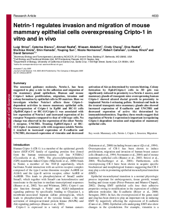
expression of Neogenin when compared to wildFig. 1. Expression of Netrin-1 and its receptors, Neogenin and UNC5H1 in mouse
type (WT) cells (Fig. 1A). No significant difference
mammary epithelial cells overexpressing Cripto-1. (A) Western blot analysis of
expression of Netrin-1, Neogenin and UNC5H1 in the wild type (WT) and Criptoin the expression of UNC5H1 between the wild
1 (Cr-1)-overexpressing EpH4 and HC-11 cells. Detection of actin in bottom
type and Cr-1-overexpressing HC-11 or EpH4 cells
panel acts as a loading control. (B) Photomicrographs (40⫻ magnification) of
was detected by western blot analysis (Fig. 1A).
immunofluorescence show expression of Netrin-1 and Neogenin in HC-11 cells.
The decreased expression of Netrin-1 and the
Nuclei are stained blue by DAPI. (C) Representative photomicrographs (20⫻
increased expression of Neogenin in Cr-1magnification) of histological sections from MMTV-CR-1 transgenic mice
overexpressing mouse mammary epithelial cells
mammary tumors containing anaplastic mesenchyme-like tumor cells and stained
compared to the wild type were confirmed by
by immunohistochemistry show low expression of Netrin-1 relative to Neogenin.
immunofluorescence analysis of HC-11/WT and
Sections were counterstained with hematoxylin.
HC-11/Cr-1 cells (Fig. 1B). Immunohistochemistry
showed decreased staining intensity for Netrin-1
compared to the intensity of staining obtained for Neogenin in
capable of reducing Cr-1 mRNA and protein expression in
areas of the MMTV-CR-1 transgenic mice mammary tumors
these cells (Fig. 2A), caused Netrin-1 and Neogenin expression
containing large anaplastic, mesenchyme-like tumor cells (Fig.
to return to levels that were comparable to those detected in
1C). Similar to the mammary epithelial cells overexpressing Crwild-type HC-11 cells by western blotting (Fig. 2B). EpH4/Cr1, the anaplastic, mesenchyme-like tumor cells in the MMTV1 cells do not express endogenous Nodal (Bianco et al., 2002).
CR-1 mammary tumors also expressed higher levels of CR-1
When Nodal-independent signaling in EpH4/Cr-1 cells was
and of biochemical markers suggestive of EMT when compared
blocked with the synthetic c-Src inhibitor PP2 (Bianco et al.,
to the more differentiated areas of the tumor (Strizzi et al.,
2003), there was an increase in Netrin-1 expression and a
2004).
decrease in Neogenin expression compared to that in untreated
cells (Fig. 2C). Treatment of EpH4/Cr-1 cells with the PI-3K
inhibitor LY was also associated with an increase in Netrin-1
Reducing Cripto-1 expression or inhibiting Cripto-1
expression and a decrease in Neogenin expression (Fig. 2C,D).
signaling rescues Netrin-1 expression in mammary
No significant effect on the expression of Netrin-1 or Neogenin
epithelial cells overexpressing Cripto-1
was detected when EpH4/Cr-1 cells were treated with the
Transfection of HC-11/Cr-1 cells with an anti-Cr-1 siRNA,
MAPK inhibitor, PD (Fig. 2C,D). These results suggest that
�JournalofCellScience
Netrin-1 regulates Cripto-1-dependent invasion
Fig. 2. Reduction of Cripto-1 expression or inhibition of Cripto-1
signaling rescues Netrin-1 expression in mammary epithelial cells
overexpressing Cripto-1. (A) Results from reverse transcriptase-PCR
and Western blot experiments show reduction of Cr-1 expression in
HC-11/Cr-1 cells transfected with a specific anti-Cr-1 siRNA.
(B) Western blot analysis of lysates from Cripto-1 (Cr-1)overexpressing HC-11 cells treated with anti-Cr-1 siRNA (Cr1+siRNA) shows increase in expression of Netrin-1 and decrease in
expression of Neogenin to levels comparable to those detected in
wild-type (WT) HC-11 cells. Cr-1/cntrl, control HC-11/Cr-1 cells not
treated with anti-Cr-1 siRNA. (C) Representative western blot
analysis shows an increase in expression of Netrin-1 and a decrease
in expression in Neogenin in HC-11/Cr-1 cells treated with the c-Src
inhibitor (PP2) or with the PI-3K/Akt inhibitor (LY) compared to
non-treated control HC-11/Cr-1 cells. (D) Histograms summarize
results from densitometric analyses of western blots performed in the
experiments described in C. Error bars indicate s.d. Treatment of
HC-11/Cr-1 cells with a MAPK inhibitor (PD) had no significant
effect on expression of Netrin-1 or Neogenin. (*P<0.05 compared to
levels in the control group).
blocking Cripto-1 signaling through a Nodal-independent
pathway, which is dependent on c-Src and PI-3K, rescues
Cripto-1-dependent loss of Netrin-1 expression.
Exogenous Netrin-1 reverses the characteristics of
epithelial-to-mesenchymal transition in mammary
epithelial cells overexpressing Cripto-1
Cripto-1 overexpression in mouse mammary cells is
associated with reduced expression of the intracellular
adhesion molecule E-cadherin and increased expression of
vimentin, which are characteristic of cells undergoing EMT
(Strizzi et al., 2004). Akt activation has also been shown to
occur during Cr-1-dependent migration of mouse mammary
4637
epithelial cells and in human breast cancer cells
overexpressing Cr-1 (Bianco et al., 2003; Normanno et al.,
2004). EpH4/Cr-1 cells grown in medium containing
exogenous rmNetrin-1 (50 ng/ml) showed an increase in the
expression of E-cadherin and a decrease in the expression of
vimentin (Fig. 3A). In addition, reduced phosphorylation of
Akt was observed in Netrin-1-treated EpH4/Cr-1 cells
compared to levels in EpH4/Cr-1 cells grown in culture
medium without exogenous rmNetrin-1 (Fig. 3A).
Mammary epithelial cells overexpressing Cripto-1 have
been shown to possess an increased capacity for invasion and
migration (Bianco et al., 2003; Normanno et al., 2004; Strizzi
et al., 2004; Wechselberger et al., 2001). In fact, when medium
containing 2% FBS was added to the bottom of the wells in
the invasion assay, EpH4/Cr-1 or HC-11/Cr-1 cells showed a
significant increase in the number of cells that invaded across
the matrix-covered membrane (Fig. 3B). However, when 25 or
50 ng/ml soluble rmNetrin-1 was added to the 2% FBScontaining medium in the bottom of the wells, the number of
invading EpH4/Cr-1 or HC-11/Cr-1 cells was significantly
decreased (Fig. 3B). Furthermore, EpH4/Cr-1 or HC-11/Cr-1
cells that were treated with 50 ng/ml exogenous rmNetrin-1
showed a decrease in Neogenin expression and an increase in
UNC5H1 expression as determined by western blotting (Fig.
3C). This suggested that Netrin-1 might bind to UNC5H1 and
may therefore be involved in producing the anti-invasive effect
that was observed in the Cr-1-overexpressing cells. To address
this possibility, we used a neutralizing antibody generated
against the extracellular domain of UNC5C with the
supposition that this antibody would also block UNC5H1
function, as the ligand binding extracellular domain of UNC5
proteins are highly conserved (Engelkamp, 2002). Indeed,
pretreatment of EpH4/Cr-1 cells with a neutralizing antibody
against UNC5C restored the invasive properties of EpH4/Cr-1
cells in the presence of rmNetrin-1 (Fig. 3D). In contrast,
pretreatment of EpH4/Cr-1 cells with a neutralizing antibody
against Neogenin did not restore the invasiveness of the cells,
but further decreased the number of invading EpH4/Cr-1 cells
across the matrix coated membranes in the presence of
rmNetrin-1 (Fig. 3D). Thus treating Cr-1-overexpressing
mouse mammary epithelial cells, which have downregulated
Netrin-1 expression, with exogenous soluble rmNetrin-1
reverses the biochemical characteristics of Cr-1-dependent
EMT, suppresses activation of Akt and reduces cell
invasiveness. In accord with reduced cell motility, there is an
upregulation of the UNC5 class of Netrin-1 receptors that act
to inhibit or to redirect the attractive responses to Netrin-1.
Mouse mammary epithelial cells overexpressing Cripto1 form fewer colonies in 3D extracellular matrix in
proximity to a Netrin-1 source
The increased ability of Eph4/Cr-1 cells compared to the wild
type, to form colonies in 3D extracellular matrix has been
previously determined (Wechselberger et al., 2001). To
determine whether exogenous Netrin-1 was capable of
affecting colony formation by EpH4/Cr-1 cells, these cells
were seeded in 3D Matrigel containing disks of filter paper that
had been preabsorbed with different concentrations of Netrin1. Spherical colonies having diameters ⭓500 m were scored
after 48 hours in equally divided areas situated proximal,
�4638
Journal of Cell Science 118 (20)
JournalofCellScience
medial or distal to the disks of blot paper preabsorbed with
either PBS or rmNetrin-1. EpH4/Cr-1 colonies were more
homogenously distributed throughout the Matrigel containing
the PBS source whereas there were significantly fewer colonies
formed in the areas proximal to the Netrin-1 source (Fig. 4A).
In some of the 3D cultures, EpH4/Cr-1 cells actually formed a
noticeable ring of growth inhibition around the source of
Netrin-1 (Fig. 4B). This effect was more evident with blot
paper preabsorbed with 100 or 200 ng/ml of rmNetrin-1. Blot
paper pre-absorbed with 400 ng/ml rmNetrin-1 did not inhibit
colony formation of EpH4/Cr-1 cells (data not shown).
Quantification of the colonies formed in Matrigel with
diameters greater than 500 m revealed a significant reduction
in the number of colonies formed by Eph4/Cr-1 in proximity
to the blot paper preabsorbed with rmNetrin-1 compared to blot
papers preabsorbed with PBS (Fig. 4C).
Effect of exogenous Netrin-1 on mammary ductal
morphogenesis in vivo
When control pellets containing cholesterol were introduced
into the virgin mammary gland of 5-week-old FVB/N or
MMTV/CR-1 mice, normal ductal elongation was observed in
whole mounts of mammary gland from these mice after 2
weeks (Fig. 4D). Ductal elongation in FVB/N mammary
glands containing pellets releasing 25 ng/day was similar to
that observed in control FVB/N mammary glands (Fig. 4D).
However, there was a significant reduction in ductal elongation
in the mammary glands of MMTV-CR-1 mice
containing pellets releasing 25 ng/day of Netrin1 (Fig. 4D,E). The inhibitory effect of Netrin-1
on mammary gland ductal elongation was not
observed in mice containing pellets releasing
greater amounts (50 ng/day) of rmNetrin-1 (data
not shown).
Immunohistochemical analysis of the terminal
end buds in the mammary glands treated with the
Netrin-1 pellets showed an overall increase in
expression of UNC5H1 throughout the terminal
end bud structures compared to MMTV-CR-1
transgenic mammary glands treated with control
pellets where UNC5H1 appeared to stain only
the peripheral area of these structures (Fig. 5).
Increased staining for E-cadherin was also
observed in the terminal end buds of MMTVCR-1 mammary glands containing the Netrin-1
pellet compared to that in the control (Fig. 5).
Finally, positive staining for P-Akt was more
intense in the epithelial and stromal cells in
mammary glands from MMTV-CR-1 mice
containing control pellets compared to
mammary glands from MMTV-CR-1 transgenic
mice implanted with the Netrin-1-releasing
pellets. Moreover, the average intensity of
staining for P-Akt in the terminal end buds as
evaluated by digital image analysis was
significantly reduced by almost one-third (32%)
Fig. 3. Exogenous Netrin-1 reverses Cripto-1-dependent epithelial-to-mesenchymal
(Range 24-42% reduction; n=5; P=0.008) in
transition. (A) Western blot analysis of lysates from EpH4/Cr-1 cells cultured in
MMTV-CR-1 mammary glands containing
growth medium containing 50 ng/ml of exogenous rmNetrin-1 (+NTN) showing
Netrin-1 pellets compared to terminal end buds
increased expression of E-cadherin (E-cad), reduced expression of vimentin (Vim)
in MMTV-CR-1 glands containing control
and reduced expression of phosphorylated-Akt (P-Akt) compared to EpH4/Cr-1
pellets (Fig. 5). Vimentin stained poorly in
cells cultured in growth medium alone (control). (B) Histogram summarizes results
MMTV-CR-1 mammary glands containing
from invasion assays showing increased invasion of EpH4/Cr-1 and HC-11/Cr-1
either control or Netrin-1 pellets and showed no
cells as compared to wild-type cells. Addition of rmNetrin-1 (25 ng/ml and
50 ng/ml) significantly reduced invasion of both Cr-1 overexpressing cell lines.
difference in the levels of expression between the
Values are the mean±s.d. of three separate experiments (*P<0.05 compared to levels
two different mammary glands (data not shown).
in the relevant control group). (C) Western blot analysis of lysates from Cr-1
overexpressing cells shows that Neogenin expression decreases and UNC5H1
expression increases when these cells are treated with exogenous soluble rmNetrin1 (+Netrin-1) as compared to untreated control cells. (D) Histogram summarizes
results from invasion assays showing that the significant inhibition of invasion of
EpH4/Cr-1 cells in the presence of 25 ng/ml of exogenous rmNetrin-1 is restored to
control level when EpH4/Cr-1 cells are pretreated with neutralizing antibody against
human UNC5C (␣-UNC5) but not when pretreated with neutralizing antibody
against Neogenin (␣-NEO). Values are the mean±s.d. of three separate experiments
(*P<0.05 compared to levels in the control group).
Discussion
The present study demonstrates that mouse
mammary epithelial cells overexpressing Cr-1 in
vitro have reduced expression of Netrin-1 and
increased expression of Neogenin. Analysis of
immunostained histological sections of
mammary tumors from MMTV-CR-1 transgenic
�Netrin-1 regulates Cripto-1-dependent invasion
4639
JournalofCellScience
Fig. 4. Formation of colonies in 3D extracellular matrix by mouse
mammary epithelial cells overexpressing Cripto-1 and ductal
elongation in mammary glands from transgenic mice overexpressing
Cripto-1 are both reduced in proximity to a Netrin-1 source.
(A) Colony formation of EpH4/Cr-1 cells was assessed in Matrigel
containing disks that were preabsorbed with PBS or rmNetrin-1
(200 ng/ml). Colonies were quantified in areas situated proximal (P),
medial (M) or distal (D) from the sources as illustrated on the
microphotographs (5⫻ magnification). (B) Detail shows the area of
growth inhibition (arrows) proximal to the disk preabsorbed with
rmNetrin-1 (100 ng/ml) surrounded by a ring of EpH4/Cr-1 cells.
(C) Summary of the results obtained from quantification of the
colonies formed by EpH4/Cr-1. The mean number of colonies of at
least 500 m in diameter (=500 m) was significantly reduced only
in the areas in proximity to the rmNetrin-1 source (*P<0.05
compared to levels in the control group). White bars represent the
number of EpH4/Cr-1 colonies in Matrigel containing PBS source
and black bars, the number of colonies formed in Matrigel containing
rmNetrin-1. Values are the mean ± s.d. of four separate experiments.
(D) Whole mount morphology (10⫻ magnification) of cholesterol
pellets (*) releasing 25 ng/day of rmNetrin-1 shows a significantly
reduced ductal elongation in the mammary gland of MMTV-CR-1
transgenic mice compared to cholesterol-only control pellets. The
Netrin-1-releasing pellets did not affect ductal elongation in
mammary glands of FVB/N mice. L, lymph node (E) Histogram
summarizing the results of ductal elongation in mammary glands
containing Netrin-1-releasing pellets. Distance was measured from
the center of the mammary gland lymph node to the tip of the
farthest growing duct and values are the mean with error bars
indicating the s.d. from experiments in FVB/N (n=4) and MMTVCR-1 (n=5) mice. A significant difference in elongation distance was
observed between the two groups (P=0.008).
mice revealed that in the areas where mammary tumors are
composed of large anaplastic, mesenchyme-like tumor cells,
immunostaining for Netrin-1 was less intense compared to that
for Neogenin. These results suggest that overexpression of
Cripto-1 is capable of modulating the expression of Netrin-1
and Neogenin.
Cripto-1 is expressed in a greater number of infiltrating
ductal carcinomas or intralobular carcinomas than in ductal
carcinomas in situ (Panico et al., 1996). In addition, expression
of CR-1 is higher in colon tumors than in the adjacent
noninvolved colon epithelium and correlates with tumor stage,
increased regional lymph node metastases and a higher rate of
colorectal cancer recurrence (Gagliardi, 1994; Kuniyasu et al.,
1991). In gastric carcinoma, the incidence of CR-1-positive
cases was more frequent in late stage, locally invasive tumors
than in early stage, noninvasive cancers (Kuniyasu, 1994).
From these studies, the association between CR-1 expression
and characteristics of tumors undergoing EMT such as local
tissue invasion, lymph node metastasis and cancer recurrence
(Gotzmann et al., 2004; Thiery, 2003b; Thiery and Chopin,
1999; Vincent-Salomon and Thiery, 2003), suggest that CR-1
may be capable of promoting a more aggressive phenotype in
human tumor cells by inducing EMT.
The overexpression of CR-1 in mammary glands of aged
multiparous mice leads to the formation of mammary papillary
adenocarcinomas (Wechselberger et al., 2005). These tumors
show evidence of EMT such as reduced expression of Ecadherin and increased expression of vimentin and snail,
especially in areas containing anaplastic, mesenchyme-like
tumor cells (Strizzi et al., 2004). In the present study, Netrin1 expression was reduced relative to Neogenin in these same
regions. Moreover, immunostaining for CR-1 was increased in
areas of the tumors exhibiting characteristics of EMT
compared to the areas of the CR-1 transgenic mouse mammary
tumors that possess a more differentiated papillary phenotype
(Strizzi et al., 2004). In this regard, the reduction of Netrin-1
expression observed in mouse mammary epithelial cells
overexpressing Cr-1 is similar to the reduced expression of
Netrin-1 observed in the mesenchyme-like tumor cells in the
mammary tumors from the CR-1 transgenic mice. Therefore,
the loss of an epithelial phenotype, which is induced by Cripto1 overexpression in mammary epithelial cells, is associated
with a reduction in Netrin-1 expression as mammary epithelial
cells exhibit a more motile and mesenchyme-like phenotype.
Targeted inhibition of Cr-1 expression with a specific siRNA
resulted in the reversion of Netrin-1 and Neogenin expression
to levels that were detected in wild-type mammary epithelial
cells, suggesting that Cr-1 may play a role in regulating Netrin-
�4640
Journal of Cell Science 118 (20)
JournalofCellScience
1 and Neogenin expression. As EpH4 cells do not express
Nodal (Bianco et al., 2002), and as treatment of EpH4/Cr-1
cells with PP2 or LY was followed by an increase in Netrin-1
expression and a decrease in Neogenin expression, this
indicates that Cripto-1 may be affecting Netrin-1 and Neogenin
expression by signaling through a pathway that involves
Nodal-independent activation of c-Src and/or PI-3K/Akt in
EpH4/Cr-1 cells. The addition of exogenous rmNetrin-1 to the
culture medium of mouse mammary epithelial cells
overexpressing Cr-1 resulted in a reduction in the expression
of Neogenin. This suggests that exogenous Netrin-1 is capable
of either reverting effects of Cr-1 on Neogenin expression or
Fig. 5. Immunostaining for human Cripto-1 in mammary glands of
MMTV/CR-1 transgenic mice. Increased expression of E-cadherin
and UNC5H1 is observed in MMTV/CR-1 mammary glands
containing Netrin-1-releasing pellets relative to expression in the
control. Peripheral staining for UNC5H1 in the terminal end buds of
MMTV/CR-1 control mammary gland was detected compared to
staining of the entire terminal end bud in MMTV/CR-1 mammary
glands containing the Netrin-1 pellets. Reduced intensity of
immunostaining of P-Akt in the epithelial structures, and almost no
staining of the stromal component was detected in MMTV/CR-1
mammary glands containing Netrin-1 pellets compared to the strong
staining for P-Akt detected in both epithelial and stromal
components of MMTV/CR-1 mammary glands implanted with
control pellets. Magnification, 40⫻.
that there may be a potential negative feedback mechanism by
which Netrin-1 regulates the expression of Neogenin. The
latter possibility has been described in other systems whereby
cells compensate for high concentrations of ligand expression
by adjusting the expression of cell surface receptors (Frank et
al., 1996).
Local release of Netrin-1 from pellets implanted in vivo in
the mammary glands of virgin MMTV-CR-1 transgenic mice
but not in control mammary glands of virgin FVB/N mice
resulted in an inhibition in the elongation of the developing
mammary ducts through the fat pad. This is interesting, as
CR-1 overexpression in the mammary gland of virgin
MMTV-CR-1 transgenic mice is associated with enhanced
ductal side branching and elongation (Wechselberger et al.,
2005). The family of UNC5 receptors can mediate, alone or
as a coreceptor with Neogenin, the repulsive effects of Netrin1 (Dickson and Keleman, 2002; Hinck, 2004; Hong et al.,
1999). Netrin-1 in the presence of an UNC5 receptor is
capable of reducing migration and filipodial extension of
endothelial cells in vitro and in vivo (Lu et al., 2004).
Likewise, Netrin-1 can cause epithelial cells of the
developing lung to migrate away from a Netrin-1 source (Liu
et al., 2004). Our in vitro data suggest that UNC5 may be
involved in mediating the negative effects of Netrin-1 on
ductal elongation. Although overexpression of Cripto-1 in the
mouse mammary epithelial cells did not affect the expression
of UNC5H1, when Cr-1 overexpressing cells are cultured in
the presence of exogenous rmNetrin-1, these cells are found
to overexpress UNC5H1 receptors. Immunohistochemical
analysis of the mammary terminal end buds of MMTV/CR-1
transgenic mice treated with Netrin-1 pellets showed
increased expression of UNC5H1 throughout the terminal
end bud compared to mammary terminal end buds in
MMTV/CR-1 transgenic mice treated with control pellets,
which showed reduced UNC5H1 staining limited to the
periphery of the terminal end bud. Thus the UNC5 receptors
may also be involved in mediating the reduction in the
number of colonies formed in Matrigel and in impairing the
migration and invasion of EpH4/Cr-1 cells across matrixcoated membranes in response to exogenous rmNetrin-1.
Interestingly, the inhibitory effects of Netrin-1 on colony
formation by Eph4/Cr-1 cells and on ductal elongation in the
mammary glands of MMTV-CR-1 transgenic mice were not
observed at higher doses (data not shown). A similar doseresponse effect for Netrin-1 activity on axon outgrowth was
also observed and has been attributed to the requirement for
Netrin-1 to induce receptor clustering that is impaired at
higher concentrations of Netrin-1 (Serafini et al., 1994).
The anti-invasive effects of Netrin-1 on EpH4/Cr-1 cells was
significantly attenuated when these cells were preincubated
with blocking antibodies against UNC5C, which is highly
homologous in the extracellular domain to mouse UNC5H1
(Engelkamp, 2002). However, preincubation of EpH4/Cr-1
cells with an anti-Neogenin blocking antibody did not have the
same effect but actually further inhibited the invasion of
Eph4/Cr-1 cells across the matrix-coated membranes. This
effect may possibly be due to a decrease in attractive cues that
the Neogenin receptors may have been capable of inducing in
EpH4/Cr-1 cells in response to Netrin-1. This result also
suggests that it is unlikely that Neogenin is acting alone
(Rajagopalan et al., 2004) or as a coreceptor with UNC5 (Hong
�JournalofCellScience
Netrin-1 regulates Cripto-1-dependent invasion
et al., 1999) in mediating repulsion in EpH4/Cr-1 cells.
However, it cannot be excluded that, alternatively or in
combination with the UNC5 receptor, Netrin-1 may have
affected Cr-1-dependent invasion and colony formation of
mammary epithelial cells and ductal elongation in mammary
glands by blocking Cripto-1 signaling. In fact, Akt activity was
reduced in EpH4/Cr-1 cells when cultured in the presence of
exogenous Netrin-1. The reduction in P-Akt expression was
also detected by immunohistochemistry in epithelial cells of
the terminal end buds and surrounding stromal cells of
MMTV/CR-1 mammary glands containing Netrin-1 pellets
compared to MMTV/CR-1 mammary glands containing
control pellets.
Mechanisms that regulate cell adhesion and migration play
a fundamental role not only during normal tissue development
and differentiation but also in the survival and spread of tumor
cells (Cavallaro and Christofori, 2004; Lee and Juliano, 2004;
Thiery, 2003a; Van Roy and Mareel, 1992). In this respect,
factors that may affect Netrin-1-dependent adhesion in
mammary epithelial cells should provide some insight into the
mechanisms involved in the spread of potential tumor cells.
The association between overexpression of Cripto-1 and
induction of biochemical changes important for EMT, such as
reduction of E-cadherin levels and increased expression of
vimentin both in vitro and in vivo, has been previously
described (Ebert et al., 2000; Strizzi et al., 2004). Furthermore,
Cripto-1 induces morphologic changes and activation of
signaling molecules known to enhance cell migration and
invasion (Bianco et al., 2003; Normanno et al., 2004;
Wechselberger et al., 2001). These findings support a potential
role for Cripto-1 during tumorigenesis.
From our data it appears that a possible mechanism by which
Cripto-1 may induce a more aggressive phenotype in
mammary epithelial cells is by reducing Netrin-1 expression
and affecting the profile of Netrin-1 receptor expression.
Exogenous Netrin-1 was capable of increasing E-cadherin and
decreasing vimentin expression in EpH4/Cr-1 thus reversing
Cr-1-dependent biochemical changes in vitro, which are
important for EMT. Immunohistochemical analysis of the
mammary terminal end buds from MMTV/CR-1 transgenic
mice treated with Netrin-1 pellets also showed an increase in
E-cadherin expression compared to mammary terminal end
buds from MMTV/CR-1 mammary glands treated with control
pellets. However, immunohistochemical analysis showed poor
expression for vimentin in the mammary glands analyzed from
both the Netrin-1-treated and control MMTV/CR-1 transgenic
mice. This latter observation may be due to the fact that
hyperplastic lesions and mammary tumors in which EMT
markers, including vimentin, are overexpressed were identified
in aged, multiparous mice (Strizzi et al., 2004). In the present
study, the Netrin-1 pellets were implanted in virgin 5-week-old
MMTV/CR-1 mice and mammary glands were harvested
shortly after, suggesting that prolonged exposure of mammary
epithelial cells to CR-1 effect is probably needed in order to
induce changes in vimentin expression. The increased invasion
and migration in epithelial cells is also a major characteristic
of Cripto-1-induced EMT. Netrin-1 was capable of reducing
Cr-1-dependent invasion and migration of mammary epithelial
cells both in vitro and in vivo. As increased expression of
Cripto-1 has been detected in a number of human cancers,
including breast cancer, it will be informative to investigate the
4641
exact relationship between Netrin-1 and Cripto-1 expression in
these tumors.
The Authors thank Brenda Wallace-Jones for her excellent
technical assistance. L.H. was supported by funds from the American
Cancer Society Research Scholar Grant #RSG0218001MGO. N.N.
was supported by funds from the Italian Association for Cancer
Research (AIRC).
References
Arakawa, H. (2004). Netrin-1 and its receptors in tumorigenesis. Nat. Rev.
Cancer 4, 978-987.
Berx, G., Becker, K. F., Hofler, H. and van Roy, F. (1998). Mutations of the
human E-cadherin (CDH1) gene. Hum. Mutat. 12, 226-237.
Bianco, C., Adkins, H. B., Wechselberger, C., Seno, M., Normanno, N., De
Luca, A., Sun, Y., Khan, N., Kenney, N., Ebert, A. et al. (2002). Cripto1 activates nodal- and ALK4-dependent and -independent signaling
pathways in mammary epithelial cells. Mol. Cell. Biol. 22, 2586-2597.
Bianco, C., Strizzi, L., Rehman, A., Normanno, N., Wechselberger, C.,
Sun, Y., Khan, N., Hirota, M., Adkins, H., Williams, K. et al. (2003). A
Nodal- and ALK4-independent signaling pathway activated by Cripto-1
through Glypican-1 and c-Src. Cancer Res. 63, 1192-1197.
Birchmeier, C., Birchmeier, W. and Brand-Saberi, B. (1996a). Epithelialmesenchymal transitions in cancer progression. Acta Anat (Basel) 156, 217226.
Birchmeier, W., Behrens, J., Weidner, K. M., Hulsken, J. and Birchmeier,
C. (1996b). Epithelial differentiation and the control of metastasis in
carcinomas. Curr. Top. Microbiol. Immunol. 213, 117-135.
Boyer, B., Valles, A. M. and Edme, N. (2000). Induction and regulation
of epithelial-mesenchymal transitions. Biochem. Pharmacol. 60, 10911099.
Brandt, R., Normanno, N., Gullick, W. J., Lin, J. H., Harkins, R.,
Schneider, D., Jones, B. W., Ciardiello, F., Persico, M. G., Armenante,
F. et al. (1994). Identification and biological characterization of an
epidermal growth factor-related protein: cripto-1. J. Biol. Chem. 269, 1732017328.
Butler, S. J. and Dodd, J. (2003). A role for BMP heterodimers in roof platemediated repulsion of commissural axons. Neuron 38, 389-401.
Cano, A., Perez-Moreno, M. A., Rodrigo, I., Locascio, A., Blanco, M. J.,
del Barrio, M. G., Portillo, F. and Nieto, M. A. (2000). The transcription
factor snail controls epithelial-mesenchymal transitions by repressing Ecadherin expression. Nat. Cell. Biol. 2, 76-83.
Cavallaro, U. and Christofori, G. (2004). Multitasking in tumor progression:
signaling functions of cell adhesion molecules. Ann. New York Acad. Sci.
1014, 58-66.
Charron, F., Stein, E., Jeong, J., McMahon, A. P. and Tessier-Lavigne, M.
(2003). The morphogen sonic hedgehog is an axonal chemoattractant that
collaborates with netrin-1 in midline axon guidance. Cell 113, 11-23.
Chisholm, A. and Tessier-Lavigne, M. (1999). Conservation and divergence
of axon guidance mechanisms. Curr. Opin. Neurobiol. 9, 603-615.
Ciccodicola, A., Dono, R., Obici, S., Simeone, A., Zollo, M. and Persico,
M. G. (1989). Molecular characterization of a gene of the ‘EGF family’
expressed in undifferentiated human NTERA2 teratocarcinoma cells.
EMBO J. 8, 1987-1991.
De Santis, M. L., Kannan, S., Smith, G. H., Seno, M., Bianco, C., Kim, N.,
Martinez-Lacaci, I., Wallace-Jones, B. and Salomon, D. S. (1997).
Cripto-1 inhibits beta-casein expression in mammary epithelial cells through
a p21ras-and phosphatidylinositol 3⬘-kinase-dependent pathway. Cell
Growth Differ. 8, 1257-1266.
Dickson, B. J. and Keleman, K. (2002). Netrins. Curr. Biol. 12, R154-R155.
Ebert, A. D., Wechselberger, C., Frank, S., Wallace-Jones, B., Seno, M.,
Martinez-Lacaci, I., Bianco, C., De Santis, M., Weitzel, H. K. and
Salomon, D. S. (1999). Cripto-1 induces phosphatidylinositol 3⬘-kinasedependent phosphorylation of AKT and glycogen synthase kinase 3beta in
human cervical carcinoma cells. Cancer Res. 59, 4502-4505.
Ebert, A. D., Wechselberger, C., Nees, M., Clair, T., Schaller, G., MartinezLacaci, I., Wallace-Jones, B., Bianco, C., Weitzel, H. K. and Salomon,
D. S. (2000). Cripto-1-induced increase in vimentin expression is associated
with enhanced migration of human Caski cervical carcinoma cells. Exp. Cell
Res. 257, 223-229.
Engelkamp, D. (2002). Cloning of three mouse Unc5 genes and their
expression patterns at mid-gestation. Mech. Dev. 118, 191-197.
�JournalofCellScience
4642
Journal of Cell Science 118 (20)
Frank, R., Adelmann-Grill, B. C., Herrmann, K., Haustein, U. F., Petri, J.
B. and Heckmann, M. (1996). Transforming growth factor-beta controls
cell-matrix interaction of microvascular dermal endothelial cells by
downregulation of integrin expression. J. Invest. Dermatol. 106, 36-41.
Fuchs, E. and Weber, K. (1994). Intermediate filaments: structure, dynamics,
function, and disease. Annu. Rev. Biochem. 63, 345-382.
Fuchs, I. B., Lichtenegger, W., Buehler, H., Henrich, W., Stein, H., KleineTebbe, A. and Schaller, G. (2002). The prognostic significance of epithelialmesenchymal transition in breast cancer. Anticancer Res. 22, 3415-3419.
Gagliardi, G., Talbot, I. C., Northover, J. M. A., Warre, A., Stamp, G. W.
H., Lalani, E.-N., Gullick, W. J. and Pignatelli, M. (1994).
Immunolocalisation of cripto andamphiregulin in rectal cancer: correlation
with prognosis. Int. J. Oncol. 4, 865-871.
Gilchrist, A. J., Meuser, R., Turchinsky, J., Shaw, A. R., Pasdar, M. and
Dixon, W. T. (2002). Cell adhesion-mediated transformation of a human
SCLC cell line is associated with the development of a normal phenotype.
Exp. Cell Res. 276, 63-78.
Gilles, C., Polette, M., Piette, J., Delvigne, A. C., Thompson, E. W.,
Foidart, J. M. and Birembaut, P. (1996). Vimentin expression in cervical
carcinomas: association with invasive and migratory potential. J. Pathol.
180, 175-180.
Gotzmann, J., Mikula, M., Eger, A., Schulte-Hermann, R., Foisner, R.,
Beug, H. and Mikulits, W. (2004). Molecular aspects of epithelial cell
plasticity: implications for local tumor invasion and metastasis. Mutat. Res.
566, 9-20.
Hajra, K. M., Chen, D. Y. and Fearon, E. R. (2002). The SLUG zincfinger protein represses E-cadherin in breast cancer. Cancer Res. 62, 16131618.
Hanna, W., Alowami, S. and Malik, A. (2003). The role of HER-2/neu
oncogene and vimentin filaments in the production of the Paget’s phenotype.
Breast J. 9, 485-490.
Hinck, L. (2004). The versatile roles of “axon guidance” cues in tissue
morphogenesis. Dev. Cell 7, 783-793.
Hong, K., Hinck, L., Nishiyama, M., Poo, M. M., Tessier-Lavigne, M. and
Stein, E. (1999). A ligand-gated association between cytoplasmic domains
of UNC5 and DCC family receptors converts netrin-induced growth cone
attraction to repulsion. Cell 97, 927-941.
Jiang, Y., Liu, M. T. and Gershon, M. D. (2003). Netrins and DCC in the
guidance of migrating neural crest-derived cells in the developing bowel and
pancreas. Dev. Biol. 258, 364-384.
Keino-Masu, K., Masu, M., Hinck, L., Leonardo, E. D., Chan, S. S.,
Culotti, J. G. and Tessier-Lavigne, M. (1996). Deleted in Colorectal
Cancer (DCC) encodes a netrin receptor. Cell 87, 175-185.
Kennedy, T. E., Serafini, T., de la Torre, J. R. and Tessier-Lavigne, M.
(1994). Netrins are diffusible chemotropic factors for commissural axons in
the embryonic spinal cord. Cell 78, 425-435.
Kenney, N. J., Huang, R. P., Johnson, G. R., Wu, J. X., Okamura, D.,
Matheny, W., Kordon, E., Gullick, W. J., Plowman, G., Smith, G. H. et
al. (1995). Detection and location of amphiregulin and Cripto-1 expression
in the developing postnatal mouse mammary gland. Mol. Reprod. Dev. 41,
277-286.
Koch, M., Murrell, J. R., Hunter, D. D., Olson, P. F., Jin, W., Keene, D. R.,
Brunken, W. J. and Burgeson, R. E. (2000). A novel member of the netrin
family, beta-netrin, shares homology with the beta chain of laminin:
identification, expression, and functional characterization. J. Cell Biol. 151,
221-234.
Kolodkin, A. L., Matthes, D. J. and Goodman, C. S. (1993). The semaphorin
genes encode a family of transmembrane and secreted growth cone guidance
molecules. Cell 75, 1389-1399.
Kuniyasu, H., Yoshida, K., Yokozaki, H., Yasui, W., Ito, H., Toge, T.,
Ciardiello, F., Persico, M. G., Saeki, T., Salomon, D. S. et al. (1991).
Expression of cripto, a novel gene of the epidermal growth factor family, in
human gastrointestinal carcinomas. Jpn. J. Cancer Res. 82, 969-973.
Kuniyasu, H., Yasui, W., Akama, Y., Akagi, M., Tohdo, H., Ji, Z. Q.,
Kitadai, Y., Yokozaki, H. and Tahara, E. (1994). Expression of cripto in
human gastric carcinomas: an association with tumor stage and prognosis.
J. Exp. Clin. Canc. Res. 13, 151-157.
Lane, E. B., Hogan, B. L., Kurkinen, M. and Garrels, J. I. (1983). Coexpression of vimentin and cytokeratins in parietal endoderm cells of early
mouse embryo. Nature 303, 701-704.
Latil, A., Chene, L., Cochant-Priollet, B., Mangin, P., Fournier, G.,
Berthon, P. and Cussenot, O. (2003). Quantification of expression of
netrins, slits and their receptors in human prostate tumors. Int. J. Cancer
103, 306-315.
Lee, J. W. and Juliano, R. (2004). Mitogenic signal transduction by
integrin- and growth factor receptor-mediated pathways. Mol. Cell. 17,
188-202.
Leonardo, E. D., Hinck, L., Masu, M., Keino-Masu, K., Ackerman, S. L.
and Tessier-Lavigne, M. (1997). Vertebrate homologues of C. elegans
UNC-5 are candidate netrin receptors. Nature 386, 833-838.
Liu, Y., Stein, E., Oliver, T., Li, Y., Brunken, W. J., Koch, M., TessierLavigne, M. and Hogan, B. L. (2004). Novel role for Netrins in regulating
epithelial behavior during lung branching morphogenesis. Curr. Biol. 14,
897-905.
Lu, X., Le Noble, F., Yuan, L., Jiang, Q., De Lafarge, B., Sugiyama, D.,
Breant, C., Claes, F., De Smet, F., Thomas, J. L. et al. (2004). The netrin
receptor UNC5B mediates guidance events controlling morphogenesis of
the vascular system. Nature 432, 179-186.
Meyerhardt, J. A., Caca, K., Eckstrand, B. C., Hu, G., Lengauer, C.,
Banavali, S., Look, A. T. and Fearon, E. R. (1999). Netrin-1: interaction
with deleted in colorectal cancer (DCC) and alterations in brain tumors and
neuroblastomas. Cell Growth Differ. 10, 35-42.
Minchiotti, G., Parisi, S., Liguori, G., Signore, M., Lania, G., Adamson,
E. D., Lago, C. T. and Persico, M. G. (2000). Membrane-anchorage of
Cripto protein by glycosylphosphatidylinositol and its distribution during
early mouse development. Mech. Dev. 90, 133-142.
Normanno, N., De Luca, A., Bianco, C., Maiello, M. R., Carriero, M. V.,
Rehman, A., Wechselberger, C., Arra, C., Strizzi, L., Sanicola, M. et al.
(2004). Cripto-1 overexpression leads to enhanced invasiveness and
resistance to anoikis in human MCF-7 breast cancer cells. J. Cell Physiol.
198, 31-39.
Panico, L., D’Antonio, A., Salvatore, G., Mezza, E., Tortora, G., De
Laurentiis, M., De Placido, S., Giordano, T., Merino, M., Salomon, D.
S. et al. (1996). Differential immunohistochemical detection of
transforming growth factor alpha, amphiregulin and CRIPTO in human
normal and malignant breast tissues. Int. J. Cancer 65, 51-56.
Park, K. W., Crouse, D., Lee, M., Karnik, S. K., Sorensen, L. K., Murphy,
K. J., Kuo, C. J. and Li, D. Y. (2004). The axonal attractant Netrin-1 is an
angiogenic factor. Proc. Natl. Acad. Sci. USA 101, 16210-16215.
Perez-Pomares, J. M. and Munoz-Chapuli, R. (2002). Epithelialmesenchymal transitions: a mesodermal cell strategy for evolutive
innovation in Metazoans. Anat. Rec. 268, 343-351.
Puschel, A. W. (1999). Semaphorins: repulsive guidance molecules show their
attractive side. Nat. Neurosci. 2, 777-778.
Qi, C. F., Liscia, D. S., Normanno, N., Merlo, G., Johnson, G. R., Gullick,
W. J., Ciardiello, F., Saeki, T., Brandt, R., Kim, N. et al. (1994).
Expression of transforming growth factor alpha, amphiregulin and cripto-1
in human breast carcinomas. Br. J. Cancer 69, 903-910.
Rajagopalan, S., Deitinghoff, L., Davis, D., Conrad, S., Skutella, T.,
Chedotal, A., Mueller, B. K. and Strittmatter, S. M. (2004). Neogenin
mediates the action of repulsive guidance molecule. Nat. Cell Biol. 6, 756762.
Salomon, D. S., Bianco, C., Ebert, A. D., Khan, N. I., De Santis, M.,
Normanno, N., Wechselberger, C., Seno, M., Williams, K., Sanicola, M.
et al. (2000). The EGF-CFC family: novel epidermal growth factor-related
proteins in development and cancer. Endocr. Relat. Cancer 7, 199-226.
Savagner, P. (2001). Leaving the neighborhood: molecular mechanisms
involved during epithelial-mesenchymal transition. BioEssays 23, 912-923.
Serafini, T., Kennedy, T. E., Galko, M. J., Mirzayan, C., Jessell, T. M. and
Tessier-Lavigne, M. (1994). The netrins define a family of axon
outgrowth-promoting proteins homologous to C. elegans UNC-6. Cell 78,
409-424.
Serafini, T., Colamarino, S. A., Leonardo, E. D., Wang, H., Beddington,
R., Skarnes, W. C. and Tessier-Lavigne, M. (1996). Netrin-1 is required
for commissural axon guidance in the developing vertebrate nervous system.
Cell 87, 1001-1014.
Srinivasan, K., Strickland, P., Valdes, A., Shin, G. C. and Hinck, L. (2003).
Netrin-1/neogenin interaction stabilizes multipotent progenitor cap cells
during mammary gland morphogenesis. Dev. Cell 4, 371-382.
Strizzi, L., Bianco, C., Normanno, N., Seno, M., Wechselberger, C.,
Wallace-Jones, B., Khan, N. I., Hirota, M., Sun, Y., Sanicola, M. et al.
(2004). Epithelial mesenchymal transition is a characteristic of hyperplasias
and tumors in mammary gland from MMTV-Cripto-1 transgenic mice. J.
Cell Physiol. 201, 266-276.
Thiebault, K., Mazelin, L., Pays, L., Llambi, F., Joly, M. O., Scoazec, J.
Y., Saurin, J. C., Romeo, G. and Mehlen, P. (2003). The netrin-1 receptors
UNC5H are putative tumor suppressors controlling cell death commitment.
Proc. Natl. Acad. Sci. USA 100, 4173-4178.
�Netrin-1 regulates Cripto-1-dependent invasion
JournalofCellScience
Thiery, J. P. (2003a). Cell adhesion in development: a complex signaling
network. Curr. Opin. Genet. Dev. 13, 365-371.
Thiery, J. P. (2003b). Epithelial-mesenchymal transitions in development and
pathologies. Curr. Opin. Cell Biol. 15, 740-746.
Thiery, J. P. and Chopin, D. (1999). Epithelial cell plasticity in development
and tumor progression. Cancer Metastasis Rev. 18, 31-42.
Van Roy, F. and Mareel, M. (1992). Tumour invasion: effects of cell adhesion
and motility. Trends Cell Biol. 2, 163-169.
Vincent-Salomon, A. and Thiery, J. P. (2003). Host microenvironment in
breast cancer development: epithelial-mesenchymal transition in breast
cancer development. Breast Cancer Res. 5, 101-106.
Vonderhaar, B. K. (1987). Local effects of EGF, alpha-TGF, and EGF-like
growth factors on lobuloalveolar development of the mouse mammary gland
in vivo. J. Cell Physiol. 132, 581-584.
Wechselberger, C., Ebert, A. D., Bianco, C., Khan, N. I., Sun, Y., WallaceJones, B., Montesano, R. and Salomon, D. S. (2001). Cripto-1 enhances
migration and branching morphogenesis of mouse mammary epithelial cells.
Exp. Cell Res. 266, 95-105.
Wechselberger, C., Strizzi, L., Kenney, N., Hirota, M., Sun, Y., Ebert, A.,
Orozco, O., Bianco, C., Khan, N., Wallace-Jones, B. et al. (2005). Human
4643
Cripto-1 overexpression in the mouse mammary gland results in the
development of hyperplasia and adenocarcinoma. Oncogene 24, 4094-4105.
Williams, M. E., Wu, S. C., McKenna, W. L. and Hinck, L. (2003). Surface
expression of the netrin receptor UNC5H1 is regulated through a protein
kinase C-interacting protein/protein kinase-dependent mechanism. J.
Neurosci. 23, 11279-11288.
Xue, C., Plieth, D., Venkov, C., Xu, C. and Neilson, E. G. (2003). The
gatekeeper effect of epithelial-mesenchymal transition regulates the
frequency of breast cancer metastasis. Cancer Res. 63, 3386-3394.
Yebra, M., Montgomery, A. M., Diaferia, G. R., Kaido, T., Silletti, S.,
Perez, B., Just, M. L., Hildbrand, S., Hurford, R., Florkiewicz, E. et al.
(2003). Recognition of the neural chemoattractant Netrin-1 by integrins
alpha6beta4 and alpha3beta1 regulates epithelial cell adhesion and
migration. Dev. Cell 5, 695-707.
Yeo, C. and Whitman, M. (2001). Nodal signals to Smads through
Cripto-dependent and Cripto-independent mechanisms. Mol. Cell 7, 949957.
Yoshikawa, S., McKinnon, R. D., Kokel, M. and Thomas, J. B. (2003). Wntmediated axon guidance via the Drosophila Derailed receptor. Nature 422,
583-588.
�